Specimen Collection &Processing MLCC-203 Eman El-Attar Msc, MD, CPHQ

Specimen Collection &Processing MLCC-203 Eman El-Attar Msc, MD, CPHQ.
Dec 15, 2015
Welcome message from author
This document is posted to help you gain knowledge. Please leave a comment to let me know what you think about it! Share it to your friends and learn new things together.
Transcript
Specimen Collection
&ProcessingMLCC-203
Eman El-AttarMsc, MD, CPHQ
ILOs
To recognize the different types of human specimens
To understand the different types of blood sampling
To understand the different types of urine samples
To know the guidelines & techniques to collect blood samples
To recognize the different types of urine samples
To understand different methods of sample preservation

Types of Specimens
blood
urine
faeces
spinal fluid
synovial fluid
amniotic fluid
pleural, pericardial, and ascitic fluids
saliva
solid tissue
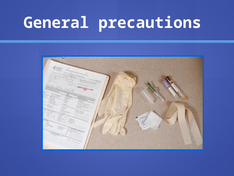
General precautions

General precautions
asks the pt for his/her name (check with the name on the wrist band)
specimen containers should be labeled with: patient name hospital or identification number location in the hospital date & time of collection
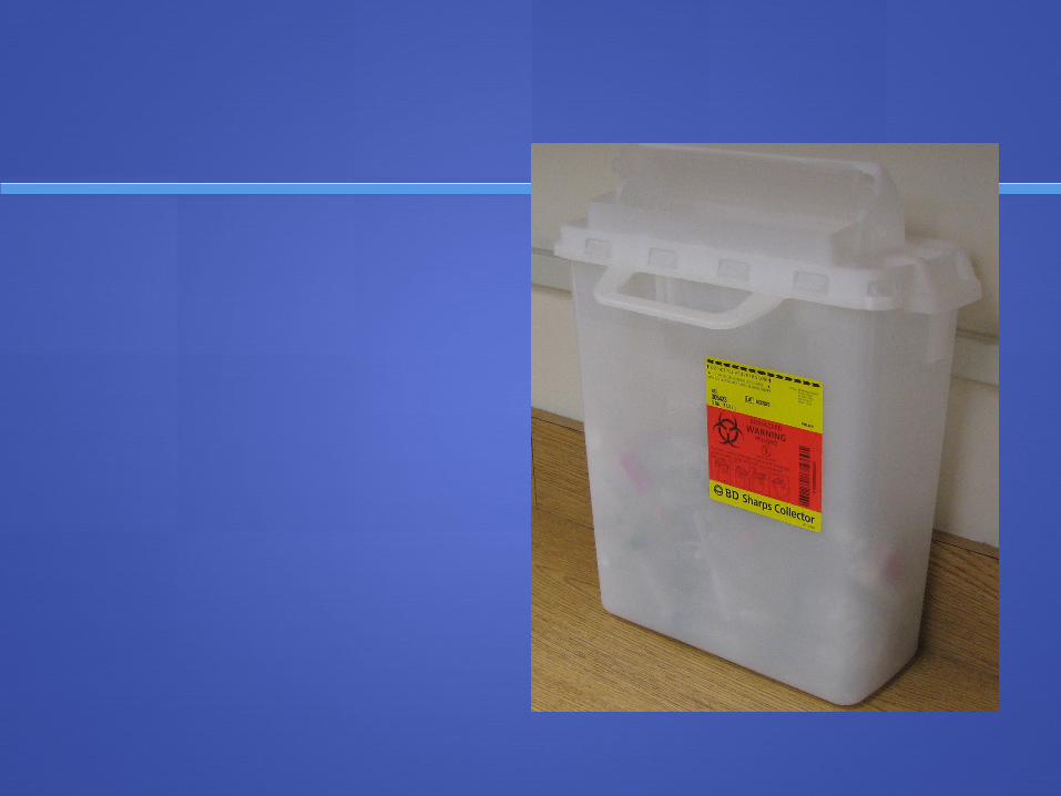
use of disposable latex rubber gloves, especially before collection of any blood specimen.
if the pt has an infectious disease, the phlebotomist should observe universal precautions (wearing of a face mask, glasses or goggles, gloves and gown).
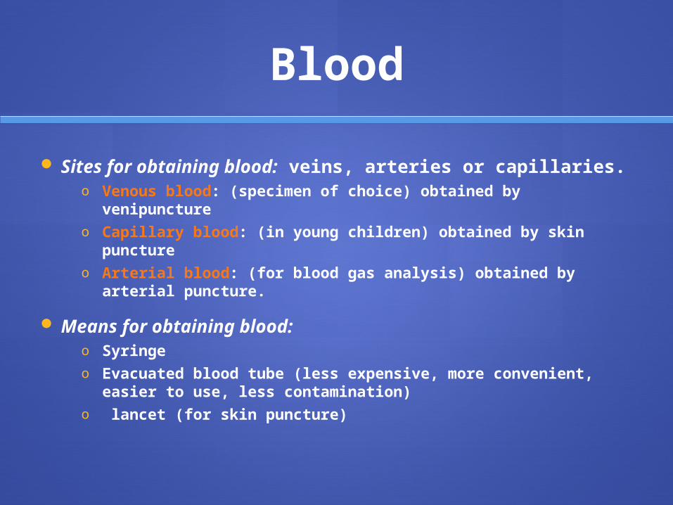
Blood
Sites for obtaining blood: veins, arteries or capillaries.o Venous blood: (specimen of choice) obtained by
venipunctureo Capillary blood: (in young children) obtained by skin
punctureo Arterial blood: (for blood gas analysis) obtained by arterial
puncture.
Means for obtaining blood:o Syringe o Evacuated blood tube (less expensive, more convenient,
easier to use, less contamination) o lancet (for skin puncture)
Precautions for venipuncture
pt should be fasting.
pt should be comfortably seated (or in supine position for at least 20 min before sampling)
pt arm should be extended (straight line from shoulder to the wrist)
The arm chosen for sampling (many pt do not prefer the arm with which they write).
In severely ill pt or multiple IV inj (eg: chemotherapy), keep the good veins for treatment
-Avoid the use of an arm with : inserted IV line. cannula or IV fistula (without physician consent). extensive scarring or hematoma at the intended site of venipuncture. In females who had a mastectomy operation, do not use the arm veins on that
side
If IV fluid infusion is running, close for 3 minute. Take sample from below the infusion site in the same arm or from the opposite arm. A suitable note should be made in the lab report form . This type of specimen is usually suitable for most analytes except those contained in the infused solneg: glucose or electrolytes.
Avoid prolonged tourniquet application (max for 1 min ) or fist pumping as well as stress . Fist pumping increases K , phosphate, lactate and Ca++
Check for the suitable time of specimen collection (diurnal variation of the analyteeg: iron, cortisol – and monitoring of drug therapy eg: digoxin level, prothrombin time).
Estimate the volume of blood needed and select the appropriate number & types of tubes for the plasma or serum tests requested.
An appropriate needle must be selected (the larger the gauge size the smaller the bore-the most commonly used gauge sizes are 19 – 22 & the usual size for an adult with normal veins is size 20. if veins are small & tend to collapse use size 21). All needles must be sterile.

Sites of venipuncture :
median cubital vein : the preferred site in adults, where the vein is both large & close to the skin surface.
Veins on the back of the hand or at the ankle : less preferred & should be avoided in persons with poor circulation (eg : DM).
Technique of venipuncture :
Selection of the area for venipuncture : by palpation.
Clean the area around the venipuncture :
By a prepackaged alcohol swab, or a gauze pad saturated with 70% isopropanol, in a circular motion and from the site outward.
By benzalkonium chloride soln (alcohol free): for alcohol determination.
NEVER by povidone-iodine ( betadine) because of possible interference with several chemistry procedures
Allow the skin to dry in the air (alcohol traces may cause haemolysis and invalid test results).Never touch the skin until venipuncture is completed.
Tourniquet application (max for 1 min): 10-15 cm above the intended puncture site, to obtain the return of venous blood to the heart, and to distend the veins. The pt should not pump fist while tourniquet is in place
Blood collection with syringe : needle is placed firmly over the nozzle of the syringe and the needle cover is
removed the syringe & needle are aligned with the vein to be entered and the needle is pushed into the vein with a 15º angle to the skin. When the initial resistance of the vessel wall during needle piercing is overcome, forward pressure on the syringe is eased and the blood is is withdrawn by gently pulling back the plunger of the syring. After needle removal from the syringe, transfer the drawn blood by gentle ejection into the appropriate tubes. The tubes containing anticoagulants (or additives) are capped & gently mixed (5-10 inversions).
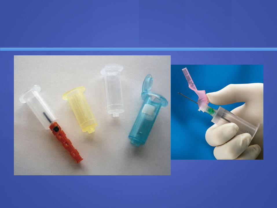
Blood collection with evacuated blood tubes : Before use, the tube should be gently tapped to move any anticoagulant from
the stopper before the needle is inserted into a vein (to preventanticoagulant from entering into the vein).
The needle is screwed into the collection tube holder, and the tube is inserted into this holder.
Technique : After skin cleaning, the needle is guided gently into the vein. Once needle is inside the vein, tube is pressed forward into the holder (to puncture
the the stopper and release the vacuum) As blood begins flowing in the tube, tourniquet is released. The tube is filled until the vacuum is exhausted, and the tube can be moved from the
holder and replaced by another if more blood is needed (this requires a shut off valve, consisting of a rubber tubing that slides over the needle opening inside the tube, to prevent blood spilling during tube exchange)
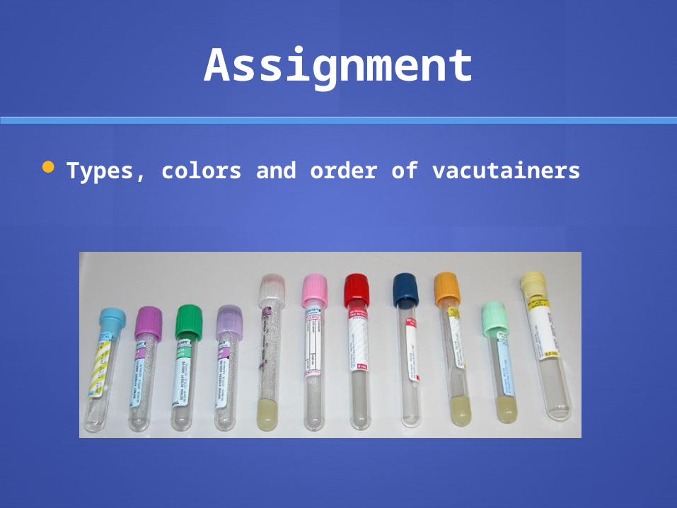
Assignment
Types, colors and order of vacutainers
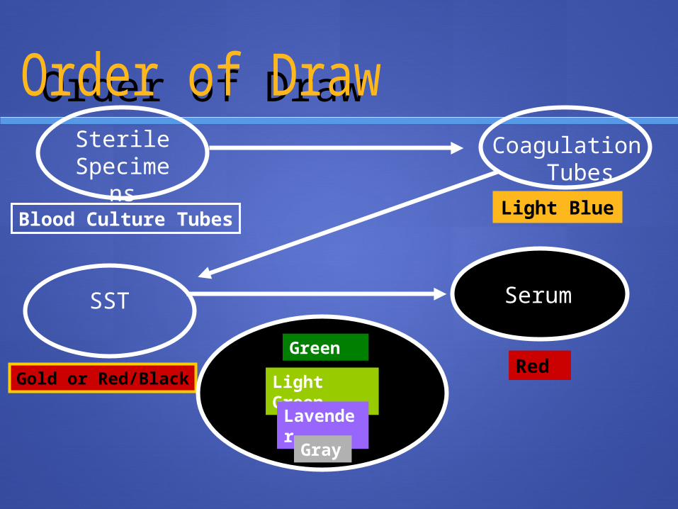
Order of DrawSterile
Specimens
Blood Culture Tubes
Coagulation Tubes
Light Blue
SST
Gold or Red/Black
Serum
RedGreen
Light GreenLavender
Gray
Adv of blood tubes over syringes:
Easy to use – low risk of contamination.
Disadv of blood tubes:
1-collection procedu-Vacuum tends to decrease by time . (check the expiry date before use)
2-You have to draw the amount of blood indicated on tubes with anticoagulants. (check for volume of draw before use).
Properties of blood tubes :
1-Two tyoesof evacuated blood tubes: those with serum separators (SST) – those without separators. In the SST, the disk between the packed cells and the supernatant.
2-Various additives (anticoagulants – preservatives) may be present, indicated by stopper colour.
3-They are also siliconizedto activate the clotting mechanism, reduce adhesion of clots to walls or stoppers & to decrease hemolysis risk.

Skin puncture:
Done when small blood volume is needed (blood glucose estimation by glucocheck- BT & CT)
Sites of skin puncture in: - Adults & grown children: finger tips – ear
lobes - Neonates (up to 1 yr of age): lateral or
medial plantar surface of the heel of the foot
Tech of skin puncture :
1- skin cleaning : a gauze pad sat with 70% isopropanol.
2- leave the alcohol to dry (to avoid hemolysis) and use the lancet to quickly puncture the skin.
3- the finger is held in a way that allows gravity to assist in blood collection
4- avoid finger massage after pricking (to avoid outflow of skin cells and tissue fluid
5- 1st blood drop is wiped away then blood drops are transferred to the collection tubes quickly (to avoid clotting) and without bubbles or froths (to minimize hemolysis) by placing the tube in contact at only a slight angle to the skin surface.
NB : a 3 min warm up of the finger prior to puncture (to improve the circulation) is done by a warm wet wash cloth.
Arterial puncture:
It requires considerable skill (performed by physicians or trained technicians or nurses)
Preferred sites: radial artery (at the wrist) – brachial artery (at the elbow) – femoral artery (in the groin)
Identify the artery by: its thick wall – pulsations
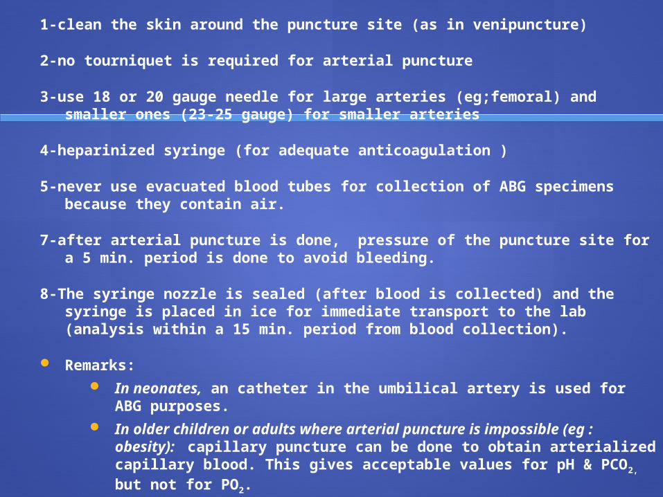
1-clean the skin around the puncture site (as in venipuncture)
2-no tourniquet is required for arterial puncture
3-use 18 or 20 gauge needle for large arteries (eg;femoral) and smaller ones (23-25 gauge) for smaller arteries
4-heparinized syringe (for adequate anticoagulation )
5-never use evacuated blood tubes for collection of ABG specimens because they contain air.
7-after arterial puncture is done, pressure of the puncture site for a 5 min. period is done to avoid bleeding.
8-The syringe nozzle is sealed (after blood is collected) and the syringe is placed in ice for immediate transport to the lab (analysis within a 15 min. period from blood collection).
Remarks: In neonates, an catheter in the umbilical artery is used for ABG
purposes. In older children or adults where arterial puncture is impossible
(eg : obesity): capillary puncture can be done to obtain arterialized capillary blood. This gives acceptable values for pH & PCO2, but not for PO2.
The preferred sites for capillary puncture in : -adults and older children : is the ear lobe -younger children or infants : is the heel
Blood collection from intravenous (IV) lines
IV fluid is shut from the CVP ( central venous pressure) catheter and 10 ml whole blood is aspirated and discarded before the specimen for analysis is drawn.
Blood may be collected from the veins of an arm below an IV linewithout interference from the infused fluid, since the infused fluid must first circulate through the heart and return to the tissues before it reaches the sample site.

Anticoagulants and preservatives for blood:
Whole blood: for analysis of : Blood gases-ammonia-some trace elements-others(glucose- glycatedHb)
Serum: for analysis of most of the analytes, particularly the serological tests (that depend on Ag- Ab reaction)
Plasma obtained with the appropriate anticoagulant: is equally effective as serum
Advantage of plasma over serum: Rapid in emergencies; serum requires a 15-30 min period for clotting
Volume of plasma is always greater than that of the serum.
Disadvantage of plasma: Fibrin clot or fragment formation upon plasma storage and risk of clogging sample
probes of automated analyzers.
Interference of fibrinogen with electrophoretic mobility resulting in confused interpretation of the electrophoretic pattern

Types of anticoagulants
Heparin:
Most widely used for clinical chemical analysis Available in the salt form (Na, K, Mg, Li, NH4): Avoid the Na and K
heparinate analyzed Na & K (use Li heparinate). EDTA (Ethylene Diamine Tetra Acetic Acid):
Uses: hematology as it preserves the cellular composition of blood EDTA salts available are: Disodium, Dipotassium, and Tripotassium
Sodium Fluoride: Uses: preservative for blood glucose NB: without an antiglycolytic agent, blood glucose decreases by
10mg/dL/hr at 25ºC, and the rate is faster in newborns and leukemic pt dt higher WBCs .
Sodium Citrate: Uses: erythrocyte sedimentation rate (ESR) – Prothrombin time (PT) &
activity – Partial thromboplastin time (PTT) & activity. Ratio of sodium citrate to blood: (both must be well mixed together)
• -1:10 for PT and PTT (0.2 mL Na citrate + 1.8 mL whole blood)• -1:5 for ESR (0.4 mL Na citrate + 1.6 mL whole blood)
Oxalates, Iodoacetate
Changes that occur to exposed blood sample
All tests should be done on a fresh blood sample, (less than 1/2 hr at R.T). A preservative could be used to decrease these changes
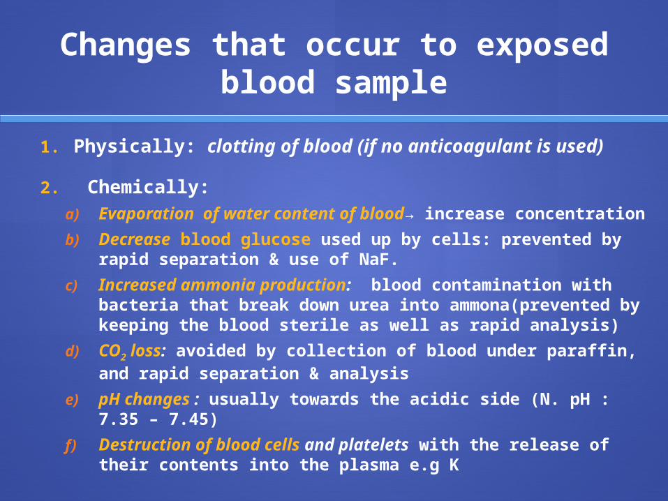
Changes that occur to exposed blood sample
1. Physically: clotting of blood (if no anticoagulant is used)
2. Chemically:a) Evaporation of water content of blood→ increase
concentration
b) Decrease blood glucose used up by cells: prevented by rapid separation & use of NaF.
c) Increased ammonia production: blood contamination with bacteria that break down urea into ammona(prevented by keeping the blood sterile as well as rapid analysis)
d) CO2 loss: avoided by collection of blood under paraffin, and rapid separation & analysis
e) pH changes : usually towards the acidic side (N. pH : 7.35 – 7.45)
f) Destruction of blood cells and platelets with the release of their contents into the plasma e.g K

Hemolysis Breakdown of RBCs with release of their Hb content. Serum shows
visual evidence of haemolysis when Hbconc exceeds 20 mg/dl
Severe haemolysis causes 1-marked increase of analytesthat are present in RBCs at a high
conc.or activity (eg: potassium, magnesium, inorganic phosphates, LDH, AST)
2-interference with some analytes in plasma (eg: Total CK & CK-MB activities)
Precautions to avoid haemolysis:1-use of wide bore needles
2-use the appropriate amounts of anticoagulants (excess heparin causes hemolysis)
3-apply minimum amount of constriction to the arm
4-allow smooth slow blood flow to the syringe to prevent froths
6-allow blood flow easily and slowly on the walls of the tube
7-gentle mixing of the blood with the anticoagulant
8-centrifugation of the blood at a medium speed in order to obtain serum or plasma

URINE
Types of urine specimens to be collected from adults: (depending on the tests to be done)
I-Untimed (Random) specimen Uses : qualitative purposes or semiquantitative purposes Time of the specimen : clean (midstream), early
morning, fasting specimen is preferred because : Constant urine composition. Most concentrated form (useful for microscopic
examination & detection of abnormal amounts of urinary constituents eg: proteins, or of unusual compounds eg:HCG).
Midstream urine is preferred as the first stream is used for washing the urethra.
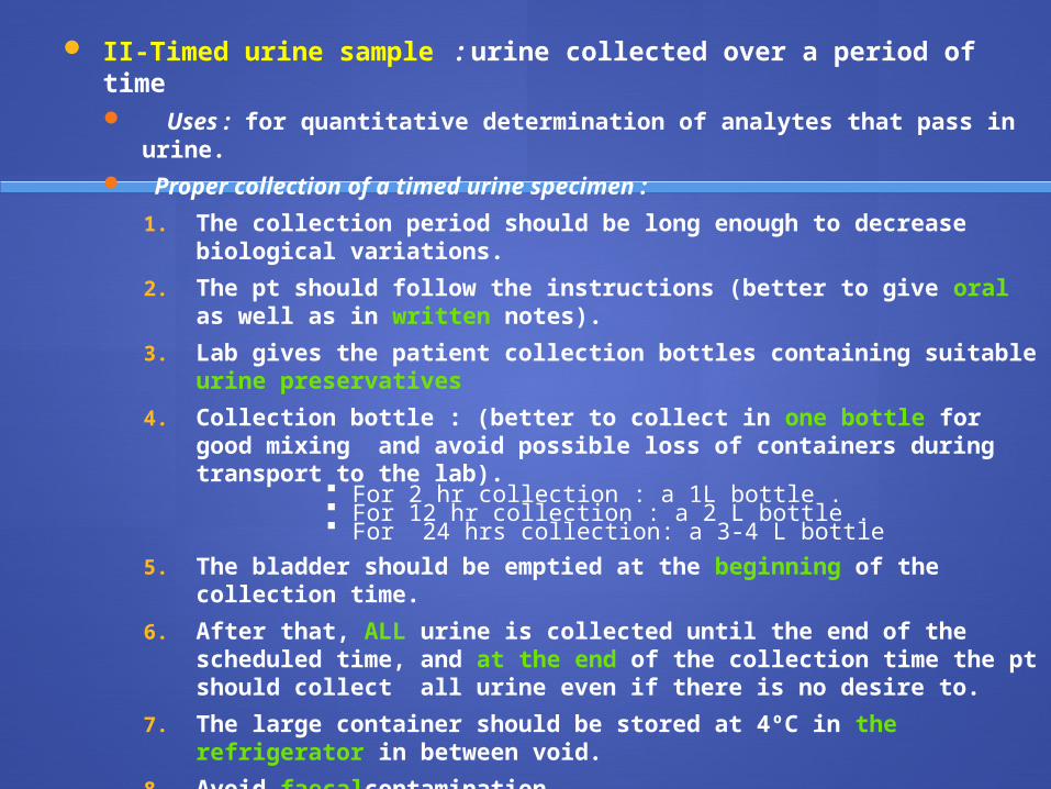
II-Timed urine sample : urine collected over a period of time Uses : for quantitative determination of analytes that pass in
urine. Proper collection of a timed urine specimen :
1. The collection period should be long enough to decrease biological variations.
2. The pt should follow the instructions (better to give oral as well as in written notes).
3. Lab gives the patient collection bottles containing suitable urine preservatives
4. Collection bottle : (better to collect in one bottle for good mixing and avoid possible loss of containers during transport to the lab).
For 2 hr collection : a 1L bottle . For 12 hr collection : a 2 L bottle . For 24 hrs collection: a 3-4 L bottle
5. The bladder should be emptied at the beginning of the collection time.
6. After that, ALL urine is collected until the end of the scheduled time, and at the end of the collection time the pt should collect all urine even if there is no desire to.
7. The large container should be stored at 4ºC in the refrigerator in between void.
8. Avoid faecalcontamination.
9. It is better to pass the urine into a small container at each voiding, then empty it into big container. This protects the patient from splashing himself with the urine preservative in the large container, particularly acids.
10. The whole sample should be mixed together and total volume recorded. Then aliquots for microscopic examination and analysis are prepared.
Urine collection from children :
- It is very difficult to collect urine
-Cleaning of the scrotal or perineal area, and remove skin oils.
-For untimed (random) specimens ; a urine bag is placed around the infant genitalia
Urine Preservatives
Function of preservatives : 1. reduce bacterial action.
2. reduce chemical decomposition.
3. solubilize constituents that might precipitate.
4. decrease oxidation or evaporation
Types of preservation : 1-physical
a) Refrigeration (2-8ºC):
b) Freezing (-20ºC) : done for small aliquots of timed urine specimens if quantitative analysis is going to be delayed.
2-chemical :c) Acidification of urine : (below pH of 3) used for 24 hrs urinary
calcium, Example : HCl 6 mol/L : 10 mL/24hrs urine, sulfamic acid : 10 gm/L, boric acid : 5 mg/30 mL
d) Urinary preservative tablets : They can be used for chemical & microscopic examination, but not for Na & K analysis.
e) Others : Formalin, Thymol and chloroform , Toluene, S odium bicarbonate (mild basic salt) or NaOH (small amount)

Changes that occur to urine on keeping :
It is better to examine a freshly voided urine as soon as possible:
I- Physical :1. colour changes : dt breakdown of the pigments
2. odour changes : as a result of bacterial growth and decomposition
3. aspect changes (turbidity) : as a result of bacterial multiplication or crystal formation
4. pH changes : low pH : as a result of increased pus cells
High pH: as a result of bacteria → ammonia production
II- Chemical :1. glucose : decrease consumption by bacteri
2. ketone bodies : decrease due to breakdown and highly volatile.
3. bilirubin&urobilinogen ; decrease due todestruction by light.
4. nitrites : False +ve: as a result of nitrite producing bacteria. False –ve: as a result of nitrite conversion to nitrogen gas.
III- Microscopic :1. 1-cells and casts : decompose
2. 2-crystals formation : especially in cold weather and in acidic pH (eg : urates, oxalates).

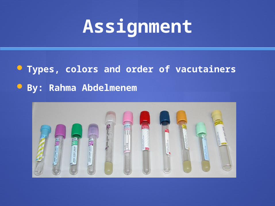
Assignment
Types, colors and order of vacutainers
By: Rahma Abdelmenem

Apply your KnowledgeStudy Questions
Alavender-topped tube is primarily used for what types of studies?
ANSWER: Hematology studies (CBC and differential)

Which color tube is used for glucose analysis and blood alcohol levels?
ANSWER: Gray-topped tubes

Which tube system is the most widely used for blood collection?
ANSWER: Evacuated tube system

When is skin puncture used ?
ANSWER: Small amount of blood is needed

THANK YOU
Related Documents


