Specific Targeting to B Cells by Lipid-Based Nanoparticles Conjugated with a Novel CD22- ScFv Kristin Loomis, BS & , Brandon Smith, BA, Yang Feng, PhD, Himanshu Garg, PhD # , Amichai Yavlovich, PhD, Ryan Campbell-Massa, BS, Dimiter S. Dimitrov, PhD, Robert Blumenthal, PhD, Xiaodong Xiao, PhD * , and Anu Puri, PhD * Center for Cancer Research Nanobiology Program, National Cancer Institute at Frederick National Institutes of Health, Frederick, MD Abstract The CD22 antigen is a viable target for therapeutic intervention for B-cell lymphomas. Several therapeutic anti-CD22 antibodies as well as an anti-CD22-based immunotoxin (HA22) are currently under investigation in clinical settings. Coupling of anti-CD22 reagents with a nano-drug delivery vehicle is projected to significantly improve treatment efficacies. Therefore, we generated a mutant of the targeting segment of HA22 (a CD22 scFv) to increase its soluble expression (mut-HA22), and conjugated it to the surface of sonicated liposomes to generate immunoliposomes (mut-HA22- liposomes). We examined liposome binding and uptake by CD22 + B-lymphocytes (BJAB) by using calcein and/or rhodamine PE-labeled liposomes. We also tested the effect of targeting on cellular toxicity with doxorubicin-loaded liposomes. We report that: (i) Binding of mut-HA22-liposomes to BJAB cells was significantly greater than liposomes not conjugated with mut-HA22 (control liposomes), and mut-HA22-liposomes bind to and are taken in by BJAB cells in a dose and temperature-dependent manner, respectively; (ii) This binding occurred via the interaction with the cellular CD22 as pre-incubation of the cells with mut-HA22 blocked subsequent liposome binding; (iii) Intracellular localization of mut-HA22-liposomes at 37°C but not at 4°C indicated that our targeted liposomes were taken up through an energy dependent process via receptor-mediated endocytosis; and (iv) Mut-HA22-liposomes loaded with doxorubicin exhibited at least 2-3 fold more accumulation of doxorubicin in BJAB cells as compared to control liposomes. Moreover, these liposomes showed at least a 2-4 fold enhanced killing of BJAB or Raji cells (CD22 + ), but not SUP- T1 cells (CD22 - ). Taken together these data suggest that these 2 nd -generation liposomes may serve as promising carriers for targeted drug delivery to treat patients suffering from B-cell lymphoma. Keywords Liposomes; targeting; B-cell Lymphoma; anti-CD22 ScFv; drug delivery *Correspondence to: Anu Puri Bldg 469/Rm 216A, P.O. Box B , Miller Drive CCR Nanobiology Program , National Cancer Institute at Frederick National Institutes of Health, Frederick, MD 21702-1201 Phone 301-846-5069; Fax: 301-846-6210 [email protected] Xiaodong Xiao Bldg 469/Rm 139, P.O. Box B , Miller Drive CCR Nanobiology Program National Cancer Institute at Frederick National Institutes of Health Frederick, MD 21702-1201 Phone: 301-846-5628; Fax: 301-846-6189 [email protected]. # Current address Center of Excellence for Infectious Disease Texas Tech University Health Sciences Center 5001 El Paso Dr El Paso TX 79905 & Department of Biomedical Engineering at Georgia Tech and Emory University Georgia Institute of Technology, Atlanta, Georgia 30332 Publisher's Disclaimer: This is a PDF file of an unedited manuscript that has been accepted for publication. As a service to our customers we are providing this early version of the manuscript. The manuscript will undergo copyediting, typesetting, and review of the resulting proof before it is published in its final citable form. Please note that during the production process errors may be discovered which could affect the content, and all legal disclaimers that apply to the journal pertain. NIH Public Access Author Manuscript Exp Mol Pathol. Author manuscript; available in PMC 2011 April 1. Published in final edited form as: Exp Mol Pathol. 2010 April ; 88(2): 238–249. doi:10.1016/j.yexmp.2010.01.006. NIH-PA Author Manuscript NIH-PA Author Manuscript NIH-PA Author Manuscript

Welcome message from author
This document is posted to help you gain knowledge. Please leave a comment to let me know what you think about it! Share it to your friends and learn new things together.
Transcript

Specific Targeting to B Cells by Lipid-Based NanoparticlesConjugated with a Novel CD22- ScFv
Kristin Loomis, BS&, Brandon Smith, BA, Yang Feng, PhD, Himanshu Garg, PhD#, AmichaiYavlovich, PhD, Ryan Campbell-Massa, BS, Dimiter S. Dimitrov, PhD, Robert Blumenthal,PhD, Xiaodong Xiao, PhD*, and Anu Puri, PhD*Center for Cancer Research Nanobiology Program, National Cancer Institute at Frederick NationalInstitutes of Health, Frederick, MD
AbstractThe CD22 antigen is a viable target for therapeutic intervention for B-cell lymphomas. Severaltherapeutic anti-CD22 antibodies as well as an anti-CD22-based immunotoxin (HA22) are currentlyunder investigation in clinical settings. Coupling of anti-CD22 reagents with a nano-drug deliveryvehicle is projected to significantly improve treatment efficacies. Therefore, we generated a mutantof the targeting segment of HA22 (a CD22 scFv) to increase its soluble expression (mut-HA22), andconjugated it to the surface of sonicated liposomes to generate immunoliposomes (mut-HA22-liposomes). We examined liposome binding and uptake by CD22+ B-lymphocytes (BJAB) by usingcalcein and/or rhodamine PE-labeled liposomes. We also tested the effect of targeting on cellulartoxicity with doxorubicin-loaded liposomes. We report that: (i) Binding of mut-HA22-liposomes toBJAB cells was significantly greater than liposomes not conjugated with mut-HA22 (controlliposomes), and mut-HA22-liposomes bind to and are taken in by BJAB cells in a dose andtemperature-dependent manner, respectively; (ii) This binding occurred via the interaction with thecellular CD22 as pre-incubation of the cells with mut-HA22 blocked subsequent liposome binding;(iii) Intracellular localization of mut-HA22-liposomes at 37°C but not at 4°C indicated that ourtargeted liposomes were taken up through an energy dependent process via receptor-mediatedendocytosis; and (iv) Mut-HA22-liposomes loaded with doxorubicin exhibited at least 2-3 fold moreaccumulation of doxorubicin in BJAB cells as compared to control liposomes. Moreover, theseliposomes showed at least a 2-4 fold enhanced killing of BJAB or Raji cells (CD22+), but not SUP-T1 cells (CD22-). Taken together these data suggest that these 2nd-generation liposomes may serveas promising carriers for targeted drug delivery to treat patients suffering from B-cell lymphoma.
KeywordsLiposomes; targeting; B-cell Lymphoma; anti-CD22 ScFv; drug delivery
*Correspondence to: Anu Puri Bldg 469/Rm 216A, P.O. Box B, Miller Drive CCR Nanobiology Program, National Cancer Instituteat Frederick National Institutes of Health, Frederick, MD 21702-1201 Phone 301-846-5069; Fax: 301-846-6210 [email protected] Xiao Bldg 469/Rm 139, P.O. Box B, Miller Drive CCR Nanobiology Program National Cancer Institute at Frederick NationalInstitutes of Health Frederick, MD 21702-1201 Phone: 301-846-5628; Fax: 301-846-6189 [email protected].#Current address Center of Excellence for Infectious Disease Texas Tech University Health Sciences Center 5001 El Paso Dr El PasoTX 79905&Department of Biomedical Engineering at Georgia Tech and Emory University Georgia Institute of Technology, Atlanta, Georgia30332Publisher's Disclaimer: This is a PDF file of an unedited manuscript that has been accepted for publication. As a service to our customerswe are providing this early version of the manuscript. The manuscript will undergo copyediting, typesetting, and review of the resultingproof before it is published in its final citable form. Please note that during the production process errors may be discovered which couldaffect the content, and all legal disclaimers that apply to the journal pertain.
NIH Public AccessAuthor ManuscriptExp Mol Pathol. Author manuscript; available in PMC 2011 April 1.
Published in final edited form as:Exp Mol Pathol. 2010 April ; 88(2): 238–249. doi:10.1016/j.yexmp.2010.01.006.
NIH
-PA Author Manuscript
NIH
-PA Author Manuscript
NIH
-PA Author Manuscript

IntroductionCD22, a B-cell specific surface molecule is a member of the Ig super-family (1-3), and is knownto be upregulated in non-Hodgkin's lymphoma (NHL) and other types of B-cell lymphomas(4;5). The transmembrane protein CD22 is a well-characterized molecule that rapidlyinternalizes upon antibody binding; the structure-function relationship of CD22 with the BCRcomplex has been investigated in detail (2;6). To treat B-cell lymphomas/malignancies,multiple approaches including CD22-specific antibodies alone (7-14), or as bioconjugates(15;16), as well as combination-immunotherapy have been developed (17-19). Among these,recombinant humanized anti-CD22 monoclonal antibodies, epratuzumab, inotuzumab andozogamicin (18) have been extensively studied, and epratuzumab is currently under clinicaltrials for NHL. Another promising treatment approach includes use of a recombinantimmunotoxin containing an anti-CD22 (Fv) fused to truncated Pseudomonas exotoxin A(HA22) (20-23). We aimed at improving the efficacy of an anti-CD22 targeted therapy byconjugating a novel CD22 specific scFv to a nano-drug delivery carrier.
Antibody-coated liposomes (immunoliposomes) have been explored for site-specific targetingof drugs and therapeutics for cancer treatment (24;25). However, in vivo success ofimmunoliposomes is subject to the availability of suitable targeting antibody molecules (thatwill trigger rapid receptor internalization) as well as formulations amenable to tunable drugrelease for cytosolic or intratumoral delivery. Various antibodies (mainly mAb-IgG) andantibody fragments (including F(ab’)2, Fab or scFvs) have been investigated forimmunoliposome studies (for a detailed review, see (24)). For example, liposomes conjugatedwith CD19 and CD20 mAbs have been examined for B-cell targeting (26;27). Among antibodyfragments, scFvs (being small in size) bear promise as targeting ligands for developing 2nd
generation immunoliposomes. To date, HER2 scFv (28;29)-conjugated liposomes for drugdelivery, and anti-TfR scFv-lipoplexes for gene delivery have been successfully developed(30;31).
Besides the use of antibodies and/or their fragments, selected cytokines (e.g. the B cellactivating factor belonging to the TNF family, mBAFF) have also been used as ligands fordelivery of liposomal drugs to B-cell lymphomas (32). A recent study by O'Donnell andcolleagues demonstrated that immunoliposomes bearing an anti-CD22 mAb (HB22.7) resultedin significantly enhanced cytotoxicity of CD22+ cells by liposome-entrapped doxorubicin, ananti-cancer drug (33). Although, mAb-coated immunoliposomes may serve as suitable vehiclesfor delivery of cancer therapeutics, the Fc domain-mediated immune responses may limit theirfuture clinical applications (34;35). Therefore, immunoliposomes coated with anti-HER2scFvs have been developed as 2nd-generation immunoliposomes (28;29;36). Previous studiesby de,Kruiff and colleagues have reported immunoliposomes to target B-cell lymphoma (37),and the biosynthetically lipid-tagged CD22 ScFv was generated using a semi-synthetic humanlibrary with potentially high affinity (38). This lipidated ScFv was incorporated into theliposomes by a detergent solubilization protocol. Since detergent solubilization method poseslimitations to encapsulate payload of anticancer agents in the aqueous compartment ofliposomes, alternate approaches are warranted to develop immunoliposomes for B-celltargeting.
This study was designed to generate immunoliposomes bearing high affinity anti-CD22 scFvfor targeted drug delivery to B-cell lymphomas. An improved anti-CD22 scFv molecule (mut-HA22, M.W. 34 kDa) was developed and cysteine residues were introduced at the C-terminusfor coupling to liposomes. Mut-HA22 bears the advantage as its parental form (HA22) hasbeen well-studied and has been examined in clinical trials in the format of immunotoxin, whereno inhibitory immune responses were reported (8). Mut-HA22 conjugation to the surface ofpreformed liposomes (loaded with calcein (as a model solute), or an anticancer drug,
Loomis et al. Page 2
Exp Mol Pathol. Author manuscript; available in PMC 2011 April 1.
NIH
-PA Author Manuscript
NIH
-PA Author Manuscript
NIH
-PA Author Manuscript

doxorubicin (DOX) in the aqueous compartment) is attained by malemide-cysteine chemistry,rendering such formulations suitable for future in vivo applications.
The observation that mut-HA22 specifically binds to cell-surface expressed CD22 renders it asuitable targeting ligand. Mut-HA22-liposomes bound to CD22+ BJAB cells in a dose-dependent manner, and were accumulated in these cells at 37°C. Our results on target-specificdelivery of DOX by mut-HA22 liposomes, and their improved cytotoxicity further provideevidence for their projected application as drug carriers.
MethodsReagents and Cell Lines
Tributylphosphine and Biogel A-0.5m were from Bio-Rad (Hercules, CA); all other reagentswere from Sigma-Aldrich Inc. (St. Louis, MO) and of analytical grade. The CD22+ human B-lymphocyte cell line, BJAB (ATCC# HB-136) and CD22- human T-lymphocyte cell linesJurkat (ATCC# CRL-10915) and SUP-T1, (ATCC# CRL-1942) were maintained in RPMI1640 media supplemented with 10% heat-inactivated fetal bovine serum (FBS) and antibiotics(Invitrogen, Carlsbad, CA). Doxorubicin-hydrochloride (DOX-HCl) (Bedford Laboratories,Bedford, OH) and Doxil (Ben Venue Laboratories, Bedford, OH) were obtained through theNIH Pharmacy, Clinical Center, Bethesda, MD.
Lipids1,2-Dipalmitoyl-sn-Glycero-3-Phosphocholine (DPPC), 1,2-Distearoyl-sn-Glycero-3-Phosphoethanolamine-N-[Maleimide(Polyethylene Glycol)2000] (DSPE-PEG2000-Maleimide), and 1,2-Dioleoyl-sn-Glycero-3-Phosphoethanolamine-N-(Lissamine RhodamineB Sulfonyl) (Lissamine Rhodamine PE) (ex/em 557/571) were purchased from Avanti PolarLipids, Inc. (Alabaster, AL). Lipid purity was routinely verified by thin layer chromatographywith a silica gel stationary phase and a 65:25:4 chloroform:methanol:water (by volume) mobilephase (39). Molybdenum Blue Spray was used to visualize phospholipids (40) and otherpossible organic contaminants were visualized by placing developed TLC plates in an iodinechamber.
Mutated HA22 scFv (mut-HA22)HA22 is an improved version of the immunotoxin BL22 with an improved affinity of the CD22targeting moiety, the scFv portion (Xiao and Dimitrov, unpublished). In an attempt to furtherincrease the affinity, we generated a mutational phage library based on HA22 scFv. In thislibrary, each clone carried totally random residues on two adjacent positions in the CDR3region. This phage library was used for panning against purified CD22-Fc (kindly provided byIra Pastan). For conjugation purposes, site-directed mutagenesis was performed using theQuickChange Site-Directed Mutagenesis Kit (Stratagene, La Jolla, CA) to introduce a cysteineresidue immediately before the stop codon of the selected HA22 scFv mutant.
Mut-HA22 binding to CD22+ cells by FACS AnalysisCD22 expression was confirmed by indirect immunostaining using the mut-HA22 and acommercially available anti-CD22 mouse IgG (MYG13 (raised against Raji cells of humanorigin, Santa Cruz Biotechnology, Inc, Santa Cruz, CA). BJAB (CD22+), SUP-T1 (CD22-), orJurkat (CD22-) cells were incubated for 15 minutes at room temperature in PBS (containing5% each of mouse serum, fetal bovine serum, goat serum, and human serum) to blocknonspecific binding sites on the cell surface. The cells were resuspended to 107 cells/mL PBSsupplemented with 5% FBS (PBS-FBS).
Loomis et al. Page 3
Exp Mol Pathol. Author manuscript; available in PMC 2011 April 1.
NIH
-PA Author Manuscript
NIH
-PA Author Manuscript
NIH
-PA Author Manuscript

To stain CD22 using mut-HA22, 2 μg of the scFv was added to cells and incubated for 30minutes in the dark at 4°C. The cells were then washed twice with cold PBS supplementedwith 1% BSA (PBS-BSA), resuspended to 107 cells/mL PBS-FBS, and incubated with 1 μgmouse anti-penta-histidine IgG (QIAGEN, Valencia, CA) for 30 minutes in the dark at 4°C.The cells were then washed twice with cold PBS-BSA, resuspended to 107 cells/mL PBS-FBS,and incubated with a 1:32 dilution of FITC-anti-mouse IgG (Sigma, Saint Louis, MO) for 30minutes in the dark at 4°C.
To stain using MYG13, 2 μg of the IgG was added to the cells and incubated for 30 minutesin the dark at 4°C. The cells were then washed twice with cold PBS-BSA, resuspended to107cells/mL PBS-FBS, and incubated with a 1:32 dilution of the FITC-anti-mouse IgG for 30minutes in the dark at 4°C. All cells were washed twice with cold PBS-BSA. Fluorescence ofthe samples was analyzed using a FACS caliber flow cytometer (Becton Dickinson, San Jose,CA), analyzing 10,000 viable cells per sample.
Preparation of Sonicated Unilamellar LiposomesLiposomes were prepared by probe sonication as described (41). Lipids (in chloroform) weremixed at desired molar ratios according to Table 1 in a glass tube, dried under streamingnitrogen to create a film, then placed in a vacuum desiccator overnight. In some preparations,rhodamine-PE (0.1mol %) was also included to monitor binding to the cells (see below).
For calcein loaded liposomes, lipid films were reconstituted in HBSE buffer (10mM HEPES,150mM NaCl, 9.1 mM EDTA, pH 7.4) containing 50 mM calcein by vortexing and heating at42°C. Multilamellar vesicles (MLVs) were sonicated in a room temperature water bath with aprobe sonicator (W-373 Heat Systems-Ultrasonics, NY) for 6 cycles of 1 minute of sonicationfollowed by 1 minute of rest. Titanium particles were removed by centrifugation (2000 xg, 5minutes). Un-encapsulated calcein was removed from liposome-entrapped calcein onSepharose CL-6B column (41). Liposome-rich fractions were pooled together and used forconjugation with mut-HA22. Inorganic phosphorus determination was used to determine theconcentration of phospholipids in the liposomes (42).
For DOX-loaded liposomes, the ammonium sulfate gradient loading method was used, aspreviously described (43). Briefly, lipid films were reconstituted in 300 mM ammonium sulfatebuffer at pH 7.4 with alternating periods of vortexing and incubation at 41°C. Sonicatedliposomes were prepared as above, and were run on a PD-10 column, (GE Healthcare LifeSciences, Piscataway, NJ), pre-equilibrated with HBS pH 7.4, to exchange the external buffer.Liposome-rich fractions were pooled, immediately incubated with DOX at the ratio of 20mgliposomal lipids to 0.5 mg DOX, and incubated for 12-16 hours at room temperature to allowfor DOX encapsulation. The DOX-loaded liposomes were separated from un-encapsulateddrug by elution on a PD-10 column equilibrated with HBSE (pH 7.4). Fractions containingliposomes were pooled together and used for conjugation with the mut-HA22 scFv. Lipidcontent was determined as above. Extent of DOX loading was assayed by monitoring DOXfluorescence (in the presence of 0.5% Triton X-100 w/v) at 485/585 nm (ex/em) using a 96-well plate reader (SpectraMax M2e, Molecular Devices, Sunnyvale, CA). Knownconcentrations of DOX or Doxil (in a range of 0.2-2.0 μg per well) using 0.2 mL total volumewere used to generate a calibration curve using the PBS+Triton X-100. Liposomal DOX wasquantitated using the DOXIL standard curve.
Conjugation of mut-HA22 to the liposome surface (mut-HA22-liposomes)The mut-HA22 scFv was reduced by incubation with tributylphosphine (2mM in HBSE buffer)under flowing argon for 4 hours. The reduced antibody was immediately added to either calceinor DOX-loaded sonicated liposomes from above. Typically, we used a ratio of 200 μg scFv
Loomis et al. Page 4
Exp Mol Pathol. Author manuscript; available in PMC 2011 April 1.
NIH
-PA Author Manuscript
NIH
-PA Author Manuscript
NIH
-PA Author Manuscript

for 10-20mg of phospholipids. The scFv-liposome mixture was incubated in the dark forapproximately 16 hours at room temperature, sealed under argon. To block free maleimidegroups, liposomes were then incubated with L-cysteine (1 mM final concentration) for 15minutes. The resulting mut-HA22 liposomes were separated from any unconjugated antibodyand L-cysteine using size exclusion chromatography (Sepharose CL-6B, GE Healthcare LifeSciences, Piscataway, NJ ), pre-equilibrated with HBSE buffer (pH 7.4). Liposome-richfractions, detected continuously by a UV detector, were pooled, and concentrated withCentriplus Centrifugal Filter Devices (regenerated cellulose filter, MWCO 30 kDa, Millipore,Bedford, MA). Antibody conjugation to the liposomes was confirmed by gel electrophoresis.Samples were loaded in a 4-12% Bis-Tris gel (NuPage) and allowed to run in MES/SDSrunning buffer at 200V for 35 minutes under reducing conditions. To visualize the DSPE-PEG2000-maleimide-mut-HA22 conjugate, the gel was stained with Microwave Blue (Protiga,Frederick, MD). To quantify conjugated mut-HA22, known concentrations of reduced freemut-HA22 were run on the same gel and stained to create a calibration curve. Odyssey infraredimaging system software (LI-COR Biosciences, Lincoln, NE) was used to calculate the amountof protein based on infrared intensity using a reciprocal fit curve. The distribution of liposomalhydrodynamic size was determined by dynamic light scattering measurements (Zetasizer,MALVERN Instruments, Worcestershire, UK) (41).
Calcein leakage from liposomes was determined by incubation of liposomes in PBSsupplemented with 0, 10 or 50% heat-inactivated FBS as described (41). Briefly, liposomeswere diluted in the buffer at 1:10 ratios and incubated at 37°C for 0-24 hours. Calcein leakagewas determined as described (41).
Analysis of the number of mut-HA22 Molecules bound to LiposomesThe following parameters were used to calculate the average number of mut-HA22 moleculesper liposome (equation below): (a) mut-HA22/mL liposome solution (14.67 μg CD22/mL);(b) Phospholipids/ mL liposome solution (0.317 mg phospholipids/mL); (c) Liposomehydrodynamic diameter (110 nm) and (d) Number of phospholipids/liposome forapproximately 110 nm liposomes (9×10⁴) (44).
(*The mut-HA22 molecules per liposome were calculated based on 100% DPPC). Therefore,the average number of mut-HA22 molecules per liposome was 90. For 1 mole% DSPE-PEG2000-MaL in the liposomes, the average number of mut-HA22 molecules per liposomewas 47.
Mut-HA22-Liposome/Cell Interaction AssaysBinding of fluorescently labeled mut-HA22 liposomes to BJAB or SUP-T1 cells was done asfollows: In a typical experiment, 107 cells/mL PBS-FBS were incubated with 0-2 μg ofliposome-conjugated mut-HA22 (or the equivalent amount of liposomal phospholipids forcontrol liposomes) for 20-40 minutes at 4°C or 37°C. The exact conditions for individualexperiments are described in figure legends. Cells were washed twice with cold PBS in 1%bovine serum albumin (PBS-BSA) and were analyzed for immunofluorescence either bymicroscopy or FACS analyzing 10,000 viable cells per sample.
Loomis et al. Page 5
Exp Mol Pathol. Author manuscript; available in PMC 2011 April 1.
NIH
-PA Author Manuscript
NIH
-PA Author Manuscript
NIH
-PA Author Manuscript

Fluorescence MicroscopyTo observe binding/uptake of liposomes, liposome-bound cells were placed in microwells andimages were captured using a NIKON Eclipse TE200 (Melville, NJ) inverted microscopesupplied with a 40X oil lens (N.A. 1.30) and processed as described (45). To further assess theuptake of liposomes, samples were analyzed using confocal microscopy. Z-stack images wereacquired with Olympus FV1000, IX81 inverted laser scanning confocal microscope (OlympusAmerica Inc., Center Valley, PA) equipped with a motorized XYZ stage (Olympus), multiArgon laser (488 nm) for visualizing calcein loaded liposomes. High-resolution 12-bit images(512 X 512 pixels) were acquired using a 60X oil immersion objective lens (Olympus PLAPONNA 1.42) coupled with 4-5 optical zoom, resulting in a lateral resolution of 0.082 microns. Wecaptured 14-16 optical slices at 1.7 micron intervals. All optical sections were captured usingline-based Kalman filter available on the FV1000 microscope. Post acquisitions, the Z-stackswere analyzed using Olympus Fluoview Viewer, version 1.6.a (Olympus America Inc.).
Cell Viability AssaysCells (5 × 10⁵/mL) were incubated in RPMI1640 phenol-red free media with variousconcentrations of DOX-loaded liposomes for one hour at 37°C. Following incubation, cellswere washed with RPMI1640 and plated in triplicates in a 96 well, flat bottom polystyreneplate in a total volume of 100 μL at 5 × 10⁴ cells per well. The incubations were continued foran additional 72 hours at 37°C, 5% CO2. In some samples, cells were incubated with equivalentconcentrations of free DOX or liposomes and the samples were incubated for 72 hours at 37°C without removal of the drug. At the end of incubations, cytotoxicity was determined usinga cell viability kit (Cell Titer Blue assay kit, Promega, Madison, WI). Cell viability wascalculated as percentage of control cells without DOX or liposomal treatment. Emptyliposomes (DPPC:DSPE-PEG2000-Maleimide, 96:4) were also used as controls to determinelipid-mediated non-specific toxicity. Using the previously determined phospholipidconcentrations of liposomal samples, equivalent amounts of empty liposomes were incubatedwith cells in the same manner as described above for DOX-loaded liposomes.
We also determined accumulation of liposomal DOX in the cells following a published protocol(46). Cells (105 per sample) were incubated with liposomes (0- 20 μg liposomal DOX per mL)in eppendorf tubes for 1 hour at 37°C. The cells were washed with culture medium withoutphenol red (0.5 mL×3). The pellets were resuspended in PBS (0.1 mL per sample), andtransferred to 96-well plates. DOX accumulation was determined as above, except the standardcurve was generated in the presence of untreated cells.
Results and Discussion(i) Preparation and Characterization of mut-HA22
Previous studies have described immunoliposomes using a biosynthetically lipid-tagged CD22ScFv, though further characterization of this ScFv (such as ligand-affinity and internalizationproperties) has not been clearly documented. We aimed at developing a CD22 scFv mutantfrom a well-characterized CD22 ScFv (HA22). From the mutational library based on HA22,a mutant with two amino acid changes in the HA22 CDR3 was selected from phage panningthat showed slightly increased binding affinity but significantly increased soluble expression(Xiao and Dimitrov, unpublished data and Figure 1A). A cysteine residue was introduced infront of its stop codon and the resulting clone was named mut-HA22.
We have demonstrated that treatment of mut-HA22 dimers with TBP (a mild reducing agent)or dithiothreitol (DTT) resulted in nearly complete reduction to monomers (see Figure 1A,right panel). TBP-reduced mut-HA22 was used for liposome conjugation (41;47). We alsoexamined the effect of mutations in the CDR3 region of HA22 on its binding to cell-surface
Loomis et al. Page 6
Exp Mol Pathol. Author manuscript; available in PMC 2011 April 1.
NIH
-PA Author Manuscript
NIH
-PA Author Manuscript
NIH
-PA Author Manuscript

expressed CD22. Since mut-HA22 contains a His-tag, an anti-His mouse antibody (+FITC-conjugated mouse Ab) was used to detect binding of mut-HA22 to cells. We show thatincubation of CD22-expressing BJAB cells with mut-HA22 resulted in CD22-specific binding(Figure 1B, top panel, solid grey lines) as compared to incubations without mut-HA22 (i.e.cells incubated only with mouse anti-His+FITC conjugated mouse Ab, Figure 1B, top panel,broken grey lines). We also tested cell-surface expression of CD22 on BJAB cells by using acommercial anti-CD22 mAb, MYG13. The results shown in Figure 1B indicate that mut-HA22bound to CD22 with similar affinity to that of MYG13 (solid black lines MYG13; broken blacklines, FITC-conjugated anti-mouse Ab alone). The background binding with secondaryantibodies to various cells shown in Figure 1B is similar to the data supplied by themanufacturer. Since we did not observe an increase in background fluorescence in CD22- cells(SUP-T1 or Jurkat T lymphocytes) after incubations with mut-HA22 or MYG13, we concludethat mut-HA22 binding to BJAB cells is CD22 antigen specific. Hence, mut-HA22 is likely toserve as a suitable ligand for CD22 targeting.
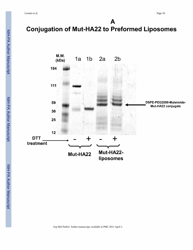
(ii) Mut-HA22- Conjugated LiposomesVarious liposome formulations were prepared according to Table 1. DPPC was used as thematrix lipid and a pegylated lipid (4 mol %) was included for future in vivo applications. DPPCliposomes have been previously demonstrated to release their contents at ≈41°C due to thephase-transition and therefore, are known to have thermosensitive properties (48). Calcein (λex/em 494 nm/517 nm) was encapsulated in the liposomes (50 mM) and some preparationsalso included a fluorescent lipid, rhodamine PE (λ ex/em 557nm/571nm). DSPE-PEG2000-MaL was included to conjugate mut-HA22 via malemide-cysteine chemistry on the surface ofliposomes (mut-HA22-liposomes). Control liposomes (without mut-HA22 conjugation) wereprepared under identical conditions except DSPE-PEG2000 (instead of DSPE- PEG2000-MaL) was included in the liposomes. The antibody conjugation to the liposome surface wasbased on a chemical reaction between maleimide of the lipid with the thiol group on the C-terminus of mut-HA22 (28;49). Conjugation of mut-HA22 to preformed DPPC/DSPE-PEG2000-maleimide liposomes yielded immunoliposomes with 90 mut-HA22 per liposome(41). Initially we examined antibody conjugation with liposomes containing either 1 mol % or4 mol % DSPE-PEG2000-MaL. The liposomes containing 1 mol% DSPE-PEG2000-MaL alsocontained 3 mol% DSPE-PEG2000 (Table 1). We used a molar ratio of 1:41 mut-HA22-PEG2000-MaL for conjugation, considering the number of lipids only in the outer monolayerof liposomes (see Methods section). Initial analysis of mut-HA22-liposome conjugation wasdone by separation of unconjugated antibody from the mut-HA22 conjugated liposomes on asize exclusion column, essentially as described (41).
To confirm mut-HA22 coupling to the liposomes, we examined a molecular weight shift ofmut-HA22 following conjugation with DSPE-PEG2000-MaL by gel electrophoresis. The dataare shown in Figure 2A. We observed a shift in molecular weight of mut-HA22 monomers(Figure 2A, lane 1b) upon conjugation with liposomes (Fig. 2A, lane 2a). DTT pre-treatmentof the conjugated liposomes did not result in any change in the electrophoresis pattern (Fig.2A, lane 2b), confirming that mut-HA22 was covalently coupled to the PEG lipid. We alsoobserved additional bands in the lanes 2a/b. Although the exact nature of additional bandsrequires further characterization, it is likely that availability of additional SH- groups on themut-HA22 may have resulted in coupling of more than one PEG lipid. Control liposomes (lipidcompositions without maleimide functional group) did not show any antibody binding abovebackground levels under identical conditions confirming the specificity of this reaction (datanot shown).
To examine the effect of mut-HA22 conjugation on the physical properties of liposomes,samples were characterized for their size distribution by dynamic light scattering
Loomis et al. Page 7
Exp Mol Pathol. Author manuscript; available in PMC 2011 April 1.
NIH
-PA Author Manuscript
NIH
-PA Author Manuscript
NIH
-PA Author Manuscript

measurements. Data presented in Figure 2B demonstrate that mut-HA22 conjugation to theliposome surface did not have any significant effect on their size distribution. Our liposomesexhibited an average size distribution of 80-100 nm, with an average 90 mut-HA22 moleculesper liposome.
(iii)Liposome Stability studies in SerumFor future medical applications, it is critical that physical properties of mut-HA22-liposomesare not altered upon storage or upon interactions with plasma components prior to targeting topermissive cells and/or tissues. Therefore, we examined stability of liposomes in the presenceof serum. Liposomes were incubated in PBS supplemented with 0-50% heat-inactivated serumat 37°C for various time periods, and extent of calcein release was measured. Results are shownin Figure 2C. An enhancement of calcein release from liposomes was observed with an increasein incubation time and percent serum in the incubation medium. Typically, 5-10% calceinleaked from mut-HA22-liposomes upon incubation in PBS containing 10% FBS for 1-24 hours.Similarly, incubations in the presence of PBS containing 50% serum resulted in 10-30% calceinleakage from mut-HA22 liposomes after 1-24 hour incubations (Figure 2C). Control liposomesshowed similar leakage data (Figure 2C). The results are representative of two independentmeasurements form the same liposome batch. Liposomes could be stored at room temperaturein the dark for up to 5 weeks without any appreciable calcein release or change in their sizedistribution (data not shown).
(iv) Interaction of mut-HA22-liposomes with Receptor Expressing CellsThe preceding section demonstrates that mut-HA22 conjugation to the liposomes occurred viathe chemical reaction between cysteines on the antibody and malemide groups on the PEGlipid of the liposomes. The antibody conjugation had no significant effect on the physicalproperties of liposomes. Therefore, our next set of experiments were designed to evaluate thebinding (and uptake) of these liposomes via mut-HA22 and CD22 interactions. We have usedBJAB (CD22+) and SUP-T1 (CD22-) cells for these studies. Liposome binding and/orinternalization via the CD22 was examined by fluorescence microscopy and by FACS analysis.
(a) Cellular association of mut-HA22-liposomes occurs via the interaction ofcell-surface expressed CD22—To investigate CD22-mediated binding of liposomes,BJAB or SUP-T1 cells were incubated with 2 μg liposome-conjugated mut-HA22/106 cells at37°C for 30 minutes, and liposome binding was determined by fluorescence measurementseither by (i) microscopy or (ii) FACS. The results are presented in Figure 3A (fluorescencemicroscopy images) and Figure 3B-D (FACS). Mut-HA22-liposomes showed significantbinding to BJAB cells (Figure 3A, a-d) based on calcein and rhodamine fluorescence. On theother hand, control liposomes showed only a low level of liposome binding. Interestingly, wedid not see any calcein with control liposomes, whereas there was background binding basedon rho-PE fluorescence. We attribute this discrepancy to calcein quenching within theliposomes. Binding of liposomes with CD22- SUP-T1 cells is shown in Figure 3A (e-h). It isclear that mut-HA22-liposomes did not show enhanced binding compared to control liposomes,corroborating our interpretation that the mut-HA22 binds specifically to the CD22 on the cellsurface. Moreover, in our initial experiments, we also tested liposomes using the lipidcomposition used for mut-HA22-liposomes but without any mut-HA22 conjugation. Theseliposomes did not show any CD22-specific binding above background, confirming CD22-specific interactions of mut-HA22-liposomes (data not shown). Next, we analyzed liposomebinding to cells by FACS analysis. The data presented in Figure 3B, clearly show that mut-HA22-liposomes bind to BJAB cells with high affinity, as compared to that of controlliposomes (Fig. 3B, top panel). In contrast to the microscopy data, we observed a slight increasein binding of mut-HA22-liposomes to SUP-T1 cells as compared to control liposomes (Figure
Loomis et al. Page 8
Exp Mol Pathol. Author manuscript; available in PMC 2011 April 1.
NIH
-PA Author Manuscript
NIH
-PA Author Manuscript
NIH
-PA Author Manuscript

3B, lower panel). Nevertheless, mut-HA22-liposome binding to BJAB cells was significantlygreater than that for SUP-T1 cells.
(b) Binding of mut-HA22-liposomes to BJAB cells is inhibited by free mut-HA22—The results presented in preceding section(s) demonstrate that mut-HA22-liposomes interactwith the CD22-expressing cells. To further establish dependence of the liposome binding viathe CD22, we pre-incubated BJAB cells with varying concentrations of unconjugated mut-HA22 (0-8 μg mut-HA22/106 cells) for 30 minutes. The incubations were performed at 4°Cto prevent internalization of mut-HA22. The unbound antibody was removed and the sampleswere incubated with calcein-loaded mut-HA22-liposomes (1 μg/106 cells) for additional 30minutes at 4°C. The liposome-associated cells were quantified by FACS. The results presentedin Figure 3C clearly demonstrate that pre-incubation of BJAB cells with free antibody blockedthe association with the liposomes. We conclude that binding of the mut-HA22 conjugatedliposomes occurs by specific interactions with cellular CD22.
(v)Effect of Liposomal mut-HA22 Density and Dose-dependence on mut-HA22-liposome-cellbinding
It has been shown earlier that immunoliposomes bear the advantage to present multivalentligand-receptor interactions, as compared to their monovalent counter-parts (50). Furthermore,the average number of antibody molecules per liposome is likely to have a significant effecton liposome binding and/or internalization by receptor-expressing cells (50). As demonstratedin our previous sections, mut-HA22-liposomes interact with CD22-receptor expressing cells.Our next experiments were designed to evaluate the optimal number of mut-HA22 moleculesper liposome and liposome to cell ratios for CD22-specific interactions.
(a) Liposome-cell association increases with an increase in mut-HA22 scFvmolecules per Liposome—In our initial experiments, we investigated the relationshipbetween amount of mut-HA22 conjugation to liposomes and the liposome binding to the targetreceptor. We generated mut-HA22 scFv conjugated liposomes using either 1 mol% (Table 1,Formulation II) or 4 mol% DSPE-PEG2000-MaL (Table 1, Formulation I). The conjugationyields in the two formulations were 47 and 90 scFv molecules/liposome respectively.Liposome-cell suspensions were incubated at 37°C for 30 minutes, and liposome binding wasobserved by monitoring calcein fluorescence by FACS. Data are summarized in Figure 4A,and the values are presented as mean fluorescence intensities. We observed an increase in cell-associated fluorescence with an increasing dosage of liposomes (Formulations I, II) for CD22-expressing BJAB cells. By comparison, a small increase in binding was observed abovebackground when SUP-T1 cells were used for incubations. It is also evident from the datapresented in Figure 4A that liposomes prepared using the 4 mol% DSPE-PEG2000-MaL lipid(Table 1, Formulation I) exhibited an increase in CD22-dependent binding as compared to theliposomes prepared using only 1 mol% DSPE-PEG2000-MaL lipid (Figure 4A). Since ourfuture objective is to develop these liposomes for treatment of B-cell lymphoma, we usedFormulation I (containing 90 scFv molecules per liposome) for further analysis.
(b) Dose-dependent binding of mut-HA22 liposomes—Since the degree ofassociation of mut-HA22-liposomes to BJAB cells was dependent both on the initial input andthe total (average) number of antibody molecules per liposome (Figure 4A), our nextexperiments were designed to evaluate the amount of liposomes needed for optimal bindingto cellular CD22. Mut-HA22-liposomes (0-2 μg liposomal protein) were incubated with BJABcells at 37°C and the extent of liposome binding was monitored by FACS analysis of increasedcell fluorescence (Figure 4B). We observed that binding of liposomes increased up to a ratioof 0-2 μg liposomal protein/106 cells Control liposomes on the other hand did not show anysignificant increase in liposome binding (a. 0 μg, b. 0.1 μg, c. 0.5 μg, d. 1 μg, e. 2μg).
Loomis et al. Page 9
Exp Mol Pathol. Author manuscript; available in PMC 2011 April 1.
NIH
-PA Author Manuscript
NIH
-PA Author Manuscript
NIH
-PA Author Manuscript
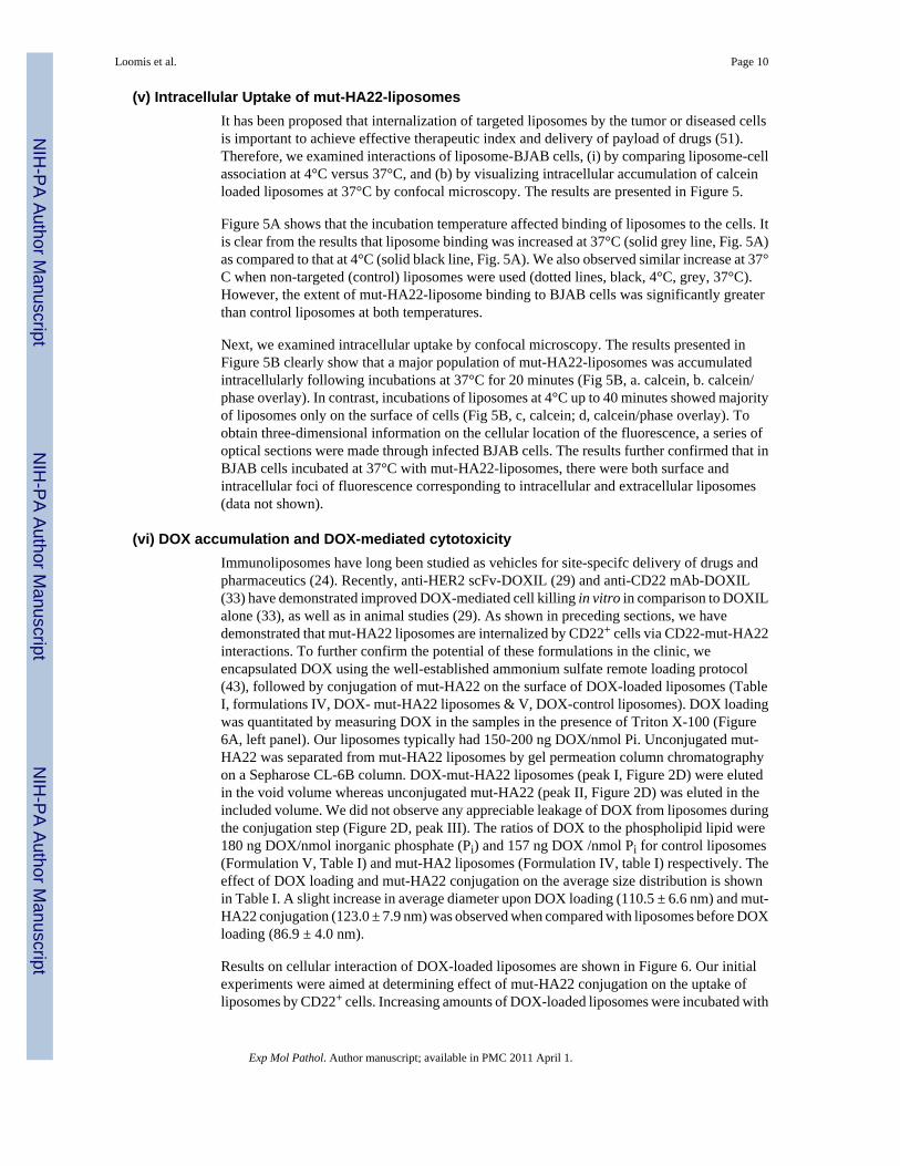
(v) Intracellular Uptake of mut-HA22-liposomesIt has been proposed that internalization of targeted liposomes by the tumor or diseased cellsis important to achieve effective therapeutic index and delivery of payload of drugs (51).Therefore, we examined interactions of liposome-BJAB cells, (i) by comparing liposome-cellassociation at 4°C versus 37°C, and (b) by visualizing intracellular accumulation of calceinloaded liposomes at 37°C by confocal microscopy. The results are presented in Figure 5.
Figure 5A shows that the incubation temperature affected binding of liposomes to the cells. Itis clear from the results that liposome binding was increased at 37°C (solid grey line, Fig. 5A)as compared to that at 4°C (solid black line, Fig. 5A). We also observed similar increase at 37°C when non-targeted (control) liposomes were used (dotted lines, black, 4°C, grey, 37°C).However, the extent of mut-HA22-liposome binding to BJAB cells was significantly greaterthan control liposomes at both temperatures.
Next, we examined intracellular uptake by confocal microscopy. The results presented inFigure 5B clearly show that a major population of mut-HA22-liposomes was accumulatedintracellularly following incubations at 37°C for 20 minutes (Fig 5B, a. calcein, b. calcein/phase overlay). In contrast, incubations of liposomes at 4°C up to 40 minutes showed majorityof liposomes only on the surface of cells (Fig 5B, c, calcein; d, calcein/phase overlay). Toobtain three-dimensional information on the cellular location of the fluorescence, a series ofoptical sections were made through infected BJAB cells. The results further confirmed that inBJAB cells incubated at 37°C with mut-HA22-liposomes, there were both surface andintracellular foci of fluorescence corresponding to intracellular and extracellular liposomes(data not shown).
(vi) DOX accumulation and DOX-mediated cytotoxicityImmunoliposomes have long been studied as vehicles for site-specifc delivery of drugs andpharmaceutics (24). Recently, anti-HER2 scFv-DOXIL (29) and anti-CD22 mAb-DOXIL(33) have demonstrated improved DOX-mediated cell killing in vitro in comparison to DOXILalone (33), as well as in animal studies (29). As shown in preceding sections, we havedemonstrated that mut-HA22 liposomes are internalized by CD22+ cells via CD22-mut-HA22interactions. To further confirm the potential of these formulations in the clinic, weencapsulated DOX using the well-established ammonium sulfate remote loading protocol(43), followed by conjugation of mut-HA22 on the surface of DOX-loaded liposomes (TableI, formulations IV, DOX- mut-HA22 liposomes & V, DOX-control liposomes). DOX loadingwas quantitated by measuring DOX in the samples in the presence of Triton X-100 (Figure6A, left panel). Our liposomes typically had 150-200 ng DOX/nmol Pi. Unconjugated mut-HA22 was separated from mut-HA22 liposomes by gel permeation column chromatographyon a Sepharose CL-6B column. DOX-mut-HA22 liposomes (peak I, Figure 2D) were elutedin the void volume whereas unconjugated mut-HA22 (peak II, Figure 2D) was eluted in theincluded volume. We did not observe any appreciable leakage of DOX from liposomes duringthe conjugation step (Figure 2D, peak III). The ratios of DOX to the phospholipid lipid were180 ng DOX/nmol inorganic phosphate (Pi) and 157 ng DOX /nmol Pi for control liposomes(Formulation V, Table I) and mut-HA2 liposomes (Formulation IV, table I) respectively. Theeffect of DOX loading and mut-HA22 conjugation on the average size distribution is shownin Table I. A slight increase in average diameter upon DOX loading (110.5 ± 6.6 nm) and mut-HA22 conjugation (123.0 ± 7.9 nm) was observed when compared with liposomes before DOXloading (86.9 ± 4.0 nm).
Results on cellular interaction of DOX-loaded liposomes are shown in Figure 6. Our initialexperiments were aimed at determining effect of mut-HA22 conjugation on the uptake ofliposomes by CD22+ cells. Increasing amounts of DOX-loaded liposomes were incubated with
Loomis et al. Page 10
Exp Mol Pathol. Author manuscript; available in PMC 2011 April 1.
NIH
-PA Author Manuscript
NIH
-PA Author Manuscript
NIH
-PA Author Manuscript

Raji cells for 1 hour at 37°C. Subsequently, unbound liposomes were removed and cellularDOX was quantitated in cell lysates (see Methods section). Data presented in Figure 6A (rightpanel) clearly show that conjugation of mut-HA22 to the liposomes resulted in at least a 2 foldincrease in accumulation of DOX in the cells. Moreover, we observed a dose-dependent DOXaccumulation in these samples. In contrast, control liposomes did not show an increase in DOXaccumulation with increasing dose of the liposomes (Figure 6A). A standard curve for DOXquantitation is also shown in Figure 6A (left panel).
Next, we examined the effect of liposomal DOX on cellular viability (Figure 6B&C). First,DOX-mediated cell killing was determined in continuous presence of DOX-loaded liposomesor free DOX. When DOX was encapsulated in liposomes, > 90 % cell killing was observed at10 μg DOX/mL for all the cell lines tested (Figure 6B (a, BJAB, b, SUP-T1), circles, controlliposomes; diamonds, mut-HA22 liposomes). On the other hand, >90% cell killing wasobserved at relatively lower DOX concentrations (1-2 μg free DOX /mL) for BJAB (solidtriangles, Figure 6B,) and Raji cells (squares, Figure 6B,c), consistent with previous studies(33). The only exception was SUP-T1 cells, as those required very high concentrations of freeDOX to achieve maximum cytotoxicity (crosses, Figure 6B, c). It is likely that intracellularDOX from SUP-T1 cells was pumped out of the cells by some membrane transportmechanisms. We also examined non-specific effects mediated by liposomal lipids (unrelatedto DOX-mediated cytotoxicity) by incubating empty liposomes (not loaded with DOX) withcells. We did not observe any cell killing at concentrations equivalent to the amounts ofliposomes used in our cytotoxicity experiments.
Although our initial results clearly demonstrated CD22-specific binding and uptake of mut-HA22 liposomes by fluorescence (Figures 3-5) and liposomal DOX accumulation (Figure 6A),the cell viability results (Figure 6B(a)) did not exhibit any selectivity by mut-HA22 liposomes.We surmise that continuous incubations of liposomes in cell cultures for 72 hours supersededthe kinetics of mut-HA22 liposomes internalization. To mimic in vivo conditions of liposomeuptake by tumors and clearance by reticuloendothelial systems, we measured cell viability for72 hours following removal of unbound liposomes after the first 60 minutes of incubation(29;33). In the previous cell viability experiment (Figure 6B), we observed maximum cellkilling by liposomal DOX at a concentration of 10 μg DOX/mL. Therefore we testedconcentrations of DOX up to 25 μg/mL in these experiments. Enhanced cytotoxicity by mut-HA22 liposomes (diamonds) versus control liposomes (circles) was observed for CD22+ cells(Figure 6C(a), Raji and (b), BJAB), but not CD22- cells (Figure 6C(c), SUP-T1). The relativeincrease in cellular toxicity (~50%) by mut-HA22 liposomes in comparison with controlliposomes is shown in Figure 6C(d) (diagonal bars, mut-HA22 liposomes; solid bars, controlliposomes). Our observations on DOX-mediated cytotoxicity of mut-HA22 liposomes toCD22+ cells are consistent with previous reports on immunoliposomes (29;33). Taken together,our fluorescence-based binding/uptake, DOX accumulation, and cytotoxicity data show thatmut-HA22 liposomes may serve as promising candidates for treatment of B-cell lymphoma.
Conclusions—Generation of high-affinity antibodies and antibody fragments is animportant parameter for their in vivo applications (52). The therapeutic index of thesebiologicals can be further enhanced by conjugation to nanoparticles (such asimmunoliposomes) to promote multivalent interactions. In addition, these targetednanoparticles have the potential to deliver a payload of chemotherapeutics to the tumor site.We have developed immunoliposomes by conjugation to a high affinity anti-CD22 scFv. Thenewly designed mut-HA22-liposomes described herein may serve as viable carriers fordelivery of chemotherapeutic agents to treat B cell lymphoma. However, further experimentsare needed to demonstrate in vivo drug delivery potential of these targeted particles.
Loomis et al. Page 11
Exp Mol Pathol. Author manuscript; available in PMC 2011 April 1.
NIH
-PA Author Manuscript
NIH
-PA Author Manuscript
NIH
-PA Author Manuscript

AcknowledgmentsWe wish to thank Ira Pastan and Mitchel Ho, Laboratory of Molecular Biology, NCI, NIH for providing reagents.This research was supported by the Intramural Research Program of the NIH, National Cancer Institute, Center forCancer Research. We thank Dr. Prabhakar Gudla and Ms. Kim Peifley (Image Analysis Laboratory, ATP, SAIC-Frederick} for help with the confocal microscopy. We thank Julie Belanger and Eli Heldman for critical reading ofthe manuscript.
Abbreviations
Mut-HA22 modified anti-CD22 scFv
DPPC 1,2-dipalmitoyl-sn-glycero-3-phosphatidylcholine (16:0 PC)
Egg PC L-α-phosphatidyl choline
DSPE-PEG2000 1, 2-disteaoryl-sn-glycero-3-phosphatidylethanolamine-N-[Methoxy(Polyethylene glycol)-2000](Ammonium Salt)
DSPE-PEG2000MaL
1,2-disteaoryl-sn-glycero-3-phosphatidylethanolamine-N-[Maleimide-(Polyethylene glycol)-2000](Ammonium Salt)
HBSE (10mMHEPES
150mM NaCl, 9.1mM EDTA, pH 7.5)
PBS phosphate buffered saline
PBS-BSA phosphate buffered saline supplemented with 1% bovine serumalbumin
PBS-FBS phosphate buffered saline supplemented with 5% fetal bovine serum
DOX doxorubicin-hydrochloride
References1. Tedder TF, Tuscano J, Sato S, Kehrl JH. CD22, a B lymphocyte-specific adhesion molecule that
regulates antigen receptor signaling. Annu Rev Immunol 1997;15:481–504. [PubMed: 9143697]2. Shan D, Press OW. Constitutive endocytosis and degradation of CD22 by human B cells. J Immunol
1995;154:4466–75. [PubMed: 7722303]3. Sato S, Tuscano JM, Inaoki M, Tedder TF. CD22 negatively and positively regulates signal transduction
through the B lymphocyte antigen receptor. Semin Immunol 1998;10:287–97. [PubMed: 9695185]4. Dorken B, Moldenhauer G, Pezzutto A, et al. HD39 (B3), a B lineage-restricted antigen whose cell
surface expression is limited to resting and activated human B lymphocytes. J Immunol1986;136:4470–9. [PubMed: 3086431]
5. Schwarting R, Stein H, Wang CY. The monoclonal antibodies alpha S-HCL 1 (alpha Leu-14) andalpha S-HCL 3 (alpha Leu-M5) allow the diagnosis of hairy cell leukemia. Blood 1985;65:974–83.[PubMed: 3978236]
6. Sliedregt LA, van Rossenberg SM, Autar R, et al. Design and synthesis of a multivalent homing devicefor targeting to murine CD22. Bioorg Med Chem 2001;9:85–97. [PubMed: 11197350]
7. Siegel AB, Goldenberg DM, Cesano A, Coleman M, Leonard JP. CD22-directed monoclonal antibodytherapy for lymphoma. Semin Oncol 2003;30:457–64. [PubMed: 12939714]
8. Leonard JP, Coleman M, Ketas JC, et al. Phase I/II trial of epratuzumab (humanized anti-CD22antibody) in indolent non-Hodgkin's lymphoma. J Clin Oncol 2003;21:3051–9. [PubMed: 12837807]
9. Leonard JP, Coleman M, Ketas JC, et al. Epratuzumab, a humanized anti-CD22 antibody, in aggressivenon-Hodgkin's lymphoma: phase I/II clinical trial results. Clin Cancer Res 2004;10:5327–34.[PubMed: 15328168]
Loomis et al. Page 12
Exp Mol Pathol. Author manuscript; available in PMC 2011 April 1.
NIH
-PA Author Manuscript
NIH
-PA Author Manuscript
NIH
-PA Author Manuscript

10. Leonard JP, Coleman M, Ketas J, et al. Combination antibody therapy with epratuzumab andrituximab in relapsed or refractory non-Hodgkin's lymphoma. J Clin Oncol 2005;23:5044–51.[PubMed: 15955901]
11. Furman RR, Coleman M, Leonard JP. Epratuzumab in non-Hodgkin's lymphomas. Curr Treat OptionsOncol 2004;5:283–8. [PubMed: 15233905]
12. Coleman M, Goldenberg DM, Siegel AB, et al. Epratuzumab: targeting B-cell malignancies throughCD22. Clin Cancer Res 2003;9:3991S–4S. [PubMed: 14506198]
13. Carnahan J, Wang P, Kendall R, et al. Epratuzumab, a humanized monoclonal antibody targetingCD22: characterization of in vitro properties. Clin Cancer Res 2003;9:3982S–90S. [PubMed:14506197]
14. Carnahan J, Stein R, Qu Z, et al. Epratuzumab, a CD22-targeting recombinant humanized antibodywith a different mode of action from rituximab. Mol Immunol 2007;44:1331–41. [PubMed:16814387]
15. Krauss J, Arndt MA, Vu BK, Newton DL, Rybak SM. Targeting malignant B-cell lymphoma with ahumanized anti-CD22 scFv-angiogenin immunoenzymedouble dagger. Br J Haematol2005;128:602–9. [PubMed: 15725080]
16. Ho M, Kreitman RJ, Onda M, Pastan I. In vitro antibody evolution targeting germline hot spots toincrease activity of an anti-CD22 immunotoxin. J Biol Chem 2005;280:607–17. [PubMed:15491997]
17. DiJoseph JF, Armellino DC, Boghaert ER, et al. Antibody-targeted chemotherapy with CMC-544: aCD22-targeted immunoconjugate of calicheamicin for the treatment of B-lymphoid malignancies.Blood 2004;103:1807–14. [PubMed: 14615373]
18. DiJoseph JF, Popplewell A, Tickle S, et al. Antibody-targeted chemotherapy of B-cell lymphomausing calicheamicin conjugated to murine or humanized antibody against CD22. Cancer ImmunolImmunother 2005;54:11–24. [PubMed: 15693135]
19. DiJoseph JF, Dougher MM, Kalyandrug LB, et al. Antitumor efficacy of a combination of CMC-544(inotuzumab ozogamicin), a CD22-targeted cytotoxic immunoconjugate of calicheamicin, andrituximab against non-Hodgkin's B-cell lymphoma. Clin Cancer Res 2006;12:242–9. [PubMed:16397048]
20. Weldon JE, Xiang L, Chertov O, et al. A protease-resistant immunotoxin against CD22 with greatlyincreased activity against CLL and diminished animal toxicity. Blood. 2008
21. Onda M, Beers R, Xiang L, Nagata S, Wang QC, Pastan I. An immunotoxin with greatly reducedimmunogenicity by identification and removal of B cell epitopes. Proc Natl Acad Sci U S A2008;105:11311–6. [PubMed: 18678888]
22. Li Z, Mahesh SP, Shen dF, et al. Eradication of tumor colonization and invasion by a B cell-specificimmunotoxin in a murine model for human primary intraocular lymphoma. Cancer Res2006;66:10586–93. [PubMed: 17079483]
23. Bang S, Nagata S, Onda M, Kreitman RJ, Pastan I. HA22 (R490A) is a recombinant immunotoxinwith increased antitumor activity without an increase in animal toxicity. Clin Cancer Res2005;11:1545–50. [PubMed: 15746059]
24. Allen TM. Ligand-targeted therapeutics in anticancer therapy. Nat Rev Cancer 2002;2:750–63.[PubMed: 12360278]
25. Torchilin VP. Targeted pharmaceutical nanocarriers for cancer therapy and imaging. AAPS J2007;9:E128–E147. [PubMed: 17614355]
26. Sapra P, Allen TM. Improved outcome when B-cell lymphoma is treated with combinations ofimmunoliposomal anticancer drugs targeted to both the CD19 and CD20 epitopes. Clin Cancer Res2004;10:2530–7. [PubMed: 15073133]
27. Sapra P, Moase EH, Ma J, Allen TM. Improved therapeutic responses in a xenograft model of humanB lymphoma (Namalwa) for liposomal vincristine versus liposomal doxorubicin targeted via anti-CD19 IgG2a or Fab’ fragments. Clin Cancer Res 2004;10:1100–11. [PubMed: 14871990]
28. Nellis DF, Giardina SL, Janini GM, et al. Preclinical manufacture of anti-HER2 liposome-inserting,scFv-PEG-lipid conjugate. 2. Conjugate micelle identity, purity, stability, and potency analysis.Biotechnol Prog 2005;21:221–32. [PubMed: 15903261]
Loomis et al. Page 13
Exp Mol Pathol. Author manuscript; available in PMC 2011 April 1.
NIH
-PA Author Manuscript
NIH
-PA Author Manuscript
NIH
-PA Author Manuscript

29. Laginha KM, Moase EH, Yu N, Huang A, Allen TM. Bioavailability and therapeutic efficacy ofHER2 scFv-targeted liposomal doxorubicin in a murine model of HER2-overexpressing breastcancer. J Drug Target 2008;16:605–10. [PubMed: 18686132]
30. Xu L, Tang WH, Huang CC, et al. Systemic p53 gene therapy of cancer with immunolipoplexestargeted by anti-transferrin receptor scFv. Mol Med 2001;7:723–34. [PubMed: 11713371]
31. Xu L, Huang CC, Huang W, et al. Systemic tumor-targeted gene delivery by anti-transferrin receptorscFv-immunoliposomes. Mol Cancer Ther 2002;1:337–46. [PubMed: 12489850]
32. Zhang L, Gao H, Chen L, et al. Tumor targeting of vincristine by mBAFF-modified PEG liposomesin B lymphoma cells. Cancer Lett 2008;269:26–36. [PubMed: 18534744]
33. O'Donnell RT, Martin SM, Ma Y, Zamboni WC, Tuscano JM. Development and characterization ofCD22-targeted pegylated-liposomal doxorubicin (IL-PLD). Invest New Drugs. 2009
34. Sapra P, Allen TM. Ligand-targeted liposomal anticancer drugs. Prog Lipid Res 2003;42:439–62.[PubMed: 12814645]
35. Lian T, Ho RJ. Trends and developments in liposome drug delivery systems. J Pharm Sci2001;90:667–80. [PubMed: 11357170]
36. Kirpotin DB, Drummond DC, Shao Y, et al. Antibody targeting of long-circulating lipidicnanoparticles does not increase tumor localization but does increase internalization in animal models.Cancer Res 2006;66:6732–40. [PubMed: 16818648]
37. de KJ, Storm G, van BL, Logtenberg T. Biosynthetically lipid-modified human scFv fragments fromphage display libraries as targeting molecules for immunoliposomes. FEBS Lett 1996;399:232–6.[PubMed: 8985152]
38. Dekruif J, Terstappen L, Boel E, Logtenberg T. Rapid Selection of Cell Subpopulation-SpecificHuman Monoclonal-Antibodies from A Synthetic Phage Antibody Library. Proceedings of theNational Academy of Sciences of the United States of America 1995;92:3938–42. [PubMed:7537380]
39. Gupta CM, Bali A. Carbamyl analogs of phosphatidylcholines. Synthesis, interaction withphospholipases and permeability behavior of their liposomes. Biochim Biophys Acta 1981;663:506–15. [PubMed: 7213783]
40. Goswami SK, Frey CF. Spray detection of phospholipids on thin-layer chromatograms. J Lipid Res1971;12:509–10. [PubMed: 5164096]
41. Puri A, Kramer-Marek G, Campbell-Massa R, et al. HER2-specific affibodyconjugatedthermosensitive liposomes (Affisomes) for improved delivery of anticancer agents. J Liposome Res2008;18:293–307. [PubMed: 18937120]
42. AMES BN, DUBIN DT. The role of polyamines in the neutralization of bacteriophagedeoxyribonucleic acid. J Biol Chem 1960;235:769–75. [PubMed: 13793161]
43. Haran G, Cohen R, Bar LK, Barenholz Y. Transmembrane ammonium sulfate gradients in liposomesproduce efficient and stable entrapment of amphipathic weak bases. Biochim Biophys Acta1993;1151:201–15. [PubMed: 8373796]
44. Zhu J, Yan F, Guo Z, Marchant RE. Surface modification of liposomes by saccharides: vesicle sizeand stability of lactosyl liposomes studied by photon correlation spectroscopy. J Colloid InterfaceSci 2005;289:542–50. [PubMed: 15922349]
45. Rawat SS, Eaton J, Gallo SA, et al. Functional expression of CD4, CXCR4, and CCR5 inglycosphingolipid-deficient mouse melanoma GM95 cells and susceptibility to HIV-1 envelopeglycoprotein-triggered membrane fusion. Virology 2004;318:55–65. [PubMed: 14972535]
46. Xiong XB, Ma Z, Lai R, Lavasanifar A. The therapeutic response to multifunctional polymeric nano-conjugates in the targeted cellular and subcellular delivery of doxorubicin. Biomaterials2010;31:757–68. [PubMed: 19818492]
47. Marty C, Schwendener RA. Cytotoxic tumor targeting with scFv antibody-modified liposomes.Methods Mol Med 2005;109:389–402. [PubMed: 15585933]
48. Yatvin MB, Weinstein JN, Dennis WH, Blumenthal R. Design of liposomes for enhanced local releaseof drugs by hyperthermia. Science 1978;202:1290–3. [PubMed: 364652]
49. Nellis DF, Ekstrom DL, Kirpotin DB, et al. Preclinical manufacture of an anti-HER2 scFv-PEG-DSPE, liposome-inserting conjugate. 1. Gram-scale production and purification. Biotechnol Prog2005;21:205–20. [PubMed: 15903260]
Loomis et al. Page 14
Exp Mol Pathol. Author manuscript; available in PMC 2011 April 1.
NIH
-PA Author Manuscript
NIH
-PA Author Manuscript
NIH
-PA Author Manuscript

50. Houck KS, Huang L. The role of multivalency in antibody mediated liposome targeting. BiochemBiophys Res Commun 1987;145:1205–10. [PubMed: 3606602]
51. Park JW, Benz CC, Martin FJ. Future directions of liposome- and immunoliposome-based cancertherapeutics. Semin Oncol 2004;31:196–205. [PubMed: 15717745]
52. Dimitrov DS, Marks JD. Therapeutic antibodies: current state and future trends--is a paradigm changecoming soon? Methods Mol Biol 2009;525:1–27. xiii. [PubMed: 19252861]
Loomis et al. Page 15
Exp Mol Pathol. Author manuscript; available in PMC 2011 April 1.
NIH
-PA Author Manuscript
NIH
-PA Author Manuscript
NIH
-PA Author Manuscript
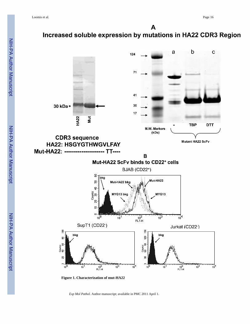
Figure 1. Characterization of mut-HA22
Loomis et al. Page 16
Exp Mol Pathol. Author manuscript; available in PMC 2011 April 1.
NIH
-PA Author Manuscript
NIH
-PA Author Manuscript
NIH
-PA Author Manuscript

Figure 1A: Analysis of mut-HA22. Mut-HA22 was analyzed by gel electrophoresis. (LeftPanel), mutations to the CDR3 region of HA22 increased soluble expression of the scFv. (RightPanel), verification of mut-HA22 reduction by gel electrophoresis: (a) mut-HA22, shown as amonomer and dimer, (b) mut-HA22 reduced with tributylphosphine (TBP), and (c) mut-HA22reduced with dithiothreitol (DTT).Figure 1B: Mut-HA22 binds to CD22-expressing cells. To analyze specific binding of mut-HA22 with CD22, BJAB cells were incubated with 2μg mut-HA22 followed by sequentialincubations with mouse anti-histidine IgG, and FITC-conjugated goat anti-mouse IgG. Acommercial anti-CD22 IgG MYG13 was used as positive control, and was stained with FITC-conjugated goat anti-mouse IgG. SUP-T1 and Jurkat cells were used as negative controls.Graphs representing corresponding cell types are indicated. Fluorescence distributions ofstained cells are: solid black curves, cells alone; black lines, immunostaining of mut-HA22;grey lines, immunostaining of MYG13; black dotted lines, mut-HA22's secondary Abs (mouseanti-his IgG and FITC conjugated goat anti-mouse IgG; dotted grey lines, MYG13's secondaryAbs (FITC-conjugated goat anti-mouse IgG). These results were reproducible from at leastthree independent experiments. Bkg, background binding.
Loomis et al. Page 17
Exp Mol Pathol. Author manuscript; available in PMC 2011 April 1.
NIH
-PA Author Manuscript
NIH
-PA Author Manuscript
NIH
-PA Author Manuscript
Loomis et al. Page 18
Exp Mol Pathol. Author manuscript; available in PMC 2011 April 1.
NIH
-PA Author Manuscript
NIH
-PA Author Manuscript
NIH
-PA Author Manuscript

Loomis et al. Page 19
Exp Mol Pathol. Author manuscript; available in PMC 2011 April 1.
NIH
-PA Author Manuscript
NIH
-PA Author Manuscript
NIH
-PA Author Manuscript

Loomis et al. Page 20
Exp Mol Pathol. Author manuscript; available in PMC 2011 April 1.
NIH
-PA Author Manuscript
NIH
-PA Author Manuscript
NIH
-PA Author Manuscript

Figure 2. Characterization of mut-HA22-liposomesFigure 2A: Determination of mut-HA22 conjugation to liposomes. Concentrated liposomeswere prepared in sample buffer with or without DTT, and were loaded on a 4-12% Bis-Trisgel (under non-reducing conditions). The gel was run in MES-SDS running buffer at 200V for35 minutes and proteins were stained with Microwave Blue. Conjugation of mut-HA22 toDSPE-PEG2000-Maleimide is verified by the molecular weight increase of free mut-HA22 toliposomal mut-HA22 (indicated by the arrow).Figure 2B: Hydrodynamic size distribution of liposomes before and after conjugation withmut-HA22. Hydrodynamic size was measured by backlight scattering in phosphate bufferedsaline with a Malvern Zetasizer Nano ZS instrument. The data are plotted as volume (top panel)and intensity (bottom panel) weighted distributions.Figure 2C: Stability of mut-HA22-liposomes in the presence of serum at 37°C. Calcein releasefrom liposomes was measured after liposome incubation at 37°C in PBS with 0, 10, or 50%FBS for 0, 1, 4, or 24 hours before and after addition of Triton X-100 (TX100). Leakage isexpressed as a percent of total, where 100% calcein leakage was obtained by addition of 20μl Triton-X100 (10 % w/v) and 0% calcein leakage was the background fluorescence valuebefore liposome incubation at 37°C. Error bars represent ± SD from at least three sampleswithin a single experiment.Figure 2D: Elution profile of DOX-loaded, mut-HA22 liposomes on Sepharose CL-6B columnTop panel: mut-HA22 conjugated liposomes were separated from free, unconjugated mut-HA22 scFv through size exclusion chromatography on a Sepharose CL-6B column (1x40cm).1 mL fractions were collected at an elution rate of 0.37 mL/min. Absorbance at 280nm (asmeasured by UV detector) is shown. Peak I: DOX-loaded, mut-HA22 conjugated liposomes;Peak II: unconjugated mut-HA22 scFv; Peak III: Free DOX.
Loomis et al. Page 21
Exp Mol Pathol. Author manuscript; available in PMC 2011 April 1.
NIH
-PA Author Manuscript
NIH
-PA Author Manuscript
NIH
-PA Author Manuscript

Bottom panel: Samples in peak I were analyzed (see Fig 2A) to confirm mut-HA22 conjugationto liposomes. (a): mut-HA22 scFv standard; (b): DOX-loaded, mut-HA22 conjugatedliposomes before and after Sepharose CL-6B column. The fractions in peak II were analyzedfor the presence of mut-HA22 by gel electrophoresis as described (see Methods section, datanot shown). Similarly, all fractions were also analyzed for DOX and peak III was assignedaccordingly (data not shown).
Loomis et al. Page 22
Exp Mol Pathol. Author manuscript; available in PMC 2011 April 1.
NIH
-PA Author Manuscript
NIH
-PA Author Manuscript
NIH
-PA Author Manuscript

Loomis et al. Page 23
Exp Mol Pathol. Author manuscript; available in PMC 2011 April 1.
NIH
-PA Author Manuscript
NIH
-PA Author Manuscript
NIH
-PA Author Manuscript

Loomis et al. Page 24
Exp Mol Pathol. Author manuscript; available in PMC 2011 April 1.
NIH
-PA Author Manuscript
NIH
-PA Author Manuscript
NIH
-PA Author Manuscript

Loomis et al. Page 25
Exp Mol Pathol. Author manuscript; available in PMC 2011 April 1.
NIH
-PA Author Manuscript
NIH
-PA Author Manuscript
NIH
-PA Author Manuscript

Figure 3. Binding of mut-HA22-liposomes to CD22-expressing cellsCells (107/mL in RPMI-1640, 10% serum) were incubated with fluorescently labeled mut-HA22-liposomes (2 μg of mut-HA22/46μg of phospholipids) or control liposomes (0 μg ofmut-HA22/46 μg of phospholipids) for 40 minutes at 37°C. At the end of incubations, cellswere centrifuged and washed twice with PBS-BSA to remove unbound liposomes; cell-boundfluorescence was examined by microscopy and FACS.Figure 3A: Binding of mut-HA22-liposomes to CD22-receptor expressing cells.Fluorescence images were captured using the Nikon Eclipse TE200 inverted microscope witha 40X oil objective (N.A. 1.30). Images shown are for calcein and rhodamine fluorescence asindicated ((a-d), BJAB cells, (e-h) SUP-T1 cells).Figure 3B: Flow cytometry analysis of incubated cells. The data shown are for calceinfluorescence.Figure 3C: Pre-incubation of cells with free mut-HA22 inhibits subsequent binding ofliposomes.BJAB cells were incubated with (b) 8, (c) 2, or (d) 0 μg of free mut-HA22 at 4°C for 30 minutesin the dark, washed twice with cold PBS-BSA, and then incubated with fluorescently labeledmut-HA22-liposomes (1μg/106 cells) at 4°C for 30 minutes ((a) cells only)).
Loomis et al. Page 26
Exp Mol Pathol. Author manuscript; available in PMC 2011 April 1.
NIH
-PA Author Manuscript
NIH
-PA Author Manuscript
NIH
-PA Author Manuscript

Figure 4.A: Binding of mut-HA22--liposomes with 1% or 4% DSPE-PEG2000-Maleimide to CD22-expressing cells.Cells (107/mL in RPMI-1640, 10% serum) were incubated with various concentrations ofcalcein-loaded mut-HA22-liposomes for 40 minutes at 37°C. At the end of incubations, cellswere centrifuged and washed twice with PBS-BSA to remove unbound liposomes; cell-bound
Loomis et al. Page 27
Exp Mol Pathol. Author manuscript; available in PMC 2011 April 1.
NIH
-PA Author Manuscript
NIH
-PA Author Manuscript
NIH
-PA Author Manuscript

fluorescence was examined by FACS. The graph represents mean fluorescence intensity values(y-axis). Three liposome preparations (see Table 1) corresponding to either 0 mol% (=0 mut-HA22/liposome), 1 mol% (=47 mut-HA22/liposome) or 4 mol% (=90 mut-HA22/liposome)were used for incubations with either BJAB or SUP-T1 cells. The amounts of liposomal mut-HA22 added are given in x-axis. Cell types are indicated at the end of each curve. The numbersin parentheses indicate the average number of mut-HA22/liposome.Figure 4B: Dose-dependent binding of mut-HA22-liposomes to BJAB cells.BJAB cells were incubated with various concentrations of calcein-loaded liposomes for 40minutes at 37°C. At the end of incubations, cells were washed and analyzed for calceinfluorescence by FACS. Concentrations of liposome-conjugated mut-HA22 added to cells: (a)cells alone, (b) 0.01 μg mut-HA22, (c) 0.05 μg mut-HA22 (d), 0.2 μg mut-HA22, (e) 0.5 μgmut-HA22, and (f) 2 μg mut-HA22. Control liposomes contained the correspondingphospholipid quantities without any mut-HA22.
Loomis et al. Page 28
Exp Mol Pathol. Author manuscript; available in PMC 2011 April 1.
NIH
-PA Author Manuscript
NIH
-PA Author Manuscript
NIH
-PA Author Manuscript

Loomis et al. Page 29
Exp Mol Pathol. Author manuscript; available in PMC 2011 April 1.
NIH
-PA Author Manuscript
NIH
-PA Author Manuscript
NIH
-PA Author Manuscript

Figure 5. Uptake of mut-HA22-liposomes by BJAB cells at 37°CBJAB cells (107/mL RPMI-1640) were incubated with liposomes at 37°C or 4°C. Cells werethen washed twice with cold PBS-BSA and kept on ice until analyzed.Figure 5A: Temperature Dependent Binding/Uptake of mut-HA22-liposomes by BJAB cells.Cells were incubated with 4 μg of liposome-conjugated mut-HA22 (or equivalent amount ofphospholipids for control liposomes) for 40 minutes at 37°C or 4°C (see methods section).Cells were then washed and rhodamine fluorescence of cells was analyzed by FACS.Figure 5B: Intracellular Localization of mut-HA22-scFv liposomes by BJAB Cells. Cells wereincubated with 0.2 μg of liposome-conjugated mut-HA22 (or equivalent amount ofphospholipids for control liposomes) for 20 or 40 minutes at 37 or 4°C. Z-stacks of 14-16optical slices at 1.7 micron intervals were acquired. Calcein fluorescence of cells is shown ina single slice of the acquired images.
Loomis et al. Page 30
Exp Mol Pathol. Author manuscript; available in PMC 2011 April 1.
NIH
-PA Author Manuscript
NIH
-PA Author Manuscript
NIH
-PA Author Manuscript

Loomis et al. Page 31
Exp Mol Pathol. Author manuscript; available in PMC 2011 April 1.
NIH
-PA Author Manuscript
NIH
-PA Author Manuscript
NIH
-PA Author Manuscript

Figure 6.A: Cellular accumulation of liposomal DOX by Raji cellsCells (3×10⁵ cells per sample) were incubated with 0.5 – 2 ug of liposomal DOX for one hourat 37°C. The cells were then washed with phenol-red free culture medium (0.5mL×3) andresuspended in 0.3 mL of PBS and transferred to a 96-well plate (0.1mL×3). Cellularaccumulation of DOX was determined using a standard curve of known DOX concentrationsin the presence of cell lysates without incubation with liposomes (left panel). Right panel:accumulation of DOX in nanograms. Solid gray bars, control liposomes; black diagonal linebars, mut-HA22 liposomes. Error bars represent ± SD of triplicate samples within a singleexperiment.Figure 6B: Cell viability in the continuous presence of liposomal DOXCells were plated in duplicates in a 96-well plate (105/0.1mL per well) and incubated with0-7.5 ug of liposomal or free DOX for 72 hours at 37°C. Cell viability was calculated takingcontrol sample (without DOX) as 100%. Values represent an average of duplicate samples.(a) and (b) Cytotoxicity by liposomal DOX: (a) BJAB (CD22+), (b) SUP-T1 (CD22-) mut-HA22 liposomes (◆) and control liposomes (●).(c) Cytotoxicity by free DOX: SUP-T1 (X), BJAB (▲), and Raji (■).Figure 6C: Cell viability following pre-incubation with liposomesCells were incubated in triplicates with liposomal DOX for 1 hour, and any unbound liposomeswere washed off. Following an additional 72-hour incubation, cell viability was determined(see Figure 6B).(a) Raji (CD22+), (b) BJAB (CD22+), and (c) SUP-T1 (CD22-) mut-HA22 liposomes (◆) andcontrol liposomes (●)(d) A snapshot of cell viability at a concentration of 10 ug/mL liposomal DOX is shown (Datafrom the respective curves in Figure 6C (a-c)). In this graph, the total number of cells remainingviable in the presence of control liposomes were taken as 100%. mut-HA22 liposomes(diagonal line bars), control liposomes (gray bars)
Loomis et al. Page 32
Exp Mol Pathol. Author manuscript; available in PMC 2011 April 1.
NIH
-PA Author Manuscript
NIH
-PA Author Manuscript
NIH
-PA Author Manuscript

NIH
-PA Author Manuscript
NIH
-PA Author Manuscript
NIH
-PA Author Manuscript
Loomis et al. Page 33
Tabl
e 1
Lipo
som
e fo
rmul
atio
ns u
sed
in th
is st
udy.
Lip
osom
esL
ipid
sm
ut-H
A22
mol
ecul
es/li
poso
me
(ave
rage
)Fl
uore
scen
t Mar
kers
* /D
rug
**Si
ze A
naly
sis (
nm)
Fluo
resc
ently
Lab
eled
mut
-HA
22 L
ipos
omes
(For
mul
atio
n I)
DPP
C (9
6 m
ol%
)D
SPE-
PEG
2000
-M
alei
mid
e (4
mol
%)
90C
alce
in/ R
hoda
min
e-PE
See
Fig.
2B
1 m
ol%
DSP
E-PE
G20
00-M
alei
mid
em
ut-H
A22
Lip
osom
es(F
orm
ulat
ion
II)
DPP
C (9
6 m
ol%
)D
SPE-
PEG
2000
-M
alei
mid
e (1
mol
%)
DSP
E-PE
G20
00 (3
mol
%)
47C
alce
in/ R
hoda
min
e-PE
See
Fig.
2B
.
Con
trol L
ipos
omes
(For
mul
atio
n II
I)D
PPC
(96
mol
%)
DSP
E-PE
G20
00 (4
mol
%)
0C
alce
in/ R
hoda
min
e-PE
See
Fig.
2B
Dru
g L
oade
d
mut
-HA
22 L
ipos
omes
(For
mul
atio
n IV
)D
PPC
(96
mol
%)
DSP
E-PE
G20
00-
Mal
eim
ide
(4 m
ol%
)
90D
oxor
ubic
in12
3.0
7.9
Con
trol L
ipos
omes
(For
mul
atio
n V
)D
PPC
(96
mol
%)
DSP
E-PE
G20
00-
Mal
eim
ide
(4 m
ol%
)
0D
oxor
ubic
in11
0.5
6.6
Empt
y Li
poso
mes
(For
mul
atio
n V
I)D
PPC
(96
mol
%)
DSP
E-PE
G20
00-
Mal
eim
ide
(4 m
ol%
)
0N
/A86
.94.
0
* Fluo
resc
ent l
ipos
omes
wer
e pr
epar
ed w
ith 5
0 m
M C
alce
in a
nd/o
r 0.1
mol
% R
hoda
min
e-PE
.
**D
oxor
ubic
in lo
adin
g w
as ty
pica
lly 1
50-2
00 n
g do
x/nm
ol P
i.
Exp Mol Pathol. Author manuscript; available in PMC 2011 April 1.
Related Documents


