Specific Biomarkers Are Associated with Docetaxel- and Gemcitabine-Resistant NSCLC Cell Lines 1 Alice Pasini* ,2 , Giulia Paganelli †,2 , Anna Tesei † , Wainer Zoli † , Emanuele Giordano* and Daniele Calistri † *Laboratory of Cellular and Molecular Engineering “S. Cavalcanti”, School of Engineering, University of Bologna, Campus of Cesena, Cesena, Italy; † Biosciences Laboratory, IRCCS Istituto Scientifico Romagnolo per lo Studio e la Cura dei Tumori (IRST), Meldola, Italy Abstract Five-year survival rate for lung cancer is limited to 10% to 15%. Therefore, the identification of novel therapeutic prognostic factors is an urgent requirement. The aim of this study is thus to highlight specific biomarkers in chemo- resistant non–small cell lung cancer cell lines. Therefore, we checked—in the control condition as well as after short- term pharmacological treatment with either docetaxel or gemcitabine—the expression of genes such as tumor suppressor genes (CDKN2A, DAPK, FHIT, GSTP1, MGMT, RARβ2, RASSF1A, and TIMP3), genes associated with drug resistance (BRCA1, COX2, ERCC1, IGFBP3, RRM1, and TUBB3), and stemness-related genes (CD133, OCT4, and SLUG) in two cellular models of squamous carcinoma (CAEP) and adenocarcinoma (RAL) of the lung originally established. Their promoter methylation profile was also evaluated. Drug-related genes were upregulated. Cisplatin resistance matched with high levels of BRCA1 and ERCC1 in both cell lines; docetaxel sensitivity of CAEP cells was associated to levels of TUBB3 lower than RAL cells. Although CAEP cells were more sensitive to gemcitabine, both cell lines showed high levels of RRM1. Stemness-related genes were downregulated in the control condition but became upregulated in docetaxel-resistant cells, indicating the selection of a population with stemness features. We did not find an unequivocal correspondence between gene expression and respective DNA promoter methylation status, suggesting the involvement of additional mechanisms of gene expression regulation. These results highlight specific biomarkers consistent with the different responses of the two cell lines to standard pharmacological treat- ments and indicate specific molecular traits for their chemoresistance. Translational Oncology (2012) 5, 461–468 Introduction Non–small cell lung cancer (NSCLC) represents a common cause of tumor death in industrialized countries where, despite a significant improvement in diagnostics, surgery, and chemotherapy, the overall 5-year survival rate is in fact limited to about 10% to 15% [1,2]. The recommended treatment for these tumors is surgery, whereas radiotherapy and chemotherapy are used for the treatment of un- resectable or locally advanced tumors and as palliative therapies for metastatic tumors [3]. In tumors without EGFR mutations, platinum compounds, taxanes and gemcitabine—alone or in combination—are the traditional drugs used in standard chemotherapy, even though tumor resistance to these treatments is common [4]. Chemoresistance can arise from different mechanisms, such as reduced drug uptake, increased drug efflux, drug detoxification, DNA repair, or defective apoptosis, depending on the specific drug target. In this respect, several studies highlight relationships among specific gene expression, therapeutic response and tumor progression, and major classes of drugs used in the treatment of NSCLC. In particular, resistance to platinum compounds is mainly the con- sequence of the overexpression of DNA repair genes, such as ERCC1 Address all correspondence to: Emanuele Giordano, MD, PhD, Laboratory of Cellular and Molecular Engineering “S. Cavalcanti”, via Venezia, 52, I-47521 Cesena, Italy. E-mail: [email protected] 1 All authors have contributed significantly and they agree with the content of the manuscript. None of the authors has any conflict of interest. 2 These authors contributed equally to this work. Received 1 August 2012; Revised 1 August 2012; Accepted 23 August 2012 Copyright © 2012 Neoplasia Press, Inc. All rights reserved 1944-7124/12/$25.00 DOI 10.1593/tlo.12256 www.transonc.com Translational Oncology Volume 5 Number 6 December 2012 pp. 461–468 461

Welcome message from author
This document is posted to help you gain knowledge. Please leave a comment to let me know what you think about it! Share it to your friends and learn new things together.
Transcript

Trans la t iona l Onco logy Volume 5 Number 6 December 2012 pp. 461–468 461
www.transonc.com
Specific Biomarkers AreAssociated with Docetaxel-and Gemcitabine-ResistantNSCLC Cell Lines1
Alice Pasini*,2, Giulia Paganelli†,2,Anna Tesei†, Wainer Zoli†, Emanuele Giordano*and Daniele Calistri†
*Laboratory of Cellular and Molecular Engineering“S. Cavalcanti”, School of Engineering, University ofBologna, Campus of Cesena, Cesena, Italy; †BiosciencesLaboratory, IRCCS Istituto Scientifico Romagnolo per loStudio e la Cura dei Tumori (IRST), Meldola, Italy
AbstractFive-year survival rate for lung cancer is limited to 10% to 15%. Therefore, the identification of novel therapeuticprognostic factors is an urgent requirement. The aim of this study is thus to highlight specific biomarkers in chemo-resistant non–small cell lung cancer cell lines. Therefore, we checked—in the control condition as well as after short-term pharmacological treatment with either docetaxel or gemcitabine—the expression of genes such as tumorsuppressor genes (CDKN2A, DAPK, FHIT, GSTP1, MGMT, RARβ2, RASSF1A, and TIMP3), genes associated withdrug resistance (BRCA1, COX2, ERCC1, IGFBP3, RRM1, and TUBB3), and stemness-related genes (CD133, OCT4,and SLUG) in two cellular models of squamous carcinoma (CAEP) and adenocarcinoma (RAL) of the lung originallyestablished. Their promoter methylation profile was also evaluated. Drug-related genes were upregulated. Cisplatinresistance matched with high levels of BRCA1 and ERCC1 in both cell lines; docetaxel sensitivity of CAEP cells wasassociated to levels of TUBB3 lower than RAL cells. Although CAEP cells were more sensitive to gemcitabine, bothcell lines showed high levels of RRM1. Stemness-related genes were downregulated in the control condition butbecame upregulated in docetaxel-resistant cells, indicating the selection of a population with stemness features.We did not find an unequivocal correspondence between gene expression and respective DNA promoter methylationstatus, suggesting the involvement of additional mechanisms of gene expression regulation. These results highlightspecific biomarkers consistent with the different responses of the two cell lines to standard pharmacological treat-ments and indicate specific molecular traits for their chemoresistance.
Translational Oncology (2012) 5, 461–468
Address all correspondence to: Emanuele Giordano, MD, PhD, Laboratory of Cellularand Molecular Engineering “S. Cavalcanti”, via Venezia, 52, I-47521 Cesena, Italy.E-mail: [email protected] authors have contributed significantly and they agree with the content of themanuscript. None of the authors has any conflict of interest.2These authors contributed equally to this work.Received 1 August 2012; Revised 1 August 2012; Accepted 23 August 2012
Copyright © 2012 Neoplasia Press, Inc. All rights reserved 1944-7124/12/$25.00DOI 10.1593/tlo.12256
IntroductionNon–small cell lung cancer (NSCLC) represents a common cause oftumor death in industrialized countries where, despite a significantimprovement in diagnostics, surgery, and chemotherapy, the overall5-year survival rate is in fact limited to about 10% to 15% [1,2].The recommended treatment for these tumors is surgery, whereasradiotherapy and chemotherapy are used for the treatment of un-resectable or locally advanced tumors and as palliative therapies formetastatic tumors [3]. In tumors without EGFR mutations, platinumcompounds, taxanes and gemcitabine—alone or in combination—arethe traditional drugs used in standard chemotherapy, even thoughtumor resistance to these treatments is common [4].Chemoresistance can arise from different mechanisms, such as
reduced drug uptake, increased drug efflux, drug detoxification, DNArepair, or defective apoptosis, depending on the specific drug target. In
this respect, several studies highlight relationships among specific geneexpression, therapeutic response and tumor progression, and majorclasses of drugs used in the treatment of NSCLC.
In particular, resistance to platinum compounds is mainly the con-sequence of the overexpression of DNA repair genes, such as ERCC1

462 Specific Biomarkers in Chemoresistant NSCLC Cells Pasini et al. Translational Oncology Vol. 5, No. 6, 2012
and BRCA1 [5–7]. During taxane treatment of NSCLC, BRCA1 ex-pression represents a marker of drug sensitivity [8,9]. However, a highexpression level of TUBB3, encoding for a member of the β-tubulinprotein family, is associated with taxane resistance in different tumors,including NSCLCs [5–7]. Conversely, high levels of FHIT expressionwere shown together with significant apoptosis in in vitro experiments[10]. NSCLC therapy may also include the use of gemcitabine, whoseintracellular active forms interfere with DNA synthesis through com-petition with deoxycytidine and inhibition of ribonucleotide reductase.Therefore, high levels of expression of the subunit 1 of ribonucleotidereductase (RRM1) are associated with increased gemcitabine resistancein NSCLC cell lines [11].
Promoter methylation is an epigenetic event usually associated withgene silencing, implicated in the development and differentiation andfrequently deregulated in cancer [12,13]. Several studies have identifieda pattern of promoter methylation and a corresponding gene expres-sion profile consistent with tumor progression and response to drugtreatment either in NSCLC patients or in cell lines. Several tumorsuppressor genes (TSGs) display a hypermethylated promoter in lungcancer, including CDKN2A, RASSF1A, RARβ2, MGMT, GSTP1,DAPK, TIMP3, and FHIT [14–17]. Aberrant methylation ofRASSF1A, DAPK, CDKN2A, and FHIT promoters was associatedwith shorter overall survival [18–21].
Evaluating gene expression and promoter methylation of specificgenes significant for monitoring NSCLC progression might thusidentify specific biomarkers that could predict disease recurrenceand help in keeping watch over therapeutic response.
In the present work, to explore this issue, we used an in vitro modelbased on two NSCLC cell lines, representative of either a squamouscarcinoma (CAEP) or an adenocarcinoma (RAL) of the lung, estab-lished and studied at the Istituto Scientifico Romagnolo per lo Studioe la Cura dei Tumori at Meldola [22–25]. In these cells, we evaluatedthe expression of genes of interest in lung cancer disease, such as a set ofTSGs (CDKN2A, DAPK, FHIT, GSTP1,MGMT, RARβ2, RASSF1A,and TIMP3) found to be implicated in lung cancer progression andtherapeutic response [19,26–32], and a set of genes associated withspecific drug resistance (BRCA1, COX2, ERCC1, IGFBP3, RRM1,and TUBB3) [3–5,33–36]. In addition, a set of genes expressed in cellswith stemness features (CD133, OCT4, and SLUG) [37–43] was alsomonitored, as increasing evidence points at small cancer cell popula-tions, defined as cancer stem cells, sustaining NSCLC resistance toantitumor agents [39,44,45] and accounting for tumor recurrence [46].
To investigate the involvement of our gene panel in the processof resistance to chemotherapy, we carried out this analysis in thebasal control condition as well as after short-term pharmacologicaltreatment with standard chemotherapeutic drugs.
Promoter methylation was also studied to evaluate how the ex-pression profile of these genes might be consistent with this epigeneticmodification.
Materials and Methods
Cell CultureThe study was performed on two EGFR wild-type NSCLC cell
lines, CAEP and RAL, derived from a squamous carcinoma and anadenocarcinoma of the lung, respectively, established and characterizedin our laboratory [22–25]. Cell lines were grown in Dulbecco’s modi-fied Eagle’s medium/HAM F12 (1:1), supplemented with 10% FBS,
2 mM L-glutamine (PAA, Pashing, Austria), and insulin (10 mg/ml;Sigma-Aldrich, Milan, Italy) in a humidified atmosphere of 95% airand 5% CO2 at 37°C. Cells in the exponential growth phase wereused for all the experiments.
Drugs and TreatmentCisplatin (Ebewe Pharma-Sandoz, Origgio, Italy), docetaxel (Sanofi-
aventis SpA, Milan, Italy), and gemcitabine (Eli Lilly Italia SpA, SestoFiorentino, Italy) were aliquoted, stored at −80°C, and freshly dilutedin culture medium before each experiment.
For in vitro chemosensitivity assay, drugs were tested at scalar dilu-tions of 1:1, 1:10, and 1:100 of plasma peak concentrations (10 μMfor cisplatin, 2 μM for docetaxel, and 40 μM for gemcitabine). Toensure that exposure times were compatible with the half-life ofthe drugs administered in a clinical setting [47–50], we analyzed theeffects after 6, 1, and 3 hours of exposure, respectively, followed by72 hours of culture in drug-free medium.
For gene expression and DNA methylation analysis, cells weretreated at either 50% inhibiting concentration (IC50) or plasma peakconcentration dosage when IC50 was not reached. These values were0.09 μM and 2 μM docetaxel and 1.98 μM and 40 μM gemcitabine,for, respectively, CAEP and RAL.
In Vitro Chemosensitivity AssayCell viability was evaluated with the sulforodamine B (SRB) assay
according to Skehan et al. [51]. Experiments were run in octoplets,and each experiment was repeated twice. Dose-response curves werecreated by Excel Software and the IC50 values were determined fromthe plots.
Gene Expression AnalysisThe analysis of gene expression was performed as reported by
Arienti et al. [52] using a real-time reverse transcription–polymerasechain reaction (PCR) method. The amount of mRNA of each genewas normalized to the endogenous references β2-microglobulin usingGene Expression Macro Software (version 1.1; Bio-Rad LaboratoriesS.r.l., Segrate (MI), Italy). Commercial control RNA, derived from anormal lung, was used as calibrator (MPV total RNA; Stratagene,Agilent Technologies Italia SpA, Cernusco, Italy). Primers for mRNAamplification were designed using Beacon Designer Software (version 4;PREMIER Biosoft International, Palo Alto, CA); sequences andannealing temperatures are available upon request.
DNA Methylation AnalysisPromoter methylation was analyzed by a standard methylation-
specific PCR (MSP) protocol from DNA extracted in phenol-chloroform.Briefly, 1 μg of purified DNA was subjected to bisulfite treatment(EZ DNAMethylation Kit; Zymo Research, Irvine, CA). The methyl-ation status of the gene promoter was determined by MSP [53] andconfirmed in two independent experiments. Total volume for MSP re-action was 15 μl, with 4 mM MgCl2, 0.5 mM each dNTP, 0.2 μMeach primer, 0.5 U of @Taq Hot Start Thermostable DNA poly-merase (EuroClone, Pero, Italy), and 1 μl of bisulfite-treated DNA.Methylation of OCT4 and CD133 gene promoters was tested by bi-sulfite sequencing, cloning PCR products in pGEM-T Easy VectorSystem (Promega, Milan, Italy). Sequencing of PCR products was per-formed using the Big Dye Terminator Cycle Sequencing Kit (AppliedBiosystems, Foster City, CA) using the Applied Biosystems 3130 Avant

Translational Oncology Vol. 5, No. 6, 2012 Specific Biomarkers in Chemoresistant NSCLC Cells Pasini et al. 463
Genetic Sequencer. Primer sequences and annealing temperatures areavailable upon request.
Statistical AnalysisGene expression of CAEP and RAL cell lines in the basal control
condition was statistically compared with two-tailed t test usingGraphPad Prism (version 4.0; GraphPad Software, San Diego, CA).Data for basal control condition versus drug treatment expression werestatistically analyzed using one-way analysis of variance with Tukey’spost test (GraphPad Prism, version 4.0; GraphPad Software). Differ-ences in gene expression were considered significant for P < .05.
Results
Pharmacological TreatmentBoth the investigated cell lines showed high resistance to cisplatin
treatment, even after treatment with the plasma peak concentration(10 μM; Figure 1A).The squamous carcinoma cells (CAEP) showed less resistance to
docetaxel and gemcitabine than the adenocarcinoma cells (RAL). Infact, the exposure of CAEP cells to 2 μM (the plasma peak concen-tration) docetaxel induced 80% cell death with an IC50 value of0.09 μM (Figure 1B). When the CAEP cells were treated with gem-citabine, an IC50 of 1.98 μM was observed with an overall survivalof about 36% at the highest dose tested (Figure 1C ). Conversely,after either docetaxel or gemcitabine exposure, IC50 values were neverreached even at the highest concentrations tested, for the adeno-carcinoma cells (RAL; Figure 1, B and C ).
Gene Expression ProfileAmong the investigated TSGs, only the CDKN2A mRNA was
highly upregulated in both lung cancer cell lines with respect to nor-mal lung tissue. MGMT transcript levels were upregulated (five-fold)in RAL but not in CAEP cells. Although similar to normal lung tissuelevels, DAPK and FHIT expression in RAL cells was significantlyhigher (respectively about 3-fold and 11-fold; P < .01) than in CAEPcells. No differences between CAEP and RAL were scored for GSTP1,RARβ2, RASSF1A, and TIMP3, although in both cell lines they weredownregulated with respect to the normal lung tissue (Figure 2).
Figure 1. Cell survival of CAEP and RAL cell lines after the treatmen
As expected, genes associated with specific drug resistance (BRCA1,ERCC1, RRM1, and TUBB3) were significantly upregulated in bothcell lines with respect to the normal lung tissue. A significant differ-ence in mRNA levels of RAL versus CAEP cells was scored forCOX2 (P < .01) and TUBB3 (P < .05), respectively about 20-foldand 5-fold increases in RAL cells. The reverse was observed forIGFBP3: It was highly upregulated (about 100-fold) in CAEP versusRAL cells (Figure 2).
In both cell lines, CD133 and OCT4 gene products, associated withstemness features, were downregulated, whereas SLUG gene productwas overexpressed with respect to normal lung tissue (Figure 2).
Upon treatment with docetaxel and gemcitabine, the expression ofthe investigated genes showed specific differences with respect to thebasal control condition (Figure 3). Docetaxel treatment in CAEP cellsdid not modify the expression of TSGs; however, the RRM1 transcriptlevel was significantly decreased (42%) with respect to the basal con-trol condition (P < .05) and OCT4 expression in treated cells increasedabout six-fold (P < .01; Figure 3A). RAL cells treated with docetaxel,in contrast, showed a significantly decreased expression of the TSGsDAPK, FHIT, MGMT, and RARβ2 (about 22%, 35%, 5%, and30%, respectively) compared to the basal control condition (P < .05;Figure 3B). CD133 expression was significantly decreased (20%; P <.05), whereas SLUG mRNA level increased about five-fold (P < .05;Figure 3B).
The only change in the expression level of the investigated genesinduced by gemcitabine in CAEP cells occurred at the TUBB3 gene,which was downregulated (52%) with respect to the basal controlcondition (P < .05; Figure 3A). In RAL cells, however, several effectswere seen, i.e., gemcitabine treatment induced decreased expressionof the TSGs DAPK, FHIT, MGMT, and RARβ2 (about 18%, 6%,7%, and 16%, respectively), compared to the basal control condition(P < .05; Figure 3B); only COX2 was downregulated (8%, P < .01)among genes associated with specific drug resistance; and finally, themRNA level of the CD133 gene was decreased (16%) with respect tothe basal control condition (P < .01; Figure 3B).
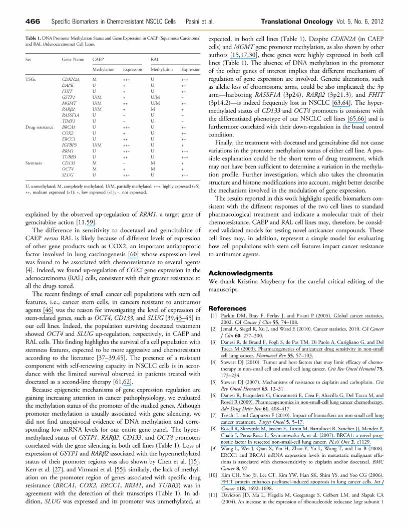
Promoter Methylation AnalysisTSG promoter was unmethylated forDAPK, RASSF1A, and TIMP3
and partially or completely methylated for GSTP1, MGMT, andRARβ2, in both cell lines. CDKN2A promoter was methylated
ts with (A) cisplatin, (B) docetaxel, and (C) gemcitabine.
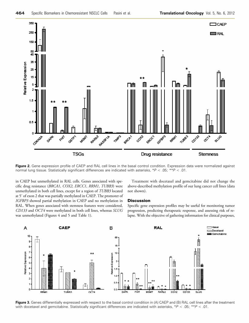
Figure 2. Gene expression profile of CAEP and RAL cell lines in the basal control condition. Expression data were normalized againstnormal lung tissue. Statistically significant differences are indicated with asterisks, *P < .05; **P < .01.
464 Specific Biomarkers in Chemoresistant NSCLC Cells Pasini et al. Translational Oncology Vol. 5, No. 6, 2012
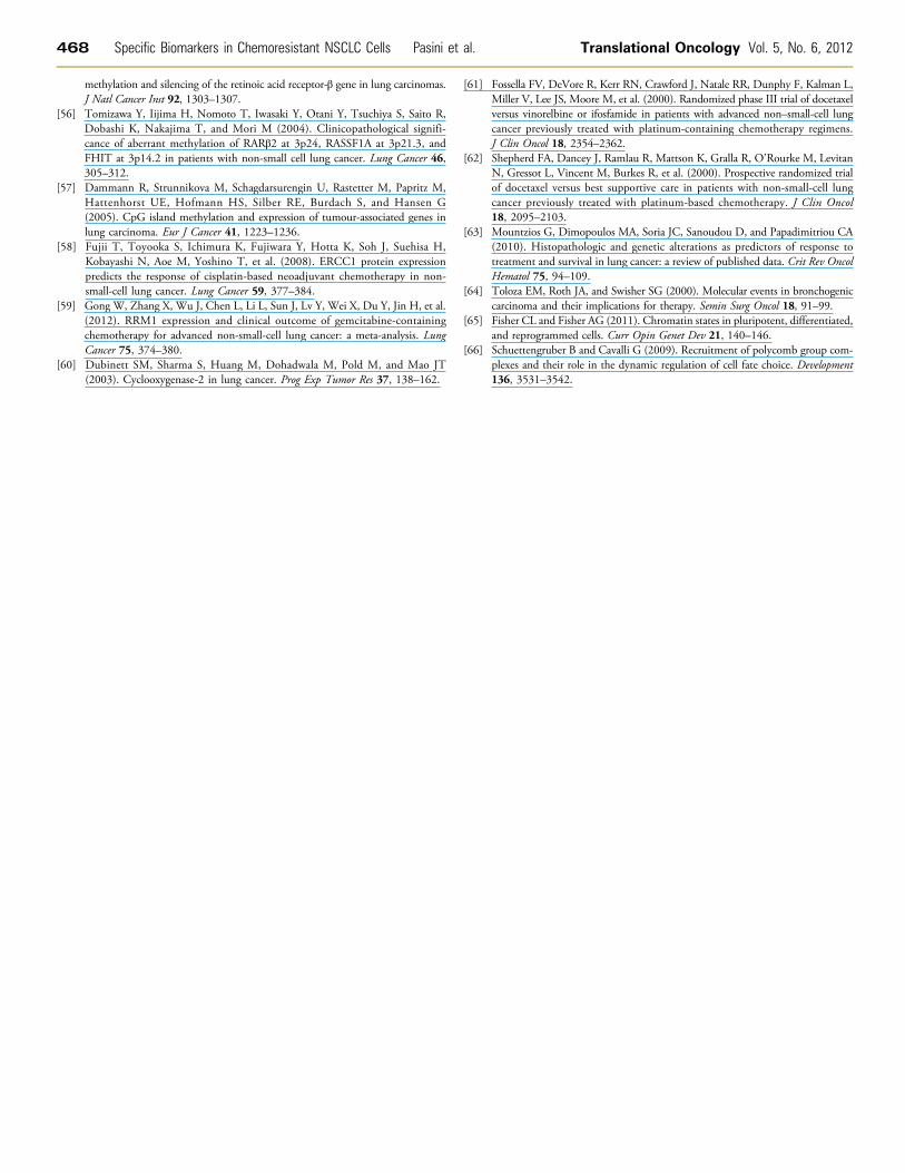
in CAEP but unmethylated in RAL cells. Genes associated with spe-cific drug resistance (BRCA1, COX2, ERCC1, RRM1, TUBB3) wereunmethylated in both cell lines, except for a region of TUBB3 locatedat 5′ of exon 2 that was partially methylated in CAEP. The promoter ofIGFBP3 showed partial methylation in CAEP and no methylation inRAL. When genes associated with stemness features were considered,CD133 and OCT4 were methylated in both cell lines, whereas SLUGwas unmethylated (Figures 4 and 5 and Table 1).
Figure 3. Genes differentially expressed with respect to the basal conwith docetaxel and gemcitabine. Statistically significant differences
Treatment with docetaxel and gemcitabine did not change theabove-described methylation profile of our lung cancer cell lines (datanot shown).
DiscussionSpecific gene expression profiles may be useful for monitoring tumorprogression, predicting therapeutic response, and assessing risk of re-lapse. With the objective of gathering information for clinical purposes,
trol condition in (A) CAEP and (B) RAL cell lines after the treatmentare indicated with asterisks, *P < .05; **P < .01.
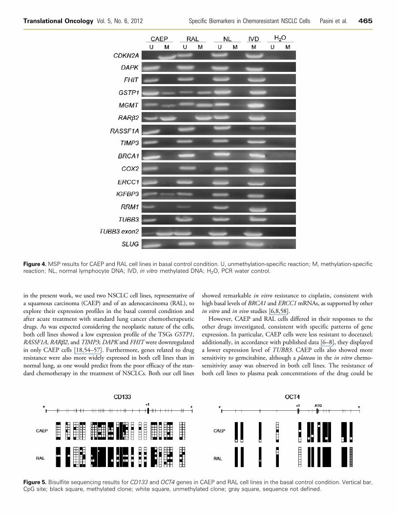
Figure 4. MSP results for CAEP and RAL cell lines in basal control condition. U, unmethylation-specific reaction; M, methylation-specificreaction; NL, normal lymphocyte DNA; IVD, in vitro methylated DNA; H2O, PCR water control.
Translational Oncology Vol. 5, No. 6, 2012 Specific Biomarkers in Chemoresistant NSCLC Cells Pasini et al. 465
in the present work, we used two NSCLC cell lines, representative ofa squamous carcinoma (CAEP) and of an adenocarcinoma (RAL), toexplore their expression profiles in the basal control condition andafter acute treatment with standard lung cancer chemotherapeuticdrugs. As was expected considering the neoplastic nature of the cells,both cell lines showed a low expression profile of the TSGs GSTP1,RASSF1A, RARβ2, and TIMP3; DAPK and FHIT were downregulatedin only CAEP cells [18,54–57]. Furthermore, genes related to drugresistance were also more widely expressed in both cell lines than innormal lung, as one would predict from the poor efficacy of the stan-dard chemotherapy in the treatment of NSCLCs. Both our cell lines
Figure 5. Bisulfite sequencing results for CD133 and OCT4 genes in CCpG site; black square, methylated clone; white square, unmethylat
showed remarkable in vitro resistance to cisplatin, consistent withhigh basal levels of BRCA1 and ERCC1mRNAs, as supported by otherin vitro and in vivo studies [6,8,58].
However, CAEP and RAL cells differed in their responses to theother drugs investigated, consistent with specific patterns of geneexpression. In particular, CAEP cells were less resistant to docetaxel;additionally, in accordance with published data [6–8], they displayeda lower expression level of TUBB3. CAEP cells also showed moresensitivity to gemcitabine, although a plateau in the in vitro chemo-sensitivity assay was observed in both cell lines. The resistance ofboth cell lines to plasma peak concentrations of the drug could be
AEP and RAL cell lines in the basal control condition. Vertical bar,ed clone; gray square, sequence not defined.
466 Specific Biomarkers in Chemoresistant NSCLC Cells Pasini et al. Translational Oncology Vol. 5, No. 6, 2012
explained by the observed up-regulation of RRM1, a target gene ofgemcitabine action [11,59].
The difference in sensitivity to docetaxel and gemcitabine ofCAEP versus RAL is likely because of different levels of expressionof other gene products such as COX2, an important antiapoptoticfactor involved in lung carcinogenesis [60] whose expression levelwas found to be associated with chemoresistance to several agents[4]. Indeed, we found up-regulation of COX2 gene expression in theadenocarcinoma (RAL) cells, consistent with their greater resistance toall the drugs tested.
The recent findings of small cancer cell populations with stem cellfeatures, i.e., cancer stem cells, in cancers resistant to antitumoragents [46] was the reason for investigating the level of expression ofstem-related genes, such as OCT4, CD133, and SLUG [39,43–45] inour cell lines. Indeed, the population surviving docetaxel treatmentshowed OCT4 and SLUG up-regulation, respectively, in CAEP andRAL cells. This finding highlights the survival of a cell population withstemness features, expected to be more aggressive and chemoresistantaccording to the literature [37–39,45]. The presence of a resistantcomponent with self-renewing capacity in NSCLC cells is in accor-dance with the limited survival observed in patients treated withdocetaxel as a second-line therapy [61,62].
Because epigenetic mechanisms of gene expression regulation aregaining increasing attention in cancer pathophysiology, we evaluatedthe methylation status of the promoter of the studied genes. Althoughpromoter methylation is usually associated with gene silencing, wedid not find unequivocal evidence of DNA methylation and corre-sponding low mRNA levels for our entire gene panel. The hyper-methylated status of GSTP1, RARβ2, CD133, and OCT4 promoterscorrelated with the gene silencing in both cell lines (Table 1). Loss ofexpression of GSTP1 and RARβ2 associated with the hypermethylatedstatus of their promoter regions was also shown by Chen et al. [15],Kerr et al. [27], and Virmani et al. [55]; similarly, the lack of methyl-ation on the promoter region of genes associated with specific drugresistance (BRCA1, COX2, ERCC1, RRM1, and TUBB3) was inagreement with the detection of their transcripts (Table 1). In ad-dition, SLUG was expressed and its promoter was unmethylated, as
expected, in both cell lines (Table 1). Despite CDKN2A (in CAEPcells) and MGMT gene promoter methylation, as also shown by otherauthors [15,17,30], these genes were highly expressed in both celllines (Table 1). The absence of DNA methylation in the promoterof the other genes of interest implies that different mechanism ofregulation of gene expression are involved. Genetic alterations, suchas allelic loss of chromosome arms, could be also implicated; the 3parm—harboring RASSF1A (3p24), RARβ2 (3p21.3), and FHIT(3p14.2)—is indeed frequently lost in NSCLC [63,64]. The hyper-methylated status of CD133 and OCT4 promoters is consistent withthe differentiated phenotype of our NSCLC cell lines [65,66] and isfurthermore correlated with their down-regulation in the basal controlcondition.
Finally, the treatment with docetaxel and gemcitabine did not causevariations in the promoter methylation status of either cell line. A pos-sible explanation could be the short term of drug treatment, whichmay not have been sufficient to determine a variation in the methyla-tion profile. Further investigation, which also takes the chromatinstructure and histone modifications into account, might better describethe mechanism involved in the modulation of gene expression.
The results reported in this work highlight specific biomarkers con-sistent with the different responses of the two cell lines to standardpharmacological treatment and indicate a molecular trait of theirchemoresistance. CAEP and RAL cell lines may, therefore, be consid-ered validated models for testing novel anticancer compounds. Thesecell lines may, in addition, represent a simple model for evaluatinghow cell populations with stem cell features impact cancer resistanceto antitumor agents.
AcknowledgmentsWe thank Kristina Mayberry for the careful critical editing of themanuscript.
References[1] Parkin DM, Bray F, Ferlay J, and Pisani P (2005). Global cancer statistics,
2002. CA Cancer J Clin 55, 74–108.[2] Jemal A, Siegel R, Xu J, and Ward E (2010). Cancer statistics, 2010. CA Cancer
J Clin 60, 277–300.[3] Danesi R, de Braud F, Fogli S, de Pas TM, Di Paolo A, Curigliano G, and Del
Tacca M (2003). Pharmacogenetics of anticancer drug sensitivity in non-smallcell lung cancer. Pharmacol Rev 55, 57–103.
[4] Stewart DJ (2010). Tumor and host factors that may limit efficacy of chemo-therapy in non-small cell and small cell lung cancer. Crit Rev Oncol Hematol 75,173–234.
[5] Stewart DJ (2007). Mechanisms of resistance to cisplatin and carboplatin. CritRev Oncol Hematol 63, 12–31.
[6] Danesi R, Pasqualetti G, Giovannetti E, Crea F, Altavilla G, Del Tacca M, andRosell R (2009). Pharmacogenomics in non-small-cell lung cancer chemotherapy.Adv Drug Deliv Rev 61, 408–417.
[7] Toschi L and Cappuzzo F (2010). Impact of biomarkers on non-small cell lungcancer treatment. Target Oncol 5, 5–17.
[8] Rosell R, Skrzypski M, Jassem E, Taron M, Bartolucci R, Sanchez JJ, Mendez P,Chaib I, Perez-Roca L, Szymanowska A, et al. (2007). BRCA1: a novel prog-nostic factor in resected non-small-cell lung cancer. PLoS One 2, e1129.
[9] Wang L, Wei J, Qian X, Yin H, Zhao Y, Yu L, Wang T, and Liu B (2008).ERCC1 and BRCA1 mRNA expression levels in metastatic malignant effu-sions is associated with chemosensitivity to cisplatin and/or docetaxel. BMCCancer 8, 97.
[10] Kim CH, Yoo JS, Lee CT, Kim YW, Han SK, Shim YS, and Yoo CG (2006).FHIT protein enhances paclitaxel-induced apoptosis in lung cancer cells. Int JCancer 118, 1692–1698.
[11] Davidson JD, Ma L, Flagella M, Geeganage S, Gelbert LM, and Slapak CA(2004). An increase in the expression of ribonucleotide reductase large subunit 1
Table 1. DNA Promoter Methylation Status and Gene Expression in CAEP (Squamous Carcinoma)and RAL (Adenocarcinoma) Cell Lines.
Set
Gene Name CAEP RALMethylation
Expression Methylation ExpressionTSGs
CDKN2A M +++ U +++ DAPK U + U ++ FHIT U + U ++ GSTP1 U/M + U/M − MGMT U/M ++ U/M ++ RARβ2 U/M + M + RASSF1A U − U − TIMP3 U − U −Drug resistance
BRCA1 U +++ U ++ COX2 U + U ++ ERCC1 U ++ U ++ IGFBP3 U/M +++ U + RRM1 U +++ U +++ TUBB3 U ++ U +++Stemness
CD133 M − M + OCT4 M + M + SLUG U +++ U +++U, unmethylated; M, completely methylated; U/M, partially methylated; +++, highly expressed (>5);++, medium expressed (>1); +, low expressed (<1); −, not expressed.

Translational Oncology Vol. 5, No. 6, 2012 Specific Biomarkers in Chemoresistant NSCLC Cells Pasini et al. 467
is associated with gemcitabine resistance in non-small cell lung cancer cell lines.Cancer Res 64, 3761–3766.
[12] Portela A and Esteller M (2010). Epigenetic modifications and human disease.Nat Biotechnol 28, 1057–1068.
[13] Berdasco M and Esteller M (2010). Aberrant epigenetic landscape in cancer:how cellular identity goes awry. Dev Cell 19, 698–711.
[14] Castro M, Grau L, Puerta P, Gimenez L, Venditti J, Quadrelli S, and Sánchez-Carbayo M (2010). Multiplexed methylation profiles of tumor suppressor genesand clinical outcome in lung cancer. J Transl Med 8, 86.
[15] Chen C, Yin N, Yin B, and Lu Q (2011). DNA methylation in thoracic neo-plasms. Cancer Lett 301, 7–16.
[16] Suzuki M and Yoshino I (2010). Aberrant methylation in non-small cell lungcancer. Surg Today 40, 602–607.
[17] Heller G, Zielinski CC, and Zöchbauer-Müller S (2010). Lung cancer: fromsingle-gene methylation to methylome profiling. Cancer Metastasis Rev 29,95–107.
[18] Burbee DG, Forgacs E, Zöchbauer-Müller S, Shivakumar L, Fong K, Gao B,Randle D, Kondo M, Virmani A, Bader S, et al. (2001). Epigenetic inactivationof RASSF1A in lung and breast cancers and malignant phenotype suppression.J Natl Cancer Inst 93, 691–699.
[19] Tang M, Xu W, Wang Q, Xiao W, and Xu R (2009). Potential of DNMT andits epigenetic regulation for lung cancer therapy. Curr Genomics 10, 336–352.
[20] Toyooka S, Suzuki M, Maruyama R, Toyooka KO, Tsukuda K, Fukuyama Y,Iizasa T, Aoe M, Date H, Fujisawa T, et al. (2004). The relationship betweenaberrant methylation and survival in non-small-cell lung cancers. Br J Cancer91, 771–774.
[21] Kim JS, Kim JW, Han J, Shim YM, Park J, and Kim DH (2006). Co-hypermethylation of p16 and FHIT promoters as a prognostic factor of re-currence in surgically resected stage I non-small cell lung cancer. Cancer Res66, 4049–4054.
[22] Gasperi-Campani A, Roncuzzi L, Ricotti L, Lenzi L, Gruppioni R, Sensi A, ZiniN, Zoli W, and Amadori D (1998). Molecular and biological features of twonew human squamous and adenocarcinoma of the lung cell lines. Cancer GenetCytogenet 107, 11–20.
[23] Zoli W, Ricotti L, Dal Susino M, Barzanti F, Frassineti GL, Folli S, Tesei A,Bacci F, and Amadori D (1999). Docetaxel and gemcitabine activity in NSCLCcell lines and in primary cultures from human lung cancer. Br J Cancer 81,609–615.
[24] Rosetti M, Zoli W, Tesei A, Ulivi P, Fabbri F, Vannini I, Brigliadori G,Granato AM, Amadori D, and Silvestrini R (2007). Iressa strengthens thecytotoxic effect of docetaxel in NSCLC models that harbor specific molecularcharacteristics. J Cell Physiol 212, 710–716.
[25] Tesei A, Brigliadori G, Carloni S, Fabbri F, Ulivi P, Arienti C, Sparatore A,Del Soldato P, Pasini A, Amadori D, et al. (2012). Organosulfur derivativesof the HDAC inhibitor valproic acid sensitize human lung cancer cell lines toapoptosis and to cisplatin cytotoxicity. J Cell Physiol 227, 3389–3396.
[26] Zöchbauer-Müller S, Minna JD, and Gazdar AF (2002). Aberrant DNAmethylation in lung cancer: biological and clinical implications. Oncologist 7,451–457.
[27] Kerr KM, Galler JS, Hagen JA, Laird PW, and Laird-Offringa IA (2007). Therole of DNA methylation in the development and progression of lung adeno-carcinoma. Dis Markers 23, 5–30.
[28] Tsou JA, Hagen JA, Carpenter CL, and Laird-Offringa IA (2002). DNAmethylation analysis: a powerful new tool for lung cancer diagnosis. Oncogene21, 5450–5461.
[29] Digel W and Lübbert M (2005). DNA methylation disturbances as noveltherapeutic target in lung cancer: preclinical and clinical results. Crit Rev OncolHematol 55, 1–11.
[30] Belinsky SA (2004). Gene-promoter hypermethylation as a biomarker in lungcancer. Nat Rev Cancer 4, 707–717.
[31] Mao L (2001). Molecular abnormalities in lung carcinogenesis and theirpotential clinical implications. Lung Cancer 34, S27–S34.
[32] Endoh H, Yatabe Y, Shimizu S, Tajima K, Kuwano H, Takahashi T, andMitsudomi T (2003). RASSF1A gene inactivation in non-small cell lung cancerand its clinical implication. Int J Cancer 106, 45–51.
[33] Adams VR and Harvey RD (2010). Histological and genetic markers for non–small-cell lung cancer: customizing treatment based on individual tumor biology.Am J Health Syst Pharm 67, S3–S9.
[34] Ibanez de Caceres I, Cortes-Sempere M, Moratilla C, Machado-Pinilla R,Rodriguez-Fanjul V, Manguán-García C, Cejas P, López-Ríos F, Paz-Ares L,
de CastroCarpeño J, et al. (2010). IGFBP-3 hypermethylation-derived defi-ciency mediates cisplatin resistance in non-small-cell lung cancer. Oncogene29, 1681–1690.
[35] Rosell R, Vergnenegre A, Liu B, Cobo M, Massuti B, Wei J, Molina MA,Costa C, Queralt C, and Taron M (2010). Biomarkers in lung oncology. PulmPharmacol Ther 23, 508–514.
[36] Kang CH, Jang BG, Kim DW, Chung DH, Kim YT, Jheon S, Sung SW, andKim JH (2010). The prognostic significance of ERCC1, BRCA1, XRCC1, andβIII-tubulin expression in patients with non-small cell lung cancer treated byplatinum- and taxane-based neoadjuvant chemotherapy and surgical resection.Lung Cancer 68, 478–483.
[37] Peacock CD and Watkins DN (2008). Cancer stem cells and the ontogeny oflung cancer. J Clin Oncol 26, 2883–2889.
[38] Pine SR, Marshall B, and Varticovski L (2008). Lung cancer stem cells. DisMarkers 24, 257–266.
[39] Levina V, Marrangoni AM, DeMarco R, Gorelik E, and Lokshin AE (2008).Drug-selected human lung cancer stem cells: cytokine network, tumorigenicand metastatic properties. PLoS One 3, e3077.
[40] Sullivan JP, Minna JD, and Shay JW (2010). Evidence for self-renewing lungcancer stem cells and their implications in tumor initiation, progression, andtargeted therapy. Cancer Metastasis Rev 29, 61–72.
[41] Bertolini G, Roz L, Perego P, Tortoreto M, Fontanella E, Gatti L, Pratesi G,Fabbri A, Andriani F, Tinelli S, et al. (2009). Highly tumorigenic lung cancerCD133+ cells display stem-like features and are spared by cisplatin treatment.Proc Natl Acad Sci USA 106, 16281–16286.
[42] Salnikov AV, Gladkich J, Moldenhauer G, Volm M, Mattern J, and Herr I(2010). CD133 is indicative for a resistance phenotype but does not representa prognostic marker for survival of non-small cell lung cancer patients. Int JCancer 126, 950–958.
[43] Tesei A, Zoli W, Arienti C, Storci G, Granato AM, Pasquinelli G, Valente S,Orrico C, Rosetti M, Vannini I, et al. (2009). Isolation of stem/progenitor cellsfrom normal lung tissue of adult humans. Cell Prolif 42, 298–308.
[44] Eramo A, Lotti F, Sette G, Pilozzi E, Biffoni M, Di Virgilio A, Conticello C, RucoL, Peschle C, and De Maria R (2008). Identification and expansion of thetumorigenic lung cancer stem cell population. Cell Death Differ 15, 504–514.
[45] Chen YC, Hsu HS, Chen YW, Tsai TH, How CK, Wang CY, Hung SC, ChangYL, Tsai ML, Lee YY, et al. (2008). Oct-4 expression maintained cancer stem-likeproperties in lung cancer-derived CD133-positive cells. PLoS One 3, e2637.
[46] Dick JE (2009). Looking ahead in cancer stem cell research. Nat Biotechnol 27,44–46.
[47] Baker SD, Zhao M, Lee CK, Verweij J, Zabelina Y, Brahmer JR, Wolff AC,Sparreboom A, and Carducci MA (2004). Comparative pharmacokinetics ofweekly and every-three-weeks docetaxel. Clin Cancer Res 10, 1976–1983.
[48] Souid AK, Dubowy RL, Blaney SM, Hershon L, Sullivan J, McLeod WD, andBernstein ML (2003). Phase I clinical and pharmacologic study of weeklycisplatin and irinotecan combined with amifostine for refractory solid tumors.Clin Cancer Res 9, 703–710.
[49] Jacobs SS, Fox E, Dennie C, Morgan LB, McCully CL, and Balis FM (2005).Plasma and cerebrospinal fluid pharmacokinetics of intravenous oxaliplatin, cis-platin, and carboplatin in nonhuman primates. Clin Cancer Res 11, 1669–1674.
[50] Mavroudis D, Pavlakou G, Blazoyiannakis G, Veslemes M, Apostolopoulou F,Kouroussis C, Kakolyris S, Agelaki S, Androulakis N, Vardakis N, et al. (2003).Sequential administration of cisplatin-etoposide followed by topotecan inpatients with extensive stage small cell lung cancer. A multicenter phase IIstudy. Lung Cancer 39, 71–76.
[51] Skehan P, Storeng R, Scudiero D, Monks A, McMahon J, Vistica D, WarrenJT, Bokesch H, Kenney S, and Boyd MR (1990). New colorimetric cytotoxicityassay for anticancer-drug screening. J Natl Cancer Inst 82, 1107–1112.
[52] Arienti C, Tesei A, Verdecchia GM, Framarini M, Virzì S, Grassi A, Scarpi E,Turci L, Silvestrini R, Amadori D, et al. (2011). Peritoneal carcinomatosis fromovarian cancer: chemosensitivity test and tissue markers as predictors of responseto chemotherapy. J Transl Med 9, 94.
[53] Herman JG, Graff JR, Myöhänen S, Nelkin BD, and Baylin SB (1996).Methylation-specific PCR: a novel PCR assay for methylation status of CpGislands. Proc Natl Acad Sci USA 93, 9821–9826.
[54] Geradts J, Fong KM, Zimmerman PV, and Minna JD (2000). Loss of Fhitexpression in non-small-cell lung cancer: correlation with molecular geneticabnormalities and clinicopathological features. Br J Cancer 82, 1191–1197.
[55] Virmani AK, Rathi A, Zöchbauer-Müller S, Sacchi N, Fukuyama Y, BryantD, Heda S, Fong KM, Thunnissen F, Minna JD, et al. (2000). Promoter
468 Specific Biomarkers in Chemoresistant NSCLC Cells Pasini et al. Translational Oncology Vol. 5, No. 6, 2012
methylation and silencing of the retinoic acid receptor-β gene in lung carcinomas.J Natl Cancer Inst 92, 1303–1307.
[56] Tomizawa Y, Iijima H, Nomoto T, Iwasaki Y, Otani Y, Tsuchiya S, Saito R,Dobashi K, Nakajima T, and Mori M (2004). Clinicopathological signifi-cance of aberrant methylation of RARβ2 at 3p24, RASSF1A at 3p21.3, andFHIT at 3p14.2 in patients with non-small cell lung cancer. Lung Cancer 46,305–312.
[57] Dammann R, Strunnikova M, Schagdarsurengin U, Rastetter M, Papritz M,Hattenhorst UE, Hofmann HS, Silber RE, Burdach S, and Hansen G(2005). CpG island methylation and expression of tumour-associated genes inlung carcinoma. Eur J Cancer 41, 1223–1236.
[58] Fujii T, Toyooka S, Ichimura K, Fujiwara Y, Hotta K, Soh J, Suehisa H,Kobayashi N, Aoe M, Yoshino T, et al. (2008). ERCC1 protein expressionpredicts the response of cisplatin-based neoadjuvant chemotherapy in non-small-cell lung cancer. Lung Cancer 59, 377–384.
[59] Gong W, Zhang X, Wu J, Chen L, Li L, Sun J, Lv Y, Wei X, Du Y, Jin H, et al.(2012). RRM1 expression and clinical outcome of gemcitabine-containingchemotherapy for advanced non-small-cell lung cancer: a meta-analysis. LungCancer 75, 374–380.
[60] Dubinett SM, Sharma S, Huang M, Dohadwala M, Pold M, and Mao JT(2003). Cyclooxygenase-2 in lung cancer. Prog Exp Tumor Res 37, 138–162.
[61] Fossella FV, DeVore R, Kerr RN, Crawford J, Natale RR, Dunphy F, Kalman L,Miller V, Lee JS, Moore M, et al. (2000). Randomized phase III trial of docetaxelversus vinorelbine or ifosfamide in patients with advanced non–small-cell lungcancer previously treated with platinum-containing chemotherapy regimens.J Clin Oncol 18, 2354–2362.
[62] Shepherd FA, Dancey J, Ramlau R, Mattson K, Gralla R, O’Rourke M, LevitanN, Gressot L, Vincent M, Burkes R, et al. (2000). Prospective randomized trialof docetaxel versus best supportive care in patients with non-small-cell lungcancer previously treated with platinum-based chemotherapy. J Clin Oncol18, 2095–2103.
[63] Mountzios G, Dimopoulos MA, Soria JC, Sanoudou D, and Papadimitriou CA(2010). Histopathologic and genetic alterations as predictors of response totreatment and survival in lung cancer: a review of published data. Crit Rev OncolHematol 75, 94–109.
[64] Toloza EM, Roth JA, and Swisher SG (2000). Molecular events in bronchogeniccarcinoma and their implications for therapy. Semin Surg Oncol 18, 91–99.
[65] Fisher CL and Fisher AG (2011). Chromatin states in pluripotent, differentiated,and reprogrammed cells. Curr Opin Genet Dev 21, 140–146.
[66] Schuettengruber B and Cavalli G (2009). Recruitment of polycomb group com-plexes and their role in the dynamic regulation of cell fate choice. Development136, 3531–3542.
Related Documents