Identification and Functional Characterization of a Novel Monotreme- Specific Antibacterial Protein Expressed during Lactation Swathi Bisana 1,2 *, Satish Kumar 2 , Peggy Rismiller 3,4 , Stewart C. Nicol 5 , Christophe Lefe ` vre 1 , Kevin R. Nicholas 1 , Julie A. Sharp 1 1 Centre for Chemistry and Biotechnology, Deakin University, Geelong, Victoria, Australia, 2 Centre for Cellular and Molecular Biology, Council of Scientific and Industrial Research (CSIR), Hyderabad, Andhra Pradesh, India, 3 Anatomical Sciences, University of Adelaide, Adelaide, South Australia, 4 Pelican Lagoon Research and Wildlife Centre, Penneshaw, South Australia, Australia, 5 School of Zoology, University of Tasmania, Hobart, Tasmania, Australia Abstract Monotremes are the only oviparous mammals and exhibit a fascinating combination of reptilian and mammalian characters. They represent a component of synapsidal reproduction by laying shelled eggs which are incubated outside the mother’s body. This is accompanied by a prototherian lactation process, marking them as representatives of early mammals. The only extant monotremes are the platypus, and the short- and long- beaked echidnas, and their distributions are limited to Australia and New Guinea. Apart for a short weaning period, milk is the sole source of nutrition and protection for the hatchlings which are altricial and immunologically naive. The duration of lactation in these mammals is prolonged relative to the gestational length and period of incubation of eggs. Much of the development of monotreme young occurs in the non-sterile ex-utero environment. Therefore the role of milk in the growth, development and disease protection of the young is of significant interest. By sequencing the cDNA of cells harvested from monotreme milk, we have identified a novel monotreme- specific transcript, and the corresponding gene was designated as the EchAMP. The expression profile of this gene in various tissues revealed that it is highly expressed in milk cells. The peptides corresponding to the EchAMP protein have been identified in a sample of echidna milk In silico analysis indicated putative antimicrobial potential for the cognate protein of EchAMP. This was further confirmed by in vitro assays using a host of bacteria. Interestingly, EchAMP did not display any activity against a commensal gut floral species. These results support the hypothesis of enhancement of survival of the young by antimicrobial bioactives of mammary gland origin and thus emphasize the protective, non- nutritional role of milk in mammals. Citation: Bisana S, Kumar S, Rismiller P, Nicol SC, Lefe `vre C, et al. (2013) Identification and Functional Characterization of a Novel Monotreme- Specific Antibacterial Protein Expressed during Lactation. PLoS ONE 8(1): e53686. doi:10.1371/journal.pone.0053686 Editor: Axel Janke, BiK-F Biodiversity and Climate Research Center, Germany Received August 2, 2012; Accepted December 3, 2012; Published January 9, 2013 Copyright: ß 2013 Bisana et al. This is an open-access article distributed under the terms of the Creative Commons Attribution License, which permits unrestricted use, distribution, and reproduction in any medium, provided the original author and source are credited. Funding: This project has been carried out with support from the in-house funds of Deakin University, Australia and CSIR- Centre for Cellular and Molecular Biology, India. The funders had no role in study design, data collection and analysis, decision to publish, or preparation of the manuscript. Competing Interests: The authors have declared that no competing interests exist. * E-mail: [email protected] Introduction Monotremes represent a fascinating combination of both reptilian and mammalian characters: they lay shelled eggs while having a prototherian lactation process [1]. The prototherian and therian lineages appear to have diverged 166–220 million years ago and modern monotremes, which are confined to Australia and New Guinea, show a mixture of specialized adaptations and plesiomorphic features [2,3,4]. All three extant monotremes are highly specialized for their specific invertebrate diet: the semi- aquatic platypus feeds principally on benthic invertebrates, the short- beaked echidna feeds on ants, termites and pasture grub and the long- beaked echidna of New Guinea feeds on a variety of invertebrates in soil, leaf litter and rotting logs. As in marsupials, much of the development of the monotreme young occurs outside the mother’s body [5] and for the tiny, altricial hatchlings, apart for a short weaning period of about 12 days [6], milk is the only source of nutrition during the period of suckling, which is prolonged relative to gestation and incubation [7]. The role of milk in the growth, development and disease protection of young is yet to be established and there is speculation that the survival of eggs and the young of monotremes is enhanced by microbial inhibitors of cutaneous or mammary gland origin [8,9]. Protective properties of milk in a vast range of mammals have been reported: for example, anti- parasitic activity of human milk lipase [10], antimicrobial activity of tammar wallaby milk cathelicidins and WFDC2 protein [11,12], antifungal activity of bovine milk lactoferrin [13] and bacteriostatic activity of murine milk whey acidic protein [14]. In this study, for the first time, we identify a novel, monotreme-specific transcript that shows abundant expres- sion in the milk cells during late- lactation. The peptides corresponding to the EchAMP protein have been identified in a sample of echidna milk. The cognate recombinant protein of EchAMP has displayed significant antibacterial activity against a host of bacteria, while showing no effect on a harmless gut commensal species. We suggest that this milk protein may have an important role in protecting the vulnerable monotreme young in the pouch and non-sterile burrow environments. PLOS ONE | www.plosone.org 1 January 2013 | Volume 8 | Issue 1 | e53686

Welcome message from author
This document is posted to help you gain knowledge. Please leave a comment to let me know what you think about it! Share it to your friends and learn new things together.
Transcript
Identification and Functional Characterization of a NovelMonotreme- Specific Antibacterial Protein Expressedduring LactationSwathi Bisana1,2*, Satish Kumar2, Peggy Rismiller3,4, Stewart C. Nicol5, Christophe Lefevre1,
Kevin R. Nicholas1, Julie A. Sharp1
1 Centre for Chemistry and Biotechnology, Deakin University, Geelong, Victoria, Australia, 2 Centre for Cellular and Molecular Biology, Council of Scientific and Industrial
Research (CSIR), Hyderabad, Andhra Pradesh, India, 3 Anatomical Sciences, University of Adelaide, Adelaide, South Australia, 4 Pelican Lagoon Research and Wildlife
Centre, Penneshaw, South Australia, Australia, 5 School of Zoology, University of Tasmania, Hobart, Tasmania, Australia
Abstract
Monotremes are the only oviparous mammals and exhibit a fascinating combination of reptilian and mammalian characters.They represent a component of synapsidal reproduction by laying shelled eggs which are incubated outside the mother’sbody. This is accompanied by a prototherian lactation process, marking them as representatives of early mammals. The onlyextant monotremes are the platypus, and the short- and long- beaked echidnas, and their distributions are limited toAustralia and New Guinea. Apart for a short weaning period, milk is the sole source of nutrition and protection for thehatchlings which are altricial and immunologically naive. The duration of lactation in these mammals is prolonged relativeto the gestational length and period of incubation of eggs. Much of the development of monotreme young occurs in thenon-sterile ex-utero environment. Therefore the role of milk in the growth, development and disease protection of theyoung is of significant interest. By sequencing the cDNA of cells harvested from monotreme milk, we have identified a novelmonotreme- specific transcript, and the corresponding gene was designated as the EchAMP. The expression profile of thisgene in various tissues revealed that it is highly expressed in milk cells. The peptides corresponding to the EchAMP proteinhave been identified in a sample of echidna milk In silico analysis indicated putative antimicrobial potential for the cognateprotein of EchAMP. This was further confirmed by in vitro assays using a host of bacteria. Interestingly, EchAMP did notdisplay any activity against a commensal gut floral species. These results support the hypothesis of enhancement of survivalof the young by antimicrobial bioactives of mammary gland origin and thus emphasize the protective, non- nutritional roleof milk in mammals.
Citation: Bisana S, Kumar S, Rismiller P, Nicol SC, Lefevre C, et al. (2013) Identification and Functional Characterization of a Novel Monotreme- SpecificAntibacterial Protein Expressed during Lactation. PLoS ONE 8(1): e53686. doi:10.1371/journal.pone.0053686
Editor: Axel Janke, BiK-F Biodiversity and Climate Research Center, Germany
Received August 2, 2012; Accepted December 3, 2012; Published January 9, 2013
Copyright: � 2013 Bisana et al. This is an open-access article distributed under the terms of the Creative Commons Attribution License, which permitsunrestricted use, distribution, and reproduction in any medium, provided the original author and source are credited.
Funding: This project has been carried out with support from the in-house funds of Deakin University, Australia and CSIR- Centre for Cellular and MolecularBiology, India. The funders had no role in study design, data collection and analysis, decision to publish, or preparation of the manuscript.
Competing Interests: The authors have declared that no competing interests exist.
* E-mail: [email protected]
Introduction
Monotremes represent a fascinating combination of both
reptilian and mammalian characters: they lay shelled eggs while
having a prototherian lactation process [1]. The prototherian and
therian lineages appear to have diverged 166–220 million years
ago and modern monotremes, which are confined to Australia and
New Guinea, show a mixture of specialized adaptations and
plesiomorphic features [2,3,4]. All three extant monotremes are
highly specialized for their specific invertebrate diet: the semi-
aquatic platypus feeds principally on benthic invertebrates, the
short- beaked echidna feeds on ants, termites and pasture grub and
the long- beaked echidna of New Guinea feeds on a variety of
invertebrates in soil, leaf litter and rotting logs. As in marsupials,
much of the development of the monotreme young occurs outside
the mother’s body [5] and for the tiny, altricial hatchlings, apart
for a short weaning period of about 12 days [6], milk is the only
source of nutrition during the period of suckling, which is
prolonged relative to gestation and incubation [7]. The role of
milk in the growth, development and disease protection of young is
yet to be established and there is speculation that the survival of
eggs and the young of monotremes is enhanced by microbial
inhibitors of cutaneous or mammary gland origin [8,9]. Protective
properties of milk in a vast range of mammals have been reported:
for example, anti- parasitic activity of human milk lipase [10],
antimicrobial activity of tammar wallaby milk cathelicidins and
WFDC2 protein [11,12], antifungal activity of bovine milk
lactoferrin [13] and bacteriostatic activity of murine milk whey
acidic protein [14]. In this study, for the first time, we identify a
novel, monotreme-specific transcript that shows abundant expres-
sion in the milk cells during late- lactation. The peptides
corresponding to the EchAMP protein have been identified in a
sample of echidna milk. The cognate recombinant protein of
EchAMP has displayed significant antibacterial activity against a
host of bacteria, while showing no effect on a harmless gut
commensal species. We suggest that this milk protein may have an
important role in protecting the vulnerable monotreme young in
the pouch and non-sterile burrow environments.
PLOS ONE | www.plosone.org 1 January 2013 | Volume 8 | Issue 1 | e53686
Materials and Methods
Ethics StatementThis work was carried out under permit from the Tasmanian
Department of Primary Industries, Water & Environment, and the
University of Tasmania Animal Ethics Committee, and through
the University of Adelaide and research permits were provided by
South Australian Department of Environment and Heritage and
complies with the Australian Code of Practice for the Care and
Use of Animals for Scientific Purposes (2004).
Milk Collection and Isolation of Mammary CellsMilk was collected from one echidna (Tachyglossus aculeatus) ‘Big
Mamma’ at two time points within the same lactation period
during late-lactation on 3rd December 2004 (sample A1) and 27th
January 2005 (sample A2) on Kangaroo Island, SA, Australia. The
samples were centrifuged at 2000 g for 5 minutes at 4uC to pellet
cells and milk was removed. Cells harvested from sample A1 was
used for the isolation of total RNA for subsequent cDNA library
construction. Milk was collected from another lactating echidna
(Tachyglossus aculeatus, Animal #4815) at two time points within
same lactation period during late-lactation on 17th January 2011
(sample B1) and 24th January 2011 (sample B2) at Lovely Banks, a
grazing property in the southern midlands, 55 km north of
Hobart, Tasmania (longitude 147u 149, latitude 42u 259S),
Australia. Prior to milk collection, the animals were anaesthetized
and injected intramuscularly with 0.2 ml of synthetic oxytocin (2
IU, Syntocin, Sandoz-Pharma, Basel, Switzerland). The mamma-
ry glands were gently massaged, squeezed and milk was collected.
Cells were removed from them as described before.
Identification and Cloning of EchAMP GeneEchAMP was found to be one of the novel, highly expressed
transcripts as determined by the cDNA sequencing of echidna
mammary cells [15]. Total RNA (0.6 ug) isolated from echidna
milk cells was used to generate cDNA using the Clontech SMART
PCR cDNA synthesis kit (Clontech, Sydney, New South Wales,
Australia) [15]. EchAMP Primers (59-CTGCATAAGCT-
TAGCGTGGATCTTGCCTCTGT-39; 59-GACTAGCTC-
GAGGTCTCTTTTGGATAAGAGGTTTGGA-39 ) were de-
signed to amplify the entire coding sequence using the Primer3
online tool (v.0.4.0). Sequences of restriction sites HindIII and
XhoI were included in the forward and reverse primers
respectively, to ease the cloning of EchAMP sequence in the
subsequent steps. EchAMP sequence was amplified from echidna
milk cell cDNA by polymerase chain reaction (PCR) by using Taq
DNA polymerase (Bioserve). PCR was performed for 35 cycles
with extension at 72uC for 1 minute and annealing at 58uC for 30
seconds in each cycle. The amplicon was run on a 1% agarose gel
to confirm the size and then was purified using QIAquick gel
extraction kit (Qiagen, Australia), following the manufacturer’s
instructions. It was then ligated using T4 DNA ligase (New
England Biolabs, USA) into HindIII and XhoI-digested c-Flag
pcDNA3 vector (Invitrogen) and then transformed into competent
DH5a cells. Positive clones were selected and verified by
sequencing.
In Silico Analysis of EchAMP Protein SequenceIn silico analysis of the EchAMP cDNA sequence revealed that it
contained an open-reading frame and was capable of translating to
a protein of 90 amino acids. SignalP 4.0 server was used to predict
the presence of a signal sequence and location of signal peptide
cleavage site in the EchAMP protein sequence [16].
Sequences of monotreme casein proteins: echidna CSN1
(GenBank Accession no. ACU25786), echidna CSN2 (GenBank
Accession no. ACU25783), echidna CSN3 (GenBank Accession
no. ACU25791), platypus CSN1 (GenBank Accession no.
ACU25780), platypus CSN2 (GenBank Accession no.
ACU25779), and platypus CSN3 (GenBank Accession
no.ACU25793) were retrieved from the NCBI website. Multiple
alignment of the signal peptides of these sequences along with that
of the EchAMP protein sequence was carried out using ClustalW
[17].
The InterPro scan sequence search online tool was used to
determine the presence of any known domains and functional sites
in the EchAMP protein [18]. Additionally, a search was carried
using the default parameters of the BLAST tools BLASTN and
TBLASTX for a matching sequence for the EchAMP nucleotide
and protein respectively against all standard databases that were
available on the Ensembl genome browser [19].
Cationicity, Hydropathicity, Alpha Helicity and Post-translational Modifications of EchAMP Protein
The grand average of hydropathicity (GRAVY) of the EchAMP
protein was determined by ProtParam software using the Kyte-
Doolittle algorithm [20] available through the Expasy Proteomics
server. Using ProParam, hydropathicity plot was generated with
window size taken as n = 7. Alpha-helicity plot was generated by
ProtScale using the Deleage and Roux scale [21] which was also
available through the Expasy Proteomics server. NetOGlyc 3.1
server was used to predict the mucin type GalNAc O-glycosylation
sites present in EchAMP protein [22].
Tissue Expression Profile of EchAMP in EchidnaAbout 500 mg of frozen tissue samples of heart, ileum, jejunum,
duodenum, stomach, liver, thyroid, spleen, kidney, testis and penis
of a male echidna were used for total RNA extraction using
Tripure Isolation reagent (Roche Diagnostics, Castle Hill, NSW,
Australia) according to the manufacturer’s guidelines. RNA was
also isolated from echidna milk cells, which represented the cells
from the mammary gland during lactation. Concentrations of
RNA were determined using a Nanodrop 2000 Micro-Volume
UV-Vis Spectrophotometer (Wilmington, DE, USA). Reverse
Transcription was performed using Superscript IIITM First Strand
Synthesis System (Invitrogen, Mount Waverly, Vic, Australia) with
1 mg of total RNA from above samples as templates. PCR was
performed using EchAMP primers and GoTaqTM DNA polymer-
ase (Promega). Amplification comprised of 30 cycles of 95uC for 30
seconds, 58uC for 30 seconds and 72uC for 1 minute and a final
extension of 72uC for 5 minutes. The clone of EchAMP in c-Flag
pcDNA3 plasmid was used as a positive control and the PCR
mixture without the template DNA served as the negative control.
To confirm the integrity of RNA and the first strand synthesis
product, echidna GAPDH was amplified using the primers 59-
GACTCATGACTACAGTCCATGCCAT-39 and 59-GGA-
CATGTAGACCATGAGGTCCAC-39. The PCR products were
checked on a 1.2% agarose gel and the DNA bands were
visualized using SYBR safe staining under UV light.
Identification of EchAMP Protein in Echidna MilkMilk samples A1, A2, B1 and B2 were used for the visualization
of total proteins by SDS- polyacryalmide gel electrophoresis. Total
protein quantification of the samples was performed using the
Micro BCA Protein assay Kit (Thermo Scientific, USA). 70 mg of
each protein sample was electrophoresed for 3 hours at 100V
using a 12% SDS-Polyacrylamide gel [23]. After staining the gel
A Monotreme-Specific Antibacterial Protein
PLOS ONE | www.plosone.org 2 January 2013 | Volume 8 | Issue 1 | e53686
with Coomassie Blue, individual bands of protein sample B1 were
excised from the gel and subjected to in-gel trypsin digestion [24].
Briefly, the excised bands were cut into tiny cubes and spun down
in micro centrifuge tubes. The gel pieces were destained with
100 mM ammonium bicarbonate: acetonitrile (1:1) solution and
incubated with vortexing for 15 minutes. This step was repeated
thrice to completely de-stain the gel pieces. They were then
washed thrice with acetonitrile at room temperature with
occasional vortexing until the gel pieces shrunk and became
white. To remove any residual acetonitrile, the gel pieces were
dried in a vacuum centrifuge. Enough trypisn buffer (10 mM
ammonium bicarbonate containing 9% acetonitrile) was added to
cover the gel pieces and the tubes were left at 4uC for 30 minutes.
If needed, more trypsin buffer was added and then 10 ml of trypsin
(10 mg/mL) was added to each tube. The tubes were kept at 4uCfor about 30 minutes and then incubated at 37uC for 16 hours.
Following this, the peptides were extracted twice by using 50%
acetonitrile containing 5% trifluoroacetic acid. The collected
peptides were dried in a vacuum centrifuge and processed further
for identification by Mass spectrometry.
In order to desalt, concentrate and purify the peptides, 10 ml
ZipTips (Merck Millipore) containing C18 resins were used
according to the manufacturer’s instructions. The peptides were
finally eluted with 8 ml of 60% acetonitrile containing 0.1%
trifluoroacetic acid. They were subsequently dried and reconsti-
tuted in 15 ml of 5% acetonitrile containing 0.1% formic acid and
13 ml was loaded onto linear trap quadrupole (LTQ)-Orbitrap
Velos instrument (Thermo Fisher Scientific). The software used for
the analysis was Thermo Proteome Discoverer 1.3.0.339. A
database containing the EchAMP protein sequence was loaded
onto the SEQUEST algorithm and the raw data (spectra) obtained
from the run was searched against this. Enzyme specificity was set
to trypsin digestion with 2 missed cleavages and methionine
oxidation as a dynamic modification. Peptide identification was
accepted if they passed the filter criteria that was set to delta CN
value 0.100, X corr vs charge values as 1.9 (+1 charge), 2.20 (+2
charge) and 3.10 (+3 charge) and protein probability as 0.001.
Identification of EchAMP Gene in the Platypus GenomeThe echidna EchAMP cDNA sequence was aligned using
BLAST against the platypus genome (Ensembl Ornithorhynchus
anatinus version 67.1) made available on the Ensembl server. The
GENSCAN Web server was used to predict the locations and
exon- intron structures of the orthologue of EchAMP gene on the
platypus genome.
Transfection of HEK-293T Cells and Collection ofConditioned Media
HEK-293T cells (Human embryonic kidney cell line) were
maintained in DMEM high glucose media containing 10% FBS
and incubated at 37uC under 5% CO2 condition. A day prior to
transfection, HEK-293T cells (106 cells per 35 mm dish) were
seeded in serum-free Opti-MEM media. On the day of
transfection, the media was replaced with 2 mL of fresh Opti-
MEM media and the cells were transfected with Lipofectamine-
2000 (Invitrogen) reagent following the manufacturer’s instruc-
tions. A pBS-KS vector containing a GFP (Green fluorescent
protein) sequence cloned in-frame was used as a transfection
control. The cells were transfected with vector c-Flag pcDNA3
containing no insert (empty vector) or the construct c-Flag
pcDNA3-EchAMP. The success of transfection was verified using
the fluorescent microscope to view the GFP-transfected cells.
Media were collected 24 and 48 hours post transfection and
centrifuged at 1000 rpm for 5 minutes to pellet the cells and
debris. The resulting media supernatant were stored as condi-
tioned media at 280uC until use.
To detect the presence of EchAMP protein in the conditioned
media, a Criterion pre-cast denaturing, 15% polyacrylaminde gel
(Biorad) was run by loading equal volumes of vector conditioned
and EchAMP conditioned media collected 24 and 48 hours post
transfection along with control HEK conditioned media, each
mixed with protein loading dye. The gel was run under denaturing
conditions at 200V for 1 hour. Following this, the protein bands
were silver stained using the Pierce Silver Stain Kit (Thermo
Scientific), following the manufacturer’s instructions.
In order to determine the presence of EchAMP protein in the
respective conditioned media, Anti-Flag M2 Affinity Gel (Sigma
Aldrich, USA) was used according the manufacturer’s instructions.
Briefly, EchAMP conditioned media collected 48 hours post
transfection was passed through the column to allow the binding of
the Flag-tagged EchAMP protein to the resin. The column was
washed thrice with Tris- buffered saline (50 mM Tris- Cl with
150 mM NaCl, pH 7.4) to remove any non-specifically bound
proteins. Elution of the bound EchAMP protein was done by
competitive elution with five one- column volumes of a Tris-
buffered saline containing 100 mg/mL of FLAG peptide (Sigma
Aldrich, USA). Samples from each stage of the purification
procedure was run on a 15% SDS-Polyacrylamide gel and silver
stained.
EchAMP conditioned media collected 48 hours post transfec-
tion was used for further assays.
Antibacterial AssaysAntibacterial assays were performed using the alamarBlue cell
viability reagent (Invitrogen), following the manufacturer’s in-
structions. All bacterial strains, Escherichia coli ATCC 2348/69,
Staphylococcus aureus ATCC 29213, Staphylococcus aureus ATCC
25923, Staphylococcus epidermidis, Salmonella enterica ATCC 43971,
Pseudomonas aeruginosa ATCC 27853 and Enterococcus faecalis ATCC
10100 were streaked on IsoSensitest (ISA) agar (Oxoid) plates and
incubated overnight at 37uC. Isolated colonies of each strain were
inoculated into 3 mL of IsoSensitest broth and grown overnight at
37uC with agitation in a shaker incubator. The overnight cultures
were inoculated into fresh ISA broth at a ratio of 1:100 and grown
at 37uC with agitation until the OD600 = 0.6. An empirically
determined count of 100 cells in 50 mL of ISA broth were added to
each well of a 96-well black wall, clear bottomed plate (Costar;
Corning Incorporated, Corning, NY, USA). Conditioned media
(40 mL of EchAMP protein or pcDNA3 empty vector control) and
alamarBlue (10 mL) were added to each well. Bacitracin (100 mg/
mL) was used as the positive control for the assay. The plates were
incubated at 37uC with shaking and fluorescence was measured
every hour at an excitation of 544 nm and emission at 590 nm
using Glomax Multi Detection System (Promega). All treatments
were performed in triplicate and experiments were repeated at
least thrice.
Statistical AnalysisStatistical analysis of all comparative data was done using the
two-tailed t-test, taking the statistical significance at P,0.05.
Results
Identification of EchAMP TranscriptSince the monotremes are protected species and access to their
tissue samples is limited, a non- invasive approach for the analysis
of their lactation was employed [15]. Echidna milk cells were
harvested from milk collected during late- lactation. The cells were
A Monotreme-Specific Antibacterial Protein
PLOS ONE | www.plosone.org 3 January 2013 | Volume 8 | Issue 1 | e53686
homogenized and processed for the isolation of purified total
RNA, which was used as a starting material to generate an echidna
cDNA library. The titre of the library was about 5.86104 cfu.
Randomly picked clones were sequenced and relative gene
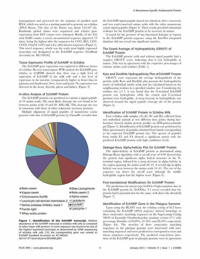
expressions from EST counts were estimated. Briefly, of the 922
total EstID counts, a novel, un-annotated sequence appeared 13
times, being the highest after the sequences for CSN2, BLG, CSN3,
CSN2b, C6orf58, CSN1 and a few other known sequences (Figure 1).
This novel sequence, which was the tenth most highly expressed
transcripts was designated as the EchAMP sequence (GenBank
Accession no. KC148542).
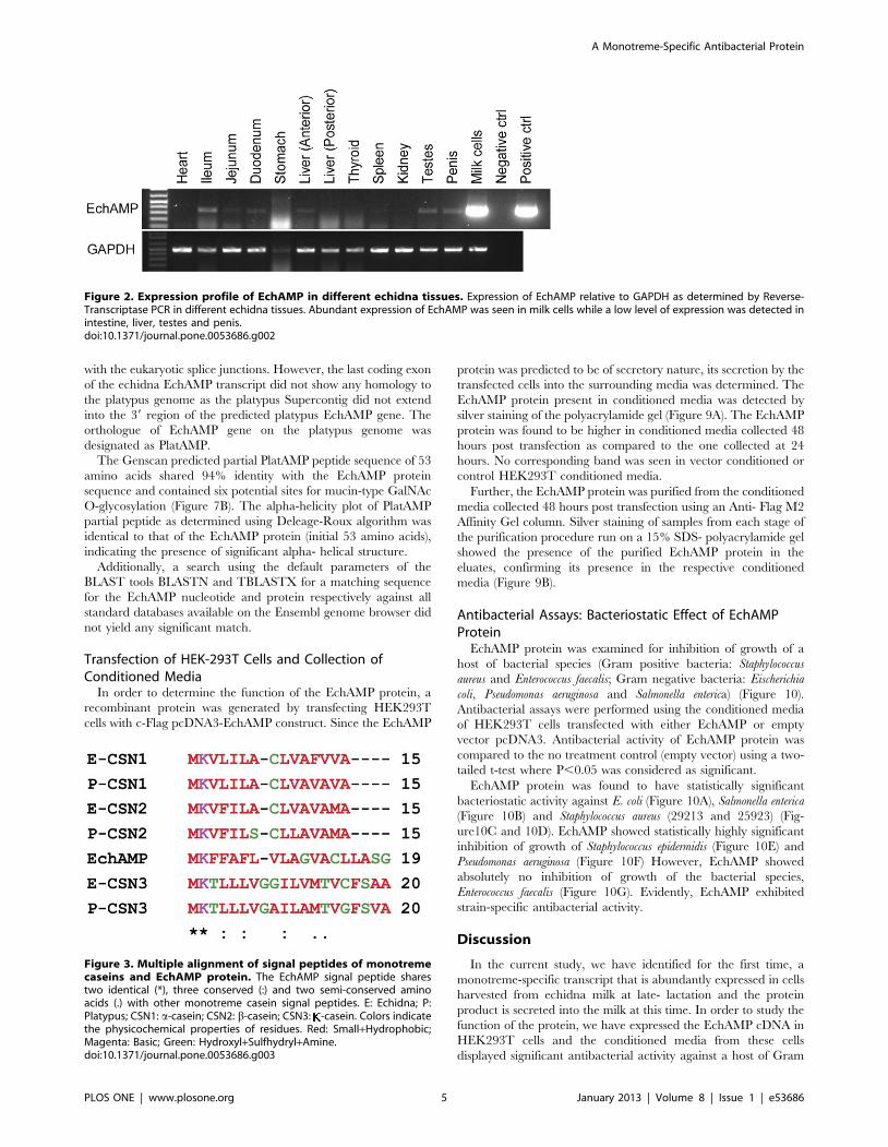
Tissue Expression Profile of EchAMP in EchidnaThe EchAMP gene expression was explored in different tissues
of echidna. Reverse transcriptase PCR analysis for EchAMP gene
relative to GAPDH showed that there was a high level of
expression of EchAMP in the milk cells and a low level of
expression in the intestine (comparatively higher in ileum than in
jejunum and duodenum), liver, testes and penis. No expression was
detected in the heart, thyroid, spleen and kidney (Figure 2).
In-silico Analysis of EchAMP ProteinThe EchAMP protein was predicted to contain a signal peptide
of 19 amino acids. The most likely cleavage site was found to be
between amino acids 19 and 20: ASG-AK. This cleavage site was
in consensus with those of other eukaryotic secretory proteins.
Multiple alignment of signal peptides of monotreme casein
proteins with that of EchAMP protein by ClustalW revealed that
the EchAMP signal peptide shared two identical, three conserved,
and two semi-conserved amino acids with the other monotreme
casein signal peptides (Figure 3). These results provided substantial
evidence for the EchAMP protein to be secretory in nature.
A search for the presence of any functional domains or repeats
in the EchAMP protein sequence using the InterPro integrated
database did not reveal any significant matches.
The Grand Average of Hydropathicity (GRAVY) ofEchAMP Protein
The EchAMP protein (with and without signal peptide) had a
negative GRAVY score, indicating that it was hydrophilic in
nature. This was in agreement with the respective percentages of
cationic amino acid residues (Table 1).
Kyte and Doolittle Hydropathicity Plot of EchAMP ProteinGRAVY score represents the average hydropathicity of the
protein while Kyte and Doolittle plot presents the hydropathicity
scores of individual amino acids and are calculated based on the
neighbouring residues in a specified window size. Considering the
window size n = 7, it was found that the N-terminal EchAMP
protein was hydrophobic while the central and C-terminal
portions were hydrophilic. A steep increase in hydrophilicity was
observed around the signal peptide cleavage site of the protein
(Figure 4).
Identification of EchAMP Protein in Echidna MilkFour echidna milk samples (A1,A2, B1 and B2) collected from
two individual animals at two different time points during late-
lactation showed similar protein profiles on SDS-polyacrylamide
gel (Figure 5). Identification of EchAMP protein was performed by
Mass spectrometry of peptides obtained from bands corresponding
to the expected EchAMP protein size. The spectra of peptides
from bands E3 and E4 showed a significant match with the
predicted EchAMP protein with high confidence levels.
Deleage-Roux Alpha-helicity Plot for EchAMP ProteinThe alpha-helicity of EchAMP protein as determined using
Deleage-Roux algorithm with a cut-off score of 0.99 showed that
the protein had significant alpha helical structure in the N-
terminal region, followed by a steep decrease in alpha helicity in
the region spanning the amino acids 20–23. A second dip in alpha
helicity was seen between the amino acids 55–65. The rest of the
sequence was above the cut-off score although the middle
hydrophilic region had the highest score (Figure 6).
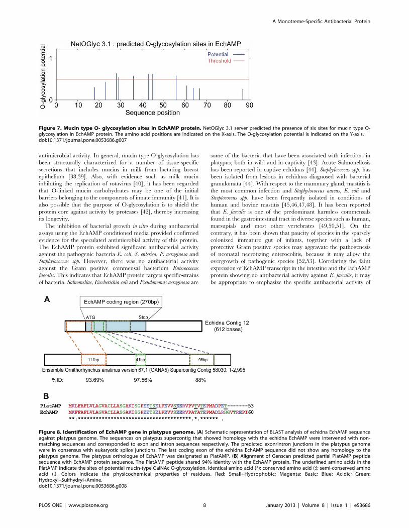
Post-translational Modifications for EchAMP ProteinThe predictions for mucin type GalNAc O-glycosylation sites in
the EchAMP protein by NetOGlyc 3.1 server revealed that the
protein had 6 potential sites for the same, well above the threshold
(Figure 7).
Identification of EchAMP Gene in the Platypus GenomeUpon using the BLAST tool, the echidna contig of 612 bases
containing the EchAMP cDNA sequence showed homology to
three consecutive matching sequences in the Supercontig Contig
58030 of Ensemble Ornithorhynchus anatinus version 67.1 with
percentage identities of 93.69%, 97.56% and 88.42% respectively
(Figure 8A). The stretches of three consecutive matching
sequences in the platypus genome were intervened with non-
matching sequences and were predicted to correspond to exon and
intron sequences respectively. The predicted exon/intron junc-
tions of the EchAMP gene in platypus genome were in agreement
Figure 1. Identification of the EchAMP transcript. Relativeabundance of the EchAMP transcript in echidna milk cells as comparedto other major milk proteins. A novel sequence was found to be one ofthe highest expressed transcripts as determined by cDNA sequencingof echidna milk cells [15]; the corresponding gene was named asEchAMP (GenBank Accession no. KC148542).doi:10.1371/journal.pone.0053686.g001
A Monotreme-Specific Antibacterial Protein
PLOS ONE | www.plosone.org 4 January 2013 | Volume 8 | Issue 1 | e53686
with the eukaryotic splice junctions. However, the last coding exon
of the echidna EchAMP transcript did not show any homology to
the platypus genome as the platypus Supercontig did not extend
into the 39 region of the predicted platypus EchAMP gene. The
orthologue of EchAMP gene on the platypus genome was
designated as PlatAMP.
The Genscan predicted partial PlatAMP peptide sequence of 53
amino acids shared 94% identity with the EchAMP protein
sequence and contained six potential sites for mucin-type GalNAc
O-glycosylation (Figure 7B). The alpha-helicity plot of PlatAMP
partial peptide as determined using Deleage-Roux algorithm was
identical to that of the EchAMP protein (initial 53 amino acids),
indicating the presence of significant alpha- helical structure.
Additionally, a search using the default parameters of the
BLAST tools BLASTN and TBLASTX for a matching sequence
for the EchAMP nucleotide and protein respectively against all
standard databases available on the Ensembl genome browser did
not yield any significant match.
Transfection of HEK-293T Cells and Collection ofConditioned Media
In order to determine the function of the EchAMP protein, a
recombinant protein was generated by transfecting HEK293T
cells with c-Flag pcDNA3-EchAMP construct. Since the EchAMP
protein was predicted to be of secretory nature, its secretion by the
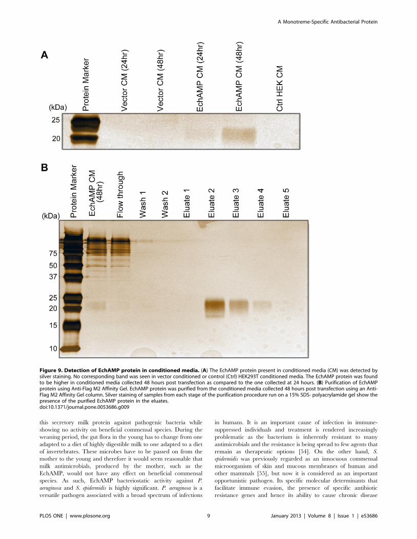
transfected cells into the surrounding media was determined. The
EchAMP protein present in conditioned media was detected by
silver staining of the polyacrylamide gel (Figure 9A). The EchAMP
protein was found to be higher in conditioned media collected 48
hours post transfection as compared to the one collected at 24
hours. No corresponding band was seen in vector conditioned or
control HEK293T conditioned media.
Further, the EchAMP protein was purified from the conditioned
media collected 48 hours post transfection using an Anti- Flag M2
Affinity Gel column. Silver staining of samples from each stage of
the purification procedure run on a 15% SDS- polyacrylamide gel
showed the presence of the purified EchAMP protein in the
eluates, confirming its presence in the respective conditioned
media (Figure 9B).
Antibacterial Assays: Bacteriostatic Effect of EchAMPProtein
EchAMP protein was examined for inhibition of growth of a
host of bacterial species (Gram positive bacteria: Staphylococcus
aureus and Enterococcus faecalis; Gram negative bacteria: Eischerichia
coli, Pseudomonas aeruginosa and Salmonella enterica) (Figure 10).
Antibacterial assays were performed using the conditioned media
of HEK293T cells transfected with either EchAMP or empty
vector pcDNA3. Antibacterial activity of EchAMP protein was
compared to the no treatment control (empty vector) using a two-
tailed t-test where P,0.05 was considered as significant.
EchAMP protein was found to have statistically significant
bacteriostatic activity against E. coli (Figure 10A), Salmonella enterica
(Figure 10B) and Staphylococcus aureus (29213 and 25923) (Fig-
ure10C and 10D). EchAMP showed statistically highly significant
inhibition of growth of Staphylococcus epidermidis (Figure 10E) and
Pseudomonas aeruginosa (Figure 10F) However, EchAMP showed
absolutely no inhibition of growth of the bacterial species,
Enterococcus faecalis (Figure 10G). Evidently, EchAMP exhibited
strain-specific antibacterial activity.
Discussion
In the current study, we have identified for the first time, a
monotreme-specific transcript that is abundantly expressed in cells
harvested from echidna milk at late- lactation and the protein
product is secreted into the milk at this time. In order to study the
function of the protein, we have expressed the EchAMP cDNA in
HEK293T cells and the conditioned media from these cells
displayed significant antibacterial activity against a host of Gram
Figure 2. Expression profile of EchAMP in different echidna tissues. Expression of EchAMP relative to GAPDH as determined by Reverse-Transcriptase PCR in different echidna tissues. Abundant expression of EchAMP was seen in milk cells while a low level of expression was detected inintestine, liver, testes and penis.doi:10.1371/journal.pone.0053686.g002
Figure 3. Multiple alignment of signal peptides of monotremecaseins and EchAMP protein. The EchAMP signal peptide sharestwo identical (*), three conserved (:) and two semi-conserved aminoacids (.) with other monotreme casein signal peptides. E: Echidna; P:Platypus; CSN1: a-casein; CSN2: b-casein; CSN3: -casein. Colors indicatethe physicochemical properties of residues. Red: Small+Hydrophobic;Magenta: Basic; Green: Hydroxyl+Sulfhydryl+Amine.doi:10.1371/journal.pone.0053686.g003
A Monotreme-Specific Antibacterial Protein
PLOS ONE | www.plosone.org 5 January 2013 | Volume 8 | Issue 1 | e53686

positive and Gram negative bacteria, confirming its predicted
activity. Consecutive sequences showing homology to the
EchAMP cDNA were found in the platypus genome, thereby
indicating the possibility of its orthologue.
Harvesting cells from the milk of protected species such as
monotremes has been pursued as a non-invasive approach for the
analysis of their lactation [15]. These milk cells were an ambiguous
mixture that could have included skin cells, immune cells,
exfoliated epithelial cells from ducts and mammary or sebaceous
glands. Presence of somatic cells in the milk of many other extant
mammals such as sheep, cattle and humans have been reported
[25,26,27]. In the current case, it was observed with cDNA
sequencing that several casein and whey protein gene cDNAs were
detected at high levels, indicating that monotreme milk cells
harvested during peak lactation are enriched in exfoliated
mammary epithelial cells. The sequence for EchAMP showed
relatively high abundance, after the sequences for CSN2, BLG,
CSN3, CSN2b, and CSN1 which are major milk protein genes.
Transcripts with relatively lower abundance than EchAMP were
other major milk protein genes such as lysozyme and WAP
suggesting that EchAMP plays a potentially prominent role during
the lactation of echidna. The non-availability of cells or RNA from
the mammary gland of a non-lactating echidna constrained us
from determining the endogenous expression of EchAMP during
the non-lactating period.
In silico analyses indicated that the cognate protein of EchAMP
would be hydrophilic and secretory in nature. This was confirmed
through the identification of peptides corresponding to EchAMP
protein in a sample of echidna milk. Although monotreme milk
has not been extensively studied, some of their main components
have been described, such as caseins [15] and whey proteins
including alpha- lactalbumin [28,29], lysozyme [30], and WAP
[31].
The discovery of three consecutive sequences in platypus
genome showing homology to EchAMP cDNA with high
percentage identity provided considerable evidence for the
presence of orthologue of EchAMP gene in platypus, designated
as PlatAMP. It is possible that the last coding exon of the echidna
EchAMP transcript did not find any homology sequence in the
platypus genome probably due to the latter’s incompleteness.
Conversely, the variation of the sequences for the same gene
among echidna and platypus, which are estimated to have shared
the last common ancestor about 21.2 million years ago [32] cannot
be ruled out. Insufficient platypus RNA samples curtailed further
attempts to derive the matching sequence for the last coding part
of the echidna EchAMP transcript by approaches such as PCR
(Polymerase chain reaction) or RACE (Rapid amplification of
cDNA ends). Nevertheless, it is noteworthy that the predicted
PlatAMP partial peptide sequence of platypus was found to be
highly similar (94%) to the EchAMP protein sequence and also
shared similar features of alpha-helicity and post- translational
modifications, indicating similar function. However, the expres-
sion of PlatAMP gene in the mammary/milk cells of platypus
needs to be confirmed. With no significant matching sequence in
any of the standard databases available at the Ensemble Genome
Browser, we propose the EchAMP gene and hence its protein to
be specific to monotremes alone.
Monotreme display a component of synapsidal reproduction by
laying eggs that are incubated in the external, non-sterile
environment and milk is the only source of nutrition during the
period of suckling, which is prolonged relative to gestation and
incubation [7], except for a short weaning period [6] It has often
been speculated that during evolution, the protolacteal secretions
Table1. GRAVY of EchAMP protein.
Protein GRAVY No. of cationic residues No. of anionic residues
EchAMP (full length; 90 aa) 20.348 14 14
EchAMP (without signal sequence; 71 aa) 20.977 13 14
GRAVY (grand average of hydropathicity) is the computed mean of hydrophobicity and hydrophilicity values for individual amino acid residues. A negative GRAVYindicates hydrophilicity while a positive value indicates hydrophobicity. aa: amino acids.doi:10.1371/journal.pone.0053686.t001
Figure 4. Kyte and Doolittle hydropathicity plot for EchAMP protein. Amino acid position is presented on the X-axis. Kyte and Doolittlehydropathicity scores (window size n = 7) for individual amino acids are on the Y-axis. The N-terminal region of EchAMP protein is hydrophobic whilethe central and C-terminal portions are hydrophilic. A steep increase in hydrophilicity is observed around the signal peptide cleavage site of theprotein.doi:10.1371/journal.pone.0053686.g004
A Monotreme-Specific Antibacterial Protein
PLOS ONE | www.plosone.org 6 January 2013 | Volume 8 | Issue 1 | e53686

enhanced the survival of eggs or the young by the virtue of their
antimicrobial properties [9,33,34]. The same concept has been
extended to monotremes, and it has been hypothesized that the
survival of egg or the young is enhanced by microbial inhibitors of
cutaneous or mammary gland origin [8]. These speculations led us
to determine if a novel monotreme- specific milk protein such as
EchAMP displayed any protective attributes. Indeed it appeared
that EchAMP protein contained an overall significant alpha-
helical structure, indicative of antimicrobial activity [35,36]. In
addition, the predictions from the Antimicrobial Peptide Database
[37] for EchAMP protein sequence were suggestive of its possible
antimicrobial potential by interacting with membranes by its
virtue to form alpha helices (data not shown). Similarly, the
predicted post-translational modification of the EchAMP protein,
especially the occurrence of the mucin type GalNAc O-glycosyl-
ation sites also suggested that EchAMP protein may carry
Figure 5. Identification of EchAMP protein in echidna milk. A1, A2 and B1, B2 represent milk samples collected from two lactating echidnas attwo different time points during their late-lactation phase. 70 mg protein of each sample was electrophoresed for 3 hours at 100V using a 12% SDS-Polyacrylamide gel. Bands E3 and E4 were excised from the gel, subjected to in-gel trypsin digestion and anlaysed by LTQ Orbitrap Velos. The spectraof peptides from these bands showed a significant match with the predicted EchAMP protein with high confidence levels.doi:10.1371/journal.pone.0053686.g005
Figure 6. Deleage-Roux apha-helicity plot for EchAMP protein. The amino acid positions are indicated on the X-axis. The alpha-helicity scoresare indicated on the Y- axis. The cut-off score was taken as 0.99. The EchAMP protein has significant alpha helical structure in the N-terminal region,followed by a steep decrease in the region spanning the amino acids 20–23. A second dip in alpha helicity is seen between the amino acids 55–65.doi:10.1371/journal.pone.0053686.g006
A Monotreme-Specific Antibacterial Protein
PLOS ONE | www.plosone.org 7 January 2013 | Volume 8 | Issue 1 | e53686
antimicrobial activity. In general, mucin type O-glycosylation has
been structurally characterized for a number of tissue-specific
secretions that includes mucins in milk from lactating breast
epithelium [38,39]. Also, with evidence such as milk mucin
inhibiting the replication of rotavirus [40], it has been regarded
that O-linked mucin carbohydrates may be one of the initial
barriers belonging to the components of innate immunity [41]. It is
also possible that the purpose of O-glycosylation is to shield the
protein core against activity by proteases [42], thereby increasing
its longevity.
The inhibition of bacterial growth in vitro during antibacterial
assays using the EchAMP conditioned media provided confirmed
evidence for the speculated antimicrobial activity of this protein.
The EchAMP protein exhibited significant antibacterial activity
against the pathogenic bacteria E. coli, S. enterica, P. aeruginosa and
Staphylococcus spp. However, there was no antibacterial activity
against the Gram positive commensal bacterium Enterococcus
faecalis. This indicates that EchAMP protein targets specific-strains
of bacteria. Salmonellae, Eischerichia coli and Pseudomonas aeruginosa are
some of the bacteria that have been associated with infections in
platypus, both in wild and in captivity [43]. Acute Salmonellosis
has been reported in captive echidnas [44]. Staphylococcus spp. has
been isolated from lesions in echidnas diagnosed with bacterial
granulomata [44]. With respect to the mammary gland, mastitis is
the most common infection and Staphylococcus aureus, E. coli and
Streptococcus spp. have been frequently isolated in conditions of
human and bovine mastitis [45,46,47,48]. It has been reported
that E. faecalis is one of the predominant harmless commensals
found in the gastrointestinal tract in diverse species such as human,
marsupials and most other vertebrates [49,50,51]. On the
contrary, it has been shown that paucity of species in the sparsely
colonized immature gut of infants, together with a lack of
protective Gram positive species may aggravate the pathogenesis
of neonatal necrotizing enterocolitis, because it may allow the
overgrowth of pathogenic species [52,53]. Correlating the faint
expression of EchAMP transcript in the intestine and the EchAMP
protein showing no antibacterial activity against E. faecalis, it may
be appropriate to emphasize the specific antibacterial activity of
Figure 7. Mucin type O- glycosylation sites in EchAMP protein. NetOGlyc 3.1 server predicted the presence of six sites for mucin type O-glycosylation in EchAMP protein. The amino acid positions are indicated on the X-axis. The O-glycosylation potential is indicated on the Y-axis.doi:10.1371/journal.pone.0053686.g007
Figure 8. Identification of EchAMP gene in platypus genome. (A) Schematic representation of BLAST analysis of echidna EchAMP sequenceagainst platypus genome. The sequences on platypus supercontig that showed homology with the echidna EchAMP were intervened with non-matching sequences and corresponded to exon and intron sequences respectively. The predicted exon/intron junctions in the platypus genomewere in consensus with eukaryotic splice junctions. The last coding exon of the echidna EchAMP sequence did not show any homology to theplatypus genome. The platypus orthologue of EchAMP was designated as PlatAMP. (B) Alignment of Genscan predicted partial PlatAMP peptidesequence with EchAMP protein sequence. The PlatAMP peptide shared 94% identity with the EchAMP protein. The underlined amino acids in thePlatAMP indicate the sites of potential mucin-type GalNAc O-glycosylation. Identical amino acid (*); conserved amino acid (:); semi-conserved aminoacid (.). Colors indicate the physicochemical properties of residues. Red: Small+Hydrophobic; Magenta: Basic; Blue: Acidic; Green:Hydroxyl+Sulfhydryl+Amine.doi:10.1371/journal.pone.0053686.g008
A Monotreme-Specific Antibacterial Protein
PLOS ONE | www.plosone.org 8 January 2013 | Volume 8 | Issue 1 | e53686
this secretory milk protein against pathogenic bacteria while
showing no activity on beneficial commensal species. During the
weaning period, the gut flora in the young has to change from one
adapted to a diet of highly digestible milk to one adapted to a diet
of invertebrates. These microbes have to be passed on from the
mother to the young and therefore it would seem reasonable that
milk antimicrobials, produced by the mother, such as the
EchAMP, would not have any effect on beneficial commensal
species. As such, EchAMP bacteriostatic activity against P.
aeruginosa and S. epidermidis is highly significant. P. aeruginosa is a
versatile pathogen associated with a broad spectrum of infections
in humans. It is an important cause of infection in immune-
suppressed individuals and treatment is rendered increasingly
problematic as the bacterium is inherently resistant to many
antimicrobials and the resistance is being spread to few agents that
remain as therapeutic options [54]. On the other hand, S.
epidermidis was previously regarded as an innocuous commensal
microorganism of skin and mucous membranes of human and
other mammals [55], but now it is considered as an important
opportunistic pathogen. Its specific molecular determinants that
facilitate immune evasion, the presence of specific antibiotic
resistance genes and hence its ability to cause chronic disease
Figure 9. Detection of EchAMP protein in conditioned media. (A) The EchAMP protein present in conditioned media (CM) was detected bysilver staining. No corresponding band was seen in vector conditioned or control (Ctrl) HEK293T conditioned media. The EchAMP protein was foundto be higher in conditioned media collected 48 hours post transfection as compared to the one collected at 24 hours. (B) Purification of EchAMPprotein using Anti-Flag M2 Affinity Gel. EchAMP protein was purified from the conditioned media collected 48 hours post transfection using an Anti-Flag M2 Affinity Gel column. Silver staining of samples from each stage of the purification procedure run on a 15% SDS- polyacrylamide gel show thepresence of the purified EchAMP protein in the eluates.doi:10.1371/journal.pone.0053686.g009
A Monotreme-Specific Antibacterial Protein
PLOS ONE | www.plosone.org 9 January 2013 | Volume 8 | Issue 1 | e53686
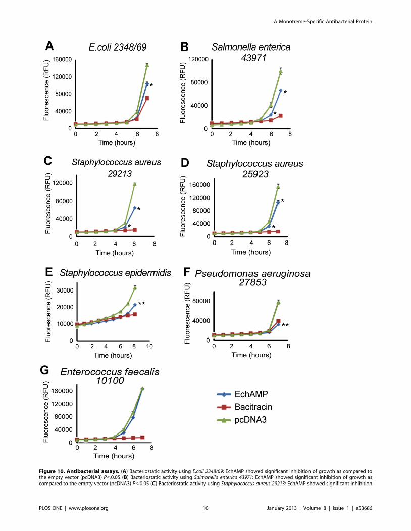
Figure 10. Antibacterial assays. (A) Bacteriostatic activity using E.coli 2348/69: EchAMP showed significant inhibition of growth as compared tothe empty vector (pcDNA3) P,0.05 (B) Bacteriostatic activity using Salmonella enterica 43971: EchAMP showed significant inhibition of growth ascompared to the empty vector (pcDNA3) P,0.05 (C) Bacteriostatic activity using Staphylococcus aureus 29213: EchAMP showed significant inhibition
A Monotreme-Specific Antibacterial Protein
PLOS ONE | www.plosone.org 10 January 2013 | Volume 8 | Issue 1 | e53686
makes it extremely difficult to treat [56,57]. It is generally seen that
infections from yet another Gram positive bacterium, S. aureus, are
usually caused from the same strain that the animal carries as a
commensal. Such infections can affect the blood stream, skin, soft
tissues and lower respiratory tracts [58,59] and perhaps antibac-
terial proteins in monotreme milk such as the EchAMP confer
protection to the young and the mammary gland of the mother
against such infections. The Gram negative bacterium S. enterica is
a highly host-adapted pathogen and its infections are a major
problem in humans as well as in livestock animals such as cattle,
pigs and chicken [60]. This bacterium is reported to contain
several pathogenicity islands which encode virulence factors that
induce inflammation in the host [61]. However, the bacterium is
able to exploit the same inflammation for nutrients and
outcompetes other bacterial species in the gut [62]. The other
Gram negative bacterium E. coli belongs to a group of
enteropathogens that exploit host epithelial cells and are the
major cause of infantile diarrhea [63,64].
Taken together, it is evident that EchAMP, a novel monotreme-
specific milk protein is capable of conferring protection to the
underdeveloped, immunologically naıve young outside the sterile
confines of the uterus, in the harsh pathogen-laden environments.
However, with the limitation of access to monotreme milk samples
across the lactation period, we are unable to deduce any specific
changes in expression of EchAMP during the lactation period.
The monotreme mammary gland lacks nipples, and therefore the
altricial young is more likely to ingest pathogens while suckling
compared to any other species. This is a significant difference
between monotremes and marsupials which also give birth to
altricial young, but the milk delivery is aided by a nipple to which
the young is attached continuously for the first 100 days of
lactation [65,66]. We propose that the evolution of nipples and
development of offspring in utero in the placental mammals
(Metatherian and Eutherian) could have led to the loss of selective
pressure for the preservation of this gene and hence its subsequent
disappearance in these species. For an example, there is a
precedence of selective loss of genes involved in gastric function in
platypus, which diverged from the Therian lineage early during
mammalian evolution. Not with-standing the high conservation in
vertebrates for more than 400 million years, genes encoding the
gastric proteases, hormone gastrin, both the subunits of the gastric
H+/K+-ATPase and the neurogenin-3 transcription factor have
either been deleted or inactivated in platypus genome, giving rise
to physiological differences in digestion between monotremes and
therians [67].
ConclusionsMonotremes are potential sources to discover new antimicro-
bials because they lay eggs and their subsequent development into
young, all occur in the non-sterile, ex-utero environment. For the
first time, we have identified a novel transcript that is specific to
monotremes and is abundantly expressed in milk cells during late-
lactation. The peptides corresponding to this transcript have been
identified in echidna milk. Conditioned media from HEK293T
cells expressing the EchAMP protein has been shown to display
antibacterial activity against a host of Gram positive and Gram
negative bacteria, while no activity was detected against a
commensal gut floral species. Our data support the hypothesis of
enhancement of survival of monotreme young by antimicrobial
bioactives of mammary gland origin [8,9]. Apart for a short
weaning period of about 12 days during which the young echidna
begins feeding outside the burrow but still takes milk from the
mother [6], milk is the sole source of nutrition for the altricial
hatchling during the period of suckling which is prolonged
compared to gestation and incubation of eggs [7]. The ex-utero
environment of the developing young is favorable for microbial
attack and as the monotreme mammary gland has no nipple, the
young is more likely to ingest microbial pathogens than the pouch
young of marsupials which are attached to the nipple. During
evolution, this would have favored the incorporation of antimi-
crobial agents into the glandular secretions in order to protect the
young [34]. Our data are consistent with the speculation that
monotreme genomes have evolved under evolutionary pressure to
protect immunologically naıve young with broad spectrum
antibiotics [68] and further emphasize an important, non-
nutritional role of monotreme milk.
Acknowledgments
We wish to thank Mr. Ashwantha Kumar Enjapoori and Ms. Ashalyn
Watt (Deakin University) for their technical guidance in performing the
antibacterial assays, Mr. Amit Kumar (Deakin University) for his assistance
in bioinformatic analysis and Mr. Heramb Kulkarni (CCMB) for his help
in proteomics- related work.
Author Contributions
Critical review of the manuscript: JS SK PR SN CL KN. Conceived and
designed the experiments: SB SK JS CL KN. Performed the experiments:
SB. Analyzed the data: SB SK JS CL KN. Contributed reagents/
materials/analysis tools: PR SN. Wrote the paper: SB.
References
1. Lefevre CM, Sharp JA, Nicholas KR (2010) Evolution of lactation: ancient
origin and extreme adaptations of the lactation system. Annu Rev Genomics
Hum Genet 11: 219–238.
2. Bininda-Emonds OR, Cardillo M, Jones KE, MacPhee RD, Beck RM, et al.
(2007) The delayed rise of present-day mammals. Nature 446: 507–512.
3. Hedges SB, Kumar S (2009) The timetree of life: OUP Oxford.
4. Nicol S (2003) Monotreme biology. Comp Biochem Physiol A Mol Integr
Physiol 136: 795–798.
5. Hayssen V (1993) Empirical and theoretical constraints on the evolution of
lactation. J Dairy Sci 76: 3213–3233.
6. Morrow GE, Nicol S (in press) Maternal Care in the Tasmanian echidna,Tachyglossus aculeatus setosus Australian Journal of Zoology.
7. Griffiths M (1978) The biology of the monotremes: Academic Press New York.8. Hayssen V, Blackburn D (1985) a-Lactalbumin and the origins of lactation.
Evolution 39: 1147–1149.
9. Oftedal OT (2012) The evolution of milk secretion and its ancient origins.Animal 6: 355–368.
10. Gillin FD, Reiner DS, Wang CS (1983) Human milk kills parasitic intestinalprotozoa. Science 221: 1290–1292.
11. Wanyonyi SS, Sharp JA, Khalil E, Lefevre C, Nicholas KR (2011) Tammar
wallaby mammary cathelicidins are differentially expressed during lactation and
of growth as compared to the empty vector (pcDNA3) P,0.05 (D) Bacteriostatic activity using Staphylococcus aureus 25923 : EchAMP showedsignificant inhibition of growth as compared to the empty vector (pcDNA3) P,0.05 (E) Bacteriostatic activity using Staphylococcus epidermidis :EchAMP showed highly significant inhibition of growth as compared to the empty vector (pcDNA3) P,0.05 (F) Bacteriostatic activity usingPseudomonas aeruginosa 27853: EchAMP showed highly significant inhibition of growth as compared to the empty vector (pcDNA3) P,0.05 (G)Bacteriostatic activity using Enterococcus faecalis 10100: EchAMP showed no inhibition of growth as compared to the empty vector and bacitracinP.0.05 (* Statistically significant result P,0.05). Each assay was performed in triplicate and the experiments were repeated at least thrice. Standarderror bars are indicated.doi:10.1371/journal.pone.0053686.g010
A Monotreme-Specific Antibacterial Protein
PLOS ONE | www.plosone.org 11 January 2013 | Volume 8 | Issue 1 | e53686
exhibit antimicrobial and cell proliferative activity. Comp Biochem Physiol A Mol
Integr Physiol 160: 431–439.12. Watt AP, Sharp JA, Lefevre C, Nicholas KR (2012) WFDC2 is differentially
expressed in the mammary gland of the tammar wallaby and provides immune
protection to the mammary gland and the developing pouch young. Dev CompImmunol 36: 584–590.
13. Manzoni P, Stolfi I, Messner H, Cattani S, Laforgia N, et al. (2012) Bovinelactoferrin prevents invasive fungal infections in very low birth weight infants: a
randomized controlled trial. Pediatrics 129: 116–123.
14. Iwamori T, Nukumi N, Itoh K, Kano K, Naito K, et al. (2010) Bacteriostaticactivity of Whey Acidic Protein (WAP). J Vet Med Sci 72: 621–625.
15. Lefevre CM, Sharp JA, Nicholas KR (2009) Characterisation of monotremecaseins reveals lineage-specific expansion of an ancestral casein locus in
mammals. Reprod Fertil Dev 21: 1015–1027.16. Petersen TN, Brunak S, von Heijne G, Nielsen H (2011) SignalP 4.0:
discriminating signal peptides from transmembrane regions. Nat Methods 8:
785–786.17. Thompson JD, Gibson TJ, Higgins DG (2002) Multiple sequence alignment
using ClustalW and ClustalX. Curr Protoc Bioinformatics Chapter 2: Unit 2 3.18. Hunter S, Jones P, Mitchell A, Apweiler R, Attwood TK, et al. (2012) InterPro
in 2011: new developments in the family and domain prediction database.
Nucleic Acids Research 40: D306–D312.19. Altschul SF, Gish W, Miller W, Myers EW, Lipman DJ (1990) Basic local
alignment search tool. J Mol Biol 215: 403–410.20. Kyte J, Doolittle RF (1982) A simple method for displaying the hydropathic
character of a protein. J Mol Biol 157: 105–132.21. Deleage G, Roux B (1987) An algorithm for protein secondary structure
prediction based on class prediction. Protein Eng 1: 289–294.
22. Julenius K, Molgaard A, Gupta R, Brunak S (2005) Prediction, conservationanalysis, and structural characterization of mammalian mucin-type O-
glycosylation sites. Glycobiology 15: 153–164.23. Sambrook J, Russell DW (2001) Molecular cloning: a laboratory manual: CSHL
press.
24. Shevchenko A, Tomas H, Havlis J, Olsen JV, Mann M (2006) In-gel digestionfor mass spectrometric characterization of proteins and proteomes. Nat Protoc 1:
2856–2860.25. Nel-Themaat L, Gomez MC, Damiani P, Wirtu G, Dresser BL, et al. (2007)
Isolation, culture and characterisation of somatic cells derived from semen andmilk of endangered sheep and eland antelope. Reprod Fertil Dev 19: 576–584.
26. Paape MJ, Tucker HA (1966) Somatic cell content variation in fraction-collected
milk. J Dairy Sci 49: 265–267.27. Lindquist S, Hansson L, Hernell O, Lonnerdal B, Normark J, et al. (1994)
Isolation of mRNA and genomic DNA from epithelial cells in human milk andamplification by PCR. Biotechniques 17: 692–696.
28. Shaw DC, Messer M, Scrivener AM, Nicholas KR, Griffiths M (1993) Isolation,
partial characterisation, and amino acid sequence of alpha-lactalbumin fromplatypus (Ornithorhynchus anatinus) milk. Biochim Biophys Acta 1161: 177–
186.29. Messer M, Griffiths M, Rismiller PD, Shaw DC (1997) Lactose synthesis in a
monotreme, the echidna (Tachyglossus aculeatus): isolation and amino acidsequence of echidna alpha-lactalbumin. Comp Biochem Physiol B Biochem Mol
Biol 118: 403–410.
30. Guss JM, Messer M, Costello M, Hardy K, Kumar V (1997) Structure of thecalcium-binding echidna milk lysozyme at 1.9 A resolution. Acta
Crystallogr D Biol Crystallogr 53: 355–363.31. Sharp JA, Lefevre C, Nicholas KR (2007) Molecular evolution of monotreme
and marsupial whey acidic protein genes. Evol Dev 9: 378–392.
32. Warren WC, Hillier LW, Marshall Graves JA, Birney E, Ponting CP, et al.(2008) Genome analysis of the platypus reveals unique signatures of evolution.
Nature 453: 175–183.33. Oftedal OT (2002) The mammary gland and its origin during synapsid
evolution. J Mammary Gland Biol Neoplasia 7: 225–252.
34. Blackburn DG, Hayssen V, Murphy CJ (1989) The origins of lactation and theevolution of milk: a review with new hypotheses. Mammal Review 19: 1–26.
35. Boman HG (2003) Antibacterial peptides: basic facts and emerging concepts.J Intern Med 254: 197–215.
36. Lai Y, Gallo RL (2009) AMPed up immunity: how antimicrobial peptides havemultiple roles in immune defense. Trends Immunol 30: 131–141.
37. Wang Z, Wang G (2004) APD: the Antimicrobial Peptide Database. Nucleic
Acids Res 32: D590–592.38. Hanisch FG, Uhlenbruck G, Peter-Katalinic J, Egge H, Dabrowski J, et al.
(1989) Structures of neutral O-linked polylactosaminoglycans on human skimmilk mucins. A novel type of linearly extended poly-N-acetyllactosamine
backbones with Gal beta(1–4)GlcNAc beta(1–6) repeating units. J Biol Chem
264: 872–883.
39. Hanisch FG, Peter-Katalinic J, Egge H, Dabrowski U, Uhlenbruck G (1990)Structures of acidic O-linked polylactosaminoglycans on human skim milk
mucins. Glycoconj J 7: 525–543.
40. Yolken RH, Peterson JA, Vonderfecht SL, Fouts ET, Midthun K, et al. (1992)
Human milk mucin inhibits rotavirus replication and prevents experimentalgastroenteritis. J Clin Invest 90: 1984–1991.
41. Hanisch FG (2001) O-glycosylation of the mucin type. Biol Chem 382: 143–149.
42. Kozarsky K, Kingsley D, Krieger M (1988) Use of a mutant cell line to study the
kinetics and function of O-linked glycosylation of low density lipoproteinreceptors. Proc Natl Acad Sci U S A 85: 4335–4339.
43. Munday BL, Whittington RJ, Stewart NJ (1998) Disease conditions andsubclinical infections of the platypus (Ornithorhynchus anatinus). Philos
Trans R Soc Lond B Biol Sci 353: 1093–1099.
44. McOrist S, Smales L (1986) Morbidity and mortality of free-living and captive
echidnas, Tachyglossus aculeatus (Shaw), in Australia. J Wildl Dis 22: 375–380.
45. Barkema HW, Green MJ, Bradley AJ, Zadoks RN (2009) Invited review: The
role of contagious disease in udder health. J Dairy Sci 92: 4717–4729.
46. Borm AA, Fox LK, Leslie KE, Hogan JS, Andrew SM, et al. (2006) Effects ofprepartum intramammary antibiotic therapy on udder health, milk production,
and reproductive performance in dairy heifers. J Dairy Sci 89: 2090–2098.
47. Bradley AJ, Green MJ (2001) Adaptation of Escherichia coli to the bovine
mammary gland. J Clin Microbiol 39: 1845–1849.
48. Oeding P (1952) Examinations on penicillin resistant, serologically homogeneous
staphylococci isolated from human mastitis. Acta Pathol Microbiol Scand 31:
145–163.
49. Fanaro S, Chierici R, Guerrini P, Vigi V (2003) Intestinal microflora in earlyinfancy: composition and development. Acta Paediatr Suppl 91: 48–55.
50. Yadav M, Stanley NF, Waring H (1972) The microbial flora of the gut of thepouch-young and the pouch of a marsupial, Setonix brachyurus. J Gen
Microbiol 70: 437–442.
51. Witte W, Wirth R, Klare I (1999) Enterococci. Chemotherapy 45: 135–145.
52. Gewolb IH, Schwalbe RS, Taciak VL, Harrison TS, Panigrahi P (1999) Stoolmicroflora in extremely low birthweight infants. Arch Dis Child Fetal Neonatal
Ed 80: F167–173.
53. Claud EC, Walker WA (2001) Hypothesis: inappropriate colonization of the
premature intestine can cause neonatal necrotizing enterocolitis. FASEB J 15:1398–1403.
54. Kerr KG, Snelling AM (2009) Pseudomonas aeruginosa: a formidable and ever-present adversary. J Hosp Infect 73: 338–344.
55. Kloos W, Schleifer K (1986) Genus IV–Staphylococcus Rosenbach 1884.Bergey’s Manual of Systemic Bacteriology 2.
56. Otto M (2009) Staphylococcus epidermidis–the ‘accidental’ pathogen. Nat RevMicrobiol 7: 555–567.
57. Heikkonen L, Palmu A, Bostman O (1986) Multiresistant Staphylococcus
epidermidis as a pathogen. Ann Clin Res 18: 80–83.
58. Williams RE, Jevons MP, Shooter RA, Hunter CJ, Girling JA, et al. (1959) Nasal
staphylococci and sepsis in hospital patients. Br Med J 2: 658–662.
59. Plata K, Rosato AE, Wegrzyn G (2009) Staphylococcus aureus as an infectious
agent: overview of biochemistry and molecular genetics of its pathogenicity. ActaBiochim Pol 56: 597–612.
60. Gopinath S, Carden S, Monack D (2012) Shedding light on Salmonella carriers.Trends Microbiol.
61. Haraga A, Ohlson MB, Miller SI (2008) Salmonellae interplay with host cells.Nat Rev Microbiol 6: 53–66.
62. Winter SE, Thiennimitr P, Winter MG, Butler BP, Huseby DL, et al. (2010) Gutinflammation provides a respiratory electron acceptor for Salmonella. Nature
467: 426–429.
63. DeVinney R, Gauthier A, Abe A, Finlay BB (1999) Enteropathogenic
Escherichia coli: a pathogen that inserts its own receptor into host cells. CellMol Life Sci 55: 961–976.
64. Finlay BB, Ruschkowski S, Kenny B, Stein M, Reinscheid DJ, et al. (1996)Enteropathogenic E. coli exploitation of host epithelial cells. Ann N Y Acad Sci
797: 26–31.
65. Tyndale-Biscoe CH, Janssens PA (1988) The developing marsupial. Models for
biomedical research: Springer-Verlag.
66. Nicholas K, Simpson K, Wilson M, Trott J, Shaw D (1997) The tammar
wallaby: a model to study putative autocrine-induced changes in milkcomposition. J Mammary Gland Biol Neoplasia 2: 299–310.
67. Ordonez GR, Hillier LW, Warren WC, Grutzner F, Lopez-Otin C, et al. (2008)Loss of genes implicated in gastric function during platypus evolution. Genome
Biol 9: R81.
68. Wang J, Wong ES, Whitley JC, Li J, Stringer JM, et al. (2011) Ancient
antimicrobial peptides kill antibiotic-resistant pathogens: Australian mammalsprovide new options. PLoS One 6: e24030.
A Monotreme-Specific Antibacterial Protein
PLOS ONE | www.plosone.org 12 January 2013 | Volume 8 | Issue 1 | e53686
Related Documents