Specialization in the Medial Temporal Lobe for Processing of Objects and Scenes Andy C.H. Lee, 1* Mark J. Buckley, 2 Sarah J. Pegman, 1 Hugo Spiers, 1 Victoria L. Scahill, 1 David Gaffan, 2 Timothy J. Bussey, 3 R. Rhys Davies, 4 Narinder Kapur, 5 John R. Hodges, 1,4 and Kim S. Graham 1 ABSTRACT: There has been considerable debate as to whether the hippocampus and perirhinal cortex may subserve both memory and per- ception. We administered a series of oddity tasks, in which subjects selected the odd stimulus from a visual array, to amnesic patients with either selective hippocampal damage (HC group) or more extensive medial temporal damage, including the perirhinal cortex (MTL group). All patients performed normally when the stimuli could be discrimi- nated using simple visual features, even if faces or complex virtual real- ity scenes were presented. Both patient groups were, however, severely impaired at scene discrimination when a significant demand was placed on processing spatial information across viewpoint independent repre- sentations, while only the MTL group showed a significant deficit in oddity judgments of faces and objects when object viewpoint inde- pendent perception was emphasized. These observations provide com- pelling evidence that the human hippocampus and perirhinal cortex are critical to processes beyond long-term declarative memory and may subserve spatial and object perception, respectively. V V C 2005 Wiley-Liss, Inc. KEY WORDS: hippocampus; perirhinal cortex; perception; memory; amnesia INTRODUCTION The different medial temporal lobe (MTL) structures, including the hippocampus, entorhinal cortex (EC), perirhinal cortex, and parahippo- campal gyrus, are thought to comprise a single long-term declarative memory system (Squire and Zola-Morgan, 1991; Zola-Morgan et al., 1994). Recent studies have, however, challenged this view and suggested that these structures may, in fact, subserve different long-term memory processes. In particular, the perirhinal cortex (BA 35/36) may mediate object recognition on the basis of familiarity and memory for stimulus– stimulus associations, while the hippocampus may subserve memory rec- ollection and navigation (Meunier et al., 1993; Murray et al., 1993; Eacott et al., 1994; Maguire et al., 1997; Buckley and Gaffan, 1998a; Aggleton and Brown, 1999; Burgess et al., 2002; Henson et al., 2003; Winters et al., 2004. Lately, there has been considerable debate as to whether specific MTL structures may also support the representation and processing of perceptual informa- tion beyond the domain of long-term declarative memory (referred to hereafter as ‘‘perception’’), with the hippocampus and perirhinal cortex contributing to spatial (O’Keefe, 1999; Gaffan, 2001) and object per- ception (Murray and Bussey, 1999; Buckley et al., 2001; Bussey et al., 2002, 2003) respectively. Animal lesion studies have suggested that the hippocampus is critical for tasks that recruit spatial memory (Morris et al., 1982; Murray et al., 1989, 1993; Hampton et al., 2004). Moreover, both animals (O’Keefe, 1976; Ono et al., 1991, 1993; Wilson and McNaughton, 1993; O’Keefe and Burgess, 1996; O’Keefe et al., 1998; Robertson et al., 1998; Hori et al., 2003) and humans (Ekstrom et al., 2003) possess hippocampal place or spatial-view cells that may signal aspects of spatial location and navigation. On the other hand, perirhinal lesioned monkeys show significant impair- ments in visual object concurrent discrimination when large stimulus set sizes, multiple distracting stimuli, or different stimulus orientations across trials are employed (Buckley and Gaffan, 1997, 1998b). One model of perirhinal cortex function (Murray and Bussey, 1999; Bussey and Saksida, 2002; Bussey et al., 2002) proposes that the ventral visual process- ing stream (Ungerleider and Mishkin, 1982) may cul- minate in the perirhinal cortex. Thus, caudal infero- temporal cortical regions (e.g., V4, TE/TEO) process simple stimulus features or basic object stimuli, while rostral regions, including the perirhinal cortex, process more complex conjunctions of stimulus features or objects and may underlie object perception. Support- ing this view, Bussey et al. (2003) found that monkeys with perirhinal cortex lesions were significantly impaired on the concurrent discrimination of pairs of two-dimensional images when the stimuli were com- posed of overlapping features, but not when the images possessed unique features. Although not a specific investigation of the feature conjunction hypothesis, Buckley et al. (2001) also found evidence for a role of the perirhinal cortex in object perception. Monkeys with perirhinal cortex 1 MRC Cognition and Brain Sciences Unit, 15 Chaucer Road, Cambridge, UK; 2 Department of Experimental Psychology, University of Oxford, Oxford, UK; 3 Department of Experimental Psychology, University of Cambridge, Cambridge, UK; 4 University Neurology Unit, University of Cambridge, Addenbrooke’s Hospital, Cambridge, UK; 5 Clinical Neuro- psychology, Addenbrooke’s Hospital, Cambridge, UK Grant sponsor: Alzheimer’s Research Trust, UK; Grant number: ART/ PG2002Cambridge. *Correspondence to: Dr. Andy Lee, MRC Cognition and Brain Sciences Unit, 15 Chaucer Road, Cambridge CB2 2EF, UK. E-mail: andy.lee@ mrc-cbu.cam.ac.uk Accepted for publication 19 May 2005 DOI 10.1002/hipo.20101 Published online 11 July 2005 in Wiley InterScience (www.interscience. wiley.com). HIPPOCAMPUS 15:782–797 (2005) V V C 2005 WILEY-LISS, INC.

Welcome message from author
This document is posted to help you gain knowledge. Please leave a comment to let me know what you think about it! Share it to your friends and learn new things together.
Transcript
Specialization in the Medial Temporal Lobe for Processingof Objects and Scenes
Andy C.H. Lee,1* Mark J. Buckley,2 Sarah J. Pegman,1 Hugo Spiers,1 Victoria L. Scahill ,1
David Gaffan,2 Timothy J. Bussey,3 R. Rhys Davies,4 Narinder Kapur,5
John R. Hodges,1,4 and Kim S. Graham1
ABSTRACT: There has been considerable debate as to whether thehippocampus and perirhinal cortex may subserve both memory and per-ception. We administered a series of oddity tasks, in which subjectsselected the odd stimulus from a visual array, to amnesic patients witheither selective hippocampal damage (HC group) or more extensivemedial temporal damage, including the perirhinal cortex (MTL group).All patients performed normally when the stimuli could be discrimi-nated using simple visual features, even if faces or complex virtual real-ity scenes were presented. Both patient groups were, however, severelyimpaired at scene discrimination when a significant demand was placedon processing spatial information across viewpoint independent repre-sentations, while only the MTL group showed a significant deficit inoddity judgments of faces and objects when object viewpoint inde-pendent perception was emphasized. These observations provide com-pelling evidence that the human hippocampus and perirhinal cortex arecritical to processes beyond long-term declarative memory and maysubserve spatial and object perception, respectively. VVC 2005 Wiley-Liss, Inc.
KEY WORDS: hippocampus; perirhinal cortex; perception; memory;amnesia
INTRODUCTION
The different medial temporal lobe (MTL) structures, including thehippocampus, entorhinal cortex (EC), perirhinal cortex, and parahippo-campal gyrus, are thought to comprise a single long-term declarativememory system (Squire and Zola-Morgan, 1991; Zola-Morgan et al.,1994). Recent studies have, however, challenged this view and suggestedthat these structures may, in fact, subserve different long-term memoryprocesses. In particular, the perirhinal cortex (BA 35/36) may mediateobject recognition on the basis of familiarity and memory for stimulus–stimulus associations, while the hippocampus may subserve memory rec-ollection and navigation (Meunier et al., 1993; Murray et al., 1993;Eacott et al., 1994; Maguire et al., 1997; Buckley and Gaffan, 1998a;
Aggleton and Brown, 1999; Burgess et al., 2002;Henson et al., 2003; Winters et al., 2004.
Lately, there has been considerable debate as towhether specific MTL structures may also support therepresentation and processing of perceptual informa-tion beyond the domain of long-term declarativememory (referred to hereafter as ‘‘perception’’), withthe hippocampus and perirhinal cortex contributing tospatial (O’Keefe, 1999; Gaffan, 2001) and object per-ception (Murray and Bussey, 1999; Buckley et al.,2001; Bussey et al., 2002, 2003) respectively. Animallesion studies have suggested that the hippocampus iscritical for tasks that recruit spatial memory (Morriset al., 1982; Murray et al., 1989, 1993; Hamptonet al., 2004). Moreover, both animals (O’Keefe, 1976;Ono et al., 1991, 1993; Wilson and McNaughton,1993; O’Keefe and Burgess, 1996; O’Keefe et al.,1998; Robertson et al., 1998; Hori et al., 2003) andhumans (Ekstrom et al., 2003) possess hippocampalplace or spatial-view cells that may signal aspects ofspatial location and navigation. On the other hand,perirhinal lesioned monkeys show significant impair-ments in visual object concurrent discrimination whenlarge stimulus set sizes, multiple distracting stimuli, ordifferent stimulus orientations across trials areemployed (Buckley and Gaffan, 1997, 1998b).
One model of perirhinal cortex function (Murrayand Bussey, 1999; Bussey and Saksida, 2002; Busseyet al., 2002) proposes that the ventral visual process-ing stream (Ungerleider and Mishkin, 1982) may cul-minate in the perirhinal cortex. Thus, caudal infero-temporal cortical regions (e.g., V4, TE/TEO) processsimple stimulus features or basic object stimuli, whilerostral regions, including the perirhinal cortex, processmore complex conjunctions of stimulus features orobjects and may underlie object perception. Support-ing this view, Bussey et al. (2003) found that monkeyswith perirhinal cortex lesions were significantlyimpaired on the concurrent discrimination of pairs oftwo-dimensional images when the stimuli were com-posed of overlapping features, but not when theimages possessed unique features.
Although not a specific investigation of the featureconjunction hypothesis, Buckley et al. (2001) alsofound evidence for a role of the perirhinal cortex inobject perception. Monkeys with perirhinal cortex
1MRC Cognition and Brain Sciences Unit, 15 Chaucer Road, Cambridge,UK; 2Department of Experimental Psychology, University of Oxford,Oxford, UK; 3Department of Experimental Psychology, University ofCambridge, Cambridge, UK; 4University Neurology Unit, University ofCambridge, Addenbrooke’s Hospital, Cambridge, UK; 5Clinical Neuro-psychology, Addenbrooke’s Hospital, Cambridge, UKGrant sponsor: Alzheimer’s Research Trust, UK; Grant number: ART/PG2002Cambridge.*Correspondence to: Dr. Andy Lee, MRC Cognition and Brain SciencesUnit, 15 Chaucer Road, Cambridge CB2 2EF, UK.E-mail: andy.lee@ mrc-cbu.cam.ac.ukAccepted for publication 19 May 2005DOI 10.1002/hipo.20101Published online 11 July 2005 in Wiley InterScience (www.interscience.wiley.com).
HIPPOCAMPUS 15:782–797 (2005)
VVC 2005 WILEY-LISS, INC.
ablations were assessed on a series of perceptual discriminationtasks in which subjects had to select the odd stimulus from anarray of similar items. The monkeys were impaired on discrim-inations that placed a significant demand on object perception,in particular, the ability to perceive and identify objects, includ-ing faces, from different viewpoints. In contrast, the ability todiscriminate objects that were presented from the same view-point remained intact, as did oddity judgment on the basis ofsimple object features (e.g., the perception of differences incolor, size, shape).
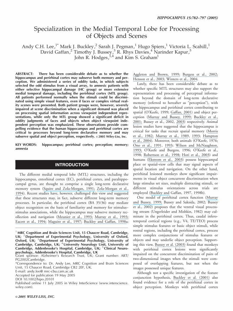
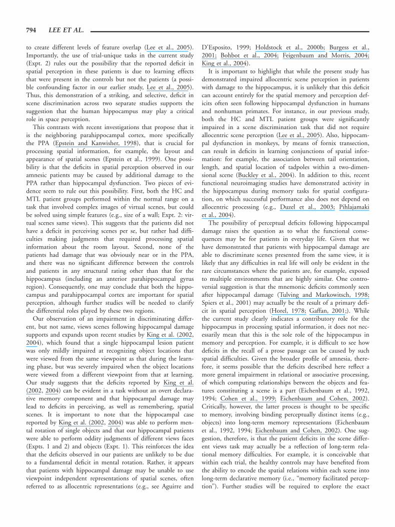
Evidence for a role of the human MTL in perception comesfrom a recent study conducted by our laboratory (Lee et al.,2005) in which fine visual discrimination of different stimuluscategories was assessed in a group of amnesic patients who hadselective hippocampal damage bilaterally (HC group), andanother group of amnesics who had larger bilateral MTL lesions(MTL group), including damage to both the hippocampus andperirhinal cortex (Table 1; Fig. 1). Consistent with the idea thatthe hippocampus and perirhinal cortex may be critical for spatialand object perception, respectively, the HC group demonstrateda selective deficit in the visual discrimination of spatial scenes,while the MTL group were significantly impaired in the discrimi-nation of spatial scenes, faces, and to a lesser extent objects.
While there has been, to our knowledge, no other study thathas examined the role of the hippocampus in spatial scene percep-tion, the findings of our recent experiment are in stark contrast toother neuropsychological investigations that have studied the roleof the human perirhinal cortex in object perception (Buffaloet al., 1998; Holdstock et al., 2000a). For instance, patients withfocal lesions that include the perirhinal cortex are able to matchvisual stimuli in tasks with minimal declarative memory demand(Buffalo et al., 1998; Holdstock et al., 2000a). Moreover, patientswith perirhinal cortex damage have been observed to perform nor-mally on oddity tasks (Stark and Squire, 2000, see also Levy et al.,2005) similar to those used in monkeys (Buckley et al., 2001).
A number of possible reasons may explain these discrepan-cies, notably (a) that the stimuli used in our original study werenot ‘‘trial-unique’’ (i.e., controls might show subtle learningeffects not present in our patients) and (b) that the reportedeffect sizes were extremely small and therefore inconclusive. In
addition, it has been proposed that the perceptual deficitsobserved in our patients could be due to additional atrophy tocortical areas that are known to be involved in the perceptionof faces and spatial scenes, such as the fusiform face area (FFA;Kanwisher et al., 1997) and the parahippocampal place area(PPA; Epstein and Kanwisher, 1998).
The aims of the current study were, therefore, 2-fold. First,we sought to determine whether the impairment in object per-ception demonstrated by the MTL group patients in the pre-vious study (Lee et al., 2005) is also evident in the context ofoddity tasks used previously in the animal literature (Buckleyet al., 2001). Second, we hoped to rule out categorically thepossibility that the object and spatial perception deficitsobserved in our earlier study (Lee et al., 2005) can beaccounted for by (a) the use of non-trial-unique stimuli and(b) concomitant atrophy to the FFA and PPA, respectively.
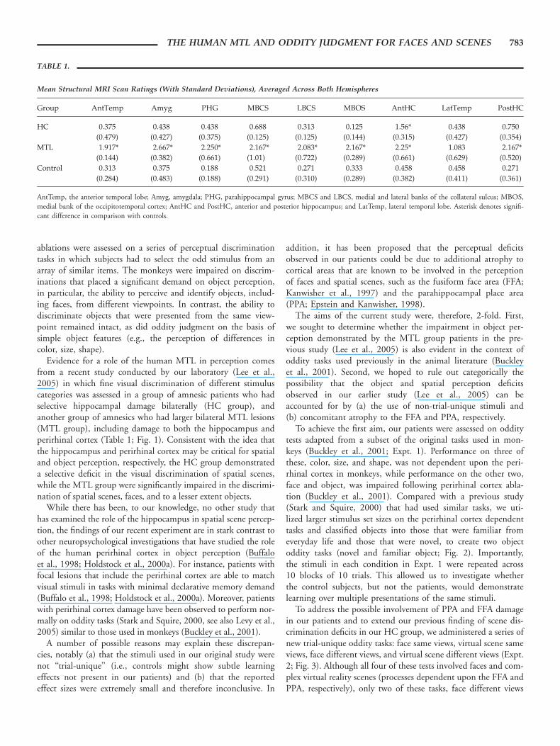
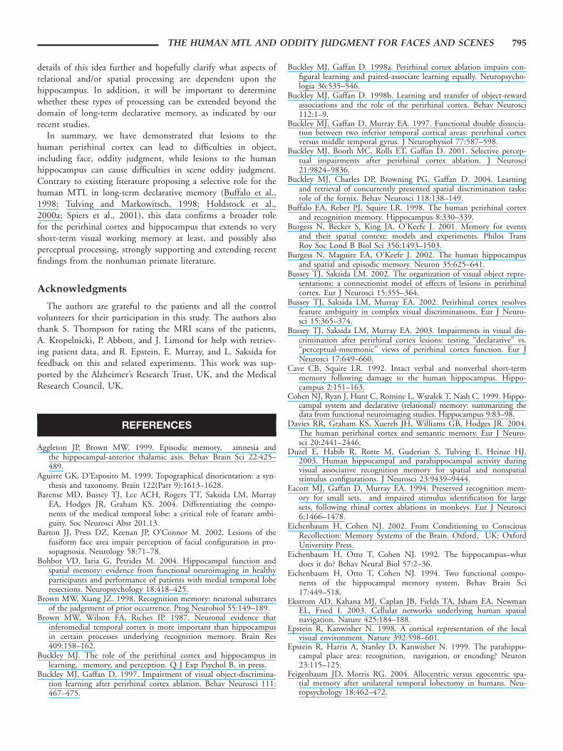
To achieve the first aim, our patients were assessed on odditytests adapted from a subset of the original tasks used in mon-keys (Buckley et al., 2001; Expt. 1). Performance on three ofthese, color, size, and shape, was not dependent upon the peri-rhinal cortex in monkeys, while performance on the other two,face and object, was impaired following perirhinal cortex abla-tion (Buckley et al., 2001). Compared with a previous study(Stark and Squire, 2000) that had used similar tasks, we uti-lized larger stimulus set sizes on the perirhinal cortex dependenttasks and classified objects into those that were familiar fromeveryday life and those that were novel, to create two objectoddity tasks (novel and familiar object; Fig. 2). Importantly,the stimuli in each condition in Expt. 1 were repeated across10 blocks of 10 trials. This allowed us to investigate whetherthe control subjects, but not the patients, would demonstratelearning over multiple presentations of the same stimuli.
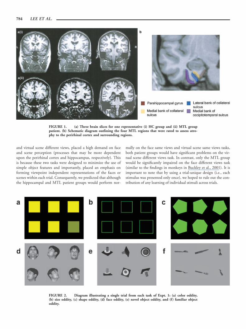
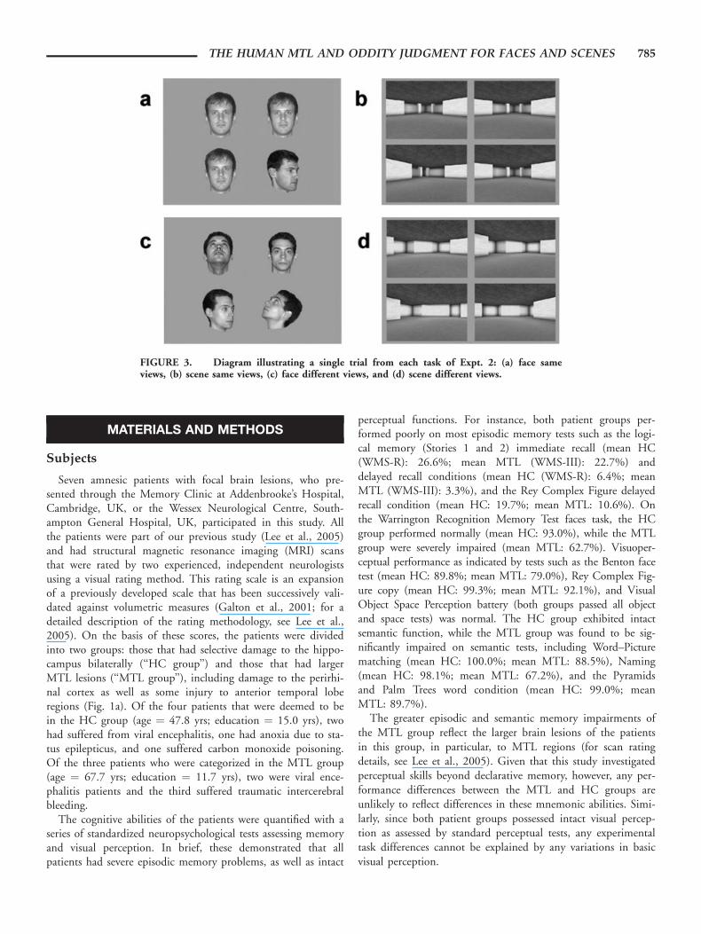
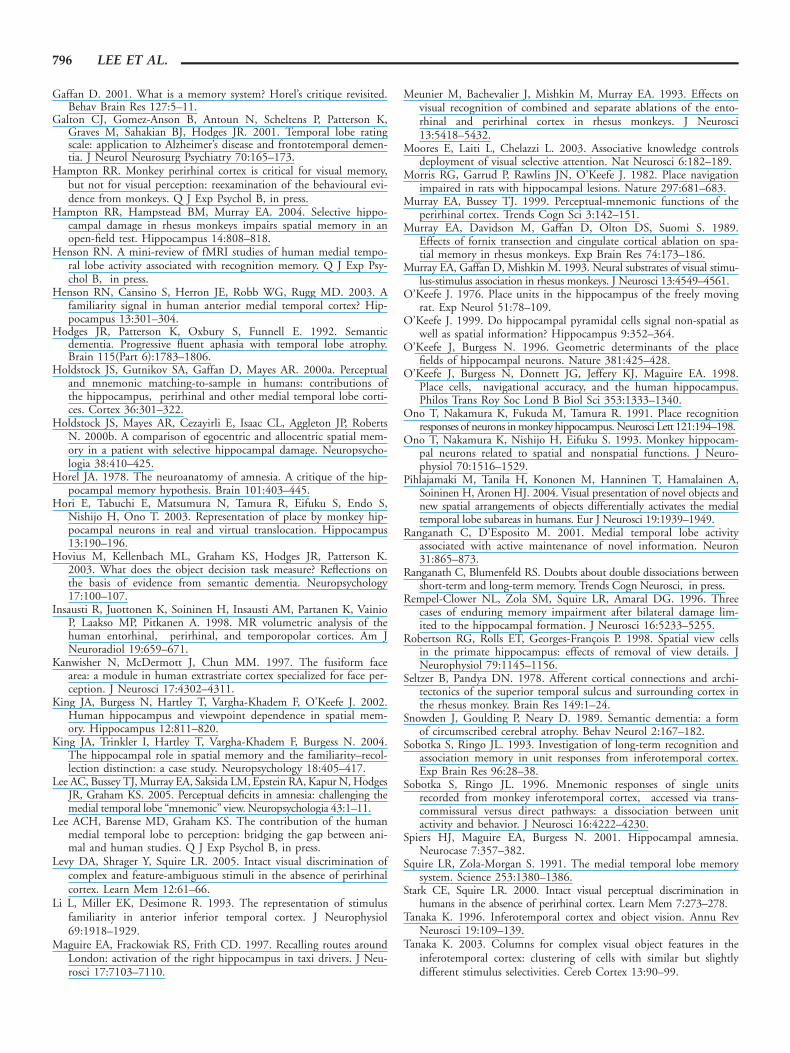
To address the possible involvement of PPA and FFA damagein our patients and to extend our previous finding of scene dis-crimination deficits in our HC group, we administered a series ofnew trial-unique oddity tasks: face same views, virtual scene sameviews, face different views, and virtual scene different views (Expt.2; Fig. 3). Although all four of these tests involved faces and com-plex virtual reality scenes (processes dependent upon the FFA andPPA, respectively), only two of these tasks, face different views
TABLE 1.
Mean Structural MRI Scan Ratings (With Standard Deviations), Averaged Across Both Hemispheres
Group AntTemp Amyg PHG MBCS LBCS MBOS AntHC LatTemp PostHC
HC 0.375 0.438 0.438 0.688 0.313 0.125 1.56* 0.438 0.750
(0.479) (0.427) (0.375) (0.125) (0.125) (0.144) (0.315) (0.427) (0.354)
MTL 1.917* 2.667* 2.250* 2.167* 2.083* 2.167* 2.25* 1.083 2.167*
(0.144) (0.382) (0.661) (1.01) (0.722) (0.289) (0.661) (0.629) (0.520)
Control 0.313 0.375 0.188 0.521 0.271 0.333 0.458 0.458 0.271
(0.284) (0.483) (0.188) (0.291) (0.310) (0.289) (0.382) (0.411) (0.361)
AntTemp, the anterior temporal lobe; Amyg, amygdala; PHG, parahippocampal gyrus; MBCS and LBCS, medial and lateral banks of the collateral sulcus; MBOS,medial bank of the occipitotemporal cortex; AntHC and PostHC, anterior and posterior hippocampus; and LatTemp, lateral temporal lobe. Asterisk denotes signifi-cant difference in comparison with controls.
THE HUMAN MTL AND ODDITY JUDGMENT FOR FACES AND SCENES 783
and virtual scene different views, placed a high demand on faceand scene perception (processes that may be more dependentupon the perirhinal cortex and hippocampus, respectively). Thisis because these two tasks were designed to minimize the use ofsimple object features and importantly, placed an emphasis onforming viewpoint independent representations of the faces orscenes within each trial. Consequently, we predicted that althoughthe hippocampal and MTL patient groups would perform nor-
mally on the face same views and virtual scene same views tasks,both patient groups would have significant problems on the vir-tual scene different views task. In contrast, only the MTL groupwould be significantly impaired on the face different views task(similar to the findings in monkeys in Buckley et al., 2001). It isimportant to note that by using a trial-unique design (i.e., eachstimulus was presented only once), we hoped to rule out the con-tribution of any learning of individual stimuli across trials.
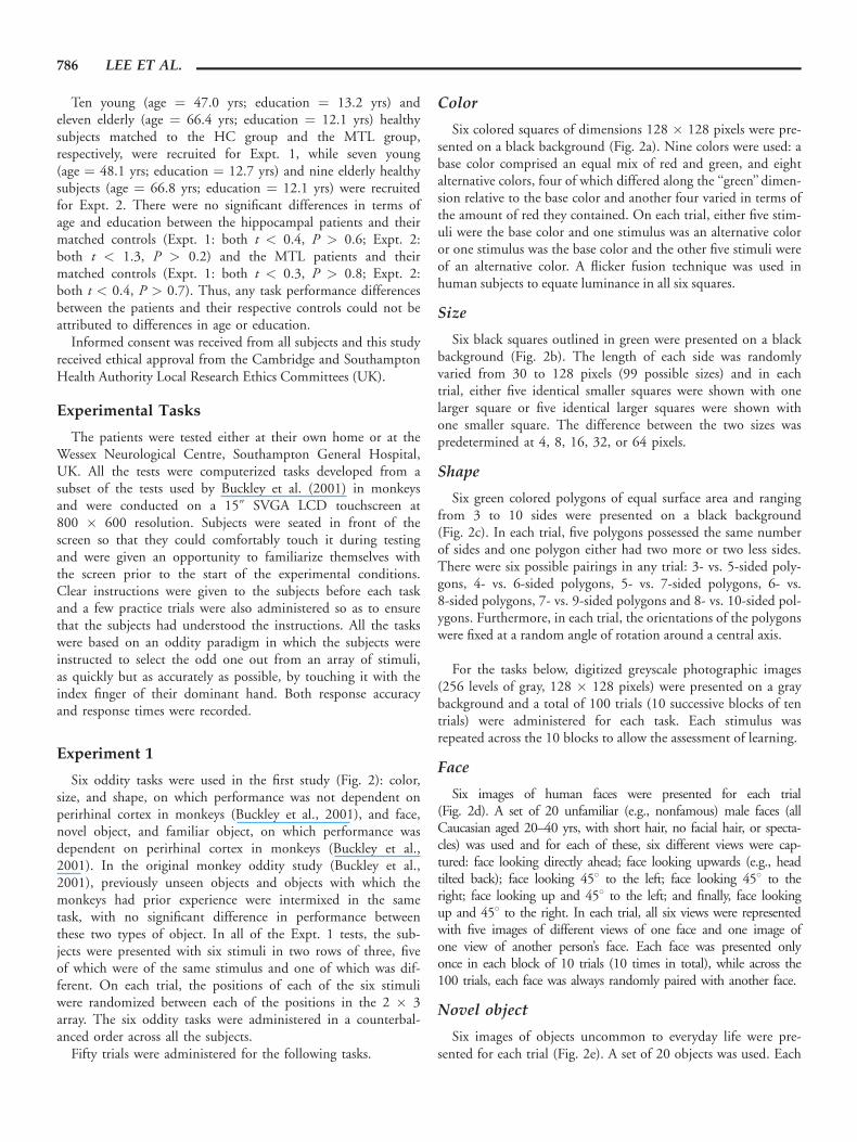
FIGURE 2. Diagram illustrating a single trial from each task of Expt. 1: (a) color oddity,(b) size oddity, (c) shape oddity, (d) face oddity, (e) novel object oddity, and (f) familiar objectoddity.
FIGURE 1. (a) Three brain slices for one representative (i) HC group and (ii) MTL grouppatient. (b) Schematic diagram outlining the four MTL regions that were rated to assess atro-phy to the perirhinal cortex and surrounding regions.
784 LEE ET AL.
MATERIALS AND METHODS
Subjects
Seven amnesic patients with focal brain lesions, who pre-sented through the Memory Clinic at Addenbrooke’s Hospital,Cambridge, UK, or the Wessex Neurological Centre, South-ampton General Hospital, UK, participated in this study. Allthe patients were part of our previous study (Lee et al., 2005)and had structural magnetic resonance imaging (MRI) scansthat were rated by two experienced, independent neurologistsusing a visual rating method. This rating scale is an expansionof a previously developed scale that has been successively vali-dated against volumetric measures (Galton et al., 2001; for adetailed description of the rating methodology, see Lee et al.,2005). On the basis of these scores, the patients were dividedinto two groups: those that had selective damage to the hippo-campus bilaterally (‘‘HC group’’) and those that had largerMTL lesions (‘‘MTL group’’), including damage to the perirhi-nal cortex as well as some injury to anterior temporal loberegions (Fig. 1a). Of the four patients that were deemed to bein the HC group (age ¼ 47.8 yrs; education ¼ 15.0 yrs), twohad suffered from viral encephalitis, one had anoxia due to sta-tus epilepticus, and one suffered carbon monoxide poisoning.Of the three patients who were categorized in the MTL group(age ¼ 67.7 yrs; education ¼ 11.7 yrs), two were viral ence-phalitis patients and the third suffered traumatic intercerebralbleeding.
The cognitive abilities of the patients were quantified with aseries of standardized neuropsychological tests assessing memoryand visual perception. In brief, these demonstrated that allpatients had severe episodic memory problems, as well as intact
perceptual functions. For instance, both patient groups per-formed poorly on most episodic memory tests such as the logi-cal memory (Stories 1 and 2) immediate recall (mean HC(WMS-R): 26.6%; mean MTL (WMS-III): 22.7%) anddelayed recall conditions (mean HC (WMS-R): 6.4%; meanMTL (WMS-III): 3.3%), and the Rey Complex Figure delayedrecall condition (mean HC: 19.7%; mean MTL: 10.6%). Onthe Warrington Recognition Memory Test faces task, the HCgroup performed normally (mean HC: 93.0%), while the MTLgroup were severely impaired (mean MTL: 62.7%). Visuoper-ceptual performance as indicated by tests such as the Benton facetest (mean HC: 89.8%; mean MTL: 79.0%), Rey Complex Fig-ure copy (mean HC: 99.3%; mean MTL: 92.1%), and VisualObject Space Perception battery (both groups passed all objectand space tests) was normal. The HC group exhibited intactsemantic function, while the MTL group was found to be sig-nificantly impaired on semantic tests, including Word–Picturematching (mean HC: 100.0%; mean MTL: 88.5%), Naming(mean HC: 98.1%; mean MTL: 67.2%), and the Pyramidsand Palm Trees word condition (mean HC: 99.0%; meanMTL: 89.7%).
The greater episodic and semantic memory impairments ofthe MTL group reflect the larger brain lesions of the patientsin this group, in particular, to MTL regions (for scan ratingdetails, see Lee et al., 2005). Given that this study investigatedperceptual skills beyond declarative memory, however, any per-formance differences between the MTL and HC groups areunlikely to reflect differences in these mnemonic abilities. Simi-larly, since both patient groups possessed intact visual percep-tion as assessed by standard perceptual tests, any experimentaltask differences cannot be explained by any variations in basicvisual perception.
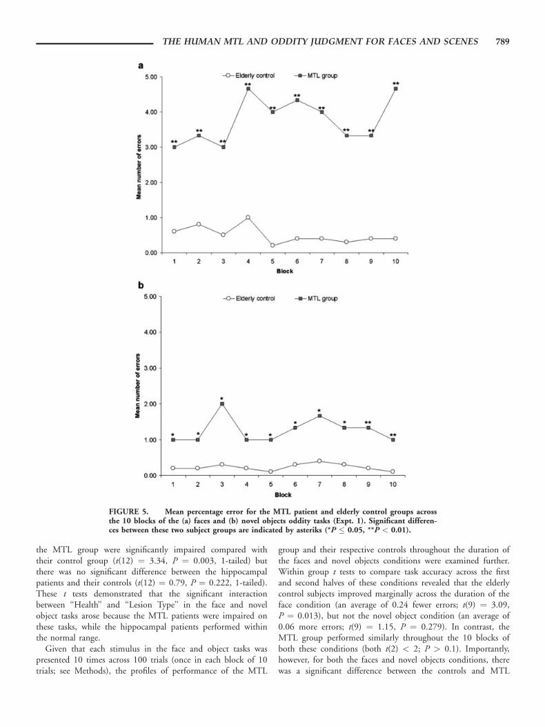
FIGURE 3. Diagram illustrating a single trial from each task of Expt. 2: (a) face sameviews, (b) scene same views, (c) face different views, and (d) scene different views.
THE HUMAN MTL AND ODDITY JUDGMENT FOR FACES AND SCENES 785
Ten young (age ¼ 47.0 yrs; education ¼ 13.2 yrs) andeleven elderly (age ¼ 66.4 yrs; education ¼ 12.1 yrs) healthysubjects matched to the HC group and the MTL group,respectively, were recruited for Expt. 1, while seven young(age ¼ 48.1 yrs; education ¼ 12.7 yrs) and nine elderly healthysubjects (age ¼ 66.8 yrs; education ¼ 12.1 yrs) were recruitedfor Expt. 2. There were no significant differences in terms ofage and education between the hippocampal patients and theirmatched controls (Expt. 1: both t < 0.4, P > 0.6; Expt. 2:both t < 1.3, P > 0.2) and the MTL patients and theirmatched controls (Expt. 1: both t < 0.3, P > 0.8; Expt. 2:both t < 0.4, P > 0.7). Thus, any task performance differencesbetween the patients and their respective controls could not beattributed to differences in age or education.
Informed consent was received from all subjects and this studyreceived ethical approval from the Cambridge and SouthamptonHealth Authority Local Research Ethics Committees (UK).
Experimental Tasks
The patients were tested either at their own home or at theWessex Neurological Centre, Southampton General Hospital,UK. All the tests were computerized tasks developed from asubset of the tests used by Buckley et al. (2001) in monkeysand were conducted on a 15@ SVGA LCD touchscreen at800 � 600 resolution. Subjects were seated in front of thescreen so that they could comfortably touch it during testingand were given an opportunity to familiarize themselves withthe screen prior to the start of the experimental conditions.Clear instructions were given to the subjects before each taskand a few practice trials were also administered so as to ensurethat the subjects had understood the instructions. All the taskswere based on an oddity paradigm in which the subjects wereinstructed to select the odd one out from an array of stimuli,as quickly but as accurately as possible, by touching it with theindex finger of their dominant hand. Both response accuracyand response times were recorded.
Experiment 1
Six oddity tasks were used in the first study (Fig. 2): color,size, and shape, on which performance was not dependent onperirhinal cortex in monkeys (Buckley et al., 2001), and face,novel object, and familiar object, on which performance wasdependent on perirhinal cortex in monkeys (Buckley et al.,2001). In the original monkey oddity study (Buckley et al.,2001), previously unseen objects and objects with which themonkeys had prior experience were intermixed in the sametask, with no significant difference in performance betweenthese two types of object. In all of the Expt. 1 tests, the sub-jects were presented with six stimuli in two rows of three, fiveof which were of the same stimulus and one of which was dif-ferent. On each trial, the positions of each of the six stimuliwere randomized between each of the positions in the 2 � 3array. The six oddity tasks were administered in a counterbal-anced order across all the subjects.
Fifty trials were administered for the following tasks.
Color
Six colored squares of dimensions 128 � 128 pixels were pre-sented on a black background (Fig. 2a). Nine colors were used: abase color comprised an equal mix of red and green, and eightalternative colors, four of which differed along the ‘‘green’’ dimen-sion relative to the base color and another four varied in terms ofthe amount of red they contained. On each trial, either five stim-uli were the base color and one stimulus was an alternative coloror one stimulus was the base color and the other five stimuli wereof an alternative color. A flicker fusion technique was used inhuman subjects to equate luminance in all six squares.
Size
Six black squares outlined in green were presented on a blackbackground (Fig. 2b). The length of each side was randomlyvaried from 30 to 128 pixels (99 possible sizes) and in eachtrial, either five identical smaller squares were shown with onelarger square or five identical larger squares were shown withone smaller square. The difference between the two sizes waspredetermined at 4, 8, 16, 32, or 64 pixels.
Shape
Six green colored polygons of equal surface area and rangingfrom 3 to 10 sides were presented on a black background(Fig. 2c). In each trial, five polygons possessed the same numberof sides and one polygon either had two more or two less sides.There were six possible pairings in any trial: 3- vs. 5-sided poly-gons, 4- vs. 6-sided polygons, 5- vs. 7-sided polygons, 6- vs.8-sided polygons, 7- vs. 9-sided polygons and 8- vs. 10-sided pol-ygons. Furthermore, in each trial, the orientations of the polygonswere fixed at a random angle of rotation around a central axis.
For the tasks below, digitized greyscale photographic images(256 levels of gray, 128 � 128 pixels) were presented on a graybackground and a total of 100 trials (10 successive blocks of tentrials) were administered for each task. Each stimulus wasrepeated across the 10 blocks to allow the assessment of learning.
Face
Six images of human faces were presented for each trial(Fig. 2d). A set of 20 unfamiliar (e.g., nonfamous) male faces (allCaucasian aged 20–40 yrs, with short hair, no facial hair, or specta-cles) was used and for each of these, six different views were cap-tured: face looking directly ahead; face looking upwards (e.g., headtilted back); face looking 458 to the left; face looking 458 to theright; face looking up and 458 to the left; and finally, face lookingup and 458 to the right. In each trial, all six views were representedwith five images of different views of one face and one image ofone view of another person’s face. Each face was presented onlyonce in each block of 10 trials (10 times in total), while across the100 trials, each face was always randomly paired with another face.
Novel object
Six images of objects uncommon to everyday life were pre-sented for each trial (Fig. 2e). A set of 20 objects was used. Each
786 LEE ET AL.
object was photographed in five different nonspecific orienta-tions. In each trial, the five views of one object were presentedwith one view of another object. Furthermore, similar to the faceoddity task, each object was presented only once in each block of10 trials (10 times in total), while across the 100 trials, eachobject was always randomly paired with another object.
Familiar object
This task was identical to the novel object task except that siximages of objects common to everyday life (e.g., kitchen utensils,office stationery) were presented for each trial (Fig. 2f).
Experiment 2
While the face and object tasks of Expt. 1 involved repeatingstimuli in order to assess the possibility of learning, the secondstudy involved four trial-unique oddity tasks (Fig. 3). Thus,any patient deficits in these tasks are unlikely to be explainedby difficulties in learning individual stimuli across trials. Per-formance on two of the tasks, ‘‘face same views’’ and ‘‘scenesame views,’’ placed a minimal demand on object and spatialscene perception, respectively, since a successful oddity judg-ment on these tasks was not dependent on the subjects beingable to use a complete viewpoint independent representation ofa face/scene (e.g., a face same views discrimination could bemade by directly matching the four simultaneously presentedfaces). Subsequently, we predicted that these two tasks were notdependent on the perirhinal cortex or hippocampus. In con-trast, object and spatial scene perception were emphasized inthe other two tasks, ‘‘face different views’’ and ‘‘scene differentviews,’’ since the subjects were required to use complete three-dimensional representations of faces and scenes within eachtrial (e.g., be able to identify the same face/scene from differentvantage points). In all of these tests, the subjects were presentedwith four stimuli in two rows of two, three of which were ofthe same stimulus and one of which was different. On eachtrial, the positions of each of the four stimuli were randomizedbetween each of the positions in the 2 � 2 array. The fouroddity tasks each consisted of 31 trials (with each stimulus onlyappearing once in each task) and were administered in a coun-terbalanced order across all the subjects. Via extensive pilotingin healthy control subjects, the two different views tasks werematched in terms of difficulty, as were the two same viewstasks. It was, however, not possible to match difficulty acrossthe same and different views tasks.
Face same views
Four images of human faces were presented for each trial ona gray background (256 levels of gray, 128 � 128 pixels)(Fig. 3a). A set of 62 unfamiliar (e.g., nonfamous) male faceswas used and for each of these, six different views were cap-tured (see Study 1 methods). In each trial, two views were pre-sented, with three images of the same view of one face and oneimage of a different view of another face. Each face was pre-sented only once and was paired with a second face matched
for skin color, similar face structure, hairstyle, and facial hair(all subjects were presented with the same pairings).
Virtual scene same views
Four images of virtual reality scenes were presented for eachtrial on a gray background (256 levels of gray, 460 � 370 pix-els) (Fig. 3b). A set of 62 scenes, created using a commerciallyavailable computer game (Deus Ex, Ion Storm L.P., Austin,TX) and a freeware software editor (Deus Ex Software Devel-opment Kit v1112f), was used and for each scene, four differ-ent views were captured. In each trial, two views were pre-sented, with three images of the same view of one scene andone image of a different view of another scene. Each scene waspresented only once and was always paired with the same scenethat was similar in appearance and only differed with respect tothe dimensions or placement of one or more aspects of thescene (e.g., a wall, window, room cavity).
Face different views
This task was identical to the ‘‘face same views’’ task exceptthat on each trial, three different views of the same face werepaired with another view of a different face (Fig. 3c).
Virtual scene different views
This task was identical to the ‘‘virtual scene same views’’ taskexcept that on each trial, three different views of the same scenewere paired with another view of a different scene (Fig. 3d).
RESULTS
Experiment 1
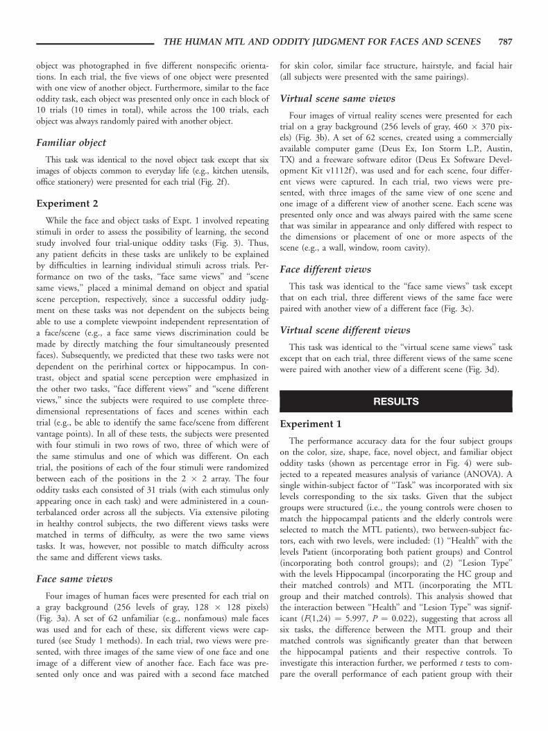
The performance accuracy data for the four subject groupson the color, size, shape, face, novel object, and familiar objectoddity tasks (shown as percentage error in Fig. 4) were sub-jected to a repeated measures analysis of variance (ANOVA). Asingle within-subject factor of ‘‘Task’’ was incorporated with sixlevels corresponding to the six tasks. Given that the subjectgroups were structured (i.e., the young controls were chosen tomatch the hippocampal patients and the elderly controls wereselected to match the MTL patients), two between-subject fac-tors, each with two levels, were included: (1) ‘‘Health’’ with thelevels Patient (incorporating both patient groups) and Control(incorporating both control groups); and (2) ‘‘Lesion Type’’with the levels Hippocampal (incorporating the HC group andtheir matched controls) and MTL (incorporating the MTLgroup and their matched controls). This analysis showed thatthe interaction between ‘‘Health’’ and ‘‘Lesion Type’’ was signif-icant (F(1,24) ¼ 5.997, P ¼ 0.022), suggesting that across allsix tasks, the difference between the MTL group and theirmatched controls was significantly greater than that betweenthe hippocampal patients and their respective controls. Toinvestigate this interaction further, we performed t tests to com-pare the overall performance of each patient group with their
THE HUMAN MTL AND ODDITY JUDGMENT FOR FACES AND SCENES 787
own control group. These t tests showed that the MTL groupperformed significantly worse, overall, than their control group(t(12) ¼ 2.51, P ¼ 0.014, 1-tailed) but the HC group werenot significantly impaired, overall, compared with their controlgroup (t(12) ¼ 0.55, P ¼ 0.296, 1-tailed).
In the ANOVA described above, the three-way interactionbetween ‘‘Health,’’ ‘‘Lesion Type,’’ and ‘‘Task’’ was also signifi-cant (F(5,120) ¼ 2.94, P ¼ 0.016). This interaction revealedthat the difference between the MTL group performance com-pared with the old control group and the HC group perform-ance compared with the young control group varied in magni-tude across the six tasks. To investigate this further, the resultsfrom each individual task were analyzed separately in univariateANOVAs. For each ANOVA, the same two between-subject
factors of ‘‘Health’’ and ‘‘Lesion Type’’ were included with adependent variable of performance. The interaction between‘‘Health’’ and ‘‘Lesion Type’’ was significant in the face task(F(1,24) ¼ 5.25, P ¼ 0.031) and in the novel object task(F(1,24) ¼ 11.50, P ¼ 0.002), but not in the color, size,shape, or familiar object tasks (all F(1,24) < 2, P > 0.1).Within each of the two tasks that showed an interactionbetween ‘‘Health’’ and ‘‘Lesion Type,’’ we then performed ttests to compare the performance of each patient group withtheir own control group. In the face task, the MTL patientsperformed significantly worse than their control group (t(12) ¼2.60, P ¼ 0.012, 1-tailed), while the HC group patients werenot significantly different from their control group (t(12) ¼1.59, P ¼ 0.070, 1-tailed). Similarly, in the novel object task,
FIGURE 4. (a) Mean percentage error (6standard error) for the four subject groups onthe Expt. 1 oddity tasks (chance performance = 75% error; young controls age-matched to HCgroup, elderly controls age-matched to MTL group). (b) Scores of individual subjects.
788 LEE ET AL.
the MTL group were significantly impaired compared withtheir control group (t(12) ¼ 3.34, P ¼ 0.003, 1-tailed) butthere was no significant difference between the hippocampalpatients and their controls (t(12) ¼ 0.79, P ¼ 0.222, 1-tailed).These t tests demonstrated that the significant interactionbetween ‘‘Health’’ and ‘‘Lesion Type’’ in the face and novelobject tasks arose because the MTL patients were impaired onthese tasks, while the hippocampal patients performed withinthe normal range.
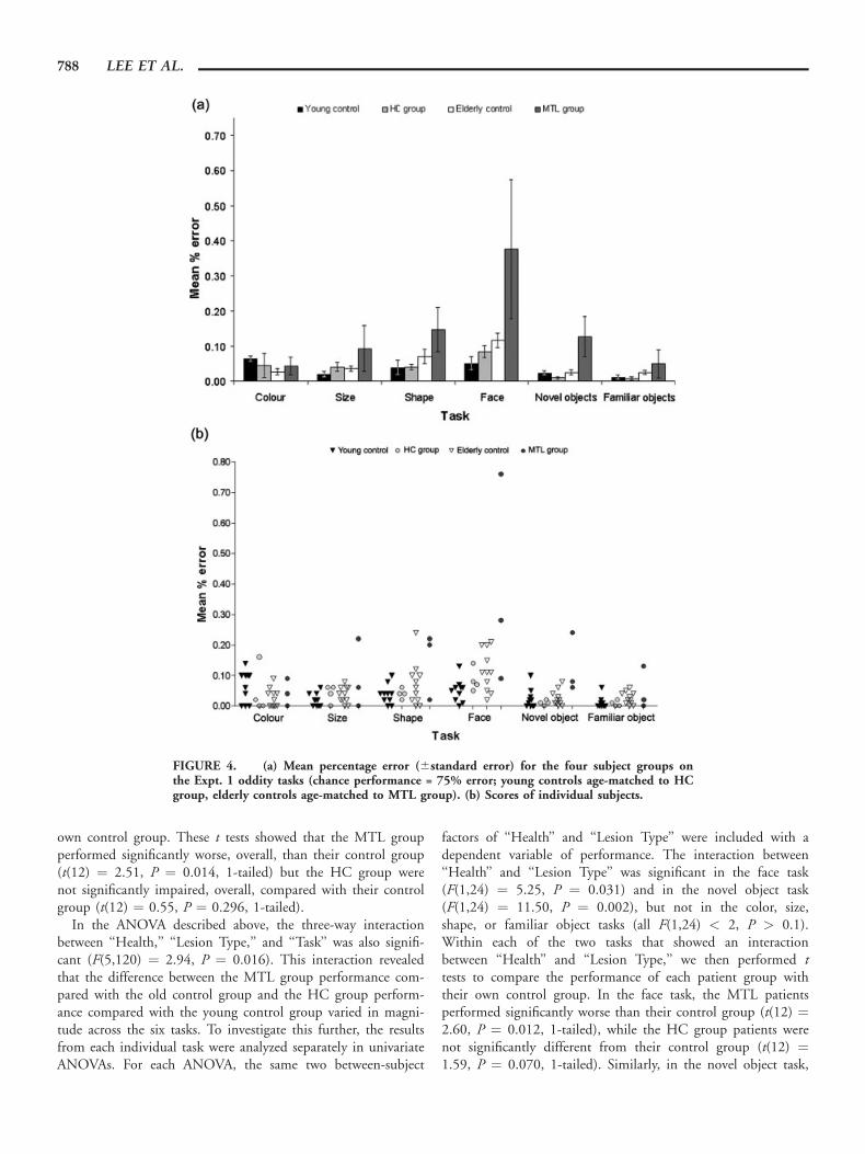
Given that each stimulus in the face and object tasks waspresented 10 times across 100 trials (once in each block of 10trials; see Methods), the profiles of performance of the MTL
group and their respective controls throughout the duration ofthe faces and novel objects conditions were examined further.Within group t tests to compare task accuracy across the firstand second halves of these conditions revealed that the elderlycontrol subjects improved marginally across the duration of theface condition (an average of 0.24 fewer errors; t(9) ¼ 3.09,P ¼ 0.013), but not the novel object condition (an average of0.06 more errors; t(9) ¼ 1.15, P ¼ 0.279). In contrast, theMTL group performed similarly throughout the 10 blocks ofboth these conditions (both t(2) < 2; P > 0.1). Importantly,however, for both the faces and novel objects conditions, therewas a significant difference between the controls and MTL
FIGURE 5. Mean percentage error for the MTL patient and elderly control groups acrossthe 10 blocks of the (a) faces and (b) novel objects oddity tasks (Expt. 1). Significant differen-ces between these two subject groups are indicated by asteriks (*P � 0.05, **P < 0.01).
THE HUMAN MTL AND ODDITY JUDGMENT FOR FACES AND SCENES 789
group even on the first block of 10 trials (both t(11) > 2; P <0.03; see Fig. 5). This suggests that it is unlikely that the defi-cits observed in the MTL group on the faces and novel objectsconditions were entirely due to learning present only in theelderly control group.
Experiment 2
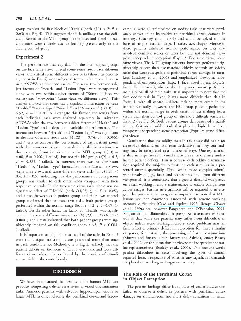
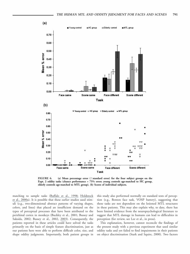
The performance accuracy data for the four subject groupson the face same views, virtual scene same views, face differentviews, and virtual scene different views tasks (shown as percent-age error in Fig. 5) were subjected to a similar repeated meas-ures ANOVA, as described earlier. The same two between-sub-ject factors of ‘‘Health’’ and ‘‘Lesion Type’’ were incorporatedalong with two within-subject factors of ‘‘Stimuli’’ (faces vs.scenes) and ‘‘Viewpoint’’ (same views vs. different views). Thisanalysis showed that there was a significant interaction between‘‘Health,’’ ‘‘Lesion Type,’’ ‘‘Stimuli,’’ and ‘‘Viewpoint’’ (F(1,19) ¼6.52, P ¼ 0.019). To investigate this further, the results fromeach individual task were analyzed separately in univariateANOVAs with the two between-subject factors of ‘‘Health’’ and‘‘Lesion Type’’ and a dependent variable of performance. Theinteraction between ‘‘Health’’ and ‘‘Lesion Type’’ was significantin the face different views task (F(1,23) ¼ 9.74, P ¼ 0.006),and t tests to compare the performance of each patient groupwith their own control group revealed that this interaction wasdue to a significant impairment in the MTL group (t(10) ¼4.00, P ¼ 0.002, 1-tailed), but not the HC group (t(9) < 0.3,P ¼ 0.388, 1-tailed). In contrast, there was no significant‘‘Health’’ by ‘‘Lesion Type’’ interaction in the face same views,scene same views, and scene different views tasks (all F(1,23) <0.4, P > 0.5), indicating that the performance of both patientgroups was similar to each other when compared with theirrespective controls. In the two same views tasks, there was nosignificant effect of ‘‘Health’’ (both F(1,23) � 4, P > 0.05),and t tests between each patient group and their own controlgroup confirmed that on these two tasks, both patient groupsperformed within the normal range (both t < 2, P > 0.07, 1-tailed). On the other hand, the factor of ‘‘Health’’ was signifi-cant in the scene different views task (F(1,23) ¼ 22.68, P <0.0001) and t tests indicated that both patient groups were sig-nificantly impaired on this condition (both t >3, P < 0.004,1-tailed).
It is important to highlight that as all of the tasks in Expt. 2were trial-unique (no stimulus was presented more than oncein each condition; see Methods), it is highly unlikely that thepatient deficits on the scene different views task and faces dif-ferent views task can be explained by the learning of stimuliacross trials in the controls only.
DISCUSSION
We have demonstrated that lesions to the human MTL canproduce compelling deficits on a series of visual discriminationtasks. Amnesic patients with selective hippocampal lesions orlarger MTL lesions, including the perirhinal cortex and hippo-
campus, were all unimpaired on oddity tasks that were previ-ously shown to be insensitive to perirhinal cortex damage inmonkeys (Buckley et al., 2001) and could be solved on thebasis of simple features (Expt. 1: color, size, shape). Moreover,these patients exhibited normal performance on tests thatinvolved complex scenes or faces but did not demand view-point independent perception (Expt. 2: face same views, scenesame views). The MTL group patients, however, performed sig-nificantly poorer than age-matched elderly controls on odditytasks that were susceptible to perirhinal cortex damage in mon-keys (Buckley et al., 2001) and emphasized viewpoint inde-pendent object perception (Expt. 1: face, novel object, Expt. 2:face different views), whereas the HC group patients performednormally on all of these tasks. It is important to note that theface oddity task in Expt. 2 was more difficult than that inExpt. 1, with all control subjects making more errors in theformer. Critically, however, the HC group patients performedwithin the normal range in both tasks, in fact making fewererrors than their control group on the more difficult version inExpt. 2 (see Fig. 6). Both patient groups demonstrated a signif-icant deficit on an oddity task that placed a high demand onviewpoint independent scene perception (Expt. 2: scene differ-ent views).
Considering that the oddity tasks in this study did not placean explicit demand on long-term declarative memory, our find-ings may be interpreted in a number of ways. One explanationis that an impairment in visual short-term memory may under-lie the patient deficits. This is because each oddity discrimina-tion required the subjects to fixate on each stimulus in a pre-sented array sequentially. Thus, when more complex stimuliwere involved (e.g., faces and scenes presented from differentviewpoints), it is conceivable that a greater demand was placedon visual working memory maintenance to enable comparisonsacross images. Further investigations will be required to investi-gate this possibility, although it is important to note that MTLlesions are not commonly associated with generic workingmemory difficulties (Cave and Squire, 1992; Rempel-Cloweret al., 1996; see, however Ranganath and D’Esposito, 2001;Ranganath and Blumenfeld, in press). An alternative explana-tion is that while the patients may suffer from difficulties inobject and/or scene working memory, these problems may, infact, reflect a primary deficit in perception for these stimuluscategories, for instance, the processing of feature conjunctions(Murray and Bussey, 1999; Bussey and Saksida, 2002; Busseyet al., 2002) or the formation of viewpoint independent stimu-lus representations (Buckley et al., 2001). This account wouldpredict difficulties in tasks involving the types of stimulireported here, irrespective of whether any significant demandsare placed on working or long-term memory.
The Role of the Perirhinal Cortexin Object Perception
The present findings differ from those of earlier studies thatfailed to observe a deficit in patients with perirhinal cortexdamage on simultaneous and short delay conditions in visual
790 LEE ET AL.
matching to sample tasks (Buffalo et al., 1998; Holdstocket al., 2000a). It is possible that these earlier studies used stim-uli (e.g., two-dimensional abstract patterns of varying shapes,colors, and lines) that placed an insufficient demand on thetypes of perceptual processes that have been attributed to theperirhinal cortex in monkeys (Buckley et al., 2001; Bussey andSaksida, 2002; Bussey et al., 2002, 2003). Consequently, thepatients reported in these articles could have solved the tasksprimarily on the basis of simple feature discrimination, just asour patients here were able to perform difficult color, size, andshape oddity judgments. Importantly, both patient groups in
this study also performed normally on standard tests of percep-tion (e.g., Benton face task, VOSP battery), suggesting thatthese tasks are not dependent on the lesioned MTL structuresin these patients. This may also explain why, to date, there hasbeen limited evidence from the neuropsychological literature tosuggest that MTL damage in humans can lead to difficulties inperception (for review, see Lee et al., in press).
This explanation, however, cannot reconcile the findings ofthe present study with a previous experiment that used similaroddity tasks and yet failed to find impairments in their patientson object discrimination (Stark and Squire, 2000). Two factors
FIGURE 6. (a) Mean percentage error (6standard error) for the four subject groups on theExpt. 2 oddity tasks (chance performance = 75% error; young controls age-matched to HC group,elderly controls age-matched to MTL group). (b) Scores of individual subjects.
THE HUMAN MTL AND ODDITY JUDGMENT FOR FACES AND SCENES 791
probably contribute to this disparity. First, the aforementionedstudy (Stark and Squire, 2000) used a combination of noveland familiar objects in their object oddity tasks. As demon-strated here, this can have a significant effect on the discrimina-tion of objects: the MTL group was significantly impaired onthe face and novel object oddity tasks, but not the familiarobject task in Expt. 1. One possible explanation for this findingis that semantic knowledge about the highly familiar objects (ofwhich existing perceptual representations may be a critical com-ponent) facilitated performance on the familiar object task,leading to faster response times and significantly improved taskaccuracy. In support of this, there have been recent reports thatsemantic knowledge can influence performance on visual proc-essing tasks, for example, by aiding the perception of object sil-houettes (Hovius et al., 2003) or impeding performance on avisual search task if the target and distractor items are semanti-cally associated (Moores et al., 2003).
A second contributory factor to the disparity between thepresent experiment and a previous study (Stark and Squire,2000) that used similar tasks is the difference in stimulus setsize. The tasks in Expt. 1 comprised 10 stimulus-unique trials(a set of 20 items, each randomly paired with another). In con-trast, 62 stimuli were used in each task of Expt. 2 to create 31stimulus-unique trials. In the previous investigation (Stark andSquire, 2000), only sets of 10 stimuli were used. It has beendemonstrated that employing a large stimulus set size is criticalfor eliciting impairment in visual concurrent discriminationtasks in monkeys with perirhinal cortex ablations (Buckley andGaffan, 1997); the successful discrimination of items within alarger set may require more precise perceptual representations,for example, due to a higher degree of feature overlap betweenstimuli across trials (Buckley and Gaffan, 1997; Buckley, inpress; Hampton, in press).
Since the performance of all subject groups on the novelobject task was comparable or better than that on the color,shape, and size oddity tasks (Expt. 1), the observed MTL groupimpairment on the novel object and face tasks cannot be onlydue to their increased difficulty. In terms of learning, the eld-erly group did demonstrate a very small performance improve-ment across the faces condition (a decrease of one quarter ofan error over 100 trials). This, however, cannot explain entirelythe MTL group deficits, since (a) the elderly control group’sperformance did not improve across the duration of the novelobjects condition; (b) in both the faces and novel objects con-ditions there was a significant difference between the MTLgroup and their respective controls from the very first 10-trialblock of the tasks (see Fig. 5); and finally, (c) the finding of aMTL group deficit in face oddity judgment in Expt. 1 wasreplicated in Expt. 2 in which no stimulus was presented morethan once in each condition.
Since the MTL group possessed atrophy to temporal loberegions beyond the perirhinal cortex, it is important to evaluatehow these areas may potentially contribute to the observed defi-cit in object/face oddity judgment. One possible argument isthat the MTL group patients’ impairment may be primarilydue to damage to higher processing visual areas such as area
TE or the FFA (Kanwisher et al., 1997). There are, however, anumber of concrete reasons to argue against this possibility.
First, structural scan ratings (see Methods; Table 1) revealedno significant difference between the MTL group and a healthycontrol group on a lateral temporal lobe rating. While it is cur-rently unclear what region in the human brain corresponds toarea TE, as first identified in the macaque brain (Von Boninand Bailey, 1947; Seltzer and Pandya, 1978), area TE in maca-ques is known to occupy the inferior and middle temporal gyri,the latter of which was included in our lateral temporal loberating. In fact, two of the three MTL group patients wereadjudged to have no additional lateral temporal lobe atrophy incomparison with a control group, and yet these patients wereimpaired on the novel object and face oddity tasks in Expt. 1and the face different views task in Expt. 2.
Second, although the MTL group did have significant dam-age to a medial inferior temporal lobe region adjacent to theperirhinal cortex (i.e., medial bank of the occipitotemporal sul-cus, see Methods), their performance profile on the oddity tasksdo not match existing knowledge of the effects of damage toarea TE in nonhuman primates. It has been demonstrated thatlesions to area TE in monkeys disrupts fine color discrimina-tion (Buckley et al., 1997). Furthermore, single-unit recordingshave suggested that cells in this region represent moderatelycomplex visual features, such as those in the simple featureoddity tasks (Expt. 1), but not objects (Tanaka, 1996). In thecurrent study, the MTL group patients were not impaired onthe simple feature oddity tasks, including judgments for coloroddity, implicating intact functioning of area TE. This mirrorsthe original findings of Buckley et al. (2001) who found thatperformance on simple feature oddity tasks was not disruptedfollowing perirhinal cortex ablations in monkeys.
Third, and more importantly, we found in the present studythat the MTL group patients performed within the normalrange on a task that involved faces but did not place a demandon viewpoint independent perception (Expt. 2: face sameviews). A similar finding has been reported in monkeys withperirhinal lesions (Buckley et al., 2001). These results underlinethe notion that lesions to perirhinal cortex do not cause a gen-eral perceptual deficit and argue against the possibility that theobserved deficits are due to damage to regions more posteriorin the temporal lobe, such as the FFA in humans. Moreover,the MTL group patients do not suffer from symptoms such asprosopagnosia that are often associated with lesions to the FFA(Barton et al., 2002). It is important to note, however, that allsubject groups performed at or close to ceiling on the face sameviews task. It is possible, therefore, that a more difficult versionof this task may lead to deficits in the MTL patient group.
Given that the MTL patients have larger lesions comparedwith the HC group (see Table 1), one possible explanation isthat it is the increased damage to the hippocampus, rather thanother MTL structures, that is causing the additional MTLgroup deficits in face and novel object oddity judgment. Evi-dence against this suggestion comes from a recent study in ourgroup (Lee et al., unpublished observations) in which the sameoddity battery in Expt. 1 was administered to patients with
792 LEE ET AL.
semantic dementia (SD), a condition associated with progres-sive, cross-modal loss of semantic knowledge (Warrington,1975; Snowden et al., 1989; Hodges et al., 1992) and dispro-portionate atrophy to the perirhinal cortex compared withother MTL structures (Davies et al., 2004), and patients withAlzheimer’s disease (AD), who typically have less atrophic peri-rhinal cortex but severe damage to the hippocampus (Davieset al., 2004). It was found that the SD patients demonstrated asimilar performance profile to the MTL group (impairedobject, but not simple feature, perception). In contrast, the ADpatients performed well on all tasks in spite of possessing hip-pocampal atrophy, an identical pattern to that seen in the HCgroup reported here. These findings suggest that damage to theperirhinal cortex is likely to underlie the MTL group deficitsobserved in the current study, rather than the larger hippocam-pal lesions in these patients. It is important to note, however,that similar to SD patients, the MTL patients in the presentstudy possessed significant damage to the anterior temporallobe (Table 1). Although the perirhinal cortex is known toextend into the temporal pole in humans (Insausti et al.,1998), it is currently impossible to clarify the possible contri-bution of other anterior temporal lobe regions to the perceptualprocesses that have been attributed to the perirhinal cortex inmonkeys, and to determine whether these regions may also becritical for these processes.
Given the replicability of the findings across different patientgroups, plus the striking convergence in the behavioral patternsseen in human patients and monkeys with perirhinal cortexinvolvement, it seems reasonable to hypothesize that the peri-rhinal cortex may be critically involved in object perception. Asdescribed in the Introduction, one model of perirhinal cortexfunction (Murray and Bussey, 1999) is that it processes com-plex conjunctions of object features (i.e., representation of com-plete objects), while more caudal regions such as area TE/TEOand V4 process moderately complex conjunctions (e.g., Tanaka,1996, 2003) and simple features. Although there has been sup-port for this model from recent monkey lesion (Bussey et al.,2002, 2003) and computational modeling (Bussey and Saksida,2002) studies, the precise details of this view remain to bedetermined. For example, it is uncertain how the perirhinalcortex may, as the suggested apex of the ventral processingstream, exert top–down influence on earlier regions such as areaV4. With respect to our present data, it is not obvious how theability to process complex conjunctions of features contributesto the perception and identification of objects from multipleviewpoints, as assessed in the oddity tasks. There are, however,indications that the object perception impairments in ourpatients cannot be explained entirely by an impairment informing viewpoint independent representations of objects. Forinstance, in Lee et al. (2005) the same MTL group patientshad a deficit in the discrimination of faces, and to a lesserextent objects, that were blended to various degrees but did notplace a demand on viewpoint independent perception. Further-more, we have data to suggest that when the degree of featureoverlap between two stimuli is controlled systematically, it ispossible to observe deficits in the discrimination of two-dimen-
sional stimuli when the level of feature overlap is high but notwhen it is low (Lee et al., in press; Barense et al., 2004).
One important question is whether the neural mechanismsthat mediate object perception in the perirhinal cortex are simi-lar to those that underlie the declarative memory functions ofthis region, for example, recognition on the basis of familiarityand memory for stimulus–stimulus associations (Meunier et al.,1993; Murray et al., 1993; Eacott et al., 1994; Buckley andGaffan, 1998a; Aggleton and Brown, 1999; Henson et al.,2003). Although our two patient groups demonstrated similarlevels of performance on immediate and delayed recall of astory and complex figure (see Methods), their scores on tests ofrecognition memory were discrepant: the HC group showednormal performance on the recognition memory test for faces(mean score ¼ 91.2%), while the MTL group were signifi-cantly impaired (mean score ¼ 62.0%). This result is consis-tent with the view that familiarity-based processes that mightsupport recognition memory but not recall are dependent uponnonhippocampal regions such as the perirhinal cortex (Meunieret al., 1993; Murray et al., 1993; Eacott et al., 1994; Buckleyand Gaffan, 1998a; Aggleton and Brown, 1999; Henson et al.,2003; Henson, in press). Importantly, however, the finding ofimpaired perceptual processing of novel faces in our currentstudy and poor recognition memory for faces does not necessa-rily mean that these deficits are caused by the same neuralimpairment.
Some indication of whether the same neuronal mechanismsunderlie both perceptual and declarative memory processes inthe perirhinal cortex come from nonhuman primate electrophy-siological studies. These have demonstrated that there are peri-rhinal neurons that decrease their firing in response to subse-quent presentations of unfamiliar objects (Brown et al., 1987;Li et al., 1993; Sobotka and Ringo, 1993), and it has been sug-gested that this mechanism may constitute the neural basis ofrecognition memory (for review, see Brown and Xiang, 1998).There are also, however, neurons that possess stimulus-specific-ity but do not exhibit decremental response patterns (Xiangand Brown, 1998), and moreover, studies have shown that rec-ognition memory can be dissociated from the decrementalresponse properties of some perirhinal neurons (Sobotka andRingo, 1996). This suggests that there may be a variety of neu-ral mechanisms that make distinct contributions to the mne-monic and perceptual processes that the perirhinal cortex hasbeen suggested to subserve.
The Role of the Hippocampusin Spatial Perception
Both amnesic patient groups showed significant difficultiesmaking oddity judgments for scenes when they were requiredto perceive the spatial arrangement of a virtual room using theinformation available from different views (Expt. 2: virtualscene different views). This observation converges with findingsfrom our previous study in which we found that hippocampaldamage resulted in a significant impairment in the visual dis-crimination of pairs of spatial scene images that were blended
THE HUMAN MTL AND ODDITY JUDGMENT FOR FACES AND SCENES 793
to create different levels of feature overlap (Lee et al., 2005).Importantly, the use of trial-unique tasks in the current study(Expt. 2) rules out the possibility that the reported deficit inspatial perception in these patients is due to learning effectsthat were present in the controls but not the patients (a possi-ble confounding factor in our earlier study, Lee et al., 2005).Thus, this demonstration of a striking, and selective, deficit inscene discrimination across two separate studies supports thesuggestion that the human hippocampus may play a criticalrole in space perception.
This contrasts with recent investigations that propose that itis the neighboring parahippocampal cortex, more specificallythe PPA (Epstein and Kanwisher, 1998), that is crucial forprocessing spatial information, for example, the layout andappearance of spatial scenes (Epstein et al., 1999). One possi-bility is that the deficits in spatial perception observed in ouramnesic patients may be caused by additional damage to thePPA rather than hippocampal dysfunction. Two pieces of evi-dence seem to rule out this possibility. First, both the HC andMTL patient groups performed within the normal range on atask that involved complex images of virtual scenes, but couldbe solved using simple features (e.g., size of a wall; Expt. 2: vir-tual scenes same views). This suggests that the patients did nothave a deficit in perceiving scenes per se, but rather had diffi-culties making judgments that required processing spatialinformation about the room layout. Second, none of thepatients had damage that was obviously near or in the PPA,and there was no significant difference between the controlsand patients in any structural rating other than that for thehippocampus (including an anterior parahippocampal gyrusregion). Consequently, one may conclude that both the hippo-campus and parahippocampal cortex are important for spatialperception, although further studies will be needed to clarifythe differential roles played by these two regions.
Our observation of an impairment in discriminating differ-ent, but not same, views scenes following hippocampal damagesupports and expands upon recent studies by King et al. (2002,2004), which found that a single hippocampal lesion patientwas only mildly impaired at recognizing object locations thatwere viewed from the same viewpoint as that during the learn-ing phase, but was severely impaired when the object locationswere viewed from a different viewpoint from that at learning.Our study suggests that the deficits reported by King et al.(2002, 2004) can be evident in a task without an overt declara-tive memory component and that hippocampal damage maylead to deficits in perceiving, as well as remembering, spatialscenes. It is important to note that the hippocampal casereported by King et al. (2002, 2004) was able to perform men-tal rotation of single objects and that our hippocampal patientswere able to perform oddity judgments of different views faces(Expts. 1 and 2) and objects (Expt. 1). This reinforces the ideathat the deficits observed in our patients are unlikely to be dueto a fundamental deficit in mental rotation. Rather, it appearsthat patients with hippocampal damage may be unable to useviewpoint independent representations of spatial scenes, oftenreferred to as allocentric representations (e.g., see Aguirre and
D’Esposito, 1999; Holdstock et al., 2000b; Burgess et al.,2001; Bohbot et al., 2004; Feigenbaum and Morris, 2004;King et al., 2004).
It is important to highlight that while the present study hasdemonstrated impaired allocentric scene perception in patientswith damage to the hippocampus, it is unlikely that this deficitcan account entirely for the spatial memory and perception def-icits often seen following hippocampal dysfunction in humansand nonhuman primates. For instance, in our previous study,both the HC and MTL patient groups were significantlyimpaired in a scene discrimination task that did not requireallocentric scene perception (Lee et al., 2005). Also, hippocam-pal dysfunction in monkeys, by means of fornix transection,can result in deficits in learning conjunctions of spatial infor-mation: for example, the association between tail orientation,length, and spatial location of tadpoles within a two-dimen-sional scene (Buckley et al., 2004). In addition to this, recentfunctional neuroimaging studies have demonstrated activity inthe hippocampus during memory tasks for spatial configura-tion, on which successful performance also does not depend onallocentric processing (e.g., Duzel et al., 2003; Pihlajamakiet al., 2004).
The possibility of perceptual deficits following hippocampaldamage raises the question as to what the functional conse-quences may be for patients in everyday life. Given that wehave demonstrated that patients with hippocampal damage areable to discriminate scenes presented from the same view, it islikely that any difficulties in real life will only be evident in therare circumstances where the patients are, for example, exposedto multiple environments that are highly similar. One contro-versial suggestion is that the mnemonic deficits commonly seenafter hippocampal damage (Tulving and Markowitsch, 1998;Spiers et al., 2001) may actually be the result of a primary defi-cit in spatial perception (Horel, 1978; Gaffan, 2001;). Whilethe current study clearly indicates a contributory role for thehippocampus in processing spatial information, it does not nec-essarily mean that this is the sole role of the hippocampus inmemory and perception. For example, it is difficult to see howdeficits in the recall of a prose passage can be caused by suchspatial difficulties. Given the broader profile of amnesia, there-fore, it seems possible that the deficits described here reflect amore general impairment in relational or associative processing,of which computing relationships between the objects and fea-tures constituting a scene is a part (Eichenbaum et al., 1992,1994; Cohen et al., 1999; Eichenbaum and Cohen, 2002).Critically, however, the latter process is thought to be specificto memory, involving binding perceptually distinct items (e.g.,objects) into long-term memory representations (Eichenbaumet al., 1992, 1994; Eichenbaum and Cohen, 2002). One sug-gestion, therefore, is that the patient deficits in the scene differ-ent views task may actually be a reflection of long-term rela-tional memory difficulties. For example, it is conceivable thatwithin each trial, the healthy controls may have benefited fromthe ability to encode the spatial relations within each scene intolong-term declarative memory (i.e., ‘‘memory facilitated percep-tion’’). Further studies will be required to explore the exact
794 LEE ET AL.
details of this idea further and hopefully clarify what aspects ofrelational and/or spatial processing are dependent upon thehippocampus. In addition, it will be important to determinewhether these types of processing can be extended beyond thedomain of long-term declarative memory, as indicated by ourrecent studies.
In summary, we have demonstrated that lesions to thehuman perirhinal cortex can lead to difficulties in object,including face, oddity judgment, while lesions to the humanhippocampus can cause difficulties in scene oddity judgment.Contrary to existing literature proposing a selective role for thehuman MTL in long-term declarative memory (Buffalo et al.,1998; Tulving and Markowitsch, 1998; Holdstock et al.,2000a; Spiers et al., 2001), this data confirms a broader rolefor the perirhinal cortex and hippocampus that extends to veryshort-term visual working memory at least, and possibly alsoperceptual processing, strongly supporting and extending recentfindings from the nonhuman primate literature.
Acknowledgments
The authors are grateful to the patients and all the controlvolunteers for their participation in this study. The authors alsothank S. Thompson for rating the MRI scans of the patients,A. Kropelnicki, P. Abbott, and J. Limond for help with retriev-ing patient data, and R. Epstein, E. Murray, and L. Saksida forfeedback on this and related experiments. This work was sup-ported by the Alzheimer’s Research Trust, UK, and the MedicalResearch Council, UK.
REFERENCES
Aggleton JP, Brown MW. 1999. Episodic memory, amnesia andthe hippocampal-anterior thalamic axis. Behav Brain Sci 22:425–489.
Aguirre GK, D’Esposito M. 1999. Topographical disorientation: a syn-thesis and taxonomy. Brain 122(Part 9):1613–1628.
Barense MD, Bussey TJ, Lee ACH, Rogers TT, Saksida LM, MurrayEA, Hodges JR, Graham KS. 2004. Differentiating the compo-nents of the medical temporal lobe: a critical role of feature ambi-guity. Soc Neurosci Abst 201.13.
Barton JJ, Press DZ, Keenan JP, O’Connor M. 2002. Lesions of thefusiform face area impair perception of facial configuration in pro-sopagnosia. Neurology 58:71–78.
Bohbot VD, Iaria G, Petrides M. 2004. Hippocampal function andspatial memory: evidence from functional neuroimaging in healthyparticipants and performance of patients with medial temporal loberesections. Neuropsychology 18:418–425.
Brown MW, Xiang JZ. 1998. Recognition memory: neuronal substratesof the judgement of prior occurrence. Prog Neurobiol 55:149–189.
Brown MW, Wilson FA, Riches IP. 1987. Neuronal evidence thatinferomedial temporal cortex is more important than hippocampusin certain processes underlying recognition memory. Brain Res409:158–162.
Buckley MJ. The role of the perirhinal cortex and hippocampus inlearning, memory, and perception. Q J Exp Psychol B, in press.
Buckley MJ, Gaffan D. 1997. Impairment of visual object-discrimina-tion learning after perirhinal cortex ablation. Behav Neurosci 111:467–475.
Buckley MJ, Gaffan D. 1998a. Perirhinal cortex ablation impairs con-figural learning and paired-associate learning equally. Neuropsycho-logia 36:535–546.
Buckley MJ, Gaffan D. 1998b. Learning and transfer of object-rewardassociations and the role of the perirhinal cortex. Behav Neurosci112:1–9.
Buckley MJ, Gaffan D, Murray EA. 1997. Functional double dissocia-tion between two inferior temporal cortical areas: perirhinal cortexversus middle temporal gyrus. J Neurophysiol 77:587–598.
Buckley MJ, Booth MC, Rolls ET, Gaffan D. 2001. Selective percep-tual impairments after perirhinal cortex ablation. J Neurosci21:9824–9836.
Buckley MJ, Charles DP, Browning PG, Gaffan D. 2004. Learningand retrieval of concurrently presented spatial discrimination tasks:role of the fornix. Behav Neurosci 118:138–149.
Buffalo EA, Reber PJ, Squire LR. 1998. The human perirhinal cortexand recognition memory. Hippocampus 8:330–339.
Burgess N, Becker S, King JA, O’Keefe J. 2001. Memory for eventsand their spatial context: models and experiments. Philos TransRoy Soc Lond B Biol Sci 356:1493–1503.
Burgess N, Maguire EA, O’Keefe J. 2002. The human hippocampusand spatial and episodic memory. Neuron 35:625–641.
Bussey TJ, Saksida LM. 2002. The organization of visual object repre-sentations: a connectionist model of effects of lesions in perirhinalcortex. Eur J Neurosci 15:355–364.
Bussey TJ, Saksida LM, Murray EA. 2002. Perirhinal cortex resolvesfeature ambiguity in complex visual discriminations. Eur J Neuro-sci 15:365–374.
Bussey TJ, Saksida LM, Murray EA. 2003. Impairments in visual dis-crimination after perirhinal cortex lesions: testing ‘‘declarative’’ vs.‘‘perceptual-mnemonic’’ views of perirhinal cortex function. Eur JNeurosci 17:649–660.
Cave CB, Squire LR. 1992. Intact verbal and nonverbal short-termmemory following damage to the human hippocampus. Hippo-campus 2:151–163.
Cohen NJ, Ryan J, Hunt C, Romine L, Wszalek T, Nash C. 1999. Hippo-campal system and declarative (relational) memory: summarizing thedata from functional neuroimaging studies. Hippocampus 9:83–98.
Davies RR, Graham KS, Xuereb JH, Williams GB, Hodges JR. 2004.The human perirhinal cortex and semantic memory. Eur J Neuro-sci 20:2441–2446.
Duzel E, Habib R, Rotte M, Guderian S, Tulving E, Heinze HJ.2003. Human hippocampal and parahippocampal activity duringvisual associative recognition memory for spatial and nonspatialstimulus configurations. J Neurosci 23:9439–9444.
Eacott MJ, Gaffan D, Murray EA. 1994. Preserved recognition mem-ory for small sets, and impaired stimulus identification for largesets, following rhinal cortex ablations in monkeys. Eur J Neurosci6:1466–1478.
Eichenbaum H, Cohen NJ. 2002. From Conditioning to ConsciousRecollection: Memory Systems of the Brain. Oxford, UK: OxfordUniversity Press.
Eichenbaum H, Otto T, Cohen NJ. 1992. The hippocampus–whatdoes it do? Behav Neural Biol 57:2–36.
Eichenbaum H, Otto T, Cohen NJ. 1994. Two functional compo-nents of the hippocampal memory system. Behav Brain Sci17:449–518.
Ekstrom AD, Kahana MJ, Caplan JB, Fields TA, Isham EA, NewmanEL, Fried I. 2003. Cellular networks underlying human spatialnavigation. Nature 425:184–188.
Epstein R, Kanwisher N. 1998. A cortical representation of the localvisual environment. Nature 392:598–601.
Epstein R, Harris A, Stanley D, Kanwisher N. 1999. The parahippo-campal place area: recognition, navigation, or encoding? Neuron23:115–125.
Feigenbaum JD, Morris RG. 2004. Allocentric versus egocentric spa-tial memory after unilateral temporal lobectomy in humans. Neu-ropsychology 18:462–472.
THE HUMAN MTL AND ODDITY JUDGMENT FOR FACES AND SCENES 795
Gaffan D. 2001. What is a memory system? Horel’s critique revisited.Behav Brain Res 127:5–11.
Galton CJ, Gomez-Anson B, Antoun N, Scheltens P, Patterson K,Graves M, Sahakian BJ, Hodges JR. 2001. Temporal lobe ratingscale: application to Alzheimer’s disease and frontotemporal demen-tia. J Neurol Neurosurg Psychiatry 70:165–173.
Hampton RR. Monkey perirhinal cortex is critical for visual memory,but not for visual perception: reexamination of the behavioural evi-dence from monkeys. Q J Exp Psychol B, in press.
Hampton RR, Hampstead BM, Murray EA. 2004. Selective hippo-campal damage in rhesus monkeys impairs spatial memory in anopen-field test. Hippocampus 14:808–818.
Henson RN. A mini-review of fMRI studies of human medial tempo-ral lobe activity associated with recognition memory. Q J Exp Psy-chol B, in press.
Henson RN, Cansino S, Herron JE, Robb WG, Rugg MD. 2003. Afamiliarity signal in human anterior medial temporal cortex? Hip-pocampus 13:301–304.
Hodges JR, Patterson K, Oxbury S, Funnell E. 1992. Semanticdementia. Progressive fluent aphasia with temporal lobe atrophy.Brain 115(Part 6):1783–1806.
Holdstock JS, Gutnikov SA, Gaffan D, Mayes AR. 2000a. Perceptualand mnemonic matching-to-sample in humans: contributions ofthe hippocampus, perirhinal and other medial temporal lobe corti-ces. Cortex 36:301–322.
Holdstock JS, Mayes AR, Cezayirli E, Isaac CL, Aggleton JP, RobertsN. 2000b. A comparison of egocentric and allocentric spatial mem-ory in a patient with selective hippocampal damage. Neuropsycho-logia 38:410–425.
Horel JA. 1978. The neuroanatomy of amnesia. A critique of the hip-pocampal memory hypothesis. Brain 101:403–445.
Hori E, Tabuchi E, Matsumura N, Tamura R, Eifuku S, Endo S,Nishijo H, Ono T. 2003. Representation of place by monkey hip-pocampal neurons in real and virtual translocation. Hippocampus13:190–196.
Hovius M, Kellenbach ML, Graham KS, Hodges JR, Patterson K.2003. What does the object decision task measure? Reflections onthe basis of evidence from semantic dementia. Neuropsychology17:100–107.
Insausti R, Juottonen K, Soininen H, Insausti AM, Partanen K, VainioP, Laakso MP, Pitkanen A. 1998. MR volumetric analysis of thehuman entorhinal, perirhinal, and temporopolar cortices. Am JNeuroradiol 19:659–671.
Kanwisher N, McDermott J, Chun MM. 1997. The fusiform facearea: a module in human extrastriate cortex specialized for face per-ception. J Neurosci 17:4302–4311.
King JA, Burgess N, Hartley T, Vargha-Khadem F, O’Keefe J. 2002.Human hippocampus and viewpoint dependence in spatial mem-ory. Hippocampus 12:811–820.
King JA, Trinkler I, Hartley T, Vargha-Khadem F, Burgess N. 2004.The hippocampal role in spatial memory and the familiarity–recol-lection distinction: a case study. Neuropsychology 18:405–417.
Lee AC, Bussey TJ,Murray EA, Saksida LM, Epstein RA, Kapur N, HodgesJR, Graham KS. 2005. Perceptual deficits in amnesia: challenging themedial temporal lobe ‘‘mnemonic’’ view. Neuropsychologia 43:1–11.
Lee ACH, Barense MD, Graham KS. The contribution of the humanmedial temporal lobe to perception: bridging the gap between ani-mal and human studies. Q J Exp Psychol B, in press.
Levy DA, Shrager Y, Squire LR. 2005. Intact visual discrimination ofcomplex and feature-ambiguous stimuli in the absence of perirhinalcortex. Learn Mem 12:61–66.
Li L, Miller EK, Desimone R. 1993. The representation of stimulusfamiliarity in anterior inferior temporal cortex. J Neurophysiol69:1918–1929.
Maguire EA, Frackowiak RS, Frith CD. 1997. Recalling routes aroundLondon: activation of the right hippocampus in taxi drivers. J Neu-rosci 17:7103–7110.
Meunier M, Bachevalier J, Mishkin M, Murray EA. 1993. Effects onvisual recognition of combined and separate ablations of the ento-rhinal and perirhinal cortex in rhesus monkeys. J Neurosci13:5418–5432.
Moores E, Laiti L, Chelazzi L. 2003. Associative knowledge controlsdeployment of visual selective attention. Nat Neurosci 6:182–189.
Morris RG, Garrud P, Rawlins JN, O’Keefe J. 1982. Place navigationimpaired in rats with hippocampal lesions. Nature 297:681–683.
Murray EA, Bussey TJ. 1999. Perceptual-mnemonic functions of theperirhinal cortex. Trends Cogn Sci 3:142–151.
Murray EA, Davidson M, Gaffan D, Olton DS, Suomi S. 1989.Effects of fornix transection and cingulate cortical ablation on spa-tial memory in rhesus monkeys. Exp Brain Res 74:173–186.
Murray EA, Gaffan D, Mishkin M. 1993. Neural substrates of visual stimu-lus-stimulus association in rhesus monkeys. J Neurosci 13:4549–4561.
O’Keefe J. 1976. Place units in the hippocampus of the freely movingrat. Exp Neurol 51:78–109.
O’Keefe J. 1999. Do hippocampal pyramidal cells signal non-spatial aswell as spatial information? Hippocampus 9:352–364.
O’Keefe J, Burgess N. 1996. Geometric determinants of the placefields of hippocampal neurons. Nature 381:425–428.
O’Keefe J, Burgess N, Donnett JG, Jeffery KJ, Maguire EA. 1998.Place cells, navigational accuracy, and the human hippocampus.Philos Trans Roy Soc Lond B Biol Sci 353:1333–1340.
Ono T, Nakamura K, Fukuda M, Tamura R. 1991. Place recognitionresponses of neurons inmonkey hippocampus.Neurosci Lett 121:194–198.
Ono T, Nakamura K, Nishijo H, Eifuku S. 1993. Monkey hippocam-pal neurons related to spatial and nonspatial functions. J Neuro-physiol 70:1516–1529.
Pihlajamaki M, Tanila H, Kononen M, Hanninen T, Hamalainen A,Soininen H, Aronen HJ. 2004. Visual presentation of novel objects andnew spatial arrangements of objects differentially activates the medialtemporal lobe subareas in humans. Eur J Neurosci 19:1939–1949.
Ranganath C, D’Esposito M. 2001. Medial temporal lobe activityassociated with active maintenance of novel information. Neuron31:865–873.
Ranganath C, Blumenfeld RS. Doubts about double dissociations betweenshort-term and long-term memory. Trends Cogn Neurosci, in press.
Rempel-Clower NL, Zola SM, Squire LR, Amaral DG. 1996. Threecases of enduring memory impairment after bilateral damage lim-ited to the hippocampal formation. J Neurosci 16:5233–5255.
Robertson RG, Rolls ET, Georges-Francois P. 1998. Spatial view cellsin the primate hippocampus: effects of removal of view details. JNeurophysiol 79:1145–1156.
Seltzer B, Pandya DN. 1978. Afferent cortical connections and archi-tectonics of the superior temporal sulcus and surrounding cortex inthe rhesus monkey. Brain Res 149:1–24.
Snowden J, Goulding P, Neary D. 1989. Semantic dementia: a formof circumscribed cerebral atrophy. Behav Neurol 2:167–182.
Sobotka S, Ringo JL. 1993. Investigation of long-term recognition andassociation memory in unit responses from inferotemporal cortex.Exp Brain Res 96:28–38.
Sobotka S, Ringo JL. 1996. Mnemonic responses of single unitsrecorded from monkey inferotemporal cortex, accessed via trans-commissural versus direct pathways: a dissociation between unitactivity and behavior. J Neurosci 16:4222–4230.
Spiers HJ, Maguire EA, Burgess N. 2001. Hippocampal amnesia.Neurocase 7:357–382.
Squire LR, Zola-Morgan S. 1991. The medial temporal lobe memorysystem. Science 253:1380–1386.
Stark CE, Squire LR. 2000. Intact visual perceptual discrimination inhumans in the absence of perirhinal cortex. Learn Mem 7:273–278.
Tanaka K. 1996. Inferotemporal cortex and object vision. Annu RevNeurosci 19:109–139.
Tanaka K. 2003. Columns for complex visual object features in theinferotemporal cortex: clustering of cells with similar but slightlydifferent stimulus selectivities. Cereb Cortex 13:90–99.
796 LEE ET AL.

Tulving E, Markowitsch HJ. 1998. Episodic and declarative memory:role of the hippocampus. Hippocampus 8:198–204.
Ungerleider L, Mishkin M. 1982. Two cortical visual systems. In:Ingle D, Goodale M, Mansfield R, editors. Analysis of visualbehavior. Cambridge, MA: MIT Press. p 549–586.
Von Bonin G, Bailey P. 1947. The neocortex of Macaca mulatta.Urbana, IL: University of Illinois.
Warrington EK. 1975. The selective impairment of semantic memory.Q J Exp Psychol 27:635–657.
Wilson MA, McNaughton BL. 1993. Dynamics of the hippocampalensemble code for space. Science 261:1055–1058.
Winters BD, Forwood SE, Cowell RA, Saksida LM, Bussey TJ. 2004. Dou-ble dissociation between the effects of peri-postrhinal cortex and hippo-campal lesions on tests of object recognition and spatial memory: hetero-geneity of function within the temporal lobe. J Neurosci 24:5901–5908.
Xiang JZ, Brown MW. 1998. Differential neuronal encoding ofnovelty, familiarity and recency in regions of the anterior temporallobe. Neuropharmacology 37:657–676.
Zola-Morgan S, Squire LR, Ramus SJ. 1994. Severity of memoryimpairment in monkeys as a function of locus and extent of dam-age within the medial temporal lobe memory system. Hippocam-pus 5:232–239.
THE HUMAN MTL AND ODDITY JUDGMENT FOR FACES AND SCENES 797
Related Documents