Welcome… 1

special investigations in abdominal pathologies
Nov 22, 2014
special investigations in abdominal pathologies
Welcome message from author
This document is posted to help you gain knowledge. Please leave a comment to let me know what you think about it! Share it to your friends and learn new things together.
Transcript

1
Welcome…

2
Special investigations inabdominal pathologies
By Dr. Hari krishnan .S 2nd yr shalya pg

1.Introduction 2.Common abdominal pathologies 3.Radiology and types 4.Abdominal X ray 5.Contrast medium X rays 6.CT abdomen 7.USG Abdomen 8.MRI abdomen 9.Invasive techniques 10.Conclusion
Contents

4
Introduction•‘Abdomen’ the major part of the body which a layman refers as the belly or the tummy, is also a mystery box, which is the abode of several vital organs.

5
Introduction
• Data from hospitals indicate that more than 25% of the population suffer from some type of abdominal disorder, causing prolonged suffering, time off work and poor quality of life.
•Disorders of the abdomen are very common and induce a significant amount of morbidity and suffering in the population.
6
Introduction‘• As the name suggest, the disorders of the abdomen are
like the mystery to be unraveled as some are trivial, some are immediately life threatening, requiring rapid diagnosis and surgery
• Therfore it requires a thorough and expeditions workup to determine the need for the operative intervention and initiate appropriate therapy
•. History and physical examination usually exclude all but a few possible causes, with final diagnosis confirmed by judicious use of laboratory and imaging tests.

7
Introduction‘
•Therfore it is essential for a physician to have complete understanding of the investigative procedures regarding various abdominal disorders • Here is a attempt to compile various investigations required to unravel the mystery of the abdomen
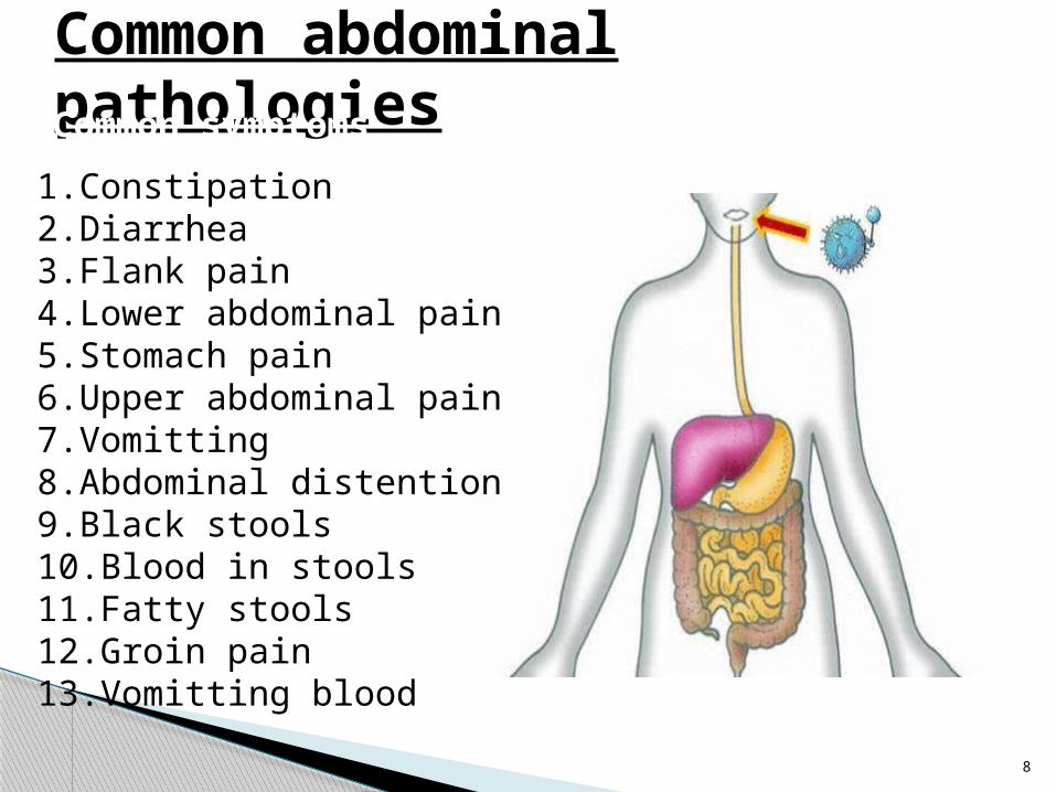
Common abdominal pathologiesCommon symptoms
1.Constipation2.Diarrhea3.Flank pain4.Lower abdominal pain5.Stomach pain6.Upper abdominal pain7.Vomitting8.Abdominal distention9.Black stools10.Blood in stools11.Fatty stools12.Groin pain13.Vomitting blood

1.Constipation
a. Low fiber dietb. Medicines side reactionsc. Oral Fe supplimentsD. haemorrhoidse. Bowel obstructionsf. Chronic renal faiureg. Faecal impactionh. Irritable bowel syndromei. Rectal cancerj. Colonic diverticulitisk. Hypothyroidisml. Colon cancerm. Paralytic ileusn. hypokalemia

2.Diarrhea
a. Gastroenteritisb. celiac diseasec. Food poisoningd. Irritable bowel syndromee. Lactose intolerancef. Medication reactiong. Bowel obstructionh. Colon canceri. Chrons diseasej. Bacterial dysentryk. Typhoid feverl. Giardiasism. Pancreatitisn. Shigellosiso. Viral infections

Flank pain/lumbar paina.Nephrolithiasisb.Utic.Muscle straind.Back traumae.Renal traumaf.Pyelonephritisg.Herpes zosterh.Renal infarcti.Renal vein thrombosisj.Low back pain (lumbago)k.Fibromyalgial.Spleen injurym.Retro peritoneal heamarrage
n.Kidney cancer/tumoro.Poly cystic kidney diseasep.Ulcerative colitisq.Myocardial infarctionr.Cholecystitiss.Appendicitist.Cholelithiasisu.Rib fracturev.Pleuritisw.Colonic diverticulitisx.Pneumonia

Lower abdominal pain
Right lower quadrant •Appendicitis •Diverticulitis •Salpingitis/Pelvic inflammatory disease •Endometritis •Endometriosis •Ectopic pregnancy •Hemorrhage or rupture of ovarian cyst •Renal calculus •Intussusception
•Pelvic/hypogastric region •Cystitis •Salpingitis/Pelvic inflammatory disease •Ectopic pregnancy •Diverticulitis •Strangulated hernia •Endometriosis •Appendicitis •Ovarian cyst •Ovarian torsion •Testicular torsion •Bladder distension •Nephrolithiasis

epigastric paina.Gastritis‘b.gastroentritisc.Constipationd.Gastro esophagal reflux (GERD)e.Viral infectionsf.Peptic ulcerg.Appendicitish.Cholangitis (infected bile duct)i.Cholecystitisj.Inguinal herniak.Irritable bowel syndromel.Cholelithiasism.Utin.Celiac disease

Upper abdominal painRight upper quadrant pain •Cholecystitis •Fatty liver or NASH •Congested liver (e.g., secondary to heart failure) •Cholangitis •Hepatitis •Gastritis or pancreatitis •Pneumonia
Left upper quadrant pain •Peptic ulcer disease •Gastritis •GERD •Spleenic infarct •Pulmonary embolism •Pancreatitis •Acute spleenomegaly •Left lower lobepneumonia
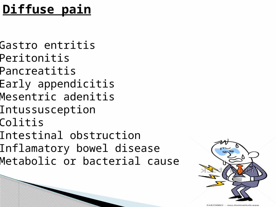
Diffuse pain
•Gastro entritis•Peritonitis•Pancreatitis•Early appendicitis•Mesentric adenitis•Intussusception•Colitis•Intestinal obstruction•Inflamatory bowel disease•Metabolic or bacterial cause

Vomiting
a.Gastro entritisb.Viral infectionsc.Medical reactionsd.Gerde.Food poisoningf.Gastritisg.Gastro intestinal bleedingh.Bulimia nervosai.Post concussive syndromej.AppendicitisK,alcoholisml.Migraine
m.Carbon monoside poisoningn. Bowel obstructiono.Diabetic ketoacidosisp.Head injuryq.Pancreaitis
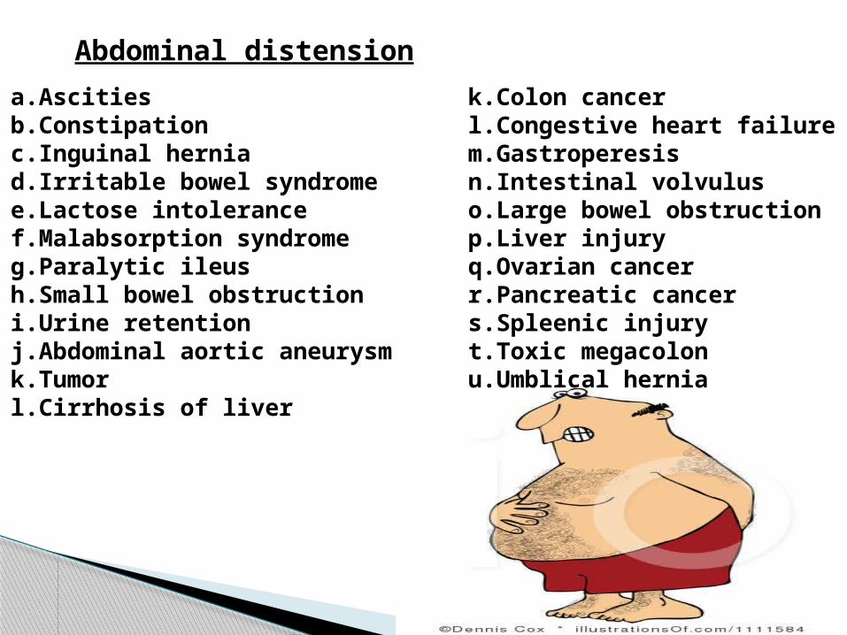
Abdominal distensiona.Ascitiesb.Constipationc.Inguinal herniad.Irritable bowel syndromee.Lactose intolerancef.Malabsorption syndromeg.Paralytic ileush.Small bowel obstructioni.Urine retentionj.Abdominal aortic aneurysmk.Tumorl.Cirrhosis of liver
k.Colon cancerl.Congestive heart failurem.Gastroperesisn.Intestinal volvuluso.Large bowel obstructionp.Liver injuryq.Ovarian cancerr.Pancreatic cancers.Spleenic injuryt.Toxic megacolonu.Umblical hernia

groin pain
a.Muscle strainb.Inguinal herniac.Epididymitisd.Nephrolithiasise.Ligament sprainf.Constipationg.UTIh.Testicular torsioni.Contusionk.Pyelo nephritis
l.Cellulitism.Bladder infectionn.Femoral herniao.lymphadenopathy
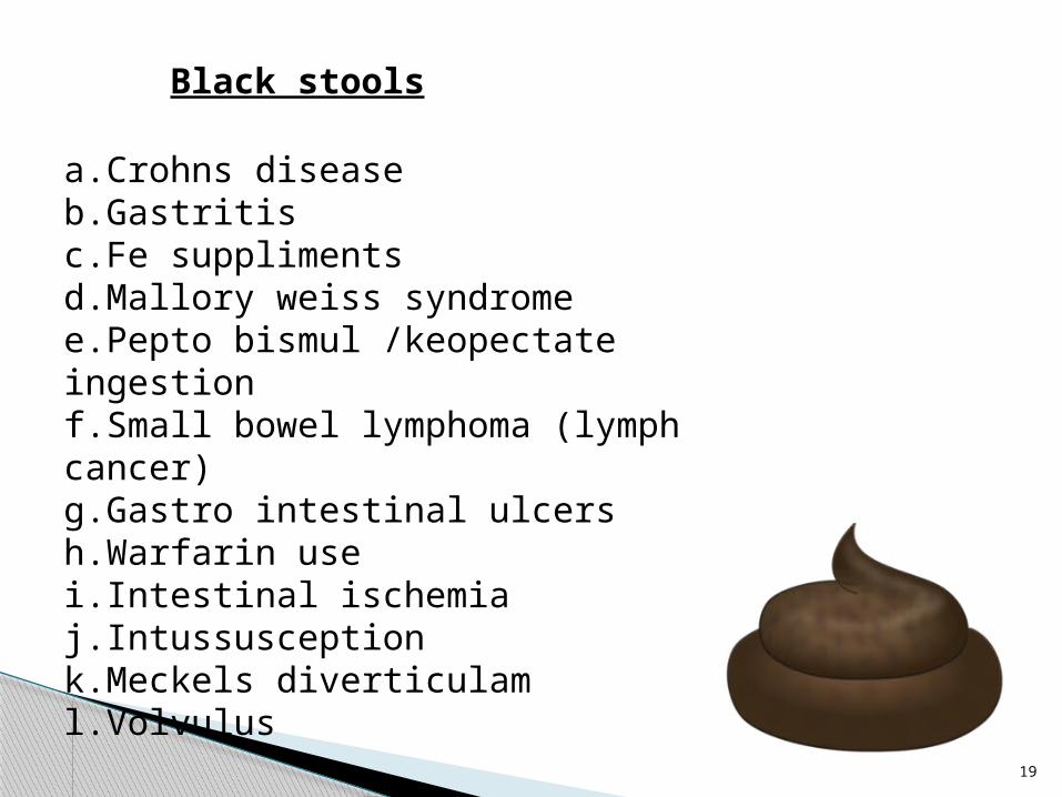
Black stools
a.Crohns diseaseb.Gastritisc.Fe supplimentsd.Mallory weiss syndromee.Pepto bismul /keopectate ingestionf.Small bowel lymphoma (lymph cancer)g.Gastro intestinal ulcersh.Warfarin usei.Intestinal ischemiaj.Intussusceptionk.Meckels diverticulaml.Volvulus

Blood in stools
a.Anal fissureb.Constipationc.Gastritisd.Gastro entritise.Hemorrhoidsf.Ulcerative colitisg.Peptic ulcerh.Crohns diseasei.Intestinal volvulus

Bloody diarrhea
a.Colitisb.Colonic diverticulitisc.Crohns diseased.Intestinal ischemiae.Ulcerative colitisf.Gastro intestianl ulcerg.Intestinal volvulush.Intussusception i.Hemolytic uremic syndromej.Amebiasis

Fatty stools
a.Bacterial over growth in intestineb.Celiac diseasec.Choledocholithiasisd.Lactose intolerancee.Pancreatitisf.Ulcerative colitisg.Cystic fibrosish.Primary scelorosing cholangitisi.Whipples disease

Vomiting blood
• Gastro entritis• Esophagitis• GERD• Thrombocytopenia• Intussuception• Mallory weiss syndrome• NSAID overdose • Tonsilitis• Intestinal volvolus• Gastritis

24
INVESTIGATIONS

25
Radiology is the branch of medicine that uses radio active substances or electromagnetic waves to create images of the body for the purpose of diagnosis and treatment.
Images can also show how effectively the body and its internal organs and structures are functioning
It is used for both therapeutic and diagnostic purposes
RADIOLOGY

26
Types1. Diagnostic radiology : type of radiology that uses
external radiation to produce images of body ,organs and other internal structures for diagnostic purpose
2. Nuclear medicine : a type of diagnostic radiology that uses a small amount of radio active substance to create image of the body organ which helps to study the structure and functions to make diagnosis/treatment of them.
3. Theraputic radiology :that branch uses radiant energy to study ,treats and manages cancer and other diseases
4. Interventional radiology: that branch which uses various imaging techniques to guide the insertion of small instruments and tools through GIT to identify and treat medical disorder .In this simultaneous diagnosis and treatment with help of Xray imaging is carried out.

27
Images produced by radiological procedures can be grouped into
A.Transmission imaging in this, a beam of high energy photons is
produced and passed through the body structure to be examined.The beam passes through less dense types of tissue such as watery secretions,blood and fat,very quickly leaving a darkened area on the film.muscle and connective tissue appear as grey while bone appear as white er:Xrays,CT,
Diagnostic radiology

28
B.Reflection imaging Imaging produced by sending high frequency sounds
to the body part or organ being studied..These sound waves bounce off various types of body tissue at varying speeds, depending upon te density of the tissue present.The bounced sound waves are sent to a computer that analyse the sound wave and produce the visual image of the body part or structure.
Eg :Ultrasound C.Emission imaging In this tiny nuclear particles or magnetic energy are
detected by a scanner and analysed by computer to produce an image of body structure or organ being examined.
Eg: MRI

29
D. Contrast medium xray• To visualise radio lucent structures like
stomach,intestines,blood vessels,etc a radio opaque substance is passed or injected into them and xray film is taken.In these indirect viewing of the recquired part with help of injecting a radio opaque substance is possible
• Eg:barium meal ,barium swallow,barium enema,intravenous pyelography ,myelogram , intravenous urogram, cystogram, urethrogram,cholesystogram,urethrogram, cholangiogram, arteriogram, venogram,lymphangiogram,sinusogram,etc

Abdominal xray• X ray imaging is performed according to the x ray penetrability of tissues under investigations.
•The formation of a radiographic image depends on the structure & size of the organs within the abdomen
2 Common position in abdominal Xrays
•Anterio posterior (AP) supine•Anterio posterior (AP) erect If patient unable to sit or stand• lateral decubitus
n
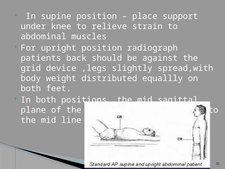
In supine position – place support under knee to relieve strain to abdominal muscles
For upright position radiograph patients back should be against the grid device ,legs slightly spread,with body weight distributed equallly on both feet.
In both positions ,the mid sagittal plane of the body should be centered to the mid line of the grid device

Techniques - Projection•P-A (relation of x-ray beam to patient)

•Bone- High (white)•Tissue- Middle (grey)•Air- Low (black)

Densities
The big two densities are:
(1) WHITE - Bone (2) BLACK - Air
The others are:
(3) DARK GREY- Fat (4) GREY- Soft tissue/water And if anything Man-made is on the film, it is:
(5) BRIGHT WHITE - Man-made

Abdominal landmark- illiac crest level of mid abdomen (L4.L5) For supine position ,the cassete of Image receptor is centered into the illiac crest and lower abdomen is
generally included on lower margin of the cassete.
For upright position, cassette is centered (5cm) above level of illiac crest ,or high enough to include the diaphragm
.Relaxation of musculature is acheieved by supporting and slightly flexing patients knee.
Ask the patient to take a deep breath,exhale completely and hold the position while not inhaling.this moves the diaphragm to a superior position that results in better visualisation of abdominal muscles

Things to look for Name,date Position of the film and view Adequate area covered or not? Preperitoneal fat lines Visualised organs are normal in size Visualised bones and joints are normal Any radio opacity Any artifacts Any calcification
Indications for abdominal Xray Used for certain defined pathology such as abnormal
gas,masses,bones and stones Undifferentiated abdominal pain with provisional diagnosis of a. toxic mega colon of IBD b.bowel obstruction c.bowel ischemia d.perforation of a viscus with abdominal free air e.KUB for renal tract calculi; 80 to 90% sensitivity if radio lucent stone >3 mm f.foreign body Radio dense tablets eg.pottasium chloride tablet Metals eg: mercury Iatrogenic eg: barium


1, 11th rib.2, Vertebral body (TH 12).3, Gas in stomach.4, Gas in colon (spleenic flexure).5, Gas in transverse colon.6, Gas in sigmoid.7, Sacrum.8, Sacroiliac joint.9, Femoral head.10, Gas in ceacum11, Iliac crest.12, Gas in colon (hepatic flexure).13, Psoas margin

Anatomy on the
Abdominal X-Ray:


Contrast medium x rays
Barium x rays are used to diagnose abnormalities of gastro intestinal tracts,such as tumour, ulcer,polyps,hernia,strictures etc..
With the use of barium sulphate , a metalic chemical that x rays cannot pass through,x rays are taken
3 types a.barium swallow b.barium meal c.barium enema

Indications in barium X ray
Pain or difficulty in swallowing Blood in the vomit Abdominal pain Bowel moment changes Chronic diarrhea / constipation Unusual bloating Bleeding from the rectum Unexplained weight loss

Barium swallow
A barium swallow is a radiographic (X-ray) examination of the upper gastrointestinal (GI) tract, specifically the pharynx (back of mouth and throat) and the esophagus . The pharynx and esophagus are made visible on X-ray film by a liquid suspension called barium sulfate (barium)
Barium is an X-ray absorber and appears white on X-ray film. When swallowed, a barium drink coats the inside walls of the pharynx and esophagus so that the swallowing motion, inside wall lining, and size and shape of these organs is visible on X-ray. This process shows differences that might not be seen on standard X-rays.

•Fluoroscopy is often used during a barium swallow. Fluoroscopy is a study of moving body structures — similar to an X-ray "movie." A continuous X-ray beam is passed through the body part being examined, and is transmitted to a TV-like monitor so that the body part and its motion can be seen in detail.
• In barium X-rays,fluoroscopy allows the radiologist to see the movement of the barium through the pharynx and esophagus as a person drinks

INDICATIONS•Cancers of the neck, pharynx, and esophagus
•Tumors
•Hiatus hernia. Upward movement of the stomach, either into or alongside the esophagus
•Structural problems. Such as diverticula, strictures, or polyps •Esophageal varices (enlarged veins)
•Muscle disorders (pharyngeal or esophageal). Such as dysphagia (difficulty swallowing) or spasms (pharyngeal or esophageal)
•Achalasia. A condition in which the lower esophageal sphincter muscle doesn't relax and allow food to pass into the stomach
•Gastroesophageal reflux disease (GERD)
• Ulcers

Barium meal
•In a barium meal test, X-ray images are taken of the stomach and the beginning of duodenum
•A barium meal usually takes less than an hour.
• The patient ingests gas pellets and citric acid to expand the stomach. Then about 3 cups (about 709 ml) of barium is ingested. The patient may move or roll over to coat the stomach and oesophagus in barium. Following these preparations, an x-ray is taken.

•There are two varieties of barium meal: single and double contrast meals.
•A single contrast meal uses only barium, a radioopaque (or positive) contrast medium, to image the upper gastrointestinal tract.
•A double contrast meal uses barium as well as a radiolucent (or negative) contrast medium such as air, nitrogen, or carbon dioxide.
•The double contrast meal is more useful as a diagnostic test, demonstrating mucosal details and allowing the detection of small mucosal lesions such as diverticula or polyps.

Barium enema•Lower gastrointestinal (GI) tract radiography, also called a lower GI or barium enema, is an x-ray examination of the large intestine, also known as the colon.
•This examination evaluates the right or ascending colon, the transverse colon, the left or descending colon, the sigmoid colon and the rectum.
•The appendix and a portion of the distal small intestine may also be included

•After the instillation of barium into the rectum, the radiologist may also fill the large intestine with air.
• Air will appear black on X-ray film, contrasting with barium's white image.
•The use of the 2 substances, barium and air, is called a double contrast study.
•The purpose of using 2 contrast substances is to achieve an enhancement of the inside wall lining of the large intestine.
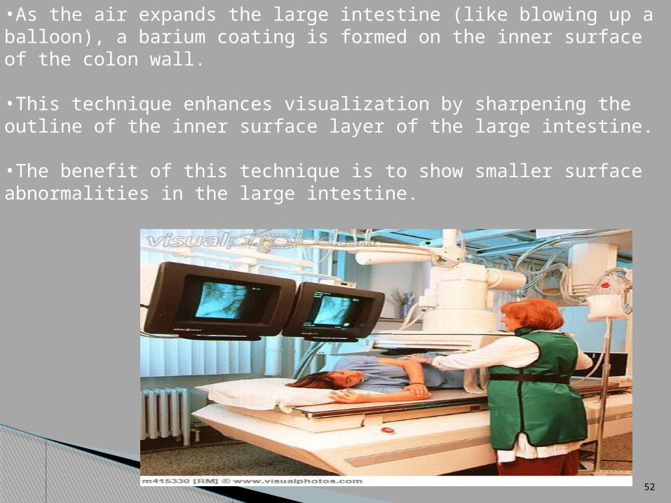
•As the air expands the large intestine (like blowing up a balloon), a barium coating is formed on the inner surface of the colon wall.
•This technique enhances visualization by sharpening the outline of the inner surface layer of the large intestine.
•The benefit of this technique is to show smaller surface abnormalities in the large intestine.
Ulcerative colitis. Ulcerations and inflammation of the large intestine.
Crohn's disease. Ulcerations and inflammation occurring in any part of the GI tract
Obstructions and polyps (growths)
Cancer
Unusual bloating or lower abdominal pain
Unexplained weight loss
Irritable bowel syndrome
Changes in bowel movements. Such as chronic diarrhea or constipation, or passing of blood, mucus, and/or pus.
Indications

• Risks of barium enema may include, but are not limited to:
Colon perforation Constipation or fecal impaction
• Contraindications for a barium enema include, but are not limited to:
Suspected bowel perforation Severe ulcerative colitis Pregnancy Toxic megacolon Acute abdominal pain

Intra venous pyelogram (IVP)
An intravenous pyelogram (IVP) is an x-ray examination of the kidneys, ureters and urinary bladder that uses iodinated contrast material injected into veins.
When a contrast material is injected into a vein in the patient's arm, it travels through the blood stream and collects in the kidneys and urinary tract, turning these areas bright white on the x-ray images
Indications kidney stones enlarged prostate tumors in the kidney, ureters or urinary bladder surgery on the urinary tract congenital anomalies of the urinary tract

The equipment typically used for this examination consists of a radiographic table, one or two x-ray tubes and a television-like monitor that is located in the examining room. Fluoroscopy, which converts x-rays into video images, is used to watch and guide progress of the procedure.
In an IVP exam, an iodine-containing contrast material is injected through a vein in the arm. The contrast material then collects in the kidneys, ureters and bladder, sharply defining their appearance in bright white on the x-ray images.
X-ray images may be maintained as hard film copy or as digital images

Computed Tomography (CT or CAT) Scan of the Abdomen•A CT or CAT scan is a diagnostic imaging procedure that uses a combination of x rays and computer technology to produce cross sectional images (slices) both horizontally and vertically of the body.
•CT scan also minimize the exposure to radiation
• In CT ,the Xray beam moves in a circle around the body.This allows many different views of the same organ or structure , and provides much greater details.
•The xray information is sent to a computer that interprets the xray data and displays it in 2 dimensional form in monitor

•CT scans can be done with or without contrast
•Contrast refers to a substance taken by mouth or injected into an intravenous line that causes the particular tissue or organ under study to be seen more clearly
•Contrast procedure may require the patient to fast before the procedure
•Usualy used in the diagnosis of tumors,internal bleeding and also to check internal damage or injuries.


Indications
•To assess the abdomen and its organs for tumors and other lesions,• injuries,• intra-abdominal bleeding, •Infections• unexplained abdominal pain• obstructions• when another type of examination, such as X-rays or physical examination, is not conclusive.•A CT scan of the abdomen may also be used to evaluate the effects of treatment on abdominal tumors. •Another use of abdominal CT is to provide guidance for biopsies and/or aspiration of tissue from the abdomen.

Benefits
•Viewing a CT scan, an experienced radiologist can diagnose many causes of abdominal pain or injury from trauma with very high accuracy, enabling faster treatment and often eliminating the need for additional, more invasive diagnostic procedures.•When pain is caused by infection and inflammation, the speed, ease and accuracy of a CT examination can reduce the risk of serious complications, such as those caused by a burstappendix or an infected fluid collection and the subsequent spread of infection.

Benefits
•CT has been shown to be a cost-effective imaging tool for a wide range of clinical problems.
•CT is less sensitive to patient movement than MRI.
•CT examinations are fast and simple; in emergencycases, they can reveal internal injuries and bleeding quickly enough to help save lives
•CT scanning is painless, noninvasive and accurate.
•A major advantage of CT is its ability to image bone, soft tissue and blood vessels all at the same time.•Unlike conventional x-rays, CT scanning provides very detailed images of many types of tissue as well as the lungs, bones, and blood vessels.

Benefits •CT can be performed if you have an implanted medical device of any kind, unlike MRI.
•CT imaging provides real-time imaging, making it a good tool for guiding minimally invasive procedures such as needle biopsies and needle aspirations of many areas of the body, particularly the lungs, abdomen, pelvis and bones.
•A diagnosis determined by CT scanning may eliminate the need for exploratory surgery and surgical biopsy.
•No radiation remains in a patient's body after a CT examination.
•X-rays used in CT scans should have no immediate side effects

Risks
•There is always a slight chance of cancer from excessive exposure to radiation.
•The effective radiation dose for this procedure varies.
•Women should always inform their physician and x-ray or CT technologist if there is any possibility that they are pregnant.
•CT scanning is, in general, not recommended for pregnant women unless medically necessary because of potential risk to the baby in the womb.
•. CT scans in children should always be done with low-dose technique

.
ULTRASONOGRAPHY (USG)
Ultrasound is a safe and painless procedure, and produces pictures of the inside parts of the body using sound waves. Ultrasound imaging, also called ultrasound scanning or sonography, involves the use of a small transducer (probe) and ultrasound gel placed directly on the skin.

.
•
High-frequency sound waves are transmitted from the probe through the gel into the body.
The transducer collects the sounds that bounce back and a computer then uses those sound waves to create an image.
Ultrasound examinations do not use ionizing radiation (as used in x-rays), thus there is no radiation exposure to the patient.
.
•The sound waves bounce of the organs like an echo and return to the transducer.
•The transducer picks up the reflected waves, which are then converted into an electronic picture of the organs.
•Different types of body tissues affect the speed at which sound waves travel.
•Sound travels the fastest through bone tissue, and moves most slowly through air.

•The speed at which the sound waves are returned to the transducer, as well as how much of the sound wave returns, is translated by the transducer as different types of tissue
•By using an additional mode of ultrasound technology during an ultrasound procedure, blood flow within the abdomen can be assessed.
•An ultrasound transducer capable of assessing blood flow contains a Doppler probe.
•The Doppler probe within the transducer evaluates the velocity and direction of blood flow in the vessel and makes the sound waves audible.

•Cysts•Tumors•Collection of pus •Obstructions•Fluid collection•Blockage in blood vessels•Infection
USES OF USG to find out

Benefits
Most ultrasound scanning is noninvasive (no needles or injections).
Occasionally, an ultrasound exam may be temporarily uncomfortable, but it is almost never painful.
Ultrasound is widely available, easy-to-use and less expensive than other imaging methods.
•

Ultrasound imaging is extremely safe and does not use any ionizing radiation.
Ultrasound scanning gives a clear picture of soft tissues that do not show up well on x-ray images.
Ultrasound provides real-time imaging, making it a good tool for guiding minimally invasive procedures such as needle biopsies and fluid aspiration.

•
MAGNETIC RESONANCE IMAGING•Non invasive technique
•MRI uses a powerful magnetic field, radio frequency pulses and a computer to produce detailed pictures of organs, soft tissues, bone and virtually all other internal body structures
•MRI does not use ionizing radiation (x-rays).The images can then be examined on a computer monitor, transmitted electronically, printed or copied to a CD

•
INDICATIONS
MR imaging of the body is performed to evaluate:
• organs of the chest and abdomen—including the heart, liver,biliary tract, kidneys, spleen, bowel, pancreas and adrenal glands.
• pelvic organs including the bladder and the reproductive organs such as the uterus and ovaries in females and the prostate gland in males.
• blood vessels (including MR Angiography).
• Lymph nodes.
•
Physicians use an MR examination to help diagnose or monitor treatment for conditions such as:
• tumors of the chest, abdomen or pelvis.• diseases of the liver, such as cirrhosis, and abnormalities of the bile ducts and pancreas• inflammatory bowel disease such as Crohn’s disease and ulcerative colitis• heart problems, such as congenital heart disease.• malformations of the blood vessels and inflammation of the vessels (vasculitis).• a fetus in the womb of a pregnant woman
• It captures excellent images of fluid and swelling, as well as active inflammation, bowel obstructions, abscesses, and fistulas, or abnormal passageways between organs.

•
Principle• MRI does not depend on ionizing radiation.
• Instead, while in the magnet, radio waves redirect alignment of hydrogen atoms that naturally exist within the body without causing any chemical changes in the tissues. As the hydrogen atoms return to their usual alignment, they emit energy that varies according to the type of body tissue in which they lie.

•
•. The MR scanner listens for this energy and creates a picture of the tissues scanned.
•The magnetic field is produced by passing an electric current through wire coils in most MRI units. Other coils, located in the machine and in some cases, placed around the part of the body being imaged, send and receive radio waves, producing signals that are detected by the coils.

•
• The traditional MRI unit is a large cylinder-shaped tube surrounded by a circular magnet. Patient will lie on a moveable examination table that slides into the center of the magnet.
• The magnetic field is produced by passing an electric current through wire coils in MRI units,placed around the part of the body being imaged, send and receive radio waves, producing signals that are detected by the coils. A computer then processes the signals and generates a series of image
• Duration : 30- 50 minutes
a.Colonoscopy With the help of a colonoscope, this procedure is used to
examine the colon,the last part of gastrointestinal tract.
A colonoscope is a long, thin, flexible tube with a miniature video camera and light at its end. The gastroenterologist will put a little bit of air into the colon as he/she inserts the scope. The camera on the end helps the physician both guide the colonoscope throughout the length of the colon and take pictures of the colon.
Colonoscopies are most commonly performed in colorectal cancer screening and prevention
Duration : 30 mins
Invasive procedures


b.Endoscopic Retrograde CholangioPancreatography
(ERCP)•During an endoscopic retrograde cholangio pancreatography, or ERCP, the gastroenterologist uses an endoscope, a long, thin, flexible tube with a light and camera at the end, through the esophagus, the stomach, and the first part of the small intestine, called the duodenum.
• Once the endoscope reaches the papilla, which is the opening of the common bile duct, the physician injects dye through these ducts, enabling x-rays to be taken

•Bile, a liquid that helps digest fat, is produced by the liver and carried to the gallbladder, where it is stored, through a series of tubes called ducts.
•The main duct from the pancreas joins the common bile duct and allows pancreatic juices to help with further digestion in the duodenum.
•After eating, both bile and pancreatic juices flow through the papilla and into the duodenum, where they mix with food and play a major role in digestion.
•A physician may recommend an ERCP if the patient is experiencing abdominal pain or develops jaundice .
• This procedure is helpful in identifying gallstones, tumors or scar tissue obstructing the bile duct.

C.Endoscopic Ultra Sonography (EUS)To examineUpper GI : esophagus,stomach and duodenumLower GI : colon,anus and rectum
•EUS involves the use of an endoscope or colonoscope, long, thin, flexible tubes with a light and camera at the end, to help guide the scope throughout the duration of the procedure.
•However, these scopes are different than those used in colonscopy and ERCP: they emit sound waves that create visual images of the digestive tract that a normal endoscope cannot detect.
•It may also be used to assess the nature of a tumor that may have been detected during a prior endoscopic procedure
• In conjunction with examination of a tissue sample obtained using a procedure called a "fine needle aspiration," EUS can help diagnose diseases of the pancreas, gallbladder and bile duct .
•Duration : 45 minutes

d.Upper GI Endoscopy
An upper GI endoscopy looks at the upper part of the gastrointestinal tract including the esophagus, the stomach and the first part of the small intestine, called the duodenum.
The gastroenterologist uses an endoscope, a long, thin, flexible tube with a light and camera at the end to help guide the scope throughout the duration of the procedure.
The camera on the end helps the physician both guide the endoscope throughout the length of the upper GI tract, and take pictures.
Indications :chronic heartburn (acid reflux), difficulty swallowing, stomach or abdominal pain, bleeding, ulcers and tumors.
Duration : 10- 15 minutes

e. Liver Biopsy A liver biopsy is used to determine the presence of
inflammation, fibrosis and to help diagnose various liver diseases.
During this procedure, the patient is fully conscious. A physician numbs the area around the liver using a local anesthetic (similar to that used by a dentist), and then using a long, narrow needle obtains a tiny piece of liver tissue.
After the procedure, the patient is kept in recovery for four hours for monitoring.

F. Double Balloon Enteroscopy Double balloon enteroscopy is a new method of examining the
small intestine that previous techniques could not reach. Double balloon enteroscopy employs a high- resolution video
endoscope with latex balloons attached at the tips that can be inflated and deflated with air from a pressure-controlled pump system.
A sequence of inflation/deflation cycles allow the scope to be advanced further into the small intestine. This technique can be performed using either an oral or anal route.
Indications : obscure gastrointestinal bleeding, Crohn's disease, unexplained diarrhea, pancreaticobiliary disease

•
CONCLUSIONThe differential diagnosis is extremely wide and definitive diagnosis is often difficult, particularly in primary care of the abdominal disorders. This is due to the many different organs within the peritoneal cavity and the potential for referred pain. But Modern medicine has been extraordinarily developed with the amalgamation of technology in the field of diagnostic, prognostic, and curative procedures. Newer technologies are being introduced each day for finer and precise understanding of human being and diseases.

•
Although the principles of Ayurveda are called immortal (that never die and are always applicable), it is a need to be contemporary with the current scientific trends for the benefit of the society and for nurturing Ayurveda. Updating Shalya tantra, by integrating with modern technologies, without changing the basic principles, is a challenging task that needs great insight in the field of Ayurveda and intellect nourished with modern tonic

SRB Manual of surgery Outline of shalya tantra Manipal surgery Webmd.com Radiopedia.com Medscape.com
References

Thank u…
Related Documents