thenerve Fall 2010 Vol 2 Issue 1 The Rise of the Cyborgs The Religious Brain Neuroscience and the Military Resolving the Line Between Genius and Insanity The Drugs and Mechanisms of General Anesthesia Spatial Navigation and the Memory Network

Welcome message from author
This document is posted to help you gain knowledge. Please leave a comment to let me know what you think about it! Share it to your friends and learn new things together.
Transcript
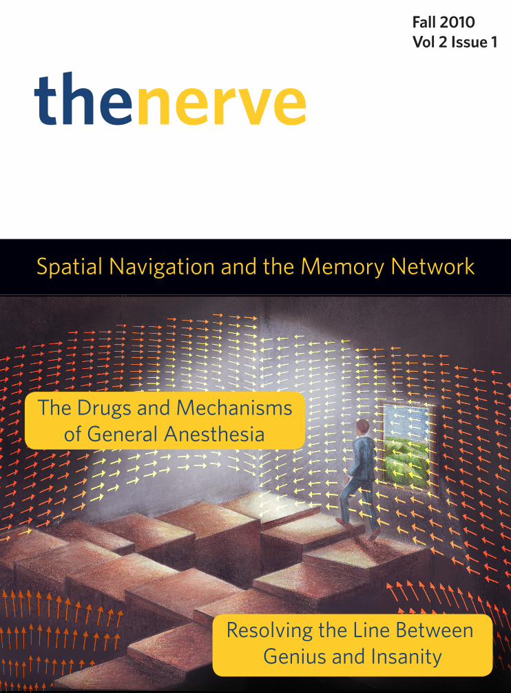
thenerveFall 2010Vol 2 Issue 1
The Rise of the Cyborgs
The Religious Brain
Neuroscience and the Military
Resolving the Line Between Genius and Insanity
The Drugs and Mechanisms of General Anesthesia
Spatial Navigation and the Memory Network

Mind and Brain Society
The Mind and Brain Society (MBS) was founded in the fall of 2008 in concert with BU’s new Undergraduate Program in Neuroscience. The group aims to create a net-work for undergraduate students who wish to take an active role in current issues and research. MBS serves as a hub for not only Neuroscience majors, but all students in-terested in Psychology, Biology, Philosophy, Computer Science, etc. Our goal is to sup-port an eager multidisciplinary undergraduate community with the conversations and resources fundamental to Neuroscience today.
Throughout the academic year, MBS hosts events spotlighting many different fac-ets of Neuroscience. We hold discussion sessions during which we informally discuss a topic of interest over coffee; previous topics include “The Neuroscience of Religion” and “NeuroEthics.” The group also hosts research presentations by BU professors and screenings of thought-provoking films containing neuroscience motifs.
SPRING 2010 | 3
CONTENTSSpring 2010 Vol. 1 Issue 2
RESEARCH IN BRIEF 5
ARTICLESSports-related Concussions and the NFL by John Batoha & Evan Stein 11
Resolving the Line Between Genius and Insanity by Frank DeVita 17
Ten Minutes to a Trance by Anuhya Caipa 22
Trick or Treatment? The Placebo Effect by Natalie Banacos 24
Brain Research and National Defense by Aisha Sohail 27
REVIEWSGeneral Anesthesia: Drugs and Mechanisms by Grigori Guitchounts 32
Spatial Navigation: Decoding the Human Memory Network by Evan Stein 41
OPINIONEvolutionary Psychology on the Couch by Devyn Buckley 49
Do brains run algorithms? by Kayla Ritchie 57
“When we speak of nature it is wrong to forget that we are ourselves a part of nature.” -Henri Matisse
Students of neuroscience decide to join the field for a myriad of reasons- not the least of which includes a sentiment of vanity. “My brain is amazing and I want
to know how it works.” At the beginning, many walk around fascinated, suddenly more in tune with how we know and experience what we do- spouting off “Did you know’s…” to unsuspecting friends and family. As study continues, we learn of tinier molecules, more complex proteins, and the most finely tuned processes. The scale becomes smaller and several steps removed from our consciousness. It is easy to consider these inner-workings as only controlled experiments under careful watch in a lab, but if we can keep in mind that such precise orchestrations occur naturally within us, the whole thing becomes even more astounding.
Welcome to the second year of The Nerve’s infancy. Once again, we are thrilled to present an issue with breadth across the field of neuroscience while spotlighting a few of our favorite topics from the last six months. We’d like to extend our deepest thanks to The Nerve staff and writers and Boston University faculty for pushing us into our sec-ond volume.
As you read each article, be a little self-centered. Con-sider the way your wiring affects your daily life. Consider the amount of electricity running through you. Consider each molecule taking its course through your brain, moving in such synchrony to make your very reading this possible! It’s a lot to think about, but the exquisitely intricate nature of the system is what makes it so extraordinary.
This and other issues of The Nerve would not have been possible without the generous help ofPaul Lipton, Howard Eichenbaum, Lindsey Clarkson, Denise Parisi, Zachary Bos, and Jarret Frank.
- Grigori Guitchounts and Kimberly LeVineEditors-In-Chief
Editors-In-ChiefGrigori Guitchounts
Kimberly LeVine
EditorsFrank DeVita
Lauren Joseph
Associate EditorsNatalie Banacos
John BatohaDevyn BuckleyMonika Chitre
Jennifer RichardsonKayla Ritchie
Evan Stein
Artistic DirectorKayla Ritchie
ArtworkMursal Atif
Devyn BuckleyMariya Marioutina
Margaret McguinnessAubrey Reuben
MBS StaffMegan Mataga
Macayla DoneganShea Gillet
AdvisorsPaul Lipton
Zachary Bos

RESEARCH IN BRIEF
FALL 2010 | 5
Pain management is an important branch of med-icine, especially in fields that consider long-term pain-related diseases such as cancer or arthritis. People ex-perience pain as a result of nerve impulses traveling through the spinal cord to the central nervous system in response to noxious stimuli. As the pain signal trav-els to the brain, it can be modulated in a number of different ways by intervening nerve fibers that cause an inhibitory effect, thus reducing pain. This interfer-ence can be experienced in the common instinct rub a wound: our body’s touch receptors connect to pain signal fibers in the upward pain pathway to the brain and slow the noxious signal’s travel by inducing vari-ous neuronal interactions. Other ways to modulate pain incorporate the use of analgesic drugs, most commonly opiates, such as morphine for acute pain, or lighter analgesics such as acetaminophen (Tylenol) and salicylic acid (Aspi-rin). Both act within the brain itself through mo-lecular interactions to modulate pain in a top down fashion through neurotransmitter sys-tems.
Neurotransmitters are a crucial piece of the puzzle in the neurosci-ence underlying pain. Current research is elu-cidating the mechanisms of anandamide, a myste-rious neurotransmitter that acts on the much-re-searched cannabinoid CB1 and CB2 receptor as well as the vaniloid receptors. We do not know much about anandamide, but its levels spike in response to pain or inflammation and these receptors bind the neu-rotransmitter, creating an analgesic affect. In a recent Nature Neuroscience study, researchers elucidated a mechanism of anandamide in the peripheral nervous system (PNS) and its action on cannabinoid recep-tors. The team created an inhibitor of fatty acid amide hydrolase (FAAH), a compound that degrades anan-damide under normal circumstances. The inhibitor, dubbed URB937, was designed to remain only in the
peripheral nervous system and suppress FAAH activ-ity, thus increasing anandamide levels in the periph-ery. As a result, behavioral responses to peripheral nerve pain and inflammation were reduced and neu-ronal activation in pain processing was suppressed in rodent models in response to increased anandamide binding at CB1 receptors.
Experiments subjected rodent models to neuro-pathic and inflammatory pain to test the efficacy of URB937 as an analgesic. For neuropathic pain, sciatic nerve injury was induced and the compound was im-mediately administered. Results showed decreased sensitivity to pain and daily administration of the drug showed consistent results. Chemically induced inflammatory swelling in the rodents was combated with URB937 with equally effective results. Spinal pain perception was also investigated by measuring
levels of the protein Fos, whose levels are elevated in response to neuronal activi-ty in the spinal cord and thus in response to pain. URB937 was able to attenuate Fos levels in regions of the neck and dorsal horn of the spinal cord, implying that the trav-eling pain signal was sup-pressed before it was able to reach the brain itself.
The suggestion of this research is that increased anandamide levels in the periphery and CB1 recep-tors can effectively attenuate
pain signals to the CNS. Since URB937 was designed to remain exclusively outside the CNS, this research establishes the existence of a previously unexplored connection between the peripheral endocannabinoid system and pain perception. Given their results, the researchers hypothesize that peripheral anandamide acts as a diffuse paracrine signal to modulate pain stimuli as they arrive in damaged tissues. This implies that pain stimulates anandamide production and CB1 receptors are present in abundance in peripheral nerve endings and throughout tissues and organs. These results open new doors to pain therapy that can
Elucidating a New Pathway for Pain Management
RESEARCH IN BRIEF
6 | THE NERVE
ADHD is a disorder known to almost everyone; it has established a prevalence in our school systems and the media. The Center for Disease Control high-lights that 3-7 percent of school-aged children suf-fer from ADHD. In the United Kingdom, where this particular study was done, it is reported that around one in 50 children is affected by the disorder. But even with such high prevalence and awareness about ADHD, the causes of the disease have escaped the sci-entific and medical community. Recentresearch from Cardiff University offers potential ground and stigma breaking information, shedding some light on this elu-sive disorder.
Children diagnosed with ADHD are restless, im-pulsive and distractible, and often experience dra-
Rare CNVs in the ADHD Genome
matic learning difficulties. The causes of ADHD are largely unknown. The general population, as well as a portion of the scientific community, has believed that it is likely attributed to poor parenting and a sugar rich diet, but this study presents the first evidence that ADHD is instead a genetic neurodevelopmental disorder.
The study conducted at Cardiff University ana-lyzed the genomes of 366 children with ADHD and 1047 controls. These children with ADHD were aged 5-17 years who were diagnosed with ADHD or hy-perkinetic disorder, but not schizophrenia or autism. The researchers genotyped single nucleotide poly-morphisms (SNP:DNA sequence variation in which a single nucleotide differs from the appropriate base pairing) for both groups, looking for and comparing copy number variants (CNVs) in their genomes.
CNVs occur when one of the two copies of a gene is missing or when there are too many copies. Their findings showed that 57 large, rare CNVs were iden-tified in the ADHD population, in contrast to the 78 CNVs that were found in the much larger control pop-ulation. This data shows that these rare CNVs were almost twice as likely to occur in children with ADHD (14%) compared to controls (7%), including a rate of CNVs that was particularly high in ADHD subjects with intellectual disability.
This data is the first of its kind to suggest a genetic basis to this disorder. The researchers also highlight that the rare CNVs identified occur in genomes in the brain that are known to correlate with susceptibility
exploit the cannabinoid system to effectively attenu-ate pain without the highly undesirable side effects produced by classical opioid pain therapy.
— Frank DeVita
original paper: Clapper JR et al. Anandamide suppresses pain initiation through a peripheral en-docannabinoid mechanism. Nature Neuroscience 13, 1265–1270. 2010.
RESEARCH IN BRIEF
FALL 2010 | 7
to autism and schizophrenia. One of these particular regions was identified on chromosome 16, which has been found crucial in the development of the brain and is affected in various major psychiatric and devel-opmental disorders.
The suggestion supported by this research is that ADHD is not based on social elements or poor parent-ing, but instead a direct genetic fault. This CNV data paired with the fact that ADHD is known to be highly heritable, points the scientific and therapeutic ap-proach to this condition in a whole new direction.
— Jennifer Richardson
original paper: Williams NM et al. Rare Chromo-somal deletions and duplications in attention-deficit hyperactivity disorder: a genome-wide analysis. The Lancet 376: 9750. 1401 - 1408. 2010.
For any student of Neuroscience, the neurotrans-mitter dopamine immediately brings to mind the pathways of“reward signaling.” However, this neu-rotransmitter also plays a critical role in regulating movement. Parkinson’s disease (PD), which injures dopaminergic neurons, unleashes a devastating effect on body control. In PD, the damaged nerve cells fire rapidly, losing all control over dopamine and causing involuntary muscle movements or tremors. Other ef-fects of the disease include rigidity, slowness, or loss of coordination. A study conducted this year by Mas-sachusetts General Hospital and Harvard Medical School revealed that caffeine and its metabolites, the parts left when it’s broken down, may provide some neuroprotective properties that resist the damaging effects of Parkinson’s disease.
Epidemiological studies have demonstrated a link between caffeine consumption and PD. In this particular study, the MPTP model of PD focuses on the effects of 1-methyl-4-phenyl-1,2,3,6-tetrahydro-pyridine (MPTP) in causing the devastating the dis-ease. The effects of caffeine on countering the actions of MPTP revealed insight into its effect against PD as a whole. Metabolites of caffeine as they appear in the human body when it is being broken down were administered to rodents so that the effects could be as similar to humans as possible.Over 80% of orally administered caffeine metabolizes to paraxanthine (1,7-dimethylxanthine), and about 16% is converted
to theobromine (3,7-dimethylxanthine) and theophyl-line (1,3-dimethylxanthine).
In the first segment of the experiment mice were given doses of saline or caffeine 10 minutes, 30 min-utes, 1 hour, 2 hours, or 6 hours before doses MPTP or saline was administered. The saline administration provided a control to compare the effects of the actual chemical. Then the same dose of caffeine or saline was administered10 minutes, 30 minutes, 1 hour, 2 hours, or 6 hours after the first. The second segment of the experiment involved testing the action of the metabolites. Caffeine, Theophylline, Paraxanthine, or saline was administered 10 minutes before MPTP or saline was administered.
Dopamine levels were measured by extracting the striatum from the right cerebral hemisphere. Caffeine was measured from the trunk blood of mice. Serum and supernatant of brain homogenates were mea-sured for caffeine and its three metabolites. The re-sults showed that caffeine was not only effective in at-tenuating dopamine depletion when co-administered with the toxin (10 minutes before MPTP), but also 30 minutes before administration. However, when caf-feine was administered earlier than 6 hours before MPTP it no longer had a significant effect. When caf-feine was administered 10 minutes, 30 minutes, or an hour after, but not 4, 8, or 24 hours after MPTP, striatal dopamine depletion was reduced significantly.
Similarly, in the second segment of the experi-ment, Caffeine metabolites theophylline and parax-anthine pre-treatments attenuated MPTP-induced dopamine depletion in mice. Caffeine not only has a neuroprotective effect against this model for PD, but acts in this role in a range of 2 hours before and after MPTP administration. However, there are differences in how caffeine is metabolized in humans versus ro-dents. The half life of caffeine is around 2.5 to 4.5 hours in humans, but about an hour in rodents. The longer half-life of caffeine in humans suggests its neu-roprotective effects may be greater than those in rats. Caffeine seems to hold great promise in preventing the damaging effects of Parkinson’s disease—news that should make any coffee or soda addicted readers happy.
— Devyn Buckley
original paper: Xu K et al. Neuroprotection by Caffeine: time course and role of its metabolites on in the MPTP model of Parkinson’s Disease. Neurosci-ence. 167:2, 475-481. 2010
Neuroprotection by Caffeine
RESEARCH IN BRIEF
8 | THE NERVE
Every day, we find ourselves making countless decisions, some of which are nearly reflexive, and others that require further rea-soning and perceptive abilities. This decision-making process also includes our introspective abil-ity—the capability to reflect on a particular decision and determine whether or not it was a correct one, a capacity that varies across indi-viduals. In a recent study published in Science Fleming, et al. display evidence of a possible neuroana-tomical basis of this introspective ability. Their findings show a corre-lation between gray-matter volume as well as white matter microstruc-ture in the anterior prefrontal cor-tex (PFC) and introspective ability.
In the study, introspective or
“metacognitive” sensitivity refers to “the ability to discriminate cor-rect from incorrect perceptual decisions.” Its accuracy, or lack thereof, will later direct an indi-vidual’s behavior and action. It was hypothesized that individual dif-ferences in these abilities would be reflected in anatomy of brain re-gions responsible for this function. In order to test this hypothesis, the variability in metacognitive sensi-tivity between individuals was ob-jectively quantified and related to interindividual differences in brain structure, which were measured with magnetic resonance imaging (MRI).
The study consisted of 32 healthy human subjects, who par-ticipated in a two-part, forced-
choice task. The first part of the task, a series of visual judgments, was meant to provide a measure of objective performance, and the difficulty of said task was varied on a per-participant basis to keep per-formance at a constant level (71%) near sensory threshold. The sec-ond part of the task required that the participants provide ratings of confidence in their decisions after each trial, rated on a one-to-six scale, with participants encour-aged to use the whole scale where one = low relative self confidence and six = high relative self confi-dence. These ratings were used to determine metacognitive ability at an individual level through the con-struction of a type II receiver oper-ating characteristic (ROC) curve.
The Introspective Brain: Accuracy and Structure
RESEARCH IN BRIEF
FALL 2010 | 9
Astrocytes are best known for their role in keeping the brain’s neurons structurally and metaboli-cally sound. However, new evidence from a study published in Science this July demonstrates that in fact, there is more to astrocytes than just support for our brain cells. In the study, Alexander V. Gourine et al. probed the chemosensitivity of astrocytes, testing whether or not astrocytes in the respiratory che-moreceptor regions of the brain-stem help regulate breathing by acting as pH sensors.
To observe the astrocytes in question, the scientists engineered rats to express the Case 12 gene, a Ca²⁺ indicator. The gene was ex-pressed in the chemoreceptive re-gion of the ventral surface of the medulla (VS) in the rats tested. When the pH was decreased by 0.2 units in the VS, Ca²⁺ concen-tration increased instantly in the rats. Astrocytes adjacent to the VS
The area between the major diago-nal and an individual’s ROC curve is a measure of the ability to link con-fidence to perceptual performance (Aroc).
Two distinct measures were then used to find whether the variability shown in introspective judgments could be predicted by variability in brain structure. First, gray-matter volume was measured from T1-weighted anatomical im-ages, and second, the fractional an-isotropy (FA) of white matter was measured from diffusion tensor images (DTI). It was found that an individual’s metacognitive ability (Aroc) was significantly correlated
with gray-matter volume in the right anterior PFC. Furthermore, gray-matter volume in this region did not correlate with task perfor-mance. It was also found that FA (a measure of white-matter integrity) in the genu of the corpus callosum was posttively dependent on Aroc.
These regions might contrib-ute to metacognition in that ante-rior subdivisions of the PFC have been implicated on high-level control of cognition and are well placed to integrate supramodal perceptual information with deci-sion output, a process thought to be key for introspective ability. Ad-ditionally, patients with lesions to
the anterior PFS show deficits in subjective reports as compared to controls, which is consistent with the theory. These findings, though they provide no direct causal rela-tion between these neuroanatomi-cal areas and metacognitive sensi-tivity, do however provide an initial window to the biological basis of the ability to link objective perfor-mance and subjective confidence.
—Lauren Joseph
original paper: Fleming SM et al. Relating Introspective Accuracy to Individual Differences in Brain Structure. Science 329, 1541. 2010.
Astrocytes Regulate Breathing
RESEARCH IN BRIEF
10 | THE NERVE
in the chemoreceptive retrotrapezoid nucleus (RTN) showed long, continuous Ca²⁺ responses. These acid-ification-induced Ca²⁺ excitatory responses were also demonstrated in in vitro preparations of brainstem slices which showed several pH-sensitive astrocytes near the blood vessels in the VS.
By inhibiting RTN neurons with tetrodotoxin (to block sodium channels) and muscimol (to block GAB-AA receptors) the researchers showed that the Ca²⁺ excitation of the astrocytes was not merely a response to excitation of nearby neurons in the RTN. While the RTN neurons were being treated with tetrodotoxin and muscimol, Ca²⁺responses in reaction to acidifi-cation still occurred in RTN astrocytes. Additionally, activation of the RTN neurons beside them did not in-duce Ca²⁺ elevation in RTN astrocytes.
As it turns out, this Ca²⁺ excitation in the VS is modulated by adenosine triphosphate (ATP). A 0.2 unit decrease in pH causes a continuous release of ATP from the VS. Testing with ATP-hydrolyzing en-zyme apyrase eliminated pH-evoked Ca²⁺ excitation, and the addition ATP receptor antagonists markedly decreased such excitation. Three different antagonists were tested, that reduced the Ca²⁺ signals by 80%, 82% and 83%. These findings suggest that ion-gated ATP receptors might play a role in this mechanism.
The excitation is spread among the chemorecep-tive astrocytes via gap junctions, but predominantly through release of ATP by exocytosis. Blocking as-trocyte gap junctions with concentrated carbenoxo-lone reduced acidification-induced Ca²⁺ excitation by about 43%, but inhibiting ATP production and release essentially eliminates excitation. In addition, RTS neurons’ resting potentials were not affected by decreased pH, but their pH-induced depolarizations decreased markedly when treated with a apyrase, and also when treated with an ATP receptor antagonist. The neurons’ pH sensitivity appears to be the result of prior ATP release.
This study also employed optogenetics to inves-tigate the chemoreceptive VS astrocytes. A mutant version of light-sensitive channelrhodopsin-2 was combined with a far red-shifted fluorescent protein (AVV-sGFAP-ChR2(H134R)-Katushka1.3) and ex-pressed in rats. Cultures and brainstem slices with the incorporated AVV-sGFAP-ChR2(H134R)-Katushka1.3 showed increases in astrocyte Ca²⁺ concentration when exposed to 470 nm light. Astrocytes with the op-togenetic construct incorporated showed immediate ATP release and prolonged depolarization of labeled RTN neurons was observed upon activation.
Anesthetized, artificially ventilated rats with their vagus nerve fibers surgically divided and transduced with AVV-sGFAP-ChR2(H134R)-Katushka1.3 on one side of the ventral brainstems were tested to observe the respiratory effects of pH-induced Ca²⁺ excita-tion. When the VS was exposed to light of the proper wavelength, respiratory activity was generated from apnea in the rats. Recordings from the phrenic nerve demonstrated increased amplitude when rats breath-ing normally were optogenetically stimulated. An ATP receptor antagonist inhibited the response of the re-spiratory system to optogentic stimulation of the as-trocytes. The side of the VS that was not transduced did not respond to illumination.
This study serves to highlight the role of astro-cytes in chemoreception that was once thought to be reserved for particular neurons in the medulla and pons. Their anatomical location - in close proximity to arteries going into the brain - allows them to keep track of what enters the brain. Clearly, they are vital components in maintaining homeostasis
— Natalie Banacos
original Paper: Gourine, Alexander V. Astrocytes Control Breathing Through pH-Dependent Release of ATP. Science. 329, 571-575. 2010.

ARTICLES
FALL 2010 | 11
Sports-related
and the NFLConcussions
by John Batoha and Evan Stein
12 | THE NERVE
By November of 2009, the National Football League season was winding down, and with a playoff berth and a shot at the Super Bowl on
the line, each new contest meant more than the last. “These are the games you don’t get back,” observed Pittsburgh Steelers wide receiver Hines Ward.
With only four weeks left in the season, teams like the Arizona Cardinals and Ward’s own Pittsburgh Steelers were prepared to put anything on the line for a victory – except their starting quarterbacks.
Ben Roethlisberger of the Steelers and Kurt War-ner of the Cardinals stayed on the bench that Sunday, both looking on as their teams lost heartbreaking games. Each looked completely healthy; Roethlis-berger had even practiced the Thursday before. But both had sustained in-game concussions a week earli-er, and in the wake of recent reports about the dangers of repetitive head trauma, elected not to play.
Unlike more apparent injuries like a torn ligament or broken bone, concussions can be hard to define, let alone diagnose. But as more information about the dangers of concussions – and their long-term implica-tions – emerges, more people are paying attention to this particularly insidious injury.
Sports-related concussion (SRC) is a common in-jury in a wide variety of contact sports. It has been commonly defined as any alteration in mental status experienced as a result of head-jarring trauma that may or may not include a loss of consciousness (LOC)9.
A concussion, also known as a mild traumatic brain injury (MTBI), can result from either a direct blow to the face or head, or as the indirect result of an impulsive force transmitted to the head6, where abrupt acceleration or deceleration impacts the brain against the skull7.
Long-term neuropsychological impairments asso-
ciated with SRC include deficits in memory, attention, and concentration, as well as a decrease in reaction time and cognitive processing speed3, 6, 7. Even repeti-tive subconcussive hits, or minor trauma-induced in-juries, that accumulate over time may eventually lead to similar cognitive impairments5.
A particularly concerning aspect of SRC is that athletes who sustain one concussion are four to six times more likely to suffer a second one than athletes without a history of concussions4, 10. Guskiewics et al also found that subsequent concussions have relative-ly lower injury thresholds, thus making the athlete more susceptible to a second injury4. This disparity seems to disqualify the notion that the increased like-lihood of a second concussion is due entirely to a ge-netic predisposition to the injury.
Long-term cumulative effects of SRC have also been identified in De Beaumont et al.’s transcranial magnetic stimulation (TMS) study investigating mo-tor cortex inhibition in concussed football players2. Their study provides significant evidence that previ-ously concussed athletes who sustain additional con-cussions exhibit long term motor system abnormali-ties2. Their conclusion – that subsequent concussions exacerbate this dysfunction – provides further evi-dence that the adverse effects of SRC are cumulative.
But while repetitive concussions can cause cogni-tive function to slowly wither, the injury can be deadly in the short term too. Consider a situation common in high school football games. Time is expiring in the first half and a teenage standout is tackled hard. The hit sends him to the ground and though his head bounces off the turf, he shakes it off and gets up. At halftime, he mentions to a teammate that he feels lightheaded and dizzy, but does not tell his coach.
In the second half he seems fine, but after receiv-
Sports-related concussions and the NFL
by John Batoha and Evan Stein
ARTICLES

ARTICLES
FALL 2010 | 13
ing several routine blows to the head, he collapses on the field. Four days later, he is pronounced brain dead at a local hospital. The autopsy shows subcor-tical ischemia, diffuse brain swelling, and a subdural hematoma.
This young athlete fell victim to a deadly condition called second-impact syndrome (SIS)3, 6, a rare but se-rious condition that occurs when an athlete does not fully recover from a previously sustained concussion before receiving a second blow to the head. Real cases mirroring this vignette occur each year and until ath-letes and trainers alike gain a better understanding of the dangers and symptoms of SRC, these tragedies will continue.
The Center for Disease Control and Preven-tion (CDCP) has estimated that between 50,000 and 300,000 athletes in the United States sustain concus-sions within one athletic season1. Even this striking number may grossly underestimate the true incidence of concussive events; the CDCP study only included athletes who experienced a loss of consciousness. Moreover, various studies concerning SRC reveal an alarmingly high rate of under-reporting of concus-sions by the athlete, attributable to a lack of educa-tion, awareness, and appreciation of a concussion as a serious medical condition.
Other reasons for the underreporting of concus-sions by the athlete include the feeling that the injury is too insignificant to report, external and internal pressure to continue playing, and simply the failure of the athlete or trainer to recognize the symptoms of SRC3, 6.
Easily overlooked, subtle symptoms such as headache, dizziness and lightheadedness can all in-dicate SRC; other symptoms include poor judgement, photophobia, and difficulty concentrating. Further complicating the diagnostic process, these common indicators often seem insignificant to the athlete, and overlap with unrelated etiologies like dehydration, lack of sleep, overtraining and hypoglycemia. More-over, the symptoms – already ambiguous enough – of-ten have a delayed onset up to several days after the time of injury6.
In the past, concussions were diagnosed only when an athlete experienced LOC or amnesia; how-ever, studies have now provided significant evidence that LOC and amnesia alone are insufficient for assess-ing the severity of, and recovery from, a concussion. To address these concerns, experts have abandoned the concussion-grading system and return-to-play guidelines, leading to the rise of a two-fold classifica-
tion system6.This new system classifies MTBI as simple or
complex, based on how long the athlete’s symptoms persist. A simple concussion is diagnosed when an athlete recovers within 7 to 10 days. A complex con-cussion occurs when the athlete has prolonged and persistent cognitive deficits, sustained multiple con-cussions, or suffers from sequelae such as convulsions or an LOC lasting more than 1 minute6. One inherent shortcoming of this classification system is that con-cussions cannot be properly diagnosed until the ath-lete fully recovers from all signs and symptoms. De-layed onset symptoms also render this classification system impractical in the acute setting.6
But while the cognitive deficits following SRC have been discussed at length in recent years, the pathology that underlies these phenomena has re-mained an enigma. It seems intuitive, for scientists and athletes alike, that repetitive head trauma could damage the brain, but until recently, no one was quite sure how to prove it. A slew of recent studies, many focused on retired NFL players, have raised some dis-turbing questions about why these deficits arise, and just how drastic their implications can be. Though still incomplete, the studies are beginning to indicate that what arises from repetitive concussions is not merely a collection of cognitive deficits, but a unique, perva-sive, and ultimately fatal pathology.
John Grimsley, a former linebacker for the Hous-ton Oilers, shot himself in the chest while cleaning his gun in February of 2008. While authorities initially ruled the death an accident, Grimsley’s family and friends were shocked that he could be so careless. They emphasized his skill with guns, and insisted that he had used them countless times without incident. They recounted observing changes in Grimsley, most notably a decline in his mental health, for some time. They also reported that the standout linebacker’s per-sonality had shifted drastically in his last years. Long known as a passive, even-keeled man, he had become emotionally unstable, and sometimes even violent.
Disturbingly, several parallel cases have emerged in recent years which, taken together, begin to build a coherent set of symptoms in retired NFL players. Justin Strzelczyk, a former Pittsburgh Steeler, led po-lice on a high-speed chase for over 40 miles in 2004 before eventually crashing his car into a tanker truck. He had reported repeated hallucinations prior to the incident.
Fellow Steeler Mike Webster died in 2002 at age 50 after bouts with depression, drug addiction, and
ARTICLES
14 | THE NERVE
homelessness. Like Grimsely and Strzelczyk, he had sustained countless blows to the head over the course of his career. These representative tragedies are only a handful of the incidents that together reveal an ex-tensive pattern of symptoms and deficits in retired NFL players. Their collective symptoms: dementia, memory loss, and emotional instability, had been re-ported before. Dementia Pugilistica, or “punch drunk” syndrome, and had already been observed in boxers.
As pathologists began examining the brains of the NFL players, they noticed clear abnormalities in their brain tissue similar to those observed in “punch drunk” fighters. Most notable were massive aggre-gations of tau protein, a common marker in several major neurological disorders, including Alzheimer’s disease (AD). Now, physicians refer to this premature buildup of tau, and the substantial behavioral abnor-malities that accompany it, as Chronic Traumatic En-cephalopathy, or CTE.
Both the symptoms and the histology of CTE manifest like a premature form of Alzheimer’s. Dis-tinguishing between the two can be difficult; a formal diagnosis of either can be made only upon postmor-tem examination. But where Alzheimer’s involves a buildup of both tau protein and accompanying beta-amyloid plaques, the brains of CTE patients only con-tain tau.
Although the neurobiological markers of CTE have become clearer, physicians are still struggling to describe the exact mechanism of the neurodegenera-tion. Some have suggested that inflammation and oxi-dative stress brought on by the force of the impact may cause tau to aggregate. Others have pointed to studies suggesting that genetics may play a role in an athlete’s susceptibility to concussions, and consequently, in CTE. Apolipoprotein E (ApoE) may make people more susceptible to the ill effects of concussions, exacerbat-ing the effect of each blow. But even with the precise biochemical processes unknown, the psychological ef-fects of CTE, and its characteristic physical markers, are impossible to ignore.
Despite the high profile cases that have emerged, and the neurological evidence to corroborate it, not all athletes are buying it. “I could see some players or teammates questioning, like, “It’s just a concussion,’” wide receiver Hines Ward told the Boston Globe. “I’ve been out there dinged up; the following week, got right back out there. I’ve lied to a couple of doctors saying I’m straight, I feel good, when I know that I’m not really straight.”
Though the toughness and machismo of many
athletes constitute perhaps the toughest hurdle in ad-dressing CTE, it was largely the efforts of a former foot-ball player that helped bring the dangers of repetitive concussions to light. In the late 1990’s, Chris Nowin-ski was a sociology concentrator at Harvard College, and an exceptional defensive lineman on the football team. He recalls sustaining several concussions dur-ing his career there. After graduation, Nowinski be-gan a stint with the World Wrestling Entertainment (WWE) circuit under the pseudonym Chris Harvard, a pretentious anti-hero who would often enter the ring reading a book.
He sustained several more concussions during his time with the WWE, finally suffering one from which he felt he never quite recovered. Experiencing head-aches, memory problems, and bouts of emotional in-stability, he moved back to Boston, taking a desk job until his symptoms subsided and he could re-enter the ring. But the symptoms persisted and the concerned wrestler began doing some research. He discovered reports of dementia pugilistica, and wondered if the mental deterioration described in boxers could occur in other athletes. He enlisted the help of Robert Can-tu, a neurosurgeon at the Boston University School of Medicine, and the two co-founded the Sports Legacy Institute to determine if other athletes were suscepti-ble to the disease. With both their publicity and their funding building, the Boston group began to study the pathology and long-term neuropsychological effects of repetitive concussions.
What they have already found has been striking. Ann McKee, a Boston University neuropathologist, has discovered CTE in each brain of the four retired pros she’s examined. And these results, however re-markable, are no anomaly. A pathologist at the Uni-versity of Pittsburgh, Bennett Omalu – the first to document neurodegeneration in an NFL player –has found evidence of CTE in eight of the nine NFL play-ers he’s examined. As case studies continue to mount, the researchers began to find support even in the ma-cho world of the NFL, where the striking data had be-gun to raise concerns among players. To date, over 150 athletes, including at least 40 NFL players, have agreed to donate their brains to Boston University for a postmortem examination to search for pathology. As the body of evidence that repetitive concussions can lead to CTE grows, the dangers of playing football at the professional level have become harder to refute, even for players.
But that doesn’t mean the NFL isn’t trying. Even as the heartbreaking stories of former athletes were
ARTICLES
FALL 2010 | 15
beginning to garner national attention, the NFL ad-ministration was doing everything in its power to downplay the dangers of concussions. When Omalu published his paper on CTE in 2005, the NFL demand-ed an immediate retraction, claiming that the study was inconclusive and misleading. They hired their own team of independent experts to evaluate the ef-fects of repetitive concussions – a move described by some as reminiscent of the tobacco industry’s early attempts to downplay the role of cigarettes in lung cancer.
But amidst the building evidence, their conten-tious response found fewer sympathetic ears, and the NFL finally began to address the concerns about con-cussions. It disbanded its team of hired experts, agree-ing instead to work with the Boston researchers to investigate CTE. Even more recently, league officials have begun to work to keep players better informed about the symptoms and effects of concussions. The New York Times reported in 2010 that teams would be required to hang an informational poster about concussions in their locker rooms. Baltimore Ravens lineman Matt Birk discussed the development with a Times reporter, “To put it out there in writing in the locker rooms, at least it’s publically acknowledging that ‘hey this is real’. There’s risks in everything you do and this one is real. You can’t sweep it under the rug anymore.” But even as the NFL began to address concerns, the most disturbing piece of information to date about CTE emerged:
Owen Thomas, the captain of the football team at the University of Pennsylvania, hanged himself in his apartment in April 2010. A postmortem examination revealed characteristic Tau aggregations in his brain, leading to the diagnosis of CTE – the first for a college player. Owen had never sustained a concussion11.
References1. CDCP. (1997). Sports-related recurrent brain injuries – United
States. Morbidity & Mortality Weekly Report 46, 224-227. 2. De Beaumont, L., Lassonde, M. Leclerc, S., Theoret, H. (2007).
Long-term and cumulative effects of sports concussion on mo-tor cortex inhibition. Neurosurgery 61, 329-337.
3. Delaney, J.S., Abuzeyad, F., Correa, J.A., Foxford, R. (2005). Rec-ognition and characteristics of concussions in the emergency department population. J. Emergency Medicine 29, 189-197.
4. Guskiewicz, K.M., McCrea, M., Marshall, S.W., Cantu, R.C., Ran-dolph, C., Barr, W., et al. (2003). Cumulative effects associated with recurrent concussion in collegiate football players: The NCAA concussion study. Journal of the American Medical As-sociation, 290(19), 2549-2555.
5. McKee, A.C., Cantu, R.C., Nowinski, C.J., Hedley-Whyte, E.T., Gavett, B.E., Budson, A.E. Santini, V.E., Lee, H., Kubilus, C.A., & Stern, R.A. (2009). Chronic traumatic encephalopathy in athletes: progressive tauopathy after repetitive head injury. Journal of Neuropathology and Experimental Neurology 68:7, 709-735.
6. Meehan, P.W., Bachur, R.G. (2009). Sport-related concussion. Pe-diatrics 123, 114-123.
7. Moser, R.S., Iverson, G.L., Echemendia, R.J., Lovell, M.R., Schatz, P., Webbe, F.M., Ruff, R.M., Barth, J.T. (2007). Neuropsychological evaluation in the diagnosis and management of sports-related concussion. Archives of Clinical Neuropsychology 22, 909-916.
8. Omalu, B.I., Hamilton, R.L., Kamboh, M.I., DeKosky, S.T., Bailes, J. (2010). Chronic traumatic encephalopathy (CTE) in a National Football League Player: Case report and emerging medicolegal practice questions. J. Forensic Nursing 6, 40-46.
9. Shuttleworth-Edwards, A.B., Radloff, S.E. (2008). Compromised visuomotor processing speed in players of Rugby Union from school through to the national adult level. Archives of Clinical Psychology, 23(5), 511-520.
10. Zemper, E.D. (2003). Two-year prospective study of relative risk of a second cerebral concussion. American Journal of Physical Medicine & Rehabilitation, 82(9), 653-659.
11. Penn’s Owen Thomas had CTE. http://sports.espn.go.com/ncf/news/story?id=5569329. Accessed 10/2010.
ARTICLES
16 | THE NERVE
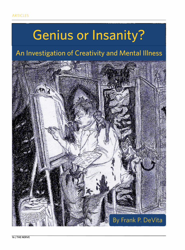
Genius or Insanity?An Investigation of Creativity and Mental Illness
By Frank P. DeVita
SPRING 2010 | 17
Introduction
Often times, creative individuals are plagued with various psychological afflictions ranging from depression and autism spectrum disor-
der to conditions such as schizophrenia or bipolar dis-order. In fact, these afflictions have beset people such as Salvador Dalí, Pablo Picasso, Sylvia Plath, Earnest Hemmingway, Pyotr Tchaikovsky and many others. A combination of mental illness and creativity spans across many different media and its bearers personify the struggle that arises from straddling the line be-tween genius and insanity. Yet it is this paradox that has produced some of history’s greatest innovations. Only now are we beginning to understand the under-lying mechanisms behind what many may be defined as both a gift and a curse. In fact, there is a neurobio-logical and psychological link between creativity and mental illness that may begin to resolve the line be-tween genius and insanity.
Creativity is defined by the Oxford English Dic-tionary as, “the use of the imagination or original ideas, especially in the production of an artistic work.”1 Through humanity’s existence, creativity has been an important factor in the evolution of global culture through its manifestations in science, visual art, music, design, architecture, politics and war. It can be argued that these human innovations start with a latent spark of creativity that evolves into a brilliant flame - but where does this spark come from? Creative potential seems uniquely human as no other species can match the human imagination.
Defining and Investigating Creativity
Creativity was defined very recently as, “the abil-ity to produce work that is at the same time novel and meaningful, as opposed to trivial and bizarre.”2 In many circles, creativity is deemed concordant with divergent thinking, the thought process used when a problem is solved by multiple unique solutions.3 It is thought that an increased ability to think divergent-ly, may imply enhanced creative potential, and this
ability is investigated through various psychological tests. Batteries such as the Torrence Tests of Creative Thinking (TTCT) present a series of tasks in picture generation to a subject who must then generate pic-tures from partial drawings and generate original works with a given set of shapes.4 A selection of the Berlin Intelligence Structure Test (BIS) also contains a figural battery with line drawing in addition to a verbal battery that asks for novel uses of various ob-jects and a numeric battery that requires generation of unique number sequences.2 In both series of tests, higher scores reflect an ability to create unique solu-tions to single problems and, in theory, are results of heightened creativity.
Over time, these tests have gauged human cogni-tion, and led to conclusions about intelligence and the creative thought process. Raymond Cattell and John Horn stated that human intelligence is compartmen-talized into fluid and crystallized ability. The fluid component grounds critical thinking, problem solv-ing, pattern recognition and learning, while the crys-tallized grounds retention of facts, formulae and other malleable information.5,6 Additionally, John Carroll thought that the mind functions within a general intel-ligence, combining the fluid/crystallized dynamic with perception, processing and specific specialized abili-ties.7 Researchers administering divergent thinking tasks have found that there are significant correlates among a defined set of these specialized abilities and divergent thinking, specifically fluency (the number of valid responses), originality (the frequency of valid responses), flexibility (number of unique categories produced), switching (ability to shift between catego-ries) and elaboration (extensive nature of responses). These factors are dynamically interacting in each in-dividual, and certain combinations theoretically serve as the foundation for divergent and creative thinking. For instance, subjects who exhibit high fluency scores often demonstrate increased flexibility of thought and the ability to create an increased number of uncom-mon solutions on the tests.2 Creative individuals have
An Investigation of the Link Between Creativity and Mental Illness
by Frank P. DeVita
ARTICLES
18 | THE NERVE
an uncanny ability to perceive related stimuli, under-stand them with great variation, and generate unique outputs through words, music, paint, prose or other media. This ability to make novel associations about incoming stimuli is the purest essence of human cre-ativity on the macroscopic level.
Extremes in Creativity
Associational ability also is affected by a widely investigated phenomenon called latent inhibition (LI). It is the varying capacity of the brain to focus on stimuli previously experienced as irrelevant or insig-nificant.9 LI can be quantified as the ability to learn to ignore irrelevant stimuli and focus on important parts of the incoming sensory stream.10 For example, in an environment with noise and other distractions, latent inhibition allows us to focus on and perform a specific task efficiently while ignoring the distractins – akin to reading with the television or radio on, but still understanding the reading. Thus, more attenu-ated/decreased latent inhibition can lead to a reduced ability to focus properly and/or learn by associaton efficiently. Essentially, LI is a filter that keeps the brain from being distracted. Decreased LI can then cause increased sensitivity to one’s environment. In the context of creativity, decreased latent inhibition can allows an individual to perceive their environment as markedly more novel. The brain is not ignoring irrele-vant stimuli, but rather interpreting it repeatedly and uniquely multiple times over. This is a crucial compo-nent of divergent thinking and forms the groundwork for making grandiose free associations about one’s en-vironment and possibly the basis for creative thought.
Latent inhibition is reduced to different extents in different creative individuals, which may enable their unique cognitive abilities.11 LI is measured and related to creativity through a combination of auditory and vi-sual discrimination tasks. Subjects are first separated into “pre-exposed” (PE) and “nonpre-exposed” (NPE) groups, and are presented with audiovisual tasks. The NPE group is presented with an audio sample of sylla-bles and a video clip that flashes a yellow circle before a target syllable, and the subject must learn to asso-ciate the appropriate sound with the visual stimulus. The PE group is first presented the same audio sample combined with white noise, then separately presented with the same audiovisual task as the NPE group. LI is measured as the speed of learning the syllable stream (the conditioned stimulus) is paired with the yellow circle (the unconditioned stimulus). Classically, the
nonpre-exposed group learns faster than the pre-exposed group, demonstrating higher levels of latent inhibition. This shows that pre-exposure to a stimulus lessens the ability to form concrete associations with that stimulus. In relation to creativity, this deficit in associational ability may enable increased free as-sociation. Creativity has been evaluated with respect to latent inhibition through LI and divergent thinking tasks plus an arts and sciences creative achievement evaluation,. It was shown that pre-exposed modest creative achievers have higher levels of latent inhi-bition than their nonpre-exposed counterparts, and that pre-exposed highly creative achievers score com-parably to their nonpre-exposed counterparts.11 This demonstrates reduced latent inhibition among pre-exposed highly creative groups and contributes to an overall trend of incrreased creativity correlated with attenuated latent inhibition.
Interestingly, reduced latent inhibition is also linked to the psychopathology of schizophrenia (SZ)10,11, creating a curious psychological link be-tween creativity and mental illness. There are many environmental, genetic, neurobiological, psychologi-cal and familial factors that lead to the development and expression of schizophrenia. However, it has been observed that latent inhibition is improved in medicated schizophrenic compared to unmedicated schizophrenic patients, and more so impaired in those who had recently experienced their first psychotic episode.12 This sheds light on the psychotic processes associated with schizophrenia, by which patients ex-press irrational paranoia, delusions of grandeur, and disconnected thought patterns. As a result of their de-creased filtering and attenuated latent inhibition, SZ patients perceive the world as markedly more novel, strange or even malicious as a result of their hyper-sensitivity to the environment. However, there is more to be learned here. While a individual with SZ may be-lieve their peers are going to kill or hurt them, may assume the consciousness of a celebrity, or perceive human emotion from static objects, they are demon-strating divergent thinking on overdrive. Since their latent inhibition is critically lowered and the incoming information stream remains relatively unfiltered, rad-ical thoughts and associations become second nature. Although these thoughts are peculiar, their foundation still lies in divergent thinking. Although schizophrenic thought demonstrates the problems in extreme diver-gent thinking, these thoughts are arguably creative at their very core.
ARTICLES
FALL 2010 | 19
Real World Evidence
Many may think that there is greater madness in the arts as compared to the sciences, and in fact, art-ists may express a greater lifetime prevalence of men-tal illness as compared to natural scientists and social scientists.14 This greater instance of mental illness in artists through their lifetimes may be accounted for by an environment that values decreased objectivity, rationality and precision as compared to fields in the natural and social sciences, where there are more stan-dards, laws and constraints. Interestingly, the mental condition of an artist often manifests itself in the tech-nical aspects of their artwork, and this is most eas-ily seen in the visual arts. Mandlebrot’s fractal theory15 ad-dresses this idea of “self similarity,” stat-ing that irregulari-ties in small parts of an object reflect the overall composition of the whole. This also applies to the creativity and men-tal illness argument in that the part is the technical attributes of an artistic work and the whole is its creator.
This notion of self-similarity by fractal theory was investigated in three forms of art: formal (exhibiting compositional emphasis), symbolic (exhibiting social realism or narration) and emotive (exhibiting abstract expressionism). Formalists such as Picasso and Matisse, exhibited the lowest instance of lifetime mental illness, while symbolic/surreal painters such as Hopper and Cezanne fell at the me-dian, and emotive modern artists such as DeKooning and Rouault demonstrated the highest instance of lifetime mental illness.14 Formalists show a more rigid and defined technical style despite abstract subject matter, which reflects their more stable mental state. Their art is a result of careful, methodic manipulation of conventions to create unique works that although strange at times, are certainly technically well defined. The symbolic/narrative painters exhibit comparable technical efficiency, but their output is a result of a
somewhat warped perception of the real world that allows them to turn the average and real into the sur-real, embodying a more feral psyche. The emotive painters exhibit overtly original technique in their ab-stract expressions that is very difficult to define and create a sense of confusion and awe in their paintings. These artists show the highest rates of mental illness, reflected by their eclectic techniques. Other painters not included in the study also embody this phenom-enon. Salvador Dali, arguably the most prominent surrealist painter in history, was a victim of paranoid personality disorder and erratic personality disorder based on his unusual behavior and personal accounts.
This phenomenon is not unique to visual art. A creativity and men-tal illness trend is also present in music and writing. Classical composers such as Tchaikovsky and Shumann have been linked to af-fective disorder16, and instances of compulsive disor-der and depression were present in the lives of jazz musi-cians Miles Davis and John Coltrane.17 Renown novelist and pioneer of con-fessional poetry Syl-via Plath and was af-
flicted with psychosis that drove her to commit suicide at home in the presence of her children at the tender age of 30, while fellow writer Earnest Hemmingway also succumbed to suicidal depression. This evidence suggests a real-world presence of a link between men-tal illness and creativity.
Biological Creativity and the Link to Mental Illness
While psychology has provided much to under-standing divergent thinking and creativity, there have been advances in neurobiology that strive to bolster theory with functional evidence. In investigating the biological basis of creativity, studies have been con-verging on the thalamus, the brain’s relay station and sensory gating system before the prefrontal cortex. The thalamus contains a plethora of chemical recep-
Innovations start with a latent spark of creativity - but where does this spark come from?

ARTICLES
20 | THE NERVE
tors for the neurotransmitter dopamine, and spe-cifically, the dopamine D2 receptor has been studied with regard to neurobiological creativity. Imaging studies (MRI/PET) have revealed significantly higher densities of D2 receptors in the thalamus relative to other parts of the brain (the only place with a higher density is the striatum), and they enable the filtering function of the thalamus to prevent information over-load in the cortex.8 These dopamine pathways play a critical role in creativity in that D2 receptor density and thalamic D2 dopamine binding potential (D2BP) decrease as divergent thinking test scores increase.8 This suggests that subjects who score high on diver-gent thinking tasks also may exhibit decreased D2BP in the thalamus and a smaller amount of D2 receptors. D2 dopamine binding in the thalamus may then have an important role in thalamic filtering and divergent sensory processing.
The dopamine system is dysfunctional in schizo-phrenia. It becomes hypersensitive, over active and particularly, dopamine binding in the striatum in-creases with intensity of positive psychotic symp-toms. These include hallucinations, delusions and dis-ordered thought.2 Interestingly enough, the dopamine pathways rendered dysfunctional in schizophrenia, bipolar disorder and (albeit to a lesser extent) de-pression show significant overlap with the thalamus and straitum – brain areas seemingly important for creativity and divergent thinking.2 Additionally, de-creased D2BP and reduced D2 receptor density in the thalamus are included in the molecular pathology of schizophrenia2,8 Studies show that as cortical and limbic D2BP decreases, intensity of positive schizo-phrenic symptoms increases,8 thus establishing the role of the D2 receptor in this mental illness. Although very controversial, this emergent piece of evidence links psychological and biological creativity through the dopamine brain pathways and D2 receptors. An analogous negative correlation also exists between divergent thinking test scores and D2 receptor den-sity in the thalamus. D2 density is lower in subjects who score high on divergent thinking tests.8 Thus, a biological link between creativity and mental illness is brought to light. Since both schizophrenic pathology and divergent thinking exhibit decreased D2 receptor density and DA binding potential, this suggests that there are associations between some of the underly-ing mechanisms of creativity and mental illness.
Given the above testament, it can be argued that the ability to think creatively arises from an increased capacity to make free associations of stimuli in the ex-
ternal environment. Also, given that the thalamus has an elevated level of D2 receptors, it has been suggest-ed that decreased D2BP decreases thalamic ability to strictly filter the incoming stream of information flow-ing rapidly to the prefrontal cortex.8 Thus, the pre-frontal gating system goes awry, allowing for input of an unfiltered stream into the frontal lobe, which could be the neurological basis for creativity. This makes sense with respect to neurobiology, as decreased fil-tering by the thalamus facilitates information flow to the prefrontal cortex and the frontal lobe, where in-put is available for free association. Since the creative mind has taken in more information, it can create more original associations as a function of mathemat-ics – if there is more information to be strewn togeth-er, there will be more output. The incoming informa-tion stream coupled with an open sensory gate in the thalamus may create the conditions for the infamous and elusive creative spark, but for some, the spark can lead to psychosis. What then chooses the path?
Proposition of a Continuum
In a Harvard study, intelligence was investigated with respect to latent inhibition in a group of pre-ex-posed individuals from a latent inhibition diagnostic to investigate IQ’s role in creativity.11 After re-adminis-tration of latent inhibition tasks and creative achieve-ment evaluations, individuals were given an IQ test, and it was found that those who demonstrated higher levels of creative achievement also demonstrated re-duced latent inhibition. Trends in IQ accounted for 20% of the variance of the results, and IQ was a signifi-cant variable. In further investigation, the group’s em-inent creative achievement (ECA) was analyzed. The researchers defined this as publishing/sale of a novel, book or poetry, sale of recorded music, patented con-struction of a prototype invention, private exhibition of original artwork or scholarship/prize for a scientif-ic discovery. The group analyzed consisted of 4 artists, 5 composers, 2 writers, 2 inventors, 3 dramatists, 7 scientists and 2 choreographers, whose IQ and latent inhibition scores were compared to a control group. These eminent creative achievers showed markedly lower levels of latent inhibition than the controls and were seven times more likely to demonstrate attenu-ated latent inhibition. Pertaining to IQ, it is suggested that a score of 120 is the threshold for creativity11, and in comparing the IQs of the subjects, 84% of the cre-ative achievers had an IQ score greater than 120 while only such was the case for 44% of the control group.
ARTICLES
FALL 2010 | 21
Further statistical analysis demonstrated a higher in-stance of reduced latent inhibition in eminent creative achievers with an IQ greater than 120.
This data and the previously presented neuro-biological and psychological ideas suggest that at-tenuated latent inhibition is associated with both high creativity and mental illness, with IQ serving as a pos-sible modulating factor. These extrema of creativity and mental illness may exist in a continuum linked by latent inhibition and modulated by IQ. Reduced latent inhibition is associated with schizophrenia in multiple cases, but may also be related to an increased likeli-hood of creativity. Furthermore, there are other corre-lations between creative individuals and schizophren-ic patients in latent inhibition and divergent thinking tasks.11 Without the modulating function of IQ, all cre-ativity could be explicitly synonymous with psychosis, but this clearly is not the case. Intelligence may serve as the mediator between an individual’s susceptibility to psychosis and ability to be creative. This also sug-gests that intelligence may influence how each indi-vidual’s mind responds to and functions under atten-uated levels of latent inhibition, which relates back to the defined set of abilities associated with creativity mentioned earlier.
Final Thoughts
What does all this mean in a larger context? Prin-cipally, it expands our knowledge of the mysterious
realm of psychosis and neural function. Through the psychopathology of mental illness, we are better able to understand what is happening in the brain at the level of neurophysiology, which will prove important in understanding the brain as a system. While we can diagnose and treat all mental illnesses , we do not understand the complete brain mechanisms of all of these disorders. Breakthrough molecular neurobiol-ogy has and will continue to help us further under-stand these conditions and open up possibilities for treatment at more localized levels. If we are able to understand specific receptor-ligand mechanics, neu-rochemistry, neuroanatomy, and neurophysiology of various mental illnesses, we will be better able to better treat them. In the context of creativity, this evidence further allows us to understand and inves-tigate our established metaphysical and psychological ideas under a microscope. Modern understanding of the mechanisms of mental illnesses allows us to de-velop new treatments and prophylactic interventions to prevent disorders before they even start. More in-terestingly however, is the idea of manipulating these mechanisms for our benefit once we fully understand them. Theoretically, it may be possible one day to modulate brain function from the level of genes and molecules, opening the door to a new realm of self-en-hancement by molecular intervention. Just think, one day you may be able to take a pill to access the cogni-tive plane of Pablo Picasso or Ludwig van Beethoven for just a few hours.
1. “Creativity” Oxford English Dictionary. Online Edition. Accessed July 2010.
2. de Manzano Ö , et al. (2010). Thinking Outside a Less Intact Box: Thalamic
Dopamine D2 Receptor Densities Are Negatively Related to Psychometric Cre-
ativity in Healthy Individuals.” PLoS ONE 5(5): e10670. doi:10.1371/journal.
pone.0010670
3. Guilford JP (1957). “Creative Abilities in The Arts.” Psychological Review Vol.
64, No. 2, pp. 110-118.
4. Kim KH (2006). “Can We Trust Creativity Tests? A Review of the Torrence
Tests of Creative Thinking.” Creativity Research Journal Vol. 18, No. 1, pp. 3-14.
5. Cattell RB (1963). “Theory of Fluid and Crystallized Intelligence: A Critical
Experiment.” Journal of Educational Psychology Vol. 54, No. 1 pp. 1-22.
6. Horn, J. L., & Cattell, R. B. (1966a) “Refinement and test of the theory of fluid
and crystallized general intelligences.” Journal of Educational Psychology Vol.
57, pp. 253-270.
7. Carroll JB (1993). “Human Cognitive Abilities: A Survey of Factor Analytic
Studies” Cambridge University Press.
8. Takahashi H, Makoto H & Suhara T (2006) “The Role of Extra-Striatal Dopa-
mine D2 Receptors in Schizophrenia.” Biological Psychiatry Vol. 59 pp. 919-
928
9. Lubow R. E. (1989) Latent Inhibition and Conditioned Attention Theory, Cam-
bridge University Press, Cambridge.
10. Gray NS, et al. (1995) “Latent Inhibition in Drug Naïve Schizophrenics: Rela-
tionship to Duration of Illness and Dopamine D2 Binding using SPET.” Schizo-
phrenia Research Vol. 17 pp. 95-107.
11. Decreased Latent Inhibition Is Associated with Increased Creative Achieve-
ment in High-Functioning Individuals (2003).J Personality and Social Psy-
chology Vol. 85, No. 3, pp. 499-506.
12. Vatil D, et al (2002). “Latent Inhibition and Schizophrenia: Pavlovian Condi-
tioning of Autonomic Responses. Schizophrenia Research Vol. 55 pp. 147-158.
13. Swerdlow NR, et al. (1996). “Latent Inhibition in Schizophrenia.” Schizo-
phrenia Research Vol. 20 pp. 91-103.
14. Ludwig AM (1998). “Method and Madness in the Arts and Sciences” Creativ-
ity Research Journal Vol. 11, No, 2, pp. 93-101.
15. Mandelbrot BB (1983) The Fractal Geometry of Nature. Various selections.
W.H. Freeman, 1st Edition, 1983.
16. Jamison, K. R. (1993) Touched With Fire: Manic Depressive Illness and the
Artistic Temperament. Various selections. New York: The Free Press.
17. Wills, GI (2003) Forty Lives in the Bebop Business: Mental Health In a Group
of Eminent Jazz Musicians. British Journal of Psychiatry Vol. 183, pp. 255-259.
ARTICLES
22 | THE NERVE
In 0.19 seconds, the Google search engine retrieves over 9,600,000 results concerning
I-Dosing, the newest Internet fad that is taking the world’s youth by storm. The next time you hear someone talking about drugs, you might get confused when you hear them talking about crazy beats and soundproof headphones. However, an increasing number of people are listening to sound files that are specifically designed to alter an in-dividual’s state of consciousness and induce a ‘high’ that is claimed to mimic the psycho-physiological effects felt after the administration of an actual psychoactive drug; this “drug trip” is called I-dosing. To feel optimal effects, the user is advised to wear stereo headphones, lie down in a comfortable position in a dark room, close his or her eyes, and drift off into the world of digi-tal-drugs1. As ludicrous as I-Dosing may seem, the scientific foundation for digital-drugs dates back to as early as the mid nineteenth cen-tury.
The Biological Basis Behind Bin-aural Beats
The human brain, being a dy-namic and adapting organ, has the capacity to rapidly alter its electri-cal activity in response to incoming information. In 1839, the percep-tive ability of the brain to encode and react to exogenous stimuli was
manipulated by German physicist Heinrich Wilhelm Dove, the father of the binaural beat phenome-non2,3. Since their discovery, binau-ral beats have been implemented both clinically and experimentally to assess the audio-perceptive abil-ities and integrity of corticosenso-ry tracts.
In general auditory situations, when a sound of a particular fre-quency hits one ear earlier than the other, the brainstem auditory processing areas, the superior ol-ive, inferior colliculus, and lateral lemniscus, detect the difference in auditory stimulation time and estimate the direction a particular sound is coming from4. However, when the two ears are simultane-ously presented with a sinusoidal sound of a slightly different fre-quency, as in binaural beat stimu-lation, the slight frequency dif-ference is not what is perceived. Instead, the result is a sinusoid whose interaural time difference changes slowly. Consequently, this frequency difference between the two tones creates a binaural beat, a sound that seems to move in space. If this resulting movement is rapid, the beat will “fill the head” rather than being perceived as coming from a particular location5.
For example, if the left ear is presented with a sound of 110 Hz while the right ear receives a frequency of 100 Hz, the sound perceived by the auditory system
will be of 105 Hz while the binau-ral beat will be of 10 Hz5. Once the binaural auditory beat travels to the brainstem’s superior olivary nucleus, where specialized nuclei that encode binaural stimulation are located, a beat of neural activity is generated4,7.
Welcome to the Digital High-Way: Advocates
As a ‘cognitive arousal’ tech-nique, the administration of bin-aural beats demands a general knowledge of the electrical behav-ior of the brain. Typically, electri-cal brain activity can be sorted into five frequency-based categories. Gamma waves (30-100Hz; usually 40Hz) are the brain’s highest fre-quency electrical waves and are present during multimodal pro-cessing of sensory information. Beta waves (14-30Hz) and alpha waves (8-13Hz) typify wakefulness and conscious cognitive function (pad). Alpha frequency waves oc-cur at more relaxed cognitive states while beta waves are produced at higher arousal states. Delta and theta waves characterize an uncon-scious state. Delta waves (1-4Hz), the lowest-frequency brainwaves, promote dreamless sleep, and theta waves (4-8Hz) are associated with REM sleep, meditation, and creativity8.
When a binaural beat is of the same frequency range as any one
Exploring I-Dosing and the Binaural Beat Phenomenon
Ten Minutes to a Trance:
by Anuhya Caipa
ARTICLES
FALL 2010 | 23
of the five brainwaves, it is claimed to be capable of altering an indi-vidual’s state of consciousness by stimulating the reticular-thalamic activating system, a neural system responsible for cognitive arousal6,8. Studies conducted with EEGs have shown that binaural beats of a theta wave frequency will induce a feeling of drowsiness in a listener. Conversely, binaural beats in the beta wave range will cause the lis-tener to become more aroused and alert, with enhanced memory and task performance7,9.
Advocates of the use of binau-ral beats for cognitive manipula-tion are convinced of its potential therapeutic effects. Robert Monroe, founder of the Monroe Institute, developed multi-layered sound files that manipulate brain waves and transform the brain into a state of “hemispheric synchronization,” an occurrence in which both hemi-spheres of the brain are in unison with each other. Research director of the Monroe Institute, F. Holmes Atwater, markets the Institute’s product by describing HemiSync as an “audio guidance program… for obtaining altered states of con-sciousness”10. There are also a few published articles concerning the investigation of HemiSync in clini-cal settings3,8,11. In preoperative settings, it was shown that patients undergoing surgery required sig-nificantly less anesthetic if they listened to a HemiSync sound file3. Further, in preoperative-associated anxiety, nearly half of patients ben-efited from listening to binaural beats8.
HemiSync, having been devel-oped before I-Dosing, set the stage for the future of digital brainwave manipulation. I-Doser Labs, one of the many designers of marketed binaural beat audio, has even de-veloped a proprietary program to
beats may be significant statistical-ly, Dr. Shinn-Cunningham says the effects of binaural stimulation on brain activity are smaller than the effects of more overt cues in music, such as the rhythmic beats that we all dance to and hear consciously.
Additionally, many experiences of a psycho-physiological ‘high’ be-ing felt by I-Dosers could be due to a placebo effect and may not even simulate the high resulting from a recreational psychoactive drug. Although it has been shown that drugs do have an effect on brain-wave activity13, it is not known if these effects can be replicated by binaural beats alone. Furthermore, drug activity on the brain is largely dose dependent, making the ap-proximation of the effects of binau-ral beats on the brain hard to mea-sure in comparison to a real high.
Regardless of the disagree-ments concerning the impact binaural beats have on cognitive arousal, the major ethical issue is the message I-Dosing is sending to unsuspecting individuals perus-ing the Internet. Although I-Dosing and other methods of brain wave manipulation may not be danger-ous or even effective, there is a concern that these methods of digi-tal stimulation will act as gateway drugs. It is up to the user to decide whether to go digital or not.
References
1. I-Doser: Binaural Brainwave Doses. [accessed 2010 July 20]; Available from: http://www.i-doser.com/.
2. Dabu-Bondoc S. Hemispheric Synchro-nized Sounds and Intraoperative Anesthetic Requirements. Anesth Analg. 2003;97:3.
3. Kliempt P, Ruta D, Ogston S, Landeck A, Martay K. Hemispheric-syn-chronisation during anaesthesia: a
present their audio stimuli. The user interface of the I-Doser pro-gram is organized into folders, with the parent folder labeled “Dose Files.” These dose files, lasting from ten to forty minutes, range from hallucinogens to club drugs and opioids. When asked about how the program works, I-Doser ex-plains, “each audio track contains [our] advanced binaural beats that will synchronize [your] brainwaves to the same state as the recreation-al dose. Mixed with [our] advanced auditory pulses are soothing back-tracks of ambient soundscapes to help the brain induce a state of mood lift, euphoria, sedation, and hallucination”1 However, I-Doser Labs does not have everyone con-vinced.
You Are Only as High as You Want to Be: Skeptics
Although there are studies publicizing the application of bin-aural beats to cognitive arousal purposes, many are skeptical of these claims and point out how few legitimate, peer-reviewed scientific papers document affects on brain state. Boston University professor Dr. Barbara Shinn-Cunningham de-scribes the true application of bin-aural beats as a prognostic tool to identify the brain’s ability of spatial hearing and auditory encoding12. Dr. Shinn-Cunningham relates methods of ‘cognitive arousal’ to other common methods to alter consciousness, such as meditat-ing or simply listening to relaxing music. She asserts that in most situations, binaural beats do not induce any considerable difference in brain wave behavior12. The brain is an easily manipulated organ, and differences in electro-cognitive activity can be a result of simply “thinking.” While effects of binaural
ARTICLES
24 | THE NERVE
double-blind randomised trial using audiotapes for intra-operative noci-ception control. Anaesthesia. 1999 Aug;54(8):769-73.
4. Barr DF, Mullin TA, Herbert PS. Appli-cation of binaural beat phenomenon with aphasic patients. Arch Otolar-yngol1977 Apr;103(4):192-4.
5. Pratt H, Starr A, Michalewski HJ, Dimitrijevic A, Bleich N, Mittel-man N. Cortical evoked potentials to an auditory illusion: binau-ral beats. Clin Neurophysiol2009 Aug;120(8):1514-24.
6. Lane JD, Kasian SJ, Owens JE, Marsh GR. Binaural auditory beats affect vigi-lance performance and mood. Physi-ol Behav. 1998 Jan;63(2):249-52.
7. Wernick JS, Starr A. Binaural interac-tion in the superior olivary com-plex of the cat: an analysis of field potentials evoked by binaural-beat stimuli. J Neurophysiol. 1968 May;31(3):428-41.
8. Padmanabhan R, Hildreth AJ, Laws D. A prospective, randomised, controlled study examining binaural beat au-dio and pre-operative anxiety in patients undergoing general anaes-thesia for day case surgery. Anaes-thesia2005 Sep;60(9):874-7.
9. Kennerly RC. An empirical investiga-tion into the effect of beta frequency binaural audio signals on four mea-sures of human memory. 1994.
10. Holmes M. Hemi-Sync and Remote Viewing. Monroe Institute 2008.
11. Lewis AK, Osborn IP, Roth R. The ef-fect of hemispheric synchronization on intraoperative analgesia. Anesth Analg2004 Feb;98(2):533-6, table of contents.
12. Shinn-Cunningham B. Personal Inter-view with Dr. Barbara Shinn-Cun-ningham. Interviewed Boston 2010.
13. Banoczi W. How some drugs affect the electroencephalogram (EEG). Am J Electroneurodiagnostic Tech-nol2005 Jun;45(2):118-29.
Introduction
Fooling our own brains is an appealing concept: everyone enjoys optical illusions, mag-
ic tricks and riddles. But can fooling our brains help heal our bodies? When it comes to taking medicine, people have consistently proven themselves to be highly suggest-ible. The color of pills, the number of pills and the brand of pills can in-fluence our assumptions about the medications we are taking. We tend to think that capsules are stronger than pills, and that injections are more powerful than medicine tak-en orally¹. Even surgery has been evaluated for its potential placebo effect: in one experiment on ar-throscopic knee surgery, both the osteoarthritis patients receiving the operation and those who just got an incision and stitches showed significant decreases in knee pain ¹,².
The preferred test to prove the effectiveness of an experimen-tal drug evaluates whether or not its effects can beat those of a pla-cebo in a double-blind, placebo-controlled, randomized trial. Many experimental drugs fail this test, or often the placebo trial may give the drug a run for its money. So, if the placebo is so difficult to beat and is devoid of side-effects, then why not use it as the treatment itself? This question is being raised by physi-cians, researchers and bioethicists: can safe, affordable treatment be found in fake medicine?
How To Trick Your Brain
By definition, the placebo ef-fect is a physiological state brought on when one anticipates a health-related result from biologically ir-relevant treatments or procedures. The placebo effect can target sev-eral different neurological systems, thus the wide range of possible effects: analgesia, mood improve-ment, and even immunosuppres-sion¹.
Brain pathways involving do-pamine are important in under-standing the placebo effect. Dopa-mine is a neurotransmitter that lets us associate a stimulus in our envi-ronment with a reward, and it is released when we are engaging in behavior that will bring about that reward. Dopamine activity in the nucleus accumbens (NAC) is evi-dent when pain relief is expected, and people who show more activity in the NAC when expecting a mon-etary reward are more noticeably affected by placebos³.
A second contributing neural system is known as the endogenous opioid system. Our brains release their own opioid substances in re-sponse to pain (endogenous opi-oids) and when that is not enough, doctors prescribe painkillers with similar chemical structures. So how do we know that natural opioids are involved in the placebo effect? Naloxone, a substance that blocks opioid receptors, has been shown
Trick or Treatment?Neuroscientific and medical applications of the placebo effect
by Natalie Banacos
ARTICLES
FALL 2010 | 25
to prevent placebo analgesia, dem-onstrating the placebo’s reliance on our brain’s natural painkillers⁴. Knowing this, conditioning studies have been able to demonstrate that expectation of pain relief is asso-ciated with the release of opioids in the brain. One such study used morphine, an opioid painkiller, and ketorolac, a non-steroidal anti-in-flammatory drug (NSAID). Subjects were conditioned to expect either morphine or ketorolac injections. After getting two real injections, they received saline placebos that triggered analgesic responses similar to the drug they had been administered previously, whether they expected pain relief or not. In-jections of naloxone, independent of the subjects’ expectations of re-ceiving a painkiller, decreased pain tolerance significantly⁵.
Both opioid and dopamine systems rely on cognitive and con-ditioning factors. For instance, when taking a biologically active substance, the effectiveness of a placebo that physically resembles an active drug will be increased in both humans and animals⁶. We are conditioned to respond to the physical act of taking medicine, but we hold an expectation in our minds of what the active drug is supposed to look like. Conditioning factors refer to drug conditioning - giving a subject a certain number of doses of a drug before replacing it with a placebo. The cognitive part of the effect is introduced when the subjects are given expectation cues, being led to think that they are or are not taking a particular substance. In terms of anatomy, the anterior cingulate cortex seems to play a role in both systems, and is activated in the placebo groups of analgesia, anxiety relief and mood improvement studies¹.
In addition to the dopamine
and opioid systems, the serotonin system has also been proven to play a role in the placebo effect. The in-volvement of this system in the pla-cebo effect is often observed in ex-periments concerning depression, although it is difficult to have a true control for the placebo groups of these studies because the subjects have often had outside interven-tion like counseling. Nonetheless, changes in brain glucose metabo-lism have been observed in similar regions of the brain in subjects tak-ing fluoxetine, an antidepressant, and subjects taking a placebo⁷.
Tricks for Treatment
The broad range of effects that
have been produced by placebos are evident in their wide experi-mental use. Although they tend to treat the symptoms rather than the cause, the inherent lack of side effects makes placebos prime can-didates for treating a variety of ail-ments. Research is being directed toward finding a useful place for placebos in medicine.
Effective methods to use place-bos as painkillers are being investi-gated. A 2001 study demonstrated that subjects tolerate pain better when they can actually see that they are being given an analgesic. When test subjects received the opioid receptor-blocker naloxone after the seeing themselves inject-ed with the NSAID ketorolac, their pain tolerance was the same as it was after only receiving a hidden injection of ketorolac⁸. In May, the Boston Globe reported on a study published in Science last year in which patients given cream to put on their arms showed much less pain-related activity in their spinal cords when told that the treatment they had been given was in fact a powerful painkiller².
Even surgical placebos - sham surgeries - have proven effective, perhaps most notably in Parkin-son’s patients. One study looked at patients who had had electrodes for deep-brain stimulation treat-ment implanted in their brains. One group of patients was told that the treatment was functioning, and an-other group was told that the stim-ulator had been dialed down. The patients who believed that their electrodes were working expected improved motor performance to result from deep-brain stimula-tion. Although they were never given the treatment, these patients associated the fake subthalamic stimulation with better motor per-formance and were able to move their hands faster. The necessary brain changes occurred within minutes⁹. In another study, after injecting Parkinson’s patients with medication that would help relieve muscle stiffness, the subjects were given a placebo and their subtha-lamic brain activity was monitored. Individual neurons in this brain re-gion became less active so that the patients’ muscles could relax and function more easily¹⁰.
Though it is an unpredictable disease, and thus difficult to con-trol for in placebo studies, multiple sclerosis (MS) has been another potential candidate for placebo remedies. Multiple sclerosis is an autoimmune disease in which the body attacks the myelin insulation covering its own nerves. In a trial of interferon-β-la treatment for MS, the group given a placebo showed a twenty percent decrease in areas of inflammation due to the body attacking its myelin, expressed as MRI lesions, compared to their baseline MRIs¹¹. Epilepsy, a simi-larly unpredictable condition, has been shown to respond to place-bos. The placebo groups of anticon-

ARTICLES
26 | THE NERVE
vulsant trials regularly show sig-nificant (more than fifty percent) reductions in seizure frequency ¹².
Placebos may also be used to address cognitive issues associated with aging. In a study of healthy adults aged 65-85, subjects taking what they were told was a cognitive enhancement pill performed better on cognitive tests than the group of subjects that had not taken any pills. In one Alzheimer’s study, fifty percent of the placebo group had shown either improvement or sta-bility in their condition six months into the trial¹³. It is interesting, then, that Alzheimer’s has been demonstrated to deter the placebo effect, perhaps because of frontal lobe impairment ¹⁴.
Tricky Business
The placebo effect is a neuro-
biological phenomenon that has the potential to aid in the treat-ment of a number of disorders. The wide range of neural systems it af-fects gives it the potential to serve as treatment for a great variety of conditions. Research directed to-ward finding medical uses for the placebo effect will undoubtedly be put under great ethical scrutiny be-fore being used clinically.
Placebos are not a new concept. They were used as early as the 16th century in the form of fake holy ob-jects given to people believed to be possessed. If the victims were upset by these false relics, priests would know that the possession was imaginary - only a real relic was thought to bother a possession victim. By the late 1700’s, placebos were used medically, but merely to placate patients. This “undoubt-edly led to the tainted reputation of placebos and placebo effects that persisted until very recently” ¹⁵. When the randomized control trial
experimental design became popu-lar after World War II, it became apparent that people responded positively to placebos. In fact, Hen-ry Beecher declared that thirty-five percent of patients responded well to placebos. This is no doubt an ex-aggeration, as confounders were not fully taken into consideration, but nonetheless placebos have be-come a big research topic in recent years ¹⁵.
A principal ethical issue sur-rounding placebo medicine is in-formed consent - the patient’s right to understand their treatment. The fact remains that placebos involve deception by definition. It can be argued, however, that this decep-tion is merely putting the effects of mental and emotional states on the body’s well-being to use. Inter-estingly, the deception can actually be lessened by letting the patient in on the fact that he or she is taking a placebo without ruining the effect. The treatment can still work be-cause there is a healing element of the treatment routine itself: simply being seen by a doctor and taking medicine promote recovery. Unfor-tunately, over an extended period of time, extinction of the body’s conditioned recovery response to this routine can occur. To maintain the placebo’s efficacy in the face of extinction, a physician could ex-plain that he or she does not fully understand the mechanisms of the prescribed drug (a placebo) but is recommending it on the grounds that it will not cause the serious side effects associated with the better-understood treatment op-tion¹⁶.
Another potential benefit in introducing placebos into clini-cal use would be a reduction in health care costs. As well as being free of side effects, placebos are very inexpensive. A recent study
in psoriasis patients showed that corticosteroids administered in smaller-than-standard doses inter-spersed with placebo doses could prove a better, safer treatment plan than regular, standard doses of the same medication¹⁷. “You’re talking about many, many, many millions of dollars a year in drug treatment costs. If [doctors] can produce ap-proximately the same therapeutic effect with less drug, then it’s ob-viously safer for the patient, and I can’t believe they wouldn’t want to look into doing this,” Robert Ader, the leader of the study at the Uni-versity of Rochester remarked in the Boston Globe². In that respect, placebo treatments irrefutably ad-dress two major problems facing the health care field today.
Conclusion
The placebo effect is an in-triguing neurological process in-volving many facets of the brain: chemically, cognitively, and ana-tomically. The diverse range of systems that appear to be involved in placebo phenomena account for the usefulness of placebos in many different kinds of clinical trials. As the highest standard for pharma-ceutical testing, the effective use of randomized, double-blind, place-bo-controlled trials has prompted those in the biomedical field to ask why placebos are not used as med-ications themselves². While treat-ing symptoms more than causes, the relief achieved with a placebo might contribute to the body’s abil-ity to heal itself. If this becomes a real movement in medicine, in-formed consent may not end up being a problem: perhaps patients will come to accept the possibil-ity that they could be issued “fake” medication for their own benefit. Studies have shown placebo effec-
ARTICLES
FALL 2010 | 27
tiveness in disorders ranging from depression to multiple sclerosis to Parkinson’s disease. On their own or alongside the medications they are imitating, placebos seem to have great potential in helping patients get safer, more affordable health care.
References:1. Oken, Barry. Placebo effects: clinical as-
pects and neurobiology. Brain 2008; 131: 2812-2823
2. Tuhus-Dubrow, Rebecca. The magic cure. The Boston Globe 2010.
3. Scott, D.J. et al. Individual differences in reward responding explain placebo-induced expectations and effects. Neu-ron 2007; 55: 325-36.
4. Levine, J.D. et al. The mechanism of pla-cebo analgesia. Lancet 1978; 2: 654-7.
5. Amanzio, M. and F. Benedetti. Neurophar-macalogical dissection of placebo an-algesia: expectation-activated opioid systems versus conditioning-activated specfic subsstems. Journal of Neuro-
science 1999; 19: 484-94. 6. Benedetti, F. The opposite effects of the
opiate antagonist naloxone and the cholecystokinin antagonist proglu-mide on placebo analgesia. Pain 1996; 64: 535-43.
7. Mayberg et al. The functional neuroanat-omy of the placebo effect. American Journal of Psychiatry 2002; 159: 728-37.
8. Amanzio M. Response variability to anal-gesics: a role for non-specific activa-tion of endogenous opioids. Pain 2001; 90: 205-15.
9. Pollo et al. Expectation modulates the re-sponse to subthalamic nucleus stimu-lation in Parkinsonian patients. Neuro-report 2002; 13:1383-6.
10. Benedetti, F. et al. Placebo-responsive Parkinson patients show decreased ac-tivity in single neurons of subthalamic nucleus. Nature Neuroscience 2004; 7: 587-8.
11. OWIMS. Evidence of interferon beta-1a dose response in relapsing-remitting MS: The OWIMS study. Neurology 199;
53: 679-86. 12. Cereghino JJ, Biton V, Abou-Khalil B, Drei-
fuss F, Gauer LJ, Leppik I.Levetiracetam for partial seizures. Neurology 2000; 55: 236–42.
13. Wilcock, G. et al. Efficacy and safety of galantamine in patients with mild to moderate Alzheimer’s disease: mul-ticentre randomised controlled trial. Galantamine International-1 Study Group. British Medical Journal 2000; 321: 1445-9.
14. Benedetti, F. et al. Loss of expectation-related mechanisms in Alzheimer’s disease makes analgesic therapies less effective. Pain 2006; 121: 133-44.
15. Finniss et al. Biological, clinical, and ethi-cal advances of placebo effects. Lancet 2010; 375: 686-95.
16. Lichtenberg, P. The ethics of the placebo in clinical practice. Journal of Medical Ethics 2004; 30: 551-554.
17. Ader et al. Conditioned Pharmacothera-peutic Effects: A Preliminary Study. Psychosomatic Medicine 2009.
18. London, Jack. The Cruise of the Snark.
Brain Research and National Defense
How Neuroscience Funding by the Department of Defense is Going to Revolutionize Science, Technology and Computation
by Aisha Sohail
Formed in 1958 in response to the successful Soviet launch of the Sputnik satellite into
space, national defense depart-ment agency DARPA (Defense Ad-vanced Research Projects Agency) has been responsible for research into breakthrough, though often highly speculative military technol-ogy. The Eisenhower administra-
tion implemented ARPA with three concrete goals: to get us into space, protect us from Soviet missile at-tacks, and develop technology to detect Soviet nuclear tests. As a na-tional security agency, DARPA has been funded entirely by taxpayer money1...
As a result of DARPA-concen-trated efforts, the United States
launched Explorer I on January 31, 1958. Despite breakthroughs in military technology such as the un-manned combat air vehicle Boeing X-45, the M16 Assault Rifle, useful radars and lasers, DARPA’s discov-ery of ARPAnet in 1973 is probably the agency’s most far-reaching and revolutionary invention as it eventually lead to the develop-
ARTICLES
28 | THE NERVE
ment of the Internet and what we now know as the World Wide Web. Although DARPA’s mission state-ment has remained the same, the agency has undergone a series of name switches between DARPA and ARPA as dependent upon ad-ditional responsibilities assumed during wartime industry2..
Since then, DARPA has evolved into an agency that supports re-search bordering on the realm of science fiction. Recent project so-licitations include BaTMAN (Bio-chronicity and Temporal Mecha-nisms Arising in Nature) – which aims to better understand the spa-tio-temporal universe, and, from there, “transform biology from a descriptive to a predictive field of science”; and RoBIN (Robustness of Biologically-Inspired Networks), which aims to, “create a dynamic biologically-inspired network of scientists and other experts for cri-sis response and complex decision support”3,4.. These two projects highlight a unique trend in military research: the desire to develop a greater understanding of biology to gain advances in national se-curity. This editorial will examine how advances in defense-funded research of brain biology, or neuro-science, will revolutionize military technology and all sciences that re-quire intensive computation.
History of DARPA and neurosci-ence
Many of DARPA’s projects in-volve the funding of neuroscience research. A google search of the subject reveals several papers ex-amining the ethical, social, and legal issues posed by the burgeon-ing field. One such Nature edito-rial titled “Silence of the neuro-engineers” spawned in 2003 with the intrepid byline: “Researchers
funded by a defense agency should stop skirting the ethical issues in-volved”. This editorial urges neuro-engineers to evaluate synergies be-tween the goals of military research and neuroscience. The editorial also recommends DARPA-funded neuroscientists be more open to answering questions from oppo-nents of the development of such technologies in order to “achieve a better quality and balance with re-searchers’ engagement”5..
Dr. Jonathan D. Moreno, an eth-ics professor at University of Penn-sylvania and editor of Science Prog-ress magazine, is another relevant source on the subject. Dr. Moreno published a book titled Mind Wars in 2006 to review DARPA’s recent accomplishments in neuroscience and offer his expertise on the ethi-cal questions posited by defense funded neuroscience research6. Along with the Nature edito-rial, Moreno also notes that most DARPA-funded neuroscientists are reluctant to debate the potential military uses of now extant tech-nologies called brain machine in-terfaces. Dr. Moreno empathizes by saying that defense-funded neuro-scientists have an “understandable reluctance to jeopardize relation-ships with research funding sourc-es”, especially with the added sen-sitivity to defense disclosures in the post 9/11 and WikiLeaks era. Given the nature of this research, DARPA-funded neuroscience ap-pears secretive and obscure to the public.
Despite the secretive nature of DARPA researchers, Moreno’s per-sistent exploration into the subject reveals a history of DARPA-funded neuroscience research that raises novel ethical, legal, and philosophi-cal issues. Past defense projects demonstrate military interests in the workings of sleep and sleep-
inhibiting pharmaceuticals such as Modafinil that can enhance cogni-tion while replacing sleep in order to build better soldiers. DARPA also funds the development of brain prostheses that allow for more ef-ficient information processing and storage in subjects6.. This research has ignited a new generation of bioethical and philosophical de-bates between the humanists ap-prehending a robot apocalypse and the transhumanists hoping to achieve a positive Singularity.
A potentially intrusive DARPA patent is one in which a wireless neuroimaging module with por-table monitors that offer both neu-ronal and vascular signals, which would ultimately be capable of reading the private brain states of its potentially unwilling subjects (See DARPA patent # SB031-010 for “Wireless Near-Infrared Devic-es for Neural Monitoring in Opera-tional Environments”). The patent reads:
“The market for non-intrusive portable monitoring by means of non-invasive brain monitoring of-fers a most exciting and significant break-through, impacting many in-dustries. Early adapters are expect-ed from the military for training under stress; medical-head trauma evaluations; educational-diagnosis of learning disabilities; and law enforcement-for interrogation.“
Moreno concludes his book by encouraging the ethical regula-tion of neurodefense technology that could be misused to threaten public health and privacy. The American government has already faced significant criticism due to reports of torture and other inhu-mane interrogation tactics at Abu Ghraib and Guantanamo Bay. In a response to public disapproval, the Obama administration took efforts to shut down Guantanamo as one
ARTICLES
FALL 2010 | 29
of its first acts, and would likewise benefit from conducting open-end-ed discussions about neurodefense technologies to prohibit the imple-mentation of publicly unpopular defense policies.
DARPA and Universities
In my significant coursework at Boston University’s (BU) Cog-nitive and Neural Systems (CNS) department, I have come to no-tice that most of the technology research at the department is fi-nanced by DARPA. Pooling most of its money from DARPA, NIH, and CELEST (Center for Excellence in Education, Science, and Technol-ogy funded by the National Sci-ence Foundation) the department accrues roughly $6-7 million dol-lars a year in funding. This funding exclusively directs the research of roughly 25 post-doctoral students and approximately 40 other grad-uate students. A reliable source within the department confirms that DARPA funding amounts to roughly $2-3 million of the de-partment’s fiscal budget. This fig-ure means that a national defense agency directs roughly 50% of the systems-level neuroscience de-partments’ research, and 25% of the department’s post-doctoral research. These funds cover the direct and indirect costs of 4 to 6 post-doctoral researchers involved in defense research through CNS. According to this source, computa-tional neuroscience laboratories at other top-level schools such as MIT receive even more of their funding from military agencies. MIT allows its defense research to be classi-fied, and laboratories such as the Draper and Lincoln laboratories in Cambridge take full advantage of that privilege; BU on the other hand requires all research to be
publishable.
SyNAPSE Project
One of BU CNS department’s more lucrative projects is called the Systems of Neuromorphic Adaptive Plastic Scalable Electronics, or the DARPA SyNAPSE project. Featuring articles in Wired, NewScientist, en-gadget, and Singularity magazine hplus, the program promises to develop “a brain inspired electron ‘chip’ that mimics that function, size, and power consumption of a biological cortex”14,15,16. This chip will use neuroscience inspired ar-chitecture to simulate cognitive abilities such as perception, plan-ning, decision-making, and motor control. Funds allocated by DARPA to prime contractors HP, HRL, and IBM have been granted only to BU, Stanford, and a handful of other laboratories7..
One way to develop such a chip is through the discovery of a memristor, a type of computer that uses neuron inspired archi-tecture to compute. The discovery of memristors has the potential to revolutionize the electronics mar-ketplace by allowing for highly ef-ficient and relatively inexpensive computation. Any technology that can benefit from faster process-ing that requires less power– cell phones, personal computers, game consoles, supercomputers, robot-ics, etc. – would ultimately benefit from the discovery of this missing circuit element. Neuroscientists will be especially thrilled with the ability of such technology to pro-cess large data sets.
How does the memristor hard-ware work?
First theorized by Leon Chua in 1971, ‘memristor’ is short for
memory resistor, and it works by registering how much current has passed through a circuit8. A mem-ristive circuit effectively stores in-formation by measuring the change in electrical resistance when cur-rent is applied. This effect can be compared to a neural synapse, which uses electrical gradients formed by the influx and efflux of sodium and potassium ions across the cellular membrane to transmit signals. The success of memristor computation depends significantly on the sensitivity of the resistor: a memristor with a high resistance can be interpreted by the computer as a “1” in processing terms, and a low resistance can be interpreted as a “0”. It would be a bit silly if neu-rons only had binary outputs to re-lay since the number of neurons re-quired to compute a simple motor task would take an infinite amount of time. Needless to say, biological and memristive processing is more efficient than traditional binary processing8.
In the past two years, HP has successfully demonstrated the use of memristors using two layers of titanium oxide to create heat-induced differences in resistance between the two layers9. Control-ling this heat-current allows the computer to record data. Earlier this year, the work of Dr. Wei Lu at University of Michigan confirmed that memristors indeed simulate synapses as electrical synaptic connections either strengthen or weaken depending on the timing of firing of two memristive circuits. This appears to occur in a Hebbian fashion – just as two neurons that fire action potentials concurrently are more likely to pass future mes-sages, a memristive synapse also becomes more likely to pass later messages between two elements that are often simultaneously ac-
ARTICLES
30 | THE NERVE
tive10 .
Why do we need memristors?
The current generation of com-putational devices suffers from an upper threshold for computational efficiency because processors can-not execute a program faster than they can fetch instructions for its execution via RAM (Random-ac-cess memory). Although increasing clock speed and transistor density, and adding processors and fea-tures such as cache memory has al-lowed for an exponential increase in computing capacity until now, processing beyond the clock speed of a few gigahertz is still too power-expensive. Hence the justification for development of memristors: if we want continued exponential growth of computational process-ing, next-generation devices will need to integrate memory and computation into one step, much like a biological SyNAPSE7.
The human brain contains about 10 billion neurons, and each neuron is connected to other neu-rons through an average of about 10,000 synapses – a big number, but a finite number, nonetheless. DARPA’s SyNAPSE project is com-mitted to scaling memristor tech-nology to biological levels and once accomplished, surpassing biologi-cal computation is the next logical step in the technological revolution wrought by memristive devices7.
Memristor chips function like flash memory and retain data even after a computer is turned off while consuming less energy, requiring less silicon and fewer transistors. Since individual memristive cir-cuits hold their own memory and processor, memristors hold the ability to tremendously expedite data processing and consequently hold the key to accelerating ad-
vances in all scientific fields, espe-cially neuroscience7. Just imagine that: a brain-inspired device being used to discover deeper complexi-ties of its very inspiration!
Other implications of faster-than-brain computation
As a technological retailer, HP’s primary interest is in the market-ing of memristors for cell phones, videogames, computers, etc. For the rest of us, however, the creation of a mere chip that can compute better than any human could bear immense philosophical weight. For example, a memristive computer with more synapses than a hu-man brain could technically hold enough associative memories to be able to pass a Turing test11. A Tur-ing test employs an interrogator who asks written questions to de-cide whether its subject is a human or machine. Surprisingly, there has been no machine to date that can convincingly pass the test and suc-cessfully mimic a human enough to actually fool one, though the dis-covery of the memristor will prob-ably change that.
In addition to this philosophi-cal weight, neural modelers will also have to bear the financial implications of such a discovery; many computational neuroscien-tists will be forced to decide wheth-er they choose to work at designing neuromimetics (i.e. brain-inspired technology) or strictly modeling neurobiological circuit processes. As a young neuroscientist (and self-proclaimed gadget junkie), I have to admit myself feeling con-flicted at times between the two fields: I would consider neuromi-metics a more lucrative field in the sense that technologies created through it will revolutionize the everyday lives of most people who
rely on computational devices. As such, neuromimeticists will prob-ably make more money, have more media exposure, and accordingly, acquire even more funding. In fact, this has prediction has already proven valid if we perform some simple calculations on the finan-cial statistics mentioned earlier: if 50% of CNS’s funding, or about $3 million dollars, is distributed in direct/indirect costs to 5 post-doc-toral defense researchers, each de-fense researcher has a crude aver-age of $600,000 in funding (which may not seem like a lot, but is en-tirely sufficient for computational modelers who often do not require expensive hardware). On the other hand, CELEST funding amounting to a roundabout of $4 million is distributed between the other 20 post-doctoral biological research-ers, which allows for an average of only $200,000 per postdoctoral researcher. Although these calcula-tions are approximate, the glaring contrast between the amount of funding awarded to both parties is too asymmetrical to be ignored.
Despite this pronounced im-balance in funding, I recognize that my loyalty is to discovering how brain processes give rise to emer-gent properties such as dreams, emotions, desires, beliefs, and ulti-mately, consciousness - seemingly infinite complexities that machines may never even be capable of re-producing. Neuroscience research-ers may be apprehensive about the increasing difficulties for computa-tional neurobiologists to compete with the amount of funding that defense agencies can offer to bio-mimetic projects such as remote sensing software designed for PackBot robots deployed in Iraq (as engineered by the brilliant minds at BU’s very own CNS Tech Lab)12.As we all know, acquiring sufficient
ARTICLES
FALL 2010 | 31
funding for research is the bane of most scientist’s careers. It seems only inevitable that systems level neuroscience departments will start producing more and more bionic research with little compa-rable growth in biological model-ing research if developing neuro-morphic technology is considered so much more profitable.
So what can we do to change this disparity? The root of the problem is buried deep within the workings of our governmen-tal system and the priorities our government chooses to assign to developing cutting-edge military technology as compared to funding biological research. In 2010, DAR-PA’s proposed budget is 3.25 bil-lion dollars, whereas the National Institute of Mental Health (NIMH) is appropriated less than half that amount by the NIH. Continuing the wars in Iraq and Afghanistan is of little everyday concern to most Americans; the research of neural dynamics that eventually leads to the eradication of schizophrenia and depression, however, could po-tentially help over 25 million peo-ple in the United States alone.
Conclusion
I surmise that the impact that memristors have on neuroscience research should generally be a pos-itive one. Allowing for the study of brain computation at a speed that is faster than the speed of brain computation will enable faster data processing, and thus, faster output of research.
Nevertheless, the impact DAR-PA funding has on neuroscience still deserves monitoring as it could direct resources away from brain research. As artificial computation becomes faster than the speed of brain computation, neuroscience
funding could be easily redirected toward developing applications inspired by neural design. Under-standing complex brain mecha-nisms such as consciousness and advancing research in organic neu-ral circuits will certainly prove use-ful not only in solving the myster-ies of complex brain diseases such as schizophrenia, depression, and epilepsy, but it will also contribute to the human understanding of bio-logical networks and how they can be integrated into a “Theory of Ev-erything”. Brain mechanisms have already proven to be infinitely more complex than any artificial intelli-gence design we could even dream of programming, and it would be a shame to waste any more time in determining them. As such, neuro-scientists should reserve the right to question whether their research is directly relevant to discovering the processes of the brain and will it help people in need?
This editorial is not against artificial intelligence inspired by neural mechanisms; it is merely a suggestion for a separation be-tween biological neuroscience and neuromimetics in order to ensure that discovering the laws of neu-robiology is at the forefront, espe-cially as it becomes more and more profitable to create technologies inspired by neural models.
References:1. “DARPA’s First 50 years”. DARPA, Web. 4 Oc-
tober 2010. 4 October 2010. HYPERLINK “http://www.darpa.mil/about.html” http://www.darpa.mil/about.html
2 “50 Years of Innovation and Discovery.” DAR-PA, Web. 4 October 2010. HYPERLINK “http://www.darpa.mil/history.html” http://www.darpa.mil/history.html.
3. “Biochronicity and Temporal Mechanisms Arising in Nature (BaTMAN).” DARPA, Web. 4 October 2010. http://www.darpa.mil/dso/solicitations/sn10-55.htm
4. “Robustness of Biologically-Inspired Net-works (RoBIN).” DARPA, Web. 4 October 2010. http://www.darpa.mil/dso/solici-tations/sn10-56.htm
5. “Silence of the neuroengineers: Researchers funded by a defense agency should stop skirting the ethical issues involved.” Edi-torial 423.6942 (2003): 787. Web. 24 Aug 2010.
6. Moreno, Jonathan D. Mind Wars: Brain Re-search and National Defense. New York, NY 10151: The Dana Foundation, 2006. Print.
7. “The SyNAPSE Project.” 4 October 2010. ce-lest.bu.edu/outreach-and-impacts/the-synapse-project
8. Chua, Leon. “Memristor - The missing Circuit Element.” Circuit Theory, IEEE Transac-tions on. 18.5 (1971): 507-519. Print.
9. Dmitri B. Strukov, Gregory S. Snider, Duncan R. Stewart & R. Stanley Williams. “The Miss-ing Memristor Found.” Nature 453: 80-83. March 2008.
10. Sung Hyun Jo, Ting Chang, Idongesit Ebong, Bhavitavya B. Bhadviya, Pinaki Mazumder, and Wei Lu. “Nanoscale Memristor Device as Synapse in Neuromorphic Systems.” Nano Letters. December 2009.
11. Turing, Alan. “Computing Machinery and In-telligence”. 1950. Mind LIX (1950): 236 pp 433–460.
12. Neurala LLC. Neurala, n.d. Web. 24 Aug 2010. http://www.neurala.com/
13. National Institute of Health: History of Ap-propriations.” National Institute of Health, n.d. Web. 24 Aug 2010. http://www.nih.gov/about/almanac/appropriations/in-dex.htm
14. “Are memristors the future of Artifical In-telligence? DARPA thinks so.” Engad-get, Web. 4 October 2010. engadget.com/2009/07/14/are-memristors-the-future-of-artifical-intelligence-darpa-think/
15. “Synapse on a chip.” Hplus magazine, Web. 4 October 2010. hplusmagazine.com/ar-ticles/ai/synapse-chip
16. “Memristor minds: The future of artificial intelligence.” NewScientist, Web. 4 Oc-tober 2010. newscientist.com/article/mg20327151.600-memristor-minds-the-future-of-artificial-intelligence.html
REVIEWS
32 | THE NERVE
General Anesthesia:
by Grigori Guitchounts
Molecules to Pink Elephants
SPRING 2010 | 33
Introduction
Gentlemen, this is no humbug,” the surgeon John Warren is said to have declared to the audience at Massachusetts General Hospital after Wil-
liam Morton’s first successful demonstration of gen-eral anesthesia on October 16th, 1846.1 Before then, surgery was a miserable experience for both patient and surgeon. The term anesthesia – literally meaning “without sensation” – was first used by the ancient Greek surgeon Dioscorides and resurrected by Dr. Oli-ver Wendell Holmes after the demonstration at MGH.
Anesthesia has indeed transformed medicine and while great improvements have been made in the clinical aspect of anesthesia since the 1840’s, the mechanism by which anesthesia produces reversible changes in central nervous system function have re-mained a mystery until recently.
In the past decade, anesthesia has grown to be used in various medical settings outside the operat-ing room. Children undergoing Magnetic Resonance Imaging (MRI), Computed Tomography (CT) scans, or endoscopy are often sedated or anesthetized to assure cooperation and the required immobility2,3. Likewise, electroconvulsive therapy, long considered more tor-ture than treatment, is now being used with the aid of general anesthesia as an effective treatment for medication-resistant depression and schizophrenic affective disorders.4
However, despite the multitude of benefits to medical procedures that anesthesia has provided, it remains a somewhat dangerous and toxic procedure, usually involving postoperative delirium5 and some-times cognitive dysfunction6; even more frightening is the fact that one in 750 patients remain aware during general anesthesia7. The intravenous general anes-
thetics etomidate and propofol are widely appreciat-ed for their anesthetizing properties, but are also as-sociated with unfavorable toxicities. Patients develop “propofol infusion syndrome” after prolonged expo-sure to the drug; this is associated with dysrhythmias, lipemia, fatty liver, metabolic acidosis, and rhabdomy-olysis8. Etomidate causes adrenal suppression, put-ting patients at high risk of mortality9. More common side effects include respiratory depression, hypoten-sion, and postoperative nausea and vomiting.
It is therefore critical for safety and the progress of medicine that better drugs and methods be developed for use in general anesthesia. This review focuses on the goals of general anesthesia, the theories behind its mechanism of action, and studies of the structure and function of the GABAA receptors that mediate anes-thetic action.
Goals of General Anesthesia
The basic idea of general anesthesia (GA) is to make the patient unaware of the surgical trauma and unmoving, and to allow the surgeon to perform his job with precision. More specifically, the goals of GAs are to reversibly induce immobility, amnesia, and hyp-nosis (unconsciousness) in the patient; a fourth and often debated goal is analgesia, which some argue is an irrelevant measure since the patient is unconscious during anesthesia.
The three goals have dissociable correlates in the brain; amnesia, consciousness, and immobility are mediated by different neural systems and therefore respond differentially to drugs. Potency of inhaled anesthetics is assessed using a scale of concentra-
REVIEWS
The Drugs and Mechanisms of General Anesthesia
by Grigori Guitchounts
“
REVIEWS
34 | THE NERVE
tions required to suppress patient response to certain stimuli. Potency is inversely related to the amount of anesthetic required – as measured by alveolar concen-tration – to reach certain behavioral goals like immo-bility after a noxious stimulus or no response to spo-ken commands. The most commonly used scale is the Median Alveolar Concentration (MAC or MAC-immo-bility), which measures end-respiratory concentra-tion of anesthetic for which half of the patients do not withdraw in response to a surgical incision10. Other common scales include MAC-awake, a concentration that prevents physical response to spoken comments, such as to squeeze the doctor’s finger; and MAC-BAR, a concentration of anesthetic that blocks autonomic responses like blood pressure or heart rate changes to a surgical incision.
That the behavioral end-goals of amnesia, uncon-sciousness, and immobility require different concen-trations of anesthetics (i.e. they have different MAC values) and that the ratios of MAC-awake to MAC-im-mobility vary among different volatile anesthetics10, suggest that the three behavioral end-goals are medi-ated by different neural mechanisms.
Immobility
A common assumption has been that all anes-thetic effects are mediated by the central nervous system (CNS) above the brainstem. However some studies have implicated the spinal cord as key to producing immobility. Experiments in goats, whose unique circulatory system makes it possible to isolate the forebrain’s circulation from that of the brainstem and spinal cord, have shown that immobility is in fact produced primarily by the spinal cord.11 Selective administration of isoflurane to the brain raises the concentration required to suppress movements in re-sponse to noxious stimuli threefold12. In contrast, ad-ministration of isoflurane to only the body (brainstem and below) induces immobility at the concentrations required for whole animals. So while the brain is able to induce immobility, it is not required, nor is it the primary actor.
Hypnosis
Hypnosis is defined as the impairment of per-ception and awareness, or loss of consciousness – a critical component of general anesthesia10. Loss of consciousness in anesthesia is remarkably similar to stages of sleep. Functional magnetic resonance imag-
ing (fMRI) studies show that the brain regions affected by sleep or anesthesia overlap significantly13. Studies of the brain during sleep may therefore inform what we know of GA-induced unconsciousness.
The thalamus acts as a major relay center in the brain, passing sensory information from the outside world up to cortex20. In the awake brain, thalamocor-tical neurons receive depolarizing input from arousal nuclei in the ventrolateral preoptic area, VLPO, which allows them to faithfully pass along information to cortical neurons in a single-spike tonic firing fashion21. In contrast, during sleep the ascending arousal input to thalamocortical cells is inhibited. Thalamocortical cells lacking this depolarization enter an oscillatory firing mode, generated by Ih and ICa
20, which subse-quently prevents the transmission of signals to cortex.
The most obvious similarities between sleep and loss of consciousness under anesthesia aside, the two states share nearly identical EEG signatures (i.e. spindles and delta waves), suggesting that the thala-mus is involved in sleep as well as anesthetic-induced loss of consciousness13. However, general anesthetics do have widespread molecular targets, such as GAB-AA receptors, which are found in a large number of inhibitory cortical interneurons. It remains unclear whether their effects in the thalamus (specifically on the reticular system) are necessary and/or sufficient for loss of consciousness.
Amnesia
The hippocampus and surrounding medial tem-poral lobe (MTL) structures are responsible for the formation and consolidation of episodic memories22. The hippocampus is therefore a likely target of gener-al anesthetics. The formation of memory depends on the integrity of Long Term Potentiation (LTP), which requires functioning NMDA receptors. It follows that inhibition of NMDA receptors by general anesthetics, such as cyclopropane and ketamine, would impair memory. Some studies also implicate the amygdala, which processes affective information and is strongly connected to the hippocampus23.
THEORIES OF ACTION
Lipid vs. Protein
At the beginning of the 20th century, Hans Meyer and Charles Overton observed a correlation between the potency of anesthetic agents and their solubility
REVIEWS
FALL 2010 | 35
in olive oil. The Meyer-Overton rule predicts that the more hydrophobic the anesthetic, the higher its po-tency. From the Meyer-Overton rule, it followed that all anesthetics must act through a common mecha-nism, probably in the cell membrane, which is a hy-drophobic lipid bilayer, and that anesthetics somehow disrupt that bilayer. And while most people agreed that the final result of the perturbation of the cell membrane was a conformational change in the struc-ture of membrane proteins, none of the lipid theories explained how this could happen13.
It was not until the latter half of the 20th century that research emphasis turned to the interactions be-tween anesthetics and proteins themselves. In an im-portant set of experiments, Franks and Lieb14 showed that the lipid-free enzyme lu-ciferase could be inhibited by anesthetics following the Mey-er-Overton rule: the higher the hydrophobicity of the anesthet-ic, the more potently it inhibit-ed luciferase. While anesthetics target diverse molecules, such as PKC10, transmembrane ion channels have received the most attention. Research since then has focused mostly on the hypothesis that anesthetics enhance the action of inhibitory ion chan-nels (GABA and glycine receptors) and inhibit excit-atory ion channels (Serotonin type 3; nicotinic ace-tylcholine; and glutamate receptors). For example, electrophysiological studies have shown that the GAB-AA receptor is a major target of intravenous general anesthetics, such as propofol and etomidate15.
Current work is focused on the structure-function relationships of the target ligand-gated ion channels (i.e. how the molecular structure of a receptor me-diates its ion-conducting function) in order to eluci-date the molecular mechanism by which such chan-nels open and close; and the sites at which anesthetic agents bind and affect those channels. While the former has been of interest to the entire neurobiol-ogy community since the 1950’s, when Hodgkin and Huxley proposed a model of the action potential that depended on a mystical conduction of ions across the cell membrane16, the latter promises the more practi-cal improvement of clinical outcomes for patients un-dergoing general anesthesia. With the elucidation of the pharmacologic interactions between proteins and anesthetics, researcher-physicians may soon have at their disposal better drugs that produce minimal side effects and toxicity.
THREE GROUPS OF ANESTHETICS
General anesthetics have been classified into three groups based on their relative potencies for be-havioral endpoints and effects on the EEG.
Group 1: Etomidate, propofol, and barbiturates
Etomidate, propofol, and barbiturates produce amnesia and hypnosis at doses far lower than those for immobility (reviewed in ref 17). These drugs act via GABAA receptors, the most abundant inhibitory ligand-gated ion channels in the brain. Convincing evidence for their action via GABAA receptors comes from mutation and transgenic animal studies.
Etomidate’s action via GAB-AA receptors has been confirmed in studies utilizing its chirality18. R(+)-etomidate is 20 times more potent at inducing Loss of Right-ing Reflex (LORR) in animals, a surrogate test of consciousness, than S(-)-etomidate. Likewise, the R(+) enantiomer modulates
GABAARs 20 times more potently than does the S(-) enantiomer.
Moreover, mutations on the GABAAR subunits af-fect GABAAR electrophysiology and in vivo respons-es to anesthetics similarly. For example, mice with a methionine residue in place of asparagine at the 265 position on the β3 subunit exhibited a four-fold reduc-tion in sensitivity to propofol and etomidate anesthe-sia, specifically immobility. The same point mutation reduces anesthetic modulation of the receptors in electrophysiology studies. The same mutation on the β2 subunit did not affect immobility or hypnosis, but reduced sedation in mice17. Furthermore, mice lacking α5 GABAAR subunits were insensitive to emotidate’s amnesic effects19. And while the β3(N265M) mutation reduces hypnosis and immobility response, it pre-serves the undesirable effect of respiratory depres-sion, showing that GABAARs are not etomidate’s only target in the body.
Group 2: Nitrous oxide, cyclopropane, and ket-amine
Drugs in Group 2 act mostly via glutamatergic NMDA receptors, as opposed to GABA receptors. Glu-tamate is the most abundant excitatory neurotrans-mitter in the brain, so it’s no surprise that inhibition
As many as one in 750 patients remain aware during general
anesthesia
REVIEWS
36 | THE NERVE
of glutamate receptors would produce anesthetic ef-fects, which among the Group 2 drugs are mostly an-algesia, weak hypnosis and immobility17.
As with etomidate, ketamine chirality has been used to correlate its anesthetic effects with NMDA receptor modulation. S-ketamine was 1.9 times more potent in inhibiting NMDA receptors in hippocampal slice preparations than was R-ketamine; the same ef-fect was observed in vivo17.
Horace Wells noted nitrous oxide’s (NO2, “laugh-ing gas”) analgesic properties while attending a demonstration in 1845; Wells’ own demonstration of nitrous oxide’s anesthetic properties at MGH was deemed a failure. Transgenic animal studies have shown that NO2 also acts via NMDA receptors. Trans-genic mice lacking the ε1 subunit (homolog of the human NR2A) of the NMDA receptor do not exhibit immobility or loss of righting reflex under NO2 ad-ministration24.
However, using an NMDA antagonist MK-801, Stabernack et al. showed that inhibition of NMDA receptors is not sufficient to produce anesthesia. Ad-ministration of MK-801 alone reduced MAC but did not produce immobility25.
Group 3: Halogenated volatile anesthetics
The halogenated volatile anesthetics, usually iso-flurane, sevoflurane, desflurane, and halothane, are the least selective of the general anesthetics. These anesthetics affect the GABAARs, two-pore K+ chan-nels, NMDA and 5HT3 receptors, Na+ channels, and other targets26.
STRUCTURE AND FUNCTION OF CYS-LOOP LI-GAND-GATED ION CHANNELS
Studies show that the GABAA receptor is a major target of halogenated volatile anesthetics and intrave-nous anesthetics such as etomidate and propofol. The GABAAR is a member of a large family of ligand-gated ion channels known as the cys-loop family for the cys-teine loop that all family members share; other mem-bers include the 5HT3, glycine, and nACh receptors. Most of what is known about the structure of the GAB-AAR comes from studies of the nicotinic acetylcholine receptor.
Nicotinic Acetylcholine Receptor
Serious investigation into the structure and
function of the nAChR began with the studies of the Torpedo electric ray nAChR27,28,29,30. Electron micros-copy revealed that the receptor is composed of five subunits organized around a central pore. Molecular cloning showed that each subunit consists of four α-helical transmembrane domains (TM1-4). TM2 lines the pore, TM1 and 3 are next to it, and TM4 faces the membrane. Importantly, TM2 is able to interact with hydrophilic molecules since it faces the water-permeable pore, while the other transmembrane do-mains interact with the hydrophobic cell membrane31.
Advances in genomics have produced large li-braries of eukaryotic and prokaryotic genes that are available for analysis. Interestingly, these libraries have brought to light a large number of cys loop / pentameric LGIC genes conserved in prokaryotes32. One such advance was the discovery of the Acetyl-choline Binding Protein (AChBP) found in the snail Lymnaea stagnalis, where it mediates neurotransmis-sion. Even though AChBP is water-soluble and lacks the transmembrane domain of mammalian nAChRs, it nonetheless forms a homopentamer with distinct acetylcholine and nicotine binding sites33. As expected from prior experiments, ligand binding occurs at the interface between subunits, with different affinities for acetylcholine, nicotine, and the agonist carbamyl-choline. While ligand binding induces local confor-mational changes, studies of signal transduction in a system that lacks the transmembrane domain that is integral for the study of channel gating have to be in-terpreted with caution.
In contrast with AChBP, which is a good model for the extracellular domain of the nAChR, the Erwinia chrysanthemi (ELIC) and Gloebacter violaceus (GLIC) channels reveal the minimal structural requirements for a functional LGIC in the Cys-loop family.
Even though ELIC and GLIC share only 16% and 20%, respectively, of the sequence with the nAChR, their structures are remarkably similar34,35. GLIC is activated by protons, while ELIC’s activating ligand has not been discovered yet. The X-ray crystallogra-phy structures of ELIC and GLIC are valuable because they are thought to show the closed and open confor-mations of the protein, respectively. Hilf and Dutzler suggest that the difference in the two conformations can explain the mechanism of gating (channel open-ing). Specifically, it is the difference in orientation of TM2 and TM3 α helices and the extracellular β sheet between the GLIC and ELIC structures that Hilf and Dutzler base their hypothesis on.
Similar to the pores of other cation channels, the
REVIEWS
FALL 2010 | 37
GLIC pore-lining TM2 domain is composed of hydro-phobic bulky residues; the intracellular half is filled with polar residues such as serine and threonine, making this part of the pore hydrophilic. Cation selec-tivity arises most likely from a conserved ring of glu-tamate residues on the intracellular end of the pore; this “intermediate ring of charges” is also found in the Torpedo and muscle nAChR36.
GABAA Receptors
The GABAA receptor is the major inhibitory li-gand-gated ion channel in CNS neurons, found in all layers of the cerebral cortex, the hippocampus and the rest of the limbic system, the cerebellum, thalamus, and brainstem37. The receptor is composed of five sub-units arranged pseudosymmetrically around a central ion-conducting pore. Nineteen subunit varieties have been cloned to date, which theoretically allows for a tremendous amount of diversity of expressed recep-tors38. However, not all possible combinations have been found assembled, and indeed the majority of the neuronal receptors are composed of α, β, and γ subunits37. Nevertheless, the abundance of subunit varieties, as well as subunit-dependent localization of the receptors in the brain make GABAARs a promising target for drug discovery39.
Upon activation by GABA, the receptor pore con-ducts Cl- and bicarbonate ions, hyperpolarizing the neuron and thereby reducing the action potential fir-ing rate. GABAARs are thought to be the major target of several general anesthetics, including etomidate, propofol, alcohols40,41, halogenated volatile anesthet-ics42, and barbiturates43. The understanding of how these and other drugs affect the GABA receptor is essential for improvements in anesthetic drugs and treatments of CNS diseases.
Structure
Using a forced subunit assembly method, Sigel and colleagues determined that receptors composed of α1β2γ2 subunits (the major isoform) are arranged γ2β2α1β2α1 counterclockwise when viewed from the synaptic cleft44. Each of the five subunits making up a GABAAR contains a large N-terminal domain, four transmembrane domains (TM1-4) and an intracel-lular domain between TM3 and TM445. The receptor conducts ions in an all-or-none fashion, transitioning among the open, closed, and desensitized states46.
Ligand binding is described in terms of an alloste-ric model in which ligands bind and affect the protein at sites distinct (and sometimes far) from the GABA site. These allosteric sites may be found in the extra-cellular and transmembrane domains of the GABAAR.
Extracellular Domain
GABAARs have two binding sites for GABA, each in the extracellular domain at the interface between the α and β subunits47; GABA binding at these sites opens the Cl- pore. Baumann et al used point mutations on linked subunits to differentiate the contributions of each site to channel gating (using free subunits does not indicate which of the two α/β interfaces has the desired mutation), showing that the α/β site where α is adjacent to γ is more sensitive to GABA; and con-versely that the α/β site where β is adjacent to γ is more sensitive to the competitive antagonists bicucul-line and muscimol48.
Classic benzodiazepines enhance inward cur-rents produced by GABA binding37. Benzodiazepines bind with high affinity at the interface between α and γ subunits, sites homologous to GABA’s at the two αβ interfaces. Rusch and Forman showed that ben-zodiazepines enhance GABA currents by enhancing GABA gating as opposed to binding. Using the Monod-Wyman-Changeux allosteric model, they were able to conclude that benzodiazepines are high-affinity but low-efficacy co-agonists, in stark contrast to general anesthetics such as etomidate, which are low-affinity high-efficacy agonists43. The differences between ben-zodiazepine and etomidate agonism are further high-lighted by single-channel studies. These studies show that etomidate lengthens the mean single-channel open time, whereas benzodiazepines do not; although single channels did open more frequently in the pres-ence of benzodiazepines or etomidate49,50.
Transmembrane Domain
A diverse set of organic compounds has been shown to act by binding to sites in the GABAAR trans-membrane domain. These include alcohols40, neuros-teroids51, barbiturates52, volatile anesthetics42, etomi-date, and propofol53,54.
Alcohols
Alcohols enhance GABA-induced currents in αβγ GABAARs40, and reduce currents in receptors with
REVIEWS
38 | THE NERVE
ρ1 subunits41. Mihic et al (1997) constructed chime-ric receptors containing sequences from the GABA ρ subunit and the glycine receptor α1 subunit (a close homolog of αβγ GABAAR). They were able to identify a region of 45 amino acids between TM2 and TM3 that are necessary and sufficient for enhancement of currents by ethanol and enflurane. Specifically, the α1S270 and β1S265 in the TM2, as well as α2A291, β1M286, and β3N265 in the TM3, were critical to eth-anol’s enhancement of GABA currents.
Barbiturates
Barbiturates are CNS depressants, and have been used through the 20th century to treat conditions such as insomnia, anxiety and epilepsy; and in the OR as inducers of anesthesia (their history is reviewed in ref 55). Like etomidate, pentobarbital enhances GABA currents at low concentrations and activates GAB-AARs directly at high concentrations52; at even high-er concentrations (millimolar) pentobarbital blocks GABA currents.
Etomidate and Propofol
At clinically relevant concentrations, the potent intravenous anesthetic etomidate enhances activation of GABAARs by GABA (lowers EC50) . This interaction directly activates GABAARs at higher concentrations, in the absence of GABA56. These two types of activa-tion suggest that GABAARs have two types of etomi-date sites – high-affinity sites that enhance GABA sen-sitivity and low-affinity sites that directly activate the receptor. These observations suggest an orthosteric model where etomidate binds to the same site as GABA to directly activate the receptor (and an allosteric site to enhance GABA activation). However, using electro-physiological methods and the Monod-Wyman-Chan-geux allosteric model, Rusch et al. (2004) showed that a single class of sites can explain both enhancement and direct activation, concluding that etomidate’s af-finity for the receptor changes depending on the re-ceptor’s state (open or closed); the open receptor has a high affinity for etomidate, whereas the closed re-ceptor has a low affinity).56
Evidence that etomidate acts at the interface of the α and β subunits comes from experiments using a photoactivatable etomidate analog, [3H]azi-etomi-date, which labels the amino acids αM236 (TM1) and βM286 (TM3).57
Mutations of these residues to tryptophan (which
is bulky and hydrophobic like etomidate), α1M236W and β2M286W, mimic etomidate presence. Stewart et al used these mutations to show that one class of sites is sufficient to explain etomidate’s effects.58 The β2M286W mutant was insensitive to etomidate direct activation and had zero GABA enhancement by etomi-date, consistent with one type of modulatory site. In contrast, the α1M236W mutant showed reduced GABA potentiation by etomidate compared to wild-type but increased direct activation. These opposite effects seem to suggest two classes of etomidate sites, but are nevertheless consistent with the interpreta-tion of one class of sites: etomidate’s efficacy is re-duced in the α1M236W mutant, but direct activation appears to be enhanced because the mutant has high spontaneous activity, which lowers the energy barrier required for channel opening.
Propofol has been thought to act near the TM2 domain at α1Ser270/β2Asn265 and TM3 domain at α1A291/β2M286. Cysteine protection experiments showed that propofol protects all four of these resi-dues, but only β2M286 in a dose-dependent manner, implicating this residue as the propofol binding site.59 However, Siegwart et al showed that the mutants β3M286W as well as β3N265M significantly reduce propofol enhancement of GABA currents and direct activation by propofol.53 Propofol’s varying effects on receptors composed of different forms of the β sub-unit may be useful in drug discovery or modification.
New or Improved Drugs
Going forward, two major branches of research must be pursued to ensure success in the develop-ment of new anesthetic drugs. These are the study of the structure and function of LGICs, as well as their subunit compositions and localization in the human brain; and drug discovery, including high-throughput assays of novel agents and chemical modification of existing drugs such as etomidate.
Efforts to improve etomidate include methoxy carbonyl- (MOC-)etomidate, which is designed to be metabolized quicker than etomidate60 and car-boetomidate, which is a significantly weaker inhibi-tor of adrenal cortisol synthesis61. The goal of these chemical modifications is to remove etomidate’s toxic properties while preserving the anesthetizing ones.
Another approach is drug discovery. Recently, Lea et al (2010) reported a high-throughput screening of compounds that interact with apoferritin, a soluble protein that mimics the GABAAR’s interaction with an-
REVIEWS
FALL 2010 | 39
esthetics.62 Of 1280 compounds, about 1% were hits. Obviously further electrophysiological, metabolic, and clinical characterizations are necessary before any of those compounds can be used as general anesthetics.
Conclusion
While surgeons and patients have enjoyed the availability of general anesthesia for over 150 years, scientists have not made progress in elucidating its mechanism of action until quite recently. That para-digm shift came in the 1980s, when Franks and Lieb showed that anesthetics act via proteins, not the lipid membrane; research since then has focused primarily on ligand-gated ion channels that mediate fast synap-tic transmission in the CNS.
Numerous techniques are available for the study of the structure and function of LGICs, including elec-tron microscopy, cysteine substitution, point muta-genesis, and x-ray crystallography. Our current con-ception of the 3D structure of the GABAAR is largely based on x-rays of the nicotinic acetylcholine recep-tor and its homologs. While the overall structure ap-pears to be conserved among the Cys-loop receptors, the fine details (including single amino acids) are dif-ferent. Until we have a definite idea of GABAAR’s 3D structure, including the orientation of the transmem-brane domains where general anesthetics act, our in-terpretations of experimental data will remain murky. Nevertheless, the past decade’s progress in research on new or improved anesthetics is bound to continue in the next.
References:
1. Barash PG, Cullen BF, Stoelting RK, Stock MC. Handbook of Clinical Anes-thesia. Lippincott Williams & Wilkins. Ed 6, 2009.
2. Malviya S et al. Sedation and general anaesthesia in childrenundergoing MRI and CT: adverse events and outcomes. British Journal of Anaes-thesia, , Vol. 84, No. 6, 743-748. 2000.
3. Wengrower D et al. Complicated endoscopic pediatric procedures us-ing deep sedation and general anesthesia are safe in the endoscopy suite. Scandinavian Journal of Gastroenterology. Volume 39, Issue 3 March 2004 , pages 283 - 286
4. Ding Z and White PF. Anesthesia for Electroconvulsive Therapy. Anesthe-sia & Analgesia. 2002 vol. 94 no. 5 1351-1364
5. Bryson GL, Wyand A: Evidence-based clinical update: General anesthesia and the risk of delirium and postoperative cognitive dysfunction. Can J Anaesth 2006; 53:669–77
6. Newman S et al. Postoperative Cognitive Dysfunction after Noncardiac Surgery: A Systematic Review. Anesthesiology; 106:572–90. 2007
7. Forman SA. Awareness during general anesthesia: concepts and contro-versies. Semin Anesth Perioperat Med Pain. 2006;25:211–218.
8. Corbett SM, Montoya ID, Moore FA: Propofol-related infusion syndrome in intensive care patients. Pharmacotherapy 28:250, 2008
9. Wagner RL, White PF, Kan PB, et al: Inhibition of adrenal steroidogenesis by the anesthetic etomidate. N Engl J Med 310:1415, 1984
10. Campagna, J. A., Miller, K. W. & Forman, S. A. Mechanisms of actions of inhaled anesthetics. N. Engl. J. Med. 348, 2110–2124 (2003).
11. Antognini JF and Schwartz K. Exaggerated anesthetic requirements in the preferentially anesthetized brain. Anesthesiology 79: 1244-1249. 1993.
12. Collins et al Anesthetic actions within the spinal cord: contributions to the state of general anesthesia. Trends Neurosci. 1995. 18:549-53
13. Franks NP. General anaesthesia: from molecular targets to neuronal pathways of sleep and arousal. Nature Reviews Neuroscience. 2008 volume 9 370-384
14. Franks NP and Lieb WR. Do general anaesthetics act by competitive binding to specific receptors? Nature 310, 599 – 601. 1984.
15. Franks NP. Molecular targets underlying general anaesthesia. British Journal of Pharmacology. 147, S72-S81. 2006.
16. Hodgkin AL, Huxley AF. A quantitative description of membrane cur-rent and its application to conduction and excitation in nerve. J Physiol. 1952 Aug;117(4):500-44.
17. Solt K and Forman SA. Correlating the clinical actions and molecular mechanisms of general anesthetics. Curr Opin Anaesthesiol. 2007.
18. Belelli D et al. The in vitro and in vivo enantioselectivity of etomidate implicates the GABAA receptor in general anaesthesia Neurophar-macology 45 (2003) 57–71
19. Cheng VY et al. Α5gabaa Receptors Mediate the Amnestic But Not Seda-tive-Hypnotic Effects of the General Anesthetic Etomidate. Journal of Neuroscience. 26(14):3713–3720. 2006
20. Steriade M, mccormick DA and Sejnowski TJ. Thalamocortical oscilla-tions in the sleeping and aroused brain. Science. 1993, 679-685
21. Mccormick, D. A. & Bal, T. Sleep and arousal: thalamocortical mecha-nisms. Annu. Rev. Neurosci. 20,185–215 (1997).
22. Eichenbaum H, Yonelinas AP, Ranganath C. The Medial Temporal Lobe and Recognition Memory. Annual Review of Neuroscience. 30, 123 -152. 2007.
23. Alkire, MT, Nathan, SV. Does the Amygdala Mediate Anesthetic-induced Amnesia?: Basolateral Amygdala Lesions Block Sevoflurane-induced Amnesia. Anesthesiology. 102(4), 754-760. 2005.
24. Sato Y, Kobayashi E, Murayama T, Mishina M, Seo N. Effect of N-methyl-D-aspartate receptor epsilon1 subunit gene disruption of the action of general anesthetic drugs in mice. Anesthesiology.102(3):557-61, 2005
25. Stabernack C et al. Spinal N-Methyl-D-Aspartate Receptors May Contribute to the Immobilizing Action of Isoflurane. Anesth Analg 2003;96:102–7
26. Forman SA and Chin VA. General anesthetics and molecular mecha-nisms of unconsciousness. Int’l Anesthesiol Clinics. 46(3), 43-53. 2008.
REVIEWS
40 | THE NERVE
27. Ballivet M, Patrick J, Lee J, Heinemann S. 1982. Molecular cloning of cdna coding for the gamma subunit of Torpedo Acetylcholine recep-tor. PNAS. 79:4466–70
28. Giraudat J, Devillers-Thiery A, Auffray C, Rougeon F, Changeux JP. 1982. Identification of a cdna clone coding for the Acetylcholine bind-ing subunit of Torpedo Marmorata acetylcholine receptor. EMBO J. 1:713–17
29. Sumikawa K, et al. 1982. The molecular cloning and characterization of cdna coding for the alpha subunit of the acetylcholine receptor. Nucleic Acids Res. 10:5809–22
30. Noda M, Takahashi H, Tanabe T, Toyosato M, Furutani Y, et al. 1982. Primary structure of alpha-subunit precursor of Torpedo califor-nica acetylcholine receptor deduced from cdna sequence. Nature 299:793–97
31. N Unwin Nicotinic Acetylcholine Receptor at 9A Resolution. Journal of molecular biology, 229 1101-1124. 1993
32. Taly A et al. Nicotinic receptors: allosteric transitions and therapeutic targets in the nervous system. Nature Reviews Drug Discovery. 2009 | 733-750.
33. Celie, P. H. Et al. Nicotine and carbamylcholine binding to nicotinic ace-tylcholine receptors as studied in achbp crystal structures. Neuron 41, 907–914 (2004).
34. Bocquet, N. Et al. A prokaryotic proton-gated ion channel from the nico-tinic acetylcholine receptor family. Nature 445, 116–119 (2007).
35. Hilf, R. J. & Dutzler, R. X-ray structure of a prokaryotic pentameric li-gand-gated ion channel. Nature 452, 375–379 (2008).
36. Unwin, N. Refined structure of the nicotinic acetylcholine receptor at 4Å resolution. J. Mol. Biol. 346, 967–989 (2005).
37. Rudolph U and Mohler H. Analysis of GABAA receptor function and dis-section of the pharmacology of benzodiazepines and general anes-thetics through mouse genetics. Annu. Rev. Pharmacol. Toxicol. 2004. 44:475–98
38. Olsen RW, Sieghart W GABAA receptors: Subtypes provide diversity of function and pharmacology. Neuropharmacology 56 (2009) 141–148
39. Whiting PJ. GABA-A receptor subtypes in the brain: A paradigm for CNS drug discovery? Drug Discovery Today Vol. 8, No. 10 May 2003
40. Dildy-Mayfield JE, et al. Actions of long chain alcohols on GABAA and glutamate receptors: relation to in vivo effects. Br J Pharmacol. 1996. 118(2): 378–384.
41. Mihic SJ et al. Sites of alcohol and volatile anaesthetic action on GABAA and glycine receptors. Nature. 389, 385-389. 1997.
42. Jenkins, A. Et al. Evidence for a common binding cavity for three gener-al anesthetics within the GABAA receptor. J. Neurosci. 21, 136. 2001.
43. Rusch D, Forman SA. Classic benzodiazepines modulate the open-close equilibrium in alpha1beta2gamma2l gamma-aminobutyric acid type A receptors. Anesthesiology. 102:783-792. 2005.
44. Baumann SW. Forced Subunit Assembly in α1β2γ2 GABAA Receptors. Vol. 277, No. 48,. 46020–46025, 2002
45. Sieghart W (2006) Structure, pharmacology, and function of GABAA receptor subtypes. Adv Pharmacol 54:231–263.
46. Chang YC. Desensitization Mechanism of GABA Receptors Revealed by Single Oocyte Binding and Receptor Function Journal Neurosci, Sep-tember 15, 2002, 22(18):7982–7990
47. Olsen RW and Sieghart W. International Union of Pharmacology. LXX. Subtypes of γ-Aminobutyric acidareceptors: Classification on the Basis of Subunit Composition, Pharmacology, and Function. Update. Pharmacol Rev 60:243–260, 2008
48. Baumann SW. Et al. Individual Properties of the Two Functional Agonist Sites in GABAA Receptors. Journal of Neuroscience, 23(35):11158 –11166. 2003
49. J. YANG and I. UCHIDA . Mechanisms of etomidate potentiation of GABA A receptor-gated currents in cultured postnatal hippocampal neu-rons. Neuroscience Vol. 73, No. 1,. 69-78, 1996.
50. Rogers CJ et al. Benzodiazepine and beta-carboline regulation of single GABAA Receptor channels of mouse spinal neurones in culture. Jour-nal of Physiology (1994), 475.1 69-82.
51. Hosie AM et al. Endogenous neurosteroids regulate GABAA receptors through two discrete transmembrane sites. Nature. Vol 444. 2006
52. Serafini R et al. Structural domains of the human GABAA receptor alpha 3 subunit involved in the actions of pentobarbital. Journal of Physiol-ogy, 524.3,649—676. 2000
53. Siegwart R, Jurd R and Rudolph U. Molecular determinants for the ac-tion of general anesthetics at recombinant a2b3c2 c-aminobutyric acida receptors. Journal of Neurochemistry, 2002, 80, 140±148
54. O’Shea SM et al. Propofol increases agonist efficacy at the GABAA re-ceptor. Brain Research 852. 344–348. 2000
55. López-Muñoz F, Ucha-Udabe R, Alamo C. The history of barbiturates a century after their clinical introduction Neuropsychiatric Disease and Treatment 2005:1(4) 329–343
56. Rusch, D., Zhong, H. & Forman, S. A. Gating allosterism at a single class of etomidate sites on α1β2γ2l GABAA receptors accounts for both direct activation and agonist modulation. J. Biol. Chem. 279, 20982–20992. 2004.
57. Li GD et al. Identification of a GABAA Receptor Anesthetic Binding Site at Subunit Interfaces by Photolabeling with an Etomidate Analog The Journal of Neuroscience. 26(45):11599-11605. 2006.
58. Stewart D, Desai R, Cheng Q, Liu A, and Forman SA. Tryptophan Muta-tions at Azi-Etomidate Photo-Incorporation Sites on α1 or β2 Sub-units Enhance GABAA Receptor Gating and Reduce Etomidate Modu-lation. Mol Pharmacol. 74(6): 1687–1695. 2008.’
59. Bali M et al. GABA-Induced Intersubunit Conformational Movement in The GABAA Receptor _1M1-_2M3 Transmembrane Subunit Inter-face: Experimental Basis for Homology Modeling of an Intravenous Anesthetic Binding Site. Journal of Neuroscience. 29(10):3083–3092. 2009.
60. JF Cotten et al. Anesthesiology 2009;111:24061. Cotten et al. Anesthesiology. 2010. 112(3):637-44. Carboetomidate: a
pyrrole analog of etomidate designed not to suppress adrenocortical function.
62. Lea WA. A High-Throughput Approach for Identification of Novel Gen-eral Anesthetics. Plos One. 2009; 4(9): e7150.

SPRING 2010 | 41
The resection of Patient H.M.’s bilateral medial temporal lobe (MTL) structures in 1957 pro-duced a deficit in encoding new episodic mem-
ories, leaving short-term memory, non-declarative memory, and other cognitive functions intact15. The discovery of the relationship between MTL damage and the development of anterograde amnesia has pro-vided evidence that these areas are responsible for memory processes. Animal models have also support-ed the hypothesis that these areas play a major role in memory function, using lesion methods that have replicated H.M.’s bilateral damage to the hippocampus and the surrounding MTL cortices20. Although animal models of memory research have provided ample evi-dence in support of this hypothesis, the development of human brain mapping techniques, such as func-tional magnetic resonance imaging (fMRI), positron emission tomography (PET), diffusion tensor imaging (DTI), magnetoencephalography (MEG) and transcra-nial magnetic stimulation (TMS), allow researchers to apply the findings from animal studies and Patient H.M.’s case to investigate both spatial and temporal aspects of the neural network underlying the human memory system, and more specifically, the role of this memory network in spatial navigation.
Due to the evidence provided by animal studies, clinical cases and H.M.’s case, two hypotheses regard-ing the role of MTL structures in declarative mem-ory have been suggested. The declarative memory hypothesis, proposed by Squire and Zola-Morgan44, posits that the MTL is critical for declarative memo-ry processes only, whereas non-declarative memory components engage areas outside of the MTL. Two years later, Cohen and Eichenbaum14 proposed a com-peting relational memory hypothesis, stating that in addition to being crucial for declarative memories, the MTL structures are also important for acquiring asso-ciations among items together in time or across time and for encoding sequences of episodic events. Sev-eral studies have been performed using animal mod-
els, but a confounding problem with animal studies on episodic memory is that animals cannot explicitly describe their declarative experience after perform-ing their task, which is a critical aspect when studying declarative memory.
In a study to provide a solution to this problem, Schenden et al.40 used fMRI and a serial reaction time task (SRTT) to actively test both hypotheses in hu-mans by seeing if the activation of the MTL is depen-dent on the subject’s conscious awareness of learning sequences. As predicted by the declarative memory hypothesis, the MTL structures should not be activat-ed when a subject is unconsciously aware of learning a sequence of events. In favor of the relational memory hypothesis, this study provided significant fMRI evi-dence that the hippocampus is activated regardless of the subject’s conscious awareness when learning high-er order sequences during the SRTT. Since the SRTT has both spatial and temporal memory components, the activation of the hippocampus during sequence learning, regardless of the subject’s conscious aware-ness, supports the hypothesis that the hippocampus is involved in binding sequential information and events into a distinct episodic experience. These studies thus provide evidence that hippocampus, along with the surrounding MTL structures, are involved and criti-cal for learning associations of both spatial and non-spatial stimuli across time and for learning sequences of events. Though these findings suggest a role of the MTL in association and sequence learning, these studies have yet to find the role of the MTL in disam-biguating between overlapping sequences of episodic events.
The role of the MTL in encoding and retrieving overlapping experiences both spatially and non-spa-tially have been further studied in animals using in-vasive techniques including single-cell recordings and selective MTL lesions. With accumulating evidence that the hippocampus is involved in encoding infor-mation about different types of episodic experiences
Decoding the human memory network:
by Evan Marc Stein
A brain mapping approach to spatial navigation
REVIEWS
42 | THE NERVE
in the same location, Wood et al.45 studied the aspect of encoding overlapping spatial experiences in rats by using a T-maze alternation task in combination with single-cell recordings of the hippocampal CA1 pyra-midal cells. Taking into account the number of hip-pocampal neurons that are specifically influenced by running, head and movement direction, Wood et al.45 found that two-thirds of the cells fired differentially when the rat was traversing through the common, central stem of the maze for both left- and right-turn trials. Significant differences between cell firing rates for both left-and right-turn trials provided evidence that CA1 cells show a selective response for both loca-tion and context of the trial type, despite the overlap-ping components of animal’s behavior and pathways traveled. In 2007, Lipton et al.29 investigated the role of the dorsocaudal medial entorinal cortex (dcMEC) neurons in the disambiguation of overlapping spatial experiences. Similar to Wood et al.’s45 study, rats were trained to perform a T-maze alternation task; how-ever, in addition to recording the activity of the CA1 neurons, they also recorded the activity of the dcMEC neurons while the rats were performing this task. An interesting robust pattern of firing of the dcMEC neu-rons, more so than the CA1 neurons, exhibited speci-ficity for distinguishing between trial-types (left- and right-turn trials), whereas the CA1 neurons showed a stronger response to the location of the rat within the T-maze. These findings support the MTL’s role in encoding and disambiguating between overlapping spatial events.
Agster et al.2 provided complementary evidence that the hippocampus is also involved in the disambig-uation of overlapping events with non-spatial stimuli. In this study, rats were presented with two alternat-ing partially overlapping sequences of six odors. Both sequences began with a distinct odor (odor 1a or 1b) followed by another distinct odor (odor 2a or 2b), indicating what sequence they were going to fol-low (either sequence a or b). Odors 3 and 4 in both sequences were kept constant to provide the degree of overlap and ambiguity between the two sequences. Odors 5a and 5b were used as a test probe, known as the critical choice point, to indicate whether or not the rats could successfully disambiguate between the two partially overlapping sequences. By comparing the performance of the control rats to the rats with selective hippocampal lesions, experimenters found that rats with hippocampal damage were significantly impaired when attempting to successfully disambigu-ate between sequences at the critical choice point
(odors 5a and 5b). This finding suggests that at the critical choice point, the hippocampus is required to recall information from the earlier segment of the se-quence in order to successfully disambiguate between sequences after experiencing the overlapping ambig-uous component (odors 3 and 4). The findings from these animal studies suggest and support the hypoth-esis that the hippocampus is critical for encoding and retrieving the information necessary to disambiguate between overlapping episodic representations of spa-tial and non-spatial events. Given the significant evi-dence provided by these important animal studies, in-vestigators sought to apply these findings to study the role of human MTL in the encoding and disambiguat-ing between overlapping representations of events by using human brain mapping techniques.
An advantage of fMRI research, as opposed to animal studies, is that fMRI provides investigators with a non-invasive tool to obtain a high spatial, but moderate temporal resolution of brain activity when subjects engage in cognitive tasks. These important techniques allow researchers to spatially map and locate what brain regions are active relative to the task the subjects are performing. With regard to the MTL and its involvement in sequence learning and re-trieval, Ross et al.38 applied fMRI to investigate if the role of the human hippocampus in sequence learn-ing and sequence retrieval. During the scan, subjects encoded and learned two overlapping (OL) and two non-overlapping (NOL) sequences, each consisting of six cropped faces per sequence. In addition to the four conditions, two random groups of six randomly pre-sented faces were also intermixed to serve as controls for this study. Importantly, post-scan tests revealed that there was no significant difference between dif-ficulty and number of errors made in each condition (100% correct in OL, 98.6% correct in NOL), thus showing that the subjects successfully learned each sequence. Analyzed fMRI data showed hippocampal activation regardless of the degree of overlap of infor-mation, thus suggesting a role of the hippocampus in both encoding and retrieval memory processes. Fur-ther analysis provided evidence that there was great-er hippocampal activity for OL than NOL condition in both the encoding and learned phases of the study, as well as greater hippocampal activity for OL than NOL at the critical choice phase. These findings sug-gest that overlapping sequences of events demands a greater activation of the hippocampus during re-trieval because participants are required to recall the necessary information from the earlier component of
REVIEWS
FALL 2010 | 43
the sequence to successfully disambiguate between overlapping episodic events.
In a similar study, Lehn et al.28 hypothesized that the MTL is sensitive to recalling naturalistic sequence of events, and aimed to evaluate the contribution of the subregions in the human MTL to the recall of temporal sequences of related events. To test their hypothesis, participants watched a novel movie depicting “true-to-life” characters and “life-like” events, explicitly tell-ing them that they would be tested on this informa-tion the next day. During the scan, participants were presented with four scenes, either from different time points (retrieve/reconstruct test) or close together in time (infer/logic test), from the movie and then were asked to place them in the correct order in which they occurred. After each test, subjects reported their level of effort (1 = little effort, 5 = a lot of effort) and the strategy they used to reconstruct the sequences of events (remembered the order, used logic, or other). Intermixed with these two test conditions, a control condition asked the subjects to evaluate a simple math equation. The results from this study showed a significantly greater activation of the hippocampus and the parahippocampal cortex (PHC) during the retrieve test when compared to the infer and control tasks. In combination with the behavioral reports, activity in the right hippocampus was positively cor-related with the accuracy of sequence recall, whereas the bilateral PHC activation did not. Most importantly, when comparing the data from the retrieve to the in-fer tasks, significant bilateral activation of the hippo-campus and the PHC during the retrieve trials reflects various mnemonic processes including the retrieval of temporal order, recollection of spatial and non-spatial contexts, and scene and landmark recognition, concurrent with similar studies6, 27, 16, 42, 25, 18. Though these findings of MTL activation for different aspects of sequence learning and retrieval have implied that these structures are critical for encoding and integrat-ing episodic events across time, perception, attention, executive function and connectivity studies have pro-vided insight to a network of areas outside of the MTL that also importantly contribute to the support of nor-mal functioning of these memory processes8, 10, 3.
In order to understand the network involved in in-tegrating various types of sensory information into an episodic experience, we must investigate the anatomi-cal and functional connections of MTL to surrounding cortices. Similar to the human prefrontal cortex (PFC), anatomical connections of prefrontal cortex (PFC) in rhesus monkeys to the MTL have been identified us-
ing the retrograde tracer horseradish peroxidase5. Barbas and Blatt5 identified ipsilateral projections from the hippocampal complex (hippocampus, pre-subiculum, and parasubiculum) to distinct areas in the lateral, medial and orbital PFC. Further analysis of their results have shown that the medial PFC receives the most input from the hippocampal region, suggest-ing the role of the medial PFC in mnemonic processes. Outside of the MTL, anatomical connectivity studies in rhesus monkeys have identified projections from the early and late stages of sensory processing cortices to both the PFC and the OFC, suggesting that these areas integrate, converge and communicate the necessary information for constructing and retrieving episodic memories4.
In humans, white matter connections, integrity, degree of diffusivity and the direction of diffusion along white matter tracts can be assessed using DTI. In a study of the relationship between episodic memory and white matter integrity, Charlton et al.12 measured the mean diffusivity (MD), fractional anisotropy (FA) and white matter hyper-intensities (WMH) of the dis-tributed network of white matter pathways support-ing episodic memory in normal aging participants. Re-sults revealed a significant decrease in immediate and delayed memory performance, as well as a decrease in FA and an increase in MD and WMH as age increased, further supporting a similar finding from a DTI study on the relationship between white matter and LTM in adolescents31. Interestingly, this study showed that hippocampal volume did not decrease when com-pared to baseline, thus suggesting that hippocampal atrophy does not significantly contribute to LTM de-cline in normal aging subjects. This finding suggests that memory decline is not specifically related to hip-pocampal atrophy, but may be attributed to the re-duction of integrity of the white matter connections between the PFC and the MTL. Though these anatomi-cal connectivity studies identified that the PFC, OFC, sensory cortices, and MTL structures communicate critical information for normal memory functioning, the specific role of these areas and their contribution to navigational processes were later investigated.
Computational models of the PFC’s involvement in goal-directed actions and connection to the hip-pocampus provide evidence that these areas must integrate and communicate information in order to successfully navigate a spatial environment24. In sup-port of these models, Churchwell et al.13 studied the prefrontal-hippocampal pathway in rats and its role in the navigation of a modified Hebb-Williams maze.
REVIEWS
44 | THE NERVE
To test the contribution of the medial PFC, OFC and the hippocampus in spatial navigation, researchers utilized a deactivation procedure involving lidocaine infusion to these specific areas. Rats with deactivated medial PFC, as well as rats with deactivated hippocam-pal cortex, were significantly impaired when encoding and retrieving the necessary information to success-fully navigate through the maze to the goal box. How-ever, the rats with OFC deactivation did not show any disruption of memory processes, suggesting a disso-ciation of function in spatial navigation between the OFC and medial PFC. These findings suggest that the
interaction of the medial PFC and the hippocampus is critical for encoding and retrieving crucial contextual information, concurrent with evidence from an MEG study on the prefrontal-hippocampal pathway11. Al-though there is an ample amount of evidence from an-imal studies showing that the hippocampus is critical for in spatial navigation,33, 45, 20, 2, 29 other studies have suggested that the OFC and medial PFC differentially contribute to long-term memory processes.5, 4, 13
In a review of the dissociable functions of the me-dial and lateral OFC19, the authors provide evidence that patients with OFC damage display an impairment in decision making, choice responses, complex rea-soning, and rule switching relative to reward values. Simple delayed match to sample tasks reveal an en-hanced activity in the medial OFC, whereas the lateral OFC exhibits enhanced activity when subjects per-form a delayed non-matching task19. Such evidence of the lateral OFC being involved in non-matching processes suggests that this area is involved in the flexible expression of a behavioral choice in the con-text where an outcome is ambiguous. With the OFC showing a flexibility of response choices relative to stimuli-reward associations in novel and familiar
contexts, its role in spatial navigation appears to be evident. However, when studying the OFC in humans, PET has been shown to be advantageous when com-pared to fMRI. Studies have proven that fMRI is sus-ceptible to signal-loss as a result of the sinus cavities in this area, thus making it difficult to investigate the OFC’s contribution to memory processes. Frey and Petrides21 utilized PET, an invasive imaging technique that involves the injection of a radioactive isotope, to examine OFC activity while subjects encoded abstract, non-spatial, non-verbal drawings. The researchers hypothesized that if the demand for active encoding
increases, activity of the OFC must increase as well. Four encoding conditions were implemented, ranging from a minimal, control encoding task (condition 1) to a maximal encoding task (condition 4). In conditions 1 and 2, subjects viewed random single presentations of familiar stimuli, whereas in conditions 3 and 4, subjects viewed random single presentations of novel stimuli. However, condition 4 subjects were instruct-ed to explicitly memorize the novel stimulus during scanning. A surprise subsequent recognition memory test was administered to participants after each scan-ning condition, which was used to assess their encod-ing performance. In support of their hypothesis, PET analysis revealed that as encoding demands increase, activation of the OFC increases as well. Importantly, there was no other area within the frontal cortex that was significantly activated during encoding condi-tions. This finding suggests a relationship between OFC and the level of encoding novel material, which also supports OFC anatomical connections to the MTL in the rhesus monkeys4. Concurrent with OFC studies on memory organizational strategies, condition 4 and subsequent memory results show that a greater use of verbal and semantic association strategies of partici-
“a confounding problem with animal studies on episodic memory is that animals cannot explicitly describe
their declarative experience”
REVIEWS
FALL 2010 | 45
pants to memorize material resulted in a better recall of the material. Although Frey and Petrides did not ob-serve any other significant activity in the frontal cor-tex, they hypothesized that the PFC, an area involved in executive functions, differentially contributes to various mnemonic processes.
The role of PFC in delayed non-match to sample tasks has provided insight to a dissociation of func-tion between the dorsolateral and ventrolateral PFC35. A result from this study demonstrates that the dor-solateral PFC is responsible for holding onto the in-formation of a stimulus in the absence of that stimu-lus and motor planning of goal-directed behaviors, whereas the ventrolateral PFC is involved in making decisions and judgments based on the stimulus infor-mation that is actively monitored by the dorsolateral PFC. Although dissociated in function, both areas are highly connected and are critical for normal memory functioning. Such a connection between these areas allows the communication of the necessary informa-tion to select the appropriate response in familiar and novel contexts. Another critical function of the PFC in memory was identified in clinical cases involving patients with frontal lobe damage. One deficit these patients exhibit is an impaired ability to freely recall lists of pictures or words. Interestingly, further exami-nation revealed that these frontal lobe patients lacked an organizational strategy when attempting to recall a list of words by randomly recalling words instead of grouping words together8. Parallel to this finding, Badre et al.3 provided significant fMRI evidence that the PFC is organized hierarchically and is critical for executive functions and cognitive control in both ab-stract and concrete contexts, a critical component of spatial navigation in a novel environment. Patients with PFC damage were tested using four types of higher order cognitive processing tasks consisting of a response task, feature task, dimension task and a context task. Each task was designed to assess and test the hierarchical organization of the PFC and its function in concrete and abstract contexts, respective-ly. Analysis of the fMRI results shows an interesting organization and dissociation of function between the anterior and posterior PFC. Patients with anterior PFC lesions were affected more on abstract than concrete tasks, whereas patients with posterior PFC lesions were affected more on concrete than abstract tasks. Though these clinical cases provide ample evidence of the PFC’s involvement in memory, widespread dam-age to other areas found in these patients may also contribute to impaired memory processes. A solution
to such a problem when investigating the relationship between a brain area and its function is by applying a non-invasive technique with a precise temporal and spatial resolution such as TMS to human brain map-ping studies. An important aspect of TMS is that single or repetitive pulses (rTMS) transiently disrupts nor-mal brain activity when applied to a given brain re-gion, thus inducing a ‘reversible lesion’22.
In order to assess the time course of the ven-trolateral PFC’s involvement in memory formation, Machizawa et al.32 applied a double-pulse TMS to both the left and right anterior inferior frontal gyrus, as well as to the control vertex site, during the encoding of sequences of words. Behavioral probes showed that participants who received TMS pulses to the PFC were significantly affected when encoding and consolidat-ing sequences of words, which paralleled the subjects decrease in recognition accuracy. Results strongly suggest that the left inferior frontal gyrus is crucial for long-term memory consolidation and the creation of an organizational mnemonic strategy to remember words, a finding similar to Frey and Petrides21. An-other TMS study investigated the role of the dorsolat-eral PFC in memory-guided responses during a spatial recognition task26. Application of rTMS to the partici-pant’s right dorsolateral PFC resulted in a decrease in performance on spatial recall probes, thus suggesting a role in spatial memory. However, it is important to note that the authors attribute this deficit of spatial recall in subjects to the impairment of preparation and execution of the memory-guided motor response and not to a deficit in retrieval. Evidence of the critical involvement and contribution of the PFC in memory formation, retrieval strategies32, decision making and goal-directed responses26, 3 from both clinical cases and human brain mapping studies strongly suggests a critical role in spatial navigation of familiar and novel environments.
The role of the MTL in spatial navigation has been thoroughly tested and well-established as a critical site for learning sequences and associations and retriev-ing distinct episodic events and sequences33 45 20 2 29 28. Additional studies on the OFC and PFC’s function and contribution to monitoring and manipulating various stimuli, memory encoding support, retrieval strate-gies, and memory-guided responses provides strong evidence that these areas are equally as important as the MTL structures when navigating to a destination of our choice8 26 3 32. In a recent fMRI study of remote memory for spatial relations and landmark identity in former taxi drivers with Alzheimer’s disease (AD) and

REVIEWS
46 | THE NERVE
encephalitis, Rosenbaum et al.37 revealed a network of areas that differentially contribute to the support of spatial memory functions. The focus of this study was on two patients with widespread damage to areas involved in mnemonic processes. Patient S.B.’s wide-spread atrophy of the hippocampus, parahippocam-pal cortex, occipitotemporal cortex, inferior parietal and orbitofrontal cortex was a result of AD, whereas Patient I.R.’s atrophy of the hippopcampus, left para-hippocampal cortex, and anterior temporal cortex was a result of viral encephalitis. Both patients were matched for age, education, and both served as taxi drivers in downtown Toronto for approximately 42 years. Controls were matched for age and education. Performance results from experiments requiring both patients to mentally navigate through remote memo-ries of spatial layouts suggested that despite MTL atrophy, only a limited proportion of old allocentric spatial memories are spared and accessible. This find-ing suggests that remote memories are stored and ac-cessible outside of the MTL, supporting fMRI evidence from a study on MTL activity during the recall of re-mote semantic memories43. A major dissociation in performance regarding landmark identification and landmark memories was found between Patients S.B and I.R. Patient S.B.’s landmark agnosia was identified during the recognition and identification landmark task. The inability to perceive and imagine familiar landmarks was attributed to his occipitotemporal cor-tex damage, a region found to be involved in object rec-ognition23. Importantly, when both Patient S.B. and I.R. were required to learn and navigate through a novel environment, Patient S.B. was significantly impaired when identifying alternate routes between start and end points after a delay, whereas Patient I.R. perfor-mance improved from five errors to no errors on the second day of testing. In addition to S.B.’s landmark ag-nosia, significant atrophy of S.B.’s right hippocampus, involved in spatial memory, provides evidence of why this patient could not encode and navigate through the novel test environment. This study provides evi-dence of a relation between widespread brain atrophy and memory dysfunction when navigating through environments; however, it does not reveal the active memory network underlying navigational processes in healthy individuals, and does not directly test how one disambiguates between routes that share com-mon elements.
In everyday life, we rely on a widespread net-work of brain areas to successfully navigate from one place to another. However, because there is a degree
of overlap of streets between these familiar routes, we often make mistakes when trying to navigate to our intended destination. In order to successfully reach our point of interest, our brain must overcome this in-terference by separating, or disambiguating, between overlapping pathways. In a recent study performed in our lab, Brown et al.7 studied the effects of contex-tual retrieval on the disambiguation of well-learned overlapping navigational routes. The researchers pos-tulated that the hippocampus and parahippocampal cortex would be activated for the retrieval of contex-tual cues and associations which aids in the success-ful disambiguation between routes, and that the OFC would also be activated when subjects were required to make the correct response after traversing the overlapping component of the maze. Prior to scan-ning, participants learned to successfully navigate through overlapping (OL) and non-overlapping (NOL) mazes. The OL conditions consisted of six mazes that were split into three pairs. The OL mazes had distinct start and end points, but converged in the middle to provide the degree of overlap between these mazes. Importantly, after traversing down the overlapping hallways, subjects were required to make the correct decision at the critical choice point (where the mazes diverged) to reach the correct end point relative to where they began. The NOL condition consisted of six distinct mazes, with no overlapping components. Unique spatial cues were provided in each hallway for both conditions to serve as contextual cues that al-lowed the participants to identify the specific maze they were in and which end point they have to navi-gate to. Post-scan interviews consisted of question regarding their use of landmark objects, how they identified the mazes, and the strategy the employed to make the correct decision at the critical choice point after navigating through the overlapping hallways. fMRI analysis of the first hall period showed a greater activation of all three hypothesized brain areas in the OL condition when compared to the NOL condition. This finding suggests that these areas are important for the retrieval of spatial context, which supports evi-dence from similar studies9 27 37 41.
At the critical choice point, the right hippocam-pus and right parahippocampal activation, as well as bilateral OFC activation, was also greater for the OL than NOL condition. Right hippocampal and para-hippocampal cortex activation suggests the retrieval of distinguishing contextual features from the first hall period, where as the OFC activation reflects the participants selection of the correct navigational re-
REVIEWS
FALL 2010 | 47
sponse based on the contextual cues and associations retrieved by the MTL. In addition to these areas, the medial parietal and retrosplenial cortices were also active in both first and critical halls. Based on the re-trieval strategies used by the participants, both areas have been suggested to play a role in thinking about one’s future, autobiographical memory, and response planning, thus aiding in the successful disambiguation of overlapping routes9 10 37 1.
Groundbreaking evidence from animal studies on memory has facilitated an overwhelming amount of research on the human memory network. Although it was believed that the MTL was specific to memory processes, human brain mapping techniques allowed investigators to identify a network of areas that are equally important and critical for supporting spatial navigation. More importantly, studying spatial navi-gation allows researchers to breakdown this simple, yet complex process that we engage in everyday. Dis-secting this phenomenon provides researchers with a better understanding of how we successfully navi-gate through our environment, as well as understand-ing the similarities and differences of neural activity across species during a spatial navigation task. With the use of human brain mapping techniques, the rela-tionship between mnemonic processes and brain ar-eas can be further investigated safely and accurately. However, one must be wary when interpreting an au-thor’s evidence and conclusions. Thus, I propose that when reading these studies, one should always read with a critical eye and mind, questioning and compar-ing result and methods to avoid any misinterpreta-tion of the data. Despite these caveats, as task designs and brain mapping technology advances, researchers hope to decode and model this neural network under-lying all cognitive processes with accurate and valid evidence.
References
1. Andrews-Hanna, J.R., Reidler, J.S., Sepulcre, J., Poulin, R., Buckner, R.L. (2010). Functional-anatomic fractionation of the brain’s default network. Neuron 65, 550-562.
2. Agster, K.L., Fortin, N.J., Eichenbaum, H. (2002). The hip-pocampus and disambiguation of overlapping sequenc-es. J. Neurosci. 22, 5760–5768.
3. Badre, D., Hoffman, J., Cooney, J.W., D’Esposito, M. (2009). Hierarchial cognitive control deficits following damage to the human frontal lobe. Nat. Neurosci. 14, 515-522.
4. Barbas, H. (2000). Connections underlying the synthesis of cognition, memory, and emotion in primate prefrontal
cortices. Brain Res. Bull. 52, 319-330. 5. Barbas, H., and Blatt, G.J. (1995). Topographically specific
hippocampal projections target functionally distinct pre-frontal areas in the rhesus monkey. Hippocampus 5, 511-533.
6. Brown, M.W., Aggleton, J.P. (2001). Recognition memory: what are the roles of the perirhinal cortex and hippocam-pus?. Nat. Neurosci. 2, 51-61.
7. Brown, T.I., Ross, R.S., Keller, J.B., Hasselmo, M.E., Stern, C.E. (2010). Which way was I going? Contextual retrieval supports the disambiguation of well-learned overlap-ping navigation routes. J. Neurosci. 21, 7414-7422.
8. Buckner, R.L., Wheeler, M.E. (2001). The cognitive Nat. Neurosci. of remembering. Nat. Neurosci. 2, 624-634.
9. Burgess, N., Maguire, E.A., Spiers, H.J., O’Keefe, J. (2001). A Temporoparietal and Prefrontal Network for Retriev-ing the Spatial Context of Lifelike Events. Neuroimage 14, 439-453.
10. Cabeza, R., Ciaramelli, E., Olson, I.R., Moscovitch, M. (2008). The parietal cortex and episodic memory: an at-tentional account. Nat. Neurosci. 9, 613-625.
11. Campo, P., Maestu, F., Ortiz, T., Capilla, A., Fernandez, S., Fernandez, A. (2005). Is the medial temporal lobe activa-tion specific for encoding long-term memories?. Neuro-image 25, 32-42.
12. Charlton, R.A., Barrick, T.R., Markus, H.S., Morris, R.G. (2009). The relationship between episodic long-term memory and white matter integrity in normal aging. Neuropsychologia 48, 114-122.
13. Churchwell, J.C., Morris, A.M., Musso, N.D., Kesner, R.P. (2010). Prefrontal and hippocampal contributions to encoding and retrieval of spatial memory. Neurobio. of Learning and Memory 93, 415-421.
14. Cohen, N. J. & Eichenbaum, H. Memory, Amnesia and the Hippocampal System (MIT Press, Cambridge, MA, 1993).
15. Corkin, S. (2002). What’s new with amnesic patient H.M.?. Nat. Neurosci. 3, 153-160.
16. Davachi, L., Mitchell, J.P., Wagner, A.D. (2003). Multiple routes to memory: Distinct medial temporal lobe pro-cesses build item and source memories. Proc. Natl. Acad. Sci. U.S.A. 100, 2157-2162.
17. Eichenbaum, H. (2000). A cortical-hippocampal system for declarative memory. Nat. Neurosci. 1, 41-61.
18. Eichenbaum, H., Yonelinas, A.P., Ranganath, C. (2007). The Medial Temporal Lobe and Recognition Memory. Annu. Rev. Neurosci. 30, 123-152.
19. Elliott, R., Dolan, R.J., Frith, C.D. (2000). Dissociable Functions in the Medial and Lateral Orbitofrontal Cor-tex: Evidence from Human Neuroimaging Studies. Cereb. Cortex 10, 308-317.
20. Fortin, N.J., Agster, K.L., Eichenbaum, H. (2002). Criti-

REVIEWS
48 | THE NERVE
cal role of the hippocampus in memory for sequences of events. Nat. Neurosci. 5, 458-462.
21. Frey, S., and Petrides, M. (2002). Orbitofrontal Cortex and Memory Formation. Neuron 36, 171-176.
22. Grafman, J., Wassermann, E. (1999). Transcranial mag-netic stimulation can measure and modulate learning and memory. Neuropsychologia 37, 159-167.
23. Grill-Spector, K. (2003). The neural basis of object per-ception. Current Opinion in Neurobio. 13, 159-166.
24. Hasselmo, M.E. (2005). A model of prefrontal cortical mechanisms for goal-directed behavior. J. Cog. Neurosci. 17, 1115-1129.
25. Hasselmo, M.E. & Eichenbaum, H. (2005). Hippocampal mechanisms for the context-dependent retrieval of epi-sodes. Neural Netw. 18, 1172-1190.
26. Hamidi, M., Tononi, G., Postle, B.R. (2008). Evaluating with role of prefrontal and parietal cortices in memory-guided response with repetitive transcranial magnetic stimulation. Neuropsychologia 47, 295-302.
27. Kohler, S., Crane, J., Milner, B. (2002). Differential con-tributions of the parahippocampal place area and the anterior hippocampus to human memory success. Hip-pocampus 12, 718-723.
28. Lehn, H., Steffenach, H.A., van Strien, N.M., Veltman, D.J., Witter, M.P., Haberg, A.K. (2009). A Specific Role of the Human Hippocampus in Recall of Temporal Sequences. J. Neurosci. 29, 3475-3484.
29. Lipton, P.A., White, J.A., Eichenbaum, H. (2007). Disam-biguation of overlapping experiences by neurons in the medial entorhinal cortex. J. Neurosci. 27, 57875795.
30. LoPresti, M.L., Schon, K., Tricarico, M.D., Swisher, J.D., Celone, K.A., Stern, C.E. (2008). Working Memory for So-cial Cues Recruits Orbitofrontal Cortex and Amygdala: A Functional Magnetic Resonance Imaging Study of De-layed Matching to Sample for Emotional Expressions. J. Neurosci. 28, 3718-3728.
31. Mabbott, D.J., Rovet, J., Noseworthy, M.D., Smith, M.L., Rockel, C. (2009). The relations between white matter and declarative memory in older children and adoles-cents. Brain Research 1294, 80-90.
32. Machizawa, M.G., Kalla, R., Walsh, V., Otten, L.J. (2010). The time course of ventrolateral prefrontal cortex in-volvement in memory formation. J. Neurophysiol. 103, 1569-1570.
33. Morris, R.G.M., Garrud, P., Rawlins, J.P., O’Keefe, J. (1982). Place navigation impaired in rats with hippocampal le-sions. Nat. Neurosci. 297, 681-683.
34. Norman, K.A., Polyn, S.M., Detre, G.J., Haxby, J.V. (2006). Beyond mind-reading: multi-voxel pattern analysis of fMRI data. Trends in Cog. Neurosci. 10, 424-430.
35. Petrides, M. (1996). Specialized systems for processing
of mnemonic information within the primate frontal cor-tex. Philos. Trans. R. Soc. Lond. B 351, 1455-1462.
36. Rosenbaum, R.S., Ziegler, M., Winocur, G., Grady, C.L., Moscovitch, M. (2004). “I Have Often Walked Down This Street Before”: fMRI Studies on the Hippocampus and Other Structures During Mental Navigation of an Old En-vironment. Hippocampus 14, 826-835.
37. Rosenbaum, R.S., Gao, F., Richards, B., Black, S.E., Mosco-vitch, M. (2005). “Where to?” Remote memory for spatial relations and landmark identity in former taxi drivers with Alzheimer’s Disease and Enchephalitis. J. Cog. Neu-rosci 17, 446-462.
38. Ross, R.S., Brown, T.I., Stern, C.E. (2009). The Retrieval of Learned Sequences Engages the Hippocampus: Evidence From fMRI. Hippocampus 19, 790-799.
39. Ross, R.S., and Slotnick, S.D. (2008). The Hippocampus is Preferentially Associated with Memory for Spatial Con-text. J. Cogn. Neurosci. 20, 432-446
40. Schendan, H.E., Searl, M.M., Melrose, R.J., Stern, C.E. (2003). An FMRI study of the role of the medial temporal lobe in implicit and explicit sequence learning. Neuron 37, 1013-1025.
41. Schon, K., Tinaz, S., Somers, D.C., Stern, C.E. (2008). De-layed match to object or place: An event-related fMRI study of short-term stimulus maintenance and the role of stimulus pre-exposure. Neuroimage 39, 857-872.
42. Schon, K., Hasselmo, M.E., LoPresti, M.L., Tricarico, M.D., Stern, C.E. (2004). Persistence of parahippocampal rep-resentations in the absence of stimulus input enhances long-term memory encoding: A functional magnetic resonance imaging study of subsequent memory after a delayed match-to-sample task. J. Neurosci. 24, 11088-11097.
43. Smith, C.N., Squire, L.R. (2009). Medial temporal lobe ac-tivity during retrieval of semantic memory is related to the age of the memory. J. Neurosci. 29, 930-938.
44. Squire, L.R., Zola-Morgan, S. (1991). The medial temporal lobe memory system. Science 253, 1380-1386.
45. Wood, E.R., Dudchenko, P.A., Robitsek, R.J., Eichenbaum, H. (2000). Hippocampal neurons encode information about different types of memory episodes occurring in the same location. Neuron 27, 623-633.
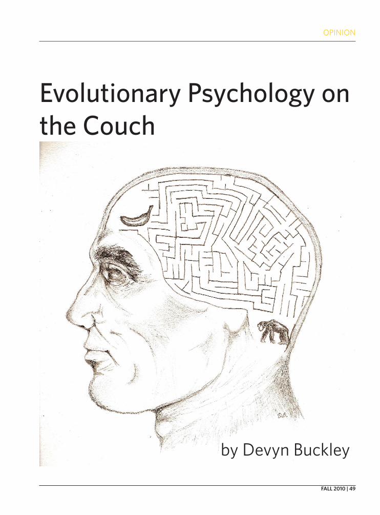
OPINION
FALL 2010 | 49
Evolutionary Psychology on the Couch
by Devyn Buckley
50 | THE NERVE
In Brief
Evolutionary psychology (EP) is the predomi-nant lens of interpretation in psychology today and is expanding into the general public as a
contemporary thought trend. As you may guess, it at-tempts to apply Darwin’s Theory of Evolution to the field of psychology. It explains observed behavior in contemporary humans as results of hard-wired pre-dispositions produced by natural selection. While speculation has its merits, many of E.P.’s claims seem reactionary and guided by an agenda, as is evidenced by the claims’ poor believability and the lack of data that support them. As a result of an ideological polar-ization occurring between modern religious extrem-ists and their counter Darwinian fundamentalists,. E.P. performs logical acrobatics to justify its speculation as fact. I aim to demonstrate the logically fallacious na-ture of E.P. and its ideological character, particularly as a counter-ideological reaction to the social sciences, religiosity, and feminism.
The Evolution of Psychology
The multitude of previous schools of thought in psychology mostly relied on the premise that the hu-man mind was a malleable thing. Many disciplines emphasized psychological malleability out of a dual motive for truth and social justice. Even John Scopes, who was favored by scientific intellectuals in the 1925 Tennessee vs. Scopes trial, which accused him of un-lawfully teaching evolution in Tennessee, taught from the textbook Civic Biology, which made racist sugges-tions and endorsed ideas such as sterilization of epi-leptics and the mentally feeble from the then popular field of eugenics.1 Anthropology and craniometry had long sought to justify social inequities on both the ra-
cial and class levels by making them natural law.Freudian psychologists emphasized the influence
of early childhood and the multiplicity of conflicting motives within an individual psychology. The Behav-iorists at the turn of the 20th century held the radical position that all aspects of psychology, including per-sonality and intelligence could be attributed to varia-tions of Pavlov’s conditioning, or learning strictly in terms of reward and punishment. In the 1950’s, hu-manism provided an almost limitless encouragement of self-actualization, positing that a well-socialized, happy being was attainable through unconditional positive regard and a general atmosphere of goodwill. Meanwhile Cognitive psychology overlapped greatly with neuroscience and studied the structures under-lying cognition. The passing of time and the formation of new schools of thought did not reject each system, but dethroned it as the principle means of psychologi-cal analysis as soon as the next came along.
E.P. emerged at the end of the 20th century, first in the form of Sociobiology. It began around 1975 when Edward O. Wilson proposed that human psychology, in addition to physiology, could be explained in terms of evolutionary adaptations in his book Sociobiology: The New Synthesis.2 It was initially unpopular due to a fear of returning social injustice. It reemerged un-der the new title of E.P. in the 1990’s with books such as The Adapted Mind (1992) by John Tooby and Leda Cosmides and David Buss’s Evolutionary Psychology: the New Science of the Mind (1995).
Reaction to the “Social” Sciences or the SSSM
Dylan Evans and Oscar Zarate, who write in Intro-ducing Evolutionary Pscyhology, published 1999, that
OPINION
Evolutionary Psychology on the Couch
by Devyn Buckley
OPINION
FALL 2010 | 51
“In the future, the study of human psychology will be completely transformed by the Darwinian approach...it won’t be called ‘Evolutionary Psychology’. It will just be called ‘psychology’.”3 David Buss titles one of his papers, “Evolutionary Psychology: A New Paradigm for Psychological Science.”4 The Handbook of Evolu-tionary Psychology, edited by Buss describes E.P. as a “scientific revolution, a profound paradigm shift in the field of psychology,” and a “paradigm within biology itself.”5
All prior schools of thought are dismissed. In The Handbook, Buss labels socio-cultural outlook “main-stream psychology,” which he defines as being “por-tioned into subdisciplines—cognitive, social, person-ality, developmental, clinical, and hybrid areas such as cognitive neuroscience,” that E.P. reveals to lack “logi-cal or scientific warrant.”5 To all of them Buss grants a false premise: the human mind is a “blank slate”. He states, “The human mind can no longer be conceived as it has been in mainstream psychology as a blank slate onto which parents, teachers, and cultures im-pose their script.”5 Buss refers to non-evolutionary ex-planation as the “myth of cultural causation.”6 All non-evolutionary facets of “mainstream psychology” are united under the appellation of the Standard Social Science Model (SSSM)5, meaning that they attempt to explain aspects of human behavior by external influ-ences rather than internal predeterminations. Those opposing the revelation of sociobiology by Wilson are described in the Handbook as “intellectuals wedded to the blank slate.”5
While previous approaches did not rely on bio-logical explanations, largely out of technological lim-itations, to say they viewed the human as entirely a blank slate would be a stretch, excepting the behavior-ists. Freud, for one, acknowledged uncontrollable im-pulses and the strength of unconscious instincts. Ad-ditionally, despite the radicalism of the Behaviorists, their conditioning experiments were integral to an understanding of learning and cannot be dismissed. Steven Pinker quotes philosopher Nelson Goodman in the foreword of The Handbook to describe alter-native psychological explanations as “a pretender, an imposter, a quack.” According to him, they are ut-terly false because they rely on “similarity, frequency, salience, and regularity” to make conclusions about behavior, explanations Pinker states are “in the eye of the beholder.”5 They do not address the “why” of behavior. However, the same methods are employed by E.P. in their observation of behavior in order to de-vise explanations for its existence. What they claim
as evidence uses the same observational techniques deemed “quack” in order to legitimatize it, namely it observes and interprets behavior in the mode of a soft science. Furthermore, if observation is in the eye of the beholder, then wouldn’t claims concerning un-observable behavior 100,000 years ago be even more so? The only difference then is that E.P. imagines itself to supersede the others because it provides a creation story for behavior, rather than stopping at what is ob-servable and imagines itself to be a science because its vocabulary incorporates biological terms.
I, Robot
Right at the time of Darwinism’s reawakening a revolution was occurring in technology affecting fields from computer science to biology. The mind be-came the brain the more the brain could be measured. David Marr, a British neuroscientist helped interpret the visual processing system from a computational standpoint.5 Noam Chomsky’s 1968 study of infants7 demonstrated the existence of innate linguistic capa-bilities, revealing the power of inborn mechanisms or modules within the mind, signaling the commence-ment of the cognitive revolution, or the mechanistic treatment of the mind. Module is a term often used in technological and economic fields because it deals with highly structured mechanisms. It is a general systems concept, meaning, it refers to “complexes of elements or components, which mutually condition and constrain one another, so that the whole complex works together, with some reasonably clearly defined overall function.”8. Neurological modules exist mostly within the realm of comprehending physical relations, analyzing visual data, processing speech and emotion. These are all areas with minimal variability among individuals in the same way that the pumping heart is more or less unvarying. E.P. grasped the notion of modularity and applied it universally to the point that the human being is a preprogrammed machine. Pink-er states that “the brain is not just like a computer—it is a computer” and that “the evolved function of a psy-chological mechanism is computational.”5
Universal modularity seems to go against neu-rological fact and observed behavior. Modularity is not synonymous with cognition. It details rigid, pre-programmed, unchanging systems. Pinker writes “humans are intelligent not because we have fewer instincts, but more.”7 Instincts are innate for every member of the species, yet, human behavior and per-sonality vary vastly among individuals. How can such

OPINION
52 | THE NERVE
a high degree of flexibility exist in an organism entirely composed of modular instinct? Either each individual possesses her or his own set of instincts or there is a multitude of strongly deviating mutants. This is not to say that modularity does not exist within the human mind, only that the model of its universality fails. Mod-ularity may operate within the realms of language, perception, and emotion, while other more complex psychology involves the interaction of memory, feel-ing and consciousness, without being the product of a rigid, preprogrammed and definitive process.
A modular system works in a deductive fash-ion—arriving at a conclusion through a rigid reason-ing system with a defined premise. The human con-sciousness, on the other hand, behaves mostly in an
inductive fashion. That is, conclusions are not reached through linear computation, but through a constant modification of experience and probabilistic deter-minations. Self-awareness is the critical ingredient to non-modularity. Jerry Fodor, a cognitive scientist and original proponent of modularity, gives the example that, although we cannot force our modular visual system to stop perceiving an optical illusion, the fact that we can understand that it is an illusion and reject it or modify our relationship to it illustrates complex cognition outside of modularity.7
The computational model of the human mind seems incongruous with observation, yet E.P. denies it for reasons unconscious to itself. E.P. reacts as it did to the social sciences, to religious and western notions. It represents a facet of the polarization marking this era between scientism and metaphysics.
The Fall of Man
Metaphysics deals with non-quantifiable experi-ence such as philosophy, art, literature, psychology,
etc. It contrasts with scientism, an outlook which ac-cepts only the quantifiable experience of being: that which can be measured and subjected to the scientific method. Scientism has become increasingly popular as technology rapidly develops and more people invest their education in the expanding sciences and areas of practical skill, partly out of economic reasons, leav-ing many schools to shut down programs in the lib-eral arts. The University of Louisiana, Lafayette ended its philosophy major, and Michigan State University eliminated its major in American Studies and Classics recently.10 Public figures posit scientistic views that collectively shape public opinion. For example, some have taken Richard Dawkins to suggest in The Selfish Gene that all human behavior is reducible to geneti-cally selfish foundations, reawakening Darwinism and
contributing to E.P. Such an outlook comes from the desire to push against age-old western notions of hu-man nature, particularly those revered by religious extremists. Evolutionary psychologists label them as stubborn, irrational, and imposing to the acquisition of a truth.
Western Culture finds humans to have some elect quality that gives them superiority not only in skill but also in essence over animals. E.P. tries to de-stroy the notion of the elect human being by placing him within the animal kingdom, but with such over-emphasis that it frames human nature as uniform and instinctual, ignoring the defining factors of humanity: self-awareness and culture. It attempts to crush any utopian notion of a human-invented paradigm toward which we progress, by over-emphasizing negative as-pects of human behavior. The Handbook of Evolution-ary Psychology takes a missionary tone, stating that “EP challenges the foundations of crucial enlighten-ment values.”5 Daniel Dennet, a philosopher and advo-cate of Dawkins’ strict adaptationism, explains in Dar-win’s Dangerous Idea natural selection as a “universal
“We have the power to turn against our own creators” - Richard Dawkins, The Selfish Gene

OPINION
FALL 2010 | 53
acid”: universal referring to its global explanatory power and acid to its corrosion of traditional Western belief.12
It is a testament to the power of cultural influence as well as to the influence of the unconscious to ob-serve that while E.P. fights traditional western values, it maintains them in its methodology. As regards love of reasoning, they are in full conformity, for they treat their conjectures as absolute truth, indicating none other than a total faith in the purity and omnipotence of their human reasoning. As regards the elect status of the human being, it is evident that they see their school of thought as superior to any other and in this manner treat themselves as a chosen elect. As regards the notion of paradigm, they treat their system as fi-nalized and refer to it as paradigmatic. Additionally, evolution itself is often treated as purposeful, direct-ed, and perfect. The adapted machine understanding of the human mind conforms not only to the desire for a paradigm, but also to the desire for an anthropomor-phic, willful designer found in the religious psychol-ogy. The religious personality is explicitly evident in Pinker’s own statement: “Darwin’s prophetic vision is being realized.”5
Most fascinating of fallacies, however, is E.P.’s du-alistic understanding of human nature despite its de-terminism. Rejecting cultural influence and free will, evolutionary psychologists perceive what Ivan fore-worns in Dostoyevsky’s The Brothers Karamazov that without God “everything is permitted.”14 God, in this case, can be understood not only as metaphysical im-petus for morality, but also as the concept of free will. To preserve morality, evolutionary psychologists sep-arate self from self and ethics from nature. In How the Mind Works Pinker states that science and ethics are “two self-contained systems…science treats people as material objects and its rules are the physical process-es that cause behavior through natural selection…eth-ics treats people as equivalent, sentient, rational, free-willed agents…” 15 He proposes two separate, equal, and entirely contradicting ways to understand the human being. Dawkins states in The Selfish Gene that “we have the power to turn against our own creators”, the genes.16 Pinker writes of his choice to never have children and how it is an “evolutionary mistake,” but that if his “genes don’t like it, they can go jump in the lake.”7 What kind of a machine has the power to defy its hardwire? What modular, goal-oriented, reduction-ist system creates a being that can behave counter to its biological fitness? If ethics are not metaphysical or religious delusions, but possess real value, then how
are they separate from the supposed biological deter-minism that shapes all thought and behavior? The par-adox arises from the profound error of rejecting the essence of human existence, partly out of radicalized counter-ideology to religion and traditional Western beliefs; that essence being metaconsciousness or the feeling of “I” that is so intimately infused with the also rejected concept of culture. They are not to be denied in simplistic systematization, as their rejection leads to impossible paradoxes and incongruities with hu-man experience.
How can a trait be biologically determined if it is not necessarily expressed? Steven Gould addresses the conundrum in The Mismeasure of Man, stating, “if innate only means possible…then everything we do is innate and the word has no meaning…flexibility is the hallmark of human evolution.” He goes on to say, “we should be wary of…viewing all brain capacities as di-rect adaptations…additional capacities are ineluctable consequences of structural design, not adaptations…our vastly more complex organic computers were also built for reasons, but possess an almost terrifying array of additional capacities—including, I suspect, most of what makes us human.”18 Thus, there are basic structures which are universally found, but complex, variable behavior made possible by basic structures is not a modular adaptation, but an expression of an incredible plethora of possibilities. A potential to act one way or the other renders both possibilities essen-tially non-determining.
Once the existence of systematized thinking is dis-covered, people accuse it of masking truth in the rigid-ity of its dogma, and shatter it by revealing a complete opposite of its creed to be true, and in their excitement fall unknowingly into another system as fierce as the first in its conviction and as sensitive towards criti-cism. Polarized against the SSSM, traditional Western notions of human nature and anti-scientific religious extremism, E.P. falls into the error of counter-ideolo-gy. E.P. forcefully defends conjecture out of ideological imperative rather than objectivity and claims conjec-ture as fact through faith in its dogmatic axioms.
Boys versus Girls
E.P. focuses primarily on sex and supposed sex differences. The most frequently used words in the titles of over 800 articles in two journals, Ethology and Sociobiology and Evolution and Human Behavior include sex, differences, human, and male in that or-der from 1997-2002 and attractiveness, sexual, facial,
OPINION
54 | THE NERVE
sex, men, female, male from 2003-2008 in that order.19 E.P.’s narrow window of focus indicates an almost fa-natical devotion to naturalizing sex stereotypes as Robert Wright, who initially popularized E.P. in his book The Moral Animal, predicted it would.21
E.P. defines the biologically determined or pro-grammed mating strategies of men and women to be inherently opposing. The textbook Psychology and Life outlines the perspective of E.P. to be such, citing Buss stating “Human males could reproduce hun-dreds of times a year if they could find willing mates. To produce a child all they need to invest is a teaspoon of sperm and a few minutes of intercourse. Women can reproduce at most once a year and each child…re-quires a huge amount of time investment and energy…the basic problem facing a male animal is to maximize the number of offspring he produces by mating with the largest number of females possible…but females have the problem of selecting…the biggest, strongest, smartest, most highest-status, most thrilling mate…but also the most committed…”22 Men, as Buss posits, have an evolved strategy to “seduce and abandon as many women as possible…” and the female strategy is to seek out resources and men of high-status. In essence, women are commitment loving and child-oriented, whereas men are stunted by commitment in the pursuit of their biological imperative to impreg-nate as many youthful, attractive women as possible.
Women seek resources, status, and “men who are older,” according to Buss.6 In the scheme of E.P. the words “willing mate” are unnecessary, but used to pre-empt extreme controversy, as happened when Craig T. Palmer and Randy Thornhill, whose paper “Why Men Rape,” stated rape was not in its essence a violent act, but “natural, biological phenomenon.” They advised the New York Times, “a woman’s risk of attack rises along with her hemline, and her willingness to social-ize without the company of ‘male protectors.’”26 They suggested men be instructed to repress natural feel-ings to prevent the act. Like other violent criminal acts, rape is not a global behavior, or an evolved trait, but one behavior out of many coming from a cogni-tively flexible species.
E.P. perpetuates stereotypes and sexual inequi-ties by making the dream of social justice a farce in the face of fact. By claiming that certain behaviors are in-nate and modular, the human will becomes ineffectual to change them. A double standard is, thus, justified in which the male is irresponsible, freewheeling, and self-indulgent, while the female is a natural care-taker, a fulfiller of responsibilities, and bearer of burdens,
most specifically child-rearing. To EP, female sexual desire is stupefying, if not ignored completely, and the monogamy of men is seen as a female victory or ef-feminacy. Christopher Ryans, author of Sex at Dawn, titles an entry on his blog on Psychology Today’s web-site, “Why Does Female Orgasm Exist?” and states how its existence has baffled evolutionary psychologists. David Buss’ book Why Women Have Sex presumes in its title the perplexing nature of the female pursuit of sex. Yet a sexual desire in women would seem more evolutionarily advantageous.
While the premise of E.P. is that an observed be-havior is adapted, they refuse to acknowledge female orgasm as an adaptation, but regard it as an enigma or as vestigial. This demonstrates the a priori nature of their beliefs, which shape interpretation through a pre-established conviction of gender roles, making them decide what is or is not an adaptation by pref-erence. If females had choice in selecting mates of high-status, then wouldn’t they have just as much of a choice in pursuing sexual pleasure and preferences? The confusion over female orgasm is really confusion over whether females have choice in pursuing sex or not. E.P. grants women choice only when it allows them to explain the evolution of a pre-existing stereo-type.
EP is the naturalizes of female objectification, and makes women natural means to ends in the eyes of men, rather than a fellow human being whom they relate to, empathize with and depend on. Their per-spective places the male as a champion of fitness and esteems him through a worship of his potential to mas-sively procreate. Warlords and keepers of harems who enslaved women are cited as examples of male fitness and sexual strategy. Psychology and Life gives as an example of a Moroccan despot, King Ismail the Blood Thirsty, who possessed many harems and fathered over 3,000 children, ignoring the fact that a warlord from several hundred years ago is not an exemplar for the biologically determined psychology of every hu-man male anymore than a stay at home dad from the 21st century is. Buss cites the Turkmen of Persia to justify women’s resource-oriented strategy and men’s sex-oriented one: the males in the wealthier half of the population left 75 percent more offspring than males in the poorer counterparts. Interpreting this as a ful-fillment of evolutionary roles ignores the entire cul-tural background which was, like the majority of soci-eties throughout history and the world, possessive of women, selling them into marriage and denying them equitable societal status. To say that the enslavement,
OPINION
FALL 2010 | 55
domination, and objectification of women is natural justifies sexual inequity and preempts social change. The same textbook Psychology and Life lists exem-plary power hierarchies as: “parent-child, teacher-student, doctor-patient, boss-worker, male-female.” This exemplifies attitudes of E.P. 22
The aggressive anti-feminist tone of E.P. is illus-trated by Ryan who states, “boys will be boys, and men will be the way they are, despite the many ways our so-ciety tries to make them change.”23 Ryan quotes Don-ald Symons saying “The sexually insatiable woman is to be found primarily, if not exclusively, in the ideology of feminism, the hopes of boys, and the fears of men.”
The stereotypes naturalized by E.P. are facts of current culture. Women are shown to be “resource obsessive,”pursuing shoes, handbagsand men with great wealth. A popular T-shirt depicts a man and woman getting married with the words “game over” underneath. A poster for the show Entourage on Spike, the men’s channel, shows two female bodies in bikinis outlining authoritative male figures in suits with the caption, “where every guy can get some.” The Evolutionist describes it as “a flood of magazines and television programmes celebrating the distinct fea-tures of “maleness” - ogling, drinking, fighting, playing football...” It describes this as a reaction to the failed attempt to modify male behavior by sociologists.6
An article in The Economist titled “Sex, Shopping, and Thinking in pink” argued that women were evolu-tionarily programmed to enjoy shopping and to be at-tracted to pink, a color designated feminine in Europe and America around 195027, since it signified the color of berries and emotional states on faces, making them good empathizers.28
Once again, the errors lie in the foundational mistakes of E.P. The dismissal of obvious social and cultural phenomena does not come from reason, but from adherence to dogma and a desire to make ste-reotypes irrefutable. What they designate as evidence is behavior selected by them to be interpreted with a priori conclusions. Perhaps a uniform history of deny-ing women equal career opportunity could have made attaching oneself to an economically stable, older man more conservative for women. Equitable social and economic status for women is fairly new, and limited geographically. A cross-cultural examination may re-veal universal cultural phenomena than a biological reality. The poor status and treatment of women, for example, is similar throughout history and through-out culture. Would this not lead to similar behavioral phenomena?
A study in Psychology and Life involved over 16,000 participants from 52 nations about their inter-est in “short-term sexual relationships,” in which men reported greater desire for sexual variety than did women.22 The survey does not validate an evolution-ary narrative, but reveals merely what those partici-pants at that point in time and space revealed about their personal motives. Take this as a testament to the nature of surveys: “In 1953, Alfred Kinsey, Ph.D., the famous sexuality researcher, found that nearly 40 per-cent of the 5,628 women he interviewed experienced at least one nocturnal orgasm (orgasms during sleep), or “wet dream,” by the time they were forty-five years old. A smaller study published in the Journal of Sex Research in 1986 found that 85 percent of the women who had experienced nocturnal orgasms had done so by the age of twenty-one, some even before they turned thirteen.”29 The dramatic increase was not due to a biological change, but a change in what partici-pants stated about themselves and also, likely, chang-ing societal attitudes about women’s sexuality.
EP’s most fatal error is its denial of humanity. It places women and men in psychological sexual con-flict in which they must attempt to trick one another to achieve satisfaction. Buss states “One of the key in-sights of evolutionary psychology is that humans have inherent conflicts of interest with other individuals, with members of their own family, with members of the opposite sex, and members of their own sex,” and describes his major interest as “topic of conflict between the sexes.”6 E.P. attitude sees all elements of nature in conflict, including the supposed modules within oneself or “scores of instincts assembled into programmes and pitted in competition,”7 as described by Pinker. This misinterprets not only the relation be-tween the genders, but evolution as a whole and criti-cal aspects of a social humanity.
Adaptation can result from mutually beneficial relationships between individuals who share the same needs. The parts of the brain, as well, work very nicely together, not because they are in competition, but because their harmony gives rise to a well func-tioning whole. In the system of E.P. there is hardly need for men to fall in love or for women to experience orgasm, as Ryans notes, yet this occurs anyway. Why not incorporate them into one’s evolutionary schema? Perhaps men and women form lasting bonds out of mutual emotional and physical need as many seem to do. Maybe it is through supporting these needs to-gether that ensures the success of both? Though we may only speculate, isn’t it probable that cave people
OPINION
56 | THE NERVE
in their small social groups facing a threatening cli-mate similarly needed steady bonds with other mem-bers both to survive as well as ensure the health of their offspring? Perhaps the attentiveness of a man to his mate and offspring ensures their success more than those who abandon them to the environment? It would seem the thing of greatest need in small groups struggling to survive would be trust and ritual culture.
Perhaps E.P. could be more appreciable if had a set of varied narratives, but it does not. Its emphasis on conflict neglects the various complex emotional and social needs that are satisfied by relationships unique to human beings. Relations between the genders are about more than just procreation, and successful hu-man procreation is about more than just copulation. By treating people as animals, not only in taxonomy, but in essence of spirit, we lose the defining quality of humanity—that is, the self-awareness and all the psychological complexity that comes with a reasoning and emoting conscious.
To Err is HumanIt is important to remember that in the noble
quest to uncover the basis of our psychology, we some-times overlook our psychology. The influences of oth-
er people, as well as the motives of the unconscious, present the greatest obfuscations to truth, while at the same time are least detected, by virtue of the very fact that they are not conscious. Societal convention and self-serving belief, the two often one in the same, con-stitute the great Lochness of the depths of the uncon-scious, powerful, ageless, and hidden, churning the waters of consciousness in the minds of even the most earnest. In the excitement of a new school of thought, it is often easy to forget that the present is also part of history and that the current era, since it is always the most modern, does not signify an arrival at final truth, but the most current contribution to a succes-sion of imperfect perspectives. Deconstructing E.P.’s methods one finds the symptoms of the ideological or religious personality, whose most integral aspect the circular logic of faith. E.P.’s assumptions concerning biological determinism and modularity are in opposi-tion to the facts of behavioral flexibility, the human-ity of self-awareness, and culture. Its beliefs are often counter-ideology to the social sciences, religiosity, and feminism. Understanding what is false is one way of understanding what is true. Deconstructing E.P. il-lumines the social politics of our era and practices a critical skepticism that aids scientific objectivity.
1. Black, Edwin. _War Against the Weak: Eugenics and America’s Campaign to Create a Master Race_. New York, N.Y.: Avalon Publishing Group Inc., 2003. [75.]
2. Webster, Gregory. “What’s in a Name?: Is “Evolutionary Psychology” Eclips-ing “Sociobiology” in Scientific Literature?.” Evolutionary Psychology Journal Volume 5(3) (2007): 683-684.
3. Evans, Dylan and Oscar Zarate. _Introducing Evolutionary Psychology_. Duxford, Cambridge: Icon Books Ltd., 1999. [169]
4. Buss, David, ed. “Evolutionary Psychology: A New Paradigm for Psychologi-cal Science.” Psychological Inquiry 6 (1) (1995): 1-30.
5. Buss, David. _The Handbook of Evolutionary Psychology_. Hoboken, New Jersey: John Wiley & Sons, Inc., 2005.
6. The Evolutionist. (1996). [Interview with David Buss, author of The Evolu-tion of Desire]. Darwin@LSE.
7. Malik, Kenan. “Darwinian Fallacies.” Prospect. 1998.8. Edquist, Charles. _Systems of innovation: technologies, institutions, and
organizations_. N.p.: Pinter, 1997.9. Addington, Larry. _America’s War in Vietnam: a short, narrative history_.
Bloomington, Indiana: Indiana University Press, 2000.10. Zerinke Kate. “Making College Relevant.” The New York Times (2010):
ED16.11. Aristotle. _Nicomachean Ethics_. Martin Ostwald. N.p.: Prentice Hall, 1962.
[17].12. Gould, Stephen J. “Darwinian Fundamentalism,” The New York Review,
June 12, 1997, pp. 34-37.13. Connecting Christians Www.connectingchristians.co.za. Web. 10 July
2010. <http://www.connectingchristians.co.za/index.php>.14. Dostoevsky, Fyodor. The Brothers Karamazov. Pevear, Richard and Larissa
Volokhonsky. First Farrar, Straus and Giroux paperback edition. U.S. & Canada: Douglas & McIntyre Ltd., 2002. [649].
15. Pinker, Steven. _How the Mind Works_. N.p.: W.W. Norton & Company, 1999.
16. Dawkins, Richard. The Selfish Gene. Great Clarendon Street, Oxford: Ox-
ford University Press, 1976.17. Welton, James and Alexander James Monahan. _An Intermediate Logic_.
E.M. Whetnall. 3. The University of Michigan: W.B. Cliver, 1928.18. Gould, Stephen J. _The Mismeasure of Man_. 500 Fifth Avenue, New York,
NY: W.W. Norton & Company, Inc., 1981. [360-1].19. Webster, Gregory D. “Hot Topics and Popular Papers in Evolutionary Psy-
chology.” Evolutionary Psychology 7 (2009).20. Herrnstein, Richard J. and Charles Murray. _The Bell Curve_. United States
of America: Free Press Paperbacks, 1994.21.Wright, Robert. _The Moral Animal: Evolutionary Psychology and Every-
day Life_. The University of Michigan: Pantheon Books, 1997.22. Zimbardo, Philip and Richard J. Gerrig. _Psychology and Life_. 18. N.p.:
Pearson Education, Inc., 2008. [363].23. Ryan, Christopher, “Sex at Dawn: Exploring the Evolutionary Origins of
Modern Sexuality.” Psychology Today. May 4, 2010. (http://www.psy-chologytoday.com/blog/sex-dawn?page=1)
24. Maines, Rachel. The Technology of Orgasm: “Hysteria,” the Vibrator, and Women’s Sexual Satisfaction. The Johns Hopkins University Press, 1998.
25. McCaughey, Martha. The Caveman Mystique. National Sexuality Resource Center. October 20, 2008. (http://nsrc.sfsu.edu/article/caveman_mys-tique_evolution_psychology)
26. Pozner, Jennifer. “In Rape Debate, Controversy Trumps Credibility.” Extra! (May/June 2000).
27. Peril, Lynn. Pink Think: Becoming a Woman in Many Uneasy Lessons. New York, N.Y.: W.W. Norton & Company, 2002.
28. “Sex, Shopping and Thinking Pink; Evolutionary Psychology.(What Wom-en Want, Evolutionarily Speaking).” The Economist (2007). Economist Newspaper Ltd. Web.
29. Health Services at Columbia, “Go Ask Alice”. Columbia University’s Health Q&A Internet Service. <http://www.goaskalice.columbia.edu/1498.html>.

OPINION
FALL 2010 | 57
Does the Brain
by Kayla Ritchie
Run Algorithms?
OPINION
58 | THE NERVE
Introduction
Computation, or information processing, can be defined as that which describes the
changes which occur in the natu-ral world. Typically an algorithm or equation, such as the ones used in neural modeling, are employed for such processing due to their ability to describe the behavior of a system according to certain de-pendent and independent condi-tions. Due to the recent increase in processing capacity of modern computers, neuroscientists and computer scientists have attempt-ed to model large scale brain archi-tectures that include entire popula-tions of neurons, or in the case of a team of Swiss researchers, the en-tire human brain, in hopes of simu-lating the processes in the brain to a point that higher cognitive func-tions would arise from the models. While this is an exciting prospect, it is unclear whether running the “Brain Program”, would actually yield genuine cognitive processes, simply because the brain is not necessarily a digital computer, and its functions not necessar-ily computational. In this paper, I pose the question: Does the brain run algorithms? I will argue that this question stems from a deeper uncertainty of whether cognitive processes are computational, and explore the implications that this may have on our ability to model the mind with computers.
What is an algorithm?
For the purpose of this article, it is appropriate to use a general purpose definition with a few clari-fications. First, an algorithm must consist of precise, unambiguous steps which require no subjective interpretation, and, resultantly, the execution of the algorithm must al-ways yield the same outcome given a particular set of inputs.
Intentional Use of Algorithms
While executing even the sim-plest tasks such as brushing one’s teeth, or navigating to a desired, familiar destination, application of an algorithm is essential for successful completion of the task. More complex tasks, such as using the quadratic formula, require sim-ilar use of step-by-step processes. However the algorithms we draw on to accomplish such everyday tasks are learned through many experiences of trial and error; each algorithm was developed indirect-ly through intentional, goal-driven behavior, and so is not inherent to the architecture of the brain. In other words, the processes which govern the underlying functions of the brain, i.e. protein synthesis and the physical and chemical basis for generating action potentials are neither learned, nor controllable through intentional behavior. Such processes are inherent in the phys-ics of the system.
Computer algorithms
Though a general purpose dictionary will explain that an al-gorithm consists of a step-by-step problem solving procedure, the computational meaning of the word has not yet been agreed upon. Computation, in its most basic defi-nition, occurs in a system which is designed to process information in the form of simple symbol ex-change, as in the 0-1 coding para-digm used in digital computers. A certain combination of such for-mally defined symbols correlates to certain commands, which can take the form of stepwise algorith-mic functions.
In most modern comput-ers, this symbol exchange corre-sponds to the presence or absence of electrical current. Hardware is designed so that input generates current fluctuations in a vast ar-ray of transistors, which eventually leads to some output mechanism. This output can be expressed, for example, through a mechanical de-vice that takes the current fluctua-tions and converts them to light on a monitor. It can also be converted into signals that direct the motion of the mechanical arm that writes on the hard disc. Note, however, that transistors are not strictly necessary for a formal symbol ex-change. Theoretically, any system upon which a formal symbol defini-tion can be imposed can be a com-puter.
Does the brain run algorithms?
by Kayla Ritchie
OPINION
FALL 2010 | 59
What is the difference between human and computer algo-rithms?
The human method of fol-lowing an algorithm requires consciousness. Humans are con-sciously aware of the task that they are supposed to complete, and use the meaning of their instructions to drive their behavior. Comput-ers run algorithms in a completely different manner due to their lack of consciousness. The computer “performs” algorithms simply through following a formal sym-bol definition that precipitates changes in the hardware and soft-ware. Though both humans and computers can use the same exact algorithm to find the value of vari-ables in a quadratic equation, the processes which underlie their behavior are radically different. But the question still remains: Are there any nonconscious algorithms (i.e. computational processes) in the brain?
What things in nature are poten-tially capable of algorithmic be-havior?
In the post-Newtonian era, all of us can observe a natural process, such as a rock falling from a cliff overhang, and can imagine a ba-sic set of laws or equations which could accurately model the rock’s motion. It is also clear to us that the rock is not following an algo-rithm, which could look something like this:
Detach oneself from cliff faceWhile(collision with ground=FALSE){ Do{Move towards the center of the earth, increasing velocity by inter-
val determined by force of gravity and mass of rock }Until(terminal_velocity ==velocity)Move towards center of the earth at terminal_velocity}
stances that make it through are small enough to pass, and do so because of a chemical gradient, or because they are chemically suited to pass through a specific chan-nel. Similarly, a cell membrane is not round because the cell actively maintains its shape due to the fact that this enables higher internal organization, but rather because it is the lowest energy configura-tion of the hydrophilic heads and hydrophobic tails of each phospho-lipid molecule in the membrane. In other words, there are no instruc-tions written somewhere to govern these behaviors; they are results of physical and chemical processes.
Unfortunately, accepting that all things are results of physical and chemical processes does not by itself refute the proposal that the brain is computational. Indeed, it is still possible to assert that all things in nature are intrinsically computational.
Is everything intrinsically com-putational?
This viewpoint has been fur-thered by such prominent phi-losophers as Daniel Dennett, who proposed that evolution was an al-gorithmic process. Others debate this claim, however. John Searle, an opponent of this type of thinking stated in his book The Rediscov-ery of the Mind, “…notions such as computation, algorithm, and pro-gram do not name intrinsic physi-cal features of a system. Compu-tational states are not discovered within physics, they are assigned to the physics”.
In order to understand this, let us remember that computation can be defined as a system of informa-tion processing that relies on the mindless exchange of symbols, i.e. the use of syntax. In much of his
Why is it clear to us that the rock is not following this algo-rithm? For one, we know that a rock does not have an input/output system. It neither has way of measuring its own velocity, nor a way to alter its velocity, and so could not follow the do… until instruction, or any of the oth-ers. Additionally, a rock does not propel itself; rather, gravity exerts a force on the rock that causes it to move in a particular direction at a particular speed. In other words, we know that a rock cannot detect its progress, and aside from not having a propulsion system, it wouldn’t be able to execute com-mands if it did have a propulsion system.
But does there exist anything in nature that does follow algo-rithms? Immediately, it should oc-cur to us that there is one prime candidate: Life. Even the most basic life forms have organization which functions as an input/out-put system. Paramecia have the ability to sense changes internally and in the environment, and thus can perform certain behaviors, e.g. food acquiring. Yet this by itself does not indicate that any algo-rithm is being followed. Biological processes are unique in their in-put/output organization, but this does not contradict the fact that all things that occur are still based on chemical and physical processes. A cell membrane is not selectively permeable because it detects each substance on its exterior and pass-es judgment on whether or not to let it through, but because the sub-
OPINION
60 | THE NERVE
work, Searle vehemently points to the fact that syntax is a formally defined system which is imposed upon a natural system. By this he means that the voltage levels of a computer which we encode as 0’s and 1’s, and in fact all state changes in any natural system, are not in themselves indicative of anything. Rather, we as observers assign symbolic meanings to each state. In this sense, nothing is intrinsi-cally computational.
Considering any given pattern found in the brain, we could read-ily ascribe a computational inter-pretation. But this does not mean that the brain is a digital computer, or that it has a separate “program level”, as has been the assumption since Marr proposed a model of the brain that separated the neurobiol-ogy from the other “higher” func-tions. As Searle said, “You don’t need to suppose that there are any rules on top of the neurophysiolog-ic structures.”
What does it mean for computer science models of the brain if the brain isn’t algorithmic?
If the brain lacks any intrinsic algorithmic processes, then there is no algorithm we’ll invent that will recreate any fundamental pro-cesses in the brain. Computational models that emphasize a top-down approach may be valuable for aca-demic fields that wish to use brain-like behavior for the purpose of improving mechanical and com-putational systems. But the top-down method of devising a compu-tational model that represents the relationship between higher level brain architectures and behaviors will fail to provide an ultimate ex-planation of how the brain works. Methods that operate under the mistaken assumption that the
brain contains “programs” of be-havior which are somehow distinct from neurobiology will never offer explanations of how behaviors ac-tually arise from the neurobiology, and so are limited in their ability to realistically portray what the brain actually does.
Could a network of Ordinary Dif-ferential Equations constitute real consciousness?
Top-down approach aside, there are those whose efforts re-volve around reconstructing the brain from the bottom-up using computers to simulate its most ba-sic components. Networks of or-dinary differential equations have been used to mimic the changes in individual neurons as they fire. But even this only provides a simulation of the processes that occur in the brain. There is still this assumption that the physiol-ogy that causes the brain to func-tion is somehow incidental. On the contrary, the physicality of any system is what causes it to behave as it does. To suppose otherwise is unscientific. Thus efforts like Tom Markram’s Blue Brain project may succeed in simulating all processes in the brain perfectly, but such a feat would not amount to creating a mind any more than simulating a tornado will amount to creating a tornado1.
Conclusion
With a certain degree of com-plexity, it is often difficult or impos-sible to decipher the processes of a system. Because the human brain is radically more complex than most natural phenomena, attempts to explain the operations and phys-ical processes which cause a brain to function fall victim to these dif-
ficulties. Often times when such a system is encountered, descrip-tions which focus on behavior, that is, on the input-output relationship displayed by the system, are put forward, rather than explanations which are based purely on physi-cal properties of the system. An indispensable tool used in nearly all math and science, the algorithm, is often used to generate similar input-output relationships in a vir-tual environment.
Upon first examination, it seems obvious that brains “run” algorithms, simply because a brain appears to us as a black box func-tion, i.e. an entity which, when giv-en specific input, produces specific output with some predictability2. However, the same could be said for many other natural phenom-ena, such as a projectile moving through the air, or the formation of phospholipid membranes. Each of these systems can be reduced to a set of inputs or initial condi-tions, which after a series of steps or computations that are defined by physical rules produce a pre-dictable set of output. Indeed, one can write algorithms which describe the behavior of each of these systems. But we have seen that our unique position as observ-ers creates the notion of a symbol, and this predisposes us towards believing that nature contains its own inherent language of compu-tation. Were it not for the philo-sophical analysis of such problems, we would forever be limited in our ability to understand the universe.
Though the implications of the brain’s lack of intrinsic algorithmic processes may seem inconsequen-tial, many areas of research oper-ate under the assumption that the brain is a computer. Of course, the majority of these projects are not concerned with creating a truly
OPINION
FALL 2010 | 61
conscious mind, but rather focus on developing applications for small-scale artificial intelligence soft-ware, in which a simulation of be-havior is appropriate. Projects that do aim to create a conscious mind, however, may be faced with severe difficulty unless they adapt their methods to reduce or eliminate the modeling of higher-level structures in the brain. Henry Markram’s Blue Brain project, for example, is attempting to model the brain at the molecular level, and so has a much higher chance of creating a mind than if he were to model the brain at a higher-level. In addition, there may be success in attempts to build a physical recreation of the brain with synthetic materials analogous to the biological ones. This approach would excel not only because it emphasizes the im-portance of the brain’s physicality, but also because it would enable a view into the precise relationships and connections between the parts of the brain, and possibly explain how such relationships contribute to consciousness.
Current research programs that wholly embrace the philo-sophical considerations related to science are rare. Even Boston University’s own Cognitive and Neural Systems department barely considers the myriad philosophi-cal problems associated with the study of the mind. Institutions that care only about the utility of the fruits of an experiment dominate the realm of scientific funding, as they should. What these institu-tions (and the scientists who ap-peal to them) tend to forget is that robust theoretical deliberation improves the chances of practical, scientific success. It should be the aim of every research effort, then, to develop a solid theoretical para-digm that places their experiments in context, and clearly defines the domain in which results may ap-pear. Still, the scientific study of the mind is relatively young. With any luck, neuro-researchers will begin to recognize the significance of the philosophy of mind in time to guarantee the success of their sci-entific endeavors.
Notes:1. That is not to say that it will
not provide valuable data on how the brain works. Simulations are created and used for their ability to supply vast amounts of knowledge about a particular system, knowl-edge which may be otherwise inac-cessible. But that is the extent of their abilities.
2. The mathematical definition of a function requires that specific input will always produce the same output, that is, the relation must be totally predictable. While it can be assumed that minute processes which occur in the brain during normal operation respond predict-ably to given inputs, the unpredict-ability of processes in the brain as a whole (due to the vast number of contingencies within the brain) causes us to view the brain’s re-sponses as probabilistic rather than deterministic. Note however that if by some terrific feat of neu-roscience we could view how all of these contingencies interacted, it may be more appropriate to think of the brain as deterministic.
Interested in Graduate Programs in Neuroscience? Check out
Neuroscience at BU:
bu.edu/neuro
Undergraduate Program in Neuroscience:The Undergraduate Program in Neuroscience is an interdisciplinary ma-jor leading to a Bachelor of Arts in Neuroscience that takes advantage of the rich neuroscience mission of multiple departments and campuses of Boston University. As a field, neuroscience has grown considerably over the last few decades through its integration of multiple disciplines; and, a current understanding of the field requires knowledge that spans traditional approaches while moving into the intersection between far-reaching technologies and new computational methods. This program combines breadth of exposure to the field as a whole with the opportu-nity for depth of experience in one of three central domains of neurosci-ence: Cellular and Systems, Cognition and Behavior, and Computational Neuroscience
Neuroscience students will have access to the extensive resources and expertise of affiliated faculty across multiple departments and colleges throughout the university. A wide array of courses are offered through the departments of Biology, Cognitive & Neural Systems, Computer Science, Mathematics & Statistics, Psychology, and Health Sciences in Sargent College. Together more than 50 upper level neuroscience elec-tives are offered, including laboratory courses and seminars.
Opportunities for independent laboratory research are available through multiple departments in the Colleges of Arts and Sciences and Engineer-ing, and at Boston University School of Medicine, including Anatomy and Neurobiology, Biochemistry, Neurology, Pathology, Pharmacology & Experimental Therapeutics, Physiology and Biophysics, and Psychia-try. Undergraduate research opportunities in neuroscience laboratories expand throughout the university across both the Charles River and Medical campuses.

Don’t toss out that term paper! Submit it to The Nerve!
We are looking for three types of papers:
1. Articles - these are light reading, requiring the reader to have little background knowledge. Typical length is around 2000 words.
2. Reviews - these are analogous to reviews that appear in professional journals. They explore the chosen topic in depth and are based on serious research of the literature. Typical length is 4000 words.
3. Opinions - these are perspectives on current trends.
Authors are encouraged to submit works that touch on any topic in the Mind and Brain Sciences. This includes, but is not limited to psychology, anthropology, philosophy, biology, com-puter science, etc.
Learn more at:bu.edu/thenerve/submissions
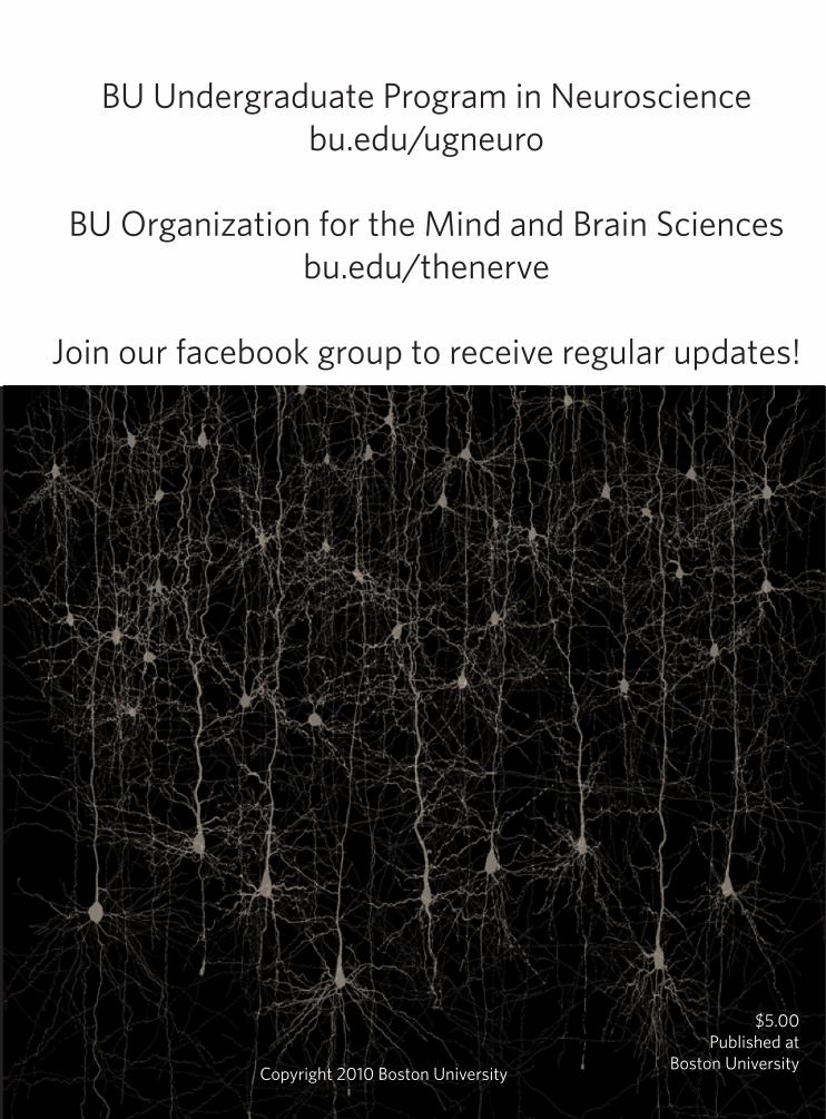
BU Undergraduate Program in Neurosciencebu.edu/ugneuro
BU Organization for the Mind and Brain Sciencesbu.edu/thenerve
Join our facebook group to receive regular updates!
Copyright 2010 Boston University
$5.00Published at
Boston University
Related Documents