BioMed Central Page 1 of 12 (page number not for citation purposes) Reproductive Biology and Endocrinology Open Access Research Spam1-associated transmission ratio distortion in mice: Elucidating the mechanism Patricia A Martin-DeLeon* 1 , Hong Zhang 1 , Carlos R Morales 2 , Yutong Zhao 1 , Michelle Rulon 1 , Barry L Barnoski 3 , Hong Chen 1 and Deni S Galileo 1 Address: 1 Department of Biological Sciences, University of Delaware, Newark, DE 19716, USA, 2 Department of Anatomy and Cell Biology, McGill University, Montreal, Quebec, Canada and 3 Department of Genetics, Thomas Jefferson University, Philadelphia, PA, USA Email: Patricia A Martin-DeLeon* - [email protected]; Hong Zhang - [email protected]; Carlos R Morales - [email protected]; Yutong Zhao - [email protected]; Michelle Rulon - [email protected]; Barry L Barnoski - [email protected]; Hong Chen - [email protected]; Deni S Galileo - [email protected] * Corresponding author Abstract Background: While transmission ratio distortion, TRD, (a deviation from Mendelian ratio) is extensive in humans and well-documented in mice, the underlying mechanisms are unknown. Our earlier studies on carriers of spontaneous mutations of mouse Sperm Adhesion Molecule 1 (Spam1) suggested that TRD results from biochemically different sperm, due to a lack of transcript sharing through the intercellular cytoplasmic bridges of spermatids. These bridges usually allow transcript sharing among genetically different spermatids which develop into biochemically and functionally equivalent sperm. Objectives: The goals of the study were to provide support for the lack of sharing (LOS) hypothesis, using transgene and null carriers of Spam1, and to determine the mechanism of Spam1-associated TRD. Methods: Carriers of Spam1-Hyal5 BAC transgenes were mated with wild-type female mice and the progeny analyzed for TRD by PCR genotyping. Sperm from transgene and Spam1 null carriers were analyzed using flow cytometry and immunocytochemistry to detect quantities of Spam1 and/or Hyal5. Transgene-bearing sperm with Spam1 overexpression were detected by fluorescence in situ hybridization. In wild-type animals, EM studies of in situ transcript hybridization of testis sections and Northern analysis of biochemically fractionated testicular RNA were performed to localize Spam1 transcript. Finally, AU-rich motifs identified in the 3' UTR of Spam1 RNA were assayed by UV cross-linking to determine their ability to interact with testicular RNA binding proteins. Results: The Tg8 line of transgene carriers had a significant (P < 0.001) TRD, due to reduced fertilizing ability of transgene-bearing sperm. These sperm retained large cytoplasmic droplets engorged with overexpressed Spam1 or Hyal5 protein. Caudal sperm from transgene carriers and caput sperm of null carriers showed a bimodal distribution of Spam1, indicating that the sperm in a male were biochemically different with respect to Spam1 quantities. Spam1 RNA was absent from the bridges, associated exclusively with the ER, and was shown to be anchored to the cytoskeleton. This compartmentalization of the transcript, mediated by cytoskeletal binding, occurs via protein interactions with 3' UTR AU-rich sequences that are likely involved in its stabilization. Conclusion: We provide strong support for the LOS hypothesis, and have elucidated the mechanism of Spam1- associated TRD. Published: 10 August 2005 Reproductive Biology and Endocrinology 2005, 3:32 doi:10.1186/1477-7827-3- 32 Received: 02 May 2005 Accepted: 10 August 2005 This article is available from: http://www.rbej.com/content/3/1/32 © 2005 Martin-DeLeon et al; licensee BioMed Central Ltd. This is an Open Access article distributed under the terms of the Creative Commons Attribution License (http://creativecommons.org/licenses/by/2.0 ), which permits unrestricted use, distribution, and reproduction in any medium, provided the original work is properly cited.

Welcome message from author
This document is posted to help you gain knowledge. Please leave a comment to let me know what you think about it! Share it to your friends and learn new things together.
Transcript
BioMed Central
Reproductive Biology and Endocrinology
ss
Open AcceResearchSpam1-associated transmission ratio distortion in mice: Elucidating the mechanismPatricia A Martin-DeLeon*1, Hong Zhang1, Carlos R Morales2, Yutong Zhao1, Michelle Rulon1, Barry L Barnoski3, Hong Chen1 and Deni S Galileo1Address: 1Department of Biological Sciences, University of Delaware, Newark, DE 19716, USA, 2Department of Anatomy and Cell Biology, McGill University, Montreal, Quebec, Canada and 3Department of Genetics, Thomas Jefferson University, Philadelphia, PA, USA
Email: Patricia A Martin-DeLeon* - [email protected]; Hong Zhang - [email protected]; Carlos R Morales - [email protected]; Yutong Zhao - [email protected]; Michelle Rulon - [email protected]; Barry L Barnoski - [email protected]; Hong Chen - [email protected]; Deni S Galileo - [email protected]
* Corresponding author
AbstractBackground: While transmission ratio distortion, TRD, (a deviation from Mendelian ratio) is extensive inhumans and well-documented in mice, the underlying mechanisms are unknown. Our earlier studies on carriersof spontaneous mutations of mouse Sperm Adhesion Molecule 1 (Spam1) suggested that TRD results frombiochemically different sperm, due to a lack of transcript sharing through the intercellular cytoplasmic bridges ofspermatids. These bridges usually allow transcript sharing among genetically different spermatids which developinto biochemically and functionally equivalent sperm.
Objectives: The goals of the study were to provide support for the lack of sharing (LOS) hypothesis, usingtransgene and null carriers of Spam1, and to determine the mechanism of Spam1-associated TRD.
Methods: Carriers of Spam1-Hyal5 BAC transgenes were mated with wild-type female mice and the progenyanalyzed for TRD by PCR genotyping. Sperm from transgene and Spam1 null carriers were analyzed using flowcytometry and immunocytochemistry to detect quantities of Spam1 and/or Hyal5. Transgene-bearing sperm withSpam1 overexpression were detected by fluorescence in situ hybridization. In wild-type animals, EM studies of insitu transcript hybridization of testis sections and Northern analysis of biochemically fractionated testicular RNAwere performed to localize Spam1 transcript. Finally, AU-rich motifs identified in the 3' UTR of Spam1 RNA wereassayed by UV cross-linking to determine their ability to interact with testicular RNA binding proteins.
Results: The Tg8 line of transgene carriers had a significant (P < 0.001) TRD, due to reduced fertilizing ability oftransgene-bearing sperm. These sperm retained large cytoplasmic droplets engorged with overexpressed Spam1or Hyal5 protein. Caudal sperm from transgene carriers and caput sperm of null carriers showed a bimodaldistribution of Spam1, indicating that the sperm in a male were biochemically different with respect to Spam1quantities. Spam1 RNA was absent from the bridges, associated exclusively with the ER, and was shown to beanchored to the cytoskeleton. This compartmentalization of the transcript, mediated by cytoskeletal binding,occurs via protein interactions with 3' UTR AU-rich sequences that are likely involved in its stabilization.
Conclusion: We provide strong support for the LOS hypothesis, and have elucidated the mechanism of Spam1-associated TRD.
Published: 10 August 2005
Reproductive Biology and Endocrinology 2005, 3:32 doi:10.1186/1477-7827-3-32
Received: 02 May 2005Accepted: 10 August 2005
This article is available from: http://www.rbej.com/content/3/1/32
© 2005 Martin-DeLeon et al; licensee BioMed Central Ltd. This is an Open Access article distributed under the terms of the Creative Commons Attribution License (http://creativecommons.org/licenses/by/2.0), which permits unrestricted use, distribution, and reproduction in any medium, provided the original work is properly cited.
Page 1 of 12(page number not for citation purposes)
Reproductive Biology and Endocrinology 2005, 3:32 http://www.rbej.com/content/3/1/32
IntroductionA remarkable feature of mammalian testicular maturationof sperm is the syncytial organization that results from thepresence of intercellular cytoplasmic bridges among thegerm cells. These bridges allow transcript sharing amonggenetically different spermatids and provide a mechanismby which these cells develop synchronously into bio-chemically and functionally equivalent sperm [1]. Studiesof spermatid-expressed genes for Protamine [2] and sev-eral X-linked sperm-specific proteins [3] provide strongevidence for transcript sharing. However sharing may notbe a global phenomenon for all spermatid-expressedgenes, particularly those encoding membrane proteins[4]. Moreover there is compelling evidence for function-ally different sperm in a male leading to TRD, as bestexemplified by mice carrying different alleles at the t-com-plex [5,6]. The TRD seen for the t-haplotypes has beenexplained by unequal sharing of post-meiotic products[1], but there is no evidence for this mechanism.
Earlier our laboratory provided evidence for a Lack-of-Sharing hypothesis (LOS) for TRDs that were discoveredin the progeny of Robertsonian (Rb) translocation-bear-ing mice [7-9], and shown to be associated with carriers ofspontaneous mutations of the murine Sperm adhesionmolecule1 (Spam1) gene [10,11]. SPAM1 encodes a widelyconserved sperm membrane protein [12] which has mul-tiple essential roles in mammalian fertilization [13]. Themurine gene which maps to proximal chromosome 6 [14]in a cluster of hyaluronidase genes containing Hyalp1,Hyal4, and Hyal5 [15], is spermatid-expressed and theRNA is transcriptionally regulated since it first appearstogether with the protein in the testis of postnatal Day21.5 mice [10]. The TRDs were seen in heterozygotes ofeither of two Rb translocations, Rb(6.15) and Rb(6.16), inwhich multiple Spam1 point mutations were shown to bepresent [11], leading to reduced expression of both theRNA and the protein [10]. We have since observed that inthese mice Hyalp1 and Hyal5 which have overlappingfunctions with Spam1 also have point mutations thatwould have contributed to the TRDs [16]. Furthermore,the fact that Spam1 null mice are fertile suggests that otherhyaluronidases are able to compensate for this gene [17].
Our LOS hypothesis for the Spam1-related TRDs is basedon our finding of compartmentalization of the RNA, asassessed by RNA-FISH [10]. This compartmentalizationprecludes transcript sharing in normal as well as mutantmice, and leads to biochemically different sperm withrespect to the protein. Importantly, the protein is insertedin the acrosomal membrane soon after formation [10].Our study showed that in males carrying different allelesof Spam1, compartmentalization leads to biochemicallyand functionally different sperm and the resulting TRD[10]. The objectives of this study were to use transgene
and null carriers of Spam1 to garner support for the LOShypothesis and to study the transcript localization in nor-mal mice to gain insights into the underlying mechanismleading to the TRD.
Materials and methodsBreeding and Transmission StudyThe studies were approved by the Animal Care Committeeat the University of Delaware and conform to the guide forthe Care and Use of Laboratory animals published by theNational Institutes of Health (publication 85-23, revised1985). A 150 kb mouse BAC (bacterial artificial chromo-some) clone, described earlier [14] and after sequencingshown to contain Spam1 and Hyal5 and their regulatoryregions, was used to generate transgenic mice, as reportedelsewhere [Zhang et al., submitted]. Male trangenicfounders, Tg8, Tg9, and Tg11, and their F1 progeny weremated to C57BL females, and transgenic and non-trans-genic offspring identified by PCR genotyping at weaning.Tail DNA samples from progeny were screened using BACvector-specific primers (F-5'AACATACGAGCCGGAAG-CAT 3' and R-5'GATTCAGGTTCATCATGCCG 3') for PCR.Additionally, four females which were mated with Tg8carriers or wild-type C57BL males (the background for thetransgenic mice) were examined for resorption sites onthe 14th day of pregnancy.
Hemizygotes or carriers for Spam1 null were generated bymating C57BL males with Spam1 null females. Null micewere obtained from the laboratory of Tadashi Baba wherethey were generated [17]. Backcross matings were then setup by pairing sexually mature hemizygous null maleswith Spam1 null females.
Flow Cytometry and ImmunoflourescenceFlow Cytometric AnalysisFlow cytometry was performed to quantify the amount ofSpam1 on the sperm surface. Caput and/or caudal spermfrom adult wild-type C57BL mice and carriers of Tg8,Tg11, and the null allele were collected in PBS and fixed in1.5% paraformaldehyde for 1 hr at RT. After washing andblocking in 2% BSA in PBS they were stained using therabbit antipeptide mouse Spam1 antiserum generatedfrom a C-terminal 15-mer (#381 – 395) oligopeptide(custom made by Zymed, San Francisco, CA) (diluted1:400) specific for Spam1 [18,19]. The secondary anti-body was FITC-conjugated goat anti-rabbit IgG (diluted1:320). After several washes of the cells, fluorescence wasmeasured (up to 30,000 sperm for each sample) using aFACScan (Becton-Dickinson, San Jose, CA) flow cytome-ter with a Lysis II software package. The FACScan instru-ment uses an argon laser at 488 nm with detectors forFITC. Prior to preparing for cytometric analysis, an aliquotof the sperm suspension was used for indirect immun-ofluorescence of Spam1.
Page 2 of 12(page number not for citation purposes)
Reproductive Biology and Endocrinology 2005, 3:32 http://www.rbej.com/content/3/1/32
Indirect ImmunofluorescenceCaput and caudal sperm recovered from adult wild-typeC57BL mice, and Tg8 and Tg11 hemizygotes and homozy-gotes were fixed and processed as above. They were thenstained with rabbit antipeptide mouse Spam1 or Hyal5antiserum generated from a 20-mer at C-terminal #474–492 (custom made by Zymed, San Francisco, CA) (diluted1:400). Peptide blocking of the antiserum showed that thesignal was specific. The secondary antibody was FITC-con-jugated goat anti-rabbit IgG (diluted 1:320). Controlswere incubated with preimmune rabbit/FITC-conjugatedsecondary antibody. The sperm were then mounted onslides in ρ-phenylenediamine antifade with 1.5 µg/ml of4' 6-diamidino-2-phenylindole (DAPI) for standard fluo-rescence microscopy. The specimens were examined usinga Zeiss Axiophot (Carl Zeiss, Oberkochen, Germany) withthe appropriate FITC filter set, and imaged using a CCD-cooled camera and IPLab software. A total of 200 spermwere examined from each group to identify abnormalexpression of Spam1. The analysis of Hyal5 was qualita-tive only.
Flow sorting of Sperm and Fluorescence In Situ HybridizationSortingAfter flow cytometric analysis, sperm were sorted in prep-aration for fluorescence in situ hybridization. A sortinggate was set on the histogram to collect sperm from thehalf of the bimodal distribution with the higher fluores-cence intensity. Sperm were sorted on the FACSCalibur inexclusion mode at an approximate sort rate of 200 events/second and collected into 50 ml tubes in PBS. They werethen pelleted and fixed with methanol-acetic acid.
Fluorescence In Situ HybridizationBAC DNA was labeled by nick-translation using Spec-trumRed™ direct-labeled dUTP (Vysis, Downers Grove,IL). Methanol-acetic acid fixed sperm were treated with 10mM dithiothreitol in 0.1 M Tris-HCl for 45 min on ice todecondense the chromatin. They were then dipped inddH2O before air-drying and hybridization was asdescribed [14].
In Situ Transcript HybridizationCD-1 mice (n = 3 per group) were anesthetized withsodium pentobarbital, and the testes were perfusedthrough the left ventricle with 4% paraformaldehyde, 0.1% glutaraldehyde, and 3% dextran sulfate in 0.05 M phos-phate buffer (pH 7.4) for 15 min. Following perfusion,the testes were removed and immersed in the same fixa-tive for 5 hr at 4°C. The tissues were then cut into smallblocks of approximately 8 mm3, embedded in 2.5%melted agarose (at 60°C), and cut into 60 µm-thick fron-tal sections with a vibrotome. Groups of 10 sections werecollected in autoclaved vials and washed three times in
RNAse-free 0.05 M phosphate buffer (pH 7.4) at roomtemperature. Glycine (1 M) was added to the buffer toneutralize aldehyde groups.
Prehybridization and hybridization procedures were per-formed as described previously [20]. Briefly, testicular sec-tions were transferred from the phosphate buffer to theprehybridization buffer containing 4 × SSC (1 × SSC is0.15 M NaCl plus 0.015 M sodium citrate) and 1 × Den-hardt's solution for 1 hr at room temperature with gentleagitation. Sections were then immersed in hybridizationbuffer containing 1 ml of 8 × SSC, 1 ml of deionized for-mamide, 100 µl of Sarkosyl (2.3 mg/ml), 200 µl of 1.2 Mphosphate, and 1.50 µg per vial of 3H-labeled Spam-1antisense probe (specific activity 1.47 × 10⁷ cpm/µg) or1.50 µg per vial of a 3H-labeled control sense probe (spe-cific activity 1.57 × 10⁷ cpm/µg). The antisense probe wasgenerated from a unique PCR fragment from the 3' UTR ofSpam1 and thus would not cross-hybridize with otherhyaluronidases. After hybridization overnight at 40°C,the sections were rinsed sequentially at the same temper-ature in 4 × SSC and 0.1 × SSC for 1.5 hr. Following thewashes, the sections were quickly dehydrated in 50%,70%, 90%, and 100% ethanol and embedded in Epon.
RadioautographyUltrathin sections (65 nm thick) were cut from selectedareas of seminiferous tubules within the epon blocks forelectron microscope radioautography. The sections wereplaced on celloidin coated glass slides, coated and dippedin Ilford L4 emulsion according to the method of Kopriwa[21]. After 3 months exposure, the sections were devel-oped in a solution physical development, which produceround silver grains [22]. The sections were then trans-ferred to electron microscopy nickel grids, immersed for45 sec in glacial acetic acid to remove the celloidin andcarbon films.
Quantitative AnalysisFor selected steps of spermiogenesis 10 EM micrographs,corresponding to 10 different cells per testis/animal, wereselected for analysis according to the method of Nadler[23]. In most of the cases 50 silver grains were scored overeach step spermatid cytoplasm. Since 85% of the silvergrains were associated to the ER, a circle with a radius of20 mm (equivalent to 0.23 µm resolution at 60,000x) wascentered over silver grains that did not overlay anyorganelle. When an organelle was found within the circle,the radioautographic silver grain was attributed to such anorganelle and considered as "exclusive". If the circleincluded more than one organelle the silver grains wasconsidered "not exclusive". According to Haddad et al.[24] and Nadler [23] this procedure permits identificationof the source of radioactivity, with a probability of 95%.For quantitative analysis steps spermatids were grouped as
Page 3 of 12(page number not for citation purposes)
Reproductive Biology and Endocrinology 2005, 3:32 http://www.rbej.com/content/3/1/32
follow: steps 1–5 and 6–8 (round spermatids), steps 9–11(early elongated spermatids), steps 12–16 (late elongatedspermatids).
Biochemical fractionation of Spam1 RNATesticular RNA was extracted from sexually mature CD-1males and free cytosolic-, cytoskeleton-bound, and mem-brane-bound fractions were separated by subcellular frac-tionation techniques as described [25]. Northern blottingwas then performed with the fractions and hybridizationcarried out sequentially using Spam1 and β-actin 32P-labeled probes.
RNA Probe labeling and in vitro label transfer Assay by UV cross-linkingA 77 bp 3' UTR fragment (nts 1909–1985) of Spam1cDNA containing AU-rich elements (AREs) was obtainedby PCR and cloned into pSTBlue-1 vector according to themanufacturer's instructions (Novagen, Madison, WI).Sense RNA probe was generated by T7 RNA polymerasetranscription of the HindIII-linearized plasmid in the pres-ence of digoxigenin-11-UTP (DIG) using an in vitro tran-scription system [Riboprobe® System (Promega, Madison,WI)] in accordance with the manufacturer's protocol.
AU-rich sequence binding protein (AUBP) assays wereperformed using UV cross-linking (UVXL) label transfer.Testes of wild-type C57BL 4–5 month-old mice were usedfor cytoplasmic protein extraction as described [26]. Totest for specificity of binding, unlabeled antisense DNAoligos (100-fold molar excess) were used in competitionassays. The oligos were mixed with the labeled probe at70°C for 10 min and renatured for 1 hr at 22°C beforeadding the protein extract. The label transfer was per-formed as described [25,26] with slight modifications.Briefly, 40 µg of cytoplasmic protein extract was incubatedwith 5 ng of digoxigenin-labeled RNA for 30 min at 22°Cin a reaction volume of 20 µl with cytoplasmic extractionbuffer. Subsequently, RNase T1 (0.3 U) was added to themixture for 10 min at 22°C, followed by heparin (final 5µg/µl) for 10 min at 22°C. The mixture was transferred toa microplate and UV-cross-linked in a GS Gene Linker™UV Chamber (3 × 10⁵ µJ, 254-nm bulbs) (BIO-RAD, Her-cules, CA) by placing it 1.0 cm from the source for 15 minon ice. The mixture was then incubated with RNase A(final concentration 100 µg/ml) at 37°C for 15 min.Sodium dodecyl sulfate (SDS) sample loading dye wasadded, samples were boiled for 3 min, subjected to 12.5%SDS-PAGE and then transferred to a nitrocellulose mem-brane according to standard protocols. Proteins on blotswere visualized using the WesternBreeze Chemilumines-cent Immunodetection kit (Invitrogen, Carlsbad, CA), fol-lowing the manufacturer's protocol.
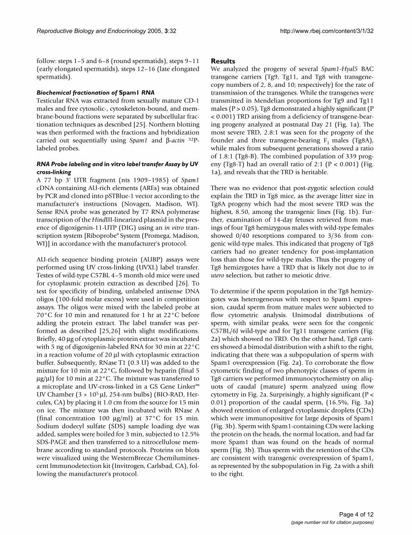
ResultsWe analyzed the progeny of several Spam1-Hyal5 BACtransgene carriers (Tg9, Tg11, and Tg8 with transgene-copy numbers of 2, 8, and 10; respectively) for the rate oftransmission of the transgenes. While the transgenes weretransmitted in Mendelian proportions for Tg9 and Tg11males (P > 0.05), Tg8 demonstrated a highly significant (P< 0.001) TRD arising from a deficiency of transgene-bear-ing progeny analyzed at postnatal Day 21 (Fig. 1a). Themost severe TRD, 2.8:1 was seen for the progeny of thefounder and three transgene-bearing F1 males (Tg8A),while males from subsequent generations showed a ratioof 1.8:1 (Tg8-B). The combined population of 339 prog-eny (Tg8-T) had an overall ratio of 2:1 (P < 0.001) (Fig.1a), and reveals that the TRD is heritable.
There was no evidence that post-zygotic selection couldexplain the TRD in Tg8 mice, as the average litter size inTg8A progeny which had the most severe TRD was thehighest, 8.50, among the transgenic lines (Fig. 1b). Fur-ther, examination of 14-day fetuses retrieved from mat-ings of four Tg8 hemizygous males with wild-type femalesshowed 0/40 resorptions compared to 3/36 from con-genic wild-type males. This indicated that progeny of Tg8carriers had no greater tendency for post-implantationloss than those for wild-type males. Thus the progeny ofTg8 hemizygotes have a TRD that is likely not due to inutero selection, but rather to meiotic drive.
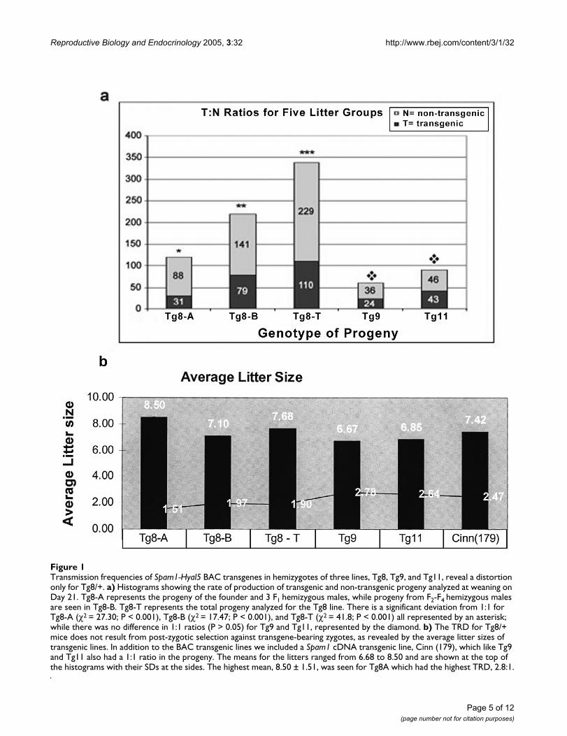
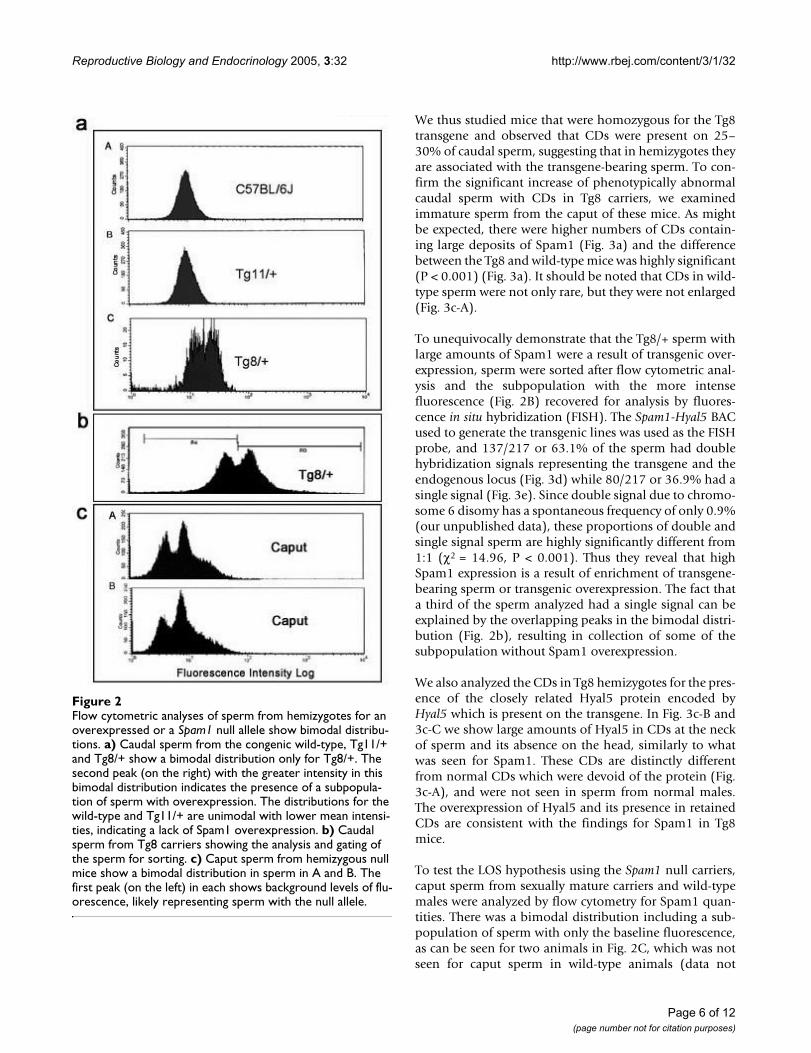
To determine if the sperm population in the Tg8 hemizy-gotes was heterogeneous with respect to Spam1 expres-sion, caudal sperm from mature males were subjected toflow cytometric analysis. Unimodal distributions ofsperm, with similar peaks, were seen for the congenicC57BL/6J wild-type and for Tg11 transgene carriers (Fig.2a) which showed no TRD. On the other hand, Tg8 carri-ers showed a bimodal distribution with a shift to the right,indicating that there was a subpopulation of sperm withSpam1 overexpression (Fig. 2a). To corroborate the flowcytometric finding of two phenotypic classes of sperm inTg8 carriers we performed immunocytochemistry on aliq-uots of caudal (mature) sperm analyzed using flowcytometry in Fig. 2a. Surprisingly, a highly significant (P <0.01) proportion of the caudal sperm, (16.5%, Fig. 3a)showed retention of enlarged cytoplasmic droplets (CDs)which were immunopositive for large deposits of Spam1(Fig. 3b). Sperm with Spam1-containing CDs were lackingthe protein on the heads, the normal location, and had farmore Spam1 than was found on the heads of normalsperm (Fig. 3b). Thus sperm with the retention of the CDsare consistent with transgenic overexpression of Spam1,as represented by the subpopulation in Fig. 2a with a shiftto the right.
Page 4 of 12(page number not for citation purposes)
Reproductive Biology and Endocrinology 2005, 3:32 http://www.rbej.com/content/3/1/32
Transmission frequencies of Spam1-Hyal5 BAC transgenes in hemizygotes of three lines, Tg8, Tg9, and Tg11, reveal a distortion only for Tg8/+Figure 1Transmission frequencies of Spam1-Hyal5 BAC transgenes in hemizygotes of three lines, Tg8, Tg9, and Tg11, reveal a distortion only for Tg8/+. a) Histograms showing the rate of production of transgenic and non-transgenic progeny analyzed at weaning on Day 21. Tg8-A represents the progeny of the founder and 3 F1 hemizygous males, while progeny from F2-F4 hemizygous males are seen in Tg8-B. Tg8-T represents the total progeny analyzed for the Tg8 line. There is a significant deviation from 1:1 for Tg8-A (χ2 = 27.30; P < 0.001), Tg8-B (χ2 = 17.47; P < 0.001), and Tg8-T (χ2 = 41.8; P < 0.001) all represented by an asterisk; while there was no difference in 1:1 ratios (P > 0.05) for Tg9 and Tg11, represented by the diamond. b) The TRD for Tg8/+ mice does not result from post-zygotic selection against transgene-bearing zygotes, as revealed by the average litter sizes of transgenic lines. In addition to the BAC transgenic lines we included a Spam1 cDNA transgenic line, Cinn (179), which like Tg9 and Tg11 also had a 1:1 ratio in the progeny. The means for the litters ranged from 6.68 to 8.50 and are shown at the top of the histograms with their SDs at the sides. The highest mean, 8.50 ± 1.51, was seen for Tg8A which had the highest TRD, 2.8:1.
Page 5 of 12(page number not for citation purposes)
Reproductive Biology and Endocrinology 2005, 3:32 http://www.rbej.com/content/3/1/32
We thus studied mice that were homozygous for the Tg8transgene and observed that CDs were present on 25–30% of caudal sperm, suggesting that in hemizygotes theyare associated with the transgene-bearing sperm. To con-firm the significant increase of phenotypically abnormalcaudal sperm with CDs in Tg8 carriers, we examinedimmature sperm from the caput of these mice. As mightbe expected, there were higher numbers of CDs contain-ing large deposits of Spam1 (Fig. 3a) and the differencebetween the Tg8 and wild-type mice was highly significant(P < 0.001) (Fig. 3a). It should be noted that CDs in wild-type sperm were not only rare, but they were not enlarged(Fig. 3c-A).
To unequivocally demonstrate that the Tg8/+ sperm withlarge amounts of Spam1 were a result of transgenic over-expression, sperm were sorted after flow cytometric anal-ysis and the subpopulation with the more intensefluorescence (Fig. 2B) recovered for analysis by fluores-cence in situ hybridization (FISH). The Spam1-Hyal5 BACused to generate the transgenic lines was used as the FISHprobe, and 137/217 or 63.1% of the sperm had doublehybridization signals representing the transgene and theendogenous locus (Fig. 3d) while 80/217 or 36.9% had asingle signal (Fig. 3e). Since double signal due to chromo-some 6 disomy has a spontaneous frequency of only 0.9%(our unpublished data), these proportions of double andsingle signal sperm are highly significantly different from1:1 (χ2 = 14.96, P < 0.001). Thus they reveal that highSpam1 expression is a result of enrichment of transgene-bearing sperm or transgenic overexpression. The fact thata third of the sperm analyzed had a single signal can beexplained by the overlapping peaks in the bimodal distri-bution (Fig. 2b), resulting in collection of some of thesubpopulation without Spam1 overexpression.
We also analyzed the CDs in Tg8 hemizygotes for the pres-ence of the closely related Hyal5 protein encoded byHyal5 which is present on the transgene. In Fig. 3c-B and3c-C we show large amounts of Hyal5 in CDs at the neckof sperm and its absence on the head, similarly to whatwas seen for Spam1. These CDs are distinctly differentfrom normal CDs which were devoid of the protein (Fig.3c-A), and were not seen in sperm from normal males.The overexpression of Hyal5 and its presence in retainedCDs are consistent with the findings for Spam1 in Tg8mice.
To test the LOS hypothesis using the Spam1 null carriers,caput sperm from sexually mature carriers and wild-typemales were analyzed by flow cytometry for Spam1 quan-tities. There was a bimodal distribution including a sub-population of sperm with only the baseline fluorescence,as can be seen for two animals in Fig. 2C, which was notseen for caput sperm in wild-type animals (data not
Flow cytometric analyses of sperm from hemizygotes for an overexpressed or a Spam1 null allele show bimodal distributionsFigure 2Flow cytometric analyses of sperm from hemizygotes for an overexpressed or a Spam1 null allele show bimodal distribu-tions. a) Caudal sperm from the congenic wild-type, Tg11/+ and Tg8/+ show a bimodal distribution only for Tg8/+. The second peak (on the right) with the greater intensity in this bimodal distribution indicates the presence of a subpopula-tion of sperm with overexpression. The distributions for the wild-type and Tg11/+ are unimodal with lower mean intensi-ties, indicating a lack of Spam1 overexpression. b) Caudal sperm from Tg8 carriers showing the analysis and gating of the sperm for sorting. c) Caput sperm from hemizygous null mice show a bimodal distribution in sperm in A and B. The first peak (on the left) in each shows background levels of flu-orescence, likely representing sperm with the null allele.
Page 6 of 12(page number not for citation purposes)
Reproductive Biology and Endocrinology 2005, 3:32 http://www.rbej.com/content/3/1/32
Immunocytochemistry demonstrates that Tg8/+ males have significantly increased numbers of sperm retaining enlarged cyto-plasmic droplets (CDs) with overexpressed Spam1 or Hyal5, and a concomitant absence of the proteins on the headsFigure 3Immunocytochemistry demonstrates that Tg8/+ males have significantly increased numbers of sperm retaining enlarged cyto-plasmic droplets (CDs) with overexpressed Spam1 or Hyal5, and a concomitant absence of the proteins on the heads. a) His-tograms showing the proportion of sperm, from populations of 200, with CDs in wild-type and Tg8/+ males. The greater than 10-fold increase in caudal sperm compared to wild-type is highly significant (χ2 = 10.8; P < 0.01), as is the >30-fold increase in caput sperm (χ2 = 28.5; P < 0.001). b) Retention of CDs with overexpressed Spam1 (green staining) in sperm taken from an aliquot used for flow cytometry in Fig. 2a. A sperm with the normal amount and normal location of Spam1 is shown for com-parison with the overexpressed protein in the CD. Note that there is non-specific background staining on the tails. c) Sperm with overexpressed Hyal5 (green staining) in enlarged CDs near the neck and the absence of the protein on the heads are seen in B and C, while A shows a normal CD without Hyal5. FISH signals on flow sorted sperm showing double signals in transgenic cells d) and a single signal in wild-type cells e).
Page 7 of 12(page number not for citation purposes)
Reproductive Biology and Endocrinology 2005, 3:32 http://www.rbej.com/content/3/1/32
shown). Note that in Fig. 2C the position of the two peaksin A and B, are consistent with the presence of sperm car-rying the null allele (background or baseline fluores-cence) and those with the normal allele.
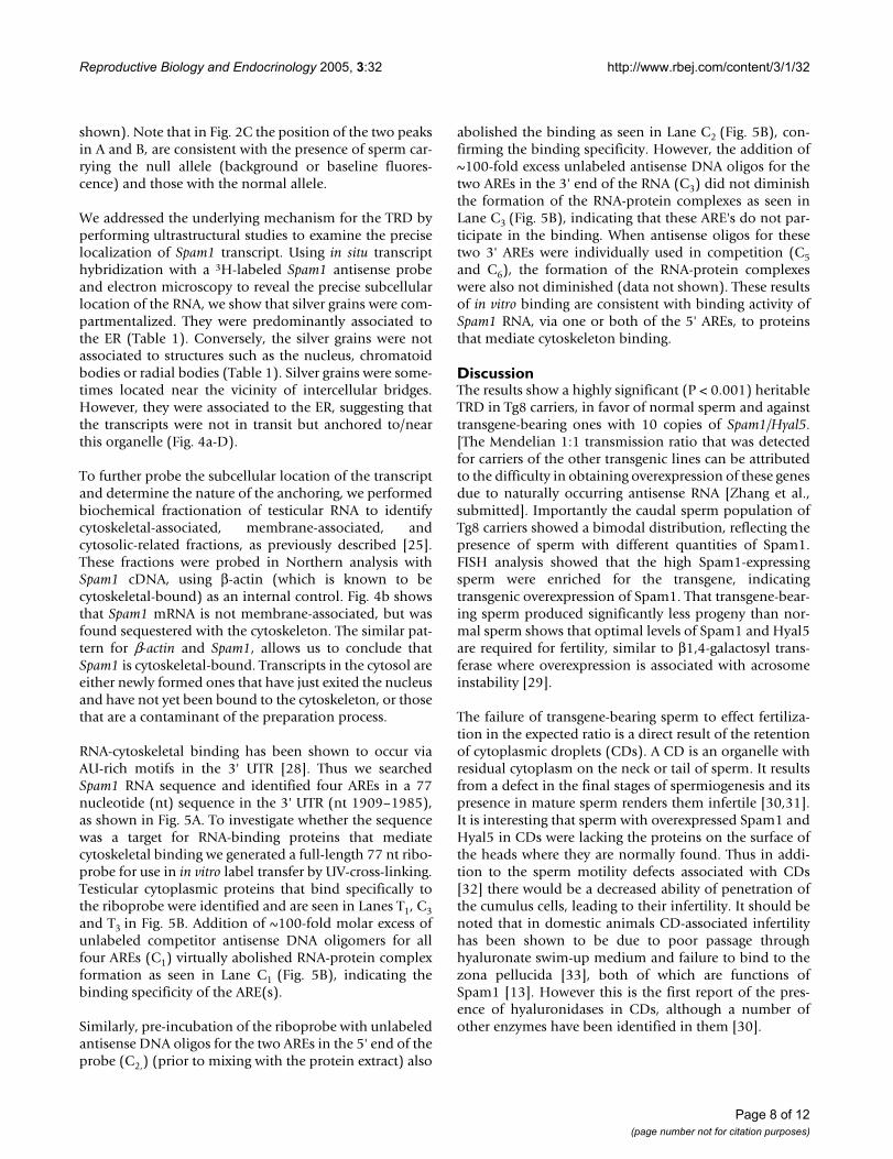
We addressed the underlying mechanism for the TRD byperforming ultrastructural studies to examine the preciselocalization of Spam1 transcript. Using in situ transcripthybridization with a 3H-labeled Spam1 antisense probeand electron microscopy to reveal the precise subcellularlocation of the RNA, we show that silver grains were com-partmentalized. They were predominantly associated tothe ER (Table 1). Conversely, the silver grains were notassociated to structures such as the nucleus, chromatoidbodies or radial bodies (Table 1). Silver grains were some-times located near the vicinity of intercellular bridges.However, they were associated to the ER, suggesting thatthe transcripts were not in transit but anchored to/nearthis organelle (Fig. 4a-D).
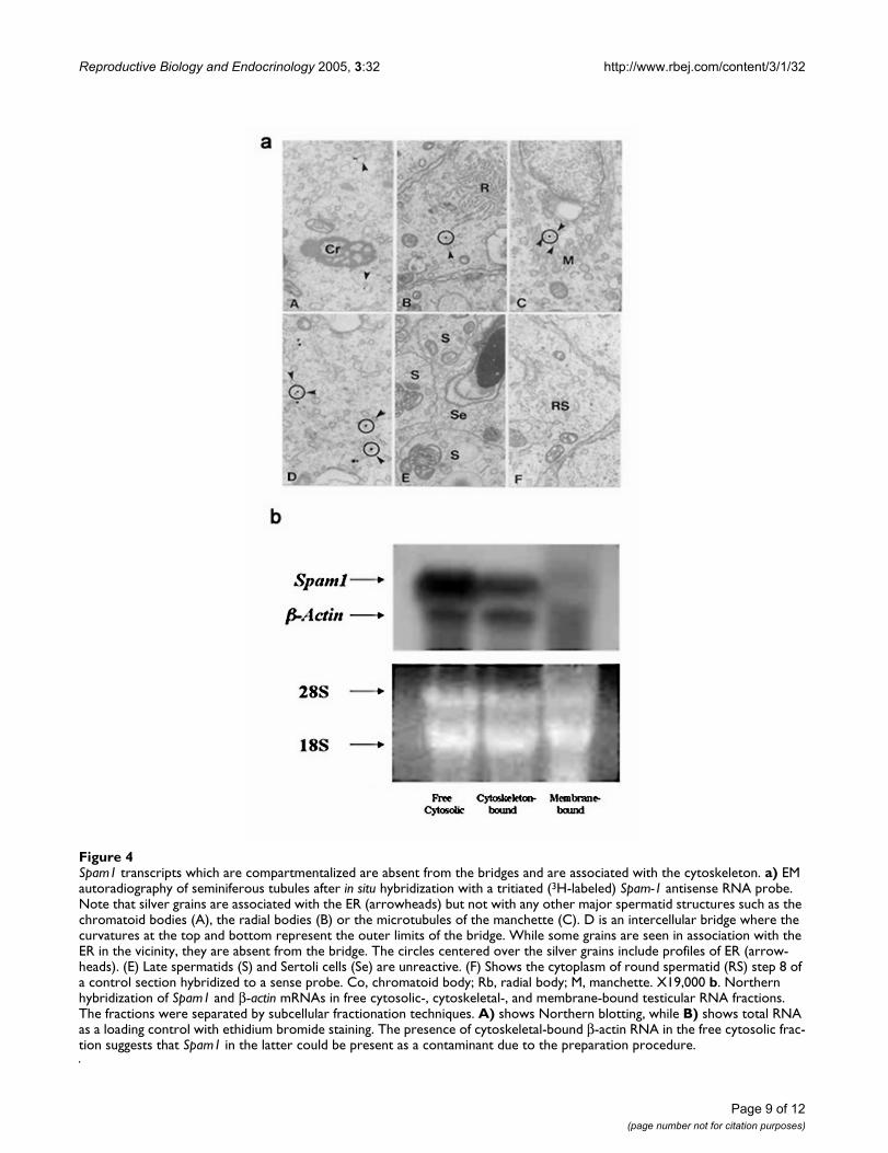
To further probe the subcellular location of the transcriptand determine the nature of the anchoring, we performedbiochemical fractionation of testicular RNA to identifycytoskeletal-associated, membrane-associated, andcytosolic-related fractions, as previously described [25].These fractions were probed in Northern analysis withSpam1 cDNA, using β-actin (which is known to becytoskeletal-bound) as an internal control. Fig. 4b showsthat Spam1 mRNA is not membrane-associated, but wasfound sequestered with the cytoskeleton. The similar pat-tern for β-actin and Spam1, allows us to conclude thatSpam1 is cytoskeletal-bound. Transcripts in the cytosol areeither newly formed ones that have just exited the nucleusand have not yet been bound to the cytoskeleton, or thosethat are a contaminant of the preparation process.
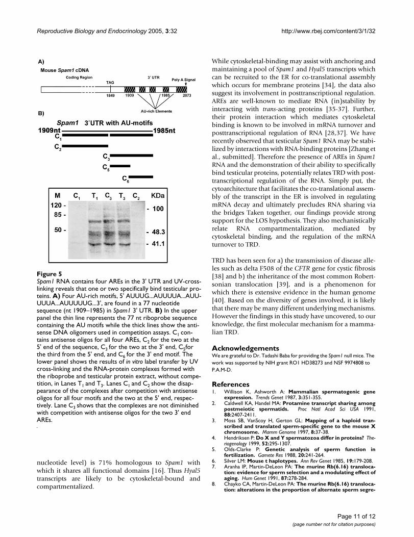
RNA-cytoskeletal binding has been shown to occur viaAU-rich motifs in the 3' UTR [28]. Thus we searchedSpam1 RNA sequence and identified four AREs in a 77nucleotide (nt) sequence in the 3' UTR (nt 1909–1985),as shown in Fig. 5A. To investigate whether the sequencewas a target for RNA-binding proteins that mediatecytoskeletal binding we generated a full-length 77 nt ribo-probe for use in in vitro label transfer by UV-cross-linking.Testicular cytoplasmic proteins that bind specifically tothe riboprobe were identified and are seen in Lanes T1, C3and T3 in Fig. 5B. Addition of ~100-fold molar excess ofunlabeled competitor antisense DNA oligomers for allfour AREs (C1) virtually abolished RNA-protein complexformation as seen in Lane C1 (Fig. 5B), indicating thebinding specificity of the ARE(s).
Similarly, pre-incubation of the riboprobe with unlabeledantisense DNA oligos for the two AREs in the 5' end of theprobe (C2,) (prior to mixing with the protein extract) also
abolished the binding as seen in Lane C2 (Fig. 5B), con-firming the binding specificity. However, the addition of~100-fold excess unlabeled antisense DNA oligos for thetwo AREs in the 3' end of the RNA (C3) did not diminishthe formation of the RNA-protein complexes as seen inLane C3 (Fig. 5B), indicating that these ARE's do not par-ticipate in the binding. When antisense oligos for thesetwo 3' AREs were individually used in competition (C5and C6), the formation of the RNA-protein complexeswere also not diminished (data not shown). These resultsof in vitro binding are consistent with binding activity ofSpam1 RNA, via one or both of the 5' AREs, to proteinsthat mediate cytoskeleton binding.
DiscussionThe results show a highly significant (P < 0.001) heritableTRD in Tg8 carriers, in favor of normal sperm and againsttransgene-bearing ones with 10 copies of Spam1/Hyal5.[The Mendelian 1:1 transmission ratio that was detectedfor carriers of the other transgenic lines can be attributedto the difficulty in obtaining overexpression of these genesdue to naturally occurring antisense RNA [Zhang et al.,submitted]. Importantly the caudal sperm population ofTg8 carriers showed a bimodal distribution, reflecting thepresence of sperm with different quantities of Spam1.FISH analysis showed that the high Spam1-expressingsperm were enriched for the transgene, indicatingtransgenic overexpression of Spam1. That transgene-bear-ing sperm produced significantly less progeny than nor-mal sperm shows that optimal levels of Spam1 and Hyal5are required for fertility, similar to β1,4-galactosyl trans-ferase where overexpression is associated with acrosomeinstability [29].
The failure of transgene-bearing sperm to effect fertiliza-tion in the expected ratio is a direct result of the retentionof cytoplasmic droplets (CDs). A CD is an organelle withresidual cytoplasm on the neck or tail of sperm. It resultsfrom a defect in the final stages of spermiogenesis and itspresence in mature sperm renders them infertile [30,31].It is interesting that sperm with overexpressed Spam1 andHyal5 in CDs were lacking the proteins on the surface ofthe heads where they are normally found. Thus in addi-tion to the sperm motility defects associated with CDs[32] there would be a decreased ability of penetration ofthe cumulus cells, leading to their infertility. It should benoted that in domestic animals CD-associated infertilityhas been shown to be due to poor passage throughhyaluronate swim-up medium and failure to bind to thezona pellucida [33], both of which are functions ofSpam1 [13]. However this is the first report of the pres-ence of hyaluronidases in CDs, although a number ofother enzymes have been identified in them [30].
Page 8 of 12(page number not for citation purposes)
Reproductive Biology and Endocrinology 2005, 3:32 http://www.rbej.com/content/3/1/32
Spam1 transcripts which are compartmentalized are absent from the bridges and are associated with the cytoskeletonFigure 4Spam1 transcripts which are compartmentalized are absent from the bridges and are associated with the cytoskeleton. a) EM autoradiography of seminiferous tubules after in situ hybridization with a tritiated (3H-labeled) Spam-1 antisense RNA probe. Note that silver grains are associated with the ER (arrowheads) but not with any other major spermatid structures such as the chromatoid bodies (A), the radial bodies (B) or the microtubules of the manchette (C). D is an intercellular bridge where the curvatures at the top and bottom represent the outer limits of the bridge. While some grains are seen in association with the ER in the vicinity, they are absent from the bridge. The circles centered over the silver grains include profiles of ER (arrow-heads). (E) Late spermatids (S) and Sertoli cells (Se) are unreactive. (F) Shows the cytoplasm of round spermatid (RS) step 8 of a control section hybridized to a sense probe. Co, chromatoid body; Rb, radial body; M, manchette. X19,000 b. Northern hybridization of Spam1 and β-actin mRNAs in free cytosolic-, cytoskeletal-, and membrane-bound testicular RNA fractions. The fractions were separated by subcellular fractionation techniques. A) shows Northern blotting, while B) shows total RNA as a loading control with ethidium bromide staining. The presence of cytoskeletal-bound β-actin RNA in the free cytosolic frac-tion suggests that Spam1 in the latter could be present as a contaminant due to the preparation procedure.
Page 9 of 12(page number not for citation purposes)
Reproductive Biology and Endocrinology 2005, 3:32 http://www.rbej.com/content/3/1/32
The fact that the number of sperm with the enlarged CDphenotype is less than 50% and 100% in Tg8 hemizygotesand homozygotes, respectively, may be due to technicalfactors such as their loss during preparation [30]. It is alsolikely that the highly regulated Spam1 silencing which ismediated by antisense transcription [Zhang et al., submit-ted] could be responsible. This silencing would be adap-tive since overexpression of Spam1 and Hyal5 leads totheir mis-expression in CDs, which are abnormallyretained and which lead to infertility. Thus in Tg8 carriersthere are functionally different sperm within a male, dueto their different quantities of Spam1 and Hyal5. It is notknown if Spam1 and Hyal5 are overexpressed in the sameCDs, consequently their co-localization will be investi-gated in future studies.
The finding of structurally and functionally differentsperm in Tg8 transgene carriers is reminiscent of the find-ings for heterozygotes of spontaneous mutant alleles ofSpam1 and is also consistent with compartmentalizationof the RNA and protein [10]. The accumulation of theoverexpressed Spam1 and Hyal5 protein in the CDs oftrangene-bearing sperm is a result of a lack of transcriptsharing between these sperm and those with the normalalleles. These observations on the transgenic modeltherefore provide support for the LOS Hypothesis in theetiology of TRDs.
Similarly, the LOS hypothesis is also supported by thefindings from carriers of Spam1 null allele. Generated byinsertion of a neo cassette in exon II (which contains thehyaluronidase domain) of Spam1 in the laboratoryTadashi Baba [17], null mice were shown to be fertile(despite a delay in cumulus penetration) due to the com-pensating effect of the redundant Hyal5 [17]. Carriers ofSpam1 null showed a bimodal distribution of Spam1 incaput sperm, with one subpopulation having backgroundlevels of fluorescence, consistent with the presence ofsperm with a null allele. Since Spam1 is expressed in theepididymis where it may be acquired by sperm during
transit [18,19] caput, but not cauda, sperm would reflectthe spermatid phenotype, and the finding from thesesperm is supportive of a lack of transcript sharing.
To determine the mechanism for the lack of transcriptsharing we focused on RNA compartmentalization. It hasbeen proposed that mRNA localization facilitates proteinsorting and that nascent polypeptide chain targeting ofmembrane proteins is a major mechanism that accountsfor mRNA localization [34]. We show that 85% of thegrains from in situ transcript hybridization localized to theER and the remaining 15% could be assigned to thisregion. This indicates that the transcripts are not dispersedin the cytoplasm where they would gain ready access tothe bridges. Table 1 shows that they are not associatedwith any major spermatid structure, such as the chroma-toid bodies (which indicates that the RNA is not storedand is not translationally regulated), the radial bodies, orthe microtubules of the manchette (Fig. 4a). Importantly,they are absent from the bridges although they may be inthe vicinity. In this connection it should be pointed outthat the absence of the transcript from the chromatoidbodies which have recently been seen to cross the bridges(Parvinen and Sassone-Corsi, personal communication),bolsters the evidence for the RNA compartmentalization.
The compartmentalization of Spam1 transcripts and theirabsence from the bridges suggest that they are anchored,and this would preclude sharing and support our LOShypothesis. Based on the restricted location of the tran-script at the ER, it was expected that Northern analysiswould reveal its association with the membrane-boundfraction. However, this was not the case as Spam1 RNAwas shown to be associated with the cytoskeletal fractionand therefore anchored. More importantly, we show thatthe transcript has AU-rich elements (AREs) in the 3' UTRthat are known to bind cytoplasmic proteins (AUBPs) thatmediate binding to the cytoskeleton. Interestingly, AREsare also present in the 3' UTR of the rodent-specific Hyal5(Genbank Accession# ABO85680) which (at the
Table 1: Distribution of radioautographic silver grains in spermatids expressed as percentages (±SD)
Steps 3–5 Steps 6–8 Steps 9–11
ER 85 ± 5 85 ± 2 87 ± 1Assigned to ER 13 ± 3 14 ± 6 13 ± 1Nucleus 0 ± 0 0 ± 0 0 ± 0Chromatoid Ba 0 ± 0 0 ± 0 0 ± 0Radial Bodies 0 ± 0 0 ± 0 0 ± 0Cytoplasmic Bb 0 ± 0 0 ± 0 0 ± 0Unknown origin 2 ± 0 1 ± 0 0 ± 0
a Chromatoid bodies, b Cytoplasmic bridges. Some of the data in this table are taken from Molecular Reproduction & Development © copyright 2004 Wiley-Liss, Inc, A Wiley Company.
Page 10 of 12(page number not for citation purposes)
Reproductive Biology and Endocrinology 2005, 3:32 http://www.rbej.com/content/3/1/32
nucleotide level) is 71% homologous to Spam1 withwhich it shares all functional domains [16]. Thus Hyal5transcripts are likely to be cytoskeletal-bound andcompartmentalized.
While cytoskeletal-binding may assist with anchoring andmaintaining a pool of Spam1 and Hyal5 transcripts whichcan be recruited to the ER for co-translational assemblywhich occurs for membrane proteins [34], the data alsosuggest its involvement in posttranscriptional regulation.AREs are well-known to mediate RNA (in)stability byinteracting with trans-acting proteins [35-37]. Further,their protein interaction which mediates cytoskeletalbinding is known to be involved in mRNA turnover andposttranscriptional regulation of RNA [28,37]. We haverecently observed that testicular Spam1 RNA may be stabi-lized by interactions with RNA-binding proteins [Zhang etal., submitted]. Therefore the presence of AREs in Spam1RNA and the demonstration of their ability to specificallybind testicular proteins, potentially relates TRD with post-transcriptional regulation of the RNA. Simply put, thecytoarchitecture that facilitates the co-translational assem-bly of the transcript in the ER is involved in regulatingmRNA decay and ultimately precludes RNA sharing viathe bridges Taken together, our findings provide strongsupport for the LOS hypothesis. They also mechanisticallyrelate RNA compartmentalization, mediated bycytoskeletal binding, and the regulation of the mRNAturnover to TRD.
TRD has been seen for a) the transmission of disease alle-les such as delta F508 of the CFTR gene for cystic fibrosis[38] and b) the inheritance of the most common Robert-sonian translocation [39], and is a phenomenon forwhich there is extensive evidence in the human genome[40]. Based on the diversity of genes involved, it is likelythat there may be many different underlying mechanisms.However the findings in this study have uncovered, to ourknowledge, the first molecular mechanism for a mamma-lian TRD.
AcknowledgementsWe are grateful to Dr. Tadashi Baba for providing the Spam1 null mice. The work was supported by NIH grant RO1 HD38273 and NSF 9974808 to P.A.M-D.
References1. Willison K, Ashworth A: Mammalian spermatogenic gene
expression. Trends Genet 1987, 3:351-355.2. Caldwell KA, Handel MA: Protamine transcript sharing among
postmeiotic spermatids. Proc Natl Acad Sci USA 1991,88:2407-2411.
3. Moss SB, VanScoy H, Gerton GL: Mapping of a haploid tran-scribed and translated sperm-specific gene to the mouse Xchromosome. Mamm Genome 1997, 8:37-38.
4. Hendriksen P: Do X and Y spermatozoa differ in proteins? The-riogenology 1999, 52:295-1307.
5. Olds-Clarke P: Genetic analysis of sperm function infertilization. Gamete Res 1988, 20:241-264.
6. Silver LM: Mouse t haplotypes. Ann Rev Genet 1985, 19:179-208.7. Aranha IP, Martin-DeLeon PA: The murine Rb(6.16) transloca-
tion: evidence for sperm selection and a modulating effect ofaging. Hum Genet 1991, 87:278-284.
8. Chayko CA, Martin-DeLeon PA: The murine Rb(6.16) transloca-tion: alterations in the proportion of alternate sperm segre-
Spam1 RNA contains four AREs in the 3' UTR and UV-cross-linking reveals that one or two specifically bind testicular pro-teinsFigure 5Spam1 RNA contains four AREs in the 3' UTR and UV-cross-linking reveals that one or two specifically bind testicular pro-teins. A) Four AU-rich motifs, 5' AUUUG...AUUUUA...AUU-UUUA...AUUUUUG...3', are found in a 77 nucleotide sequence (nt 1909–1985) in Spam1 3' UTR. B) In the upper panel the thin line represents the 77 nt riboprobe sequence containing the AU motifs while the thick lines show the anti-sense DNA oligomers used in competition assays. C1 con-tains antisense oligos for all four AREs, C2 for the two at the 5' end of the sequence, C3 for the two at the 3' end, C5for the third from the 5' end, and C6 for the 3' end motif. The lower panel shows the results of in vitro label transfer by UV cross-linking and the RNA-protein complexes formed with the riboprobe and testicular protein extract, without compe-tition, in Lanes T1 and T3. Lanes C1 and C2 show the disap-pearance of the complexes after competition with antisense oligos for all four motifs and the two at the 5' end, respec-tively. Lane C3 shows that the complexes are not diminished with competition with antisense oligos for the two 3' end AREs.
Page 11 of 12(page number not for citation purposes)
http://www.ncbi.nlm.nih.gov/entrez/query.fcgi?cmd=Retrieve&db=PubMed&dopt=Abstract&list_uids=2006178
http://www.ncbi.nlm.nih.gov/entrez/query.fcgi?cmd=Retrieve&db=PubMed&dopt=Abstract&list_uids=2006178
http://www.ncbi.nlm.nih.gov/entrez/query.fcgi?cmd=Retrieve&db=PubMed&dopt=Abstract&list_uids=9021145
http://www.ncbi.nlm.nih.gov/entrez/query.fcgi?cmd=Retrieve&db=PubMed&dopt=Abstract&list_uids=9021145
http://www.ncbi.nlm.nih.gov/entrez/query.fcgi?cmd=Retrieve&db=PubMed&dopt=Abstract&list_uids=9021145
http://www.ncbi.nlm.nih.gov/entrez/query.fcgi?cmd=Retrieve&db=PubMed&dopt=Abstract&list_uids=3069685
http://www.ncbi.nlm.nih.gov/entrez/query.fcgi?cmd=Retrieve&db=PubMed&dopt=Abstract&list_uids=3069685
http://www.ncbi.nlm.nih.gov/entrez/query.fcgi?cmd=Retrieve&db=PubMed&dopt=Abstract&list_uids=3909941
http://www.ncbi.nlm.nih.gov/entrez/query.fcgi?cmd=Retrieve&db=PubMed&dopt=Abstract&list_uids=1864601
http://www.ncbi.nlm.nih.gov/entrez/query.fcgi?cmd=Retrieve&db=PubMed&dopt=Abstract&list_uids=1864601
http://www.ncbi.nlm.nih.gov/entrez/query.fcgi?cmd=Retrieve&db=PubMed&dopt=Abstract&list_uids=1864601
Reproductive Biology and Endocrinology 2005, 3:32 http://www.rbej.com/content/3/1/32
Publish with BioMed Central and every scientist can read your work free of charge
"BioMed Central will be the most significant development for disseminating the results of biomedical research in our lifetime."
Sir Paul Nurse, Cancer Research UK
Your research papers will be:
available free of charge to the entire biomedical community
peer reviewed and published immediately upon acceptance
cited in PubMed and archived on PubMed Central
yours — you keep the copyright
Submit your manuscript here:http://www.biomedcentral.com/info/publishing_adv.asp
BioMedcentral
gants effecting fertilization in vitro and in vivo. Hum Genet1992, 90:79-85.
9. Aranha IP, Martin-DeLeon PA: Mouse chromosome 6 in Rbtranslocations: consequences in singly and doubly hetero-zygous males. Cytogenet Cell Genet 1995, 69:253-259.
10. Zheng Y, Deng X, Martin-DeLeon PA: Lack of sharing of Spam1(Ph-20) among mouse spermatids and transmission ratiodistortion. Biol Reprod 2001, 64:1730-1738.
11. Zheng Y, Deng X, Zhao Y, Zhang H, Martin-DeLeon PA: Spam1(PH-20) mutations and sperm dysfunction in mice with theRb(6.16) or Rb(6.15) translocation. Mamm Genome 2001,12:822-829.
12. Lathrop WF, Carmichael EP, Myles DG, Primakof P: cDNA cloningreveals the molecular structure of a sperm surface protein,PH-20, involved in sperm-egg adhesion and the wide distri-bution of its gene among mammals. J Cell Biol 1990,111:2939-2949.
13. Cherr GN, Yudin AI, Overstreet JW: The dual functions of GPI-anchored PH-20: hyaluronidase and intracellular signaling.Matrix Biol 2001, 20:515-525.
14. Deng X, Moran J, Copeland NG, Gilbert DJ, Jenkins NA, Primakoff P,Martin-DeLeon PA: The mouse Spam1 maps to proximal chro-mosome 6 and is a candidate for the sperm dysfunction inRb(6.16)24Lub and Rb(6.15)1Ald heterozygotes. MammGenome 1997, 8:94-97.
15. Csoka AB, Frost GI, Stern R: The six hyaluronidase-like genes inthe human and mouse genomes. Matrix Biol 2001, 20:499-508.
16. Zhang H, Shertok S, Miller K, Martin-DeLeon PA: Sperm Dysfunc-tion in the Rb(6.16)- and Rb(6.15)-bearing Mice Revisited:Involvement of Hyalp1 and Hyal5. Mol Reprod Dev 2005. epub-lished Aug 4, DOI 10. 1002/mrd.20360
17. Baba D, Kashiwabara S, Honda A, Yamagata K, Wu Q, Ikawa M,Okabe M, Baba T: Mouse sperm lacking cell surface hyaluroni-dase PH-20 can pass through the layer of cumulus cells andfertilize the egg. J Biol Chem 2002, 277:30310-30314.
18. Zhang H, Martin-DeLeon PA: Mouse epididymal Spam1 (pH-20)is released in the luminal fluid with its lipid anchor. J Androl2003, 24:51-58.
19. Zhang H, Morales CR, Badran H, El-Alfy M, Martin-DeLeon PA:Spam1 (PH-20) Expression in the Extratesticular Duct andAccessory Organs of the mouse: A Possible Role in SpermFluid Reabsorption. Biol Reprod 2004, 71:1101-1107.
20. Morales CR, Badran H, El-Alfy M, Men H, Zhang H, Martin-DeLeonPA: Cytoplasmic localization during testicular biogenesis ofthe murine mRNA for Spam1 (PH-20), a protein involved inacrosomal exocytosis. Mol Reprod Dev 2004, 69:475-482.
21. Kopriwa B: A reliable standardized method for ultrastructuralelectron microscope radioautography. Histochemistry 1973,37:1-17.
22. Kopriwa B: A comparison of various procedures for fine graindevelopment in electron radioautographiy. Histochemistry1975, 44:201-224.
23. Nadler NJ: The interpretation of the grain counts in electronmicroscope radioautography. J Cell Biol 1971, 49:877-882.
24. Haddad A, Smith MD, Herscovics A, Nadler NJ, Leblond CP: Radio-autographic study of in vivo and in vitro incorporation offucose-3H into thyroglobulin by rat thyroid follicular cells. JCell Biol 1971, 49:856-882.
25. Hesketh JE, Campbell GP, Whitelaw PF: c-myc mRNA in cytoskel-etal-bound polysomes in fibroblasts. Biochem J 1991,274:607-609.
26. Thomson AM, Rogers JT, Walker CE, Staton JM, Leedman PJ: Opti-mized RNA gel-shift and UV cross-linking assays for charac-terization of cytoplasmic RNA-protein interactions.Biotechniques 1999, 25:1032-1042.
27. Balmer LA, Beveridge DJ, Jazayeri JA, Thoson AM, Walker CE, Leed-man PJ: Identification of a novel AU-Rich element in the 3'Untranslated region of epidermal growth factor receptormRNA that is the target for regulated RNA-bindingproteins. Mol Cellular Biol 2001, 21:2070-2084.
28. Henics T, Nagy E, Szekeres-Bartho J: Interaction of AU-richsequence binding proteins with actin: possible involvementof the actin cytoskeleton in lymphokine mRNA turnover. JCell Physiol 1997, 173:19-27.
29. Shur BD, Hall NG: A role for mouse sperm surface galactosyl-transferase in sperm binding to the egg zona pellucida. J CellBiol 1982, 95:574-579.
30. Oko R, Hermo L, Chan PT, Fazel A, Bergeron JJ: The cytoplasmicdroplet of rat epididymal spermatozoa contains saccular ele-ments with Golgi characteristics. J Cell Biol 1993, 123:809-821.
31. Cooper TG: Cytoplasmic droplets: the good, the bad or justconfusing? Hum Reprod 2005, 20:9-11.
32. Cooper TG, Yeung C-H, Fetic S, Sobhani A, Nieschlag E: Cytoplas-mic droplets are normal structures of human sperm but arenot well preserved by routine procedures for assessingsperm morphology. Hum Reprod 2004, 19:2283-2288.
33. Thundathil J, Palasz AT, Barth AD, Mapletoft RJ: The use of in vitrofertilization techniques to investigate the fertilizing ability ofbovine sperm with proximal cytoplasmic droplets. AnimReprod Sci 2001, 65:181-192.
34. Lawrence JB, Singer RH: Intracellular localization of messengerRNAs for cytoskeletal proteins. Cell 1986, 45:407-415.
35. Tourriere H, Chebil K, Tazi J: mRNA degradation machines ineukaryotic cells. Biochimie 2002, 84:821-837.
36. Chen C-YA, Xu N, Shyu A-B: mRNA decay mediated by two dis-tinct AU-rich elements from c-fos and granulocyte-macro-phage colony-stimulating factor transcripts: differentdeadenylation kinetics and uncoupling from translation. MolCell Biol 1995, 15:5777-5788.
37. Bevilacqua Bevilacqua A, Ceriani M, Capaccioli S, Nicolin A: Post-Transcriptional Regulation of Gene Expression by Degrada-tion of Messenger RNAs. J Cell Physiol 2003, 195:356-372.
38. Kitzis A, Chomel JC, Haliassos A, Tesson L, Kaplan JC, Feingold J,Giraud G, Lable A, Dastugue B, Dumur V: Unusual segregation ofcystic fibrosis alleles. Nature 1988, 366:316.
39. Pardo Manuel de Villena F, Sapienza C: Transmission ratio distor-tion in offspring of heterozygous female carriers of Robert-sonian translocations. Hum Genet 2001, 108:31-36.
40. Zollner S, Wen X, Hanchard NA, Herbert MA, Ober C, Pritchard JK:Evidence for extensive transmission distortion in the humangenome. Am J Hum Gen 2004, 74:62-72.
Page 12 of 12(page number not for citation purposes)
http://www.ncbi.nlm.nih.gov/entrez/query.fcgi?cmd=Retrieve&db=PubMed&dopt=Abstract&list_uids=1427792
http://www.ncbi.nlm.nih.gov/entrez/query.fcgi?cmd=Retrieve&db=PubMed&dopt=Abstract&list_uids=7698024
http://www.ncbi.nlm.nih.gov/entrez/query.fcgi?cmd=Retrieve&db=PubMed&dopt=Abstract&list_uids=7698024
http://www.ncbi.nlm.nih.gov/entrez/query.fcgi?cmd=Retrieve&db=PubMed&dopt=Abstract&list_uids=7698024
http://www.ncbi.nlm.nih.gov/entrez/query.fcgi?cmd=Retrieve&db=PubMed&dopt=Abstract&list_uids=2269661
http://www.ncbi.nlm.nih.gov/entrez/query.fcgi?cmd=Retrieve&db=PubMed&dopt=Abstract&list_uids=2269661
http://www.ncbi.nlm.nih.gov/entrez/query.fcgi?cmd=Retrieve&db=PubMed&dopt=Abstract&list_uids=2269661
http://www.ncbi.nlm.nih.gov/entrez/query.fcgi?cmd=Retrieve&db=PubMed&dopt=Abstract&list_uids=9060406
http://www.ncbi.nlm.nih.gov/entrez/query.fcgi?cmd=Retrieve&db=PubMed&dopt=Abstract&list_uids=9060406
http://www.ncbi.nlm.nih.gov/entrez/query.fcgi?cmd=Retrieve&db=PubMed&dopt=Abstract&list_uids=9060406
http://www.ncbi.nlm.nih.gov/entrez/query.fcgi?cmd=Retrieve&db=PubMed&dopt=Abstract&list_uids=4129434
http://www.ncbi.nlm.nih.gov/entrez/query.fcgi?cmd=Retrieve&db=PubMed&dopt=Abstract&list_uids=4129434
http://www.ncbi.nlm.nih.gov/entrez/query.fcgi?cmd=Retrieve&db=PubMed&dopt=Abstract&list_uids=1184416
http://www.ncbi.nlm.nih.gov/entrez/query.fcgi?cmd=Retrieve&db=PubMed&dopt=Abstract&list_uids=1184416
http://www.ncbi.nlm.nih.gov/entrez/query.fcgi?cmd=Retrieve&db=PubMed&dopt=Abstract&list_uids=5092210
http://www.ncbi.nlm.nih.gov/entrez/query.fcgi?cmd=Retrieve&db=PubMed&dopt=Abstract&list_uids=5092210
http://www.ncbi.nlm.nih.gov/entrez/query.fcgi?cmd=Retrieve&db=PubMed&dopt=Abstract&list_uids=4326459
http://www.ncbi.nlm.nih.gov/entrez/query.fcgi?cmd=Retrieve&db=PubMed&dopt=Abstract&list_uids=2006923
http://www.ncbi.nlm.nih.gov/entrez/query.fcgi?cmd=Retrieve&db=PubMed&dopt=Abstract&list_uids=2006923
http://www.ncbi.nlm.nih.gov/entrez/query.fcgi?cmd=Retrieve&db=PubMed&dopt=Abstract&list_uids=9326445
http://www.ncbi.nlm.nih.gov/entrez/query.fcgi?cmd=Retrieve&db=PubMed&dopt=Abstract&list_uids=9326445
http://www.ncbi.nlm.nih.gov/entrez/query.fcgi?cmd=Retrieve&db=PubMed&dopt=Abstract&list_uids=9326445
http://www.ncbi.nlm.nih.gov/entrez/query.fcgi?cmd=Retrieve&db=PubMed&dopt=Abstract&list_uids=6815212
http://www.ncbi.nlm.nih.gov/entrez/query.fcgi?cmd=Retrieve&db=PubMed&dopt=Abstract&list_uids=6815212
http://www.ncbi.nlm.nih.gov/entrez/query.fcgi?cmd=Retrieve&db=PubMed&dopt=Abstract&list_uids=8227142
http://www.ncbi.nlm.nih.gov/entrez/query.fcgi?cmd=Retrieve&db=PubMed&dopt=Abstract&list_uids=8227142
http://www.ncbi.nlm.nih.gov/entrez/query.fcgi?cmd=Retrieve&db=PubMed&dopt=Abstract&list_uids=8227142
http://www.ncbi.nlm.nih.gov/entrez/query.fcgi?cmd=Retrieve&db=PubMed&dopt=Abstract&list_uids=3698103
http://www.ncbi.nlm.nih.gov/entrez/query.fcgi?cmd=Retrieve&db=PubMed&dopt=Abstract&list_uids=3698103
http://www.ncbi.nlm.nih.gov/entrez/query.fcgi?cmd=Retrieve&db=PubMed&dopt=Abstract&list_uids=7565731
http://www.ncbi.nlm.nih.gov/entrez/query.fcgi?cmd=Retrieve&db=PubMed&dopt=Abstract&list_uids=7565731
Related Documents