SpaK/SpaR Two-component System Characterized by a Structure-driven Domain-fusion Method and in Vitro Phosphorylation Studies Anu Chakicherla 1 , Carol L. Ecale Zhou 1 *, Martha Ligon Dang 2 , Virginia Rodriguez 3 , J. Norman Hansen 4 , Adam Zemla 1 1 Computing Applications and Research Department, Lawrence Livermore National Laboratory, Livermore, California, United States of America, 2 Sacred Hearts Academy, Honolulu, Hawaii, United States of America, 3 Genome Technology Branch, National Human Genome Research Institute, National Institutes of Health, Bethesda, Maryland, United States of America, 4 Department of Chemistry and Biochemistry, University of Maryland, College Park, Maryland, United States of America Abstract Here we introduce a quantitative structure-driven computational domain-fusion method, which we used to predict the structures of proteins believed to be involved in regulation of the subtilin pathway in Bacillus subtilis, and used to predict a protein-protein complex formed by interaction between the proteins. Homology modeling of SpaK and SpaR yielded preliminary structural models based on a best template for SpaK comprising a dimer of a histidine kinase, and for SpaR a response regulator protein. Our LGA code was used to identify multi-domain proteins with structure homology to both modeled structures, yielding a set of domain-fusion templates then used to model a hypothetical SpaK/SpaR complex. The models were used to identify putative functional residues and residues at the protein-protein interface, and bioinformatics was used to compare functionally and structurally relevant residues in corresponding positions among proteins with structural homology to the templates. Models of the complex were evaluated in light of known properties of the functional residues within two-component systems involving His-Asp phosphorelays. Based on this analysis, a phosphotransferase complexed with a beryllofluoride was selected as the optimal template for modeling a SpaK/SpaR complex conformation. In vitro phosphorylation studies performed using wild type and site-directed SpaK mutant proteins validated the predictions derived from application of the structure-driven domain-fusion method: SpaK was phosphorylated in the presence of 32 P- ATP and the phosphate moiety was subsequently transferred to SpaR, supporting the hypothesis that SpaK and SpaR function as sensor and response regulator, respectively, in a two-component signal transduction system, and furthermore suggesting that the structure-driven domain-fusion approach correctly predicted a physical interaction between SpaK and SpaR. Our domain-fusion algorithm leverages quantitative structure information and provides a tool for generation of hypotheses regarding protein function, which can then be tested using empirical methods. Citation: Chakicherla A, Zhou CLE, Dang ML, Rodriguez V, Hansen JN, et al. (2009) SpaK/SpaR Two-component System Characterized by a Structure-driven Domain-fusion Method and in Vitro Phosphorylation Studies. PLoS Comput Biol 5(6): e1000401. doi:10.1371/journal.pcbi.1000401 Editor: Anna R. Panchenko, National Institutes of Health, United States of America Received December 11, 2008; Accepted May 4, 2009; Published June 5, 2009 Copyright: ß 2009 Chakicherla et al. This is an open-access article distributed under the terms of the Creative Commons Attribution License, which permits unrestricted use, distribution, and reproduction in any medium, provided the original author and source are credited. Funding: Prepared by Lawrence Livermore National Laboratory under Contract DE-AC52-07NA27344. The bioinformatics work was supported by an LLNL-LLNS internally funded grant to CZ and AZ through the Laboratory Directed Research and Development program, and the experimental work was supported by grant R01-AI24454-12 to NH from the National Institute of Allergy and Infectious Diseases, NIH. The funders had no role in study design, data collection and analysis, decision to publish, or preparation of the manuscript. Competing Interests: The authors have declared that no competing interests exist. * E-mail: [email protected] Introduction Because proteins so frequently function in coordination with other proteins, identification and characterization of protein- protein complexes are essential aspects of protein sequence annotation and function determination [1]. A variety of empirical [2–4] and computational [5–14] methods for identifying putative protein-protein interactions have been reported. Of particular note is the Rosetta Stone approach for identifying interacting partners based on the theory of gene fusion, whereby protein domains that are encoded separately in one species may be homologous to domains that are ‘‘fused’’ in the same open reading frame in another species [15–17]. Whereas sequence-based domain fusion methods can be highly successful in identifying putative functional relationships among proteins, the reliance on sequence homology limits detection to protein sequences with adequate levels of sequence identity. Another approach to identifying putative protein-protein interactions is described by Lu and coworkers [18], whereby sequence-based searches against the PDB database were performed in order to identify multi- domain structures having at least one domain with good sequence identity to each putative interacting protein. However, the sensitivity of this search method is also dependent on the levels of sequence identity between the proteins of interest and the sequences of the domains within the identified PDB domain-fusion template. Kundrotas and Alexov [6] explored the use of structure- based comparisons in the identification of multi-domain templates for homology modeling of complex structures. In this work, it was determined that a structure-based protocol performed consider- ably better than did a sequence-based protocol in recovering known protein-protein interacting partners (86% recovery as opposed to 19%) in searches against a database of known PLoS Computational Biology | www.ploscompbiol.org 1 June 2009 | Volume 5 | Issue 6 | e1000401

Welcome message from author
This document is posted to help you gain knowledge. Please leave a comment to let me know what you think about it! Share it to your friends and learn new things together.
Transcript
SpaK/SpaR Two-component System Characterized by aStructure-driven Domain-fusion Method and in VitroPhosphorylation StudiesAnu Chakicherla1, Carol L. Ecale Zhou1*, Martha Ligon Dang2, Virginia Rodriguez3, J. Norman Hansen4,
Adam Zemla1
1 Computing Applications and Research Department, Lawrence Livermore National Laboratory, Livermore, California, United States of America, 2 Sacred Hearts Academy,
Honolulu, Hawaii, United States of America, 3 Genome Technology Branch, National Human Genome Research Institute, National Institutes of Health, Bethesda, Maryland,
United States of America, 4 Department of Chemistry and Biochemistry, University of Maryland, College Park, Maryland, United States of America
Abstract
Here we introduce a quantitative structure-driven computational domain-fusion method, which we used to predict thestructures of proteins believed to be involved in regulation of the subtilin pathway in Bacillus subtilis, and used to predict aprotein-protein complex formed by interaction between the proteins. Homology modeling of SpaK and SpaR yieldedpreliminary structural models based on a best template for SpaK comprising a dimer of a histidine kinase, and for SpaR aresponse regulator protein. Our LGA code was used to identify multi-domain proteins with structure homology to bothmodeled structures, yielding a set of domain-fusion templates then used to model a hypothetical SpaK/SpaR complex. Themodels were used to identify putative functional residues and residues at the protein-protein interface, and bioinformaticswas used to compare functionally and structurally relevant residues in corresponding positions among proteins withstructural homology to the templates. Models of the complex were evaluated in light of known properties of the functionalresidues within two-component systems involving His-Asp phosphorelays. Based on this analysis, a phosphotransferasecomplexed with a beryllofluoride was selected as the optimal template for modeling a SpaK/SpaR complex conformation. Invitro phosphorylation studies performed using wild type and site-directed SpaK mutant proteins validated the predictionsderived from application of the structure-driven domain-fusion method: SpaK was phosphorylated in the presence of 32P-ATP and the phosphate moiety was subsequently transferred to SpaR, supporting the hypothesis that SpaK and SpaRfunction as sensor and response regulator, respectively, in a two-component signal transduction system, and furthermoresuggesting that the structure-driven domain-fusion approach correctly predicted a physical interaction between SpaK andSpaR. Our domain-fusion algorithm leverages quantitative structure information and provides a tool for generation ofhypotheses regarding protein function, which can then be tested using empirical methods.
Citation: Chakicherla A, Zhou CLE, Dang ML, Rodriguez V, Hansen JN, et al. (2009) SpaK/SpaR Two-component System Characterized by a Structure-drivenDomain-fusion Method and in Vitro Phosphorylation Studies. PLoS Comput Biol 5(6): e1000401. doi:10.1371/journal.pcbi.1000401
Editor: Anna R. Panchenko, National Institutes of Health, United States of America
Received December 11, 2008; Accepted May 4, 2009; Published June 5, 2009
Copyright: � 2009 Chakicherla et al. This is an open-access article distributed under the terms of the Creative Commons Attribution License, which permitsunrestricted use, distribution, and reproduction in any medium, provided the original author and source are credited.
Funding: Prepared by Lawrence Livermore National Laboratory under Contract DE-AC52-07NA27344. The bioinformatics work was supported by an LLNL-LLNSinternally funded grant to CZ and AZ through the Laboratory Directed Research and Development program, and the experimental work was supported by grantR01-AI24454-12 to NH from the National Institute of Allergy and Infectious Diseases, NIH. The funders had no role in study design, data collection and analysis,decision to publish, or preparation of the manuscript.
Competing Interests: The authors have declared that no competing interests exist.
* E-mail: [email protected]
Introduction
Because proteins so frequently function in coordination with
other proteins, identification and characterization of protein-
protein complexes are essential aspects of protein sequence
annotation and function determination [1]. A variety of empirical
[2–4] and computational [5–14] methods for identifying putative
protein-protein interactions have been reported. Of particular
note is the Rosetta Stone approach for identifying interacting
partners based on the theory of gene fusion, whereby protein
domains that are encoded separately in one species may be
homologous to domains that are ‘‘fused’’ in the same open reading
frame in another species [15–17]. Whereas sequence-based
domain fusion methods can be highly successful in identifying
putative functional relationships among proteins, the reliance on
sequence homology limits detection to protein sequences with
adequate levels of sequence identity. Another approach to
identifying putative protein-protein interactions is described by
Lu and coworkers [18], whereby sequence-based searches against
the PDB database were performed in order to identify multi-
domain structures having at least one domain with good sequence
identity to each putative interacting protein. However, the
sensitivity of this search method is also dependent on the levels
of sequence identity between the proteins of interest and the
sequences of the domains within the identified PDB domain-fusion
template. Kundrotas and Alexov [6] explored the use of structure-
based comparisons in the identification of multi-domain templates
for homology modeling of complex structures. In this work, it was
determined that a structure-based protocol performed consider-
ably better than did a sequence-based protocol in recovering
known protein-protein interacting partners (86% recovery as
opposed to 19%) in searches against a database of known
PLoS Computational Biology | www.ploscompbiol.org 1 June 2009 | Volume 5 | Issue 6 | e1000401
complexes, indicating that the structure-based method was more
sensitive in detecting remote homologs.
We describe the application of a quantitative structure-based
comparison method to the identification of putative protein-
protein interactions, and show that this approach increases
sensitivity in detecting putative interactions at low (,20%) levels
of sequence identity, based on the general principle that structure
homology is more highly conserved in evolution than is sequence
homology [19]. Our approach, therefore, involves the generation
of a structure model, based on adequate (typically .30%)
sequence identity to a PDB domain, followed by structure-based
homology searches against PDB to identify multi-domain
structures with adequate structure identity [20] to the model of
each putative interacting protein. Thus, we propose that our
structure-driven domain-fusion method can be used to identify
domain-fusion templates for modeling protein-protein interaction
complexes, and that such searches may prove to be more sensitive
than sequence-based searches alone.
To explore this approach, we selected as the subject of our study
a protein-protein interaction that is representative of a common
class of biological control systems, known as the two-component
signal transduction system [21–24]: the interaction of SpaK and
SpaR from Bacillus subtilis, which regulate the biosynthesis of
subtilin, an antimicrobial peptide lantiobiotic that inhibits growth
of a broad range of pathogenic Gram positive bacteria [25–27]. In
this study we introduce a structural bioinformatics methodology
for identification of putative protein-protein complexes, and we
apply it to characterize the interactions between SpaK and SpaR.
We generate structure homology models of SpaK and SpaR, and
then use these models to identify multi-domain protein structures
that have good structure homology to the models. Using one of the
so-identified domain-fusion templates, we generate a model
representing a hypothetical physical interaction between SpaK
and SpaR, which enables further analyses of residues involved in
the protein-protein interaction. In this way we extend the well-
known sequence-based domain-fusion method by leveraging
structural data, and use it to generate hypotheses regarding the
interactions between the two proteins. We further report the
results of biochemical studies on wild type and mutant proteins
that characterize the interactions between SpaK and SpaR, and
we assess the resulting structural model of a putative SpaK/SpaR
complex arising from our structure-driven domain-fusion ap-
proach. Furthermore, our biochemical analyses confirm that SpaK
autophosphorylates and subsequently transfers a phosphoryl group
to SpaR.
Materials and Methods
Homology modeling of SpaK and SpaR proteinsSpaK (gi: 6226707, Uniprot P33113) and SpaR (gi: 417799,
Uniprot P33112) protein sequences were input to the AS2TS
protein structure modeling system ([28]; http://as2ts.llnl.gov/),
which generated initial homology models based on structures
taken from the Protein Databank (PDB) (version released
December 11, 2007). Structural templates having global sequence
homology to each of SpaK and SpaR were further studied by
examining domain-level homology.
As no suitable template for the N-terminal domain (218
residues) of SpaK was identified, this domain was not modeled.
Based on match length (227 residues), e-value (4e-57), and
sequence identity (28%), PDB entry 2c2a_A, a sensor histidine
kinase from Thermotoga maritima, was identified as the primary
template for modeling SpaK (Fig. 1). Additional templates
identified by AS2TS are shown in Supplemental Results Table
S1. Two domains of SpaK (SpaK_d1: residues 219–300 and
SpaK_d2: 301–459) were modeled separately, pending determi-
nation of relative conformation to be provided by structure-driven
domain-fusion analysis (see Results). Although identification of a
structure template with acceptable global sequence homology
enables initial model construction, there often remain sub-
sequences in the protein of interest that do not correspond to
any portion of the template due to insertions or deletions relative
to that template. For this reason, and in order to construct as
complete a model as possible to confirm the fitness of the modeled
complex, the Local-Global Alignment (LGA) modeler gap-filling
procedure (in-house software) was used to construct necessary
loops, gaps or insertions by ‘‘grafting’’ in suitable regions from
related structures in PDB.
Similarly, SpaR was modeled as two separate domains,
comprising residues SpaR_d1: 1–117 and SpaR_d2: 118–220.
The N-terminal domain was initially modeled based on the
structural template 1mvo_A (crystal structure of the PhoP receiver
domain from Bacillus subtilis), which showed the highest level of
sequence identity (46%) to that domain (see Supplemental Results
Table S2). In order to complete the model, the LGA gap-filling
procedure was used to construct regions of missing coordinates.
PDB entry 2gwr_A, a response regulator protein from Mycobac-
terium tuberculosis, was identified as the primary template for
homology modeling of the C-terminal domain of SpaR (match
length 216, e-value 9e-58, sequence identity 30%). This template
was also used for the construction of the domain orientation
(Fig. 2). Further refinement of the constructed SpaK and SpaR
models was performed based on the structure comparison of
modeled domains with other PDB templates that were structurally
identified by a PDB-search procedure using LGA and the PDB
release of July 8, 2008. In all created models the positioning of the
sidechains for residues that were identical in the template were
copied to the models, and the coordinates for missing side chain
atoms were predicted using SCWRL [29].
Structure-driven domain-fusion template identificationThe LGA software ([20], http://as2ts.llnl.gov/lga/) was used to
perform structure homology searches against the PDB database to
identify all entries with detected (LGA_S. = 35%) structural
Author Summary
Because proteins so frequently function in coordinationwith other proteins, identification and characterization ofthe interactions among proteins are essential for under-standing how proteins work. Computational methods foridentification of protein-protein interactions have beenlimited by the degree to which proteins are similar insequence. However, methods that leverage structureinformation can overcome this limitation of sequence-based methods; the three-dimensional information pro-vided by structure enables identification of relatedproteins even when their sequences are dissimilar. In thiswork we present a quantitative method for identificationof protein interacting partners, and we demonstrate its usein modeling the structure of a hypothetical complexbetween two proteins that function in a bacterial signalingsystem. This quantitative approach comprises a tool forgeneration of hypotheses regarding protein function,which can then be tested using empirical methods, andprovides a basis for high-throughput prediction of protein-protein interactions, which could be applied on a whole-genome scale.
Structure-driven Domain Fusion
PLoS Computational Biology | www.ploscompbiol.org 2 June 2009 | Volume 5 | Issue 6 | e1000401
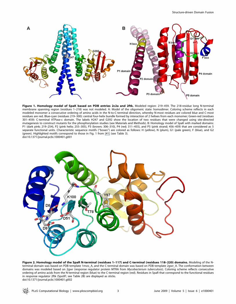
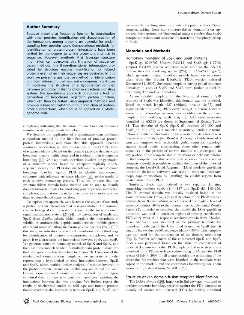
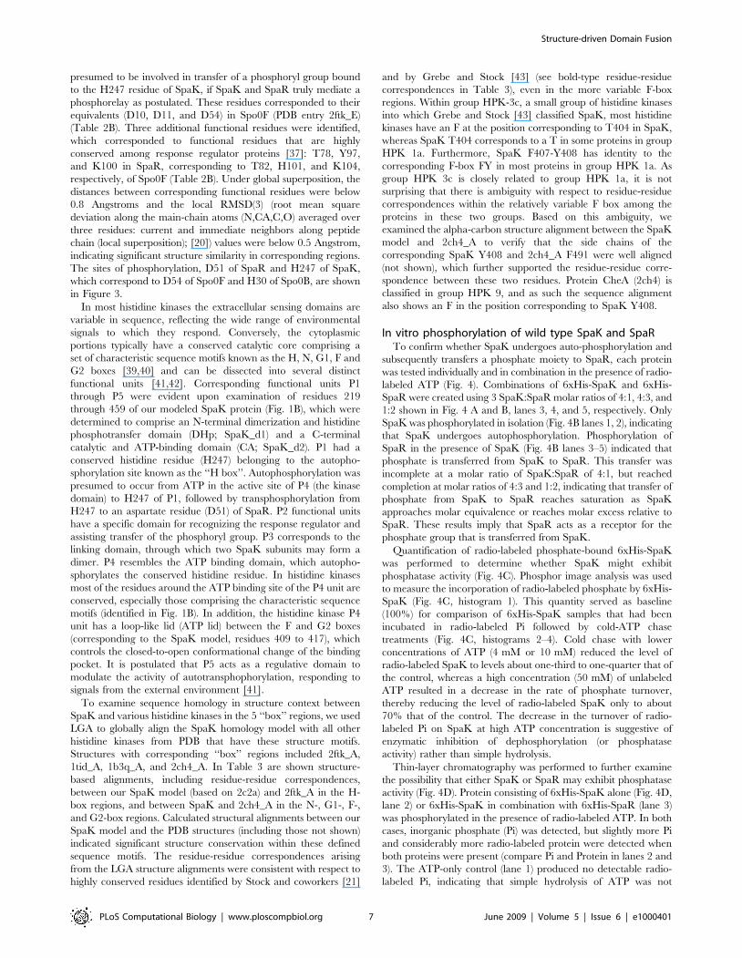
Figure 1. Homology model of SpaK based on PDB entries 2c2a and 2ftk. Modeled region: 219–459. The 218-residue long N-terminalmembrane spanning region (residues 1–218) was not modeled. A: Model of the oligomeric state: homodimer. Coloring scheme reflects in eachmodeled monomer a consecutive ordering of amino acids in the N-to-C-terminal direction, whereby N-most residues are colored blue and C-mostresidues are red. Blue-cyan (residues 219–300): central four-helix bundle formed by interaction of 2 helixes from each monomer; Green-red (residues301–459): C-terminal ATPase-c domain. The labels H247 and G392 show the location of two residues that were changed using site-directedmutagenesis to construct mutants for the phosphorylation studies (see Materials and Methods). B: Homology model of SpaK with marked domains:P1 (dark pink; 219–254), P2 (pink helix; 255–305), P3 (brown; 306–310), P4 (red; 311–455), and P5 (pink strand; 456–459) that are considered as 5separate functional units. Characteristic sequence motifs (‘‘boxes’’) are colored as follows: H (yellow), N (plum), G1 (pale green), F (blue), and G2(green). Highlighted motifs correspond to those in Fig. 1 from [41] (see Table 3).doi:10.1371/journal.pcbi.1000401.g001
Figure 2. Homology model of the SpaR N-terminal (residues 1–117) and C-terminal (residues 118–220) domains. Modeling of the N-terminal domain was based on PDB template 1mvo_A, and the C-terminal domain was based on PDB template 2gwr_A. The conformation betweendomains was modeled based on 2gwr (response regulator protein MTRA from Mycobacterium tuberculosis). Coloring scheme reflects consecutiveordering of amino acids from the N-terminal region (blue) to the C-terminal region (red). Residues in SpaR that correspond to the functional residuesin response regulator 2ftk (Spo0F; see Table 2B) are displayed as sticks.doi:10.1371/journal.pcbi.1000401.g002
Structure-driven Domain Fusion
PLoS Computational Biology | www.ploscompbiol.org 3 June 2009 | Volume 5 | Issue 6 | e1000401
similarity to any of the four modeled domains (see above) within
the homology models of SpaK and SpaR. We selected an LGS_S
cutoff value of 35% based on our observation that the number and
quality of hits increased rapidly at LGA_S, = 33% (data not
shown) and based on previous work [30] that determined the
minimal structure homology needed to assure quality of structure
alignment. Those entries with homology to both respective
domains of SpaK and SpaR were selected as putative domain-
fusion templates for modeling a SpaK/SpaR complex (Table 1).
Reported in Table 1 are the sequence identities between SpaK or
SpaR compared to each corresponding domain-fusion template,
whereby residue-residue correspondences were extracted from the
structure alignments between the models and the domain-fusion
templates. We do not report the PSI-BLAST calculated sequence
identities, as these are highly inaccurate and meaningless when
calculated from sequence alignments at low levels of sequence
identity (i.e., below 10%).
Cloning and expression of histidine-tagged proteinsSpaK and SpaR
The spaK and spaR genes were isolated from Bacillus subtilus
strain LH45, a subtilin-producing derivative of strain 168 [31].
Synthetic oligonucleotide primers were used to amplify spaR using
methods described previously [32,33]. Briefly, the commercial
vector pQE31 (obtained from Qiagen, Valencia, CA), was digested
with EcoRI and HindIII, and a fragment containing a truncated
spaK gene encoding the C-terminal half of SpaK was cloned into
the multipurpose cloning site of the QE31 vector to construct the
pQE31-spaK expression vector (Supplemental Fig. S1A). (Note
that we succeeded in expressing only the C-terminal residues of
SpaK, as the full-length gene did not yield an expression product.)
The pQE31-spaR vector was similarly constructed (details are
shown in Supplemental Fig. S1B. Vectors (MLD[pQE31-spaR]
and MLD[pQE31-spaK]) were transformed into JM109. For
expression of the histidine-tagged proteins, the expression plasmids
MLD[pQE31-spaK] and MLD[pQE31-spaR] were transformed
into M15[pREP4] competent cells (Qiagen), and expressed
according to the manufacturer’s protocol. Expressed His-tagged
proteins were purified using a Ni-NTA resin from Novagen to
form slurries that were used to pack a 1.6 cm column, and eluted
proteins were dialyzed against a storage buffer and stored in 50-ul
aliquots at 80uC. A working stock was stored for several weeks at
20uC. Protein concentrations were determined by Bio-Rad protein
assay using the manufacturer’s protocol.
Construction of mutant SpaK proteinsMutant SpaK proteins were prepared by Ana-Gen Technologies
(Palo Alto, CA) using the Stratagene QuikChange Mutagenesis Kit.
Synthetic forward and corresponding reverse complement oligonu-
cleotide primers were prepared for each of two mutations
introduced into SpaK (altered nucleotides are indicated in bold
type): at position H247 the histidine was changed to glutamine using
forward primer 59-GTGCTTTGGCACAAGAGATCAAGAT-
TCCG-39 and reverse primer: 59-CGGAATCTTGATCTC-
TTGTGCCAAAGCAC-39, and at position G392 the glycine was
changed to alanine using forward primer 59-GTAAAAGA-
CACGGCAAATGGATTTTCGG-39 and reverse primer 59-
CCGAAAATCCATTTGCCGTGTCTTTTAC-39.
Table 1. Candidate domain-fusion templates for structure modeling of a SpaK/SpaR complex.
Template - SpaK/R1,2 N13 N24 N5 RMSD6 Seq_ID7 LGA_S8
1f51_A - SpaK_d2 181 159 104 2.56 6.73 42.32
1f51_E - SpaR_d1 119 117 116 1.41 25 93.11
2ftk_A - SpaK_d2 181 159 106 2.58 6.6 42.86
2ftk_E - SpaR_d1 119 117 116 1.11 24.14 95.71
1th8_A - SpaK_d2 132 159 95 2.34 17.89 42.99
1th8_B - SpaR_d1 115 117 76 2.71 7.89 39.3
1thn_A - SpaK_d2 136 159 99 2.23 17.17 45.15
1thn_B - SpaR_d1 114 117 75 2.75 6.67 38.68
1tid_A - SpaK_d2 136 159 98 2.23 17.35 44.47
1tid_B - SpaR_d1 119 117 76 2.88 6.58 38.52
1til_A - SpaK_d2 141 159 101 2.19 16.83 45.47
1til_B - SpaR_d1 117 117 71 2.96 4.23 37.11
1The domains from the structure models of SpaK and SpaR were compared with all structures from PDB. Listed are those domain-fusion templates for which at least onedomain from each of SpaK and SpaR had structure similarity LGA_S. = 35%.
2The residue ranges in modeled SpaK domains are: SpaK_d1: 219–300 and SpaK_d2: 301–459, and the residue ranges in modeled SpaR domains are: SpaR_d1: 1–117and SpaR_d2: 118–220.
3N1 denotes a number of residues in the structural domain-fusion template.4N2 denotes the number of residues in the corresponding domain from SpaK or SpaR.5N denotes the number of superimposed C-alpha atoms that fit under a distance of 5.0 Angstroms.6RMSD is the root mean square deviation of N corresponding C-alpha atom pairs from the calculated structural alignment.7Seq_ID denotes the sequence identity in % between the domain-fusion template and the corresponding SpaK or SpaR domain calculated from the structuralalignment.
8LGA_S is a measure of the level of structure similarity [20] identified between the domain-fusion template and the corresponding domain from SpaK or SpaR.Domains from the structural models of SpaK and SpaR were compared with all structures from PDB. Listed are the domain-fusion templates that for at least one domainfrom the SpaK or SpaR model had a level of structure similarity LGA_S above 37%. LGA_S scores are reported for alignments between each modeled domain of SpaK orSpaR and a domain-fusion template domain. The residue ranges in modeled SpaK domains were: SpaK_d1: 219–300 and SpaK_d2: 301–459, and the residue ranges inmodeled SpaR domains were: SpaR_d1: 1–117 and SpaR_d2: 118–220.doi:10.1371/journal.pcbi.1000401.t001
Structure-driven Domain Fusion
PLoS Computational Biology | www.ploscompbiol.org 4 June 2009 | Volume 5 | Issue 6 | e1000401
In vitro phosphorylation and de-phosphorylation assaysPhosphorylation reactions were performed with each histidine-
tagged SpaK wild type and mutant protein in the absence and
presence of histidine-tagged SpaR. Upon addition of 32P-labeled
ATP, reaction mixtures were incubated for 20 minutes at room
temperature, after which the reactions were stopped by addition of
56 phosphorylation sample buffer, then electrophoresed on a
12.5% SDS polyacrylamide gel. The gel was stained with
Coomassie blue, dried, and autoradiographed using Kodak X-
OMAT AR film.
Phosphorimage analysis was performed to quantify incorporation
and turnover of phosphate in assays involving phosphorylation of
6xHis-SpaK. Four samples of protein were incubated in the
presence of 32P-labeled ATP, of which three were followed by cold
chase treatment with unlabeled 4 mM, 10 mM, or 50 mM ATP,
using reaction conditions described previously [34]. Samples were
run on a 12.5% SDS-PAGE gel and subjected to autoradiography
(not shown) and phosphorimaging. Image intensities of the
radiolabeled-phosphorylated SpaK gel bands were analyzed using
the Molecular Dynamics Phosphorimager 400.
Thin-layer chromatography was performed using Polygram
Cell 300 PEI cellulose plates as described previously [35]. 6xHis-
SpaK and 6xHis-SpaR were incubated individually (SpaK) or in
combination with 32P-labeled ATP in the absence or presence of
EDTA. One ul aliquots from each reaction were spotted onto
TLC plates, and chromatography was carried out in 0.75 M
KH2PO4, pH 3.75, after which the plate was dried and
autoradiographed.
Results
Structure-driven domain-fusion analysis and protein-protein complex modeling
The AS2TS protein structure modeling system [28] yielded over
30 and over 140 PDB structures suitable as templates for modeling
each of SpaK and SpaR, respectively, from which were selected
sets of the closest templates with sequence identities ranging from
13% to 28% for SpaK and 24% to 46% for SpaR (see
Supplemental Data Tables S1, S2). LGA-mediated structure
homology searches against the PDB database using constructed
structural models of domains from SpaK (SpaK_d1, SpaK_d2)
and SpaR (SpaR_d1, SpaR_d2) yielded 6 domain-fusion tem-
plates with structural homology (i.e., similarity based on structure
alignment; [20]) ranging from LGA_S = 37% to 95%, and root
mean square deviation (RMSD) calculated on superimposed C-
alpha atoms ranging from 1.11 to 2.96 (Table 1). Identification of
domain-fusion templates suggested that SpaK and SpaR interact
forming an interface between domain 2 of SpaK and domain 1 of
SpaR. Sequence identities of SpaK and SpaR to corresponding
template sequences ranged from 4% to 25%, but in no instance
was sequence identity greater than 7% simultaneously to both
SpaK_d2 and SpaR_d1. Structural comparison of all identified
domain fusion template structures showed that they clustered into
two distinct conformations, yielding the following groups: (1)
1f51_AE and 2ftk_AE (Spo0F/Spo0B from B. subtilus), and (2)
1th8_AB, 1thn_AB, 1tid_AB and 1til_AB (SpoIIAB/SpoIIAA
from B. stearothermophilus). PDB entry 2ftk was determined to be the
optimal domain-fusion template for modeling a SpaK/SpaR
complex based on the highest structure similarity to the
corresponding two modeled domains: SpaK_d2 and SpaR_d1,
and based on the expected intermolecular distance between the
putative functional residues H247 of SpaK and D51 of SpaR that
were predicted as active site residues (His and Asp) critical for
exchanging a phosphoryl group [36]. In order to form a covalent
bond with the phosphoryl group, the distances between atoms N of
His and O of Asp were expected to be in the range of about 5
Angstroms. The models created based on templates 1f51 and 2ftk
satisfied this requirement. 2ftk was also used to complete the
homology model of SpaK (Fig. 1) by providing relative positioning
of the central (SpaK_d1) and C-terminal (SpaK_d2) domains. The
SpaK/SpaR complex was modeled as a trimer, comprising a
SpaK homo-dimer and a SpaR monomer, based on the domain
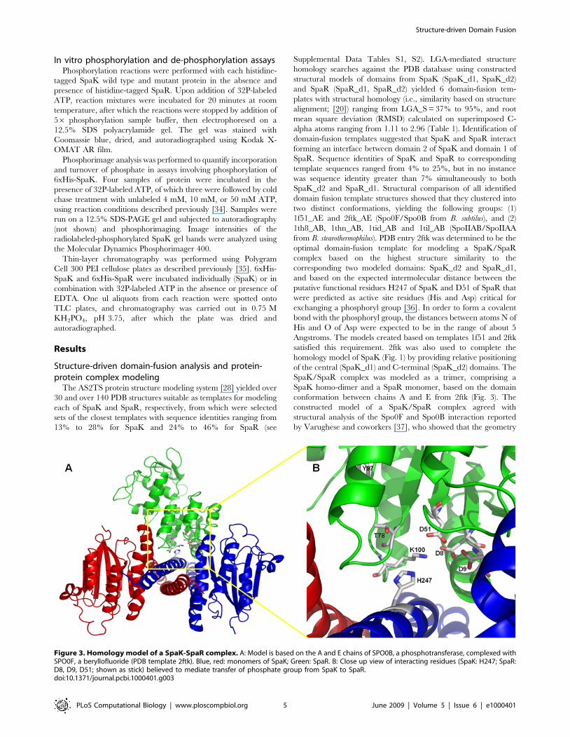
conformation between chains A and E from 2ftk (Fig. 3). The
constructed model of a SpaK/SpaR complex agreed with
structural analysis of the Spo0F and Spo0B interaction reported
by Varughese and coworkers [37], who showed that the geometry
Figure 3. Homology model of a SpaK-SpaR complex. A: Model is based on the A and E chains of SPO0B, a phosphotransferase, complexed withSPO0F, a beryllofluoride (PDB template 2ftk). Blue, red: monomers of SpaK; Green: SpaR. B: Close up view of interacting residues (SpaK: H247; SpaR:D8, D9, D51; shown as stick) believed to mediate transfer of phosphate group from SpaK to SpaR.doi:10.1371/journal.pcbi.1000401.g003
Structure-driven Domain Fusion
PLoS Computational Biology | www.ploscompbiol.org 5 June 2009 | Volume 5 | Issue 6 | e1000401
of Spo0F binding to Spo0B favors an associative mechanism for
phosphoryl transfer. In order to visualize the autophosphorylation
of the histidine kinase, and the subsequent phosphoryl transfer to
Spo0F, they generated in silico models representing these reaction
steps, proposing Spo0B as a model for the autokinase domain of
KinA (histidine kinase, consisting of an N-terminal sensor domain
and a C-terminal autokinase domain). The level of sequence
identity between KinA and SpaK is about 27%, and the KinA
sensor domain comprises three PAS (Per-Arnt-Sim) domains that
correspond to the N-terminal part of SpaK (1–218; not modeled).
The autokinase domain corresponds to the modeled C-terminal
part (219–459) of SpaK, and consists of a phosphotransferase
subdomain and an ATP binding subdomain. In modeling SpaK
we followed Varuguese and coauthors’ suggestion that the four-
helix bundle of Spo0B is formed through the dimerization of two
helical hairpins from two monomers, and that it is a prototype for
the phosphotransferase domains of histidine kinases (see Fig. 1A).
This concept is supported by the high degree of structure similarity
between the C-terminal domain of Spo0B and the ATP binding
domains of histidine kinases, as well as by a report [38] of the
crystal structure of the entire cytoplasmic portion of a histidine
kinase (a PDB structure, 2c2a), which we used as a primary
template for modeling individual domains of SpaK.
Informatics analysis of functional residues and sequencemotifs in a hypothetical SpaK/SpaR complex
Inspection of the constructed SpaK/SpaR complex (Fig. 3A)
allowed us to identify specific residues putatively involved in the
interaction between SpaK and SpaR or believed to mediate
transfer of phosphate from SpaK to SpaR (Fig. 3B). Specifically,
we identified the histidine residue at position H247 in SpaK that
corresponds to the histidine H30 that is phosphorylated in Spo0B
(PDB entry 2ftk_A) (Table 2A), and we identified 3 aspartate
residues in close proximity in SpaR (D8, D9, and D51), which we
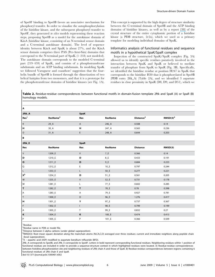
Table 2. Residue-residue correspondences between functional motifs in domain-fusion template 2ftk and SpaK (A) or SpaR (B)homology models.
A
2ftk_A SpaK
Res1 ResName2 Res ResName Distance3 RMSD(3)4
R 29_A A 246_A 0.508 0.14
H 30_A H 247_A 0.565 0.236
D 31_A E 248_A 0.644 0.203
B
2ftk_E SpaR
Res ResName Res ResName Distance RMSD(3)
V 1209_E V 7_E 0.366 0.14
D 1210_E D 8_E 0.433 0.191
D 1211_E D 9_E 0.684 0.223
Q 1212_E E 10_E 0.797 0.244
L 1253_E L 50_E 0.277 0.221
X5 1254_E D 51_E 0.561 0.205
M 1255_E V 52_E 0.731 0.178
M 1281_E L 77_E 0.602 0.285
T 1282_E T 78_E 0.78 0.398
A 1283_E A 79_E 0.927 0.781
T 1300_E D 96_E 1.276 0.417
H 1301_E Y 97_E 0.737 0.367
F 1302_E I 98_E 0.799 0.199
A 1303_E T 99_E 0.832 0.27
K 1304_E K 100_E 0.474 0.413
P 1305_E P 101_E 0.366 0.509
1Residue.2Residue name in PDB or model file.3Distance between C-alpha carbons (under global superposition).4RMSD(3): Root mean square deviation along the mainchain atoms (N,CA,C,O) averaged over three residues: current and immediate neighbors along peptide chain(local superposition).
5X – aspartic acid (ASP) modified to aspartate beryllium trifluoride (BFD).2ftk_A corresponds to Spo0B, and 2ftk_E corresponds to Spo0F. Letters in bold represent corresponding functional residues. Neighboring residues within 1 position offunctional residues are included in order to provide a sequence-structure context in which highlighted residues were located. A) Residue-residue correspondencesbetween histidine phosphorylation site and neighboring residues of 2ftk chain A and those of SpaK. B) Residue-residue correspondences between regions containing 6functional residues of 2ftk chain E and SpaR.doi:10.1371/journal.pcbi.1000401.t002
Structure-driven Domain Fusion
PLoS Computational Biology | www.ploscompbiol.org 6 June 2009 | Volume 5 | Issue 6 | e1000401
presumed to be involved in transfer of a phosphoryl group bound
to the H247 residue of SpaK, if SpaK and SpaR truly mediate a
phosphorelay as postulated. These residues corresponded to their
equivalents (D10, D11, and D54) in Spo0F (PDB entry 2ftk_E)
(Table 2B). Three additional functional residues were identified,
which corresponded to functional residues that are highly
conserved among response regulator proteins [37]: T78, Y97,
and K100 in SpaR, corresponding to T82, H101, and K104,
respectively, of Spo0F (Table 2B). Under global superposition, the
distances between corresponding functional residues were below
0.8 Angstroms and the local RMSD(3) (root mean square
deviation along the main-chain atoms (N,CA,C,O) averaged over
three residues: current and immediate neighbors along peptide
chain (local superposition); [20]) values were below 0.5 Angstrom,
indicating significant structure similarity in corresponding regions.
The sites of phosphorylation, D51 of SpaR and H247 of SpaK,
which correspond to D54 of Spo0F and H30 of Spo0B, are shown
in Figure 3.
In most histidine kinases the extracellular sensing domains are
variable in sequence, reflecting the wide range of environmental
signals to which they respond. Conversely, the cytoplasmic
portions typically have a conserved catalytic core comprising a
set of characteristic sequence motifs known as the H, N, G1, F and
G2 boxes [39,40] and can be dissected into several distinct
functional units [41,42]. Corresponding functional units P1
through P5 were evident upon examination of residues 219
through 459 of our modeled SpaK protein (Fig. 1B), which were
determined to comprise an N-terminal dimerization and histidine
phosphotransfer domain (DHp; SpaK_d1) and a C-terminal
catalytic and ATP-binding domain (CA; SpaK_d2). P1 had a
conserved histidine residue (H247) belonging to the autopho-
sphorylation site known as the ‘‘H box’’. Autophosphorylation was
presumed to occur from ATP in the active site of P4 (the kinase
domain) to H247 of P1, followed by transphosphorylation from
H247 to an aspartate residue (D51) of SpaR. P2 functional units
have a specific domain for recognizing the response regulator and
assisting transfer of the phosphoryl group. P3 corresponds to the
linking domain, through which two SpaK subunits may form a
dimer. P4 resembles the ATP binding domain, which autopho-
sphorylates the conserved histidine residue. In histidine kinases
most of the residues around the ATP binding site of the P4 unit are
conserved, especially those comprising the characteristic sequence
motifs (identified in Fig. 1B). In addition, the histidine kinase P4
unit has a loop-like lid (ATP lid) between the F and G2 boxes
(corresponding to the SpaK model, residues 409 to 417), which
controls the closed-to-open conformational change of the binding
pocket. It is postulated that P5 acts as a regulative domain to
modulate the activity of autotransphophorylation, responding to
signals from the external environment [41].
To examine sequence homology in structure context between
SpaK and various histidine kinases in the 5 ‘‘box’’ regions, we used
LGA to globally align the SpaK homology model with all other
histidine kinases from PDB that have these structure motifs.
Structures with corresponding ‘‘box’’ regions included 2ftk_A,
1tid_A, 1b3q_A, and 2ch4_A. In Table 3 are shown structure-
based alignments, including residue-residue correspondences,
between our SpaK model (based on 2c2a) and 2ftk_A in the H-
box regions, and between SpaK and 2ch4_A in the N-, G1-, F-,
and G2-box regions. Calculated structural alignments between our
SpaK model and the PDB structures (including those not shown)
indicated significant structure conservation within these defined
sequence motifs. The residue-residue correspondences arising
from the LGA structure alignments were consistent with respect to
highly conserved residues identified by Stock and coworkers [21]
and by Grebe and Stock [43] (see bold-type residue-residue
correspondences in Table 3), even in the more variable F-box
regions. Within group HPK-3c, a small group of histidine kinases
into which Grebe and Stock [43] classified SpaK, most histidine
kinases have an F at the position corresponding to T404 in SpaK,
whereas SpaK T404 corresponds to a T in some proteins in group
HPK 1a. Furthermore, SpaK F407-Y408 has identity to the
corresponding F-box FY in most proteins in group HPK 1a. As
group HPK 3c is closely related to group HPK 1a, it is not
surprising that there is ambiguity with respect to residue-residue
correspondences within the relatively variable F box among the
proteins in these two groups. Based on this ambiguity, we
examined the alpha-carbon structure alignment between the SpaK
model and 2ch4_A to verify that the side chains of the
corresponding SpaK Y408 and 2ch4_A F491 were well aligned
(not shown), which further supported the residue-residue corre-
spondence between these two residues. Protein CheA (2ch4) is
classified in group HPK 9, and as such the sequence alignment
also shows an F in the position corresponding to SpaK Y408.
In vitro phosphorylation of wild type SpaK and SpaRTo confirm whether SpaK undergoes auto-phosphorylation and
subsequently transfers a phosphate moiety to SpaR, each protein
was tested individually and in combination in the presence of radio-
labeled ATP (Fig. 4). Combinations of 6xHis-SpaK and 6xHis-
SpaR were created using 3 SpaK:SpaR molar ratios of 4:1, 4:3, and
1:2 shown in Fig. 4 A and B, lanes 3, 4, and 5, respectively. Only
SpaK was phosphorylated in isolation (Fig. 4B lanes 1, 2), indicating
that SpaK undergoes autophosphorylation. Phosphorylation of
SpaR in the presence of SpaK (Fig. 4B lanes 3–5) indicated that
phosphate is transferred from SpaK to SpaR. This transfer was
incomplete at a molar ratio of SpaK:SpaR of 4:1, but reached
completion at molar ratios of 4:3 and 1:2, indicating that transfer of
phosphate from SpaK to SpaR reaches saturation as SpaK
approaches molar equivalence or reaches molar excess relative to
SpaR. These results imply that SpaR acts as a receptor for the
phosphate group that is transferred from SpaK.
Quantification of radio-labeled phosphate-bound 6xHis-SpaK
was performed to determine whether SpaK might exhibit
phosphatase activity (Fig. 4C). Phosphor image analysis was used
to measure the incorporation of radio-labeled phosphate by 6xHis-
SpaK (Fig. 4C, histogram 1). This quantity served as baseline
(100%) for comparison of 6xHis-SpaK samples that had been
incubated in radio-labeled Pi followed by cold-ATP chase
treatments (Fig. 4C, histograms 2–4). Cold chase with lower
concentrations of ATP (4 mM or 10 mM) reduced the level of
radio-labeled SpaK to levels about one-third to one-quarter that of
the control, whereas a high concentration (50 mM) of unlabeled
ATP resulted in a decrease in the rate of phosphate turnover,
thereby reducing the level of radio-labeled SpaK only to about
70% that of the control. The decrease in the turnover of radio-
labeled Pi on SpaK at high ATP concentration is suggestive of
enzymatic inhibition of dephosphorylation (or phosphatase
activity) rather than simple hydrolysis.
Thin-layer chromatography was performed to further examine
the possibility that either SpaK or SpaR may exhibit phosphatase
activity (Fig. 4D). Protein consisting of 6xHis-SpaK alone (Fig. 4D,
lane 2) or 6xHis-SpaK in combination with 6xHis-SpaR (lane 3)
was phosphorylated in the presence of radio-labeled ATP. In both
cases, inorganic phosphate (Pi) was detected, but slightly more Pi
and considerably more radio-labeled protein were detected when
both proteins were present (compare Pi and Protein in lanes 2 and
3). The ATP-only control (lane 1) produced no detectable radio-
labeled Pi, indicating that simple hydrolysis of ATP was not
Structure-driven Domain Fusion
PLoS Computational Biology | www.ploscompbiol.org 7 June 2009 | Volume 5 | Issue 6 | e1000401
occurring. Furthermore, when phosphorylation was performed in
the presence of EDTA, some phosphorylated protein was
observed, although no inorganic phosphate was detected (Fig. 4D
lane 4). This result, taken together with Fig. C, which suggested
the presence of enzymatic phosphatase activity, supports the claim
that SpaK (and possibly also SpaR) may possess enzymatic
phosphatase activity.
Mutational analysis of SpaK and intermolecularcomplementation of SpaK monomers
Based on amino acid sequence alignment with other histidine
kinases, the highly conserved histidine at position H247 was
presumed to be the site of possible auto-phosphorylation, and a
glycine located at position G392 in the C-terminal end of SpaK
was determined to correspond to the conserved DXG motif of the
nucleotide binding domain in related histidine kinases (Fig. 1A,
Fig. 1B: H box and G1 box). In the superfamily of phosphotrans-
ferases, the conserved residues that form a corresponding motif
(DXG in actin, GTG in hexokinase/glycerol kinase, and GNG in
acetate and propionate kinases) are observed to be present in
binding to a- and b-phosphate groups of the nucleotide [44].
Because several histidine kinases are believed to exist as homo-
dimers and it is believed that phosphorylation occurs in trans, in
which one monomer binds ATP in the nucleotide-binding domain
and then transfers the phosphoryl group to a histidine located in
the other monomer, we postulated that mutations at either of these
positions might reduce or abolish auto-phosphorylation of SpaK,
but that complementation between mutants might occur,
effectively restoring function. We used site-directed mutagenesis
to construct two mutants (see Materials and Methods): one in
which the histidine at position H247 was changed to a glutamine
(H247Q), and the other in which the glycine at position G392 was
changed to alanine (G392A). Locations of mutated residues are
shown in Fig. 1A. Phosphorylation studies of mutants H247Q and
G392A revealed that both mutations resulted in loss of
phosphorylation when each mutant was tested individually (Fig. 5
A, B; lanes 4, 5) or when individually combined with SpaR
(Fig. 5B; lanes 9, 10). However, when the mutant proteins were
combined, a detectable amount (approximately 25% that of wild
type) of auto-phosphorylation was observed (Fig. 5B, lane 6),
suggesting that complementation between the mutants had
occurred, and supporting the hypothesis that SpaK forms a
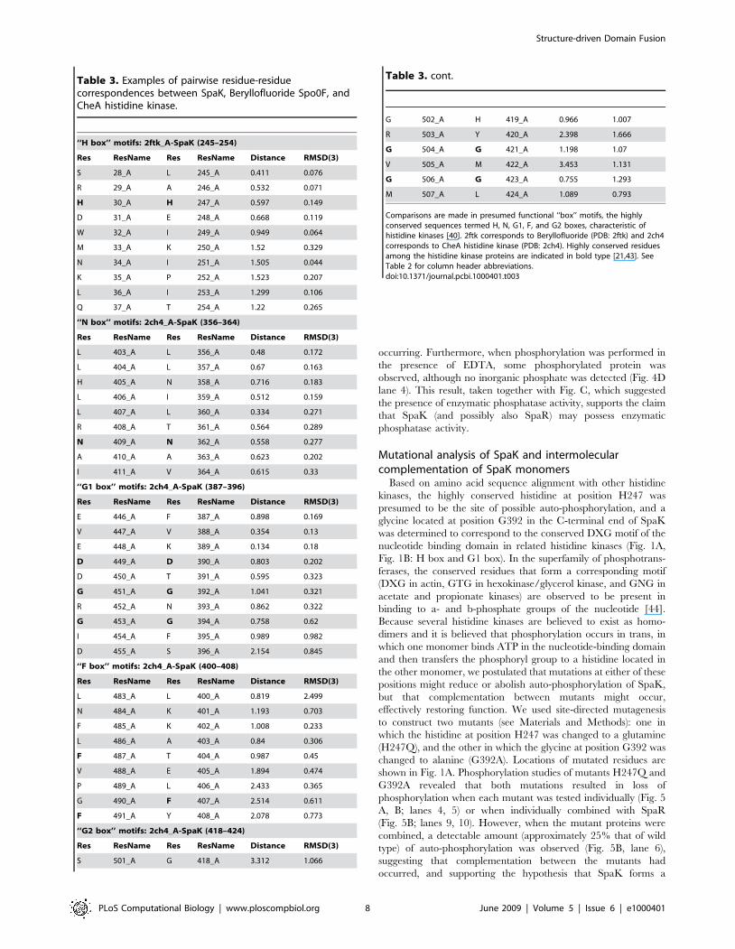
Table 3. Examples of pairwise residue-residuecorrespondences between SpaK, Beryllofluoride Spo0F, andCheA histidine kinase.
‘‘H box’’ motifs: 2ftk_A-SpaK (245–254)
Res ResName Res ResName Distance RMSD(3)
S 28_A L 245_A 0.411 0.076
R 29_A A 246_A 0.532 0.071
H 30_A H 247_A 0.597 0.149
D 31_A E 248_A 0.668 0.119
W 32_A I 249_A 0.949 0.064
M 33_A K 250_A 1.52 0.329
N 34_A I 251_A 1.505 0.044
K 35_A P 252_A 1.523 0.207
L 36_A I 253_A 1.299 0.106
Q 37_A T 254_A 1.22 0.265
‘‘N box’’ motifs: 2ch4_A-SpaK (356–364)
Res ResName Res ResName Distance RMSD(3)
L 403_A L 356_A 0.48 0.172
L 404_A L 357_A 0.67 0.163
H 405_A N 358_A 0.716 0.183
L 406_A I 359_A 0.512 0.159
L 407_A L 360_A 0.334 0.271
R 408_A T 361_A 0.564 0.289
N 409_A N 362_A 0.558 0.277
A 410_A A 363_A 0.623 0.202
I 411_A V 364_A 0.615 0.33
‘‘G1 box’’ motifs: 2ch4_A-SpaK (387–396)
Res ResName Res ResName Distance RMSD(3)
E 446_A F 387_A 0.898 0.169
V 447_A V 388_A 0.354 0.13
E 448_A K 389_A 0.134 0.18
D 449_A D 390_A 0.803 0.202
D 450_A T 391_A 0.595 0.323
G 451_A G 392_A 1.041 0.321
R 452_A N 393_A 0.862 0.322
G 453_A G 394_A 0.758 0.62
I 454_A F 395_A 0.989 0.982
D 455_A S 396_A 2.154 0.845
‘‘F box’’ motifs: 2ch4_A-SpaK (400–408)
Res ResName Res ResName Distance RMSD(3)
L 483_A L 400_A 0.819 2.499
N 484_A K 401_A 1.193 0.703
F 485_A K 402_A 1.008 0.233
L 486_A A 403_A 0.84 0.306
F 487_A T 404_A 0.987 0.45
V 488_A E 405_A 1.894 0.474
P 489_A L 406_A 2.433 0.365
G 490_A F 407_A 2.514 0.611
F 491_A Y 408_A 2.078 0.773
‘‘G2 box’’ motifs: 2ch4_A-SpaK (418–424)
Res ResName Res ResName Distance RMSD(3)
S 501_A G 418_A 3.312 1.066
G 502_A H 419_A 0.966 1.007
R 503_A Y 420_A 2.398 1.666
G 504_A G 421_A 1.198 1.07
V 505_A M 422_A 3.453 1.131
G 506_A G 423_A 0.755 1.293
M 507_A L 424_A 1.089 0.793
Comparisons are made in presumed functional ‘‘box’’ motifs, the highlyconserved sequences termed H, N, G1, F, and G2 boxes, characteristic ofhistidine kinases [40]. 2ftk corresponds to Beryllofluoride (PDB: 2ftk) and 2ch4corresponds to CheA histidine kinase (PDB: 2ch4). Highly conserved residuesamong the histidine kinase proteins are indicated in bold type [21,43]. SeeTable 2 for column header abbreviations.doi:10.1371/journal.pcbi.1000401.t003
Table 3. cont.
Structure-driven Domain Fusion
PLoS Computational Biology | www.ploscompbiol.org 8 June 2009 | Volume 5 | Issue 6 | e1000401
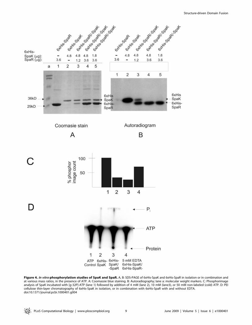
Figure 4. In vitro phosphorylation studies of SpaK and SpaR. A, B: SDS-PAGE of 6xHis-SpaK and 6xHis-SpaR in isolation or in combination andat various mass ratios, in the presence of ATP. A: Coomassie blue staining. B: Autoradiography; lane a: molecular weight markers. C: Phosphorimageanalysis of SpaK incubated with [g-32P]-ATP (lane 1) followed by addition of 4 mM (lane 2), 10 mM (lane3), or 50 mM non-labeled (cold) ATP. D: PEIcellulose thin-layer chromatography of 6xHis-SpaK in isolation, or in combination with 6xHis-SpaR with and without EDTA.doi:10.1371/journal.pcbi.1000401.g004
Structure-driven Domain Fusion
PLoS Computational Biology | www.ploscompbiol.org 9 June 2009 | Volume 5 | Issue 6 | e1000401
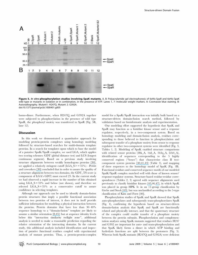
homo-dimer. Furthermore, when H247Q and G392A together
were subjected to phosphorylation in the presence of wild type
SpaR, the phosphoryl moiety was transferred to SpaR (Fig. 5B,
lane 12).
Discussion
In this work we demonstrated a quantitative approach for
modeling protein-protein complexes using homology modeling
followed by structure-based searches for multi-domain template
proteins. In a search for templates upon which to base the model
of a putative SpaK/SpaR complex, we used LGA, which applies
two scoring schemes: GDT (global distance test) and LCS (longest
continuous segment). Based on a previous study involving
structure alignments between weakly homologous proteins [30],
we applied a relatively stringent cutoff (LGA_S. = 35%)—Pettitt
and coworkers [30] concluded that in order to assure the quality of
a structure alignment between two domains, the GDT_TS score (a
component of LGA’s GDT) must exceed 25. In the current study
we had observed a rapid increase in the number of hits obtained
using LGA_S = 33% and below (not shown), and therefore we
selected LGA_S = 35% as a conservative cutoff to assure
confidence in selecting templates.
Although our approach can be used to identify domain-fusion
protein structures that imply a possible functional association
between two proteins of interest, it does not in itself provide
sufficient information for modeling a physical interaction between
the proteins. Protein domains that have less than 30–40%
sequence homology to a ‘‘domain-fusion’’ template are likely to
assume a similar orientation [8,45]–but at sequence identity levels
below this ‘‘interaction similarity twilight zone’’, additional
analysis is needed to make a reasonable prediction regarding the
relative orientation of the interacting domains. In the current
study, this additional analysis included identification and inspec-
tion of putative functional residues coupled with experimental
analysis of mutant proteins. Thus, a protein-protein-complex
model for a SpaK/SpaR interaction was initially built based on a
structure-driven domain-fusion search method, followed by
validation based on bioinformatic analysis and experimentation.
Our modeling effort supported the hypothesis that SpaK and
SpaR may function as a histidine kinase sensor and a response
regulator, respectively, in a two-component system. Based on
homology modeling and domain-fusion analysis, residues corre-
sponding to those believed to function in phosphorylation and
subsequent transfer of a phosphate moiety from sensor to response
regulator in other two-component systems were identified (Fig. 3,
Tables 1, 2). Modeling of SpaK enabled structure comparisons
with related sensor proteins (2ftk_A, 1tid_A, 1b3q_A, 2ch4_A),
identification of sequences corresponding to the 5 highly
conserved regions (‘‘boxes’’) that characterize class II two-
component system proteins [40,41,43] (Table 3), and mapping
of these sequences to the homology model of SpaK (Fig. 1B).
Functional residues and conserved sequence motifs of our modeled
SpaK/SpaR complex matched well with those of known sensor/
response-regulator systems. Structure-based residue-residue corre-
spondences (Tables 2, 3) agreed with sequence alignments used
previously to classify histidine kinases [43,46,47], in which SpaK
was placed in group HPK 3c in an 11-group classification by
Grebe and Stock [43], but was unclassified according to the 5-type
classification of Kim and Forst [46].
Phosphorylation studies of SpaK and SpaR showed that SpaK
auto-phosphorylates and subsequently trans-phosphorylates SpaR
(Fig. 4), confirming the hypothesis based on structure-driven
domain-fusion analysis that SpaK and SpaR are functionally
related and physically interact, and that the quaternary structure
of the complex could enable transfer of a phosphate moiety
between the protein subunits. Phosphorylation and complemen-
tation analyses using SpaK mutants suggested that residues H247
and G392 are important for auto- and trans-phosphorylation and
that SpaK likely forms a dimer in which ATP binding and
hydrolysis functions are split between the protomers (Fig. 5).
Whereas both SpaK mutants (H247Q and G392A) were deficient
Figure 5. In vitro phosphorylation studies involving SpaK mutants. A, B: Polyacrylamide gel electrophoresis of 6xHis-SpaR and 6xHis-SpaKwild type or mutants in isolation or in combination, in the presence of ATP. Lanes 1, 7: molecular weight markers. A: Coomassie blue staining. B:Autoradiography. Mutant1: H247Q, Mutant 2: G392A.doi:10.1371/journal.pcbi.1000401.g005
Structure-driven Domain Fusion
PLoS Computational Biology | www.ploscompbiol.org 10 June 2009 | Volume 5 | Issue 6 | e1000401
in auto-phosphorylation (Fig. 5, lanes 4,5), this function was
apparently restored when the mutants were combined (Fig. 5, lane
6), suggesting that complementation had occurred between the
mutants. Complementation between H247Q and G392A also
apparently restored trans-phosphorylation, as evidenced by
phosphorylation of SpaR in the presence of both mutants (Fig. 5,
lane 12). In an equimolar mixture of mutants H247Q and G392A,
one would expect that approximately one-half of the resulting
dimers would comprise a protomer of each mutant. Furthermore,
phosphorylation would occur from the H247Q mutant to the
G392A mutant, but not in the other direction, since G392A should
not be able to bind ATP. Therefore the levels of auto-
phosphorylation or trans-phosphorylation would not be expected
to exceed one-half those of wild type SpaK. Also, although the
H247Q/G392A mixed dimer may have had restored function, it
would be expected to have functioned at less than the efficiency of
a wild type SpaK dimer; since dimer formations between non-
productive forms would occur, one would expect phosphorylation
to proceed more slowly than in the wt. This is consistent with the
observation that phosphorylation of or by H247Q combined with
G392A (lanes 6, 12) occurred at levels considerably below those of
wild type SpaK (lanes 3, 8).
In modeling the interaction between SpaK and SpaR we
identified 6 suitable domain-fusion templates (Table 1), which
were structurally clustered into two groups (see Results), each
having a distinct conformation. Both groups displayed the same
interaction pose with respect to the domain-domain interaction.
Although each of the identified domain-fusion templates would
have yielded a SpaK/SpaR complex model consistent with the
experimental data, the criteria for selecting 2ftk as the domain-
fusion template were based on combined structural identities
between domains of 2ftk and the SpaK and SpaR models, on the
resulting distance between putative functional residues involved in
phosphate transfer (Fig. 3), and on the presence of a helical bundle
domain, which enabled construction of a complete model.
Interestingly, the domain-domain conformation between the
helical bundle and the ATPase domains of 2c2a, used for
modeling SpaK, differed from that of the corresponding domains
within 2ftk. This difference suggests the possibility that a
conformational change might take place when SpaK interacts
with SpaR. Furthermore, it should be noted that the phospho-
transfer in Spo0B-Spo0F (2ftk) occurs in the opposite direction
(Asp to His) as that demonstrated here in SpaK-SpaR (Figs. 4, 5).
This is not surprising, and does not diminish the value of 2ftk as a
template for modeling a SpaK/SpaR interaction, given the
considerable mechanistic diversity observed among structurally
conserved domains comprising sensor/response-regulator systems
[48].
Although structure modeling and experiments involving pho-
phorylation studies strongly suggest functional and physical
interactions between SpaK and SpaR, we cannot be entirely
certain that our quaternary structure is correct with respect to
domain composition, conformation, or orientation, as the
methodology is dependent on existing structural data within
PDB; it is possible that none of the domain-fusion templates
detected by our approach is truly representative of the physical
interaction between SpaK and SpaR, as homology modeling is, by
definition, data driven. Due to the low sequence homologies
between SpaK and SpaR and the identified domain-fusion
templates, one could not conclude with any degree of certainty
based solely on template identification that the interaction pose
modeled here is likely to be correct [8]. However, combining
bioinformatics analysis of known functional motifs (sequence
‘‘boxes’’) and putative interacting residues with experimental
evidence of function allows us to assert the value of the homology
model of a putative SpaK/SpaR protein-protein complex. Our
approach detects existing putative domain-fusion templates, which
may suggest testable hypotheses regarding quaternary structure
and function; a structure-based approach for identification of
‘‘Rosetta Stone’’ proteins greatly enhances structure-function
hypothesis generation by providing structural context for putative
functional residues. Additional bioinformatics analyses of a
putative protein-protein complex model, which may verify the
correctness of the model, include alignments of modified sequence
profiles [7], for example, which use quantitative methods applied
at the domain-domain interface to evaluate the likelihood of a
stable interaction.
Although many two-component signal transduction systems
have been identified by sequence homology, we wish to point out
that a purely sequence-based approach would not have yielded the
structural domain-fusion templates that were identified in this
study. The strength of our approach is in its ability to identify
putative domain-fusion templates based on structure homology
searches in cases where sequence identities between the proteins of
interest and the putative domain-fusion templates are low.
Sequence identities of candidate domain-fusion templates to
domains of SpaK and SpaR ranged from 4% to 25%, but in no
instance was sequence identity greater than 7% simultaneously to
both (Table 1). This point is emphasized by the lack of sufficient
sequence-based evidence for linking these proteins using the
standard domain-fusion approach: as of this writing, SpaK and
SpaR are not linked in this way, for example, in Prolinks [5], nor
did we find them linked by other sequence-based or empirical
methods in DIP, BIND/BOND, MIPS, IntAct, MPIDB, or
InterPreTS [49–54]. Homology modeling of SpaK and SpaR
using a standard methodology [28] and subsequent structure-
based searches using a quantitative structure comparison algo-
rithm [20] is what enabled a more sensitive, structure-based
homology search against PDB. In conclusion, our method
provides a basis upon which a high-throughput system for
identification of putative protein-protein interactions could be
built on a whole-genome scale.
Supporting Information
Figure S1 Construction of vectors for expression of SpaK and
SpaR proteins. A) Expression vectors pQE-31-spaK. B) pQE-31-
spaR.
Found at: doi:10.1371/journal.pcbi.1000401.s001 (0.38 MB TIF)
Table S1 Candidate templates for homology modeling of SpaK
monomer.
Found at: doi:10.1371/journal.pcbi.1000401.s002 (0.06 MB PDF)
Table S2 Candidate templates for homology modeling of SpaR.
Found at: doi:10.1371/journal.pcbi.1000401.s003 (0.06 MB PDF)
Author Contributions
Conceived and designed the experiments: AC CLEZ MLD VR JNH AZ.
Performed the experiments: AC CLEZ MLD VR AZ. Analyzed the data:
AC CLEZ MLD VR JNH AZ. Contributed reagents/materials/analysis
tools: JNH AZ. Wrote the paper: CLEZ AZ. Acquired funding: CLEZ
JNH AZ.
Structure-driven Domain Fusion
PLoS Computational Biology | www.ploscompbiol.org 11 June 2009 | Volume 5 | Issue 6 | e1000401
References
1. Kumar A, Snyder M (2002) Protein complexes take the bait. Nature 415:
123–124.2. Phizicky EM, Fields S (1995) Protein-protein interactions: methods for detection
and analysis. Microbiological Reviews 59: 94–123.3. Shoemaker BA, Panchenko AR (2007) Deciphering protein-protein interactions.
Part I. Experimental Techniques and Databases. PLoS Computational Biology
3: 0337–0344. doi:10.1371/journal.pcbi.0030042.4. Uetz P, Glot L, Cagney G, Mansfield TA, Judson RS, et al. (2000) A
comprehensive analysis of protein-protein interactions in Saccharomyces cerevisiae.Nature 403: 623–627.
5. Bowers PM, Pellegrini M, Thompson MJ, Fierro J, Yeates TO, Eisenberg D
(2004) Prolinks: a database of functional linkages derived from coevolution.Genome Biology 5: R35.
6. Kundrotas PJ, Alexov E (2006) Predicting 3D structures of transient protein-protein complexes by homology. Biochimica et Biophysica Acta 1764:
1498–1511.7. Kundrotas PJ, Lensink MF, Alexov E (2008) Homology-based modeling of 3D
structures of protein-protein complexes using alignments of modified sequence
profiles. International Journal of Biological Macromolecules 43: 198–208.8. Launay G, Simonson T (2008) Homology modeling of protein-protein
complexes: a simple method and its possibilities and limitations. BMCBioinformatics 9: 427–442.
9. Marcotte EM, Pelligrini M, Ng H-L, Rice DW, Yeates TO, Eisenberg D (1999)
Detecting protein function and protein-protein interactions from genomesequences. Science 285: 751–753.
10. Pellegrini M, Marcotte EM, Thompton MJ, Eisenberg D, Yeates TO (1999)Assigning protein functions by comparative genome analysis: protein phyloge-
netic profiles. Proc Natl Acad Sci USA 96: 4285–4288.11. Salwinski L, Eisenberg D (2003) Computational methods for protein-protein
interaction analysis. Current Opinion in Structural Biology 13: 377–382.
12. Szilaghyi A, Grimm V, Arakaki AK, Skolnick J (2005) Prediction of physicalprotein-protein interactions. Physical Biology 2: S1–S6. doi:10.1088/1478-
3975/2/2/S01.13. Shoemaker BA, Panchenko AR (2007) Deciphering protein-protein interactions.
Part II. Computational methods to predict protein and domain interaction
partners. PLoS Computational Biology 3: 0595–0601. doi:10.1371/journal.pcbi.0030043.
14. Teichmann SA, Murzin AG, Chothia C (2001) Determination of proteinfunction, evolution and interactions by structural genomics. Current Opinion in
Structural Biology 11: 354–363.15. Marcotte EM (2000) Computational genetics: finding protein function by
nonhomology methods. Current Opinion in Structural Biology 10: 359–365.
16. Pace HC, Hodawadekar SC, Draganescu A, Huang J, Bieganovski P,Pekarsky Y, Croce CM, Brenner C (2000) Crystal structure of the worm
NitFhit Rosetta Stone protein reveals a Nit tetramer binding two Fhit dimmers.Current Biology 10: 907–917.
17. Chia J-M, Kolatar PR (2004) Implications for domain fusion protein-protein
interactions based on structural information. BMC Bioinformatics 5: 161.doi:10.1186/1471-2105-50161.
18. Lu L, Lu H, Skolnick J (2002) MULTIPROSPECTOR: An algorithm for theprediction of protein-protein interactions by multimeric threading. Protein:
Structure, Function, and Genetics 49: 350–364.19. Rost B (1997) Protein structure sustain evolutionary drift. Fold Des 2: S19–S24.
20. Zemla A (2003) LGA—a method for finding 3D similarities in protein structures.
Nucleic Acids Research 31: 3370–3374.21. Stock J, Ninfa AJ, Stock AM (1989) Protein phosphorylation and regulation of
adaptive response in bacteria. Microbiological Reviews, American Soc.Microbiol Dec.1989: 450–490.
22. Galperin MY (2006) Structural classification of bacterial response regulators:
Diversity of output domains and domain combinations. Journal of Bacteriology188: 4169–4182.
23. Kleerebezem M, Quadri LEN, Kuipers OP, de Vos WM (1997) Quorumsensing by peptide pheromones and two-component signal-transduction systems
in gram-positive bacteria. Molecular Microbiology 24: 895–904.
24. Skerker JM, Prasol MS, Perchuk BS, Biondi EG, Laub MT (2005) Two-component signal transduction pathways regulating growth and cell cycle
progression in a bacterium: A system-level analysis. PLoS Biology 3: e334.doi:10.1371/journal.pbio.0030334.
25. Kleerebezem M, Bongers R, Rutten G, de Vos WM, Kuipers OP (2004)Autoregulation of subtilin biosynthesis in Bacillus subtilis: the role of the spa-box
in subtilin-responsive promoters. Peptides 25: 1415–1424.
26. Klein C, Kaletta C, Entian KD (1993) Biosynthesis of the lantibiotic subtilin isregulated by a histidine kinase/response regulator system. Applied and
Environmental Microbiology 59: 296–303.27. Stein T, Borchert S, Conrad B, Feesche J, Hofemeister B, Hofemeister J,
Entian K-D (2002) Two different lantibiotic-like peptides originate from the
ericin gene cluster of Bacillus subtilis A1/3. Journal of Bacteriology 184:1703–1711.
28. Zemla A, Ecale Zhou C, Slezak T, Kuczmarski T, Rama D, Torres C,
Sawicka D, Barsky D (2005) AS2TS system for protein structure modeling andanalysis. Nucleic Acids Research 33: W111–W115.
29. Canutescu AA, Shelenkov AA, Dunbrack Jr RL (2003) A graph theoryalgorithm for protein side-chain prediction. Protein Science 12: 2001–2014.
30. Pettitt CS, McGuffin LJ, Jones DT (2005) Improving sequence-based fold
recognition by use of 3D model quality assessment. Bioinformatics 21:3509–3515.
31. Liu W, Hansen N (1991) Conversion of Bacillus subtilis 168 to a subtilin producerby site-directed mutagenesis. Journal of Bacteriology 173: 7387–7390.
32. Banerjee S, Hansen JN (1988) Structure and expression of a gene encoding the
precursor of subtilin, a small protein antibiotic. Journal of Biological Chemistry263: 9508–9514.
33. Buchman GW, Banerjee S, Hansen JN (1988) Structure, expression, andevolution of a gene encoding the precursor of nisin, a small protein antibiotic.
Journal of Biological Chemistry 263: 16260–16266.34. Satola S, Kirchman PA, Moran CP (1991) Spo0A binds to a promoter used by
sigmaA RNA polymerase during sporulation in Bacillus subtilis. Proceedings of the
National Academy of Science USA 88: 4533–4537.35. Jiang M, Shao W, Perego M, Hoch JA (2000) Multiple histidine kinases regulate
entry into stationary phase and sporulation in Bacillus subtilis. MolecularMicrobiology 38: 535–542.
36. Zapf J, Madhusudan USen, Hoch J, Varughese K (2000) A transient interaction
between two phosphorelay proteins trapped in a crystal lattice reveals themechanism of molecular recognition and phosphotransfer in signal transduction.
Structure v.8(8): 851–862.37. Varughese KI, Tsigelny I, Zhao H (2006) The crystal structure of beryllofluoride
Spo0F in complex with the phosphotransferase Spo0B represents a phospho-transfer pretransition state. Journal of Bacteriology 188: 4970–7.
38. Marina A, Waldburger C, Hendrickson WA (2005) Structure of the entire
cytoplasmic portion of a sensor histidine-kinase protein. EMBO 24: 4247–4259.39. Bilwes AM, Alex LA, Crane BR, Simon MI (1999) Structure of CheA, a signal-
transducing histidine kinase. Cell 96: 131–141.40. Zhang W, Culley DE, Wu G, Brockman FJ (2006) Two-component signal
transduction systems of Desulfovibrio vulgaris: structural and phylogenetic analysis
and deduction of putative cognate pairs. Journal of Molecular Evolution 62:473–87.
41. Zhang J, Xu Y, Shen J, Luo X, Chen J, Chen K, Zhu W, Jiang H (2005)Dynamic mechanism for the autophosphorylation of CheH histidine kinase:
molecular dynamics simulations. Journal of the American Chemical Society127(33): 11709–11719.
42. Park SY, Borbat PP, Gonzalez-Bonet G, Bhatnagar J, Pollard AM, Freed JH,
Bilwes AM, Crane BR (2006) Reconstruction of the chemotaxis receptor–kinaseassembly. Nature 5: 400–407.
43. Grebe TW, Stock JF (1999) The histidine protein kinase superfamily. Advancesin Microbial Physiology 41: 139–227.
44. Simanshu DK, Savithri HS, Murthy MRN (2005) Crystal structures of ADP and
AMPPNP-bound propionate kinase (TdcD) from Salmonella typhimurium:comparison with members of acetate and sugar kinase/heat shock cognate
70/actin superfamily. Journal of Molecular Biology 352: 876–892.45. Aloy P, Ceulemans H, Stark A, Russell RB (2003) The relationship between
sequence and interaction divergence in proteins. Journal of Molecular Biology332: 989–998.
46. Kim D-j, Forst S (2001) Genomic analysis of the histidine kinase family in
bacteria and archea. Microbiology 147: 1197–1212.47. Wolanin PM, Thomason PA, Stock JB (2002) Histidine protein kinases: key
signal transducers outside the animal kingdom. Genome Biology 3(10): reviews3013.1–3013.8.
48. Gao R, Mack TR, Stock AM (2007) Bacterial response regulators: versatile
regulatory strategies from common domains. TRENDS in Biochemical Sciences32: 225–234.
49. Salwinski L, Miller CS, Smith AJ, Pettit FK, Bowie JU, Eisenberg D (2004) Thedatabase of interacting proteins: 2004 update. Nucleic Acids Research 32:
D449–51.
50. Alfarano C, Andrade CE, Anthony K, Bahroos N, Bajec M, et al. (2005) Thebiomolecular interaction network database and related tools 2005 update.
Nucleic Acids Research 33: D418–D424.51. Mewes HW, Frishman D, Mayer KF, Munsterkotter M, Noubibou O, Pagel P,
Rattei T, Oesterheld M, Ruepp A, Stumpflen V (2006) MIPS: analysis andannotation of proteins from whole genomes in 2005. Nucleic Acids Research 34:
D169–D172.
52. Kerrien S, Alam-Faruque Y, Aranda B, Bancarz I, Bridge A, et al. (2007)IntAct—open source resource for molecular interaction data. Nucleic Acids
Research 35: D561–D565.53. Goll J, Rajagopala SV, Shiau SC, Wu H, Lamb BT, Uetz P (2008) MPIDB: the
microbial protein interaction database. Bioinformatics 24: 1743–44.
54. Aloy P, Russel RB (2002) InterPreTS: protein interaction prediction throughtertiary structure. Bioinformatics 19: 161–162.
Structure-driven Domain Fusion
PLoS Computational Biology | www.ploscompbiol.org 12 June 2009 | Volume 5 | Issue 6 | e1000401
Related Documents