ORIGINAL ARTICLE SOX9 regulates ERBB signalling in pancreatic cancer development Adrien Grimont, 1 Andreia V Pinho, 2,3 Mark J Cowley, 2,3 Cécile Augereau, 1 Amanda Mawson, 2,3 Marc Giry-Laterrière, 2,3 Géraldine Van den Steen, 1 Nicola Waddell, 3,4 Marina Pajic, 2,3,5 Christine Sempoux, 6 Jianmin Wu, 2,3,5 Sean M Grimmond, 3,4,7 Andrew V Biankin, 2,3,5,7 Frédéric P Lemaigre, 1 Ilse Rooman, 2,3,5 Patrick Jacquemin 1 ▸ Additional material is published online only. To view please visit the journal online (http://dx.doi.org/10.1136/ gutjnl-2014-307075). For numbered affiliations see end of article. Correspondence to Dr Ilse Rooman, The Kinghorn Cancer Centre, Garvan Institute of Medical Research, 370 Victoria St, NSW 2010 Darlinghurst, Sydney, Australia; [email protected]; and, Dr Patrick Jacquemin, Université catholique de Louvain, de Duve Institute, Avenue Hippocrate 75/ B1.75.03, Brussels B-1200, Belgium; patrick.jacquemin@ uclouvain.be. IR and PJ contributed equally. Received 20 February 2014 Revised 12 September 2014 Accepted 1 October 2014 Published Online First 21 October 2014 To cite: Grimont A, Pinho AV, Cowley MJ, et al. Gut 2015;64:1790–1799. ABSTRACT Objective The transcription factor SOX9 was recently shown to stimulate ductal gene expression in pancreatic acinar-to-ductal metaplasia and to accelerate development of premalignant lesions preceding pancreatic ductal adenocarcinoma (PDAC). Here, we investigate how SOX9 operates in pancreatic tumourigenesis. Design We analysed genomic and transcriptomic data from surgically resected PDAC and extended the expression analysis to xenografts from PDAC samples and to PDAC cell lines. SOX9 expression was manipulated in human cell lines and mouse models developing PDAC. Results We found genetic aberrations in the SOX9 gene in about 15% of patient tumours. Most PDAC samples strongly express SOX9 protein, and SOX9 levels are higher in classical PDAC. This tumour subtype is associated with better patient outcome, and cell lines of this subtype respond to therapy targeting epidermal growth factor receptor (EGFR/ERBB1) signalling, a pathway essential for pancreatic tumourigenesis. In human PDAC, high expression of SOX9 correlates with expression of genes belonging to the ERBB pathway. In particular, ERBB2 expression in PDAC cell lines is stimulated by SOX9. Inactivating Sox9 expression in mice confirmed its role in PDAC initiation; it demonstrated that Sox9 stimulates expression of several members of the ERBB pathway and is required for ERBB signalling activity. Conclusions By integrating data from patient samples and mouse models, we found that SOX9 regulates the ERBB pathway throughout pancreatic tumourigenesis. Our work opens perspectives for therapy targeting tumourigenic mechanisms. INTRODUCTION Pancreatic ductal adenocarcinoma (PDAC) is one of the most aggressive cancers. It is characterised by a rising incidence and a poor patient outcome that has remained unchanged since 40 years. Poor prog- nosis mainly results from resistance to therapy and late diagnosis, thereby reflecting our incomplete understanding of the molecular mechanisms that drive PDAC initiation and progression. 1 PDAC is nearly always associated with activating mutations in the proto-oncogene KRAS, as recently confirmed by exome sequencing. 2 Analysis of gen- etically modified mouse models suggests that acinar cells undergo acinar-to-ductal metaplasia (ADM), a Significance of this study What is already known on this subject? ▸ Pancreatic ductal adenocarcinoma (PDAC) is a leading cause of cancer death. A better understanding of the mechanisms driving the disease will contribute to better treatments. ▸ Mouse models suggest that PDAC can originate from metaplastic acinar cells that evolve into neoplastic lesions called pancreatic intraepithelial neoplasia (PanIN). Activating mutations in the proto-oncogene KRAS is an initiating event in more than 90% of PDAC. ▸ SRY-related human motility group (HMG) box factor 9 (Sox9) is required for acinar-to-ductal metaplasia (ADM) and PanIN formation in a mouse model of PDAC induced by oncogenic Kras. How Sox9 promotes PanIN formation remains an open question. ▸ Epidermal growth factor receptor (EGFR/ERBB1) is also essential for ADM and Kras-induced pancreatic tumourigenesis. How EGFR and its related pathway are regulated in PDAC is still unknown. What are the new findings? ▸ In humans, pancreatic tumours showing high expression of SOX9 preferentially belong to the classical PDAC subtype. It is known that cells in this subtype respond to EGFR-directed therapy. ▸ In human PDAC samples, high expression of SOX9 correlates with high expression of several members of the EGFR pathway, in particular ERBB2. ▸ Both in humans and in mice, SOX9 regulates the expression of ERBB2, the preferred coreceptor of EGFR. ▸ Analysis of a mouse model of PDAC reveals that Sox9 controls the activity of EGFR pathway. This mechanism explains at least, in part, the role of Sox9 in PanIN formation. 1790 Grimont A, et al. Gut 2015;64:1790–1799. doi:10.1136/gutjnl-2014-307075 Pancreas group.bmj.com on January 11, 2016 - Published by http://gut.bmj.com/ Downloaded from

Welcome message from author
This document is posted to help you gain knowledge. Please leave a comment to let me know what you think about it! Share it to your friends and learn new things together.
Transcript
ORIGINAL ARTICLE
SOX9 regulates ERBB signalling in pancreatic cancerdevelopmentAdrien Grimont,1 Andreia V Pinho,2,3 Mark J Cowley,2,3 Cécile Augereau,1
Amanda Mawson,2,3 Marc Giry-Laterrière,2,3 Géraldine Van den Steen,1
Nicola Waddell,3,4 Marina Pajic,2,3,5 Christine Sempoux,6 Jianmin Wu,2,3,5
Sean M Grimmond,3,4,7 Andrew V Biankin,2,3,5,7 Frédéric P Lemaigre,1
Ilse Rooman,2,3,5 Patrick Jacquemin1
▸ Additional material ispublished online only. To viewplease visit the journal online(http://dx.doi.org/10.1136/gutjnl-2014-307075).
For numbered affiliations seeend of article.
Correspondence toDr Ilse Rooman, The KinghornCancer Centre, Garvan Instituteof Medical Research,370 Victoria St, NSW 2010Darlinghurst, Sydney, Australia;[email protected]; and,Dr Patrick Jacquemin,Université catholique deLouvain, de Duve Institute,Avenue Hippocrate 75/B1.75.03, Brussels B-1200,Belgium; [email protected].
IR and PJ contributed equally.
Received 20 February 2014Revised 12 September 2014Accepted 1 October 2014Published Online First21 October 2014
To cite: Grimont A,Pinho AV, Cowley MJ, et al.Gut 2015;64:1790–1799.
ABSTRACTObjective The transcription factor SOX9 was recentlyshown to stimulate ductal gene expression in pancreaticacinar-to-ductal metaplasia and to acceleratedevelopment of premalignant lesions precedingpancreatic ductal adenocarcinoma (PDAC). Here, weinvestigate how SOX9 operates in pancreatictumourigenesis.Design We analysed genomic and transcriptomic datafrom surgically resected PDAC and extended theexpression analysis to xenografts from PDAC samplesand to PDAC cell lines. SOX9 expression wasmanipulated in human cell lines and mouse modelsdeveloping PDAC.Results We found genetic aberrations in the SOX9gene in about 15% of patient tumours. Most PDACsamples strongly express SOX9 protein, and SOX9 levelsare higher in classical PDAC. This tumour subtype isassociated with better patient outcome, and cell lines ofthis subtype respond to therapy targeting epidermalgrowth factor receptor (EGFR/ERBB1) signalling, apathway essential for pancreatic tumourigenesis. Inhuman PDAC, high expression of SOX9 correlates withexpression of genes belonging to the ERBB pathway. Inparticular, ERBB2 expression in PDAC cell lines isstimulated by SOX9. Inactivating Sox9 expression in miceconfirmed its role in PDAC initiation; it demonstratedthat Sox9 stimulates expression of several members ofthe ERBB pathway and is required for ERBB signallingactivity.Conclusions By integrating data from patient samplesand mouse models, we found that SOX9 regulates theERBB pathway throughout pancreatic tumourigenesis.Our work opens perspectives for therapy targetingtumourigenic mechanisms.
INTRODUCTIONPancreatic ductal adenocarcinoma (PDAC) is one ofthe most aggressive cancers. It is characterised by arising incidence and a poor patient outcome thathas remained unchanged since 40 years. Poor prog-nosis mainly results from resistance to therapy andlate diagnosis, thereby reflecting our incompleteunderstanding of the molecular mechanisms thatdrive PDAC initiation and progression.1
PDAC is nearly always associated with activatingmutations in the proto-oncogene KRAS, as recently
confirmed by exome sequencing.2 Analysis of gen-etically modified mouse models suggests that acinarcells undergo acinar-to-ductal metaplasia (ADM), a
Significance of this study
What is already known on this subject?▸ Pancreatic ductal adenocarcinoma (PDAC) is a
leading cause of cancer death. A betterunderstanding of the mechanisms driving thedisease will contribute to better treatments.
▸ Mouse models suggest that PDAC can originatefrom metaplastic acinar cells that evolve intoneoplastic lesions called pancreaticintraepithelial neoplasia (PanIN). Activatingmutations in the proto-oncogene KRAS is aninitiating event in more than 90% of PDAC.
▸ SRY-related human motility group (HMG) boxfactor 9 (Sox9) is required for acinar-to-ductalmetaplasia (ADM) and PanIN formation in amouse model of PDAC induced by oncogenicKras. How Sox9 promotes PanIN formationremains an open question.
▸ Epidermal growth factor receptor (EGFR/ERBB1)is also essential for ADM and Kras-inducedpancreatic tumourigenesis. How EGFR and itsrelated pathway are regulated in PDAC is stillunknown.
What are the new findings?▸ In humans, pancreatic tumours showing high
expression of SOX9 preferentially belong to theclassical PDAC subtype. It is known that cells inthis subtype respond to EGFR-directed therapy.
▸ In human PDAC samples, high expression ofSOX9 correlates with high expression of severalmembers of the EGFR pathway, in particularERBB2.
▸ Both in humans and in mice, SOX9 regulatesthe expression of ERBB2, the preferredcoreceptor of EGFR.
▸ Analysis of a mouse model of PDAC revealsthat Sox9 controls the activity of EGFRpathway. This mechanism explains at least, inpart, the role of Sox9 in PanIN formation.
1790 Grimont A, et al. Gut 2015;64:1790–1799. doi:10.1136/gutjnl-2014-307075
Pancreas
group.bmj.com on January 11, 2016 - Published by http://gut.bmj.com/Downloaded from
process in which the acinar cells are phenotypically converted toduct-like cells.3–5 In the presence of activating Kras mutations,metaplastic lesions give rise to non-invasive ductal lesions calledpancreatic intraepithelial neoplasia (PanIN), which on activationof other proto-oncogenes or loss of tumour suppressor genes,eventually evolve into PDAC.3 6 7 This model of tumourigenesisis further supported by the observation in humans that pancrea-titis, an inflammatory disease of the pancreas with pronouncedADM, is associated with increased risk of PDAC.4 8 Therefore,it is essential to unravel the mechanisms that underlie the earlystages of pancreatic tumourigenesis.
Loss of acinar differentiation, recapitulation of embryonic fea-tures typical of ductal differentiation, inflammation and onco-genic Kras expression are intimately linked in the oncogenicprocess.9–11 Recent observations indicate that the epidermalgrowth factor receptor (EGFR/ERBB1) pathway contributes toADM and is essential for Kras-induced pancreatic tumourigen-esis.12 13 EGFR signalling stimulates RAS, leading to robustextracellular-signal-regulated kinase (ERK) activation, which isrequired for pancreatic transformation.12
The human motility group (HMG)-box transcription factorSOX9 is a regulator of pancreatic duct cell fate.14 We previouslyreported that in inflammatory conditions, SOX9 is expressed inmetaplastic acinar cells and contributes to ADM.15 Othersshowed that Sox9 is critical for PDAC initiation, although theprecise mechanism by which Sox9 operates in this context is tobe elucidated.5
In the present work, we further addressed the function ofSOX9 in the pathogenesis of PDAC. Using bioinformatic ana-lysis of gene expression profiles in human PDAC and experi-mental manipulation of SOX9 levels in cultured cell lines andin vivo mouse models, we found that SOX9 regulates the activ-ity of ERBB signalling. Therefore, our work establishes a func-tional link between two key players in PDAC tumourigenesis,namely SOX9 and EGFR signalling.
MATERIALS AND METHODSAcquisition of human PDAC samples and genomic,transcriptomic and survival analysisBiospecimens were provided by Australian Pancreatic CancerGenome Initiative (APGI; ethical approval: HREC/11/RPAH/
329—The Molecular Pathology of Pancreatic Cancer, incorpor-ating the APGI, approved by Sydney Local Health District—RPA Zone, protocol X11-0220).
Representation of SOX9 mutations was analysed using exomesequencing on tumour–normal pairs from APGI.2 Somatic copynumber alterations were detected using Illumina Omni1M Quadsingle nucleotide polymorphism (SNP) arrays and analysedusing GenoCN,16 as in Biankin et al.2
The model of SOX9 functional domains was constructedfrom refs17 and 18, and the helix domains from UniProt. SOX9was annotated by naturally occurring variants reported byUniProt and somatic variants from COSMIC. The point muta-tion described here was previously reported by the authors,2
and the functional impact of this mutation was assessed byPolyphen219 and SIFT.20
Gene expression data are stored at Gene Expression Omnibus(GEO; accession GSE50827), processed as in Biankin et al,2 andinclude data of 103 primary tumours, 95 of which containdisease-specific survival, and 97 of which contain overall sur-vival annotation. The correlation between SOX9 gene expres-sion and any other gene on the microarray was tested by bothPearson and Spearman’s rank correlation. Student t test wasapplied to test difference of SOX9 expression between twoPDAC subtypes defined by Collisson et al.21 KOBAS22 23 wasused for pathway enrichment analysis. The hypergeometricaltest was selected to test statistical enrichment of KEGG andReactome pathways, and the p values were corrected for mul-tiple comparisons.24
Analysis was also performed on 5 tumours with the lowestand from 5 tumours with the highest SOX9 expression. Genesthat were differentially expressed were assessed using LimmaGP(V.19.4; Cowley et al, manuscript in preparation; available athttp://pwbc.garvan.unsw.edu.au/gp). LimmaGP produced a pre-ranked file, mapping each gene to its t-statistic as a measure ofdifferential expression; for genes with multiple probes, thevalue from the probe with the largest absolute t-statistic wasreported. Using this preranked file, gene set enrichment analysis(GSEA) was performed using GSEApreRanked (V.3.0; softwareavailable at http://pwbc.garvan.unsw.edu.au/gp), which is aGenePattern wrapper around GSEA in preranked mode,25 using1000 permutations, and the geneset collection comprising theGene Ontology Biological Process terms defined in theMolecular Signatures Database (V.3.0).26
Median survival was estimated using the Kaplan–Meiermethod and the difference between survival curves was testedby the log-rank test. Survival analysis was performed using theR/Bioconductor survival package.
Mouse experimentationMice received humane care according to the criteria listed bythe National Academy of Sciences. Sox9f/f, LSL-KrasG12D andElaCreERT2 mice have been described.27–29 All strains weremaintained in an enriched CD1 background. Six-week-old micewere treated with tamoxifen (Sigma) and 4-hydroxytamoxifen(4OT; Sigma) dissolved in corn oil (Sigma; 30 and 0.3 mg/mL,respectively). At least 10 days after tamoxifen injections, acutepancreatitis was induced by 6 hourly intraperitoneal injectionsof cerulein (Sigma, 125 mg/kg), every other day, for 5 days. Theinitial day of treatment was day 1 (D1). Mice were sacrificed onday 7 (D7). To induce chronic pancreatitis, at the end of theacute cerulein treatment, the mice continued to receive a dailyinjection of cerulein, 5 days/week. Mice were sacrificed on day65 (D65). It is worth noting that, after tamoxifen injections,
Significance of this study
How might it impact on clinical practice in theforeseeable future?▸ To determine the expression level of SOX9 in a PDAC
sample will contribute to identify the molecular subtype ofthe tumour.
▸ To define the expression signature of SOX9 and otherselected genes in PDAC samples could help to predict theresponse of these tumours to ERBB-directed therapy andsurvival outcomes.
▸ The present results warrant future explorations of the role ofERBB2 in PDAC initiation and progression. Identifying animportant function for this protein in pancreatic cancer willopen the available therapeutic arsenal targeting ERBB2 toPDAC treatment, possibly in combination with EGFR-directedtherapy.
Grimont A, et al. Gut 2015;64:1790–1799. doi:10.1136/gutjnl-2014-307075 1791
Pancreas
group.bmj.com on January 11, 2016 - Published by http://gut.bmj.com/Downloaded from
and before cerulein treatment, pancreata of all genotypes werehistologically normal (see online supplementary figure S12).
Immunohistochemistry and immunofluorescenceImmunohistochemistry (IHC) or immunofluorescence was per-formed as in Prévot et al.30 Additional details are provided inonline supplementary methods a.
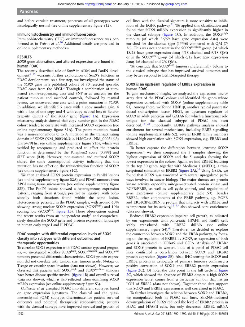
RESULTSSOX9 gene aberrations and altered expression are found inhuman PDACThe recently described role of Sox9 in ADM and PanIN devel-opment5 15 warrants further exploration of Sox9’s function inPDAC development. As a first step, we investigated the status ofthe SOX9 gene in a published cohort of 90 resected sporadicPDAC cases from the APGI.2 Through a combination of auto-mated exome-sequencing data and SNP array analysis on thepatient tumours and matched controls, followed by manualreview, we uncovered one case with a point mutation in SOX9.In addition, we identified 5 cases with a copy number gain, 4with a loss of one copy and 4 with copy neutral loss of hetero-zygosity (LOH) of the SOX9 gene (figure 1A). Expressionmicroarray analysis showed that copy number gain in the PDACcohort tended to correlate with increased SOX9 expression (seeonline supplementary figure S1A). The point mutation foundwas a non-synonymous C to A mutation in the transactivatingdomain of SOX9 (NM_000346.3: c.1436C>A; NM_000346.3:p.Pro479His; see online supplementary figure S1B), which wasverified by resequencing and predicted to affect the proteinfunction as determined by the Polyphen2 score (0.998) andSIFT score (0.0). However, non-mutated and mutated SOX9shared the same transcriptional activity, indicating that thismutation has no effect on the transactivation function of SOX9(see online supplementary figure S1C).
We then analysed SOX9 protein expression in PanIN lesions(see online supplementary figure S2A) and PDAC tumours fromAPGI using tissue microarrays (see online supplementary figureS2B). The PanIN lesions showed a heterogeneous expressionpattern, ranging from strongly positive to negative, with occa-sionally both situations found within the same lesion.Heterogeneity persisted in the PDAC samples, with around 60%showing strong nuclear SOX9 expression (SOX9high) and 10%scoring low (SOX9low; figure 1B). These observations extendthe recent results from an independent study5 and comprehen-sively describe the SOX9 gene and protein expression landscapein human early stage I and II PDAC.
PDAC samples with differential expression levels of SOX9classify into subtypes with different outcomes andtherapeutic opportunitiesTo correlate SOX9 expression with PDAC tumour type and progno-sis, we investigated whether SOX9high, SOX9medium and SOX9low
tumours presented differential characteristics. SOX9 protein expres-sion did not correlate with tumour size, tumour grade, N-stage orT-stage or vascular space invasion (data not shown). However, weobserved that patients with SOX9high and SOX9medium tumourshave better disease-specific survival (figure 1B) and overall survival(data not shown), which is also reflected when examining SOX9mRNA expression (see online supplementary figure S3).
Collisson et al classified PDAC into different subtypes basedon gene expression signatures. The classical and the quasi-mesenchymal (QM) subtypes discriminate for patient survivaloutcomes and potential therapeutic responsiveness; patientswith the classical subtype have improved survival and growth of
cell lines with the classical signature is more sensitive to inhib-ition of the EGFR pathway.21 We applied this classification andfound that SOX9 mRNA expression is significantly higher inthe classical subtype (figure 1C). In addition, the SOX9high
tumours (of which 36/69 have gene expression data) wereenriched for the classical type (15/36) compared with QM (7/36). This was not apparent in the SOX9medium group (of which18/29 have gene expression data; 4/18 classical and 6/18 QM)or in the SOX9low group (of which 6/12 have gene expressiondata; 1/6 classical and 2/6 QM).
We conclude that SOX9high tumours preferentially belong tothe classical subtype that has improved survival outcomes andmay better respond to EGFR-targeted therapy.
SOX9 is an upstream regulator of ERBB2 expression inhuman PDACTo gain mechanistic insight, we analysed the expression micro-array data of the PDAC cohort and identified the genes whoseexpression correlated with SOX9 (online supplementary tableS1). Among these, we found HNF1β, another typical pancreaticductal transcription factor, HES1, an upstream regulator ofSOX9 in adult pancreas and GATA6 for which a functional roleunique for the classical subtype of PDAC has beendescribed.14 21 Importantly, KOBAS pathway analysis showedenrichment for several mechanisms, including ERBB signalling(online supplementary table S2). Several ERBB family membersshowed high correlation with SOX9 expression, e.g. ERBB3 andERBB2.
To better capture the differences between ‘extreme SOX9expressors’, we then compared the 5 samples showing thehighest expression of SOX9 and the 5 samples showing thelowest expression in the cohort. Again, we find ERBB2 featuringin the top 30 genes, together with Mediator 1 (MED1), a tran-scriptional stimulator of ERBB2 (figure 2A).31 Using GSEA, wefound that SOX9 was associated with several upregulated path-ways involved in cancer biology. The major themes are proteinkinase activity, especially mitogen-activated protein kinase andEGFR/ERBB, as well as cell cycle control, and regulation ofgene expression (online supplementary table S3). BesidesERBB2, other components of the ERBB pathway, e.g. EGFRand ERBB2IP/ERBIN, a protein that interacts with ERBB2 andis important for its activity,32 33 also showed core enrichment(data not shown).
Reduced ERBB2 expression impaired cell growth, as indicatedby our experiments with pancreatic HPAFII and PanIN cellsstably transduced with ERBB2 shRNA (see onlinesupplementary figure S4).6 Therefore, we decided to explorethe connection between SOX9 and the ERBB pathway, by focus-ing on the regulation of ERBB2 by SOX9, as expression of bothgenes is associated in KOBAS and GSEA. Analysis of ERBB2and SOX9 protein in western blots of a panel of PDAC celllines confirmed a correlation between SOX9 and ERBB2protein expression (figure 2B). Also, IHC scoring for SOX9 andERBB2 protein in xenografts of primary tumours confirmed apositive correlation of SOX9 and ERBB2 protein expression(figure 2C). Of note, the data point in the full circle in figure2C, which showed the absence of ERBB2 despite a high SOX9expression score, comes from a particular tumour that has anLOH of ERBB2 (data not shown). Together these data supportthat SOX9 and ERBB2 expression is well correlated in PDAC.
To further investigate the relation between SOX9 and ERBB2,we manipulated both in PDAC cell lines. ShRNA-mediateddownregulation of SOX9 reduced the level of ERBB2 protein inHPAC and HPAFII cells, but only decreased ERBB2 mRNA
1792 Grimont A, et al. Gut 2015;64:1790–1799. doi:10.1136/gutjnl-2014-307075
Pancreas
group.bmj.com on January 11, 2016 - Published by http://gut.bmj.com/Downloaded from
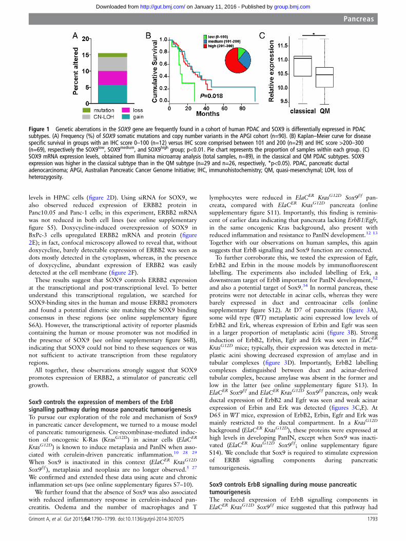
levels in HPAC cells (figure 2D). Using siRNA for SOX9, wealso observed reduced expression of ERBB2 protein inPanc10.05 and Panc-1 cells; in this experiment, ERBB2 mRNAwas not reduced in both cell lines (see online supplementaryfigure S5). Doxycycline-induced overexpression of SOX9 inBxPc-3 cells upregulated ERBB2 mRNA and protein (figure2E); in fact, confocal microscopy allowed to reveal that, withoutdoxycycline, barely detectable expression of ERBB2 was seen asdots mostly detected in the cytoplasm, whereas, in the presenceof doxycycline, abundant expression of ERBB2 was easilydetected at the cell membrane (figure 2F).
These results suggest that SOX9 controls ERBB2 expressionat the transcriptional and post-transcriptional level. To betterunderstand this transcriptional regulation, we searched forSOX9-binding sites in the human and mouse ERBB2 promotersand found a potential dimeric site matching the SOX9 bindingconsensus in these regions (see online supplementary figureS6A). However, the transcriptional activity of reporter plasmidscontaining the human or mouse promoter was not modified inthe presence of SOX9 (see online supplementary figure S6B),indicating that SOX9 could not bind to these sequences or wasnot sufficient to activate transcription from these regulatoryregions.
All together, these observations strongly suggest that SOX9promotes expression of ERBB2, a stimulator of pancreatic cellgrowth.
Sox9 controls the expression of members of the ErbBsignalling pathway during mouse pancreatic tumourigenesisTo pursue our exploration of the role and mechanism of Sox9in pancreatic cancer development, we turned to a mouse modelof pancreatic tumourigenesis. Cre-recombinase-mediated induc-tion of oncogenic K-Ras (KrasG12D) in acinar cells (ElaCER
KrasG12D) is known to induce metaplasia and PanIN when asso-ciated with cerulein-driven pancreatic inflammation.10 28 29
When Sox9 is inactivated in this context (ElaCER KrasG12D
Sox9f/f ), metaplasia and neoplasia are no longer observed.5 27
We confirmed and extended these data using acute and chronicinflammation set-ups (see online supplementary figures S7–10).
We further found that the absence of Sox9 was also associatedwith reduced inflammatory response in cerulein-induced pan-creatitis. Oedema and the number of macrophages and T
lymphocytes were reduced in ElaCER KrasG12D Sox9f/f pan-creata, compared with ElaCER KrasG12D pancreata (onlinesupplementary figure S11). Importantly, this finding is reminis-cent of earlier data indicating that pancreata lacking ErbB1/Egfr,in the same oncogenic Kras background, also present withreduced inflammation and resistance to PanIN development.12 13
Together with our observations on human samples, this againsuggests that ErbB signalling and Sox9 function are connected.
To further corroborate this, we tested the expression of Egfr,ErbB2 and Erbin in the mouse models by immunofluorescentlabelling. The experiments also included labelling of Erk, adownstream target of ErbB important for PanIN development,12
and also a potential target of Sox9.34 In normal pancreas, theseproteins were not detectable in acinar cells, whereas they werebarely expressed in duct and centroacinar cells (onlinesupplementary figure S12). At D7 of pancreatitis (figure 3A),some wild type (WT) metaplastic acini expressed low levels ofErbB2 and Erk, whereas expression of Erbin and Egfr was seenin a larger proportion of metaplastic acini (figure 3B). Stronginduction of ErbB2, Erbin, Egfr and Erk was seen in ElaCER
KrasG12D mice; typically, their expression was detected in meta-plastic acini showing decreased expression of amylase and intubular complexes (figure 3D). Importantly, ErbB2 labellingcomplexes distinguished between duct and acinar-derivedtubular complex, because amylase was absent in the former andlow in the latter (see online supplementary figure S13). InElaCER Sox9f/f and ElaCER KrasG12D Sox9f/f pancreas, only weakductal expression of ErbB2 and Egfr was seen and weak acinarexpression of Erbin and Erk was detected (figures 3C,E). AtD65 in WT mice, expression of ErbB2, Erbin, Egfr and Erk wasmainly restricted to the ductal compartment. In a KrasG12D
background (ElaCER KrasG12D), these proteins were expressed athigh levels in developing PanIN, except when Sox9 was inacti-vated (ElaCER KrasG12D Sox9f/f; online supplementary figureS14). We conclude that Sox9 is required to stimulate expressionof ERBB signalling components during pancreatictumourigenesis.
Sox9 controls ErbB signalling during mouse pancreatictumourigenesisThe reduced expression of ErbB signalling components inElaCER KrasG12D Sox9f/f mice suggested that this pathway had
Figure 1 Genetic aberrations in the SOX9 gene are frequently found in a cohort of human PDAC and SOX9 is differentially expressed in PDACsubtypes. (A) Frequency (%) of SOX9 somatic mutations and copy number variants in the APGI cohort (n=90). (B) Kaplan–Meier curve for diseasespecific survival in groups with an IHC score 0–100 (n=12) versus IHC score comprised between 101 and 200 (n=29) and IHC score >200–300(n=69), respectively the SOX9low, SOX9medium, and SOX9high group; p<0.01. Pie chart represents the proportion of samples within each group. (C)SOX9 mRNA expression levels, obtained from Illumina microarray analysis (total samples, n=89), in the classical and QM PDAC subtypes. SOX9expression was higher in the classical subtype than in the QM subtype (n=29 and n=26, respectively, *p<0.05). PDAC, pancreatic ductaladenocarcinoma; APGI, Australian Pancreatic Cancer Genome Initiative; IHC, immunohistochemistry; QM, quasi-mesenchymal; LOH, loss ofheterozygosity.
Grimont A, et al. Gut 2015;64:1790–1799. doi:10.1136/gutjnl-2014-307075 1793
Pancreas
group.bmj.com on January 11, 2016 - Published by http://gut.bmj.com/Downloaded from
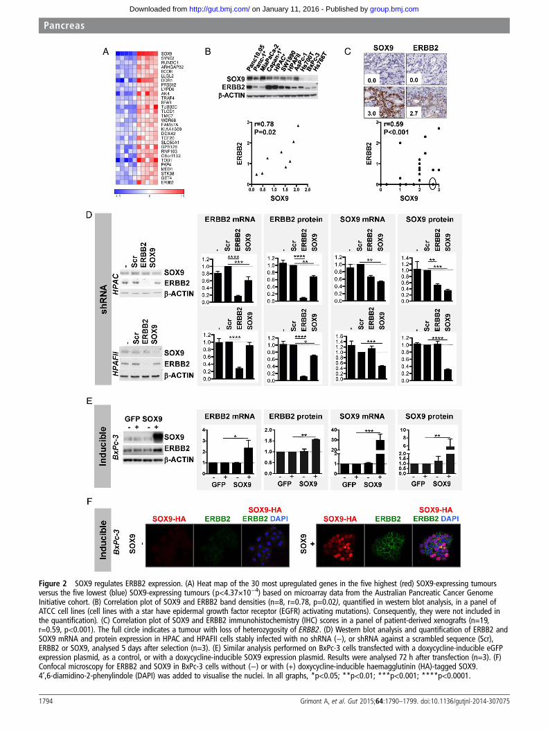
Figure 2 SOX9 regulates ERBB2 expression. (A) Heat map of the 30 most upregulated genes in the five highest (red) SOX9-expressing tumoursversus the five lowest (blue) SOX9-expressing tumours (p<4.37×10⁻⁴) based on microarray data from the Australian Pancreatic Cancer GenomeInitiative cohort. (B) Correlation plot of SOX9 and ERBB2 band densities (n=8, r=0.78, p=0.02), quantified in western blot analysis, in a panel ofATCC cell lines (cell lines with a star have epidermal growth factor receptor (EGFR) activating mutations). Consequently, they were not included inthe quantification). (C) Correlation plot of SOX9 and ERBB2 immunohistochemistry (IHC) scores in a panel of patient-derived xenografts (n=19,r=0.59, p<0.001). The full circle indicates a tumour with loss of heterozygosity of ERBB2. (D) Western blot analysis and quantification of ERBB2 andSOX9 mRNA and protein expression in HPAC and HPAFII cells stably infected with no shRNA (−), or shRNA against a scrambled sequence (Scr),ERBB2 or SOX9, analysed 5 days after selection (n=3). (E) Similar analysis performed on BxPc-3 cells transfected with a doxycycline-inducible eGFPexpression plasmid, as a control, or with a doxycycline-inducible SOX9 expression plasmid. Results were analysed 72 h after transfection (n=3). (F)Confocal microscopy for ERBB2 and SOX9 in BxPc-3 cells without (−) or with (+) doxycycline-inducible haemagglutinin (HA)-tagged SOX9.4’,6-diamidino-2-phenylindole (DAPI) was added to visualise the nuclei. In all graphs, *p<0.05; **p<0.01; ***p<0.001; ****p<0.0001.
1794 Grimont A, et al. Gut 2015;64:1790–1799. doi:10.1136/gutjnl-2014-307075
Pancreas
group.bmj.com on January 11, 2016 - Published by http://gut.bmj.com/Downloaded from
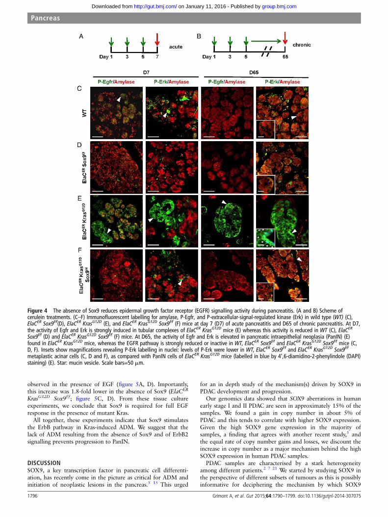
little or no activity in these mice. Phosphorylation of Egfr andErk is associated with activation of the ErbB pathway and wastaken for further investigation of pathway activity. In theabsence of pancreatitis, staining for P-Egfr and P-Erk was barelydetected in the ducts of the tested genotypes (see onlinesupplementary figure S12). At D7 of acute pancreatitis (figure4A), in WT mice, P-Egfr and P-Erk were detected in a subset ofmetaplastic acinar cells (figure 4C). These phosphorylated formswere no longer detected in the acinar compartment in theabsence of Sox9 (figure 4D). In contrast, in ElaCER KrasG12D
mice, strong levels of P-Egfr and P-Erk were found in metaplas-tic acini and tubular complexes (figure 4E). In ElaCER KrasG12D
Sox9f/f mice, no P-Egfr and weak levels of P-Erk were detectedin acinar cells (figure 4E). In the chronic setup (figure 4B), atD65, Erk was weakly phosphorylated in WT, ElaCER Sox9f/f andElaCER KrasG12D Sox9f/f mice, whereas high levels of phos-phorylated Erk were detected in PanINs of ElaCER KrasG12D
mice. P-Egfr was detected only in PanINs of ElaCER KrasG12D
mice (figure 4E). These data indicate that detection of P-Egfrand P-Erk and activation of the ErbB pathway are dependent onSox9 in the oncogenic Kras background.
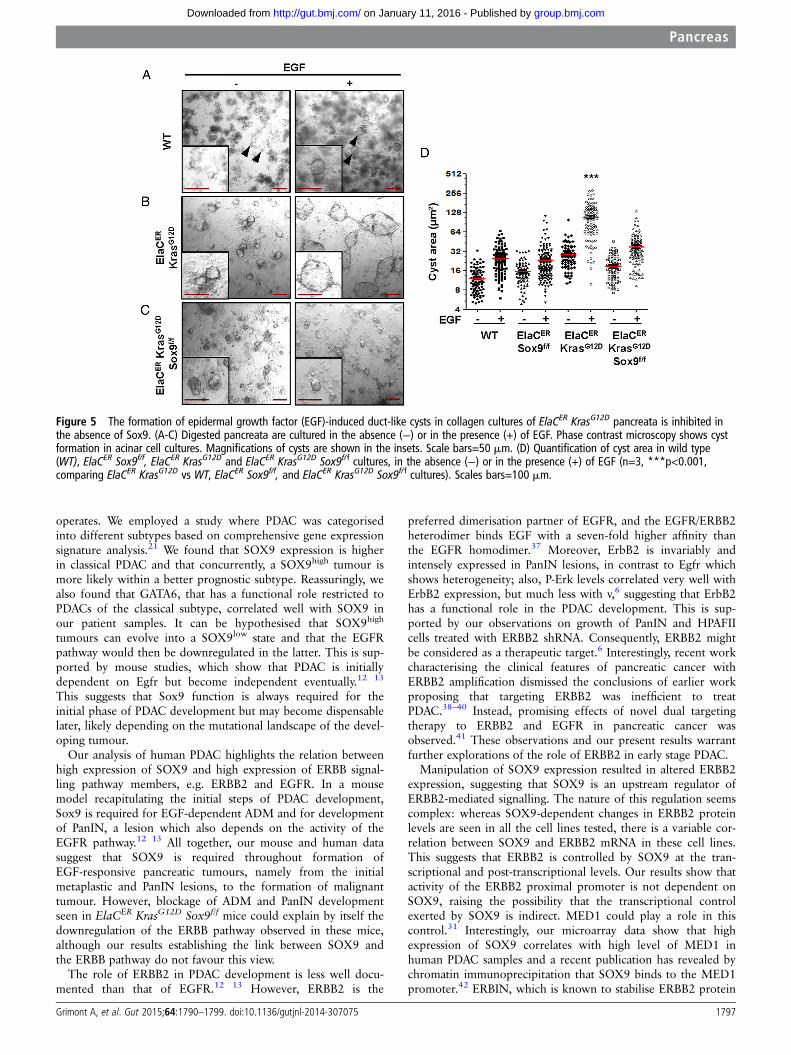
To further test the function of the ErbB pathway in theabsence of Sox9, pancreatic cell clusters, obtained by enzymaticdigestion, were embedded in collagen gel and cultured in theabsence or in the presence of epidermal growth factor (EGF). Insuch conditions, acinar cells undergo a metaplastic change andconvert into duct-like cysts in an EGF-dependent process.35 Inthe absence of EGF, the size of cysts was similar in WT andElaCER Sox9f/f tissue cultures (figure 5A, D). ElaCER KrasG12D
showed significantly larger cysts, confirming the propensity ofoncogenic Kras to produce metaplastic structures in theseculture conditions (figure 5B, D).36 EGF induced a 2.0-fold and1.5-fold increase in cyst size in WT and ElaCER Sox9f/f cultures,respectively; this indicates that Sox9 does not significantly affectthe function of EGF in basal conditions (figure 5A, D). InElaCER KrasG12D cultures, a 3.8-fold increase in cyst size was
Figure 3 Sox9 controls expression of members of the epidermal growth factor receptor (EGFR) pathway during pancreatitis. (A) Scheme of ceruleintreatment. (B-E) Immunofluorescent labelling of amylase, ErbB2, Erbin, Egfr and extracellular-signal-regulated kinase (Erk) in wild type (WT) (B),ElaCER Sox9f/f (C), ElaCER KrasG12D (D) and ElaCER KrasG12D Sox9f/f (E) mice at day 7 (D7) of acute pancreatitis. The expression of ErbB2, Erbin, Egfrand Erk is strongly induced in metaplastic acini and tubular complexes in the ElaCER KrasG12D pancreas (arrowhead, D), whereas it is only weaklyinduced in the WT metaplastic acinar cells (arrowhead, B). In ElaCER Sox9f/f (C) and ElaCER KrasG12D Sox9f/f (E) pancreas, this expression is restrictedto the apical pole of the metaplastic acinar cells for Erbin (C and E) or to the ducts, for ErbB2 (C and E) and Egfr (C and E). In both genotypes, Erkexpression is weakly observed in metaplastic acinar cells (arrowhead, E). Magnifications of WT and ElaCER KrasG12D metaplastic cells are shown inthe insets (B and D). Scale bars=50 mm.
Grimont A, et al. Gut 2015;64:1790–1799. doi:10.1136/gutjnl-2014-307075 1795
Pancreas
group.bmj.com on January 11, 2016 - Published by http://gut.bmj.com/Downloaded from
observed in the presence of EGF (figure 5A, D). Importantly,this increase was 1.8-fold lower in the absence of Sox9 (ElaCER
KrasG12D Sox9f/f; figure 5C, D). From these tissue cultureexperiments, we conclude that Sox9 is required for full EGFresponse in the presence of mutant Kras.
All together, these experiments indicate that Sox9 stimulatesthe ErbB pathway in Kras-induced ADM. We suggest that thelack of ADM resulting from the absence of Sox9 and of ErbB2signalling prevents progression to PanIN.
DISCUSSIONSOX9, a key transcription factor in pancreatic cell differenti-ation, has recently come in the picture as critical for ADM andinitiation of neoplastic lesions in the pancreas.5 15 This urged
for an in depth study of the mechanism(s) driven by SOX9 inPDAC development and progression.
Our genomics data showed that SOX9 aberrations in humanearly stage I and II PDAC are seen in approximately 15% of thesamples. We found a gain in copy number in about 5% ofPDAC and this tends to correlate with higher SOX9 expression.Given the high SOX9 gene expression in the majority ofsamples, a finding that agrees with another recent study,5 andthe equal rate of copy number gains and losses, we discount theincrease in copy number as a major mechanism behind the highSOX9 expression in human PDAC samples.
PDAC samples are characterised by a stark heterogeneityamong different patients.2 7 21 We started by studying SOX9 inthe perspective of different subsets of tumours as this is possiblyinformative for deciphering the mechanism by which SOX9
Figure 4 The absence of Sox9 reduces epidermal growth factor receptor (EGFR) signalling activity during pancreatitis. (A and B) Scheme ofcerulein treatments. (C–F) Immunofluorescent labelling for amylase, P-Egfr, and P-extracellular-signal-regulated kinase (Erk) in wild type (WT) (C),ElaCER Sox9f/f(D), ElaCER KrasG12D (E), and ElaCER KrasG12D Sox9f/f (F) mice at day 7 (D7) of acute pancreatitis and D65 of chronic pancreatitis. At D7,the activity of Egfr and Erk is strongly induced in tubular complexes of ElaCER KrasG12D mice (E) whereas this activity is reduced in WT (C), ElaCER
Sox9f/f (D) and ElaCER KrasG12D Sox9f/f (F) mice. At D65, the activity of Egfr and Erk is elevated in pancreatic intraepithelial neoplasia (PanIN) (E)found in ElaCER KrasG12D mice, whereas the EGFR pathway is strongly reduced or inactive in WT, ElaCER Sox9f/f and ElaCER KrasG12D Sox9f/f mice (C,D, F). Insets show magnifications revealing P-Erk labelling in nuclei: levels of P-Erk were lower in WT, ElaCER Sox9f/f and ElaCER KrasG12D Sox9f/f
metaplastic acinar cells (C, D and F), as compared with PanIN cells of ElaCER KrasG12D mice (labelled in blue by 4’,6-diamidino-2-phenylindole (DAPI)staining) (E). Star: mucin vesicle. Scale bars=50 mm.
1796 Grimont A, et al. Gut 2015;64:1790–1799. doi:10.1136/gutjnl-2014-307075
Pancreas
group.bmj.com on January 11, 2016 - Published by http://gut.bmj.com/Downloaded from
operates. We employed a study where PDAC was categorisedinto different subtypes based on comprehensive gene expressionsignature analysis.21 We found that SOX9 expression is higherin classical PDAC and that concurrently, a SOX9high tumour ismore likely within a better prognostic subtype. Reassuringly, wealso found that GATA6, that has a functional role restricted toPDACs of the classical subtype, correlated well with SOX9 inour patient samples. It can be hypothesised that SOX9high
tumours can evolve into a SOX9low state and that the EGFRpathway would then be downregulated in the latter. This is sup-ported by mouse studies, which show that PDAC is initiallydependent on Egfr but become independent eventually.12 13
This suggests that Sox9 function is always required for theinitial phase of PDAC development but may become dispensablelater, likely depending on the mutational landscape of the devel-oping tumour.
Our analysis of human PDAC highlights the relation betweenhigh expression of SOX9 and high expression of ERBB signal-ling pathway members, e.g. ERBB2 and EGFR. In a mousemodel recapitulating the initial steps of PDAC development,Sox9 is required for EGF-dependent ADM and for developmentof PanIN, a lesion which also depends on the activity of theEGFR pathway.12 13 All together, our mouse and human datasuggest that SOX9 is required throughout formation ofEGF-responsive pancreatic tumours, namely from the initialmetaplastic and PanIN lesions, to the formation of malignanttumour. However, blockage of ADM and PanIN developmentseen in ElaCER KrasG12D Sox9f/f mice could explain by itself thedownregulation of the ERBB pathway observed in these mice,although our results establishing the link between SOX9 andthe ERBB pathway do not favour this view.
The role of ERBB2 in PDAC development is less well docu-mented than that of EGFR.12 13 However, ERBB2 is the
preferred dimerisation partner of EGFR, and the EGFR/ERBB2heterodimer binds EGF with a seven-fold higher affinity thanthe EGFR homodimer.37 Moreover, ErbB2 is invariably andintensely expressed in PanIN lesions, in contrast to Egfr whichshows heterogeneity; also, P-Erk levels correlated very well withErbB2 expression, but much less with v,6 suggesting that ErbB2has a functional role in the PDAC development. This is sup-ported by our observations on growth of PanIN and HPAFIIcells treated with ERBB2 shRNA. Consequently, ERBB2 mightbe considered as a therapeutic target.6 Interestingly, recent workcharacterising the clinical features of pancreatic cancer withERBB2 amplification dismissed the conclusions of earlier workproposing that targeting ERBB2 was inefficient to treatPDAC.38–40 Instead, promising effects of novel dual targetingtherapy to ERBB2 and EGFR in pancreatic cancer wasobserved.41 These observations and our present results warrantfurther explorations of the role of ERBB2 in early stage PDAC.
Manipulation of SOX9 expression resulted in altered ERBB2expression, suggesting that SOX9 is an upstream regulator ofERBB2-mediated signalling. The nature of this regulation seemscomplex: whereas SOX9-dependent changes in ERBB2 proteinlevels are seen in all the cell lines tested, there is a variable cor-relation between SOX9 and ERBB2 mRNA in these cell lines.This suggests that ERBB2 is controlled by SOX9 at the tran-scriptional and post-transcriptional levels. Our results show thatactivity of the ERBB2 proximal promoter is not dependent onSOX9, raising the possibility that the transcriptional controlexerted by SOX9 is indirect. MED1 could play a role in thiscontrol.31 Interestingly, our microarray data show that highexpression of SOX9 correlates with high level of MED1 inhuman PDAC samples and a recent publication has revealed bychromatin immunoprecipitation that SOX9 binds to the MED1promoter.42 ERBIN, which is known to stabilise ERBB2 protein
Figure 5 The formation of epidermal growth factor (EGF)-induced duct-like cysts in collagen cultures of ElaCER KrasG12D pancreata is inhibited inthe absence of Sox9. (A-C) Digested pancreata are cultured in the absence (−) or in the presence (+) of EGF. Phase contrast microscopy shows cystformation in acinar cell cultures. Magnifications of cysts are shown in the insets. Scale bars=50 mm. (D) Quantification of cyst area in wild type(WT), ElaCER Sox9f/f, ElaCER KrasG12D and ElaCER KrasG12D Sox9f/f cultures, in the absence (−) or in the presence (+) of EGF (n=3, ***p<0.001,comparing ElaCER KrasG12D vs WT, ElaCER Sox9f/f, and ElaCER KrasG12D Sox9f/f cultures). Scales bars=100 mm.
Grimont A, et al. Gut 2015;64:1790–1799. doi:10.1136/gutjnl-2014-307075 1797
Pancreas
group.bmj.com on January 11, 2016 - Published by http://gut.bmj.com/Downloaded from
and to restrict its localisation to the cell membrane,32 33 likelyplays a role in the post-transcriptional control. Thus, high levelsof SOX9 are associated with high level of ERBIN in humanPDAC samples and downregulation of ERBIN expression is seenin ElaCER KrasG12D Sox9f/f mice. Interestingly, when SOX9 wasoverexpressed in BxPC3 cells, increased localisation of ERBB2at the cell membrane was associated with an upregulation ofERBIN expression (data not shown).
Intriguingly, a number of immune-related pathways weredownregulated in SOX9 high-expressing tumours, includingimmune response, response to virus, adaptive immune responseand cytokine biosynthesis and secretion, primarily driven bychanges in FOXP3, CD40L, SFTPD, TLR7 and 8, as well as anumber of chemokines, cytokines and interleukins. This furtherunderscores the functional differences between SOX9high andSOX9low tumours.
In conclusion, our results uncover the regulation of EGFR/ERBB2 by SOX9 in PDAC development, a finding that opensperspective for further exploration of therapeutic interventiontargeting EGFR and ERBB2 during pancreatic cancerdevelopment.
Author affiliations1Université catholique de Louvain, de Duve Institute, Brussels, Belgium2Cancer Research Division, The Kinghorn Cancer Centre, Garvan Institute of MedicalResearch, Sydney, Australia3Australian Pancreatic Cancer Genome Initiative4Queensland Centre for Medical Genomics, Institute for Molecular Bioscience, TheUniversity of Queensland, Queensland, Australia5St Vincent’s Clinical School, University New South Wales, Australia6Department of Pathology, Université catholique de Louvain, Cliniques UniversitairesSt Luc, Brussels, Belgium7Wolfson Wohl Cancer Centre, University of Glasgow, Scotland, UK
Acknowledgements We thank M. Sander and M. Hebrok for sharing results priorto publication, S. Konieczny for advice, Rolf Kemler and the Developmental StudiesHybridoma Bank (University of Iowa) for CK19 antibody, L. Mei for Erbin antibody,B. de Crombrugghe for p89 plasmid, P.P. Prévot for discussions, and D. Stoffers,D. Tuveson and G. Scherer for providing the ElaCreER, LSL KrasG12D and Sox9f/f mice,respectively. We thank B. Pastorelli, B. Pirlot and J. Pettitt for expert technicalassistance.
Contributors AG: study concept and design, acquisition of data, analysis andinterpretation of data, statistical analysis and drafting of manuscript. AVP, MJC,AM and JW: acquisition of data, analysis and interpretation of data and statisticalanalysis. CA, MG-L, GVdS and NW: acquisition of data, analysis and interpretationof data. MP: technical or material support. CS: analysis and interpretation of data.SMG and AVB: obtaining funding, technical or material support. FPL, IR and PJ:study concept and design, analysis and interpretation of data, drafting ofmanuscript and obtaining funding.
Funding This work was supported by grants from the Cancer Institute NSW;National Health and Medical Research Council of Australia; Australian Government:Department of Innovation, Industry, Science, Research and Tertiary Education;Australian Cancer Research Foundation; Queensland Government; University ofQueensland; Cancer Council NSW; Garvan Institute of Medical Research; AvnerNahmani Pancreatic Cancer Research Foundation; R.T. Hall Trust; Petre Foundation;Jane Hemstritch in memory of Philip Hemstritch; Gastroenterological Society ofAustralia; American Association for Cancer Research Landon Foundation—INNOVATOR Award; Royal Australasian College of Surgeons; Royal AustralasianCollege of Physicians; Royal College of Pathologists of Australasia; HGSC-BCM:NHGRI U54 HG003273; CPRIT grant RP101353-P7 (Tumour Banking for GenomicResearch and Clinical Translation Site 1); Fondation contre le Cancer (Belgium);Fonds de la Recherche Scientifique-FNRS (Belgium); Université catholique deLouvain. AG was supported by a grant from EU FP7 (Marie Curie Initial TrainingNetwork BOLD) and a Télévie fellowship. AVP and MJC are supported by earlycareer fellowships from Cancer Institute NSW. CA holds a Télévie fellowship, IR is aFuture Research Fellow of the Cancer Institute NSW (10/FRL2-03) and PJ is aResearch Associate of the Fonds de la Recherche Scientifique-FNRS.
Competing interests None.
Ethics approval Sydney Local Health District—RPA Zone, protocol X11-0220.
Provenance and peer review Not commissioned; externally peer reviewed.
Data sharing statement Unpublished data are available upon e-mail request tothe corresponding authors.
REFERENCES1 Costello E, Greenhalf W, Neoptolemos JP. New biomarkers and targets in pancreatic
cancer and their application to treatment. Nat Rev Gastroenterol Hepatol2012;9:435–44.
2 Biankin AV, Waddell N, Kassahn KS, et al. Pancreatic cancer genomes revealaberrations in axon guidance pathway genes. Nature 2012;491:399–405.
3 Guerra C, Barbacid M. Genetically engineered mouse models of pancreaticadenocarcinoma. Mol Oncol 2013;7:232–47.
4 Rooman I, Real FX. Pancreatic ductal adenocarcinoma and acinar cells: a matter ofdifferentiation and development? Gut 2012;61:449–58.
5 Kopp JL, von Figura G, Mayes E, et al. Identification of Sox9-dependentacinar-to-ductal reprogramming as the principal mechanism for initiation ofpancreatic ductal adenocarcinoma. Cancer Cell 2012;22:737–50.
6 Hingorani SR, Wang L, Multani AS, et al. Trp53R172H and KrasG12D cooperate topromote chromosomal instability and widely metastatic pancreatic ductaladenocarcinoma in mice. Cancer Cell 2005;7:469–83.
7 Jones S, Zhang X, Parsons DW, et al. Core signaling pathways in human pancreaticcancers revealed by global genomic analyses. Science 2008;321:1801–6.
8 Pinho AV, Chantrill L, Rooman I. Chronic pancreatitis: a path to pancreatic cancer.Cancer Lett 2014;345:203–9.
9 Pinho AV, Rooman I, Reichert M, et al. Adult pancreatic acinar cells dedifferentiateto an embryonic progenitor phenotype with concomitant activation of a senescenceprogramme that is present in chronic pancreatitis. Gut 2011;60:958–66.
10 Guerra C, Schuhmacher AJ, Canamero M, et al. Chronic pancreatitis is essential forinduction of pancreatic ductal adenocarcinoma by K-Ras oncogenes in adult mice.Cancer Cell 2007;11:291–302.
11 Collins MA, Bednar F, Zhang Y, et al. Oncogenic Kras is required for both theinitiation and maintenance of pancreatic cancer in mice. J Clin Invest2012;122:639–53.
12 Ardito CM, Gruner BM, Takeuchi KK, et al. EGF receptor is required forKRAS-induced pancreatic tumorigenesis. Cancer Cell 2012;22:304–17.
13 Navas C, Hernandez-Porras I, Schuhmacher AJ, et al. EGF receptor signaling isessential for k-ras oncogene-driven pancreatic ductal adenocarcinoma. Cancer Cell2012;22:318–30.
14 Shih HP, Kopp JL, Sandhu M, et al. A Notch-dependent molecular circuitry initiatespancreatic endocrine and ductal cell differentiation. Development2012;139:2488–99.
15 Prevot PP, Simion A, Grimont A, et al. Role of the ductal transcription factors HNF6and Sox9 in pancreatic acinar-to-ductal metaplasia. Gut 2012;61:1723–32.
16 Sun M, Li N, Dong W, et al. Copy-number mutations on chromosome17q24.2-q24.3 in congenital generalized hypertrichosis terminalis with or withoutgingival hyperplasia. Am J Hum Genet 2009;84:807–13.
17 Coustry F, Oh CD, Hattori T, et al. The dimerization domain of SOX9 is required fortranscription activation of a chondrocyte-specific chromatin DNA template. NucleicAcids Res 2010;38:6018–28.
18 Sudbeck P, Schmitz ML, Baeuerle PA, et al. Sex reversal by loss of the C-terminaltransactivation domain of human SOX9. Nat Genet 1996;13:230–2.
19 Adzhubei IA, Schmidt S, Peshkin L, et al. A method and server for predictingdamaging missense mutations. Nat Methods 2010;7:248–9.
20 Ng PC, Henikoff S. SIFT: Predicting amino acid changes that affect protein function.Nucleic Acids Res 2003;31:3812–14.
21 Collisson EA, Sadanandam A, Olson P, et al. Subtypes of pancreatic ductaladenocarcinoma and their differing responses to therapy. Nat Med 2011;17:500–3.
22 Wu J, Mao X, Cai T, et al. KOBAS server: a web-based platform for automatedannotation and pathway identification. Nucleic Acids Res 2006;34:W720–4.
23 Xie C, Mao X, Huang J, et al. KOBAS 2.0: a web server for annotation andidentification of enriched pathways and diseases. Nucleic Acids Res 2011;39:W316–22.
24 Storey JD. A direct approach to false discovery rates. J Royal Stat Soc B2002;64:479–98.
25 Subramanian A, Tamayo P, Mootha VK, et al. Gene set enrichment analysis: aknowledge-based approach for interpreting genome-wide expression profiles. ProcNatl Acad Sci USA 2005;102:15545–50.
26 Liberzon A, Subramanian A, Pinchback R, et al. Molecular signatures database(MSigDB) 3.0. Bioinformatics 2011;27:1739–40.
27 Kist R, Schrewe H, Balling R, et al. Conditional inactivation of Sox9: a mouse modelfor campomelic dysplasia. Genesis 2002;32:121–3.
28 Hingorani SR, Petricoin EF, Maitra A, et al. Preinvasive and invasive ductalpancreatic cancer and its early detection in the mouse. Cancer Cell 2003;4:437–50.
29 Desai BM, Oliver-Krasinski J, De Leon DD, et al. Preexisting pancreatic acinar cellscontribute to acinar cell, but not islet beta cell, regeneration. J Clin Invest2007;117:971–7.
1798 Grimont A, et al. Gut 2015;64:1790–1799. doi:10.1136/gutjnl-2014-307075
Pancreas
group.bmj.com on January 11, 2016 - Published by http://gut.bmj.com/Downloaded from
30 Prévot PP, Augereau C, Simion A, et al. Let-7b and miR-495 stimulatedifferentiation and prevent metaplasia of pancreatic acinar cells by repressing HNF6.Gastroenterology 2013;145:668–78 e3.
31 Cui J, Germer K, Wu T, et al. Cross-talk between HER2 and MED1 regulatestamoxifen resistance of human breast cancer cells. Cancer Res 2012;72:5625–34.
32 Borg JP, Marchetto S, Le Bivic A, et al. ERBIN: a basolateral PDZ protein thatinteracts with the mammalian ERBB2/HER2 receptor. Nat Cell Biol 2000;2:407–14.
33 Tao Y, Dai P, Liu Y, et al. Erbin regulates NRG1 signaling and myelination. Proc NatlAcad Sci USA 2009;106:9477–82.
34 Oh CD, Maity SN, Lu JF, et al. Identification of SOX9 interaction sites in thegenome of chondrocytes. PLoS ONE 2010;5:e10113.
35 Means AL, Meszoely IM, Suzuki K, et al. Pancreatic epithelial plasticity mediated byacinar cell transdifferentiation and generation of nestin-positive intermediates.Development 2005;132:3767–76.
36 Shi G, Direnzo D, Qu C, et al. Maintenance of acinar cell organization is critical topreventing Kras-induced acinar-ductal metaplasia. Oncogene 2013;32:1950–8.
37 Li Y, Macdonald-Obermann J, Westfall C, et al. Quantitation of the effect of ErbB2on epidermal growth factor receptor binding and dimerization. J Biol Chem2012;287:31116–25.
38 Safran H, Iannitti D, Ramanathan R, et al. Herceptin and gemcitabine for metastaticpancreatic cancers that overexpress HER-2/neu. Cancer Invest 2004;22:706–12.
39 Harder J, Ihorst G, Heinemann V, et al. Multicentre phase II trial of trastuzumaband capecitabine in patients with HER2 overexpressing metastatic pancreatic cancer.Br J Cancer 2012;106:1033–8.
40 Chou A, Waddell N, Cowley MJ, et al. Clinical and molecular characterization ofHER2 amplified-pancreatic cancer. Genome Med 2013;5:78.
41 Maron R, Schechter B, Mancini M, et al. Inhibition of pancreatic carcinoma byhomo- and heterocombinations of antibodies against EGF-receptor and its kinHER2/ErbB-2. Proc Natl Acad Sci USA 2013;110:15389–94.
42 Li Y, Zheng M, Lau YF. The Sex-Determining Factors SRY and SOX9 Regulate SimilarTarget Genes and Promote Testis Cord Formation during Testicular Differentiation.Cell Rep 2014;8:723–33.
Grimont A, et al. Gut 2015;64:1790–1799. doi:10.1136/gutjnl-2014-307075 1799
Pancreas
group.bmj.com on January 11, 2016 - Published by http://gut.bmj.com/Downloaded from
pancreatic cancer developmentSOX9 regulates ERBB signalling in
Patrick JacqueminGrimmond, Andrew V Biankin, Frédéric P Lemaigre, Ilse Rooman and Waddell, Marina Pajic, Christine Sempoux, Jianmin Wu, Sean MAmanda Mawson, Marc Giry-Laterrière, Géraldine Van den Steen, Nicola Adrien Grimont, Andreia V Pinho, Mark J Cowley, Cécile Augereau,
doi: 10.1136/gutjnl-2014-3070752015 64: 1790-1799 originally published online October 21, 2014Gut
http://gut.bmj.com/content/64/11/1790Updated information and services can be found at:
These include:
MaterialSupplementary
htmlhttp://gut.bmj.com/content/suppl/2014/10/21/gutjnl-2014-307075.DC1.Supplementary material can be found at:
References #BIBLhttp://gut.bmj.com/content/64/11/1790
This article cites 42 articles, 15 of which you can access for free at:
serviceEmail alerting
box at the top right corner of the online article. Receive free email alerts when new articles cite this article. Sign up in the
CollectionsTopic Articles on similar topics can be found in the following collections
(643)Pancreatic cancer (1924)Pancreas and biliary tract
Notes
http://group.bmj.com/group/rights-licensing/permissionsTo request permissions go to:
http://journals.bmj.com/cgi/reprintformTo order reprints go to:
http://group.bmj.com/subscribe/To subscribe to BMJ go to:
group.bmj.com on January 11, 2016 - Published by http://gut.bmj.com/Downloaded from
Related Documents