Sonic hedgehog-patched Gli signaling in the developing rat prostate gland: lobe-specific suppression by neonatal estrogens reduces ductal growth and branching Yongbing Pu, Liwei Huang, Gail S. Prins * Department of Urology, College of Medicine, University of Illinois at Chicago, Chicago, IL 60612, USA Received for publication 10 February 2004, revised 11 June 2004, accepted 11 June 2004 Available online 20 July 2004 Abstract While prostate gland development is dependent on androgens, other hormones including retinoids and estrogens can influence this process. Brief exposure to high-dose estrogen during the neonatal period in rats leads to permanent, lobe-specific aberrations in the prostate gland, a phenomenon referred to as developmental estrogenization. We have previously shown that this response is mediated through alterations in steroid receptor expression; however, further downstream mechanisms remain unclear. Herein, we examined Sonic hedgehog (Shh)-patched (ptc)-gli in the developing rat prostate gland, its role in branching morphogenesis, and the effects of neonatal estrogens on its expression and localization to determine whether a disturbance in this signaling pathway is involved in mediating the estrogenized phenotype. Shh was expressed in epithelial cells at the distal tips of elongating ducts in discreet, heterogeneous foci, while ptc and gli1–3 were expressed in the adjacent mesenchymal cells in the developing gland. The addition of Shh protein to cultured neonatal prostates reduced ductal growth and branching, decreased Fgf10 transcript, and increased Bmp4 expression in the adjacent mesenchyme. Shh-induced growth suppression was reversed by exogenous Fgf10, but not noggin, indicating that Fgf10 suppression is the proximate cause of the growth inhibition. A model is proposed to show how highly localized Shh expression along with regulation of downstream morphogens participates in dichotomous branching during prostate morphogenesis. Neonatal exposure to high-dose estradiol suppressed Shh, ptc, gli1, and gli3 expressions and concomitantly blocked ductal branching in the dorsal and lateral prostate lobes specifically. In contrast, ventral lobe branching and Shh-ptc-gli expression were minimally affected by estrogen exposure. Organ culture studies with lateral prostates confirmed that estradiol suppressed Shh-ptc-gli expression directly at the prostatic level. Taken together, the present findings indicate that lobe-specific decreases in Shh-ptc-gli expression are involved in mediating estradiol-induced suppression of dorsal and lateral lobe ductal growth and branching during prostate morphogenesis. D 2004 Elsevier Inc. All rights reserved. Keywords: Sonic hedgehog; Patched; Gli; Fgf10; Bmp4; Prostate; Estrogen; Estradiol Introduction Prostate gland development is dependent on androgens which stimulate ductal outgrowth, branching morphogene- sis, cellular differentiation, and onset of secretory activity (George and Peterson, 1988; Siiteri and Wilson, 1974). In addition, there is evidence that other hormones including estrogens and retinoids can influence these processes, al- though their exact role in normal prostatic development is less clear (Aboseif et al., 1997; Price, 1963; Seo et al., 1997). In humans, prostate morphogenesis occurs entirely in utero under the influence of testosterone secreted from the fetal testes (Lowsley, 1912; Shapiro, 1990). There is also clear indication that rising maternal estrogens during the third trimester have a direct effect on the human prostate since the epithelium develops marked squamous metaplasia which sloughs at birth when maternal estrogen levels abruptly fall (Brody and Goldman, 1940; Zondek and Zondek, 1975). Furthermore, maternal exposure to pharma- cological levels of diethylstilbestrol (DES) has been shown to induce prostatic abnormalities in human offspring (Dris- coll and Taylor, 1980). Consequently, it has been proposed that excessive estrogenization during prostatic development 0012-1606/$ - see front matter D 2004 Elsevier Inc. All rights reserved. doi:10.1016/j.ydbio.2004.06.002 * Corresponding author. Department of Urology, University of Illinois at Chicago, M/C 955, 820 S. Wood St., Chicago, IL 60612. Fax: +1-312- 996-1291. E-mail address: [email protected] (G.S. Prins). www.elsevier.com/locate/ydbio Developmental Biology 273 (2004) 257 – 275

Welcome message from author
This document is posted to help you gain knowledge. Please leave a comment to let me know what you think about it! Share it to your friends and learn new things together.
Transcript

www.elsevier.com/locate/ydbio
Developmental Biology 273 (2004) 257–275
Sonic hedgehog-patched Gli signaling in the developing rat prostate gland:
lobe-specific suppression by neonatal estrogens reduces ductal
growth and branching
Yongbing Pu, Liwei Huang, Gail S. Prins*
Department of Urology, College of Medicine, University of Illinois at Chicago, Chicago, IL 60612, USA
Received for publication 10 February 2004, revised 11 June 2004, accepted 11 June 2004
Available online 20 July 2004
Abstract
While prostate gland development is dependent on androgens, other hormones including retinoids and estrogens can influence this
process. Brief exposure to high-dose estrogen during the neonatal period in rats leads to permanent, lobe-specific aberrations in the prostate
gland, a phenomenon referred to as developmental estrogenization. We have previously shown that this response is mediated through
alterations in steroid receptor expression; however, further downstream mechanisms remain unclear. Herein, we examined Sonic hedgehog
(Shh)-patched (ptc)-gli in the developing rat prostate gland, its role in branching morphogenesis, and the effects of neonatal estrogens on its
expression and localization to determine whether a disturbance in this signaling pathway is involved in mediating the estrogenized
phenotype. Shh was expressed in epithelial cells at the distal tips of elongating ducts in discreet, heterogeneous foci, while ptc and gli1–3
were expressed in the adjacent mesenchymal cells in the developing gland. The addition of Shh protein to cultured neonatal prostates reduced
ductal growth and branching, decreased Fgf10 transcript, and increased Bmp4 expression in the adjacent mesenchyme. Shh-induced growth
suppression was reversed by exogenous Fgf10, but not noggin, indicating that Fgf10 suppression is the proximate cause of the growth
inhibition. A model is proposed to show how highly localized Shh expression along with regulation of downstream morphogens participates
in dichotomous branching during prostate morphogenesis. Neonatal exposure to high-dose estradiol suppressed Shh, ptc, gli1, and gli3
expressions and concomitantly blocked ductal branching in the dorsal and lateral prostate lobes specifically. In contrast, ventral lobe
branching and Shh-ptc-gli expression were minimally affected by estrogen exposure. Organ culture studies with lateral prostates confirmed
that estradiol suppressed Shh-ptc-gli expression directly at the prostatic level. Taken together, the present findings indicate that lobe-specific
decreases in Shh-ptc-gli expression are involved in mediating estradiol-induced suppression of dorsal and lateral lobe ductal growth and
branching during prostate morphogenesis.
D 2004 Elsevier Inc. All rights reserved.
Keywords: Sonic hedgehog; Patched; Gli; Fgf10; Bmp4; Prostate; Estrogen; Estradiol
Introduction less clear (Aboseif et al., 1997; Price, 1963; Seo et al.,
Prostate gland development is dependent on androgens
which stimulate ductal outgrowth, branching morphogene-
sis, cellular differentiation, and onset of secretory activity
(George and Peterson, 1988; Siiteri and Wilson, 1974). In
addition, there is evidence that other hormones including
estrogens and retinoids can influence these processes, al-
though their exact role in normal prostatic development is
0012-1606/$ - see front matter D 2004 Elsevier Inc. All rights reserved.
doi:10.1016/j.ydbio.2004.06.002
* Corresponding author. Department of Urology, University of Illinois
at Chicago, M/C 955, 820 S. Wood St., Chicago, IL 60612. Fax: +1-312-
996-1291.
E-mail address: [email protected] (G.S. Prins).
1997). In humans, prostate morphogenesis occurs entirely in
utero under the influence of testosterone secreted from the
fetal testes (Lowsley, 1912; Shapiro, 1990). There is also
clear indication that rising maternal estrogens during the
third trimester have a direct effect on the human prostate
since the epithelium develops marked squamous metaplasia
which sloughs at birth when maternal estrogen levels
abruptly fall (Brody and Goldman, 1940; Zondek and
Zondek, 1975). Furthermore, maternal exposure to pharma-
cological levels of diethylstilbestrol (DES) has been shown
to induce prostatic abnormalities in human offspring (Dris-
coll and Taylor, 1980). Consequently, it has been proposed
that excessive estrogenization during prostatic development
Y. Pu et al. / Developmental Biology 273 (2004) 257–275258
may contribute to the high incidence of benign prostatic
hyperplasia (BPH) and prostatic carcinoma currently ob-
served in the aging male population (Santti et al., 1994).
Prostate gland development in the rodent is initiated late
in gestation as buds emerge from the urogenital sinus
(UGS), and in contrast to humans, the gland undergoes
extensive branching morphogenesis and cellular differenti-
ation during the postnatal period (Hayashi et al., 1991).
Thus, the neonatal rodent prostate gland has emerged as a
useful model for fetal prostate development in humans.
Brief exposure of rodents to estrogens during the neonatal
period has been shown to have permanent, irreversible, and
dose-dependent effects on the prostate gland’s morphology,
cellular organization, and function (Prins, 1992; Prins and
Birch, 1995; Pylkkanen et al., 1993; Rajfer and Coffey,
1979; vom Saal et al., 1997). If estrogenic exposures are
high, the permanent imprints include reduced prostatic
growth, epithelial differentiation defects, altered secretory
function, and aging-associated dysplasia similar to prostatic
intraepithelial neoplasia or PIN (Naslund and Coffey, 1986;
Prins, 1992, 1997; Prins et al., 1993; Rajfer and Coffey,
1979). In the rat model, the responses are lobe-specific with
differentiation abnormalities and adult-onset dysplasia
occurring with the highest frequency and severity in the
ventral prostate lobe (VP), while the dorsal (DP) and lateral
(LP) lobes show greater inhibition of ductal branching and
complexity (Prins, 1992, 1997). This process, referred to as
estrogenic imprinting or developmental estrogenization, is
used as a model to evaluate the role of exogenous and
endogenous estrogens as a potential predisposing factor for
prostate diseases later in life.
The mechanism of developmental estrogenization of the
prostate gland is not well understood. Previous studies from
our laboratory using estrogen receptor (ER) knockout mice
for ERa and ERh determined that the effects are mediated
through stromal ERa which is transiently up-regulated
following neonatal estrogen exposure (Prins and Birch,
1997). Furthermore, the expression of several steroid recep-
tors which mediate steroid action in the developing prostate
is drastically altered both temporally and quantitatively by
neonatal estrogens. Most notably, androgen receptor (AR) is
markedly down-regulated, while ERa, progesterone recep-
tor (PR), and retinoic acid receptors (RARa, RARh, andRXRa) are significantly up-regulated in a cell-specific
manner (Prins and Birch, 1995, 1997; Prins et al., 1998,
2001a,b, 2002; Pu et al., 2003; Woodham et al., 2003). In
addition, retinoid metabolizing enzymes and retinoid levels
are affected by estrogenization in a lobe-specific manner
(Prins et al., 2002; Pu et al., 2003). The net effect of these
alterations is that the developing prostate is no longer under
predominant androgen regulation through AR but is rather
driven by estrogens, progesterone, and retinoid signals. We
currently hypothesize that critical developmental genes
which dictate prostate morphogenesis may be downstream
targets of steroid action in the prostate gland. Furthermore,
we predict that the expression levels and patterns of these
steroid-regulated developmental genes will be disturbed by
the prostatic steroid receptor shift as a result of neonatal
estrogenic exposure. In the present study, we test the
hypothesis that the Shh-ptc-gli signaling pathway may be
altered in response to prostatic estrogenization and that this
may, in part, mediate specific aspects of that phenotype.
Sonic hedgehog (Shh) is a secreted glycoprotein pro-
duced by epithelial cells at mesenchymal interfaces in
developing tissues where it is involved in determination of
cell fate, proliferation, and embryonic patterning (see review
by Ingham and McMahon, 2001). Shh is expressed in a
spatially defined manner in developing glands, most com-
monly at the apical edge of outgrowing ducts. This secreted
morphogen binds to membrane-bound patched (ptc) recep-
tors on adjacent mesenchymal cells, thus establishing epi-
thelial–mesenchymal cross-talk. Liganding of ptc by Shh
relieves its inhibition on smoothened (smo) which allows for
activation of Gli transcription factors that directly mediate
Shh’s effects. In vertebrates, there are three known Gli
transcripts; gli1, gli2, and gli3 which have both redundant
and unique actions. Importantly, gli1 and gli2 are transcrip-
tional activators, while gli3 is believed to be a transcrip-
tional repressor (Meyer and Roelink, 2003) which permits
tight regulation of Shh actions. Both short-range and long-
range actions of Shh have been described which differ as a
function of concentration gradients (Gritli-Linde et al.,
2001). In most structures, Shh is considered to be a critical
regulatory morphogen since it regulates the expression of
other secreted morphogens and homeobox genes (Chuang
and McMahon, 2003; Haraguchi et al., 2001; Perriton et al.,
2002; Roberts et al., 1995, 1998; Schneider et al., 2000). It
has also been shown to induce ptc and gli1 expression, thus
establishing an autoregulatory loop (Marigo and Babin,
1996).
Recently, the spatiotemporal expression of Shh-ptc-gli1
was characterized for the murine prostate gland (Lamm et
al., 2002), while Shh-ptc mRNA expression was temporally
described for the rat ventral prostate (Freestone et al., 2003).
Similar to the lung, Shh was localized to the distal tips of the
outgrowing murine prostatic buds, while ptc and gli1 were
expressed in the adjacent mesenchyme. Expression of these
signaling molecules was highest in the perinatal UGS
complex and levels declined as prostate morphogenesis
proceeded. Since studies with Shh null mice demonstrated
that prostatic budding could be initiated by testosterone in
the absence of Shh, it does not appear to be necessary for
prostatic induction (Berman et al., 2004; Freestone et al.,
2003). Nonetheless, inhibition of Shh action in UGS and
prostate organ cultures by cyclopamine, an inhibitor of smo,
demonstrated that Shh plays an important role in prostate
ductal outgrowth, patterning, and epithelial differentiation
during glandular morphogenesis (Berman et al., 2004;
Freestone et al., 2003; Lamm et al., 2002). The exact nature
of that role, however, remains unclear.
Tight regulation of developmental gene expression in
specific spatiotemporal patterns is critical for normal deve-

Y. Pu et al. / Developmental Biology 273 (2004) 257–275 259
lopment of all structures. However, little is known about the
mechanisms involved in activating Shh gene expression in
general and in the prostate specifically. There is evidence
that spatiotemporal expression of Shh can be regulated by
steroids in several organs. Retinoids have been shown to
regulate Shh expression in the developing lungs, central
nervous system, wing buds, and skeleton (Cardoso et al.,
1996; Helms et al., 1994, 1997; Suzuki et al., 1999; Tamura
et al., 1997). While testosterone is not required for Shh
expression in the UGS, it can up-regulate transcript levels of
Shh (Freestone et al., 2003; Podlasek et al., 1999). Further-
more, the localization of Shh expression in the mouse
prostate is dramatically influenced by dihydrotestosterone,
suggesting that androgens may regulate a critical male-
specific spatial expression pattern (Lamm et al., 2002). It
is therefore possible that a shift in steroidal regulation of
prostate morphogenesis from androgen dominance to estro-
gen, progesterone, and/or retinoid dominance, as occurs
following neonatal estrogen exposure, may alter the expres-
sion of Shh and/or its cognate signaling molecules.
Since a staged spatiotemporal accounting of Shh, ptc, and
gli1–3 expression is not complete for the rat prostate lobes,
we first present a detailed characterization of these mole-
cules during the active phases of rat prostate branching
morphogenesis. The potential role for Shh during prostate
development was next examined in a neonatal prostate
organ culture system using exogenous Shh or anti-Shh
antibodies with subsequent analysis of branching morpho-
genesis, prostatic Fgf10, and Bmp4 expression. We then
sought to determine whether Shh, ptc, and/or gli1, gli2, and
gli3 are regulated by estrogens in the developing prostate
gland and whether a disturbance of this pathway is involved
in mediating the neonatal estrogenization of the prostate.
Our findings indicate that Shh-ptc-gli expression is sup-
pressed by estradiol in a lobe-specific manner and suggest
that this may play a role in reduced ductal outgrowth and
branching defects in the dorsolateral prostate.
Materials and methods
Animals
All rats were handled in accordance with the principles
and procedures of the Guiding Principles for the Care and
Use of Animal Research, and the experiments were ap-
proved by the Institutional Animal Care Committee. Timed
pregnant female Sprague–Dawley rats were purchased from
Zivic-Miller (Pittsburgh, PA), housed individually in a
temperature-controlled (21jC) and light-controlled (14
h L–10 h D) room and fed standard Purina rat chow
(Ralston-Purina, St. Louis, MO) ad libitum. The day of
birth was designated as day 0. All males from a single
mother were assigned to one of two groups and treated on
postnatal days (pnd) 0, 3, and 5 with subcutaneous injec-
tions of either 25 Ag 17h-estradiol-3-benzoate (Sigma–
Aldrich Chemical Co., St. Louis, MO) in 25 Al of peanutoil (Arachis sp.) or oil alone as controls. Pups from both
treatment groups were killed by decapitation on pnd 1, 3, 6,
10, 30, or 90, and the UGS-prostate complexes were
dissected for subsequent analysis. Thus, pups killed on
pnd 1 and 3 were exposed to a single dose of estradiol on
day 0, while offspring killed on pnd 6 and later were
exposed three times to estradiol. After completion of mor-
phogenesis (pnd 30), prostatic ductal complexity was ex-
amined in each treatment group. The LPs were separated
into LP1 and LP2 ductal arrays and microdissected in 0.5%
collagenase IV. The number of terminal ductal tips was
counted manually under the microscope for each LP1 and
LP2, and data were analyzed by Student t test. Due to
complexity of the VP at pnd 30, accurate ductal tip and
branch point counting for this lobe were not possible.
In vitro estrogenic exposure
Since the in vivo estrogenic effects on Shh-ptc-gli
expression were concentrated in the LP, in vitro experiments
were performed with this lobe to determine if the effect was
mediated directly at the prostatic level. LPs were isolated on
pnd 0 and cultured on Millicell-CM filters (Millipore Corp.,
Bedford, MA) floating in 2-ml medium in a six-well plates.
One side of the LP from each animal was cultured in basal
organ culture medium (BOCM), while the contralateral lobe
from each animal was cultured in BOCM with 20 AM 17h-estradiol (Sigma–Aldrich). Previous in vitro studies over a
fivefold log dose range had established that this concentra-
tion produced strong but not complete growth inhibition of
the prostate lobes (Putz and Prins, 2002). The BOCM
consisted of DMEM/F-12 (Invitrogen/GIBCO) containing
10�8 M testosterone (Sigma–Aldrich), 50 Ag/ml gentamy-
cin, and 1� insulin–transferrin–selenium (Invitrogen). The
LPs (n = 10) were cultured in a 5% CO2 incubator for 6 days
with medium changed every 48 h. Photographs were cap-
tured with a Burle video camera and Snappy 3.0 software to
monitor growth and determine 2-D area. At pnd 6, the
prostate compartment was used for RNA isolation and real-
time RT–PCR.
Prostate organ culture with Shh protein, SHH beads, and
anti- Shh antibodies
To examine the effects of exogenous Shh on prostate
branching, VPs and LPs were isolated on pnd 0 and
separately cultured as described above for 4–6 days in
BOCM with 2 Ag/ml mouse Shh protein (R&D Systems)
or 2 Ag/ml BSA (contralateral lobes). This was replicated on
six separate sets of tissue. To determine the effect of
exogenous Shh on gene expression, eight additional paired
cultures were terminated after 18 h (i.e., before observable
growth alterations), RNAwas isolated, and Fgf10 and Bmp4
mRNA were quantitated by real-time RT–PCR. In other
cultures, VPs from pnd 0 pups were cultured for 4–6 days
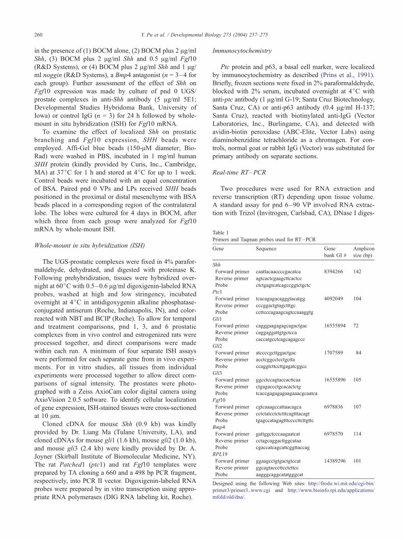
Table 1
Primers and Taqman probes used for RT–PCR
Gene Sequence Gene
bank GI #
Amplicon
size (bp)
Shh
Forward primer caattacaaccccgacatca 8394266 142
Reverse primer agtcactcgaagcttcactcc
Probe ctctgagtcatcagccggtctgctc
Ptc1
Forward primer tcacagagacagggtacatgg 4092049 104
Reverse primer cccggactgtagctttgc
Probe ccttcccagaagcagtccaaaggtg
Gli1
Forward primer cagggaagagagcagactgac 16555894 72
Reverse primer caggaggattgtgctcca
Probe caccatgcctcagcagagccc
Gli2
Forward primer atccccgcttggactgac 1707589 84
Reverse primer acctcggcctcctgctta
Probe ccaggtcttccttgagatcggcc
Gli3
Forward primer ggcctccagtaccacttcaa 16555896 105
Reverse primer ctgagaccctgcacactctg
Probe tcaccgagagagaagaaacgcaatca
Fgf10
Forward primer cgtcaaagccattaacagca 6978836 107
Reverse primer cctctatcctctctttcagtttacagt
Probe tgagccatagagtttccccttcttgttc
Bmp4
Forward primer gattggctcccaagaatcat 6978570 114
Reverse primer cctagcaggacttggcataa
Probe cgaccatcagcattcggttaccag
RPL19
Forward primer ggaagcctgtgactgtccat 14389296 101
Reverse primer ggcagtacccttcctcttcc
Probe aagggcaggcatatgggcat
Designed using the following Web sites: http://frodo.wi.mit.edu/cgi-bin/
primer3/primer3_www.cgi and http://www.bioinfo.rpi.edu/applications/
mfold/old/dna/.
Y. Pu et al. / Developmental Bio260
in the presence of (1) BOCM alone, (2) BOCM plus 2 Ag/ml
Shh, (3) BOCM plus 2 Ag/ml Shh and 0.5 Ag/ml Fgf10
(R&D Systems), or (4) BOCM plus 2 Ag/ml Shh and 1 Ag/ml noggin (R&D Systems), a Bmp4 antagonist (n = 3–4 for
each group). Further assessment of the effect of Shh on
Fgf10 expression was made by culture of pnd 0 UGS/
prostate complexes in anti-Shh antibody (5 Ag/ml 5E1;
Developmental Studies Hybridoma Bank, University of
Iowa) or control IgG (n = 3) for 24 h followed by whole-
mount in situ hybridization (ISH) for Fgf10 mRNA.
To examine the effect of localized Shh on prostatic
branching and Fgf10 expression, SHH beads were
employed. Affi-Gel blue beads (150-AM diameter; Bio-
Rad) were washed in PBS, incubated in 1 mg/ml human
SHH protein (kindly provided by Curis, Inc., Cambridge,
MA) at 37jC for 1 h and stored at 4jC for up to 1 week.
Control beads were incubated with an equal concentration
of BSA. Paired pnd 0 VPs and LPs received SHH beads
positioned in the proximal or distal mesenchyme with BSA
beads placed in a corresponding region of the contralateral
lobe. The lobes were cultured for 4 days in BOCM, after
which three from each group were analyzed for Fgf10
mRNA by whole-mount ISH.
Whole-mount in situ hybridization (ISH)
The UGS-prostatic complexes were fixed in 4% parafor-
maldehyde, dehydrated, and digested with proteinase K.
Following prehybridization, tissues were hybridized over-
night at 60jC with 0.5–0.6 Ag/ml digoxigenin-labeled RNA
probes, washed at high and low stringency, incubated
overnight at 4jC in antidigoxygenin alkaline phosphatase-
conjugated antiserum (Roche, Indianapolis, IN), and color-
reacted with NBT and BCIP (Roche). To allow for temporal
and treatment comparisons, pnd 1, 3, and 6 prostatic
complexes from in vivo control and estrogenized rats were
processed together, and direct comparisons were made
within each run. A minimum of four separate ISH assays
were performed for each separate gene from in vivo experi-
ments. For in vitro studies, all tissues from individual
experiments were processed together to allow direct com-
parisons of signal intensity. The prostates were photo-
graphed with a Zeiss AxioCam color digital camera using
AxioVision 2.0.5 software. To identify cellular localization
of gene expression, ISH-stained tissues were cross-sectioned
at 10 Am.
Cloned cDNA for mouse Shh (0.9 kb) was kindly
provided by Dr. Liang Ma (Tulane University, LA), and
cloned cDNAs for mouse gli1 (1.6 kb), mouse gli2 (1.0 kb),
and mouse gli3 (2.4 kb) were kindly provided by Dr. A.
Joyner (Skirball Institute of Biomolecular Medicine, NY).
The rat Patched1 (ptc1) and rat Fgf10 templates were
prepared by TA cloning a 660 and a 498 bp PCR fragment,
respectively, into PCR II vector. Digoxigenin-labeled RNA
probes were prepared by in vitro transcription using appro-
priate RNA polymerases (DIG RNA labeling kit, Roche).
Immunocytochemistry
Ptc protein and p63, a basal cell marker, were localized
by immunocytochemistry as described (Prins et al., 1991).
Briefly, frozen sections were fixed in 2% paraformaldehyde,
blocked with 2% serum, incubated overnight at 4jC with
anti-ptc antibody (1 Ag/ml G-19; Santa Cruz Biotechnology,
Santa Cruz, CA) or anti-p63 antibody (0.4 Ag/ml H-137;
Santa Cruz), reacted with biotinylated anti-IgG (Vector
Laboratories, Inc., Burlingame, CA), and detected with
avidin-biotin peroxidase (ABC-Elite, Vector Labs) using
diaminobenzidine tetrachloride as a chromagen. For con-
trols, normal goat or rabbit IgG (Vector) was substituted for
primary antibody on separate sections.
Real-time RT–PCR
Two procedures were used for RNA extraction and
reverse transcription (RT) depending upon tissue volume.
A standard assay for pnd 6–90 VP involved RNA extrac-
tion with Trizol (Invitrogen, Carlsbad, CA), DNase I diges-
logy 273 (2004) 257–275
Y. Pu et al. / Developmental Biology 273 (2004) 257–275 261
tion (Roche), and RT with AMV at 42jC for 60 min using
the RT System (Promega, Madison, WI). A microassay for
smaller tissues (pnd 1–6 VP, pnd 6–10 LP and DP) used
RNeasy Kit (Qiagen, Valencia, CA) for RNA extraction,
On-Column DNase I Digestion, and RT with MMLV at
37jC for 60 min using First Strand cDNA Synthesis Kit
(Fermentas Inc., Hanover, MD). Random primers were used
for reverse transcription.
The exon spanning primers and dual-labeled probe sets
used for PCR are shown in Table 1. For dual-labeled probes,
the 5V-reporters were FAM for Shh, gli1, Fgf10, and Bmp4;
Hex for ptc, gli2, and RPL19; and Texas red for gli3, and the
3V was labeled with black hole quencher. Plasmids contain-
ing each DNA sequence (Shh, ptc1, gli1, gli2, gli3, Fgf10,
Bmp4, and RPL19) were cloned with TOPO TA cloning kit
(Invitrogen) and used for standard curves in each reaction to
directly quantitate target DNA levels. Ribosomal protein
L19 (RPL19) was quantitated and served as an internal
reference for normalization. Direct comparisons of RPL19
per unit total RNA revealed no effect of estrogen treatment
in developing prostates. Real-time PCR was performed in
duplex with Platinum qPCR Supermixture-UDG (Invitro-
gen) using the iCycler (Biorad, Hercules, CA). Reaction
conditions were optimized for single gene and multiplex
PCR, and the cycle conditions were 95jC for 3 min and 40
cycles of 95jC for 15 s and 60jC for 30 s. Optical data
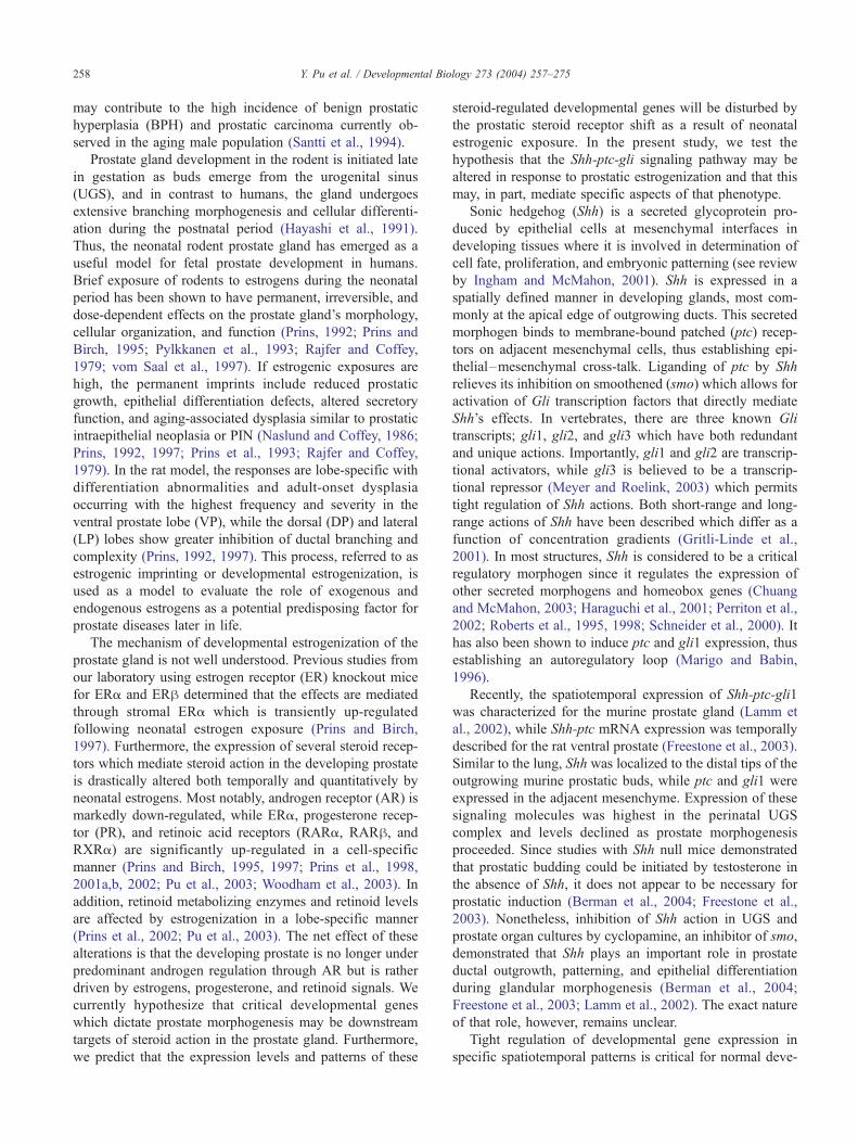
Fig. 1. Whole-mount ISH of Shh transcript in the developing prostate of contro
comparison of signal intensity. (A) E20 UGS; Shh is expressed along the central
(VP), dorsal (DP), and lateral (LP) prostate lobes and the coagulating gland (CG).
complex at separate focal planes. Shh is localized to the distal tips of the emerging e
UGS-prostate complex at separate focal planes. Shh transcript remains at the distal
prostatic region is reduced as compared to pnd1. (D) Cross section of pnd 6 VP fro
is expressed exclusively in the epithelial cells at the distal tips of the ducts. (E)
demonstrates antisense Shh probe specificity. (F) High-power view of pnd1 VP sho
regional heterogeneity of the Shh signal. (G) The distal tip of a pnd1 VP following
of Shh are observed at the distal tips (arrows) with adjacent regions of lower signa
Shh signal with focal patches of high expression (arrows) at the distal tips. Ur ind
Am; in D, G, H = 50 Am.
obtained by real-time PCR were analyzed with the manu-
facturer’s software (iCycle Optical System Interface Version
3.0). Each assay was repeated three to ten times using
different tissues. Statistical analysis involved ANOVA and
two-tailed Student t test (Sigma Plot, Version 8.02, SPSS
Inc., Chicago, IL).
Results
Shh-ptc-gli expression in the developing rat prostate
Whole-mount ISH combined with real-time RT–PCR
was used to determine the spatiotemporal expression pat-
terns of Shh, ptc, and gli1, 2, and 3 in the rat prostate gland
during the active period of branching morphogenesis. There
were no noticeable differences between the VP, DP, or LP
with regards to expression patterns of these hedgehog
signaling molecules. Prostatic budding is initiated at fetal
day 18.5 of a 21-day gestation in the rat when UGS
epithelial cells penetrate into the surrounding UGS mesen-
chyme in the dorsal, lateral, and ventral directions. At fetal
day 20 (E20), the earliest time point examined in the present
study, there was robust Shh mRNA expression in the
prostatic buds in all three lobes with the greatest concen-
tration in the central-to-distal aspects of these protruding
l rats. Examples shown in A–C were processed together to allow direct
to distal regions of the emerging ducts. Arrows denote the budding ventral
(B) Postnatal day 1 LP (top) and VP (bottom) from the same UGS-prostate
pithelial ducts. (C) Postnatal day 6 LP (top) and VP (bottom) from the same
tips of the lateral LP1 and LP2 ducts and the VP. The signal intensity in each
m whole-mount ISH at low (bottom) and high (top) power confirms that Shh
Sense Shh probe on pnd 3 UGS-prostatic complex shows no signal and
wn in B shows the strongest Shh mRNA intensity at the distal tips as well as
treatment with dispase to remove surrounding mesenchyme. Expression foci
l intensity. (H) The distal aspect of a pnd3 VP duct displays heterogeneous
icates urethra; E, epithelium; M, mesenchyme. Bars in A, B, C, E, F = 200
Y. Pu et al. / Developmental Biology 273 (2004) 257–275262
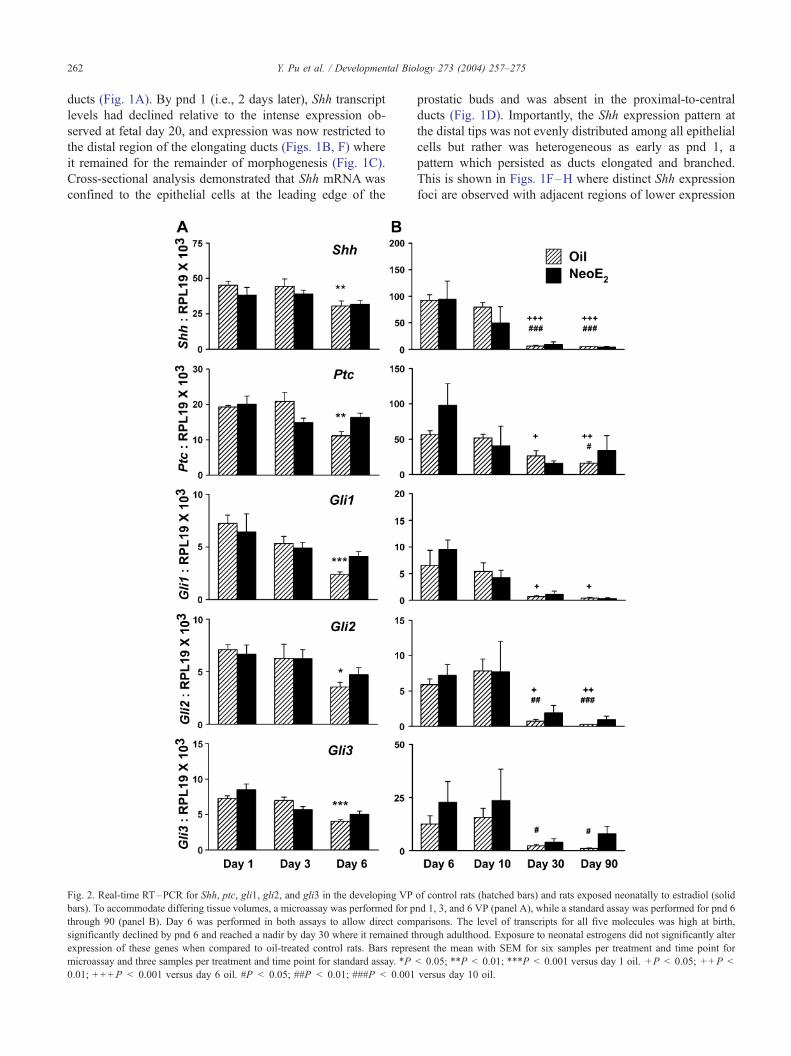
ducts (Fig. 1A). By pnd 1 (i.e., 2 days later), Shh transcript
levels had declined relative to the intense expression ob-
served at fetal day 20, and expression was now restricted to
the distal region of the elongating ducts (Figs. 1B, F) where
it remained for the remainder of morphogenesis (Fig. 1C).
Cross-sectional analysis demonstrated that Shh mRNA was
confined to the epithelial cells at the leading edge of the
Fig. 2. Real-time RT–PCR for Shh, ptc, gli1, gli2, and gli3 in the developing VP
bars). To accommodate differing tissue volumes, a microassay was performed for p
through 90 (panel B). Day 6 was performed in both assays to allow direct com
significantly declined by pnd 6 and reached a nadir by day 30 where it remained t
expression of these genes when compared to oil-treated control rats. Bars repres
microassay and three samples per treatment and time point for standard assay. *P
0.01; + + +P < 0.001 versus day 6 oil. #P < 0.05; ##P < 0.01; ###P < 0.001
prostatic buds and was absent in the proximal-to-central
ducts (Fig. 1D). Importantly, the Shh expression pattern at
the distal tips was not evenly distributed among all epithelial
cells but rather was heterogeneous as early as pnd 1, a
pattern which persisted as ducts elongated and branched.
This is shown in Figs. 1F–H where distinct Shh expression
foci are observed with adjacent regions of lower expression
of control rats (hatched bars) and rats exposed neonatally to estradiol (solid
nd 1, 3, and 6 VP (panel A), while a standard assay was performed for pnd 6
parisons. The level of transcripts for all five molecules was high at birth,
hrough adulthood. Exposure to neonatal estrogens did not significantly alter
ent the mean with SEM for six samples per treatment and time point for
< 0.05; **P < 0.01; ***P < 0.001 versus day 1 oil. +P < 0.05; + +P <
versus day 10 oil.
Y. Pu et al. / Developmental Biology 273 (2004) 257–275 263
levels. By pnd 6, there was a noticeable decline in overall
Shh signal intensity by whole-mount ISH (Fig. 1C), and this
was confirmed by RT–PCR which showed a significant
reduction in Shh transcript levels at pnd 6 as compared to
pnd 1 (Fig. 2A, oil controls in hatched bars). By day 30,
when prostatic branching is complete, Shh transcript levels
reached a nadir where they remained through adulthood
(Fig. 2B, hatched bars).
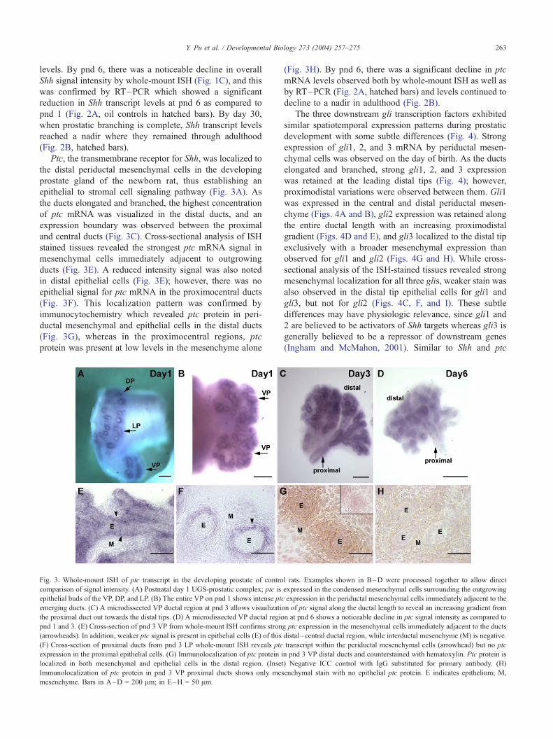
Ptc, the transmembrane receptor for Shh, was localized to
the distal periductal mesenchymal cells in the developing
prostate gland of the newborn rat, thus establishing an
epithelial to stromal cell signaling pathway (Fig. 3A). As
the ducts elongated and branched, the highest concentration
of ptc mRNA was visualized in the distal ducts, and an
expression boundary was observed between the proximal
and central ducts (Fig. 3C). Cross-sectional analysis of ISH
stained tissues revealed the strongest ptc mRNA signal in
mesenchymal cells immediately adjacent to outgrowing
ducts (Fig. 3E). A reduced intensity signal was also noted
in distal epithelial cells (Fig. 3E); however, there was no
epithelial signal for ptc mRNA in the proximocentral ducts
(Fig. 3F). This localization pattern was confirmed by
immunocytochemistry which revealed ptc protein in peri-
ductal mesenchymal and epithelial cells in the distal ducts
(Fig. 3G), whereas in the proximocentral regions, ptc
protein was present at low levels in the mesenchyme alone
Fig. 3. Whole-mount ISH of ptc transcript in the developing prostate of contro
comparison of signal intensity. (A) Postnatal day 1 UGS-prostatic complex; ptc is
epithelial buds of the VP, DP, and LP. (B) The entire VP on pnd 1 shows intense ptc
emerging ducts. (C) A microdissected VP ductal region at pnd 3 allows visualizatio
the proximal duct out towards the distal tips. (D) A microdissected VP ductal regio
pnd 1 and 3. (E) Cross-section of pnd 3 VP from whole-mount ISH confirms stron
(arrowheads). In addition, weaker ptc signal is present in epithelial cells (E) of this
(F) Cross-section of proximal ducts from pnd 3 LP whole-mount ISH reveals ptc
expression in the proximal epithelial cells. (G) Immunolocalization of ptc protein i
localized in both mesenchymal and epithelial cells in the distal region. (Inset
Immunolocalization of ptc protein in pnd 3 VP proximal ducts shows only mes
mesenchyme. Bars in A–D = 200 Am; in E–H = 50 Am.
(Fig. 3H). By pnd 6, there was a significant decline in ptc
mRNA levels observed both by whole-mount ISH as well as
by RT–PCR (Fig. 2A, hatched bars) and levels continued to
decline to a nadir in adulthood (Fig. 2B).
The three downstream gli transcription factors exhibited
similar spatiotemporal expression patterns during prostatic
development with some subtle differences (Fig. 4). Strong
expression of gli1, 2, and 3 mRNA by periductal mesen-
chymal cells was observed on the day of birth. As the ducts
elongated and branched, strong gli1, 2, and 3 expression
was retained at the leading distal tips (Fig. 4); however,
proximodistal variations were observed between them. Gli1
was expressed in the central and distal periductal mesen-
chyme (Figs. 4A and B), gli2 expression was retained along
the entire ductal length with an increasing proximodistal
gradient (Figs. 4D and E), and gli3 localized to the distal tip
exclusively with a broader mesenchymal expression than
observed for gli1 and gli2 (Figs. 4G and H). While cross-
sectional analysis of the ISH-stained tissues revealed strong
mesenchymal localization for all three glis, weaker stain was
also observed in the distal tip epithelial cells for gli1 and
gli3, but not for gli2 (Figs. 4C, F, and I). These subtle
differences may have physiologic relevance, since gli1 and
2 are believed to be activators of Shh targets whereas gli3 is
generally believed to be a repressor of downstream genes
(Ingham and McMahon, 2001). Similar to Shh and ptc
l rats. Examples shown in B–D were processed together to allow direct
expressed in the condensed mesenchymal cells surrounding the outgrowing
expression in the periductal mesenchymal cells immediately adjacent to the
n of ptc signal along the ductal length to reveal an increasing gradient from
n at pnd 6 shows a noticeable decline in ptc signal intensity as compared to
g ptc expression in the mesenchymal cells immediately adjacent to the ducts
distal–central ductal region, while interductal mesenchyme (M) is negative.
transcript within the periductal mesenchymal cells (arrowhead) but no ptc
n pnd 3 VP distal ducts and counterstained with hematoxylin. Ptc protein is
) Negative ICC control with IgG substituted for primary antibody. (H)
enchymal stain with no epithelial ptc protein. E indicates epithelium; M,
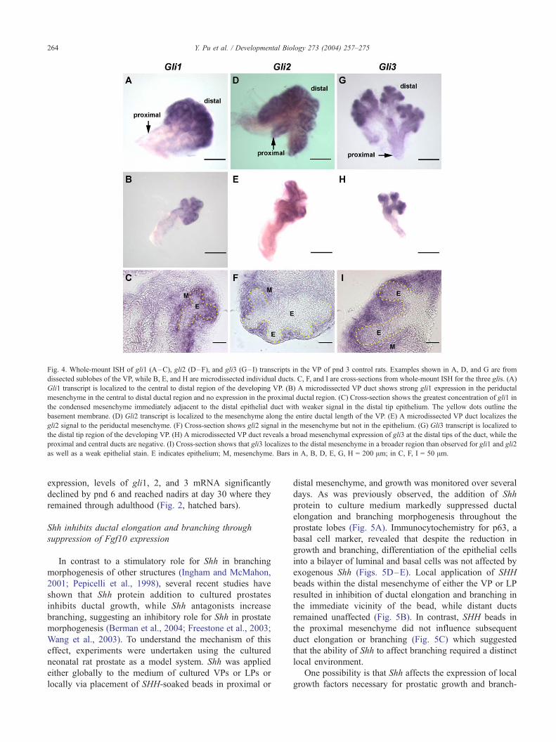
Fig. 4. Whole-mount ISH of gli1 (A–C), gli2 (D–F), and gli3 (G–I) transcripts in the VP of pnd 3 control rats. Examples shown in A, D, and G are from
dissected sublobes of the VP, while B, E, and H are microdissected individual ducts. C, F, and I are cross-sections from whole-mount ISH for the three glis. (A)
Gli1 transcript is localized to the central to distal region of the developing VP. (B) A microdissected VP duct shows strong gli1 expression in the periductal
mesenchyme in the central to distal ductal region and no expression in the proximal ductal region. (C) Cross-section shows the greatest concentration of gli1 in
the condensed mesenchyme immediately adjacent to the distal epithelial duct with weaker signal in the distal tip epithelium. The yellow dots outline the
basement membrane. (D) Gli2 transcript is localized to the mesenchyme along the entire ductal length of the VP. (E) A microdissected VP duct localizes the
gli2 signal to the periductal mesenchyme. (F) Cross-section shows gli2 signal in the mesenchyme but not in the epithelium. (G) Gli3 transcript is localized to
the distal tip region of the developing VP. (H) A microdissected VP duct reveals a broad mesenchymal expression of gli3 at the distal tips of the duct, while the
proximal and central ducts are negative. (I) Cross-section shows that gli3 localizes to the distal mesenchyme in a broader region than observed for gli1 and gli2
as well as a weak epithelial stain. E indicates epithelium; M, mesenchyme. Bars in A, B, D, E, G, H = 200 Am; in C, F, I = 50 Am.
Y. Pu et al. / Developmental Biology 273 (2004) 257–275264
expression, levels of gli1, 2, and 3 mRNA significantly
declined by pnd 6 and reached nadirs at day 30 where they
remained through adulthood (Fig. 2, hatched bars).
Shh inhibits ductal elongation and branching through
suppression of Fgf10 expression
In contrast to a stimulatory role for Shh in branching
morphogenesis of other structures (Ingham and McMahon,
2001; Pepicelli et al., 1998), several recent studies have
shown that Shh protein addition to cultured prostates
inhibits ductal growth, while Shh antagonists increase
branching, suggesting an inhibitory role for Shh in prostate
morphogenesis (Berman et al., 2004; Freestone et al., 2003;
Wang et al., 2003). To understand the mechanism of this
effect, experiments were undertaken using the cultured
neonatal rat prostate as a model system. Shh was applied
either globally to the medium of cultured VPs or LPs or
locally via placement of SHH-soaked beads in proximal or
distal mesenchyme, and growth was monitored over several
days. As was previously observed, the addition of Shh
protein to culture medium markedly suppressed ductal
elongation and branching morphogenesis throughout the
prostate lobes (Fig. 5A). Immunocytochemistry for p63, a
basal cell marker, revealed that despite the reduction in
growth and branching, differentiation of the epithelial cells
into a bilayer of luminal and basal cells was not affected by
exogenous Shh (Figs. 5D–E). Local application of SHH
beads within the distal mesenchyme of either the VP or LP
resulted in inhibition of ductal elongation and branching in
the immediate vicinity of the bead, while distant ducts
remained unaffected (Fig. 5B). In contrast, SHH beads in
the proximal mesenchyme did not influence subsequent
duct elongation or branching (Fig. 5C) which suggested
that the ability of Shh to affect branching required a distinct
local environment.
One possibility is that Shh affects the expression of local
growth factors necessary for prostatic growth and branch-

Y. Pu et al. / Developmental Biology 273 (2004) 257–275 265
ing. To address this, the expression of Fgf10 and Bmp4
mRNAwas assessed as a function of Shh manipulation since
(1) both of these mesenchymal genes are known down-
stream targets of Shh in other systems (Haraguchi et al.,
2001), (2) Fgf10 is expressed in the distal, but not proximal
mesenchyme in the developing prostate (Thomson and
Cunha, 1999), (3) Fgf10 plays an stimulatory role in
prostatic branching morphogenesis (Donjacour et al.,
2003), and (4) Bmp4 plays an inhibitory role in prostatic
ductal outgrowth and branching (Lamm et al., 2001). Using
RT–PCR, Fgf10 and Bmp4 transcripts were quantitated in
VPs and LPs cultured with Shh protein in the medium. Since
this culture system results in a marked shift in the prostatic
epithelial–stromal ratio, gene expression was assessed be-
fore observable growth alterations. In both lobes, Shh
protein significantly down-regulated Fgf10 expression and
increased Bmp4 expression within 18 h, suggesting that the
growth inhibitory effects of Shh may be mediated through
alterations in the expression of these morphogens (Fig. 6A).
To address this directly, replacement experiments were
conducted with exogenous Fgf10 protein or noggin, a
Bmp4 antagonist, added to prostates cultured with Shh
protein for 4–6 days. As shown in Fig. 6B, Shh protein
suppressed ductal elongation and branching of the VP, and
this was largely overridden by exogenous Fgf10. In contrast,
antagonism of Bmp4 with noggin was unable to reverse the
growth suppression by Shh protein (Fig. 6C). Prostates
cultured with either Fgf10 or noggin alone in the presence
of 10 nM testosterone exhibited a modest increase in
prostate size and branching (data not shown); however, this
was not significant when measured by 2-D analysis. As an
alternate approach to demonstrate Fgf10 regulation by Shh,
UGS-prostatic complexes were removed on pnd 0 and
cultured for 24 h with anti-Shh or control antibody (n =
3). The expression of ptc, which is directly stimulated by
Shh, was repressed which confirms that the antibodies
functionally blocked Shh action (Fig. 7A). Importantly,
Fgf10 expression was markedly increased in Shh-inhibited
prostates when compared to controls as determined by
wmISH (Fig. 7B). Taken together, these data indicate that
the prostatic growth suppression by Shh is mediated through
suppression of mesenchymal Fgf10.
Since Shh is exclusively expressed in distal ductal tips of
the branching prostate, we examined the local regulation of
Fgf10 expression by SHH beads implanted along the distal
mesenchyme. Prostatic-UGS complexes were removed on
pnd 0; SHH or BSA beads were placed along the LP and VP
Fig. 5. Cultured VP and LP following application of Shh or BSA. Paired
lobes from a single rat were treated with either BSA or Shh to allow direct
comparisons of Shh treatment. (A) VPs (top) cultured for 3 days and LPs
(bottom) cultured for 2 days in the presence of BSA (left) or Shh protein
added directly to the culture medium. Shh addition suppressed ductal
outgrowth and branching in both lobes. (B) BSA beads (left arrowheads)
and SHH beads (right arrowheads) were placed in the distal mesenchyme of
VP (top) and LP (bottom) paired lobes on pnd 0 and cultured for 4 days
(VP) or 3 days (LP). Local application of SHH resulted in localized ductal
growth and branching inhibition in the immediate vicinity of the bead. (C)
BSA beads (left) and SHH beads (right) implanted in the VP proximal
mesenchyme (arrowheads) on pnd 0 and cultured for 4 days showed no
inhibition of ductal growth and branching by SHH. (D) Immunocytochem-
istry for p63 (basal cell marker) in a VP grown for 6 days in basal medium
with BSA shows a bilayer of stained basal cells along the basement
membrane and unstained luminal cells above the basal layer which
indicates differentiation of the epithelial cells. Basal cells lie adjacent to the
basement membrane in an intermittent pattern in the proximal ducts and a
continuous pattern in the distal tips which follows the differentiation wave
of luminal epithelial cells in a proximal-to-distal fashion during prostate
morphogenesis. (E) p63 Immunostain of a VP cultured for 6 days in Shh
protein. Although the branching pattern is inhibited as compared to the
contralateral lobe in BSA (D), the differentiation of the epithelial cells as
revealed by the basal cell pattern is the same as the BSA-control cultures.
Bars in A–C = 500 Am; in D–E = 50 Am.

Fig. 6. (A) Real-time RT–PCR data showing the effect of Shh protein on VP and LP expression of Fgf10 and Bmp4 mRNA after 18 h of culture. In both lobes,
Shh protein (solid bars) inhibited Fgf10 expression and increased prostatic Bmp4 expression as compared to BSA controls (hatched bars). Bars represent the
mean F SEM for eight samples per treatment. *P < 0.05; ***P < 0.001 Shh versus BSA. (B) VP cultured for 6 days in basal medium with BSA (left), Shh
protein (center) or Shh + Fgf10 (right). The Shh cultures with or without Fgf10 are contralateral lobes to allow direct comparisons. Ductal growth and branching
inhibition due to Shh protein was reversed by exogenous Fgf10. (C) VP cultured for 4 days in basal medium with BSA (left), Shh protein (center), or Shh +
noggin (right). The Shh cultures with or without noggin are contralateral lobes. Ductal growth and branching inhibition due to Shh protein was not affected by
the Bmp4 antagonist. Bar in B–C = 500 Am.
Y. Pu et al. / Developmental Biology 273 (2004) 257–275266
surfaces and cultured for 24 h. Whole-mount ISH for Fgf10
revealed a strong reduction in mesenchymal Fgf10 mRNA
expression in explants with SHH beads as compared to the
BSA controls (Figs. 7C–D). Similarly, in VPs cultured for 3
days with SHH beads implanted in the distal mesenchyme,
Fgf10 expression was reduced in the immediate vicinity of
the SHH beads where ductal growth and branching were
inhibited (Figs. 7E–F). In contrast, Fgf10 expression was
unaffected at a distance from the beads where branching was
normal as well as in the immediate vicinity of BSA beads
(Fig. 7E). These data indicate that Shh at distinct distal sites
causes local down-regulation of Fgf10 expression in the
adjacent mesenchyme.
Neonatal estradiol exposure suppresses Shh-ptc-gli
expression in a lobe-specific manner
Exposure to estradiol in vivo on pnd 0, 3, and 5
reduced Shh-ptc and gli mRNA expression in the DP
and LP. In contrast, expression of these signaling mole-
cules in the VP was not significantly affected. As early as
pnd 1, wmISH consistently revealed strong suppression of
Shh transcript in the LP and DP regions when tissues from
control and estrogen-treated rats were processed together
(Fig. 8A, top), while Shh expression was similar between
the two groups in the VP (Fig. 8A, bottom). This sup-
pression of Shh expression in the dorsolateral prostate
persisted through pnd 6 (Fig. 8B). Furthermore, real-time
RT–PCR of Shh mRNA levels in day 6 and 10 LP and DP
revealed a significant reduction following estradiol expo-
sure in those lobes specifically (Fig. 9), while no statistical
difference was noted in the VP between the treatment
groups at any time point (Fig. 2). Similar to Shh, estradiol
reduced ptc expression in the LP and DP as early as pnd 1
(Fig. 8C), and this effect persisted through pnd 6 for the
LP and pnd 10 for the DP (Figs. 8D and 9). In contrast,
VP ptc expression was not affected by estradiol exposure
at any time point (Figs. 2A and B; 8C and D). Likewise,
gli1 and gli3 expressions were suppressed in the LP and
DP through pnd 10 following neonatal estradiol exposure
(Figs. 8E and F; 9), while expression in the VP was not
changed (Figs. 2 and 8E and F). Notably, gli2 expression
was not affected by this steroid in any of the prostate lobes
(Figs. 2 and 9). It is noteworthy that estrogen did not affect
the localization of the hedgehog signaling molecules in the
prostate lobes.
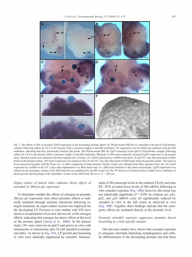
Fig. 7. The effects of Shh on prostatic Fgf10 expression in the developing prostate gland. (A) Whole-mount ISH for ptc transcript in the pnd 0 UGS-prostate
complex following culture for 24 h in the absence (left) or presence (right) of anti-Shh antibodies. Ptc expression was lost following treatment with anti-Shh
antibodies, indicating that they functionally blocked Shh action. (B) Whole-mount ISH for Fgf10 transcript in the pnd 0 UGS-prostate complex following
culture for 24 h in the absence (left) or presence (right) of anti-Shh antibodies. Blockade of Shh action markedly increased Fgf10 expression in all prostate
lobes. Identical results were obtained with three separate sets of tissues. (C) Fgf10 expression by wmISH in the pnd1 LP and VP 1 day after placement of BSA
beads on the prostate surface. (D) Fgf10 expression was reduced in the LP and VP 1 day after placement of SHH beads along the prostate surface. The tissue in
D was processed together with the tissue in C to allow comparison of stain intensity. Similar results were obtained from three separate tissue sets. (E) Fgf10
expression by wmISH in the VP 3 days after implantation of a BSA bead (top) or a SHH bead (bottom) in the distal mesenchyme. Fgf10 expression was
reduced in the immediate vicinity of the SHH bead but was unaffected by the BSA bead. (F) The VP shown in E (bottom) before wmISH shows inhibition of
ductal growth and branching in the immediate vicinity of the SHH bead. Bar in A–F = 200 Am.
Y. Pu et al. / Developmental Biology 273 (2004) 257–275 267
Organ culture of lateral lobes indicates direct effects of
estradiol on Shh-ptc-gli expression
To determine whether the effects of estrogen on prostatic
Shh-ptc-gli expression were direct prostatic effects or indi-
rectly mediated through systemic alterations following es-
trogen treatment, an organ culture system was employed for
the developing LP. Previous in vitro studies with VPs have
shown a recapitulation of several, but not all, of the estrogen
effects, indicating that estrogen has direct effects at the level
of the prostate gland (Jarred et al., 2000). In the present
study, LPs were removed on pnd 0 and grown in vitro with
testosterone or testosterone plus 20 AM estradiol (contralat-
eral lobe). As shown in Fig. 10A, LP growth and branching
in vitro were markedly suppressed by estradiol. Measure-
ment of Shh transcript levels in the cultured LPs by real-time
RT–PCR revealed lower levels of Shh mRNA following in
vitro estradiol exposure (Fig. 10B); however, this trend was
not statistically significant (P = 0.09). In contrast, ptc, gli1,
gli2, and gli3 mRNA were all significantly reduced by
estradiol in vitro to the full extent as observed in vivo
(Fig. 10B). Together, these findings indicate that the estro-
genic effects are mediated directly at the prostatic level.
Neonatal estradiol exposure suppresses prostatic ductal
branching in a lobe-specific manner
Our previous studies have shown that neonatal exposure
to estrogens interrupts branching morphogenesis and cellu-
lar differentiation in the developing prostate and that these
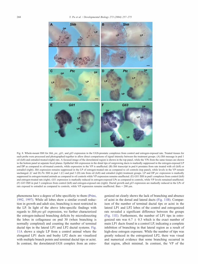
Fig. 8. Whole-mount ISH for Shh, ptc, gli1, and gli3 expression in the UGS-prostatic complexes from control and estrogen-exposed rats. Treated tissues for
each probe were processed and photographed together to allow direct comparisons of signal intensity between the treatment groups. (A) Shh message in pnd 1
oil (left) and estradiol-treated (right) rats. A focused image of the dorsolateral region is shown in the top panel, while the VPs from the same tissues are shown
in the bottom panel at separate focal planes. Epithelial Shh expression in the distal tips of outgrowing ducts is markedly suppressed in the estrogen-exposed LP
and DP as compared to oil-treated controls, while expression in the VP is unaffected. (B) Shh transcript in pnd 6 prostates from rats treated with oil (left) or
estradiol (right). Shh expression remains suppressed in the LP of estrogen-treated rats as compared to oil controls (top panel), while levels in the VP remain
unchanged. (C and D) Ptc ISH in pnd 1 (C) and pnd 3 (D) rats from oil (left) and estradiol (right) treatment groups. LP and DP ptc expression is markedly
suppressed in estrogen-treated animals as compared to oil controls while VP expression remains unaffected. (E) Gli1 ISH in pnd1 complexes from control (left)
and estrogen-treated rats (right). Gli1 expression is markedly reduced in estrogen-exposed LPs as compared to controls, while VP levels remained unaffected.
(F) Gli3 ISH in pnd 3 complexes from control (left) and estrogen-exposed rats (right). Ductal growth and gli3 expression are markedly reduced in the LPs of
rats exposed to estradiol as compared to controls, while VP expression remains unaffected. Bars = 200 Am.
Y. Pu et al. / Developmental Biology 273 (2004) 257–275268
phenomena have a degree of lobe specificity to them (Prins,
1992, 1997). While all lobes show a similar overall reduc-
tion in growth and adult size, branching is most restricted in
the LP. In light of the above lobe-specific findings with
regards to Shh-ptc-gli expression, we further characterized
the estrogen-induced branching deficits by microdissecting
the lobes in collagenase on pnd 30 (when branching is
normally completed) and counting the number of terminal
ductal tips in the lateral LP1 and LP2 ductal systems. Fig.
11A shows a single LP from a control animal where the
elongated LP1 ducts and bushy LP2 ducts are visualized
with multiple branch points and terminal ductal tips or acini.
In contrast, the dorsolateral-UGS complex from an estro-
genized rat clearly shows the lack of branching and absence
of acini in the dorsal and lateral ducts (Fig. 11B). Compar-
ison of the number of terminal ductal tips or acini in the
lateral LP1 and LP2 lobes of the control and estrogenized
rats revealed a significant difference between the groups
(Fig. 11E). Furthermore, the number of LP1 tips in estro-
genized rats was 6.7 F 0.3 which is the exact number of
main LP1 ducts found in a control LP, indicating a complete
inhibition of branching in that lateral region as a result of
high-dose estrogen exposure. While the number of tips was
greatly reduced in the estrogenized LP2, there was visual
and numerical evidence that some branching occurred in
that region, albeit minimal. In contrast, the VP of the

Fig. 9. Real-time RT–PCR for Shh, ptc, gli1, gli2, and gli3 in the LP and
DP of control (hatched bars) and estrogen-exposed rats (NeoE2; solid bars)
on pnd 6 and 10. Bars represent the mean with SEM for four to ten samples
per treatment and time point. *P < 0.05; **P < 0.01; ***P < 0.001 for
NeoE2 versus oil at specific time points.
Y. Pu et al. / Developmental Biology 273 (2004) 257–275 269
estrogenized prostate (Fig. 11D), although significantly
reduced in size as compared to the control ventral lobe
(Fig. 11C), exhibited a complex branching pattern, so much
so that an accurate counting of the branch points and
terminal tips was not possible.
Discussion
Expression and localization of Shh-ptc-gli1–3 in the
developing rat prostate lobes
The present study provides a clear spatiotemporal picture
of the expression patterns of the Shh-ptc-gli signaling
molecules in the separate rat prostate lobes during develop-
ment. The patterns described are in close agreement with
similar findings for the mouse prostate gland (Lamm et al.,
2002; Podlasek et al., 1999) and the developing rat ventral
lobe (Freestone et al., 2003). Overall, the expression of the
hedgehog signaling molecules is high at birth when budding
is underway and rapidly declines over the next several days
when morphogenesis is complete. Whole-mount ISH of the
UGS-prostatic complex from fetal day 20 to pnd 6 showed
that, during the initial budding phase, Shh has a broad
epithelial expression along the ductal length which rapidly
transitions into a distal tip epithelial expression pattern as
the ducts elongate and branch. Furthermore, the expression
of Shh at the tips is not uniform but rather appears
heterogeneous with foci of higher Shh expression at specific
sites which may allow for highly localized Shh actions at
those sites. The highest concentration of ptc and gli1–3
transcripts was localized to the condensed mesenchymal
cells adjacent to the elongating epithelial ducts in the distal
regions, which in total provides for the greatest Shh signal
being transmitted at the distal aspect of the developing
gland. Similar patterning for Shh-ptc has been observed in
several developing structures with axis polarity including
the lungs (Helms et al., 1997; Pepicelli et al., 1998), external
genitalia (Haraguchi et al., 2001), and limbs, (Yang et al.,
1997) suggesting a common mechanistic role for this para-
crine signal. It is noteworthy that ptc receptor and gli1 and
gli3 transcription factors were also localized, albeit at lower
expression levels, in the distal tip epithelial cells but were
not expressed in the proximal or central ductal epithelial
cells. A low level of ptc was previously detected by RT–
PCR in isolated epithelial cells of the mouse prostate (Lamm
et al., 2002). Thus, in addition to paracrine signaling, Shh
may have a role in directly activating genes in the distal
epithelial cells in an autocrine manner.
Distal signaling center in the developing prostate
The highly localized expression of Shh at the leading
edge of the outgrowing ducts is undoubtedly of critical
importance in its functional role. Several other developmen-
tal genes, including Nkx3.1 (Bhatia-Gaur et al., 1999; Prins
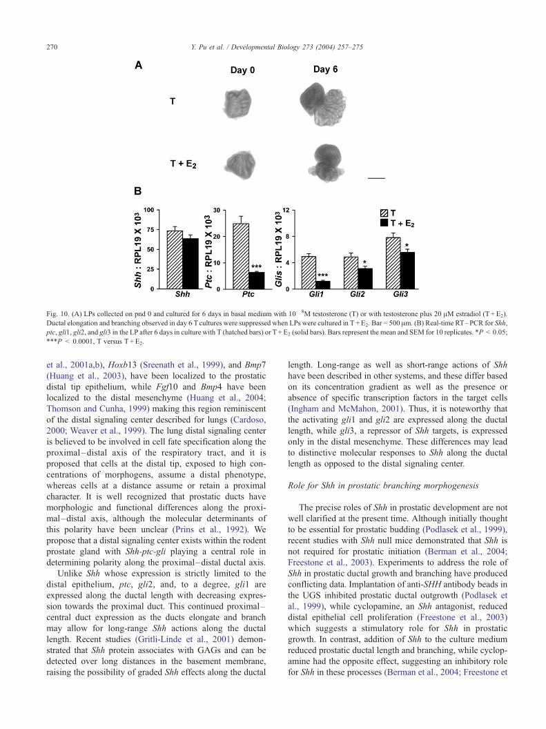
Fig. 10. (A) LPs collected on pnd 0 and cultured for 6 days in basal medium with 10� 8M testosterone (T) or with testosterone plus 20 AM estradiol (T +E2).
Ductal elongation and branching observed in day 6 Tcultures were suppressed when LPs were cultured in T +E2. Bar = 500 Am. (B) Real-time RT–PCR for Shh,
ptc, gli1, gli2, and gli3 in the LP after 6 days in culture with T (hatched bars) or T +E2 (solid bars). Bars represent the mean and SEM for 10 replicates. *P < 0.05;
***P < 0.0001, T versus T +E2.
Y. Pu et al. / Developmental Biology 273 (2004) 257–275270
et al., 2001a,b), Hoxb13 (Sreenath et al., 1999), and Bmp7
(Huang et al., 2003), have been localized to the prostatic
distal tip epithelium, while Fgf10 and Bmp4 have been
localized to the distal mesenchyme (Huang et al., 2004;
Thomson and Cunha, 1999) making this region reminiscent
of the distal signaling center described for lungs (Cardoso,
2000; Weaver et al., 1999). The lung distal signaling center
is believed to be involved in cell fate specification along the
proximal–distal axis of the respiratory tract, and it is
proposed that cells at the distal tip, exposed to high con-
centrations of morphogens, assume a distal phenotype,
whereas cells at a distance assume or retain a proximal
character. It is well recognized that prostatic ducts have
morphologic and functional differences along the proxi-
mal–distal axis, although the molecular determinants of
this polarity have been unclear (Prins et al., 1992). We
propose that a distal signaling center exists within the rodent
prostate gland with Shh-ptc-gli playing a central role in
determining polarity along the proximal–distal ductal axis.
Unlike Shh whose expression is strictly limited to the
distal epithelium, ptc, gli2, and, to a degree, gli1 are
expressed along the ductal length with decreasing expres-
sion towards the proximal duct. This continued proximal–
central duct expression as the ducts elongate and branch
may allow for long-range Shh actions along the ductal
length. Recent studies (Gritli-Linde et al., 2001) demon-
strated that Shh protein associates with GAGs and can be
detected over long distances in the basement membrane,
raising the possibility of graded Shh effects along the ductal
length. Long-range as well as short-range actions of Shh
have been described in other systems, and these differ based
on its concentration gradient as well as the presence or
absence of specific transcription factors in the target cells
(Ingham and McMahon, 2001). Thus, it is noteworthy that
the activating gli1 and gli2 are expressed along the ductal
length, while gli3, a repressor of Shh targets, is expressed
only in the distal mesenchyme. These differences may lead
to distinctive molecular responses to Shh along the ductal
length as opposed to the distal signaling center.
Role for Shh in prostatic branching morphogenesis
The precise roles of Shh in prostatic development are not
well clarified at the present time. Although initially thought
to be essential for prostatic budding (Podlasek et al., 1999),
recent studies with Shh null mice demonstrated that Shh is
not required for prostatic initiation (Berman et al., 2004;
Freestone et al., 2003). Experiments to address the role of
Shh in prostatic ductal growth and branching have produced
conflicting data. Implantation of anti-SHH antibody beads in
the UGS inhibited prostatic ductal outgrowth (Podlasek et
al., 1999), while cyclopamine, an Shh antagonist, reduced
distal epithelial cell proliferation (Freestone et al., 2003)
which suggests a stimulatory role for Shh in prostatic
growth. In contrast, addition of Shh to the culture medium
reduced prostatic ductal length and branching, while cyclop-
amine had the opposite effect, suggesting an inhibitory role
for Shh in these processes (Berman et al., 2004; Freestone et
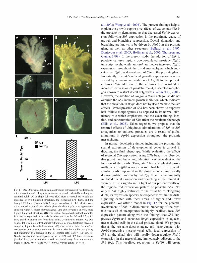
Fig. 11. Day 30 prostate lobes from control and estrogenized rats following
microdissection and collagenase treatment to visualize ductal branching and
terminal acini. (A) A single LP (one side) from a control rat reveals the
presence of two branched structures, the elongated LP1 ducts, and the
bushy LP2 ducts. (Bottom left) A single microdissected LP1 duct reveals
the extended proximal duct which gives the duct a palm tree appearance.
(Bottom right) A single microdissected LP2 duct reveals a shorter, more
highly branched structure. (B) The entire dorsolateral-urethral complex
from an estrogenized rat reveals the short ducts in the DP and LP which
have failed to branch and form distal acini. Ur indicates urethra. (C) One
ventral lobe from a control animal without collagenase treatment reveals a
complex, highly branched structure. (D) One ventral lobe form of an
estrogenized rat reveals a reduction in overall size but similar complexity
and branching as observed in the oil control rats. Bars = 500 Am. (E)
Number of terminal ductal tips (acini) in the LP1 and LP2 ducts of control
(hatched bars) and estradiol-exposed rats (solid bars). Bars represent the
mean F SEM. *P < 0.05; **P < 0.0001 versus control (n = 3).
Y. Pu et al. / Developmental Biology 273 (2004) 257–275 271
al., 2003; Wang et al., 2003). The present findings help to
explain the growth suppressive effects of exogenous Shh in
the prostate by demonstrating that decreased Fgf10 expres-
sion following Shh application is the proximate cause of
growth and branching suppression. Ductal elongation and
branching are known to be driven by Fgf10 in the prostate
gland as well as other structures (Bellusci et al., 1997;
Donjacour et al., 2003; Hoffman et al., 2002; Thomson and
Cunha, 1999). In the present study, the addition of Shh to
prostate cultures rapidly down-regulated prostatic Fgf10
transcript levels, while anti-Shh antibodies increased Fgf10
expression throughout the distal mesenchyme which indi-
cates that Fgf10 is downstream of Shh in the prostate gland.
Importantly, the Shh-induced growth suppression was re-
versed by concomitant addition of Fgf10 to the prostate
cultures. Shh addition to the cultures also resulted in
increased expression of prostatic Bmp4, a secreted morpho-
gen known to restrict ductal outgrowth (Lamm et al., 2001).
However, the addition of noggin, a Bmp4 antagonist, did not
override the Shh-induced growth inhibition which indicates
that the elevation in Bmp4 does not by itself mediate the Shh
effects. Overexpression of Shh has been shown to suppress
hair follicle morphogenesis as opposed to its normal stim-
ulatory role which emphasizes that the exact timing, loca-
tion, and concentration of Shh affect the resultant phenotype
(Ellis et al., 2003). Taken together, we propose that the
reported effects of ubiquitous administration of Shh or Shh
antagonists to cultured prostates are a result of global
alterations in Fgf10 expression throughout the prostatic
mesenchyme.
In normal developing tissues including the prostate, the
spatial expression of developmental genes is critical in
dictating the final phenotype. While evaluating the effects
of regional Shh application using SHH beads, we observed
that growth and branching inhibition was dependent on the
location of the beads. Thus, SHH beads implanted proxi-
mally, where Fgf10 is not expressed, had little effect, while
similar beads implanted in the distal mesenchyme locally
down-regulated mesenchymal Fgf10 and concomitantly
inhibited ductal elongation and branching in the immediate
vicinity. This is significant in light of our present results on
the regionalized expression pattern of prostatic Shh. Not
only is Shh highly restricted to the distal tip of elongating
ducts, its expression appears heterogeneous within the distal
signaling center with focal areas of higher and lower
expression. We offer a model in Fig. 12 for the potential
involvement of Shh in dichotomous branching of the pros-
tate ducts which incorporates the highly localized, focal Shh
expression pattern along with the findings that Shh sup-
presses Fgf10 and enhances Bmp4 expression in adjacent
mesenchymal cells in the distal prostate gland. We propose
that as the prostatic ducts elongate and make contact with
Fgf10-expressing mesenchymal cells, focal expression of
Shh at the distal tips will locally down-regulate Fgf10
expression in the mesenchyme immediately adjacent to the
Shh foci. This localized reduction in Fgf10 will create
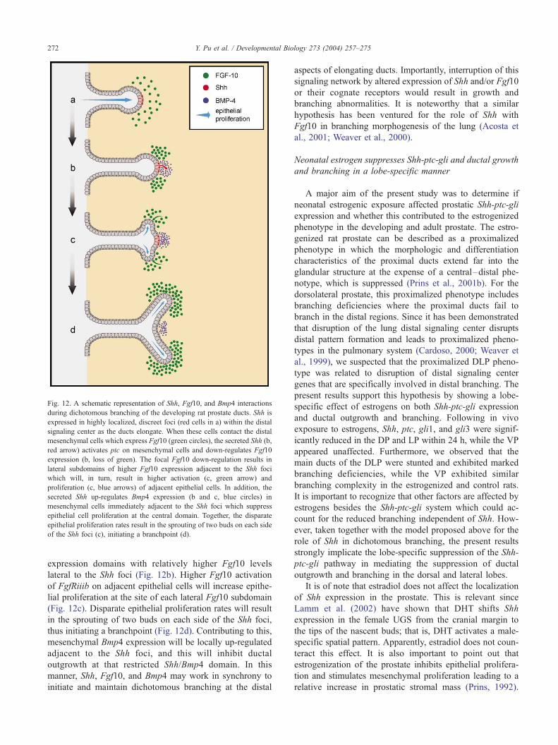
Fig. 12. A schematic representation of Shh, Fgf10, and Bmp4 interactions
during dichotomous branching of the developing rat prostate ducts. Shh is
expressed in highly localized, discreet foci (red cells in a) within the distal
signaling center as the ducts elongate. When these cells contact the distal
mesenchymal cells which express Fgf10 (green circles), the secreted Shh (b,
red arrow) activates ptc on mesenchymal cells and down-regulates Fgf10
expression (b, loss of green). The focal Fgf10 down-regulation results in
lateral subdomains of higher Fgf10 expression adjacent to the Shh foci
which will, in turn, result in higher activation (c, green arrow) and
proliferation (c, blue arrows) of adjacent epithelial cells. In addition, the
secreted Shh up-regulates Bmp4 expression (b and c, blue circles) in
mesenchymal cells immediately adjacent to the Shh foci which suppress
epithelial cell proliferation at the central domain. Together, the disparate
epithelial proliferation rates result in the sprouting of two buds on each side
of the Shh foci (c), initiating a branchpoint (d).
Y. Pu et al. / Developmental Biology 273 (2004) 257–275272
expression domains with relatively higher Fgf10 levels
lateral to the Shh foci (Fig. 12b). Higher Fgf10 activation
of FgfRiiib on adjacent epithelial cells will increase epithe-
lial proliferation at the site of each lateral Fgf10 subdomain
(Fig. 12c). Disparate epithelial proliferation rates will result
in the sprouting of two buds on each side of the Shh foci,
thus initiating a branchpoint (Fig. 12d). Contributing to this,
mesenchymal Bmp4 expression will be locally up-regulated
adjacent to the Shh foci, and this will inhibit ductal
outgrowth at that restricted Shh/Bmp4 domain. In this
manner, Shh, Fgf10, and Bmp4 may work in synchrony to
initiate and maintain dichotomous branching at the distal
aspects of elongating ducts. Importantly, interruption of this
signaling network by altered expression of Shh and/or Fgf10
or their cognate receptors would result in growth and
branching abnormalities. It is noteworthy that a similar
hypothesis has been ventured for the role of Shh with
Fgf10 in branching morphogenesis of the lung (Acosta et
al., 2001; Weaver et al., 2000).
Neonatal estrogen suppresses Shh-ptc-gli and ductal growth
and branching in a lobe-specific manner
A major aim of the present study was to determine if
neonatal estrogenic exposure affected prostatic Shh-ptc-gli
expression and whether this contributed to the estrogenized
phenotype in the developing and adult prostate. The estro-
genized rat prostate can be described as a proximalized
phenotype in which the morphologic and differentiation
characteristics of the proximal ducts extend far into the
glandular structure at the expense of a central–distal phe-
notype, which is suppressed (Prins et al., 2001b). For the
dorsolateral prostate, this proximalized phenotype includes
branching deficiencies where the proximal ducts fail to
branch in the distal regions. Since it has been demonstrated
that disruption of the lung distal signaling center disrupts
distal pattern formation and leads to proximalized pheno-
types in the pulmonary system (Cardoso, 2000; Weaver et
al., 1999), we suspected that the proximalized DLP pheno-
type was related to disruption of distal signaling center
genes that are specifically involved in distal branching. The
present results support this hypothesis by showing a lobe-
specific effect of estrogens on both Shh-ptc-gli expression
and ductal outgrowth and branching. Following in vivo
exposure to estrogens, Shh, ptc, gli1, and gli3 were signif-
icantly reduced in the DP and LP within 24 h, while the VP
appeared unaffected. Furthermore, we observed that the
main ducts of the DLP were stunted and exhibited marked
branching deficiencies, while the VP exhibited similar
branching complexity in the estrogenized and control rats.
It is important to recognize that other factors are affected by
estrogens besides the Shh-ptc-gli system which could ac-
count for the reduced branching independent of Shh. How-
ever, taken together with the model proposed above for the
role of Shh in dichotomous branching, the present results
strongly implicate the lobe-specific suppression of the Shh-
ptc-gli pathway in mediating the suppression of ductal
outgrowth and branching in the dorsal and lateral lobes.
It is of note that estradiol does not affect the localization
of Shh expression in the prostate. This is relevant since
Lamm et al. (2002) have shown that DHT shifts Shh
expression in the female UGS from the cranial margin to
the tips of the nascent buds; that is, DHT activates a male-
specific spatial pattern. Apparently, estradiol does not coun-
teract this effect. It is also important to point out that
estrogenization of the prostate inhibits epithelial prolifera-
tion and stimulates mesenchymal proliferation leading to a
relative increase in prostatic stromal mass (Prins, 1992).

Y. Pu et al. / Developmental Biology 273 (2004) 257–275 273
However, this shift in cell ratios cannot account for the
decreased ptc and gli expression in the present study since
the relative number of stromal cells increases following
estrogenization yet mesenchymal ptc and gli decrease.
The organ culture studies demonstrated that estradiol is
capable of suppressing LP Shh-ptc-gli expression in vitro
which indicates that the estrogenic effects are mediated
directly at the prostatic level. However, it is noteworthy
that the estrogenic suppression of Shh mRNA was not as
marked in vitro as observed in vivo which implicates the
additional involvement of a systemic factor in lowering
prostatic Shh levels. One potential candidate is the de-
creased circulating androgens as a result of neonatal estro-
gen exposure (Corbier et al., 1992; Haavisto et al., 2001;
Prins, 1992). Although androgens are not required for Shh
expression in the UGS, they have been shown to increase
levels of Shh transcript (Freestone et al., 2003; Lamm et al.,
2002) which raises the possibility that androgens have a
supplemental role in maintaining normal Shh expression.
We propose that a combination of reduced circulating
androgens together with direct effects of estrogen may result
in the in vivo suppression of DP and LP Shh levels. We also
predict that the direct prostatic effects of estrogen on
epithelial Shh expression are mediated through paracrine
factors since ERa, which initiates the events, is expressed in
periductal mesenchymal cells (Prins and Birch, 1997; Prins
et al., 2001a,b). In contrast to Shh, estrogenic regulation of
ptc and gli1–3 was fully recapitulated in vitro, indicating
that their suppression is mediated entirely at the prostatic
level. It is of interest that ptc and glis are markedly down-
regulated by estradiol in vitro when Shh levels are only
marginally suppressed which suggests that their reduced
levels are not secondary to reduced Shh expression but may
be direct targets of estrogen action through the up-regulated
ERa in the mesenchyme. Alternatively, estrogens may
indirectly function at the prostatic level through retinoids
via RAR/RXRs (Prins et al., 2002; Pu et al., 2003) or
through the ERa-induced progesterone receptor in the
mesenchymal cells (Sabharwal et al., 2000).
The molecular basis for lobe-specific effects of estrogen
on Shh and its downstream signaling molecules is not clear
at present. Our previous studies have shown similar ERa
localization and up-regulation following neonatal estrogen
exposure in all prostate lobes; thus, the presence or absence
of the initiating receptor is not a factor in lobe specificity
(Prins and Birch, 1997). Likewise, AR is immediately
down-regulated to a similar degree in all lobes following
neonatal estrogenization (Prins and Birch, 1995). Recently,
we observed a lobe-specific expression pattern for RXRg,
retinaldehyde dehydrogenase 2 (RALDH2), and CYP 26
which form and degrade intracellular retinoic acid, respec-
tively, as well as cellular retinoic acid binding proteins,
CRABP 1 and 2 which sequester and regulate retinoic acid
availability within cells (Pu et al., 2003). Transcript levels
for each of these molecules were high in the developing DP
and LP but were minimally expressed in the VP, suggesting
a greater developmental role for retinoids in the DLP.
Furthermore, neonatal estrogens drastically reduced
RALDH2 and CYP26 transcripts and suppressed CRABP
1 and 2 levels in the DP and LP specifically which may
compromise retinoid availability. This is highly significant
since retinoic acids have been shown to be critical regulators
of Shh expression in multiple developing structures (Car-
doso et al., 1996; Goyette et al., 2000; Helms et al., 1994,
1997; Niederreither et al., 2002). Future studies are aimed at
examining the potential lobe-specific retinoid regulation of
Shh-ptc-gli in the DLP.
In closing, lobe-specific responses to neonatal estrogens
have been previously described, but their molecular basis
has been unclear. While the VP is the site of epithelial
differentiation defects and adult-onset dysplasia, the LP
epithelium shows minimal differentiation abnormalities but
major branching deficiencies (Prins, 1997). These distinc-
tive regional responses to the same initial compound suggest
that they are mediated through separate pathways. Previous
studies have shown that permanent reductions in AR and
ERh are specific to the VP and are thought to contribute to
adult dysplasia in that lobe (Prins, 1992; Prins et al., 1993,
1998). Since the present data indicate that the Shh-ptc-gli
pathway participates in prostatic branching morphogenesis
through localized regulation of other critical morphogens, it
is likely that the regional suppression of the Shh-ptc-gli
signaling system in the DP and LP contributes to the lobe-
specific growth deficits in that prostatic region.
Acknowledgments
The authors would like to thank Dr. Liang Ma at Tulane
University for assistance in establishing whole-mount ISH
and for generously providing the Shh cDNA plasmid, Dr.
Alexander Joyner at New York University for generously
providing the mouse cDNA plasmids for gli1, gli2, and gli3,
Lynn Birch for technical assistance, and Dr. Oliver Putz for
graphic renditions of our data. Work was supported by NIH
grant DK-40890.
References
Aboseif, S.R., Dahiya, R., Narayan, P., Cunha, G.R., 1997. Effect of ret-
inoic acid on prostatic development. Prostate 31, 161–167.
Acosta, J.M., Thebaud, B., Castillo, C., Mailleu, A., Tefft, D., Wuenschell,
C., Anderson, K.D., Bourbon, J., Thiery, J.-P., Bellusci, S., Warburton,
D., 2001. Novel mechanisms in murine nitrofen-induced pulmonary
hypoplasia: FGF-10 rescue in culture. Am. J. Physiol.: Lung Cell.
Mol. Physiol. 281, L250–L257.
Bellusci, S., Grindley, J., Emoto, H., Itoh, N., Hogan, B., 1997. Fibroblast
growth factor 10 (FGF10) and branching morphogenesis in the embry-
onic mouse lung. Development 124, 4867–4878.
Berman, D.M., Desai, N., Wang, X., Karhadkar, S.S., Reynon, M., Abate-
Shen, C., Beachy, P.A., Shen, M.M., 2004. Roles for hedgehog signal-
ing in androgen production and prostate ductal morphogenesis. Dev.
Biol. 267, 387–398.
Y. Pu et al. / Developmental Biology 273 (2004) 257–275274
Bhatia-Gaur, R., Donjacour, A.A., Sciavolino, P.J., Kim, M., Desai, N.,
Young, P., Norton, C.R., Gridley, T., Cardiff, R.D., Cunha, G.R., Abate-
Shen, C., Shen, M.M., 1999. Roles for Nkx3.1 in prostate development
and cancer. Genes Dev. 13, 966–977.
Brody, H., Goldman, S.F., 1940. Metaplasia of the epithelium of the pros-
tatic glands, utricle and urethra of the fetus and newborn infant. Arch.
Pathol. Lab. Med. 29, 494.
Cardoso, W.V., 2000. Lung morphogenesis revisited: old facts, current
ideas. Dev. Dyn. 219, 121–130.
Cardoso, W.V., Mitsialis, S.A., Brody, J.S., Williams, M.C., 1996. Retinoic
acid alters the expression of pattern-related genes in the developing rat
lung. Dev. Dyn. 207, 47–59.
Chuang, P.-T., McMahon, A.P., 2003. Branching morphogenesis of the
lung: new molecular insights into an old problem. Trends Cell Biol.
13, 86–91.
Corbier, P., Allioli, N., Roffi, J., 1992. Variations de la reponse testiculaire
a hCG au cours de la periode perinatale chez le Rat: infulence des
oestrogenes. Arch. Int. Physiol., Biochim. Biophys. 100, 389–397.
Donjacour, A.A., Thomson, A.A., Cunha, G.R., 2003. FGF-10 plays an
essential role in the growth of the fetal prostate. Dev. Biol. 261, 39–54.
Driscoll, S.G., Taylor, S.H., 1980. Effects of prenatal maternal estrogen on
the male urogenital system. Obstet. Gynecol. 56, 537–542.
Ellis, T., Smyth, I., Riley, E., Bowles, J., Adolphe, C., Rothnagel, J.A.,
Wicking, C., Wainwright, B.J., 2003. Overexpression of Sonic hedge-
hog suppresses embryonic hair follicle morphogenesis. Dev. Biol. 263,
203–215.
Freestone, S.H., Marker, P., Grace, O.C., Tomlinson, D.C., Cunha, G.R.,
Harnden, P., Thomson, A.A., 2003. Sonic hedgehog regulates prostatic
growth and epithelial differentiation. Dev. Biol. 264, 352–362.
George, F.W., Peterson, K.G., 1988. 5a-dihydrotestosterone formation is
necessary for embryogenesis of the rat prostate. Endocrinology 122,
1159–1164.
Goyette, P., Allan, D., Peschard, P., Chen, C.F., Wang, W., Lohnes, D.,
2000. Regulation of Gli activity by all-trans retinoic acid in mouse
keratinocytes. Cancer Res. 60, 5386–5389.
Gritli-Linde, A., Lewis, P., McMahon, A.P., Linde, A., 2001. The where-
abouts of a morphogen: direct evidence for short- and graded long-range
activity of hedgehog signaling peptides. Dev. Biol. 236, 364–386.
Haavisto, T., Nurmela, K., Pohjanvirta, R., Huuskonen, H., El-Gehani, F.,
Paranko, J., 2001. Prenatal testosterone and luteinizing hormone levels
in male rats exposed during pregnancy to 2,3,7,8-tetrachlorodibenzo-p-
dioxin and diethylstilbestrol. Mol. Cell. Endocrinol. 178, 169–179.
Haraguchi, R., Mo, R., Hui, C., Motoyama, J., Makino, S., Shiroishi, T.,
Gaffield, W., Yamada, G., 2001. Unique functions of Sonic hedgehog
signaling during external genitalia development. Development 128,
4241–4250.
Hayashi, N., Sugimura, Y., Kawamura, J., Donjacour, A.A., Cunha, G.R.,
1991. Morphological and functional heterogeneity in the rat prostatic
gland. Biol. Reprod. 45, 308–321.
Helms, J.A., Thaller, C., Eichele, G., 1994. Relationship between retinoic
acid and Sonic hedgehog, two polarizing signals in the chick wing bud.
Development 120, 3267–3274.
Helms, J.A., Kim, C.H., Hu, D., Minkoff, R., Thaller, C., Eichele, G., 1997.
Sonic hedgehog participates in craniofacial morphogenesis and is
down-regulated by teratogenic doses of retinoic acid. Dev. Biol. 187,
25–35.
Hoffman, M.P., Kidder, B.L., Steinberg, Z.L., Lakhani, S., Ho, S.M., Klein-
man, H.K., Larsen, M., 2002. Gene expression profiles of mouse sub-
mandibular gland development: FGFR1 regulates branching
morphogenesis in vitro through BMP- and FGF-dependent mecha-
nisms. Development 129, 5767–5778.
Huang, L., Pu, Y., Alam, S., Prins, G.S., 2003. Neonatal estrogen exposure
disrupts prostate morphogenesis through alterations in BMP-4, BMP-7
and FGF-10 expression. The Endocrine Society’s 85th Annual Meeting.
Philadelphia, PA. The Endocrine Society, Chevy Chase, MD, p. 334
(Abstract # P332-113).
Huang, L., Pu, Y., Alam, S., Birch, L., Prins, G.S., 2004. Estrogenic reg-
ulation of signaling pathways and homeobox genes during rat prostate
development. J. Androl. 25, 330–337.
Ingham, P.W., McMahon, A.P., 2001. Review: hedgehog signaling in
animal development: paradigms and principles. Genes Dev. 15,
3059–3087.
Jarred, R., Cancilla, B., Prins, G.S., Thayer, K., Cunha, G.R., Risbridger,
G., 2000. Evidence that estrogens directly alter androgen-regulated
prostate development. Endocrinology 141, 3471–3477.
Lamm, M.L., Podlasek, C.A., Barnett, D.H., Lee, J., Clemens, J.Q., Heb-
ner, C.M., Bushman, W., 2001. Mesenchymal factor bone morphoge-
netic protein 4 restricts ductal budding and branching morphogenesis in
the developing prostate. Dev. Biol. 232, 301–314.
Lamm, M.L., Catbagan, W.S., Laciak, R.J., Barnett, D.H., Hebner, C.M.,
Gaffield, W., Walterhouse, D., Iannaccone, P., Bushman, W., 2002.
Sonic hedgehog activates mesenchymal Gli1 expression during prostate
ductal bud formation. Dev. Biol. 249, 349–366.
Lowsley, O.S., 1912. The development of the human prostate gland with
reference to the development of other structures at the neck of the
urinary bladder. Am. J. Anat. 13, 299–348.
Marigo, V., Babin, C.J., 1996. Regulation of patched by Sonic hedgehog
in the developing neural tube. Proc. Natl. Acad. Sci. U. S. A. 93,
9347–9351.
Meyer, N.P., Roelink, H., 2003. The amino-terminal region of Gli3 antag-
onizes the Shh response and acts in dorsoventral fate specification in the
developing spinal cord. Dev. Biol. 257, 343–355.
Naslund, M.J., Coffey, D.S., 1986. The differential effects of neonatal
androgen, estrogen and progesterone on adult rat prostate growth.
J. Urol. 136, 1136–1140.
Niederreither, K., Vermot, J., Schugbaur, B., Chanbon, P., Dolle, P., 2002.
Embryonic retinoic acid synthesis is required for forelimb growth and
anteroposterior patterning in the mouse. Development 129, 3563–3574.
Pepicelli, C., Lewis, P., McMahon, A., 1998. Sonic hedgehog regulates
branching morphogenesis in the mammalian lung. Curr. Biol. 8,
1083–1086.
Perriton, C.L., Powles, N., Chiang, C., Maconochie, M.K., Cohn, M.J.,
2002. Sonic hedgehog signaling from the urethral epithelium controls
external genital development. Dev. Biol. 247, 26–46.
Podlasek, C.A., Barnett, D.H., Clemens, J.Q., Bak, P.M., Bushman, W.,
1999. Prostate development requires Sonic hedgehog expressed by the
urogenital sinus epithelium. Dev. Biol. 209, 28–39.
Price, D., 1963. Comparative aspects of development and structure in the
prostate. In: Vollmer, E.P. (Ed.), Biology of the Prostate and Related
Tissues, vol. 12. National Cancer Institute, Washington, DC, pp. 1–27.
Prins, G.S., 1992. Neonatal estrogen exposure induces lobe-specific alter-
ations in adult rat prostate androgen receptor expression. Endocrinology
130, 3703–3714.
Prins, G.S., 1997. Developmental estrogenization of the prostate gland. In:
Naz, R.K. (Ed.), Prostate: Basic and Clinical Aspects. C.R.C. Press,
Boca Raton, pp. 247–265 (Chapter 10).
Prins, G.S., Birch, L., 1995. The developmental pattern of androgen recep-
tor expression in rat prostate lobes is altered after neonatal exposure to
estrogen. Endocrinology 136, 1303–1314.
Prins, G.S., Birch, L., 1997. Neonatal estrogen exposure up-regulates es-
trogen receptor expression in the developing and adult rat prostate
lobes. Endocrinology 138, 1801–1809.
Prins, G.S., Birch, L., Greene, G.L., 1991. Androgen receptor localization
in different cell types of the adult rat prostate. Endocrinology 129,
3187–3199.
Prins, G.S., Cooke, P.S., Birch, L., Donjacour, A.A., Yalcinkaya, T.M.,
Siiteri, P.K., Cunha, G.R., 1992. Androgen receptor expression and
5a-reductase activity along the proximal–distal axis of the rat prostatic
duct. Endocrinology 130, 3066–3073.
Prins, G.S., Woodham, C., Lepinske, M., Birch, L., 1993. Effects of neo-
natal estrogen exposure on prostatic secretory genes and their correla-
tion with androgen receptor expression in the separate prostate lobes of
the adult rat. Endocrinology 132, 2387–2398.
Prins, G.S., Marmer, M., Woodham, C., Chang, W.Y., Kuiper, G., Gustafs-

Y. Pu et al. / Developmental Biology 273 (2004) 257–275 275
son, J.A., Birch, L., 1998. Estrogen receptor-h messenger ribonucleic
acid ontogeny in the prostate of normal and neonatally estrogenized
rats. Endocrinology 139, 874–883.
Prins, G.S., Birch, L., Couse, J.F., Choi, I., Katzenellenbogen, B., Korach,
K.S., 2001a. Estrogen imprinting of the developing prostate gland in
mediated through stromal estrogen receptor a: studies with aERKO and
hERKO mice. Cancer Res. 61, 6089–6097.
Prins, G.S., Birch, L., Habermann, H., Chang, W.Y., Tebeau, C., Putz, O.,
Bieberich, C., 2001b. Influence of neonatal estrogens on rat prostate
development. Reprod. Fertil. Dev. 13, 241–252.
Prins, G.S., Chang, W.Y., Wang, Y., van Breemen, R.B., 2002. Retinoic acid
receptors and retinoids are up-regulated in the developing and adult rat
prostate by neonatal estrogen exposure. Endocrinology 143, 3628–3640.
Pu, Y., Deng, L., Davies, P.J.P., Prins, G.S., 2003. Retinoic acid metabo-
lizing enzymes, binding proteins and RXRs are differentially expressed
in the developing and adult rat prostate lobes and are altered by neonatal
estrogens in a lobe-specific manner. The Endocrine Society’s 85th An-
nual Meeting, Philadelphia, PA. The Endocrine Society, Chevy Chase,
MD, p. 530 (Abstract # P533-236).
Putz, O., Prins, G.S., 2002. Prostate gland development and estrogenic
imprinting. In: Burnstein, K.L. (Ed.), Steroid Hormones and Cell Cycle
Regulation. Kluwer Academic Publishers, Boston, MA, pp. 73–89.
Pylkkanen, L., Makela, S., Valve, E., Harkonen, P., Toikkanen, S., Santti,
R., 1993. Prostatic dysplasia associated with increased expression of C-
myc in neonatally estrogenized mice. J. Urol. 149, 1593–1601.
Rajfer, J., Coffey, D.S., 1979. Effects of neonatal steroids on male sex
tissues. Invest. Urol. 17, 3–8.
Roberts, D.J., Johnson, R.L., Burke, A.C., Nelson, C.E., Morgan, B.A.,
Tabin, C., 1995. Sonic hedgehog is an endodermal signal inducing
Bmp4 and Hox gene during induction and regulation of the chick hind-
gut. Development 121, 3163–3174.
Roberts, D.J., Smith, D.M., Goff, D.J., Tabin, C., 1998. Epithelial–mes-
enchymal signaling during the regionalization of the chick gut. Deve-
lopment 125, 2791–2801.
Sabharwal, V., Putz, O., Prins, G.S., 2000. Neonatal estrogen exposure
induces progesterone receptor expression in the developing prostate
gland. 95th Annual Meeting of the American Urologic Association,
Atlanta. The Endocrine Society, Chevy Chase, MD, p. 97.
Santti, R., Newbold, R.R., Makela, S., Pylkkanen, L., McLachlan, J.A.,
1994. Developmental estrogenization and prostatic neoplasia. Prostate
24, 67–78.
Schneider, A., Brand, T., Zweigerdt, R., Arnold, H., 2000. Targeted dis-
ruption of the Nkx3.1 gene in mice results in morphogenetic defects of
minor salivary glands: parallels to glandular duct morphogenesis in
prostate. Mech. Dev. 95, 163–174.
Seo, R., McGuire, M., Chung, M., Bushman, W., 1997. Inhibition of pros-
tate ductal morphogenesis by retinoic acid. J. Urol. 158, 931–935.
Shapiro, E., 1990. Embryologic development of the prostate. Urol. Clin.
North Am. 17, 487–493.
Siiteri, P.K., Wilson, J.D., 1974. Testosterone formation and metabolism
during male sexual differentiation in the human embryo. JCEM 38,
113–125.
Sreenath, T., Orosz, A., Fujita, K., Bieberich, C.J., 1999. Androgen-in-
dependent expression of hoxb-13 in the mouse prostate. Prostate 41,
203–207.
Suzuki, T., Oohara, I., Kurokawa, T., 1999. Retinoic acid given at late
embryonic stage depresses Sonic hedgehog and Hoxd-4 expression in
the pharyngeal area and induces skeletal malformation in flounder (Par-
ralichthys olivaceus) embryos. Dev. Growth Differ. 41, 143–152.
Tamura, K., Yokouchi, Y., Kuroiwa, A., Ide, H., 1997. Retinoic acid
changes the proximodistal development competence and affinity of
distal cells in the developing chick limb bud. Dev. Biol. 188, 224–234.
Thomson, A.A., Cunha, G.R., 1999. Prostatic growth and development are
regulated by FGF10. Development 126, 3693–3701.
vom Saal, F.S., Timms, B.G., Montano, M.M., Palanza, P., Thayer, K.A.,
Nagel, S.C., Dhar, M.D., Ganjam, V.K., Parmigiani, S., Welshons,
W.V., 1997. Prostate enlargement in mice due to fetal exposure to
low doses of estradiol or diethylstilbestrol and opposite effects at high
doses. Proc. Natl. Acad. Sci. U. S. A. 94, 2056–2061.
Wang, B., Shou, J., Ross, S., Koeppen, H., de Sauvage, F.J., Gao, W.-Q.,
2003. Inhibition of epithelial ductal branching in the prostate by Sonic
hedgehog is indirectly mediated by stromal cells. J. Biol. Chem. 278,
18506–18513.
Weaver, M., Yingling, J.M., Dunn, N.R., Bellusci, S., Hogan, B.L.M.,
1999. Bmp signaling regulates proximal–distal differentiation of endo-
derm in mouse lung development. Development 126, 4005–4015.
Weaver, M., Dunn, N.R., Hogan, B.L., 2000. BMP4 and FGF10 play
opposing roles during lung bud morphogenesis. Development 127,
2695–2704.
Woodham, C., Birch, L., Prins, G.S., 2003. Neonatal estrogens down reg-
ulate prostatic androgen receptor levels through a proteosome-mediated
protein degradation pathway. Endocrinology 144, 4841–4850.
Yang, Y., Drossopoulou, G., Chuang, P.T., Duprez, D., Marti, E., Buncrot,
N., Vargession, N., Clarke, J., Niswander, L., McMahon, A., Tickle, C.,
1997. Relationship between dose, distance and time in Sonic hedge-
hog-mediated regulation of anteroposterior polarity in the chick limb.
Development 124, 4393–4404.
Zondek, T., Zondek, L.H., 1975. The fetal and neonatal prostate. In: Goland,
M. (Ed.), Normal and Abnormal Growth of the Prostate. Thomas C.
Thomas, Springfield, IL, pp. 5–28.
Related Documents


