2/17/2016 1 Slide 1 JSOMTC, SWMG(A) Musculoskeletal Trauma PFN: SOMEML12 Hours: 1.0 Instructor: Slide 2 JSOMTC, SWMG(A) References Tintinalli’s Emergency Medicine, 7 th Edition, 2010 Tactical Trauma Protocols PHTLS, 7 th Edition, 2010 Slide 3 JSOMTC, SWMG(A) Terminal Learning Objective Action: Communicate knowledge of musculoskeletal trauma Conditions: Given a lecture in a classroom environment Standards: Received a minimum score of 75% on a written exam IAW course standards

Welcome message from author
This document is posted to help you gain knowledge. Please leave a comment to let me know what you think about it! Share it to your friends and learn new things together.
Transcript

2/17/2016
1
Slide 1JSOMTC, SWMG(A)
Musculoskeletal TraumaPFN: SOMEML12
Hours: 1.0
Instructor:
Slide 2JSOMTC, SWMG(A)
References
Tintinalli’s Emergency Medicine, 7th Edition, 2010
Tactical Trauma Protocols
PHTLS, 7th Edition, 2010
Slide 3JSOMTC, SWMG(A)
Terminal Learning Objective
Action: Communicate knowledge of musculoskeletal trauma
Conditions: Given a lecture in a classroom environment
Standards: Received a minimum score of 75% on a written exam IAW course standards

2/17/2016
2
Slide 4JSOMTC, SWMG(A)
Reason
Slide 5JSOMTC, SWMG(A)
Agenda
Identify the clinical presentation, and management of rhabdomyolysis
Identify the clinical presentation, and management of compartment syndrome
Identify the clinical presentation, and management of crush injury and crush syndrome
Slide 6JSOMTC, SWMG(A)
Rhabdomyolysis

2/17/2016
3
Slide 7JSOMTC, SWMG(A)
Rhabdomyolysis
Rhabdomyolysis is a serious syndrome due to a direct or indirect muscle injury
It results from the death of muscle fibers and release of their contents into the bloodstream
This can lead to complications such as renal (kidney) failure
Slide 8JSOMTC, SWMG(A)
RhabdomyolysisCauses
Must think about risk factors because classic signs may be absent in 50% of cases
Extreme muscle strain, especially in someone who is an untrained athlete. This can happen in elite athletes too. And it can be more dangerous if there is more muscle mass to break down
Crush injuries
Long‐lasting muscle compression
Electrical shock injury, burn, heat stroke
Slide 9JSOMTC, SWMG(A)
RhabdomyolysisCauses
Strenuous exercise or unaccustomed muscular activity (i.e. siezure)
Drug or alcohol abuse
Certain disease processes (i.e. viruses)
Bacterial infections leading to toxins in tissues or the bloodstream (sepsis)

2/17/2016
4
Slide 10JSOMTC, SWMG(A)
Clinical Presentation
Muscle pain, especially in the shoulders, thighs or lower back
Muscle weakness or trouble moving arms or legs
Abdominal pain
Nausea or vomiting
Fever, rapid heart rate
Confusion, dehydration, fever, or lack of consciousness
Slide 11JSOMTC, SWMG(A)
Clinical Presentation
Dark (tea‐colored) urine
Oliguria or anuria
Urine or serum myoglobin
Dipstick + for blood, but no intact RBCs on spun specimen
Slide 12JSOMTC, SWMG(A)
Complications Early
Hyperkalemia
Hypocalcemia
Hepatic inflammation
Cardiac arrhythmia and/or arrest
Late
Hypercalcemia
Renal failure
DIC

2/17/2016
5
Slide 13JSOMTC, SWMG(A)
Complications
Early or late
Compartment syndrome
•May occur after fluid resuscitation. This serious compression of nerves, blood vessels, and muscles can cause tissue damage and problems with blood flow.
Slide 14JSOMTC, SWMG(A)
Management (TMEP)Aggressive hydration is the cornerstone of
treatment
Normal saline 1 ‐ 2L bolus IV/IO followed by 500 ml ‐1L per hour
Avoid Ringer’s lactate due to the potassium content
Titrate to achieve target urine output of > 200 ml/hour
• Consider urinary alkalinization to achieve urine pH > 6.5 Mix Sodium Bicarbonate 40 mEq (1 ampule/bristojet) in 500 ml normal saline. Run at 100 ml/h
Slide 15JSOMTC, SWMG(A)
Management (TMEP)
Reassess vital signs and mental status frequently
Foley catheter to facilitate measuring urine output
Utilize cardiac monitoring if available
Electrolyte and metabolic complications can cause dysrhythmias
Urgent evacuation
2/17/2016
6
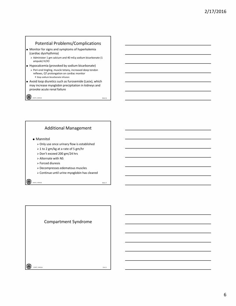
Slide 16JSOMTC, SWMG(A)
Potential Problems/Complications Monitor for signs and symptoms of hyperkalemia (cardiac dysrhythmia)
Administer 1 gm calcium and 40 mEq sodium bicarbonate (1 ampule) IV/IO
Hypocalcemia (provoked by sodium bicarbonate)
Peri‐oral tingling, muscle tetany, increased deep tendon reflexes, QT prolongation on cardiac monitor • Stop sodium bicarbonate infusion
Avoid loop diuretics such as furosemide (Lasix), which may increase myoglobin precipitation in kidneys and provoke acute renal failure
Slide 17JSOMTC, SWMG(A)
Additional Management
Mannitol
Only use once urinary flow is established
1 to 2 gm/kg at a rate of 5 gm/hr
Don’t exceed 200 gm/24 hrs
Alternate with NS
Forced diuresis
Decompresses edematous muscles
Continue until urine myoglobin has cleared
Slide 18JSOMTC, SWMG(A)
Compartment Syndrome

2/17/2016
7
Slide 19JSOMTC, SWMG(A)
Compartment Syndrome
Occurs when excessive pressure builds up inside an enclosed space in the body
Increased compartment pressure compromises circulation and function of compartment tissues
Prolonged elevation of tissue pressure leads to muscle death and nerve damage
Slide 20JSOMTC, SWMG(A)
Common Causes
Any mechanism that increases the volume of blood or tissue within the compartment can cause a compartment syndrome
External forces
Tight cast or constrictive dressing
Venous tourniquet
Vascular
Hemorrhage
Ischemic‐reperfusion injury
Slide 21JSOMTC, SWMG(A)
Common Causes
Orthopedic
Tibial and forearm fractures
Soft tissue injury
Prolonged limb compression
Crush injury
Burns
Envenomation

2/17/2016
8
Slide 22JSOMTC, SWMG(A)
Clinical Presentation
The classic “P’s”
Pain
Parasthesia
Poikilothermia
Pallor
Pressure
Paralysis
Pulselessness
Slide 23JSOMTC, SWMG(A)
Pain
Disproportionate to physical exam findings
Earliest and most consistent sign
Aggravated by passive stretching
Most sensitive sign
Diffuse, deep, unremitting, and poorly localized
Not relieved with immobilization
Often difficult to control with narcotics
Slide 24JSOMTC, SWMG(A)
Paresthesia
Tingling, burning
Diminished sensation
Two‐point discrimination
Light touch
Sensory disturbance generally precedes motor dysfunction

2/17/2016
9
Slide 25JSOMTC, SWMG(A)
Poikilothermia, Pallor, and Pressure
Poikilothermia
Cold distal extremity compared to the contralateral side
Pallor
Pale color
Pressure
Compartment is swollen and firm on palpation
Measurable increase in tissue pressure
Slide 26JSOMTC, SWMG(A)
Pressure
Compartment pressure can be measured
Commercial kits available
Measure pressure within 5 cm of any fracture
Slide 27JSOMTC, SWMG(A)
Paralysis and Pulselessness
Paralysis
Late finding from prolonged ischemia
Permanent damage may already be present
Pulselessness
Least reliable sign
CS is a disorder of microvasculature and rarely affects major vessels
Pulses are present in 90% of patients

2/17/2016
10
Slide 28JSOMTC, SWMG(A)
Clinical Presentation
Maintain a high index of suspicion with polytrauma patients
Pain greater than expected due to the injury alone is your earliest and best clue
Open wounds do not exclude CS
The presence of pulses and normal capillary refill does not exclude CS
CS can develop up to 48 hours after the event
Slide 29JSOMTC, SWMG(A)
Complications
Rhabdomyolysis
Myoglobinuria
Renal failure
Volkmann contracture
Slide 30JSOMTC, SWMG(A)
Management
Maintain extremity at heart level ‐
DO NOT ELEVATE
Supplemental oxygen
Fluid resuscitation
Remove constrictive casts or dressings
Evacuate to surgical facility for immediate fasciotomy

2/17/2016
11
Slide 31JSOMTC, SWMG(A)
Surgical FasciotomyNot in your scope of practice
Long incisions through the skin and fascia
Allows tissue swelling without pressure elevation
May require delayed grafting
Should be performed immediately after making the diagnosis
Slide 32JSOMTC, SWMG(A)
Crush Injury and Crush Syndrome
Slide 33JSOMTC, SWMG(A)
Injury vs Syndrome
Crush INJURY ‐ occurs when a body part is subjected to a high degree of force or pressure
Can produce compartment syndrome
Crush SYNDROME ‐ the systemic manifestation of muscle cell and capillary endothelial damage‐ what happens after the injury occurs

2/17/2016
12
Slide 34JSOMTC, SWMG(A)
Crush Syndrome
Major shock and renal failure after a crushing injury to skeletal muscle
Imagine “rhabdomyolysis on steroids”
Slide 35JSOMTC, SWMG(A)
Reperfusion Syndrome
Devastating systemic effects can occur when the crushing pressure is suddenly released, without proper preparation of the patient
Initiates inflammatory response
Hypovolemia, hypotension, myocardial depression, hyperkalemia, and acidosis
Immediate release of toxins can have catastrophic systemic consequences
Slide 36JSOMTC, SWMG(A)
Clinical Presentation
MOI
Obvious external signs of crush injury
Lacerations, degloving, deformity, or pain
Compartment syndrome
Rhabdomyolysis, myoglobinuria, and/or renal failure

2/17/2016
13
Slide 37JSOMTC, SWMG(A)
Management (TMEP)
Special Considerations
Be aware of development of crush syndrome starting as early as 4 hours post injury
The medications used to treat crush syndrome are not part of the standard ATP aid bag and require development of a separate crush injury kit
Slide 38JSOMTC, SWMG(A)
Management (TMEP)
Warnings
The principles of hypotensive resuscitation according to TCCC DO NOT apply in the setting of extremity crush injury requiring extrication
In the setting of a crush injury associated with non compressible (thoracic, abdominal, pelvic) hemorrhage, aggressive fluid resuscitation may result in increased hemorrhage
Slide 39JSOMTC, SWMG(A)
Management (TMEP)
Warnings
With extremity injuries, tourniquets should NOT be applied during Phase 1 unless there is hemorrhage which is not controllable by other means
Be aware of development of cardiac dysrhythmias due to hyperkalemia immediately following extrication

2/17/2016
14
Slide 40JSOMTC, SWMG(A)
Management (TMEP)
Management
Broken down into three phases
• Phase 1: Immediate (while attempting extrication)
• Phase 2: Immediately prior to extrication
• Phase 3: Immediately following extrication
Slide 41JSOMTC, SWMG(A)
Management (TMEP)
Phase 1: Immediate (while attempting extrication)
Maintain patent airway (NPA, OPA, etc.) and adequate ventilation
Monitor O2 sat with pulse ox and administer high flow oxygen if available
Slide 42JSOMTC, SWMG(A)
Management (TMEP)
Phase 1: Immediate (cont.)
Give initial bolus of 1 to 1.5 L of NS PRIOR to attempts at extrication and continue at 1.5 L/hr
• Ringer’s lactate is not recommended due to the potassium content
Maintain urine output at greater than or equal to 200 cc/hr. If possible, insert Foley catheter.
Assess and reassess mental status
Follow Pain Management Protocol (TMEP)

2/17/2016
15
Slide 43JSOMTC, SWMG(A)
Management (TMEP)
Phase 1: Immediate (cont.)
Consider prophylactic antibiotics ‐
• Ertapenem (Invanz) 1 gm IV
Utilize cardiac monitoring if available
Mannitol (administer 1 to 2 gm/kg at a rate of 5gm/hr)
• Ensure urine output has been established prior to using Mannitol
Slide 44JSOMTC, SWMG(A)
Management (TMEP)
Phase 2: Immediately PRIOR to extrication
Apply tourniquets to crushed extremities, if possible
Sodium Bicarbonate ‐
•1 mEq/kg IV immediately prior to extrication.
• Additional dosing of Sodium bicarbonate may be required if dysrhythmias or cardiac arrest persist after giving calcium chloride or gluconate.
Slide 45JSOMTC, SWMG(A)
Management (TMEP)
Phase 3: Immediately FOLLOWING extrication
CPR should be initiated if cardiac arrest develops following extrication. DO NOT follow the TCCC guidelines on cardiac arrest.
If extrication is greater then 4 hours OR in the presence of dysrhythmias, administer Calcium Chloride (1 gm, 10 ml of 10% solution) or Calcium Gluconate (1 gm, 10 ml of 10% solution)

2/17/2016
16
Slide 46JSOMTC, SWMG(A)
Management (TMEP)
Phase 3: Immediately FOLLOWING extrication
(cont.)
Calcium should not be given in bicarbonate containing solutions due to precipitation of calcium carbonate
Additional dosing of Sodium bicarbonate may be required if dysrhythmias or cardiac arrest persist after giving calcium chloride or gluconate
Following extrication, once the patient is stabilized, be prepared to treat hyperkalemia as tourniquets are released
Slide 47JSOMTC, SWMG(A)
Questions?
Slide 48JSOMTC, SWMG(A)
Terminal Learning Objective
Action: Communicate knowledge of musculoskeletal trauma
Conditions: Given a lecture in a classroom environment
Standards: Received a minimum score of 75% on a written exam IAW course standards

2/17/2016
17
Slide 49JSOMTC, SWMG(A)
Agenda
Identify the clinical presentation, and management of rhabdomyolysis
Identify the clinical presentation, and management of compartment syndrome
Identify the clinical presentation, and management of crush injury and crush syndrome
Slide 50JSOMTC, SWMG(A)
Reason
Related Documents