INTRODUCTION Multiparticulates, including micro- and nano-dimensional drug delivery vehicles have excellent and broad prospects in the pharmaceutical field. Scientists from various disciplines have been harnessed by the superior outcomes obtained from such carrier systems, viz. greater therapeutic efficacy with reduced dosing frequency (1). The in vivo 433 Acta Pharm. 62 (2012) 433–472 Review DOI: 10.2478/v10007-012-0040-z Solid lipid based nanocarriers: An overview CHANDRAKANTSING PARDESHI 1 * PRAVIN RAJPUT 1 VEENA BELGAMWAR 1 AVINASH TEKADE 2 GANESH PATIL 3 KAPIL CHAUDHARY 4 ABHIJEET SONJE 5 1 R. C. Patel Institute of Pharmaceutical Education and Research Shirpur-425405, India 2 Rajarshi Shahu College of Pharmacy and Research, Pune-411033, India 3 H. R. Patel Institute of Pharmaceutical Education and Research Shirpur-425405, India 4 Formulation Research and Development Department, Marksans Pharma., Ltd., Verna Industrial Estate, Verna-403722 India 5 Baxil Pharma, Pvt. Ltd., Nainital Highway, Shyampur Haridwar-249408 India Accepted September 12, 2012 In the era of nanoparticulate controlled and site specific drug delivery systems, use of solid lipids to produce first generation lipid nanoparticles, solid lipid nanopar- ticles (SLN), became a revolutionary approach in the early nineties. The present review is designed to provide an insight into how SLN are finding a niche as promis- ing nanovectors and forms a sound basis to troubleshoot the existing problems associated with traditional sys- tems. Herein, authors had tried to highlight the frontline aspects prominent to SLN. An updated list of lipids, ad- vanced forms of SLN, methods of preparation, character- ization parameters, and various routes of administration of SLN are explored in-depth. Stability, toxicity, stealth- ing, targeting efficiency and other prospectives of SLN are also discussed in detail. The present discussion em- bodies the potential of SLN, now being looked up by va- rious research groups around the world for their utility in the core areas of pharmaceutical sciences, thereby urg- ing pharmaceutical industries to foster their scale-up. Keywords: colloidal drug carrier, solid lipid nanoparticles (SLN), stability, targeting efficiency, cytotoxicity, steal- thing of SLN * Correspondence; e-mail: chandrakantpardeshi11@gmail.com; cvpardeshi@rcpatelpharmacy.co.in

Welcome message from author
This document is posted to help you gain knowledge. Please leave a comment to let me know what you think about it! Share it to your friends and learn new things together.
Transcript

INTRODUCTION
Multiparticulates, including micro- and nano-dimensional drug delivery vehicleshave excellent and broad prospects in the pharmaceutical field. Scientists from variousdisciplines have been harnessed by the superior outcomes obtained from such carriersystems, viz. greater therapeutic efficacy with reduced dosing frequency (1). The in vivo
433
Acta Pharm. 62 (2012) 433–472 ReviewDOI: 10.2478/v10007-012-0040-z
Solid lipid based nanocarriers: An overview
CHANDRAKANTSING PARDESHI1*PRAVIN RAJPUT1
VEENA BELGAMWAR1
AVINASH TEKADE2
GANESH PATIL3
KAPIL CHAUDHARY4
ABHIJEET SONJE5
1 R. C. Patel Institute of PharmaceuticalEducation and ResearchShirpur-425405, India
2 Rajarshi Shahu College of Pharmacyand Research, Pune-411033, India
3 H. R. Patel Institute of PharmaceuticalEducation and ResearchShirpur-425405, India
4 Formulation Research and DevelopmentDepartment, Marksans Pharma., Ltd., VernaIndustrial Estate, Verna-403722 India
5 Baxil Pharma, Pvt. Ltd., NainitalHighway, ShyampurHaridwar-249408 India
Accepted September 12, 2012
In the era of nanoparticulate controlled and site specificdrug delivery systems, use of solid lipids to producefirst generation lipid nanoparticles, solid lipid nanopar-ticles (SLN), became a revolutionary approach in theearly nineties. The present review is designed to providean insight into how SLN are finding a niche as promis-ing nanovectors and forms a sound basis to troubleshootthe existing problems associated with traditional sys-tems. Herein, authors had tried to highlight the frontlineaspects prominent to SLN. An updated list of lipids, ad-vanced forms of SLN, methods of preparation, character-ization parameters, and various routes of administrationof SLN are explored in-depth. Stability, toxicity, stealth-ing, targeting efficiency and other prospectives of SLNare also discussed in detail. The present discussion em-bodies the potential of SLN, now being looked up by va-rious research groups around the world for their utilityin the core areas of pharmaceutical sciences, thereby urg-ing pharmaceutical industries to foster their scale-up.
Keywords: colloidal drug carrier, solid lipid nanoparticles(SLN), stability, targeting efficiency, cytotoxicity, steal-thing of SLN
* Correspondence; e-mail: [email protected]; [email protected]

biodistribution of any drug that longer depends on the properties of that drug but ismainly determined by a carrier system which enables a controlled release of bioactivesto the localized site as per specific needs of the therapy (2).
When colloidal drug carriers (nanometric size range) are compared with micropar-ticles (micrometer range) and implants (several millimeters), it has been found thatmicroparticles and implants based on biodegradable polyesters permit controlled andlocalized drug release over a period of weeks to months after s.c. or i.v. injection/im-plantation, but are too large for drug targeting and intravenous administration. There-fore, colloidal drug carriers hold great promise for achieving the target of controlled andsite specific drug release and have hence attracted wide attention of researchers and for-mulation scientists. Traditional colloidal drug carrier systems include nanoemulsions,nanosuspensions, nanoparticles, micelles, liposomes, polymer-drug conjugates (2, 4).
The existence of different colloidal drug carrier systems may raise the question tothe readers’ mind about which of these might be the most suitable carrier system for adesired purpose. To answer this question, the following aspects should be taken intoconsideration:
(i) drug loading capacity(ii) sufficient drug targeting(iii) in vivo fate of the carrier system (interaction with the surrounding biological
fluid, degradation rate, accumulation in organs, etc.)(iv) toxicity, acute as well as chronic(v) storage stability, physical as well as chemical(vi) large scale production(vii) overall cost of formulation.
A new class of colloidal drug carriers, solid lipid nanoparticles (SLNs), emerged inthe early nineties and is now being looked up by researchers around the world for ex-ploitation of their potential applications in the core areas of pharmaceutical sciences,like drug delivery, clinical and therapeutic medicine as well as other sciences. Polymericnanoparticles, instead of having the advantage of possible chemical modifications inclu-ding synthesis of block- and co-polymers, have their own well-known limitations suchas polymer cytotoxicity, polymer degradation, high cost due to lack of a suitable largescale production method and scarcity of their approval by the regulatory authorities thatpose major hurdles for their utilization in clinical medicine (3). Thus, since the begin-ning of the nineties, attention of various research groups has been focused on an alterna-tive to polymeric nanoparticles, the solid lipid nanoparticles (SLN).
Recent updates have shown that new drug development is not the prime concernfor efficient drug therapy. Theoretical predictions and in vitro experimental data some-times follow the disappointing in vivo results leading to therapy failure. The main rea-sons for therapy failure include:
(i) poor drug solubility excluding i.v. injection of aqueous drug solution(ii) poor absorption, rapid metabolism and excretion (e.g. proteins, peptides, etc.)(iii) drug distribution to non-targeted sites combined with high drug toxicity (e.g.,
anticancer drugs)
434
C. Pardeshi et al.: Solid lipid based nanocarriers: An overview, Acta Pharm. 62 (2012) 433–472.
(iv) high fluctuations in drug plasma levels due to unpredictable bioavailability af-ter peroral administration, including influence of food on plasma levels (e.g.,cyclosporine).
To troubleshoot these formidable problems, a novel and promising nano-drug car-rier system, solid lipid nanoparticles, was introduced. SLNs are one generation abovethe sub-micron sized lipid emulsions where the liquid lipid (oil) has been substituted bya solid lipid and are mainly composed of a physiological lipid dispersed in water or inaqueous surfactant solution. Replacement of liquid oil with solid lipid represents a mile-stone in achieving controlled drug release, because the mobility of a drug in solid lipidis usually lower compared to liquid oil, which made them attractive for their potentialuse in improving the performance of pharmaceuticals, nutraceuticals and other suchmaterials (2–4).
In the present review, the authors have focused on the overview of SLNs, problemsassociated with them, broad prospects of SLNs as a drug delivery system in the pharma-ceutical field and related trends in SLNs.
ADVANTAGES AND DISADVANTAGES OF SLNs
It has been proposed that SLNs combine the advantages of traditional colloidal drugcarriers but avoid some of their major disadvantages (2, 5). The proposed advantages in-clude:
(i) controlled and targeted drug release(ii) possible high drug payload(iii) feasibility of carrying both hydrophilic and lipophilic drug(iv) water based formulation avoids organic solvents(v) physiological lipids decrease the prevalence of acute or chronic toxicity; no re-
ported biotoxicity of the carrier system(vi) improved drug stability(vii) most lipids being biodegradable, SLNs have excellent biocompatibility(viii)less expensive than polymeric or surfactant based carriers(ix) easy to scale up and sterilize(x) easy to validate(xi) easy to gain regulatory approval.
The potential disadvantages of SLNs include:(i) poor drug loading capacity of drugs having limited solubility in lipid melt(ii) relatively very high water content of dispersions (70–99.9 %)(iii) drug expulsion after polymeric transition during storage.
The drug loading capacity of conventional SLNs is depends on the following fac-tors:
(i) solubility of the drug in lipid melts
435
C. Pardeshi et al.: Solid lipid based nanocarriers: An overview, Acta Pharm. 62 (2012) 433–472.
(ii) structure of the lipid matrix(iii) polymorphic state of the lipid matrix.
If the lipid matrix consists of very similar molecules (viz. tristearin or tripalmitin),then a perfect crystal with few imperfections is formed. A highly ordered crystal latticecannot accommodate large amounts of the drug, since the incorporated drugs are loca-ted between fatty acid chains, between lipid layers and also in imperfections. Therefore,the use of more complex lipids is more convenient for higher drug loading.
ADVANCED FORMS OF SLNs
Nanostructured lipid carriers (NLCs)
Nanostructured lipid carriers, introduced at the turn of the millennium, represent anew and improved generation of SLNs and are made of a solid lipid matrix entrappingliquid lipid nanocompartments, the blend being solid at body temperature (6). This newgeneration of lipid carriers (NLCs) was introduced to overcome the problems associatedwith SLNs, such as limited drug loading capacity, drug expulsion during storage andadjustment of drug release, long-term physical stability of the suspension, etc.
Production procedures are identical for both lipid particles, SLNs and NLCs. Thesolid lipid or solid-liquid lipid blend is melted, the pharmaceutical or cosmetic active isdissolved in melted lipid phase subsequently dispersed in a hot aqueous surfactant/sta-bilizer solution of equivalent temperature under high speed stirring. Obtained pre--emulsion is homogenized in a high pressure homogenizer yielding a hot o/w nano-emulsion. After cooling, emulsion droplets crystallize, forming lipid nanoparticles withsolid particle matrices, depending on the starting material, either SLN or NLC (7).
Three models of NLCs were proposed. In the first model, also known as »imperfecttype NLC«, particles are prepared from a lipid mixture of spatially different lipids (likeglycerides) composed of different fatty acids. Use of spatially different lipids leads tolarger distances between the fatty acid chains of glycerides and general imperfection ofthe crystal lattice. This would provide more space for accommodation of guest mole-cules in molecular form or as amorphous clusters. High drug loading could be achievedand drug expulsion from the lipid matrix during storage could be prevented with thismodel, due to distortion of the crystal lattice. This suggests that an increased number ofimperfections leads to increased drug loading capacity and one could say that »the per-fectness« of the NLC system lies in the »imperfectness« in its crystal lattice (Fig. 1) (7).Fang et al. (8) found enhanced permeation and controlled release of psoralens with NLCformulations compared to SLN formulations and concluded that NLCs can be poten-tially exploited as carriers for psoriasis therapies.
The second model is also known as »multiple type NLC«, where drugs showinghigher solubility in oils than in solid lipids can be dissolved in oil and yet be protectedfrom degradation by the surrounding solid lipids. Multiple type NLCs are analogous tow/o/w multiple emulsions since these are oil-in-solid lipid-in-water dispersions.
436
C. Pardeshi et al.: Solid lipid based nanocarriers: An overview, Acta Pharm. 62 (2012) 433–472.

The third model, also known as »amorphous type NLC«, prevents the ongoing ex-pulsion of the drug caused by crystallization or transformation of the solid lipid. Here,the particles are solid but crystallization upon cooling is avoided by using special lipidssuch as hydroxyl octacosanyl, hydroxyl stearate, isopropyl myristate, etc.
NLCs have mostly been extensively investigated for topical and dermatological pre-parations in the delivery of clotrimazole (9), celecoxib (10), ascorbyl palmitate (11), flu-ticasone (12) and so on.
Lipid drug conjugates (LDCs)
Lower drug loading capacity of hydrophilic actives was a major issue in SLNs dueto partitioning effects during the production process. Highly potent-low dose drugs canbe suitably incorporated only in the solid lipid matrix (13).
Polymer lipid hybrid nanoparticles (PLNs)
Polymer-lipid hybrid nanoparticles hold great promise as a drug delivery vehicle inthe treatment of a myriad of diseases such as breast cancer (14, 15). A PLN comprisesthree distinct functional components:
(i) hydrophobic polymeric core to encapsulate poorly water-soluble drugs(ii) hydrophilic polymeric shell to enhance PLN stability and circulation half-life(iii) lipid monolayer at the core and shell interface to promote drug retention inside
the polymeric core (16).
Interactions among these components play an important role for successful fabrica-tion and performance of PLNs (17).
These hybrid NPs combine the merits of both liposomes and polymeric nanopar-ticles, two of the most popular drug delivery vehicles approved for clinical use, therebyserving as a robust drug delivery platform (16). It has been shown in vitro that hybridNPs possess the ability of carrying poorly water-soluble drugs with high encapsulationand loading yields, tunable and sustained drug release profiles, excellent serum stability,and differential targeting of cells (18).
437
C. Pardeshi et al.: Solid lipid based nanocarriers: An overview, Acta Pharm. 62 (2012) 433–472.
Fig. 1. Formation of: a) per-fect crystal lattice in SLN us-ing similar lipid molecules,and b) imperfections in NLCusing spatially different li-pids.

GENERAL INGREDIENTS USED IN THE PREPARATION OF SLNs
General ingredients used in the preparation of SLNs are given in Table I.
438
C. Pardeshi et al.: Solid lipid based nanocarriers: An overview, Acta Pharm. 62 (2012) 433–472.
Table I. Ingredients used for the production of solid lipid nanoparticles
Ingredient References
LIPIDS
Triglycerides
TricaprinTrilaurinTrimyristin [Dynasan® 114]
Tripalmitin [Dynasan® 116]
Tristearin [Dynasan® 118]
Hydrogenated coco-glycerides (Softisan® 142)
[128]
[57, 58, 129]
[24, 57, 130]
[24, 57, 66, 131]
[57]
[132]
Hard fats
Witepsol W® 35Witepsol H® 35Witepsol E® 85Witepsol S® 51Witepsol S® 55
[131]
[130]
[130]
[60]
[60]
Glyceryl monostearate [Imwitor® 900]
Glyceryl behenate [Compritol 888® ATO]
Glyceryl palmitostearate [Precirol®ATO 5]
Glyceryl caprate [Campul® MCM C10]
[104, 133]
[58, 63, 129, 134]
[134]
[122]
Cetyl palmitate [134]
Stearic acidPalmitic acidDecanoic acidBehenic acid
[31, 32, 51, 133, 135, 136]
[137]
[137]
[51]
Acidan N12 [51]
BeeswaxCarnauba waxCacao butter
[138, 139]
[139]
[140]
Emulsifiers
Soybean lecithinEgg lecithinPhosphatidylcholine
[23, 57, 58, 66, 130, 131]
[66]
[23, 51, 133, 141]
Poloxamer 188Poloxamer 182Poloxamer 237Poloxamer 238
[23, 76, 131]
[132]
[142]
[142]

METHODS OF SLN PREPARATION
High shear homogenization
Since the fifties, high pressure homogenization technique has been used for the pro-duction of emulsions for parenteral nutrition on a commercial scale in the pharmaceuti-cal sector, but it has emerged as the most reliable and powerful technique for prepara-tion of solid lipid nanoparticles since the beginning of the nineties. Muller et al. (5) re-ported the influence of various process parameters, viz., surfactant concentration, stora-ge time, and crystallinity of the lipid matrix on degradation rate and crystallinity ofnanoparticles after production of SLN formulations.
Two general approaches, hot homogenization and cold homogenization, can be usedfor the production of SLN by the high pressure homogenization technique. In both ap-proaches, the primary step involves incorporation of the drug into bulk lipid by dissolv-ing or dispersing the drug in lipid melt maintained at a temperature above the lipidmelting point.
Schematic procedure for SLN production by hot and cold homogenization techni-ques is shown in Fig. 2.
439
C. Pardeshi et al.: Solid lipid based nanocarriers: An overview, Acta Pharm. 62 (2012) 433–472.
Poloxamer 338Poloxamer 407Poloxamine 908
[142, 143]
[142, 144]
[142, 144]
Polysorbate 20Polysorbate 21Polysorbate 80
[145�
[146�
[34�
Tyloxapol [21�
Sodium cholateSodium glycocholateSodium taurocholateSodium taurodeoxycholateSodium tauroglycocholate
[132, 34]
[141]
[141]
[31, 32, 34, 51, 135, 136, 137]
[147]
ButanolButyric acidPEG 660
[32, 135, 136]
[31, 136]
[122]
Dioctyl sodium sulfosuccinate [136]
Charge modifiers
StearylamineDicetyl phosphate
[23]
[23]
Dispersing agents
Polyvinyl alcohol [148]
Shell forming material
Curdlan [122]
Modified after ref. 2.
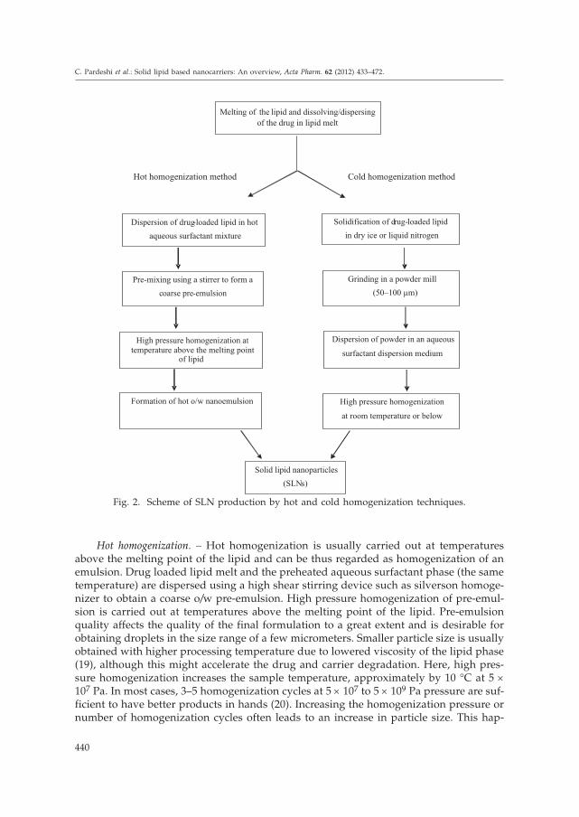
Hot homogenization. – Hot homogenization is usually carried out at temperaturesabove the melting point of the lipid and can be thus regarded as homogenization of anemulsion. Drug loaded lipid melt and the preheated aqueous surfactant phase (the sametemperature) are dispersed using a high shear stirring device such as silverson homoge-nizer to obtain a coarse o/w pre-emulsion. High pressure homogenization of pre-emul-sion is carried out at temperatures above the melting point of the lipid. Pre-emulsionquality affects the quality of the final formulation to a great extent and is desirable forobtaining droplets in the size range of a few micrometers. Smaller particle size is usuallyobtained with higher processing temperature due to lowered viscosity of the lipid phase(19), although this might accelerate the drug and carrier degradation. Here, high pres-sure homogenization increases the sample temperature, approximately by 10 °C at 5 ´
107 Pa. In most cases, 3–5 homogenization cycles at 5 ´ 107 to 5 ´ 109 Pa pressure are suf-ficient to have better products in hands (20). Increasing the homogenization pressure ornumber of homogenization cycles often leads to an increase in particle size. This hap-
440
C. Pardeshi et al.: Solid lipid based nanocarriers: An overview, Acta Pharm. 62 (2012) 433–472.
Hot homogenization method Cold homogenization method
theMelting of lipid and dissolving/dispersing
of the drug in lipid melt
Solidification of drug-loaded lipid
in dry ice or liquid nitrogen
Dispersion of drug-loaded lipid in hot
aqueous surfactant mixture
Grinding in a powder mill
(50–100 µm)
Pre-mixing using a stirrer to form a
coarse pre-emulsion
Dispersion of powder in an aqueous
surfactant dispersion medium
High pressure homogenization attemperature above the melting point
of lipid
High pressure homogenization
at room temperature or below
Formation of hot o/w nanoemulsion
Solid lipid nanoparticles
(SLNs)
Fig. 2. Scheme of SLN production by hot and cold homogenization techniques.

pens because of the coalescence of particles as a result of their high kinetic energy (21).Hot homogenization primarily gives a nanoemulsion due to the presence of molten lipidand solid particles are expected to be produced by cooling of the obtained sample toroom temperature or even below. Because of the nanometric particle size and the pres-ence of emulsifiers or surfactants, lipid crystallization may be retarded and the samplemay remain as a supercooled melt for several months (22).
There should be a difference between the lipid melting point and homogenizationtemperature big enough to prevent melting of the lipid in homogenizer. Here it is a pointto be concentrated that the high pressure homogenization process itself increases thetemperature of the product, approximately by 10 °C at 5 ´ 107 Pa per homogenizationcycle. Hot homogenization technique can be also applied for temperature sensitivedrugs because exposure to increased temperature is relatively short. In case of highlytemperature-sensitive drugs, the cold homogenization technique may be preferred. Thesame technique can be used while formulating hydrophilic drugs because they wouldhave a partition between the melted lipid and aqueous phase during the homogeniza-tion process (4). Venkateswarlu et al. (23) developed SLN delivery systems of clozapine bythe hot homogenization technique followed by ultrasonication using a variety of lipidsalong with the charge modifier stearylamine and studied the effect of the charge modi-fier, various triglycerides, pH of the dialysis membrane and sterilization on the particlesize, zeta potential, entrapment efficiency and drug release kinetics of clozapine fromthe solid lipid nanoparticle formulation.
Cold homogenization. – The prime objective of the cold homogenization technique de-velopment was to rectify the problems associated with the hot homogenization techni-que like temperature-mediated accelerated degradation of drug payload, partitioningand hence drug loss into aqueous phase during homogenization, uncertain polymorphictransitions of the lipids due to complexity of the nanoemulsion crystallization step lead-ing to several modifications and/or supercooled melts (3).
The primary step is similar to the hot homogenization technique and it involves so-lubilization or dispersion of the drug into the lipid melt. The drug-loaded lipid melt issolidified rapidly by means of liquid nitrogen or dry ice to ensure homogeneous drugdistribution into the lipid matrix. The drug containing solid lipid is then pulverized byball or mortar milling to yield lipid microparticles of the typical size range of 50–100 mi-crons. Low temperature enhances the lipid fragility, resulting in further particle commu-nition. These solid lipid microparticles are then dispersed in cold surfactant solutionyielding a pre-suspension, which is then homogenized at or below room temperaturewith appropriate temperature control and regulation, keeping in mind the usual rise intemperature during high pressure processing. At this stage, cavitational forces are play-ing their role and are strong enough to break lipid microparticles directly into the solidlipid nanoparticles (20). As cold homogenization is carried out with solid lipid and isthus regarded as »high pressure milling of a suspension«. This process of cold homoge-nization minimizes thermal exposure of the product but does not prevent it completelybecause of the melting of the lipid-drug mixture in the primary step. In general, com-pared to the hot homogenization technique, larger particle sizes and broader size distri-butions are observed with the cold homogenization technique. Cold homogenization isalso preferred over hot homogenization, in fact it avoids or minimizes the melting of thelipid and thereby minimizes the loss of hydrophilic drugs to the aqueous phase. Further,
441
C. Pardeshi et al.: Solid lipid based nanocarriers: An overview, Acta Pharm. 62 (2012) 433–472.

to minimize the loss of hydrophilic drugs to aqueous phase of SLN dispersion, it is ad-visable to replace water by liquids with low solubility for such drugs, like oils or PEG6000. Production of SLN in oils or PEG 6000 is advantageous for oral drug delivery be-cause this dispersion can be directly filled into soft gelatin capsules (4).
High shear homogenization and ultrasound
High shear homogenization and ultrasound are dispersing techniques that were ini-tially used for the preparation of solid lipid nanodispersions. Both of these methods areeasy to handle and hence widely used. However, the presence of microparticles and me-tal contaminants has a major influence on the nanodispersion quality and has to be com-promised if ultrasound is used (1). Venkateswarlu (24) prepared lovastatin SLN usinghot homogenization followed by ultrasonication.
Ultrasonication (high speed homogenization)
Vyas and Khar reviewed (25) the preparation of vesicular lipid particles (liposomes)of the nanometric size range using the ultrasonication technique. Two methods of so-nication are commonly used based on the use of either a bath or probe tip ultrasonic dis-integrator. Bath sonicator is employed for large volumes of diluted lipid dispersionswhereas the probe tip sonicator is more suitable for dispersions, which require high en-ergy in a small volume (e.g., high concentration of lipids, or a viscous aqueous phase).This technique is most widely used since the equipments required are common in everylab.
Probe tip sonicators supply a high energy input to lipid dispersions but sometimescause lipid degradation due to overheating of the lipid dispersion. Sonication tips alsotend to release metal particles into the dispersion, which must be removed by centrifu-gation prior to use. For these reasons, bath sonicators are preferred over probe tip so-nicators. The most formidable problem associated with this technique is broader particlesize distribution of several micrometers. This, upon storage, may give signs of physicalinstability, like particle growth and particle aggregation. Efforts have been made by vari-ous research groups to prepare a stable formulation using combined high speed stirringand ultrasonication technique conducted at high temperature (3). Size and size distribu-tion of the lipid dispersion are influenced by the composition and concentration of li-pids, sonication time and power, and temperature (26).
Microemulsion based technique
Microemulsion has to be produced at a temperature above the melting point of thelipid so as to form a microemulsion with the lipid solid at room temperature (27). Thesemicroemulsions are clear or slightly bluish solutions composed of a lipophilic phase,surfactant, in most cases a co-surfactant, and water. Lipid (fatty acid and/or triglyceri-de) is melted first; a mixture of surfactant, co-surfactant and water is heated to the sametemperature as the lipid phase and added to the lipid melt under mild stirring. Whencompounds are mixed in a correct ratio, a transparent and thermodynamically stablemicroemulsion system is formed. This microemulsion is then dispersed in a cold aque-
442
C. Pardeshi et al.: Solid lipid based nanocarriers: An overview, Acta Pharm. 62 (2012) 433–472.

ous medium (2–3 °C) under mild mechanical mixing to precipitate the lipid phase form-ing fine particles (28, 29). Addition of a microemulsion to water leads to precipitation ofthe lipid phase forming fine particles. This effect is exploited in the SLN preparation me-thod developed by Gasco (30).
Large-scale production of SLN by the microemulsion technique is also feasible andmajor process parameters that need to be considered during scaling-up include micro-emulsion and water temperature, temperature flows in water medium and thermody-namics of mixing, which should not change, or only a little, during scaling-up to main-tain the same product characteristics. The first attempt was made by Morel et al. (31, 32),to encapsulate peptide drugs into SLN using a warm w/o/w microemulsion techniqueand incorporated [D-Trp-6] LHRH and thymopentin.
Double emulsion based technique
Hammady et al. (33) exploited biphasic polymeric nanospheres to co-encapsulate bothhydrophilic and lipophilic drug molecules using the double emulsion technique, whichis a two-step emulsification procedure. In the first step, a primary w/o emulsion is ob-tained by dispersing the aqueous solution of the hydrophilic drug molecule into organicphase (previously prepared by dissolving the polymer, lipophilic drug molecule andsurfactant, with or without co-surfactant, into an organic solvent), and vortexing and si-multaneous homogenization with a high speed stirrer. This primary w/o emulsion isthen, in the second step, syringed into an aqueous solution to obtain w/o/w multipleemulsions by high pressure homogenization. Further stirring under reduced pressureresults in extraction and evaporation of organic solvent with subsequent hardening ofthe nanospheres.
Solvent emulsification-diffusion technique
Trotta et al. (34) described a preparation method for nanoparticles based on emulsi-fication of an organic solution of a solid lipid in an aqueous emulsifier solution, follo-wed by dilution of the emulsion with water. The method is called »solvent emulsifica-tion-diffusion method«. In this proposed approach, the lipid was dissolved in a water--saturated organic solvent and this organic solution was first emulsified with a solvent--saturated aqueous solution containing emulsifier to form a pre-emulsion. Lipid nano-particles were precipitated by quickly adding water into the initial pre-emulsion to extractthe solvent into continuous phase and to give an SLN nanodispersion. This nanodisper-sion was then washed by ultrafiltration to remove residual solvent and lyophilized.
Battaglia et al. (35) reported the preparation of insulin-loaded SLN by the solvent--in-water emulsification-solvent diffusion technique using isovaleric acid (IVA) as organicphase, glyceryl monostearate (GMS) as lipid, soy lecithin and sodium taurodeoxycholateas emulsifiers. Their investigations demonstrated that SLN seem to have interesting pos-sibilities as a delivery system for oral administration of insulin.
Yuan et al. (36) developed solid lipid nanoparticles in a nanoreactor system usingthe solvent diffusion method to improve the drug loading capacity and entrapment effi-ciency of SLN. Nanoemulsion is one of the nanoreactors generally referred to as mini-emulsion or microemulsion (10–100 nm). The investigation demonstrated that the solvent
443
C. Pardeshi et al.: Solid lipid based nanocarriers: An overview, Acta Pharm. 62 (2012) 433–472.
diffusion method with w/o miniemulsion as nanoreactor system favors the formation ofSLN with higher drug loading capacity and smaller particle size, thereby enhancing thetargeting potential and oral bioavailability of SLNs.
Emusification-solvent evaporation technique
This technique involves three steps of preparation:(i) Preparation of organic phase: lipophilic material is first dissolved in an appro-
priate volume of organic solvent by magnetic stirring.(ii) Pre-emulsification step: lipid containing organic phase is dispersed in an ap-
propriate volume of aqueous solution using a high speed homogenizer in orderto form a coarse pre-emulsion.
(iii) Nanoemulsification step: the resulting coarse pre-emulsion is immediately pas-sed through a high-pressure homogenizer at an operating pressure to obtainnanodispersion. Obtained nanodispersion is then kept on the magnetic stirrerovernight, sometimes in a fume hood to drive off the organic solvent. Uponsolvent evaporation, nanodispersion is formed by precipitation of lipid mate-rial in aqueous medium. Solidified nanodispersion is then filtered through asintered glass filter to remove lipid and drug agglomerates.
Nanoparticles obtained by this method are small, monodisperse with high encapsu-lation efficiency. The process can be automated and scaled-up for the production of alarge amount of nanoparticles (37, 38).
Spray-drying based technique
When particle dimensions are reduced to the nanometric size range, their physicaland chemical properties deviate from bulk properties and such particles suffer from ma-ny problems related to their surface and thermal stability, shape preservation, handlingand assembly into a suitable device (39). Prolonged long-term stability, especially for i.v.administered systems, can be achieved when stored as dry products. Thus a spray-dry-ing technique was investigated for SLN as an alternative approach to lyophilization toconvert liquid dispersions into a dry system. This less cost-intensive technique couldproduce fine, dust-free powder particles with a spherical shape, which are non-agglom-erated and nearly monodisperse, with a controlled particle size (40). Spherical particlesare of practical importance since, in general, they have better rheological properties thanirregular particles (41). Freitas and Muller (42) recommend the use of lipids with themelting point > 70 °C for spray-drying. Fessi (2006) (43) reported the use of the spray--drying technique for conversion of the nanocapsule suspension into redispersible driedsolid particles, as an alternative to the lyophilization technique.
Supercritical fluid based technique
This is a relatively novel technique for SLN production and has a major advantageof processing without organic solvent. SLN can be prepared by rapid expansion of su-percritical carbon dioxide solutions (RESS method) (44). Carbon dioxide (99.99 %) wasgood choice of solvent for this method (3). Thote and Gupta (45) produced nanoparticles
444
C. Pardeshi et al.: Solid lipid based nanocarriers: An overview, Acta Pharm. 62 (2012) 433–472.
of the hydrophilic drug, dexamethasone phosphate using supercritical carbon dioxide(CO2), the technique being known as supercritical antisolvent technique with enhancedmass transfer (45).
Desolvation technique
Vandervoort and Ludwig (46) produced drug-loaded gelatin nanoparticles by usingthe desolvation technique. Here, the drug is dispersed into an aqueous gelatin solutionby continuous stirring. Then, to induce desolvation, desolvating agent is added to theabove solution until permanent faint turbidity is obtained. Finally, an aqueous solutionof the cross-linking agent is added, with continuous stirring, to harden the nanoparti-cles. A desolvating agent (e.g., alcohol, acetone or salt solution) is added to the aqueousgelatin solution to dehydrate the gelatin molecules, indicated by a rise in solution tur-bidity due to the change in conformation of gelatin molecules from stretched to coiled.In that study, the preparation and properties of gelatin nanoparticles loaded with bothhydrophilic pilocarpine hydrochloride and lipophilic hydrocortisone were reported. Ifone tries to use a lipid matrix, instead of or along with the polymer, it would be possibleto increase the loading and entrapment of lipophilic drugs.
EFFECT OF FORMULATION INGREDIENTS ON SLN QUALITY
Major formulation ingredients affecting SLN quality include lipid and emulsifier orsurfactant and co-surfactant.
Influence of the lipid
Critical parameters for nanoparticle formation with different lipids include(i) rate of lipid crystallization(ii) lipid hydrophilicity (influence on self-emulsifying properties)(iii) shape of the lipid crystal (and therefore the surface area).
It was also a common finding that most of the lipids used were composed of a mix-ture of several chemical compounds. A small difference in lipid composition (e.g., pres-ence of impurities) might have a great impact on the quality of SLN dispersions (bychanging the zeta potential, retarding the crystallization process) (2).
Paliwal et al. (47) reported the effect of lipid core material on the particle size of me-thotrexate-loaded SLN for enhancing the oral bioavailability of methotrexate via lym-phatic delivery, produced by the solvent-diffusion method in an aqueous system andstabilized by 1 % (m/m) soya lecithin. Particle size of Compritol® 888 ATO (glyceryl be-henate) SLNs was found to be smaller (120.1 ± 0.8 nm) than the size of stearic acid (130.0± 3.2 nm), tristearin (140.4 ± 6.4 nm) and glycerol monosterate (166.2 ± 8.1 nm) SLNs. Itwas concluded that SLNs produced by a highly lipophilic lipid (Compritol® 888 ATO)showed more promising results, both in vitro and in vivo, as compared to other lipid corematerials.
445
C. Pardeshi et al.: Solid lipid based nanocarriers: An overview, Acta Pharm. 62 (2012) 433–472.

Influence of the emulsifier
Choice of emulsifier and its concentration is of great importance in determining thequality of SLN dispersion (2). Investigating the influence of four different emulsifiers(i.e., cholic acid sodium salt, Lipoid E80, Poloxamer 407 and Tween 80) of the same con-centration (0.5 %) on the degradation rate of SLN prepared by the hot homogenizationtechnique using three different lipids (i.e., cetylpalmitate, Dynasan 116 and Dynasan118). Olbrich and Muller (48) concluded that the longer the ethyleneoxide chains inemulsifier molecule, the more hindered is the anchoring of the lipase/colipase complexand consequently the SLN degradation. Also, the longer are the fatty acid chains in gly-cerides, the slower the degradation rate of SLN. These results could be used to adjustSLN degradation and consequently drug release in a controlled way.
Chen et al. (49) studied the influence of lipophilic emulsifiers on oral absorption oflovastatin from NLC formulations and found NLCs with Myverol to be more stable inthe gastric environment compared to those with soybean phosphatidylcholin (SPC). Xieet al. (50) worked out the effect of the polymeric emulsifier poly(lactic-co-glycolic acid)(PLGA) at concentration levels of 4 and 16 % on the entrapment efficiency and loadingcapacity of hydrophilic protein-loaded SLN prepared by the w/o/w double emulsion--solvent evaporation technique and both were found to be enhanced significantly asPLGA concentration was increased from 4 to 16 %, in all formulations.
STERILIZATION AND LYOPHILIZATION OF SLNs
Sterilization
SLN formulations intended for parenteral and ocular administration must be ster-ile. Sterilization by autoclaving at 121 °C cannot be possible for lipid-based formulationsprepared using poloxamers, sterically stabilized polymers, since this temperature seemsto be too close to the critical flocculation temperature (CFT) of polymers. This is knownto collapse partially the polymer adsorption layer and due to insufficient stabilization ofthe system, particle aggregation occurs. This problem could be avoided by reducing theautoclaving temperature from 121 to 110 °C and prolonging the autoclaving time (51).
However, autoclaving is possible in lecithin-stabilized SLN formulations. Formula-tion melts at autoclaving temperature lead to the formation of a hot o/w microemulsionin the autoclave and recrystallize during the cooling cycle producing SLN particles lar-ger than the initial ones due to coalescence of some nanodroplets during recrystalliza-tion. But, these nanodroplets may not be sufficiently stabilized because SLNs are washedoff before and after sterilization and amounts of surfactant and co-surfactant are smallerin a hot emulsion system (52).
Lyophilization
Lyophilization or freeze drying has been considered an excellent technique of dry-ing and improving the long-term stability of various pharmaceutical products, including
446
C. Pardeshi et al.: Solid lipid based nanocarriers: An overview, Acta Pharm. 62 (2012) 433–472.

vaccines, viruses, proteins, peptides and colloidal carriers like nanoparticles, nanoemul-sions, and liposomes.
Stresses that destabilize the colloidal nanoparticulate suspension may be generatedduring freeze drying, particularly stresses of freezing and dehydration. Aggregation andsometimes irreversible fusion of nanoparticles may destabilize the colloidal nanoparti-culate system. To get rid of this problem, special excipients must be added before freez-ing to protect the nanoparticulate suspension. The added excipients that protect the sys-tem from freezing stress are called cryoprotectants and those protecting from the dryingstress are called lyoprotectants.
Among the long list of cryoprotectants cited in literature, the most frequently usedcryoprotectants include sugars like trehalose, glucose, sucrose, and mannitol. These sug-ars get vitrified at a specific temperature, denoted as Tg (glass transition temperature).These cryoprotectants immobilize the nanoparticles within their glassy matrix and pro-tect them against destabilization by aggregation. In general, freezing must be carried outbelow Tg of an amorphous sample or below Teu (eutectic crystallization temperature) ofcrystalline samples of cryoprotectants (53).
Particle isolation hypothesis is one of the mechanisms for nanoparticle stabilizationby cryoprotectants during the freezing step. Sugars isolate individual particles in unfro-zen fraction and thereby prevent particle aggregation during freezing above Tg. Sugarvitrification is not required for this effect (54).
Water replacement hypothesis is a major mechanism for nanoparticle stabilizationby lyoprotectants during the drying step. These lyoprotectants are known to form hy-drogen bonds with the surface polar groups of nanoparticles and serve as water substi-tutes to preserve the native structure of nanoparticles, thereby stabilizing the system (55).
Freeze dried nanoparticles should have the following desirable characteristics:(i) preservation of primary physical and chemical characteristics of the product(ii) long-term stability(iii) acceptable relative humidity.
DRUG INCORPORATION INTO SLNs
A vast number of drugs with a great variety of lipophilicity and structure have beenstudied to investigating their incorporation into SLNs. Drug incorporation implies local-ization of the drug in the solid lipid matrix (2). Drug loading capacity is one of impor-tant parameters to judge the suitability of a drug carrier system. Loading capacity is ex-pressed in percent related to the lipid phase (matrix lipid + drug) (4).
Factors determining drug loading capacity in lipid include:(i) drug solubility in melted lipid(ii) miscibility of drug melt and lipid melt(iii) chemical and physical structure of solid lipid matrix(iv) polymorphic state of lipid material.
447
C. Pardeshi et al.: Solid lipid based nanocarriers: An overview, Acta Pharm. 62 (2012) 433–472.

Sufficiently high solubility of the drug in the lipid melt is a prime prerequisite toobtain sufficient loading capacity. Typically, solubility should be higher than requiredbecause it decreases while the lipid melt is being cooled down and might even be lowerin solid lipid. Addition of solubilizers and use of mono- and diglycerides in the lipid,used as matrix material, serves to promote drug solubilization in the lipid melt.
Chemical nature of the lipid also needs to be considered because lipids which formhighly crystalline particles with a perfect crystal lattice lead to drug expulsion. Morecomplex lipids being mixtures of mono-, di- and triglycerides along with fatty acids ofdifferent chain length, form less perfect crystals with many imperfections offering morespace to accommodate drugs. Chemically polydisperse lipids, which are more likely tobe used in cosmetics and dermal preparations, showed very efficient drug incorporationcapacities.
Polymorphic state of the lipid is also a parameter of great importance in determin-ing drug incorporation. Crystallization of the lipid in nanoparticles is different compa-red to that in bulk material; lipid nanoparticles recrystallize at least partially in a-form,whereas bulk lipids tend to recrystallize preferentially in b’-modification transformingrapidly into the b-form (56). Increasing formation of more stable modifications leads to amore perfect lattice and the number of imperfections decreases, which means formationof b’/b-modifications promoting drug expulsion. In general, this transformation is slo-wer for long-chain than for short-chain triglycerides. An optimal SLN carrier can be pro-duced in a controlled way when a certain fraction of a-form can be created and preser-ved during storage time. By doing this, a normal SLN carrier transforms to an intelligentdrug delivery system by having a built-in trigger mechanism to initiate transformationfrom a- to b-forms (57). Triggering factors for the transformations are, e.g., temperatureand water loss of SLN dispersion, e.g., after topical application. Thus, it was concludedthat drug incorporation decreases in the following order:
supercooled melt < a-form < b’-form < b-form.
Muller (4) reported three drug incorporation models (as shown in Fig. 3): solid solu-tion model, core-shell model (drug-enriched shell) and core-shell model (drug-enrichedcore).
448
C. Pardeshi et al.: Solid lipid based nanocarriers: An overview, Acta Pharm. 62 (2012) 433–472.
Fig. 3. Drug incorporation models:a) solid solution model, b) core--shell model with drug-enrichedshell, c) core-shell model withdrug-enriched core (4).

SLN matrix is a solid solution (i.e., drug molecularly dispersed in lipid matrix) whenparticles are produced by the cold homogenization technique and using no surfactant orno drug-solubilizing surfactant.
The core-shell model with a drug-enriched shell could be obtained when there isre-partitioning of the drug to the lipid phase during cooling of the obtained nanoemul-sion, in the hot homogenization technique.
The core-shell model with the drug-enriched core could be obtained when the drugprecipitates first, before recrystallization of the lipid. It should be obtained when dis-solving a drug in the lipid melt at or close to its saturation solubility. The proposed me-chanism behind this fact is that nanoemulsion cooling will lead to supersaturation of thedrug in the melted lipid and ultimately to drug crystallization prior to lipid crystalliza-tion. Further cooling will finally lead to recrystallization of the lipid surrounding thedrug core as a membrane. This lipid membrane will only contain the drug in a concen-tration corresponding to saturation solubility of the drug at recrystallization tempera-ture of the lipid. Finally, it will result in a drug-enriched core surrounded by a lipid shell.
DRUG RELEASE FROM SLNs
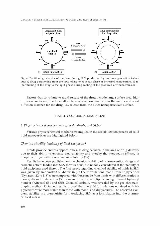
Investigations have shown that several studies concerning optimization of produc-tion parameters, long-term stability, recrystallization behavior, morphological character-ization and in vivo toxicity have been undertaken to date. Additionally, investigations ofdrug incorporation and release served as an important tool in the design, development,and evaluation of potential drug carrier systems. However, there are many studies con-cerning drug incorporation into SLN but data on release mechanisms are still scarce. Amajor problem in early work with lipid nanoparticles was the generally observed burstrelease of drugs (58). The amount of drug in the outer shell and on particle surface isgenerally released in the form of a burst, while the drug incorporated in particle core re-leases in a prolonged manner. Therefore, the extent of burst release can be controlled bycontrolling drug solubility in the aqueous phase during production, which, in turn, canbe controlled via the temperature employed and the surfactant concentration used.Higher temperature and higher surfactant concentration increase the burst whereas pro-duction at room temperature avoids partitioning of the drug into water phase and sub-sequent re-partitioning into lipid phase, thereby showing no burst release at all; the par-titioning behavior is depicted in Fig. 4. To avoid or minimize the burst release, SLN canbe produced surfactant-free or with surfactants unable to solubilize the drug (4). Olbrichand Muller (48) showed that the lipid and the emulsifier are responsible for enzymaticdegradation of the lipid matrix by lipase. Since lipases require a lipid interface for en-zyme activation, appropriate balance between steric stabilizers and other surfactantsshould be optimized to modify drug release and particle degradation. Therefore, hydro-philic coating over lipid nanoparticles is not easily recognized by these enzymes (3).
Venkateswarlu and Manjunath (23) observed an inverse relationship between thepercent drug release and the partition coefficient of clozapine from clozapine-loadedsolid lipid nanoparticles prepared by the cold homogenization technique.
449
C. Pardeshi et al.: Solid lipid based nanocarriers: An overview, Acta Pharm. 62 (2012) 433–472.
Factors that contribute to rapid release of the drug include large surface area, highdiffusion coefficient due to small molecular size, low viscosity in the matrix and shortdiffusion distance for the drug, i.e., release from the outer nanoparticulate surface.
STABILITY CONSIDERATIONS IN SLNs
I. Physicochemical mechanisms of destabilization of SLNs
Various physicochemical mechanisms implied in the destabilization process of solidlipid nanoparticles are highlighted below.
Chemical stability (stability of lipid excipients)
Lipids provide endless opportunities, as drug carriers, in the area of drug deliverydue to their ability to enhance bioavailability and thereby the therapeutic efficacy oflipophilic drugs with poor aqueous solubility (59).
Results have been published on the chemical stability of pharmaceutical drugs andcosmetic actives loaded into SLN formulations, but nobody considered at the stability oflipid excipients used therein. The first report regarding chemical stability of lipids in SLNwas given by Radomska-Soukharev (60). SLN formulations made from triglycerides(Dynasan 112 to 118) were compared with those made from lipids with different ratios ofmono-, di- and triglycerides (Softisan and Imwitor) and lipids having different hydroxylnumber (Witepsol S51 and S55). Chemical stability was revealed by the gas chromato-graphic method. Obtained results proved that the SLN formulations obtained with tri-glycerides were more stable than those with mono- and diglycerides. The observed exci-pient stability is a prerequisite for introducing SLN as a formulation into the pharma-ceutical market.
450
C. Pardeshi et al.: Solid lipid based nanocarriers: An overview, Acta Pharm. 62 (2012) 433–472.
Fig. 4. Partitioning behavior of the drug during SLN production by hot homogenization techni-que: a) drug partitioning from the lipid phase to aqueous phase at increased temperature, b) re--partitioning of the drug to the lipid phase during cooling of the produced o/w nanoemulsion.

Triglycerides undergo a hydrolytic reaction, leading to the formation of mono- anddi-glycerides with free fatty acids, but due to their internal location in SLNs, they areless susceptible to hydrolysis than phospholipids located externally in liposomes (61).
Physical stability
Modifications in the used lipids and colloidal lipid dispersions mostly affect thephysical stability of SLNs.
Lipid modifications
Crystallization-polymorphic transitions. – Sustained drug release of lipoidal nanopar-ticulate systems essentially relies on the solid state of the lipid matrix. Solidification ofparticles after the production process must, therefore, be ensured. However some lipidmaterials may not crystallize in the colloidally dispersed state and dispersions should beregarded as emulsions of supercooled melts rather than dispersions of solid particles.Crystallization of bulk triglycerides from the melt usually occurs in the metastablea-form after rapid cooling (57).
In case of supercooled melts, the lipid crystallization may not occur although thedispersion is stored at a temperature below the melting point of the lipid (2).
Transformation of lipid particles, after crystallization, from less stable to more sta-ble is referred to as polymorphic transition. Crystallization of bulk triglycerides from themelt after rapid cooling usually occurs in the metastable a-form, which transforms viab’-form into the stable b-form upon heating or storage. The kinetics of these polymor-phic transitions are faster in colloidally dispersed mixed triglycerides than in bulk andalso in shorter-chain triglycerides than in those with longer chains (57).
Thermoanalysis and X-ray diffraction pattern of SLN dispersions helps reveal thecrystallization behavior, time course of polymorphic transitions, enthalpy, fusion tem-perature and the degree of crystallinity of melt-homogenized glyceride nanoparticle dis-persions (62).
Gelling phenomena. – Very rapid and unpredictable process of transformation of alow-viscosity SLN dispersion into a viscous gel is referred to as a gelling phenomenon.In most cases, the process of gelation is irreversible (2). High temperature, light, andshear stress are some of the promoters of gelation (63). Gel formation may also occurdue to high lipid concentration, high ionic strength (64), rapid crystallization of the lipid(57) and intense contact of the SLN dispersion with other surfaces, like the surface of thepacking material (65). All gelation promoters increase the kinetic energy of the particlesand favor their collision.
Addition of co-emulsifying surfactants with high mobility (e.g., glycocholate) wasfound to retard or prevent gel formation (66). Storage in dark at 8 °C prevented parti-cle growth. Also, fat samples stored under a nitrogen atmosphere were more stablethan the samples filled under regular air (63). Zeta potential is a good predictor ofgelation phenomena. An increased or decreased zeta potential indicates the beginningof gelation (67).
451
C. Pardeshi et al.: Solid lipid based nanocarriers: An overview, Acta Pharm. 62 (2012) 433–472.

Dispersion modification
Ostwald ripening. – Ostwald ripening in SLN dispersions corresponds to an increasein particle size due to the dissolution of smaller crystals and deposition of dissolved ma-terial on larger surfaces, leading to the growth of large particles at the expense of smallerones (61).
Coalescence. – Rigid solid particles are expected to be stable against coalescence butSLN dispersions tend to form a cream or gel after particle aggregation.
II. Storage stability of SLNs
Solid lipid nanoparticles based on solid lipids and stabilized by surfactant, even inaqueous dispersions, possess long-term physical stability of at least two years. However,depending on the composition and storage conditions, lipoidal systems are critical interms of their stability (68). Gelation phenomena, increase in particle size, particle aggre-gation and drug expulsion from the lipid carrier systems are the most formidable prob-lems of storage stability of SLN (2). Storage conditions like exposure to light, tempera-ture and packing material are also some major stability determining factors.
Freitas and Muller (63) studied the effect of light, temperature and packing materialon zeta potential, particle size and gelation of SLN using an SLN dispersion consistingof Compritol 888 ATO stabilized with Poloxamer 188.
Influence of light
SLN dispersions were exposed to various light conditions such as dark, daylightand artificial light during storage in white and brown glass vials. Storage in white glassvials under artificial light induced rapid gelation with immediate particle growth, whilethe gelation process was slower under daylight. Dark exposure results in slow particlegrowth and gelation after one month of storage. Under all conditions, zeta potentialdropped compared to that immediately after production, with slight variations underdifferent conditions. The SLN dispersion system changed after exposure to differentlight conditions, leading to reduced zeta potential due to modification of the crystalliza-tion form of lipids. Results proved that light radiation had a destabilizing effect and fur-ther increase in the intensity of light radiation accelerated particle growth and gellingprocess significantly. In general, introduction of energy to the system leads to particlegrowth and subsequent gelation and high energetic radiations like UV lead to increaseddestabilization of a SLN system (63).
Physical stability for more than 3 years of lipid suspensions stored at room tempe-rature in white glass bottles under daylight has been reported (4).
Influence of temperature
Temperature, unlike light exposure, corresponds to energy input to the system andcan lead to changes in the crystalline structure of the lipids and high temperatures ge-nerally cause destabilization of the SLN system due to reduction in zeta potential (61).Freitas and co-workers (63) studied the effect of different temperature conditions such as
452
C. Pardeshi et al.: Solid lipid based nanocarriers: An overview, Acta Pharm. 62 (2012) 433–472.
8, 20 and 50 °C on the particle size and zeta potential of Compritol SLN and observedthat storage at higher temperatures led to rapid particle growth with reduction of zetapotential of the SLN system fastest at 50 °C.
Influence of packing material
The influence of packing material on SLN physical stability was less pronouncedthan temperature. But aggregation and gelling can be promoted by the inner surface.Stability improvement has been observed with some SLN systems with a gelling ten-dency when they were packed in plastic containers instead of glass vials (62).
Freitas and Muller (63) packed Compritol SLN in siliconized and in untreated glassvials and stored under optimum conditions (at 8 °C, in dark) to assess whether silico-nization of the glass surface had any influence on the stability. They observed that theparticle diameters of SLN systems in untreated glass vials were slightly larger comparedto those in siliconized ones while the zeta potential of SLN systems in siliconized vialswas slightly higher than those in untreated vials. It was found that minimization of SLNadherence to the surface of the vial by siliconization minimizes aggregation and conse-quently improves physical stability. Hence, it was concluded that siliconization of theglass surface has a stabilizing effect.
ROUTES OF ADMINISTRATION AND BIODISTRIBUTION OF SLNs
The in vivo biodistribution of solid lipid nanoparticles will mainly depend on theroute of administration and interactions of SLN with biological surroundings which, ingeneral, include two types of processes: distribution processes (adsorption of biologicalmaterials on the particle surface and desorption of SLN components into the biologicalsurrounding) and enzymatic processes (lipid degradation by lipases and esterases).
Physiological or physiologically related lipids or waxes generally constitute theSLNs. Therefore, the in vivo fate of the carrier, to a large extent, occurs through the path-ways of transportation and metabolism present in the body. Lipases, the enzymes pres-ent in various organs and tissues of the body, are most responsible for SLN degradation.Lipases split the ester linkage and form partial glycerides or glycerol and free fatty ac-ids. Activation by an oil/water interface, which opens the catalytic centre, is a prerequi-site for lipases to act (69, 70). Solid lipid nanoparticles show different degradation rates,in vitro, by the lipolytic enzyme pancreatin lipase as a function of their composition (li-pid matrix, stabilizing surfactant (48).
Peroral administration
Aqueous dispersions or SLN-loaded traditional dosage forms (tablets, capsules, pel-lets or powders in sachets) may serve as peroral administration forms of SLN. Particleaggregation of SLN dispersion occurs in the microclimate of the stomach due to acidityand high ionic strength. It can be expected that food will have a great impact on SLNperformance; however, to our knowledge no experimental data have been published onthis issue till date. The question concerning the effect of the stomach and pancreatic li-
453
C. Pardeshi et al.: Solid lipid based nanocarriers: An overview, Acta Pharm. 62 (2012) 433–472.

pases on SLN degradation in vivo still remains open. Unfortunately, only a few in vivostudies have been performed so far.
Pandey et al. (71) formulated and evaluated the chemotherapeutic potential of solidlipid nanoparticles incorporating antitubercular drugs following oral administration tomice and suggested that oral SLN based antitubercular drug therapy forms a sound ba-sis for reducing dosing frequency and improving patient compliance for better manage-ment of tuberculosis. Zhang et al. (72) administered orally insulin-loaded SLN andWGA-modified SLN to rats and demonstrated that both of these formulations promotedthe intestinal absorption of insulin after oral administration.
Parenteral administration
Parenteral drug delivery took a major leap after successful development of the sub-micronic parenteral fat emulsion in the 1960s. Quick commercialization of submicronemulsion based products, such as Diazemuls (diazepam) and Diprivan (propofol), indi-cated the interest of pharmaceutical industries in colloidal carriers. Since then, therehave been continuous efforts to develop novel colloidal nanocarriers for improved pa-renteral delivery (73). Wissing et al. (74) reviewed, in detail, the bioactivity of SLN afterparenteral administration, i.e., tolerability, toxicology, cellular uptake, albumin adsorp-tion, pharmacokinetics, tissue distribution and drug targeting.
Gasco and coworkers (75) studied the pharmacokinetics and tissue distribution ofstealth and non stealth SLN loaded with doxorubicin after i.v. administration to rats andfound prolonged circulation time of SLN compared to the commercial doxorubicin solu-tion.
Yang et al. (76) studied the pharmacokinetics, body distribution and specific drugtargeting of camptothecin after i.v. injection in mice. In comparison with commercialcamptothecin solution, SLN was found to give much higher AUC/dose and mean resi-dence times (MRT), especially in brain, heart and reticuloendothelial cells containing or-gans. The highest AUC ratio of SLN to drug solution among the tested organs was foundin the brain.
Reddy et al. (77) studied the influence of the route of administration on tumor up-take and biodistribution of etoposide loaded solid lipid nanoparticles in mice bearingDalton’s lymphoma after subcutaneous, intravenous and intraperitoneal injections. Itwas observed that subcutaneous injection reduced the biodistribution of SLN to all thetissues studied, whereas intravenous injection resulted in lower levels of etoposide-loa-ded SLN in RES rich organs compared to free etoposide. SLN experienced significantlyhigher brain distribution after intraperitoneal injection, indicating its potential applica-tion in targeting etoposide to brain tumors.
Transdermal administration
Since the epidermal lipids are found in high amounts in the penetration barrier, li-pid carriers (liposomes, SLN, NLC, etc.) attaching themselves to the skin surface and al-lowing lipid exchange between the outermost layers of the stratum corneum and thecarrier appear promising (78).
454
C. Pardeshi et al.: Solid lipid based nanocarriers: An overview, Acta Pharm. 62 (2012) 433–472.
Incorporation of SLN dispersion in an ointment or gel, by reduction of the lipidcontent of the SLN dispersion, is necessary to achieve a formulation that can be easilyadministered to the skin (3). Cosmetic field offers interesting applications of SLN due totheir UV reflecting properties. UV reflectance is related to the solid state of lipids andwas not evident in nanoemulsions of comparable composition. These observations opena new application area for the development of SLN-based UV protective systems (2).
The short time-to-market and big potential area of SLNs are topical products basedon SLN technology, which means pharmaceutical and cosmetic formulations. A majorstep forward was the development of »intelligent SLNs« (ISLN), which release incorpo-rated drug in a controlled fashion after it receives a triggering impulse. Such triggeringimpulses are an increase in temperature or loss of water from the SLN dispersion orSLN-containing cream (2). This effect was exploited for controlled release of vitamin Aloaded in a glyceryl behenate SLN dispersion (79, 80) and flurbiprofen-loaded SLN-gelformulation (81).
Pulmonary administration
Growing attention has been given to the potential of pulmonary route as an alterna-tive to the non-invasive local and systemic delivery of therapeutic agents using lipidparticles, since it provides a large absorptive mucosal area. The lung offers a large sur-face area for drug absorption and the alveolar epithelium allows rapid drug absorption.What makes pulmonary administration of many drugs very promising, particularly forproteins and peptides, is the presence of various metabolic activities and pathways,which may be different from those present in the GI tract. The superior physicochemicalcharacteristics of SLNs make them more suitable as an appropriate delivery system dueto correlation between the diameter within the nanometric range, biocompatible compo-sition and deep-lung deposition ability. Prolonged drug serum concentration and lungretention are both achievable by means of the particulate colloidal drug carrier systemincluding SLNs (82).
Liu et al. (83) developed novel nebulizer-compatible solid lipid nanoparticles forpulmonary delivery of insulin by the reverse micelle-double emulsion technique andtheir finding suggested that SLN could be used as a potential carrier for pulmonary de-livery of insulin improving its stability as well as bioavailability.
Ocular administration
Eyes are among the most readily accessible organs in terms of their location in thebody, yet drug delivery to eye tissues is particularly problematic. Delivery of drugs viananotechnology-based products fulfils three main objectives: enhanced drug permea-tion, controlled drug release and higher targeting potential. Attama et al. (84) preparedsodium diclofenac-loaded lipid nanoparticles combining the homolipid from goat (goatfat) and a phospholipid, with high encapsulation efficiency applying hot high-pressurehomogenization technique. Administration of this formulation to bioengineered humancornea demonstrated sustained release of the analgesic drug. Furthermore, permeationof sodium diclofenac through the corneal construct was improved by surface tailoring ofnanoparticles with phospholipid, which showed better performance for ocular adminis-tration.
455
C. Pardeshi et al.: Solid lipid based nanocarriers: An overview, Acta Pharm. 62 (2012) 433–472.

TARGETING EFFICIENCY OF SLNs
One of the most challenging aspects in pharmaceutical research is targeted deliveryof drug molecules to a specific organ, tissue or specific cellular sites. By developing col-loidal delivery systems such as liposomes, micelles and nanoparticles, a new frontierwas opened for improving drug delivery.
Brain targeting
Nanosystems employed for the development of drug delivery systems intended forCNS targeting include polymeric nanoparticles, nanospheres, nanosuspensions, nano-emulsions, nanogels, nano-micelles, nano-liposomes, nanofibres, nanorobots, solid lipidnanoparticles (SLNs), nanostructured lipid carriers (NLCs) and lipid drug conjugates(LDCs) (85). The most formidable obstacles that often impede drug delivery to the brainare characterized by the presence of relatively impermeable endothelial cells with tightjunctions, enzymatic activity and the presence of active efflux transporter mechanismslike P-glycoprotein efflux. However, the bioacceptable and biocompatible nature ofSLNs makes them less toxic compared to polymeric nanoparticles and they are taken upby the brain because of their lipidic nature. SLNs below 200 nm have increased bloodcirculation and hence an increase in the time during which the drug remains in contactwith BBB and is taken up by the brain (86).
Mistry et al. (87) suggested that the existence of a direct nose-to-brain delivery routefor nanoparticles administered to the nasal cavity and transported via the olfactory epi-thelium and/or the trigeminal nerves directly to the CNS is relevant in the field of drugdelivery as well as new developments in nanotechnology. Proteins and peptides (P/P)have been pointed out as holding great promise for the treatment of various neurodege-nerative diseases. A major challenge in this regard, however, is the delivery of P/P drugsover the blood-brain-barrier (BBB). Technology based approaches (comprising functio-nalized nanocarriers and liposomes) and pharmacological strategies (such as the use ofcarriers and chimeric peptide technology) appear to be the most promising ones to facil-itate enhanced P/P drug delivery over BBB (88).
Microphage targeting
Microphages play a central role in mediating inflammatory responses and also ser-ve as a reservoir for microorganisms involved in life-threatening infectious diseases. Theuse of drug-loaded nanoparticles represents a good alternative to avoid or at least de-crease side effects and increase efficacy, compared to potent drugs which often induceunwanted side effects when applied as free forms for treating the microphage-mediateddiseases (89).
Epidermal targeting
Chen et al. (90) formulated solid lipid nanoparticles loaded with podophyllotoxin(POD) stabilized with Poloxamer 188 (P-SLN) and Polysorbate 80 (T-SLN) and evalu-ated as topical carriers for epidermal targeting of POD. Penetration of POD from P-SLN,
456
C. Pardeshi et al.: Solid lipid based nanocarriers: An overview, Acta Pharm. 62 (2012) 433–472.

by fluorescence microscopy, seemed to follow two pathways along the stratum corneumand hair follicle route. Imaging revealed that P-SLN had a strong localization of PODwithin the epidermis. To sum up, P-SLN provides a good epidermal targeting effect andmay be a promising carrier for topical delivery of drugs like POD.
CELLULAR UPTAKE AND CYTOTOXICITY/NEUROTOXICITY ASPECTS OF SLNs
Solid lipid nanoparticles are well tolerated in a biological system because they arecomposed of physiological lipids. In vitro cytotoxicity and viability determinations wereperformed on suspensions of human granulocytes using the dimethylthiazolyl-diphe-nyltetrazolium (MTT) test to assess their tolerability at cellular levels. The results ofcytotoxicity studies (MTT assay) indicated that SLNs are less toxic than polymeric nano-particles (91, 92).
Yuan et al. (93) investigated the cellular uptake and cytotoxicity of paclitaxel-loadedsolid lipid nanoparticles composed of different lipid materials. The order of cellular up-take ability was: glycerol tristearate SLN > monostearin SLN > stearic acid SLN > Com-pritol 888 ATO SLN. Incorporation of polyethylene glycol monostearate (PEG-SA) intoSLN could enhance cellular uptake but it did not increase cytotoxicity whereas the intro-duction of folic acid-stearic acid (FA-SA) into SLN could enhance cellular uptake andcytotoxicity, which indicated a potential application of the later for targeting tumor ther-apy.
Recent observations suggested that several NPs such as Polysorbate-80 coated poly-butyl cyanoacrylate (PBCA) NPs are able to cross the BBB through intravenous admini-stration followed by accumulation in the brain resulting in severe neurotoxicity (94).There are already a few reports that observed the neurotoxicity of nanoparticles both invitro (95) and in vivo (96, 97).
CHARACTERIZATION OF SLN QUALITY AND STRUCTURE
Adequate and appropriate characterization of SLNs is essential for quality controlof formulations. However, characterization of SLN is not an easy task due to the colloi-dal size range and complex nature of delivery systems. The key parameters to be evalu-ated and the techniques used for SLNs characterization are the following:
Particle size/size distribution
Particle size/size distribution of SLN may be studied using photon correlationspectroscopy (PCS), laser diffraction (LD), transmission electron microscopy (TEM),scanning electron microscopy (SEM), atomic force microscopy (AFM), scanning tunnel-ing microscopy (STM) or freeze fracture electron microscopy (FFEM). Among these, PCSand LD are the most commonly employed techniques for routine measurement of parti-cle size. PCS measures the fluctuation of the intensity of scattered light caused by parti-cle movement. PCS covers a range from a few nanometers to about 3 mm. However, PCSis not able to detect larger microparticles. They can be visualized by LD measurements.
457
C. Pardeshi et al.: Solid lipid based nanocarriers: An overview, Acta Pharm. 62 (2012) 433–472.
The method of laser diffraction (LD) is based on the dependence of diffraction angleon particle radius (Fraunhofer spectra). Smaller particles cause more intense scatteringat high angles compared to larger ones. LD covers a broader size range from nanometersto a lower millimeter range. Development of the polarization intensity differential scat-tering (PIDS) technique greatly enhanced the sensitivity of LD to smaller particles. How-ever, both of these techniques are always recommended to be used simultaneously. Itshould be noted that both of these techniques do not »measure« particle size, but theydetect light scattering effects which are further used to calculate particle size. Difficultiesmay arise both in PCS and LD measurements in case of samples which are non-uniformin size and, therefore, use of additional techniques like light microscopy, although notsensitive enough to the nanometric size range, is recommended or electron microscopywhich gives direct information on the particle shape (3, 98).
Zeta potential
One could draw predictions regarding the storage stability of colloidal dispersionsfrom zeta potential measurements. This is an important formulation characteristic ofSLN since, in general, high zeta potential values are likely to cause particle deaggrega-tion due to electric repulsion. However, systems containing steric stabilizers or hydro-philic surface appendages do not follow strictly this rule, because adsorption of stericstabilizers will decrease the zeta potential due to a shift in the particle shear plane (3, 4).
Dynamic light scattering (DLS)
Dynamic light scattering (DLS), also known as Photon Correlation Spectroscopy(PCS) or quasi-elastic light scattering (QELS), records the variation in intensity of scat-tered light on the microsecond time scale. This variation results from the interference oflight scattered by individual particles under the influence of Brownian motion and isquantified by compilation of an autocorrelation function. This function is fitted to an ex-ponential, or some combination or modification thereof, with the corresponding decayconstant(s) being related to the diffusion coefficient(s). Particle size can be calculated fromthis coefficient using standard assumptions of spherical size, low concentration, andknown viscosity of the suspending medium. Rapid analysis, lack of required calibration,and sensitivity to submicron sized particles are some of the merits of this method. Spec-tral decomposition capability of dynamic light scattering enables one to estimate themolecular weight of each nanoparticle (3, 99). Thus, DLS is a nonintrusive, sensitive andpowerful analytical tool used to characterize macromolecules and colloids in solution(100).
Static light scattering/Fraunhofer diffraction (SLS)
Static light scattering (SLS) is an ensemble method in which the pattern of light scat-tered from a solution of particles is collected and fitted to fundamental electromagneticequations in which particle size is the primary variable. This method is rapid and rug-ged, but it requires more cleanliness than DLS and advanced knowledge of optical prop-erties of the particles (3).
458
C. Pardeshi et al.: Solid lipid based nanocarriers: An overview, Acta Pharm. 62 (2012) 433–472.

Field flow fractionation (FFF)
Field flow fractionation (FFF) was used to separate particles due to their Stoke’s ra-dius. In this technique, particles are separated in a flow through a thin channel underthe influence of a field force perpendicular to the flow. The flow in the channel has aparabolic pattern, with highest speed in the centre, such that smaller particles are trans-ported faster and eluted earlier. All eluted fractions are analyzed by multi-angle lightscattering (MALS) where a photometer records scattering signals of the particles andcalculates the size-weighted radius. MALS is able to measure the particles ranging from10 nm to approximately 1 µm. The two subtechniques of FFF, thermal FFF and cross--flow FFF, were successfully employed to separate and analyze the particles according totheir size and diffusion coefficients. Two variations of flow FFF, symmetric flow andasymmetric flow, were reported by Dulog and Schauer (101).
Acoustic method
Acoustic spectroscopy deals with determination of the size of nanoparticles by mea-suring the attenuation of sound waves through the fitting of physically relevant equa-tions. In addition, the oscillating electric field generated by the movement of chargedparticles under the influence of acoustic energy can be detected to provide informationon surface charge (3, 102).
Nuclear magnetic resonance (NMR)
NMR can be used to determine both the size and the qualitative nature of nano-particles. Selectivity afforded by the chemical shift compliments the sensitivity to molec-ular mobility to provide information on the physicochemical status of the componentswithin nanoparticles (103).
Electron microscopy
SEM (scanning electron microscopy) and TEM (transmission electron microscopy)are useful for direct observation of nanoparticles and the former method is preferential-ly used for physical characterization of nanoparticles, particularly their morphologicalexamination. However, vacuum drying used in sample preparation of SEM causesshrinkage of the SLN size (104). TEM has a smaller size limit of detection, provides goodvalidation for other methods, but one must be cognizant of the statistically small samplesize (3).
Force microscopy/scanning force microscopy
Atomic force microscopy (AFM). – In AFM, a probe tip with atomic scale sharpness israstered across the sample to produce a topological map based on the forces at play be-tween the tip and the surface. The probe can be dragged across the sample (contact mo-de), or is allowed to hover just above (noncontact mode) (3). AFM is ideally suited forcharacterizing the nanoparticles because it offers the capability of 3D visualization, bothqualitative and quantitative information of the sample topology, including morphology,
459
C. Pardeshi et al.: Solid lipid based nanocarriers: An overview, Acta Pharm. 62 (2012) 433–472.
CVP
Highlight

surface texture and roughness and, more important in this case, the size of the nano-particles (100). Also, ultrahigh resolution can be obtained with this technique. However,the deposition method used for AFM causes the SLN to form small clusters (104).
Magnetic force microscopy (MFM). – Albrecht et al. (105) combined the AFM and MFMtechniques to study bacterial magnetosomes, comprised of membrane-bound magnetiteparticles. AFM imaging confirmed the existence of single isolated magnetosomes with asize of about 40 nm while MFM addressed the magnetic properties of single magneto-somes. Their results demonstrated that AFM and MFM could be used as powerful toolsfor the development of magnetosomes for medical applications.
Freeze fracture electron microscopy (FFEM). – Freeze fracture electron microscopy isused for morphological examination of lipidic particles. [kalko et al. (106) used FFEMfor detection of the appearance of protein bound to the surface of intact and microflui-dized liposomes and its influence on their surface morphology. Surface bound proteinwas observed by FFEM and was confirmed by immunogold cryo microscopy.
Scanning tunneling microscopy (STM). – The technique of scanning tunneling micros-copy (STM) was developed to examine the surface characteristics of desiccated and hy-drated phospholipid bilayers in liposomes. It is expected to be useful in assessing themolecular organization of liposomes with distinct surface characteristics, molecular pro-cesses occurring during membrane dehydration, and the organization of phospholipidsin the viscinity of membrane-bound proteins, lectins or polymers (107).
X-ray diffraction (XRD)
The degree of crystallinity of a solid can be assessed from the geometric scatteringof radiation from the crystal planes of that solid using the X-ray powder diffraction tech-nique. Robach et al. (108) investigated the structure of ~100 nm thick films of FePt nano-particles by the same technique. Investigations of the physical state, melting behaviorand recrystallization of lipids in the colloidal carrier systems can be performed usingboth X-ray diffraction and synchrotron radiation X-ray diffraction (small and wide an-gle) techniques (56).
Thermal analysis
Differential scanning calorimetry (DSC) can be used to determine the nature anddegree of crystallinity of lipids within the nanoparticles by measuring the glass and mel-ting temperatures, and their associated enthalpies (2). Schubert et al. (109) evaluated bythermoanalysis the influence of lecithin, an amphiphilic lipid, within the lipid matrix onthe crystallization behavior of lipids.
APPLICATIONS OF SLNs
Long-term stability and ease of large scale production impart immense importanceto the solid lipid nanoparticles, which forms the basis of biodegradable and biocompa-
460
C. Pardeshi et al.: Solid lipid based nanocarriers: An overview, Acta Pharm. 62 (2012) 433–472.

tible colloidal drug delivery systems. SLNs find potential applications in the followingareas of interest:
Cosmeceuticals
SLN have been employed for dermal application of cosmeceuticals like molecularsunscreens (110) and as carriers for UV blockers (111). Cosmetic benefits of lipid nano-particles include enhancement of the chemical stability of actives, film formation on skin,controlled occlusion, skin hydration, drug targeting, enhanced skin bioavailability andphysical stability of lipid nanoparticles as topical formulations (8, 112). An in vivo studyshowed increased skin hydration, by 31 %, after 4 weeks after addition of 4 % SLN to aconventional cream formulation (113).
Topical delivery
Among lipid nanoparticles, both SLN and NLC have been used in topical applica-tion of various pharmaceutical actives such as podophyllotoxin (90), tretinoin (114), iso-tretinoin (115), flurbiprofen (81), psoralen (8), vitamin A (79, 80). Lipid-based nanoformu-lation fulfils three main objectives: controlled drug release, enhanced drug permeationand site specific drug delivery (84). Dermal penetration barriers contain a high concen-tration of epidermal lipids and lipid based carriers appear to be promising by attachingthemselves to the skin surface, allowing lipid exchange between the outermost layers ofstratum corneum (116).
Gene vector carrier delivery
Rapid elimination of naked plasmid DNA from the circulation after intravenous ad-ministration calls for the development of an optimized DNA delivery system for effec-tive and successful clinical use. Cationic solid lipid nanoparticles can bind to the DNAdirectly via electrostatic interactions, thereby mediating gene transfection in vitro as wellas in vivo (117) and acting as a non-viral gene delivery vector carrier for DNA, plasmidDNA and nucleic acids (118, 119).
Chemotherapy
Khuller and co-workers exploited the chemotherapeutic potential of oral solid lipidnanoparticles for the delivery of antitubercular drugs such as rifampicin, isoniazid andpyrazinamide. The system was found to reduce the dosing frequency and enhance pa-tient compliance (71).
SLN-based anticancer drug delivery systems have been shown to be superior overthe conventional ones and promising for improved cancer chemotherapy (120). Doxoru-bicin, an anticancer drug encapsulated into PLN, an advanced form of SLN, was foundto be very effective in treating multidrug resistant breast cancer (121). Inspiring efficacyand tumor targeting have been reported to be enhanced with SLNs loaded with variousanticancer drugs like doxorubicin (122) and mitoxantron (123).
461
C. Pardeshi et al.: Solid lipid based nanocarriers: An overview, Acta Pharm. 62 (2012) 433–472.

Vaccine adjuvant
Adjuvants are used in vaccination to enhance the immune response (2). The lipid--based nanocarriers for antigen delivery mainly include liposomes, immunostimulatingcomplexes (ISCOMs), and NLC, again an advanced form of SLN. Major characteristicsthat make nanocarriers unique vaccine adjuvants are firstly their effective increase of theamount of antigens reaching the immune system, secondly that they control the releaseof antigens over prolonged periods of time and finally that they can add their immuno-stimulating or immunomodulating actions synergistically to their primary antigen de-livery function (124).
STEALTHING OF SLNs
Major obstacles to the systemic use of nanoparticulate colloidal drug carriers desig-ned to deliver the active pharmaceutical ingredients to their destination sites are posedby the reticuloendothelial system (RES) and opsonization process which recognize themas foreign products and quickly clear them from systemic circulation. The rate of RESuptake of colloidal nanoparticulates is so rapid that it significantly reduces the circula-tion half-life of these carriers. Opsonization refers to the clearance of nanoparticulatesfrom the blood stream through their interaction with blood plasma proteins (opsonins),resulting in attachment of particles to the membrane of macrophages.
A great deal of work has been recently devoted for the surface modification ofnanoparticulate carriers to deter them from the opsonization process and RES uptakethrough coating, binding or grafting of certain materials to the surface of nanoparticles,which could impart stealth behavior (25, 125).
Hydrophilic and flexible polymer coating on the surface of nanoparticles is thoughtto mask the surface from marking the particles with serum components (opsonins) forRES uptake. These surface modified nanoparticles are called sterically stabilized nano-particles or stealth nanoparticles. The stealthing phenomenon has been successfully em-ployed and explored in SLNs (126).
FUTURE PROSPECTS AND CHALLENGES
Research must continue in such a direction to provide improved efficacy, drug load-ing, targeting and lowering of the drug dose, thereby overcoming the toxicity challengesof this carrier system (127).
In addition, structure and dynamics of SLNs on the molecular level, both in vitroand in vivo, stability, targeting, toxicity and aspects related to interactions of SLNs withtheir biological surrounding pose a challenge that should be explored in the near futureby various research groups around the globe.
462
C. Pardeshi et al.: Solid lipid based nanocarriers: An overview, Acta Pharm. 62 (2012) 433–472.

CONCLUSIONS
A drug delivery system is said to be successful only if, along with the academic re-search groups, pharmaceutical industries undertake its development. Since SLNs havealready been proven as good formulations in cosmeceuticals and other allied fields, theymust occupy a considerable place in the pharmaceutical market. To exploit the broad ap-plications of lipid based nanoparticulate formulations, it is essential that the pharmaceu-tical industries specialized in the development of new drug delivery systems should en-gage in novel formulation technology to promote their scale up and bring them onto thepharmacist’s shelves.
REFERENCES
1. C. V. Pardeshi, P. V. Rajput, V. S. Belgamwar and A. R. Tekade, Formulation, optimization andevaluation of spray-dried mucoadhesive microspheres as intranasal carriers for valsartan, J. Mi-croencapsul. 29 (2011) 103–114; DOI: 10.3109/02652048.2011.630106.
2. W. Mehnert and K. Mader, Solid lipid nanoparticles: Production, characterization and applica-tions, Adv. Drug. Del. Rev. 47 (2001) 165–196; DOI: 10.1016/S0169-409X(01)00105-3.
3. S. Mukherjee, S. Ray and R. S. Thakur, Solid lipid nanoparticles: A modern formulation ap-proach in drug delivery system, Ind. J. Pharm. Sci. 71 (2009) 349–358.
4. R. H. Müller, K. Mäder and S. Gohla, Solid lipid nanoparticles (SLN) for controlled drug deliv-ery – A review of the state of the art, Eur. J. Pharm. Biopharm. 50 (2000) 161–177.
5. R. H. Müller, M. Radtke and S. A. Wissing, Nanostructured lipid matrices for improved micro-encapsulation of drugs, Int. J. Pharm. 242 (2002) 121–128; DOI: 10.1016/S0378-5173(02)00180-1.
6. R. H. Müller, M. Radtke and S. A. Wissing, Solid lipid nanoparticles (SLN) and nanostructuredlipid carriers (NLC) in cosmetic and dermatological preparations, Adv. Drug Del. Rev. 54 (Suppl.1) (2002) S131–S155; DOI: 10.1016/S0169–409X(02)00118-7.
7. R. H. Müller, R. D. Petersen, A. Hommoss and J. Pardeike, Nanostructured lipid carriers (NLC)for cosmetic dermal products, Adv. Drug Del. Rev. 59 (2007) 522–530; DOI: 10.1016/j.addr.2007.04.012.
8. J. Y. Fang, C. L. Fang, C. H. Liu and Y. H. Su, Lipid nanoparticles as vehicles for psoralen deliv-ery: Solid lipid nanoparticles (SLN) versus nanostructured lipid carriers, Eur. J. Pharm. Bio-pharm. 70 (2008) 633–640; DOI: 10.1016/j.ejpb.2008.05.008.
9. E. B. Souto, S. A. Wissing, C. M. Barbosa and R. H. Müller, Development of a controlled releaseformulation based on SLN and NLC for topical clotrimazole delivery, Int. J. Pharm. 278 (2004)71–77; DOI: 10.1016/j.ijpharm.2004.02.032.
10. M. Joshi and V. Patravale, Nanostructured lipid carriers (NLC) based gel of celecoxib, Int. J.Pharm. 346 (2008) 124–132; DOI: 10.1016/j.ijpharm.2007.05.060.
11. V. Teeranachaideekul, R. H. Müller and V. B. Junyaprasert, Encapsulation of ascorbyl palmitatein nanostructured lipid carriers (NLC) – Effect of formulation parameters on physicochemicalstability, Int. J. Pharm. 340 (2007) 198–206; DOI: 10.1016/j.ijpharm.2007.03.022.
12. S. Doktorovova, J. Araujo, M. L. Garcia, E. Rakovsky and E. B. Souto, Formulating fluticasonepropionate in novel PEG-containing nanostructured lipid carriers (PEG-NLC), Colloid SurfacecB. 75 (2010) 538–542; DOI: 10.1016/j.colsurfb.2009.09.033.
463
C. Pardeshi et al.: Solid lipid based nanocarriers: An overview, Acta Pharm. 62 (2012) 433–472.

13. C. Olbrich, A. Gessner, W. Schroder, O. Kayser and R. H. Müller, Lipid-drug conjugate of thehydrophilic drug diminazine-cytotoxicity testing and mouse serum adsorption, J. Control. Re-lease 96 (2004) 425– 435; DOI: 10.1016/j.jconrel.2004.02.024.
14. H. L. Wong, A. M. Rauth, R. Bendayan and X. Y. Wu, In-vivo evaluation of a new polymer-lipidhybrid nanoparticle (PLN) formulation of doxorubicin in a murine solid tumor model, Eur. J.Pharm. Biopharm. 65 (2007) 300–308; DOI: 10.1016/j.ejpb.2006.10.022.
15. H. L. Wong, A. M. Rauth, R. Bendayan and X. Y. Wu, Simultaneous delivery of doxorubicin andGG918 (Elacridar) by new polymer-lipid hybrid nanoparticles (PLN) for enhanced treatmentfor multidrug-resistant breast cancer, J. Control. Release 116 (2006) 275–284; DOI: 10.1016/j.jconrel.2006.09.007.
16. L. Zhang, J. M. Chan, F. X. Gu, A. Z. Wang, A. F. Radovic-Moreno, F. Alexis, R. Langer and O. C.Farokhzad, Self-assembled lipid-polymer hybrid nanoparticles: A robust drug delivery plat-form, ACS Nano. 2 (2008) 1696–1702; DOI: 10.1021/nn800275r.
17. Y. Li, H. L. Wong, A. J. Shuhendler, A. M. Rauth and X. Y. Wu, Molecular interactions, internalstructure and drug release kinetics of rationally developed polymer-lipid hybrid nanoparticles,J. Control. Release. 128 (2008) 60–70; DOI: 10.1016/j.jconrel.2008.02.014.
18. C. Salvador-Morales, L. Zhang, R. Langer and O. C. Farokhzad, Immunocompatibility proper-ties of polymer-lipid hybrid nanoparticles with heterogeneous surface functional groups, Bio-materials 30 (2009) 2231–2240; DOI: 10.1016/j.biomaterials.2009.01.005.
19. R. Lander, W. Manger, M. Scouloudis, A. Ku, C. Davis and A. Lee, Gaulin homogenization: amechanistic study, Biotechnol. Prog. 16 (2000) 80–85; DOI: 10.1021/bp990135c.
20. R. H. Müller, S. Benita and B. Bohm, Emulsions and nanosuspensions for the formulation ofpoorly soluble drugs, Int. J. Pharm. 212 (2001) 143–144.
21. B. Siekmann and K. Westesen, Solid lipid nanoparticles stabilized by tyloxapol, Eur. J. Pharm.Sci. 2 (1994) 117–194; DOI: 10.1016/0928-0987(94)90407-3.
22. H. Bunjes, B. Siekmann and K. Westesen, Emulsions of supercooled melts-a novel drug delivery sys-tem, in Submicron Emulsions in Drug Targeting and Delivery, Ed. S. Benita, Hardwood AcademicPublishers, Amsterdam 1998, pp. 175–204.
23. V. Venkateswarlu and K. Manjunath, Preparation, characterization and in-vitro release kineticsof clozapine solid lipid nanoparticles, J. Control. Release 95 (2004) 627–638; DOI: 10.1016/j.jconrel.2004.01.005.
24. S. Gande, V. Vobalaboina, M. Kopparam, V. Venkateswarlu and S. Vemula, Preparation, charac-terization, and in vitro and in vivo evaluation of lovastatin solid lipid nanoparticles, AAPSPharmSciTech. 8 (2007) E1-E9.
25. S. P. Vyas and R. K. Khar, Targeted and Controlled Drug Delivery: A Novel Carrier System, 1st ed.,CBS Publishers and Distributors, New Delhi 2002, pp. 346–348.
26. S. Xie, L. Zhu, Z. Dong, X. Wang, Y. Wang, X. Li and W. Zhou, Preparation, characterization andpharmacokinetics of enrofloxacin loaded solid lipid nanoparticles: Influences of fatty acids,Colloid Surface B 83 (2011) 382–387; DOI: 10.1016/j.colsurfb.2010.12.014; DOI: 10.1016/j.colsurfb.2010.12.014.
27. A. V. Heydenreich, R. Westmeier, N. Pedersen, H. S. Poulsen and H. G. Kristensen, Preparationand purification of cationic solid lipid nanospheres-effects on particle size, physical stabilityand cell toxicity, Int. J. Pharm. 254 (2003) 83–87; DOI: 10.1016/S0378-5173(02)00688-9.
28. N. K. Jain, Advances in Controlled and Novel Drug Delivery, 1st ed., CBS Publishers and Distribu-tors, New Delhi 2001, pp. 418–424.
29. H. Zhou, T. Gu, D. Yang, Z. Jiang and J. Zeng, Griseofulvin solid lipid nanoparticles based onmicroemulsion technique, Adv. Mater. Res. 197-198 (2011) 47–50; DOI: 10.4028/www.scientific.net/AMR.197-198.47.
464
C. Pardeshi et al.: Solid lipid based nanocarriers: An overview, Acta Pharm. 62 (2012) 433–472.

30. M. R. Gasco and L. P. Antonelli, Method for producing solid lipid nanospheres having a narrow sizedistribution, US Pat. 5,250,236, 05 Oct. 1993.
31. S. Morel, M. R. Gasco and R. Cavalli, Incorporation in lipospheres of [D-Trp-6]LHRH, Int. J.Pharm. 105 (1994) RI-R3; DOI: 10.1016/0378-5173(94)90466-9.
32. S. Morel, E. Ugazio, R. Cavalli and M. R. Gasco, Thymopentin in solid lipid nanoparticles, Int. J.Pharm. 132 (1996) 259–261; DOI: 10.1016/0378-5173(95)04388-8.
33. T. Hammady, A. El-Gindy, E. Lejmi, R. S. Dhanikula, P. Moreau and P. Hildgen, Characteristicsand properties of nanospheres co-loaded with lipophilic and hydrophilic drug models, Int. J.Pharm. 369 (2009) 185–195; DOI: 10.1016/j.ijpharm.2008.10.034.
34. M. Trotta, F. Debernardi and O. Caputo, Preparation of solid lipid nanoparticles by solventemulsification-diffusion technique, Int. J. Pharm. 257 (2003) 153–160; DOI: 10.1016/S0378-5173(03)00135-2.
35. L. Battaglia, M. Trotta, M. M. E. G. P. A. Solid lipid nanoparticles formed by solvent-in-eateremulsion-diffusion technique, J. Microencapsul. 5 (2009) 394–402.
36. H. Yuan, L. F. Huang, Y. Z. Du, X. Y. Ying, J. You, F. Q. Hu and S. Zeng, Solid lipid nanoparticlesprepared by solvent diffusion method in nanoreactor system, Colloid Surface B 61 (2008) 132–137; DOI: 10.1016/j.colsurfb.2007.07.015.
37. J. Jaiswal, S. K. Gupta and J. Kreuter, Preparation of biodegradable cyclosporine nanoparticlesby high-pressure emulsification-solvent evaporation process, J. Control Release 96 (2004) 169–178; DOI: 10.1016/j.jconrel.2004.01.017.
38. B. Sjostrom and B. Bergenstahl, Preparation of submicron drug particles in lecithin stabilizedo/w emulsions I. Model studies of the precipitation of cholesteryl acetate, Int. J. Pharm. 88 (1992)53–62; DOI: 10.1016/0378-5173(92)90303-J.
39. K. Okuyama, M. Abdullah, I. W. Lenggoro and F. Iskandar, Preparation of functional nanostruc-tured particles by spray drying, Adv. Powder Technol. 17 (2006) 587–611; DOI: 10.1163/156855206778917733.
40. K. Okuyama and I. W. Lenggoro, Preparation of nanoparticles via spray route, Chem. Eng. Sci.58 (2003) 537–547; DOI: 10.1016/S0009-2509(02)00578-X.
41. P. Luo and T. G. Nieh, Synthesis of ultrafine hydroxyapatite particles by spray dry method, Ma-ter. Sci. Eng. C 3 (1995) 75–78; DOI: 10.1016/0928-4931(95)00089-5.
42. C. Freitas and R. H. Müller, Spray-drying of solid lipid nanoparticles (SLNTM), Eur. J. Pharm.Biopharm. 46 (1998) 145–151; DOI: 10.1016/S0939-6411(97)00172-0.
43. P. Tewa-Tange, S. Briancon and H. Fessi, Preparation of redispersible dry nanocapsules bymeans of spray-drying: Development and characterisation, Eur. J. Pharm. Sci. 30 (2007) 124–135;DOI: 10.1016/j.ejps.2006.10.006.
44. P. M. Gosselin, R. Thibert, M. Preda and J. N. McMullen, Polymorphic properties of micronizedcarbamazepine produced by RESS, Int. J. Pharm. 252 (2003) 225–233; DOI: 10.1016/S0378-5173(02)00649-X.
45. A. J. Thote and R. B. Gupta, Formation of nanoparticles of a hydrophilic drug using supercriti-cal carbon dioxide and microencapsulation for sustained release, Nanomedicine 1 (2005) 85–90;DOI: 10.1016/j.nano.2004.12.001.
46. J. Vandervoort and A. Ludwig, Preparation and evaluation of drug loaded gelatin nanoparticlesfor topical ophthalmic use, Eur. J. Pharm. Biopharm. 57 (2004) 251–261; DOI: 10.1016/S0939-6411(03)00187-5.
47. R. Paliwal, S. Rai, B. Vaidya, K. Khatri, A. K. Goyal, N. Mishra, A. Mehta and S. P. Vyas, Effectof lipid core material on characteristics of solid lipid nanoparticles designed for oral lymphaticdelivery, Nanomedicine 5 (2009) 184–191; DOI: 10.1016/j.nano.2008.08.003.
48. C. Olbrich and R. H. Müller, Enzymatic degradation of SLN – Effect of surfactant and surfactantmixtures, Int. J. Pharm. 180 (1999) 31–39; DOI: 10.1016/S0378-5173(98)00404-9.
465
C. Pardeshi et al.: Solid lipid based nanocarriers: An overview, Acta Pharm. 62 (2012) 433–472.

49. C. C. Chen, T. H. Tsai, Z. R. Huang and J. Y. Fang, Effects of lipophilic emulsifiers on the oraladministration of lovastatin from nanostructured lipid carriers: Physicochemical characteriza-tion and pharmacokinetics, Eur. J. Pharm. Biopharm. 74 (2010) 474–482; DOI: 10.1016/j.ejpb.2009.12.008.
50. S. Y. Xie, S. L. Wang, B. K. Zhao, C. Han, M. Wang and W. Z. Zhou, Effect of PLGA as a poly-meric emulsifier on preparation of hydrophilic protein-loaded solid lipid nanoparticles, ColloidSurface B 67 (2008) 199–204; DOI: 10.1016/j.colsurfb.2008.08.018.
51. R. Cavalli, O. Caputo, M. E. Carlotti, M. Trotta, C. Scarnecchia and M. R. Gasco, Sterilizationand freeze-drying of drug-free and drug-loaded solid lipid nanoparticles, Int. J. Pharm. 148(1997) 47–54; DOI: 10.1016/S0378-5173(96)04822-3.
52. C. Schwarz, W. Mehnert, J. S. Lucks and R. H. Müller, Solid lipid nanoparticles (SLN) for con-trolled drug delivery. I. Production, characterisation and sterilization, J. Control Release 30 (1994)83–96.
53. W. Abdelwahed, G. Degobert, S. Stainmesse and H. Fessi, Freeze-drying of nanoparticles: For-mulation, process and storage considerations, Adv. Drug Del. Rev. 58 (2006) 1688–1713; DOI: 10.1016/j.addr.2006.09.017.
54. S. D. Allison, Md. C. Molina and T. J. Anchordoquy, Stabilization of lipid/DNA complexes dur-ing the freezing step of the lyophilization process: the particle isolation hypothesis, Biochim.Biophys. Acta. 1468 (2000) 127–138; DOI: 10.1016/S0005-2736(00)00251-0.
55. J. H. Crowe, J. F. Carpenter and L. M. Crowe, The role of vitrification in anhydrobiosis, Annu.Rev. Physiol. 60 (1998) 73–103; DOI: 10.1146/annurev.physiol.60.1.73.
56. K. Westesen, B. Siekmann and M. H. J. Koch, Investigations on the physical state of lipid nano-particles by synchrotron radiation X-ray diffraction, Int. J. Pharm. 93 (1993) 189–199; DOI: 10.1016/0378-5173(93)90177-H.
57. H. Bunjes, K. Westesen and M. H. J. Koch, Crystallization tendencies and polymorphic transi-tions in triglyceride nanoparticles, Int. J. Pharm. 129 (1996) 159–173; DOI: 10.1016/0378-5173(95)04286-5.
58. A. Z. Muhlen, C. Schwarz and W. Mehnert, Solid lipid nanoparticles (SLN) for controlled drugdelivery – Drug release and release mechanism, Eur. J. Pharm. Biopharm. 45 (1998) 149–155.
59. S. Chakraborty, D. Shukla, B. Mishra and S. Singh, Lipid – An emerging platform for oral deliv-ery of drugs with poor bioavailability, Eur. J. Pharm. Biopharm. 73 (2009) 1–15; DOI: 10.1016/j.ejpb.2009.06.001.
60. A. Radomska-Soukharev, Stability of lipid excipients in solid lipid nanoparticles, Adv. Drug Del.Rev. 59 (2007) 411–418; DOI: 10.1016/j.addr.2007.04.004.
61. B. Heurtault, P. Saulnier, B. Pech, J.-E. Proust and J. P. Benoit, Physico-chemical stability of col-loidal lipid particles, Biomaterials 24 (2003) 4283–4300; DOI: 10.1016/S0142-9612(03)00331-4.
62. B. Siekmann and K. Westesen, Thermoanalysis of recrystallization process of melt homogenisedglyceride nanoparticles, Colloid Surface B 3 (1994) 159–175.
63. C. Freitas and R. H. Müller, Effect of light and temperature on zeta potential and physical sta-bility of solid lipid nanoparticle (SLNTM) dispersions, Int. J. Pharm. 168 (1998) 221–229.
64. C. Freitas and R. H. Müller, Stability determination of solid lipid nanoparticles (SLN) in aque-ous dispersion after addition of electrolyte, J. Microencapsul. 16 (1999) 59–71; DOI: 10.1080/026520499289310.
65. C. Freitas and R. H. Müller, Correlation between long term stability of solid lipid nanoparticles(SLNTM) and crystallinity of lipid phase, Eur. J. Pharm. Biopharm. 47 (1999) 125–132.
66. K. Westesen and B. Siekmann, Investigation of the gel formation of phospholipid-stabilized so-lid lipid nanoparticles, Int. J. Pharm. 151 (1997) 35–45; DOI: 10.1016/S0378-5173(97)04890-4.
466
C. Pardeshi et al.: Solid lipid based nanocarriers: An overview, Acta Pharm. 62 (2012) 433–472.
67. R. H. Müller and S. Heinemann, Fat emulsions for parenteral nutrition. III. Lipofundin MCT/LCT regimens for total parenteral nutrition (TPN) with low electrolyte load, Int. J. Pharm. 101(1994) 175–189; DOI: 10.1016/0378-5173(94)90213-5.
68. C. Freitas, J. Lucks and R. H. Müller, Effect of storage conditions on long-term stability of »solidlipid nanoparticles« (SLN) in aqueous dispersion, Eur. J. Pharm. Sci. 2 (1994) 117–194; DOI:10.1016/0928-0987(94)90411-1.
69. B. Borgstrom, Importance of phospholipids, pancreatic phospholipase A2, and fatty acid for thedigestion of dietary fat: in vitro experiments with the porcine enzymes, Gastroenterology 78(1980) 954–962.
70. R. O. Scow and T. Olivecrona, Effect of albumin on products formed from chylomicron triacyl-glycerol by lipoprotein lipase in vitro, Biochim. Biophys. Acta. 487 (1977) 472–486; DOI: 10.1016/0005-2760(77)90217-X.
71. R. Pandey, S. Sharma and G. K. Khuller, Oral solid lipid nanoparticle-based antitubercular che-motherapy, Tuberculosis 85 (2005) 415–420, DOI: 10.1016/j.tube.2005.08.009.
72. N. Zhang, Q. Ping, G. Huang, W. Xua, Y. Cheng and X. Han, Lectin-modified solid lipid nano-particles as carriers for oral administration of insulin, Int. J. Pharm. 327 (2006) 153–159; DOI:10.1016/j.ijpharm.2006.07.026.
73. M. D. Joshi and R. H. Müller, Lipid nanoparticles for parenteral delivery of actives, Eur. J. Pharm.Biopharm. 71 (2009) 161–172; DOI: 10.1016/j.ejpb.2008.09.003.
74. S. A. Wissing, O. Kayser and R. H. Müller, Solid lipid nanoparticles for parenteral drug deliv-ery, Adv. Drug Del. Rev. 56 (2004) 1257–1272; DOI: 10.1016/j.addr.2003.12.002.
75. A. Fundarò, O, R. Cavalli, A. Bargoni, D. Vighetto, G. P. Zara and M. R. Gasco, Non-stealth andstealth solid lipid nanoparticles (SLN) carrying doxorubicin: pharmacokinetics and tissue Dis-tribution after i.v. administration to rats, Pharmacol. Res. 42 (2000) 337–343; DOI: 10.1006/phrs.2000.0695.
76. S. C. Yang, L. F. Lu, Y. Cai, J. B. Zhu, B. W. Liang and C. Z. Yanga, Body distribution in mice ofintravenously injected camptothecin solid lipid nanoparticles and targeting effect on brain,J. Control. Release 59 (1999) 299–307; DOI: 10.1016/S0168-3659(99)00007-3.
77. L. H. Reddy, R. K. Sharma, K. Chuttani, A. K. Mishra and R. S. R. Murthy, Influence of adminis-tration route on tumor uptake and biodistribution of etoposide loaded solid lipid nanoparticlesin Dalton’s lymphoma tumor bearing mice, J. Control. Release 105 (2005) 185–198; DOI: 10.1016/j.jconrel.2005.02.028.
78. M. Schafer-Korting, W. Mehnert and H. C. Korting, Lipid nanoparticles for improved topicalapplication of drugs for skin diseases, Adv. Drug Del. Rev. 59 (2007) 427–443; DOI: 10.1016/j.addr.2007.04.006.
79. V. Jenning, M. Schafer-Korting and S. Gohla, Vitamin A-loaded solid lipid nanoparticles for to-pical use: drug release properties, J. Control. Release 66 (2000) 115–126; DOI: 10.1016/S0168-3659(99)00007-3.
80. P. V. Pople and K. K. Singh, Development and evaluation of topical formulation containingsolid lipid nanoparticles of vitamin A, AAPS PharmSciTech. 4 (2006) E1-E7; DOI: 10.1208/pt070491.
81. S. K. Jain, M. K. Chourasia, R. Masuriha, V. Soni, A. Jain, Nitin K. Jain and Y. Gupta, Solid lipidnanoparticles bearing flurbiprofen for transdermal delivery, Drug Del. 12 (2005) 207–215; DOI:10.1080/10717540590952591.
82. A. J. Almeida and E. Souto, Solid lipid nanoparticles as a drug delivery system for peptides andproteins, Ad.v Drug Del. Rev. 59 (2007) 478–490; DOI: 10.1016/j.addr.2007.04.007.
83. J. Liu, T. Gong, H. Fu, C. Wang, X. Wang, Q. Chena, Q. Zhang, Q. Hea and Z. Zhang, Solid lipidnanoparticles for pulmonary delivery of insulin, Int. J. Pharm. 356 (2008) 333–344; DOI: 10.1016/j.ijpharm.2008.01.008.
467
C. Pardeshi et al.: Solid lipid based nanocarriers: An overview, Acta Pharm. 62 (2012) 433–472.

84. J. Araujo, E. Gonzalez, M. A. Egea, M. L. Garcia and E. B. Souto, Nanomedicines for ocularNSAIDs: safety on drug delivery, Nanomedicine 5 (2009) 394–401; DOI: 10.1016/j.nano.2009.02.003.
85. M. I. Alam, S. Beg, A. Samad, S. Baboota, K. Kohli, J. Ali, A. Ahuja and M. Akbar, Strategy foreffective brain drug delivery, Eur. J. Pharm. Sci. 40 (2010) 385–403; DOI: 10.1016/j.ejps.2010.05.003.
86. I. P. Kaur, R. Bhandari, S. Bhandari and V. Kakkar, Potential of solid lipid nanoparticles in braintargeting, J. Control. Release 127 (2008) 97–100; DOI: 10.1016/j.jconrel.2007.12.018.
87. A. Mistry, S. Stolnik and L. Illum, Nanoparticles for direct nose-to-brain delivery of drugs, Int.J. Pharm. 379 (2009) 146–157; DOI: 10.1016/j.ijpharm.2009.06.019.
88. I. Brasnjevic, H. W. M. Steinbusch, C. Schmitz and P. Martinez-Martinez, Delivery of peptideand protein drugs over the blood-brain barrier, Prog. Neurobiol. 87 (2009) 212–251; DOI: 10.1016/j.pneurobio.2008.12.002.
89. F. Chellat, Y. Merhi, A. Moreau and L. H. Yahia, Therapeutic potential of nanoparticulate sys-tems for macrophage targeting, Biomaterials 26 (2005) 7260–7275; DOI: 10.1016/j.biomaterials.2005.05.044.
90. H. Chen, X. Chang, D. Du, W. Liu, J. Liu, T. Weng, Y. Yang, H. Xu and X. Yang, Podophyllo-toxin-loaded solid lipid nanoparticles for epidermal targeting, J. Control. Release 110 (2006) 296–306; DOI: 10.1016/j.jconrel.2005.09.052.
91. R. H. Müller, S. Maaben, H. Weyhers, F. Specht and J. S. Lucks, Cytotoxicity of magnetite-load-ed polylactide, polylactide/glycolide particles and solid lipid nanoparticles, Int. J. Pharm. 138(1996) 85–94; DOI: 10.1016/0378-5173(96)04539-5.
92. D. M. Radolfi, P. D. Marcato, R. A. Silva, G. Z. Justo and N. Duran, In vitro cytotoxicity assay ofsolid lipid nanoparticles in epithelial and dermal cells, J. Phys. Conf. Ser. 304 (2011) 1–4; DOI:10.1088/1742-6596/304/1/012032.
93. H. Yuan, J. Miao, Y. Z. Du, J. You, F. Q. Hu and S. Zeng, Cellular uptake of solid lipid nanopar-ticles and cytotoxicity of encapsulated paclitaxel in A549 cancer cells, Int. J. Pharm. 348 (2008)137–145; DOI: 10.1016/j.ijpharm.2007.07.012.
94. J. C. Olivier, Drug transport to brain with targeted nanoparticles, NeuroRx. 1 ( 2005) 108–119;DOI: 10.1602/neurorx.2.1.108.
95. T. R. Pisanic II, J. D. Blackwell, V. I. Shubayev, R. R. Fiñones and S. Jin, Nanotoxicity of iron ox-ide nanoparticle internalization in growing neurons, Biomaterials 28 (2007) 2572–2581; DOI: 10.1016/j.biomaterials.2007.01.043.
96. H. C. Fischer, W. C. Chan, Nanotoxicity: the growing need for in vivo study, Curr. Opin. Bio-technol. 18 (2007) 565–571; DOI: 10.1016/j.copbio.2007.11.008.
97. Y. L. Hu and J. Q. Gao, Potential neurotoxicity of nanoparticles, Int. J. Pharm. 394 (2010) 115–121;DOI: 10.1016/j.ijpharm.2010.04.026.
98. K. Jores, W. Mehnert, M. Drechsler, H. Bunjes, C. Johann and K. Mäder, Investigations on thestructure of solid lipid nanoparticles (SLN) and oil-loaded solid lipid nanoparticles by photoncorrelation spectroscopy, field-flow fractionation and transmission electron microscopy, J. Con-trol. Release 95 (2004) 217–227; DOI: 10.1016/j.jconrel.2003.11.012.
99. S. Chakraborty, B. Sahoo, I. Teraoka and R. A. Gross, Solution properties of starch nanoparticlesin water and DMSO as studied by dynamic light scattering, Carbohydr Polym. 60 (2005) 475–481;DOI: 10.1016/j.carbpol.2005.03.011.
100. B. G. Zanetti-Ramos, M. B. Fritzen-Garcia, C. S. de Oliveira, A. A. Pasa, V. Soldi, R. Borsali andT. B. Creczynski-Pasa, Dynamic light scattering and atomic force microscopy techniques forsize determination of polyurethane nanoparticles, Mater. Sci. Eng. C. Mater. Biol. App. 29 (2009)638–640; DOI: 10.1016/j.msec.2008.10.040.
468
C. Pardeshi et al.: Solid lipid based nanocarriers: An overview, Acta Pharm. 62 (2012) 433–472.

101. L. Dulog and T. Schauer, Field flow fractionation for particle size determination, Prog. Org.Coat. 28 (1996) 25–31; DOI: 10.1016/0300-9440(95)00584-6.
102. A. S. Dukhin, P. J. Goetz, X. Fang and P. Somasundaran, Monitoring nanoparticles in the pres-ence of larger particles in liquids using acoustics and electron microscopy, J. Colloid Inter. Sci.342 (2010) 18–25; DOI: 10.1016/j.jcis.2009.07.001.
103. V. Jenning, K. Mäder and S. H. Gohla, Solid lipid nanoparticles (SLN™) based on binary mix-tures of liquid and solid lipids: 1H-NMR study, Int. J. Pharm. 205 (2000) 15–21; DOI: 10.1016/S0378-5173(00)00462-2.
104. A. Dubes, H. Parrot-Lopez, W. Abdelwahed, G. Degobert, H. Fessi, P. Shahgaldian and A. W.Coleman, Scanning electron microscopy and atomic force microscopy imaging of solid lipidnanoparticles derived from amphiphilic cyclodextrins, Eur. J. Pharm. Biopharm. 55 (2003) 279–282; DOI: 10.1016/S0939-6411(03)00020-1.
105. M. Albrecht, V. Janke, S. Sievers, U. Siegner, D. Schulerb and U. Heyen, Scanning force micros-copy study of biogenic nanoparticles for medical applications, J. Magn. Magn. Mater. 290–291(2005) 269–271; DOI: 10.1016/j.jmmm.2004.11.206.
106. N. [kalko, J. Bouwstra, F. Spies, M. Stuart, P. M. Frederik and G. Gregoriadis, Morphologicalobservations on liposomes bearing covalently bound protein: Studies with freeze-fracture andcryo electron microscopy and small angle X-ray scattering techniques, Biochim. Biophys. Acta1370 (1998) 151–160; DOI: 10.1016/S0005-2736(97)00256-3.
107. K. Fowler, L. A. Bottomley and H. Schreier, Surface topography of phospholipid bilayer andvesicles (liposomes) by scanning tunnelling microscopy (STM), J. Control. Release 22 (1992)283–292; DOI: 10.1016/0168-3659(92)90103-X.
108. O. Robach, C. Quiros, S. M. Valvidares, C. J. Walker and S. Ferrer, Structure and Pt magnetismof FePt nanoparticles investigated with X-ray diffraction, J. Magn. Magn. Mater. 264 (2003) 202–208; DOI: 10.1016/S0304-8853(03)00205-1.
109. M. A. Schubert, B. C. Schicke and C. C. Muller-Goymann, Thermal analysis of the crystalliza-tion and melting behaviour of lipid matrices and lipid nanoparticles containing high amountsof lecithin, Int. J. Pharm. 298 (2005) 242–254; DOI: 10.1016/j.ijpharm.2005.04.014.
110. S. A. Wissing and R. H. Müller, Solid lipid nanoparticles as carrier for sunscreens: in vitro re-lease and in vivo skin penetration, J. Control. Release 81 (2002) 225–233; DOI: 10.1016/S0168-3659(02)00056-1.
111. C. Song and S. Liu, A new healthy sunscreen system for human: Solid lipid nannoparticles ascarrier for 3,4,5-trimethoxybenzoylchitin and the improvement by adding vitamin E, Int. J.Biol. Macromol. 36 (2005) 116–119; DOI: 10.1016/j.ijbiomac.2005.05.003.
112. J. Pardeike, A. Hommoss and R. H. Müller, Lipid nanoparticles (SLN, NLC) in cosmetic andpharmaceutical dermal products, Int. J. Pharm. 366 (2009) 170–184; DOI: 10.1016/j.ijpharm.2008.10.003.
113. S. A. Wissing and R. H. Müller, Cosmetic applications for solid lipid nanoparticles (SLN), Int. J.Pharm. 254 (2003) 65–68; DOI: 10.1016/S0378-5173(02)00684-1.
114. K. A. Shah, A. A. Date, M. D. Joshi and V. B. Patravale, Solid lipid nanoparticles (SLN) of tre-tinoin: Potential in topical delivery, Int. J. Pharm. 345 (2007) 163–171; DOI: 10.1016/j.ijpharm.2007.05.061.
115. J. Liu, W. Hu, H. Chen, Q. Ni, H. Xu and X. Yang, Isotretinoin-loaded solid lipid nanoparticleswith skin targeting for topical delivery, Int. J. Pharm. 328 (2007) 191–195; DOI: 10.1016/j.ijpharm.2006.08.007.
116. M. S. Korting, W. Mehnert and H. C. Korting, Lipid nanoparticles for improved topical appli-cation of drugs for skin diseases, Adv. Drug Del. Rev. 59 (2007) 427–443; DOI: 10.1016/j.addr.2007.04.006.
469
C. Pardeshi et al.: Solid lipid based nanocarriers: An overview, Acta Pharm. 62 (2012) 433–472.
117. A. del Pozo-Rodrigueza, D. Delgadoa, M. A. Solinis, J. L. Pedraza, E. Echevarria, J. M. Rodri-guez and A. R. Gascona, Solid lipid nanoparticles as potential tools for gene therapy: In vivoprotein expression after intravenous administration, Int. J. Pharm. 385 (2010) 157–162; DOI: 10.1016/j.ijpharm.2009.10.020.
118. S. H. Choi, S. E. Jin, M. K. Lee, S. J. Lim, J. S. Park, B. G. Kim, W. S. Ahn and C. K. Kim, Novelcationic solid lipid nanoparticles enhanced p53 gene transfer to lung cancer cells, Eur. J. Pharm.Biopharm. 68 (2008) 545–554; DOI: 10.1016/j.ejpb.2007.07.011.
119. N. Pedersen, S. Hansen, A. V. Heydenreich, H. G. Kristensen and H. S. Poulsen, Solid lipidnanoparticles can effectively bind DNA, streptavidin and biotinylated ligands, Eur. J. Pharm.Biopharm. 62 (2006) 155–162; DOI: 10.1016/j.ejpb.2005.09.003.
120. H. L. Wong, R. Bendayan, A. M. Rauth, Y. Li and X. Y. Wu, Chemotherapy with anticancerdrugs encapsulated in solid lipid nanoparticles, Adv. Drug Del. Rev. 59 (2007) 491–504; DOI:10.1016/j.addr.2007.04.008.
121. H. L. Wong, R. Bendayan, A. M. Rauth and X. Y. Wu, Simultaneous delivery of doxorubicinand GG918 (Elacridar) by new Polymer-Lipid Hybrid Nanoparticles (PLN) for enhanced treat-ment of multidrug-resistant breast cancer, J. Control. Release 116 (2006) 275–284; DOI: 10.1016/j.jconrel.2006.09.007.
122. R. K. Subedi, K. W. Kang and H. K. Choi, Preparation and characterization of solid lipid nano-particles loaded with doxorubicin, Eur. J. Pharm. Sci. 37 (2009) 508–513; DOI: 10.1016/j.ejps.2009.04.008.
123. B. Lu, S. B. Xiong, H. Yang, X. D. Yin and R. B. Chao, Solid lipid nanoparticles of mitoxantronefor local injection against breast cancer and its lymph node metastases, Eur. J. Pharm. Sci. 28(2006) 86–95; DOI: 10.1016/j.ejps.2006.01.001.
124. N. Csaba, M. Garcia-Fuentes and M. J. Alonso, Nanoparticles for nasal vaccination, Adv. DrugDel. Rev. 61 (2009) 140–157; DOI: 10.1016/j.addr.2008.09.005.
125. S. M. Moghimi and J. Szebeni, Stealth liposomes and long circulating nanoparticles: critical is-sues in pharmacokinetics, opsonization and protein-binding properties, Prog. Lipid Res. 42(2003) 463–478; DOI: 10.1016/S0163-7827(03)00033-X.
126. Y. Wang and W. Wu, In situ evading of phagocytic uptake of stealth solid lipid nanoparticlesby mouse peritoneal macrophages, Drug Deliv. 3 (2006) 189–192; DOI: 10.1080/10717540- 500-315330.
127. M. R. Gasco, Lipid nanoparticles: perspectives and challenges, Adv. Drug Del. Rev. 59 (2007)377–378; DOI: 10.1016/j.addr.2007.05.004.
128. A. J. Domb, Long acting injectable oxytetracycline-liposphere formulations, Int. J. Pharm. 124(1995) 271–278; DOI: 10.1016/0378-5173(95)00098-4.
129. C. Schwarz and W. Mehnert, Freeze-drying of drug-free and drug-loaded solid lipid nanopar-ticles (SLN), Int. J. Pharm. 157 (1997) 171–179; DOI: 10.1016/S0378-5173(97)00222-6.
130. K. Westesen, H. Bunjes and M. H. J. Koch, Physicochemical characterization of lipid nanopar-ticles and evaluation of their drug loading capacity and sustained release potential, J. Control.Release 48 (1997) 223–236; DOI: 10.1016/S0168-3659(97)00046-1.
131. K. Westesen, B. Siekmann and M. H. J. Koch, Investigations on the physical state of lipid nano-particles by synchrotron radiation X-ray diffraction, Int. J. Pharm. 93 (1993) 189–199; DOI: 10.1016/0378-5173(93)90177-H.
132. A. J. Almeida, S. Runge and R. H. Müller, Peptide-loaded solid lipid nanoparticles (SLN): in-fluence of production parameters, Int. J. Pharm. 149 (1997) 255–265; DOI: 10.1016/S0378-5173(97)04885-0.
133. R. Cavalli, E. Peira, O. Caputo and M. R. Gasco, Solid lipid nanoparticles as carriers of hydro-cortisone and progesterone complexes with a-cyclodextrins, Int. J. Pharm. 182 (1999) 59–69.
470
C. Pardeshi et al.: Solid lipid based nanocarriers: An overview, Acta Pharm. 62 (2012) 433–472.
134. H. Ali, A. B. Shirode, P. W. Sylvester and S. Nazzal, Preparation and in vitro antiproliferativeeffect of tocotrienol loaded lipid nanoparticles, Colloid Surface A 353 (2010) 43–51; DOI: 10.1016/j.colsurfa.2009.10.020.
135. R. Cavalli, S. Morel, M. R. Gasco, P. Chetoni and M. F. Saettone, Preparation and evaluation invitro of colloidal lipospheres containing pilocarpine as ion pair, Int. J. Pharm. 117 (1995) 243–246; DOI: 10.1016/0378-5173(94)00339-7.
136. S. Morel, E. Terreno, E. Ugazio, S. Aime and M. R. Gasco, NMR relaxometric investigations ofsolid lipid nanoparticles (SLN) containing gadolinium(III) complexes, Eur. J. Pharm. Biopharm.45 (1998) 157–163; DOI: 10.1016/S0939-6411(97)00107-0.
137. M. R. Gasco, R. Cavalli and M. E. Carlotti, Timolol in lipospheres, Pharmazie 47 (1992) 119–121.
138. A. A. Attama and C. C. Müller-Goymann, Effect of beeswax modification on the lipid matrixand solid lipid nanoparticle crystallinity, Colloid Surface A 315 (2008) 189–195; DOI: 10.1016/j.colsurfa.2007.07.035.
139. S. Kheradmandia, E. Vasheghani-Farahani, M. Nosrati and F. Atyabi, Preparation and charac-terization of ketoprofen-loaded solid lipid nanoparticles made from beeswax and carnaubawax, Nanomedicine 6 (2010) 753–759; DOI: 10.1016/j.nano.2010.06.003.
140. B. D. Kim, K. Na and H. K. Choi, Preparation and characterization of solid lipid nanoparticles(SLN) made of cacao butter and curdlan, Eur. J. Pharm. Sci. 24 (2005) 199–205; DOI: 10.1016/j.ejps.2004.10.008.
141. C. Bocca, O. Caputo, R. Cavalli, L. Gabrial, A. Miglietta and M. R. Gasco, Phagocytic uptake offluorescent stealth and non-stealth solid lipid nanoparticles, Int. J. Pharm. 175 (1998) 185–193;DOI: 10.1016/S0378-5173(98)00282-8.
142. T. M. Goppert and R. H. Müller, Protein adsorption patterns on poloxamer- and poloxamine--stabilized solid lipid nanoparticles (SLN), Eur. J. Pharm. Biopharm. 60 (2005) 361–372; DOI: 10.1016/j.ejpb.2005.02.006.
143. H. M. Redhead, S. S. Davis and L. Illum, Drug delivery in poly(lactide-co-glycolide) nanopar-ticles surface modified with poloxamer 407 and poloxamine 908: in vitro characterisation andin vivo evaluation, J. Control. Release 70 (2001) 353–363.
144. C. Olbrich, O. Kayser and R. H. Müller, Lipase degradation of Dynasan 114 and 116 solid lipidnanoparticles (SLN) – effect of surfactants, storage time and crystallinity, Int. J. Pharm. 237(2002) 119–128; DOI: 10.1016/S0378-5173(02)00035-2.
145. C. C. Shen, W. L. Tseng and M. M. Hsieh, Selective enrichment of aminothiols using polysor-bate 20-capped gold nanoparticles followed by capillary electrophoresis with laser-inducedfluorescence, J. Chromatogr. A 1216 (2009) 288–293; DOI: 10.1016/j.chroma.2008.11.044.
146. L. D. Marzio, C. Marianecci, M. Petrone, F. Renaldi and M. Carafa, Novel pH-sensitive non--ionic surfactant vesicles: comparison between Tween 21 and Tween 20, Colloid Surface B 82(2011) 18–24; DOI: 10.1016/j.colsurfb.2010.08.004.
147. L. H. Reddy, K. Vivek, N. Bakshi and R. S. R. Murthy, Tamoxifen citrate loaded solid lipid na-noparticles (SLN™): Preparation, characterization, in vitro drug release, and pharmacokineticevaluation, Pharm. Dev. Technol. 11 (2006) 167–177; 2006, DOI: 10.1080/10837450600561265.
148. F. Q. Hu, H. Yuan, H. H. Zhang and M. Fang, Preparation of solid lipid nanoparticles withclobetasol propionate by a novel solvent diffusion method in aqueous system and physicoche-mical characterization, Int. J. Pharm. 239 (2002) 121–128; DOI: 10.1016/S0378-5173(02)00081-9.
471
C. Pardeshi et al.: Solid lipid based nanocarriers: An overview, Acta Pharm. 62 (2012) 433–472.

S A @ E T A K
Nanonosa~i na bazi ~vrstih lipida: Pregled
CHANDRAKANTSING PARDESHI, PRAVIN RAJPUT, VEENA BELGAMWAR, AVINASH TEKADE,GANESH PATIL, KAPIL CHAUDHARY i ABHIJEET SONJE
Pojava nano~estica za kontroliranu i ciljanu isporuku lijekova izra|enih iz ~vrstih li-pida (SLN) imala je ranih devedesetih godina revolucionarno zna~enje. U ovom pregled-nom radu opisani su SLN sustavi kao korisni nanovektori za isporuku lijekova. Autoriisti~u prednosti SLN sustava, daju pregled lipida za njihovu izradu, opisuju metode pri-prave, karakterizacijske parametre i razli~ite na~ine primjene SLN-a. Osim toga, detaljnose raspravlja o njihovoj stabilnosti, toksi~nosti te mogu}nosti ciljane isporuke. Istaknutesu mogu}nosti koje pru`aju SLNi u podru~ju farmaceutskih znanosti i njihova mogu}aprimjena u farmaceutskoj industriji.
Klju~ne rije~i: koloidni nosa~i lijekova, nano~estice na bazi ~vrstih lipida (SLN), stabilnost, mogu-}nost ciljane isporuke, citotoksi~nost, pritajenost
R. C. Patel Institute of Pharmaceutical Education and Research, Shirpur-425405, India
Rajarshi Shahu College of Pharmacy and Research, Pune-411033, India
H. R. Patel Institute of Pharmaceutical Education and Research, Shirpur-425405, India
Formulation Research and Development Department, Marksans Pharma. Ltd., Verna Industrial Estate,Verna-403722, India
Baxil Pharma. Pvt. Ltd., Nainital Highway, Shyampur, Haridwar-249408 India
472
C. Pardeshi et al.: Solid lipid based nanocarriers: An overview, Acta Pharm. 62 (2012) 433–472.
Related Documents