Sodium Pentaborate Pentahydrate and Pluronic Containing Hydrogel Increases Cutaneous Wound Healing In Vitro and In Vivo Ayşegül Doğan & Selami Demirci & Ahmet B. Çağlayan & Ertuğrul Kılıç & Mehmet Y. Günal & Ünal Uslu & Alev Cumbul & Fikrettin Şahin Received: 24 June 2014 /Accepted: 7 August 2014 # Springer Science+Business Media New York 2014 Abstract After a disruption of skin integrity, the body pro- duces an immediate response followed by a functional and comparable regeneration period, referred to as wound healing. Although normal wounds do not need much attention during the healing period, chronic (non-healing) wounds are the major challenge of current dermatological applications. Therefore, developing new, safe, and effective wound healing drugs has always been an attractive area of international research. In the current study, sodium pentaborate pentahydrate (NaB), pluronics (Plu; F68 and F127), and their combinations were investigated for their wound healing ac- tivities, using in vitro and in vivo approaches. The results revealed that NaB significantly increased migration capacity and superoxide dismutase activity in primary human fibro- blasts. Combinations of optimized concentrations for pluronic block co-polymers further increased cell migration, and the messenger RNA (mRNA) expression levels of important growth factor and cytokines (vascular endothelial growth factor (VEGF), transforming growth factor beta (TGF-β), and tumor necrosis factor alpha (TNF-α)). NaB containing hydrogel co-formulated with pluronics was also investigated for their wound healing activities using a full thickness wound model in rats. Macroscopic and histopathological analysis confirmed that wounds in combination gel-treated groups healed faster than those of control groups. NaB/Plu gel appli- cation was found to increase wound contraction and collagen deposition in the wound area. Therefore, our results suggest that NaB, and its pluronics combination, could be used in dermatological clinics and be a future solution for chronic wounds. However, further studies should be conducted to explore its exact action of mechanism and effects of this formulation on chronic wounds. Keywords Boron . Pluronics . F127 . F68 . Wound healing Introduction After a disruption of skin integrity, the body produces an immediate response followed by a functional and comparable regeneration period, referred to as wound healing. The wound healing process consists of five main orderly but temporally overlapping phases; homeostasis and inflammation, granula- tion tissue formation, neovascularization, reepithelialization, and remodeling [1]. These phases are tightly regulated by a cascade of external and internal stimuli such as growth factors and cytokines in a well-orchestrated manner, resulting in regeneration and restoration of the damaged skin [2]. Although normal wounds do not need much attention during the healing period, chronic (non-healing) wounds, experi- enced by approximately 6.5 million patients in USA alone, are a major challenge of the current dermatological applica- tions [3]. These wounds impose a heavy worldwide social, economic, and health burden due to a lack of efficient wound healing agents. Therefore, developing new, safe, and effective wound healing drugs has always been an attractive area of international research. A. Doğan : S. Demirci : F. Şahin (*) Department of Genetics and Bioengineering, Faculty of Engineering and Architecture, Yeditepe University Kayisdagi, Istanbul, Turkey 34755 e-mail: [email protected] A. B. Çağlayan : E. Kılıç : M. Y. Günal Department of Physiology, Faculty of Medicine, Istanbul Medipol University, Istanbul, Turkey 34810 Ü. Uslu : A. Cumbul Department of Histology and Embryology, Faculty of Medicine, Yeditepe University, Istanbul, Turkey 34755 Biol Trace Elem Res DOI 10.1007/s12011-014-0104-7

Welcome message from author
This document is posted to help you gain knowledge. Please leave a comment to let me know what you think about it! Share it to your friends and learn new things together.
Transcript
Sodium Pentaborate Pentahydrate and PluronicContaining Hydrogel Increases CutaneousWound Healing In Vitro and In Vivo
Ayşegül Doğan & Selami Demirci & Ahmet B. Çağlayan &
Ertuğrul Kılıç & Mehmet Y. Günal & Ünal Uslu &
Alev Cumbul & Fikrettin Şahin
Received: 24 June 2014 /Accepted: 7 August 2014# Springer Science+Business Media New York 2014
Abstract After a disruption of skin integrity, the body pro-duces an immediate response followed by a functional andcomparable regeneration period, referred to as wound healing.Although normal wounds do not need much attention duringthe healing period, chronic (non-healing) wounds are themajor challenge of current dermatological applications.Therefore, developing new, safe, and effective wound healingdrugs has always been an attractive area of internationalresearch. In the current study, sodium pentaboratepentahydrate (NaB), pluronics (Plu; F68 and F127), and theircombinations were investigated for their wound healing ac-tivities, using in vitro and in vivo approaches. The resultsrevealed that NaB significantly increased migration capacityand superoxide dismutase activity in primary human fibro-blasts. Combinations of optimized concentrations for pluronicblock co-polymers further increased cell migration, and themessenger RNA (mRNA) expression levels of importantgrowth factor and cytokines (vascular endothelial growthfactor (VEGF), transforming growth factor beta (TGF-β),and tumor necrosis factor alpha (TNF-α)). NaB containinghydrogel co-formulated with pluronics was also investigatedfor their wound healing activities using a full thickness woundmodel in rats. Macroscopic and histopathological analysis
confirmed that wounds in combination gel-treated groupshealed faster than those of control groups. NaB/Plu gel appli-cation was found to increase wound contraction and collagendeposition in the wound area. Therefore, our results suggestthat NaB, and its pluronics combination, could be used indermatological clinics and be a future solution for chronicwounds. However, further studies should be conducted toexplore its exact action of mechanism and effects of thisformulation on chronic wounds.
Keywords Boron . Pluronics . F127 . F68 .Wound healing
Introduction
After a disruption of skin integrity, the body produces animmediate response followed by a functional and comparableregeneration period, referred to as wound healing. The woundhealing process consists of five main orderly but temporallyoverlapping phases; homeostasis and inflammation, granula-tion tissue formation, neovascularization, reepithelialization,and remodeling [1]. These phases are tightly regulated by acascade of external and internal stimuli such as growth factorsand cytokines in a well-orchestrated manner, resulting inregeneration and restoration of the damaged skin [2].Although normal wounds do not need much attention duringthe healing period, chronic (non-healing) wounds, experi-enced by approximately 6.5 million patients in USA alone,are a major challenge of the current dermatological applica-tions [3]. These wounds impose a heavy worldwide social,economic, and health burden due to a lack of efficient woundhealing agents. Therefore, developing new, safe, and effectivewound healing drugs has always been an attractive area ofinternational research.
A. Doğan : S. Demirci : F. Şahin (*)Department of Genetics and Bioengineering, Faculty of Engineeringand Architecture, Yeditepe University Kayisdagi, Istanbul, Turkey34755e-mail: [email protected]
A. B. Çağlayan : E. Kılıç :M. Y. GünalDepartment of Physiology, Faculty of Medicine, Istanbul MedipolUniversity, Istanbul, Turkey 34810
Ü. Uslu :A. CumbulDepartment of Histology and Embryology, Faculty of Medicine,Yeditepe University, Istanbul, Turkey 34755
Biol Trace Elem ResDOI 10.1007/s12011-014-0104-7
Boron has been recognized as an important micronutrientin plant physiology for almost 100 years [4], while limitedstudies have reported its vital roles in animal and humansystems without exploring its exact mode of action. It hasbeen reported to be involved in embryogenesis [5], bonegrowth and maintenance [6], immune responses [7], hormoneaction [8], and brain and psychological functions [9] of animaland human metabolism. Besides, it has been shown to in-crease the wound healing rate in a few studies. Boric acid (3%solution) treatment of deep wounds in an intensive care unitdecreased hospitalization time by enhancing granulation tis-sue formation [10]. Although there are a few similar studiesreflecting boron’s positive effects on wound healing, the datasupplied in the literature does not sufficiently explore itspotential role for use in dermatological clinics.
Poloxamers, known as pluronics or kolliphors, are non-ionic and amphipathic triblock copolymers consisting of abackbone of poly(ethylene oxide)-b-poly(propylene oxide)-b-poly(ethylene oxide) (PEO-PPO-PEO), which can formmicelles and hydrogels at above critical gel concentrations,under proper conditions [11]. These synthetic polymers havebeen used in a wide array of biomedical areas includingmedical, pharmaceutical, and the cosmetic industry [12].They are mainly used in delivery of drugs such as therapeuticproteins, chemicals, cytokines, and antimicrobial agents [13].Poloxamers have also been used for wound healing agentdelivery studies [14, 15]. Other than being used as drugcarriers, two important members of this family, F68 andF127, have been shown to be effective in wound healingthemselves, by inhibiting inflammation and stimulatinggrowth factor expression [16, 17].
In the present study, we evaluated the effects of a sodiumpentaborate pentahydrate (NaB) containing carbopol-basedgel composition, co-formulated with poloxamers (F68 andF127) on wound healing both in vitro and in vivo using a fullthickness wound model in rats. This is the first study provedexcisional wound healing properties of NaB-poloxamer con-taining hydrogel formulations.
Materials and Methods
Cell Lines and Culture Conditions
Primary human dermal fibroblasts (HF) were isolated fromneonatal foreskin as described before [18], after the informedconsent of patients and ethics committee approval of KocaeliUniversity were taken. Cells were maintained in Dulbecco’smodified Eagle’s medium (DMEM) (Invitrogen, Carlsbad,CA) at 37 °C and 5 % CO2 in a humidified incubator. Cellswere trypsinized after they reach 80 % confluence. HF cellswere characterized by collagen type I (ab292, Abcam,Cambridge, MA) immunostaining according to the protocol
described before [19]. All experiments were repeated at leastthree times.
Cell Viability Assay
NaB was kindly obtained from National Boron ResearchInstitute-BOREN (Ankara, Turkey) and prepared in a culturemedium at a stock concentration of 0.1 g/ml. Followingfiltration using a 0.2-μm filter (Sartorius AG, Göttingen,Germany), consecutive dilutions were prepared in DMEM.Pluronic block co-polymers (F68 and F127) were purchasedfrom BASF Corporation (Badische Anilin und Soda-Fabrik,Ludwigshafen-am-Rhein, Germany), and stock solutionswere prepared as described previously [20]. A total of10 mg/ml concentration of each block copolymer were dis-solved in PBS incubating on ice. The stock solution wasfiltered through a 0.2-μm filter and subsequently diluted to1 mg/ml in DMEM. Five different concentrations of NaB (10,15, 20, 75, and 100 μg/ml) and three different concentrations(5, 10, and 20 μg/ml) of pluronics were prepared in DMEM.Effects of pluronics and NaB were tested on the cell viabilityof HF. Briefly, cells were seeded onto 96-well plates (TPP,Switzerland) at a concentration of 5×10³ cells/well, and fresh-ly prepared reagents were added. Cells were maintained at37 °C and 5 % CO2 in a humidified incubator for differenttime intervals (24, 48, and 72 h), and the viability was mea-sured using the MTS assay (CellTiter96 Aqueous OneSolution; Promega, Southampton, UK) according to the man-ufacturer’s instructions.
Scratch Assay
Scratch assays were conducted according to the protocolpreviously described [21]. The cells were used for scratchwound healing assay to test the effects of pluronics(10 μg/ml for each), NaB (15 μg/ml), and their combinationson the migratory potential of the cells. Cells were seeded onto12-well plates (TPP, Switzerland) to a final cell density of 1×10⁵ cells/well and incubated in a humidified incubator over-night at 37 °C and 5 % CO2. Adherent cells were scratchedwith a sterile 1,000-μl tip, and the medium was immediatelychanged with fresh medium containing specified concentra-tions of the reagents. Cells were observed under an invertedmicroscope (Nikon Eclipse TE200, Nikon, Tokyo, Japan),and pictures were taken at different time intervals (0, 12, and24 h).
Real-Time (RT) PCR Assay
SYBR Green Real-Time PCR method was used to determinethe gene expression levels of selected growth factors andcytokines. Briefly, total RNAs were isolated using HighPure RNA isolation kit (Roche, USA) according to the
Doğan et al.
manufacturer’s instructions. High Fidelity complementaryDNA (cDNA) synthesis kit (Roche, USA) was used to syn-thesize cDNAs. Specific primers designed by using PrimerBLAST online software of The National Center forBiotechnology (NCBI) against vascular endothelial growthfactor (VEGF), tumor necrosis factor alpha (TNF-α),transforming growth factor beta (TGF-β), and fibroblastgrowth factor 7 (FGF7) genes were mixed with cDNAs andMaxima™ SYBR Green qPCR Master Mix (2×) (Fermentas,USA) in a final volume of 20 μl. GAPDH gene was used asthe housekeeping gene for normalization of the data. Primersequences are shown in Table 1. All RT-PCR experimentswere performed using an iCycler RT-PCR (Bio-Rad,Hercules, CA, USA, icycler iQ Optical Module) detectionsystem.
Antioxidant Enzyme Activity
Superoxide dismutase (SOD) activity was measured using acommercially available spectrophotometric-based SODAssay Kit (Sigma-Aldrich, St. Louis, MO) according to themanufacturer’s instructions.
In Vivo Studies
Gel Preparation
Carbopol hydrogels were prepared by dispersing 1 % (w/v)polymer (Carbopol Ultrez-21, Lubrizol, USA) in distilledwater. A total of 1 M sodium hydroxide solution (definedquantity) used as the neutralizing agent for the gelation ofthe polymer was added to adjust the pH of hydrogels to bebetween 6 and 7. The hydrogel without any active ingredientwas used as a vehicle in the animal experiments and referredto as hydrogel. Then, NaB, F68, and F127 were mixed into ablank hydrogel at a final concentration of 3% (w/v), 2% (w/v),and 2 % (w/v), respectively. The gel formulation was stored at4 °C until it completely dissolved (approximately 24 h). TheNaB, F68, and F127 containing hydrogel was used in the
animal experiments as an active formulation and is referredto as gel combination (NaB/Plu) group.
Animals
Healthy adult male Spraque-Dawley rats (200–250 g, n=24)were housed in standard laboratory conditions. The rats weresubjected to 12-h light/dark cycle at a constant temperature of23 °C and fed with food and water ad libitum. An approval forthe use of animals and the protocol was obtained fromYeditepeUniversity Ethics Committee of Experimental Animal Use andthe Research Scientific Committee at the same institution.
Excisional Wound Creation
The rats anesthetized intraperitoneally (i.p.) with pentobarbi-tone sodium (40mg/kg) were divided into three groups (n=8).Dorsal skin of the rats was shaved and cleaned with 70 %ethanol and iodine solution. Full thickness excisional wounds(approx. 6 mm in diameter, 2 mm in depth) were created, andthe animals were housed individually in disinfected cagesafter recovery from anesthesia. Animals were left untreated(group 1) or treated with vehicle hydrogel (group 2) or NaB(3 % w/v), F68 (2 % w/v), and F127 (2 % w/v) containinghydrogel (group 3). Respective gel formulations were appliedtopically twice a day for 7 days. Photographs of each woundwith an internal scale were taken at day 0, 2, 4, 6, and 7 tocalculate wound contraction. Wound surface areas were cal-culated using Image J software (NIH).
Histopathology
The animals were sacrificed at day 7 with an overdose ofdiethyl ether. The skin samples from each animal were im-mersed into 10% neutral formaldehyde. Furthermore, the skinwas processed in ascending concentrations of ethanol, clearedwith xylene (Leica tissue processor, TP1020; Leica,Germany), and embedded in paraffin (Leica embedding sta-tion, EG1160; Leica, Germany). The paraffin-embedded tis-sue blocks were cut with a Microm HM 325 (Microm,
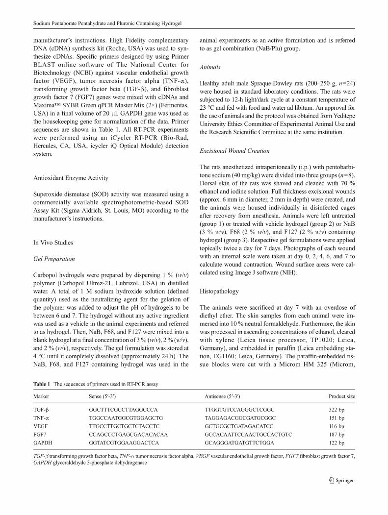
Table 1 The sequences of primers used in RT-PCR assay
Marker Sense (5′-3′) Antisense (5′-3′) Product size
TGF-β GGCTTTCGCCTTAGGCCCA TTGGTGTCCAGGGCTCGGC 322 bp
TNF-α TGGCCAATGGCGTGGAGCTG TAGGAGACGGCGATGCGGC 151 bp
VEGF TTGCCTTGCTGCTCTACCTC GCTGCGCTGATAGACATCC 116 bp
FGF7 CCAGCCCTGAGCGACACACAA GCCACAATTCCAACTGCCACTGTC 187 bp
GAPDH GGTATCGTGGAAGGACTCA GCAGGGATGATGTTCTGGA 122 bp
TGF-β transforming growth factor beta, TNF-α tumor necrosis factor alpha, VEGF vascular endothelial growth factor, FGF7 fibroblast growth factor 7,GAPDH glyceraldehyde 3-phosphate dehydrogenase
Sodium Pentaborate Pentahydrate and Pluronic Containing Hydrogel
Germany) microtome. For each animal, four randomly takentissue sections (5 μm) were processed for hematoxylin andeosin (H&E) and Masson trichrome stain using standardizedprogramme in Leica autostainer XL (Leica, Germany). Alltissue sections were examined under the Leica DM 4000microscopy system by experienced histologists, who wereuninformed of the treatments (Ü.U and A.C.). The scoringwas performed according to article [22], and each slide wasscored by a histological criteria ranging from 0 to 2 (seeFig. 4b). In case that some criteria may alter in differentgroups, six individual parameters, epidermal regeneration,thickness of epidermis, thickness of granulation tissue, inflam-matory cell infiltration, proliferation density of fibroblasts,and collagen deposition were scored separately.
Statistical Analysis
One-way analysis of variance (ANOVA) followed by Tukeypost hoc test was performed for multiple comparisons of datausing GraphPad Prism statistical software 5.0 (GraphPadSoftware, La Jolla, CA, USA). The values of P<0.05 wereconsidered statistically significant.
Results
Cell Viability
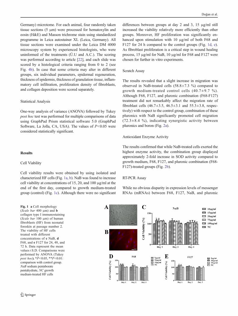
Cell viability results were obtained by using isolated andcharacterized HF cells (Fig. 1a, b). NaB was found to increasecell viability at concentrations of 15, 20, and 100 μg/ml at theend of the first day, compared to growth medium-treatedgroup (control) (Fig. 1c). Although there were no significant
differences between groups at day 2 and 3, 15 μg/ml stillincreased the viability relatively more efficiently than othergroups. Moreover, HF proliferation was significantly en-hanced upon stimulation with 10 μg/ml of both F68 andF127 for 24 h compared to the control groups (Fig. 1d, e).As fibroblast proliferation is a critical step in wound healingprocess, 15 μg/ml for NaB, 10 μg/ml for F68 and F127 werechosen for further in vitro experiments.
Scratch Assay
The results revealed that a slight increase in migration wasobserved in NaB-treated cells (58.8±7.3 %) compared togrowth medium-treated control cells (40.7±9.7 %).Although F68, F127, and pluronic combination (F68-F127)treatment did not remarkably affect the migration rate offibroblast cells (46.7±3.5, 46.5±3.1 and 55.3±3.8, respec-tively) with respect to the control group, combination of thesepluronics with NaB significantly promoted cell migration(72.3±8.4 %), indicating synergistic activity betweenpluronics and boron (Fig. 2a).
Antioxidant Enzyme Activity
The results confirmed that while NaB-treated cells exerted thehighest enzyme activity, the combination group displayedapproximately 2-fold increase in SOD activity compared togrowth medium, F68, F127, and pluronic combination (F68-F127)-treated groups (Fig. 2b).
RT-PCR Assay
While no obvious disparity in expression levels of messengerRNAs (mRNAs) between F68, F127, NaB, and pluronic
Fig. 1 a Cell morphology(Scale bar 400 μm) and bcollagen type I immunostaining(Scale bar 100 μm) of humanfibroblasts (HF) from neonatalforeskin at passage number 2.The viability of HF cellstreated with differentconcentrations of c NaB, dF68, and e F127 for 24, 48, and72 h. Data represent the meanvalues±S.D. Comparisons wereperformed by ANOVA (Tukeypost hoc). *P<0.05, **P<0.01:comparison with control group.NaB sodium pentaboratepentahydrate, NC growthmedium-treated HF cells
Doğan et al.
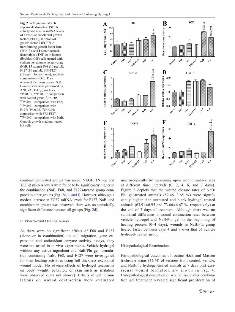
combination-treated groups was noted, VEGF, TNF-α, andTGF-βmRNA levels were found to be significantly higher inthe combination (NaB, F68, and F127)-treated group com-pared to other groups (Fig. 2c, e, and f). However, although amodest increase in FGF7 mRNA levels for F127, NaB, andcombination groups was observed, there was no statisticallysignificant difference between all groups (Fig. 2d).
In Vivo Wound Healing Assays
As there were no significant effects of F68 and F127(alone or in combination) on cell migration, gene ex-pression and antioxidant enzyme activity assays, theywere not tested in in vivo experiments. Vehicle hydrogelwithout any active ingredient and NaB/Plu gel formula-tion containing NaB, F68, and F127 were investigatedfor their healing activities using full thickness excisionalwound model. No adverse effects of hydrogel treatmentson body weight, behavior, or skin such as irritationwere observed (data not shown). Effects of gel formu-la t ions on wound cont rac t ion were evalua ted
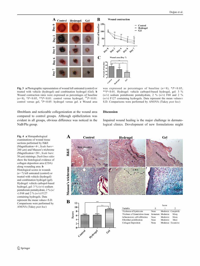
macroscopically by measuring open wound surface areaat different time intervals (0, 2, 4, 6, and 7 days).Figure 3 depicts that the wound closure rates of NaB/Plu gel-treated animals (82.46±3.65 %) were signifi-cantly higher than untreated and blank hydrogel treatedanimals (63.91±6.95 and 73.66±6.67 %, respectively) atthe end of 7 days of treatment. Although there was nostatistical difference in wound contraction rates betweenvehicle hydrogel and NaB/Plu gel in the beginning ofhealing process (0–4 days), wounds in NaB/Plu grouphealed faster between days 4 and 7 over that of vehiclehydrogel-treated group.
Histopathological Examinations
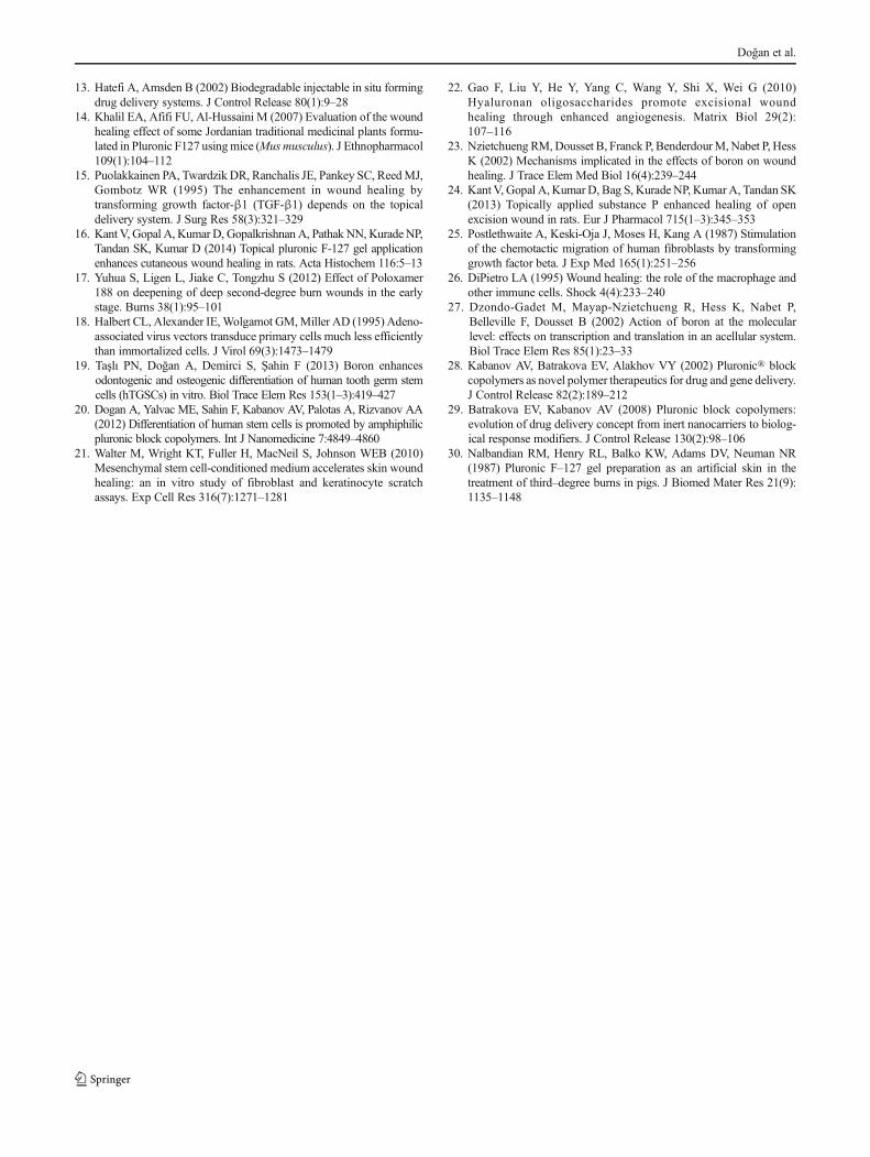
Histopathological outcomes of routine H&E and Massontrichrome stains (TCM) of sections from control, vehicle,and NaB/Plu hydrogel-treated animals at 7 days post exci-s iona l wound fo rma t ion a re shown in F ig . 4 .Histopathological evaluation of wound tissue after combina-tion gel treatment revealed significant proliferation of
Fig. 2 a Migration rates, bsuperoxide dismutase (SOD)activity and relative mRNA levelsof c vascular endothelial growthfactor (VEGF), d fibroblastgrowth factor 7 (FGF7), etransforming growth factor beta(TGF-β), and f tumor necrosisfactor alpha (TNF-α) in humanfibroblast (HF) cells treated withsodium pentaborate pentahydrate(NaB, 15 μg/ml), F68 (10 μg/ml),F127 (10 μg/ml), F68-F127(10 μg/ml for each one), and theircombinations (Gel). Datarepresent the mean values±S.D.Comparisons were performed byANOVA (Tukey post hoc).*P<0.05, **P<0.01: comparisonwith control group; #P<0.05,##P<0.01: comparison with F68;§§P<0.01: comparison withF127; †P<0.05, ††P<0.01:comparison with F68-F127;♦♦P<0.01: comparison with NaB.Control: growth medium-treatedHF cells
Sodium Pentaborate Pentahydrate and Pluronic Containing Hydrogel
fibroblasts and noticeable collegenization at the wound areacompared to control groups. Although epithelization wasevident in all groups, obvious difference was noticed in theNaB/Plu group.
Discussion
Impaired wound healing is the major challenge in dermato-logical clinics. Development of new formulations might
Fig. 3 a Photographic representation of wound left untreated (control) ortreated with vehicle (hydrogel) and combination hydrogel (Gel). bWound contraction rates were expressed as percentages of baseline(n=8), *P<0.05, **P<0.01: control versus hydrogel, ##P<0.01:control versus gel, §P<0.05: hydrogel versus gel. c Wound area
was expressed as percentages of baseline (n=8), *P<0.05,**P<0.01. Hydrogel: vehicle carbopol-based hydrogel, gel: 3 %(w/v) sodium pentaborate pentahydrate, 2 % (w/v) F68 and 2 %(w/v) F127 containing hydrogels. Data represent the mean values±S.D. Comparisons were performed by ANOVA (Tukey post hoc)
Fig. 4 a Histopathologicalexaminations of wound tissuesections performed by H&E(Magnification=4×, Scale bars=260 μm) and Masson’s trichrome(Magnification=20×, Scale bars:50μm) stainings.Dash lines sidesshow the histological evidence ofcollagen deposition area (CDA)along wounding area. bHistological scores in wounds(n=7) left untreated (control) ortreated with vehicle (hydrogel)and combination hydrogel (gel).Hydrogel: vehicle carbopol-basedhydrogel, gel: 3 % (w/v) sodiumpentaborate pentahydrate, 2 % (w/v) F68 and 2 % (w/v) F127containing hydrogels. Datarepresent the mean values±S.D.Comparisons were performed byANOVA (Tukey post hoc)
Doğan et al.

provide an opportunity for treatment of hard-to-heal wounds.In the current study, we developed a new hydrogel formula-tion containing NaB (boron source) and pluronics (F68 andF127) for effective and functional wound healing. Althoughthe exact action of mechanism has not been elucidated yet,boron has been claimed to increase wound healing activity[23]. In line with these findings, boron-enhanced migration offibroblast and combining pluronic F68 and F127 with NaBfurther increased cell migration. In agreement with the in vitrofindings, NaB and pluronics containing hydrogel formulationsaccelerated wound healing in full thickness rat excisionalwounds.
TGF-β is a vital growth factor which mediates al-most each phase of the wound healing process includinginflammation, angiogenesis, cell proliferation, and extra-cellular matrix production [24]. Moreover, TGF-β hasbeen proven to stimulate human fibroblast migrationin vitro [25]. In this line, the migratory action ofNaB/Plu gel on fibroblast cells could be explained byhigh TGF-β expression. TGF-β also regulates other keygrowth factors and cytokine production and release fromwound resident cells. One of the TGF-β signaling reg-ulated cytokines, TNF-α, is necessary for the initiationof an inflammatory response and macrophage infiltration[26]. Increased TNF-α levels in the combination (NaB,F68, and F127)-treated cells might be associated withpro-inflammatory actions of the gel formulation.Treating cells with gel combination also increased theexpression of VEGF, a pro-angiogenic factor likeTGF-β. Consistent with the current findings, anotherstudy found that NaB treatment increased VEGF andTGF-β expressions but not FGF1 and TNF-α expres-sions [27]. Moreover, combining F68 and F127 withNaB caused a significant increase in TGF-β, VEGF,and TNF-α expression levels. Pluronics have been re-ported to be used in drug and gene delivery applica-tions, along with a diagnosis as a carrying agent ofcontrasting chemicals [28]. As pluronic block copoly-mers form micelles, dependent on temperature and con-centration, they increase the activity and transport effi-ciency of the drug incorporated into the core of micellesby interacting targeted cell membrane and its surfaceproteins [29]. Therefore, the reason of the highesthealing activity in combination gel-treated cells andwounds might be attributed to enhanced boron transpor-tation due to pluronic micelle formation. A secondpossible explanation for the synergistic activity of boronand pluronics could be the direct healing effect of thepluronics. Pluronic F127 gel treatment has increasedVEGF and TGF-β expressions, microvessel density,and wound contraction [24]. In addition, F127 treatmenthas been found to increase third degree burn woundhealing in pigs [30].
Conclusion
The overall data suggest that a combination of boron andpluronics increases fibroblast migration, antioxidant enzymeactivity, growth factor expression levels, and acute cutaneouswound healing. Although the results are encouraging, furtherstudies are highly warranted to elucidate the exact mechanismof the synergistic activity between pluronics and boron. Inaddition, this formulation should be tested in chronic woundssuch as trauma, diabetic, decubitus, and venous leg ulcers inorder to explore its full potential in dermatological science.Finally, as the formulation provides a remarkable increase infibroblast proliferation and collagen synthesis, potential use ofthe formulation for cosmetic purposes should be investigated.
Acknowledgment This study was supported by Yeditepe University.The authors thank to Dr. Andrew John Harvey for his advises on language.
Conflict of Interest The authors deny any conflicts of interest.
References
1. Singer AJ, Clark RA (1999) Cutaneous wound healing. N Engl JMed 341(10):738–746
2. Werner S, Grose R (2003) Regulation of wound healing by growthfactors and cytokines. Physiol Rev 83(3):835–870
3. Sen CK, Gordillo GM, Roy S, Kirsner R, Lambert L, Hunt TK,Gottrup F, Gurtner GC, Longaker MT (2009) Human skin wounds: amajor and snowballing threat to public health and the economy.Wound Repair Regen 17(6):763–771
4. Mazé P (1915) Détermination des éléments minéraux raresnécessaires au développement du maïs. C R Hebd Seances AcadSci 160:211–214
5. Eckhert CD, Rowe RI (1999) Embryonic dysplasia and adult retinaldystrophy in boron–deficient zebrafish. J Trace Elem ExpMed 12(3):213–219
6. GorustovichAA, Steimetz T, Nielsen FH, GuglielmottiMB (2008) Ahistomorphometric study of alveolar bone modelling and remodel-ling in mice fed a boron-deficient diet. Arch Oral Biol 53(7):677–682
7. Hunt CD (2003) Dietary boron: an overview of the evidence for itsrole in immune function. J Trace Elem Exp Med 16(4):291–306
8. Sheng MH-C, Taper LJ, Veit H, Qian H, Ritchey SJ, Lau K-HW(2001) Dietary boron supplementation enhanced the action of estro-gen, but not that of parathyroid hormone, to improve trabecular bonequality in ovariectomized rats. Biol Trace Elem Res 82(1–3):109–123
9. Penland JG (1998) The importance of boron nutrition for brain andpsychological function. Biol Trace Elem Res 66(1–3):299–317
10. Blech M, Martin C, Borrelly J, Hartemann P (1990) Treatment ofdeep wounds with loss of tissue. Value of a 3 percent boric acidsolution. Presse Med 19(22):1050–1052
11. Heilmann S, Küchler S, Wischke C, Lendlein A, Stein C, Schäfer-Korting M (2013) A thermosensitive morphine-containing hydrogelfor the treatment of large-scale skin wounds. Int J Pharm 444(1):96–102
12. Ruel-Gariépy E, Leroux J-C (2004) In situ-forming hydrogels-reviewof temperature-sensitive systems. Eur J Pharm Biopharm 58(2):409–426
Sodium Pentaborate Pentahydrate and Pluronic Containing Hydrogel
13. Hatefi A, Amsden B (2002) Biodegradable injectable in situ formingdrug delivery systems. J Control Release 80(1):9–28
14. Khalil EA, Afifi FU, Al-Hussaini M (2007) Evaluation of the woundhealing effect of some Jordanian traditional medicinal plants formu-lated in Pluronic F127 usingmice (Musmusculus). J Ethnopharmacol109(1):104–112
15. Puolakkainen PA, Twardzik DR, Ranchalis JE, Pankey SC, ReedMJ,Gombotz WR (1995) The enhancement in wound healing bytransforming growth factor-β1 (TGF-β1) depends on the topicaldelivery system. J Surg Res 58(3):321–329
16. Kant V, Gopal A, Kumar D, Gopalkrishnan A, Pathak NN, Kurade NP,Tandan SK, Kumar D (2014) Topical pluronic F-127 gel applicationenhances cutaneous wound healing in rats. Acta Histochem 116:5–13
17. Yuhua S, Ligen L, Jiake C, Tongzhu S (2012) Effect of Poloxamer188 on deepening of deep second-degree burn wounds in the earlystage. Burns 38(1):95–101
18. Halbert CL, Alexander IE, Wolgamot GM,Miller AD (1995) Adeno-associated virus vectors transduce primary cells much less efficientlythan immortalized cells. J Virol 69(3):1473–1479
19. Taşlı PN, Doğan A, Demirci S, Şahin F (2013) Boron enhancesodontogenic and osteogenic differentiation of human tooth germ stemcells (hTGSCs) in vitro. Biol Trace Elem Res 153(1–3):419–427
20. Dogan A, Yalvac ME, Sahin F, Kabanov AV, Palotas A, Rizvanov AA(2012) Differentiation of human stem cells is promoted by amphiphilicpluronic block copolymers. Int J Nanomedicine 7:4849–4860
21. Walter M, Wright KT, Fuller H, MacNeil S, Johnson WEB (2010)Mesenchymal stem cell-conditioned medium accelerates skin woundhealing: an in vitro study of fibroblast and keratinocyte scratchassays. Exp Cell Res 316(7):1271–1281
22. Gao F, Liu Y, He Y, Yang C, Wang Y, Shi X, Wei G (2010)Hyaluronan oligosaccharides promote excisional woundhealing through enhanced angiogenesis. Matrix Biol 29(2):107–116
23. Nzietchueng RM,Dousset B, Franck P, BenderdourM,Nabet P, HessK (2002) Mechanisms implicated in the effects of boron on woundhealing. J Trace Elem Med Biol 16(4):239–244
24. Kant V, Gopal A, Kumar D, Bag S, Kurade NP, KumarA, Tandan SK(2013) Topically applied substance P enhanced healing of openexcision wound in rats. Eur J Pharmacol 715(1–3):345–353
25. Postlethwaite A, Keski-Oja J, Moses H, Kang A (1987) Stimulationof the chemotactic migration of human fibroblasts by transforminggrowth factor beta. J Exp Med 165(1):251–256
26. DiPietro LA (1995) Wound healing: the role of the macrophage andother immune cells. Shock 4(4):233–240
27. Dzondo-Gadet M, Mayap-Nzietchueng R, Hess K, Nabet P,Belleville F, Dousset B (2002) Action of boron at the molecularlevel: effects on transcription and translation in an acellular system.Biol Trace Elem Res 85(1):23–33
28. Kabanov AV, Batrakova EV, Alakhov VY (2002) Pluronic® blockcopolymers as novel polymer therapeutics for drug and gene delivery.J Control Release 82(2):189–212
29. Batrakova EV, Kabanov AV (2008) Pluronic block copolymers:evolution of drug delivery concept from inert nanocarriers to biolog-ical response modifiers. J Control Release 130(2):98–106
30. Nalbandian RM, Henry RL, Balko KW, Adams DV, Neuman NR(1987) Pluronic F–127 gel preparation as an artificial skin in thetreatment of third–degree burns in pigs. J Biomed Mater Res 21(9):1135–1148
Doğan et al.
Related Documents