Behavioural Neurology Social cognition impairment in genetic frontotemporal dementia within the GENFI cohort Lucy L. Russell a , Caroline V. Greaves a , Martina Bocchetta a , Jennifer Nicholas b,c , Rhian S. Convery a , Katrina Moore a , David M. Cash a,d , John van Swieten e , Lize Jiskoot a,e , Fermin Moreno f , Raquel Sanchez-Valle g , Barbara Borroni h , Robert Laforce Jr. i , Mario Masellis j , Maria Carmela Tartaglia k , Caroline Graff l , Emanuela Rotondo m , Daniela Galimberti m,n , James B. Rowe o , Elizabeth Finger p , Matthis Synofzik q , Rik Vandenberghe r , Alexandre de Mendonc ¸a s , Fabrizio Tagliavini t , Isabel Santana u , Simon Ducharme v , Chris Butler w , Alex Gerhard x,y , Johannes Levin z , Adrian Danek z , Markus Otto aa , Jason D. Warren a and Jonathan D. Rohrer a,* , on behalf of the Genetic FTD Initiative, GENFI 1 a Dementia Research Centre, Department of Neurodegenerative Disease, London, UK b Department of Medical Statistics, London School of Hygiene and Tropical Medicine, London, UK c Institute of Prion Disease, UCL Queen Square Institute of Neurology, London, UK d Centre for Medical Image Computing, Department of Medical Physics and Biomedical Engineering, University College London, London, UK e Department of Neurology, Erasmus Medical Centre, Rotterdam, Netherlands f Cognitive Disorders Unit, Department of Neurology, Donostia University Hospital, San Sebastian, Gipuzkoa, Spain g Alzheimer's Disease and Other Cognitive Disorders Unit, Neurology Service, Hospital Clı´nic, Barcelona, Spain h Centre for Neurodegenerative Disorders, Neurology Unit, Department of Clinical and Experimental Sciences, University of Brescia, Brescia, Italy i Clinique Interdisciplinaire de M emoire, D epartement des Sciences Neurologiques du CHU de Qu ebec, Universit e Laval, Qu ebec, Canada j Sunnybrook Health Sciences Centre, Sunnybrook Research Institute, University of Toronto, Toronto, Canada k Tanz Centre for Research in Neurodegenerative Diseases, University of Toronto, Toronto, Canada l Department of Geriatric Medicine, Karolinska University Hospital-Huddinge, Stockholm, Sweden m University of Milan, Centro Dino Ferrari, Milan, Italy n Fondazione Ca’ Granda, IRCCS Ospedale Policlinico, Milan, Italy o Department of Clinical Neurosciences, University of Cambridge, Cambridge, UK p Department of Clinical Neurological Sciences, University of Western Ontario, London, Ontario, Canada q Department of Neurodegenerative Diseases, Hertie-Institute for Clinical Brain Research and Center of Neurology, University of Tu ¨ bingen, Tu ¨ bingen, Germany * Corresponding author. Dementia Research Centre, Department of Neurodegenerative Disease, UCL Institute of Neurology, Queen Square, London, WC1N 3BG, UK. E-mail address: [email protected] (J.D. Rohrer). 1 List of GENFI consortium authors is listed in Appendix section. Available online at www.sciencedirect.com ScienceDirect Journal homepage: www.elsevier.com/locate/cortex cortex 133 (2020) 384 e398 https://doi.org/10.1016/j.cortex.2020.08.023 0010-9452/© 2020 The Author(s). Published by Elsevier Ltd. This is an open access article under the CC BY license (http:// creativecommons.org/licenses/by/4.0/).

Welcome message from author
This document is posted to help you gain knowledge. Please leave a comment to let me know what you think about it! Share it to your friends and learn new things together.
Transcript
www.sciencedirect.com
c o r t e x 1 3 3 ( 2 0 2 0 ) 3 8 4e3 9 8
Available online at
ScienceDirect
Journal homepage: www.elsevier.com/locate/cortex
Behavioural Neurology
Social cognition impairment in geneticfrontotemporal dementia within the GENFI cohort
Lucy L. Russell a, Caroline V. Greaves a, Martina Bocchetta a,Jennifer Nicholas b,c, Rhian S. Convery a, Katrina Moore a,David M. Cash a,d, John van Swieten e, Lize Jiskoot a,e, Fermin Moreno f,Raquel Sanchez-Valle g, Barbara Borroni h, Robert Laforce Jr. i,Mario Masellis j, Maria Carmela Tartaglia k, Caroline Graff l,Emanuela Rotondo m, Daniela Galimberti m,n, James B. Rowe o,Elizabeth Finger p, Matthis Synofzik q, Rik Vandenberghe r,Alexandre de Mendonca s, Fabrizio Tagliavini t, Isabel Santana u,Simon Ducharme v, Chris Butler w, Alex Gerhard x,y, Johannes Levin z,Adrian Danek z, Markus Otto aa, Jason D. Warren a andJonathan D. Rohrer a,*, on behalf of the Genetic FTD Initiative, GENFI1
a Dementia Research Centre, Department of Neurodegenerative Disease, London, UKb Department of Medical Statistics, London School of Hygiene and Tropical Medicine, London, UKc Institute of Prion Disease, UCL Queen Square Institute of Neurology, London, UKd Centre for Medical Image Computing, Department of Medical Physics and Biomedical Engineering, University
College London, London, UKe Department of Neurology, Erasmus Medical Centre, Rotterdam, Netherlandsf Cognitive Disorders Unit, Department of Neurology, Donostia University Hospital, San Sebastian, Gipuzkoa, Spaing Alzheimer's Disease and Other Cognitive Disorders Unit, Neurology Service, Hospital Clınic, Barcelona, Spainh Centre for Neurodegenerative Disorders, Neurology Unit, Department of Clinical and Experimental Sciences,
University of Brescia, Brescia, Italyi Clinique Interdisciplinaire de M�emoire, D�epartement des Sciences Neurologiques du CHU de Qu�ebec, Universit�e
Laval, Qu�ebec, Canadaj Sunnybrook Health Sciences Centre, Sunnybrook Research Institute, University of Toronto, Toronto, Canadak Tanz Centre for Research in Neurodegenerative Diseases, University of Toronto, Toronto, Canadal Department of Geriatric Medicine, Karolinska University Hospital-Huddinge, Stockholm, Swedenm University of Milan, Centro Dino Ferrari, Milan, Italyn Fondazione Ca’ Granda, IRCCS Ospedale Policlinico, Milan, Italyo Department of Clinical Neurosciences, University of Cambridge, Cambridge, UKp Department of Clinical Neurological Sciences, University of Western Ontario, London, Ontario, Canadaq Department of Neurodegenerative Diseases, Hertie-Institute for Clinical Brain Research and Center of Neurology,
University of Tubingen, Tubingen, Germany
* Corresponding author. Dementia Research Centre, Department of Neurodegenerative Disease, UCL Institute of Neurology, QueenSquare, London, WC1N 3BG, UK.
E-mail address: [email protected] (J.D. Rohrer).1 List of GENFI consortium authors is listed in Appendix section.
https://doi.org/10.1016/j.cortex.2020.08.0230010-9452/© 2020 The Author(s). Published by Elsevier Ltd. This is an open access article under the CC BY license (http://creativecommons.org/licenses/by/4.0/).
c o r t e x 1 3 3 ( 2 0 2 0 ) 3 8 4e3 9 8 385
r Laboratory for Cognitive Neurology, Department of Neurosciences, KU Leuven, Leuven, Belgiums Faculty of Medicine, University of Lisbon, Lisbon, Portugalt Fondazione Istituto di Ricovero e Cura a Carattere Scientifico Istituto Neurologica Carlo Besta, Milano, Italyu Faculty of Medicine, University of Coimbra, Coimbra, Portugalv Department of Psychiatry, McGill University, Montreal, Qu�ebec, Canadaw Department of Clinical Neurology, University of Oxford, Oxford, UKx Division of Neuroscience and Experimental Psychology, Wolfson Molecular Imaging Centre, University of
Manchester, Manchester, UKy Departments of Geriatric Medicine and Nuclear Medicine, University of Duisburg- Essen, Germanyz Department of Neurology, Ludwig-Maximilians-University, Munich, Germanyaa Department of Neurology, University of Ulm, Ulm, Germany
a r t i c l e i n f o
Article history:
Received 31 March 2020
Reviewed 11 May 2020
Revised 6 July 2020
Accepted 22 August 2020
Action editor Brad Dickerson
Published online 26 September 2020
Keywords:
Frontotemporal dementia
Theory of mind
Emotion processing
Faux pas
Facial emotion recognition
C9orf72
Progranulin
MAPT
a b s t r a c t
A key symptom of frontotemporal dementia (FTD) is difficulty interacting socially with
others. Social cognition problems in FTD include impaired emotion processing and theory
of mind difficulties, and whilst these have been studied extensively in sporadic FTD, few
studies have investigated them in familial FTD. Facial Emotion Recognition (FER) and Faux
Pas (FP) recognition tests were used to study social cognition within the Genetic Fronto-
temporal Dementia Initiative (GENFI), a large familial FTD cohort of C9orf72, GRN, and
MAPT mutation carriers. 627 participants undertook at least one of the tasks, and were
separated into mutation-negative healthy controls, presymptomatic mutation carriers
(split into early and late groups) and symptomatic mutation carriers. Groups were
compared using a linear regression model with bootstrapping, adjusting for age, sex,
education, and for the FP recognition test, language. Neural correlates of social cognition
deficits were explored using a voxel-based morphometry (VBM) study. All three of the
symptomatic genetic groups were impaired on both tasks with no significant difference
between them. However, prior to onset, only the late presymptomatic C9orf72 mutation
carriers on the FER test were impaired compared to the control group, with a subanalysis
showing differences particularly in fear and sadness. The VBM analysis revealed that
impaired social cognition was mainly associated with a left hemisphere predominant
network of regions involving particularly the striatum, orbitofrontal cortex and insula,
and to a lesser extent the inferomedial temporal lobe and other areas of the frontal lobe.
In conclusion, theory of mind and emotion processing abilities are impaired in familial
FTD, with early changes occurring prior to symptom onset in C9orf72 presymptomatic
mutation carriers. Future work should investigate how performance changes over time, in
order to gain a clearer insight into social cognitive impairment over the course of the
disease.
© 2020 The Author(s). Published by Elsevier Ltd. This is an open access article under the CC
BY license (http://creativecommons.org/licenses/by/4.0/).
1. Introduction
The impairment of social skills is one of the most prominent
symptoms experienced by people with frontotemporal de-
mentia (FTD) (Adenzato, Cavallo, & Enrici, 2010; Kumfor and
Piguet, 2012). The different neural processes that underlie
such skills are generally grouped together within the term
‘social cognition’ (Adolphs, 2009), and include a number of
abilities that have been shown to be impaired in FTD,
including recognition of others' emotions, and ‘theory of
mind’, the ability to understand that others have thoughts and
beliefs (Gregory et al., 2002; Lough and Hodges, 2002; Rosen
et al., 2006; Adenzato et al., 2010; Omar, Rohrer, Hailstone, &
Warren, 2011; Kumfor and Piguet, 2012).
Whilst there have been a number of studies exploring
these skills in sporadic FTD, few have focused on people with
the genetic forms of FTD, characterized usually by mutations
in the progranulin (GRN), tau (MAPT) and chromosome 9 open
reading frame 72 (C9orf72) genes (Jiskoot et al., 2016, 2018,
Cheran et al., 2019). So far, these studies have been relatively
small and often focused on one (Cheran et al., 2019) or two
(Jiskoot et al., 2016, 2018) of the genetic groups, showing
change only in specific questionnaires, or when groups were
followed longitudinally.
The Genetic FTD Initiative (GENFI) is an international ge-
netic FTD cohort study, aimed at investigating early bio-
markers, including measures of cognition (Rohrer et al.,
2015). Using this cohort we therefore aimed to assess
emotion processing and theory of mind abilities in a large
c o r t e x 1 3 3 ( 2 0 2 0 ) 3 8 4e3 9 8386
cohort of presymptomatic and symptomatic individuals with
mutations in the C9orf72, GRN and MAPT genes, with the
hypothesis that social cognitive deficits would become
apparent only late in the presymptomatic period or when
symptomatic.
2. Methods
We report how we determined our sample size, all data ex-
clusions (if any), all inclusion/exclusion criteria, whether in-
clusion/exclusion criteria were established prior to data
analysis, all manipulations, and all measures in the study.
2.1. Participants
Participants were recruited from the fourth data freeze of the
GENFI study including sites in the UK, Canada, Sweden,
Netherlands, Belgium, Spain, Portugal, Italy and Germany. Of
the 680 participants consecutively enrolled in the study, 627
undertook at least one test of social cognition: 246 who tested
negative for the mutation within the family, and therefore
acted as the controls, 159 C9orf72 expansion carriers, 155 GRN
mutation carriers, and 67 MAPT mutation carriers (Table 1).
Mutation carriers were classified as either symptomatic or
presymptomatic based on clinician judgement. Participants
were only classified as symptomatic if the clinician judged
that symptoms were present, consistent with a diagnosis of a
degenerative disorder, and progressive in nature (Table S1).
The presymptomatic carriers were further split into those
further than five years from estimated symptom onset (based
on the mean age at onset in the family), called the ‘early’
group, and those within five years of estimated onset, called
the ‘late’ group. Diagnoses in the symptomatic group were as
follows: MAPT mutation carriers, 17 bvFTD, 1 other; GRN mu-
tation carriers, 15 bvFTD, 16 primary progressive aphasia
(PPA), 1 other; C9orf72 expansion carriers, 38 bvFTD, 10 FTD
with amyotrophic lateral sclerosis, 1 PPA, 1 progressive
supranuclear palsy and 3 other.
All participants underwent the standardized GENFI clinical
assessment including medical history, physical examination,
the Mini-Mental State Examination (MMSE), and the Clinical
Dementia Rating Scale with the National Alzheimer Coordi-
nating Centre FTLD sum of boxes score (FTLD-CDR-SOB). De-
mographics are shown in Table 1. There was a significant
difference in sex between the groups (p ¼ .018): the symp-
tomatic C9orf72 mutation carriers had a significantly higher
percentage of men than the early and late C9orf72 mutation
carriers and the control group (p ¼ .013, p ¼ .002 and p ¼ .001
respectively). There was also a significant difference in age
between the groups: all early presymptomatic mutation car-
riers were significantly younger than the control group (all
p < .001), and all late presymptomatic mutation carriers and
symptomatic mutation carriers were significantly older than
controls (all p < .001) except for the late MAPT mutation car-
riers in which no difference was observed (p ¼ .239). There
were also differences between the groups in education: the
symptomatic C9orf72 and GRN mutation carriers had signifi-
cantly lower levels of education than the control group did
(p ¼ .007 and p < .001 respectively). No significant differences
in disease severity were observed between the symptomatic
genetic groups or between the late presymptomatic groups,
based on their FTLD-CDR-SOB. However, the early GRN pre-
symptomatic mutation carrier did have a significantly lower
FTLD-CDR-SOB scores than the other two early groups.
2.2. Testing of social cognition
Social cognition was tested in the GENFI cohort using the
shortened version of the Social Cognition and Emotional
Assessment, known as the mini-SEA (Bertoux et al., 2012;
Funkiewiez, Bertoux, de Souza, L�evy, & Dubois, 2012) which
consists of a test of facial emotion recognition and a test of
theory of mind. It was designed specifically for people with
FTD, with initial studies showing deficits in FTD compared
with healthy controls, with people with Alzheimer's disease,
and also those with major depressive disorder (Guevara et al.,
2015; Narme, Mouras, Roussel, Devendeville, & Godefroy,
2013; Torralva, Gleichgerrcht, Torres Ardila, Roca, & Manes,
2015).
2.2.1. Experiment 1: facial emotion recognition (FER) testThe FER test is a shortened version of the standard Ekman
faces task (Ekman, Ellsworth, Friesen, Goldstein, & Krasner,
1972), with participants asked to recognise whether faces are
showing one of either six universal emotions (happiness,
surprise, anger, fear, disgust and sadness) or a neutral
expression. Participants are presented with 35 different faces
(five items for each emotion) and are required to select the
correct emotional label that matches the emotion of the face.
2.2.2. Experiment 2: faux pas (FP) recognition testThe FP recognition test contains a series of 10 short cartoon
stories describing scenarios involving social inconveniences,
known as ‘faux pas’; five of the stories contain a faux pas, the
other five do not. The task requires individuals to be able to
infer another's thoughts or beliefs. A structured questionnaire
asks how and why the social faux pas has occurred. Partici-
pants can score a maximum of 40 on this task, 10 points for
the control stories and 30 points for the faux pas stories.
2.3. Statistical analysis
In the control group, we explored the relationship of the FER
and FP recognition tests to age (Spearman rank correlation),
sex (ManneWhitney U test) and years of education (Spearman
rank correlation). For the FP recognition test, we explored the
effect of the different language versions using a linear
regression.
Scores on the two social cognitive tests (and the individual
emotion scores on the FER test) were compared between the
groups using linear regression, adjusting for age, sex and ed-
ucation (and language for the FP recognition test) with 95%
bias-corrected bootstrapped confidence intervals with 1000
repetitions (as the data was not normally distributed).
A subanalysis of the effect of phenotype was also per-
formed using the same methodology as the main analysis:
scores on the two social cognitive tests were compared
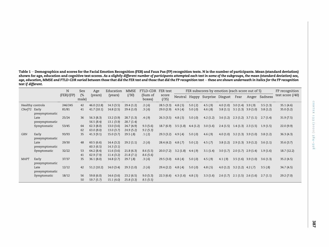
Table 1 e Demographics and scores for the Facial Emotion Recognition (FER) and Faux Pas (FP) recognition tests. N is the number of participants. Mean (standard deviation)shown for age, education and cognitive test scores. As a slightly different number of participants attempted each test in some of the subgroups, the mean (standard deviation) sex,age, education, MMSE and FTLD-CDR varied between those that did the FER test and those that did the FP recognition teste these are shown underneath in italics for the FP recognitiontest if different.
N(FER)/(FP)
Sex(%
male)
Age(years)
Education(years)
MMSE(/30)
FTLD-CDR(Sum ofboxes)
FER testscore(/35)
FER subscores by emotion (each score out of 5) FP recognitiontest score (/40)Neutral Happy Surprise Disgust Fear Anger Sadness
Healthy controls 246/245 42 46.0 (12.8) 14.3 (3.5) 29.4 (1.2) .2 (.6) 28.5 (3.3) 4.8 (.5) 5.0 (.2) 4.5 (.9) 4.0 (1.0) 3.0 (1.4) 3.9 (.9) 3.5 (1.3) 35.1 (4.6)
C9orf72 Early
presymptomatic
81/81 41 41.7 (10.1) 14.8 (2.5) 29.4 (1.0) .3 (.6) 29.0 (2.9) 4.9 (.4) 5.0 (.0) 4.6 (.8) 3.8 (1.1) 3.1 (1.3) 3.9 (1.0) 3.8 (1.2) 35.0 (5.2)
Late
presymptomatic
25/24 36 56.3 (8.3)
56.5 (8.4)
13.2 (3.9)
13.1 (3.9)
28.7 (1.3)
28.7 (1.4)
.4 (.9) 26.3 (3.5) 4.8 (.5) 5.0 (.0) 4.2 (1.2) 3.6 (1.2) 2.3 (1.2) 3.7 (1.1) 2.7 (1.4) 31.9 (7.5)
Symptomatic 53/45 64
62
62.3 (8.0)
63.0 (8.0)
13.0 (3.6)
13.0 (3.7)
24.7 (4.9)
24.9 (5.2)
9.3 (5.6)
9.2 (5.3)
18.7 (6.9) 3.5 (1.8) 4.4 (1.2) 3.0 (1.6) 2.4 (1.5) 1.4 (1.3) 2.3 (1.5) 1.9 (1.5) 22.0 (9.9)
GRN Early
presymptomatic
93/93 35 41.3 (9.1) 15.0 (3.7) 29.5 (.8) .1 (.2) 29.3 (3.2) 4.9 (.4) 5.0 (.0) 4.6 (.9) 4.0 (1.0) 3.2 (1.3) 3.9 (1.0) 3.8 (1.2) 36.3 (4.3)
Late
presymptomatic
29/30 48 60.5 (6.6)
60.3 (6.5)
14.4 (3.2)
14.3 (3.1)
29.2 (1.1) .2 (.6) 28.4 (4.2) 4.8 (.7) 5.0 (.2) 4.5 (.7) 3.8 (1.2) 2.9 (1.3) 3.9 (1.2) 3.6 (1.1) 35.6 (3.7)
Symptomatic 32/22 53
41
64.2 (8.4)
62.9 (7.9)
11.6 (3.6)
11.4 (3.2)
21.8 (6.3)
21.8 (7.1)
8.6 (5.5)
8.6 (5.6)
20.0 (7.2) 3.2 (1.8) 4.4 (.9) 3.1 (1.4) 3.0 (1.7) 2.0 (1.7) 2.9 (1.4) 1.9 (1.6) 18.7 (12.2)
MAPT Early
presymptomatic
37/37 35 36.1 (8.0) 14.8 (2.7) 29.7 (.8) .3 (.6) 29.5 (3.0) 4.8 (.4) 5.0 (.0) 4.5 (.9) 4.1 (.9) 3.5 (1.6) 3.9 (1.0) 3.6 (1.3) 35.2 (4.5)
Late
presymptomatic
12/12 42 51.2 (10.2) 14.0 (3.4) 29.3 (1.0) .2 (.6) 29.4 (2.2) 4.8 (.4) 5.0 (.0) 4.8 (.5) 4.0 (1.2) 3.2 (1.2) 4.2 (.7) 3.5 (.8) 34.7 (4.5)
Symptomatic 18/12 56
50
59.8 (6.0)
59.7 (5.7)
14.6 (3.6)
15.1 (4.0)
23.2 (6.5)
25.8 (3.3)
9.0 (5.3)
8.5 (5.5)
22.3 (6.6) 4.3 (1.6) 4.8 (.5) 3.3 (1.6) 2.6 (1.7) 2.1 (1.5) 2.6 (1.6) 2.7 (1.1) 29.2 (7.0)
cortex
133
(2020)384e398
387
c o r t e x 1 3 3 ( 2 0 2 0 ) 3 8 4e3 9 8388
between the different clinical syndromes within the symp-
tomatic mutation carriers as well as with controls.
2.4. Imaging analysis
Participants underwent volumetric T1-weighted MRI using
the GENFI protocol. A variety of 3T scanners were used across
the sites: Siemens Trio, Siemens Skyra, Siemens Prisma,
Phillips and General Electric. The scan protocols were
designed at the start of the GENFI study to ensure that there
was adequatematching between the scanners and the quality
of the images. All scans were quality checked and those with
movements or artefacts were removed. Furthermore, if any
participants displayed moderate to severe vascular disease or
any other brain lesions, they were also excluded from the
analysis.
Voxel-based morphometry (VBM) was performed using
Statistical Parametric Mapping (SPM) 12 software, version
6685 (www.fil.ion.ucl.ac.uk/spm), running under Matlab
R2014a (Mathworks, USA). The T1-weighted images were
normalized and segmented into grey matter (GM), white
matter (WM) and cerebrospinal fluid (CSF) probability maps,
by using standard procedures and the fast-diffeomorphic
image registration algorithm (DARTEL) (Ashburner, 2007).
GM segmentations were affine transformed into the Montreal
Neurological Institute (MNI) space, modulated and smoothed
using a Gaussian kernel with 6 mm full-width at half
maximum before analysis. Finally, a mask was applied as re-
ported in Ridgway et al., 2009. Study-specific templates were
created based on the subjects included in the specific analysis.
At each stage, all segmentations were reviewed visually. Total
intracranial volume (TIV) was calculated using SPM (Malone
et al., 2015).
In order to explore the relationship between performance
on the tests and GM density, multiple regression models for
each genetic group were used to correlate the GM tissue
maps to the FER and FP performance in mutation carriers
(both symptomatic and presymptomatic individuals com-
bined). 319 scans were used for the FER analysis and 309
scans were used for the FP analysis (C9orf72 expansion car-
riers: FER ¼ 132, FP ¼ 128, GRN mutation carriers: FER ¼ 132,
FP ¼ 129, and MAPTmutation carriers: FER ¼ 55, FP ¼ 52) were
included in the imaging analysis. Control participants were
not included in any of the analysis. Age, sex, scanner type
and TIV were included as nuisance covariates. The Family-
Wise Error (FWE) rate for multiple comparisons correction
was set at .05. If there were no findings at that strict level of
correction, results were reviewed at an uncorrected p value of
.001.
No part of the study procedure or analyses were pre-
registered prior to the research being conducted. The condi-
tions of our ethics approval do not permit public archiving of
individual anonymised data. Readers seeking access to the
data should contact the corresponding author. Access will be
granted to named individuals in accordance with ethical
procedures governing the reuse of sensitive data, including
completion of a data sharing agreement. All stimuli and sta-
tistical code have been archived at: https://osf.io/m8yp7/?
view_only¼949ba796b549 4b7b87d37766adf840bf.
3. Results
3.1. Experiment 1: facial emotion recognition (FER) test
3.1.1. Healthy controlsFER test score was not significantly correlated with either age
(rho ¼ �.12, p ¼ .063) (Table S2) or education (rho ¼ .13,
p ¼ .051) (Table S3) within the controls. However, there was a
significant effect of sex (p ¼ .031): mean (standard deviation)
score overall in controls was 28.5 (3.3), with a higher score of
29.1 (3.1) in females (n ¼ 143), compared with 28.2 (3.2) in
males (n ¼ 103).
Overall, controls scored between 19 and 34 out of a total
possible score of 35, with cumulative frequency shown in
Table S4. A cut-off score below the 5th percentile is commonly
considered to be abnormal: for the FER test a score of below 23
would therefore be considered outside the normal range, with
a score of 23 considered borderline abnormal.
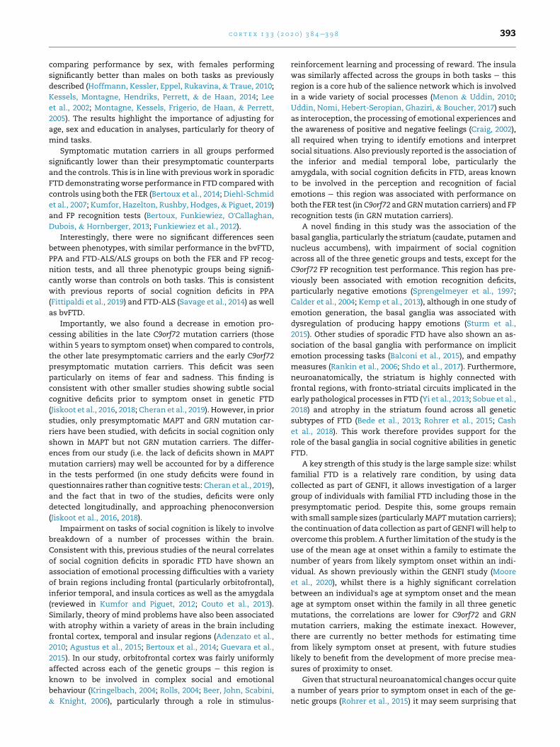
3.1.2. Mutation carriersAll of the three symptomatic mutation carrier groups scored
significantly lower on the FER test compared with controls
(Table 1, Table S4, Fig. 1): C9orf72 mean 18.7 (standard devia-
tion 6.9), GRN 20.0 (7.2) andMAPT 22.3 (6.6), with no significant
difference between the disease groups.
Within each genetic group, scores were significantly lower
in the symptomatic group compared with both the early and
late presymptomatic groups (Table 1, Table S5, Fig. 1).
The C9orf72 late presymptomatic group performed signifi-
cantly lower than both the C9orf72 early presymptomatic
group and the controls (Table 1, Table S5, Fig. 1): late pre-
symptomatic group 26.3 (3.5), early presymptomatic group
29.0 (2.9). No significant differences were seen between the
other presymptomatic groups and controls.
3.1.3. Phenotypic analysisAll phenotypic groups [bvFTD (19.6 {6.3}), PPA (22.0 {6.4}) and
an FTD-ALS/ALS group (18.4 {8.1})] were significantly impaired
on the FER test compared with controls, with no significant
differences between any of the clinical syndromes (Table S6
and Table S7).
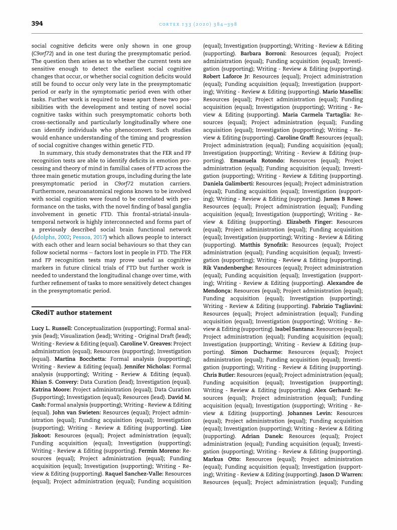
3.1.4. Imaging analysisIn C9orf72 mutation carriers, FER test score was positively
associated with bilateral insula involvement, as well as atro-
phy in the left frontal lobe (middle frontal gyrus and orbito-
frontal cortex), left basal ganglia (putamen and caudate) and
right amygdala (Table S8, Fig. 2).
For the GRNmutation carriers, performance was positively
correlated with a left hemisphere predominant network of
areas involving the insula, frontal lobe, inferomedial temporal
lobe, cingulate, basal ganglia (putamen and caudate) and
thalamus (Table S8, Fig. 2).
In the MAPT mutation group FER test score positively
correlatedwith two small clusters, one in the left basal ganglia
and one in the left orbitofrontal cortex when correcting for
multiple comparisons. At an uncorrected p value of <.001,there was also an association with the left insula and
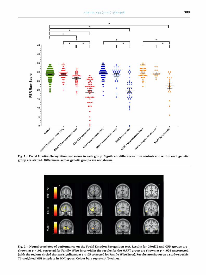
Fig. 1 e Facial Emotion Recognition test scores in each group. Significant differences from controls and within each genetic
group are starred. Differences across genetic groups are not shown.
Fig. 2 e Neural correlates of performance on the Facial Emotion Recognition test. Results for C9orf72 and GRN groups are
shown at p < .05, corrected for Family Wise Error whilst the results for the MAPT group are shown at p < .001 uncorrected
(with the regions circled that are significant at p < .05 corrected for FamilyWise Error). Results are shown on a study-specific
T1-weighted MRI template in MNI space. Colour bars represent T-values.
c o r t e x 1 3 3 ( 2 0 2 0 ) 3 8 4e3 9 8 389
c o r t e x 1 3 3 ( 2 0 2 0 ) 3 8 4e3 9 8390
inferomedial temporal lobe aswell as bilateral superior frontal
and orbitofrontal regions (Table S8, Fig. 2).
3.1.5. Subanalysis of performance on individual emotionsIdentification of negative emotions (fear, anger, sadness and
disgust) was in general worse than the recognition of positive
ones (happiness and surprise) in each of the groups (including
controls).
In almost all of the emotions, the symptomatic groups
scored worse than controls (Table 1, Fig. 3). Only in the
symptomatic MAPT mutation group for happiness and fear
was there no significant difference.
In the presymptomatic groups, the C9orf72 late presymp-
tomatic group scored significantly lower than controls on both
fear and sadness, but not on the other emotions (Fig. 3). No
other significant differences were seen in the presymptomatic
groups compared with controls.
3.2. Experiment 2: faux pas (FP) recognition test
3.2.1. Healthy controlsAs the FP recognition test was performed in eight different
language versions, we initially compared the performance in
controls across these language groups (Table S9). Significant
differences were seen between the languages when adjusting
for age, sex and education and therefore languagewas used as
a covariate in the analysis.
FP recognition test score correlated with age (rho ¼ �.21,
p < .001) (Table S10) and education (rho ¼ .18, p ¼ .005) (Table
S11) within the controls and there was an effect of sex
(p ¼ .006): mean (standard deviation) score overall in controls
was 35.1 (4.6), with a higher score of 35.7 (4.7) in females
(n ¼ 142), compared with 34.3 (4.7) in males (n ¼ 103).
Overall, controls scored between 19 and 40 out of a total
possible score of 40, with cumulative frequency shown in
Table S12. A cut-off score below the 5th percentile is
commonly considered to be abnormal: for the FP recognition
test a score of below 26 would therefore be considered outside
the normal range, with a score of 26 considered borderline
abnormal.
We also compared performance in controls (n¼ 245) across
the FER and FP recognition tests, where there was a significant
but weak correlation: rho ¼ .20, p ¼ .002.
3.2.2. Mutation carriersAll of the three symptomatic mutation carrier groups scored
significantly lower on the FP recognition test compared with
controls (Table 1, Table S13, Fig. 4): C9orf72 22.0 (9.9), GRN 18.7
(12.2) and MAPT 29.2 (7.0), with significantly worse perfor-
mance in the C9orf72 and GRN groups compared with the
MAPT group.
Within each genetic group, scores were significantly lower
in the symptomatic group compared with both the early and
late presymptomatic groups (Table 1, Table S13, Fig. 4).
No significant differences were seen between any of the
presymptomatic groups and controls.
3.2.3. Phenotypic analysisAll phenotypic groups [bvFTD (23.1 {10.0}), PPA (21.8 {14.6}) and
an FTD-ALS/ALS group (21.1 {12.1})] were significantly
impaired on the FP recognition test compared with controls,
with no significant differences between any of the clinical
syndromes (Table S14 and Table S15).
3.2.4. Imaging analysisIn the C9orf72mutation carriers, FP recognition test score was
positively correlated with grey matter density in the left su-
perior frontal gyrus, middle temporal gyrus, precuneus and
lingual gyrus, as well as the insula and temporal lobe in the
right hemisphere (Table S16, Fig. 5).
For the GRN mutation carriers, performance on the FP task
was positively correlated with grey matter density in a pre-
dominantly left-sided network of regions including the basal
ganglia, frontal lobe (orbitofrontal cortex, superior and infe-
rior frontal gyri), insula, and temporal lobe (both medial i.e.
amygdala and hippocampus, and other regions).
In the MAPT mutation carriers, there were no significant
correlations when corrected for multiple comparisons. At an
uncorrected p-value <.001, FP recognition test score was
associated with atrophy in the left basal ganglia and left more
than right orbitofrontal cortex mainly.
4. Discussion
In this study we have demonstrated that both the FER and FP
recognition tests are able to detect social cognition deficits in
familial forms of FTD during the symptomatic period, but only
the FER test was able to detect presymptomatic deficits
(particularly in the negative emotions of fear and sadness),
specifically within C9orf72 expansion carriers in proximity to
symptom onset. Neural correlates varied across the different
genetic groups with a left hemisphere predominant basal
ganglia-orbitofrontal-insula network implicated across all
three genetic groups on both tasks, except in the C9orf72 group
on the FP recognition test.
Investigation of mutation-negative members of families
within the GENFI cohort has allowed us to study the perfor-
mance of the mini-SEA in a larger healthy control population
than previously, generating normative data across age, sex
and education that can be used in other studies. We show a
significant decline in performance with age with the theory of
mind task consistent with the previous literature (Maylor,
Moulson, Muncer, & Taylor, 2002; Pardini & Nichelli, 2009;
Wang & Su, 2006). Prior studies have also shown an age-
related decline in emotion processing (Mill, Allik, Realo, &
Valk, 2009; Sullivan, Ruffman, & Hutton, 2007, pp. P53eP60;
West et al., 2012), although in our study the correlation was
weak with only a trend to significance (p ¼ .063). A similar
pattern was shown in the correlation with education (worse
score with less years of education) with a weak but significant
correlation on the FP recognition test and only a trend to sig-
nificance in the FER test. Clearer differences were seen when
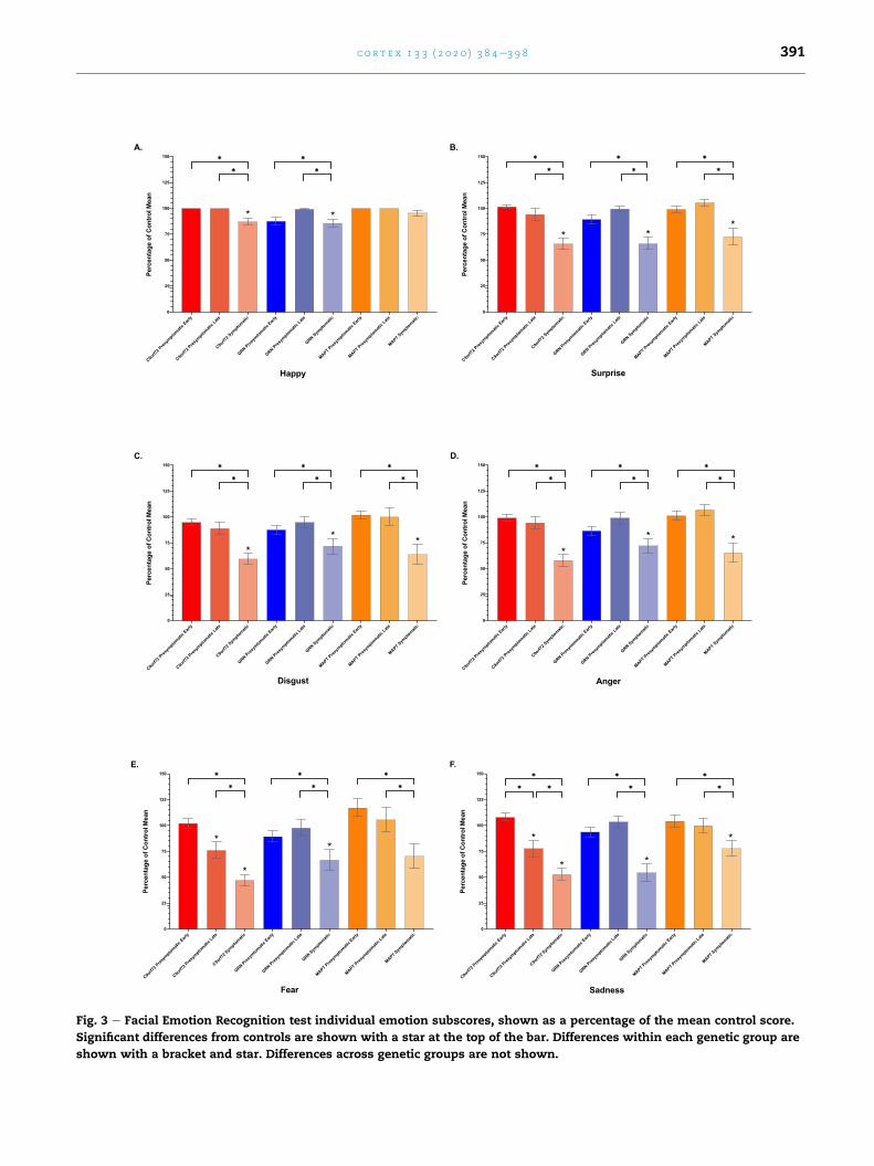
Fig. 3 e Facial Emotion Recognition test individual emotion subscores, shown as a percentage of the mean control score.
Significant differences from controls are shown with a star at the top of the bar. Differences within each genetic group are
shown with a bracket and star. Differences across genetic groups are not shown.
c o r t e x 1 3 3 ( 2 0 2 0 ) 3 8 4e3 9 8 391
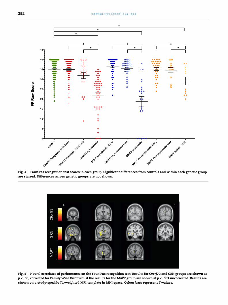
Fig. 4 e Faux Pas recognition test scores in each group. Significant differences from controls and within each genetic group
are starred. Differences across genetic groups are not shown.
Fig. 5 e Neural correlates of performance on the Faux Pas recognition test. Results for C9orf72 and GRN groups are shown at
p < .05, corrected for Family Wise Error whilst the results for the MAPT group are shown at p < .001 uncorrected. Results are
shown on a study-specific T1-weighted MRI template in MNI space. Colour bars represent T-values.
c o r t e x 1 3 3 ( 2 0 2 0 ) 3 8 4e3 9 8392
c o r t e x 1 3 3 ( 2 0 2 0 ) 3 8 4e3 9 8 393
comparing performance by sex, with females performing
significantly better than males on both tasks as previously
described (Hoffmann, Kessler, Eppel, Rukavina, & Traue, 2010;
Kessels, Montagne, Hendriks, Perrett, & de Haan, 2014; Lee
et al., 2002; Montagne, Kessels, Frigerio, de Haan, & Perrett,
2005). The results highlight the importance of adjusting for
age, sex and education in analyses, particularly for theory of
mind tasks.
Symptomatic mutation carriers in all groups performed
significantly lower than their presymptomatic counterparts
and the controls. This is in line with previous work in sporadic
FTD demonstratingworse performance in FTD comparedwith
controls using both the FER (Bertoux et al., 2014; Diehl-Schmid
et al., 2007; Kumfor, Hazelton, Rushby, Hodges,& Piguet, 2019)
and FP recognition tests (Bertoux, Funkiewiez, O'Callaghan,Dubois, & Hornberger, 2013; Funkiewiez et al., 2012).
Interestingly, there were no significant differences seen
between phenotypes, with similar performance in the bvFTD,
PPA and FTD-ALS/ALS groups on both the FER and FP recog-
nition tests, and all three phenotypic groups being signifi-
cantly worse than controls on both tasks. This is consistent
with previous reports of social cognition deficits in PPA
(Fittipaldi et al., 2019) and FTD-ALS (Savage et al., 2014) as well
as bvFTD.
Importantly, we also found a decrease in emotion pro-
cessing abilities in the late C9orf72 mutation carriers (those
within 5 years to symptom onset) when compared to controls,
the other late presymptomatic carriers and the early C9orf72
presymptomatic mutation carriers. This deficit was seen
particularly on items of fear and sadness. This finding is
consistent with other smaller studies showing subtle social
cognitive deficits prior to symptom onset in genetic FTD
(Jiskoot et al., 2016, 2018; Cheran et al., 2019). However, in prior
studies, only presymptomatic MAPT and GRN mutation car-
riers have been studied, with deficits in social cognition only
shown in MAPT but not GRN mutation carriers. The differ-
ences from our study (i.e. the lack of deficits shown in MAPT
mutation carriers) may well be accounted for by a difference
in the tests performed (in one study deficits were found in
questionnaires rather than cognitive tests: Cheran et al., 2019),
and the fact that in two of the studies, deficits were only
detected longitudinally, and approaching phenoconversion
(Jiskoot et al., 2016, 2018).
Impairment on tasks of social cognition is likely to involve
breakdown of a number of processes within the brain.
Consistent with this, previous studies of the neural correlates
of social cognition deficits in sporadic FTD have shown an
association of emotional processing difficulties with a variety
of brain regions including frontal (particularly orbitofrontal),
inferior temporal, and insula cortices as well as the amygdala
(reviewed in Kumfor and Piguet, 2012; Couto et al., 2013).
Similarly, theory of mind problems have also been associated
with atrophy within a variety of areas in the brain including
frontal cortex, temporal and insular regions (Adenzato et al.,
2010; Agustus et al., 2015; Bertoux et al., 2014; Guevara et al.,
2015). In our study, orbitofrontal cortex was fairly uniformly
affected across each of the genetic groups e this region is
known to be involved in complex social and emotional
behaviour (Kringelbach, 2004; Rolls, 2004; Beer, John, Scabini,
& Knight, 2006), particularly through a role in stimulus-
reinforcement learning and processing of reward. The insula
was similarly affected across the groups in both tasks e this
region is a core hub of the salience network which is involved
in a wide variety of social processes (Menon & Uddin, 2010;
Uddin, Nomi, Hebert-Seropian, Ghaziri, & Boucher, 2017) such
as interoception, the processing of emotional experiences and
the awareness of positive and negative feelings (Craig, 2002),
all required when trying to identify emotions and interpret
social situations. Also previously reported is the association of
the inferior and medial temporal lobe, particularly the
amygdala, with social cognition deficits in FTD, areas known
to be involved in the perception and recognition of facial
emotions e this region was associated with performance on
both the FER test (in C9orf72 andGRNmutation carriers) and FP
recognition tests (in GRN mutation carriers).
A novel finding in this study was the association of the
basal ganglia, particularly the striatum (caudate, putamen and
nucleus accumbens), with impairment of social cognition
across all of the three genetic groups and tests, except for the
C9orf72 FP recognition test performance. This region has pre-
viously been associated with emotion recognition deficits,
particularly negative emotions (Sprengelmeyer et al., 1997;
Calder et al., 2004; Kemp et al., 2013), although in one study of
emotion generation, the basal ganglia was associated with
dysregulation of producing happy emotions (Sturm et al.,
2015). Other studies of sporadic FTD have also shown an as-
sociation of the basal ganglia with performance on implicit
emotion processing tasks (Balconi et al., 2015), and empathy
measures (Rankin et al., 2006; Shdo et al., 2017). Furthermore,
neuroanatomically, the striatum is highly connected with
frontal regions, with fronto-striatal circuits implicated in the
early pathological processes in FTD (Yi et al., 2013; Sobue et al.,
2018) and atrophy in the striatum found across all genetic
subtypes of FTD (Bede et al., 2013; Rohrer et al., 2015; Cash
et al., 2018). This work therefore provides support for the
role of the basal ganglia in social cognitive abilities in genetic
FTD.
A key strength of this study is the large sample size: whilst
familial FTD is a relatively rare condition, by using data
collected as part of GENFI, it allows investigation of a larger
group of individuals with familial FTD including those in the
presymptomatic period. Despite this, some groups remain
with small sample sizes (particularlyMAPTmutation carriers);
the continuation of data collection as part of GENFIwill help to
overcome this problem. A further limitation of the study is the
use of the mean age at onset within a family to estimate the
number of years from likely symptom onset within an indi-
vidual. As shown previously within the GENFI study (Moore
et al., 2020), whilst there is a highly significant correlation
between an individual's age at symptom onset and the mean
age at symptom onset within the family in all three genetic
mutations, the correlations are lower for C9orf72 and GRN
mutation carriers, making the estimate inexact. However,
there are currently no better methods for estimating time
from likely symptom onset at present, with future studies
likely to benefit from the development of more precise mea-
sures of proximity to onset.
Given that structural neuroanatomical changes occur quite
a number of years prior to symptom onset in each of the ge-
netic groups (Rohrer et al., 2015) it may seem surprising that
c o r t e x 1 3 3 ( 2 0 2 0 ) 3 8 4e3 9 8394
social cognitive deficits were only shown in one group
(C9orf72) and in one test during the presymptomatic period.
The question then arises as to whether the current tests are
sensitive enough to detect the earliest social cognitive
changes that occur, or whether social cognition deficits would
still be found to occur only very late in the presymptomatic
period or early in the symptomatic period even with other
tasks. Further work is required to tease apart these two pos-
sibilities with the development and testing of novel social
cognitive tasks within such presymptomatic cohorts both
cross-sectionally and particularly longitudinally where one
can identify individuals who phenoconvert. Such studies
would enhance understanding of the timing and progression
of social cognitive changes within genetic FTD.
In summary, this study demonstrates that the FER and FP
recognition tests are able to identify deficits in emotion pro-
cessing and theory of mind in familial cases of FTD across the
three main genetic mutation groups, including during the late
presymptomatic period in C9orf72 mutation carriers.
Furthermore, neuroanatomical regions known to be involved
with social cognition were found to be correlated with per-
formance on the tasks, with the novel finding of basal ganglia
involvement in genetic FTD. This frontal-striatal-insula-
temporal network is highly interconnected and forms part of
a previously described social brain functional network
(Adolphs, 2002; Pessoa, 2017) which allows people to interact
with each other and learn social behaviours so that they can
follow societal norms e factors lost in people in FTD. The FER
and FP recognition tests may prove useful as cognitive
markers in future clinical trials of FTD but further work is
needed to understand the longitudinal change over time, with
further refinement of tasks tomore sensitively detect changes
in the presymptomatic period.
CRediT author statement
Lucy L. Russell: Conceptualization (supporting); Formal anal-
ysis (lead); Visualization (lead); Writing - Original Draft (lead);
Writing - Review& Editing (equal).Caroline V. Greaves: Project
administration (equal); Resources (supporting); Investigation
(equal). Martina Bocchetta: Formal analysis (supporting);
Writing - Review & Editing (equal). Jennifer Nicholas: Formal
analysis (supporting); Writing - Review & Editing (equal).
Rhian S. Convery: Data Curation (lead); Investigation (equal).
Katrina Moore: Project administration (equal); Data Curation
(Supporting); Investigation (equal); Resources (lead). David M.
Cash: Formal analysis (supporting); Writing - Review& Editing
(equal). John van Swieten: Resources (equal); Project admin-
istration (equal); Funding acquisition (equal); Investigation
(supporting); Writing - Review & Editing (supporting). Lize
Jiskoot: Resources (equal); Project administration (equal);
Funding acquisition (equal); Investigation (supporting);
Writing - Review & Editing (supporting). Fermin Moreno: Re-
sources (equal); Project administration (equal); Funding
acquisition (equal); Investigation (supporting); Writing - Re-
view & Editing (supporting). Raquel Sanchez-Valle: Resources
(equal); Project administration (equal); Funding acquisition
(equal); Investigation (supporting); Writing - Review & Editing
(supporting). Barbara Borroni: Resources (equal); Project
administration (equal); Funding acquisition (equal); Investi-
gation (supporting); Writing - Review & Editing (supporting).
Robert Laforce Jr: Resources (equal); Project administration
(equal); Funding acquisition (equal); Investigation (support-
ing); Writing - Review & Editing (supporting). Mario Masellis:
Resources (equal); Project administration (equal); Funding
acquisition (equal); Investigation (supporting); Writing - Re-
view & Editing (supporting). Maria Carmela Tartaglia: Re-
sources (equal); Project administration (equal); Funding
acquisition (equal); Investigation (supporting); Writing - Re-
view & Editing (supporting). Caroline Graff: Resources (equal);
Project administration (equal); Funding acquisition (equal);
Investigation (supporting); Writing - Review & Editing (sup-
porting). Emanuela Rotondo: Resources (equal); Project
administration (equal); Funding acquisition (equal); Investi-
gation (supporting); Writing - Review & Editing (supporting).
Daniela Galimberti: Resources (equal); Project administration
(equal); Funding acquisition (equal); Investigation (support-
ing); Writing - Review & Editing (supporting). James B Rowe:
Resources (equal); Project administration (equal); Funding
acquisition (equal); Investigation (supporting); Writing - Re-
view & Editing (supporting). Elizabeth Finger: Resources
(equal); Project administration (equal); Funding acquisition
(equal); Investigation (supporting); Writing - Review & Editing
(supporting). Matthis Synofzik: Resources (equal); Project
administration (equal); Funding acquisition (equal); Investi-
gation (supporting); Writing - Review & Editing (supporting).
Rik Vandenberghe: Resources (equal); Project administration
(equal); Funding acquisition (equal); Investigation (support-
ing); Writing - Review & Editing (supporting). Alexandre de
Mendonca: Resources (equal); Project administration (equal);
Funding acquisition (equal); Investigation (supporting);
Writing - Review & Editing (supporting). Fabrizio Tagliavini:
Resources (equal); Project administration (equal); Funding
acquisition (equal); Investigation (supporting); Writing - Re-
view& Editing (supporting). Isabel Santana: Resources (equal);
Project administration (equal); Funding acquisition (equal);
Investigation (supporting); Writing - Review & Editing (sup-
porting). Simon Ducharme: Resources (equal); Project
administration (equal); Funding acquisition (equal); Investi-
gation (supporting); Writing - Review & Editing (supporting).
Chris Butler: Resources (equal); Project administration (equal);
Funding acquisition (equal); Investigation (supporting);
Writing - Review & Editing (supporting). Alex Gerhard: Re-
sources (equal); Project administration (equal); Funding
acquisition (equal); Investigation (supporting); Writing - Re-
view & Editing (supporting). Johannes Levin: Resources
(equal); Project administration (equal); Funding acquisition
(equal); Investigation (supporting); Writing - Review & Editing
(supporting). Adrian Danek: Resources (equal); Project
administration (equal); Funding acquisition (equal); Investi-
gation (supporting); Writing - Review & Editing (supporting).
Markus Otto: Resources (equal); Project administration
(equal); Funding acquisition (equal); Investigation (support-
ing); Writing - Review & Editing (supporting). Jason D Warren:
Resources (equal); Project administration (equal); Funding
c o r t e x 1 3 3 ( 2 0 2 0 ) 3 8 4e3 9 8 395
acquisition (equal); Investigation (supporting); Writing - Re-
view & Editing (supporting). Jonathan D Rohrer: Conceptuali-
zation (lead); Supervision (lead); Formal analysis (supporting);
Writing - Review & Editing (equal); Project administration
(equal); Funding acquisition (lead).
Open practices
The study in this article earned an Open Data badge for
transparent practices. Statistical analysis from this study will
be made available on reasonable request.
Acknowledgements
We thank the research participants for their contribution to
the study. The Dementia Research Centre is supported by
Alzheimer's Research UK, Alzheimer's Society, Brain Research
UK, and TheWolfson Foundation. Thisworkwas supported by
the NIHR UCLH Biomedical Research Centre, the Leonard
Wolfson Experimental Neurology Centre (LWENC) Clinical
Research Facility, and the UK Dementia Research Institute,
which receives its funding fromUK DRI Ltd., funded by the UK
Medical Research Council, Alzheimer's Society and Alz-
heimer's Research UK. JDR is supported by an MRC Clinician
Scientist Fellowship (MR/M008525/1) and has received funding
from the NIHR Rare Disease Translational Research Collabo-
ration (BRC149/NS/MH), the Bluefield Project and the Associ-
ation for Frontotemporal Degeneration. This work was also
supported by the MRC UK GENFI grant (MR/M023664/1). MB is
supported by a Fellowship award from the Alzheimer's Soci-
ety, UK (AS-JF-19a-004-517). Several authors of this publica-
tion aremembers of the European Reference Network for Rare
Neurological Diseases - Project ID No 739510. RS-V is sup-
ported by an Alzheimer’s Research UK Clinical Research
Training Fellowship (ARUK-CRF2017B-2). JCVS was supported
by the Dioraphte Foundation grant 09-02-03-00, the Associa-
tion for Frontotemporal Dementias Research Grant 2009, The
Netherlands Organization for Scientific Research (NWO) grant
HCMI 056-13-018, ZonMw Memorabel (Deltaplan Dementie,
project number 733 051 042), Alzheimer Nederland and the
Bluefield project. CG received funding from JPND-Prefrontals
VR Dnr 529-2014-7504, VR 2015-02926 and 2018-02754, the
Swedish FTD Initiative-Sch€orling Foundation, Alzheimer
Foundation, Brain Foundation and Stockholm County Council
ALF. DG received support from the EU Joint Programme -
Neurodegenerative Disease Research (JPND) and the Italian
Ministry of Health (PreFrontALS) grant 733051042. RS-V has
received funding from Fundacio Marato de TV3, Spain (grant
no. 20143810). FM received funding from the Tau Consortium
and the Center for Networked Biomedical Research on
Neurodegenerative Disease (CIBERNED). JBR has received
funding from the Wellcome Trust (103838) and the National
Institute for Health Research (NIHR) Bambridge Biomedical
Research Centre.MOhas received funding fromBMBF (FTLDc).
MM has received funding from a Canadian Institute of Health
Research operating grant and the Weston Brain institute and
Ontario Brain Institute. RV has received funding from the
Mady Browaeys Fund for Research into Frontotemporal De-
mentia. EF has received funding from a Canadian Institute of
Health Research grant #327387.
Appendix
List of GENFI consortium authors:
Martin N. Rossor ab, Nick C. Fox ab, Ione O.C. Woollacott ab,
Rachelle Shafei ab, Carolin Heller ab,ac, Rita Guerreiro ac, Jose
Bras ac, David L. Thomas ad, Simon Mead ae, Lieke Meeter af,
Jessica Panman af, Janne Papma af, Jackie Poos af, Rick van
Minkelen ag, Yolanda Pijnenburg ah, Myriam Barandiaran ai,aj,
Bego~na Indakoetxea ai,aj, Alazne Gabilondo aj, Mikel Tainta aj,
Maria de Arriba aj, Ana Gorostidi aj, Miren Zulaica aj, Jorge
Villanua ak Zigor Diazal, Sergi Borrego-Ecija am, JaumeOlivesam,
Albert Llad�o am, Mircea Balasa am, Anna Antonell am, Nuria Bar-
gallo an, Enrico Premi ao, Maura Cosseddu ao, Stefano Gazzina ao,
Alessandro Padovani ao, RobertoGasparotti ap, SilvanaArchetti aq,
Sandra Black ar, Sara Mitchell ar, Ekaterina Rogaeva as, Morris
Freedman at, RonKeren au, Daid Tang-Wai av, Linn €Oijerstedt aw,
ChristinAnderssonax,Vesna Jelic ay,HakanThonberg az,Andrea
Arighi ba,bb, Chiara Fenoglio ba,bb, Elio Scarpini ba,bb, Giorgio
Fumagalli ba,bb,bc, Thomas Cope bd, Carolyn Timberlake bd,
Timothy Rittman bd, Christen Shoesmith be, Robart Bartha bf,bg,
Rosa Rademakers bh, Carlo Wilke bi,bj, Hans-Otto Karnarth bk,
Benjamin Bender bl, Rose Bruffaerts bm, Philip Vandamme bn,
Mathieu Vandenbulcke bo,bp, Catarina B. Ferreira bq, Gabriel
Miltenberger br, Carolina Maruta bs, Ana Verdelho bt, S�onia
Afonsobu,RicardoTaipabv,PaolaCaroppobw,GiuseppeDiFedebw,
Giorgio Giaccone bw, Cristina Muscio, bw, Sara Prioni bw, Veronica
Redaelli bw, Giacomina Rossi bw, Pietro Tiraboschi bw, Diana
Durobx, Maria R. Almeida bx, Miguel Castelo-Branco bx, Maria J.
Leit~ao by, Miguel Tabuas-Pereira bz, Beatriz Santiago bz, Serge
Gauthier ca, Pedro Rosa-Neto cb, Michele Veldsman cc, Paul
Thompson cd, Tobias Langheinrich cd, Catharina Prix ce, Tobias
Hoegen ce, Elisabeth Wlasich ce, Sandra Loosli ce, Sonja Scho-
necker ce, Elisa Semler cf, Sarah Anderl-Straub cf.
Affiliations:abDementia Research Centre, Department of Neurodegen-
erative Disease, UCL Institute of Neurology, Queen Square,
London, UK.acDementia Research Institute, Department of Neurode-
generative Disease, UCL Institute of Neurology, Queen Square,
London, UK.adNeuroimaging Analysis Centre, Department of Brain
Repair and Rehabilitation, UCL Institute of Neurology, Queen
Square, London, UK.aeMRC Prion Unit, Department of Neurodegenerative Dis-
ease, UCL Institute of Neurology, Queen Square, London, UK.afDepartment of Neurology, Erasmus Medical Centre, Rot-
terdam, Netherlands.agDepartment of Clinical Genetics, Erasmus Medical
Centre, Rotterdam, Netherlands.ahAmsterdam University Medical Centre, Amsterdam
VUmc, Amsterdam, Netherlands.aiCognitive Disorders Unit, Department of Neurology,
Donostia University Hospital, San Sebastian, Gipuzkoa, Spain.ajNeuroscience Area, Biodonostia Health Research Insitute,
San Sebastian, Gipuzkoa, Spain.
c o r t e x 1 3 3 ( 2 0 2 0 ) 3 8 4e3 9 8396
akOSATEK, University of Donostia, San Sebastian, Gipuz-
koa, Spain.alCITA Alzheimer, San Sebastian, Gipuzkoa, Spain.amAlzheimer's Disease and Other Cognitive Disorders Unit,
Neurology Service, Hospital Clınic, Barcelona, Spain.anImaging Diagnostic Center, Hospital Clınic, Barcelona,
Spain.aoCentre for Neurodegenerative Disorders, Neurology Unit,
Department of Clinical and Experimental Sciences, University
of Brescia, Brescia, Italy.apNeuroradiology Unit, University of Brescia, Brescia, Italy.aqBiotechnology Laboratory, Department of Diagnostics,
Spedali Civili Hospital, Brescia, Italy.arSunnybrook Health Sciences Centre, Sunnybrook
Research Institute, University of Toronto, Toronto, Canada.asTanz Centre for Research in Neurodegenerative Diseases,
University of Toronto, Toronto, Canada.atBaycrest Health Sciences, Rotman Research Institute,
University of Toronto, Toronto, Canada.auThe University Health Network, Toronto Rehabilitation
Institute, Toronto, Canada.avThe University Health Network, Krembil Research Insti-
tute, Toronto, Canada.awDepartment of Geriatric Medicine, Karolinska University
Hospital-Huddinge, Stockholm, Sweden.axDepartment of Clinical Neuroscience, Karolinska Insti-
tutet, Stockholm, Sweden.ayDivision of Clinical Geriatrics, Karolinska Institutet,
Stockholm, Sweden.azCenter for Alzheimer Research, Divison of Neuro-
geriatrics, Karolinska Institutet, Stockholm, Sweden.baFondazione IRCCS Ca’ Granda Ospedale Maggiore Poli-
clinico, Neurodegenerative Diseases Unit, Milan, Italy.bbUniversity of Milan, Centro Dino Ferrari, Milan, Italy.bcDepartment of Neurosciences, Psychology, Drug
Research and Child Health (NEUROFARBA), University of
Florence, Florence, Italy.bdDepartment of Clinical Neurosciences, University of
Cambridge, Cambridge, UK.beDepartment of Clinical Neurological Sciences, University
of Western Ontario, London, Ontario Canada.bfDepartment of Medical Biophysics, The University of
Western Ontario, London, Ontario, Canada.bgCentre for Functional and Metabolic Mapping, Robarts
Research Institute, The University of Western Ontario, Lon-
don, Ontario, Canada.bhDepartment of Neuroscience, Mayo Clinic, Jacksonville,
FL, USA.biDepartment of Neurodegenerative Diseases, Hertie-
Institute for Clinical Brain Research and Center of Neurology,
University of Tubingen, Tubingen, Germany.bjCenter for Neurodegenerative Diseases (DZNE), Tubingen,
Germany.bkDivision of Neuropsychology, Hertie-Institute for Clinical
Brain Research and Center of Neurology, University of
Tubingen, Tubingen, Germany.blDepartment of Diagnostic and Interventional Neuroradi-
ology, University of Tubingen, Tubingen, Germany.bmLaboratory for Cognitive Neurology, Department of
Neurosciences, KU Leuven, Leuven, Belgium.
bnNeurology Service, University Hospitals Leuven, Belgium,
Laboratory for Neurobiology, VIB-KU Leuven Centre for Brain
Research, Leuven, Belgium.boGeriatric Psychiatry Service, University Hospitals Leuven,
Belgium.bpNeuropsychiatry, Department of Neurosciences, KU
Leuven, Leuven, Belgium.bqLaboratory of Neurosciences, Institute of Molecular
Medicine, Faculty of Medicine, University of Lisbon, Lisbon,
Portugal.brFaculty of Medicine, University of Lisbon, Lisbon,
Portugal.bsLaboratory of Language Research, Centro de Estudos Egas
Moniz, Faculty of Medicine, University of Lisbon, Lisbon,
Portugal.btDepartment of Neurosciences and Mental Health, Centro
Hospitalar Lisboa Norte - Hospital de Santa Maria & Faculty of
Medicine, University of Lisbon, Lisbon, Portugal.buInstituto Ciencias Nucleares Aplicadas a Saude, Uni-
versidade de Coimbra, Coimbra, Portugal.bvNeuropathology Unit and Department of Neurology,
Centro Hospitalar do Porto - Hospital de Santo Ant�onio,
Oporto, Portugal.bwFondazione IRCCS Istituto Neurologico Carlo Besta,
Milano, Italy.bxFaculty of Medicine, University of Coimbra, Coimbra,
Portugal.byCentre of Neurosciences and Cell Biology, Universidade
de Coimbra, Coimbra, Portugal.bzNeurology Department, Centro Hospitalar e Universitario
de Coimbra, Coimbra, Portugal.caAlzheimer Disease Research Unit, McGill Centre for
Studies in Aging, Department of Neurology & Neurosurgery,
McGill University, Montreal, Qu�ebec, Canada.cbTranslational Neuroimaging Laboratory, McGill Centre
for Studies in Aging, McGill University, Montreal, Qu�ebec,
Canada.ccNuffield Department of Clinical Neurosciences, Medical
Sciences Division, University of Oxford, Oxford, UK.cdFaculty of Biology, Medicine and Health, Division of
Neuroscience and Experimental Psychology, University of
Manchester, Manchester, UK.ceNeurologische Klinik, Ludwig-Maximilians-Universit€at
Munchen, Munich, Germany.cfDepartment of Neurology, University of Ulm, Ulm,
Germany.
Supplementary data
Supplementary data to this article can be found online at
https://doi.org/10.1016/j.cortex.2020.08.023.
r e f e r e n c e s
Adenzato, M., Cavallo, M., & Enrici, I. (2010). Theory of mindability in the behavioural variant of frontotemporal dementia:An analysis of the neural, cognitive, and social levels.Neuropsychologia, 48(1), 2e12.
c o r t e x 1 3 3 ( 2 0 2 0 ) 3 8 4e3 9 8 397
Adolphs, R. (2002). Neural systems for recognizing emotion.Current opinion in neurobiology, 12(2), 169e177.
Adolphs, R. (2009). The social brain: neural basis of socialknowledge. Annual Review of Psychology, 60, 693e716.
Agustus, J. L., Mahoney, C. J., Downey, L. E., Omar, R., Cohen, M.,White, M. J., & Warren, J. D. (2015). Functional MRI of musicemotion processing in frontotemporal dementia. Annals ofNew York Academy Science, 1337(1), 232e240.
Ashburner, J. (2007). A fast diffeomorphic image registrationalgorithm. Neuroimage, 38(1), 95e113.
Balconi, M., Cotelli, M., Brambilla, M., Manenti, R., Cosseddu, M.,Premi, E., & Borroni, B. (2015). Understanding emotions infrontotemporal dementia: The explicit and implicit emotionalcue mismatch. Journal of Alzheimer's Disease, 46(1), 211e225.
Bede, P., Elamin, M., Byrne, S., McLaughlin, R. L., Kenna, K.,Vajda, A., et al. (2013 Dec 10). Basal ganglia involvement inamyotrophic lateral sclerosis. Neurology, 81(24), 2107e2115.
Beer, J. S., John, O. P., Scabini, D., & Knight, R. T. (2006).Orbitofrontal cortex and social behavior: Integrating self-monitoring and emotion-cognition interactions. Journal ofCognitive Neuroscience, 18(6), 871e879.
Bertoux, M., Delavest, M., de Souza, L. C., Funkiewiez, A.,Lepine, J.-P., Fossati, P., & Sarazin, M. (2012). Social Cognitionand Emotional Assessment differentiates frontotemporaldementia from depression. Journal of Neurology, Neurosurgeryand Psychiatry, 83(4), 411e416.
Bertoux, M., Funkiewiez, A., O'Callaghan, C., Dubois, B., &Hornberger, M. (2013). Sensitivity and specificity ofventromedial prefrontal cortex tests in behavioral variantfrontotemporal dementia. Alzheimer's & Dementia: the Journal ofthe Alzheimer's Association, 9(5 Suppl), S84eS94.
Bertoux, M., Volle, E., De Souza, L., Funkiewiez, A., Dubois, B., &Habert, M. (2014). Neural correlates of the mini-SEA (Socialcognition and Emotional Assessment) in behavioral variantfrontotemporal dementia. Brain Imaging and Behavior, 8(1),1e6.
Calder, A. J., Keane, J., Lawrence, A. D., & Manes, F. (2004 Sep).Impaired recognition of anger following damage to the ventralstriatum. Brain: a Journal of Neurology, 127(Pt 9), 1958e1969.
Cash, D. M., Bocchetta, M., Thomas, D. L., Dick, K. M., vanSwieten, J. C., Borroni, B., & Rowe, J. B. (2018). Patterns of graymatter atrophy in genetic frontotemporal dementia: Resultsfrom the GENFI study. Neurobiology of Aging, 62, 191e196.
Cheran, G., Wu, L., Lee, S., Manoochehri, M., Cines, S., Fallon, E.,et al. (2019 Feb). Cognitive indicators of preclinical behaviouralvariant frontotemporal dementia in MAPT carriers. Journal ofthe International Neuropsychological Society: JINS, 25(2), 184e194.
Couto, B., Manes, F., Montanes, P., Matallana, D., Reyes, P.,Velasquez, M., & Ibanez, A. (2013). Structural neuroimaging ofsocial cognition in progressive non-fluent aphasia andbehavioral variant of frontotemporal dementia. Frontiers inHuman Neuroscience, 7, 467.
Craig, A. D. (2002). How do you feel? Interoception: The sense ofthe physiological condition of the body. Nature ReviewsNeuroscience, 3(8), 655e666.
Diehl-Schmid, J., Pohl, C., Ruprecht, C., Wagenpfeil, S., Foerstl, H.,& Kurz, A. (2007). The Ekman 60 Faces Test as a diagnosticinstrument in frontotemporal dementia. Archives of ClinicalNeuropsychology, 22(4), 459e464.
Ekman, P., Ellsworth, P., Friesen, W. V., Goldstein, A. P., &Krasner, L. (1972). Emotion in the human face.
Fittipaldi, S., Ibanez, A., Baez, S., Manes, F., Sedeno, L., &Garcia, A. M. (2019). More than words: Social cognition acrossvariants of primary progressive aphasia. Neuroscience andBiobehavioral Reviews, 100, 263e284.
Funkiewiez, A., Bertoux, M., de Souza, L. C., L�evy, R., & Dubois, B.(2012). The SEA (social cognition and emotional assessment):A clinical neuropsychological tool for early diagnosis of frontal
variant of frontotemporal lobar degeneration. Neuropsychology,26(1), 81.
Gregory, C., Lough, S., Stone, V., Erzinclioglu, S., Martin, L., Baron-Cohen, S., et al. (2002 Apr). Theory of mind in patients withfrontal variant frontotemporal dementia and Alzheimer'sdisease: Theoretical and practical implications. Brain: a Journalof Neurology, 125(Pt 4), 752e764.
Guevara, A. B., Knutson, K. M., Wassermann, E. M., Pulaski, S.,Grafman, J., & Krueger, F. (2015). Theory of mind impairmentin patients with behavioural variant fronto-temporaldementia (bv-FTD) increases caregiver burden. Age and Ageing,44(5), 891e895.
Hoffmann, H., Kessler, H., Eppel, T., Rukavina, S., & Traue, H. C.(2010). Expression intensity, gender and facial emotionrecognition: Women recognize only subtle facial emotionsbetter than men. Acta psychologica, 135(3), 278e283.
Jiskoot, L. C., Dopper, E. G., Heijer, T., Timman, R., vanMinkelen, R., van Swieten, J. C., et al. (2016). Presymptomaticcognitive decline in familial frontotemporal dementia: Alongitudinal study. Neurology, 87(4), 384e391.
Jiskoot, L.C.,Panman, J. L., vanAsseldonk,L., Franzen,S.,Meeter,L.H.,Kaat, L. D., & vanMinkelen, R. (2018). Longitudinal cognitivebiomarkers predicting symptom onset in presymptomaticfrontotemporal dementia. Journal of Neurology, 1e12.
Kemp, J., Berthel, M.-C., Dufour, A., Despres, O., Henry, A.,Namer, I. J., & Sellal, F. (2013). Caudate nucleus and socialcognition: Neuropsychological and SPECT evidence from apatient with focal caudate lesion. Cortex; a Journal Devoted Tothe Study of the Nervous System and Behavior, 49(2), 559e571.
Kessels, R. P., Montagne, B., Hendriks, A. W., Perrett, D. I., & deHaan, E. H. (2014). Assessment of perception of morphed facialexpressions using the Emotion Recognition Task: Normativedata from healthy participants aged 8e75. Journal ofNeuropsychology, 8(1), 75e93.
Kringelbach, M. (2004). The functional neuroanatomy of thehuman orbitofrontal cortex: Evidence from neuroimaging andneuropsychology. Progress in Neurobiology, 72(5), 341e372.
Kumfor, F., Hazelton, J. L., Rushby, J. A., Hodges, J. R., & Piguet, O.(2019). Facial expressiveness and physiological arousal infrontotemporal dementia: Phenotypic clinical profiles andneural correlates. Cognitive, Affective & Behavioral Neuroscience,19(1), 197e210, 2019 Feb.
Kumfor, F., & Piguet, O. (2012). Disturbance of emotion processingin frontotemporal dementia: A synthesis of cognitive andneuroimaging findings. Neuropsychology Review, 22(3), 280e297.
Lee, T. M. C., Liu, H.-L., Hoosain, R., Liao, W.-T., Wu, C.-T.,Yuen, K. S. L., & Gao, J.-H. (2002). Gender differences in neuralcorrelates of recognition of happy and sad faces in humansassessed by functional magnetic resonance imaging.Neuroscience Letters, 333(1), 13e16.
Lough, S., & Hodges, J. R. (2002 Aug). Measuring and modifyingabnormal social cognition in frontal variant frontotemporaldementia. Journal of Psychosomatic Research, 53(2), 639e646.
Malone, I. B., Leung, K. K., Clegg, S., Barnes, J., Whitwell, J. L.,Ashburner, J., & Ridgway, G. R. (2015). Accurate automaticestimation of total intracranial volume: A nuisance variablewith less nuisance. Neuroimage, 104, 366e372.
Maylor, E. A., Moulson, J. M., Muncer, A. M., & Taylor, L. A. (2002).Does performance on theory of mind tasks decline in old age?British Journal of Psychology, 93(4), 465e485.
Menon, V., & Uddin, L. Q. (2010). Saliency, switching, attentionand control: A network model of insula function. BrainStructure & Function, 214(5e6), 655e667.
Mill, A., Allik, J., Realo, A., & Valk, R. (2009). Age-relateddifferences in emotion recognition ability: A cross-sectionalstudy. Emotion, 9(5), 619.
Montagne, B., Kessels, R. P., Frigerio, E., de Haan, E. H., &Perrett, D. I. (2005). Sex differences in the perception of
c o r t e x 1 3 3 ( 2 0 2 0 ) 3 8 4e3 9 8398
affective facial expressions: Do men really lack emotionalsensitivity? Cognitive Processing, 6(2), 136e141.
Moore, K. M., Nicholas, J., Grossman, M., McMillan, C. T.,Irwin, D. J., Massimo, L., et al., FTD Prevention Initiative.(2020). Age at symptom onset and death and disease durationin genetic frontotemporal dementia: An internationalretrospective cohort study. Lancet Neurology, 19(2), 145e156.
Narme, P., Mouras, H., Roussel, M., Devendeville, A., &Godefroy, O. (2013). Assessment of socioemotional processesfacilitates the distinction between frontotemporal lobardegeneration and Alzheimer's disease. Journal of ClinicalExperimental Neuropsychology, 35(7), 728e744.
Omar, R., Rohrer, J. D., Hailstone, J. C., & Warren, J. D. (2011).Structural neuroanatomy of face processing in frontotemporallobar degeneration. Neurologia I Neurochirurgia Polska, 82(12),1341e1343.
Pardini, M., & Nichelli, P. F. (2009). Age-related decline inmentalizing skills across adult life span. Experimental AgingResearch, 35(1), 98e106.
Pessoa, L. (2017). A network model of the emotional brain. Trendsin cognitive sciences, 21(5), 357e371.
Rankin, K. P., Gorno-Tempini, M. L., Allison, S. C., Stanley, C. M.,Glenn, S., Weiner, M. W., et al. (2006). Structural anatomy ofempathy in neurodegenerative disease. Brain: a Journal ofNeurology, 129(11), 2945e2956.
Ridgway, G. R., Omar, R., Ourselin, S., Hill, D. L., Warren, J. D., &Fox, N. C. (2009). Issues with threshold masking in voxel-basedmorphometry of atrophied brains. Neuroimage, 44(1), 99e111.
Rohrer, J., Nicholas, J. M., Cash, D. M., van Swieten, J., Dopper, E.,Jiskoot, L., & Clegg, S. (2015). Presymptomatic cognitive andneuroanatomical changes in genetic frontotemporal dementiain the genetic frontotemporal dementia initiative (GENFI)study: A cross-sectional analysis. Lancet Neurology, 14(3),253e262.
Rolls, E. T. (2004). The functions of the orbitofrontal cortex. Brainand Cognition, 55(1), 11e29.
Rosen, H. J., Wilson, M. R., Schauer, G. F., Allison, S., Gorno-Tempini, M. L., Pace-Savitsky, C., et al. (2006).Neuroanatomical correlates of impaired recognition ofemotion in dementia. Neuropsychologia, 44(3), 365e373.
Savage, S. A., Lillo, P., Kumfor, F., Kiernan, M. C., Piguet, O., &Hodges, J. R. (2014). Emotion processing deficits distinguishpure amyotrophic lateral sclerosis from frontotemporaldementia. Amyotrophic Lateral Sclerosis & FrontotemporalDegeneration, 15(1e2), 39e46.
Shdo, S. M., Ranasinghe, K. G., Gola, K. A., Mielke, C. J.,Sukhanov, P. V., Miller, B. L., et al. (2017). Deconstructingempathy: Neuroanatomical dissociations between affectsharing and prosocial motivation using a patient lesion model.Neuropsychologia, 116, 126e135.
Sobue, G., Ishigaki, S., & Watanabe, H. (2018 Jul 12). Pathogenesisof frontotemporal lobar degeneration: Insights from loss offunction theory and early involvement of the caudate nucleus.The Florida Nurse, 12, 473.
Sprengelmeyer, R., Young, A., Pundt, I., Sprengelmeyer, A.,Calder, A., Berrios, G., & Sartory, G. (1997). Disgust implicatedin obsessiveecompulsive disorder. Proceedings of the RoyalSociety of London. Series B: Biological Sciences, 264(1389),1767e1773.
Sturm, V. E., Yokoyama, J. S., Eckart, J. A., Zakrzewski, J.,Rosen, H. J., Miller, B. L., et al. (2015 Mar). Damage to leftfrontal regulatory circuits produces greater positive emotionalreactivity in frontotemporal dementia. Cortex; a Journal DevotedTo the Study of the Nervous System and Behavior, 64, 55e67.
Sullivan, S., Ruffman, T., & Hutton, S. B. (2007). Agedifferences in emotion recognition skills and the visualscanning of emotion faces. The Journals of Gerontology: Series B,62(1), P53eP60.
Torralva, T., Gleichgerrcht, E., Torres Ardila, M. J., Roca, M., &Manes, F. F. (2015). Differential cognitive and affective theoryof mind abilities at mild and moderate stages of behavioralvariant frontotemporal dementia. Cognitive and BehavioralNeurology: Official Journal of the Society for Behavioral andCognitive Neurology, 28(2), 63e70.
Uddin, L. Q., Nomi, J. S., Hebert-Seropian, B., Ghaziri, J., &Boucher, O. (2017). Structure and function of the humaninsula. Journal of clinical neurophysiology: official publication of theAmerican Electroencephalographic Society, 34(4), 300.
Wang, Y., & Su, Y. (2006). Theory of mind in old adults: Theperformance on Happ�e’s stories and faux pas stories.Psychologia, 49(4), 228e237.
West, J. T., Horning, S. M., Klebe, K. J., Foster, S. M., Cornwell, R. E.,Perrett, D., & Davis, H. P. (2012). Age effects on emotionrecognition in facial displays: From 20 to 89 years of age.Experimental aging research, 38(2), 146e168.
Yi, D. S., Bertoux, M., Mioshi, E., Hodges, J. R., & Hornberger, M.(2013). Fronto-striatal atrophy correlates ofneuropsychiatric dysfunction in frontotemporal dementia(FTD) and Alzheimer's disease (AD). Dement Neuropsychol, 7(1),75e82.
Related Documents