SLEEP, Vol. 30, No. 11, 2007 1577 INTRODUCTION ASPERGER SYNDROME (AS) AND HIGH-FUNCTION- ING AUTISM (HFA) ARE PERVASIVE DEVELOPMENTAL DISORDERS (PDD) CHARACTERIZED BY NORMAL intel- ligence, social deficits, rigid ritualistic behaviors, interests, or activities, and communication problems, primarily in HFA. As- perger syndrome differs from high-functioning autism because of the lack of clinically significant cognitive or language delay. 1 Several studies have shown that sleep disorders are common in children and adults with different forms of PDD, i.e. AS, HFA, or low-functioning autism (LFA). 2-12 Children with LFA show a high rate of sleep problems in respect to other groups of children with intellectual disabilities, mainly represented by difficulty in falling asleep, restless sleep, frequent awakenings, and reduced total sleep time during the night. 2-5 Similarly, in subjects with AS or HFA, questionnaire and/or actigraphic studies reported a high frequency of insomnia and other sleep problems associated with daytime sleepiness. 2,4,6,7,13 However, some studies showed that children with AS present sleep disturbances (difficulties in initiating and maintaining sleep, shorter sleep duration, earlier morning awakenings, and paras- omnias) more frequently than controls and autistic children. 2,8-11 Conversely, a recent study including children with AS and low- functioning autism confirmed the presence of sleep disturbances like bedtime resistance, insomnia, parasomnias, sleep disordered breathing, morning awakening problems, and daytime sleepiness but also showed that the type of PDD was not significantly related to sleep problems. 14 Reduced total sleep time has been the only constant finding in polysomnographic (PSG) studies of PDD children 15-16 ; a re- cent study failed to demonstrate PSG abnormalities and excluded the presence of sleep apnea, parasomnias, and sleep-related sei- zures. 16 Recently, we evaluated sleep microstructure in children with autistic spectrum disorders by means of the cyclic alternating pattern (CAP) and showed the presence of subtle alterations of NREM sleep: CAP rate during slow wave sleep (SWS) was lower than that of normal controls, mainly caused by to a reduction of A1 CAP subtypes. 15 Very few studies have evaluated the sleep structure of adults with AS, and even fewer have been performed in children. 17-19 Godbout et al. 17 found a decreased sleep time in the first two- thirds of the night, increased number of shifts into REM sleep from a waking epoch, signs of REM sleep disruption, and an in- crease of K complexes in stage 2. The study by Limoges et al. 19 represented the first evaluation of sleep patterns in a group comprising solely adults with AS/HFA and without any psychiatric or neurological comorbidity. They Sleep Architecture and NREM Alterations in Children and Adolescents with Asperger Syndrome Oliviero Bruni, MD 1 ; Raffaele Ferri, MD 2 ; Elena Vittori, MD 1 ; Luana Novelli, PhD 1 ; Manuela Vignati, MD 3 ; Maria C. Porfirio, MD 3 ; Debora Aricò, PsyD 2 ; Paola Bernabei, MD 1 ; Paolo Curatolo, MD 3 1 Center for Pediatric Sleep Disorders, Department of Developmental Neurology and Psychiatry, University “La Sapienza,” Rome, Italy; 2 Sleep Research Centre, Department of Neurology, Oasi Institute for Research on Mental Retardation and Brain Aging (IRCCS), Troina, Italy; 3 Department of Neurosciences, Child Neurology and Psychiatry Unit, University Tor Vergata, Rome, Italy Sleep in Asperger Syndrome—Bruni et al Disclosure Statement This is not an industry supported study. The authors have indicated no finan- cial conflicts of interest. Submitted for publication March, 2007 Accepted for publication June, 2007 Address correspondence to: Oliviero Bruni, MD, Center for Pediatric Sleep Disorders, Department of Developmental Neurology and Psychiatry Univer- sity of Rome “La Sapienza” Via dei Sabelli 108 - 00185 – Rome, Italy; Tel: 39 0644712257; Fax: 39 06 4957857; E-mail: [email protected] SLEEP IN ASPERGER SYNDROME Study Objectives: To analyze sleep in children with Asperger syndrome (AS) by means of standard sleep questionnaires, to evaluate sleep archi- tecture and NREM sleep alterations by means of cyclic alternating pattern (CAP) and to correlate objective sleep parameters with cognitive behav- ioral measures. Design: Cross-sectional study involving validated sleep questionnaires, neuropsychological scales, and PSG recording. Setting: Sleep medicine center. Participants: Eight children with AS, 10 children with autism, and 12 healthy control children. Interventions: N/A Measurements and Results: Children with AS had a higher prevalence of problems of initiating sleep and daytime sleepiness. Sleep architecture parameters showed minor differences between the 3 groups. CAP param- eters showed an increased percentage of A1 and a decreased percent- age of A2 subtypes in subjects with AS vs. controls. All A subtype indexes (number per hour of NREM sleep) were decreased, mostly in sleep stage 2 but not in SWS. With respect to children with autism, subjects with AS showed increased CAP rate in SWS and A1 percentage. In subjects with AS, verbal IQ had a significant positive correlation with total CAP rate and CAP rate in SWS and with global and SWS A1 index. The percentage of A2 negatively correlated with full scale IQ, verbal and performance IQ. CBCL total score correlated positively with CAP rate and A1 index while externalizing score correlated negatively with A3%. Conclusions: This study shows peculiar CAP modifications in children with AS and represents an attempt to correlate the quantification of sleep EEG oscillations with the degree of mental ability/disability. Keywords: Asperger syndrome, autism, sleepiness, polysomnography, child behavior checklist, autism diagnostic observation schedule, cyclic alternating pattern Citation: Bruni O; Ferri R; Vittori E; Novelli L; Vignati M; Porfirio MC; Aricò D; Bernabei P; Curatolo P. Sleep architecture and NREM al- terations in children and adolescents with asperger syndrome. SLEEP 2007;30(11):1577-1585.

Welcome message from author
This document is posted to help you gain knowledge. Please leave a comment to let me know what you think about it! Share it to your friends and learn new things together.
Transcript
SLEEP, Vol. 30, No. 11, 2007 1577
INTRODUCTION
ASPERGER SYNDROME (AS) AND HIGH-FUNCTION-ING AUTISM (HFA) ARE PERVASIVE DEVELOPMENTAL DISORDERS (PDD) CHARACTERIZED BY NORMAL intel-ligence, social deficits, rigid ritualistic behaviors, interests, or activities, and communication problems, primarily in HFA. As-perger syndrome differs from high-functioning autism because of the lack of clinically significant cognitive or language delay.1
Several studies have shown that sleep disorders are common in children and adults with different forms of PDD, i.e. AS, HFA, or low-functioning autism (LFA).2-12 Children with LFA show a high rate of sleep problems in respect to other groups of children with intellectual disabilities, mainly represented by difficulty in falling asleep, restless sleep, frequent awakenings, and reduced total sleep time during the night.2-5 Similarly, in subjects with AS or HFA, questionnaire and/or actigraphic studies reported a high frequency of insomnia and other sleep problems associated with daytime sleepiness.2,4,6,7,13
However, some studies showed that children with AS present sleep disturbances (difficulties in initiating and maintaining sleep, shorter sleep duration, earlier morning awakenings, and paras-omnias) more frequently than controls and autistic children.2,8-11 Conversely, a recent study including children with AS and low-functioning autism confirmed the presence of sleep disturbances like bedtime resistance, insomnia, parasomnias, sleep disordered breathing, morning awakening problems, and daytime sleepiness but also showed that the type of PDD was not significantly related to sleep problems.14
Reduced total sleep time has been the only constant finding in polysomnographic (PSG) studies of PDD children15-16; a re-cent study failed to demonstrate PSG abnormalities and excluded the presence of sleep apnea, parasomnias, and sleep-related sei-zures.16
Recently, we evaluated sleep microstructure in children with autistic spectrum disorders by means of the cyclic alternating pattern (CAP) and showed the presence of subtle alterations of NREM sleep: CAP rate during slow wave sleep (SWS) was lower than that of normal controls, mainly caused by to a reduction of A1 CAP subtypes.15
Very few studies have evaluated the sleep structure of adults with AS, and even fewer have been performed in children.17-19 Godbout et al.17 found a decreased sleep time in the first two-thirds of the night, increased number of shifts into REM sleep from a waking epoch, signs of REM sleep disruption, and an in-crease of K complexes in stage 2.
The study by Limoges et al.19 represented the first evaluation of sleep patterns in a group comprising solely adults with AS/HFA and without any psychiatric or neurological comorbidity. They
Sleep Architecture and NREM Alterations in Children and Adolescents with Asperger SyndromeOliviero Bruni, MD1; Raffaele Ferri, MD2; Elena Vittori, MD1; Luana Novelli, PhD1; Manuela Vignati, MD3; Maria C. Porfirio, MD3; Debora Aricò, PsyD2; Paola Bernabei, MD1; Paolo Curatolo, MD3
1Center for Pediatric Sleep Disorders, Department of Developmental Neurology and Psychiatry, University “La Sapienza,” Rome, Italy; 2Sleep Research Centre, Department of Neurology, Oasi Institute for Research on Mental Retardation and Brain Aging (IRCCS), Troina, Italy; 3Department of Neurosciences, Child Neurology and Psychiatry Unit, University Tor Vergata, Rome, Italy
Sleep in Asperger Syndrome—Bruni et al
Disclosure StatementThis is not an industry supported study. The authors have indicated no finan-cial conflicts of interest.
Submitted for publication March, 2007Accepted for publication June, 2007Address correspondence to: Oliviero Bruni, MD, Center for Pediatric Sleep Disorders, Department of Developmental Neurology and Psychiatry Univer-sity of Rome “La Sapienza” Via dei Sabelli 108 - 00185 – Rome, Italy; Tel: 39 0644712257; Fax: 39 06 4957857; E-mail: [email protected]
SLEEP IN ASPERGER SYNDROME
Study Objectives: To analyze sleep in children with Asperger syndrome (AS) by means of standard sleep questionnaires, to evaluate sleep archi-tecture and NREM sleep alterations by means of cyclic alternating pattern (CAP) and to correlate objective sleep parameters with cognitive behav-ioral measures.Design: Cross-sectional study involving validated sleep questionnaires, neuropsychological scales, and PSG recording.Setting: Sleep medicine center. Participants: Eight children with AS, 10 children with autism, and 12 healthy control children. Interventions: N/AMeasurements and Results: Children with AS had a higher prevalence of problems of initiating sleep and daytime sleepiness. Sleep architecture parameters showed minor differences between the 3 groups. CAP param-eters showed an increased percentage of A1 and a decreased percent-age of A2 subtypes in subjects with AS vs. controls. All A subtype indexes (number per hour of NREM sleep) were decreased, mostly in sleep stage
2 but not in SWS. With respect to children with autism, subjects with AS showed increased CAP rate in SWS and A1 percentage. In subjects with AS, verbal IQ had a significant positive correlation with total CAP rate and CAP rate in SWS and with global and SWS A1 index. The percentage of A2 negatively correlated with full scale IQ, verbal and performance IQ. CBCL total score correlated positively with CAP rate and A1 index while externalizing score correlated negatively with A3%. Conclusions: This study shows peculiar CAP modifications in children with AS and represents an attempt to correlate the quantification of sleep EEG oscillations with the degree of mental ability/disability.Keywords: Asperger syndrome, autism, sleepiness, polysomnography, child behavior checklist, autism diagnostic observation schedule, cyclic alternating patternCitation: Bruni O; Ferri R; Vittori E; Novelli L; Vignati M; Porfirio MC; Aricò D; Bernabei P; Curatolo P. Sleep architecture and NREM al-terations in children and adolescents with asperger syndrome. SLEEP 2007;30(11):1577-1585.
SLEEP, Vol. 30, No. 11, 2007 1578
did not find differences in sleep structure between AS and HFA, apart from the density of EEG sleep spindles, which was signifi-cantly reduced in individuals with AS. Finally a PSG study on 20 young adults with AS showed only a non significant trend towards decreased relative delta power and increased theta power in slow wave sleep in the AS group.18
Since very few studies tried to analyze sleep structure in chil-dren with AS, the aim of our investigation was to analyze sleep in children with AS by means of standard sleep questionnaires20-21
and to evaluate sleep architecture and NREM sleep microstruc-ture by means of CAP. 22
METHODS
Participants
For the purpose of this study, we recruited 8 subjects with AS, 10 with autism, and 12 typically developing children.
Children with Asperger Syndrome
Patients with AS were recruited at the Child Neuropsychiatry Clinic of the University of Rome “Tor Vergata” and at the Depart-ment of Developmental Neurology and Psychiatry of the University of Rome “La Sapienza.” Upon approval by the Ethics Committee, the parents of first 10 patients formally diagnosed with AS were asked to have their children’s sleep recorded at the sleep laboratory. The parents of 8 out of these 10 patients with AS accepted. Thus, the AS group consisted of 8 children aged 12.7±2.6 years (range 7-15), 7 of whom were boys. All children had a clinical diagnosis of AS; they had no history of serious physical health problems, no history of epileptic seizures or mental retardation, and did not cur-rently take medication. Moreover, no subjects with AS were obese, and the parents did not report snoring at the time of the study. All of them were registered at the Asperger Group Association that con-nects the families of children affected by AS in Italy.
All parents were interviewed in order to be sure that their children fulfilled the ICD-10 diagnostic criteria for Asperger syndrome23:
a) No clinically significant general delay in spoken or receptive language or cognitive development;
b) Qualitative abnormalities in reciprocal social interaction (cri-teria as for autism);
c) Unusually intense, circumscribed interest or restricted, re-petitive, and stereotyped patterns of behavior, interests, and activities (criteria as for autism)
The clinical diagnosis of AS was based on comprehensive multidisciplinary assessments (including neuropsychiatric ex-amination, speech and communication testing, and neuropsy-chological testing, parent interviews, and child observation) and were verified using the Autism Diagnostic Observation Schedule (ADOS), a semi-structured, standardized assessment of commu-nication, social interaction and play for individuals with autism spectrum disorders.24
Children with Autism
A total of 10 children affected by autism (9 males, 1 female; mean age 11.9±2.5 years, age range 7-15) attending the Depart-
ment of Developmental Neurology and Psychiatry were recruited for this study. The diagnosis of autism was made according to the DSM-IV criteria for autistic disorder1 and a score on the Child-hood Autism Rating Scale25 (CARS) >30. CARS is one of the most widely used scales to evaluate the degree and profiles of autism in children and to distinguish them from developmentally handicapped children who are not autistic. CARS consists of 15 items (relationships with people, imitation, affect, use of body, re-lation to non-human objects, adaptation to environmental change, visual responsiveness, auditory responsiveness, near receptor re-sponsiveness, anxiety reaction, verbal communication, non-ver-bal communication, activity level, intellectual functioning, and general impressions), and each item scores from 1.0 (normal) to 4.0 (severely abnormal) in units of 0.5. The total CARS score is obtained by summing up each item score and thus ranges from 15.0 to 60.0.
All patients were also mentally retarded and were drug-free from at least 2 weeks before the study began. All subjects were evaluated from the neuroimaging (including brain computed to-mography scans or magnetic resonance imaging) and the neuro-physiological points of view (EEG) and showed no neurological focal signs, seizures or paroxysmal EEG abnormalities. This al-lowed us to rule out the possibility of gross neurological impair-ment. No other known medical conditions were associated with autism in these patients; specifically, craniofacial abnormalities, fragile-X syndrome or other chromosome abnormalities, phe-nylketonuria or other metabolic diseases, neurofibromatosis, and tuberous sclerosis were excluded. No subjects with autism in this study were obese, and the parents did not report snoring at the time of the study. The intelligence level was measured by means of the Wechsler Intelligence Scale for Children Revised. Seven children had moderate mental retardation (corresponding to I.Q. in the range 40−55) and 3 had severe mental retardation (I.Q. in the range 25−40). All children with autism had a CARS score higher than 30 (range 33−50).
Informed consent was obtained by the parents of all partici-pants to the study. All children were living at home.
Control Children
The PSG data of the 2 patient groups were compared with those of a control group formed by 12 children matched for age (5 females and 7 males, mean age 12.6±3.7 years, range 7-15 years). All subjects had no anamnesis of neuropsychiatric disorders, no current axis I or axis II psychiatric disorders, and no complaints of sleep problem.
Measures
Sleep-Related Subjective Measures in Subjects with AS
Sleep Questionnaire
A questionnaire arranged for the study of sleep characteristics of children and adolescents20 was filled out by the parents. The sleep questionnaire consisted of 45 items in a Likert-type scale with values 1–5 (1 = never; 2 = occasionally; 3 = often; 4 = very often; 5 = always) to measure individual differences in several areas of sleep disorders (disorders of initiating and maintaining sleep, sleep-wake transition disorders, other parasomnias, night
Sleep in Asperger Syndrome—Bruni et al
SLEEP, Vol. 30, No. 11, 2007 1579
waking, movement disorders during sleep, sleep breathing prob-lems, excessive daytime somnolence, and morning symptoms). We considered as cut-off value a score of 3 or higher. The ques-tionnaire assessed sleep behaviors and disorders observed during the last 6 months of the subject’s life and the mothers of the chil-dren with AS filled out the questionnaire.
Pediatric Daytime Sleepiness Scale
Since sleepiness is reported to be a common complaint in children with AS, we administered the Pediatric Daytime Sleepiness Scale (PDSS)21 to them. The PDSS is an 8-item questionnaire for evaluat-ing the relationship between daytime sleepiness and school-related outcomes. Each question is rated on a 4-point scale (generally ex-pressed as 0= never, 1 = seldom, 2 = sometimes, 3 = frequently, 4 = always), with higher scores indicating more sleepiness. The PDSS was completed by the parent or legal guardian of the patient; scores were considered in the pathological range if above 16.
Behavioral/cognitive Measures in Subjects with AS
Autism Diagnostic Observation Schedule
Children with a clinical diagnosis of AS were given the Autism Diagnostic Observation Schedule (ADOS) to validate the diagno-sis. The ADOS is a semi-structured, standardized assessment of communication, social interaction, and play or imaginative use of materials for individuals who have been referred because of possible autism spectrum disorders.24 A classification of AS was determined using algorithm items and specified cutoffs. ADOS inclusion criteria were a score above the cut-off point in the 3 rel-evant areas: ADOS A: language and communication (cut-off for autism=3; cut-off for autistic spectrum =2); ADOS B: social in-teractions (cut-off for autism=6; cut-off for autistic spectrum =4); ADOS A+B (cut-off for autism=10; cut-off for autistic spectrum =7) and an absence of delay for language and of language abnor-malities typical of autism (echolia, stereotyped behavior, pronoun reversal) in past or current behaviors.
Child Behavior Checklist
The Child Behavior Checklist (CBCL) is the most well-de-veloped, empirically derived behavior rating scale available for assessing psychopathology and social competence in children.26 The CBCL is a parentally completed questionnaire used to exam-ine daytime behavior in children. This questionnaire yields 8 fac-tors: withdrawn, somatic complaints, anxious/depressed, social problems, thought problems, attention-hyperactive, rule-break-ing behavior, and aggressive behavior; as well as 3 global scores for Externalizing and Internalizing behaviors and Total behavior score. By definition, T-scores of ≥70 (≥98th percentile) are in the clinical range, less than <65 (<93rd percentile) are in the normal range, and between 65 and 70 (93rd-98th percentile) are in the borderline clinical range.
Wechsler Intelligence Scale for Children
Intelligence (IQ) estimates were obtained using the Wechsler Intelligence Scale for Children—Third Edition Revised. WISC-III consists of standardized, individually administered tests that yield 3
intelligence measures: verbal IQ (VIQ), performance IQ (PIQ), and a full-scale (verbal+performance) IQ (FSIQ). In normal controls, these measures have a mean of 100 (SD=15). The IQ testing was performed before the sleep study at the initial evaluation.
Polysomnographic Measures
For this study, subjects with AS, children with autism and con-trol children whose parents accepted to participate and who were collaborative, underwent a PSG overnight recording in the Sleep Laboratory of the Department of Developmental Neurology and Psychiatry, after one adaptation night to avoid the first-night ef-fect.
The PSG montage included at least 8 EEG channels (Fp1, Fp2, C3, C4, T3, T4, O1, O2) referenced to the contralateral mastoid, left and right electrooculogram (EOG), chin electromyogram (submental EMG), left and right tibialis EMG, electrocardiogram (ECG). All recordings started at the patients’ usual bedtime and continued until spontaneous awakening.
Sleep Architecture.
In all subjects, sleep was subdivided into 30-second epochs and sleep stages were scored according to the standard criteria by Rechtschaffen and Kales.27
The following conventional sleep parameters were evaluated:• Time in bed (TIB);• Sleep period time (SPT): time from sleep onset to sleep end;• Total sleep time (TST): the time from sleep onset to the end
of the final sleep epoch minus time awake;• Sleep latency (SL): time from lights out to sleep onset, de-
fined as the first of 2 consecutive epochs of sleep stage 1 or one epoch of any other stage, in minutes;
• REM latency (RL): time from sleep onset to the first REM sleep epoch;
• Number of stage shifts/hour (SS/h);• Number of awakenings/hour (AWN/h);• Sleep efficiency (SE%): the percentage ratio between total
sleep time and time in bed (TST/TIB*100);• Percentage of SPT spent in wakefulness after sleep onset
(WASO%), i.e., the time spent awake between sleep onset and end of sleep;
• Percentage of SPT spent in sleep stages 1 (S1%), 2 (S2%), slow wave sleep (SWS%), and REM sleep (REM%);
• PLM parameters. Periodic leg movements in sleep were scored according to standard WASM criteria.28 Leg move-ments during sleep were defined as an activation of the tibialis anterior muscles lasting between 0.5 and 10 s, with an amplitude of the EMG signal from the 2 tibialis anterior muscles below 2 μV at rest and exceeding 7 to 10 μV for small voluntary flexions of the foot. Leg movements were included when the EMG increased to 8 μV or higher above the resting baseline (e.g., 10 μV for a baseline of 2 μV); the ending point was when the EMG decreased to less than 2 μV above the resting level and remained below that value for 0.5 seconds; all these values were calculated on the rectified EMG signals. PLM were identified as sequences of 4 or more leg movements, separated by at least 5 s and no more than 90 s, according to the WASM criteria. A PLM index >5 was considered significant.
Sleep in Asperger Syndrome—Bruni et al
SLEEP, Vol. 30, No. 11, 2007 1580
Cyclic Alternating Pattern.
CAP was scored following the criteria by Terzano et al.22 CAP is a periodic EEG activity of NREM sleep characterized by repeated spontaneous sequences of transient events (phase A), recurring at intervals up to 2 min long. The return to background activity identifies the interval that separates the repetitive elements (phase B). In particular, phase-A candidates are scored within a CAP se-quence only if they precede and/or are followed by another phase A in the temporal range of 2-60 s. If there were 3 consecutive A-phases followed by a NCAP condition, the CAP sequence is stopped at the end of the second B-phase and the third A-phase A is quantified as non-CAP. This is because the CAP procedure is based on the succession of complete CAP cycles (phase A + phase B).
CAP A phases have been subdivided into a 3-stage hierarchy of arousal strength:• A1: A phases with synchronized EEG patterns (intermit-
tent alpha rhythm in S1; sequences of K-complexes or delta bursts in the other NREM stages), associated with mild or trivial polygraphic variations;
• A2: A phases with desynchronized EEG patterns preceded by or mixed with slow high-voltage waves (K complexes with alpha and beta activities, K-alpha, arousals with slow wave synchronization), linked with a moderate increase of muscle tone and/or cardiorespiratory rate;
• A3: A phases with desynchronized EEG patterns alone (tran-sient activation phases or arousals) or exceeding 2/3 of the phase A length, and coupled with a remarkable enhancement of muscle tone and/or cardiorespiratory rate.
The following CAP parameters were measured:• CAP rate (percentage of total NREM sleep time occupied by
CAP sequences);• percentage and duration of each A phase subtype;• A1 index (number of phases A1 per hour of NREM sleep, and
of S1, S2, and SWS sleep stage);• A2 index (number of phases A2 per hour of NREM sleep, and
of S1, S2, and SWS sleep stage);• A3 index (number of phases A3 per hour of NREM sleep, and
of S1, S2 and SWS sleep stage);• duration of B phases;• number and duration of CAP sequences.
All these variables were analyzed by means of the Hypnolab 1.2 sleep software analysis (SWS Soft, Italy). All the recordings were visually scored by one of the investigators (OB) and the sleep parameters derived were tabulated for statistical analysis.
Statistical Analysis
For the PSG studies, the comparisons between sleep param-eters obtained in children with AS vs. normal controls and chil-dren with autism was conducted using the nonparametric Mann-Whitney test for independent data sets. In order to avoid Type I errors, we also calculated the effect size of the diagnosis on each value in order to measure the strength of the relationship between variables, using the Cohen’s d value.29 Cohen’s d is de-fined as the difference between 2 means divided by the pooled standard deviation for those means. According to Cohen, 0.2 is indicative of a small effect, 0.5 of a medium and 0.8 of a large effect size.
The Spearman correlation was used in order to evaluate the significant relationships between PSG parameters and behavioral/cognitive measures. Differences were considered as statistically significant when P <0.05.
The commercially available software STATISTICA (StatSoft, Inc. 2004, version 6. www.statsoft.com) was used for all statisti-cal tests.
RESULTS
Subjective measures in subjects with AS
Sleep Questionnaire
Parental reports showed that children with AS had a high prev-alence of some sleep disorders and mainly problems related to initiating sleep (reluctant to go to bed: 50%; need for light or TV in the bedroom: 75%; difficulty getting to sleep at night: 87%; falling asleep sweating: 75%) and sleep restlessness (nocturnal hyperkinesias: 50%) together with morning problems and day-time sleepiness (feeling unrefreshed upon morning awakening: 50%; difficulty in waking up in the morning 87%; daytime som-nolence 87%). No problems related to night awakenings, respira-tory disturbances during sleep, or parasomnias were reported by the parents at the respective questionnaire items.
Pediatric Daytime Sleepiness Scale
PDSS mean score was 16.5±3.4 (range 12-23) in the AS group. Five out of 8 subjects (62.5%) had pathological score (corre-sponding to a PDSS score > 16)
Behavioral/cognitive measures
The AS group mean ADOS scores were: ADOS A: 3.4 (range 1−5); ADOS B: 7.2 (range 4−8); ADOS A+B: 10.6 (range 9−12). The IQ mean scores were: Full-Scale IQ 92.5 (range 82-122), Ver-bal IQ 99.4 (range 84-111), Performance IQ 87.4 (range 72-118). The CBCL mean scores were: Total 70.1 (range 63-83), External-izing 57.2 (range 47-76) and Internalizing 67.6 (range 49-85).
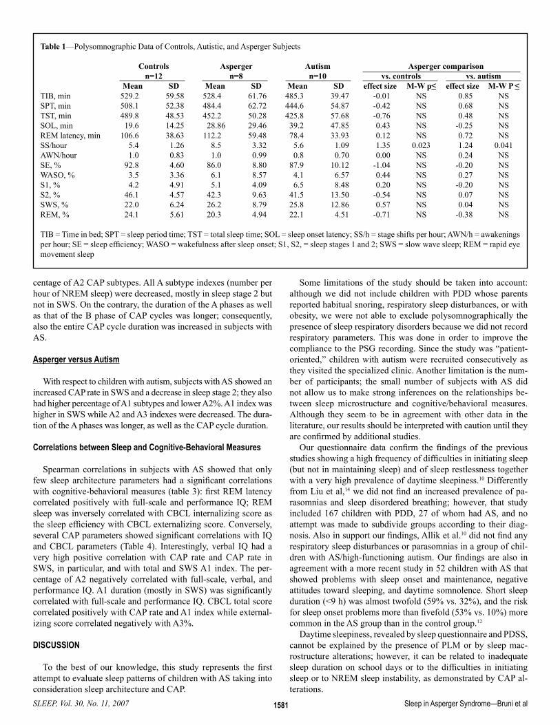
Sleep Architecture
Table 1 shows the statistical analysis of the sleep architecture pa-rameters obtained from the 3 groups. We compared AS vs. autism and vs. typically developing children. No statistical differences were found on almost all variables; only the number of stage shifts per hour was significantly higher in subjects with AS.
All AS subjects had a PLMI with values <5 with a range be-tween 0.14 and 3.79.
Cyclic Alternating Pattern
Asperger versus Controls
The assessment of sleep microstructure (Table 2) revealed that subjects with AS had several differences from normal controls, regarding CAP parameters; they showed a lower total CAP rate in sleep stages 1 and 2 but not in SWS, moreover, they showed an increased percentage of A1 (Figureure. 1), and a decreased per-
Sleep in Asperger Syndrome—Bruni et al
SLEEP, Vol. 30, No. 11, 2007 1581
centage of A2 CAP subtypes. All A subtype indexes (number per hour of NREM sleep) were decreased, mostly in sleep stage 2 but not in SWS. On the contrary, the duration of the A phases as well as that of the B phase of CAP cycles was longer; consequently, also the entire CAP cycle duration was increased in subjects with AS.
Asperger versus Autism
With respect to children with autism, subjects with AS showed an increased CAP rate in SWS and a decrease in sleep stage 2; they also had higher percentage of A1 subtypes and lower A2%. A1 index was higher in SWS while A2 and A3 indexes were decreased. The dura-tion of the A phases was longer, as well as the CAP cycle duration.
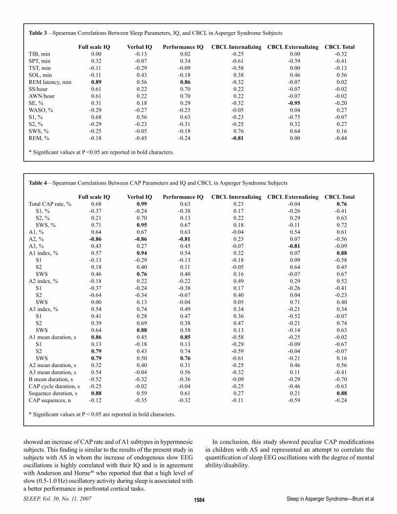
Correlations between Sleep and Cognitive-Behavioral Measures
Spearman correlations in subjects with AS showed that only few sleep architecture parameters had a significant correlations with cognitive-behavioral measures (table 3): first REM latency correlated positively with full-scale and performance IQ; REM sleep was inversely correlated with CBCL internalizing score as the sleep efficiency with CBCL externalizing score. Conversely, several CAP parameters showed significant correlations with IQ and CBCL parameters (Table 4). Interestingly, verbal IQ had a very high positive correlation with CAP rate and CAP rate in SWS, in particular, and with total and SWS A1 index. The per-centage of A2 negatively correlated with full-scale, verbal, and performance IQ. A1 duration (mostly in SWS) was significantly correlated with full-scale and performance IQ. CBCL total score correlated positively with CAP rate and A1 index while external-izing score correlated negatively with A3%.
DISCUSSION
To the best of our knowledge, this study represents the first attempt to evaluate sleep patterns of children with AS taking into consideration sleep architecture and CAP.
Some limitations of the study should be taken into account: although we did not include children with PDD whose parents reported habitual snoring, respiratory sleep disturbances, or with obesity, we were not able to exclude polysomnographically the presence of sleep respiratory disorders because we did not record respiratory parameters. This was done in order to improve the compliance to the PSG recording. Since the study was “patient-oriented,” children with autism were recruited consecutively as they visited the specialized clinic. Another limitation is the num-ber of participants; the small number of subjects with AS did not allow us to make strong inferences on the relationships be-tween sleep microstructure and cognitive/behavioral measures. Although they seem to be in agreement with other data in the literature, our results should be interpreted with caution until they are confirmed by additional studies.
Our questionnaire data confirm the findings of the previous studies showing a high frequency of difficulties in initiating sleep (but not in maintaining sleep) and of sleep restlessness together with a very high prevalence of daytime sleepiness.10 Differently from Liu et al,14 we did not find an increased prevalence of pa-rasomnias and sleep disordered breathing; however, that study included 167 children with PDD, 27 of whom had AS, and no attempt was made to subdivide groups according to their diag-nosis. Also in support our findings, Allik et al.10 did not find any respiratory sleep disturbances or parasomnias in a group of chil-dren with AS/high-functioning autism. Our findings are also in agreement with a more recent study in 52 children with AS that showed problems with sleep onset and maintenance, negative attitudes toward sleeping, and daytime somnolence. Short sleep duration (<9 h) was almost twofold (59% vs. 32%), and the risk for sleep onset problems more than fivefold (53% vs. 10%) more common in the AS group than in the control group.12
Daytime sleepiness, revealed by sleep questionnaire and PDSS, cannot be explained by the presence of PLM or by sleep mac-rostructure alterations; however, it can be related to inadequate sleep duration on school days or to the difficulties in initiating sleep or to NREM sleep instability, as demonstrated by CAP al-terations.
Table 1—Polysomnographic Data of Controls, Autistic, and Asperger Subjects
Controls Asperger Autism Asperger comparison n=12 n=8 n=10 vs. controls vs. autism Mean SD Mean SD Mean SD effect size M-W p≤ effect size M-W P ≤TIB, min 529.2 59.58 528.4 61.76 485.3 39.47 -0.01 NS 0.85 NSSPT, min 508.1 52.38 484.4 62.72 444.6 54.87 -0.42 NS 0.68 NSTST, min 489.8 48.53 452.2 50.28 425.8 57.68 -0.76 NS 0.48 NSSOL, min 19.6 14.25 28.86 29.46 39.2 47.85 0.43 NS -0.25 NSREM latency, min 106.6 38.63 112.2 59.48 78.4 33.93 0.12 NS 0.72 NSSS/hour 5.4 1.26 8.5 3.32 5.6 1.09 1.35 0.023 1.24 0.041AWN/hour 1.0 0.83 1.0 0.99 0.8 0.70 0.00 NS 0.24 NSSE, % 92.8 4.60 86.0 8.80 87.9 10.12 -1.04 NS -0.20 NSWASO, % 3.5 3.36 6.1 8.57 4.1 6.57 0.44 NS 0.27 NSS1, % 4.2 4.91 5.1 4.09 6.5 8.48 0.20 NS -0.20 NSS2, % 46.1 4.57 42.3 9.63 41.5 13.50 -0.54 NS 0.07 NSSWS, % 22.0 6.24 26.2 8.79 25.8 12.86 0.57 NS 0.04 NSREM, % 24.1 5.61 20.3 4.94 22.1 4.51 -0.71 NS -0.38 NS
TIB = Time in bed; SPT = sleep period time; TST = total sleep time; SOL = sleep onset latency; SS/h = stage shifts per hour; AWN/h = awakenings per hour; SE = sleep efficiency; WASO = wakefulness after sleep onset; S1, S2, = sleep stages 1 and 2; SWS = slow wave sleep; REM = rapid eye movement sleep
Sleep in Asperger Syndrome—Bruni et al

SLEEP, Vol. 30, No. 11, 2007 1582
The sleep architecture parameters of our 3 groups did not differ except for the number of stage shifts per hour (significantly higher in subjects with AS) and total sleep time (reduced in subjects with autism vs. controls). Although not significant, we found a trend to-ward increased sleep onset latency in children with autism or AS.
In the past, very few studies evaluated sleep structure in adults with AS and even less in children. Godbout et al.17 showed in 8 subjects with AS aged 7-53 years an increased number of shifts into REM sleep from a waking epoch, and signs of REM sleep disruption. The increased number of stage shifts is similar to our finding but we did not evaluate the specific signs of REM sleep disruption. Tani et al.18 found in adults with AS a polysomno-graphic profile similar to that of controls, while Limoges et al.19 found increased wakefulness after sleep onset and light sleep (stages 1 and 2) with a decrease of SWS.
The issue of the presence of periodic limb movements (PLM) in AS is controversial. Two PSG evaluations reported an abnor-mally high PLM index in some patients with AS, including a 7-year-old child.17,19 This finding has not been confirmed by another recent actigraphic report which showed no differences in the oc-currence of PLM in adults with AS or in controls.30 We did not find increased PLM index in our group and whether AS presents a particular susceptibility to this dopamine-dependent sleep dis-order is still in need of determination. Our report is in agreement with the actigraphy-based study that showed no difference in oc-currence of PLM between adults with AS and controls.30
Conversely, our CAP analysis revealed several differences be-tween the 3 subject groups. We have already published a paper on sleep microstructure of children with autism15 in which we showed a low CAP rate, compared to normal controls, associated with a decreased A1 index during SWS and increased A2 and A3 indexes during light sleep. We have replicated these findings in this new study and we have also found interesting differences be-tween children with autism and AS. Paradoxically, subjects with AS showed an increase of CAP rate in SWS and of A1 index and A1 percentage vs. children with autism and have CAP rate in SWS similar to that of normal controls, and even increased percentage of A1 subtypes.
This finding might be interpreted as an index of increased slow wave activity amplitude oscillations during sleep in these subjects. Although this can be difficult to explain, we have to consider that similar findings have been reported in the literature; the few stud-ies that tried to evaluate sleep microstructure by analyzing phasic events showed a significantly lower density of sleep spindles and an increase of K complexes during stage 2 in patients with AS17; the presence of fewer EEG sleep spindles was confirmed by the same group in a larger study.19
It has been shown that spindles are associated with sleep pro-tecting mechanisms and are considered to be microstates gating the sensory input toward the cortex in an inhibitory way31 and therefore inhibit slow oscillations and arousals: a transient EEG arousal was associated with a lower incidence of spindles and, on
Figure 1—CAP During Slow Wave Sleep in a Patient with Asperger Syndrome
Sleep in Asperger Syndrome—Bruni et al

SLEEP, Vol. 30, No. 11, 2007 1583
the other side, a sort of suppression of evoked K complexes dur-ing spindling bursts has been observed.32
Additionally, sleep EEG spectral analysis studies demonstrated the existence of a reciprocal relationship between sigma and delta activity: the time course of sigma activity and spindle density across subsequent sleep cycles goes in an opposite direction from that of slow wave activity (SWA) and SWS, and the time evolu-tion of sigma activity and spindle density within the first sleep cycle is inverse to that of SWA and SWS.33
We already pointed out that important relationships might exist between the dynamical organization of CAP and that of spindles because the B phase of CAP seems to correspond to a period of relative inhibition of the sigma activity.34 In this respect, it is in-teresting to note that similar results have been reported for evoked arousals which show, in the post-stimulus period (5-20 s), a sig-nificant reduction in power of the sigma band35; this phenomenon has been interpreted as a possible stand-by state needed to main-tain sleep continuity but ready to reach another higher level of arousal, by improving phasically the thalamo-cortical sensory in-flow.35 In summary, the decrease in number of spindles in patients with AS might lead to an increase of NREM instability, expressed by a relative increase of slow oscillations, mainly of CAP A1 sub-types, at least in some sleep stage (SWS).
Moreover, postmortem studies on cortical organization of min-icolumns in young patients with autism or AS showed that single
cortical radial minicolumns are narrower and more numerous, with more dispersed cells and reduced neurophils in individuals with AS.36 This abnormal column morphology and cell distribu-tion might possibly lead to a different pattern of connections, such as a pattern of GABAergic interneurons delimiting the terri-tory of each column by lateral inhibition. Casanova et al.35 further hypothesized that if thalamic terminals are unaffected, this may result in a greater innervation of minicolumns by the thalamic output that might be reflected by an increase of slow oscillations in sleep EEG of these subjects.19
The other important result of our study is the high level of cor-relation between CAP A1 subtypes and cognitive measures: ver-bal IQ was highly correlated with CAP rate in SWS and with A1 index and A1 index in SWS. A2 percentage negatively correlated with total, verbal, and performance IQ.
It is assumed that subjects with Asperger syndrome are excep-tionally talented or skilled in one or more particular areas. This may be explained by superior memory abilities in specific fields. In this respect, there is compelling evidence that sleep contributes to the long-term consolidation of new memories.36 This function of sleep has been linked to slow (<1 Hz) potential EEG oscillations (the main EEG components of CAP A1 subtypes), which predomi-nantly arise from the prefrontal neocortex and characterize slow wave sleep.37,38 In a study on the possible role of sleep microstruc-ture in individuals with superior memory, Ferini Strambi et al.39
Table 2—CAP Parameters Obtained in the 3 Groups of Subjects Included in this Study
Controls Asperger Autism Asperger comparison n=12 n=8 n=10 vs. controls vs. autism Mean SD Mean SD Mean SD effect size M-W P ≤ effect size M-W P ≤Total CAP rate, % 42.54 8.05 29.53 15.52 37.02 8.39 -1.13 0.035 -0.62 NSS1, % 46.17 17.43 10.27 22.81 44.90 20.45 -1.82 0.007 -1.61 0.0084S2, % 39.71 10.69 15.89 13.56 42.78 12.73 -2.00 0.004 -2.05 0.0084SWS, % 48.57 11.76 58.57 23.40 31.19 15.78 0.58 NS 1.41 0.0248A1, % 69.40 9.05 80.24 4.29 64.90 9.22 1.43 0.009 2.05 0.001A2, % 19.08 6.19 8.59 4.45 21.05 7.48 -1.88 0.003 -1.97 0.003A3, % 11.51 5.23 11.14 4.59 14.03 6.21 -0.07 NS -0.52 NSA1 index, % 52.52 11.16 35.59 21.57 38.73 10.26 -1.06 0.042 -0.19 NSS1 45.97 21.47 8.70 22.62 30.88 23.05 -1.70 0.004 -0.97 0.028S2 46.30 11.11 21.61 16.86 44.68 15.59 -1.81 0.011 -1.43 0.021SWS 85.88 21.88 82.77 30.56 47.77 23.44 -0.12 NS 1.31 0.040A2 index, % 15.37 6.91 2.51 1.23 13.10 7.10 -2.36 0.0001 -1.97 0.0001S1 8.60 7.07 9.30 22.41 9.03 9.81 0.05 NS 0.02 NSS2 19.71 8.78 4.36 2.27 21.42 10.89 -2.19 0.0001 -2.05 0.001SWS 9.99 5.60 4.26 2.48 4.45 1.73 -1.23 0.018 -0.09 NSA3 index, % 8.94 4.71 3.09 2.31 8.11 3.20 -1.48 0.003 -1.76 0.008S1 24.39 16.65 4.61 5.17 30.47 23.74 -1.48 0.005 -1.43 0.022S2 11.91 5.93 5.96 2.52 10.80 3.63 -1.22 0.008 -1.52 0.011SWS 2.88 2.01 4.37 2.45 4.66 3.56 0.68 NS -0.09 NSA1 mean duration, s 4.69 0.16 9.01 1.90 5.04 0.24 3.63 0.0001 3.13 0.0001S1 5.62 2.09 4.03 7.76 5.34 2.11 -0.31 NS -0.24 NSS2 4.33 0.16 7.94 2.40 4.67 0.34 2.40 0.0001 2.03 0.0001SWS 4.97 0.43 9.44 1.99 5.41 0.26 3.48 0.0001 3.03 0.0001A2 mean duration, s 6.60 0.64 12.87 2.00 6.67 0.58 4.67 0.0001 4.45 0.0001A3 mean duration, s 11.96 1.18 20.26 2.90 12.56 0.98 4.09 0.0001 3.75 0.0001B mean duration, s 16.96 1.94 21.00 3.94 19.06 2.23 1.40 0.022 0.63 NSCAP cycle duration, s 22.82 1.86 31.16 3.80 25.54 2.35 3.00 0.0001 1.83 0.006Sequence duration, s 180.66 39.89 268.97 158.42 182.79 31.54 0.85 NS 0.80 NSCAP sequences, n 52.50 6.76 24.71 3.09 40.00 8.04 -4.94 0.0001 -2.40 0.006
M-W = Mann-Whitney
Sleep in Asperger Syndrome—Bruni et al
SLEEP, Vol. 30, No. 11, 2007 1584
showed an increase of CAP rate and of A1 subtypes in hypermnesic subjects. This finding is similar to the results of the present study in subjects with AS in whom the increase of endogenous slow EEG oscillations is highly correlated with their IQ and is in agreement with Anderson and Horne40 who reported that that a high level of slow (0.5-1.0 Hz) oscillatory activity during sleep is associated with a better performance in prefrontal cortical tasks.
In conclusion, this study showed peculiar CAP modifications in children with AS and represented an attempt to correlate the quantification of sleep EEG oscillations with the degree of mental ability/disability.
Table 3—Spearman Correlations Between Sleep Parameters, IQ, and CBCL in Asperger Syndrome Subjects
Full scale IQ Verbal IQ Performance IQ CBCL Internalizing CBCL Externalizing CBCL TotalTIB, min 0.00 -0.13 0.02 -0.25 0.00 -0.32SPT, min 0.32 -0.07 0.34 -0.61 -0.39 -0.41TST, min -0.11 -0.29 -0.09 -0.58 0.00 -0.13SOL, min -0.11 0.43 -0.18 0.38 0.46 0.56REM latency, min 0.89 0.56 0.86 -0.32 -0.07 0.02SS/hour 0.61 0.22 0.70 0.22 -0.07 -0.02AWN/hour 0.61 0.22 0.70 0.22 -0.07 -0.02SE, % 0.31 0.18 0.29 -0.32 -0.95 -0.20WASO, % -0.29 -0.27 -0.25 -0.05 0.04 0.27S1, % 0.68 0.56 0.63 -0.23 -0.75 -0.07S2, % -0.29 -0.23 -0.31 -0.25 0.32 0.27SWS, % -0.25 -0.05 -0.18 0.76 0.64 0.16REM, % -0.18 -0.45 -0.24 -0.81 0.00 -0.44
* Significant values at P <0.05 are reported in bold characters.
Table 4—Spearman Correlations Between CAP Parameters and IQ and CBCL in Asperger Syndrome Subjects
Full scale IQ Verbal IQ Performance IQ CBCL Internalizing CBCL Externalizing CBCL TotalTotal CAP rate, % 0.68 0.99 0.63 0.23 -0.04 0.76 S1, % -0.37 -0.24 -0.38 0.17 -0.26 -0.41 S2, % 0.21 0.70 0.13 0.22 0.29 0.63 SWS, % 0.71 0.95 0.67 0.18 -0.11 0.72A1, % 0.64 0.67 0.63 -0.04 0.54 0.61A2, % -0.86 -0.86 -0.81 0.23 0.07 -0.56A3, % 0.43 0.27 0.45 -0.07 -0.81 -0.09A1 index, % 0.57 0.94 0.54 0.32 0.07 0.88 S1 -0.13 -0.29 -0.13 -0.18 0.09 -0.58 S2 0.18 0.40 0.11 -0.05 0.64 0.45 SWS 0.46 0.76 0.40 0.16 -0.07 0.67A2 index, % -0.18 0.22 -0.22 0.49 0.29 0.52 S1 -0.37 -0.24 -0.38 0.17 -0.26 -0.41 S2 -0.64 -0.34 -0.67 0.40 0.04 -0.23 SWS 0.00 0.13 -0.04 0.05 0.71 0.40A3 index, % 0.54 0.74 0.49 0.34 -0.21 0.34 S1 0.41 0.28 0.47 0.36 -0.52 -0.07 S2 0.39 0.69 0.38 0.47 -0.21 0.74 SWS 0.64 0.88 0.58 0.13 -0.14 0.63A1 mean duration, s 0.86 0.45 0.85 -0.58 -0.25 -0.02 S1 0.13 -0.18 0.13 -0.29 -0.09 -0.67 S2 0.79 0.43 0.74 -0.59 -0.04 -0.07 SWS 0.79 0.50 0.76 -0.61 -0.21 0.16A2 mean duration, s 0.32 0.40 0.31 -0.25 0.46 0.56A3 mean duration, s 0.54 -0.04 0.56 -0.32 0.11 -0.41B mean duration, s -0.52 -0.32 -0.36 -0.09 -0.29 -0.70CAP cycle duration, s -0.25 -0.02 -0.04 -0.25 -0.46 -0.63Sequence duration, s 0.88 0.59 0.61 0.27 0.21 0.88CAP sequences, n -0.12 -0.35 -0.32 -0.11 -0.59 -0.24
* Significant values at P < 0.05 are reported in bold characters.
Sleep in Asperger Syndrome—Bruni et al
SLEEP, Vol. 30, No. 11, 2007 1585
REFERENCES
1. American Psychiatric Association: Diagnostic and Statistical Manu-al of Mental Disorders. 4th ed. Washington, DC: APA; 2000.
2. Richdale AL, Prior MR. The sleep/wake rhythm in children with autism. Eur Child Adol Psychiatry 1995;4:175-86.
3. Wiggs L, Stores G. Sleep patterns and sleep disorders in children with autistic spectrum disorders: insights using parent report and actigraphy. Dev Med Child Neurol 2004;46:372-80.
4 Patzold LM, Richdale AL, Tonge BJ. An investigation into sleep characteristics of children with autism and Asperger’s disorder. J Ped Child Health 1998;34:528-33.
5. Richdale AL. Sleep in children with autism and Asperger syndrome. In Stores G, Wiggs L, eds. Sleep disturbance in children and adoles-cents with disorders of development: its significance and manage-ment. Oxford: MacKeith Press, 2001:181-95
6. Paavonen EJ, Nieminen-von Wendt T, Vanhala R, Aronen ET, von Wendt L. Effectiveness of melatonin in the treatment of sleep distur-bances in children with Asperger disorder. J Child Adolesc Psychop-harmacol 2003;13:83-95.
7. Tani P, Lindberg N, Nieminen-Von Wendt T, et al. Insomnia is a frequent finding in adults with Asperger syndrome. BMC Psychiatry 2003;3:12.
8. Couturier JL, Speechley KN, Steele M, Norman R, Stringer B, Nicolson R. Parental perception of sleep problems in children of normal intelligence with pervasive developmental disorders: preva-lence, severity, and pattern. J Am Acad Child Adolesc Psychiatry 2005;44: 815-22.
9. Polimeni MA, Richdale AL, Francis AJP. A survey of sleep prob-lems in autism, Asperger’s disorder and typically developing chil-dren. J Intellect Disabil Res 2005;49:260-8.
10. Allik H, Larsson JO, Smedje H. Insomnia in school-age children with Asperger syndrome or high-functioning autism. BMC Psychia-try 2006;6:18.
11. Allik H, Larsson J, Smedje H. Sleep patterns of school-age children with Asperger syndrome or high-functioning autism. J Autism Dev Disord 2006;36:585-95.
12. Paavonen EJ, Vehkalahti K, von Wendt L, Nieminen-von Wendt T, Aronen ET. Sleep in children with Asperger syndrome. J Autism Dev Disord 2007, in press.
13. Oyane N, Bjorvatn B. Sleep disturbances in adolescents and young adults with autism and Asperger syndrome. Autism 2005;9:83-94.
14. Liu X, Hubbard JA, Fabes RA, Adam JB. Sleep disturbances and correlates of children with autism spectrum disorders. Child Psy-chiatry Hum Dev 2006;37:179-91.
15. Miano S, Bruni O, Elia M, et al. Sleep in children with autistic spec-trum disorder: a questionnaire and polysomnographic study. Sleep Med 2007, doi:10.1016/j.sleep.2007.01.014
16. Malow BA, Marzec ML, McGrew SG, et al. Characterizing sleep in children with autism spectrum disorders: a multidimensional ap-proach. Sleep 2006;29:1563-71.
17. Godbout R, Bergeron C, Limoges E, Stip E, Mottron L. A Laboratory study of sleep in Asperger’s syndrome. Neuroreport 2000;11:127-30.
18. Tani P, Lindberg N, Nieminen-von Wendt T, et al. Sleep in young adults with Asperger syndrome. Neuropsychobiology 2004;50:147-52.
19. Limoges E, Mottron L, Bolduc C, Berthiaume C, Godbout R. Atypical sleep architecture and the autism phenotype. Brain 2005;128:1049-61.
20. Bruni O, Fabrizi P, Ottaviano S, Cortesi F, Giannotti F, Guidetti V. Prevalence of sleep disorders in childhood and adolescence with headache: a case-control study. Cephalalgia 1997;17:492-8.
21. Drake C, Nickel C, Burduvali E, Roth T, Jefferson C, Pietro B. The pediatric daytime sleepiness scale (PDSS): sleep habits and school outcomes in middle-school children. Sleep 2003;26:455-8.
22. Terzano MG, Parrino L, Smerieri A, et al. Atlas, rules, and record-
ing techniques for the scoring of cyclic alternating pattern (CAP) in human sleep. Sleep Med 2001;2:537-53.
23. World Health Organization: International classification of disease (10th ed., chap. 5). Mental and behavioral disorders. Diagnostic cri-teria for research. Geneva: World Health Organization; 1993.
24. Lord C, Risi S, Lambrecht L, et al: The autism diagnostic observa-tion schedule-generic: a standard measure of social and communi-cation deficits associated with the spectrum of autism. J Aut Dev Disord 2002;30:205-23.
25. Schopler E, Reichler RJ, DeVellis RF, Daly K. Toward objective classification of childhood autism: Childhood Autism Rating Scale (CARS). J Autism Dev Disord 1980;10:91-103.
26. Achenbach TM, Rescorla LA. Manual for the ASEBA School Age Forms and Profiles. Burlington: University of Vermont, Research Center for Children, Youth, and Families, 2001.
27. Rechtschaffen A, Kales A. A manual of standardized terminology, techniques, and scoring system for sleep stages of human subjects. Washington: Washington Public Health service; US Government Printing Office, 1968
28. Zucconi M, Ferri R, Allen R, et al. The official World Association of Sleep Medicine (WASM) standards for recording and scoring pe-riodic leg movements in sleep (PLMS) and wakefulness (PLMW) developed in collaboration with a task force from the Internation-al Restless Legs Syndrome Study Group (IRLSSG). Sleep Med 2006;7:175-83.
29. Cohen J. Statistical power analysis for the behavioral sciences, 2nd ed. Hillsdale, NJ: Erlbaum; 1988.
30. Tani P, Tuisku K, Lindberg N, et al. Is Asperger syndrome associ-ated with abnormal nocturnal motor phenomena? Psychiatry Clin Neurosci 2006, 60:527-8.
31. Steriade M, McCormick DA, Sejnowski TJ. Thalamocortical oscil-lations in the sleeping and aroused brain. Science 1993;262:679-85.
32. Naitoh P, Antony Baas V, Muzet A, Ehrhart J. Dynamic relation of sleep spindles and K-complexes to spontaneous phasic arousal in sleeping human subjects. Sleep 1982;5:58-72.
33. Ferri R, Elia M, Musumeci SA, Pettinato S. The time course of high-frequency bands (15-45 Hz) in all-night spectral analysis of sleep EEG. Clin Neurophysiol 2000;111:1258-65.
34. Ferri R, Bruni O, Miano S, Plazzi G, Terzano MG. All-night EEG power spectral analysis of the cyclic alternating pattern components in young adult subjects. Clin Neurophysiol 2005;116:2429-40.
35. Halasz P. Hierarchy of micro-arousals and the microstructure of sleep. Neurophysiol Clin 1998;28:461-75.
36. Casanova MF, Buxhoeveden DP, Switala AE, Roy E. Asperger’s syn-drome and cortical neuropathology. J Child Neurol 2002;17:142-5.
37. Stickgold R. Sleep-dependent memory consolidation. Nature 2005;437:1272-8.
38. Ferri R, Bruni O, Miano S, Terzano MG. Topographic mapping of the spectral components of the cyclic alternating pattern (CAP). Sleep Med 2005;6:29-36.
39. Ferini-Strambi L, Ortelli P, Castronovo V, Cappa S. Increased pe-riodic arousal fluctuations during non-REM sleep are associated to superior memory Brain Res Bull 2004;63:439-42.
40. Anderson C, Horne JA. Prefrontal cortex: links between low fre-quency delta EEG in sleep and neuropsychological performance in healthy, older people. Psychophysiology 2003; 40:349-57.
Sleep in Asperger Syndrome—Bruni et al
Related Documents