Welcome message from author
This document is posted to help you gain knowledge. Please leave a comment to let me know what you think about it! Share it to your friends and learn new things together.
Transcript
Skin Moisturization
edited by
James J. LeydenUniversity of Pennsylvania School of Medicine
Philadelphia, Pennsylvania
Anthony V. RawlingsUnilever Research
Bebington, Wirral, United Kingdom
Marcel Dekker, Inc. New York • BaselTM
Copyright © 2002 by Marcel Dekker, Inc. All Rights Reserved.
ISBN: 0-8247-0643-9
This book is printed on acid-free paper.
HeadquartersMarcel Dekker, Inc.270 Madison Avenue, New York, NY 10016tel: 212-696-9000; fax: 212-685-4540
Eastern Hemisphere DistributionMarcel Dekker AGHutgasse 4, Postfach 812, CH-4001 Basel, Switzerlandtel: 41-61-261-8482; fax: 41-61-261-8896
World Wide Webhttp://www.dekker.com
The publisher offers discounts on this book when ordered in bulk quantities. For more in-formation, write to Special Sales/Professional Marketing at the headquarters addressabove.
Copyright © 2002 by Marcel Dekker, Inc. All Rights Reserved.
Neither this book nor any part may be reproduced or transmitted in any form or by anymeans, electronic or mechanical, including photocopying, microfilming, and recording, orby any information storage and retrieval system, without permission in writing from thepublisher.
Current printing (last digit):10 9 8 7 6 5 4 3 2 1
PRINTED IN THE UNITED STATES OF AMERICA

About the Series
The Cosmetic Science and Technology series was conceived to permit discussion of a broad
range of current knowledge and theories of cosmetic science and technology. The series is
composed of both books written by a single author and edited volumes with a number of
contributors. Authorities from industry, academia, and the government participate in writing
these books.
The aim of the series is to cover the many facets of cosmetic science and technology. Topics are
drawn from a wide spectrum of disciplines ranging from chemistry, physics, biochemistry, and
analytical and consumer evaluations to safety, efficacy, toxicity, and regulatory questions.
Organic, inorganic, physical and polymer chemistry, emulsion and lipid technology,
microbiology, dermatology, and toxicology all play important roles in cosmetic science.
There is little commonality in the scientific methods, processes, and formulations required for
the wide variety of cosmetics and toiletries in the market. Products range from preparations for
hair, oral, and skin care to lipsticks, nail polishes and extenders, deodorants, body powders and
aerosols, to quasi-pharmaceutical over-the-counter products such as antiperspirants, dandruff
shampoos, antimicrobial soaps, and acne and sun screen products.
Cosmetics and toiletries represent a highly diversified field involving many subsections of
science and “art.” Even in these days of high technology, art and intuition continue to play an
important part in the development of formulations, their evaluation, selection of raw materials,
and, perhaps most importantly, the successful marketing of new products. The application of
more sophisticated scientific methodologies that gained steam in the 1980s has increased in such
areas as claim substantiation, safety testing, product testing, and chemical analysis and has led to
a better understanding of the properties of skin and hair. Molecular modeling techniques are
beginning to be applied to data obtained in skin sensory studies.
Emphasis in the Cosmetic Science and Technology series is placed on reporting the current
status of cosmetic technology and science and changing regulatory climates and presenting
historical reviews. The series has now grown to 26 books dealing with the constantly changing
technologies and trends in the cosmetic industry, including globalization. Several of the volumes
have been translated into Japanese and Chinese. Contributions range from highly sophisticated
and scientific treatises to primers and presentations of practical applications. Authors are
encouraged to present their own concepts as well as established theories. Contributors have been
asked not to shy away from fields that are in a state of transition, nor to hesitate to present
detailed discussions of their own work. Altogether, we intend to develop in this series a
collection of critical surveys and ideas covering diverse phases of the cosmetic industry.
The 13 chapters in Multifunctional Cosmetics cover multifunctional products for hair, nail, oral,
and skin care, as well as products with enhanced sunscreen and antimicrobial properties Several
chapters deal with the development of claim support data, the role of packaging, and consumer
research on the perception of multifunctional cosmetic products. The authors keep in mind that
in the case of cosmetics, it is not only the physical effects that can be measured on the skin or
hair, but also the sensory effects that have to be taken into account. Cosmetics can have a
psychological and social impact that cannot be underestimated.
I want to thank all the contributors for participating in this project and particularly the editors,
Perry Romanowski and Randy Schueller, for conceiving, organizing, and coordinating this book.
It is the second book that they have contributed to this series and we appreciate their efforts.
Special thanks are due to Sandra Beberman and Erin Nihill of the editorial and production staff
at Marcel Dekker, Inc. Finally, I would like to thank my wife, Eva, without whose constant
support and editorial help I would not have undertaken this project.
Eric Jungermann, Ph.D.
COSMETIC SCIENCE AND TECHNOLOGY
Series Editor
ERIC JUNGERMANN
Jungermann Associates, Inc.Phoenix, Arizona
1. Cosmetic and Drug Preservation: Principles and Practice, edited byJon J. Kabara
2. The Cosmetic Industry: Scientific and Regulatory Foundations, editedby Norman F. Estrin
3. Cosmetic Product Testing: A Modern Psychophysical Approach,Howard R. Moskowitz
4. Cosmetic Analysis: Selective Methods and Techniques, edited by P.Boré
5. Cosmetic Safety: A Primer for Cosmetic Scientists, edited by James H.Whittam
6. Oral Hygiene Products and Practice, Morton Pader7. Antiperspirants and Deodorants, edited by Karl Laden and Carl B.
Felger8. Clinical Safety and Efficacy Testing of Cosmetics, edited by William C.
Waggoner9. Methods for Cutaneous Investigation, edited by Robert L. Rietschel
and Thomas S. Spencer10. Sunscreens: Development, Evaluation, and Regulatory Aspects, edited
by Nicholas J. Lowe and Nadim A. Shaath11. Glycerine: A Key Cosmetic Ingredient, edited by Eric Jungermann and
Norman O. V. Sonntag12. Handbook of Cosmetic Microbiology, Donald S. Orth13. Rheological Properties of Cosmetics and Toiletries, edited by Dennis
Laba14. Consumer Testing and Evaluation of Personal Care Products, Howard
R. Moskowitz15. Sunscreens: Development, Evaluation, and Regulatory Aspects. Sec-
ond Edition, Revised and Expanded, edited by Nicholas J. Lowe, Na-dim A. Shaath, and Madhu A. Pathak
16. Preservative-Free and Self-Preserving Cosmetics and Drugs:Principles and Practice, edited by Jon J. Kabara and Donald S. Orth
17. Hair and Hair Care, edited by Dale H. Johnson18. Cosmetic Claims Substantiation, edited by Louise B. Aust19. Novel Cosmetic Delivery Systems, edited by Shlomo Magdassi and
Elka Touitou20. Antiperspirants and Deodorants: Second Edition, Revised and Ex-
panded, edited by Karl Laden21. Conditioning Agents for Hair and Skin, edited by Randy Schueller and
Perry Romanowski
22. Principles of Polymer Science and Technology in Cosmetics and Per-sonal Care, edited by E. Desmond Goddard and James V. Gruber
23. Cosmeceuticals: Drugs vs. Cosmetics, edited by Peter Elsner andHoward I. Maibach
24. Cosmetic Lipids and the Skin Barrier, edited by Thomas Förster25. Skin Moisturization, edited by James J. Leyden and Anthony V. Raw-
lings26. Multifunctional Cosmetics, edited by Randy Schueller and Perry Roma-
nowski
ADDITIONAL VOLUMES IN PREPARATION
Series Introduction
The Cosmetic Science and Technology series was conceived to permit discussionof a broad range of current knowledge and theories in the field. The series is com-posed of books either written by one or more authors or edited with multiple con-tributors. Authorities from industry, academia, and the government are participat-ing in writing these books. The purpose of this series is to cover the many facetsof cosmetic science and technology. Topics are drawn from a wide spectrum ofdisciplines ranging from chemistry, to physics, to biochemistry, and include ana-lytical and consumer evaluations, safety, efficacy, toxicity, and regulatory ques-tions. Organic, inorganic, physical, and polymer chemistry, emulsion and lipidtechnology, microbiology, dermatology, and toxicology all play important rolesin cosmetic science.
There is little commonality in the scientific methods, processes, and formu-lations required for the wide variety of cosmetics and toiletries in the market.Products range from preparations for hair care, oral care, and skin care to lip-sticks, nail polishes and extenders, deodorants, and body powders and aerosols, toquasi-pharmaceutical over-the-counter products such as antiperspirants, dandruffshampoos, antimicrobial soaps, and acne and sunscreen products.
Cosmetics and toiletries represent a highly diversified field involving manysubsections of science and “art.” Even in these days of high technology, art andintuition continue to play an important part in the development of formulations,
iii
iv Series Introduction
their evaluation, the selection of raw materials, and, perhaps most importantly,the successful marketing of new products. The move toward the application ofmore sophisticated scientific methodologies that gained momentum in the 1980shas grown in such areas as claim substantiation, safety testing, product testing,and chemical analysis and has led to a better understanding of the properties ofskin and hair. Molecular modeling techniques are beginning to be applied to dataobtained in skin sensory studies.
Emphasis in the Cosmetic Science and Technology series is placed on re-porting the current status of cosmetic technology and science, changing regulato-ry climates, and historical reviews. The series has grown to over 20 books dealingwith the constantly changing technologies and trends in the cosmetic industry, in-cluding globalization. Several of the books have been translated into Japaneseand Chinese. Contributions range from highly sophisticated and scientific treatis-es to primers, practical applications, and pragmatic presentations. Authors are en-couraged to present their own concepts, as well as established theories. Contribu-tors have been asked not to shy away from fields that are in a state of transition,nor to hesitate to present detailed discussions of their own work. Our intention isto develop the series into a collection of critical surveys and ideas covering di-verse phases of the cosmetic industry.
Skin Moisturizers, the twenty-fifth book published in the series, representsa truly global effort. The 28 chapters cover the following areas: the stratumcorneum and epidermal biology, xerotic skin conditions, efficacy of moisturizersand moisturizing ingredients, evaluation methodologies, formulation, and safetyand regulatory considerations. Ten chapters have been contributed by authorsfrom the United States, nine from the United Kingdom, four from Japan, and theremainder from France, Germany, Italy, and Belgium.
Skin moisturization and moisturizers represent the dominant growth area incosmetics and toiletries, reflecting the consumer’s perpetual interest in lookingyoung. Youthful, healthy skin is perceived as soft, moisturized, and free of wrin-kles. Moisturizing products have become the proverbial “hope in a bottle” result-ing in the creation of thousands of products and moisturizing claims. This interestin youthful skin becomes even more important as the population ages and con-cerns over dry skin conditions increase. Practical formulation chemists have longrealized that there are two basic mechanisms perceived as moisturization: hydra-tion with water-miscible agents (glycerine is the classical example) and occlusion(classically, petrolatum). The concept of moisturization is, of course, far morecomplicated. The stratum corneum is recognized as a heterogeneous system ofprotein-enriched cells embedded in lipid-laden intercellular domains. It is an epi-dermal barrier governing water penetration and loss, cohesion, and desquama-tion. The dependence of skin conditioning on the lipids in these systems is due tothe fact that essential fatty acids play an important role, together with the naturalmoisturizing factor, a mixture of hydroscopic water-soluble substances, such as
vSeries Introduction
lactic acid and PCA. In addition, collagen, hyaluronic acid, and elastin play a rolein these systems. This book identifies these new concepts, increases our under-standing of the skin and skin moisturization, and provides the scientific basis ofskin moisturization.
I would like to thank the contributors for participating in this project andparticularly the editors, Drs. James Leyden and Anthony Rawlings for conceiv-ing, organizing, and coordinating this book. Special thanks are extended to San-dra Beberman and the editorial and production staff at Marcel Dekker, Inc. Final-ly, I thank my wife, Eva, without whose constant support and editorial help Iwould not have undertaken this project.
Eric Jungermann, Ph.D.

Preface
The focus of this book is the scientific basis of skin moisturization. The contentsrange from biological aspects of the skin through active ingredients and their for-mulation, evaluation methodology, and the regulatory and safety aspects of skinmoisturizers. This book will be an invaluable resource for dermatologists, cos-metic scientists, and clinical scientists interested in treatment of xerotic skin con-ditions. Each chapter reviews the relevant literature in the particular area andgives an up-to-date account of recent research findings. The biology of the epi-dermis and stratum corneum is the subject of intense review, as well as changes instructure and function in a variety of xerotic skin conditions. Overviews of clini-cal and consumer testing approaches together with ex vivo evaluation proceduresare presented in the evaluation section. The action efficacy and formulation ofvarious moisturizing ingredients are also covered, including emollients, humec-tants, ceramides and other barrier lipids, alphahydroxyacids, and enzymes. The fi-nal section discusses safety and regulatory guidelines in the industry.
This book is a result of contributions by experts in their own areas and isthe work of an international team. The authors represent a cross-section of the sci-entific community in academia as well as industrial research. Cosmetic scientists,dermatologists, and researchers will find this book a valuable, in-depth account ofskin moisturization.
James J. LeydenAnthony V. Rawlings
vii

Contents
Series Introduction Eric Jungermann iiiPreface viiContributors xiii
INTRODUCTION
1. The Skin Moisturizer Marketplace 1Anthony W. Johnson
STRATUM CORNEUM AND EPIDERMAL BIOLOGY
2. Stratum Corneum Ceramides and Their Role in Skin Barrier Function 31Gopinathan K. Menon and Lars Nórlen
3. Stratum Corneum Moisturizing Factors 61Clive R. Harding and Ian R. Scott
ix
x Contents
4. Desquamation and the Role of Stratum Corneum Enzymes 81Junko Sato
5. The Cornified Envelope: Its Role in Stratum Corneum Structure and Maturation 95Allan Watkinson, Clive R. Harding, and Anthony V. Rawlings
XEROTIC SKIN CONDITIONS
6. Dry and Xerotic Skin Conditions 119Anthony V. Rawlings, Clive R. Harding, Allan Watkinson, and Ian R. Scott
7. Sensitive Skin and Moisturization 145Paolo U. Giacomoni, Neelam Muizzuddin, Rose Marie Sparacio,Edward Pelle, Thomas Mammone, Kenneth Marenus, and Daniel Maes
8. Photodamage and Dry Skin 155James J. Leyden and Robert Lavker
9. Atopic Dermatitis 165Anna Di Nardo and Philip W. Wertz
10. Psoriasis and Ichthyoses 179Ruby Ghadially
11. Solvent-, Surfactant-, and Tape Stripping–Induced Xerosis 203Mitsuhiro Denda
EFFICACY OF MOISTURIZERS AND MOISTURIZING INGREDIENTS
12. Clinical Effects of Emollients on Skin 223Joachim Fluhr, Walter M. Holleran, and Enzo Berardesca
13. Humectants 245Anthony V. Rawlings, Clive R. Harding, Allan Watkinson, PremChandar, and Ian R. Scott
xiContents
14. Ceramides as Natural Moisturizing Factors and Their Efficacy in Dry Skin 267Genji Imokawa
15. Phosphatidylcholine and Skin Hydration 303Miklos Ghyczy and Vladimir Vacata
16. Hydroxyacids 323Anthony W. Johnson
17. Salicylic Acid and Derivatives 353Jean Luc Lévêque and Didier Saint-Léger
18. The Efficacy, Stability, and Safety of Topically Applied Protease in Treating Xerotic Skin 365David J. Pocalyko, Prem Chandar, Clive R. Harding, Lynn Blaikie, Allan Watkinson, and Anthony V. Rawlings
19. Enzymes in Cleansers 385Takuji Masunaga
20. Moisturizing Cleansers 405Kavssery P. Ananthapadmanabhan, Kumar Subramanyan, andGail B. Rattinger
EVALUATION METHODOLOGIES
21. Consumer Testing Methods 433Steven S. Braddon, Gwendolyn S. Jarrett, and Alejandra M. Muñoz
22. Clinical Testing of Moisturizers 465Gregory Nole
23. Noninvasive Instrumental Methods for Assessing Moisturizers 499Gary L. Grove, Charles Zerweck, and Elizabeth Pierce
24. Laboratory-Based Ex Vivo Assessment of Stratum Corneum Function 529Claudine Piérard-Franchimont, Marc Paye, Veroniqué Goffin,and Gérald E. Piérard
xii Contents
FORMULATION
25. Formulation of Skin Moisturizers 547Steve Barton
26. Formulation and Assessment of Moisturizing Cleansers 585David C. Story and Frederick Anthony Simion
SAFETY AND REGULATORY
27. Safety Assessment of Cosmetic Products 611Christopher Flower
28. Regulatory Assessment of Cosmetic Products 635Simon Young
Index 651
Contributors
Kavssery P. Ananthapadmanabhan, Ph.D. Principal Research Scientist, SkinCare and Cleansing Department, Unilever Research, Edgewater Laboratory,Edgewater, New Jersey
Steve Barton, M.Sc., C.Biol. Skincare Scientific Adviser, Strategic MarketingUnit, The Boots Company, Nottingham, United Kingdom
Enzo Berardesca, M.D. Department of Clinical Dermatology, University ofPavia, San Matteo, Pavia, Italy
Lynn Blaikie, Ph.D. Unilever Research, Colworth Laboratory, Sharnbrook,Bedford, United Kingdom
Steven S. Braddon, Ph.D. Senior Research Consumer Test Coordinator, De-partment of Consumer Science, Unilever Home and Personal Care North Amer-ica, Trumbull, Connecticut
Prem Chandar, Ph.D. Research Scientist, Skin Care and Cleansing Depart-ment, Unilever Research, Edgewater Laboratory, Edgewater, New Jersey
xiii
xiv Contributors
Mitsuhiro Denda, Ph.D. Research Scientist, Skin Biology Research Laborato-ries, Shiseido Life Science Research Center, Yokohama, Japan
Anna Di Nardo, Ph.D., M.D. Department of Dermatology, University of Mo-dena, Modena, Italy
Christopher Flower, M.Sc., Ph.D., C.Biol. M.I.Biol. Head of Safety and Tox-icology, The Cosmetic, Toiletry, and Perfumery Association, London, UnitedKingdom
Joachim Fluhr, Ph.D. University of Pavia, San Matteo, Pavia, Italy, and Uni-versity of California, San Francisco, California
Ruby Ghadially, MB, Ch.B., F.R.C.P.(C) Veterans Administration MedicalCenter and University of California School of Medicine, San Francisco, Cali-fornia
Miklos Ghyczy, Ph.D. Director, Applications Research, Nattermann Phospho-lipid GmbH, Cologne, Germany
Paolo U. Giacomoni, Ph.D. Executive Director, Research and Development,Clinique Laboratories, Inc., Melville, New York
Veroniqué Goffin, M.D., Ph.D. Department of Dermatology, University Med-ical Center Sart Tilman, Liège, Belgium
Gary L. Grove, Ph.D. KGL Skin Study Center, Broomall, Pennsylvania
Clive R. Harding, M.Sc. Research Biochemist, Department of Cell and Mole-cular Biology and Biorecognition, Unilever Research, Colworth Laboratory,Sharnbrook, Bedford, United Kingdom
Walter M. Holleran, Ph.D. Associate Adjunct Professor, Department of Der-matology and Pharmaceutical Chemistry, University of California, San Francis-co, San Francisco, California
Genji Imokawa, Ph.D. Kao Biological Science Laboratories, Haga, Tochigi,Japan
Gwendolyn S. Jarrett, B.S. Manager, Consumer Science, Department of Re-search and Development, Unilever Home and Personal Care North America,Trumbull, Connecticut
xvContributors
Anthony W. Johnson, Ph.D. Manager, Skin/Bioscience, Global TechnologyCenter, Unilever Home and Personal Care North America, Trumbull, Connecticut
Robert Lavker, Ph.D. Department of Dermatology, University of Pennsylva-nia School of Medicine, Philadelphia, Pennsylvania
Jean Luc Lévêque, Ph.D. L’Oréal, Clichy, France
James J. Leyden, Ph.D. Department of Dermatology, University of Pennsyl-vania School of Medicine, Philadelphia, Pennsylvania
Daniel Maes, Ph.D. Vice President, Department of Biological Research, EsteeLauder, Melville, New York
Thomas Mammone, Ph.D. Director, Department of Skin Biology, Estee Laud-er, Melville, New York
Kenneth Marenus, Ph.D. Estee Lauder, Melville, New York
Takuji Masunaga, Ph.D. Manager, Fundamental Research Laboratory, KoséCorporation, Tokyo, Japan
Gopinathan K. Menon, Ph.D. Senior Research Fellow and Head, Skin Biolo-gy Research, Global Research and Development, Avon Products, Inc., Suffern,New York
Neelam Muizzuddin, Ph.D. Director, Biological Research Department, EsteeLauder, Melville, New York
Alejandra M. Muñoz, M.Sc. President, International Resources for Insightsand Solutions, Mountainside, New Jersey
Gregory Nole, B.Sc. Manager, Biophysical Evaluation Department, UnileverHome and Personal Care North America, Trumbull, Connecticut
Lars Norlén, Ph.D. Department of Physics, University of Geneva, Geneva,Switzerland
Marc Paye, Ph.D. Colgate-Palmolive, Milmort, Belgium
Edward Pelle, B.S., M.S. Principal Scientist, Research and Development, Es-tee Lauder, Melville, New York
xvi Contributors
Gérald E. Piérard, M.D., Ph.D. Professor, Department of Dermatology, Uni-versity Medical Center Sart Tilman, Liège, Belgium
Claudine Piérard-Franchimont, M.D., Ph.D. Department of Dermatology,University Medical Center Sart Tilman, Liège, Belgium
Elizabeth Pierce, B.A. Clinical Research Specialist, KGL Skin Study Center,Broomall, Pennsylvania
David J. Pocalyko, Ph.D. Category Platform Manager, Department of SkinBioscience, Unilever Research, Edgewater Laboratory, Edgewater, New Jersey
Gail B. Rattinger, Ph.D. Category Platform Manager, Skin Care and Cleans-ing Department, Unilever Research, Edgewater Laboratory, Edgewater, New Jer-sey
Anthony V. Rawlings, Ph.D. Science Area Leader, Biosciences Department,Unilever Research, Port Sunlight Laboratory, Bebington, Wirral, United King-dom
Didier Saint-Leger, Ph.D. Staff Prospective, Research and Development, L’Oréal, Clichy, France
Junko Sato, Ph.D. Shiseido Research Center, Yokohama, Japan
Ian R. Scott, Ph.D. Chief Scientist, Unilever Research, Edgewater Laboratory,Edgewater, New Jersey
Frederick Anthony Simion, Ph.D. Research Principal, Product Development,The Andrew Jergens Company, Cincinnati, Ohio
Rose Marie Sparacio Director, Clinical Research, Biological Research Divi-sion, Estee Lauder, Melville, New York
David C. Story, B.S., M.S., R.Ph. Associate Director, Product Development,The Andrew Jergens Company, Cincinnati, Ohio
Kumar Subramanyan, Ph.D. Research Scientist, Skin Care and CleansingDepartment, Unilever Research, Edgewater Laboratory, Edgewater, New Jersey
Vladimir Vacata, Ph.D. Biophysicist, Institute for Hygiene and Public Health,University of Bonn, Bonn, Germany
xviiContributors
Allan Watkinson, Ph.D., D.I.C. Research Scientist, Department of Skin andHair Biology, Unilever Research, Colworth Laboratory, Sharnbrook, Bedford,United Kingdom
Philip W. Wertz, Ph.D. Professor, Dows Institute for Dental Research, Univer-sity of Iowa, Iowa City, Iowa
Simon Young Head of Regulatory Affairs, Unilever Research, Port SunlightLaboratory, Bebington, Wirral, United Kingdom
Charles Zerweck, Ph.D. KGL Skin Study Center, Broomall, Pennsylvania

1The Skin Moisturizer Marketplace
Anthony W. JohnsonUnilever Home and Personal Care North AmericaTrumbull, Connecticut
1 INTRODUCTION
Nearly everyone has used a skin moisturizer product. In fact many people use amoisturizer every day of their life. Moisturizers are so familiar we seldom thinkto ask “what is a moisturizer?” A visit to the local supermarket, conveniencestore, or pharmacy should surely provide the answer. And, yes, the products onthe moisturizer shelves do appear to be much the same, a variety of creams andlotions. But why are there so many different creams and lotions? And what are allthese other moisturizers? There are sprays and foams, gels and serums, oils andjelly, balms and lipsticks, foundations and mascara, and even sunscreens, all la-beled as moisturizing. And there are more. Back in the cleansing aisle we find barsoaps and shower liquids described as moisturizers. Moisturizing baby wipes andmoisturizing tissues are on display in the paper and disposable products section.In hair care we encounter moisturizing shampoos and conditioners and somemoisturizing hair colorants. There are even some moisturizing antiperspirants! Itseems that nearly everything on the personal care shelves is moisturizing, so whatis a moisturizer?
Each of the products mentioned has a label (pack copy) that describes theproduct, lists the ingredients, provides instructions for use, and describes the ben-efits to be expected. With all this information it should be easy to discover what a
1
2 Johnson
moisturizer is. However, the mass of pack label information is often confusing forthe average consumer. The concept of a product to keep skin moisturized is sim-ple enough, but why are there so many different products to do this? How can aconsumer decide which product to buy? Some moisturizers appear to contain oneor more special moisturizing ingredients, whereas other products that claim to behighly effective skin moisturizers do not. Some moisturizers are described as nat-ural in a way that suggests that naturalness is important. But many moisturizersseem not to be natural and yet are apparently excellent moisturizing products.Then there are moisturizers described for different types of skin, for differentparts of the body, for different times of the day, for younger or older consumers,and for different ethnic groups. New products keep appearing and old favoritesseem to disappear for no particular reason. With such a vast array of products, somany different ingredients in these products, and so much information aboutproducts—in advertising, in women’s magazines, and now everywhere on the in-ternet—the marketplace for moisturizers can seem bewildering.
In fact there is structure to the moisturizer market and there are reasons forall the different products, although not very obvious ones. The purpose of thischapter is to explain the moisturizer market and why there are so many differentmoisturizing products. Explaining consumer needs and the structure, dynamics,and driving forces of the moisturizer marketplace will do this, providing a back-cloth to the detailed scientific and technical chapters of the book.
2 THE MARKETPLACE
Products for the care of skin are part of a larger category of consumer products forpersonal care and hygiene. Personal care embraces skin care as well as hair andoral care products, with skin care the largest of the three categories. Skin care isbig business. The global skincare industry was valued at $20 billion in 1997, withfacial care products accounting for $10.6 billion, over 50% [1]. There was enor-mous growth of the personal care market in the last two decades of the 20th cen-tury, building on the continuous evolution of skin care over 50 years or more [2].That growth continues, fueled by intense global competition to satisfy ever-in-creasing consumer expectations. As we shall see, consumer expectations are driv-en by the claims and promises of skin product manufacturers and encouraged byhealth and beauty writers in a plethora of specialist magazines. Since 1999, moreand more of this communication has reached consumers via the internet, wherethe quality of information is widely variable. The difficulty for consumers seek-ing information on the world wide web is to distinguish accurate informationfrom misinformation and fantasy.
The personal care market is segmented according to classes of trade. Thereare several segmentation schemes but the main practical divisions of the market-place are (1) mass market, (2) prestige, and (3) direct sale. There are subdivisions
3The Skin Moisturizer Marketplace
of these segments that vary around the world, particularly between regions with“mature” markets and those with so-called developing and emerging (D&E) mar-kets. Nevertheless, the main segments can be found in all countries.
2.1 Mass Market Products
Mass market is usually divided into food (the major supermarket chains), drug(the major pharmacy chains), and mass (all other retail outlets). Historically massmarket outlets were the local store selling a full range of domestic goods at a pricethe working consumer could afford. Skin care products like other products werealways branded products from manufacturers. Each store stocked a limited rangeof products and the marketplace was supplied by a relatively small number ofmanufacturers. During the 1960s to 1980s there was an expansion in the numberof manufacturers followed by contraction and consolidation of the big players inthe 1990s. By 1999, a handful of multibillion dollar major international compa-nies dominated the global skin care market [3,4].
With the advent of supermarkets it was not long before the emergence of anew category of product, the store brand, or distributor own brands (DOBs). Su-permarket chains recognized that their national sales networks gave them the op-portunity to sell their own products alongside the branded products of manufac-turers at a discounted price. Supermarkets usually obtain their own brandproducts from custom manufacturers. Some manufacturers have developed linesof products at budget prices specifically to compete with the store brands as lowcost products. Store brands are typically good basic products, but manufacturerbranded products usually have a little extra in performance or esthetic qualities.However, the branded products cost a little more. The consumer has a choice.
2.2 Prestige Products
Prestige products are the specialist skin care products sold in department stores atindividual counter areas for each manufacturer. The counters are staffed by “cos-metic consultants” who provide one-on-one skin care advice and product recom-mendations to consumers. Prestige manufacturers sell mostly face care products,color cosmetics, and fragrances. Most prestige moisturizers are face care prod-ucts. Unlike the mass market, there are relatively few hand and body moisturizersin prestige, a reflection that face care is the overwhelming priority for most wo-men. Similarly, prestige manufactures sell facial cleansers but relatively few bodyand hand cleansing products. Most specialist skin care products are good mois-turizing formulations containing additional ingredients intended to promote aparticular skin benefit. Many specialist moisturizers are intended to help reducethe visible signs of skin aging—lines, wrinkles, laxity, uneven pigmentation. An-other term for prestige products is upscale, implying something better than regu-
4 Johnson
lar mass market products. Prestige products certainly cost a great deal more thanmass market products, but there is no simple measure to assess relative value.However, prestige products are typically more complex than mass market prod-ucts and are sold in more elaborate containers and packaging with the promise ofa wider range of skin benefits. Many women see prestige products as special andlikely to do more for their skin than the less expensive mass brands. There is anemotional element in the consumer assessment of prestige products. Using a spe-cial moisturizer can make a difference to self-image and confidence.
2.3 Upper Mass
At one time there was a very clear separation between mass market skin careproducts and the specialist products in prestige. However, during the 1990s man-ufacturers of mass market products developed ranges of products including mois-turizers that offered a promise and performance that was previously the exclusivedomain of the prestige sector. These products are more expensive than the basicmass market products and offer a broader range of skin care benefits. This sectorof mass market skin care is sometimes referred to as upper mass. Examples of up-per mass products in the year 2000 were L’Oreal’s Plenitude range, Ponds’s AgeDefying range, and Oil of Olay.
2.4 Direct Sale
The direct sale segment of the market includes those manufacturers who sell di-rect to consumers rather than through a retail store. Avon is the archetypal directsale organization, with a long-standing international direct sale business. The tra-ditional direct sale operation is based on a network of representatives who inter-act directly with consumers. Mail order from catalogs is another method of directsale that has operated for many years. In advanced markets, with the UnitedStates leading the way, catalog sales are being progressively replaced by directorder from TV. Special programs, known as infomercials, have evolved that arepart advertising and part information, intended to induce the consumer to place atelephone order from home. The best infomercials are valuable sources of skincare information and education for the consumer, but there are others that peddleunsubstantiated claims, folklore, and other misinformation. As is often the case inthe skin care marketplace, it is difficult for the average consumer to distinguishgood information from bad. This is a major issue with the latest channel for directsale of skin care products, the internet. However, web sites of major manufactur-ers are usually reliable because these manufacturers have the resources to get itright and also the business imperative to protect their image and reputation.
5The Skin Moisturizer Marketplace
2.5 The Breakdown of Market Segmentation
As the marketplace evolves rapidly in the internet world of 2001 and beyond, theboundaries between skin care categories become less clear. Mass marketers areselling via the internet, direct sale companies are entering the retail arena, andprestige marketers are setting up specialist stores outside of department stores [5].
3 REGIONAL VARIATION OF SKIN CARE MARKETS
Dynamics underlying the continuing development of skin care markets aroundthe world are economic prosperity and scientific progress. Mature markets likethe United States, Japan, and Western Europe are highly developed with a widerange of products available to consumers through multiple levels of trade. Never-theless, growth of these markets continues, driven by innovation, prosperity, andever-increasing longevity. As people live longer they give greater priority tomaintaining a youthful appearance. At one time it was assumed that skin agingwas inevitable, that lines and wrinkles, sags and bags, were unavoidable. We nowrealize that a great deal of skin aging change is due to environmental factors, par-ticularly ultraviolet radiation, and is therefore avoidable [6,7]. Even if not avoid-ed, we now have the capability to eliminate many of the unwanted signs of skinaging using laser resurfacing of skin [8]. The improvement in skin appearancefrom laser surgery can be very dramatic [9]. Now that the laser has shown us thatold skin can be rejuvenated to look and function as it was decades earlier, con-sumer expectations have been raised. Many consumers believe that it will not belong before topical skin care products will achieve the impressive results obtainedwith lasers. Belief is strong that there is a fountain of youth after all, just waitingto be discovered. This belief is a key driving force in the skin care market place.It is the reason why so many consumers are prepared to keep on trying each newtechnology in skin care. This strong consumer pull provides an incentive for man-ufacturers and stimulates intensive innovation of skin care products [10].
In D&E markets such as China, Eastern Europe, parts of Africa, and SouthAmerica, the skin care marketplace includes all of the classes of trade describedbut with a different balance between sectors compared to developed markets.Mass market outlets predominate with a concentration on low price/discountproducts. Prestige is limited to a few department stores in major cities. In the1990s the D&E markets were where the developed markets had been 30–40 yearsearlier, but catching up very fast, propelled by modern communications and ad-vances in technology.
The development of mass communications was a critical factor in the ex-plosive growth of the skin care and moisturizer market in the last half of the 20thcentury. The 50 years that spanned the discovery of DNA in 1953 to the mapping
6 Johnson
of the entire human genome by the year 2000 saw the skin care market progressfrom bars of soap, basic moisturizers, and make-up to a sophisticated, complex,highly structured, multibillion dollar segment of the consumer product market-place. In 1950 there was one product for everyone; by year 2000 there were thou-sands of products to chose from. The competitive advantage of providing con-sumer choice led to highly customized products. The multiplication and diversityof product types and compositions have been linked to accelerating scientificprogress and increased understanding of consumer needs and motivations. As de-scribed later, each variation of need creates an opportunity for a new or differentproduct.
The wide choice now available to consumers is bewildering to many, andregrettably the information available to help them make their choices is not al-ways reliable. To understand the moisturizer marketplace we need to understandthe consumer need for moisturizers and the ways in which moisturizer productssatisfy these needs.
4 THE CARE OF NORMAL SKIN
The skin is without doubt the most complex organ of the human body and the onewith most need for everyday care and attention. The approximately two squaremeters of skin covering the average adult body provides a remarkable protectiveinterface with the outside world, both physical and immunological. But skin doesmuch more than protect. It is our means for adjusting to variations in environ-mental temperatures through elegant controls that regulate the microcirculation.The skin provides us with our ability to feel and sense ourselves, and others, andour environment, though touch, pain, temperature, and pressure receptors. Theappearance and feel of skin are central to human interpersonal perception and at-traction, while pheromones released on the skin are drivers of sexual attractionand activity. Our skin plays a vital role in maintaining our physical and mentalhealth [11]. Keeping this most important tissue in best condition has many advan-tages for the individual, and therefore the care of skin has always been a priorityof human behavior in all races and all cultures throughout history.
Cleansing and moisturizing are the two basic processes for keeping skin ingood condition [12]. Cleansing is necessary to remove environmental dirt, skinsecretions, and microorganisms that would otherwise produce odors and disease.Cleansing is more than keeping skin clean, it is a contribution to keeping skinhealthy. Important as cleansers are for keeping skin clean and healthy, they arepotentially damaging to the skin’s outer protective layer, the stratum corneum.Cleansers deplete the stratum corneum of water by disturbing the skin’s normalmechanisms for maintaining optimum water content [13]. Cleansing is thereforea major factor creating the need for moisturizing products. However, it is not onlycleansers that rob the skin of moisture: UV damage, environmental factors (water,
7The Skin Moisturizer Marketplace
detergents), age, and skin diseases can all come into play (see Sec. 7). Moisturiz-ers are definitely needed once the stratum corneum thickens and becomes flakyand rough, otherwise there can be rapid deterioration with cracking, inflamma-tion, exudation, and bleeding.
5 NATURAL MECHANISMS OF SKINMOISTURIZATION
As detailed in several reviews [14,15] and explained in more detail in other chap-ters of this book, the structure of the stratum corneum is often likened to thebricks and mortar of a wall. The bricks are the dead skin cells of the stratumcorneum (corneocytes), and these are embedded in a matrix of intracellular lipidbilayers (the mortar). Corneocytes are flat pancakelike protein structures approx-imately 1 µm thick and 50–80 µm in diameter. The protein matrix of corneocytescontains a specific mix of hygroscopic low molecular weight compounds thatkeep the corneocytes hydrated. The main components of this mix, collectivelyknown as skin’s natural moisturizing factor (NMF), are lactic acid, urea, varioussalts, and amino acids derived from degradation of the protein filaggrin in thelower regions of the stratum corneum. There are three types of lipid that combineto form the intercellular lipid matrix of the stratum corneum. These are fattyacids, ceramides, and cholesterol. Each lipid type is bipolar with a hydrophilic(water loving) head group/region and a hydrophobic (water hating) side chain/re-gion. When thrown together these lipids spontaneously form alternating layers ofhydrophilic and hydrophobic regions. It is these alternating lipid bilayers thatform the water barrier of the stratum corneum. The layers control the movementof water through the stratum corneum, measured as trans-epidermal water loss(TEWL) and also form a seal around each of the corneocytes, locking in theNMF, which being water soluble would otherwise diffuse away.
Distilling this to the essential components, skin has two mechanisms for re-taining moisture:
1. Natural moisturizing factor within the protein matrix of corneocytes2. Triple lipid bilayers around and between corneocytes
Moisture is required in the stratum corneum, particularly in the superficial layers
1. To keep the stratum corneum soft, supple, and flexible2. To activate desquamation (exfoliation)
Desquamation, the shedding of corneocytes from the skin surface, is an enzymicprocess (degradation of desmosomes) which requires an optimum water activity.If desquamation is impaired, superficial corneocytes remain attached to those be-low and pile up as visible flakes on the skin surface and are responsible for the
8 Johnson
characteristic dullness, white scaly appearance, roughness, and flaking of dryskin.
6 CLEANSING CREATES NEED FOR MOISTURIZERS
Cleansers are of two types, surfactant based or oil/solvent based. Surfactant typesare most common and are used for general cleansing. Oil- and solvent-basedcleansers have specific applications such as removal of make-up, engine grime,oil-based paints, and other oily soils. Surfactant- and oil-based cleansers damagethe skin in two ways. By somewhat different mechanisms, both can disturb, dis-solve, and remove the intercellular lipid bilayers of the stratum corneum and bothcan interact with and damage the protein composition of corneocytes, the “dead”cells of the stratum corneum [16]. Damage to corneocytes releases and washesaway the NMF dispersed throughout the protein matrix of the cells. In this waythe cleansing process tends to remove the two skin components essential to keepthe outer stratum corneum hydrated, the lipids and NMF. Not all cleansers andcleansing routines are bad for skin. The extent to which cleansers cause dry skindepends upon the formulation of the cleanser and the duration and frequency ofskin contact. Repetitive and excessive contact with cleansers and water, as can bethe case for nurses, mothers with a number of infants, etc., can be very drying andirritating to skin. On the other hand, limited contact with mild cleansers can helpto maintain skin in good condition.
The biological and physicochemical mechanisms by which optimum hy-dration of the stratum corneum facilitates desquamation and maintains skin flexi-bility will be described in other chapters. Likewise the mechanisms of surfactantskin interaction are described elsewhere [17]. But it is the understanding of thesemechanisms that spawned the wide range of “moisturizing cleansers” available inthe skin marketplace by the year 2000.
Manufacturers have used two strategies to address the issue that cleansingdamages and dries the skin:
1. To formulate less damaging cleansing products2. To add moisturizing ingredients to cleansers to compensate for damage
The first branded cleansers to become widely available were bars of soap in thelate 1800s. Soap is the sodium salt of fatty acids, made by adding caustic soda totriglycerides of plant or animal origin [18]. The triglycerides are hydrolyzedforming soap molecules and releasing glycerol. The early products were crudeblocks of unrefined soap, promoted more for washing clothing than for cleansingthe body. Soap is a very effective cleanser but also very effective at strippinglipids and NMF from the skin [19]. The effectiveness, lathering action, and dry-ing effects of soap are all related to chain length. C12 chain lengths are best forlathering but also the most irritating [20]. Longer chain lengths are less soluble,
9The Skin Moisturizer Marketplace
making them less irritating, less drying, and more resistant to mushing in the soapbowl. Bars of soap usually contain a range of chain lengths, often in proportionsof 80/20 or 70/30 longer chain (C16/18 and above)–to–shorter chain (C12/14)soap molecules.
Before considering how the search for less drying and more moisturizingcleansers led to the diversity of cleanser products in the current marketplace it isappropriate to switch attention to skin moisturizers. Moisturizers arose from aconsumer need to treat and prevent dry skin, but over the years the term moistur-izer and the technology of moisturizers has evolved to address all aspects of skincare required to keep normal skin in healthy youthful condition. However, dryskin remains the most common problem of normal skin and if left uncheckedopens the door to irritation, impaired function, and accelerated skin aging. In theconsumer products marketplace “skin aging” is not a statement of chronology butan expression of premature decline of function and appearance.
7 SKIN MOISTURIZERS
In simplest terms a moisturizer is a product designed to restore and maintain op-timum hydration of the stratum corneum. Notwithstanding the thousands of mois-turizer products available to consumers there are only two cosmetic ways to dothis:
1. The first way is to increase water-holding capacity of the stratumcorneum by external application of hygroscopic ingredients, collec-tively known as humectants. These ingredients serve to replace skinNMF that has been washed away or otherwise depleted. Humectantsact in the same way as NMF, and indeed some of the humectants com-monly used in moisturizers are components of the skin NMF, e.g., lac-tic acid and urea.
2. The second way is to trap water in the stratum corneum by depositingan impermeable layer of water-insoluble oily material on the skin sur-face. Oily materials mimic the effect of the natural lipid bilayers of theskin to restrict evaporation from the surface and to seal NMF/humec-tants in corneocytes. These oily emollient materials also help to restoreimpaired water barrier function in regions where natural skin lipidshave been lost.
Oily materials that form stable continuous films on the skin surface, e.g.,petrolatum, are known as occlusives; they occlude the skin surface. There areother oils and lipids used in moisturizers that are less sticky and greasy thanpetrolatum, but also less effective at sealing the stratum corneum. These other fat-ty materials are often referred to as emollients, reflecting their ability to renderskin soft, supple, and flexible by lubricating and moisturizing. The term emollient
10 Johnson
is also used to describe fully formulated products containing oils and lipids. Fatsand lipids are terms used interchangeably by cosmetic scientists. “Oil” and“emollient” are the descriptors used most commonly on product packaging be-cause “fat” and “lipid” have negative connotations for many consumers.
Emulsions are the most effective way to combine oils and water-soluble in-gredients in a single product suitable for application to skin. Stable emulsions areformed using ingredients called emulsifiers. Simple emulsions are moisturizing,but adding a humectant ingredient to an emulsion greatly enhances moisturizingeffectiveness.
Already we see that there is scope for a wide range of moisturizer formula-tions based on combinations of many oils, many humectants, and different typesof emulsion. It could be imagined that there would be little reason to choose be-tween one emulsion moisturizer over the next and therefore no need for multiplevariations in composition of products in the marketplace. In fact the ability toadapt and tweak compositions to achieve an almost endless variety of productformulations has enabled manufacturers to customize moisturizers to meet themany variations of consumer need and consumer preference. These variations re-late to the following main factors:
1. Esthetic preference. Consumers vary greatly in their appreciation ofproduct attributes, particularly product in-use properties like producttexture, speed of absorption, rub-in, and after-use feel. Given thatmany products are similar in delivery of actual skin benefit it is oftenesthetic factors which ultimately determine purchase intent. Someconsumers like heavy products and some like light products, whilesome are greatly influenced by fragrance. Because fragrance prefer-ence is very personal and very important to consumers it is often theattribute that drives consumer selection of personal care products.Many moisturizers are only lightly perfumed so that they appeal tothe widest possible range of consumers. Given that skin benefit tech-nology in the marketplace is usually at par between major manufac-turers over any extended period of time, it is often esthetics andclaims that determine the consumer’s choice of skin moisturizerproduct.
2. Perceptions of product performance. Consumers will chose whatthey think works. Perception of performance is complex, related toactual performance (perceived benefit) and the impact of concept andcommunication (how compelling is the product proposition, how ap-pealing and convincing are the product claims). For example, manywomen who believe that dry skin leads to wrinkles perceive moistur-izers as essential for maintaining youthful skin condition. Note thatusing moisturizing products does not prevent wrinkling except for
11The Skin Moisturizer Marketplace
those products that contain sunscreens. In the consumer product mar-ketplace it is what the consumer thinks/believes about performancethat drives preference and purchase. Therefore perception of perfor-mance is ultimately paramount, notwithstanding all the clinical eval-uations that may be conducted by the manufacturers [21]. Perceptionof skin problems and of product performance is influenced by exter-nal factors. For example, perception of skin oiliness is increased withincreased temperature and humidity. Some individuals who have dryskin in winter may feel that their skin is oily in the summer.
3. Skin type. Facial skin is usually categorized as normal, oily, dry, orcombination. Superimposed on these skin types is skin sensitivity—with approximately 40–50% of female consumers classifying theirfacial skin as sensitive [22,23]. Body skin is less variable. The mainsubdivisions are dry and dry/sensitive. Individuals with an atopic trait(i.e., have suffered with atopic dermatitis or have it in their familyand therefore in their genes) have a tendency toward dry, itchy, andeasily irritated skin [24]. Many women experience changes in theirskin related to menopause, particularly increased dryness [25,26].
4. Environment/climate. As described in the next section, environmentalconditions are key drivers of dry skin conditions; heavy duty prod-ucts are required in very harsh conditions, whereas much lighterproducts are suitable for milder climates.
5. Ethnic skin. The variations of consumer needs for moisturizers relat-ed to ethnic origins are less than might be imagined. Although thereare several differences in skin physiology between different races[27,28], other than the obvious differences in pigmentation, themechanisms of dry skin formation are essentially the same in all skintypes. However, dry skin once formed impacts dark skin appearancemore than lighter skin. Slight dryness that is hardly perceptible onwhite skin imparts a distinctive gray ashy appearance to black skin.Apart from this and the obvious variations of need for sun protection,it seems that cultural difference more than different skin needs ex-plains the variation of basic product types and attributes seen in dif-ferent regions of the world [29,30].
6. Emotional factors. The fact that perception can play a critical role inconsumer perception of product benefits introduces a new elementfor considering the moisturizer marketplace—there is an emotionalcomponent to consumer assessment of product performance and ben-efits. Therefore, moisturizers like other skin care products are devel-oped to satisfy a mix of functional and emotional needs. The prestigesector in particular seeks to address the emotional component of con-sumer skin care needs.
12 Johnson
7. Body parts. Consumer concerns and needs for skin care start with theface and may or may not move to the body. This distinction betweenface and body is mirrored in the marketplace where there is a cleardistinction between face and body (including hands) products. With-in the two main categories of face care and body care there are furthersubdivisions. For face care there are general moisturizing products,eye area moisturizers, and products intended for the neck. For thebody there are all-purpose products and then products specific forhand care, foot care, heels and elbows, thigh area, and breasts andchest areas.
8. Occupation. Some occupations are more challenging and damagingto skin condition than others. Deep sea fishing and nursing are exam-ples of outdoor and indoor occupations which subject the hands inparticular to very drying activities, long periods of soaking in nearfreezing water for North Atlantic fishermen and multiple hand wash-es in the case of nurses. These are two somewhat extreme examplesbut there are many more. Heavy barrier creams are often available inthe workplace but also need to be available for general sale. Severedry skin doesn’t observe an eight hour day. Indoor environments canalso adversely affect the skin, particularly the drying effects of airconditioning. The work environment is a significant factor determin-ing skin condition for many people [31].
9. Travel. While not a major factor in determining product types in themarketplace, it is of interest that air travel moves people from one en-vironment to another more quickly than the ability of their skin toadapt. Skin adjusts its level of NMF to match what is needed in theprevailing environment. In a hot humid environment production ofNMF is less than in a cold dry environment. It takes several days fora new level to be established whereas a person flies from a humid to adrying environment in a matter of hours. The drying out starts withthe low humidity on the plane, which explains the moisturizing lotionincluded in the comfort pack provided to first and business class pas-sengers!
10. Age. The moisturizer market shows an age segmentation that relatesto the changing needs of skin through life. Specific consumer needsfor moisturizers have developed within the age spectrum. Youngsters,particularly females, are becoming appearance aware at younger andyounger ages. At the other end of the spectrum, we have a new gener-ation of appearance conscious seniors determined to look as youngfor their age as modern technology will allow [32]. In between, thereis a growing appreciation that relationship between skin conditionand age is influenced greatly by environmental exposure to skin-dam-
13The Skin Moisturizer Marketplace
aging forces such as UV from sunlight [33] and, in the case of fe-males, some significant negative changes in skin condition that occurduring menopause [34]. These extensions of consumer interest, ac-tive involvement, and associated needs provide additional areas ofopportunity for skin care manufacturers.
We now start to see why there are so many different moisturizer products inthe marketplace even though there are basically only two methods to moisturizeskin. The large number of products arises when we factor in all the variables.There are many different types of humectant, occlusive, emollient, and sensoryingredients that impact the skin, as well as emulsifiers and other ingredients of theproduct base (excipients).
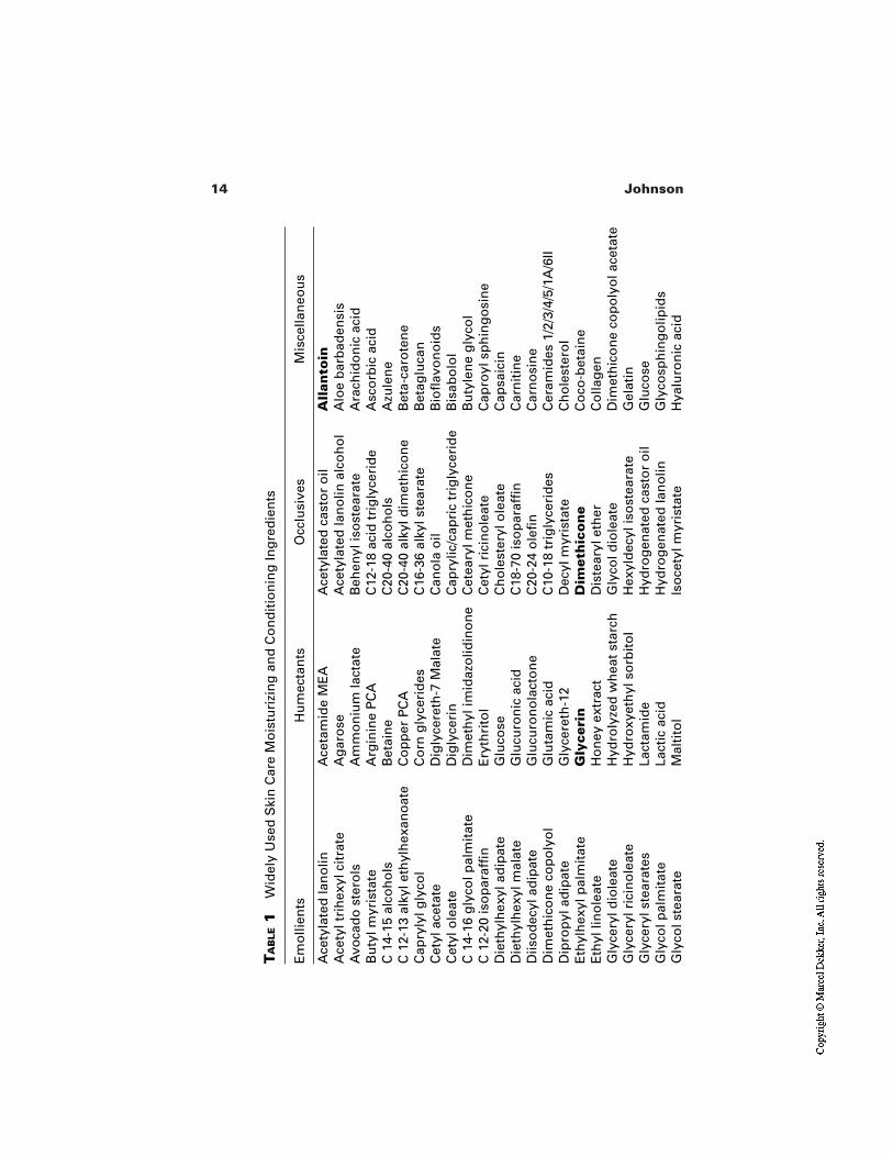
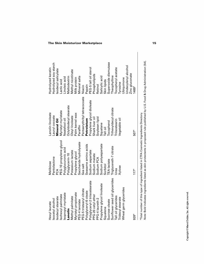
The CTFA Cosmetic Ingredient Handbook lists the many thousands of in-gredients used in skin care products [35]. There are over 3000 ingredients listedas emollient, humectant, occlusive, or miscellaneous skin conditioning agents.Some of the more widely used of these ingredients are detailed in Table 1. Emol-lients are defined as cosmetic ingredients which help maintain the soft, smooth,and pliable appearance of skin. Emollients function by their ability to remain onthe skin surface or in the stratum corneum to act as lubricants, to reduce flaking,and to improve the skin’s appearance. Humectants are cosmetic ingredients in-tended to increase the water content of the top layers of skin. This group of ingre-dients includes primary hygroscopic agents employed for this specific purpose.Occlusives are cosmetic ingredients which retard the evaporation of water fromthe skin surface. By blocking the evaporative loss of water, occlusive materialsincrease the water content of skin. The miscellaneous group is defined as cosmet-ic ingredients used to create special effects on skin. This group includes sub-stances believed to enhance the appearance of dry or damaged skin and substan-tive materials which adhere to the skin to reduce flaking and restore suppleness.
These long lists of ingredients are used in countless combinations and lev-els to produce the myriad skin care products now available to consumers every-where. The variations in formula are designed to account for different skin types,different ethnic needs, and different environmental conditions. Further variationsare made to achieve differentiated formulations and compelling claims, somefunctional and some designed to provide empathy with consumer emotionalneeds. And there are still more variations to tailor products to a particular marketsegment and for either face care or body care. Leaving aside the detailed arith-metic, it is clear that the number of legitimate product variations is very large. Itis an impossible task for the consumer to try more than a small proportion of allthese products to decide which might be best suited. Instead, most consumers areguided to the products they purchase by advertising, promotions, and the recom-mendations of specialist magazines and skin care professionals. These are keydrivers of the skin care/moisturizer marketplace and will be reviewed later.
14 Johnson
TA
BLE
1W
idel
y U
sed
Ski
n C
are
Mo
istu
rizi
ng
an
d C
on
dit
ion
ing
Ing
red
ien
ts
Ace
tyla
ted
lan
olin
Ace
tam
ide
ME
AA
cety
late
d c
asto
r o
ilA
llan
toin
Ace
tyl t
rih
exyl
cit
rate
Ag
aro
seA
cety
late
d la
no
lin a
lco
ho
lA
loe
bar
bad
ensi
sA
voca
do
ste
rols
Am
mo
niu
m la
ctat
eB
ehen
yl is
ost
eara
teA
rach
ido
nic
aci
dB
uty
l myr
ista
teA
rgin
ine
PC
AC
12-1
8 ac
id t
rig
lyce
rid
eA
sco
rbic
aci
dC
14-
15 a
lco
ho
lsB
etai
ne
C20
-40
alco
ho
lsA
zule
ne
C 1
2-13
alk
yl e
thyl
hex
ano
ate
Co
pp
er P
CA
C20
-40
alky
l dim
eth
ico
ne
Bet
a-ca
rote
ne
Cap
ryly
l gly
col
Co
rn g
lyce
rid
esC
16-3
6 al
kyl s
tear
ate
Bet
aglu
can
Cet
yl a
ceta
teD
igly
cere
th-7
Mal
ate
Can
ola
oil
Bio
flav
on
oid
sC
etyl
ole
ate
Dig
lyce
rin
Cap
rylic
/cap
ric
trig
lyce
rid
eB
isab
olo
lC
14-
16 g
lyco
l pal
mit
ate
Dim
eth
yl im
idaz
olid
ino
ne
Cet
eary
l met
hic
on
eB
uty
len
e g
lyco
lC
12-
20 is
op
araf
fin
Ery
thri
tol
Cet
yl r
icin
ole
ate
Cap
royl
sp
hin
go
sin
eD
ieth
ylh
exyl
ad
ipat
eG
luco
seC
ho
lest
eryl
ole
ate
Cap
saic
inD
ieth
ylh
exyl
mal
ate
Glu
curo
nic
aci
dC
18-7
0 is
op
araf
fin
Car
nit
ine
Diis
od
ecyl
ad
ipat
eG
lucu
ron
ola
cto
ne
C20
-24
ole
fin
Car
no
sin
eD
imet
hic
on
e co
po
lyo
lG
luta
mic
aci
dC
10-1
8 tr
igly
ceri
des
Cer
amid
es 1
/2/3
/4/5
/1A
/6II
Dip
rop
yl a
dip
ate
Gly
cere
th-1
2D
ecyl
myr
ista
teC
ho
lest
ero
lE
thyl
hex
yl p
alm
itat
eG
lyce
rin
Dim
eth
ico
ne
Co
co-b
etai
ne
Eth
yl li
no
leat
eH
on
ey e
xtra
ctD
iste
aryl
eth
erC
olla
gen
Gly
cery
l dio
leat
eH
ydro
lyze
d w
hea
t st
arch
Gly
col d
iole
ate
Dim
eth
ico
ne
cop
oly
ol a
ceta
teG
lyce
ryl r
icin
ole
ate
Hyd
roxy
eth
yl s
orb
ito
lH
exyl
dec
yl is
ost
eara
teG
elat
inG
lyce
ryl s
tear
ates
Lact
amid
eH
ydro
gen
ated
cas
tor
oil
Glu
cose
Gly
col p
alm
itat
eLa
ctic
aci
dH
ydro
gen
ated
lan
olin
Gly
cosp
hin
go
lipid
sG
lyco
l ste
arat
eM
alti
tol
Iso
cety
l myr
ista
teH
yalu
ron
ic a
cid
Em
olli
ents
Hu
mec
tan
tsO
cclu
sive
sM
isce
llan
eou
s
15The Skin Moisturizer Marketplace
Hex
yl la
ura
teM
elib
iose
Lan
olin
lin
ole
ate
Hyd
roly
zed
ker
atin
Iso
cety
l alc
oh
ol
Pan
tola
cto
ne
Lau
ryl c
oco
ate
Hyd
roly
zed
so
y st
arch
Iso
dec
yl s
tear
ate
PC
AM
iner
al O
ilIs
od
ecyl
sal
icyl
ate
Iso
hex
yl p
alm
itat
eP
EG
-10
pro
pyl
ene
gly
col
Myr
isty
l myr
ista
teLa
ctic
aci
dIs
op
rop
yl m
yris
tate
Po
lyg
lucu
ron
ic a
cid
Nea
tsfo
ol o
ilLi
no
leic
aci
dLa
no
lin
Po
lyg
lyce
rin
-10
Oct
yld
od
ecyl
ste
arat
eM
alto
dex
trin
Met
hyl
pal
mit
ate
Po
tass
ium
lact
ate
Ole
yl li
no
leat
eM
eth
yl n
ico
tin
ate
Myr
isty
l pro
pio
nat
eP
rop
ylen
e g
lyco
lP
alm
ker
nel
wax
Milk
pro
tein
PE
G-4
lan
ola
teS
acch
arid
e h
ydro
lysa
teP
araf
fin
Min
eral
sal
tsP
EG
-5 t
rist
eary
l cit
rate
Sea
sal
tP
enta
eryt
hri
tyl t
etra
coco
ate
Nia
cin
Po
lyg
lyce
ryl-
6 o
leat
eS
easa
me
amin
o a
cid
sP
etro
latu
mP
apai
nP
oly
gly
cery
l-2
triis
ost
eara
teS
od
ium
asp
arta
teP
rop
ylen
e g
lyco
l dio
leat
eP
EG
-5 t
all o
il st
ero
lP
PG
-20
cety
l eth
erS
od
ium
mal
ate
Sh
ark
liver
oil
Ph
osp
ho
lipid
sP
PG
-4 la
ure
th-2
So
diu
m P
CA
So
ybea
n li
pid
Ret
ino
lP
rop
ylen
e g
lyco
l lin
ole
ate
So
diu
m p
oly
asp
arta
teS
qu
alan
eS
alic
ylic
aci
dS
qu
alen
eS
orb
ito
lTa
ll o
ilS
kin
lip
ids
Su
cro
se o
leat
eT
EA
-lac
tate
Toco
ph
ero
lS
up
ero
xid
e d
ism
uta
seS
un
flo
wer
see
d o
il g
lyce
rid
esTr
igly
cere
th-7
cit
rate
Trih
exyl
dec
yl c
itra
teT
heo
ph
yllin
eTa
ll o
il g
lyce
rid
esU
rea
Triis
ost
eari
nTo
cop
her
yl a
ceta
teTr
idec
yl s
tear
ate
Xyl
ose
Veg
etab
le o
ilTy
rosi
ne
Wh
eat
ger
m g
lyce
rid
esU
biq
uin
on
eU
nd
ecyl
enyl
alc
oh
ol
Zin
c g
luco
nat
e92
8a11
7a50
7a14
89a
aTo
tal n
um
ber
of
this
typ
e o
f in
gre
die
nt
liste
d in
CT
FA C
osm
etic
Ing
red
ien
ts D
irec
tory
.N
ote
:Bo
ld in
dic
ates
ing
red
ien
ts li
sted
as
skin
pro
tect
ants
in p
rop
ose
d r
ule
pu
blis
hed
by
U.S
. Fo
od
& D
rug
Ad
min
istr
atio
n [
54].
16 Johnson
Effective moisturizers must do more than simply restore water to the stra-tum corneum. They must also facilitate the recovery of dry damaged skin andprovide protection against future damage and further water loss [36]. Modernmoisturizer products perform these functions and often a great deal more. Mois-turizers have become the vehicle for providing a wider range of skin care benefitsintended to maintain and improve overall skin condition. Before considering themore broadly based benefits of moisturizer products we will review the factorswhich influence the consumer need for moisturization.
7.1 Factors Influencing the Need for and Types of Moisturizers
Environmental factors other than cleansers induce and exacerbate dry skin [37].Some of the drying factors actually remove water from the skin, while others dis-turb or damage the skin processes for holding water, namely, the lipid bilayersand NMF. Product variations designed to address the impact of environmental in-fluences on skin condition add further to the diversity of moisturizer products inthe marketplace.
Anything that removes water from the skin surface faster than it can be re-placed by normal trans-epidermal water movement will disturb desquamationand cause the signs of skin dryness described. It must be remembered that dryskin is only dry (lacking water) in the superficial layers of the stratum corneum.These layers become dry because they lose the ability to hold water even thoughwater is available from the lower regions of the stratum corneum.
Cleansers and water are the main factors damaging the water-holdingmechanisms of the superficial stratum corneum. Wind and low humidity are themain environmental factors removing water from damaged regions of the superfi-cial stratum corneum. In the same way that wind dries clothes on the washing lineby increasing evaporation it dries out corneocytes at the skin surface. How effec-tively the wind removes water depends on humidity and the amount of NMF inthe stratum corneum. Relative humidity is the percentage of water in air com-pared with saturated water content at that same temperature. When the relativehumidity is high the skin’s NMF has little difficulty in holding water in the pro-tein matrix of corneocytes. At low humidity the NMF is unable to hold wateragainst the pull of low partial pressure at the skin surface. If the NMF is depleted,there is nothing to hold water at low relative humidity (RH) and the skin surfacebecomes very dry.
Temperature can also play a role in determining dry skin condition. Coldtemperature has two effects. The colder the air, the less water it can hold, so skinin equilibrium with 60% RH cold air has much less moisture than skin in equilib-rium with 60% RH warm air. Also, cold temperature greatly reduces the mobilityand flexibility of stratum corneum lipids and predisposes it to physical cracks in
17The Skin Moisturizer Marketplace
regions like knuckles where skin is subject to stretching forces. Normal hotweather temperatures are not drying unless the relative humidity is low. Howev-er, the UVB in strong sunlight can interfere with the skin’s normal mechanismsfor generating NMF, resulting in a deficiency of NMF that predisposes to dry,flaky skin.
Having considered factors which actually remove water from skin, the nextgroup of skin drying agents are those that disturb the skin processes for water re-tention. Cleansers, we have seen, are potentially very damaging to the skin’s wa-ter-holding mechanisms. Perhaps surprisingly, water itself can also be very dry-ing by washing away NMF. Overexposure to solar UVB radiation can reduce theNMF content of skin by interfering with filaggrin degradation in the mid-lowerregions of the stratum corneum. Ultraviolet radiation also interacts with stratumcorneum lipids to generate lipid peroxides, and these are a further contribution todry skin by disturbing the regularity and efficiency of the lipid bilayers.
Each of the different circumstances leading to dry skin conditions createsthe opportunity for a customized product [38]. In 1950, a few basic moisturizerswere available, but by 2000 there was not only a separate product for each even-tuality but often multiple product offerings, each trying to be a little differentfrom the next. To argue that not all these products are necessary is to invite the re-sponse that choice is good for the consumer. And so it is, provided the consumeris able to make an informed choice with comfort and confidence. It appears thatthe marketplace has reached such a degree of complexity that many consumerssimply find a zone of comfort and disregard the rest. This encourages manufac-tures to intensify their efforts to attract consumers to their products. Notwith-standing these efforts, it is consumers who ultimately determine products that lastin the market place. There may be thousands of moisturizer products on sale atany one time, but only a few of these products have real staying power. The restdisappear in a continuous cycle of withdrawal and replacement. Products that arenot successful are discontinued and replaced by new products containing new in-gredients and making new claims.
8 THE PRODUCT CYCLE
Because moisturizers fullfil such a fundamental consumer need they are a hugecategory of the consumer products market. Moisturizers are big business allaround the world, and the moisturizer market is intensively competitive. Eachmanufacturer is vying with all others to gain the largest possible share of market.Manufacturers do this by supporting their existing products with advertising (TV,print, radio, and others) and promotions (discounts, bonus offerings, product tie-in competitions, etc.) and by launching new products. Advertising support for ex-isting products is very expensive and launching a new product is even more ex-pensive, particularly for large manufacturers. In developed markets like the
18 Johnson
United States the failure rate for new product launches is about 95%. Approxi-mately 19 of every 20 newly launched products are not successful and disappearwithin a year or so. Most of these failures are from smaller manufacturers whocan afford to be entrepreneurial. They are able to try products and recycle quick-ly when not successful. The cost of investment for large manufacturers is sohigh—millions of dollars in both development costs and advertising support fornew product launches—that they have to be sure that a new product has highpotential for success before they enter the marketplace. They do this using so-phisticated consumer testing, test marketing, and ancillary techniques that enablean estimate of approximate market share for a new product. Only product devel-opments that show a high probability of success proceed to launch.
9 FACE AND BODY SEGMENTS OF THEMOISTURIZER MARKETPLACE
As indicated, the skin care and moisturizer market is divided between face careand hand and body care products. The dynamics of each of these two market cat-egories are very different. There are thousands of moisturizer products for theface and relatively few for the body. This reflects the different consumer needs forthe face and body.
9.1 Facial Moisturizers
The face is our interface with the outside world. The face is also the part of thebody that most shows the signs of aging. The face is constantly exposed to the en-vironment, whereas clothing may protect other parts of the body. Lines and wrin-kles appear on the face but not much on the body. The recognition of the first per-manent wrinkle is a pivotal moment for most people and perhaps surprisingly isoften experienced in the early 30s. At one time facial moisturizers were simplymoisturizing products. They were used to balance the drying effects of cleansingand to protect the skin against the elements—moisturized skin is better able to re-sist a drying environment. Moisturized skin also looks healthier and more radiantthan dry skin. Facial moisturizers have always contained emollients, with orwithout humectants, typically in esthetically pleasing formulations. More recent-ly, moisturizers have become the vehicle to address other problems of facial skin,particularly those age-related changes which are perceived as the visible signs ofaging. Products designed to address the signs of aging are known as anti-agingproducts [39,40].
Historically, anti-aging was the province of the prestige marketplace with avariety of ingredients added to moisturizers to create anti-aging products. For ex-ample, in the 1970s a number of products contained placental extract as a skin re-juvenating ingredient. At that time, mass market products mostly continued to of-
19The Skin Moisturizer Marketplace
fer moisturizing benefits as the way to maintain skin in healthy condition andlooking younger for longer. This changed in 1992 with the introduction of alpha-hydroxy acids (AHAs) in mass market moisturizers, the first really effective anti-aging technology introduced in mass market skin care products [41]. As ex-plained in chapter 16 dedicated to AHAs, these ingredients produce clinicallydemonstrated and consumer perceivable improvements in the visible signs of fa-cial aging. The use of AHAs was so successful that it created a new category ofmoisturizing product, initially in the United States and then extending around theworld. Interestingly, an AHA (lactic acid) had been used as a moisturizing ingre-dient in mass market moisturizers since the early 1970s [42], but it turned out thata low pH is necessary for the anti-aging benefits beyond simple moisturizing (pH3.8 is used in mass market AHA moisturizers).
AHAs transformed the moisturizer marketplace, not only by creating a newsector, but by enhancing the credibility of anti-aging claims and creating a newexpectation in mass market consumers. The success of AHAs and associatedchange in consumer need stimulated manufacturers to search for even more ef-fective ingredients for anti-aging moisturizers. The result has been intensified re-search by both skin care manufacturers and the ingredient supply industry [43]. Inthe period between 1992 and 2000, several ingredients were promoted as the nextgeneration of cosmetic anti-aging technology. The main contenders were retinoland retinol esters, vitamin C and stable derivatives of vitamin C, other anti-oxi-dant vitamins, and a variety of botanical and marine extracts. The efficacy ofthese ingredients is discussed in other chapters of this volume, but each has beenthe platform for a new range of products in the marketplace. Impressive and com-pelling claims of anti-aging benefits have been made for each technology, but sofar nothing has made a step change in consumer perceived efficacy comparablewith that seen when AHAs were introduced in 1992.
9.2 Hand and Body Moisturizers
The main consumer skin care need for the body is universal, an all-family need totreat and prevent dry skin. There are many products that do this very effectively.In fact, the treatment and prevention of dry skin is such a well-satisfied need thatit has become difficult for manufacturers to find a competitive edge. With all lead-ing dry skin products similarly very effective for everyday dry skin we see manu-facturers broadening the benefits of hand and body moisturizers as a way toachieve novelty and competitive edge [44,45]. A recent significant developmentis the addition of sunscreens to regular hand and body moisturizers [46].
Other body care needs exist but have a narrower focus. For example, wo-men with photodamaged hand and arm skin seek moisturizers to address the as-sociated signs of aging, particularly age spots and coarse/crepey texture (crinkledappearance). In older women, thinning, sagging, and laxity are additional prob-
20 Johnson
lems of skin that are unmet needs. A specific problem for a surprisingly high pro-portion of women is cellulite [47], creating a need for products to eliminate or re-duce the cellulite appearance of thighs and buttocks. There are moisturizer prod-ucts with additional specific ingredients aimed at each of these consumer needs.
Products to treat and prevent dry skin are the main products in the hand andbody moisturizer market worldwide, but there are some regional differences.Moisturizers to maintain a fair skin color are a large segment of the moisturizermarket in India and Southeast Asia, a reflection that uneven skin tone is the num-ber one skin care concern of some consumers [48].
Cleansing and the environment induce dry skin, but there are also personalfactors that come into play. There are some people, particularly atopics, who haveno overt skin disease but who are more prone to develop dry skin than the rest ofthe population [49]. The atopic condition is explained later in chapter 9. Atopicstend to have dry skin all year round regardless of weather, and they often developsevere dry skin in winter or in drying environments [50]. It is now recognized thatmany cases of occupational hand dermatitis occur in atopics and reflect their de-creased ability to resist conditions that dry out the skin, leaving them more sus-ceptible to environmental irritants. The number of atopics in the population hasbeen rising steadily around the world since the 1970s [51]. Because many indi-viduals with an atopic tendency are unaware of their condition they often contin-ue the patterns of product use and environmental contacts that promote and prop-agate their dry skin condition.
Dry skin is without doubt the most common skin problem for consumersaround the world, but for many it is not a serious skin problem. Some consumersare content to live with some level of dry skin and regard this simply as their nor-mal skin condition. Use of body moisturizers tends to parallel cleansing routines.In the United States and increasingly around the world, daily showering has be-come a common practice. This represents a considerable challenge to the naturalmoisturizing processes of the skin, and in drying weather it is not long beforeconsumers experience distinctly dry and itchy skin after each and every shower.Use of a body moisturizer becomes a necessity. As we will see later, the need formoisturizing to combat the drying effects of showering led to the development ofan entirely new product category in the skin care market place, the moisturizingbody wash [52].
The market for hand and body moisturizers shows a clear segmentationbased on price and positioning. The main categories are value/low price brands,everyday products, therapeutic, and cosmetic. Within each category there are twomain product forms, creams and lotions, with lotions the most common. There isalso petroleum jelly that is unique as a treatment for dry skin and as a skin pro-tectant.
Value brands are low price products, usually store brands or “unknown”products that sell at a discount to the rest of the market. Everyday products in-
21The Skin Moisturizer Marketplace
clude the main branded products that offer performance at a competitive price.Therapeutic is a smaller category with products that sell at a significant premiumover everyday brands based on a positioning of superior efficacy. Products in allthese categories are effective for treating and preventing dry skin. Relative effec-tiveness, as measured by controlled clinical trials, varies (but not greatly) and de-pends on the particular moisturizing ingredients and their concentration in theformulation. Although there are differences in effectiveness for most consumers,most of the time there is little practical difference for dealing with everyday dryskin needs. The consumers who could be expected to most notice small differ-ences in efficacy are those with the greatest problems and needs. This would bethe groups exposed to particularly harsh conditions or individuals with a personalincreased susceptibility to developing dry skin, particularly the 20–30% of thepopulation who have an atopic tendency.
Product effectiveness and product esthetic properties pull in different direc-tions; in general, the greater the content of oils and humectants, the greater the ef-ficacy but also the heavier the product for rubbing into the skin.
The cosmetic category of the hand and body moisturizer market includesproducts that are light, elegant, and esthetically pleasing. These formulations con-tain lower levels of humectants and emollient oils than mainline dry skin productsto achieve faster rub-in and better skin feel. Cosmetic moisturizers usually have afeminine positioning, often centered around fragrance, botanicals, and other emo-tive ingredients. Nevertheless, many cosmetic category products contain suffi-cient moisturizing ingredients to be effective for their main use as daily mainte-nance products to keep skin moisturized and in good condition, rather than totreat dry skin.
9.3 Other Subdivisions of the Body Category
By now the reader has a good appreciation that nearly every subdivision of con-sumer activity, both physical and emotional, represents a consumer need that im-mediately becomes a stimulus for moisturizer products customized to that need.The main hand and body lotions described are good for general use, but withinthe broad category it is possible to find moisturizers targeted at rather specificneeds, such as care of the finger nails and for foot care.
10 SKIN MOISTURIZERS AND DERMATOLOGY
Dermatologists dealing with skin diseases that predispose toward development ofdry skin need their patients to use skin care products that help rather than worsentheir underlying skin condition. They need their patients to use a mild cleanserand moisturizing cream or lotion. The nonprescription products recommended bydermatologists are the same as those available to the general consumer. Although
22 Johnson
the dry skin experienced by skin disease patients and by consumers generallymay have different origins, the solution is much the same—a well-formulatedemollient cream or moisturizing lotion [53]. However, patients often have moreintractable dry skin than the average consumer and therefore need the heaviermore effective moisturizing formulations.
11 REGULATORY CATEGORIES OF SKIN MOISTURIZERS
Moisturizers generally are not specifically regulated in the United States. There isno requirement for premarketing approval or registration. However, in the UnitedStates there is an over-the-counter (OTC) drug category for many everyday skincare products, including acne, first aid, and antibacterial treatments as well assunscreen and skin protectants [54]. Some hand and body moisturizers fall withinthe aegis of the OTC skin protectant monograph and are therefore subject to con-trols including labeling requirements and allowed claims. Monograph productstypically must contain particular “active” ingredients (see Table 1). In the case ofthe OTC skin protectant monograph, two specified active ingredients are petrola-tum and dimethicone, at specified minimum levels—30% or higher for petrola-tum and 1% or higher for dimethicone. If the difference between these concentra-tions is surprising it is a reflection that limit values built into regulations oftenreflect the actual compositions of products in the marketplace at the time that reg-ulations were formulated. There is no scientific rationale for the large differencesin the monograph minimum levels of these two ingredients. The OTC monographproducts are required to list active ingredients on the pack above and separatefrom the list of other ingredients. Most U.S. consumers are not aware of the OTCmonograph system and therefore may wonder why some moisturizers have activeingredients and other similar products do not. This regulatory overlay is a furthercomplication for consumers trying to understand the skin moisturizer market.
As described in later chapters on regulations, the European and Japaneseregulation of skin moisturizers is different from the United States. European reg-ulations tend to control the ingredients used in products and therefore, unlike theUnited States, do not lead to separate regulated and unregulated products in themarketplace. Japan is somewhat like Europe in the sense that the main regulationimpacting skin and moisturizers is a quasi drug regulation that directs what ingre-dients can be used in cosmetics, including moisturizers.
12 COSMECEUTICAL
Readers will find the term cosmeceutical used frequently in cosmetic and skincare journals, magazines, and other publications [55]. If you visit a prestigecounter in the department store, the consultant may offer you the latest cosme-
23The Skin Moisturizer Marketplace
ceutical treatment to combat aging skin. The term is widely used to describemoisturizing products that go beyond moisturizing to offer some additional skinbenefit [56]. There have been international conferences to present and review therole and future trends for cosmeceuticals [57]. Many people have come to believethat “cosmeceutical” is a regulatory category of consumer products. Nothingcould be further from the truth [58]. The term has no regulatory status anywherearound the world, although the Japanese quasi drug category is close in concept tothe now accepted use of the term cosmeceutical.
There now is a generation who believe that the term cosmeceutical was in-troduced in the early 1990s with the emergence of alpha-hydroxy acid anti-agingproducts as the first moisturizers to do more than moisturize the skin. In fact theterm cosmeceutical originated in 1962 [59] and was expanded upon years later byDr. Albert Kligman [60]. In the early 1980s, at a symposium organized by the So-ciety of Cosmetic Chemists, Dr. Kligman pointed out that simple moisturizershad a profound effect on the stratum corneum. They clearly had a beneficial effecton the stuctural elements and proper functioning of the stratum corneum. Theywere cosmetics having a therapeutic action; they were “cosmeceuticals.” Tenyears later, Vermeer put forward proposals to define and regulate cosmeceuticals[61], but the cosmetics industry in the United States remains bound by legislationdeveloped in 1937 that defined any material having an effect on the structure orfunction of skin as a drug and subject to drug regulations.
13 MOISTURIZING CLEANSERS
13.1 Cleansing Bars
The skin care industry understood many years ago that bar soap caused dry skin,creating a need for products to restore moisture. Clearly there would be a con-sumer benefit by reducing the drying action of soaps. There are two types of soapbars, the regular opaque colored bars everyone is familiar with and clear glycerinbars which are made by a completely different manufacturing process. Regularsoap is made by hydrolyzing natural fats and oils (glycerides) to yields fatty acidand glycerol, separating out the glycerol, and converting the fatty acid to soap(soap is the sodium salt of fatty acid). Glycerin bars are made by leaving in theglycerin to produce a different form of regular soap that develops transparencywhen aged under controlled conditions for 2 months.
Back in the 1960s there were two approaches to reduce the drying action ofsoaps:
1. To balance the chain length distribution toward the milder longer chainlengths
2. To add fatty acid to the soap mix to produce so-called superfatted soap
24 Johnson
While these variations were a little milder than regular soap, it was not until theearly 1960s that there was a real advance in reducing the drying effects of cleans-ing. This was the introduction of a cleansing bar based on nonsoap synthetic de-tergent, fatty acid isethionate, and addition of a large proportion of fatty acid tothe bar (>20%). This product, called Dove, was dramatically milder than soap[62] and was unequaled in the marketplace until its patents expired in 1992, atwhich time other manufacturers copied the technology to make their own versionof the mild synthetic detergent (syndet) bar. The Dove bar was not only less dry-ing than soap but deposited some fatty acid on skin during the washing process.As discussed, fatty acids are one of the three lipid types that make up the intercel-lular lipid bilayers of the stratum corneum. The deposition of fatty acid by Dovelargely compensated for the natural fatty acid lost during the wash process. Be-cause the milder syndet bars induce less dryness than soap bars, regular use of amild syndet bar, particularly in cold drying weather, helps keep skin relativelymore moisturized than regular use of soap. In addition to regular soap bars thereare translucent/transparent bars, often referred to as glycerin bars, that are inter-mediate between regular soap and syndet bars for skin drying [63]. By year 2000,the cleansing bar market was still predominantly soap bars (60%), syndet bars(25%), and combination bars, mixtures of soap and syndet (15%). However, thebar market had evolved from simple cleansing to offering a wider range of bene-fits [64].
13.2 Moisturizing Cleansing Liquids
Liquid detergent products are an alternative to bar soap for hand cleansing andgeneral body cleansing, particularly in the shower. The first shower products weresimple formulations based on combinations of relatively mild surfactants. Theseproducts were generally less drying than soap but also less effective for cleansing.However, during the 1990s, frequent showering, often daily, was the norm, andthis was more for refreshment and removal of body odors than for washing awaydirt and grime. The cleansing power of soap was not needed. Liquid products, al-though usually milder than soap, were still inclined to leave the user feeling tightand dry after showering. This problem created a product opportunity, and skincare manufacturers responded with a new category of shower products calledmoisturizing body washes. These product were very different from the previous-ly available shower liquids, most particularly by containing a high level of emol-lient oil and a deposition system, often polymer based, to promote deposition ofemollients and resistance to rinsing away. The moisturizing benefits of theseproducts, particularly when skin is a little dry before showering, is readily per-ceived and many women report that they have less need to use a moisturizing lo-tion after showering. The skin moisturizing effect can also be demonstrated clini-
25The Skin Moisturizer Marketplace
cally using instruments to reveal an increase in hydration and visual grading todemonstrate a reduction of visual dryness [65].
13.3 Moisturizing Cleansing Wipes
The disposable wipes segment of the cleansing market has been growing steadilysince the late 1980s. The common products are everyday wipes and baby wipes,but toward the end of the 1990s a number of new disposable wipe type productsappeared. There were make-up removal wipes, antibacterial cleansing wipes,foaming cloths for facial cleansing, and skin moisturizing wipes. While most wetwipes provide a transient hydration of skin, wipes designed to be moisturizingusually contain a humectant such as glycerol.
14 SKIN MOISTURIZERS AND SUN PROTECTION
Sunscreens containing specific moisturizing ingredients were introduced in theUnited States in the 1980s. These products contained the usual sunscreen ingredi-ents to give a full range of sun protection factors (SPF) as well as humectants formoisturizing benefit. Over the years many manufacturers have added moisturiz-ing ingredients to their sunscreen product range. Notwithstanding this extensionof benefits, products in the sunscreen segment of the marketplace are seasonalproducts intended primarily to protect against sunburn.
In addition to sunscreen products containing moisturizing ingredients thereare face and body moisturizing products which contain sunscreen ingredients. Formany years facial moisturizing creams and lotions have contained sunscreens forprotection against lines, wrinkles, and other long-term effects of solar UV. In thiscontext sunscreens are used as anti-aging ingredients. Facial products are seg-mented into day and night products. Not surprisingly, it is the day creams thatcontain sunscreens. Most manufacturers also offer day creams without sun-screens, as these are lighter formulations and preferred by some consumers.
Prior to 1998, sunscreens were not added to body moisturizers, but now sev-eral leading manufacturers of hand and body products have at least one variantcontaining sunscreen in their dry skin product range. These products are intendedto provide protection from everyday UV exposure in addition to treating and pre-venting dry skin. It is interesting to note that one manufacturer introduced a dryskin product containing sunscreens in the 1980s but the product failed. Consumersdid not perceive a need for such a product at that time and did not buy it. Now, over20 years later and with intensive medical and media focus on escalating skin can-cer rates, protection from everyday UV exposure makes sense for consumers andthe new generation of sunscreen moisturizers seem assured of success.
26 Johnson
15 THE MEDIA AND THE MESSAGE
With so many different moisturizers available it is not surprising that many con-sumers earnestly seek information to guide their choice of skin care products.One thing is certain: no consumer could ever try all the available skin products todetermine in practice the ones most appropriate for one’s individual needs. Fortu-nately, or maybe not, information on skin care and skin care products is every-where: magazine and newspaper articles and advertisements; radio and televisionprograms and advertisements; products labels; promotional pamphlets and junkmail; friends, acquaintances, relatives, coworkers, beauty consultants, and derma-tologists; and now on the internet.
Some of the information from these sources is excellent; regrettably someis little more than piffle. Most of the information on skin care products and tech-nologies falls between these two extremes. One common source of misinforma-tion is the expectation that an activity demonstrated for an ingredient in a test tubesystem will apply when the ingredient is incorporated in a moisturizer and ap-plied to skin. Unfortunately, a lot of misinformation cycles between the differentsources and acquires familiarity and credibility in the process. How much of whatyou know about skin care products is what you have heard or read in sources oth-er than peer reviewed scientific journals?
The internet has become a major source of information for skin care prod-ucts with numerous sites offering both products and advice [66]. All of the majormanufacturers have web sites to promote their products and to provide relevanttechnical information. Because the internet is open to all and not regulated forcontent, it may seem that anything goes. However, the web sites of the majormanufacturers can be regarded as reasonably reliable because the posted informa-tion will have been subject to internal legal review and approval.
Prior to the internet, and probably still true for a majority of consumers,specialist magazines were the primary source for information and advice aboutmoisturizers and skin care generally. Magazines come and go but all have thesame categories of contents. Most women’s magazines have a selected target au-dience such as teenagers, young mothers, sophisticated women, executives, olderwomen, active seniors, or other groups. All the magazines have many pages de-voted to advertisements for skin care, beauty, and fashion items. Unlike adver-tisements on television, which must package the product message into a fewmemorable sound bites, full-page print advertisements can contain a great deal ofinformation, although usually in headline form and with no reference to source orexplanation. All magazines have a number of feature articles and regular sectionsthat typically include reviews of new products, market trends, and skin health is-sues and treatments. Each article in a magazine is selectively tailored to the par-ticular readership of that magazine.
The reliable sources of information on skin care issues and effective treat-
27The Skin Moisturizer Marketplace
ments are the peer reviewed journals. There is the International Journal of Cos-metic Science, several peer reviewed journals in dermatology, such as the Journalof Investigative Dermatology, and several more broadly based medical journals,such as the New England Journal of Medicine. These journals are not the normalreading for most of the individuals who write or contribute to skin care articles.
16 THE FUTURE
The trend in the skin care market at this time is ever increasing specialization andcustomization of products [67,68]. Accompanying this trend is an explosion of in-formation that serves to direct consumers toward products most relevant to indi-vidual needs and also creates an expectation that these needs can be satisfied.Hope has always been ahead of technology in skin care. Expectations, based onwhat is communicated from the marketplace, are also moving ahead of technolo-gy. The challenge for the marketplace is to provide good information as well asbetter products. The consumer needs both.
There is a book to be written about the sources and communication of skincare information. While information about skin care products and technologiesseems almost endless there is an issue of reliability, but not because of any mali-cious intent to deceive. There is an element of truth in almost everything that isreported. Much of the misinformation that could be identified as unsound by anexpert is very plausible and can often seem reasonable to the informed nonexpert.In this situation how does the consumer figure out what to believe and what to re-ject? One answer is to ensure the widest possible readership of books such as this.Another answer is the consumer answer: what matters is not what is said or writ-ten but only how a product performs. If a product works, the consumer will findit. If the consumer is led to expect performance but it is not delivered, the con-sumer will be disappointed, move on to something else, and the product will die.
The mass market for moisturizers started over 100 years ago with productssuch as Vaseline Petroleum Jelly and Pond’s Cold Cream as single products tomeet most every need for body and face care. From this humble beginning themarketplace has evolved to the sophistication and customization of the 21st cen-tury. Moisturizers certainly work. Regular use will keep the skin in good condi-tion and help to maintain that good condition over time. Addition of a select num-ber of ingredients, as described elsewhere in the book, has enhanced the benefitsof moisturizers beyond simple hydration of the stratum corneum, but these ad-vanced products fall well short of satisfying unmet consumer skin care needs. Theskin moisturizer marketplace is highly fragmented and very complex. Thereseems little reason to expect this will change unless and until there are some bigbreakthroughs in the science and technology of skin care—not just another ingre-dient or product form adding incremental benefit, but something fundamental.The science fiction writers have envisioned wands to diagnose and treat the skin.
28 Johnson
The medical profession is making gene therapy a reality for select disease condi-tions. A wand or a gene product that worked for skin would certainly change theface of the moisturizer market. In the meantime, what you read in this book islikely to remain the current position for a long time to come.
REFERENCES
1. Global Skin Care. Datamonitor. 1998. www.datamonitor.com.2. Nacht S. 50 years of advances in skin care. Cosmet Toil 1995; 110(12):69–82.3. Bitz K. The international top 30. Happi 2000; 37(8):74–75.4. Branna T. The top 50. Happi 2000; 37(7):67–68.5. Hartfield E. Is it the end of the mass market? Cosmet Int 2000; 23(538):8.6. Benedetto AV. The environment and skin aging. Clin Dermatol 1998; 16:129–139.7. Fisher GJ, Wang AQ, Datta SC, Varani J, Kang S, Voorhees JJ. Pathophysiology of
premature skin aging induced by ultraviolet light. N Engl J Med 1997;337(20):1419–1428.
8. Ratner D, Tse Y, Marchell N, Goldman MP, Fitzpatrick RE, Fader DJ. Cutaneouslaser resurfacing. J Am Acad Dermatol 1999; 41:365–389.
9. Guttman C. Branching into cosmetic procedures no stretch. Dermatol Times 1999;20(2):1,23.
10. Tenerelli MJ. The State of the skincare industry revealed. Global Cosmet Ind 1999;164(6):42–48.
11. O’Sullivan RL, Lipper G, Lerner EA. The Neuro-immuno-cutaneous-endocrine net-work: relationship of mind and skin. Arch Dermatol 1998; 134:1431–1433.
12. Johnson AW, Nettesheim S. The care of normal skin. In: Arndt KA, Leboit PE,Robinson JK, Wintroub BU, eds. Cutaneous Medicine and Surgery. Vol. 1: Philadel-phia: WB Saunders 1995:75–83.
13. Polefka TG. Surfactant interactions with skin. In: Zoller U, Broze G, eds. Handbookof Detergents. Part A: Properties. New York: Marcel Dekker, 1999:433–468.
14. Rawlings AV, Scott IR, Harding CR, Bowser PA. Stratum corneum moisturization atthe molecular level. J Invest Dermatol 1994; 103:731–740.
15. Johnson AW. Dry skin. Recent advances in research and therapy: a continuing edu-cation program for pharmacists. Drug Store News for the Pharmacist 1994; 4:51–58.
16. Dykes P. Surfactants and the skin. Int J Cosmet Sci 1998; 20:53–61.17. Misra M, Ananthapadmanabhan KP, Hoyberg K, Gursky RP, Prowell S, Aronson M.
Correlation between surfactant-induced ultrastructural changes in epidermis andtransepidermal water loss. J Soc Cosmet Chem 1997; 48:219–234.
18. Whalley GR. Solid soap phases. Happi 1998; 35(7):72–74.19. Rawlings AV, Watkinson A, Rogers J, Mayo A, Hope J, Scott IR. Abnormalities in
stratum corneum structure, lipid composition, and desmosome degradation in soap-induced winter xerosis. J Soc Cosmet Chem 1994; 45:203–220.
20. Prottey C, Ferguson T. Factors which determine the skin irritation potential of soapsand detergents. J Soc Cosmet Chem 1975; 26:29–46.
21. Wiechers JW, Wortel VAL. Bridging the language gap between cosmetic formulatorsand consumers. Cosmet Toil 2000; 115(5):33–41.
29The Skin Moisturizer Marketplace
22. Morizot F, Guinot C, Lopez S, Le Fur I, Tschachler E. Sensitive skin: analysis of symptoms, perceived causes and possible mechanisms. Cosmet Toil 2000;115(11):83–89.
23. Bogensberger G, Baumann L. What you should know about “sensitive skin.” Skin &Aging 1999; 7(9):75–78.
24. Nassif A, Chan SC, Storrs FJ, Hanifin JM. Abnormal skin irritancy in atopic der-matitis and in atopy without dermatitis. Arch Dermatol 1994; 130:1402–1407.
25. Bolognia JL, Braverman IM, Rousseau ME, Sarrel PM. Skin changes in menopause.Maturitas 1989; 11:295–304.
26. McKinlay SM. The normal menopause transition: an overview. Maturitas 1996;23:137–145.
27. Robinson MK. Population differences in skin structure and physiology and the sus-ceptibility to irritant and allergic contact dermatitis: implications for skin safety test-ing and risk assessment. Contact Dermatitis 1999; 41:65–79.
28. Berardesca E, Maibach H. Racial differences in skin pathophysiology. J Am AcadDermatol 1996; 34:667–672.
29. MacDonald V. Ethnic skin care. Happi 2000; 37(10):65–81.30. Cosgrove J. Emerging trends in multicultural skin care needs. Soap & Cosmetics
2000; 76(12):58–60.31. Funke U, Fartasch M, Diepgen TL. Incidence of work-related hand eczema during
apprenticeship: first results of a prospective cohort study in the car industry. ContactDermatitis 2001; 44:166–172.
32. Billek DE. Cosmetics for elderly people. Cosmet Toil 1996; 111(July):31–37.33. Yaar M, Gilchrest BS. Aging versus photoaging: postulated mechanisms and effec-
tors. J Invest Dermatol 1998; 3:47–51.34. Callens A, Vallans L, Leconte P, Berson M, Gull Y, Lorelle G. Does hormonal skin
aging exist? A study of the influence of different hormone therapy regimens on theskin of postmenopausal women using non-invasive measurement techniques. Der-matology 1996; 193:289–294.
35. Wenninger JA, Canterbery RC, McEwen GN Jr, eds. International Cosmetic Ingredi-ent Dictionary and Handbook. Vol. 2. 8th ed. Washington, D.C.: The Cosmetic, Toi-letry, and Fragrance Association, 2000:1767–1785.
36. Halkier-Sorensen L. Understanding skin barrier dysfunction in dry skin patients.Skin & Aging 1999; 7(5):60–64.
37. Parish WE. Chemical irritation and predisposing environmental stress (cold windand hard water). In: Marks R, Plewig G, eds. The Environmental Threat to the Skin.London: Martin Dunitz, 1992:185–193.
38. Idson B. Dry skin moisturizing and emolliency. Cosmet Toil 1992; 107(7):69–68.39. Owen DR. Anti-aging technology for skincare 1999. Global Cosmet Ind 1999;
164(2):38–43.40. Owen DR. Anti-aging technology for skincare. Part II 1999. Global Cosmet Ind
1999; 164(3):40–43.41. Hermitte R. Aged skin, retinoids, and alpha hydroxy acids. Cosmet Toil 1992;
107(7):63–64.42. Middleton JD. Development of a skin cream designed to reduce dry and flaky skin, J
Soc Cosmet Chem 1974; 25:519–534.
30 Johnson
43. Radd BL. Antiwrinkle ingredients. Skin Inc 1997; 9(1):89–99.44. Hand/body lotions solid performers. Chain Drug Rev 1999; November:21–24.45. Hand, body creams incorporate traits of facial care items. Chain Drug Rev 1999;
January:51.46. Nole GE, Johnson AW, Cheney MC, Znaiden A. Cumulative lifetime UVR exposure
in the United States and the effect of various levels of sunscreen protection. CosmetDermatol 1999; July:23–26.
47. Kligman AM. Cellulite: facts and fiction. J Geriatr Dermatol 1997; 5(4):136–139.48. Tenerelli MJ. Ethnic skin-care: special products for a special sector. Global Cosmet
Ind 2000; 167(4):32–37.49. Loffler H, Effendy I. Skin susceptibility of atopic individuals. Contact Dermatitis
1999; 40:239–242.50. Leung DYM, Soter NA. Cellular and immunologic mechanisms in atopic dermatitis.
J Am Acad Dermatol 2001; 44(Suppl 1):1–12.51. Taieb A. Hypothesis: from epidermal barrier dysfunction to atopic disorders. Contact
Dermatitis 1999; 41:177–180.52. Marchie MK. Liquids move up in the soap market. Happi 2000; 37(12):72–84.53. Emollients: current uses and future trends. Skin & Aging 1999; 7(Suppl 6):5–15.54. US FDA Skin protectant drug products for over-the-counter human use. 48 Fed. Reg.
6820, February 15, 1983; amended 58 Fed. Reg. 54458, October 21, 1993; amended59 Fed. Reg. 28767, June 3, 1994.
55. Labous J. Mother nature shows. Cosmet Int 2000; 24(547):8–9.56. Levine N, Draelos ZD. Rising tide of cosmeceuticals provokes physician questions.
Dermatol Times 2001; 22(1):59–60.57. Steinberg DC. Cosmeceuticals: an advanced forum for manufacturers. Cosmet Toil
1996; 111(6):43–49.58. Branna T. Is the industry really ready for cosmeceuticals? Happi 1996; 33(8):60–66.59. Reed RE. The definition of “cosmeceutical.” J Soc Cosmet Chem 1962; 13:103–106.60. Vermeer BJ. Cosmeceuticals, a proposal for rational definition, evaluation, and regu-
lation. Arch Dermatol 1996; 132:337–340.61. Kligman AM. Why cosmeceuticals. Cosmet Toil 1993; 108(8):37–38.62. Frosch PJ, Kligman AM. The soap chamber test. A new method for assessing the ir-
ritancy of soaps. J Am Acad Dermatol 1979; 1:35.63. Whalley GR. Better formulations for today’s bar soaps. Happi 2000; 37(12):86–88.64. Kintish L. Soap: it’s not just for cleansing anymore. Soap/Cosmet/Chem Specialties
1998; 74(10):50–54.65. Patrick E, Tallman DM. Studies exploring moisturization potential of personal wash-
ing products. American Academy of Dermatology Academy, Chicago, IL, July31–August 4, 1998.
66. The growing beauty of the internet. Cosmet Int 2000; 24(543):4.67. Skin care in the 21st century. Soap/Cosmet/Chem Specialties 1996; 72(4):68–73.68. MacDonald V. The skin care market. Happi 2000; 37(5):114–124.
2Stratum Corneum Ceramides and Their Rolein Skin Barrier Function
Gopinathan K. MenonAvon Products, Inc., Suffern, New York
Lars NorlénUniversity of Geneva, Geneva, Switzerland
1 INTRODUCTION
Although encompassing a multitude of attributes, in its most widely appreciatedcontext the skin barrier function refers to the epidermal barrier to water perme-ability. Indeed, this is one of the most crucial of integumentary functions thatmake terrestrial life possible. Large-scale damage to this barrier, as in third-de-gree burns, results in death by dehydration (due to unchecked water loss). An in-tact impermeable barrier allows the organism to soak in water without flooding itsinternal organs and keeps out many xenobiotics. The stratum corneum (SC), atough, paper-thin superficial layer of skin, has evolved to meet this primary re-quirement. Compared to the rest of the skin, which weighs around 16% of the to-tal body weight, the mass of SC is rather insignificant. However, its average sur-face area (1.6 to 1.9 m2 in an adult person) is a clear indication of its functionalsignificance to the integumentary system. The skin serves as a primary defense, asensory and an excretory organ, a key to temperature regulation, and a visual sig-nal for intraspecific communication. Its barrier function extends to UV, oxidants,and immune barriers, as well as barriers in interracial relations that have shaped
31
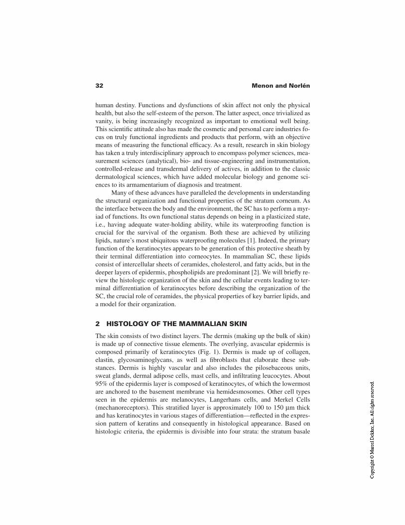
32 Menon and Norlén
human destiny. Functions and dysfunctions of skin affect not only the physicalhealth, but also the self-esteem of the person. The latter aspect, once trivialized asvanity, is being increasingly recognized as important to emotional well being.This scientific attitude also has made the cosmetic and personal care industries fo-cus on truly functional ingredients and products that perform, with an objectivemeans of measuring the functional efficacy. As a result, research in skin biologyhas taken a truly interdisciplinary approach to encompass polymer sciences, mea-surement sciences (analytical), bio- and tissue-engineering and instrumentation,controlled-release and transdermal delivery of actives, in addition to the classicdermatological sciences, which have added molecular biology and genome sci-ences to its armamentarium of diagnosis and treatment.
Many of these advances have paralleled the developments in understandingthe structural organization and functional properties of the stratum corneum. Asthe interface between the body and the environment, the SC has to perform a myr-iad of functions. Its own functional status depends on being in a plasticized state,i.e., having adequate water-holding ability, while its waterproofing function iscrucial for the survival of the organism. Both these are achieved by utilizinglipids, nature’s most ubiquitous waterproofing molecules [1]. Indeed, the primaryfunction of the keratinocytes appears to be generation of this protective sheath bytheir terminal differentiation into corneocytes. In mammalian SC, these lipidsconsist of intercellular sheets of ceramides, cholesterol, and fatty acids, but in thedeeper layers of epidermis, phospholipids are predominant [2]. We will briefly re-view the histologic organization of the skin and the cellular events leading to ter-minal differentiation of keratinocytes before describing the organization of theSC, the crucial role of ceramides, the physical properties of key barrier lipids, anda model for their organization.
2 HISTOLOGY OF THE MAMMALIAN SKIN
The skin consists of two distinct layers. The dermis (making up the bulk of skin)is made up of connective tissue elements. The overlying, avascular epidermis iscomposed primarily of keratinocytes (Fig. 1). Dermis is made up of collagen,elastin, glycosaminoglycans, as well as fibroblasts that elaborate these sub-stances. Dermis is highly vascular and also includes the pilosebaceous units,sweat glands, dermal adipose cells, mast cells, and infiltrating leucocytes. About95% of the epidermis layer is composed of keratinocytes, of which the lowermostare anchored to the basement membrane via hemidesmosomes. Other cell typesseen in the epidermis are melanocytes, Langerhans cells, and Merkel Cells(mechanoreceptors). This stratified layer is approximately 100 to 150 µm thickand has keratinocytes in various stages of differentiation—reflected in the expres-sion pattern of keratins and consequently in histological appearance. Based onhistologic criteria, the epidermis is divisible into four strata: the stratum basale
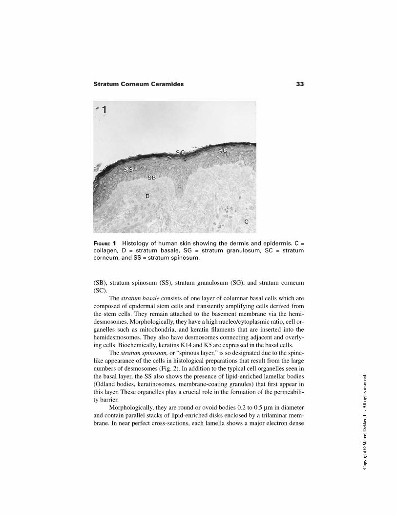
33Stratum Corneum Ceramides
FIGURE 1 Histology of human skin showing the dermis and epidermis. C =collagen, D = stratum basale, SG = stratum granulosum, SC = stratumcorneum, and SS = stratum spinosum.
(SB), stratum spinosum (SS), stratum granulosum (SG), and stratum corneum(SC).
The stratum basale consists of one layer of columnar basal cells which arecomposed of epidermal stem cells and transiently amplifying cells derived fromthe stem cells. They remain attached to the basement membrane via the hemi-desmosomes. Morphologically, they have a high nucleo/cytoplasmic ratio, cell or-ganelles such as mitochondria, and keratin filaments that are inserted into thehemidesmosomes. They also have desmosomes connecting adjacent and overly-ing cells. Biochemically, keratins K14 and K5 are expressed in the basal cells.
The stratum spinosum, or “spinous layer,” is so designated due to the spine-like appearance of the cells in histological preparations that result from the largenumbers of desmosomes (Fig. 2). In addition to the typical cell organelles seen inthe basal layer, the SS also shows the presence of lipid-enriched lamellar bodies(Odland bodies, keratinosomes, membrane-coating granules) that first appear inthis layer. These organelles play a crucial role in the formation of the permeabili-ty barrier.
Morphologically, they are round or ovoid bodies 0.2 to 0.5 µm in diameterand contain parallel stacks of lipid-enriched disks enclosed by a trilaminar mem-brane. In near perfect cross-sections, each lamella shows a major electron dense
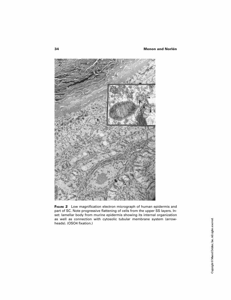
34 Menon and Norlén
FIGURE 2 Low magnification electron micrograph of human epidermis andpart of SC. Note progressive flattening of cells from the upper SS layers. In-set: lamellar body from murine epidermis showing its internal organizationas well as connection with cytosolic tubular membrane system (arrow-heads). (OSO4 fixation.)
35Stratum Corneum Ceramides
band that is shared by electron lucent material divided centrally by a minor elec-tron dense band (Fig. 2 inset). Their appearance marks the dual aspects of epider-mal differentiation, viz., protein and lipid synthesis. Proteins that are expressed inthe SS are keratins 1 and 10. An increase in cellular keratin filaments is noticeablecompared to the basal cells. In the upper layers of the SS, the cells begin to flattenand elongate. Above this layer is the stratum granulosum.
The stratum granulosum layer is characterized by the presence of distinct,darkly staining keratohyalin granules (KHGs), composed of profilaggrin, loricrin,and a cysteine-rich protein as well as keratins 1 and 10. Keratohyalin granules be-come progressively larger in the upper granulocytes (Fig. 3) due to a quantitativeincrease in keratin synthesis. The filaggrin subunits of profilaggrin play the roleof matrix molecule to aggregate and align the keratin filaments. Keratin filamentsin upper granular layers are highly phosphorylated and have extensive disulfidebonds, compared to the cell layers below. The increase in protein synthesis is ac-companied by an upregulation in lipogenesis as well, reflected in the boost innumbers of lamellar bodies reaching their highest density in the uppermost gran-ulocytes, where they occupy about 20% of the cell cytosol. The uppermost cellsin the SG display a unique structural and functional organization of the lamellarbodies (LBs), consistent with their readiness to terminally differentiate into a cor-neocyte, during which the lamellar bodies are secreted to the extracellular do-mains. As seen in electron micrographs of oblique sections, they are highly polar-ized in the apical cytosol of upper granulocytes. A battery of techniques, such asconfocal scanning and electron microscopy, together with enzyme cytochemistry[3] show that in these secretory cells, lamellar bodies are interconnected and ap-pear to bud off a transgolgi-like network. Biochemical characterization of theLBs by preparing an enriched fraction [4] as well as by cytochemical studies [2]show that they are enriched in glucosyl ceramides, phospholipids, and choles-terol, as well as hydrolytic enzymes like lipases, sphingomyelinase, β-glucosyl-cerebrosidase, and phosphodiesterases. Once secreted, their lipid contents areprocessed by the co-secreted enzymes, transforming the short stacks of probarri-er lipids into the ceramide-enriched final barrier lipid structures.
Some rare electron microscopic images (Fig. 4 upper inset) also suggest that the disk structures within individual lamellar bodies are already continuous,having an accordionlike folded pattern, and that these contents unfurl on secre-tion. However, whatever form the disks are within the LBs, further fusion of the secreted contents mediated by co-secreted enzymes and/or fusogenic lipidsformed due to enzyme activity (lysophospholipids) are involved in formation ofthe SC extracellular bilayers. A recently described, unique arrangement of thelamellar body secretory system within the secretory granular cell explains theability for rapid LB secretion to support the homeostasis and/or rapid repair of the permeability barrier [3]. Confocal microscopic images first suggested that
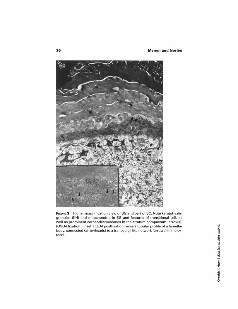
36 Menon and Norlén
FIGURE 3 Higher magnification view of SG and part of SC. Note keratohyalingranules (KH) and mitochondria in SG and features of transitional cell, aswell as prominent corneodesmosomes in the stratum compactum (arrows).(OSO4 fixation.) Inset: RUO4 postfixation reveals tubular profile of a lamellarbody, connected (arrowheads) to a transgolgi-like network (arrows) in the cy-tosol.
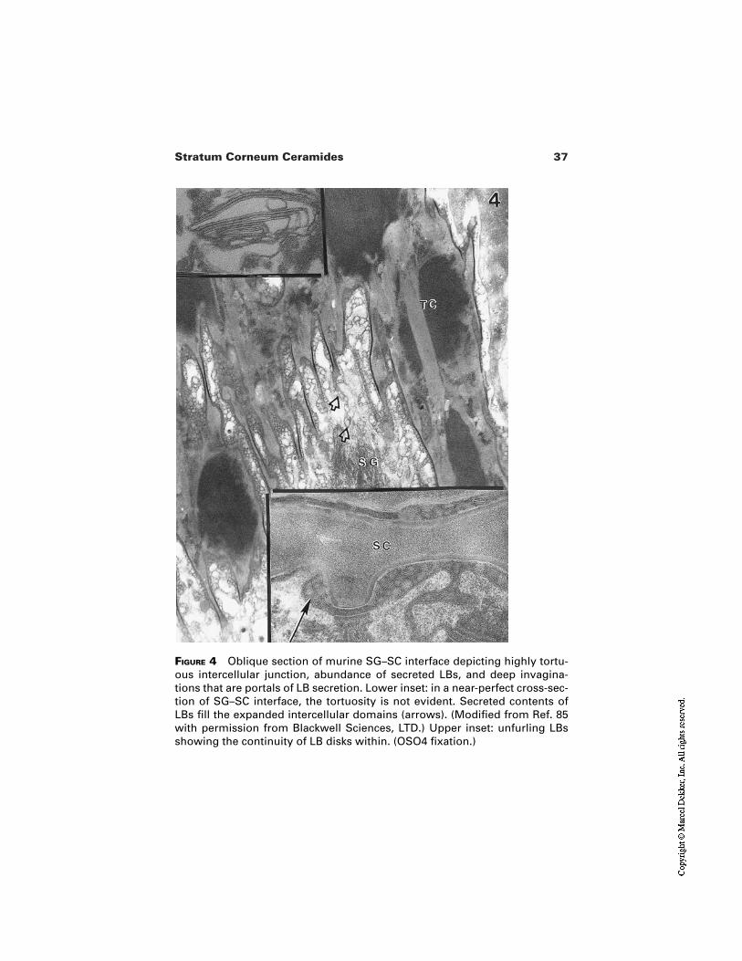
37Stratum Corneum Ceramides
FIGURE 4 Oblique section of murine SG–SC interface depicting highly tortu-ous intercellular junction, abundance of secreted LBs, and deep invagina-tions that are portals of LB secretion. Lower inset: in a near-perfect cross-sec-tion of SG–SC interface, the tortuosity is not evident. Secreted contents ofLBs fill the expanded intercellular domains (arrows). (Modified from Ref. 85with permission from Blackwell Sciences, LTD.) Upper inset: unfurling LBsshowing the continuity of LB disks within. (OSO4 fixation.)
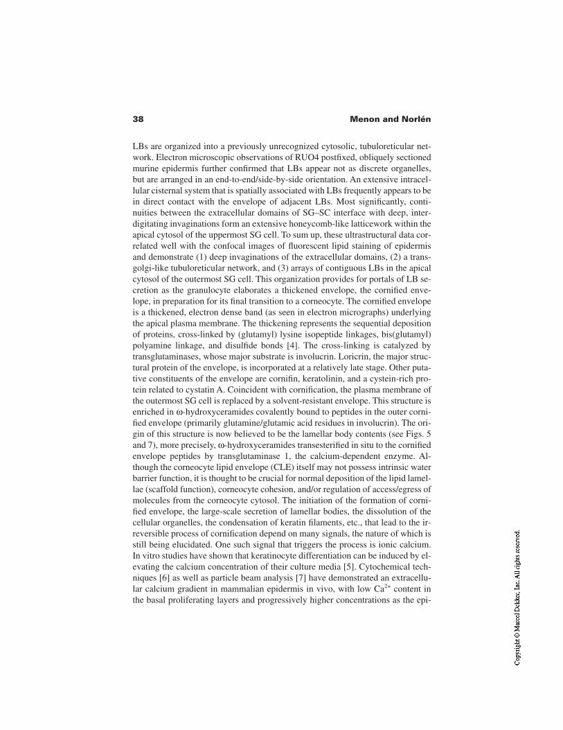
38 Menon and Norlén
LBs are organized into a previously unrecognized cytosolic, tubuloreticular net-work. Electron microscopic observations of RUO4 postfixed, obliquely sectionedmurine epidermis further confirmed that LBs appear not as discrete organelles, but are arranged in an end-to-end/side-by-side orientation. An extensive intracel-lular cisternal system that is spatially associated with LBs frequently appears to bein direct contact with the envelope of adjacent LBs. Most significantly, conti-nuities between the extracellular domains of SG–SC interface with deep, inter-digitating invaginations form an extensive honeycomb-like latticework within theapical cytosol of the uppermost SG cell. To sum up, these ultrastructural data cor-related well with the confocal images of fluorescent lipid staining of epidermis and demonstrate (1) deep invaginations of the extracellular domains, (2) a trans-golgi-like tubuloreticular network, and (3) arrays of contiguous LBs in the apicalcytosol of the outermost SG cell. This organization provides for portals of LB se-cretion as the granulocyte elaborates a thickened envelope, the cornified enve-lope, in preparation for its final transition to a corneocyte. The cornified envelopeis a thickened, electron dense band (as seen in electron micrographs) underlyingthe apical plasma membrane. The thickening represents the sequential depositionof proteins, cross-linked by (glutamyl) lysine isopeptide linkages, bis(glutamyl)polyamine linkage, and disulfide bonds [4]. The cross-linking is catalyzed bytransglutaminases, whose major substrate is involucrin. Loricrin, the major struc-tural protein of the envelope, is incorporated at a relatively late stage. Other puta-tive constituents of the envelope are cornifin, keratolinin, and a cystein-rich pro-tein related to cystatin A. Coincident with cornification, the plasma membrane ofthe outermost SG cell is replaced by a solvent-resistant envelope. This structure isenriched in ω-hydroxyceramides covalently bound to peptides in the outer corni-fied envelope (primarily glutamine/glutamic acid residues in involucrin). The ori-gin of this structure is now believed to be the lamellar body contents (see Figs. 5and 7), more precisely, ω-hydroxyceramides transesterified in situ to the cornifiedenvelope peptides by transglutaminase 1, the calcium-dependent enzyme. Al-though the corneocyte lipid envelope (CLE) itself may not possess intrinsic waterbarrier function, it is thought to be crucial for normal deposition of the lipid lamel-lae (scaffold function), corneocyte cohesion, and/or regulation of access/egress ofmolecules from the corneocyte cytosol. The initiation of the formation of corni-fied envelope, the large-scale secretion of lamellar bodies, the dissolution of thecellular organelles, the condensation of keratin filaments, etc., that lead to the ir-reversible process of cornification depend on many signals, the nature of which isstill being elucidated. One such signal that triggers the process is ionic calcium. In vitro studies have shown that keratinocyte differentiation can be induced by el-evating the calcium concentration of their culture media [5]. Cytochemical tech-niques [6] as well as particle beam analysis [7] have demonstrated an extracellu-lar calcium gradient in mammalian epidermis in vivo, with low Ca2+ content in the basal proliferating layers and progressively higher concentrations as the epi-
39Stratum Corneum Ceramides
FIGURE 5 High magnification view of SG–SC interface of murine epidermisfollowing RUO4 postfixation. Note superior visualization of lipid-enriched LBcontents. Postsecretory processing of LB derived lipid disks such as end-to-end fusion and their transformation into broad, compact bilayers at the distalportions of the intercellular (extracellular) domain. The RUO4 technique illu-minates details of the relation between desmosomes (D) and the lipid disksthat seem to be anchoring onto the desmosomes. While cytosolic LBs arestained, keratohyalin granules and other cytosolic structures are obliteratedby RUO4. (Modified from Ref. 22.)
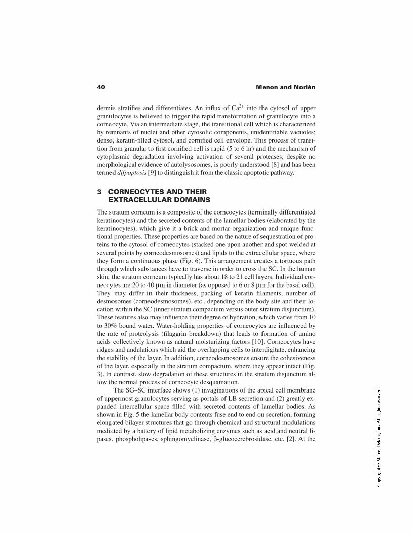
40 Menon and Norlén
dermis stratifies and differentiates. An influx of Ca2+ into the cytosol of uppergranulocytes is believed to trigger the rapid transformation of granulocyte into acorneocyte. Via an intermediate stage, the transitional cell which is characterizedby remnants of nuclei and other cytosolic components, unidentifiable vacuoles;dense, keratin-filled cytosol, and cornified cell envelope. This process of transi-tion from granular to first cornified cell is rapid (5 to 6 hr) and the mechanism ofcytoplasmic degradation involving activation of several proteases, despite nomorphological evidence of autolysosomes, is poorly understood [8] and has beentermed difpoptosis [9] to distinguish it from the classic apoptotic pathway.
3 CORNEOCYTES AND THEIR EXTRACELLULAR DOMAINS
The stratum corneum is a composite of the corneocytes (terminally differentiatedkeratinocytes) and the secreted contents of the lamellar bodies (elaborated by thekeratinocytes), which give it a brick-and-mortar organization and unique func-tional properties. These properties are based on the nature of sequestration of pro-teins to the cytosol of corneocytes (stacked one upon another and spot-welded atseveral points by corneodesmosomes) and lipids to the extracellular space, wherethey form a continuous phase (Fig. 6). This arrangement creates a tortuous paththrough which substances have to traverse in order to cross the SC. In the humanskin, the stratum corneum typically has about 18 to 21 cell layers. Individual cor-neocytes are 20 to 40 µm in diameter (as opposed to 6 or 8 µm for the basal cell).They may differ in their thickness, packing of keratin filaments, number ofdesmosomes (corneodesmosomes), etc., depending on the body site and their lo-cation within the SC (inner stratum compactum versus outer stratum disjunctum).These features also may influence their degree of hydration, which varies from 10to 30% bound water. Water-holding properties of corneocytes are influenced bythe rate of proteolysis (filaggrin breakdown) that leads to formation of aminoacids collectively known as natural moisturizing factors [10]. Corneocytes haveridges and undulations which aid the overlapping cells to interdigitate, enhancingthe stability of the layer. In addition, corneodesmosomes ensure the cohesivenessof the layer, especially in the stratum compactum, where they appear intact (Fig.3). In contrast, slow degradation of these structures in the stratum disjunctum al-low the normal process of corneocyte desquamation.
The SG–SC interface shows (1) invaginations of the apical cell membraneof uppermost granulocytes serving as portals of LB secretion and (2) greatly ex-panded intercellular space filled with secreted contents of lamellar bodies. Asshown in Fig. 5 the lamellar body contents fuse end to end on secretion, formingelongated bilayer structures that go through chemical and structural modulationsmediated by a battery of lipid metabolizing enzymes such as acid and neutral li-pases, phospholipases, sphingomyelinase, β-glucocerebrosidase, etc. [2]. At the
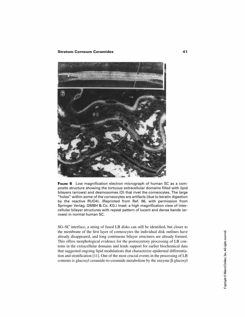
41Stratum Corneum Ceramides
FIGURE 6 Low magnification electron micrograph of human SC as a com-posite structure showing the tortuous extracellular domains filled with lipidbilayers (arrows) and desmosomes (D) that rivet the corneocytes. The large“holes” within some of the corneocytes are artifacts (due to keratin digestionby the reactive RUO4). (Reprinted from Ref. 86, with permission fromSpringer Verlag, GMBH & Co. KG.) Inset: a high magnification view of inter-cellular bilayer structures with repeat pattern of lucent and dense bands (ar-rows) in normal human SC.
SG–SC interface, a string of fused LB disks can still be identified, but closer tothe membrane of the first layer of corneocytes the individual disk outlines havealready disappeared, and long continuous bilayer structures are already formed.This offers morphological evidence for the postsecretory processing of LB con-tents in the extracellular domains and lends support for earlier biochemical datathat suggested ongoing lipid modulations that characterize epidermal differentia-tion and stratification [11]. One of the most crucial events in the processing of LBcontents is glucosyl ceramide-to-ceramide metabolism by the enzyme β-glucosyl
42 Menon and Norlén
cerebrosidase. Investigations by Holleran et al. [12,13] have shown that the pro-barrier lipids originating from lamellar bodies have a predominance of glucosy-lated lipids, and that the enzyme β-glucocerebrosidase is crucial in removing theglucose moiety and giving rise to the ceramides of stratum corneum. Inhibitors ofthe enzyme, as well as genetic deficiency of β-glucocerebrosidase, lead to well-characterized lipid processing defects, structurally immature lipid bilayers in theSC, and consequent barrier abnormalities and defective desquamation. Thedesmosomes initially appear to provide anchorage to the disk contents of LBs.But toward the outer SC, they begin to appear surrounded by lipid lamellae.Lamellar body–derived proteases are involved in degradation of desmosomesleading to the normal process of desquamation, from the outer SC (stratum dis-junctum). However, the lipid bilayers may protect the stratum compactum fromproteolytic degradation, ensuring integrity of this stratum that is crucial to thebarrier function. An interesting observation that sugars protect desmosomes [14]may have implications in the specific sequence of lipid processing during barrierformation. Other aspects of extracellular lipid processing include phospholipid tofree fatty acids by phospholipases, and cholesterol sulfate to cholesterol bysteroid sulfatases. Several observations linking enzyme deficiencies and abnor-mal lipid structural morphology in the SC correlate well with the functional sig-nificance of the three major lipid species and their critical ratios to the barrier. Thesequestration of lipids as membrane bilayers within the SC extracellular domainscan be appreciated from the ultrastructural appearance of RUO4-stained SC. Sta-tic images in electron micrographs provide a glimpse, albeit a frozen moment, ofthe dynamic postsecretory modulations in the probarrier lipids (Fig. 5). Whereasthe proximal parts of the SG–SC interface contain separate disks or those in theprocess of fusing with each other, close to the membrane of the first corneocytethe fused LB disks have already formed continuous lipid bilayers. The basic unitpattern of the bilayers consists of a series of six electron lucent lamallae alternat-ing with five electron dense lamellae (Fig. 6, inset). Double and triple basic unitsoccur frequently. The basic unit structures persist all the way to the outermost lay-er of SC (Fig. 6), although contamination with sebum results in loss of the tightarrays of bilayers. Additionally, the structural relation of the bilayers to the cor-neodesmosomes shows gradual changes associated with the progressive degrada-tion of desmosomal structures. Ultrastructurally, the process of desmosomalbreakdown involves (1) the formation of electron lucent areas in their core and(2) eventual expansion or ballooning of the cores to form the lacunar domainswhich are gradually engulfed by the extracellular bilayers. The near total segre-gation of lipids to intercellular domains of SC was also confirmed by isolating SCmembrane sandwiches containing trapped intercellular lipids [15]. These prepa-rations comprised about 50% lipids by weight, accounting to over 80% of SClipids, and had the same lipid profile of whole SC. Additionally, it had the samefreeze fracture and x-ray diffraction pattern of whole SC [16]. These lipids arecomposed of ceramides, cholesterol, and fatty acids [17] present in roughly
43Stratum Corneum Ceramides
equimolar ratios, in addition to small amounts of triglycerides, glycosphin-golipids, and cholesterol sulfate that are detected in the SC [18]. Ceramidesamount to approximately 50% of the total lipid mass and 40% of the total numberof lipids, and are crucial to the lipid organization of the SC barrier [19]. Based onchromatographic separation, six classes of ceramides were indentified in porcineepidermis. However, chromatographic profile of human stratum corneum some-what differs from that of porcine stratum corneum due to differences in amide-linked fatty acids. Hence a new system of nomenclature based on structure, ratherthan chromatographic mobility, was proposed by Motta et al. [32]. This system,named CER FB (F indicates the type of amide-linked fatty acids; B indicates thebase), the details of which are given by DiNardo and Wertz elsewhere in this vol-ume, is adopted here (Table 1). Of these, ceramide 1 is believed to be uniquelysignificant in the formation of the covalently bound lipid envelope of corneocytes[20]. Ceramide 1 consists of sphingosine and long chain unsaturated, mono- anddi-unsaturated ω-hydroxy acids in the amide linkage. Cholesterol is the secondmost abundant lipid in the SC and amounts to approximately 25% by weight or30 mol% of SC [21] and is crucial for promoting the intermixing of different lipidspecies. Free fatty acids account for about 10% of SC lipids or 15 mol%, and con-sist predominantly of long chain saturated fatty acids having more than 20 carbonatoms. Oleic (6%) and linoleic (2%) are the only unsaturated fatty acids detectedas free in the SC [17].
Deficiencies in any one of these three lipid species result in barrier abnor-malities characterized by increased trans-epidermal water loss (TEWL) as well asobservable alterations in the ultrastructural features of the SC extracellular do-mains [22]. These abnormalities could arise from experimental inhibition of keyepidermal enzymes involved in synthesis of cholesterol (HMG Co A reductase),glycolipid synthesis (serine palmitoyl transferase), fatty acid synthesis (fatty acylco carboxylase), or in extracellular processing of glycolipids that are secreted asprobarrier lipids via the lamellar body secretory system (β-glucocerebrosidase).These barrier defects also lead to epidermal hyperproliferation, as well as dry,flaky skin conditions. Epidermal sterologenesis has been shown to be indepen-dent of circulating levels of cholesterol [23], and hence systemic cholesterol-low-ering drugs do not usually impact the epidermal barrier. However, a few caseswhere hypocholesteremic drugs have resulted in skin barrier defects and scalyskin have been reported [24]. Much of the research in this area has centeredaround ceramide deficiency and will be discussed. Other dermatological condi-tions that arise due to deficiency of enzymes/activators include the Netherton syn-drome.
4 SPECIAL ROLE OF CERAMIDES
In the past two decades, the special role of ceramides in skin barrier has attractedconsiderable research efforts, broadly divided in the following categories: (1)
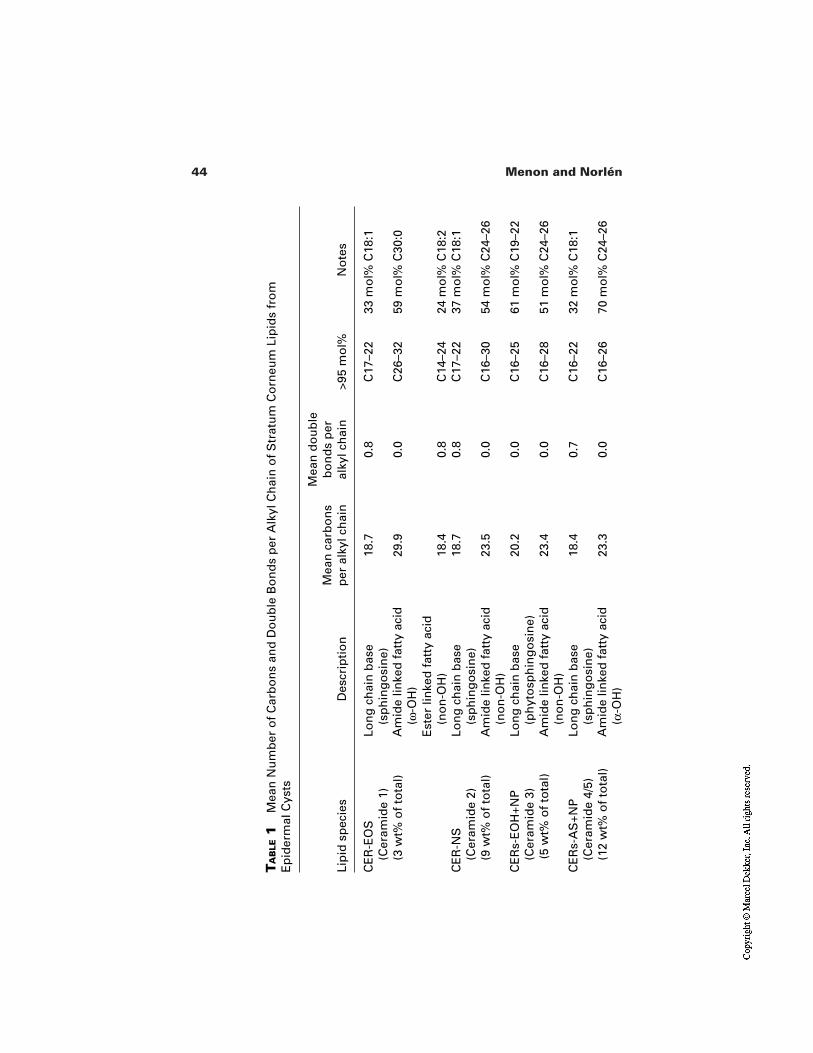
44 Menon and Norlén
TA
BLE
1M
ean
Nu
mb
er o
f C
arb
on
s an
d D
ou
ble
Bo
nd
s p
er A
lkyl
Ch
ain
of
Str
atu
m C
orn
eum
Lip
ids
fro
m
Ep
ider
mal
Cys
ts
Lip
id s
pec
ies
Des
crip
tio
nM
ean
car
bo
ns
per
alk
yl c
hai
n
Mea
n d
ou
ble
b
on
ds
per
al
kyl c
hai
n>9
5 m
ol%
No
tes
CE
R-E
OS
(Cer
amid
e 1)
Lo
ng
ch
ain
bas
e (s
ph
ing
osi
ne)
18.7
0.8
C17
–22
33 m
ol%
C18
:1
Am
ide
linke
d f
atty
aci
d
(ω-O
H)
29.9
0.0
C26
–32
59 m
ol%
C30
:0
Est
er li
nke
d f
atty
aci
d
(no
n-O
H)
18.4
0.8
C14
–24
24 m
ol%
C18
:2C
ER
-NS
(Cer
amid
e 2)
Lo
ng
ch
ain
bas
e (s
ph
ing
osi
ne)
18.7
0.8
C17
–22
37 m
ol%
C18
:1
Am
ide
linke
d f
atty
aci
d
(no
n-O
H)
23.5
0.0
C16
–30
54 m
ol%
C24
–26
CE
Rs-
EO
H+N
P(C
eram
ide
3)
Lon
g c
hai
n b
ase
(ph
yto
sph
ing
osi
ne)
20.2
0.0
C16
–25
61 m
ol%
C19
–22
Am
ide
linke
d f
atty
aci
d
(no
n-O
H)
23.4
0.0
C16
–28
51 m
ol%
C24
–26
CE
Rs-
AS
+NP
(Cer
amid
e 4/
5)
Lon
g c
hai
n b
ase
(sp
hin
go
sin
e)18
.40.
7C
16–2
232
mo
l% C
18:1
Am
ide
linke
d f
atty
aci
d
(α-O
H)
23.3
0.0
C16
–26
70 m
ol%
C24
–26
(3 w
t% o
f to
tal)
(9 w
t% o
f to
tal)
(5 w
t% o
f to
tal)
(12
wt%
of
tota
l)
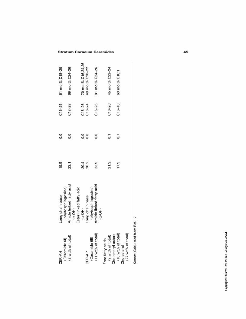
45Stratum Corneum Ceramides
CE
R-A
H(C
eram
ide
6I)
Lon
g c
hai
n b
ase
(ph
yto
sph
ing
osi
ne)
19.5
0.0
C16
–25
61 m
ol%
C18
–20
Am
ide
linke
d f
atty
aci
d
(α-O
H)
23.1
0.0
C18
–28
69 m
ol%
C24
–26
Est
er li
nke
d f
atty
aci
d
(α-O
H)
20.4
0.0
C16
–26
70 m
ol%
C16
,24,
26C
ER
-AP
(Cer
amid
e 6I
I)
Lon
g c
hai
n b
ase
(ph
yto
sph
ing
osi
ne)
20.2
0.0
C16
–24
48 m
ol%
C20
–22
Am
ide
linke
d f
atty
aci
d
(α-O
H)
23.9
0.0
C16
–26
81 m
ol%
C24
–26
Free
fat
ty a
cid
s (9
wt%
of
tota
l)21
.30.
1C
16–2
645
mo
l% C
22–2
4C
ho
lest
eryl
est
ers
(10
wt%
of
tota
l)17
.90.
7C
16–1
869
mo
l% C
18:1
Ch
ole
ster
ol
(27
wt%
of
tota
l)
So
urc
e:C
alcu
late
d f
rom
Ref
. 17.
(2 w
t% o
f to
tal)
(11
wt%
of
tota
l)
46 Menon and Norlén
Physical chemical investigations on lipid mixtures that mimic SC lipid composi-tion and the impact of different types and ratios of lipids on these parameters,[19,25]; (2) in vitro studies on keratinocytes, with emphasis on ceramide synthe-sis and transport into lamellar bodies [26]; (3) in vivo studies of barrier repair inanimal models, experimental conditions of altered ceramide synthesis and metab-olism, as well as transgenic animals with enzyme deficiencies [27–29]; and (4)quantification of ceramide levels and types in human skin disorders such as atopicdermatitis [30,31] and psoriasis [32]. All of these approaches have provided valu-able information on the multiple roles of this crucial lipid species, not only in bar-rier formation, but also in regulating epidermal homeostasis (via cell prolifera-tion, apoptosis) and skin microbial population [33].
Although the details of the molecular organization of lipids in the SC hasnot been clearly elucidated, studies using mixtures of lipids that mimic SC com-position (or are extracted from SC) with a diverse range of physical techniquessuch as x-ray, TEM, DSC, AFM, NMR, and FTIR show that they are organized inlamellar bilayer structures in which the lipid chains are highly ordered [34–40].Bouwstra et al. [35] indicated that at an equimolar CHOL/CER molar ratio, thelamellar organization is least sensitive to a variation in CER composition, whileat a reduced CHOL/CER molar ratio, the CER composition plays a more promi-nent role in the lamellar phases.
Studies on keratinocyte cultures by Madison et al. [26] have shown that ce-ramide glucosyltransferase (CGT), the golgi enzyme responsible for lamellarbody glucosylceramides, is upregulated on inducing keratinocyte differentiation,and parallels the appearance of lamellar bodies. They also found that ceramidesare converted to glucosylceramides within the golgi, which points to a transgolgiorigin of lamellar bodies. Besides, electron microscopic images of LBs showedshapes consistent with cross-sections of tubules or buds from tubules, in additionto vesicles, similar to what was described in vivo (Fig. 3 inset) [3].
Several in vivo studies by Holleran and colleagues [13,27,28] have firmlyestablished the crucial role of ceramide synthesis in barrier repair process and β-glucocerebrosidase enzyme in converting the probarrier lipids (β Gluc Cer) toceramides, i.e., processing of the secreted LB contents during barrier repair. Ad-ditionally, inhibition of the enzyme by topically applied inhibitors (in normalskin) as well as the deficiency of the enzyme in transgenic animals, lead to defec-tive processing of LB contents, resulting in morphologically abnormal lipid bi-layers and increased TEWL. Similar defects occur in patients of Gaucher’s dis-ease. The role of ceramides in formation of the corneocyte lipid envelope wasmentioned earlier in this chapter. A very recent study by Behne et al. [40] provid-ed the first direct evidence for the crucial role of ω-hydroxy ceramides in CLE aswell as barrier function. They found that aminobenzotriazole (ABT), an inhibitorof ω-hydroxylation, significantly inhibited the ω-hydroxylation of very longchain fatty acids in cultured human keratinocytes, but did not alter the synthesis
47Stratum Corneum Ceramides
of other ceramides and fatty acid species. Topical application of ABT to murineepidermis following barrier disruption (tape stripping) led to a significant delay inbarrier recovery. The barrier abnormality resulting from ABT treatment (but notfrom its inactive chemical analog used as a control) correlated with (1) signifi-cantly decreased ω-hydroxyceramides in both the unbound and covalently boundpools of ceramides; (2) pronounced alterations in LB internal structure; (3) ab-normal SC extracellular lamellar membranes; and (4) ultrastructural evidence ofnumerous foci with absent CLE. These observations provided firm support to ear-lier contentions that the CLE is a significant component of the epidermal perme-ability barrier. Figure 7 provides a schematic representation of the CLE formationand the steps affected by ABT treatment.
Compromised barrier functions that correlate with alterations in SC ce-ramide levels have been documented in seasonal changes [41], aging [42], psori-asis [33], and in atopic dermatitis [30], which have been addressed in other chap-ters of this volume.
It is also worth mentioning here that a wealth of literature exists on the rolesof ions, cytokines, and various ligands for the superfamily of nuclear receptors, inbarrier development as well as maintaining the barrier homeostasis. For these as-pects, the reader is referred to two of the recent reviews [43,44].
5 PHYSICAL PROPERTIES OF SKIN BARRIER LIPIDS
5.1 Ceramides
Ceramides are sphingolipids that consist of a long chain amino alcohol (sphingo-sine or one of its derivatives) to which a long chain fatty acid is linked via anamide bond (Fig. 8) [45]. The sphingosine molecule behaves like a surfactant inits free form since it is charged at physiological pH [46]. In addition, it contains acouple of hydroxyl groups along the chain.
Sphingolipids may undergo a plethora of lyotropic and thermotropic phasetransitions, which in part may be due to different packing arrangements of theirtwo hydrocarbon chains. This is probably because there is typically a consider-able length difference between the carbon chain of the amino alcohol (usually an18-carbon sphingosine or phytosphingosine base) and a saturated very long (usu-ally 24-carbon) amide-linked fatty acid [47].
Pascher [48] determined the crystal structure of the ceramide group ofsphingolipids. Crystal space groups of membrane ceramides have not yet been re-ported. However, the crystal forms of N-tetracosanoylphytosphingosine (i.e., C18phytosphingosine with an amide-linked C24 fatty acid) pack in a splayed chainconformation where the phytosphingosine and fatty acid chains form separatematrices. However, packing with folded molecules, like in liquid crystalline bio-logical membranes, can be obtained by crystallization from solvent. A detailed
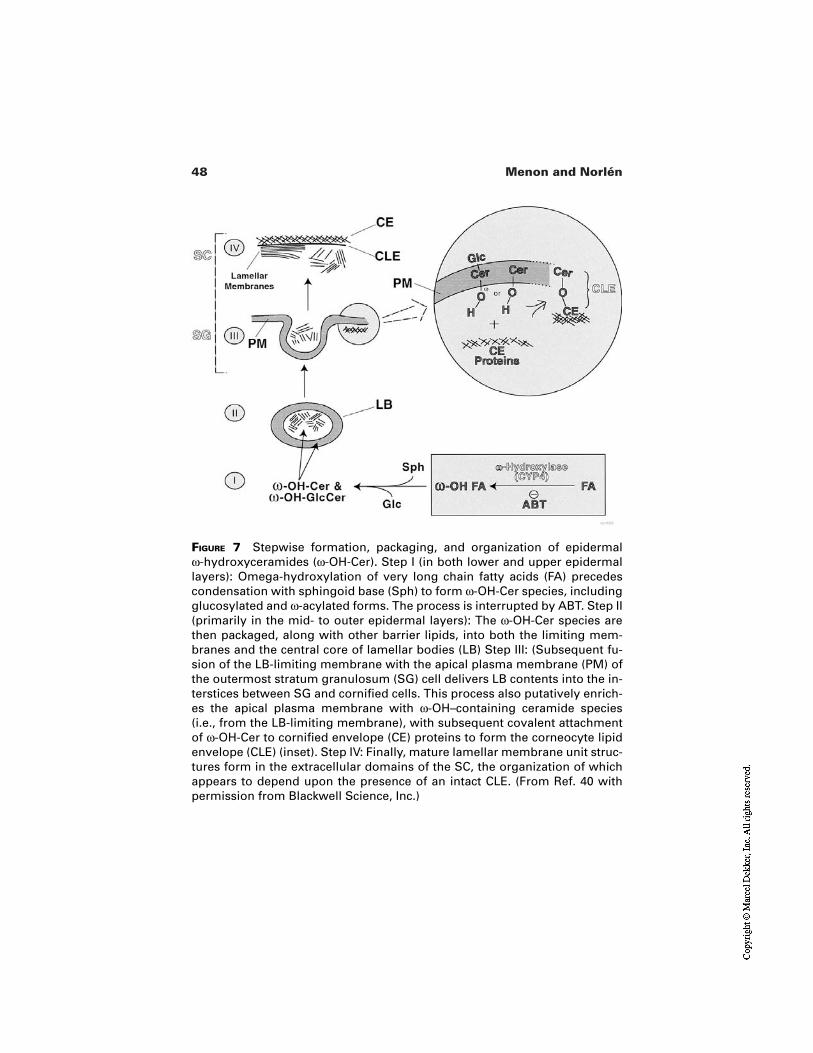
48 Menon and Norlén
FIGURE 7 Stepwise formation, packaging, and organization of epidermal ω-hydroxyceramides (ω-OH-Cer). Step I (in both lower and upper epidermallayers): Omega-hydroxylation of very long chain fatty acids (FA) precedescondensation with sphingoid base (Sph) to form ω-OH-Cer species, includingglucosylated and ω-acylated forms. The process is interrupted by ABT. Step II(primarily in the mid- to outer epidermal layers): The ω-OH-Cer species arethen packaged, along with other barrier lipids, into both the limiting mem-branes and the central core of lamellar bodies (LB) Step III: (Subsequent fu-sion of the LB-limiting membrane with the apical plasma membrane (PM) ofthe outermost stratum granulosum (SG) cell delivers LB contents into the in-terstices between SG and cornified cells. This process also putatively enrich-es the apical plasma membrane with ω-OH–containing ceramide species(i.e., from the LB-limiting membrane), with subsequent covalent attachmentof ω-OH-Cer to cornified envelope (CE) proteins to form the corneocyte lipidenvelope (CLE) (inset). Step IV: Finally, mature lamellar membrane unit struc-tures form in the extracellular domains of the SC, the organization of whichappears to depend upon the presence of an intact CLE. (From Ref. 40 withpermission from Blackwell Science, Inc.)
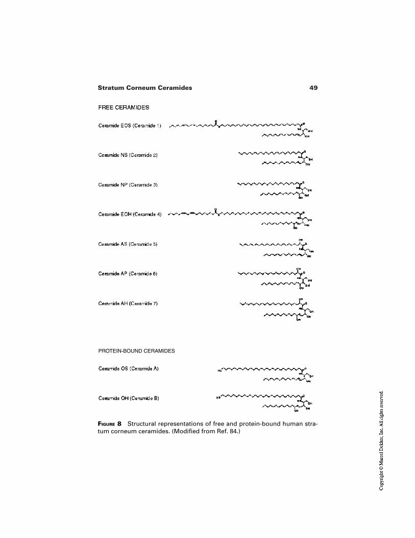
49Stratum Corneum Ceramides
FIGURE 8 Structural representations of free and protein-bound human stra-tum corneum ceramides. (Modified from Ref. 84.)
PROTEIN-BOUND CERAMIDES
50 Menon and Norlén
structural analysis of this crystalline phase is still lacking, but it is assumed thatthe fatty acid tails interdigitate deeply with the opposite half of the bilayer. Thehexagonal packing (α form, or plastic-crystal) exists from 106°C down to 21°C.However, the diffraction pattern undergoes a continuous, and reversible, changefrom a hexagonal to a more orthorhombic packing, where the hexagonal or or-thorhombic subcell as axis remains constant (∼5 Å), while the bs axis continuous-ly decreases from approximately 8.8 Å in the hexagonal matrix to 7.5 Å in the or-thorhombic matrix. According to Dahlén and Pascher [47], the transformation ofthe fatty acid chain matrix proceeds continuously over a large temperature rangeuntil at 21°C the short phytosphingosine chains also change their packing state[47].
The structure of a plasma membrane cerebroside has been shown to form abilayer structure with tilted chains [49]. Above 70°C, cerebrosides from bovinebrain give an L α phase (i.e., a lamellar liquid crystalline phase) where the hydro-carbon chains are melted and consequently there is solely a crystalline periodici-ty in the direction corresponding to the bilayer thickness [50]. No other liquidcrystalline phases have been observed [46]. Electron paramagnetic resonancestudies of cerebrosides and phosphatidylcholine in liposomes show a similar mo-bility for the fatty acid chains of the two lipids.
At physiological skin temperatures (28 to 32°C), the ceramides of SC havebeen claimed to be necessary for the existence of an L β structure (i.e., a lamellargel structure) with alkyl chains pointing in a direction perpendicular to the bilay-er plane [51] that is free of both crystalline cholesterol and liquid crystalline HII
character [52].
5.2 Cholesterol
Cholesterol is the next most abundant lipid species among the skin barrier lipidsin the SC extracellular domains, at approximately 30 mol% [53,54]. The physicalproperties of cholesterol are much less known than its biochemistry. Cholesterolmonohydrate, the biological crystalline form of cholesterol [55], forms a bilayerwith hydroxyl groups and water forming a hydrogen bonded sheet [56]. It is clearthat the phase behavior of cholesterol in biological membranes is exceedinglycomplicated. In general, cholesterol decreases the chain mobility and reduces themean molecular polar head group area of lipids in the liquid crystalline state,while it increases the chain mobility of lipids in the gel state [57]. It also decreas-es transition enthalpies of biological membranes and broadens transition regionsfrom gel to liquid crystalline state in model membranes [57,58]. At high concen-trations (>30 mol%) cholesterol prevents alterations of the bilayer structure in bi-ological membranes (dielaidoyl phosphatidylethanolamine, DEPE). However, atlow concentrations (<20 mol%), cholesterol stabilizes reversed structures [58].Cholesterol incorporation into phosphatidylcholine (PC) bilayers increases the
51Stratum Corneum Ceramides
phase transition temperatures for lamellar phases having hydrocarbon chains of16 or fewer carbons. It also decreases the phase transition temperature for lamel-lar phases having hydrocarbon chains of 18 or more carbons. At 50 mol% choles-terol, a separate cholesterol phase forms and a cooperative lipid transition is nolonger observable by differential scanning calorimetry (DSC) [59].
For cholesterol dipalmitoyl phosphatidylcholine (DPPC) mixtures withhigh cholesterol concentration (>25 mol%) at low temperature, the bilayer be-haves as a liquid with much reduced area compressibility (i.e., a viscous liquid).At these high concentrations, the cholesterol strongly favors the liquid phase overthe solid phase, since the liquid phase is stable down to temperatures far belowthe transition temperature (Tm). In the liquid phase, cholesterol increases the con-formational order of the alkyl chains but does not induce a concomitant decreaseof molecular mobility. At low concentrations, cholesterol is almost as soluble inordered solid as in disordered liquid phase [60].
It has been suggested that cholesterol can operate as a line-active substance(cf. two-dimensional surfactant, or “lineactant”) situated at the interzone betweencrystalline and liquid crystalline structures [61]. Thus cholesterol is likely to pro-mote intermixing of different lipid species. However, two-dimensional, elongatedmicrocrystals have been observed in binary monolayers of synthetic ceramidesand cholesterol [62]. Recently, detergent-resistant membrane domains (DRMs)have been isolated from a variety of eukaryotic cells. These are composed of amixture of saturated long acyl chain sphingolipids and cholesterol and exist as aliquid ordered structure (i.e., properties intermediate between those of the gel andliquid crystalline states) [63–65].
In mixtures of fatty acids and their soaps (30 wt% water), more than 20mol% cholesterol is needed for crystalline cholesterol to appear [52,66]. In SClipid model membranes [cholesterol/bovine brain type III ceramides/mediumchain fatty acids (C14–C18) (35 mol% unsaturated), 26.2:27.4:46.6 (30 wt% wa-ter)] the ceramides were required for solubilization of the cholesterol [52]. Also,no separate crystalline cholesterol phase was observed in a skin lipid model mix-ture containing human ceramide 3/cholesterol/palmitic acid/oleic acid (neutral-ized to 53 mol%), 1:1:0.5:0.5 (32 wt% water) [74].
5.3 Free Fatty Acids
The third or fourth most abundant lipid species among SC barrier lipids is satu-rated long chain free fatty acids (approximately 10–15 mol%) (Table 1)[53,54,67]. In an aqueous milieu, free fatty acids are partly or fully ionized, acid-soaps (AS) or soaps (S), and their physical properties greatly differ from fullyprotonated fatty acids. The nomenclature of fatty acid crystal structures is basedon X-ray long spacings (i.e., angle of tilt of the hydrocarbon chains toward the bi-layer plane). Saturated even- and odd-chained fatty acid crystals are either organ-
52 Menon and Norlén
ized in parallel triclinic or perpendicular orthorhombic packing. Unsaturated fat-ty acids form bilayers with tilted hydrocarbon chains where there is a change inthe direction of tilt at the cis double bond [46].
Condensed phase diagrams of dry free fatty acid-soap mixtures show com-plex phase behavior. Unique crystal unit cells of both the acid and the soap areformed below the chain melting temperature. The palmitic acid-sodium palmitatephase diagram of McBain and Field [45] shows that, except for an acid and soapcrystal, both an acid-soap crystal (AS, S = 0.50) and an acid-soap 2 crystal (AS2,S = 0.67) are formed in the dry state. The effect on phase behavior by neutraliza-tion is well exemplified by the oleic acid-sodium oleate–water system above thechain melting temperature. Here a decreased salt concentration or increased pHfavors the formation of normal phases due to an increased interfacial charge den-sity (and consequently larger effective lipid headgroup areas) [68]. At lipid–waterinterfaces, fatty acids are more difficult to ionize the higher the negative surfacecharge density. It is evident that segregation of fatty acids in lipid mixtures canlead to increased surface charge densities locally.
5.4 Cholesteryl Esters
These are the fourth most abundant lipid species in SC barrier lipids (approxi-mately 10–15 mol%) (Table 1) [52,53]. In cholesteryl oleate (constituting about70% of SC cholesteryl esters), the crystal space group is monoclinic with the mol-ecules packed antiparallelly and with long axes tilted about 30° with respect tothe layer planes [69]. Generally, the interlocking of cholesteryl stacks determinesthe tilt of molecular long axes. The ester chain part may exhibit disorder as wellas considerable thermal motion and the hydrocarbon chains are therefore notpacked according to a repeating subcell pattern [45]. In contrast to the boomerangshape of crystal structures of oleic acid, cholesteryl oleate exhibits an almoststraight chain. In the cholesteryl oleate the kink section is larger [the cis doublebond is situated in the middle of the chain, C(36) = C(37)], but the overall pertur-bation of the chain is smaller than for oleic acid [45].
Liquid crystals of cholesteryl esters show exceedingly complex phase be-havior. They can form smetic (i.e., having long-range order in the direction of thelong axis of the molecules), cholesteric, and blue phases depending on the chainlength of the fatty acid [46,69]. For the cholesteryl esters the smetic phase is sim-ilar to the L α phase [46]. The cholesteric phase is a twisted type of nematic phase(i.e., the lipid molecules are aligned side by side, but not in specific layers), whereeach molecule is slightly displaced in relation to the next giving rise to a helicalarrangement of the molecules. Because it was first described for cholesteryl es-ters, it was termed cholesteric liquid crystalline phase [45]. The blue phases arecubic and the blue color is due to the long periodicity of the phases.
53Stratum Corneum Ceramides
5.5 Cholesterol Sulfate
Cholesterol sulfate, the fifth most abundant lipid species of SC lipids (approxi-mately 1–5 mol%), is almost exclusively found in the interzone between the SGand SC. Further up in the SC, the relative amount of cholesterol sulfate decreasesrapidly [70,71]. Cholesterol sulfate crystalizes as its sodium salt with two mole-cules of water. The predominant aqueous phase is a gel phase (α form). Water isneeded for a certain degree of ionization, which is in turn required for the electy-rostatic repulsion and swelling [46,71].
In model membranes of equimolar quantities of ceramide and cholesterolsulfate, it has been shown that one molecule of ceramide and one molecule ofcholesterol sulfate together bind about 11 molecules of water, as compared topure cholesterol sulfate membranes where each molecule of cholesterol sulfatebinds about 12 molecules of water [72]. This indicates that cholesterol sulfate is amuch stronger surfactant than ceramide, and that cholesterol sulfate therefore hasa very different role in the stratum corneum as compared to ceramides and othernonpolar lipids. The accumulation of cholesterol sulfate at the SG–SC interzone,and the fact that cholesterol sulfate accumulation in the SC as a whole may be re-sponsible for barrier abnormality in recessive X-linked ichthyosis [71], also sup-ports this notion. It is therefore proposed here that cholesterol sulfate under nor-mal conditions takes active part in the formation of the skin barrier, but is not partof the SC intercellular, stacked, lamellar lipid matrix constituting the barrier.
6 THE PLASTIC-CRYSTAL, OR SINGLE GEL PHASE,MODEL FOR THE STRUCTURE AND FUNCTION OFTHE PERMEABILITY BARRIER
The principal objective of the permeability barrier is to be as tight as possible, ex-cept for a minute “leakage” of water needed for the hydration of the keratin in thecorneocytes (for plasticizing the SC). The integument must also ensure that thebarrier capacity is optimal, even under widely and abruptly changing ambientconditions (e.g., temperature, pH, salt concentrations, relative humidity, etc.).Consequently, (1) sudden transitions of the physical state of the intercellular lipidmatrix of SC, with possibly different permeabilities between the two phases, oneither side of the transition temperature and (2) phase separation between lipids,where permeabilities could be locally enhanced at the interface between differentdomains [73], will therefore be avoided as much as possible. Thus from a func-tional point of view, the skin barrier should be as homogeneous as possible (i.e.,ideal physical state, no abrupt transitions, no large differences in permeability be-tween different morphologies, and as little phase separation as possible). Thiscan, however, only be achieved by heterogeneity in the lipid composition, which
54 Menon and Norlén
broadens phase transition zones, stabilizes an ideal lipid morphology, and ensuresthat the lamellar structures remain intact and that no pores or nonlamellar struc-tures are induced. It has therefore been proposed that the mammalian SC barrieris composed of lipids in a lamellar arrangement that are in a plastic-crystallinestate (i.e., α form or “gel” state) stabilized by cholesterol and heterogeneities inhydrocarbon chain length distributions (i.e., broad chain length distributions)with or without water present between the lamallae [74].
Cholesterol is proposed to be the key component for the structure and func-tion of the skin barrier while the ceramides are regarded as constituting the bulklipid matrix [74]. This is mainly because cholesterol stabilizes “gel” phases[58,59] and probably promotes intermixing of different lipid species [61]. In fact,a depressant effect on the water permeability has been observed on addition ofcholesterol to ceramide containing sphingomyelin and phosphatidylcholine mem-branes [4,52,75,76]. Further, a high cholesterol/ceramide ratio has been shown torender the lamellar lipid organization of mixtures of SC lipids less sensitive tovariations in skin ceramide composition [19]. Since the most salient features ofskin barrier lipid composition are (1) widespread heterogeneity and (2) an almostcomplete dominance of saturated very long chain lipid acyl chains with broad,very stable, chain length distributions [53,54] (cf. Table 1), it is logical to suggestthat the extracellular lipid matrix of the SC is a plastic-crystal (i.e., “gel” phase)stabilized by cholesterol and heterogeneities in hydrocarbon chain length distri-butions with or without water present between the lamallae [74]. The stable alkylchain distributions of ceramides and free fatty acids (mainly C24:0–C26:0, Table1) [53,54] and the large relative amounts of cholesterol may aid intermixing ofdifferent lipid species. Consequently, the combination of (1) a lamellar plastic-crystalline structure (i.e., with low permeability as well as relatively low viscosi-ty and great tendency to remain in a lamellar conformation due to the presence ofcholesterol) and (2) relatively impermeable corneocytes would present a barrierof maximum resistance irrespective of environmental conditions, i.e., ideal in abiological context [74].
The proposed structural morphology of the epidermal permeability barrierof terrestrial mammals is a lamellar plastic-crystalline lipid structure, either
1. Without water, with ceramides in the “hairpin” conformation (i.e., thetwo alkyl chains pointing in the same direction) and/or splayed chainconformation (i.e., the two alkyl chains pointing in opposite directions)[77], which is supported by the absence of swelling of the intercellularlipid matrix upon hydration of the SC [19]or
2. With water, which is supported by the finding that hydration of the SCdecreases lipid transition temperatures [78] and increases lipid disor-
55Stratum Corneum Ceramides
dering [79]. The presence of water will necessitate a hairpin ceramideconformation or mixed splayed chain and hairpin ceramides [74].
The notion of a single and coherent lamellar “gel” phase in the stratumcorneum is not inconsistent with the WAXD findings of Bouwstra et al. [81] andthe electron diffraction studies of Pilgram et al. [82], who reported coexistence oforthorhombic (β form) and hexagonal (α form) chain packing lattices in isolatedstratum corneum. This is because for ceramides the phase transition from hexag-onal to a more orthorhombic chain packing is thought to be reversible and contin-uous (cf. previous discussion and Ref. 47). This implies that the same amide-linked fatty acid chain may have an orthorhombic packing in the upper part (i.e.,closest to the polar headgroup) and a looser, more hexagonal packing in the low-er part (i.e., the end of the hydrocarbon chain) at the same time in cholestrol-defi-cient regions. However, the “impurity,” or compositional heterogeneity (i.e.,many different chain lengths), of the crystal remains unperturbed (i.e., no lateraldiffusion of molecules takes place during the continuous phase transition). Thisimplies in turn that even in the case of almost complete “orthorhomicization” ofthe molecular chain packing in cholestrol-deficient regions, the single and coher-ent “gel” crystal remains intact, i.e., no phase separation occurs.
In conclusion, it is proposed that the extraordinary barrier capacity of ter-restrial mammalian skin is due to the presence of a single and coherent plastic-crystalline lipid structure (i.e., “gel” phase) in the SC extracellular space. Theproposed model could fully account for the extraordinary barrier capacity ofmammalian skin and differs in a most significant way from earlier models, in thatit predicts that no phase separation [e.g., between liquid crystalline and “gel”phases or between hexagonal (“gel” phases) and orthorhombic phases] is presentin the intact barrier structure.
ACKNOWLEDGMENTS
The present work was made possible by the generous support from the Wenner-Gren Foundations (L.N.), the Swedish Council for Work Life Research (96-0486;98-0552) (L.N.), and the Edward Welander Foundation (L.N.). We acknowledgethe excellent editorial help from Sheri Vanderzee, Avon Products, Inc., GlobalR&D, Suffern, NY, and Dr. W. Holleran (UCSF), who kindly provided Figure 7.
REFERENCES
1. Hadley NF. Lipid water barriers in biological systems. Prog Lipid Res 1989;28:1–34.
56 Menon and Norlén
2. Elias PM, Menon GK. Structural and biochemical correlates of the epidermal per-meability barrier. In: Elias PM, ed. Advances in Lipid Research. Vol. 24. Skin Lipids.New York: Academic Press, 1991:1–26.
3. Elias PM, Cullander C, Mauro T, Rassner U, Komuves L, Brown B, Menon GK. Thesecretory granular cell: the outermost granular cell as a specialized secretory cell. JInvest Dermatol Symp Oroc 1998; 3:87–100.
4. Schaefer H, Redelmeier TE. The Skin Barrier—Principles of Percutaneous Absorp-tion. Basel:Karger, 1996.
5. Hennings H, Michael D, Cheng C, Steinert P, Holbrook K, Yuspa SH. Calcium regu-lation of growth and differentiation of mouse epidermal cells in culture. Cell 1980;19:245–254.
6. Menon GK, Elias PM, Grayson S. Ionic calcium reservoirs in mammalian epidermis:ultrastructural localization with ion capture cytochemistry. J Invest Dermatol 1985;84:508–512.
7. Warner RR. The distribution and functions of physiological elements in skin. In: Lo-den M, Maiback HI, eds. Dry Skin and Moisturizers: Chemistry and Function. BocaRaton: CRC Press, 2000:71–88.
8. Holbrook K. Ultrastructure of the epidermis. In: Leigh IM, Lane BE, Watt FM, eds.The Keratinocyte Handbook. Oxford: Cambridge University Press, 1994:3–39.
9. Whitfield JF. Calcium: cell cycle driver, differentiator and killer. New York: Chap-man and Hall, 1997.
10. Rawlings AV, Scott IR, Harding CR, Bowser PA. Stratum corneum moisturization atthe molecular level. J Invest Dermatol 1994; 103:731–740.
11. Lampe MA, Burlingame AL, Whitney J, Williams ML, Brown B, Roitman BE, EliasPM. Human stratum corneum lipids: characterization and regional variations. J LipidRes 1983; 24:120–130.
12. Holleran WM, Takagi Y, Menon GK, Jackson SM, Lee JM, Feingold KR, Elias PM.Permeability barrier requirements regulate epidermal β-glucosylceramidase. J LipidRes 1994; 35:903–911.
13. Holleran WM, Takagi Y, Menon GK, Legler G, Feingold KR, Elias PM. Processingof epidermal glucosylceramides is required for optimal mammalian cutaneous per-meability barrier function. J Clin Invest 1993; 91:1656–1664.
14. Chapman SJ, Walsh A. Desmosomes, corneosomes and desquamation: an ultrastruc-tural study of pig epidermis. Arch Dermatol Res 1990; 262:304–310.
15. Grayson S, Elias PM. Isolation and lipid biochemical characterization of stratumcorneum membrane complexes: implications for the cutaneous permeability barrier.J Invest Dermatol 1982; 78:128–135.
16. Elias PM, Feingold KR. Lipid-related barriers and gradients in the epidermis. AnnNY Acad Sci 1988; 548:4–13.
17. Wertz PN, Downing DL. Epidermal lipids. In: Goldsmith LA, ed. Physiology, Bio-chemistry and Molecular Biology of the Skin. New York: Oxford University Press,1991:205–236.
18. Schurer N, Elias PM. The biochemistry and functions of stratum corneum lipids. In:Elias PM, ed. Advances in Lipid Research. Vol. 24. New York: Academic Press,1991:27–56.
57Stratum Corneum Ceramides
19. Bouwstra JA, Dubbelaar FER, Gooris GS, Weerheim AM, Ponec M. The role of ce-ramide composition in the lipid organization of the skin barrier. Biochim BiophysActa 1999; 1419:127–136.
20. Wertz PN, Downing DL. Ceramides of pig epidermis: structure determination. JLipid Res 1983; 24:759–765.
21. Norlén L, Nicander I, Lundh-Rozell B, Ollmar S, Forslind B. Inter and intra individ-ual differences in stratum corneum lipid content related to physical parameters ofskin barrier function in-vivo. J Invest Dermatol 1999; 112:72–77.
22. Menon GK, Ghadially R. Morphology of lipid alterations in the epidermis: a review.Micros Res Technique 1997; 37:180–192.
23. Menon GK, Feingold KR, Moser AH, Brown B, Elias PM. De novo sterologenesis inthe skin. II. Regulation by cutaneous barrier requirements. J Lipid Res 1985;26:418–427.
24. Feldman R, Mainetti C, Saurat J-M. Skin lesions due to treatment with simvastatin(Zocor). Dermatology 1993; 186:272.
25. Bouwstra JA, Thewalt J, Gooris GS, Kitson NA. Model membrane approach to theepidermal permeability barrier: an x-ray diffraction study. Biochemistry 1998;36:7717–7725.
26. Madison KC, Sando GN, Howard EJ, True CA, Gilbert D, Swartzendruber DC,Wertz PN. Lamellar granule biogenesis: a role for ceramide glucosyltransferase,lysosomal enzyme transport and the Golgi. J Invest Dermatol Symp Proc 1998;3:80–86.
27. Holleran WM, Man M-Q, Gao WN, Menon GK, Elias PM, Feingold KR. Sphin-golipids are required for mammalian barrier function. II. Inhibition of sphingolipidsynthesis delays barrier recovery after acute perturbation. J Clin Invest 1991;88:1338–1345.
28. Holleran WM, Sidransky E, Menon GK, Fartasch M, Grundman J-U, Ginns EI, EliasPM. Consequences of (β-)glucocerebrosidase deficiency in epidermis: ultrastructureand permeability barrier alterations in Gaucher’s disease. J Clin Invest 1994;93:1756–1764.
29. Doering T, Holleran WM, Potraz A, Vielhaber G, Elias PM, Suzuki K, Sandhoff K.Sphingolipid activator proteins are required for epidermal permeability barrier for-mation. J Biol Chem 1999; 274:11038–11045.
30. Di Nardo A, Wertz P, Giannetti A, Seidenari S. Ceramide and cholesterol composi-tion of the skin of patients with atopic dermatitis. Acta Derm Venereol 1998;78:27–30.
31. Hara J, Higuchi K, Okamoto R, Kawashima M, Imokawa G. High-expression ofsphingomyelin deacylase is an important determinant of ceramide deficiency leadingto barrier disruption in atopic dermatitis. J Invest Dermatol 2000; 115:406–413.
32. Motta S, Monti MS, Mellesi L, Caputo R, Carelli S, Ghidoni R. Ceramide composi-tion of the psoriatic scale. Biochim Biophys Acta 1993; 1182:147–151.
33. Geilen CC, Wieder T, Orfanos CE. Ceramide signaling: regulatory role in cell prolif-eration, differentiation, and apoptosis in human epidermis. Arch Dermatol Res 1997;289:559–566.
34. White SH, Mirejovsky D, King GI. Structure of lamellar lipid domains and corneo-
58 Menon and Norlén
cyte envelopes of murine stratum corneum: an x-ray diffraction study. Biochemistry1988; 27:3725–3732.
35. Bouwstra JA, Dubbelaar FER, Gooris GS, Ponec M. The lipid organization in theskin barrier. Acta Derm Venereol 2000; 208(Suppl):23–30.
36. Kitson N, Thewalt J, Lafleur M, Bloom M. A model membrane approach to the epi-dermal permeability barrier. Biochemistry 1994; 33:6707–6715.
37. Gay CL, Guy RH, Golden GM, Mak VHW, Francoeur ML. Characterization of lowtemperature (i.e., <65°C) lipid transitions in human stratum corneum. J Invest Der-matol 1994; 103:233–239.
38. Moore DJ, Rerek ME. Insights into the molecular organization of lipids in the skinbarrier from infrared spectroscopy studies of stratum corneum lipid models. ActaDerm Venereol 2000; 208(Suppl):16–22.
39. Pilgram GSK. A Close Look at the Stratum Corneum Lipid Organization by Cryo-electron Diffraction: Significance for the Barrier Function of Human Skin. Ph.D.thesis, Leiden University, Amsterdam. 2000.
40. Behne M, Uchida Y, Seki T, de Montellano PO, Elias PM, Holleran WM. Omega-Hydroxyceramides are required for corneocyte envelope (CLE) formation and nor-mal epidermal permeability barrier function. J Invest Dermatol 2000; 114:185–192.
41. Yoshikawa N, Imokawa G, Akimoto K, Jin K, Higuchi Y, Kawashima M. Regionalanalysis of ceramides within the stratum corneum in relation to seasonal changes.Dermatology 1994; 188:207–214.
42. Denda M, Koyama J, Hori J, Hori I, Takahashi M, Hara M, Tagami H. Age- and sex-dependent changes in stratum corneum sphingolipids. Arch Dermatol Res 1993;285:415–417.
43. Feingold KR. Permeability barrier homeostasis: its biochemical basis and regulation.Cosmet Toil 1997; 112:49–59.
44. Williams ML, Elias PM, Feingold KR. Regulation and differentiation in newbornhuman keratinocytes by endogenous ligands of nuclear hormone receptors. J SkinBarr Res 2000; 2:3–26.
45. Small DM. The physical chemistry of lipids. Handbook of Lipid Research. NewYork: Plenum Press, 1986.
46. Larsson K. Lipids: Molecular Organization, Physical Functions and Technical Appli-cations. Dundee, Scotland: The Oily Press, 1994.
47. Dahlén B, Pascher I. Molecular arrangements in sphingolipids. Thermotropic phasebehavior or tetracosanoylphytosphingosine. Chem Phys Lipids 1979; 24:119–133.
48. Pascher I. Molecular arrangements in sphingolipids. Conformation and hydrogenbonding of ceramide and their implication on membrane stability and permeability.Biochim Biophys Acta 1976; 455:433–451.
49. Pascher I, Sundell S. Molecular arrangements of sphingolipids: the crystal structureof cerebrosides. Chem Phys Lipids 1977; 20:175–191.
50. Friedel MG. Les estats mesomorphes de la matiere. Annals de Physique 1922; 273(November–December):474.
51. Evans FD, Wennerstrom H. The Colloidal Domain: Where Physics, Chemistry, Biol-ogy and Technology Meet. New York: VCH Publishers, 1994.
52. Lieckfeldt R, Villalain J, Gomez-Fernandez JC, Lee G. Diffusivity and structural
59Stratum Corneum Ceramides
polymorphism in some model stratum corneum membrane systems. Biochim Bio-phys Acta 1993; 1151:182–188.
53. Wertz PW, Downing DT. Covalently bound hydroxyacylsphingosine in the stratumcorneum. Biochim Biophys Acta 1987; 917:108–111.
54. Norlén L, Nicander I, Lundsjo A, Cronholm T, Forslind B. A new HPLC-basedmethod for quantitative analysis of inner stratum corneum lipids with special refer-ence to the free fatty acid fraction. Arch Dermatol Res 1998; 290:508–516.
55. Bogren H, Larsson K. An X-ray diffraction study of crystalline cholesterol in somepathological deposits in man. Biochim Biophys Acta 1963; 75:65–69.
56. Craven BM. Crystal structure of cholesterol monohydrate. Nature 1976;260:727–729.
57. de Kruyff B, van Dijck PWM, Demel RA, Scjuijff A, Brants F, van Deenen LLM.Non-random distribution of cholesterol in phosphatidylcholine bilayers. BiochimBiophys Acta 1974; 356:1–7.
58. Takahashi H, Sinoda K, Hatta I. Effects of cholesterol on the lamellar and the invert-ed hexagonal phases of dielaidoyl phosphatidylethanolamine. Biochim Biophys Acta1996; 1289:209–216.
59. McMullen TPW, McElhaney RN. New aspects of interaction of cholesterol with di-palmitoyl phosphatidylcholine bilayers as revealed by high-sensitivity differentialscanning calorimetry. Biochim Biophys Acta 1995; 1234:90–98.
60. Ipsen JH, Karlström G, Mouritsen OG, Wennerström H, Zuckermann MJ. Phaseequilibria in the phospphatidylcholine-cholesterol system. Biochim Biophys Acta1987; 905:162–172.
61. Sparr E, Ekelund K, Engblom J, Engstrom S, Wennerstrom H. An AFM study oflipid monolayers. II. The effect of cholesterol on fatty acids. Langmuir 1999;15:6950–6955.
62. Ekelund K, Eriksson L, Sparr E. Rectangular solid domains in ceramide–cholesterolmonolayers—2D crystals. Biochim Biophys Acta 2000; 1464:1–6.
63. Ahmed SN, Brown DA, London E. On the origin of sphingolipid/cholesterol-richdetergent-insoluble membranes: physiological concentrations of cholesterol andsphingolipid induce formation of a detergent-insoluble, liquid ordered lipid phase inmodel membranes. Biochemistry 1977; 36:10944–10953.
64. Brown DA, London E. Structure of detergent-resistant membrane domains: doesphase separation occur in biological membranes? Biochem Biophys Res Commun1997; 240:1–7.
65. Brown RE. Sphingolipid organizationn in biomembranes: what physical studies ofmodel membranes reveal. J Cell Sci 1998; 111:1–9.
66. Engblom J, Engström S, Jonsson B. Phase coexistence of cholestyerol-fatty acidmixtures and the effects of the penetration enhancer Azone. J Control Rel 1998;52:271–280.
67. Norlén L, Engblom J, Anderson M, Forslind B. A new computer based evaporimetersystem for rapid and precise measurements of water diffusion through stratumcorneum in-vitro. J Invest Dermatol 1999; 113:533–540.
68. Engblom J, Engström S, Fontell K. The effect of skin penetration enhancer Azone onfatty acid-sodium soap-water mixtures. J Control Rel 1995; 33:299–305.
60 Menon and Norlén
69. Craven BM, Guerina NG. The crystal structure of cholesterol oleate. Chem PhysLipids 1979; 24:91–98.
70. Long SA, Wertz PW, Strauss JS, Downing DT. Human stratum corneum polar lipidsand desquamation. Arch Dermatol Res 1985; 277:284–287.
71. Zettersten E, Man M-Q, Sato J, Denda M, Farell A, Ghadially R, Williams ML, Fein-gold KR, Elias PM. Recessive X-linked ichthyosis: role of cholesterol sulfate accu-mulation in the barrier abnormality. J Invest Dermatol 1998; 111:784–790.
72. Abrahamson J, Abrahamson S, Hellquist B, Larsson K, Pascher I, Sundell S. Choles-teryl sulfate and phosphate in the solid state and in aqueous solutions. Chem PhysLipids 1977; 19:213–222.
73. Faure C, Tranchant J-F, Duforc EJ. Interfacial hydration of ceramide in stratumcorneum model membrane measured by H NMR of D2O. J Chim Phys 1998;95:480–486.
74. Clerc SG, Thompson TE. Permeability of dimyristoyl phosphatidylcholine/dipalmi-toyl phosphatidylcholine bilayer membranes with coexisting gel and liquid crys-talline phases. Biophys J 1995; 68:2333–2341.
75. Norlén L. The plastic-crystal model: a new theory for structure, function and forma-tion of the mammalian skin barrier. J Invest Dermatol. Submitted.
76. Finkelstein A, Cass A. Effect of cholesterol on the water permeability of thin lipidmembranes. Nature 1967; 216:717–718.
77. Fettiplace R. The influence of lipid on the water permeability of artificial mem-branes. Biochim Biophys Acta 1978; 513:1–10.
78. Corkery RW, Hyde ST. On the swelling of amphiphiles in water. Langmuir 1996;12:5528–5529.
79. Golden GM, Guzek DB, Harris RR, McKie JE, Potts RO. Lipid thermotropic transi-tions in human stratum corneum. J Invest Dermatol 1986; 86:255–259.
80. Alonso A, Meirelles NC, Tabak M. Effects of hydration upon the fluidity of intercel-lular membranes of stratum corneum: an EPR study. Biochim Biophys Acta 1995;1237:6–15.
81. Bouwstra JA, Gooris GS, Salmon-de Vries MA, Van der Spek JA, Bras W. Structureof human stratum corneum as a function of temperature and hydration: a wide-anglex-ray diffraction study. Int J Pharmaceut 1992; 84:205–216.
82. Pilgram GSK, Engelsma-van Pelt AM, Bouwstra JA, Koerten HK. Electron diffrac-tion provides new information on human stratum corneum lipid organization studiedin relation to depth and temperature. J Invest Dermatol 1999; 113(3):101–107.
83. Forslind B. A domain mosaic model of the skin barrier. Acta Derm Venereol(Stockh.) 1994; 74:1–6.
84. Robson KJ, Stewart ME, Michelsen S, Lazo ND, Downing DT. 6-hydroxy-4-sphin-genine in human epidermal ceramides. J Lipid Res 1994; 35:2060–2068.
85. Halkier-Sorensen L, Menon GK, Elias PM, Thestrup-Pederson K, Ferngold K. Cuta-neous barrier function after cold exposure in hairless mice: A model to demonstratehow cold interferes with barrier homeostasis among workers in the fish processingindustry. Br J Dermatol 1995; 132:391–401.
86. Menon GK, Elias PM. In: Hengge UR, Volc-Platzer B, eds. The Skin and Gene Ther-apy. Berlin: Springer-Verlag, 2001:3–26.
3Stratum Corneum Moisturizing Factors
Clive R. HardingUnilever Research, Colworth Laboratory, Sharnbrook, Bedford, United Kingdom
Ian R. ScottUnilever Research, Edgewater Laboratory, Edgewater, New Jersey
1 INTRODUCTION
The skin is a complex structure that affords protection from the ravages of the ex-ternal environment. The outermost layer, the stratum corneum (SC) represents thetrue interface with the environment and is a magnificent example of the success-ful adaptation of a tissue. Its efficient function is a prerequisite for terrestrial lifeitself, and it has become highly specialized to protect against the invasion of mi-croorganisms and toxic agents and, perhaps most critically, to limit loss of water.The SC is heterogeneous in structure and at the simplest level has been likened toa brick wall in which the noncontinuous, essentially proteinaceous, terminallydifferentiated keratinocytes or corneocytes (bricks) are embedded in the continu-ous matrix of specialized lipids (mortar) [1]. The SC consists typically of 12–16layers of flattened corneocytes [2]. Corneocytes have a mean thickness of around1µm and a mean surface area of approximately 1000 µm2, but ultimately the sur-face area is dependent upon age, anatomical location and conditions that influ-ence epidermal proliferation such as UV irradiation [3].
The corneocyte itself is devoid of intracellular organelles and cytoplasm
61
62 Harding and Scott
and consists almost entirely of a keratin macrofibrillar matrix stabilized throughinter- and intrakeratin chain disulfide bonds encapsulated within a protein shellcalled the cornified cell envelope [4]. This latter structure is composed of a num-ber of specialized proteins that are extensively cross-linked through the action ofat least two members of the transglutaminase family [5]. The cornified envelopeis 15–20 nm thick [6], consisting of two components: a 15-nm-thick layer com-posed of defined structural proteins [7] and a 5-nm-thick layer of ceramide lipids[8] that are covalently attached to the protein envelope on the extracellular sur-face. The overall integrity of the SC itself is achieved primarily through special-ized intercellular protein structures called corneodesmosomes [9,10], which ef-fectively rivet the corneocytes together but which ultimately must be degraded tofacilitate desquamation.
Within this complex structure, water plays a vital function, influencingelasticity, tensile strength, barrier characteristics, electrical resistance, and ofcourse the overall appearance of the skin. In the absence of water the SC is an in-trinsically rigid and brittle structure prone to cracking. Quite simply the SC mustremain hydrated to maintain its integrity, and in healthy skin the tissue containsgreater than 10% water [11]. Ultimately the state of hydration of the SC is gov-erned by three factors: first, the water that reaches it from the underlying epider-mis, second, water lost from the surface by evaporation, and, third, the intrinsicability of this layer to hold water.
The maintenance of water balance in the SC is preserved through two ma-jor biophysical mechanisms. The first of these is the intercellular lamellar lipidsthat provide a very effective barrier to the passage of water through the tissue[12,13]. The second mechanism is provided by the natural moisturizing factor(NMF), a term first coined by Jacobi in 1959 [14] to describe the complex mix-ture of low molecular weight, water-soluble compounds present within the cor-neocytes [15]. Collectively, the NMF components have the ability to bind wateragainst the desiccating action of the environment and thereby maintain tissue hy-dration. The highly structured intercellular lipid lamellae as well as the restrictingof water movement through the SC also effectively prevent the highly water-sol-uble NMF from leaching out of the surface layers of the skin.
In this chapter we will concentrate on the second of these two complemen-tary mechanisms and consider the origin, nature, and role of the NMF. We willdescribe in detail our understanding of the complex yet elegant biochemical path-way leading to its production and consider how this process has shaped our think-ing of the SC as a dynamic tissue responding to the external environment.
2 NATURAL MOISTURIZING FACTOR
The NMF consists of a mixture of amino acids, organic acids, urea, and inorgan-ic ions (see Ref. 16). These compounds are collectively present at high concen-trations within the cell and may represent 10% dry weight of the SC [17]. The
63Stratum Corneum Moisturizing Factors
major constituents apart from amino acids are sodium lactate, urea, and pyrroli-done carboxylic acid (PCA).
The importance of the NMF lies in the fact that its constituent chemicals, inparticular, sodium lactate and PCA salts, are intensely hygroscopic. Essentiallythey absorb atmospheric water and dissolve in their own water of hydration there-by acting as very efficient humectants. Biologically, this property allows the out-ermost layers of the SC to maintain liquid water against the desiccating action ofthe environment. Traditionally it was felt that this liquid water plasticized the SC,keeping it resilient by preventing cracking and flaking that might occur due tomechanical stresses.
In this chapter, with one exception, we will not discuss the properties of the individual components of the NMF in any detail. The exception is urocanicacid. Although historically considered as a component of the NMF, urocanic acidhas unique properties which influence not only the SC but also the body as awhole.
2.1 Urocanic Acid
Urocanic acid (UCA) is formed by the action of the enzyme histidase [18] on freehistidine present within the SC. As this enzyme is only found in one other organin the body, the liver, its presence in the SC is significant. Urocanic acid absorbsultraviolet light in the most damaging part of the solar spectrum and upon UV ex-posure is induced to isomerize from the naturally occurring trans isomer to the cisisomer. For many years UCA was considered to be an important part of the skindefense against UV-induced damage [19,20]. However, the in vivo efficacy ofUCA as a natural photoprotective agent is debatable. In a clinical study [21] a 5%trans-UCA–containing formulation provided a minimal SPF of 1.58 (despite con-taining 20–200 times the level of UCA naturally present in the SC). The lack ofcorrelation between naturally occurring levels of UCA in the SC and the UV sen-sitivity of each subject determined by the minimal erythemal dose further sup-ports the lack of a truly significant photoprotective effect of UCA [21,22].
cis-UCA has been demonstrated to initiate suppression of selected immuneresponses and mimics the effects of UVB irradiation in suppressing delayed hy-persensitivity responses in herpes simplex virus infection [23]. The precise mech-anism by which cis-UCA alters the immune system remains to be clarified, butthere is both supporting [24,25] and contradicting [26] evidence that histamine-like receptors are involved. Recent evidence suggests that UCA isomers may alsoserve as natural scavengers of hydroxyl radicals generated through UV exposure[27]. The levels of cis-UCA present in the SC are, not surprisingly, prone to sea-sonal variation. Norval and coworkers [28] have found that in Western Europeansthe levels of cis-UCA on exposed body sites peak in July/August at close to50–60% of total UCA. During the winter months the percentage of cis-UCA fellbelow 7% for all body sites.
64 Harding and Scott
3 MECHANISM OF ACTION
The view of NMF as humectants is simplistic; and the precise mechanism bywhich these molecules collectively influence SC functionality, the role of othermolecules (lipids) in water retention, and indeed the precise locations of waterwithin the SC remain points of considerable discussion. Three species of waterare identifiable in the SC: tightly bound primary water of hydration bound to po-lar sites on SC proteins; less tightly bound secondary water, hydrogen-bonded toprimary water of hydration, which increases in an amount up to around 40% totalwater content, and, finally, bulk liquid water which does not appear until about40% total water is reached. It is the secondary water that is most dependent on thepresence of NMF [29]. Nuclear magnetic resonance (NMR) studies by Vavasouret al. [30] indicated that there is a single major pool of water which resides with-in a relatively homogeneous and large compartment of the SC that they conclud-ed must be within the corneocyte itself, which as we will see later is consistentwith the initial localization of NMF. Based on T2 NMR relaxation times these au-thors also concluded that the water within the corneocyte interacts strongly withmacromolecules, namely keratins. Imokawa and coworkers has demonstratedthat the depletion of water-extractable materials from acetone/ether-treated SCcauses a marked increase in molecular interaction between the individual fila-ments of keratin fibers as measured by C13 NMR [31]. This increased interactionbetween keratin filaments could be reversed by the application of water-ex-tractable material back to the SC, specifically the neutral and basic free aminoacids. These observations led Imokawa to conclude that the NMF plays the criti-cal role in reducing the intermolecular forces between the nonhelical regions ofthe keratin filaments through interaction with water molecules, and is thereforevital in providing keratin fiber assembly with enhanced molecular mobility.
Imokawa also concludes from these and many studies [32] that the structur-al lipids play a considerable role in the water-holding potential of the SC. Appli-cation of acetone/ether (1:1) to human skin induces a lasting chapped and scalyskin, with a significant decrease in water content, despite the fact that such treat-ment could not induce a substantial release of NMF. The defect in water-holdingproperties in such solvent-damaged skin appears directly related to the depletionof intercellular lipids, especially the ceramides, which comprise up to 50% of thetotal SC lipids.
4 THE ORIGIN OF THE SKIN’S NMF
Until the early 1980s the source of the NMF and what controlled the levels towhich these compounds accumulated in the SC were essentially unknown. Dowl-ing and Naylor [33] disproved suggestions that NMF was formed by the concen-tration of sweat, and it became generally accepted that the majority of NMF ele-
65Stratum Corneum Moisturizing Factors
ments were formed by the general degradation of nonessential, nonkeratinousprotein during the process of terminal differentiation: “The dustbin hypothesis.”Studies conducted in our own laboratory indicated strongly that this was not thecase, and that there was a single unique protein responsible for the generation offree amino acids in the SC. This protein was a high molecular weight, histidine-rich protein (Mr >350,000), intrinsically very basic and with an unusual aminoacid composition [34]. The extensive phosphorylation of serine residues withinthe protein rendered it extremely insoluble, and upon dialysis from urea contain-ing buffers the purified protein formed dense aggregates essentially indistinguish-able under microscopic examination from intact keratohyalin granules (KHG). Inlight of the earlier pioneering studies of Ugel [35], and the elegant autoradio-graphic studies conducted by Fukuyama and Epstein, which localized tritiatedhistidine-labeled proteins to the granular layer [36], we concluded that this pro-tein was a major component of the KHG.
The most significant evidence supporting the theory that this protein wasthe source of the amino acid–derived components of the NMF was deduced fromthe remarkable similarity between the amino acid profile of the protein and that ofthe NMF itself (Fig. 1) [37]. This similarity became even more striking with thediscovery that several of the free amino acids produced from the protein degrada-tion were subsequently and specifically modified by reactions taking place withinthe SC to produce functional molecules. Glutamine was converted to PCA itself[38], and, as already discussed, histidine was converted to urocanic acid [22].When these and other enzymatic pathways active within the SC were taken intoaccount the conclusion was that the histidine-rich protein represented the onlysource of the free amino acids [37,39].
Further studies indicated that this protein was rapidly dephosphorylatedduring the transition of the mature granular cell into the corneocyte and then un-derwent selective proteolytic processing to form lower molecular weight, and sol-uble, basic species within the SC [40]. Based upon their ability to aggregate ker-atin fibers in vitro into macrostructures reminiscent of the keratin pattern seen inthe SC vivo [41], Dale and coworkers named this class of basic proteins filaggrins[42]. The phosphorylated, high molecular weight precursor protein subsequentlybecame known as profilaggrin. However, regardless of this putative structuralrole, and consistent with the biochemical evidence outlined herein, filaggrin wasshown to be a transient component of the SC. Radiolabel pulse chase [37], im-munohistochemical, and biochemical studies [43] revealed that filaggrin did notpersist beyond the deepest two to three layers of the SC. Once formed it becameextensively deiminated through the activity of the enzyme peptidylarginine deim-inase, which served to reduce the affinity of the filaggrin/keratin complex [40,77],and, second, it was rapidly and completely degraded through small peptides tofree amino acids. This fate of filaggrin within the SC mirrors electrical conduc-tance measurements undertaken on isolated SC [44] which indicate that water-
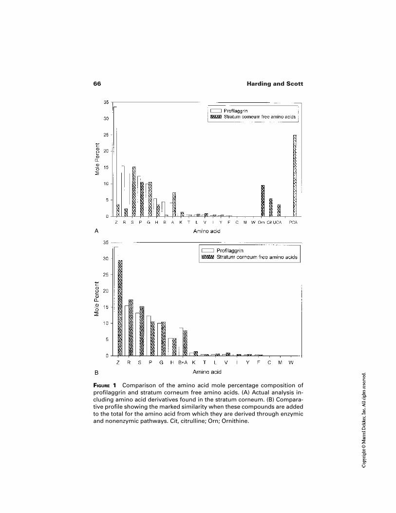
66 Harding and Scott
FIGURE 1 Comparison of the amino acid mole percentage composition ofprofilaggrin and stratum corneum free amino acids. (A) Actual analysis in-cluding amino acid derivatives found in the stratum corneum. (B) Compara-tive profile showing the marked similarity when these compounds are addedto the total for the amino acid from which they are derived through enzymicand nonenzymic pathways. Cit, citrulline; Orn; Ornithine.
A
B
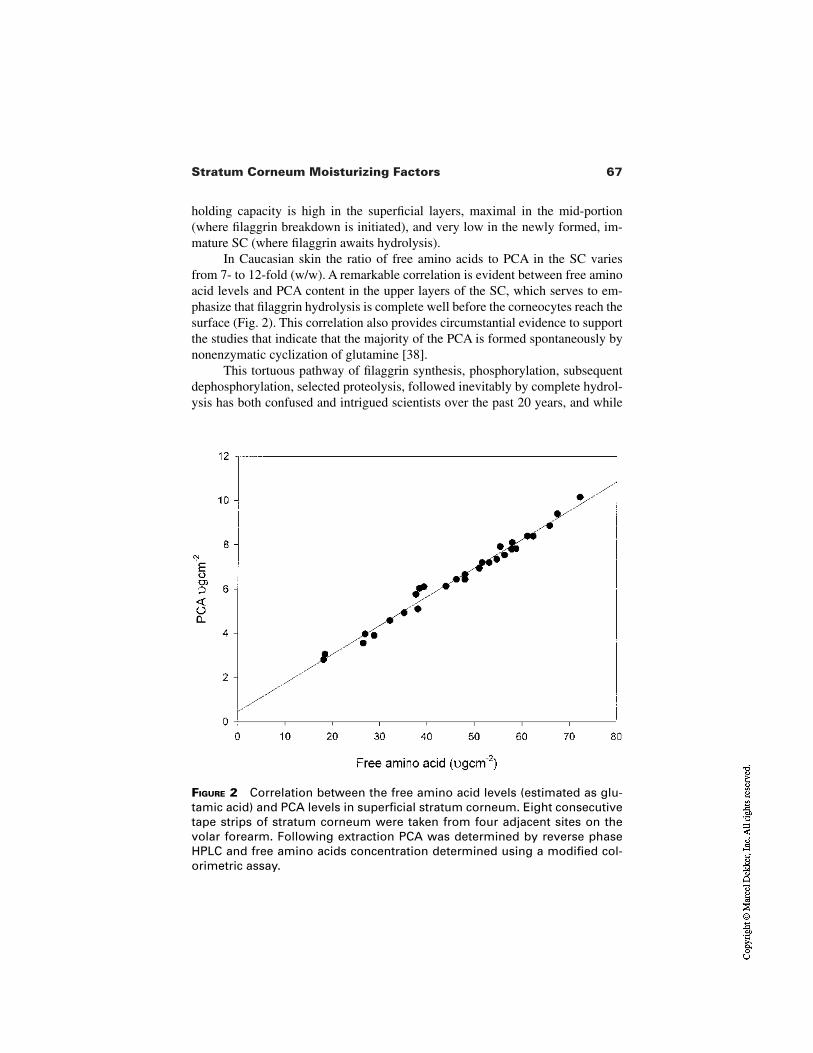
67Stratum Corneum Moisturizing Factors
FIGURE 2 Correlation between the free amino acid levels (estimated as glu-tamic acid) and PCA levels in superficial stratum corneum. Eight consecutivetape strips of stratum corneum were taken from four adjacent sites on thevolar forearm. Following extraction PCA was determined by reverse phaseHPLC and free amino acids concentration determined using a modified col-orimetric assay.
holding capacity is high in the superficial layers, maximal in the mid-portion(where filaggrin breakdown is initiated), and very low in the newly formed, im-mature SC (where filaggrin awaits hydrolysis).
In Caucasian skin the ratio of free amino acids to PCA in the SC variesfrom 7- to 12-fold (w/w). A remarkable correlation is evident between free aminoacid levels and PCA content in the upper layers of the SC, which serves to em-phasize that filaggrin hydrolysis is complete well before the corneocytes reach thesurface (Fig. 2). This correlation also provides circumstantial evidence to supportthe studies that indicate that the majority of the PCA is formed spontaneously bynonenzymatic cyclization of glutamine [38].
This tortuous pathway of filaggrin synthesis, phosphorylation, subsequentdephosphorylation, selected proteolysis, followed inevitably by complete hydrol-ysis has both confused and intrigued scientists over the past 20 years, and while
68 Harding and Scott
many questions remain unresolved, researchers have begun to unravel some ofthe mysteries associated with this unique protein.
In the following sections we will seek to explain how some of the apparentcomplexity of profilaggrin/filaggrin processing merely reflects nature’s logicalroute to generate NMF effectively within the SC. We will also consider evidencethat suggests further functions for this enigmatic class of proteins in terminal dif-ferentiation await clarification.
5 PROFILAGGRIN: SYNTHESIS, STRUCTURE, AND PROCESSING
Profilaggrin is first expressed in the granular layer and many studies have nowconfirmed the localization of profilaggrin to the KHG, and specifically to the so-called F-granules [45]. The profilaggrin molecule itself consists of multiple re-peats of filaggrin joined by short hydrophobic linker peptides and flanked by N- and C-terminal domains. Ten to twelve filaggrin repeats are observed in thehuman protein and the repeat number is inherited in classical Mendelian fashion[46]. The filaggrin repeats show marked heterogeneity occurring in nearly 40% ofthe amino acid residues, whereas the linker peptides sequences are highly con-served. The N-terminal domain is subdivided into an A domain, which containsS100-like Ca2+ binding domains, and a B domain of unknown function [47]. Ex-pression of filaggrin constructs in cell systems has indicated that both the filag-grin unit and the linker peptide are required for granule formation [48]. The dis-appearance of KHG (and hence the processing of profilaggrin), concomitant withthe conversion of the granular cell into a corneocyte, argues strongly that thismolecule plays an important role in the terminal differentiation process. Calciumbinding in the N-terminal region of profilaggrin is likely to be pivotal in thisprocess, just as it is to the terminal differentiation process itself. Although the na-ture of the critical initiating step in profilaggrin processing remains conjectural,several of the enzymatic processes occurring are now understood. Both the aminoterminus and the C terminus beyond the last filaggrin repeat are cleaved [49,50].The unique amino terminus with the distinct Ca2+ binding domains undergoesspecific proteolysis [51], as do the linker regions. Extensive dephosphorylationby one or more phosphatases including PP2A [52], or possibly an acid phospho-protein phosphatase [53], also occurs. Somewhat surprisingly the in vitro studiesconducted do not support a critical role for dephosphorylation in changing thesolubility of the protein, although the behavior of the macroprotein is likely to besignificantly different from the low molecular weight filaggrin constructs pre-pared to study protein processing. The presence of glycosaminoglycan-like mate-rial associated with the keratohyalin granule and first alluded to by Fukuyama[54] may yet prove to play a critical role in influencing profilaggrin/filaggrin in-solubility.
69Stratum Corneum Moisturizing Factors
Collectively these studies indicate that profilaggrin processing is extremelycomplex. This complexity may represent the mechanism by which the cell main-tains control of this critical process and thereby prevents premature collapsing ofthe cytoskeleton by inappropriate and untimely release of active filaggrin, whichwould have drastic consequences for the keratinocyte [55].
6 KERATIN/FILAGGRIN INTERACTION ANDFILAGGRIN PROTEOLYSIS
Although the association of filaggrin with keratin intermediate filaments to formmacrofibrils is a proven property in vitro [40,42], the in vivo relevance of thisfunction remains controversial. Named for this property before the realization ofits other perhaps more significant role, there is convincing and often overlookedevidence that casts doubt upon the need for a specific keratin aggregating proteinfor the SC. Significantly, close-packed keratin structures can be produced in vitrosimply by adding inorganic salts to purified keratin preparations [56]. The speciesspecificity of most antifilaggrin antibodies, the marked heterogeneity in the sizeof the mature filaggrin protein, and the diversification of amino acid sequence in-dicate that size and sequence of the protein are not critical for keratin aggregatingability. Indeed recent studies [48] indicate that filaggrin sequences as short as 16residues (against a mature filaggrin repeat size of 324 residues) can effectivelybind intermediate filaments.
Significantly, the ability of other, unrelated but importantly intrinsically ba-sic proteins such as histones to effectively aggregate keratin (I. R. Scott, unpub-lished observations) indicates that this particular property is more dependent onoverall charge distribution than on a precise sequence. Detailed studies led Mack[57] and coworkers to propose the “ionic zipper model” to explain filaggrin’s ag-gregating potential. This model stipulates that filaggrin binds to filaments throughsimple ionic or H-bonding interactions between positionally conserved positiveand negative charges (evident in the secondary structure of the B-turns of filag-grin) and the conserved distribution of positive and negative regions on the roddomains in keratin filaments.
The importance of filaggrin ionic charge for effective keratin interaction invivo is emphasized by the loss of keratin binding affinity of filaggrin as the posi-tively charged arginine residues within the protein are progressively converted toneutral citrulline residues by the action of peptidylarginine deiminase [40]. Thisdeimination is not restricted to filaggrin, and keratin is also deiminated during SCmaturation [77]. Further studies suggest that the ureido group on the citrullinefunctions to unfold proteins through a combination of a decrease in net charge,loss of potential ionic bonds, and interference with H-bonds [58]. As we shall dis-cuss later dissociation of the filaggrin/keratin complex is a prerequisite for effec-tive filaggrin proteolysis.
On close consideration of species differences the more highly conserved
70 Harding and Scott
nature of the linker regions and phosphorylation sites inevitably leads one to thetentative conclusion that the constraints in the evolution of profilaggrin/filaggrinare more strongly linked to the processing events than to protein function itself.The in vivo evidence arguing against a critical keratin-aggregating role for filag-grin is even more compelling. In certain pathological conditions [59,60,61] thepoor expression of filaggrin has no apparent effect on the keratin pattern observedin the affected SC. The same is true for the hard palate lining the oral cavity. Thistissue is highly keratinized with a well-ordered keratin pattern [62], but no im-munologically detectable filaggrin in the SC (Fig. 3a) [63].
7 FILAGGRIN IN ORAL EPITHELIA
The varied epithelia lining the oral cavity provide a unique opportunity to studyfurther the fate and functions of filaggrin. In the hard palate, profilaggrin is readi-ly detectable, but the apparent complete absence of processing to filaggrin infersan intrinsic, unknown function for profilaggrin itself. Likewise, if one accepts that the primary role of filaggrin is as a source of NMF, then whilst the presence of these molecules are undoubtedly critical for the exposed skin surface, onewould argue that NMF (and hence filaggrin) should be absent, or reduced, in thewet-surfaced keratinized epithelia of the oral cavity, since these tissues have norequirement for hygroscopic material. Some keratinized tissues do lack filaggrin,but in others it is clearly present, for example, in the junctional region between the hard and soft palate (Fig. 3c). Logically, therefore, one must seek furtherfunctions for this complex family of proteins or their breakdown products. Thepossibility that filaggrin breakdown products play a role in controlling ker-atinocyte differentiation (i.e., proteolyzed elements act as chalones), although at-tractive, has not been substantiated [64], and no known filaggrin sequence match-es that of the putative pentapeptide reported by Elgjo et al. to inhibit epidermalproliferation [65]. A third function for filaggrin—as a component of the cornifiedcell envelope—has been proposed [6,66], and human cornified cell envelopes con-tain around 10% filaggrin. If this cross-linked filaggrin retains an ability to interactwith keratin, it may function to promote subsequent cross-linking of elements ofthe internal keratin macrofibrillar network into the cornified cell envelope.
However, it is an understanding of the subtleties of profilaggrin functional-ity itself that may prove pivotal in describing the control of, and sequence ofevents leading to, terminal differentiation. Recent evidence points to a role of thecleaved N-terminal peptide of profilaggrin having a potential role in inducing anapoptotic process in transitional cells just below the SC [67], and similarly filag-grin itself is also hypothesized to aid in the terminal differentiation process by po-tentiating apoptotic machinery [68].
Against a background of continued debate over whether filaggrin is a truestructural protein, one fact is irrefutable, filaggrin is only present within the in-
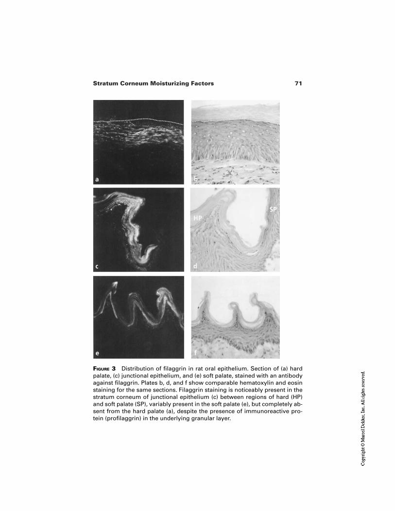
71Stratum Corneum Moisturizing Factors
FIGURE 3 Distribution of filaggrin in rat oral epithelium. Section of (a) hardpalate, (c) junctional epithelium, and (e) soft palate, stained with an antibodyagainst filaggrin. Plates b, d, and f show comparable hematoxylin and eosinstaining for the same sections. Filaggrin staining is noticeably present in thestratum corneum of junctional epithelium (c) between regions of hard (HP)and soft palate (SP), variably present in the soft palate (e), but completely ab-sent from the hard palate (a), despite the presence of immunoreactive pro-tein (profilaggrin) in the underlying granular layer.
72 Harding and Scott
nermost layers of the SC, and hence any structural role within the SC is transient.Nevertheless, within the newly formed layers of the SC, filaggrin may serve aspecific short-lived function as a “scaffold” protein helping in the overall stabi-lization of the keratins macrofibrils. This proposal is based on the observation thatwhen this protein is added in small amounts to keratin microfilaments, it cat-alyzes interchain disulfide bonds in keratin leading to macrofibril formation andsubsequent insolubility [69].
8 ACTIVATION OF FILAGGRIN PROTEOLYSIS
As emphasized by immunolocalization studies, the degradation of filaggrin with-in the SC is abrupt and dramatic. Many proteases with the potential to degrade fi-laggrin in vitro have been characterized in the SC [70,71], but unequivocaldemonstration that these proteases serve a comparable role in vivo remainselusive.
In contrast, the “trigger” which initiates filaggrin proteolysis at a precisestage of SC maturation was first identified in our laboratory in 1986 [43] follow-ing careful observation on patterns of filaggrin distribution in the SC after UV ir-radiation. In these studies it was observed that the effective filaggrin half-lifewithin the SC was reduced dramatically to maintain the same overall distributionwithin the deepest layers, despite the greatly accelerated rate of cell turnover dur-ing the hyperplastic response to the UV stimulus. This indicated that filaggrin hy-drolysis was not dictated by the age of a particular corneocyte, but was insteaddependent upon the cell reaching a critical point during its transit through the SCto the skin surface.
From further studies in developing tissue we concluded that the signal initi-ating filaggrin breakdown was, in fact, the gradient of water activity existingacross the SC [72].
In developing tissue (and as we have seen in certain oral epithelia) there isno indication of any proteolytic breakdown of filaggrin in the outer regions of theSC. However, within a few hours of birth, the breakdown of filaggrin is initiatedin these regions. This triggering could be prevented in a very humid environment.Subsequent studies on filaggrin breakdown in isolated SC revealed that hydroly-sis only occurred if the SC was maintained within a certain range of humidity(70–95%). These studies indicated that it is the very process of SC dehydrationthat initiates filaggrin hydrolysis.
Similarly, if the skin is occluded for a long period [73], filaggrin hydrolysisis blocked, the corneocytes remain filled with the protein, and the filaggrin-de-rived NMF components of the SC fall close to zero (Fig. 4). Under these condi-tions of occlusion, although filaggrin is not degraded, the process of deiminationcontinues unabated and, in the absence of proteolysis, eventually produces a formof filaggrin that is incapable of interacting with keratin. These and more recent
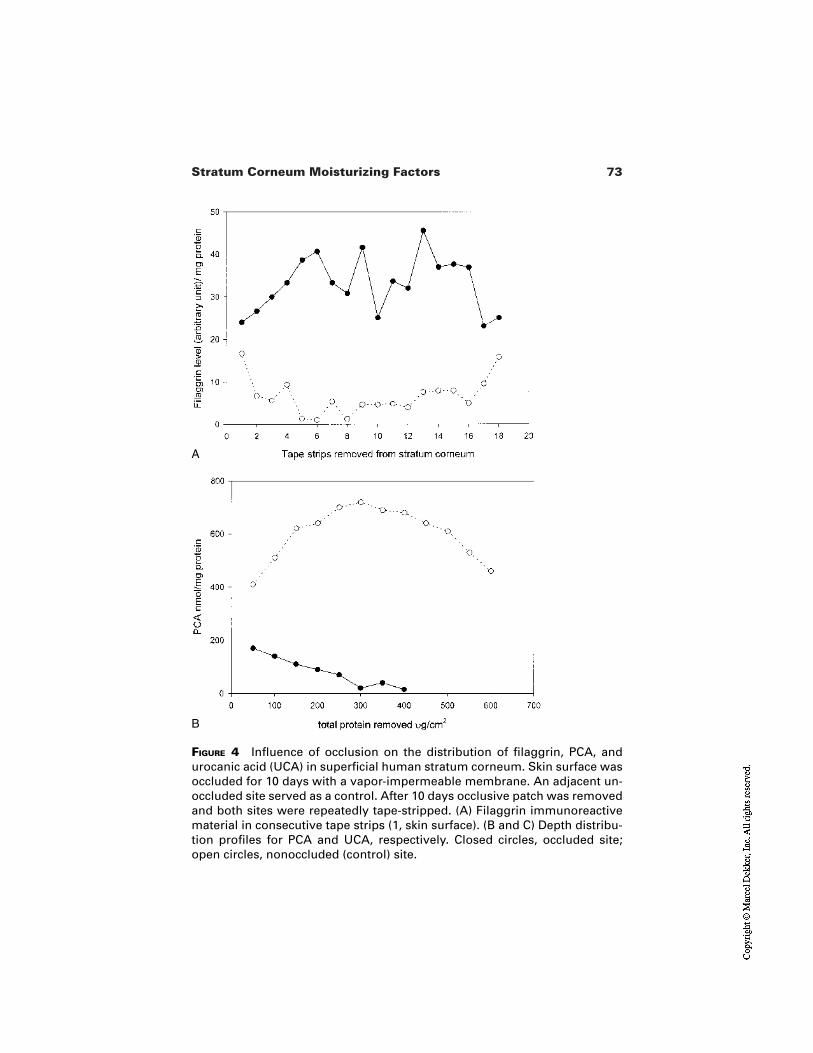
73Stratum Corneum Moisturizing Factors
FIGURE 4 Influence of occlusion on the distribution of filaggrin, PCA, andurocanic acid (UCA) in superficial human stratum corneum. Skin surface wasoccluded for 10 days with a vapor-impermeable membrane. An adjacent un-occluded site served as a control. After 10 days occlusive patch was removedand both sites were repeatedly tape-stripped. (A) Filaggrin immunoreactivematerial in consecutive tape strips (1, skin surface). (B and C) Depth distribu-tion profiles for PCA and UCA, respectively. Closed circles, occluded site;open circles, nonoccluded (control) site.
B
A
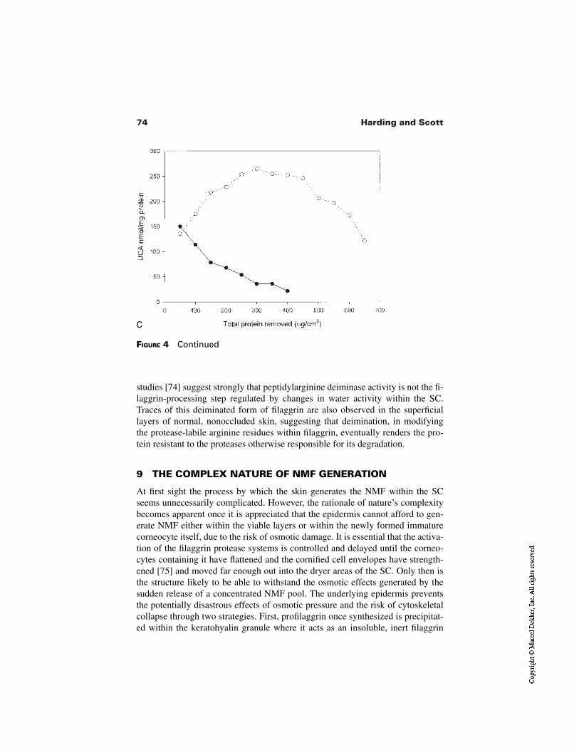
74 Harding and Scott
FIGURE 4 Continued
C
studies [74] suggest strongly that peptidylarginine deiminase activity is not the fi-laggrin-processing step regulated by changes in water activity within the SC.Traces of this deiminated form of filaggrin are also observed in the superficiallayers of normal, nonoccluded skin, suggesting that deimination, in modifyingthe protease-labile arginine residues within filaggrin, eventually renders the pro-tein resistant to the proteases otherwise responsible for its degradation.
9 THE COMPLEX NATURE OF NMF GENERATION
At first sight the process by which the skin generates the NMF within the SCseems unnecessarily complicated. However, the rationale of nature’s complexitybecomes apparent once it is appreciated that the epidermis cannot afford to gen-erate NMF either within the viable layers or within the newly formed immaturecorneocyte itself, due to the risk of osmotic damage. It is essential that the activa-tion of the filaggrin protease systems is controlled and delayed until the corneo-cytes containing it have flattened and the cornified cell envelopes have strength-ened [75] and moved far enough out into the dryer areas of the SC. Only then isthe structure likely to be able to withstand the osmotic effects generated by thesudden release of a concentrated NMF pool. The underlying epidermis preventsthe potentially disastrous effects of osmotic pressure and the risk of cytoskeletalcollapse through two strategies. First, profilaggrin once synthesized is precipitat-ed within the keratohyalin granule where it acts as an insoluble, inert filaggrin
75Stratum Corneum Moisturizing Factors
precursor (profilaggrin cannot aggregate keratin). Most importantly, within thekeratohyalin granule, profilaggrin exists as an osmotically inactive repository ofthe NMF. Second, the interaction between keratin and filaggrin forms a proteolyt-ically resistant complex [69] that prevents premature proteolysis of the filaggrin(an intrinsically labile protein containing 10–15 mol% arginine residues) duringthe intensely hydrolytic processes that accompany SC formation. The value ofthis property of the filaggrin/keratin complex should not be underestimated. It isessential that filaggrin is exquisitely sensitive to proteolysis so that it can be com-pletely and rapidly degraded to NMF when required, but first it must survive themassive and general cellular proteolysis that accompanies SC formation. Thisrepresents an enormous challenge for such a labile protein. Forming a complexwith keratin provides the mechanism by which filaggrin can escape untimely hy-drolysis. Indeed this may be the raison d’être of the affinity of filaggrin for ker-atin. As we have indicated, keratin does not need filaggrin to aggregate properly,but filaggrin may need keratin to elude the massive hydrolytic processes associat-ed with SC formation. The importance of peptidylarginine deiminase is now evi-dent. Deimination of filaggrin is essential to enable its subsequent dissociation,and ultimate hydrolysis.
In summary, these mechanisms are part of a subtle process which ensuresthat it is only as filaggrin-containing corneocytes migrate upward from the deep-est layers and begin to dry out that proteases, by a poorly understood mechanism(but one intimately related to decreased water activity), are activated and theNMF is produced. The point at which this hydrolysis is initiated is independent ofthe age of the corneocyte [72] and is dictated ultimately by the environmental hu-midity. When the weather is humid, the proteolysis occurs almost at the outer sur-face; in conditions of extreme low humidity, the proteolysis is initiated deep with-in the tissue so that all but the deepest layers contain the NMF required to preventdesiccation. The SC has thus developed an elegant self-adjusting moisturizationmechanism to respond to the different climatic conditions to which it is exposed.
The various processes leading from profilaggrin synthesis to conversion tofilaggrin and then to NMF are under tight control. However, as we shall considerin Chapter 6 of this volume, these mechanisms are readily perturbed in differentways by a range of external factors including UV light, exposure to surfactants,and of course changes in the environmental humidity. These very different factorscan, in isolation or in concert, contribute to the complex phenomenon recognizedas dry skin.
10 CONCLUSIONS
Natural moisturizing factor is essential for the correct functioning of the SC.Working together with the interlamellar lipids it assists in the retention of waterwithin the corneocytes vital for the barrier and mechanical properties of the SC.Our understanding of the terminal differentiation and SC maturation processes
76 Harding and Scott
has increased enormously over the last two decades, and it has now become clearthat by maintaining hydration of the SC, the NMF also facilitates key biochemi-cal events. The coordinated activity of specific proteases is essential for optimumSC function, and these hydrolytic processes can only function in the presence ofwater, which is effectively maintained by NMF. Perhaps the most striking exam-ple of this is the regulation of the proteases (“filaggrinases”) within the corneo-cyte which are responsible ultimately for the generation of the NMF itself. In-sights into the process of NMF generation dismiss the notion that the SC issimply a passive barrier and emphasize the dynamic, responsive nature of thisunique tissue.
REFERENCES
1. Elias PM. Epidermal lipids, barrier function and desquamation. J Invest Dermatol1983; 80(Suppl 1):44–49.
2. Zhen YX, Suetake T, Tagami H. Number of cell layers of the stratum corneum innormal skin—relationship to the anatomical location on the body, age, sex and phys-ical parameters. Arch Dermatol Res 1999; 291:555–559.
3. Marks R, Barton SP. The significance of the size and shape of corneocytes. In: MarksR, Plewig G, eds. Stratum Corneum. Berlin: Springer-Verlag, 1983:161–170.
4. Ricé RH, Green L. Cornified envelope of terminally differentiated human epidermalkeratinocytes consists of cross-linked proteins. Cell 1977; 11:417–422.
5. Reichert U, Michel S, Scmidt R. The cornified envelope: a key structure of terminal-ly differentiated keratinocytes. In: Darmon M, Blumberg M, eds. Molecular Biologyof the Skin: The Keratinocyte. New York: Academic Press, 1994:107–150.
6. Jarnik M, Simon MN, Steven AC. Cornified cell envelope assembly: a model basedon electron microscopic determinations of thickness and projected density. Cell Biol1998; 111:1051–1061.
7. Steinert PM, Marekov LN. The proteins elafin, filaggrin, keratin intermediate fila-ments, loricrin, and small proline-rich proteins are isodipeptide cross-linked compo-nents of the human cornified cell-envelope. J Biol Chem 1995; 270:17702–17711.
8. Wertz PW, Madison KC, Downing DT. Covalently bound lipids of human SC. J In-vest Dermatol 1989; 92:109–111.
9. Chapman S, Walsh A. Desmosomes, corneosomes and desquamation, an ultrastruc-tural study of adult pig epidermis. Arch Dermatol Res 1990; 282:304–310.
10. Skerrow CJ, Clelland DG, Skerrow D. Changes to desmosomal antigens and lectin-binding sires during differentiation in normal epidermis: a quantitative ultrastructur-al study. J Cell Sci 1989; 92:667–677.
11. Blank IH. Factors which influence the water content of the SC. J Invest Dermatol1952; 18:433–430.
12. Wertz PW, Miethke MC, Long SA, Strauss JS, Downing DT. Composition of ce-ramides from human SC and comedones. J Invest Dermatol 1985; 8:410–412.
13. Elias PM, Menon GK. Structural and lipid biochemical correlates of the epidermalpermeability barrier. In: Elias, PM, ed. Advances in lipid research. Vol. 240 NewYork: Academic Press, 1991:1–26.
77Stratum Corneum Moisturizing Factors
14. Jacobi O. About the mechanism of moisture regulation in the horny layer of the skin.Proc Scient Sect Toil Goods Assoc 1959; 31:22–26.
15. Tabachnick J, Labadie JH. Studies on the biochemistry of epidermis. IV. The freeamino acids, ammonia, urea and pyrrolidone carboxylic acid content of convention-al and germ free albino guinea pig epidermis. J Invest Dermatol 1970; 54:24–31.
16. Cler EJ, Fourtanier A. L’acide pyrrolidone carboxylique (PCA) et la peau. Int J Cos-met Sci 1981; 3:101.
17. Trianse SJ. The search for the ideal moisturizer. Cosmetics and Perfumery 1974;89:57.
18. Scott IR. Factors controlling the expressed activity of histidine ammonia lyase in theepidermis and the resulting accumulation of urocanic acid. Biochem J 1981;194:829–838.
19. Angelin JH. Urocanic acid a natural sunscreen. Cosmet Toil 1976; 91:47–49.20. Baden HP, Pathak MA. The metabolism and function of urocanic acid in the skin. J
Invest Dermatol 1967; 48:11–17.21. Olivarius FD, Wulf HC, Crosby J, Norval M. The sunscreening effect of urocanic
acid. Photodermatol Photoimmunol Photomedicine 1996; 12:95–99.22. Olivarius FD, Wulf HC, Therkilsden P, Poulson T, Crosby J, Norval M. Urocanic
acid isomers: relation to body site, pigmentation, SC thickness and photosensitivity.Arch Dermatol Res 1997; 289:501–505.
23. de Fabo EC, Noonan FP. Mechanism of immune suppression by ultraviolet irradia-tion in vivo. J Exp Med 1983; 157:84–98.
24. Gilmour JW, Norval M, Simpson TJ, Neuvoken K, Pasenen P. The role of histamine-like receptors in immunosuppression of delayed-hypersensitivity induced by cis-uro-canic acid. Photodermatol Photoimmunol Photomedicine 1993; 9:250–254.
25. Koizumi H, Shimizu T, Nishino H, Ohkawara A. cis-Urocanic acid attenuates hista-mine-receptor mediated activation of adenylate cyclase and increase in intracellularCa2+. Arch Dermatol Res 1998; 290:264–269.
26. Laihia JK, Attila M, Neuvonen K, Pasenen P, Tuomisto L, Jansen CT. Urocanic acidbinds to GABA but not to histamine (H-1, H-2, or H-3) receptors. J Invest Dermatol1998; 111:705–706.
27. Kammeyer A, Eggelte TA, Bos JD, Teunissen MBM. Urocanic acid isomers aregood hydroxyl radical scavengers: a comparative study with structural analoguesand with uric acid. Biochim Biophys Acta 1999; 1428:117–120.
28. Olivarius FD, Wulf HC, Crosby J, Norval M. Seasonal variations in urocanic acidisomers in human skin. Photochemistry and Photobiology 1997; 66:119–123.
29. Takahashi M, Kawasaki K, Tanaka M, Ohra S, Tsuda Y. The mechanism of SC plas-ticisation with water. In: Marks R, Pine PA, eds. Bioengineering and the Skin. Lan-caster: MTP Press, 1981:161–170.
30. Vavasour I, Kitson N, MacKay A. What’s water got to do with it? A nuclear magnet-ic resonance study of molecular motion in pig SC. J Invest Dermatol Symp Proc1998; 3:101–104.
31. Jokura Y, Ishikawa S, Tokuda H, Imokawa G. Molecular analysis of elastic proper-ties of the SC by solid-state C-13 nuclear magnetic resonance spectroscopy. J InvestDermatol 1995; 104:806–812.
32. Imokawa G. Skin moisturizers: development and clinical use of ceramides. In: Lo-
78 Harding and Scott
den M, Maibach H, eds. Dry Skin and Moisturizers. London: CRC Press,2000:269–298.
33. Dowling GB, Naylor PFD. The source of free amino acids in keratin scrapings. Br JDermatol 1960; 72:59–63.
34. Scott IR, Harding CR. Studies on the synthesis and degradation of a histidine richphosphoprotein from mammalian epidermis. Biochim Biophys Acta 1981;669:65–78.
35. Ugel AR. Bovine keratohyalin: anatomical, histochemical, ultrastructural and bio-chemical studies. J Invest Dermatol 1975; 65:118–126.
36. Fukuyama K, Epstein WL. A comparative autoradiographic study of keratohyalingranules containing histidine and cysteine. J Ultrastruct Res 1975; 51:314–325.
37. Scott IR, Harding CR, Barrett JG. Histidine rich proteins of the keratohyalin gran-ules: source of the free amino acids, urocanic acid and pyrrolidone carboxylic acid inthe SC. Biochim Biophys Acta 1982; 719:110–117.
38. Barrett JG, Scott IR. Pyrrolidone carboxylic acid synthesis in guinea pig epidermis.J Invest Dermatol 1983; 81:122–124.
39. Horii I, Kawasaki K, Hoyama J, Nakajima Y, Ohazaki K, Seiji M. Histidine-rich pro-teins as a possible source of free amino acids of stratum corneum. J Dermatol(Tokyo) 1983; 10:25–33.
40. Harding CR, Scott IR. Histidine-rich proteins (filaggrins). Structural and functionalheterogeneity during epidermal differentiation. J Mol Biol 1983; 170:651–673.
41. Brody I. The keratinization of epidermal cells of newborn guinea pig skin as re-vealed by electron microscopy. J Ultrastruct Res 1959; 2:482–511.
42. Steinert PM, Cantieri JS, Teller JD, Lonsdale-Eccles JD, Dale BA. Characterizationof a class of cationic proteins that specifically interact with intermediate filaments.Proc Natl Acad Sci USA 1981; 78:4097–4101.
43. Scott IR. Alterations in the metabolism of filaggrin in the skin after chemical and ul-traviolet induced erythema. J Invest Dermatol 1986; 87:460–465.
44. Hashimotokumasaka K, Horii H, Tagami H. In vitro comparison of water holding ca-pacity of the superficial and deeper layers of the stratum corneum. Arch DermatolRes 1991; 283:342–346.
45. Steven AC, Bisher ME, Roop DR, Steinert PM. Biosynthetic pathways of filaggrinand loricrin—two major proteins expressed in terminally differentiated epidermalkeratinocytes. J Struct Biol 1990; 104:150–162.
46. Gan SQ, McBride O, Idler WW, Markova N, Steinert PM. Organisation, structureand polymorphisms of the human profilaggrin gene. Biochemistry 1990;29:9432–9440.
47. Presland RB, Bassuk JA, Kimball JR, Dale BA. Characterization of two distinct cal-cium binding sites in the amino terminus of profilaggrin. J Invest Dermatol 1995;104:218–223.
48. Kuechle M, Thulin CD, Presland RB, Dale BA. Profilaggrin requires both linker andfilaggrin peptide sequences to form granules: implications for profilaggrin process-ing in vivo. J Invest Dermatol 1999; 112:843–852.
49. Yamazaki M, Ishidoh K, Suga Y, Saido TC, Kawashima S, Suzuki K, Kominami E,Ogawa H. Cytoplasmic processing of human profilaggrin by active µ-calpain.Biochem Biophys Res Comm 1997; 235:652–656.
79Stratum Corneum Moisturizing Factors
50. Resing KA, Thulin C, Whiting K, Al-Alawa N, Mostad S. Characterization of profi-laggrin endoproteinase. 1. A regulated cytoplasmic endoproteinase of epidermis. JBiol Chem 1995; 270:28193–28298.
51. Presland RB, Kimball JR, Kautsky MB, Lewis SP, Lo CY, Dale BA. Evidence ofspecific proteolytic cleavage of the N-terminal domain of human profilaggrin duringepidermal differentiation. J Invest Dermatol 1997; 108:170–178.
52. Kam E, Resing KA, Lin SK, Dale BA. Identification of rat epidermal profilaggrinphosphatase as a member of the protein phosphatase 2A family. J Cell Sci 1993;106:219–226.
53. Ohno J, Fukuyama K, Hara A, Epstein WL. Immuno-histochemical and enzyme-his-tochemical detection of phosphoprotein phosphatase in rat epidermis. J HistochemCytochem 1989; 37:629–634.
54. Kimura H, Fukuyama K, Epstein WL. Effects of hyaluronidase and neuriminidaseon immunoreactivity of histidine-rich protein in new born rat epidermis. J InvestDermatol 1981; 76:452–458.
55. Dale BA, Presland RB, Lewis SP, Underwood RA, Fleckman P. Transient expressionof epidermal filaggrin in cultured cells causes collapse of intermediate filament net-works with alteration of cell shape and nuclear integrity. J Invest Dermatol 1997;108:179–187.
56. Fukuyama K, Murozuka T, Caldwell R, Epstein WL. Divalent cation stimulation ofin vitro fibre assembly from epidermal keratin protein. J Cell Sci 1978; 33:255–263.
57. Mack JW, Steven AC, Steinert PM. The mechanism of interaction of filaggrin withintermediate filaments. J Mol Biol 1993; 232:50–66.
58. Tarcsa E, Marekov LN, Mei G, Melino G, Lee SC, Steinert PM. Protein unfolding bypeptidylarginine deiminase—substrate specificity and structural relationships of thenatural substrates trichohyalin and filaggrin. J Biol Chem 1996; 271:30709–30716.
59. Sybert VP, Dale BA, Holbrook KA. Ichthyosis vulgaris: identification of a defect inthe synthesis of filaggrin correlated with an absence of keratohyalin granules. J In-vest Dermatol 1985; 84:191–194.
60. Manabe M, Sanchez M, Sun TT, Dale BA. Interaction of filaggrin with keratin fila-ments during advanced stages of normal human epidermal differentiation and inIchthyosis vulgaris. Differentiation 1991; 48:43–50.
61. Weidenthaler K, Hauber I, Anton-Lamprecht I. Is filaggrin really a filament-aggre-gating protein in vivo? Arch Dermatol Res 1993; 285:111–120.
62. Schroeder HE. Differentiation of human oral stratified epithelium. London: S Kar-ger, 1981:35–67.
63. Scott IR, Harding CR. Profilaggrin phosphatase: a key step in the pathway of epithe-lial differentiation. J Invest Dermatol (abstr) 1991; 96:1006.
64. Mansbridge J, Knapp M. Effects of filaggrin breakdown on the growth and matura-tion of keratinocytes. Arch Dermatol Res 1987; 279:465–469.
65. Jensen PKA, Elgjo K, Laerum OD. Synthetic epidermal pentapeptide and relatedgrowth regulatory peptides inhibit proliferation and enhance differentiation in pri-mary and regenerating cultures of human epidermal-keratinocytes. Cell Sci 1990;97:51–58.
66. Richards S, Scott IR, Harding CR, Liddell E, Curtis GC. Evidence for filaggrin as acomponent of the cell envelope of the newborn rat. Biochem J 1988; 253:153–160.
80 Harding and Scott
67. Ishida-Yamamoto A, Tanaka H, Nakane H, Takahashi H, Hashimoto Y, Iizuka H.Programmed cell death in normal epidermis and loricrin keratoderma. Multiple func-tions of profilaggrin in keratinisation. J Invest Dermatol Symp Proc 1999;4:145–149.
68. Kuechle MK, Presland RB, Lewis SP, Fleckman P, Dale BA. Inducible expression offilaggrin increases keratinocyte susceptibility to apoptotic cell death. Cell Death andDifferentiation 2000; 7:566–573.
69. Steinert PM. Epidermal keratin: filaments and matrix In: Marks R, Plewig G, eds.Stratum Corneum. Berlin: Springer-Verlag, 1983:25–38.
70. Kawada A, Hara K, Morimoto K, Hiruma M, Ishibashi A. Rat epidermal cathepsinB: purification and characterization of proteolytic properties towards filaggrin andsynthetic substrates. Int J Biochem, Cell Biol 1995; 27:175–183.
71. Kawada A, Hara K, Hiruma M, Noguchi H, Ishibashi A. Rat epidermal cathepsin L-proteinase: purification and some hydrolytic properties towards filaggrin and syn-thetic substrates. J Biochem (Tokyo) 1995; 118:332–337.
72. Scott IR, Harding CR. Filaggrin breakdown to water binding components during de-velopment of the rat SC is controlled by the water activity of the environment. DevBiol 1986; 115:84–92.
73. Scott IR, Harding CR. Physiological effects of occlusion-filaggrin retention (abstr).Proc Dermatol 1993; 2000:773.
74. Akiyama K, Senshu T. Dynamic aspects of protein deimination in developing mouseepidermis. Exp Dermatol 1999; 8:177–186.
75. Harding CR, Long S, Rogers J, Banks J, Zhang Z, Bush A. The cornified cell enve-lope: an important marker of stratum corneum maturation in healthy and dry skin(abstr). J Invest Dermatol 1999; 112:306.
76. Nirunsuksiri W, Presland RB, Brumbaugh SG, Dale BA, Fleckman P. Decreasedprofilaggrin expression in Ichthyosis vulgaris is a result of selectively impaired post-translational control. J Biol Chem 1995; 270:871–876.
77. Senshu T, Kan SH, Ogawa H, Manabe M, Asaga H. Preferential deimination of ker-atin K1 and filaggrin during the terminal differentiation of human epidermis.Biochem Biophys Res Comm. 1996; 225:712–719.
4Desquamation and the Role of StratumCorneum Enzymes
Junko SatoShiseido Research Center, Yokohama, Japan
In the surface area of the skin, layers of corneocytes are tightly and stably boundto each other to form the stratum corneum (SC), the thin but tough barrier at theoutermost area of the skin directly facing the external world. Each corneocyteoriginates from a keratinocyte which is actively proliferating in the epidermis un-der the SC, i.e., new layers of the corneocyte are supplied continuously from be-low the SC (Fig. 1). Concurrently, corneocytes serially detach and smoothly dropoff from the skin surface for replacement to maintain the integrity and thicknessof the SC, keeping it healthy. This shedding process of the corneocyte from theSC, desquamation, has strongly attracted the interest of many skin researchers be-cause this is the process which regulates the condition of the skin.
The data obtained from the earlier “SC disaggregation tests,” in which anSC sheet was incubated in a buffer solution and dissociation of corneocytes wasmeasured, suggested that desquamation is controlled by an enzyme (or a set ofenzymes) since heat treatment of the SC sheet irreversibly blocked the disaggre-gation [1]. The adhesive substance(s) connecting corneocytes in the SC has re-mained unidentified for a long time since the corneocytes are embedded in alipid-enriched intercellular matrix. Bissett et al. proposed a charming hypothesisin 1987 that calcium ion–dependent proteinous linker molecules on the surface ofcorneocytes should connect the cells in the SC, as they observed promoted disso-
81
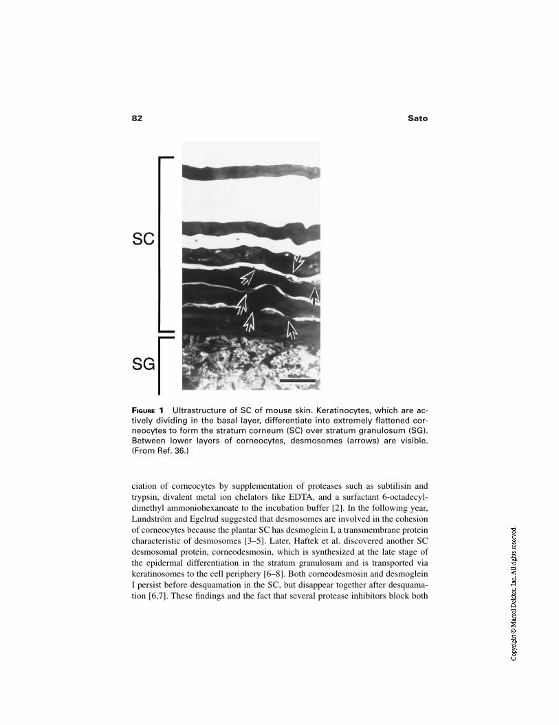
82 Sato
FIGURE 1 Ultrastructure of SC of mouse skin. Keratinocytes, which are ac-tively dividing in the basal layer, differentiate into extremely flattened cor-neocytes to form the stratum corneum (SC) over stratum granulosum (SG).Between lower layers of corneocytes, desmosomes (arrows) are visible.(From Ref. 36.)
ciation of corneocytes by supplementation of proteases such as subtilisin andtrypsin, divalent metal ion chelators like EDTA, and a surfactant 6-octadecyl-dimethyl ammoniohexanoate to the incubation buffer [2]. In the following year,Lundström and Egelrud suggested that desmosomes are involved in the cohesionof corneocytes because the plantar SC has desmoglein I, a transmembrane proteincharacteristic of desmosomes [3–5]. Later, Haftek et al. discovered another SCdesmosomal protein, corneodesmosin, which is synthesized at the late stage ofthe epidermal differentiation in the stratum granulosum and is transported viakeratinosomes to the cell periphery [6–8]. Both corneodesmosin and desmogleinI persist before desquamation in the SC, but disappear together after desquama-tion [6,7]. These findings and the fact that several protease inhibitors block both
83Desquamation and the Role of Enzymes
desquamation in vivo and the disaggregation of the SC in vitro suggest thatbreakdown of desmosomal proteins by proteolytic digestion catalyzed by the pro-tease is the principal event of desquamation.
So far, several different protease activities have been detected in the SC[1,9–11]. Some of them have been purified and further characterized [12,13]. Theresults of these analyses suggested that it is not one particular enzyme but a com-bination of enzymes that is required for the shedding process of corneocytes. Inaddition, some factors of the SC such as the water content, cholesterol sulfate,calcium ion, and pH may also play substantial roles in desquamation, althoughhow these factors regulate desquamation still remains mostly unknown. Inhibi-tion of SC protease activities by endogenous inhibitors or oxidation of substratemolecules potentially connecting corneocytes may regulate the cell shedding inthe SC as well. In this chapter, I review the SC proteases implied to be involvedin desquamation mainly from the biochemical and molecular view points, as wellas other SC factors which may be critical for regulation of desquamation.
1 PROTEASES
The inhibition profile obtained from SC disaggregation tests suggested that pro-teolytic digestion of adhesive molecules on the corneocytes leading to desquama-tion is mainly catalyzed by serine proteases because aprotinin, a specific inhibitorof serine proteases, inhibited disaggregation severely, but those protease in-hibitors that block other types of proteases exhibited no effect [14]. Topical appli-cation of serine protease inhibitors induced scales on the skin [15] indicating thatfunction of this type of protease is indeed required in vivo. Two different serineproteases, the chymotrypsin-like protease and the trypsin-like protease, have beenisolated from the SC and characterized already. Recently, two other types of pro-teases, a cathepsin D type aspartic protease and a cysteine protease, were found inthe SC and these proteases seem to be secondary proteases for desquamation andmay be involved in the fine adjustment of the shedding process.
1.1 Chymotrypsin-Like Serine Protease
The first SC protease whose activity was detected by the disaggregation test wasthe chymotrypsin-like serine protease in the plantar SC [5,17]. Later this enzymewas shown immunohistochemically to be distributed in all types of SC on thewhole body [18]. The active form of this enzyme, with an apparent molecularweight of 25 kD but molecular weight of 28 kD after reduction and full denatura-tion, was purified from the plantar SC [12]. Hansson et al. cloned of the cDNA ofthis enzyme [19]. There are two mRNA species of this enzyme, one is 1.2 and theother 2.0 kilo bases in size. Both of them are highly expressed only in skin; how-ever, very low expression was detected in the brain and kidney, or undetectable in
84 Sato
other tissues. A full-length cDNA of 968 nucleotides was cloned from the kera-tinocyte cDNA library by immunoscreening with the rabbit antisera raised againstthe purified enzyme, and it was confirmed that this enzyme has most of the high-ly conserved residues among various serine proteases, such as the active triad his-tidine–aspartate–serine residues. On the other hand, the overall identity in theamino acid sequence to other chymotryptic proteases (pancreatic chymotrypsin,cathepsin G, mast cell chymase) was less than 40%. Moreover, one position in theprimary specificity pocket [20] is occupied by a bulky asparagine residue where arather smaller serine or alanine residue is normally found in other chymotrypticenzymes. These structural differences from other chymotryptic enzymes maycause the peculiar substrate specificity and inhibitor profile exclusively exhibitedby this enzyme [12].
The SC chymotrypsin-like protease is primarily synthesized as an inactiveprecursor protein of 253 amino acid residues. From the N terminus of the precur-sor, a signal peptide of 22 amino acid residues is cleaved off, and the resulting in-termediate protein, still inactive because of the seven–amino acid propeptide atthe N terminus, is stored in lamellar bodies harbored in the keratinocyte [18].When a keratinocyte differentiates into a corneocyte, an intermediate is releasedinto the extracellular spaces, and there the intermediate is processed by a trypticenzyme to be the fully active protease.
The chymotrypsin-like protease may be the principal protease which di-gests the adhesive molecules connecting corneocytes. Nevertheless, the fact thatthe SC of an aged individual is thicker than that of a youth despite the absence oflarge differences in the activity of the chymotrypsin-like protease between thetwo SC [21] suggests that some factor(s) other than this enzyme itself is essentialfor regulation of desquamation.
1.2 Trypsin-Like Protease
The SC in the human skin contains another serine protease activity other than thechymotrypsin-like protease [1,5,12,14]. In a zymography experiment, the twobands of about 30 kD that developed on the SDS-polyacrylamide gel containing1% casein disappeared when tryptic proteolysis was specifically inhibited by leu-peptin, while two other bands of about 25 kD, which were attributed to the chy-motrypsin-like protease, were not affected by the inhibitor (Fig. 2). This suggeststhat the human SC contains a trypsin-like enzyme [1]. Brattsand and Egelrud pu-rified the enzyme and determined its primary structure by cDNA cloning [13].The amino acid sequence of this enzyme is similar to those of the other serineproteases which have trypsin-like substrate specificity: the porcine enamel matrixprotease exhibited the highest score (55% similarity) in the homology search inthe available amino acid sequences in the database, followed by the human andmouse neuropsin. However, expression of the mRNA of this enzyme is high in
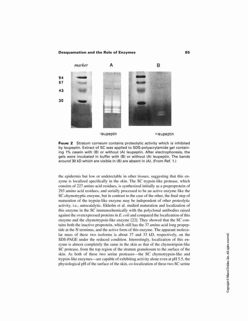
85Desquamation and the Role of Enzymes
FIGURE 2 Stratum corneum contains proteolytic activity which is inhibitedby leupeptin. Extract of SC was applied to SDS-polyacrylamide gel contain-ing 1% casein with (B) or without (A) leupeptin. After electrophoresis, thegels were incubated in buffer with (B) or without (A) leupeptin. The bandsaround 30 kD which are visible in (B) are absent in (A). (From Ref. 1.)
the epidermis but low or undetectable in other tissues, suggesting that this en-zyme is localized specifically in the skin. The SC trypsin-like protease, whichconsists of 227 amino acid residues, is synthesized initially as a preproprotein of293 amino acid residues, and serially processed to be an active enzyme like theSC chymotryptic enzyme, but in contrast to the case of the other, the final step ofmaturation of the trypsin-like enzyme may be independent of other proteolyticactivity, i.e., autocatalytic. Ekholm et al. studied maturation and localization ofthis enzyme in the SC immunochemically with the polyclonal antibodies raisedagainst the overexpressed proteins in E. coli and compared the localization of thisenzyme and the chymotrypsin-like enzyme [22]. They showed that the SC con-tains both the inactive proprotein, which still has the 37 amino acid long propep-tide at the N terminus, and the active form of this enzyme. The apparant molecu-lar mass of these two isoforms is about 37 and 33 kD, respectively, on theSDS-PAGE under the reduced condition. Interestingly, localization of this en-zyme is almost completely the same in the skin as that of the chymotripsin-likeSC protease, from the top region of the stratum granulosum to the surface of theskin. As both of these two serine proteases—the SC chymotrypsin-like andtrypsin-like enzymes—are capable of exhibiting activity alone even at pH 5.5, thephysiological pH of the surface of the skin, co-localization of these two SC serine
86 Sato
proteases suggests that the chymotrypsin-like enzyme can be activated through-out the depth of the SC.
1.3 Other Proteases Found in SC
As ordinary serine proteases prefer a neutral to alkaline condition, they are un-likely to be fully active in an acidic environment like the skin surface, even if theycan exhibit limited activities. Indeed, the fact that PMSF, a specific inhibitoragainst the serine protease, inhibits the SC disaggregation at pH 5.0 no more than30% suggested that there should be other types of proteases which can promotedissociation of the SC at such a low pH [11]. Horikoshi et al. searched for a pro-tease which can function at an acidic pH and discovered a new SC protease otherthan the serine protease, the cathepsin D type aspartic protease [23]. This enzymeis active between pH 2 to 6 with the optimum pH for activity being 3. Immuno-chemical data suggested that this enzyme is primarily synthesized in the kera-tinocyte as the inactive precursor of 52 kD and processed to the 48-kD intermedi-ate probably in the lysozomes, and is finally activated to the functional 33-kDform at the boundary between the stratum granulosum and SC. As an in vitro ex-periment using liver cathepsin D as the substitute for the SC enzyme sufficientlysuggested that this type of protease can have a function in desquamation [11], fur-ther characterization of the SC enzyme itself is awaited in order to clarify whetherthis enzyme is indeed involved in desquamation.
It has been reported that the epidermis contains cystein protease activity[10,24,25]. Watkinson reported that the cystein protease, which can be detected inall types of SC except for the palmoplantar SC, is active between pH 3 and 7, ex-hibits its highest activity at pH 6, and a partially purified extract exhibiting thecystein protease activity can degrade desmocollin in vitro [10]. However, the factthat E-64, the cysteine protease inhibitor, has no inhibitory effect in the SC disag-gregation test [11,14,16] suggests that involvement of this enzyme in desquama-tion is probably negligible compared to that of other proteases.
2 OTHER SC FACTORS REGULATING DESQUAMATION
2.1 Cholesterol Sulfate
The content of cholesterol sulfate in the epidermis increases gradually along withthe differentiation of the keratinocyte and reaches its maximum (about 5% of thetotal lipid) in the stratum granulosum, then drops at the boundary between thestratum granulosum and SC [26]. Within the normal SC, a difference in the con-tent of cholesterol sulfate is also observed: it is higher in deeper layers of the SCand lower close to the surface of the skin where few desmosomes are observed[27]. On the other hand, the abnormally thickened SC of the patient sufferingfrom recessive X-linked ichthyosis (RXLI), a disease which is caused by a defi-ciency of steroid sulfatase, accumulates fivefold more cholesterol sulfate than the
87Desquamation and the Role of Enzymes
normal SC, and the desmosome which is normally absent from the outer SC is ob-served even in the outermost layers of the SC [28]. As the transit time of nucleat-ed cell layers of epidermis is normal, this symptom—retention hyperkeratosis—found in the SC of the RXLI patient is thought to be caused by delay of turnoverof the SC [26]. These facts suggested that cholesterol sulfate in the SC is involvedin the regulation of desquamation [29].
Topical application of cholesterol sulfate to the normal skin induced abnor-mal scaliness and an increase in thickness of the SC [30,31] and prolonged per-sistence of desmoglein I [15] without affecting the labeling index. However,phosphatidylcholine did not exhibit these effects [15]. In addition, cholesterolsulfate inhibited dissociation of the cells in the SC disaggregation test, but otheramphopathic lipids—phosphatidylcholine, palmitic acid, and taurocholic acid—exhibited no inhibitory effect [15]. These results suggest that cholesterol sulfatecan directly regulate the cell shedding in the SC and it is not simply because thissubstance has a detergent effect. Indeed cholesterol sulfate can inhibit activitiesof the serine protease: porcine pancreatic chymotrypsin and bovine pancreatictrypsin are inhibited competitively by cholesterol sulfate with Ki values of 2.1micromolar and 5.5 micromolar, respectively, but cholesterol, phosphatidyl-choline, palmitic acid, and taurocholic acid did not show any inhibitory activity inthe same enzyme assay [15]. In addition, the physicochemical properties of cho-lesterol sulfate may also lead to inhibition of desquamation in vivo. The study ofbehavior of lamellar phase suggested that cholesterol sulfate is one of the key fac-tors which stabilize dissolution of cholesterol in the lamellar phases, i.e., choles-terol sulfate should stabilize the whole SC lamellar phase [32]. Therefore choles-terol sulfate in the SC may regulate the action of the SC protease in two ways:inhibiting its activity directly and obstructing the accessibility of the enzymes tothe substrate with the stabilized lamellar phases of the intercellular lipids.
2.2 Water Content
Rawlings et al. showed that desmosomal degradation is inhibited under a dry con-dition as compared to a moist condition [33]. As environmental humidity influ-ences the water content of SC [34,35], the water content of the SC has been sug-gested to be one of the important factors regulating desquamation.
Does the humidity of the environment alone alter the water content of SCand affect desquamation in vivo? To answer this question, we examined thechange of the skin condition of mice kept under controlled humidity conditions[36]. The water content of the SC of mice kept under a dry condition (relative hu-midity of 10%) showed a decrease on and after day 1. Scales were induced on theskin surface and the thickness of SC increased without inducing epidermal hyper-plasia on day 3. The tryptic activity assayed in vitro was the same in the dry SCand the control, suggesting that the dry SC contains the same amount of thetrypsin-like protease as the control at that time point, but more undegraded
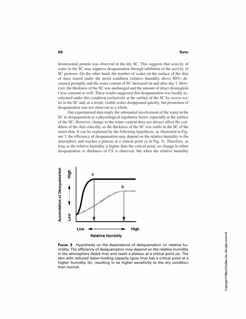
88 Sato
desmosomal protein was observed in the dry SC. This suggests that scarcity ofwater in the SC may suppress desquamation through inhibition of the activity ofSC protease. On the other hand, the number of scales on the surface of the skin of mice reared under the moist condition (relative humidity above 80%) de-creased promptly and the water content of SC increased on and after day 1. How-ever, the thickness of the SC was unchanged and the amount of intact desmogleinI was constant as well. These results suggested that desquamation was locally ac-celerated under this condition exclusively at the surface of the SC by excess wa-ter in the SC and, as a result, visible scales disappeared quickly, but promotion ofdesquamation was not observed as a whole.
Our experimental data imply the substantial involvement of the water in theSC in desquamation as a physiological regulatory factor, especially at the surfaceof the SC. However, change in the water content does not always affect the con-dition of the skin critically, as the thickness of the SC was stable in the SC of themoist skin. It can be explained by the following hypothesis: as illustrated in Fig-ure 3, the efficiency of desquamation may depend on the relative humidity in theatmosphere and reaches a plateau at a critical point (a in Fig. 3). Therefore, aslong as the relative humidity is higher than the critical point, no change in eitherdesquamation or thickness of CS is observed, but when the relative humidity
FIGURE 3 Hypothesis on the dependence of desquamation on relative hu-midity. The efficiency of desquamation may depend on the relative humidityin the atmosphere (black line) and reach a plateau at a critical point (a). Theskin with reduced water-holding capacity (gray line) has a critical point at ahigher humidity (b), resulting in its higher sensitivity to the dry conditionthan normal.
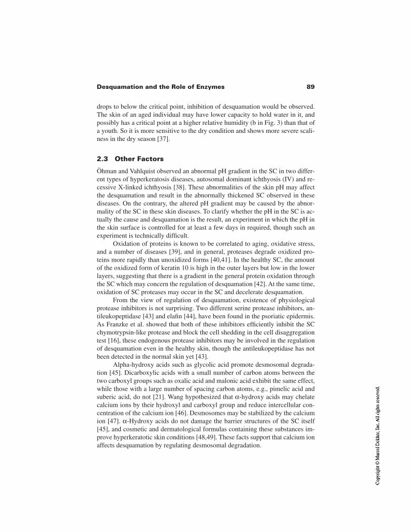
89Desquamation and the Role of Enzymes
drops to below the critical point, inhibition of desquamation would be observed.The skin of an aged individual may have lower capacity to hold water in it, andpossibly has a critical point at a higher relative humidity (b in Fig. 3) than that ofa youth. So it is more sensitive to the dry condition and shows more severe scali-ness in the dry season [37].
2.3 Other Factors
Öhman and Vahlquist observed an abnormal pH gradient in the SC in two differ-ent types of hyperkeratosis diseases, autosomal dominant ichthyosis (IV) and re-cessive X-linked ichthyosis [38]. These abnormalities of the skin pH may affectthe desquamation and result in the abnormally thickened SC observed in thesediseases. On the contrary, the altered pH gradient may be caused by the abnor-mality of the SC in these skin diseases. To clarify whether the pH in the SC is ac-tually the cause and desquamation is the result, an experiment in which the pH inthe skin surface is controlled for at least a few days in required, though such anexperiment is technically difficult.
Oxidation of proteins is known to be correlated to aging, oxidative stress,and a number of diseases [39], and in general, proteases degrade oxidized pro-teins more rapidly than unoxidized forms [40,41]. In the healthy SC, the amountof the oxidized form of keratin 10 is high in the outer layers but low in the lowerlayers, suggesting that there is a gradient in the general protein oxidation throughthe SC which may concern the regulation of desquamation [42]. At the same time,oxidation of SC proteases may occur in the SC and decelerate desquamation.
From the view of regulation of desquamation, existence of physiologicalprotease inhibitors is not surprising. Two different serine protease inhibitors, an-tileukopeptidase [43] and elafin [44], have been found in the psoriatic epidermis.As Franzke et al. showed that both of these inhibitors efficiently inhibit the SCchymotrypsin-like protease and block the cell shedding in the cell disaggregationtest [16], these endogenous protease inhibitors may be involved in the regulationof desquamation even in the healthy skin, though the antileukopeptidase has notbeen detected in the normal skin yet [43].
Alpha-hydroxy acids such as glycolic acid promote desmosomal degrada-tion [45]. Dicarboxylic acids with a small number of carbon atoms between thetwo carboxyl groups such as oxalic acid and malonic acid exhibit the same effect,while those with a large number of spacing carbon atoms, e.g., pimelic acid andsuberic acid, do not [21]. Wang hypothesized that α-hydroxy acids may chelatecalcium ions by their hydroxyl and carboxyl group and reduce intercellular con-centration of the calcium ion [46]. Desmosomes may be stabilized by the calciumion [47]. α-Hydroxy acids do not damage the barrier structures of the SC itself[45], and cosmetic and dermatological formulas containing these substances im-prove hyperkeratotic skin conditions [48,49]. These facts support that calcium ionaffects desquamation by regulating desmosomal degradation.
90 Sato
FIGURE 4 Scheme of desquamation and regulatory factors in the stratumcorneum.
3 PROSPECTS
Despite the rapid advance in the past 20 years (summarized in Fig. 4), there re-mains many mysteries in the process and regulation of desquamation. Severalregulatory factors have been proposed; some of them may actually regulatedesquamation, but others may be the products which merely reflect the changeoccurring in the SC. Further studies will enable their discrimination.
As the SC exhibits many critical functions of the skin, such as barrier func-tion, it must be able to respond to the environmental changes dynamically andsteadily. Scaliness and thickening of the SC began within 3 days after shift to alow environmental humidity [36]. Following these alterations of the condition ofthe SC, hyperproliferation in the nucleated cell layer under the SC began in the
91Desquamation and the Role of Enzymes
second week accompanied with changes in barrier homeostasis and number ofkeratohyalin granules [50], although these changes were still not obvious at thisstage [36]. An environmental signal which triggers these secondary changes in-volving the whole epidermis may be transferred to the nucleated cell layer possi-bly via the SC water flux, which becomes obvious within 12 h after the event[51]. Therefore, the SC can counteract the environmental change both in shortand long time ranges with its autonomous function (desquamation) and those sec-ondary alterations which require the aid of the downward epidermis.
Some moisturizers work quite well, but their action mechanisms in the skinare not so simple and are largely unknown. As the skin has the ability to adapt tothe environment, it is desirable to encourage the ability to improve the conditionof the damaged skin. Elucidation of the whole mechanism of desquamation willlead to new insight into skin care methodology, new substances, and new applica-tion techniques.
REFERENCES
1. Suzuki Y, Nomura J, Hori J, Koyama J, Takahashi M, Horii I. Detection and charac-terization of endogenous protease associated with desquamation of stratum corneum.Arch Dermatol Res 1993; 285:372–377.
2. Bissett DL, McBride JF, Patrick LF. Role of protein and calcium in stratum corneumcell cohesion. Arch Dermatol Res 1987; 279:184–189.
3. Egelrud T, Lundström A. Immunochemical analyses of the distribution of thedesmosomal protein desmoglein I in different layers of plantar epidermis. Acta DermVenereol (Stockh) 1989; 69:470–476.
4. Lundström A, Egelrud T. Cell shedding from plantar stratum corneum in vitro in-volves endogenous proteolysis of the desmosomal protein desmoglein I. J InvestDermatol 1990; 94:216–220.
5. Lundström A, Egelrud T. Stratum corneum chymotryptic enzyme: a proteinasewhich may be generally present in the stratum corneum and with a possible involve-ment in desquamation. Acta Derm Venereol (Stockh) 1991; 71:471–474.
6. Serre G, Mils V, Haftek M. Identification of late differentiation antigens of humancornified epithelia, expressed in re-organized desmosomes and bound to cross-linkedenvelope. J Invest Dermatol 1991; 97:1061–1072.
7. Haftek M, Serre G, Mils V, Thivolet J. Immunocytochemical evidence of the possi-ble role of keratinocyte cross-linked envelopes in stratum corneum cohesion. J His-tochem Cytochem 1991; 39:153–158.
8. Montézin M, Simon M, Guerrin M, Serre G. Corneodesmosin, a corneodesmosome-specific basic protein, is expressed in the cornified epithelia of the pig, guinea pig, ratand mouse. Exp Cell Res 1997; 231:132–140.
9. Lundström A, Egelrud T. Cell shedding from human plantar skin in vitro: evidenceof its dependence on endogenous proteolysis. J Invest Dermatol 1988; 91:340–343.
10. Watkinson A. Stratum corneum thiol protease (SCTP): a novel cysteine protease oflate epidermal differentiation. Arch Dermatol Res 1999; 291:260–268.
92 Sato
11. Horikoshi T, Igarashi S, Uchida H, Brysk H, Brysk MM. Role of endogenous cathep-sin D–like and chymotrypsin-like proteolysis in human epidermal desquamation. BrJ Dermatol 1999; 141:453–459.
12. Egelrud T. Purification and preliminary characterization of stratum corneum chy-motryptic enzyme: a proteinase which may be involved in desquamation. J InvestDermatol 1993; 101:200–204.
13. Brattsand M, Egelrud T. Purification, molecular cloning, and expression of a humanstratum corneum trypsin-like serine protease with possible function in desquama-tion. J Biol Chem 1999; 274:30033–30040.
14. Suzuki Y, Nomura J, Koyama J, Horii I. The role of protease in stratum corneum: in-volvement in stratum corneum desquamation. Arch Dermatol Res 1994;286:3249–253.
15. Sato J, Denda M, Nakanishi J, Nomura J, Koyama J. Cholesterol sulfate inhibits pro-teases which are involved in desquamation of stratum corneum. J Invest Dermatol1998; 111:189–193.
16. Franzke C-W, Baici A, Bartels J, Christophers E, Wiedow O. Antileukoprotease in-hibits stratum corneum chymotryptic enzyme. J Biol Chem 1996; 271:21886–21890.
17. Egelrud T, Lundström A. A chymotrypsin-like proteinase that may be involved indesquamation in plantar stratum corneum. Arch Dermatol Res 1991; 283:108–112.
18. Sondell B, Thornell L-E, Egelrud T. Evidence that stratum corneum chymotrypticenzyme is transported to the stratum corneum extracellular space via lamellar bod-ies. J Invest Dermatol 1995; 104:819–823.
19. Hansson L, Strömqvist M, Bäckman A, Wallbrandt P, Carlstein A, Egelrud T.Cloning, expression, and characterization of stratum corneum chymotryptic enzyme.J Biol Chem 1994; 269:19420–19426.
20. Polgár L. In hydrolytic enzymes. In: Chaplin MF, Kennedy JF, eds. New Compre-hensive Biochemistry. Vol. 16. Amsterdam: Elssevier Science, 1987:159–200.
21. Koyama J, Nakanishi J, Masuda Y, Sato J, Nomura J, Suzuki Y, Nakayama Y. Themechanism of desquamation in the stratum corneum and its relevance to skin care.In: Tagami H, Parrish JA, Ozawa T, eds. Skin: Interface of a Living System. TheNetherlands: Elsevier Science, 1998:73–86.
22. Ekholm IE, Brattsand M, Egelrud T. Stratum corneum tryptic enzyme in normal epi-dermis: a missing link in the desquamation process? J Invest Dermatol 2000;114:56–63.
23. Horikoshi T, Arany I, Rajaraman S, Chen S-H, Brysk H, Lei G, Tyring SK, BryskMM. Isoforms of cathepsin D and human epidermal differentiation. Biochimie 1998;80:605–612.
24. Ito Y, Fukuyama K, Yabe K, Epstein WL. Purification and properties of aminoen-dopeptidase from rat epidermis. J Invest Dermatol 1984; 83:265–269.
25. Fukuyama K, Ito Y, Yabe K, Epstein WL. Immunological detection if a cystein pro-tease in the skin and other tissues. Cell Tissue Res 1985; 240:417–423.
26. Williams ML. Epidermal lipids and scaling diseases of the skin. Seminars in Derma-tology 1992; 11:169–175.
27. Cox P, Squier CA. Variations in lipids in different layers of porcine epidermis. J In-vest Dermatol 1986; 87:741–744.
28. Mesquita-Guimaraes J. X-Linked ichthyosis. Dermatolgica 1981; 162:157–166.
93Desquamation and the Role of Enzymes
29. Williams ML. Lipids in normal and pathological desquamation. In: PM Elias, ed.Advances in Lipid Research. Vol. 24. San Diego: Academic Press, 1991:211–262.
30. Maloney ME, Williams ML, Epstein EH, Michael Y, Law L, Fritsch PO, Elias PM.Lipids in the pathogenesis of ichthyosis: topical cholesterol sulfate–induced scalingin hairless mice. J Invest Dermatol 1984; 83:252–256.
31. Elias PM, Williams ML, Maloney ME, Bonifas JA, Brawn BE, Grayson S, EpsteinEH. Stratum corneum lipids in disorders of cornification. J Clin Invest 1984;74:1414–1421.
32. Bouwstra JA, Gooris GS, Dubbelaar EFR, Ponec M. Cholesterol sulfate and calciumaffect stratum corneum lipid organization over a wide temperature range. J Lipid Res1999; 40:2303–2312.
33. Rawlings AV, Harding C, Watkinson A, Banks J, Ackerman C, Sabin R. The effect ofglycerol and humidity on desmosome degradation in stratum corneum. Arch Derma-tol Res 1995; 287:457–464.
34. Blank IH. Factors which influence the water content of the stratum corneum. J InvestDermatol 1952; 18:433–440.
35. Hashimoto-Kumasaka K, Takahashi K, Tagami H. Electrical measurement of thewater content of the stratum corneum in vivo and in vitro under various conditions:comparison between skin surface hygrometer and corneometer in evaluation of theskin surface hydration state. Acta Derm Venereol 1993; 73:335–339.
36. Sato J, Denda M, Nakanishi J, Koyama J. Dry condition affects desquamation ofstratum corneum in vivo. J Derm Sci 1998; 18:163–169.
37. Horii I, Nakayama Y, Obata M, Tagami H. Stratum corneum hydration and aminoacid content in xerotic skin. Br J Dermatol 1989; 121:587–592.
38. Öhman H, Vahlquist A. The pH gradient over the stratum corneum differs in X-linked recessive and autosomal dominant ichthyosis: a clue to the molecular ori-gin of the “acid skin mantle’’? J Invest Dermatol 1998; 111:674–677.
39. Oliver CN, Ahn B-W, Moerman EJ, Goldstein S, Stadtman ER. Age-related changesin oxidized proteins. J Biol Chem 1987; 262:5488.
40. Davies KJA, Lin SW, Pacifici RE. Protein damage and degradation by oxygen radi-cals: degradation of denatured protein. J Biol Chem 1987; 262:9914–9920.
41. Stadtman ER. Protein oxidation and aging. Science 1992; 257:1220–1224.42. Thiele JJ, Hsieh SN, Briviba K, Sies H. Protein oxidation in human stratum
corneum: susceptibility of keratins to oxidation in vitro and presence of a keratin ox-idation gradient in vivo. J Invest Dermatol 1999; 113:335–339.
43. Molhuizen HOF, Alkemade HAC, Zeeuwen PLJM, de Jongh GJ, Wieringa B,Schalkwijk J. SKALP/elafin: an elastase in inhibitor from cultured human kera-tinocytes. J Biol Chem 1993; 268:12028–12032.
44. Wiedow O, Schröder J, Gregory H, Young JA, Chrisophers E. Elafin: an elastase-specific inhibitor of human skin. J Biol Chem 1990; 265:14791–14795.
45. Fartasch M, Teal J, Menon GK. Mode of action of glycolic acid on human stratumcorneum: ultrastructural and functional evaluation of the epidermal barrier. ArchDermatol Res 1997; 289:404–409.
46. Wang X. A theory for the mechanism of action of the α-hydroxy acids applied to theskin. Med Hypotheses 1999; 53:380–382.
47. Skerrow CJ. Desmosomal proteins. In: Bereiter-Hahn J, Matoltsy AG, Richards KS,
94 Sato
eds. Biology of the Integument. Vol. 2. Vertebrates. Berlin: Springer-Verlag,1986:762–787.
48. Van Scott EJ, Yu RJ. Hyperkeratinization, corneocyte cohesion and alpha hydroxyacids. J Am Acad Dermatol 1974; 11:867–879.
49. Van Scott EJ, Yu RJ. Alpha hydroxy acids: therapeutic potential. Can J Dermatol1989; 1:108–112.
50. Denda M, Sato J, Masuda Y, Tsuchiya T, Kuramoto M, Koyama J, Elias PM, Fein-gold KE. Exposure to a dry environment enhances epidermal permeability barrierfunction. J Invest Dermatol 1998; 111:858–863.
51. Sato J, Denda M, Ashida Y, Koyama J. Loss of water from the stratum corneum in-duces epidermal DNA synthesis in hairless mice. Arch Dermatol Res 1998;290:634–637.
5The Cornified Envelope: Its Role in StratumCorneum Structure and Maturation
Allan Watkinson and Clive R. HardingUnilever Research, Colworth Laboratory, Sharnbrook, Bedford, United Kingdom
Anthony V. RawlingsUnilever Research, Port Sunlight Laboratory, Bebington, Wirral,United Kingdom
1 INTRODUCTION
The stratum corneum is the outermost layer of the skin and is the principal barri-er tissue preventing water loss from the body and providing mechanical protec-tion. It is produced as the end product of epidermal terminal differentiation, aprocess which involves several coordinated biochemical processes. The final re-sult is a tissue that prevents water loss by the formation of a continuous matrix ofhighly organized lipid lamellae, into which is embedded an extensive network of“dead” cells called corneocytes [1,2].
Corneocyte morphology is crucial to the role of the stratum corneum as amechanically resistant barrier tissue. The cells themselves are platelike with di-mensions of approximately 50 µm across and 1 µm thin, stacked in layers, thenumber of which varying throughout the body [3–5]. As a remnant of the differ-entiating keratinocytes, corneocytes retain structural characteristics of the parentcell; they are packed with keratin intermediate filaments that provide the structur-
95
96 Watkinson et al.
al integrity and also an elasticity to resist stretching and compressional forces.Furthermore, as with the keratinocytes, they are linked together by rivetlikedesmosomes to provide stratum corneum cohesion and integrity, although in thestratum corneum the desmosomes are modified structures and termed cor-neodesmosomes [6,7]. The most marked difference between corneocytes andtheir parent keratinocytes is that the former are largely devoid of cellular or-ganelles, which degrade during corneocyte formation. As a result the corneocytesare only capable of catabolic reactions [8].
Since the corneocyte is localized within the hydrophobic lipid lamellae andrequires a mechanically stable structure, the standard phospholipid-based plasmamembrane used by cells in an aqueous environment is not a suitable cellular en-velope. Instead the corneocyte replaces the phospholipid membrane with a high-ly cross-linked structure made up of specialized structural proteins. Hence a “pro-tein cage” called the cornified envelope (CE) encapsulates the corneocyte.Morphologically, this structure can be detected by electron microscopy as anelectron-dense layer of approximately 15 nm thick, adjacent to the intercellularspaces [9–12]. The CE has a more electron-lucent outer coating of approximately5 nm thick which represents the covalently bound envelope [13,14].
The purpose of this chapter is to review the role of the CE in stratumcorneum structure and function. However, it must be realized that all the structur-al elements of the corneocyte, the CE, the intermediate filaments, and the cor-neodesmosomes, are interlinked, making these cells, and indeed the stratumcorneum itself, a giant macromolecule. Hence alterations in any one of thesestructures will drastically affect the others.
2 CORNIFIED ENVELOPE BIOCHEMISTRY
The CE has been likened to a protein cage. It is comprised of specialized struc-tural proteins linked by specific γ-glutamyl-ε-lysine and γ-glutamyl-polyamineisopeptide bonds, and to a lesser extent disulfide linkages [15–18]. This results ineach corneocyte being enclosed by a protein shell that in essence is a singlemacromolecule. In addition the specialized nature of the γ-glutamyl-ε-lysine con-fers a resistance to general proteolytic action. Moreover, a characteristic of theCE is that it is insoluble in SDS/reducing agent conditions, which are used in theisolation of these structures from stratum corneum tissue [19].
The biochemical composition of the CE is not homogeneous. The outer-most region of the CE, adjacent to the intercellular space, is rich in the protein in-volucrin [20–22]. This is a rodlike protein rich in glutamine/glutamate and there-fore suited as major structural protein in the CE; although only a specifiedfraction of the glutamate residues are cross-linked in vivo. Deeper within the CEstructure, involucrin gives way to be replaced by the protein loricrin. Loricrin isan insoluble protein rich in glycine, serine, and cysteine and comprises the majorcomponent of the CE [23–26]. Probably the third most important component of
97The Cornified Envelope
the CE are the small proline-rich proteins (SPRs). The SPRs comprise a family ofsmall proteins which are characterized into three groups; SPR 1 through 3, ofwhich SPR 1 and 2 have been identified as epidermal CE components [21,26,27].It is believed that these proteins function as cross-linking agents strengthening theCE against mechanical forces, supported by the correlation of CE SPR contentwith mechanical stress [28–30]. Additional minor CE components include cys-tatin-α (keratolinin) [31–33], elafin (SKALP) [26], envoplakin [34], periplakin[35], cystine-rich envelope protein (CREP) [36], S100 proteins, annexins, plas-minogen activator inhibitor 2 [21] and filaggrin [37,38]. In addition, since the CEinteracts with corneocyte structures such as the keratin intermediate filaments andthe corneodesmosomes, keratins and desmoplakins have also been detectedcross-linked to CE proteins [12,21,26].
2.1 Cornified Envelope Formation
The major function of the cornified envelope is to produce a mechanically robustalternative to the phospholipid-based plasma membranes of viable keratinocytes.Instantly replacing the keratinocyte plasma membrane is not feasible; hence oneof the processes of epidermal terminal differentiation is to construct the CE whilethe plasma membrane is intact, prior to cornification.
The enzymes responsible for catalyzing γ-glutamyl-ε-lysine isopeptidebond formation are the transglutaminases (TGases) [15,39], Transglutaminasesare Ca2+-dependent enzymes and form a bond between the γ-glutamyl aminogroup and a primary amino group, the latter being normal provided by the sidechain of lysine or by the polyamines putrescine and spermidine (Fig. 1)[16,17].
The epidermis produces three types of TGase. TGase 1 (TGase K) is ex-pressed suprabasally, arising in the mid-spinous layer [40–42]. It is produced as a106-KDa protein and due to N- and S-linked fatty acyl groups at its N-terminalregion is membrane bound [43]. Following synthesis the protein is proteolytical-ly processed to give soluble components of 10, 33, and 67 KDa, the complex ofwhich is membrane bound and has enhanced activity [44]. TGase 3 (TGase E) isproduced later in terminal differentiation in the granular layer [45–47]. It is pro-duced as a soluble 77-KDa inactive pro-enzyme but subsequently cleaved to givethe 50-KDa active form. TGase 2 (TGase C) is ubiquitous and has no role in CEformation but may play a role in apoptosis [48].
TGase 1 is accredited with initiating CE formation due to its earlier appear-ance in epidermal differentiation [42]. The first steps in CE construction appear tobe the production of a scaffold using soluble involucrin as the major component[22]. Cornified envelope construction occurs immediately adjacent to the innerface of the keratinocyte plasma membrane. Precisely how this complex structureassembles is not fully understood. Studies using cultured keratinocytes suggestthat involucrin, cross-linked in a head–tail and head–head orientation, spreads outfrom interdesmosomal regions to link with the desmosomal complexes, anchor-
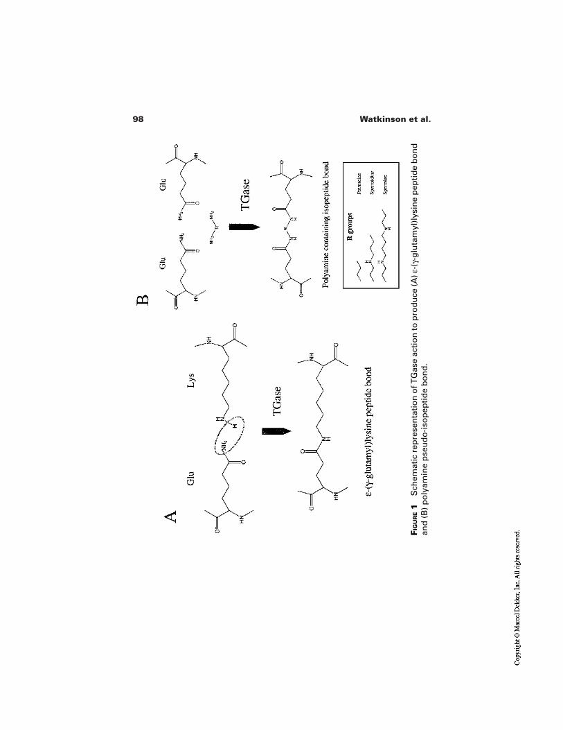
98 Watkinson et al.
FIG
UR
E1
Sch
emat
ic r
epre
sen
tati
on
of T
Gas
e ac
tio
n to
pro
du
ce (A
) ε-(
γ-g
luta
myl
)lys
ine
pep
tid
e b
on
dan
d (
B)
po
lyam
ine
pse
ud
o-i
sop
epti
de
bo
nd
.
99The Cornified Envelope
ing the forming CE into the keratinocyte cytoskeleton and tissue matrix [49]. In-volucrin molecules may self-assemble on the keratinocyte plasma membrane byinteracting with phosphatidylserine residues in a calcium-dependent manner andsubsequently be linked by the action of membrane-bound TGase 1 [50]. Addi-tional elements linked with this scaffold assembly process are envoplakin andpossibly periplakin [49]. Envoplakin co-localizes with involucrin in the earlysteps of this process and similarly spreads from interdesmosomal regions to linkwith the desmosomes to produce a continuous layer adjacent to the plasma mem-brane. Once the involucrin envoplakin scaffold is produced, the structure isstrengthened by the addition of loricrin, SPRs, and other CE components. Inter-estingly, it has been shown that loricrin associates initially with the desmosomalplaque in the granular layer keratinocytes and subsequently becomes incorporat-ed in the CE during or after the cornification process [51]. This suggests that thedesmosome may be a focal point for the accumulation and subsequent attachmentof CE precursor proteins to the scaffold, possibly by the action of the annexins[21,51,52].
The synthesis of TGase 3 in the granular layer, subsequent to that of TGase1, provides a supplementary soluble pool of TGase enzyme to facilitate CE com-pletion. As the CE forms, membrane-bound TGase 1 is likely to become, at leastpartially, entrapped. TGase 3 has a higher affinity for loricrin than other CE com-ponents [53] and since this protein comprises about 70% of the CE, this suggeststhat it is the main enzyme involved in the completion of CE construction. How-ever, both TGase 1 and 3 are required for the strengthening phase since SPR in-corporation appears to require both enzymes, each acting at a different site on themolecule. For these same reasons, TGase 3 may also be the major enzyme of CEmaturation within the stratum corneum, but currently there is little evidence tosupport this concept.
Isopeptide bonds are not the sole covalent bond involved in CE structure.Disulfide bonds also link CE components, being produced by the enzyme sul-fydryl oxidase [54,55]. Little is known about their contribution to CE structure,especially since they are normally lost in CE isolation due to the use of reducingagents. Cysteine-containing CE components that could be involved in disulfidebond formation include CREP and loricrin [24,25,36]. The major function ofdisulfide bonds within the CE may be between loricrin and the keratins of the in-termediate filaments, providing an additional link into the keratinocyte/corneo-cyte cytoskeleton [25].
2.2 Covalently-Bound Lipid
Whereas the polar head groups of the phospholipids in the plasma membrane al-low interaction with the aqueous environment, in the SC the CE must interactwith the hydrophobic lipid lamellae in the intercellular spaces. Since the outer re-
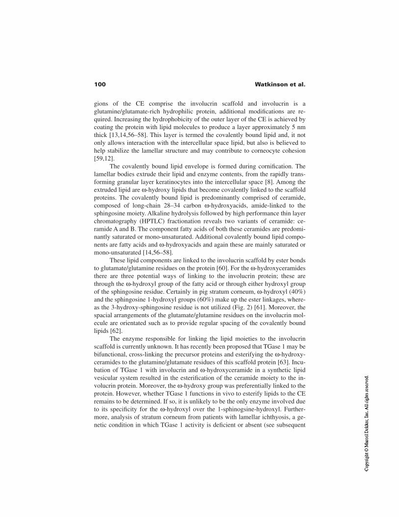
100 Watkinson et al.
gions of the CE comprise the involucrin scaffold and involucrin is aglutamine/glutamate-rich hydrophilic protein, additional modifications are re-quired. Increasing the hydrophobicity of the outer layer of the CE is achieved bycoating the protein with lipid molecules to produce a layer approximately 5 nmthick [13,14,56–58]. This layer is termed the covalently bound lipid and, it notonly allows interaction with the intercellular space lipid, but also is believed tohelp stabilize the lamellar structure and may contribute to corneocyte cohesion[59,12].
The covalently bound lipid envelope is formed during cornification. Thelamellar bodies extrude their lipid and enzyme contents, from the rapidly trans-forming granular layer keratinocytes into the intercellular space [8]. Among theextruded lipid are ω-hydroxy lipids that become covalently linked to the scaffoldproteins. The covalently bound lipid is predominantly comprised of ceramide,composed of long-chain 28–34 carbon ω-hydroxyacids, amide-linked to thesphingosine moiety. Alkaline hydrolysis followed by high performance thin layerchromatography (HPTLC) fractionation reveals two variants of ceramide: ce-ramide A and B. The component fatty acids of both these ceramides are predomi-nantly saturated or mono-unsaturated. Additional covalently bound lipid compo-nents are fatty acids and ω-hydroxyacids and again these are mainly saturated ormono-unsaturated [14,56–58].
These lipid components are linked to the involucrin scaffold by ester bondsto glutamate/glutamine residues on the protein [60]. For the ω-hydroxyceramidesthere are three potential ways of linking to the involucrin protein; these arethrough the ω-hydroxyl group of the fatty acid or through either hydroxyl groupof the sphingosine residue. Certainly in pig stratum corneum, ω-hydroxyl (40%)and the sphingosine 1-hydroxyl groups (60%) make up the ester linkages, where-as the 3-hydroxy-sphingosine residue is not utilized (Fig. 2) [61]. Moreover, thespacial arrangements of the glutamate/glutamine residues on the involucrin mol-ecule are orientated such as to provide regular spacing of the covalently boundlipids [62].
The enzyme responsible for linking the lipid moieties to the involucrinscaffold is currently unknown. It has recently been proposed that TGase 1 may bebifunctional, cross-linking the precursor proteins and esterifying the ω-hydroxy-ceramides to the glutamine/glutamate residues of this scaffold protein [63]. Incu-bation of TGase 1 with involucrin and ω-hydroxyceramide in a synthetic lipidvesicular system resulted in the esterification of the ceramide moiety to the in-volucrin protein. Moreover, the ω-hydroxy group was preferentially linked to theprotein. However, whether TGase 1 functions in vivo to esterify lipids to the CEremains to be determined. If so, it is unlikely to be the only enzyme involved dueto its specificity for the ω-hydroxyl over the 1-sphinogsine-hydroxyl. Further-more, analysis of stratum corneum from patients with lamellar ichthyosis, a ge-netic condition in which TGase 1 activity is deficient or absent (see subsequent
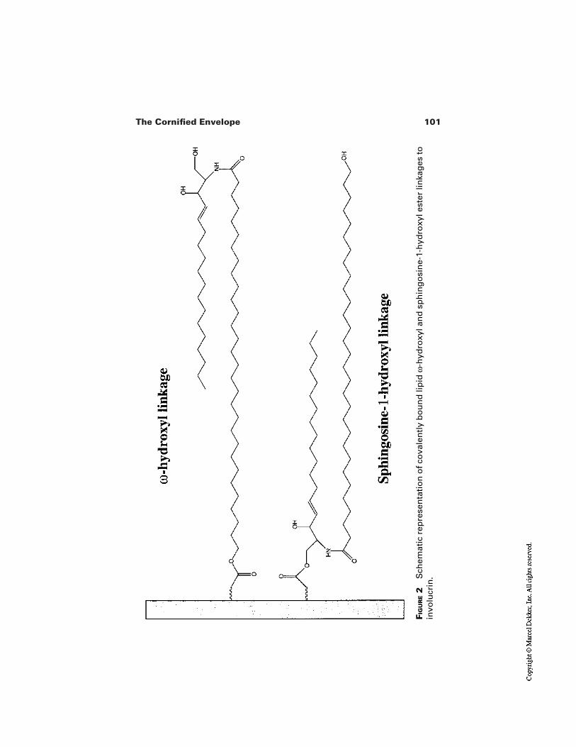
101The Cornified Envelope
FIG
UR
E2
Sch
emat
ic r
epre
sen
tati
on
of c
ova
len
tly
bo
un
d li
pid
ω-h
ydro
xyl a
nd
sp
hin
go
sin
e-1-
hyd
roxy
l est
er li
nka
ges
toin
volu
crin
.
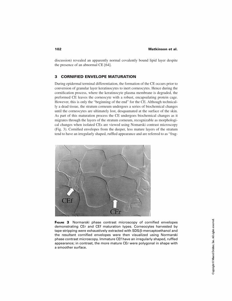
102 Watkinson et al.
discussion) revealed an apparently normal covalently bound lipid layer despitethe presence of an abnormal CE [64].
3 CORNIFIED ENVELOPE MATURATION
During epidermal terminal differentiation, the formation of the CE occurs prior toconversion of granular layer keratinocytes to inert corneocytes. Hence during thecornification process, where the keratinocyte plasma membrane is degraded, thepreformed CE leaves the corneocyte with a robust, encapsulating protein cage.However, this is only the “beginning of the end” for the CE. Although technical-ly a dead tissue, the stratum corneum undergoes a series of biochemical changesuntil the corneocytes are ultimately lost, desquamated at the surface of the skin.As part of this maturation process the CE undergoes biochemical changes as itmigrates through the layers of the stratum corneum, recognizable as morphologi-cal changes when isolated CEs are viewed using Nomarski contrast microscopy(Fig. 3). Cornified envelopes from the deeper, less mature layers of the stratumtend to have an irregularly shaped, ruffled appearance and are referred to as “frag-
FIGURE 3 Normarski phase contrast microscopy of cornified envelopesdemonstrating CEr and CEf maturation types. Corneocytes harvested bytape-stripping were exhaustively extracted with SDS/β-mercaptoethanol andthe resultant cornified envelopes were then visualized using Normarskiphase contrast microscopy. Immature CEf have an irregularly shaped, ruffledappearance; in contrast, the more mature CEr were polygonal in shape witha smoother surface.
103The Cornified Envelope
ile” envelopes (CEf). In contrast, CE from the more mature, peripheral layers ofthe stratum corneum tend to be more polygonal in shape with a smoother surfaceand are called “rigid” cornified envelopes (CEr) [65]. The reasons for these mor-phological differences between mature and immature CE are unknown.Cyanogen bromide cleavage of CE to produce peptide maps did not reveal anysignificant difference between the two variants [66]. However, as well as beingdiscernible under the microscope, the two CE variants can be differentiated bytheir interaction with the fluorescent label tetramethylrhodamine isothiocyanate(TRITC). CEr stain with an intense yellow-orange fluorescence whilst CEf stainweakly under the same conditions [18,66] (Fig. 4). This staining pattern can beused to follow the CE maturation process by determining the bulk fluorescence oftape-stripped CE which have been treated with TRITC. In the deeper SC, lowerlevels of fluorescence are detected, consistent with there being predominantly im-mature CEf; as the CE approach the surface of the SC, there is a marked increasein fluorescence (tape strips 9–12) consistent with a rapid, rather than gradual,maturation event to produce CEr (Fig. 5).
Interestingly, this maturation-associated change in fluorescence occurs afterfilaggrin has largely been hydrolyzed and therefore appears to be closely associ-
FIGURE 4 Fluorescence and Normarski phase contrast microscopy of TRITCstained cornified envelopes demonstrating increased fluorescence labelingof CEr compared to CEf. Exhaustively extracted corneocytes were treatedwith TRITC (200 µl/mL) for 2 hr, then washed and visualized by fluorescencemicroscopy followed by Normarski phase contrast microscopy. The matureCEr show increased fluorescence labeling compared to the immature CEf.
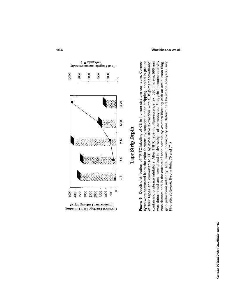
104 Watkinson et al.
FIG
UR
E5
Dep
th d
istr
ibu
tio
n o
f T
RIT
C l
abel
ing
of
CE
in
hu
man
str
atu
m c
orn
eum
. C
orn
eo-
cyte
s w
ere
har
vest
ed f
rom
th
e vo
lar
fore
arm
by
seq
uen
tial
tap
e-st
rip
pin
g, p
oo
led
in g
rou
ps
of
fou
r ta
pes
an
d c
on
vert
ed t
o C
E b
y ex
hau
stiv
e ex
trac
tio
n w
ith
SD
S/β
-mer
cap
toet
han
ol
con
tain
ing
pro
teas
e in
hib
ito
rs. A
fter
TR
ITC
lab
elin
g, fl
uo
resc
ence
(ex
, 530
nm
; em
, 590
nm
)w
as d
eter
min
ed a
nd
no
rmal
ized
to
dry
wei
gh
t o
f co
rneo
cyte
s. F
ilag
gri
n i
mm
un
ore
acti
vity
was
det
erm
ined
in t
he
extr
act
of
each
sam
ple
by
wes
tern
blo
ttin
g w
ith
an
an
tih
um
an fi
lag
-g
rin
po
lycl
on
al a
nti
bo
dy.
To
tal
imm
un
ore
acti
vity
was
det
erm
ined
by
imag
e an
alys
is u
sin
gP
ho
reti
x so
ftw
are.
(Fr
om
Ref
s. 7
0 an
d 7
1.)
units
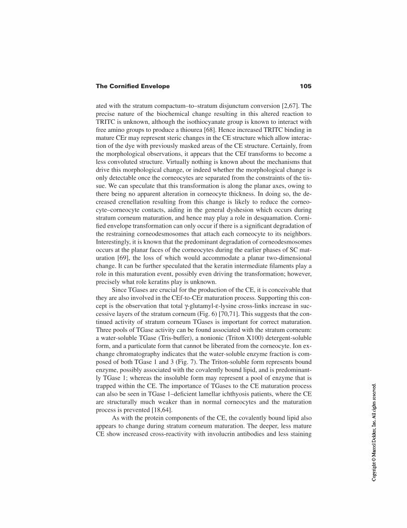
105The Cornified Envelope
ated with the stratum compactum–to–stratum disjunctum conversion [2,67]. Theprecise nature of the biochemical change resulting in this altered reaction toTRITC is unknown, although the isothiocyanate group is known to interact withfree amino groups to produce a thiourea [68]. Hence increased TRITC binding inmature CEr may represent steric changes in the CE structure which allow interac-tion of the dye with previously masked areas of the CE structure. Certainly, fromthe morphological observations, it appears that the CEf transforms to become aless convoluted structure. Virtually nothing is known about the mechanisms thatdrive this morphological change, or indeed whether the morphological change isonly detectable once the corneocytes are separated from the constraints of the tis-sue. We can speculate that this transformation is along the planar axes, owing tothere being no apparent alteration in corneocyte thickness. In doing so, the de-creased crenellation resulting from this change is likely to reduce the corneo-cyte–corneocyte contacts, aiding in the general dyshesion which occurs duringstratum corneum maturation, and hence may play a role in desquamation. Corni-fied envelope transformation can only occur if there is a significant degradation ofthe restraining corneodesmosomes that attach each corneocyte to its neighbors.Interestingly, it is known that the predominant degradation of corneodesmosomesoccurs at the planar faces of the corneocytes during the earlier phases of SC mat-uration [69], the loss of which would accommodate a planar two-dimensionalchange. It can be further speculated that the keratin intermediate filaments play arole in this maturation event, possibly even driving the transformation; however,precisely what role keratins play is unknown.
Since TGases are crucial for the production of the CE, it is conceivable thatthey are also involved in the CEf-to-CEr maturation process. Supporting this con-cept is the observation that total γ-glutamyl-ε-lysine cross-links increase in suc-cessive layers of the stratum corneum (Fig. 6) [70,71]. This suggests that the con-tinued activity of stratum corneum TGases is important for correct maturation.Three pools of TGase activity can be found associated with the stratum corneum:a water-soluble TGase (Tris-buffer), a nonionic (Triton X100) detergent-solubleform, and a particulate form that cannot be liberated from the corneocyte. Ion ex-change chromatography indicates that the water-soluble enzyme fraction is com-posed of both TGase 1 and 3 (Fig. 7). The Triton-soluble form represents boundenzyme, possibly associated with the covalently bound lipid, and is predominant-ly TGase 1; whereas the insoluble form may represent a pool of enzyme that istrapped within the CE. The importance of TGases to the CE maturation processcan also be seen in TGase 1–deficient lamellar ichthyosis patients, where the CEare structurally much weaker than in normal corneocytes and the maturationprocess is prevented [18,64].
As with the protein components of the CE, the covalently bound lipid alsoappears to change during stratum corneum maturation. The deeper, less matureCE show increased cross-reactivity with involucrin antibodies and less staining

106 Watkinson et al.
FIGURE 6 Distribution profile of ε-(γ-glutamyl)lysine cross-links in CE recov-ered from human stratum corneum. Consecutive tape strips were taken,pooled in groups of five and the corneocytes were exhaustively extracted inSDS/β-mercaptoethanol. γ-Glutamyl(lysine) cross-links were isolated fromthe CE by exhaustive protease digestion. The isolated ε-(γ-glutamyl)lysinedipeptide cross-links were quantified by reverse phase HPLC.* p < 0.001compared to the surface γ-glutamyl(lysine) cross-link content (n = 9). (FromRefs. 70 and 71.)
with the lipid stain Oil Red O. In contrast, the mature, peripheral CEr show lessinvolucrin cross-reactivity and increased Oil Red O staining [72]. If confirmed,the increase in Oil Red O staining indicates that the CE progressively build uptheir covalently bound lipid. Furthermore, it suggests that the esterifying enzymesretain activity throughout the SC, presumably supplied with ω-hydroxyceramidesfrom the intercellular space lipid lamellae. If indeed TGase 1 functions in this ca-pacity, this would be consistent with the detection of TGase activity in the TritonX100 extracts of SC. The progressive reduction of involucrin labeling supportsthe continued enlargement of the covalently bound lipid covering due to stericalinhibition of antibody binding. However this could also be explained by an in-crease in involucrin cross-linking with maturation [73].
4 CORNIFIED ENVELOPES AND SKIN CONDITION
In considering the role of CE in skin condition, the necessity of isolating thesestructures with SDS and reducing agent has meant that they are devoid of the oth-
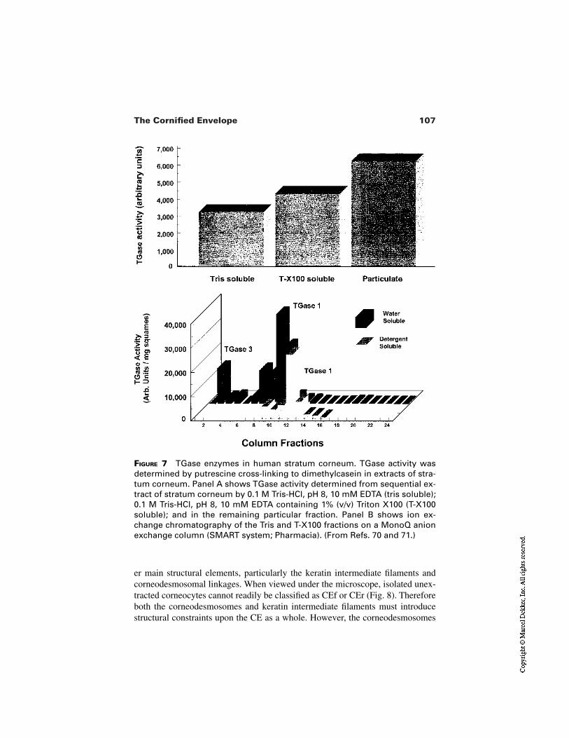
107The Cornified Envelope
FIGURE 7 TGase enzymes in human stratum corneum. TGase activity wasdetermined by putrescine cross-linking to dimethylcasein in extracts of stra-tum corneum. Panel A shows TGase activity determined from sequential ex-tract of stratum corneum by 0.1 M Tris-HCl, pH 8, 10 mM EDTA (tris soluble);0.1 M Tris-HCl, pH 8, 10 mM EDTA containing 1% (v/v) Triton X100 (T-X100soluble); and in the remaining particular fraction. Panel B shows ion ex-change chromatography of the Tris and T-X100 fractions on a MonoQ anionexchange column (SMART system; Pharmacia). (From Refs. 70 and 71.)
er main structural elements, particularly the keratin intermediate filaments andcorneodesmosomal linkages. When viewed under the microscope, isolated unex-tracted corneocytes cannot readily be classified as CEf or CEr (Fig. 8). Thereforeboth the corneodesmosomes and keratin intermediate filaments must introducestructural constraints upon the CE as a whole. However, the corneodesmosomes
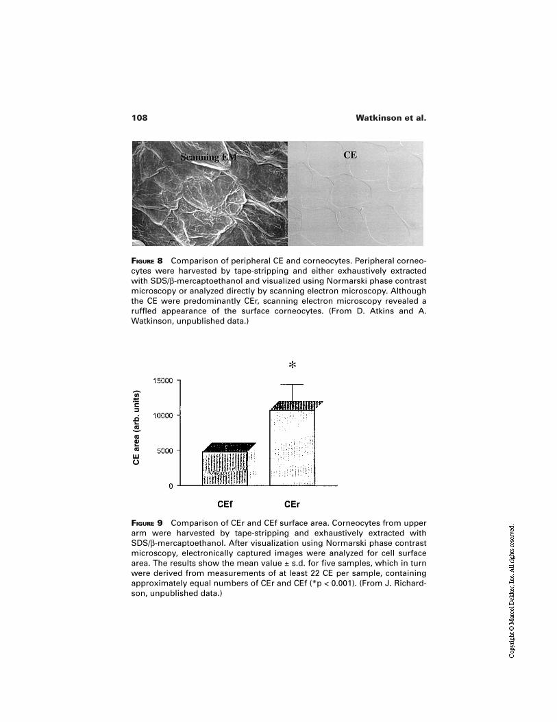
108 Watkinson et al.
FIGURE 8 Comparison of peripheral CE and corneocytes. Peripheral corneo-cytes were harvested by tape-stripping and either exhaustively extractedwith SDS/β-mercaptoethanol and visualized using Normarski phase contrastmicroscopy or analyzed directly by scanning electron microscopy. Althoughthe CE were predominantly CEr, scanning electron microscopy revealed aruffled appearance of the surface corneocytes. (From D. Atkins and A.Watkinson, unpublished data.)
FIGURE 9 Comparison of CEr and CEf surface area. Corneocytes from upperarm were harvested by tape-stripping and exhaustively extracted withSDS/β-mercaptoethanol. After visualization using Normarski phase contrastmicroscopy, electronically captured images were analyzed for cell surfacearea. The results show the mean value ± s.d. for five samples, which in turnwere derived from measurements of at least 22 CE per sample, containingapproximately equal numbers of CEr and CEf (*p < 0.001). (From J. Richard-son, unpublished data.)
CE
are
a (a
rb.u
nit
s)
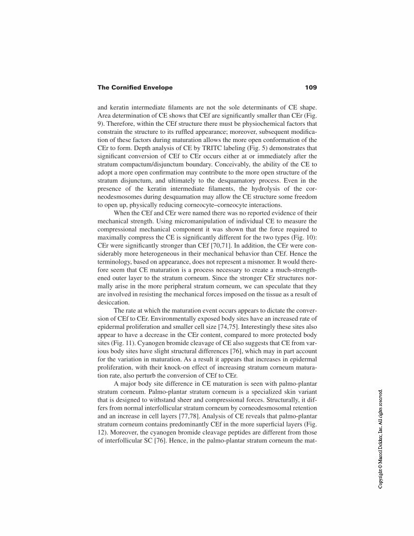
109The Cornified Envelope
and keratin intermediate filaments are not the sole determinants of CE shape.Area determination of CE shows that CEf are significantly smaller than CEr (Fig.9). Therefore, within the CEf structure there must be physiochemical factors thatconstrain the structure to its ruffled appearance; moreover, subsequent modifica-tion of these factors during maturation allows the more open conformation of theCEr to form. Depth analysis of CE by TRITC labeling (Fig. 5) demonstrates thatsignificant conversion of CEf to CEr occurs either at or immediately after thestratum compactum/disjunctum boundary. Conceivably, the ability of the CE toadopt a more open confirmation may contribute to the more open structure of thestratum disjunctum, and ultimately to the desquamatory process. Even in thepresence of the keratin intermediate filaments, the hydrolysis of the cor-neodesmosomes during desquamation may allow the CE structure some freedomto open up, physically reducing corneocyte–corneocyte interactions.
When the CEf and CEr were named there was no reported evidence of theirmechanical strength. Using micromanipulation of individual CE to measure thecompressional mechanical component it was shown that the force required tomaximally compress the CE is significantly different for the two types (Fig. 10):CEr were significantly stronger than CEf [70,71]. In addition, the CEr were con-siderably more heterogeneous in their mechanical behavior than CEf. Hence theterminology, based on appearance, does not represent a misnomer. It would there-fore seem that CE maturation is a process necessary to create a much-strength-ened outer layer to the stratum corneum. Since the stronger CEr structures nor-mally arise in the more peripheral stratum corneum, we can speculate that theyare involved in resisting the mechanical forces imposed on the tissue as a result ofdesiccation.
The rate at which the maturation event occurs appears to dictate the conver-sion of CEf to CEr. Environmentally exposed body sites have an increased rate ofepidermal proliferation and smaller cell size [74,75]. Interestingly these sites alsoappear to have a decrease in the CEr content, compared to more protected bodysites (Fig. 11). Cyanogen bromide cleavage of CE also suggests that CE from var-ious body sites have slight structural differences [76], which may in part accountfor the variation in maturation. As a result it appears that increases in epidermalproliferation, with their knock-on effect of increasing stratum corneum matura-tion rate, also perturb the conversion of CEf to CEr.
A major body site difference in CE maturation is seen with palmo-plantarstratum corneum. Palmo-plantar stratum corneum is a specialized skin variantthat is designed to withstand sheer and compressional forces. Structurally, it dif-fers from normal interfollicular stratum corneum by corneodesmosomal retentionand an increase in cell layers [77,78]. Analysis of CE reveals that palmo-plantarstratum corneum contains predominantly CEf in the more superficial layers (Fig.12). Moreover, the cyanogen bromide cleavage peptides are different from thoseof interfollicular SC [76]. Hence, in the palmo-plantar stratum corneum the mat-
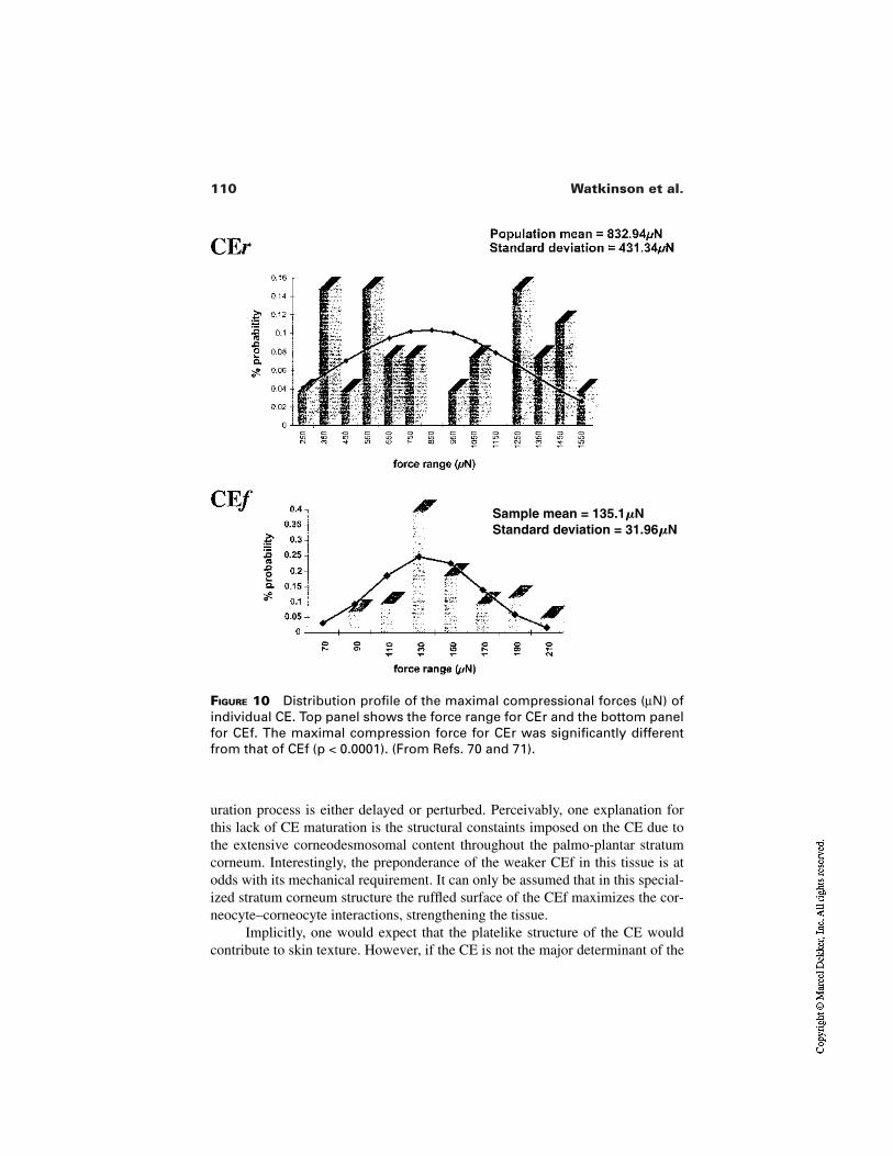
110 Watkinson et al.
FIGURE 10 Distribution profile of the maximal compressional forces (µN) ofindividual CE. Top panel shows the force range for CEr and the bottom panelfor CEf. The maximal compression force for CEr was significantly differentfrom that of CEf (p < 0.0001). (From Refs. 70 and 71).
uration process is either delayed or perturbed. Perceivably, one explanation forthis lack of CE maturation is the structural constaints imposed on the CE due tothe extensive corneodesmosomal content throughout the palmo-plantar stratumcorneum. Interestingly, the preponderance of the weaker CEf in this tissue is atodds with its mechanical requirement. It can only be assumed that in this special-ized stratum corneum structure the ruffled surface of the CEf maximizes the cor-neocyte–corneocyte interactions, strengthening the tissue.
Implicitly, one would expect that the platelike structure of the CE wouldcontribute to skin texture. However, if the CE is not the major determinant of the
Sample mean = 135.1NStandard deviation = 31.96N
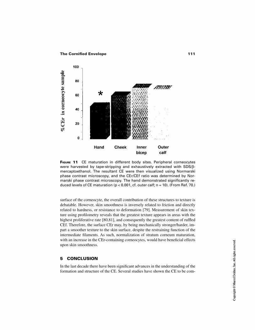
111The Cornified Envelope
FIGURE 11 CE maturation in different body sites. Peripheral corneocyteswere harvested by tape-stripping and exhaustively extracted with SDS/β-mercaptoethanol. The resultant CE were then visualized using Normarskiphase contrast microscopy, and the CEr/CEf ratio was determined by Nor-marski phase contrast microscopy. The hand demonstrated significantly re-duced levels of CE maturation (p < 0.001, cf. outer calf; n = 10). (From Ref. 70.)
surface of the corneocyte, the overall contribution of these structures to texture isdebatable. However, skin smoothness is inversely related to friction and directlyrelated to hardness, or resistance to deformation [79]. Measurement of skin tex-ture using profilometry reveals that the greatest texture appears in areas with thehighest proliferative rate [80,81], and consequently the greatest content of ruffledCEf. Therefore, the surface CEr may, by being mechanically stronger/harder, im-part a smoother texture to the skin surface, despite the restraining function of theintermediate filaments. As such, normalization of stratum corneum maturation,with an increase in the CEr-containing corneocytes, would have beneficial effectsupon skin smoothness.
5 CONCLUSION
In the last decade there have been significant advances in the understanding of theformation and structure of the CE. Several studies have shown the CE to be com-
112 Watkinson et al.
FIGURE 12 Comparison of peripheral CE from plantar and interfollicular stra-tum corneum. Peripheral corneocytes from either the sole of the foot or theaxilla were harvested by tape-stripping and exhaustively extracted withSDS/β-mercaptoethanol. Normarski phase contrast microscopy revealed thatthe interfollicular CE were predominantly CEr, whereas the plantar CE werepredominantly CEf. (From J. Richardson and A. Watkinson, unpublisheddata.)
posed of highly specialized structural proteins, which are laid down in a coordi-nated fashion to produce a robust protein cage encompassing the corneocyte. Ithas also been shown that once formed, the CE then undergoes a maturationprocess as it migrates through the stratum corneum, until it is eventually lost dur-ing desquamation. Our understanding of how this maturation process occurs andwhat this means for interaction with other structural elements of the stratumcorneum is far from complete. It is inconceivable that the morphological and bio-chemical changes associated with the maturation event do not influence the con-dition of the skin. Indeed, perturbations of CE maturation invariably are associat-ed with pathological skin conditions. A major challenge for the future researchremains a more complete understanding of this maturation process and how it im-pinges on stratum corneum structure and skin condition.
ACKNOWLEDGMENT
I would like to thank Mrs. J. Richardson for her invaluable assistance in pro-ducing this manuscript. In addition I would like to thank Mr. P. Coan and Mr. D. Atkins for technical assistance with the TRITC analysis and SEM, re-spectively.
113The Cornified Envelope
REFERENCES
1. Elias PM. Epidermal lipids, barrier function and desquamation. J Invest Dermatol1983; 80(Suppl):44–49.
2. Harding CR, Watkinson A, Rawlings AV, Scott IR. Dry skin, moisturization and cor-neodesmolysis. Int J Cosmet Sci 2000; 22:21–52.
3. Marks R, Barton SP. The significance of the size and shape of corneocytes. In: MarksR, Plewig G, eds. Stratum Corneum. Berlin: Springer-Verlag, 1983:161–170.
4. Plewig G, Scheuber E, Reuter B, Waidelich W. Thickness of corneocytes. In: MarksR, Plewig G, eds. Stratum Corneum. Berlin: Springer-Verlag, 1983:171–174.
5. Zhen YX, Suetake T, Tagami H. Number of cell layers of the stratum corneum innormal skin—relationship to the anatomical location on the body, age, sex and phys-ical parameters. Arch Dermatol Res 1999; 291:555–559.
6. Allen TD, Potten CS. Desmosomal form, fate and function in mammalian epidermis.J Ultrastruct Res 1975; 5:94–105.
7. Chapman SJ, Walsh, A. Desmosomes, corneosomes and desquamation. An ultra-structural study of adult pig epidermis. Arch Dermatol Res 1990; 282:304–310.
8. Holbrook KA. Biologic structure and function: perspectives on morphologic ap-proaches to the study of the granular layer keratinocytes. J Invest Dermatol 1989;92:84S–104S.
9. Matolsky AG, Balsamo CA. A study of the components of the cornified epithelium inskin. J Biophys Biochem Cytol 1955; 1:339–360.
10. Brody I. The modified plasma membranes of the transition and horny cells in normalhuman epidermis as revealed by electron microscopy. Acta Derm Venereol 1969;49:128–138.
11. Farbman AI. Plasma membrane changes during keratinization. Anat Rev 1966;156:269–282.
12. Haftek M, Serre G, Mils V, Thivolet J. Immunocytochemical evidence for a possiblerole of cross-linked keratinocyte envelopes in stratum-corneum cohesion. J His-tochem Cytochem 1991; 39:1531–1538.
13. Lavker RM. Membrane coating granules: the fate of the discharged lamellae. J Ul-trastruct Res 1976; 55:79–86.
14. Swartzendruber DC, Wertz PW, Madison KC, Downing DT. Evidence that the cor-neocyte has a chemically bound lipid envelope. J Invest Dermatol 1987;88:709–713.
15. Rice RH, Green H. Cornified envelope of terminally differentiated human epidermalkeratinocytes consists of cross-linked protein. Cell 1977; 11:417–422.
16. Martinet N, Beninati S, Nigra TP, Folk JE. N-(1,8)-bis(γ-Glutamyl)spermidine cross-linking in epidermal-cell envelopes—comparison of cross-link levels in normal andpsoriatic cell envelopes. Biochem J 1990; 271:305–308.
17. Steinert PM. Structural-mechanical integration of keratin intermediate filamentswith cell peripheral structures in the cornified epidermal keratinocyte. Biol Bull1998; 194:367–368.
18. Reichert U, Michel S, Schmidt R. The cornified envelope: a key structure of termi-nally differentiating keratinocytes. In: Darmon M, Blumenberg M, eds. MolecularBiology of the Skin: The keratinocyte. New York: Academic Press, 1993:107–150.
114 Watkinson et al.
19. Sun TT, Green H. Differentiation of epidermal keratinocyte in cell culture—forma-tion of cornified envelope. Cell 1976; 9:511–521.
20. Robinson NA, LaCelle PT, Eckert RL. Involucrin is a covalently crosslinked con-stituent of highly purified epidermal corneocytes: evidence for a common pattern ofinvolucrin crosslinking in vivo and in vitro. J Invest Dermatol 1996; 107:101–107.
21. Robinson NA, Lapic S, Welter JF, Eckert RL. S100A11, S100A10, annexin I,desmosomal proteins, small proline-rich proteins, plasminogen activator inhibitor-2,and involucrin are components of the cornified envelope of cultured human epider-mal keratinocytes. J Biol Chem 1997; 272:12035–12046.
22. Steinert PM, Marekov LN. Direct evidence that involucrin is a major early isopep-tide crosslinked component of the keratinocyte cornified cell envelope. J Biol Chem1997; 272:2021–2030.
23. Mehrel T, Hohl D, Rothnagel JA, Longley MA, Bundman D, Cheng C, Lichti U,Bisher ME, Steven AC, Steinert PM, Yuspa SH, Roop DR. Identification of a majorkeratinocyte cell-envelope protein, loricrin. Cell 1990; 61:1103–1112.
24. Hohl D, Mehrel T, Lichti U, Turner ML, Roop DR, Steinert PM. Characterization ofhuman loricrin—structure and function of a new class of epidermal-cell envelopeproteins. J Biol Chem 1991; 266:6626–6636.
25. Hohl D, Roop D. Loricrin. In: Darmon M, Blumenberg M, eds. Molecular Biologyof the Skin: The Keratinocyte. New York: Academic Press, 1993: 151–179.
26. Steinert PM, Marekov LN. The proteins elafin, filaggrin, keratin intermediate fila-ments, loricrin, and small proline-rich protein-1 and protein-2 are isodipeptide cross-linked components of the human epidermal cornified cell-envelope. J Biol Chem1995; 270:17702–17711.
27. Hohl D, Deviragh PA, Amiguetbarras F, Gibbs S, Backendorf C, Huber M. The smallproline-rich proteins constitute a multigene family of differentially regulated corni-fied cell-envelope precursor proteins. J Invest Dermatol 1995; 104:902–909.
28. Jarnik M, Kartasova T, Steinert PM, Lichti U, Steven AC. Differential expressionand cell envelope incorporation of small proline-rich protein 1 in different cornifiedepithelia. J Cell Sci 1996; 109:1381–1391.
29. Steinert PM, Candi E, Kartasova T, Marekov L. Small proline-rich proteins arecross-bridging proteins in the cornified cell envelopes of stratified squamous epithe-lia. J Struct Biol 1998; 122:76–85.
30. Steinert PM, Kartasova T, Marekov LN. Biochemical evidence that small proline-rich proteins and trichohyalin function in epithelia by modulation of the biomechan-ical properties of their cornified cell envelopes. J Biol Chem 1998; 273:11758–11769.
31. Zettergren JG, Peterson LL, Wuepper KD. Keratolinin—the soluble substrate of epi-dermal transglutaminase from human and bovine tissue. Proc Natl Acad Sci 1984;81:238–242.
32. Takahashi M, Tezuka T, Katunuma N. Phosphorylated cystatin-alpha is a naturalsubstrate of epidermal transglutaminase for formation of skin cornified envelope.FEBS Lett 1992; 308:79–82.
33. Takahashi M, Tezuka T, Katunuma N. Filaggrin linker segment peptide and cystatin-α are parts of a complex of the cornified envelope of epidermis. Arch Biochem Bio-phys 1996; 329:123–126.
115The Cornified Envelope
34. Ruhrberg C, Hajibagheri MAN, Simon M, Dooley TP, Watt FM. Envoplakin, a nov-el precursor of the cornified envelope that has homology to desmoplakin. J Cell Biol1996; 134:715–729.
35. Ruhrberg C, Hajibagheri MAN, Parry DAD, Watt FM. Periplakin, a novel compo-nent of cornified envelopes and desmosomes that belongs to the plakin family andforms complexes with envoplakin. J Cell Biol 1997; 139:1835–1849.
36. Tezuka T, Takahashi M. The cystine-rich envelope protein from human epidermalstratum-corneum cells. J Invest Dermatol 1987; 88:47–51.
37. Richards S, Scott IR, Harding CR, Liddel JE, Powell GM, Curtis CG. Filaggrin—anovel component of the cornified envelope of the newborn rat. Biochem J 1988;253(1):153–160.
38. Simon M, Haftek M, Sebbag M, Montezin M, Girbal-Neuhauser E, Schmitt D, SerreG. Evidence that filaggrin is a component of cornified cell envelopes in human plan-tar epidermis. Biochem J 1996; 317:173–177.
39. Folk JE. Transglutaminases. Ann Rev Biochem 1980; 49:517–531.40. Thacher SM, Rice RH. Keratinocyte-specific transglutaminase of cultured human
epidermal cells—relation to cross-linked envelope formation and terminal differenti-ation. Cell 1985; 40:685–695.
41. Kim IG, McBride OW, Wang M, Kim SY, Idler WW, Steinert PM. Structure and or-ganization of the human transglutaminase-1 gene. J Biol Chem 1992; 267:7710–7717.
42. Kim SY, Chung SI, Yoneda K, Steinert PM. Expression of transglutaminase-1 in hu-man epidermis. J Invest Dermatol 1995; 104:211–217.
43. Kim SY, Chung SI, Steinert PM. Highly-active soluble processed forms of the trans-glutaminase-1 enzyme in epidermal keratinocytes. J Biol Chem 1995; 270:18026–18035.
44. Ogawa H, Goldsmith LA. Human epidermal transglutaminase—preparation andproperties. J Biol Chem 1976; 251:7281–7288.
45. Ogawa H, Goldsmith LA. Human epidermal transglutaminase. 2. Immunologicalproperties. J Invest Dermatol 1977; 68:32–35.
46. Negi M, Colbert MC, Goldsmith LA. High-molecular-weight human epidermaltransglutaminase. J Invest Dermatol 1985; 85:75–78.
47. Kim IG, Gorman JJ, Park SC, Chung SI, Steinert PM. The deduced sequence of thenovel protransglutaminase-E (TGase3) of human and mouse. J Biol Chem 1993;268:12682–12690.
48. Reichert U, Fesus L. Programmed cell death. Retinoids Today and Tomorrow 1991;24:31–34.
49. Steinert PM, Marekov LN. Initiation of assembly of the cell envelope barrier struc-ture of stratified squamous epithelia. Mol Biol Cell 1999; 10:4247–4261.
50. Nemes Z, Marekov LN, Steinert PM. Involucrin cross-linking by transglutaminase1—binding to membranes directs residue specificity. J Biol Chem 1999; 274:11013–11021.
51. Ishida-Yamamoto A, Tanaka H, Nakane H, Takahashi H, Izuka H. Antigen retrievalof loricrin epitopes at desmosomal areas of cornified cell envelopes: an immunoelec-tron microscopic analysis. Exp Dermatol 1999; 8:402–406.
52. Raknerud N. The ultrastructure of the interfollicular epidermis of the hairless (hr/hr)
116 Watkinson et al.
mouse. III. Desmosomal transformation during keratinization. J Ultrastruct Res1975; 52:32–51.
53. Candi E, Melino G, Mei G, Tarcsa E, Chung SI, Marekov LN, Steinert PM. Bio-chemical, structural, and transglutaminase substrate properties of human loricrin, themajor epidermal cornified cell-envelope protein. J Biol Chem 1995; 270:26382–26390.
54. Yamada H, Takamori K, Ogawa H. Localization and some properties of skinsulfhydryl oxidase. Arch Dermatol Res 1987; 279:194–197.
55. Takamori K, Thorpe JM, Goldsmith LA. Skin sulfhydryl oxidase—purification andsome properties. Biochim Biophys Acta 1980; 615:309–323.
56. Wertz PW, Downing DT. Covalently bound α-hydroxyacylsphingosine in the stra-tum corneum. Biochim Biophys Acta 1987; 917:108–111.
57. Wertz PW, Madison KC, Downing DT. Covalently bound lipids of human stratumcorneum. J Invest Dermatol 1989; 92:109–111.
58. Wertz PW, Downing DT. Epidermal lipids. In: Goldsmith LA, ed. Physiology, Bio-chemistry and Molecular Biology of the Skin. Oxford: Oxford University Press,1991:205–236.
59. Wertz PW, Swartzendruber DC, Kitko DJ, Madison KC, Downing DT. The role ofcorneocyte lipid envelopes in cohesion of the stratum corneum. J Invest Dermatol1989; 93:169–172.
60. Marekov LN, Steinert PM. Ceramides are bound to structural proteins of the humanforeskin epidermal cornified cell envelope. J Biol Chem 1998; 273:17763–17770.
61. Downing DT, Stewart ME, Lazo N. Forty per cent of porcine corneocyte envelope α-hydroxyceramides are bound to protein through their α-hydroxyl and sixty percentthrough their sphingosine 1-hydroxyl. J Invest Dermatol 1999; 112:575.
62. Stewart ME, Lazo LD, Downing DT. Modeling the topology of the corneocyte. J In-vest Dermatol. 2000; 114:838.
63. Nemes Z, Marekov LN, Fesus L, Steinert PM. A novel function for transglutaminase1: attachment of long-chain omega-hydroxyceramides to involucrin by ester bondformation. Proc Natl Acad Sci 1999; 96:8402–8407.
64. Elias PM, Uchida Y, Rice RH, Komuves L, Holleran WM. Formation of partialcornified envelopes and replete corneocyte-lipid envelope in patients with lamellarichthyosis. J Invest Dermatol. 2000; 114:758.
65. Michel S, Schmidt R, Shroot B, Reichert U. Morphological and biochemical charac-terization of the cornified envelopes from human epidermal-keratinocytes of differ-ent origin. J Invest Dermatol 1988; 91:11–15.
66. Michel S, Reichert U. L’enveloppe cornee: une structure caracteristique des corneo-cytes. Rev Eur Dermatol. MST 1992; 4:9–17.
67. Bowser PA, White RJ. Isolation, barrier properties and lipid analysis of stratum com-pactum, a discrete region of the stratum corneum. Br J Dermatol 1985; 112:1–14.
68. Haugland RP. In: Handbook of Fluorescent Probes and Research Chemicals. 8th ed.Molecular Probes Inc.
69. Chapman SJ, Walsh A. Desmosomes, corneosomes and desquamation. An ultrastruc-tural study of adult pig epidermis. Arch Dermatol Res 1990; 282:304–310.
70. Harding C, Long S, Rogers J, Banks J, Zhang Z, Bush A. The cornified cell enve-
117The Cornified Envelope
lope: an important marker of stratum corneum maturation in healthy and dry skin. JInvest Dermatol 1999; 112:306.
71. Harding C, Rawlings AV, Long S, Rogers J, Banks J, Zhang Z, Bush A. The cornifiedcell envelope: an important marker of stratum corneum maturation in healthy anddry skin. In: Lal M, Lillford PJ, Niak VM, Prakash V, eds. Supramolecular and Col-loidal Structures in Biomaterials and Biosubstances. London: Imperial College Pressand The Royal Society, 2000:389–406.
72. Hirao T, Denda M, Takahashi M. Regional heterogeneity in hydrophobicity of corni-fied envelopes from human stratum corneum. J Invest Dermatol 1999; 113:460.
73. Ishida-Yamamoto A, Eady RAJ, Watt FM, Roop DR, Hohl D, Iizuka H. Immuno-electron microscopic analysis of cornified cell envelope formation in normal andpsoriatic epidermis. J Histochem Cytochem 1996; 44:167–175.
74. Grove GL, Kligman AM. Corneocyte size as an indirect measure of epidermal pro-liferative activity. In: Marks R, Plewig G, eds. Stratum Corneum. Berlin: Springer-Verlag, 1983:191–195.
75. Corcuff P, Delesalle G, Schaffer H. Quantitative aspects of corneocytes. J Soc Cos-met Chem 1983; 34:177–190.
76. Legrain V, Michel S, Ortonne JP, Reichert U. Intraindividual and interindividualvariations in cornified envelope peptide composition in normal and psoriatic skin.Arch Dermatol Res 1991; 283:512–515.
77. Skerrow CJ, Clelland DG, Skerrow D. Changes to desmosomal and lectin bindingsties during differentiation in normal human epidermis: a quantitative ultrastructuralstudy. J Cell Sci 1989; 92:667–677.
78. King IA, Wood MJ, Fryer PR. Desmoglein II–derived glycopeptides in human epi-dermis. J Invest Dermatol 1989; 92:22–26.
79. Cussler EL, Zlotnick SJ, Shaw MC. Texture perceived with fingers. Perception &Psychophysics. 1977; 21:504–512.
80. Schrader K, Bielfeldt S. Comparative studies of skin roughness measurements byimage analysis and several in vivo skin testing methods. J Soc Cosmet Chem 1991;42:385–391.
81. Fiedler M, Meier WD, Hoppe U. Texture analysis of the surface of the human skin.Skin Pharmacol. 1995; 8:252–265.

6Dry and Xerotic Skin Conditions
Anthony V. RawlingsUnilever Research, Port Sunlight Laboratory, Bebington, Wirral, United Kingdom
Clive R. Harding and Allan WatkinsonUnilever Research, Colworth Laboratory, Sharnbrook, Bedford,United Kingdom
Ian R. ScottUnilever Research, Edgewater Laboratory, Edgewater, New Jersey
1 INTRODUCTION
“Dry skin” is a term used by consumers, cosmetic scientists, and dermatologists.Although this condition remains one of the most common of human disorders, ithas never been defined unambiguously [1]. Usually it is described in terms ofsymptomatology, its physical signs, and its etiology with names such as xerosis,dermatitis, winter itch, rough skin, dry skin, and chapping. Moreover, dry skin issometimes mistakenly considered as the opposite end of the spectrum to oilyskin, and indeed early investigators believed dry skin to be a result of reduced se-bum secretion. However, dry skin is characterized by a rough, scaly and flakyskin surface, especially in low humidity conditions and is often associated withthe somatory sensations of tightness, itch, and pain [2].
Winter itch was first described in 1874 by Duhring [3], and its seasonal na-ture was confirmed in later studies. Decades later, Gaul [4] related the problem tothe presence of dry air as measured by dew points, and the early work of Irwin
119
120 Rawlings et al.
Blank in the 1950s proved that the low moisture content of the skin is a prime fac-tor in precipitating this condition [5]. Low temperature and humidity are not theonly factors that induce a dry skin condition; dry skin also occurs after excessivesun exposure or after the use of soaps and surfactants. Indeed, dry scaly skin is acharacteristic feature in a wide range of more serious pathological conditions thataffect the underlying epidermis.
During the last 50 years many scientists have tried to unravel the complexbiological and physical perturbations that occur in this vexing condition, and inrecent years our understanding of the biochemistry of the stratum corneum hasadvanced enormously [6]. It is now generally acknowledged that the stratumcorneum is a dynamic tissue in which many enzymatic reactions are carefullyregulated to ensure the proper maturation of the tissue to enable it to be fullyfunctional. It has also become apparent that skin scaling is the result of a pertur-bation of the stratum corneum maturation process and especially that of desqua-mation. Many factors contribute to aberrant desquamation, although in winter dryskin this is primarily a result of environmental stresses. In this chapter, we willconsider how perturbation of the normal functioning of the stratum corneum canprecipitate the formation of dry skin.
2 DRY SKIN—THE CLINICAL CONDITION
Irrespective of body site, dry skin is characterized clinically by its rough look andfeel. Several studies have been conducted to describe the visual condition on theface, hands, and legs. Indeed, the clinical severity of the dry skin (Fig. 1) is nor-mally graded according to the following criteria of Kligman [7]:
Grade 1 Normal skin.Grade 2 Mild xerosis characterized by small flakes of dry skin and
whitening of dermatoglyphic triangles.Grade 3 Moderate xerosis, small dry flakes giving a light powdery ap-
pearance to the hand. Corners of dermatoglyphic triangles have startedto uplift.
Grade 4 Well-defined xerosis, the entire length of a number of dermato-glyphic triangles have uplifted to generate large dry skin flakes. Rough-ness is very evident.
This type of analysis, however, is subjective, and to give a more objective assess-ment of dry skin a variety of noninvasive instruments are used to characterize thecondition. Leveque et al. [8] have used a variety of instrumental techniques. Inthese studies dramatic decreases in facial stratum corneum flexibility and conduc-tance have been observed as skin dryness scores increase (Fig. 2). As expectedtrans-epidermal water loss also increases with increasing dryness.
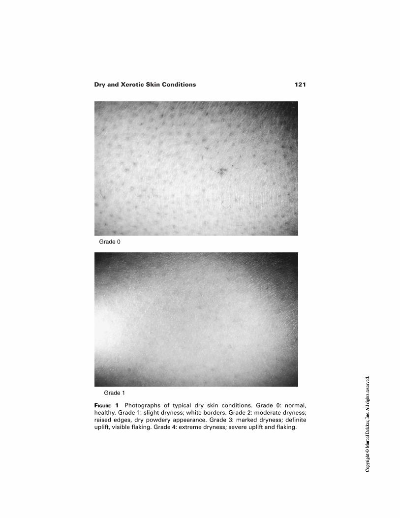
121Dry and Xerotic Skin Conditions
FIGURE 1 Photographs of typical dry skin conditions. Grade 0: normal,healthy. Grade 1: slight dryness; white borders. Grade 2: moderate dryness;raised edges, dry powdery appearance. Grade 3: marked dryness; definiteuplift, visible flaking. Grade 4: extreme dryness; severe uplift and flaking.
Grade 1
Grade 0
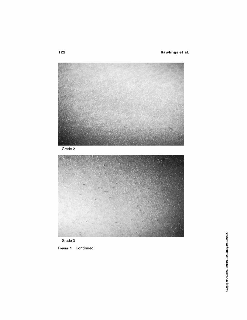
122 Rawlings et al.
Grade 2
Grade 3
FIGURE 1 Continued
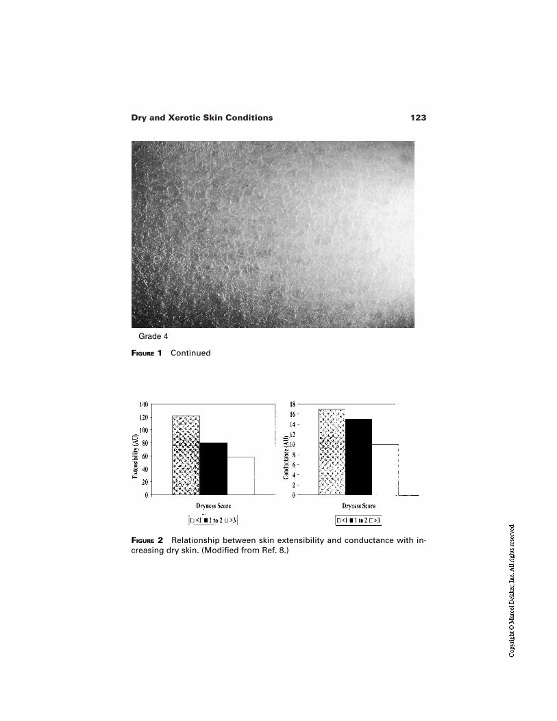
123Dry and Xerotic Skin Conditions
FIGURE 1 Continued
Grade 4
FIGURE 2 Relationship between skin extensibility and conductance with in-creasing dry skin. (Modified from Ref. 8.)

124 Rawlings et al.
Crucial to the stratum corneum condition is the state of proliferation/differ-entiation of the underlying epidermis. Leveque et al. [8] demonstrated that cor-neocyte size decreased with increasing dry skin indicating that the perturbation ofthe stratum corneum is associated with an increase in epidermal proliferation.This is consistent with the work of Elias et al. [9], who demonstrated increases inepidermal proliferation following barrier damage, i.e., increased transepidermalwater loss. Increases in epidermal proliferation are known to lead to smaller cor-neocytes [10]. Others have shown that the stratum corneum is thicker and revealscracks in dry skin conditions [11]. Thus, compromised stratum corneum in dryskin leads to an increase in epidermopoiesis, a less flexible tissue (a propertylinked to both stratum corneum water content and thickness) together with a re-duced barrier to water loss. These changes are associated with altered skin surfacemorphology [12]. Surface sebum does not appear to be a factor in dry skin sincesebum secretion rates were shown not to correlate with the dry skin scores. Col-lectively these results indicate that the normal functioning of the stratum corneumis compromised in dry skin.
3 BIOLOGY OF STRATUM CORNEUM IN NORMALAND DRY SKIN
Normal desquamation occurs following complete but gradual destruction of cor-neodesmosomes [13] leading to the imperceivable loss of individual cells fromthe surface of the stratum corneum. The process is intimately dependent upon thecomposition and organization of the intercellular lipids, levels and activity ofstratum corneum glycosidases, and proteases together with effective tissue hydra-tion. Early biochemical studies comparing the differences between normal anddry stratum corneum focused exclusively on changes in stratum corneum barrierlipids [14,15]. We believed, however, that a more holistic approach needed to betaken to properly understand the condition [6,16,17]. In the following sections wedescribe changes in stratum corneum morphology, lipid levels, corneodesmo-some persistence, protease activity, corneocyte envelope morphology and naturalmoisturizing factor levels associated with the appearance of dry skin.
3.1 Stratum Corneum Morphology in Normal andDry Skin
There is evidence that stratum corneum lipid lamellae and corneodesmosomes aremodified during the normal desquamation process and these changes are essentialto reduce cohesion in the peripheral layers [18,19]. In contrast, in dry skin thelipid structure becomes totally disorganized and corneodesmosomes persist intothe outer layers of the stratum corneum. This is best exemplified by electron mi-croscope studies [16].
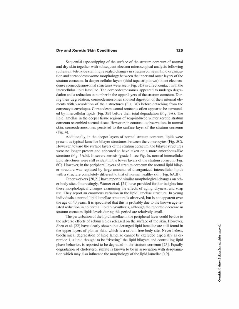
125Dry and Xerotic Skin Conditions
Sequential tape-stripping of the surface of the stratum corneum of normaland dry skin together with subsequent electron microscopical analysis followingruthenium tetroxide staining revealed changes in stratum corneum lipid organiza-tion and corneodesmosome morphology between the inner and outer layers of thestratum corneum. In deeper cellular layers (third tape strip down) intact electron-dense corneodesmosomal structures were seen (Fig. 3D) in direct contact with theintercellular lipid lamellae. The corneodesmosomes appeared to undergo degra-dation and a reduction in number in the upper layers of the stratum corneum. Dur-ing their degradation, corneodesmosomes showed digestion of their internal ele-ments with vacuolation of their structures (Fig. 3C) before detaching from thecorneocyte envelopes. Corneodesmosomal remnants often appear to be surround-ed by intercellular lipids (Fig. 3B) before their total degradation (Fig. 3A). Thelipid lamellae in the deeper tissue regions of soap-induced winter xerotic stratumcorneum resembled normal tissue. However, in contrast to observations in normalskin, corneodesmosomes persisted to the surface layer of the stratum corneum(Fig. 4).
Additionally, in the deeper layers of normal stratum corneum, lipids werepresent as typical lamellae bilayer structures between the corneocytes (Fig. 5C).However, toward the surface layers of the stratum corneum, the bilayer structureswere no longer present and appeared to have taken on a more amorphous-likestructure (Fig. 5A,B). In severe xerosis (grade 4; see Fig. 6), normal intercellularlipid structures were still evident in the lower layers of the stratum corneum (Fig.6C). However, in the peripheral layers of stratum corneum the normal lipid bilay-er structure was replaced by large amounts of disorganized intercellular lipidswith a structure completely different to that of normal healthy skin (Fig. 6A,B).
Other workers [20,21] have reported similar morphological changes on oth-er body sites. Interestingly, Warner et al. [21] have provided further insights intothese morphological changes examining the effects of aging, dryness, and soapuse. They report an enormous variation in the lipid lamellae structure. In youngindividuals a normal lipid lamellae structure is observed, but is not apparent overthe age of 40 years. It is speculated that this is probably due to the known age-re-lated reduction in epidermal lipid biosynthesis, although the reported decrease instratum corneum lipids levels during this period are relatively small.
The perturbation of the lipid lamellae in the peripheral layer could be due tothe adverse effects of sebum lipids released on the surface of the skin. However,Sheu et al. [22] have clearly shown that deranged lipid lamellae are still found inthe upper layers of plantar skin, which is a sebum-free body site. Nevertheless,biochemical degradation of lipid lamellae cannot be excluded especially as ce-ramide 1, a lipid thought to be “riveting” the lipid bilayers and controlling lipidphase behavior, is reported to be degraded in the stratum corneum [23]. Equallydegradation of cholesterol sulfate is known to be in association with desquama-tion which may also influence the morphology of the lipid lamellae [19].
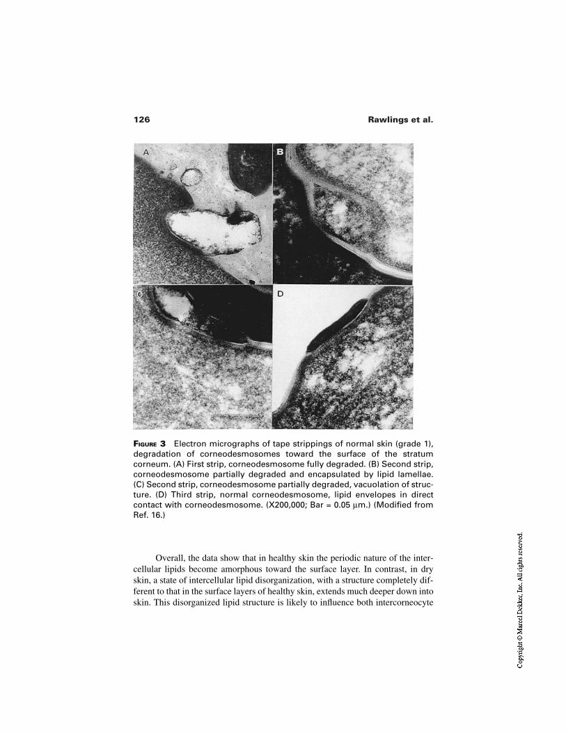
126 Rawlings et al.
FIGURE 3 Electron micrographs of tape strippings of normal skin (grade 1),degradation of corneodesmosomes toward the surface of the stratumcorneum. (A) First strip, corneodesmosome fully degraded. (B) Second strip,corneodesmosome partially degraded and encapsulated by lipid lamellae.(C) Second strip, corneodesmosome partially degraded, vacuolation of struc-ture. (D) Third strip, normal corneodesmosome, lipid envelopes in directcontact with corneodesmosome. (X200,000; Bar = 0.05 µm.) (Modified fromRef. 16.)
C
Overall, the data show that in healthy skin the periodic nature of the inter-cellular lipids become amorphous toward the surface layer. In contrast, in dryskin, a state of intercellular lipid disorganization, with a structure completely dif-ferent to that in the surface layers of healthy skin, extends much deeper down intoskin. This disorganized lipid structure is likely to influence both intercorneocyte
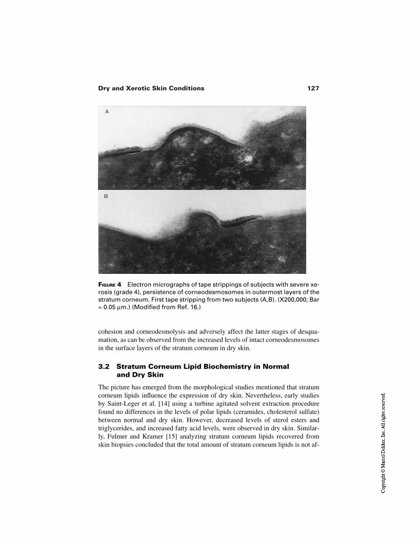
127Dry and Xerotic Skin Conditions
FIGURE 4 Electron micrographs of tape strippings of subjects with severe xe-rosis (grade 4), persistence of corneodesmosomes in outermost layers of thestratum corneum. First tape stripping from two subjects (A,B). (X200,000; Bar= 0.05 µm.) (Modified from Ref. 16.)
cohesion and corneodesmolysis and adversely affect the latter stages of desqua-mation, as can be observed from the increased levels of intact corneodesmosomesin the surface layers of the stratum corneum in dry skin.
3.2 Stratum Corneum Lipid Biochemistry in Normaland Dry Skin
The picture has emerged from the morphological studies mentioned that stratumcorneum lipids influence the expression of dry skin. Nevertheless, early studiesby Saint-Leger et al. [14] using a turbine agitated solvent extraction procedurefound no differences in the levels of polar lipids (ceramides, cholesterol sulfate)between normal and dry skin. However, decreased levels of sterol esters andtriglycerides, and increased fatty acid levels, were observed in dry skin. Similar-ly, Fulmer and Kramer [15] analyzing stratum corneum lipids recovered fromskin biopsies concluded that the total amount of stratum corneum lipids is not af-
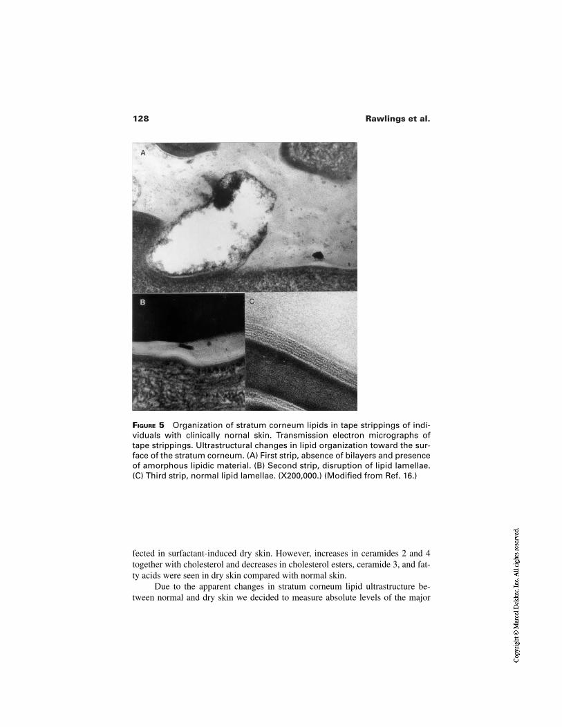
128 Rawlings et al.
FIGURE 5 Organization of stratum corneum lipids in tape strippings of indi-viduals with clinically nornal skin. Transmission electron micrographs oftape strippings. Ultrastructural changes in lipid organization toward the sur-face of the stratum corneum. (A) First strip, absence of bilayers and presenceof amorphous lipidic material. (B) Second strip, disruption of lipid lamellae.(C) Third strip, normal lipid lamellae. (X200,000.) (Modified from Ref. 16.)
fected in surfactant-induced dry skin. However, increases in ceramides 2 and 4together with cholesterol and decreases in cholesterol esters, ceramide 3, and fat-ty acids were seen in dry skin compared with normal skin.
Due to the apparent changes in stratum corneum lipid ultrastructure be-tween normal and dry skin we decided to measure absolute levels of the major
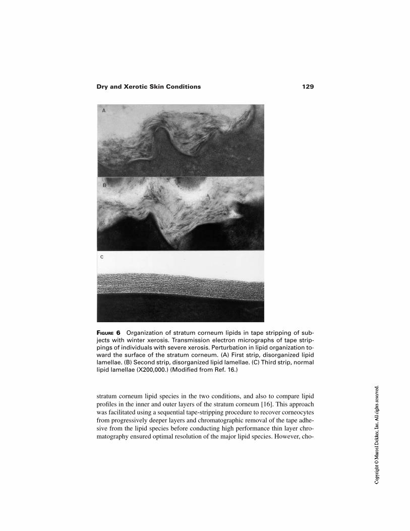
129Dry and Xerotic Skin Conditions
FIGURE 6 Organization of stratum corneum lipids in tape stripping of sub-jects with winter xerosis. Transmission electron micrographs of tape strip-pings of individuals with severe xerosis. Perturbation in lipid organization to-ward the surface of the stratum corneum. (A) First strip, disorganized lipidlamellae. (B) Second strip, disorganized lipid lamellae. (C) Third strip, normallipid lamellae (X200,000.) (Modified from Ref. 16.)
stratum corneum lipid species in the two conditions, and also to compare lipidprofiles in the inner and outer layers of the stratum corneum [16]. This approachwas facilitated using a sequential tape-stripping procedure to recover corneocytesfrom progressively deeper layers and chromatographic removal of the tape adhe-sive from the lipid species before conducting high performance thin layer chro-matography ensured optimal resolution of the major lipid species. However, cho-
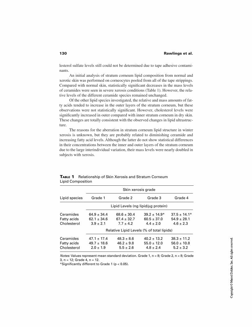
130 Rawlings et al.
lesterol sulfate levels still could not be determined due to tape adhesive contami-nants.
An initial analysis of stratum corneum lipid composition from normal andxerotic skin was performed on corneocytes pooled from all of the tape strippings.Compared with normal skin, statistically significant decreases in the mass levelsof ceramides were seen in severe xerosis conditions (Table 1). However, the rela-tive levels of the different ceramide species remained unchanged.
Of the other lipid species investigated, the relative and mass amounts of fat-ty acids tended to increase in the outer layers of the stratum corneum, but theseobservations were not statistically significant. However, cholesterol levels weresignificantly increased in outer compared with inner stratum corneum in dry skin.These changes are totally consistent with the observed changes in lipid ultrastruc-ture.
The reasons for the aberration in stratum corneum lipid structure in winterxerosis is unknown, but they are probably related to diminishing ceramide andincreasing fatty acid levels. Although the latter do not show statistical differencesin their concentrations between the inner and outer layers of the stratum corneumdue to the large interindividual variation, their mass levels were nearly doubled insubjects with xerosis.
TABLE 1 Relationship of Skin Xerosis and Stratum Corneum Lipid Composition
Skin xerosis grade
Lipid species Grade 1 Grade 2 Grade 3 Grade 4
Lipid Levels (ng lipid/µg protein)
Ceramides 64.9 ± 34.4 68.6 ± 30.4 39.2 ± 14.9* 37.5 ± 14.1*Fatty acids 62.1 ± 34.6 67.4 ± 32.7 60.5 ± 37.0 54.9 ± 28.1Cholesterol 3.9 ± 2.1 7.7 ± 4.2 4.4 ± 2.0 4.6 ± 2.3
Relative Lipid Levels (% of total lipids)
Ceramides 47.1 ± 17.4 48.3 ± 8.6 40.2 ± 13.2 38.3 ± 11.2Fatty acids 49.7 ± 18.6 46.2 ± 9.8 55.0 ± 12.0 56.0 ± 10.8Cholesterol 2.0 ± 1.9 5.5 ± 2.6 4.8 ± 2.4 5.2 ± 3.2
Notes: Values represent mean standard deviation. Grade 1, n = 8; Grade 2, n = 8; Grade3, n = 12; Grade 4, n = 12.*Significantly different to Grade 1 (p < 0.05).
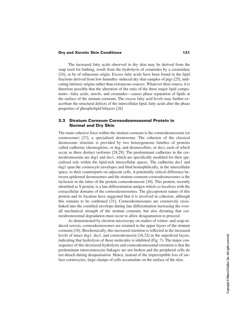
131Dry and Xerotic Skin Conditions
The increased fatty acids observed in dry skin may be derived from thesoap used for bathing, result from the hydrolysis of ceramides by a ceramidase[24], or be of sebaceous origin. Excess fatty acids have been found in the lipidfractions derived from low humidity–induced dry skin samples of pigs [25], indi-cating intrinsic origins rather than extraneous sources. Whatever their source, it istherefore possible that the alteration of the ratio of the three major lipid compo-nents—fatty acids, sterols, and ceramides—causes phase separation of lipids atthe surface of the stratum corneum. The excess fatty acid levels may further ex-acerbate the structural defects of the intercellular lipid; fatty acids alter the phaseproperties of phospholipid bilayers [26]
3.3 Stratum Corneum Corneodesmosomal Protein inNormal and Dry Skin
The main cohesive force within the stratum corneum is the corneodesmosome (orcorneosome) [27], a specialized desmosome. The cohesion of the classicaldesmosome structure is provided by two heterogeneous families of proteinscalled cadherins (desmogleins, or dsg, and desmocollins, or dsc), each of whichoccur as three distinct isoforms [28,29]. The predominant cadherins in the cor-neodesmosome are dsg1 and dsc1, which are specifically modified for their spe-cialized role within the lipid-rich intercellular spaces. The cadherins dsc1 anddsg1 span the corneocyte envelopes and bind homophilically, in the intercellularspace, to their counterparts on adjacent cells. A potentially critical difference be-tween epidermal desmosomes and the stratum corneum corneodesmosomes is theinclusion in the latter of the protein corneodesmosin [30]. This protein, recentlyidentified as S protein, is a late differentiation antigen which co-localizes with theextracellular domains of the corneodesmosomes. The glycoprotein nature of thisprotein and its location have suggested that it is involved in cohesion, althoughthis remains to be confirmed [31]. Corneodesmosomes are extensively cross-linked into the cornified envelope during late differentiation increasing the over-all mechanical strength of the stratum corneum, but also dictating that cor-neodesmosomal degradation must occur to allow desquamation to proceed.
As demonstrated by electron microscopy on studies of winter- and soap-in-duced xerosis, corneodesmosomes are retained in the upper layers of the stratumcorneum [16]. Biochemically, this increased retention is reflected in the increasedlevels of intact dsg1, dsc1, and corneodesmosin [16,32] in the superficial layers,indicating that hydrolysis of these molecules is inhibited (Fig. 7). The major con-sequence of this decreased hydrolysis and corneodesmosomal retention is that thepredominant intercorneocyte linkages are not broken and the peripheral cells donot detach during desquamation. Hence, instead of the imperceptible loss of sur-face corneocytes, large clumps of cells accumulate on the surface of the skin.
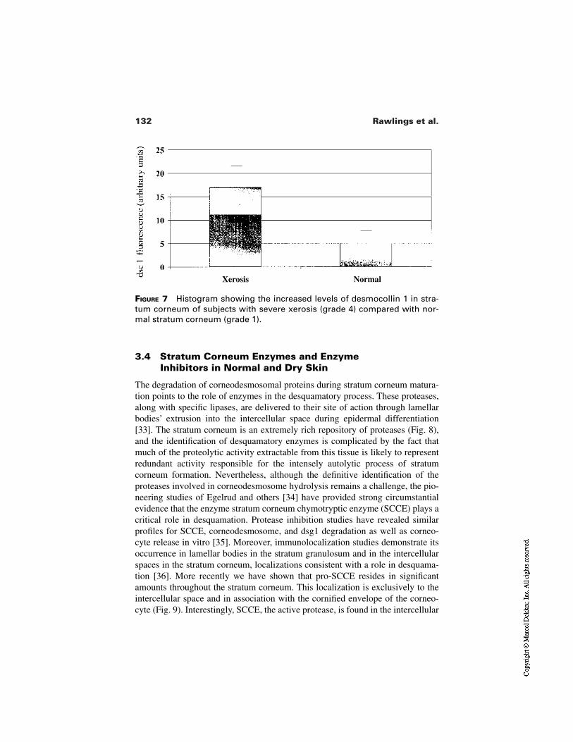
132 Rawlings et al.
FIGURE 7 Histogram showing the increased levels of desmocollin 1 in stra-tum corneum of subjects with severe xerosis (grade 4) compared with nor-mal stratum corneum (grade 1).
3.4 Stratum Corneum Enzymes and EnzymeInhibitors in Normal and Dry Skin
The degradation of corneodesmosomal proteins during stratum corneum matura-tion points to the role of enzymes in the desquamatory process. These proteases,along with specific lipases, are delivered to their site of action through lamellarbodies’ extrusion into the intercellular space during epidermal differentiation[33]. The stratum corneum is an extremely rich repository of proteases (Fig. 8),and the identification of desquamatory enzymes is complicated by the fact thatmuch of the proteolytic activity extractable from this tissue is likely to representredundant activity responsible for the intensely autolytic process of stratumcorneum formation. Nevertheless, although the definitive identification of theproteases involved in corneodesmosome hydrolysis remains a challenge, the pio-neering studies of Egelrud and others [34] have provided strong circumstantialevidence that the enzyme stratum corneum chymotryptic enzyme (SCCE) plays acritical role in desquamation. Protease inhibition studies have revealed similarprofiles for SCCE, corneodesmosome, and dsg1 degradation as well as corneo-cyte release in vitro [35]. Moreover, immunolocalization studies demonstrate itsoccurrence in lamellar bodies in the stratum granulosum and in the intercellularspaces in the stratum corneum, localizations consistent with a role in desquama-tion [36]. More recently we have shown that pro-SCCE resides in significantamounts throughout the stratum corneum. This localization is exclusively to theintercellular space and in association with the cornified envelope of the corneo-cyte (Fig. 9). Interestingly, SCCE, the active protease, is found in the intercellular
Xerosis Normal
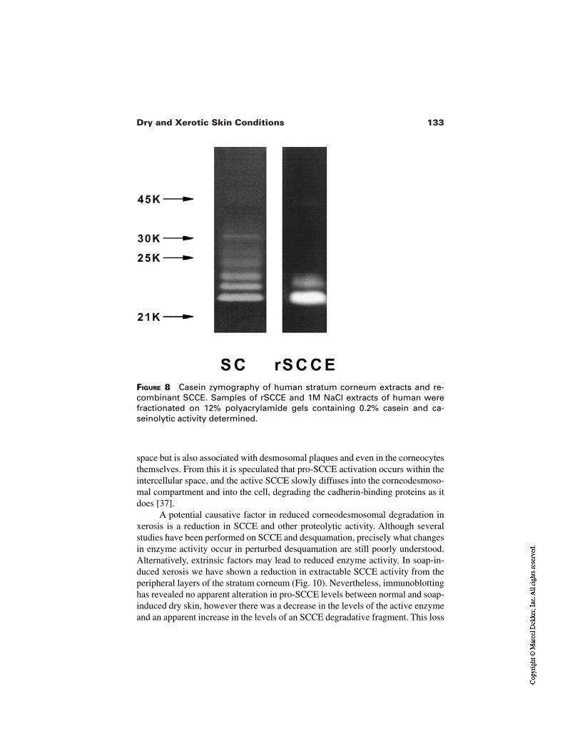
133Dry and Xerotic Skin Conditions
FIGURE 8 Casein zymography of human stratum corneum extracts and re-combinant SCCE. Samples of rSCCE and 1M NaCl extracts of human werefractionated on 12% polyacrylamide gels containing 0.2% casein and ca-seinolytic activity determined.
space but is also associated with desmosomal plaques and even in the corneocytesthemselves. From this it is speculated that pro-SCCE activation occurs within theintercellular space, and the active SCCE slowly diffuses into the corneodesmoso-mal compartment and into the cell, degrading the cadherin-binding proteins as itdoes [37].
A potential causative factor in reduced corneodesmosomal degradation inxerosis is a reduction in SCCE and other proteolytic activity. Although severalstudies have been performed on SCCE and desquamation, precisely what changesin enzyme activity occur in perturbed desquamation are still poorly understood.Alternatively, extrinsic factors may lead to reduced enzyme activity. In soap-in-duced xerosis we have shown a reduction in extractable SCCE activity from theperipheral layers of the stratum corneum (Fig. 10). Nevertheless, immunoblottinghas revealed no apparent alteration in pro-SCCE levels between normal and soap-induced dry skin, however there was a decrease in the levels of the active enzymeand an apparent increase in the levels of an SCCE degradative fragment. This loss
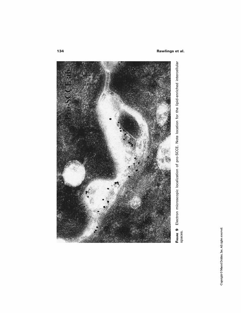
134 Rawlings et al.
FIG
UR
E9
Ele
ctro
n m
icro
sco
pic
lo
caliz
atio
n o
f p
ro-S
CC
E.
No
te l
oca
tio
n f
or
the
lipid
-en
rich
ed i
nte
rcel
lula
rsp
aces
.
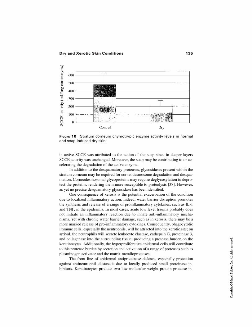
135Dry and Xerotic Skin Conditions
FIGURE 10 Stratum corneum chymotrypic enzyme activity levels in normaland soap-induced dry skin.
in active SCCE was attributed to the action of the soap since in deeper layersSCCE activity was unchanged. Moreover, the soap may be contributing to or ac-celerating the degradation of the active enzyme.
In addition to the desquamatory proteases, glycosidases present within thestratum corneum may be required for corneodesmosome degradation and desqua-mation. Corneodesmosomal glycoproteins may require deglycosylation to depro-tect the proteins, rendering them more susceptible to proteolysis [38]. However,as yet no precise desquamatory glycosidase has been identified.
One consequence of xerosis is the potential exacerbation of the conditiondue to localized inflammatory action. Indeed, water barrier disruption promotesthe synthesis and release of a range of proinflammatory cytokines, such as IL-1and TNF, in the epidermis. In most cases, acute low level trauma probably doesnot initiate an inflammatory reaction due to innate anti-inflammatory mecha-nisms. Yet with chronic water barrier damage, such as in xerosis, there may be amore marked release of pro-inflammatory cytokines. Consequently, phagocytoticimmune cells, especially the neutrophils, will be attracted into the xerotic site; onarrival, the neutrophils will secrete leukocyte elastase, cathepsin G, proteinase 3,and collagenase into the surrounding tissue, producing a protease burden on thekeratinocytes. Additionally, the hyperproliferative epidermal cells will contributeto this protease burden by secretion and activation of a range of proteases such asplasminogen activator and the matrix metalloproteases.
The front line of epidermal antiproteinase defence, especially protectionagainst antineutrophil elastase,is due to locally produced small proteinase in-hibitors. Keratinocytes produce two low molecular weight protein protease in-
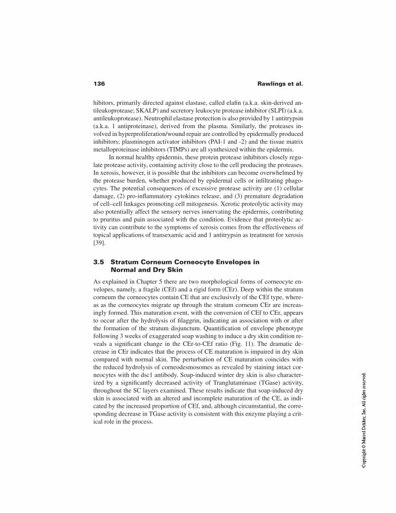
136 Rawlings et al.
hibitors, primarily directed against elastase, called elafin (a.k.a. skin-derived an-tileukoprotease; SKALP) and secretory leukocyte protease inhibitor (SLPI) (a.k.a.antileukoprotease). Neutrophil elastase protection is also provided by 1 antitrypsin(a.k.a. 1 antiproteinase), derived from the plasma. Similarly, the proteases in-volved in hyperproliferation/wound repair are controlled by epidermally producedinhibitors; plasminogen activator inhibitors (PAI-1 and -2) and the tissue matrixmetalloproteinase inhibitors (TIMPs) are all synthesized within the epidermis.
In normal healthy epidermis, these protein protease inhibitors closely regu-late protease activity, containing activity close to the cell producing the proteases.In xerosis, however, it is possible that the inhibitors can become overwhelmed bythe protease burden, whether produced by epidermal cells or infiltrating phago-cytes. The potential consequences of excessive protease activity are (1) cellulardamage, (2) pro-inflammatory cytokines release, and (3) premature degradationof cell–cell linkages promoting cell mitogenesis. Xerotic proteolytic activity mayalso potentially affect the sensory nerves innervating the epidermis, contributingto pruritus and pain associated with the condition. Evidence that proteolytic ac-tivity can contribute to the symptoms of xerosis comes from the effectiveness oftopical applications of transexamic acid and 1 antitrypsin as treatment for xerosis[39].
3.5 Stratum Corneum Corneocyte Envelopes inNormal and Dry Skin
As explained in Chapter 5 there are two morphological forms of corneocyte en-velopes, namely, a fragile (CEf) and a rigid form (CEr). Deep within the stratumcorneum the corneocytes contain CE that are exclusively of the CEf type, where-as as the corneocytes migrate up through the stratum corneum CEr are increas-ingly formed. This maturation event, with the conversion of CEf to CEr, appearsto occur after the hydrolysis of filaggrin, indicating an association with or afterthe formation of the stratum disjunctum. Quantification of envelope phenotypefollowing 3 weeks of exaggerated soap washing to induce a dry skin condition re-veals a significant change in the CEr-to-CEf ratio (Fig. 11). The dramatic de-crease in CEr indicates that the process of CE maturation is impaired in dry skincompared with normal skin. The perturbation of CE maturation coincides withthe reduced hydrolysis of corneodesmosomes as revealed by staining intact cor-neocytes with the dsc1 antibody. Soap-induced winter dry skin is also character-ized by a significantly decreased activity of Tranglutaminase (TGase) activity,throughout the SC layers examined. These results indicate that soap-induced dryskin is associated with an altered and incomplete maturation of the CE, as indi-cated by the increased proportion of CEf, and, although circumstantial, the corre-sponding decrease in TGase activity is consistent with this enzyme playing a crit-ical role in the process.
137Dry and Xerotic Skin Conditions
FIGURE 11 Percentage distribution of rigid (CEr) and fragile (CEf) envelopesrecovered from normal (untreated) and soap-dried SC. Dry skin was generat-ed on the volar forearm of six individuals by the exaggerated daily use of aharsh soap bar for 3 weeks. The other forearm remained untreated through-out this time and served as a control site. Samples of CE recovered after thistime were stained with TRITC and viewed under fluorescent microscope. Theproportion of the two envelope types were averaged following examinationof photographs taken from five separate fields/sample. *p < 0.05; **p <0.001.
Perturbation of CE maturation is also evident in more serious skin patholo-gies. In the hyperproliferative diseases such as psoriasis and ichthyoses the con-ditions are characterized by an increase in the content of the immature CEf,demonstrating further correlation of hyperproliferation with CE maturation.These occur due to altered epidermal hyperproliferation and differentiation.However, in some (e.g., lamellar ichthyosis) no TGase 1 activity is apparent.
In conclusion, however, regardless of the underlying mechanisms perturbedin these conditions it is clear that reduced CE maturation and reduced cor-neodesmosomal hydrolysis are common features of dry, flaky skin conditions.
3.6 Stratum Corneum Natural Moisturizing Factor
As already discussed by Harding and Scott [40], natural moisturizing factors(NMF) are critically important for maintaining the hydration and flexibility of thestratum corneum, and reduced NMF levels are implicated in the appearance andpersistence of dry skin conditions. A significant correlation exists between hydra-
138 Rawlings et al.
tion state of the SC and its amino acid content in elderly individuals with skin xe-rosis [41], but is less clear in others [44]. Free amino acid levels have been re-ported to decrease significantly in dry, scaly skin induced experimentally byrepetitive tape-stripping [43] or by surfactant damage [44].
In our own laboratory we have found that aged skin has intrinsically lowerNMF levels compared with young skin, and this reflects a general reduced syn-thesis of profilaggrin. These conclusions are supported by electron microscopystudies which indicate that a decreased number of keratohyalin granules [45], oc-cupying a reduced volume within the cell, is found in senile xerosis. The declinein NMF production appears to reflect the cumulative effects of actinic damage asit was observed in SC recovered from the back of the hand (photodamaged), butnot from the inner aspect of the biceps (photoprotected). It is likely that in agedskin, loss of NMF may become more pronounced as elderly individuals alsoshow an age-related decline in water barrier repair which may lead to increasedleaching of the water-soluble compounds from the surface layers [46].
Although decreased synthesis of profilaggrin and increased leaching fromsurfactant-damaged skin [47] are undoubtedly major factors leading to decreasedlevels of NMF in the superficial SC, the direct impact of sudden changes in envi-ronmental humidity on NMF generation should not be ignored. As we have al-ready described elsewhere, the hydrolysis of filaggrin is critically regulated bythe external relative humidity. The rapid decrease in environmental humidity andtemperature, commonly associated with the onset of winter xerosis, is likely to re-sult in a transient but acute perturbation of filaggrin proteolysis, as the proteasesresponsible are not fully activated. Similarly, a chronic perturbation in the effi-ciency of filaggrin proteolysis due to frequent and rapid changes in environmen-tal humidity is also likely to contribute to the poor skin condition prevalent in cer-tain individuals who endure constantly changing humidity conditions (e.g., flightattendants).
The classical, transient dry skin associated with newborn infants [48] canalso be explained by the delay in production of NMF as the SC slowly equili-brates from the aqueous environment of the womb (where, of course, filaggrin, al-though synthesized, is not hydrolyzed to the ambient external humidity when pro-teolysis is initiated).
The dry flaky skin which characteristically appears several days after acuteUVB damage is NMF deficient [49]. Studies suggest that this reflects initial dam-age to the granular layer and a subsequent decreased synthesis of profilaggrin,rather than any dramatic loss of hydrolysis of existing filaggrin. Indeed the char-acteristic skin flaking seen following UV damage can, under histological exami-nation, be seen to occur through the layer of NMF-deficient corneocytes formedprematurely during the initial UV insult. As we have described, many elements ofnormal SC maturation are disrupted in xerosis, and increased NMF leaching from
139Dry and Xerotic Skin Conditions
cornecytes may also occur due to parallel perturbations in lamellar granule syn-thesis and associated damage to the water barrier [50].
An inability to retain water due to defective NMF production/retention willof course not only impact the mechanical properties of the SC—a critical, and of-ten overlooked role of water in the SC is in ensuring the activity of a variety ofhydrolytic enzymes involved in various aspects of SC maturation and desquama-tion. When the tissue is desiccated a loss of intrinsic hydrolytic enzyme activityleads to ineffective corneodesmosomal degradation and consequent skin scaling.
The various processes leading from profilaggrin synthesis to conversion tofilaggrin and then to NMF are under tight control. However, these controls areperturbed in different ways by a range of factors including UV light, exposure tosurfactants, and, of course, changes in environmental humidity processes. Thesevery different causes can all lead to reduced NMF levels and contribute to thecomplex phenomenon known as dry skin.
4 SUMMARY
A dry skin condition is the result of a range of environmental and pathologicalfactors that disrupt the normal epidermal differentiation and stratum corneummaturation processes. It is a complex phenomenon involving several interdepen-dent biochemical events in the stratum corneum. Morphologically, dry skin dif-fers from normal due to a retention of corneodesmosomes in the peripheraldesquamating layers of the stratum corneum. This retention of corneodesmo-somes is the cause of the skin flaking associated with the xerotic condition; theabnormal retention of intercorneocyte links results in large clumps of corneocytesbreaking off, i.e., scale, as opposed to the imperceptible loss of single cells. In ad-dition, degradation or disruption of the stratum corneum multiple lipid bilayersoccurs in several layers at the tissue periphery, rather than in the final desquamat-ing layer. This results in a collapse of the bilayer structure to give a disorganizedlipid matrix, which may be due to an observed decrease in ceramide levels and in-crease in fatty acid levels in peripheral layers of dry skin. Another component ofthe stratum corneum intercellular region, the putative desquamatory enzyme,SCCE, has been shown to be decreased in the outer layers of the stratum corneumin dry skin. Finally, the perturbations that cause dry skin even affect the structureof the corneocytes. We have similarly found aberrant maturation of the cornifiedenvelopes, with increases in the fragile morphology in dry skin. Also there is adecrease in the production or proteolytic processing of filaggrin to produce thenatural moisturizing factors; decreased levels of these hydroscopic molecules willresult in a reduced ability to retain water within the tissue.
Each of these elements of stratum corneum maturation are crucial to the
140 Rawlings et al.
process as a whole. Hence, perturbation of one element is likely to have knock-oneffects to other aspects of maturation. As such, disruption of the lipid bilayers islikely to allow greater leaching of the NMFs, reducing the water content of thetissue. This in turn will reduce the activity of enzymes such as SCCE and TGase,perturbing corneodesmosomal degradation and CE maturation, respectively. Thedecreased water content will also reduce the elasticity of the corneocyte struc-tures, increasing the likelihood of the skin cracking.
The most common cause of stratum corneum maturation disruption is theaffect of the environment and bathing habits. Some surfactants are known to ef-fect the stratum corneum lipids and enzymes resulting in dry skin. However themajor contributor to xerosis is the environmental humidity. This regulates the wa-ter content of the peripheral stratum corneum, influencing the enzymes involvedin the final stages of maturation and desquamation. It is this crucial requirementof water in the peripheral stratum corneum that results in moisturizing agents stillbeing the most frequently used treatment for common dry skin.
REFERENCES
1. Pierard GE. What does dry skin mean? Int J Dermatol 1987; 23:167–168.2. Rudikoff D. The effect of dryness on skin. Clin Dermatol 1998; 16:99–107.3. During LA. Pruritis himalis an undescribed form of pruritis. Phila Med Times 1874;
4:225–230.4. Gaul LE, Underwood GB. Relation of dewpoint and barometric pressure to chapping
of normal skin. J Invest Dermatol 1952; 19:9–19.5. Blank IH. Further observations on factors which influence the water content of the
stratum corneum. J Invest Dermatol 1953; 21:259–271.6. Harding CR, Watkinson A, Scott IR, Rawlings AV. Dry skin, moisturisation and cor-
neodesmolysis. Int J Cosmet Sci 2000; 22:21–52.7. Kligman A. Regression method for assessing the efficacy of moisturizers. Cosmet
Toil 1978; 93:27–35.8. Leveque JL, Grove G, de Rigal J, Corcuff P, Kligman AM, Saint-Leger D. Biophys-
ical characterisation of dry facial skin. J Soc Cosmet Chem 1987; 82:171–177.9. Proksch E, Feingold KR, Man MQ, Elias PM. Barrier function regulates epidermal
DNA synthesis. J Clin Invest 1991; 87:1668–1673.10. Grove GL. Exfoliative cytological procedures as a non-intrusive method for derma-
tological studies. J Invest Dermatol 1979; 73:67–74.11. Grove GL, Lavker RM, Holzle E, Kligman AM. Use of non-intrusive tests to moni-
tor age associated changes in human skin. J Soc Cosmet Chem 1980; 32:15–26.12. Sato J, Yanai M, Hirao T, Denda M. Water content and thickness of the stratum
corneum contribute to skin surface morphology. Arch Dermatol Res 2000;292:412–417.
13. Sato J. Desquamation and the role of stratum corneum enzymes in skin moisturiza-tion. In: Leyden J, Rawlings AV, eds. Skin Moisturization. New York: MarcelDekker (in press).
141Dry and Xerotic Skin Conditions
14. Saint-Leger D, Francois AM, Leveque JL, Stuudemeuyer T, Kligman AM, GroveGL. Stratum corneum lipids in winter xerosis. Dermatologica 1989; 178:151–155.
15. Fulmer AW, Dramer GJ. Stratum corneum abnormalities in surfactant induced dryscaly skin. J Invest Dermatol 1989; 80:598–602.
16. Rawlings AV, Watkinson A, Rogers J, Mayo AM, Hope J, Scott IR. Abnormalities instratum corneum structure, lipid composition and desmosome degradation in soap-induced winter xerosis. J Cosmet Chem 1994; 45:203–220.
17. Harding CR, Long S, Rogers J, Banks J, Zhang Z, Bush A. The cornified cell enve-lope: an important market of stratum corneum maturation in healthy and dry skin(abstr). J Invest Dermatol 1999; 112:306.
18. Chapman SJ, Walsh A, Jackson SM, Friedmann PM. Lipids, proteins and corneocyteadhesion. Arch Dermatol Res 1991; 283:1729–1732.
19. Ranasinghe AW, Wertz PW, Downing DT, McKenzie I. Lipid composition of cohe-sive and desquamated corneocytes from mouse ear. J Invest Dermatol 1985;94:216–220.
20. Berry N, Charmeil C, Goujon C, Silvy A, Girard P, Corcuff P, Montastier A. A clini-cal, biometrological and ultrastructural study of xerotic skin. Int J Cosmet Sci 1999;21:241–252.
21. Warner RR, Boissy YL. Effect of moisturising products on the structure of lipids inthe outer stratum corneum of human. In: Loden M, Maibach HH, eds. Dry Skin andMoisturisers: Chemistry and Function. CRC Press, 2000; 349–372.
22. Sheu H, Chao S, Wong T, Lee Y, Tsai J. Human skin surface lipid film: an ultrastruc-tural study and interaction with corneocytes and intercellular lipid lamellae of thestratum corneum. Br J Dermatol 1999; 140:385–391.
23. Bowser P. Essential fatty acids and their role in correcting skin abnormalities. Cos-metic Dermatol 1993; (suppl):11–12.
24. Wertz PW, Downing DT. Epidermal ceramide hydrolase. J Invest Dermatol 1990;94:590.
25. Bissett DC, McBride JF. Use of the domestic pig as an animal mode of human dryskin. In: Maibach H, Lowe P, eds. Model in Dermatology. Vol. 1. Basel: Karger,1985:159–168.
26. McKersie BD, Grove JH, Gowe LM. Free fatty acid effects on leakage, phase prop-erties and fusion of fully hydrated model membranes. Biochim Biophys Acta 1989;982:156–160.
27. Chapman S, Walsh A. Desmosomes, corneosomes and desquamation. Arch Derma-tol Res 1990; 283:1729–1732.
28. King IA, O’Brien TJ, Buxton RS. Expression of the skin type desmosomal cadherinsDSC1 is closely linked to the keratinisation of epithelial tissues during mouse devel-opment. J Invest Dermatol 1996; 107:531–538.
29. King IA, Dryst BD, Hunt DM, Kruger M, Arnemann J, Buxton RS. Hierarchical ex-pression of desmosomes and cadherins during stratified epithelial morphogenesis inthe mouse. Differentiation 1997; 62:83–96.
30. Montezin M, Simon M, Guerrin M, Serre G. Corneodesmosin, a corneodesmosome-specific basic protein is expressed in the cornified epithelia of the pig, guinea pig, ratand mouse. Exp Cell Res 1997; 231:132–140.
31. Simon M, Montezin M, Guerrin M, Durieu X, Serre G. Characterisation and purifi-
142 Rawlings et al.
cation of human corneodesmosin, an epidermal basic glycoprotein associated withcorneocyte specific modified desmosomes. J Biol Chem 1997; 272:31770–31776.
32. Bernard D, Camus C, Nguyen QL, Serre G. Proteolysis of corneodesmosomal pro-teins in winter xerosis. J Invest Dermatol 1995; 105:176.
33. Mennen GK, Ghadially R, Williams ML, Elias P. Lamellar bodies as delivery sys-tems of hydrolitc enzymes, implications for normal and abnormal desquamation. BrJ Dermatol 1992; 126:337–345.
34. Egelrud T. Purification and preliminary characterisation of stratum corneumchynotriptic enzymes—a proteinase that may be involved in desquamation. J InvestDermatol 1993; 101:200–204.
35. Lundstrom A, Egelrud T. Evidence that cell shedding from plantar stratum corneumin vitro involves endegenous proteolysis of the desmoglein 1. J Invest Dermatol1990; 94:216–220.
36. Sondell B, Thornall LE, Egelrud T. Evidence that stratum corneum chymotryptic en-zyme is transported to the stratum corneum extracellular space via lamellar bodies. JInvest Dermatol 1995; 104:891–823.
37. Watkinson A, Smith C, Coan P, Wiedow O. The role of pro-SCCE and SCCE indesquamation. IFSCC Magazine 2000; 3:45–49.
38. Walsh A, Chapman S. Sugars protect desmosomal proteins from proteolysis. Br JDermatol 1990; 122:289.
39. Denda M, Kitamura K, Elias PM, Feingold KR. trans-4-(Aminomethyl) cyclohex-ane carboxylic acid (T-AMCHA), an anti-fibrinolytic agent, accelerates barrier re-covery and prevents the epidermal hyperplasia induced by epidermal injury in hair-less mice and humans. J Invest Dermatol 1997; 109:84–90.
40. Harding CR, Scott IR. Natural moisturing factor. In: Leyden J, Rawlings AV, eds.Skin Moisturization. New York: Marcel Dekker (in press).
41. Horii I, Nakayama Y, Obata M, Tagami H. SC by hydration and amino acid contentin xerotic skin. Br J Dermatol 1989; 121:587–592.
42. Jacobsen TM, Yuksel KU, Geesin JC, Gordon JS, Lane AT, Gracy RW. Effects ofageing and xerosis on the amino acid composition of human skin. J Invest Dermatol1990; 95:296–300.
43. Denda M, Horii J, Koyama J, Yoshida S, Nanba R, Takahashi M, Horii I, YamamotoA. SC Sphingolipids and free amino acids in experimentally induced scaly skin.Arch Dermatol Res 1992; 285:363–367.
44. Koyama J, Horii I, Kawasaki K, Nakayama Y, Morikawa Y, Mitsui T, Kumagai H.Free amino acids of stratum corneum as a biochemical marker to evaluate dry skin. JSoc Cosmet Chem 1984; 35:183–195.
45. Tezuka T. Electron microscopical changes in xerotic senilis epidermis. Its abnormalmembrane coating granule formation. Dermatologica 1983; 166:57.
46. Zetterson EM, Ghadially R, Feingold KR, Crumrine D, Elias PM. Optimal ratios oftopical stratum corneum lipids improve barrier recovery in chronologically agedskin. J Am Acad Dermatol 1997; 37:403–408.
47. Scott IR, Harding CR. A filaggrin analogue to increase natural moisturising factorsynthesis in skin (abstr). Dermatology 2000 1993; 773.
48. Saijo S, Tagami H. Dry skin of newborn infants: functional analysis of the stratumcorneum. Petiatr Dermatol 1991; 8:155–159.
143Dry and Xerotic Skin Conditions
49. Tsuchiya T, Horii I, Nakayama Y. Interrelationship between the change in the watercontent of the stratum corneum and the amount of natural moisturising factor of thestratum corneum after UVB irradiation. J Soc Cosmet Chem Japan 1998; 22:10–15.
50. Holleran WM, Uchida Y, Halkier-Sorensen L, Haratake A, Hara M, Epstein JH, EliasPM. Structural and biochemical basis for the UVB-induced alterations in epidermalbarrier function. Photodermatol Photoimmunol Photomedicine 1997; 13:117–128.

7Sensitive Skin and Moisturization
Paolo U. GiacomoniClinique Laboratories, Inc., Melville, New York
Neelam Muizzuddin, Rose Marie Sparacio, Edward Pelle,Thomas Mammone, Kenneth Marenus, and Daniel MaesEstee Lauder, Melville, New York
1 INTRODUCTION
Skin moisturization is a state of the surface of the skin, which is more often rec-ognized by the individuals when moisturization is lacking, and when one has skinconditions that can be called dry, very dry, rough, or even ichthyotic. The mois-turization of the upper part of the skin is likely to be dictated by the presence oflipids, water, urea, and other compounds. It can also be considered to be the con-sequence of how well the outer envelope of the skin opposes the evaporation.Several authors have undertaken to measure the water content of the outer surfaceof the skin. Other authors have emphasized the importance of the so-called trans-epidermal water loss (TEWL), expressed as grams of water per square meter perhour. The capability of the skin to oppose water evaporation can be equated to itscapability to provide an overall barrier. The measure of TEWL provides informa-tion on the changes in moisturization induced by a treatment, which does not af-
145
146 Giacomoni et al.
fect the barrier, and on changes of the barrier properties induced by a treatment,which does not affect moisturization.
Skin sensitivity is a self-assessed diagnosis of a physiological state thatlacks rigorous clinical definition, complete etiological analysis, and accurate di-agnostic tools. This undesirable state of the skin is characterized by a disagree-able feeling on the surface of the skin or by the observation of hyper-reactivity ofthe skin when it is exposed to mild environmental conditions such as water, woolfabrics, or cosmetics. According to Draelos [1], approximately 40% of the popu-lation believes it possesses the characteristics of sensitive skin, as determined byconsumer marketing surveys. The characteristics of sensitive skin are the onesfelt when, in response to topical application of cosmetics and toiletries, stinging,burning, pruritus, erythema, and desquamation are observed. Yet, as late as in1997, Draelos noted, “Given the current incomplete knowledge of the sensitiveskin condition, it is impossible to arrive at a consensus regarding the definitionand origins of sensitive skin” [1].
The definitions of sensitive skin and of skin moisturization are partiallysubjective, and different people do react differently to the feeling of dry skin. Ithas been therefore particularly difficult to design experimental protocols and tointerpret the results of experiments in the field of skin sensitivity and moisturiza-tion. These experiments are generally aimed at pointing out physiological andmolecular properties able to allow one to better understand the phenomenon ofsensitive skin.
Skin sensitivity and skin dryness are also encountered in mature individu-als, and questions have been asked about the correlation between the appearanceof skin sensitivity and the onset of those physiological phenomena that character-ize aging in women.
In this chapter we summarize some of the experimental results obtained inthis field and the interpretations that have been proposed.
2 TESTING METHODS
2.1 Testing for Sensitive Skin
Many tests are available to determine whether the sensitive behavior is the conse-quence of specific skin conditions, such as rosacea, contact dermatitis, acne, anddry skin, or the consequence of the etiologically undefined skin sensitivity of agiven population to topically applied compounds. Among these, we would like torecall the cumulative irritancy test [2], repeat insult patch test [3], chamber scari-fication test [4], and the soap chamber test [5]. All these tests are performed bytopical application of compounds after a penetration-enhancing treatment of theskin.
147Sensitive Skin and Moisturization
2.2 Testing for Skin Moisturization
Instruments for measuring skin moisturization do exist. They measure the numer-ical values of physical parameters of the surface of the skin which can somehowbe correlated to the content of water of the upper part of the epidermis and of thestratum corneum. The dermal content of water can be assessed by nuclear mag-netic resonance or by high frequency ultrasound [6], but these techniques hardlyprovide information about the state of hydration of the surface. This can be as-sessed by electrical devices able to measure the electrical impedance of the outerpart of the skin (less than 0.1 mm deep) [7] as it is understood that the conductiv-ity increases with the content of water on the surface of the skin. It has been re-ported that these instruments do not provide consistent results [8] and have to becalibrated with one another.
In addition to the direct measurement of the water concentration in the toplayers of the skin, some techniques allow for the evaluation of the indirect conse-quence of the presence of water in the stratum corneum. Measurements of the pli-ability of the horny layer in vivo using the gas bearing electrodynamometer [9]have been shown to correlate directly with the water content of the stratumcorneum. The advantage of this technique is that it is not subjected to all the in-terference known to affect the direct measurement of the water content of the skinby conductimetry (presence of hydroxyl anions or metal cations, for example).
Interestingly enough, the measure of the amount of water molecules (in thegaseous state) above the surface is in good correlation with the electrical mea-surements. Indeed when the conductivity is low (low content of water in the out-er surface of the skin), the TEWL is high. It is thus not unreasonable to considerthat a high value of the concentrations of water above the skin is the consequenceof high concentrations of water below the surface (as in the case of an edema).One could also conclude that high TEWL is associated with a poor skin barrierfunction. If this conclusion holds, then one can suggest that a barrier unable tokeep the water inside will also be less efficient in maintaining molecules to whichwe are continuously exposed out of the skin. Since environmental factors are of-ten associate to phenomena of irritation or sensitivity, it might be interesting tolook for a correlation, if any, between barrier function and skin sensitivity.
3 PROPERTIES OF SENSITIVE SKIN
Experiments performed in our laboratories have allowed us to recognize thatthere is a negative correlation between the self-assessed sensitivity of the skin andthe barrier function of the skin of the same individuals, measured as susceptibili-ty to respond to standard irritant treatment [10]. Observations were performed bycomparing the results obtained in two cohorts of volunteers, one of people esti-
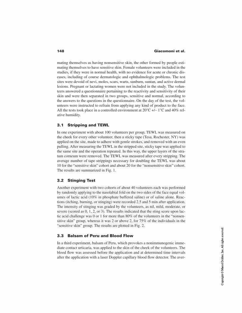
148 Giacomoni et al.
mating themselves as having nonsensitive skin, the other formed by people esti-mating themselves to have sensitive skin. Female volunteers were included in thestudies, if they were in normal health, with no evidence for acute or chronic dis-eases, including of course dermatologic and ophthalmologic problems. The testsites were devoid of nevi, moles, scars, warts, sunburn, suntan, and active dermallesions. Pregnant or lactating women were not included in the study. The volun-teers answered a questionnaire pertaining to the reactivity and sensitivity of theirskin and were then separated in two groups, sensitive and normal, according tothe answers to the questions in the questionnaire. On the day of the test, the vol-unteers were instructed to refrain from applying any kind of product to the face.All the tests took place in a controlled environment at 20°C +/– 1°C and 40% rel-ative humidity.
3.1 Stripping and TEWL
In one experiment with about 100 volunteers per group, TEWL was measured onthe cheek for every other volunteer, then a sticky tape (Tesa, Rochester, NY) wasapplied on the site, made to adhere with gentle strokes, and removed with an evenpulling. After measuring the TEWL in the stripped site, sticky tape was applied tothe same site and the operation repeated. In this way, the upper layers of the stra-tum corneum were removed. The TEWL was measured after every stripping. Theaverage number of tape strippings necessary for doubling the TEWL was about10 for the “sensitive skin” cohort and about 20 for the “nonsensitive skin” cohort.The results are summarized in Fig. 1.
3.2 Stinging Test
Another experiment with two cohorts of about 40 volunteers each was performedby randomly applying to the nasolabial fold on the two sides of the face equal vol-umes of lactic acid (10% in phosphate buffered saline) or of saline alone. Reac-tions (itching, burning, or stinging) were recorded 2.5 and 5 min after application.The intensity of stinging was graded by the volunteers, as nil, mild, moderate, orsevere (scored as 0, 1, 2, or 3). The results indicated that the sting score upon lac-tic acid challenge was 0 or 1 for more than 80% of the volunteers in the “nonsen-sitive skin” group, whereas it was 2 or above 2, for 75% of the individuals in the“sensitive skin” group. The results are plotted in Fig. 2.
3.3 Balsam of Peru and Blood Flow
In a third experiment, balsam of Peru, which provokes a nonimmunogenic imme-diate contact urticaria, was applied to the skin of the cheek of the volunteers. Theblood flow was assessed before the application and at determined time intervalsafter the application with a laser Doppler capillary blood flow detector. The aver-
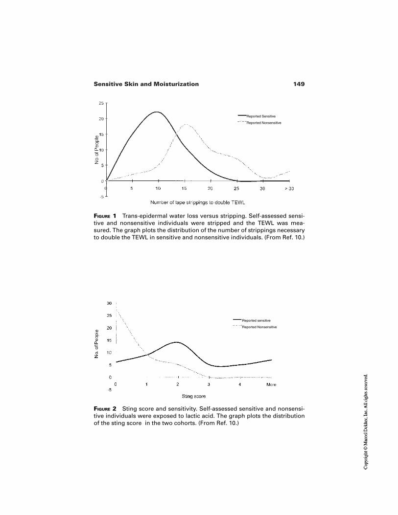
149Sensitive Skin and Moisturization
FIGURE 1 Trans-epidermal water loss versus stripping. Self-assessed sensi-tive and nonsensitive individuals were stripped and the TEWL was mea-sured. The graph plots the distribution of the number of strippings necessaryto double the TEWL in sensitive and nonsensitive individuals. (From Ref. 10.)
FIGURE 2 Sting score and sensitivity. Self-assessed sensitive and nonsensi-tive individuals were exposed to lactic acid. The graph plots the distributionof the sting score in the two cohorts. (From Ref. 10.)
Reported Sensitive
Reported Nonsensitive
Reported sensitive
Reported Nonsensitive
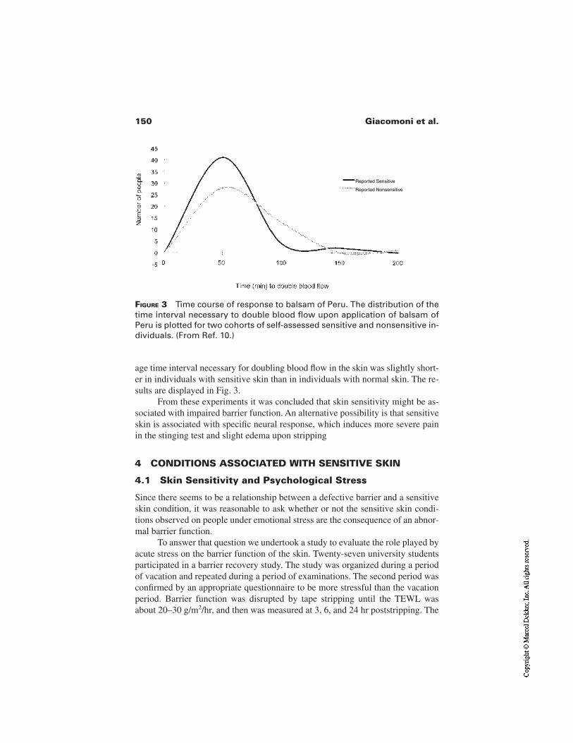
150 Giacomoni et al.
age time interval necessary for doubling blood flow in the skin was slightly short-er in individuals with sensitive skin than in individuals with normal skin. The re-sults are displayed in Fig. 3.
From these experiments it was concluded that skin sensitivity might be as-sociated with impaired barrier function. An alternative possibility is that sensitiveskin is associated with specific neural response, which induces more severe painin the stinging test and slight edema upon stripping
4 CONDITIONS ASSOCIATED WITH SENSITIVE SKIN
4.1 Skin Sensitivity and Psychological Stress
Since there seems to be a relationship between a defective barrier and a sensitiveskin condition, it was reasonable to ask whether or not the sensitive skin condi-tions observed on people under emotional stress are the consequence of an abnor-mal barrier function.
To answer that question we undertook a study to evaluate the role played byacute stress on the barrier function of the skin. Twenty-seven university studentsparticipated in a barrier recovery study. The study was organized during a periodof vacation and repeated during a period of examinations. The second period wasconfirmed by an appropriate questionnaire to be more stressful than the vacationperiod. Barrier function was disrupted by tape stripping until the TEWL wasabout 20–30 g/m2/hr, and then was measured at 3, 6, and 24 hr poststripping. The
FIGURE 3 Time course of response to balsam of Peru. The distribution of thetime interval necessary to double blood flow upon application of balsam ofPeru is plotted for two cohorts of self-assessed sensitive and nonsensitive in-dividuals. (From Ref. 10.)
Reported Sensitive
Reported Nonsensitive
151Sensitive Skin and Moisturization
rate of change of TEWL was used as an indicator of the recovery of barrier func-tion. The results indicate that the recovery of barrier function is more rapid in anonstressful than in a stressful situation. These results point out that psychologi-cal stress plays a role on the kinetics of recovery of disrupted barrier [11].
4.2 Skin Sensitivity and Age
Data published in the literature indicate that skin sensitivity declares itself or in-creases in the years of the onset of menopause [12]. This increase in sensitivitycannot be attributed to the thickness of the stratum corneum, which is known notto change with age [18]. It is not even the consequence of a change in skin thick-ness, since in three groups of premenopausal, perimenopausal, and early post-menopausal women, the average skin thickness did not change in a significativeway (2.28 +/– 0.39, 2.18 +/– 0.35, and 2.02 +/– 0.36 mm, respectively) [14]. It isindeed known that skin becomes thinner in the years after the onset of menopause[15]. Other authors have explored the percutaneous absorption of xenobioticssuch as hydrocortisone or testosterone on the forearm of pre- and postmenopausalwomen [16] and in two groups of young and old men [17]. They did not observedifferences in the percutaneous absorption in the two groups of women [16], butobserved that permeation of hydrocortisone, benzoic acid, acetyl salicylic acid,and caffeine were significantly lower in the group of old men [17]. They conclud-ed: “It is a common misconception that older skin has a diminished barrier capac-ity, and that percutaneous absorption is therefore greater” [16].
In a study performed in our laboratory, we have analyzed the TEWL of 223women aged between 21 and 79. The results are reported in Table 1. From thesedata it appears that the TEWL is low for young women, it increases by more than25% for women in their maturity, and returns to lower values above the age of 50.These data can be interpreted by saying that the older individuals have functionalbarrier, as already suggested by the studies of Howard Maibach [16,17], whereaswomen in their maturity have impaired barrier because they live a more stressfullife, in agreement with published data [11,18]. Thirty-eight out of the 223 panelistsreported themselves as having sensitive skin. Measurements of TEWL on these“sensitive skin” panelists are reported in Table 2. It appears that in the age groupsbetween 31 and 50, the vast majority of the individual levels of TEWL are abovethe average of each group (see Table 1). This allows one to conclude that sensitiveskin can be associated with high trans-epidermal water loss.
5 DISCUSSION
Understanding the link between skin moisturization and skin sensitivity is of par-ticular interest not only to the physiologist and the dermatologist, but also to thesupplier of skin care products for cosmetics.
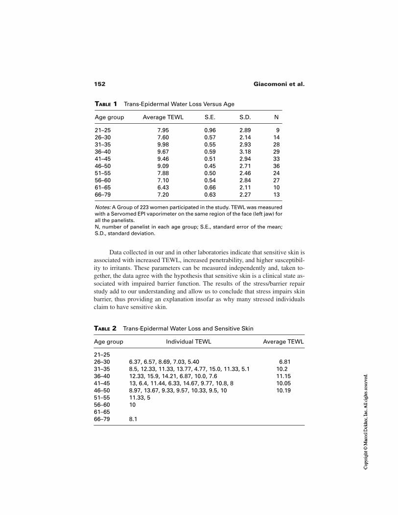
152 Giacomoni et al.
Data collected in our and in other laboratories indicate that sensitive skin isassociated with increased TEWL, increased penetrability, and higher susceptibil-ity to irritants. These parameters can be measured independently and, taken to-gether, the data agree with the hypothesis that sensitive skin is a clinical state as-sociated with impaired barrier function. The results of the stress/barrier repairstudy add to our understanding and allow us to conclude that stress impairs skinbarrier, thus providing an explanation insofar as why many stressed individualsclaim to have sensitive skin.
TABLE 1 Trans-Epidermal Water Loss Versus Age
Age group Average TEWL S.E. S.D. N
21–25 7.95 0.96 2.89 926–30 7.60 0.57 2.14 1431–35 9.98 0.55 2.93 2836–40 9.67 0.59 3.18 2941–45 9.46 0.51 2.94 3346–50 9.09 0.45 2.71 3651–55 7.88 0.50 2.46 2456–60 7.10 0.54 2.84 2761–65 6.43 0.66 2.11 1066–79 7.20 0.63 2.27 13
Notes: A Group of 223 women participated in the study. TEWL was measuredwith a Servomed EPI vaporimeter on the same region of the face (left jaw) forall the panelists.N, number of panelist in each age group; S.E., standard error of the mean;S.D., standard deviation.
TABLE 2 Trans-Epidermal Water Loss and Sensitive Skin
Age group Individual TEWL Average TEWL
21–2526–30 6.37, 6.57, 8.69, 7.03, 5.40 6.8131–35 8.5, 12.33, 11.33, 13.77, 4.77, 15.0, 11.33, 5.1 10.236–40 12.33, 15.9, 14.21, 6.87, 10.0, 7.6 11.1541–45 13, 6.4, 11.44, 6.33, 14.67, 9.77, 10.8, 8 10.0546–50 8.97, 13.67, 9.33, 9.57, 10.33, 9.5, 10 10.1951–55 11.33, 556–60 1061–6566–79 8.1
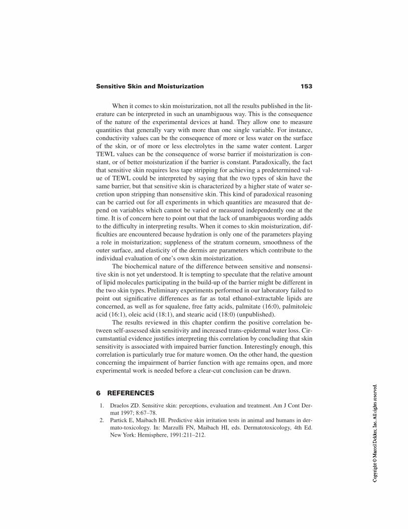
153Sensitive Skin and Moisturization
When it comes to skin moisturization, not all the results published in the lit-erature can be interpreted in such an unambiguous way. This is the consequenceof the nature of the experimental devices at hand. They allow one to measurequantities that generally vary with more than one single variable. For instance,conductivity values can be the consequence of more or less water on the surfaceof the skin, or of more or less electrolytes in the same water content. LargerTEWL values can be the consequence of worse barrier if moisturization is con-stant, or of better moisturization if the barrier is constant. Paradoxically, the factthat sensitive skin requires less tape stripping for achieving a predetermined val-ue of TEWL could be interpreted by saying that the two types of skin have thesame barrier, but that sensitive skin is characterized by a higher state of water se-cretion upon stripping than nonsensitive skin. This kind of paradoxical reasoningcan be carried out for all experiments in which quantities are measured that de-pend on variables which cannot be varied or measured independently one at thetime. It is of concern here to point out that the lack of unambiguous wording addsto the difficulty in interpreting results. When it comes to skin moisturization, dif-ficulties are encountered because hydration is only one of the parameters playinga role in moisturization; suppleness of the stratum corneum, smoothness of theouter surface, and elasticity of the dermis are parameters which contribute to theindividual evaluation of one’s own skin moisturization.
The biochemical nature of the difference between sensitive and nonsensi-tive skin is not yet understood. It is tempting to speculate that the relative amountof lipid molecules participating in the build-up of the barrier might be different inthe two skin types. Preliminary experiments performed in our laboratory failed topoint out significative differences as far as total ethanol-extractable lipids areconcerned, as well as for squalene, free fatty acids, palmitate (16:0), palmitoleicacid (16:1), oleic acid (18:1), and stearic acid (18:0) (unpublished).
The results reviewed in this chapter confirm the positive correlation be-tween self-assessed skin sensitivity and increased trans-epidermal water loss. Cir-cumstantial evidence justifies interpreting this correlation by concluding that skinsensitivity is associated with impaired barrier function. Interestingly enough, thiscorrelation is particularly true for mature women. On the other hand, the questionconcerning the impairment of barrier function with age remains open, and moreexperimental work is needed before a clear-cut conclusion can be drawn.
6 REFERENCES
1. Draelos ZD. Sensitive skin: perceptions, evaluation and treatment. Am J Cont Der-mat 1997; 8:67–78.
2. Partick E, Maibach HI. Predictive skin irritation tests in animal and humans in der-mato-toxicology. In: Marzulli FN, Maibach HI, eds. Dermatotoxicology, 4th Ed.New York: Hemisphere, 1991:211–212.
154 Giacomoni et al.
3. Shelanski HV, Shelanski MV. A new technique of human patch test. Proc Sci SecToilet Good Assoc 1953; 19:46–49.
4. Frosch PJ, Kligman AM. The chamber scarification test for assessing irritancy. ContDermat 1976; 2:314–324.
5. Agner T, Serup J. Quantification of the DMSO response: a test for assessement ofsensitive skin. Clin Exp Dermatol 1989; 14:214–217.
6. Gniadecka M, Quistorff B. Assessment of dermal water by high-frequency ultra-sound: comparative studies with nuclear magnetic resonance. Br J Dermatol 1996;135:218–224.
7. Blichmann CW, Serup J. Assessment of skin moisture. Measurement of electric con-ductance, capacitance and transepidermal water loss. Acta Der Venereol 1988;68:284–290.
8. Van Neste D. Comparative study of normal and rough human skin hydration in vivo:evaluation with four different instruments. J Dermatol Sci 1991; 2:119–124.
9. Maes D, Short J, Turek B, Reinstein J. In vivo measuring of skin softness using thegas bearing electrodynamometer. Int J Cosmet Sci 1983; 5:189–200.
10. Muizzuddin N, Marenus KD, Maes DH. Factors defining sensitive skin and its treat-ment. Am J Cont Dermat 1998; 9:170–175.
11. Garg A, Chren MM, Sands LP, Matsui MS, Marenus KD, Feingold KR, Elias PM.Psychological stress perturbs epidermal permeability barrier homeostasis. Arch Der-matol 2001; 137:53–59.
12. Paquet F, Pierard-Franchimont C, Fumal I, Goffin V, Paye M, Pierard GE. Sensitiveskin at menopause; dew point and electrometric properties of the stratum corneum.Maturitas 1998; 28:221–227.
13. Gilchrest BA. Aging of skin. In: Fitzpatrick TB, Eisen ZA, Wolff K, Freedberg IM,Austen KF, eds. Dermatology in General Medicine. New York: McGraw-Hill,1993:150–157.
14. Panyakhamlerd K, Chotnopparatpattara P, Taechakraichana N, Kukulprasong A,Chaikittisilpa S, Limpaphayom K. Skin thickness in different menopausal status. JMed Assoc Thai 1999; 82:352–356.
15. Brincat MP. Hormone replacement therapy and the skin. Maturitas 2000;35:107–117.
16. Oriba HA, Bucks DA, Maibach HI. Percutaneous absorption of hydrocortisone andtestosterone on the vulva and on the forearm: effect of menopause and site. Br J Der-matol 1996; 134:229–233.
17. Roskos KV, Maibach HI, Guy RH. The effect of aging on percutaneous absorption inman. J Pharmacokinet Biopharm 1989; 17:617–630.
18. Denda M, Tsuchiya T, Elias PM, Feingold KR. Stress alters cutaneous permeabilitybarrier homeostasis Am J Physiol Regul Integr Comp Physiol 2000; 278:367–372.
8Photodamage and Dry Skin
James J. Leyden and Robert LavkerUniversity of Pennsylvania School of Medicine, Philadelphia, Pennsylvania
Over the past 40 years, considerable evidence has been accumulated from a widerange of experimental studies in animals and humans to clearly indicate that ul-traviolet radiation (UVR) from sun exposure has multiple profound effects onskin. Both acute and chronic effects are well described [1]. Ultraviolet radiation isresponsible for skin cancer, photoaging, and photosensitivity diseases. In addi-tion, profound immunological effects have been identified which account in partfor the beneficial effects of UVR in many diseases such as psoriasis, atopic der-matitis, mycosis fungoid, and vitiligo.
Ultraviolet light is artificially divided into very short wave UVC (none cur-rently reaches the earth’s surface), UVB (290 to 320 nm), and UVA, which is di-vided into UVA II (320 to 340 nm) and UVA I (340 to 400 nm). Ultraviolet Amakes up approximately 95% of the UVR to which we are exposed. Until rela-tively recently, the main focus of research had been directed toward UVB and itsrole in cancer and immune modulation; UVB wavelengths are far more energeticthan UVA and clearly are the dominant factor in squamous cell formation andplay an important role in basal cell cancer. In the past decade, in vivo studies inhuman volunteers have shown that repeated low doses of UVA II and I compa-rable to those obtained during everyday activities can also have profound effectsin skin. Table 1 summarizes the work of many investigators and indicates all
155

156 Leyden and Lavker
wavelengths have profound biological effects on all components and cell typesin skin.
1 EFFECTS OF PHOTODAMAGE
Photoaging and photodamage are terms used to describe the consequences ofchronic exposure to ultraviolet light. Cumulative injury results in a constellationof histological and clinical findings (Table 2). Kligman first described the hall-mark of chronically sun damaged skin viz. the accumulation of disorganized,coarse bundles of fibers which stain like normal elastic tissue [2]. He coined the
TABLE 1 Relative Effects of UVA on Skin Components and Cell Types
UVA II UVA I
Stratum corneum thickness (dry skin) +++ +++Epidermal thickness ++ ++Apoptosis +++ +Langerhans cell depletion +++ +Damage to elastin ++ +++Sebaceous gland hypertrophy ++ ++Telangiectasias ++ ?
TABLE 2 Histological and Clinical Manifestations of Photoaging
Clinical signs
Stratum corneum thickening andmicrofissures “Dry,” flaky rough skin
DNA damage to basal cells andkeratinocytes; dysplasia, neoplasia
Actinic keratosis; basal andsquamous cell cancer
Melanocytic hyperplasia, dysplasia Lentigos (age spots)Epidermal inclusion cysts MiliaFollicular epithelial hyperkeratosis Solar comedonesThickened, disorganized elastic fibers;
decrease collagen WrinklingTelangiectactic vessels; decrease in
papillary dermis Telangiectasis; sallownessSebaceous gland hypertrophy Sebaceous hypertrophy
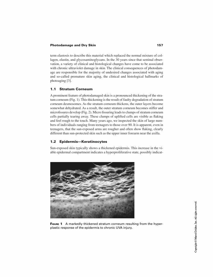
157Photodamage and Dry Skin
term elastosis to describe this material which replaced the normal mixture of col-lagen, elastin, and glycosaminoglycans. In the 30 years since that seminal obser-vation, a variety of clinical and histological changes have come to be associatedwith chronic ultraviolet damage in skin. The clinical consequences of photodam-age are responsible for the majority of undesired changes associated with agingand so-called premature skin aging, the clinical and histological hallmarks ofphotoaging [3].
1.1 Stratum Corneum
A prominent feature of photodamaged skin is a pronounced thickening of the stra-tum corneum (Fig. 1). This thickening is the result of faulty degradation of stratumcorneum desmosomes. As the stratum corneum thickens, the outer layers becomesomewhat dehydrated. As a result, the outer stratum corneum becomes stiffer andmicrofissures develop (Fig. 2). Micro fissuring leads to clumps of stratum corneumcells partially tearing away. These clumps of uplifted cells are visible as flakingand feel rough to the touch. Many years ago, we inspected the skin of large num-bers of individuals ranging from teenagers to those over 90. It is apparent, even inteenagers, that the sun-exposed arms are rougher and often show flaking, clearlydifferent than sun-protected skin such as the upper inner forearm near the axilla.
1.2 Epidermis—Keratinocytes
Sun-exposed skin typically shows a thickened epidermis. This increase in the vi-able epidermal compartment indicates a hyperproliferative state, possibly indicat-
FIGURE 1 A markedly thickened stratum corneum resulting from the hyper-plastic response of the epidermis to chronic UVA injury.
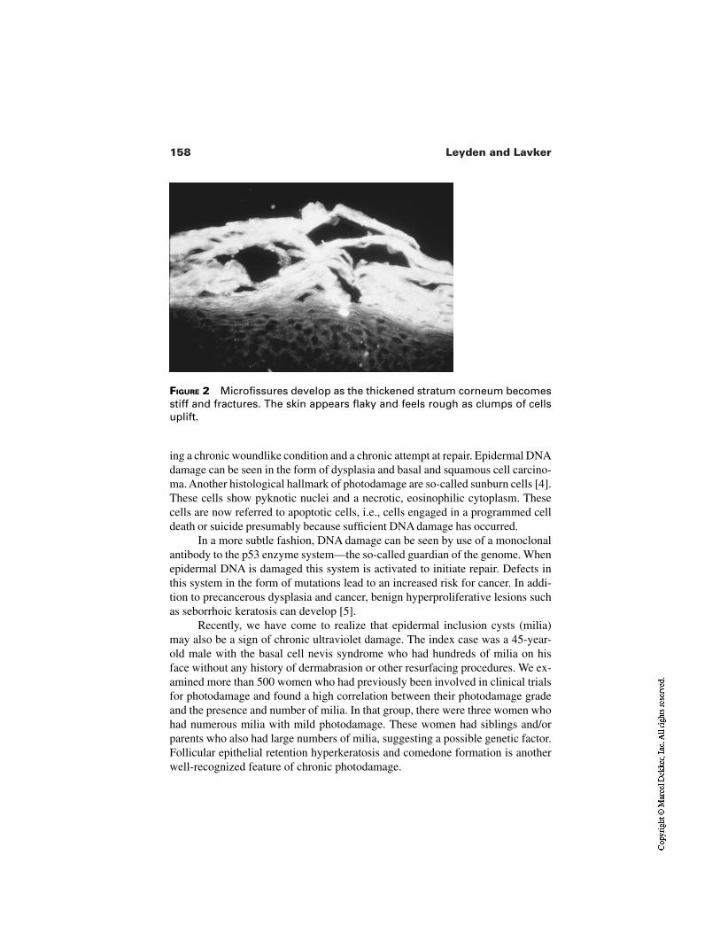
158 Leyden and Lavker
FIGURE 2 Microfissures develop as the thickened stratum corneum becomesstiff and fractures. The skin appears flaky and feels rough as clumps of cellsuplift.
ing a chronic woundlike condition and a chronic attempt at repair. Epidermal DNAdamage can be seen in the form of dysplasia and basal and squamous cell carcino-ma. Another histological hallmark of photodamage are so-called sunburn cells [4].These cells show pyknotic nuclei and a necrotic, eosinophilic cytoplasm. Thesecells are now referred to apoptotic cells, i.e., cells engaged in a programmed celldeath or suicide presumably because sufficient DNA damage has occurred.
In a more subtle fashion, DNA damage can be seen by use of a monoclonalantibody to the p53 enzyme system—the so-called guardian of the genome. Whenepidermal DNA is damaged this system is activated to initiate repair. Defects inthis system in the form of mutations lead to an increased risk for cancer. In addi-tion to precancerous dysplasia and cancer, benign hyperproliferative lesions suchas seborrhoic keratosis can develop [5].
Recently, we have come to realize that epidermal inclusion cysts (milia)may also be a sign of chronic ultraviolet damage. The index case was a 45-year-old male with the basal cell nevis syndrome who had hundreds of milia on hisface without any history of dermabrasion or other resurfacing procedures. We ex-amined more than 500 women who had previously been involved in clinical trialsfor photodamage and found a high correlation between their photodamage gradeand the presence and number of milia. In that group, there were three women whohad numerous milia with mild photodamage. These women had siblings and/orparents who also had large numbers of milia, suggesting a possible genetic factor.Follicular epithelial retention hyperkeratosis and comedone formation is anotherwell-recognized feature of chronic photodamage.
159Photodamage and Dry Skin
1.3 Epidermis—Melanocytes
Increased numbers of melanocytes and melanocytic hyperplasia resulting in solarlentigos, age spots, and sunburn freckles are well-recognized consequences ofchronic ultraviolet damage. The role of ultraviolet light in melanoma remains un-settled in terms of which wavelengths are involved and how central to melanomadevelopment UV is, but none doubt its importance.
1.4 Dermis—Matrix
A histological hallmark of photoaging is a replacement of the normal dermal ma-trix of collagen, elastin, and glycosaminoglycans by large bundles of coarse elas-tic fibers and decreased collagen. The clinical consequence of elastosis is pro-nounced wrinkling and in advanced cases a yellowish cobblestone appearanceassociated with pronounced sagging. This process is often accompanied by abrisk neutophilic infiltrate which often can be appreciated clinically and is re-ferred to as heliodermatitis or dermatoheliosis [6]. Neutrophil elastases may playa prominent role in damage to elastin and the subsequent wrinkling or sagging.The role of UV in the degeneration of surrounding collagen is now more fully un-derstood and is the consequence of both UVA and UVB and the effect of ultravi-olet light increasing the activity of metalloproteinases [7]. This family of 14 dif-ferent proteinases can act on a broad range of substrates and can be activated invivo by a single exposure to UV. The mechanism(s) of elastosis remain unclear.The major consequence of these changes in the dermal matrix is wrinkling. Wrin-kles don’t have a histological marker but rather can be best thought of as stressfractures from material which has aged. The radiating quality of wrinkles is simi-lar to that seen in materials such as buildings and bridges. With more pronouncedchanges, skin “settles,” which is seen clinically as sagging.
1.5 Dermis—Vasculature
Two changes can occur. Some patients show loss of the papillary plexus, flatten-ing of the rete ridges, and loss of the papillary dermis. Clinically, these individu-als have a sallow washed-out appearance. The other finding is that of a prolifera-tive response producing dilated, enlarged vessels in the papillary and mid-dermis.Clinically, these are seen as telangiectasis.
1.6 Dermis—Sebaceous Gland
Sebaceous gland enlargement is another feature of chronic ultraviolet damage.Clinically, this can be seen as small, yellowish nodules or in more advanced cas-es as a thick, coarsening of skin with large, dilated follicular openings from whichsebaceous material can be squeezed out.
160 Leyden and Lavker
TABLE 3 Surface Texture in Sun-Exposed and -Protected Skin
Exposed Protected
Young (mean 29) 15 ± 6 5 ± 3Middle age (mean 45) 40 ± 10 10 ± 6Older (mean 65) 65 ± 13 30 ± 7
Note: Visual analog score 0 = none; 100 = very severe “dry skin.”
2 VULNERABLE PHENOTYPES
It has been known for many years that fair skinned individuals, particularly thoseof Celtic ancestry, are particularly vulnerable to the acute adverse effects of ultra-violet light, i.e., sunburn. More recently, we have come to realize that there is aphenotype who appears to be more vulnerable to the chronic adverse effects ofUV. These individuals have red hair, blue eyes, and have a Celtic background, buthave the ability to tan often fairly deeply, after suffering initial burning. Typical-ly, they have a very fair Celtic parent who burns and tans little or not at all, whilethe other parent tans easily and rarely burns. The vulnerable offspring is able tosustain more exposure because their tan prevents burning. Usually, these individ-uals are infrequent users of high-SPF sunscreens and pride themselves on theirability to tan while family members can’t. These individuals develop dry skin, aleathery wrinkling, and pigmentary changes at a relatively early age (late 20s toearly 30s) and by their mid-40s they tend to look older and are extremely unhap-py.
“Dry skin” is a prominent feature of actinically damaged skin. It is such aprominent feature that the lay public has, with the help of many cosmetic compa-nies, come to view dry skin as the causative agent for other signs of photodam-age, most notably wrinkling. As is detailed in other chapters, dry skin is really anabnormally thickened stratum corneum which becomes stiff and cracks. The re-sulting uplifted clumps of cells become visible as flakes and skin develops arough texture.
3 SKIN TEXTURE IN EXPOSED AND PROTECTED SITES
Some years ago, we examined a large number of people ranging from 11 to 70years in age. The outer, exposed area of both forearms and the upper inner, sun-protected arm were graded using a visual analog scale. The results are summa-rized in Table 3. In all age groups the exposed sites clearly were rougher to touch,and visible differences were also common. Even in teenagers and young adults,
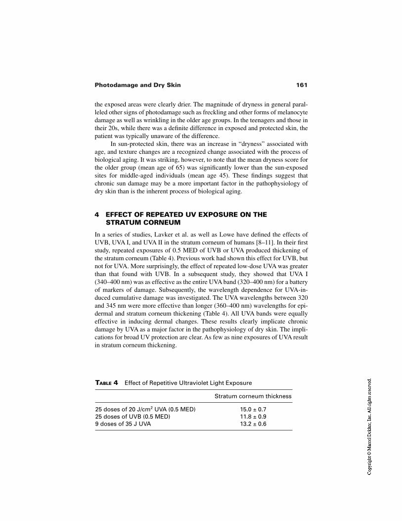
161Photodamage and Dry Skin
TABLE 4 Effect of Repetitive Ultraviolet Light Exposure
Stratum corneum thickness
25 doses of 20 J/cm2 UVA (0.5 MED) 15.0 ± 0.725 doses of UVB (0.5 MED) 11.8 ± 0.99 doses of 35 J UVA 13.2 ± 0.6
the exposed areas were clearly drier. The magnitude of dryness in general paral-leled other signs of photodamage such as freckling and other forms of melanocytedamage as well as wrinkling in the older age groups. In the teenagers and those intheir 20s, while there was a definite difference in exposed and protected skin, thepatient was typically unaware of the difference.
In sun-protected skin, there was an increase in “dryness” associated withage, and texture changes are a recognized change associated with the process ofbiological aging. It was striking, however, to note that the mean dryness score forthe older group (mean age of 65) was significantly lower than the sun-exposedsites for middle-aged individuals (mean age 45). These findings suggest thatchronic sun damage may be a more important factor in the pathophysiology ofdry skin than is the inherent process of biological aging.
4 EFFECT OF REPEATED UV EXPOSURE ON THESTRATUM CORNEUM
In a series of studies, Lavker et al. as well as Lowe have defined the effects ofUVB, UVA I, and UVA II in the stratum corneum of humans [8–11]. In their firststudy, repeated exposures of 0.5 MED of UVB or UVA produced thickening ofthe stratum corneum (Table 4). Previous work had shown this effect for UVB, butnot for UVA. More surprisingly, the effect of repeated low-dose UVA was greaterthan that found with UVB. In a subsequent study, they showed that UVA I(340–400 nm) was as effective as the entire UVA band (320–400 nm) for a batteryof markers of damage. Subsequently, the wavelength dependence for UVA-in-duced cumulative damage was investigated. The UVA wavelengths between 320and 345 nm were more effective than longer (360–400 nm) wavelengths for epi-dermal and stratum corneum thickening (Table 4). All UVA bands were equallyeffective in inducing dermal changes. These results clearly implicate chronicdamage by UVA as a major factor in the pathophysiology of dry skin. The impli-cations for broad UV protection are clear. As few as nine exposures of UVA resultin stratum corneum thickening.
162 Leyden and Lavker
In studies in which the biological effectiveness of various bands within theUVA spectrum were compared for the effects of cumulative damage, both theshorter UVA II wavelengths and longer UVA I are found to be responsible forstratum corneum thickening. Thus at equivalent suberythematogenic doses, UVAappears to be more effective than UVB in inducing stratum corneum thickening.The findings of Lavker et al. suggest that cumulative doses greater than 315 J/cm2
result in stratum corneum thickening.It is important to note that the doses of UVA used in these experiments are
relative to daily exposure for many people. A few hours exposure will permit ac-cumulation of comparable doses of UVA.
Recent evidence accumulating from a variety of phenotypes of dry skinsuch as seen in atopic dermatitis, inherited metabolic disorders, and detergent-chapped skin all point to a disturbance in the balance of intracellular lipids of thestratum corneum.
Permeability barrier function and orderly corneocyte desquamation requirethe organization of three nonpolar lipids, ceramides, free fatty acids, and choles-terol into extracellular lamellar membrane structure within the intercellularspaces of the stratum corneum. These lipids are delivered through the secretion ofepidermal lamellar bodies. Although present in approximately equimolar ratios,ceramide predominates by weight, accounting for approximately 50%. In addi-tion, lamellar bodies secrete hydrolytic lipid hydrolases such as β-glucocerebrosi-dase and secretory phospholipase, which process glucosylceramide, phospho-lipids, ceramide, and free fatty acids. Sphingomyelin is another source ofceramide through hydrolysis by sphingomyelinase. In Gaucher’s disease, defi-ciency in β-glucocerebrosidase is associated with dry skin and defective stratumcorneum function [12]. Likewise in Niemann–Pick disease deficiency in acid-sphingomyelinase is associated with similar findings [13]. In atopic dermatitis,there is abnormal expression of sphingomyelin deacylase, resulting in decreasedlevels of ceramide by competing for glucocerebroside and sphingomyelin [14].The result is abnormal stratum corneum in terms of barrier and desquamation.Chronic exposure to detergents results in extraction of intracellular lipids and theformation of dry skin [15]. In all of these studies, the final pathway points to a de-crease in ceramides, particularly ceramide 1. Ceramide 1 is rich in linoleic acidand is believed to play an important role in regulating water content. Decrease inthis capacity may play a crucial role in the desquamation process. In hereditaryicthyosis a deficiency of cholesterol sulfatase results in an extreme expression ofabnormal desquamation viz. severely thickened stratum corneum.
The mechanisms by which repeated low-dose UVB and UVA induce abnor-mal desquamation are unknown. In recent studies (R Lavker, unpublished obser-vations) using a monoclonal antibody to lamellar bodies, an increase secretion ofthese lamellae was found. Any molecular or biochemical aberrations remain to be
163Photodamage and Dry Skin
elucidated. However, it is likely that an abnormality in one or more pathwaysleading to ceramide formation may be involved in UVR-induced dry skin.
Another possible pathway that may play a role in UVR-induced dry skincould involve the recently described mitochondrial DNA (mt DNA) mutations[16]. Such mutations have been linked with other chronic degenerative diseasessuch as Alzheimer’s, progressive external opthalmoplegia, and Keans–Sayre syn-drome. In addition, mitochondrial DNA mutations have been proposed to play arole in the biological process of aging.
Higher mutation frequency of mt DNA has been found in chronically sun-exposed skin, and more recent work has established a direct link between UVAradiation–induced oxidative stress and the most frequent mt DNA mutation [17].This interesting story is currently viewed as most relevant to dermal fibroblastand dermal matrix changes. However, the possibility of such changes playing arole in epidermal changes remains a possibility.
Current evidence developed over the last 10 years clearly implicates chron-ic ultraviolet damage as a key factor in the development of dry skin. Both UVBand UVA are implicated, with the latter probably the more important causativefactor. Studies indicate that cumulative low-dose UVB and UVA induce stratumcorneum changes after as few as nine exposures. While the skin possesses capa-bility to repair UV damage—best seen in the photodamage mouse model—theaccumulation of UV damage clearly overwhelms repair mechanisms. Even inyoung adults, the clinical consequences of chronic UV damage can be appreciat-ed. The implications for prevention are clear—broad spectrum UV protection,possibly in combination with blends of anti-oxidants to mute oxidative stress notprevented by UV-absorbing agents.
REFERENCES
1. Gilchrest BA. Skin aging and photoaging: an overview. J Am Acad Dermatol 1989;21:610–613.
2. Kligman A. Early destruction effects of sunlight in human skin. J Am Med Assoc1969; 210:2377–2380.
3. Kligman AM, Lavker RM. Cutaneous aging: the differences between intrinsic agingand photoaging. J Cutan Aging Cosmet Dermatol 1988; 1:5–11.
4. Gilchrest BA, Soter NA, Stoff JS, Mihm MC. The human sunburn reaction: histo-logic and biochemical studies. J Am Acad Dermatol 1981; 5:411–422.
5. Granstein RD, Sober AJ. Current concepts in ultraviolet carcinogenis. Proc Soc ExpBiol Med 1982; 170:115–125.
6. Lavker RM. Structural alteration in exposed and unexposed human skin. J InvestDermatol 1979; 73:59–66.
7. Kligman LH, Akin FJ, Kligman AM. The contribution of UVA and UVB to connec-tive tissue damage in hairless mice. J Invest Dermatol 1985; 84:272–276.

164 Leyden and Lavker
8. Lavker R, Kaidbey K. The spectral dependence for UVA-induced cumulating dam-age in human skin. J Invest Dermatol 199; 108:17–21.
9. Lavker RM, Gerberick GF, Veres D, Irwin CJ, Kaidby KH. Cumulative effects fromrepeated exposures to sunerythemal doses of UVB and UVA in human skin. J AmAcad Dermatol 1995; 32:53–62.
10. Lavker RM, Veres DA, Irwin CJ, Kaidbey KH. Quantitative assessment of cumula-tive damage from repetitive exposures to suberythemogenic doses of UVA in humanskin. Photochem Photobiol 1995; 62:348–352.
11. Lowe NJ, Meyerd DP, Wieder JM, Luftman D, Borget T, Lehman MD, Johnson AW,Scott IR. Low doses of repetitive ultraviolet A induce morphologic changes in hu-man skin. J Invest Dermatol 1995; 105:739–743.
12. Hulleran WM, Ginns EI, Menon GK, et al. Consequences of beta-glucocerebrosi-dase deficiency in epidermis: ultrastructure and permeability barrier alterations inGaucher’s disese. J Clin Invest; 93:1756–1764.
13. Schmuth M, Man M, Weber F, et al. Permeability barrier disorder in Niemann–Pickdisease: sphingomyelin-ceramide processing required for normal barrier homeosta-sis. J Invest Dermatol 2000; 115:459–466.
14. Hara J, Higachi K, Okamoto R, et al. High expression of sphingomyelin deacylase isan important determinant of ceramide deficiency leading to barrier disruption inatopic dermatitis. J Invest Dermatol 2000; 115:406–413.
15. Rawlings AV, Watkinson A, Rogers J, et al. Abnormalities in stratum corneum struc-ture, lipid composition and desmosome degredation in soap induced winter xerosis.J Cosmet Chem 1994; 45:203–220.
16. Berneburg M, Gattermann N, Stege H, et al. Chronically ultraviolet-exposed humanskin shows a higher mutation frequency of mitochondrial DNA. Photochem Photo-biol 1997; 66:271–275.
17. Bernburg M, Grether-Bech S, Kurten V, et al. Singlet oxygen mediates UVA-inducedgeneration of photoaging-associated mitochondrial corneum deletum.
9Atopic Dermatitis
Anna Di NardoUniversity of Modena, Modena, Italy
Philip W. WertzDows Institute for Dental Research, University of Iowa, Iowa City, Iowa
1 ATOPIC DERMATITIS
Widespread regions of dry itchy skin is one of the prominent clinical features ofatopic dermatitis [1]. The intense itch is the most characteristic feature of this dis-ease, and the consequences include scratching and eczematous lesions, as illus-trated in Figure 1. Scratching can lead to disruption of the stratum corneum andinfection. This disease is generally associated with asthma, allergic rhinitis, andelevated levels of IgE. There is frequently a family history of atopic dermatitis,indicating a genetic component in the etiology of the disease. All of these factorsare taken into account in arriving at a clinical diagnosis.
The onset of atopic dermatitis most frequently occurs during the first yearof life, and most cases become evident before age 5 [2]. Only rarely is there anadult onset. In most patients atopic dermatitis spontaneously resolves by aboutage 20, although it can be a lifelong disease. Adults who have been atopic oftenhave unusually sensitive skin.
The differentiation process in atopic epidermis is notably altered. At a his-tologic level the intercellular spaces in the viable portion of the atopic epidermisappear to be swollen with fluid, and the spinous layer is thickened. In the vicinity
165
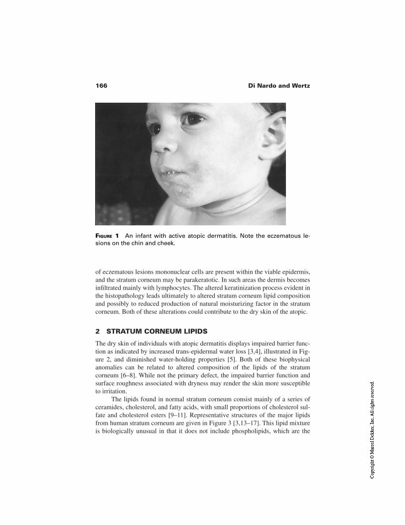
166 Di Nardo and Wertz
of eczematous lesions mononuclear cells are present within the viable epidermis,and the stratum corneum may be parakeratotic. In such areas the dermis becomesinfiltrated mainly with lymphocytes. The altered keratinization process evident inthe histopathology leads ultimately to altered stratum corneum lipid compositionand possibly to reduced production of natural moisturizing factor in the stratumcorneum. Both of these alterations could contribute to the dry skin of the atopic.
2 STRATUM CORNEUM LIPIDS
The dry skin of individuals with atopic dermatitis displays impaired barrier func-tion as indicated by increased trans-epidermal water loss [3,4], illustrated in Fig-ure 2, and diminished water-holding properties [5]. Both of these biophysicalanomalies can be related to altered composition of the lipids of the stratumcorneum [6–8]. While not the primary defect, the impaired barrier function andsurface roughness associated with dryness may render the skin more susceptibleto irritation.
The lipids found in normal stratum corneum consist mainly of a series ofceramides, cholesterol, and fatty acids, with small proportions of cholesterol sul-fate and cholesterol esters [9–11]. Representative structures of the major lipidsfrom human stratum corneum are given in Figure 3 [3,13–17]. This lipid mixtureis biologically unusual in that it does not include phospholipids, which are the
FIGURE 1 An infant with active atopic dermatitis. Note the eczematous le-sions on the chin and cheek.
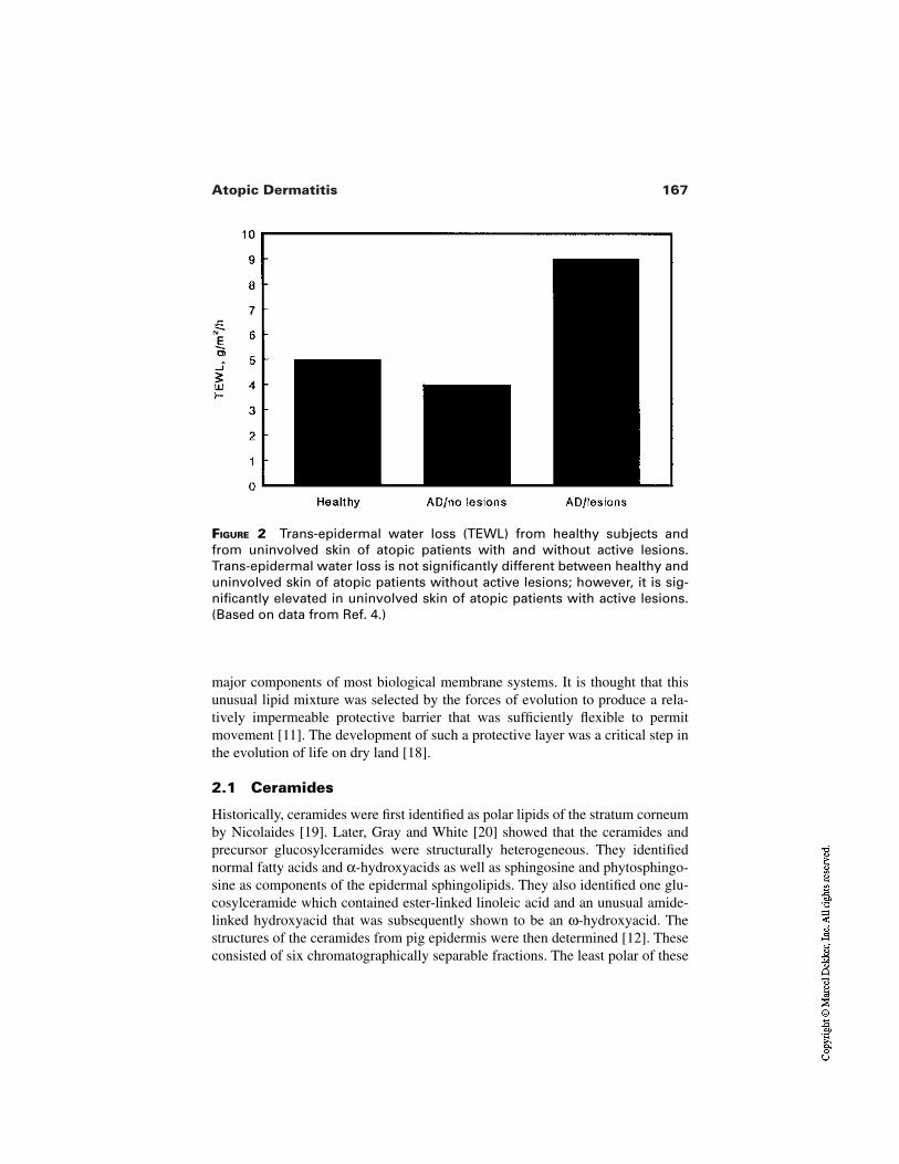
167Atopic Dermatitis
FIGURE 2 Trans-epidermal water loss (TEWL) from healthy subjects andfrom uninvolved skin of atopic patients with and without active lesions.Trans-epidermal water loss is not significantly different between healthy anduninvolved skin of atopic patients without active lesions; however, it is sig-nificantly elevated in uninvolved skin of atopic patients with active lesions.(Based on data from Ref. 4.)
major components of most biological membrane systems. It is thought that thisunusual lipid mixture was selected by the forces of evolution to produce a rela-tively impermeable protective barrier that was sufficiently flexible to permitmovement [11]. The development of such a protective layer was a critical step inthe evolution of life on dry land [18].
2.1 Ceramides
Historically, ceramides were first identified as polar lipids of the stratum corneumby Nicolaides [19]. Later, Gray and White [20] showed that the ceramides andprecursor glucosylceramides were structurally heterogeneous. They identifiednormal fatty acids and α-hydroxyacids as well as sphingosine and phytosphingo-sine as components of the epidermal sphingolipids. They also identified one glu-cosylceramide which contained ester-linked linoleic acid and an unusual amide-linked hydroxyacid that was subsequently shown to be an ω-hydroxyacid. Thestructures of the ceramides from pig epidermis were then determined [12]. Theseconsisted of six chromatographically separable fractions. The least polar of these
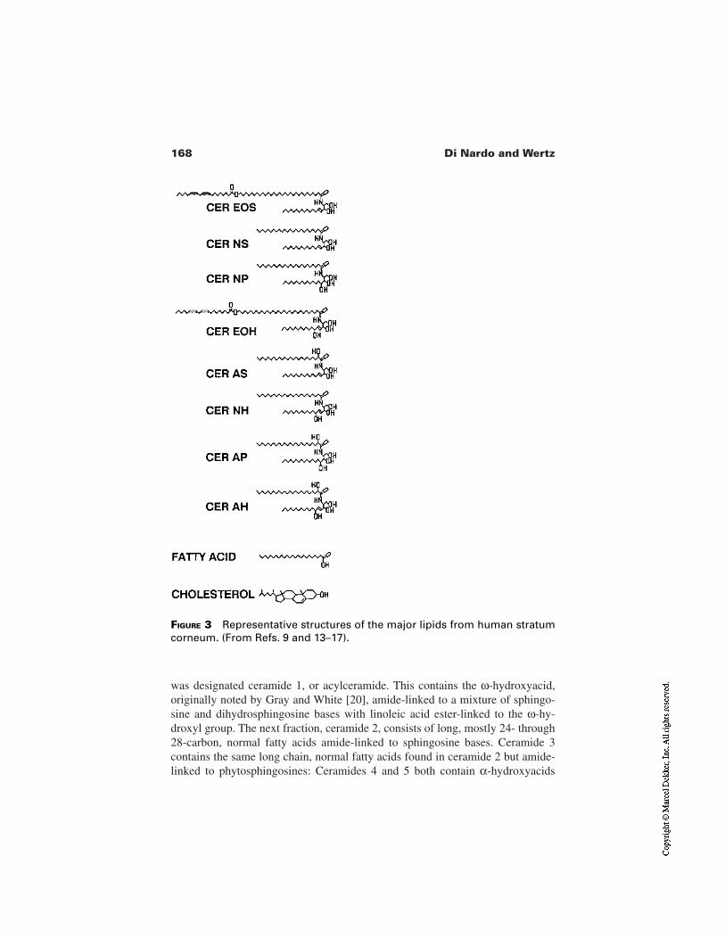
168 Di Nardo and Wertz
was designated ceramide 1, or acylceramide. This contains the ω-hydroxyacid,originally noted by Gray and White [20], amide-linked to a mixture of sphingo-sine and dihydrosphingosine bases with linoleic acid ester-linked to the ω-hy-droxyl group. The next fraction, ceramide 2, consists of long, mostly 24- through28-carbon, normal fatty acids amide-linked to sphingosine bases. Ceramide 3contains the same long chain, normal fatty acids found in ceramide 2 but amide-linked to phytosphingosines: Ceramides 4 and 5 both contain α-hydroxyacids
FIGURE 3 Representative structures of the major lipids from human stratumcorneum. (From Refs. 9 and 13–17).
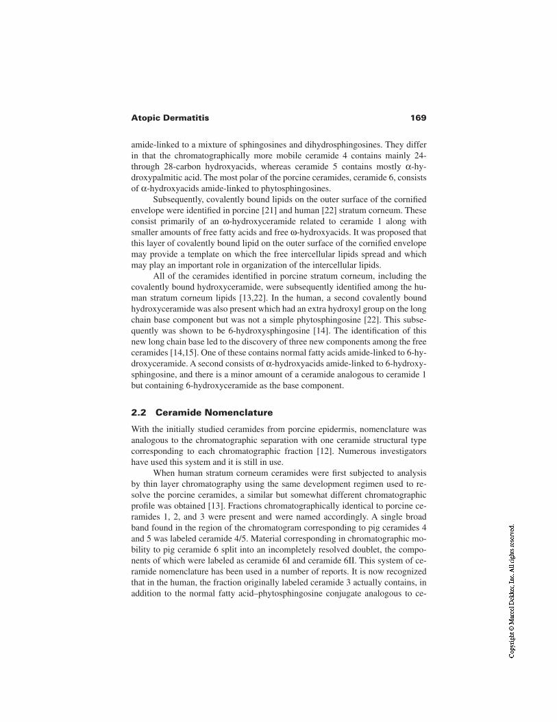
169Atopic Dermatitis
amide-linked to a mixture of sphingosines and dihydrosphingosines. They differin that the chromatographically more mobile ceramide 4 contains mainly 24-through 28-carbon hydroxyacids, whereas ceramide 5 contains mostly α-hy-droxypalmitic acid. The most polar of the porcine ceramides, ceramide 6, consistsof α-hydroxyacids amide-linked to phytosphingosines.
Subsequently, covalently bound lipids on the outer surface of the cornifiedenvelope were identified in porcine [21] and human [22] stratum corneum. Theseconsist primarily of an ω-hydroxyceramide related to ceramide 1 along withsmaller amounts of free fatty acids and free ω-hydroxyacids. It was proposed thatthis layer of covalently bound lipid on the outer surface of the cornified envelopemay provide a template on which the free intercellular lipids spread and whichmay play an important role in organization of the intercellular lipids.
All of the ceramides identified in porcine stratum corneum, including thecovalently bound hydroxyceramide, were subsequently identified among the hu-man stratum corneum lipids [13,22]. In the human, a second covalently boundhydroxyceramide was also present which had an extra hydroxyl group on the longchain base component but was not a simple phytosphingosine [22]. This subse-quently was shown to be 6-hydroxysphingosine [14]. The identification of thisnew long chain base led to the discovery of three new components among the freeceramides [14,15]. One of these contains normal fatty acids amide-linked to 6-hy-droxyceramide. A second consists of α-hydroxyacids amide-linked to 6-hydroxy-sphingosine, and there is a minor amount of a ceramide analogous to ceramide 1but containing 6-hydroxyceramide as the base component.
2.2 Ceramide Nomenclature
With the initially studied ceramides from porcine epidermis, nomenclature wasanalogous to the chromatographic separation with one ceramide structural typecorresponding to each chromatographic fraction [12]. Numerous investigatorshave used this system and it is still in use.
When human stratum corneum ceramides were first subjected to analysisby thin layer chromatography using the same development regimen used to re-solve the porcine ceramides, a similar but somewhat different chromatographicprofile was obtained [13]. Fractions chromatographically identical to porcine ce-ramides 1, 2, and 3 were present and were named accordingly. A single broadband found in the region of the chromatogram corresponding to pig ceramides 4and 5 was labeled ceramide 4/5. Material corresponding in chromatographic mo-bility to pig ceramide 6 split into an incompletely resolved doublet, the compo-nents of which were labeled as ceramide 6I and ceramide 6II. This system of ce-ramide nomenclature has been used in a number of reports. It is now recognizedthat in the human, the fraction originally labeled ceramide 3 actually contains, inaddition to the normal fatty acid–phytosphingosine conjugate analogous to ce-
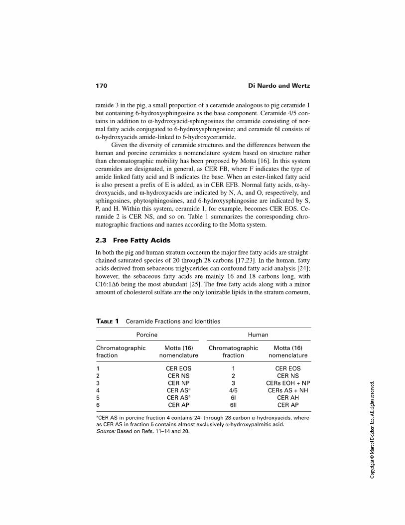
170 Di Nardo and Wertz
TABLE 1 Ceramide Fractions and Identities
Porcine Human
Chromatographicfraction
Motta (16)nomenclature
Chromatographicfraction
Motta (16)nomenclature
1 CER EOS 1 CER EOS2 CER NS 2 CER NS3 CER NP 3 CERs EOH + NP4 CER ASa 4/5 CERs AS + NH5 CER ASa 6I CER AH6 CER AP 6II CER AP
aCER AS in porcine fraction 4 contains 24- through 28-carbon α-hydroxyacids, where-as CER AS in fraction 5 contains almost exclusively α-hydroxypalmitic acid.Source: Based on Refs. 11–14 and 20.
ramide 3 in the pig, a small proportion of a ceramide analogous to pig ceramide 1but containing 6-hydroxysphingosine as the base component. Ceramide 4/5 con-tains in addition to α-hydroxyacid-sphingosines the ceramide consisting of nor-mal fatty acids conjugated to 6-hydroxysphingosine; and ceramide 6I consists ofα-hydroxyacids amide-linked to 6-hydroxyceramide.
Given the diversity of ceramide structures and the differences between thehuman and porcine ceramides a nomenclature system based on structure ratherthan chromatographic mobility has been proposed by Motta [16]. In this systemceramides are designated, in general, as CER FB, where F indicates the type ofamide linked fatty acid and B indicates the base. When an ester-linked fatty acidis also present a prefix of E is added, as in CER EFB. Normal fatty acids, α-hy-droxyacids, and ω-hydroxyacids are indicated by N, A, and O, respectively, andsphingosines, phytosphingosines, and 6-hydroxysphingosine are indicated by S,P, and H. Within this system, ceramide 1, for example, becomes CER EOS. Ce-ramide 2 is CER NS, and so on. Table 1 summarizes the corresponding chro-matographic fractions and names according to the Motta system.
2.3 Free Fatty Acids
In both the pig and human stratum corneum the major free fatty acids are straight-chained saturated species of 20 through 28 carbons [17,23]. In the human, fattyacids derived from sebaceous triglycerides can confound fatty acid analysis [24];however, the sebaceous fatty acids are mainly 16 and 18 carbons long, withC16:1∆6 being the most abundant [25]. The free fatty acids along with a minoramount of cholesterol sulfate are the only ionizable lipids in the stratum corneum,
171Atopic Dermatitis
and it has been suggested that this is important for the formation of a lamellarphase [24].
2.4 Cholesterol and Derivatives
Cholesterol is a major lipid component and the sole sterol in both porcine and hu-man stratum corneum [17]. Cholesterol sulfate is a minor stratum corneum com-ponent, but it has been implicated in the regulation of desquamation. Both with anorgan culture model and with human skin in vivo, it has been demonstrated thathydrolysis of cholesterol sulfate accompanies the desquamation process while allother lipids survive cell shedding intact [13,26]. In addition, there is a genetic dis-ease, recessive X-linked ichthyosis, in which the sulfatase that would normallyhydrolyze cholesterol sulfate is defective [27] and cholesterol sulfate is present atabnormally high levels in the stratum corneum and elsewhere [28]. In this diseasedesquamation does not proceed normally and the skin surface can become roughand scaly. Degradation of the desmosomes between corneocytes is a necessarystep leading to desquamation, and several serine proteases have been implicated.It has been suggested that hydrolysis of cholesterol sulfate may be required topermit proteolysis of the desmosomes [29], and recently it has been demonstratedthat cholesterol sulfate is a serine protease inhibitor [30–32]. Cholesterol estershave been considered a marker of keratinization [33]. The principal fatty acidfound in epidermal cholesterol esters is oleate [23]. Cholesterol esters are notthemselves membrane-forming lipids and are generally not well incorporated intomembranes formed from other lipids. It has been suggested the cholesterol estersphase separate from other lipids within the intercellular spaces of the stratumcorneum. This could provide a mechanism for keeping oleate, a known perme-ability enhancer [34], out of the membrane domains, thereby preserving barrierfunction [11,24].
2.5 Phase Behavior and Organization
All of the ceramides and free fatty acids in epidermal stratum corneum are rodlikeor cylindrical in shape, which makes them ideal for the formation of highly or-dered gel phase membrane domains [11,24]. Cholesterol is capable of either de-creasing or increasing the fluidity of membranes, depending upon the proportionsand natures of the other lipids. It has been suggested that cholesterol serves toprovide a degree of plasticity to what would otherwise be highly rigid and possi-bly brittle membranes. In this view the ceramides and fatty acids are essential forthe barrier function of the skin, and the cholesterol is required to permit flexingwithout cracking the stratum corneum. A model that has been advanced and thatis consistent with these suggestions is the domain mosaic model [35]. In thismodel the intercellular lamellae consist of gel phase domains within a continuousliquid crystalline domain. Molecules crossing these membranes would penetrate
172 Di Nardo and Wertz
through the more fluid liquid crystalline domains more readily than through thegel phase, and the greatest flux would occur at the phase boundaries.
Examination of stratum corneum by transmission electron microscopy fol-lowing treatment with ruthenium tetroxide has revealed that the number of lamel-lae across the intercellular space varies widely, but in most regions there are mul-tiples of three lamellae with a broad–narrow–broad spacing [36,37]. The overallthickness of one broad–narrow–broad unit is 13 nm [36]. This lamellar spacinghas also been confirmed by x-ray diffraction [38]. Near the ends of the corneo-cytes there is frequently one broad–narrow–broad unit. It has been proposed thatthis consists of the covalently bound lipid layers on either side of the intercellularspace with an intervening layer formed by eversion of the sphingosine tails of thehydroxyceramides [11,24]. Some of the spaces would be filled by free lipid. Inthis model, the central narrow lamella is highly interdigitated and serves to effec-tively link adjacent corneocytes at their ends. Between the broad flat surfacesthere are generally six or more lamellae, and it is thought that the linoleate-con-taining acylceramide (ceramide 1, CER EOS) plays an essential role in formationof the lamellar arrangements with six bands and higher multiples of three. In thesix-band pattern it is thought that there is a central pair of bilayers that are linkedtogether through the action of acylceramide. The ω-hydroxyacyl portion of themolecule is thought to span one bilayer, while the linoleate tail inserts into thesecond bilayer. On either side of the intercellular space is the covalently boundlipid, and between the central pair of bilayers are narrow lamellae that are thoughtto contain sphingosine chains from the covalently bound hydroxyceramides andlinoleate chains from acylceramides in the central pair of bilayers. In accord withthis proposed role for acylceramide a 13-nm lamellar phase has been reconstitut-ed from extracted stratum corneum lipid and appears to require acylceramide[38]. It should be noted that the interactions of the covalently bound lipids andacylceramides link adjacent corneocytes in the vertical direction.
3 STRATUM CORNEUM LIPIDS IN ATOPIC DRY SKIN
Based on similarities between atopic dry skin and experimental essential fattyacid deficiency, Melnik et al. [39] investigated the ceramide content of atopic dryskin compared to age- and gender-matched normal controls. The skin in essentialfatty acid–deficient animals, like that in atopic dermatitis patients, is rough anddry and displays increased trans-epidermal water loss [40]. Ceramide proportionsand structures are known to be altered as essential fatty acid deficiency develops[41,42]. It was found that the proportion of total ceramides is significantly lowerin the stratum corneum of atopic subjects [39]. A subsequent more-detailed studydemonstrated reduced total ceramides in both lumbar and plantar stratumcorneum in atopic subjects [43]. In addition, the proportion of free fatty acids wasreduced in the lumbar stratum corneum and nails of the atopic subjects, but not inthe plantar stratum corneum. In the nails there was a lower level of ceramides in
173Atopic Dermatitis
atopics compared to controls, but the difference did not achieve statistical signif-icance. No significant differences were seen between the atopic subjects com-pared to normal controls in the levels of cholesterol sulfate or of several seba-ceous lipids. Cholesterol was not analyzed. It was suggested that alteration of thekeratinization process leading to impaired ceramide synthesis may underlieatopic dry skin and increased trans-epidermal water loss in atopic dermatitis pa-tients.
A more recent study by Yamamoto et al. [44] examined six chromatograph-ically separable fractions of ceramides collected from the volar forearms of atopicsubjects and normal control subjects. This study demonstrated that proportions ofacylceramide were significantly reduced in atopic subjects compared to controls.No other differences in ceramide proportions were noted; however, it should bepointed out that although six fractions were reported, several pairs of fractionswere incompletely resolved on the chromatograms precluding their accuratequantitation. Acylceramide fractions were isolated from the atopic and controlsubjects, and the compositions of the ester-linked fatty acids were determined bygas–liquid chromatography and compared. The only significant difference thatwas found was a higher proportion of C18:1 in the acylceramide from atopic sub-jects. Interestingly, in essential fatty acid deficiency the proportion of oleic acid inthe acylceramide does increase; however, it does so at the expense of linoleicacid. In the case of atopic dermatitis there appears to be nonspecific replacementof ester-linked fatty acids.
The finding that atopic dry skin has a reduced proportion of acylceramidewas confirmed by Matsumoto et al. [45], and it was found that “normal” regionsof skin on atopic subjects had a ceramide profile that did not significantly differfrom that found for control subjects.
In another recent study, it was confirmed that the amount of acylceramideper unit weight of stratum corneum was lower in atopic dermatitis patients withactive lesions than in control subjects [4]. In this study the levels of ceramidefractions 2 and 3 as well as cholesterol sulfate were also found to be lower. In ad-dition the total ceramide-to-cholesterol ratio was lower in atopic skin comparedto normal controls. This suggests that the stratum corneum lipid anomalies maycorrelate with the severity of disease.
The total ceramide content of atopic stratum corneum from several studiesis summarized in Figure 4. The reduced ceramide content of atopic stratumcorneum may, at least in part, reflect increased activity of sphingomyelin deacy-lase in the viable portion of the epidermis [46].
4 MANAGEMENT OF DRY SKIN IN ATOPIC DERMATITIS
Dry skin reflects lower water content at the skin surface which is assessed eitherby measurement of conductance or capacitance of the skin surface [1]. Studies
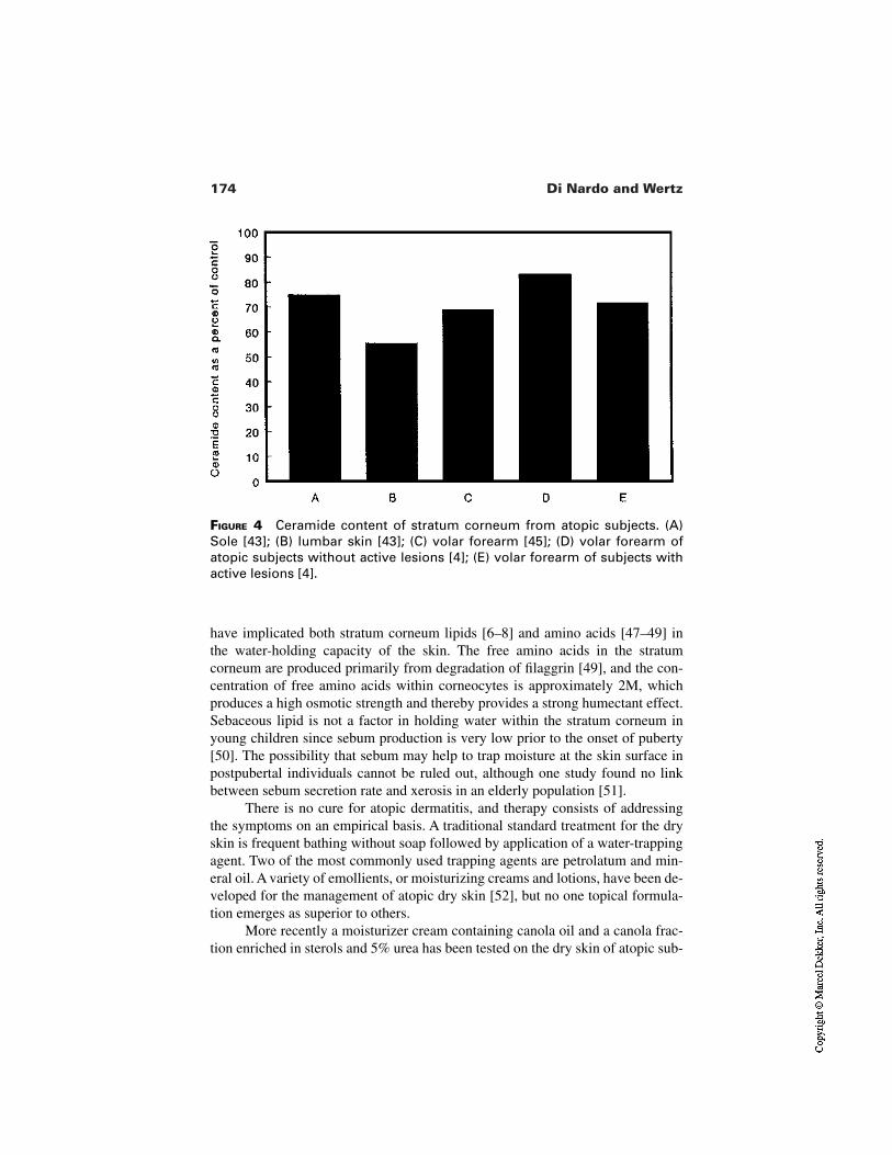
174 Di Nardo and Wertz
FIGURE 4 Ceramide content of stratum corneum from atopic subjects. (A)Sole [43]; (B) lumbar skin [43]; (C) volar forearm [45]; (D) volar forearm ofatopic subjects without active lesions [4]; (E) volar forearm of subjects withactive lesions [4].
have implicated both stratum corneum lipids [6–8] and amino acids [47–49] inthe water-holding capacity of the skin. The free amino acids in the stratumcorneum are produced primarily from degradation of filaggrin [49], and the con-centration of free amino acids within corneocytes is approximately 2M, whichproduces a high osmotic strength and thereby provides a strong humectant effect.Sebaceous lipid is not a factor in holding water within the stratum corneum inyoung children since sebum production is very low prior to the onset of puberty[50]. The possibility that sebum may help to trap moisture at the skin surface inpostpubertal individuals cannot be ruled out, although one study found no linkbetween sebum secretion rate and xerosis in an elderly population [51].
There is no cure for atopic dermatitis, and therapy consists of addressingthe symptoms on an empirical basis. A traditional standard treatment for the dryskin is frequent bathing without soap followed by application of a water-trappingagent. Two of the most commonly used trapping agents are petrolatum and min-eral oil. A variety of emollients, or moisturizing creams and lotions, have been de-veloped for the management of atopic dry skin [52], but no one topical formula-tion emerges as superior to others.
More recently a moisturizer cream containing canola oil and a canola frac-tion enriched in sterols and 5% urea has been tested on the dry skin of atopic sub-
175Atopic Dermatitis
jects [53]. Overall, after twice daily treatment for 20 days subjects showed in-creased water content as judged by skin capacitance and improved barrier func-tion as indicated by decreased trans-epidermal water loss. The subjects also be-came less susceptible to irritation by sodium dodecyl sulfate. In another recentstudy, it was found that after a mineral oil–based moisturizer containing glyceroland several humectants had been applied twice daily to atopic dry skin for 5 con-secutive days there was substantially increased high-frequency conductance, in-dicating increased water content, that persisted for several days after the cessationof moisturizer application [54]. Interestingly, the treatment had no effect on trans-epidermal water loss.
A second approach to treatment of atopic dermatitis is the use of topicalsteroids [55]. During times of flaring a mid-strength anti-inflammatory steroidointment should be applied within 3 min after bathing for best results. This shouldbe done twice daily. Corticosteroids represent the only medication proven to beeffective for management of atopic dermatitis.
A reasonable therapeutic strategy would be to attempt to normalize the de-fective lipid and humectant components of the xerotic stratum corneum; howev-er, most approaches to date have mainly relied upon providing a variety of topicalhumectants in a water-trapping vehicle. The more detailed knowledge of the na-ture of the lipid defects associated with atopic dry skin should make it possible toarrive at formulations that would normalize this aspect of the stratum corneum.The increased commercial availability of different types of ceramides [56] shouldalso facilitate this approach. In fact, it has been shown that topically applied lipidmixtures can accelerate barrier recovery after barrier disruption by differentmeans, and this effect depends upon the proportions of the lipids in the formula-tion [57]. More research is necessary on the free amino acids in stratum corneumof atopic subjects before it could become possible to exploit this area therapeuti-cally.
REFERENCES
1. Loden M. Biophysical properties of dry atopic and normal skin with special refer-ence to effects of skin care products. Acta Derm Venereol 1995; 192(Suppl):1–48.
2. Leung DYM, Rhodes AR, Geha RS, Schneider L, Ring J. Atopic dermatitis (atopiceczema). In: Fitzpatrick TB, Eisen AZ, Wolf K, Freedberg IM, Austen KF, eds. Der-matology in General Medicine. 4th ed. New York: McGraw-Hill, 1993:1543–1564.
3. Werner Y, Lindberg M. Transepidermal water loss in dry and clinically normal skinin patients with atopic dermatitis. Acta Derm Venereol 1985; 65:102–105.
4. Di Nardo A, Wertz P, Giannetti A, Seidenari S. Ceramide and cholesterol compositionof the skin of patients with atopic dermatitis. Acta Derm Venereol 1998; 78:27–30.
5. Thune P. Evaluation of the hydration and water-holding capacity in atopic skin andso-called dry skin. Acta Derm Venereol 1989; 144(Suppl):133–135.
176 Di Nardo and Wertz
6. Akimoto K, Yoshikawa N, Higaki Y, Kawashima M, Imokawa G. Quantitativeanalysis of stratum corneum lipids in xerosis and asteatotic eczema. J Invest Derma-tol 1993; 20:1–6.
7. Imokawa G, Kuno H, Kawai M. Stratum corneum lipids serve as a bound-watermodulator. J Invest Dermatol 1991; 96:845–851.
8. Imokawa G, Akasaki S, Minematsu Y, Kawai M. Importance of intercellular lipids inwater-retention properties of the stratum corneum: induction and recovery study ofsurfactant dry skin. Arch Dermatol Res 1989; 281:45–51.
9. Gray GM, Yardley HJ. Different populations of pig epidermal cells: isolation andlipid composition. J Lipid Res 1975; 16:441–447.
10. Schurer NY, Elias PM. The biochemistry and function of stratum corneum lipids.Adv Lipid Res 1991; 24:27–56.
11. Wertz PW. Lipids and barrier function of the skin. Acta Derm Venereol 2000;208(Suppl):1–5.
12. Wertz PW, Downing DT. Ceramides of pig epidermis: structure determination. JLipid Res 1983; 24:759–765.
13. Long SA, Wertz PW, Strauss JS, Downing DT. Human stratum corneum polar lipidsand desquamation. Arch Dermatol Res 1985; 277:284–287.
14. Robson KJ, Stewart ME, Michelsen S, Lazo ND, Downing DT. 6-Hydroxy-4-sphin-genine in human epidermal ceramides. J Lipid Res 1994; 35:2060–2068.
15. Stewart ME, Downing DT. A new 6-hydroxy-4-sphingenine–containing ceramide inhuman skin. J Lipid Res 1999; 40:1434–1439.
16. Motta SM, Monti M, Sesana S, Caputo R, Carelli S, Ghidoni R. Ceramide composi-tion of the psoriatic scale. Biochim Biophys Acta 1993; 1182:147–151.
17. Wertz PW, Downing DT. Epidermal lipids. In: Goldsmith L, ed. Physiology, Bio-chemistry and Molecular Biology of the Skin. New York: Oxford University Press,1991:205–238.
18. Attenborough D. Life on Earth. Boston: Little, Brown & Company, 1980.19. Nicolaides N. Skin lipids. II. Lipid class composition of samples from various
species and anatomical sites. J Am Oil Chem Soc 1965; 42:691–702.20. Gray GM, White RJ. Glycosphingolipids and ceramides in human and pig epider-
mis. J Invest Dermatol 1978; 70:336–341.21. Wertz PW, Downing DT. Covalently bound ω-hydroxyacylsphingosine in the stra-
tum corneum. Biochim Biophys Acta 1987; 917:108–111.22. Wertz PW, Madison KC, Downing DT. Covalently bound lipids of human stratum
corneum. J Invest Dermatol 1989; 91:109–111.23. Wertz PW, Downing DT. Composition and morphology of epidermal cyst lipids. J
Invest Dermatol 1987; 89:419–425.24. Wertz PW, van den Bergh BAI. The physical, chemical and functional properties of
lipids in the skin and other biological barriers. Chem Phys Lipids 1998; 91:85–96.25. Nicolaides N, Fu HC, Ansari MNA, Rice GR. The fatty acids of wax esters and sterol
esters from vernix caseosa and from human skin surface lipid. Lipids 1972;7:506–517.
26. Ranasinghe AW, Wertz PW, Downing DT, Mackenzie IC. Lipid composition of co-hesive and desquamated corneocytes from mouse ear skin. J Invest Dermatol 1986;86:187–190.
177Atopic Dermatitis
27. Shapiro LJ, Weiss R, Buxman MM, Vidgoff J, Dimond RL. Enzymatic basis of typi-cal X-linked ichthyosis. Lancet 1978; ii:756–757.
28. Williams ML. The ichthyoses—pathogenesis and prenatal diagnosis: a review of re-cent advances. Pediatr Sermatol 1983; 1:1–24.
29. Wertz PW, Squier CA. Cellular and molecular basis of barrier function in oral ep-ithelium. Crit Rev Therap Drug Carrier Syst 1991; 8:237–269.
30. Iwamori M, Iwamori Y, Ito N. Regulation of the activities of thrombin and plasminby cholesterol sulfate as a physiological inhibitor in human plasma. J Biochem 1999;125:594–601.
31. Ito N, Iwamori Y, Hanaoka K, Iwamori M. Inhibition of pancreatic elastase by sul-fated lipids in intestinal mucosa. J Biochem 1998; 123:107–114.
32. Iwamori M, Iwamori Y, Ito N. Sulfated lipids as inhibitors of pancreatic trypsin andchymotrypsin in epithelium of the mammalian digestive tract. Biochem Biophys ResComm 1997; 237:262–265.
33. Yardley HJ, Summerly R. Lipid composition and metabolism in normal and diseasedepidermis. Pharmacol Ther 1981; 13:357–383.
34. Mak VHW, Potts RO, Guy RH. Oleic acid concentration and effect in human stratumcorneum: non-invasive determination by attenuated total reflectance infrared spec-troscopy in vivo. J Control Rel 1990; 12:67–75.
35. Forslind B. A domain mosaic model of the skin barrier. Acta Derm Venereol 1994;74:1–6.
36. Madison KC, Swartzendruber DC, Wertz PW, Downing DT. Presence of intact inter-cellular lamellae in the upper layers of the stratum corneum. J Invest Dermatol 1987;88:714–718.
37. Swartzendruber DC, Manganaro A, Madison KC, Kremer M, Wertz PW, Squier CA.Organization of the intercellular spaces of porcine epidermal and palatal stratumcorneum: a quantitative study employing ruthenium tetroxide. Cell Tissue Res 1995;279:271–276.
38. Bouwstra JA, Gooris GS, Dubbelaar FE, Weerheim AM, Ijzerman AP, Ponec M.Role of ceramide 1 in the molecular organization of the stratum corneum lipids. JLipid Res 1998; 39:186–196.
39. Melnik B, Hollmann J, Plewig G. Decreased stratum corneum ceramides in atopicindividuals—a pathobiochemical factor in xerosis? Br J Dermatol 1988;119:547–549.
40. Holman RT. Essential fatty acid deficiency. Prog Chem Fats Other Lipids 1968;9:275–248.
41. Wertz PW, Cho ES, Downing DT. Effects of essential fatty acid deficiency on theepidermal sphingolipids of the rat. Biochim Biophys Acta 1983; 753:350–355.
42. Melton JL, Wertz PW, Swartzendruber DC, Downing DT. Effects of essential fattyacid deficiency on epidermal ω-acylsphingolipids and transepidermal water loss inyoung pigs. Biochim Biophys Acta 1987; 921:191–197.
43. Melnik B, Hollmann J, Hofmann U, Yuh M-S, Plewig G. Lipid composition of outerstratum corneum and nails in atopic and control subjects. Arch Dermatol Res 1990;282:549–551.
44. Yamamoto A, Serizawa S, Ito M, Sato Y. Stratum corneum lipid abnormalities inatopic dermatitis. Arch Dermatol Res 1991; 283:219–223.
178 Di Nardo and Wertz
45. Matsumoto M, Umemoto N, Sugiura H, Uehara M. Difference in ceramide composi-tion between “dry” and “normal” skin in patients with atopic dermatitis. Acta DermVenereol 1999; 79:246–247.
46. Hara J, Higuchi K, Okamoto R, Kawashima M, Imokawa G. High-expression ofsphingomyelin deacylase is an important determinant of ceramide deficiency leadingto barrier disruption in atopic dermatitis. J Invest Dermatol 2000; 115:406–413.
47. Tanaka M, Okada M, Zhen YX, Inamura N, Kitano T, Shirai S, Sakamoto K, Inamu-ra T, Tagami H. Decreased hydration state of the stratum corneum and reducedamino acid content of the skin surface in patients with seasonal allergic rhinitis. Br JDermatol 1998; 139:618–621.
48. Yamamura T, Tezuka T. The water-holding capacity of the stratum corneum mea-sured by 1H-NMR. J Invest Dermatol 1989; 93:160–164.
49. Scott IR, Harding CR. Filaggrin breakdown to water binding compounds during de-velopment of the rat stratum corneum is controlled by the water activity of the envi-ronment. Dev Biol 1986; 115:84–92.
50. Strauss JS, Pochi PE. The hormonal control of the pilosebaceous unit. In: Toda K, ed.Biology and Disease of the Hair. Baltimore: University Park Press, 1975:231–245.
51. Frantz RA, Kinney CK, Downing DT. Variables associated with skin dryness in theelderly. Nursing Res 1986; 35:98–100.
52. Burr S. Emollients for managing dry skin conditions. Prof Nurse 1999; 15:43–48.53. Loden M, Anderson AC, Lindberg M. Improvement in skin barrier function in pa-
tients with atopic dermatitis after treatment with a moisturizing cream (Canoderm).Br J Dermatol 1999; 140:264–267.
54. Tabata N, O’Goshi K, Zhen YX, Kligman AM, Tagami H. Biophysical assessment ofpersistent effects of moisturizers after their daily applications: evaluation of cor-neotherapy. Dermatology 2000; 200:308–313.
55. Sidbury R, Hanifin JM. Old, new, and emerging therapies for atopic dermatitis. Der-matol Clin 2000; 18:1–9.
56. Michniak BB, Wertz PW. Ceramides and lipids. In: Barel A, Maibach HI, Paye M,eds. Handbook of Cosmetic Science and Technology. New York: Marcel Dekker2000:45–56.
57. Zettersten EM, Ghadially R, Feingold KR, Crumrine D, Elias PM. Optimal ratios oftopical stratum corneum lipids improve barrier recovery in chronologically agedskin. J Am Acad Dermatol 1997; 37:403–408.
10Psoriasis and Ichthyoses
Ruby GhadiallyVeterans Administration Medical Center and University ofCalifornia School of Medicine, San Francisco, California
1 INTRODUCTION
Dramatic changes in epidermal differentiation and stratum corneum structure andfunction occur in psoriasis and in the ichthyoses. In this chapter the differences inepidermal structure, composition, and function will be discussed, together with asummary of relevant topical technologies to improve the skin conditions dis-cussed.
Mammalian stratum corneum comprises a two-compartment system oflipid-depleted corneocytes embedded in a lipid-enriched intercellular matrix (re-viewed in Ref. 1). These intercellular lipids are organized into a series of broadlamellar bilayers that regulate permeability barrier function and participate in thecohesion and desquamation of the stratum corneum (reviewed in Ref. 2). The dis-eases discussed in this chapter are all characterized by a mild to severe compro-mise in epidermal permeability barrier function, basally or in response to stressesto the barrier.
In normal stratum corneum, poorly understood changes occur in the stra-tum corneum interstices that lead to the orderly detachment of individual corneo-cytes at the skin surface. Alterations in lipid composition [3], the physical-chem-ical state of the lamellar bilayers [4], and degradation of nonlipid constituentssuch as desmosomes [5] have all been implicated as mediators of normal desqua-
179
180 Ghadially
mation. The extent to which one or more of these processes is responsible for theabnormal desquamation of the disorders of cornification is still not known [2,6].
Epidermal permeability barrier integrity requires the organization of stra-tum corneum lipids into extracellular lamellar bilayers, following the secretion ofepidermal lamellar body contents at the stratum granulosum–stratum corneum in-terface [6]. With the advent of ruthenium tetroxide postfixation it is possible toobtain ultrastructural images of the stratum corneum interstices on a routine basis[7–10], and a detailed tableau is emerging of the intercellular bilayer system innormal and diseased stratum corneum.
Ultrastructural examination of normal stratum corneum reveals that thelamellae within the intercellular domains of normal human stratum corneum ex-hibit a similar organization and substructure to previous descriptions of porcine[7] and murine [8] stratum corneum. In optimal cross-sections, these membranescan be seen to comprise three types of electron-lucent lamellae that alternate witha single type of electron-dense lamella (Fig. 1). From the corneocyte envelopeoutward, the electron-lucent lamellae comprise, first, a continuous sheet immedi-ately exterior to the cornified envelope [7,8,11]. The succeeding lamellae are or-ganized external to adjacent lamellae with the center of the interrupted, lucentlamellae serving as the plane of symmetry. Each series of four electron-lucentlamellae alternating with five electron-dense lamellae, comprises the basic unit(Fig. 1 inset). At many points in the interstices this basic unit expands incremen-tally and suddenly by the addition of arrays of continuous electron-dense and -lu-cent lamellae. A “doublet” comprises two basic units minus one interrupted, elec-tron-lucent lamella and two electron-dense lamellae, which result from sharing ofthese structures by two adjacent basic units. “Triplets” are the largest units ob-served in normal human stratum corneum.
Staining of lamellar bodies in the stratum granulosum with standard osmi-um tetroxide fixation provides excellent preservation of lamellar body structure.Briefly, cross-sectional images of lamellar bodies in normal epidermis demon-strated a trilaminar limiting membrane, and internal lamellar disklike structures,consisting of prominent dense lamellae separated by an electron-lucent band anddivided centrally by a minor, striated electron-dense band (Fig. 2 inset).
Acute perturbations of the permeability barrier; e.g., solvent applications ortape-stripping, stimulate a sequence of homeostatic mechanisms, including (1)rapid secretion of pre-formed lamellar body contents; (2) generation of nascentlamellar bodies; (3) accelerated intercellular deposition of newly formed lamellarbody contents; and (4) extracellular processing of lamellar body contents by co-localized hydrolytic enzymes into lamellar basic unit structures [12]. The lamel-lar body secretory response to barrier disruption is both fueled by and requires aburst in lipid synthesis [13–15]. Likewise, in the essential fatty acid–deficientmouse (a chronic barrier perturbation model often used as an analog for psoria-sis), increased numbers of defective lamellar bodies, decreased extracellular
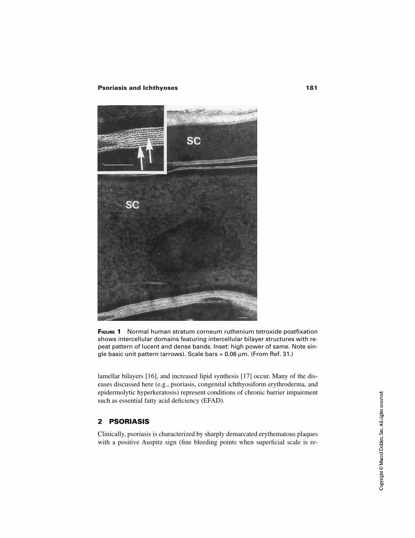
181Psoriasis and Ichthyoses
FIGURE 1 Normal human stratum corneum ruthenium tetroxide postfixationshows intercellular domains featuring intercellular bilayer structures with re-peat pattern of lucent and dense bands. Inset: high power of same. Note sin-gle basic unit pattern (arrows). Scale bars = 0.06 µm. (From Ref. 31.)
lamellar bilayers [16], and increased lipid synthesis [17] occur. Many of the dis-eases discussed here (e.g., psoriasis, congenital ichthyosiform erythroderma, andepidermolytic hyperkeratosis) represent conditions of chronic barrier impairmentsuch as essential fatty acid deficiency (EFAD).
2 PSORIASIS
Clinically, psoriasis is characterized by sharply demarcated erythematous plaqueswith a positive Auspitz sign (fine bleeding points when superficial scale is re-
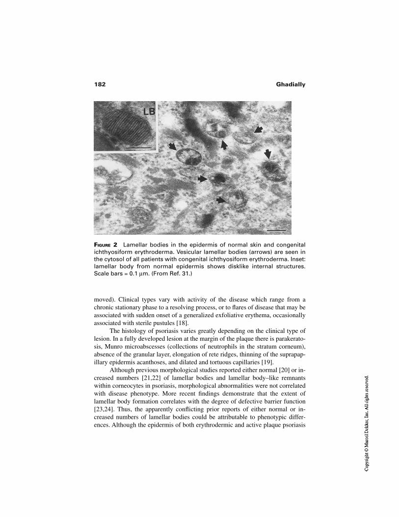
182 Ghadially
FIGURE 2 Lamellar bodies in the epidermis of normal skin and congenitalichthyosiform erythroderma. Vesicular lamellar bodies (arrows) are seen inthe cytosol of all patients with congenital ichthyosiform erythroderma. Inset:lamellar body from normal epidermis shows disklike internal structures.Scale bars = 0.1 µm. (From Ref. 31.)
moved). Clinical types vary with activity of the disease which range from achronic stationary phase to a resolving process, or to flares of disease that may beassociated with sudden onset of a generalized exfoliative erythema, occasionallyassociated with sterile pustules [18].
The histology of psoriasis varies greatly depending on the clinical type oflesion. In a fully developed lesion at the margin of the plaque there is parakerato-sis, Munro microabscesses (collections of neutrophils in the stratum corneum),absence of the granular layer, elongation of rete ridges, thinning of the suprapap-illary epidermis acanthoses, and dilated and tortuous capillaries [19].
Although previous morphological studies reported either normal [20] or in-creased numbers [21,22] of lamellar bodies and lamellar body–like remnantswithin corneocytes in psoriasis, morphological abnormalities were not correlatedwith disease phenotype. More recent findings demonstrate that the extent oflamellar body formation correlates with the degree of defective barrier function[23,24]. Thus, the apparently conflicting prior reports of either normal or in-creased numbers of lamellar bodies could be attributable to phenotypic differ-ences. Although the epidermis of both erythrodermic and active plaque psoriasis
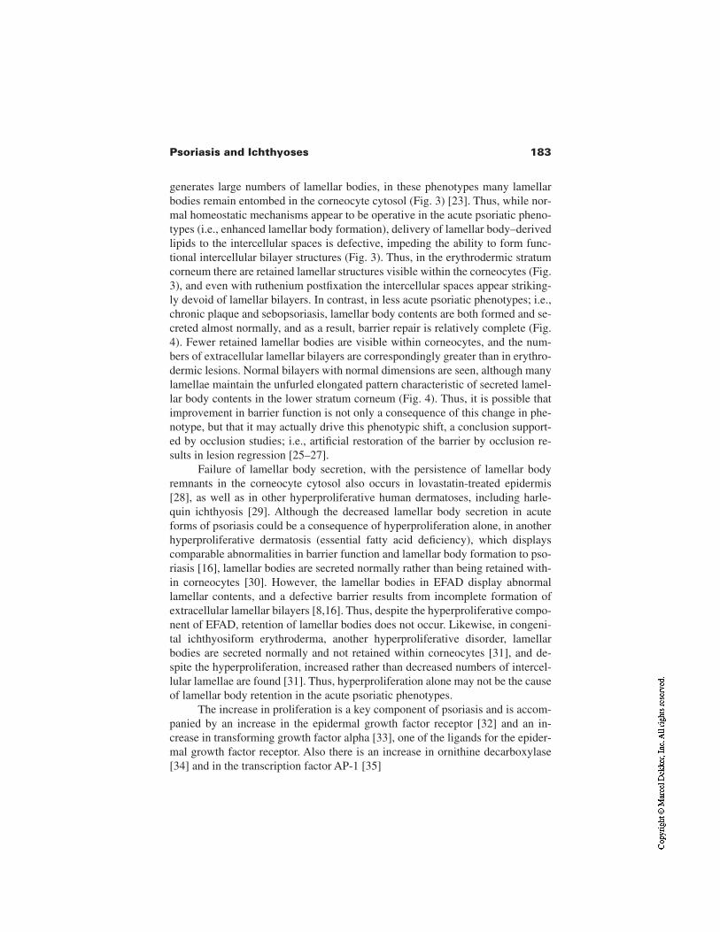
183Psoriasis and Ichthyoses
generates large numbers of lamellar bodies, in these phenotypes many lamellarbodies remain entombed in the corneocyte cytosol (Fig. 3) [23]. Thus, while nor-mal homeostatic mechanisms appear to be operative in the acute psoriatic pheno-types (i.e., enhanced lamellar body formation), delivery of lamellar body–derivedlipids to the intercellular spaces is defective, impeding the ability to form func-tional intercellular bilayer structures (Fig. 3). Thus, in the erythrodermic stratumcorneum there are retained lamellar structures visible within the corneocytes (Fig.3), and even with ruthenium postfixation the intercellular spaces appear striking-ly devoid of lamellar bilayers. In contrast, in less acute psoriatic phenotypes; i.e.,chronic plaque and sebopsoriasis, lamellar body contents are both formed and se-creted almost normally, and as a result, barrier repair is relatively complete (Fig.4). Fewer retained lamellar bodies are visible within corneocytes, and the num-bers of extracellular lamellar bilayers are correspondingly greater than in erythro-dermic lesions. Normal bilayers with normal dimensions are seen, although manylamellae maintain the unfurled elongated pattern characteristic of secreted lamel-lar body contents in the lower stratum corneum (Fig. 4). Thus, it is possible thatimprovement in barrier function is not only a consequence of this change in phe-notype, but that it may actually drive this phenotypic shift, a conclusion support-ed by occlusion studies; i.e., artificial restoration of the barrier by occlusion re-sults in lesion regression [25–27].
Failure of lamellar body secretion, with the persistence of lamellar bodyremnants in the corneocyte cytosol also occurs in lovastatin-treated epidermis[28], as well as in other hyperproliferative human dermatoses, including harle-quin ichthyosis [29]. Although the decreased lamellar body secretion in acuteforms of psoriasis could be a consequence of hyperproliferation alone, in anotherhyperproliferative dermatosis (essential fatty acid deficiency), which displayscomparable abnormalities in barrier function and lamellar body formation to pso-riasis [16], lamellar bodies are secreted normally rather than being retained with-in corneocytes [30]. However, the lamellar bodies in EFAD display abnormallamellar contents, and a defective barrier results from incomplete formation ofextracellular lamellar bilayers [8,16]. Thus, despite the hyperproliferative compo-nent of EFAD, retention of lamellar bodies does not occur. Likewise, in congeni-tal ichthyosiform erythroderma, another hyperproliferative disorder, lamellarbodies are secreted normally and not retained within corneocytes [31], and de-spite the hyperproliferation, increased rather than decreased numbers of intercel-lular lamellae are found [31]. Thus, hyperproliferation alone may not be the causeof lamellar body retention in the acute psoriatic phenotypes.
The increase in proliferation is a key component of psoriasis and is accom-panied by an increase in the epidermal growth factor receptor [32] and an in-crease in transforming growth factor alpha [33], one of the ligands for the epider-mal growth factor receptor. Also there is an increase in ornithine decarboxylase[34] and in the transcription factor AP-1 [35]
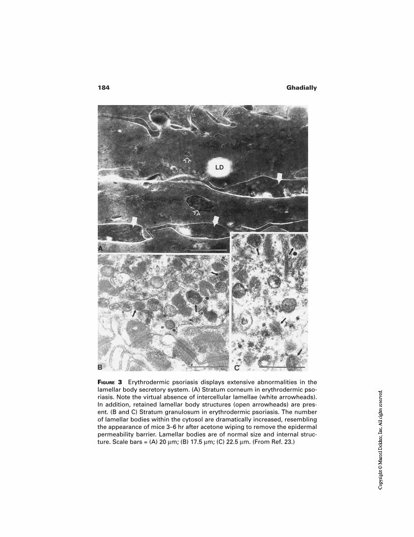
184 Ghadially
FIGURE 3 Erythrodermic psoriasis displays extensive abnormalities in thelamellar body secretory system. (A) Stratum corneum in erythrodermic pso-riasis. Note the virtual absence of intercellular lamellae (white arrowheads).In addition, retained lamellar body structures (open arrowheads) are pres-ent. (B and C) Stratum granulosum in erythrodermic psoriasis. The numberof lamellar bodies within the cytosol are dramatically increased, resemblingthe appearance of mice 3–6 hr after acetone wiping to remove the epidermalpermeability barrier. Lamellar bodies are of normal size and internal struc-ture. Scale bars = (A) 20 µm; (B) 17.5 µm; (C) 22.5 µm. (From Ref. 23.)
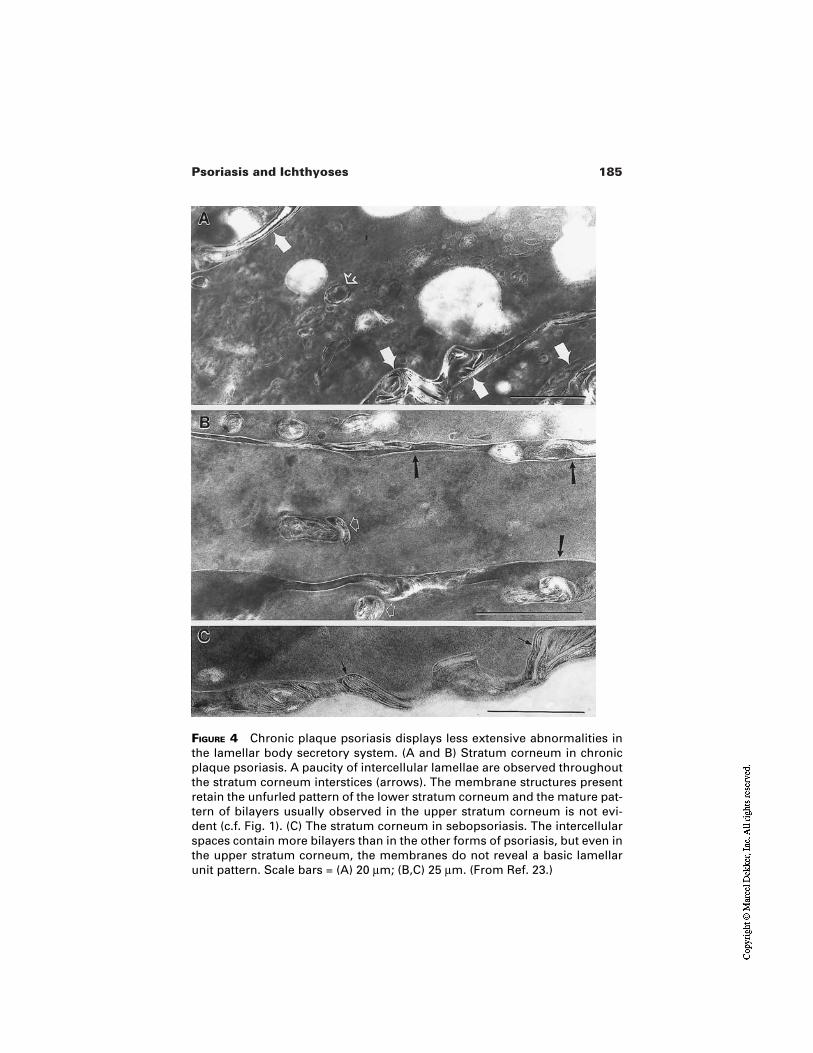
185Psoriasis and Ichthyoses
FIGURE 4 Chronic plaque psoriasis displays less extensive abnormalities inthe lamellar body secretory system. (A and B) Stratum corneum in chronicplaque psoriasis. A paucity of intercellular lamellae are observed throughoutthe stratum corneum interstices (arrows). The membrane structures presentretain the unfurled pattern of the lower stratum corneum and the mature pat-tern of bilayers usually observed in the upper stratum corneum is not evi-dent (c.f. Fig. 1). (C) The stratum corneum in sebopsoriasis. The intercellularspaces contain more bilayers than in the other forms of psoriasis, but even inthe upper stratum corneum, the membranes do not reveal a basic lamellarunit pattern. Scale bars = (A) 20 µm; (B,C) 25 µm. (From Ref. 23.)
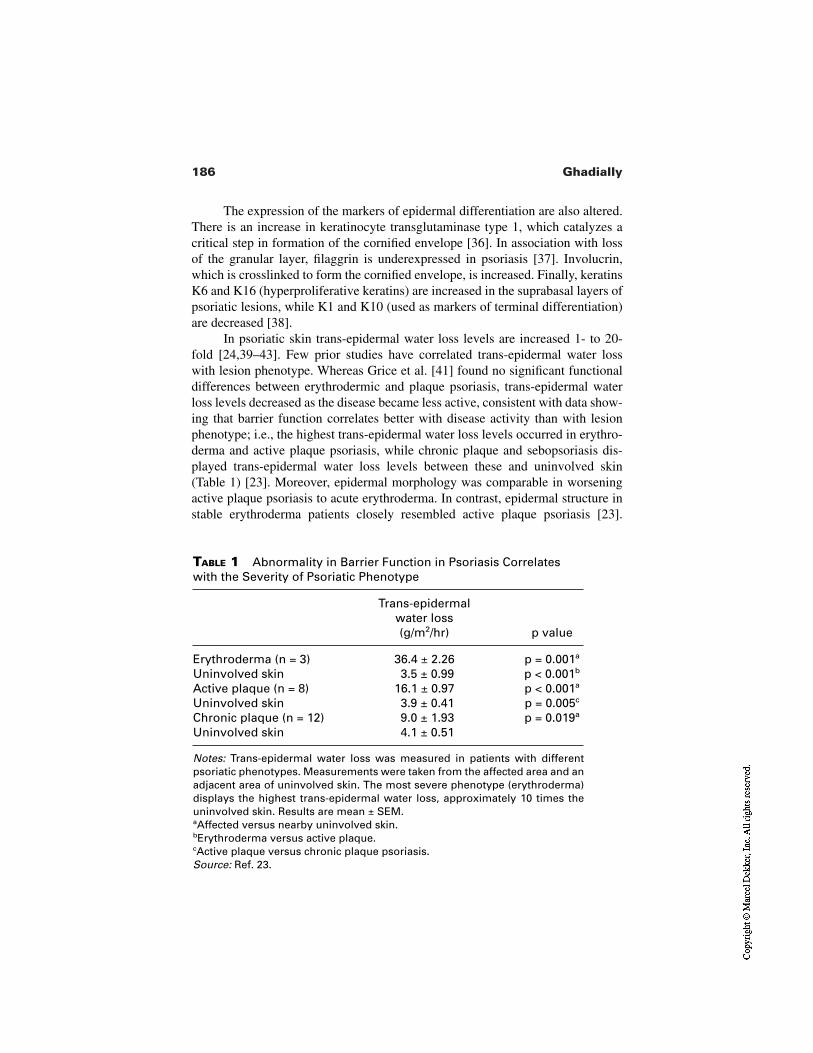
186 Ghadially
TABLE 1 Abnormality in Barrier Function in Psoriasis Correlateswith the Severity of Psoriatic Phenotype
Trans-epidermal water loss (g/m2/hr) p value
Erythroderma (n = 3) 36.4 ± 2.26 p = 0.001a
Uninvolved skin 3.5 ± 0.99 p < 0.001b
Active plaque (n = 8) 16.1 ± 0.97 p < 0.001a
Uninvolved skin 3.9 ± 0.41 p = 0.005c
Chronic plaque (n = 12) 9.0 ± 1.93 p = 0.019a
Uninvolved skin 4.1 ± 0.51
Notes: Trans-epidermal water loss was measured in patients with differentpsoriatic phenotypes. Measurements were taken from the affected area and anadjacent area of uninvolved skin. The most severe phenotype (erythroderma)displays the highest trans-epidermal water loss, approximately 10 times theuninvolved skin. Results are mean ± SEM.aAffected versus nearby uninvolved skin.bErythroderma versus active plaque.cActive plaque versus chronic plaque psoriasis.Source: Ref. 23.
The expression of the markers of epidermal differentiation are also altered.There is an increase in keratinocyte transglutaminase type 1, which catalyzes acritical step in formation of the cornified envelope [36]. In association with lossof the granular layer, filaggrin is underexpressed in psoriasis [37]. Involucrin,which is crosslinked to form the cornified envelope, is increased. Finally, keratinsK6 and K16 (hyperproliferative keratins) are increased in the suprabasal layers ofpsoriatic lesions, while K1 and K10 (used as markers of terminal differentiation)are decreased [38].
In psoriatic skin trans-epidermal water loss levels are increased 1- to 20-fold [24,39–43]. Few prior studies have correlated trans-epidermal water losswith lesion phenotype. Whereas Grice et al. [41] found no significant functionaldifferences between erythrodermic and plaque psoriasis, trans-epidermal waterloss levels decreased as the disease became less active, consistent with data show-ing that barrier function correlates better with disease activity than with lesionphenotype; i.e., the highest trans-epidermal water loss levels occurred in erythro-derma and active plaque psoriasis, while chronic plaque and sebopsoriasis dis-played trans-epidermal water loss levels between these and uninvolved skin(Table 1) [23]. Moreover, epidermal morphology was comparable in worseningactive plaque psoriasis to acute erythroderma. In contrast, epidermal structure instable erythroderma patients closely resembled active plaque psoriasis [23].
187Psoriasis and Ichthyoses
These results suggest that barrier repair mechanisms are operative in psoriasis;and they appear to be, in part, successful at normalizing barrier function, perhapsleading to more chronic psoriatic phenotypes. Likewise, with artificial barrierrestoration by occlusion alone, psoriatic lesions usually regress [25–27].
Much indirect evidence, including the data summarized here, supports thehypothesis that the primary trigger for psoriasis may arise in the epidermis, andthat the disease-specific inflammatory infiltrate may be recruited secondarily[44,45]. Psoriatic epidermis transplanted onto nude mice retains its psoriatic mor-phology, as well as its increased labeling index [46]. A similar persistence of phe-notype occurs in transplants of the flaky skin mouse model of psoriasis [47].Moreover, although some studies suggest that psoriatic fibroblasts drive the dis-ease [48], others have found that psoriatic fibroblasts do not direct hyperprolifer-ation, nor do normal fibroblasts inhibit psoriatic hyperproliferation [49]. Further-more, barrier abrogation stimulates DNA synthesis [50], as well as provoking arapid increase of both epidermal cytokine mRNA and protein generation [44,51].Finally, Nickoloff et al. [45] showed that epidermal production of cytokines fol-lowing barrier abrogation precedes movement of inflammatory cells from the cir-culation into the dermis or epidermis. Together, these findings suggest that thedermal inflammatory components of psoriasis may be recruited subsequent to pri-mary events arising in the epidermis. These findings also are consistent with theoccurrence of the Koebner phenomenon [52] and the observation that occlusionalone clears many psoriatic lesions [25–27].
3 ICHTHYOSIS VULGARIS
Ichthyosis vulgaris, the most common of the ichthyotic conditions discussed here,is an autosomal dominant condition that usually starts in childhood and presentsas fine white scales on the extensor surfaces of the extremities and the trunk.
Ichthyosis vulgaris is characterized histologically by mild hyperkeratosisand reduced or absent keratohyalin granules. Ultrastructural evaluation hasshown that although the stratum corneum is thicker than normal, and keratohyalingranules are absent, the typical keratin pattern of normal skin is seen suggestingthat filaggrin is not essential for keratin filament aggregation [53]. There is abnor-mal persistence of desmosomes in the stratum corneum [54].
Biochemically, profilaggrin (a major component of keratohyaline granules)and filaggrin are reduced or absent. Little profilaggrin mRNA was detected by insitu hybridization in vivo, and in keratinocytes profilaggrin was less than 10% ofnormal, while the mRNA was 30–60% of controls. Furthermore, expression ofkeratin K1 and loricrin (other markers of epidermal differentiation) were not af-fected [55]. The degree of biochemical abnormality correlated with the ultrastruc-tural quantitation and with the severity of the clinical disorder [53]. Trypsin-likeand chymotrypsin-like serine proteases are involved in the degradation of
188 Ghadially
desmoglein [56], a transmembrane protein within desmosomes. The enzymaticactivities of trypsin-like and chymotrypsin-like serine proteases were significant-ly decreased in ichthyosis vulgaris [56].
Basal trans-epidermal water loss shows a small but significant increase inichthyosis vulgaris [40,57]. Furthermore, a loss of pH gradient occurs, reflectedby a higher skin surface pH, and a neutral pH of 7 obtained with removal of onlyhalf of the stratum corneum, as compared to normal skin where the “acid mantle”penetrates deep into the skin [58]. This could be explained by the depletion ofacidic proteins urocanic acid and pyrrolidone carboxylic acid due to filaggrin de-ficiency, which results in a delay in the accumulation of protons and moves thepH gradient outward in ichthyosis vulgaris [58].
4 RECESSIVE X-LINKED ICHTHYOSIS
Recessive X-linked ichthyosis is characterized by a generalized desquamation oflarge, adherent, dark brown scales, more extensive on the extensor aspects of thelimbs. Extracutaneous manifestations include corneal opacities and cryp-torchidism [59]. This condition is caused by a deficit in steroid sulfatase [60,61].The deletion or mutation of the gene encoding for the enzyme steroid sulfatasehas been localized to the distal short arm of the X chromosome (Xp22.3) [62].
Histology shows compact hyperkeratosis, with a normal or slightly thick-ened stratum granulosum [59]. This is a retention hyperkeratosis with a normalrate of cell turnover [59]. Ultrastructural studies reveal an increase in the numberand volume of keratohyalin granules. Desmosomal disks are visible even in themost superficial layers of the epidermis, suggesting increased intercellular cohe-siveness. Cells of the stratum corneum contain large numbers of melonosomes,probably due to decreased degradation, and resulting in the dark scale seen clini-cally [63,64]. Examination of the intercellular lamellar domains of the stratumcorneum [65] reveals lamellae that are fragmented and disrupted, with extensivenonlamellar domains within the extracellular space (Fig. 5). In a study of lipidmixtures using small angle x-ray diffraction, it was found that both an increase inpH (7.4 at the stratum granulosum–stratum corneum interface versus 5 at the skinsurface) and an increase in cholesterol sulfate promote the formation of a normallamellar phase as seen in vivo, suggesting that cholesterol sulfate may be requiredto dissolve cholesterol in the lamellar phases and to stabilize stratum corneumlipid organization. Therefore, a drop in cholesterol sulfate content in the superfi-cial layers of the stratum corneum is expected to destabilize the lipid lamellarphases and facilitate the desquamation process [66].
The epidermal permeability barrier is slightly impaired in recessive X-linked ichthyosis despite the great hyperkeratosis. Whereas a single study of 13patients with recessive X-linked ichthyosis found no statistical change in basaltrans-epidermal water loss [67], basal trans-epidermal water loss was slightly butsignificantly increased in other studies [40,57,65]. This increase in trans-epider-
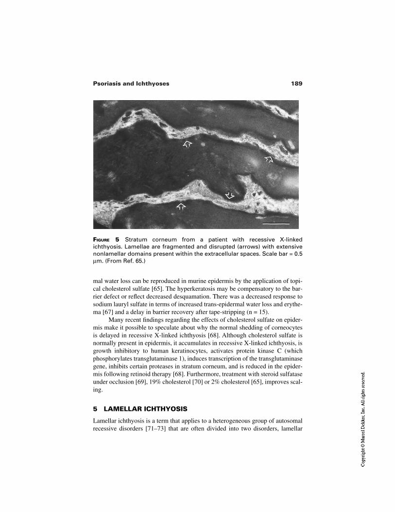
189Psoriasis and Ichthyoses
FIGURE 5 Stratum corneum from a patient with recessive X-linkedichthyosis. Lamellae are fragmented and disrupted (arrows) with extensivenonlamellar domains present within the extracellular spaces. Scale bar = 0.5µm. (From Ref. 65.)
mal water loss can be reproduced in murine epidermis by the application of topi-cal cholesterol sulfate [65]. The hyperkeratosis may be compensatory to the bar-rier defect or reflect decreased desquamation. There was a decreased response tosodium lauryl sulfate in terms of increased trans-epidermal water loss and erythe-ma [67] and a delay in barrier recovery after tape-stripping (n = 15).
Many recent findings regarding the effects of cholesterol sulfate on epider-mis make it possible to speculate about why the normal shedding of corneocytesis delayed in recessive X-linked ichthyosis [68]. Although cholesterol sulfate isnormally present in epidermis, it accumulates in recessive X-linked ichthyosis, isgrowth inhibitory to human keratinocytes, activates protein kinase C (whichphosphorylates transglutaminase 1), induces transcription of the transglutaminasegene, inhibits certain proteases in stratum corneum, and is reduced in the epider-mis following retinoid therapy [68]. Furthermore, treatment with steroid sulfataseunder occlusion [69], 19% cholesterol [70] or 2% cholesterol [65], improves scal-ing.
5 LAMELLAR ICHTHYOSIS
Lamellar ichthyosis is a term that applies to a heterogeneous group of autosomalrecessive disorders [71–73] that are often divided into two disorders, lamellar
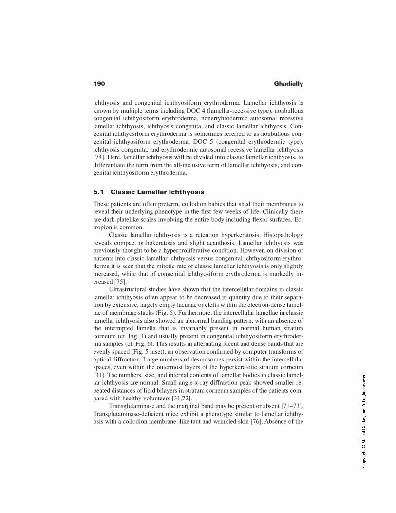
190 Ghadially
ichthyosis and congenital ichthyosiform erythroderma. Lamellar ichthyosis isknown by multiple terms including DOC 4 (lamellar-recessive type), nonbullouscongenital ichthyosiform erythroderma, nonertyhrodermic autosomal recessivelamellar ichthyosis, ichthyosis congenita, and classic lamellar ichthyosis. Con-genital ichthyosiform erythroderma is sometimes referred to as nonbullous con-genital ichthyosiform erythroderma, DOC 5 (congenital erythrodermic type),ichthyosis congenita, and erythrodermic autosomal recessive lamellar ichthyosis[74]. Here, lamellar ichthyosis will be divided into classic lamellar ichthyosis, todifferentiate the term from the all-inclusive term of lamellar ichthyosis, and con-genital ichthyosiform erythroderma.
5.1 Classic Lamellar Ichthyosis
These patients are often preterm, collodion babies that shed their membranes toreveal their underlying phenotype in the first few weeks of life. Clinically thereare dark platelike scales involving the entire body including flexor surfaces. Ec-tropion is common.
Classic lamellar ichthyosis is a retention hyperkeratosis. Histopathologyreveals compact orthokeratosis and slight acanthosis. Lamellar ichthyosis waspreviously thought to be a hyperproliferative condition. However, on division ofpatients into classic lamellar ichthyosis versus congenital ichthyosiform erythro-derma it is seen that the mitotic rate of classic lamellar ichthyosis is only slightlyincreased, while that of congenital ichthyosiform erythroderma is markedly in-creased [75].
Ultrastructural studies have shown that the intercellular domains in classiclamellar ichthyosis often appear to be decreased in quantity due to their separa-tion by extensive, largely empty lacunae or clefts within the electron-dense lamel-lae of membrane stacks (Fig. 6). Furthermore, the intercellular lamellae in classiclamellar ichthyosis also showed an abnormal banding pattern, with an absence ofthe interrupted lamella that is invariably present in normal human stratumcorneum (cf. Fig. 1) and usually present in congenital ichthyosiform erythroder-ma samples (cf. Fig. 6). This results in alternating lucent and dense bands that areevenly spaced (Fig. 5 inset), an observation confirmed by computer transforms ofoptical diffraction. Large numbers of desmosomes persist within the intercellularspaces, even within the outermost layers of the hyperkeratotic stratum corneum[31]. The numbers, size, and internal contents of lamellar bodies in classic lamel-lar ichthyosis are normal. Small angle x-ray diffraction peak showed smaller re-peated distances of lipid bilayers in stratum corneum samples of the patients com-pared with healthy volunteers [31,72].
Transglutaminase and the marginal band may be present or absent [71–73].Transglutaminase-deficient mice exhibit a phenotype similar to lamellar ichthy-osis with a collodion membrane–like taut and wrinkled skin [76]. Absence of the
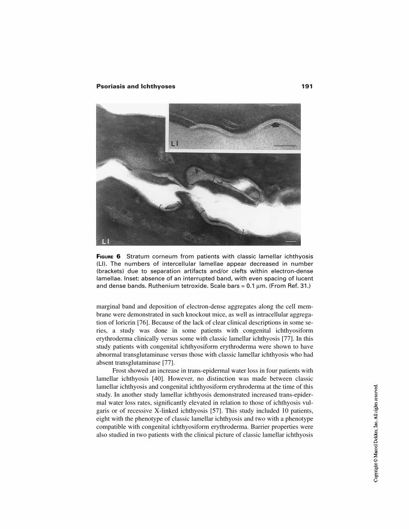
191Psoriasis and Ichthyoses
FIGURE 6 Stratum corneum from patients with classic lamellar ichthyosis(LI). The numbers of intercellular lamellae appear decreased in number(brackets) due to separation artifacts and/or clefts within electron-denselamellae. Inset: absence of an interrupted band, with even spacing of lucentand dense bands. Ruthenium tetroxide. Scale bars = 0.1 µm. (From Ref. 31.)
marginal band and deposition of electron-dense aggregates along the cell mem-brane were demonstrated in such knockout mice, as well as intracellular aggrega-tion of loricrin [76]. Because of the lack of clear clinical descriptions in some se-ries, a study was done in some patients with congenital ichthyosiformerythroderma clinically versus some with classic lamellar ichthyosis [77]. In thisstudy patients with congenital ichthyosiform erythroderma were shown to haveabnormal transglutaminase versus those with classic lamellar ichthyosis who hadabsent transglutaminase [77].
Frost showed an increase in trans-epidermal water loss in four patients withlamellar ichthyosis [40]. However, no distinction was made between classiclamellar ichthyosis and congenital ichthyosiform erythroderma at the time of thisstudy. In another study lamellar ichthyosis demonstrated increased trans-epider-mal water loss rates, significantly elevated in relation to those of ichthyosis vul-garis or of recessive X-linked ichthyosis [57]. This study included 10 patients,eight with the phenotype of classic lamellar ichthyosis and two with a phenotypecompatible with congenital ichthyosiform erythroderma. Barrier properties werealso studied in two patients with the clinical picture of classic lamellar ichthyosis
192 Ghadially
and one with the picture of congenital ichthyosiform erythroderma (a sibling ofone of the classic lamellar ichthyosis type subjects) [72]. Trans-epidermal waterloss was significantly increased in all. Stratum corneum lipid profiles showed sig-nificant differences in the relative ceramide fractions in these patients [72].
5.2 Congenital Ichthyosiform Erythroderma
Often born as collodion babies, these patients have erthroderma and fine whitescales. They may also have ectropion. In contrast to classic lamellar ichthyosis,this is a hyperproliferative state.
Histology shows features of hyperproliferation including some parakerato-sis (not seen in classic lamellar ichthyosis) and much greater acanthosis than thatseen in classic lamellar ichthyosis [78]. Also in contrast to classic lamellarichthyosis, the degree of hyperkeratosis is much less severe. Ultrastructurally, us-ing ruthenium tetroxide postfixation, chracteristic findings for congenitalichthyosiform erythroderma (CIE) included (1) foci containing excessive num-bers of lamellae in stacks (Fig. 6; cf. Fig. 1); (2) a predominance of incompletelyformed and/or disorganized lamellar arrays (Fig. 6C); (3) shortened arrays oflamellar body–derived membranes (Fig. 6C); (4) variations in the substructure ofindividual lamellae, both within the lucent and dense bands (Fig. 6D,E) and ab-normal interlamellar dimensions by x-ray diffraction; (5) electron-lucent do-mains, presumably representing nonlamellar phases because of the presence offlocculent, amorphous material (Fig. 6B) (such domains occurred interspersed be-tween stacks of lamellar bilayers); and (6) desmosomes, which normally deterio-rate above the first six to eight layers of normal stratum corneum, persisting inabundance in all of the 25-plus layers of the stratum corneum [31]. Increasednumbers of lamellar bodies of decreased size were observed in the stratum gran-ulosum in all CIE patients. Moreover, the internal contents of lamellar bodiesfrom congenital ichthyosiform erythroderma epidermis were distinctly abnormal,most appearing empty or containing only fragments of lamellar structures (Fig.2), creating a vacuolated appearance [31].
Studies of barrier function have been in combination with patients withclassic lamellar ichthyosis (see preceding).
6 EPIDERMOLYTIC HYPERKERATOSIS
Epidermolytic hyperkeratosis (also known as bullous congenital ichthyosiformerythroderma) is an autosomal dominant condition, although 50% of cases aresporadic and probably new mutations [74]. Patients develop blistering on the ex-tensor surfaces and later localized areas of severe hyperkeratosis. However, epi-dermolytic hyperkeratosis is clinically heterogeneous [79]. After studying 52 pa-tients with epidermolytic hyperkeratosis DiGiovanna and Bale divided the
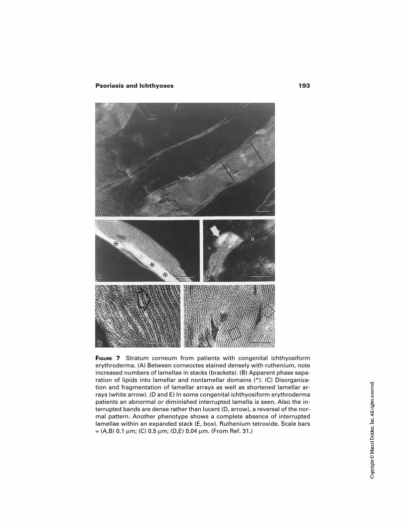
193Psoriasis and Ichthyoses
FIGURE 7 Stratum corneum from patients with congenital ichthyosiformerythroderma. (A) Between corneoctes stained densely with ruthenium, noteincreased numbers of lamellae in stacks (brackets). (B) Apparent phase sepa-ration of lipids into lamellar and nonlamellar domains (*). (C) Disorganiza-tion and fragmentation of lamellar arrays as well as shortened lamellar ar-rays (white arrow). (D and E) In some congenital ichthyosiform erythrodermapatients an abnormal or diminished interrupted lamella is seen. Also the in-terrupted bands are dense rather than lucent (D, arrow), a reversal of the nor-mal pattern. Another phenotype shows a complete absence of interruptedlamellae within an expanded stack (E, box). Ruthenium tetroxide. Scale bars= (A,B) 0.1 µm; (C) 0.5 µm; (D,E) 0.04 µm. (From Ref. 31.)
194 Ghadially
patients into those with palm and sole involvement and those without. Each ofthese two groups was divided further into three groups. Epidermolytic hyperker-atosis occurs as a result of mutations in conserved regions of keratins K1 andK10. There appears to be some correlation between the type of epidermolytic hy-perkeratosis and the mutation; most patients with palm and sole involvementhave keratin 1 mutations near the beginning of the 1A rod domain, while mostwithout palm and sole involvement display keratin 10 mutations [79,80].
Histological and ultrastructural examination of the epidermis shows athickened stratum corneum and marked vacuolation of the suprabasal layer. Elec-tron microscopy shows tonofilament clumping around the nucleus of suprabasalkeratinocytes. Thus, it seems that in some patients there is a point mutation in K1or K10 that appears to weaken the suprabasal keratin network and impair the me-chanical stability of the epidermis resulting in the hyperkeratosis, fragility, andblister formation [81].
Frost et al. [40] demonstrated a marked increase in trans-epidermal waterloss in seven patients with epidermolytic hyperkeratosis. In a mouse model ofepidermolytic hyperkeratosis [82] electron microscopy using ruthenium tetroxidepostfixed skin samples demonstrated normal extrusion and morphology of lamel-lar bodies as well as the formation of normal lamellar layers [83]. However, therewere significant changes in ceramide subpopulations of the stratum corneumlipids. The total amount of ceramide 2 was elevated, whereas ceramides 1, 3, 4,and 5 were decreased among total stratum corneum lipids. The amount of the ce-ramide precursors sphingomyelin and glucosylceramide was reduced in the stra-tum corneum without accompanying changes in the mRNA for acid sphin-gomyelinase [83].
7 TREATMENT
Most of these conditions are lifelong disturbances that rely on the use of continu-ous treatment throughout life. Broad aims are to moisturize, effect keratolysis,prevent evaporation, and humidify the environment. These conditions may bedisabling conditions requiring extensive treatment multiple times daily or may bemild, with only occasional emollient use needed. In treating chronic conditionsthe cosmetic acceptability of the creams prescribed is of utmost importance to en-sure compliance with therapy. The selection of the cream base (hydrophilic ver-sus lipophilic, nonocclusive versus semi-occlusive) is key not only for the phar-macological effect, but for compliance. Also, the presence of erosions or fissuresmay preclude the use of certain more irritating creams.
Little specific treatment is available for ichthyosis vulgaris, although itmight seem obvious to replace the lack of natural moisturizing factor (NMF)composed of hygroscopic, amino acid–derived breakdown products from filag-grin and keratohyalin [84].
195Psoriasis and Ichthyoses
The milder forms of recessive X-linked ichthyosis improve with emollientsand keratolytics. Commonly used agents include urea, 2–10%, lactic acid, 12%,and propylene glycol, 10–25% [68]. Cholesterol-containing creams improved re-cessive X-linked ichthyosis in a mouse model [65] as well as in human subjects[65,70]. Topical isotretinoin may help in recessive X-linked ichthyosis [85].Liarazole is a newer agent that may be ueful for recessive X-linked ichthyosis andlamellar ichthyosis and seems to work by increasing the endogenous levels ofretinoic acid [86]. Recessive X-linked ichthyosis gets better in the summermonths and may be improved by UV or climate therapy. It also improves withage. Treatment focuses on prevention by genetic counselling and prenatal diagno-sis. However, single gene recessive genetic skin disorders offer attractive proto-types for the development of therapeutic cutaneous gene delivery. For example,Jensen et al. transfected recessive X-linked ichthyosis keratinocytes with thesteroid sulfatase gene and induced the enzyme activity of the cells and a normalphenotype in culture [87]. Furthermore, a new retroviral expression vector wasproduced and utilized to effect steroid sulfatase gene transfer to primary ker-atinocytes from recessive X-linked ichthyosis patients. Transduced and uncor-rected recessive X-linked ichthyosis keratinocytes, along with normal controls,were then grafted onto immunodeficient mice to regenerate full thickness humanepidermis. Unmodified recessive X-linked ichthyosis keratinocytes regenerated ahyperkeratotic epidermis lacking steroid sulfatase expression with defective skinbarrier function, effectively recapitulating the human disease in vivo. Transducedrecessive X-linked ichthyosis keratinocytes from the same patients, however, re-generated epidermis histologically indistinguishable from that formed by ker-atinocytes from patients with normal skin. Transduced recessive X-linkedichthyosis epidermis demonstrated steroid sulfatase expression in vivo by im-munostaining as well as a normalization of histologic appearance at 5 weeks post-grafting. The resulting transduced recessive X-linked ichthyosis epidermis alsodemonstrated a return of barrier function parameters to normal [88].
Although the introduction of systemic retinoids in the late 1970s helpedmany lamellar ichthyosis patients, the mainstay of therapy remains external andwill probably do so until gene therapy finds its way into the therapeutic repertoire[68]. It has been shown that by combining two or more keratolytic agents andmoisturizers in the same base it is usually possible to achieve additive or evensynergistic effects without using irritating concentrations of either ingredient[89]. In a double-blind trial of four different cream mixtures in 20 patients withlamellar ichthyosis a mixture of 5% lactic acid and 20% propylene glycol wassignificantly more effective than either product alone in the same vehicle [90].However, interestingly in these studies trans-epidermal water loss was further in-creased, revealing one issue with treating a symptom rather than the disease itself.Treatment regimens differ from country to country and center to center. For ex-ample, whereas urea-containing lipophilic creams are popular in many European
196 Ghadially
countries, mixtures containing propylene glycol or alpha-hydroxyacids seem tobe the first choice in the United States and many other countries [68]. Specificdrugs have been used topically including retinoids, liarozole, and calcipotriol.However the risks of extensive use of these drugs topically in a defective barrierstate is obvious.
Prenatal diagnosis is now possible for lamellar ichthyosis [91,92]. Choateet al. [93,94] grafted transglutaminase-deficient lamellar ichthyosis skin onto im-munodeficient mice. They then transfected the keratinocytes with transglutami-nase and showed restored involucrin cross-linking and normal epidermal archi-tecture with restored cutaneous barrier function. These findings suggest that notonly lipids, but also structural proteins are important for the normal function ofthe epidermal permeability barrier.
The aim in treating epidermolytic hyperkeratosis is to reduce the hyperker-atosis without aggravating the erosive component. This is certainly the concernwhen topical [95] or oral retinoids are used for this condition. However, used cor-rectly topical tretinoin may be effective in some patients with epidermolytic hy-perkeratosis [68]. The mainstay of therapy for many patients includes blandemollients, topical antiseptics, and intermittent use of antibiotics. The antibioticpreparations prevent and treat the bacterial overgrowth that occurs in this condi-tion. Prenatal diagnosis is available for this condition [96].
REFERENCES
1. Elias PM, Menon GK. Structural and lipid biochemical correlates of the epidermalpermeability barrier. Adv Lipid Res 1991; 24:1–23.
2. Williams ML. Lipids in normal and pathological desquamation. Adv Lipid Res1991; 24(3):211–262.
3. Lampe MA, Williams ML, Elias PM. Human epidermal lipids: characterization andmodulations during differentiation. J Lipid Res 1983; 24(2):131–140.
4. Rehfeld SJ et al. Calorimetric and electron spin resonance examination of lipid phasetransitions in human stratum corneum: molecular basis for normal cohesion and ab-normal desquamation in recessive X-linked ichthyosis. J Invest Dermatol 1988;91(5):499–505.
5. Egelrud T, Hofer PA, Lundström A. Proteolytic degradation of desmosomes in plan-tar stratum corneum leads to cell dissociation in vitro. Acta Derm Venereol 1988;68(2):93–97.
6. Williams ML, Elias PM. Genetically transmitted, generalized disorders of cornifica-tion. The ichthyoses. Dermatol Clin 1987; 5(1):155–178.
7. Swartzendruber DC, et al. Molecular models of the intercellular lipid lamellae inmammalian stratum corneum. J Invest Dermatol 1989; 92(2):251–257.
8. Hou SY, et al. Membrane structures in normal and essential fatty acid–deficient stra-tum corneum: characterization by ruthenium tetroxide staining and x-ray diffraction.J Invest Dermatol 1991; 96(2):215–223.
197Psoriasis and Ichthyoses
9. Fartasch M. Epidermal barrier in disorders of the skin. Microsc Res Tech 1997;38(4):361–372.
10. Fartasch M, Williams ML, Elias PM. Altered lamellar body secretion and stratumcorneum membrane structure in Netherton syndrome: differentiation from other in-fantile erythrodermas and pathogenic implications. Arch Dermatol 1999;135(7):823–832.
11. Swartzendruber DC, et al. Evidence that the corneocyte has a chemically bound lipidenvelope. J Invest Dermatol 1987; 88(6):709–713.
12. Menon GK, Feingold KR, Elias PM. Lamellar body secretory response to barrierdisruption. J Invest Dermatol 1992; 98(3):279–289.
13. Menon GK, et al. De novo sterologenesis in the skin. II. Regulation by cutaneousbarrier requirements. J Lipid Res 1985; 26:418–427.
14. Grubauer G, Feingold KR, Elias PM. The relationship of epidermal lipogenesis tocutaneous barrier function. J Lipid Res 1987; 28(6):746–752.
15. Holleran WM, et al. Sphingolipids are required for mammalian epidermal barrierfunction. Inhibition of sphingolipid synthesis delays barrier recovery after acute per-turbation. J Clin Invest 1991; 88(4):1338–1345.
16. Elias PM, Brown BE. The mammalian cutaneous permeability barrier: defective bar-rier function in essential fatty acid deficiency correlates with abnormal intercellularlipid deposition. Lab Invest 1978; 39(6):574–583.
17. Feingold KR, et al. The effect of essential fatty acid deficiency on cutaneous sterolsynthesis. J Invest Dermatol 1986; 87:588–591.
18. Christophers E, Sterry W. In: Fitzpatrick TB, Eizen AZ, Wolff K, Freedberg IM,Auskn KF, eds. Dermatology in General Medicine. New York: McGraw-Hills1993:490–514.
19. Toussaint S, Kamino H. In: Elder, DEA, ed. Lever’s Histopathology of the Skin.Philadelphia: Lippincott-Raven, 1997:151–184.
20. Bonneville MA, Weinstock M, Wilgram GF. An electron microscope study of celladhesion in psoiatic epidermis. J Ultrastr Res 1968; 23(1):15–43.
21. Mottaz JH, Zelickson AS. Keratinosomes in psoriatic skin. Acta Derm Venereol1975; 55(2):81–85.
22. Lupulescu AP, Chadwick JM, Downham TFD. Ultrastructural and cell surfacechanges of human psoriatic skin following Goeckerman therapy. J Cutan Pathol1979; 6(5):347–363.
23. Ghadially R, Reed JT, Elias PM. Stratum corneum structure and function correlateswith phenotype in psoriasis. J Invest Dermatol 1996; 107(4):558–564.
24. Motta S, et al. Abnormality of water barrier function in psoriasis. Role of ceramidefractions. Arch Dermatol 1994; 130(4):452–456.
25. Baxter DL, Stoughton RB. Mitotic index of psoriatic lesions treated with anthralin,glucocorticosteriod and occlusion only. J Invest Dermatol 1970; 54(5):410–412.
26. Friedman SJ. Management of psoriasis vulgaris with a hydrocolloid occlusive dress-ing. Arch Dermatol 1987; 123(8):1046–1052.
27. Shore RN. Clearing of psoriatic lesions after the application of tape [letter]. N Engl JMed 1985; 312(4):246.
28. Feingold KR, et al. Cholesterol synthesis is required for cutaneous barrier function inmice. J Clin Invest 1990; 86:1738–1745.
198 Ghadially
29. Hashimoto K, Khan S. Harlequin fetus with abnormal lamellar granules and giantmitochondria. J Cutan Pathol 1992; 19(3):247–252.
30. Menon GK, et al. Lamellar bodies as delivery systems of hydrolytic enzymes: impli-cations for normal cohesion and abnormal desquamation. Br J Dermatol 1992;126:337–345.
31. Ghadially R, et al. Membrane structural abnormalities in the stratum corneum of theautosomal recessive ichthyoses. J Invest Dermatol 1992; 99(6):755–763.
32. Nanney LB, et al. Altered [125I] epidermal growth factor binding and receptor distri-bution in psoriasis. J Invest Dermatol 1986; 86:260–265.
33. Elder JT, et al. Overexpression of transforming growth factor a in psoriatic epider-mis. Science 1989; 243:811–814.
34. Kagramanova AT, Tishchenko LD, Berezov TT. [The ornithine decarboxylase activ-ity of the epidermis in psoriasis as a biochemical index of the hyperproliferativeprocess]. Biulleten Eksperimentalnoi Biologii i Meditsiny 1993; 115(6):618–620.
35. Nagpal S, Athanikar J, Chandraratna RA. Separation of transactivation and AP1 an-tagonism functions of retinoic acid receptor alpha. J Biol Chem 1995; 270(2):923–927.
36. Schroeder WT, et al. Type I keratinocyte transglutaminase: expression in human skinand psoriasis. J Invest Dermatol 1992; 99(1):27–34.
37. Bernard BA, et al. Abnormal sequence of expression of differentiation markers inpsoriatic epidermis: inversion of two steps in the differentiation program? J InvestDermatol 1988; 90(6):801–805.
38. Thewes M, et al. Normal psoriatic epidermis expression of hyperproliferation-asso-ciated keratins. Arch Dermatol Res 1991;283(7):465–471.
39. Felsher Z, Rothman S. The insensible perspiration of the skin in hyperkeratotic dis-orders. J Invest Dermatol 1945; 6:271–278.
40. Frost P, et al. Ichthyosiform dermatoses. 3. Studies of transepidermal water loss.Arch Dermatol 1968; 98(3):230–233.
41. Grice KA, Bettley FR. Skin water loss and accidental hypothermia in psoriasis,ichthyosis, and erythroderma. Br Med J 1967; 4(573):195–198.
42. Grice K, Sattar H, Baker H. The cutaneous barrier to salts and water in psoriasis andin normal skin. Br J Dermatol 1973; 88(5):459–463.
43. Tagami H, Yoshikuni K. Interrelationship between water-barrier and reservoir func-tions of pathologic stratum corneum. Arch Dermatol 1985; 121(5):642–645.
44. Wood LC, et al. Cutaneous barrier perturbation stimulates cytokine production in theepidermis of mice. J Clin Invest 1992; 90(2):482–487.
45. Nickoloff BJ, Naidu Y. Perturbation of epidermal barrier function correlates with ini-tiation of cytokine cascade in human skin. J Am Acad Dermatol 1994; 30:535–546.
46. Fraki JE, Briggaman RA, Lazarus GS. Transplantation of psoriatic skin onto nudemice. J Invest Dermatol 1983; 80(3)(Suppl): 31s–35s.
47. Sundberg JP, et al. Full-thickness skin grafts from flaky skin mice to nude mice:maintenance of the psoriasiform phenotype. J Invest Dermatol 1994; 102(5):781–788.
48. Saiag P, et al. Psoriatic fibroblasts induce hyperproliferation of normal keratinocytesin a skin equivalent model in vitro. Science 1985; 230(4726):669–672.
49. Priestly GC, Lord R. Fibroblast–keratinocyte interactions in psoriasis: failure of pso-
199Psoriasis and Ichthyoses
riatic fibroblasts to stimulate keratinocyte proliferation in vitro. Br J Dermatol 1990;123(4):467–472.
50. Proksch E, et al. Barrier function regulates epidermal DNA synthesis. J Clin Invest1991; 87(5):1668–1673.
51. Wood LC, et al. Barrier function coordinately regulates epidermal IL-1 and IL-1RAmRNA levels. Exp Dermatol 1994; 3:56–60.
52. Eyre RW, Krueger GG. Response to injury of skin involved and uninvolved withpsoriasis, and its relation to disease activity: Koebner and ‘reverse’ Koebner reac-tions. Br J Dermatol 1982; 106(2):153–159.
53. Sybert VP, Dale BA, Holbrook KA. Ichthyosis vulgaris: identification of a defect insynthesis of filaggrin correlated with an absence of keratohyaline granules. J InvestDermatol 1985; 84(3):191–194.
54. Elsayed-Ali, H, Barton S, Marks R. Stereological studies of desmosomes inichthyosis vulgaris. Br J Dermatol 1992; 126(1):24–28.
55. Nirunsuksiri W, et al. Decreased profilaggrin expression in ichthyosis vulgaris is a result of selectively impaired posttranscriptional control. J Biol Chem 1995;270(2):871–876.
56. Suzuki Y, et al. The role of two endogenous proteases of the stratum corneum indegradation of desmoglein-1 and their reduced activity in the skin of ichthyotic pa-tients. Br J Dermatol 1996; 134(3):460–464.
57. Lavrijsen AP, et al. Barrier function parameters in various keratinization disorders:transepidermal water loss and vascular response to hexyl nicotinate. Br J Dermatol1993; 129(5):547–553.
58. Ohman H, Vahlquist A. The pH gradient over the stratum corneum differs in X-linked recessive and autosomal dominant ichthyosis: a clue to the molecular originof the “acid skin mantle”? J Invest Dermatol 1998; 111(4):674–677.
59. Hernández-Martín A, González-Sarmiento R, De Unamuno P. X-linked ichthyosis:an update. Br J Dermatol 1999; 141(4):617–627.
60. Koppe G, et al. X-linked icthyosis. A sulphatase deficiency. Arch Disease Childhood1978; 53(10):803–806.
61. Webster D, et al. X-linked ichthyosis due to steroid-sulphatase deficiency. Lancet1978; 1(8055):70–72.
62. Ballabio A, et al. Isolation and characterization of a steroid sulfatase cDNA clone:genomic deletions in patients with X-chromosome-linked ichthyosis. Proc Natl AcadSci USA 1987; 84(13):4519–4523.
63. Feinstein A, Ackerman AB, Ziprkowski L. Histology of autosomal dominantichthyosis vulgaris and X-linked ichthyosis. Arch Dermatol 1970; 101(5):524–527.
64. Mesquita-Guimarães, J. X-linked ichthyosis. Ultrastructural study of 4 cases. Der-matologica 1981; 162(3):157–166.
65. Zettersten E, et al. Recessive X-linked ichthyosis: role of cholesterol-sulfate accu-mulation in the barrier abnormality. J Invest Dermatol 1998; 111(5):784–790.
66. Bouwstra JA, et al. Cholesterol sulfate and calcium affect stratum corneum lipid or-ganization over a wide temperature range. J Lipid Res 1999; 40(12):2303–2312.
67. Johansen JD, et al. Skin barrier properties in patients with recessive X-linkedichthyosis. Acta Derm Venereol 1995; 75(3):202–204.
200 Ghadially
68. Vahlquist A. Ichthyosis—an inborn dryness of the skin. In: Dry Skin and Moisturiz-ers: Chemistry and Function Boca Raton: CRC Press, 2000:121–133.
69. Yoshiike T, et al. The effect of steroid sulphatase on stratum corneum shedding in pa-tients with X-linked ichthyosis. Br J Dermatol 1985; 113(6):641–643.
70. Lykkesfeldt G, Høyer H. Topical cholesterol treatment of recessive X-linked ichthy-osis. Lancet 1983; 2(8363):1337–1338.
71. Hohl D, Huber M, Frenk E. Analysis of the cornified cell envelope in lamellarichthyosis [see comments]. Arch Dermatol 1993; 129(5):618–624.
72. Lavrijsen AP, et al. Reduced skin barrier function parallels abnormal stratumcorneum lipid organization in patients with lamellar ichthyosis. J Invest Dermatol1995; 105(4):619–624.
73. Huber M, et al. Lamellar ichthyosis is genetically heterogeneous—cases with nor-mal keratinocyte transglutaminase [see comments]. J Invest Dermatol 1995;105(5):653–654.
74. Ammirati CT, Mallory SB. The major inherited disorders of cornification. New ad-vances in pathogenesis. Dermatol Clin 1998; 16(3):497–508.
75. Hazell M, Marks R. Clinical, histologic, and cell kinetic discriminants betweenlamellar ichthyosis and nonbullous congenital ichthyosiform erythroderma. ArchDermatol 1985; 121(4):489–493.
76. Matsuki M, et al. Defective stratum corneum and early neonatal death in mice lack-ing the gene for transglutaminase 1 (keratinocyte transglutaminase). Proc Natl AcadSci USA 1998; 95(3):1044–1049.
77. Choate KA, Williams ML, Khavari PA. Abnormal transglutaminase 1 expressionpattern in a subset of patients with erythrodermic autosomal recessive ichthyosis. JInvest Dermatol 1998; 110(1):8–12.
78. Williams ML, Elias PM. Heterogeneity in autosomal recessive ichthyosis. Clinicaland biochemical differentiation of lamellar ichthyosis and nonbullous congenitalichthyosiform erythroderma. Arch Dermatol 1985; 121(4):477–488.
79. DiGiovanna JJ, Bale SJ. Clinical heterogeneity in epidermolytic hyperkeratosis.Arch Dermatol 1994; 130(8):1026–1035.
80. Yang JM, et al. Arginine in the beginning of the 1A rod domain of the keratin 10 geneis the hot spot for the mutation in epidermolytic hyperkeratosis. J Dermatol Sci1999; 19(2):126–133.
81. Smack DP, Korge BP, James WD. Keratin and keratinization. J Am Acad Dermatol1994; 30(1):85–102.
82. Porter RM, et al. The relationship between hyperproliferation and epidermal thick-ening in a mouse model for BCIE. J Invest Dermatol 1998; 110(6):951–957.
83. Reichelt J, et al. Normal ultrastructure, but altered stratum corneum lipid and proteincomposition in a mouse model for epidermolytic hyperkeratosis. J Invest Dermatol1999; 113(3):329–334.
84. Scott IR, Harding CR, Barrett JG. Histidine-rich protein of the keratohyalin gran-ules. Source of the free amino acids, urocanic acid and pyrrolidone carboxylic acidin the stratum corneum. Biochim Biophys Acta 1982; 719(1):110–117.
85. Steijlen PM, et al. Topical treatment of ichthyoses and Darier’s disease with 13-cis-retinoic acid. A clinical and immunohistochemical study. Arch Dermatol Res 1993;285(4):221–226.
201Psoriasis and Ichthyoses
86. Lucker GP, et al. Oral treatment of ichthyosis by the cytochrome P-450 inhibitorliarozole. Br J Dermatol 1997; 136(1):71–75.
87. Jensen TG, et al. Correction of steroid sulfatase deficiency by gene transfer intobasal cells of tissue-cultured epidermis from patients with recessive X-linkedichthyosis. Exper Cell Res 1993; 209(2):392–397.
88. Freiberg RA, et al. A model of corrective gene transfer in X-linked ichthyosis. HumMol Gene 1997; 6(6):927–933.
89. Gnemo A, Vahlquist A. Lamellar ichthyosis is markedly improved by a novel combi-nation of emollients [letter]. Br J Dermatol 1997; 137(6):1017–1018.
90. Ganemo A, Virtanen M, Vahlquist A. Improved topical treatment of lamellarichthyosis: a double-blind study of four different cream formulations. Br J Dermatol2000; 141(6):1027–1032.
91. Perry TB, et al. Prenatal diagnosis of congenital non-bullous ichthyosiform erythro-derma (lamellar ichthyosis). Prenatal Diagnosis 1987; 7(3):145–155.
92. Akiyama M, Holbrook KA. Analysis of skin-derived amniotic fluid cells in the sec-ond trimester; detection of severe genodermatoses expressed in the fetal period. J In-vest Dermatol 1994; 103(5):674–677.
93. Choate KA, et al. Transglutaminase 1 delivery to lamellar ichthyosis keratinocytes.Hum Gene Ther 1996; 7(18):2247–2253.
94. Choate KA, et al. Corrective gene transfer in the human skin disorder lamellarichthyosis. Nat Med 1996; 2(11):1263–1267.
95. Schorr WF, Papa CM. Epidermolytic hyperkeratosis. Effect of tretinoin therapy onthe clinical course and the basic defects in the stratum corneum. Arch Dermatol1973; 107(4):556–562.
96. Holbrook KA, et al. Epidermolytic hyperkeratosis: ultrastructure and biochemistryof skin and amniotic fluid cells from two affected fetuses and a newborn infant. J In-vest Dermatol 1983; 80(4):222–227.

11Solvent-, Surfactant-, and Tape Stripping–Induced Xerosis
Mitsuhiro DendaShiseido Life Science Research Center, Yokohama, Japan
1 INTRODUCTION
Dry skin is commonly observed in various dermatoses such as atopic dermatitis,psoriasis, ichthyosis, and xenile xerosis [1]. Dermatitis induced by environmentalfactors such as exposure to a detergent, organic solvent, low humidity, and UV ir-radiation also show skin surface dryness [1]. Dry, scaly skin is characterized by adecrease in the water-retention capacity of the stratum corneum [2] with watercontent decreased to less than 10%. Hyperkeratosis, abnormal scaling, and epi-dermal hyperplasia are usually observed in dry skin [3]. Patients often suffer fromitching. In some cases, the skin barrier function of the stratum corneum is de-creased and trans-epidermal water loss (TEWL) is increased because of abnor-mality in barrier homeostasis [3]. In modern life, various environmental factorsmight induce the xerosis. For example, dry scaly skin has been reported amongindustrial painters in Japan [4]. People working in the industry also had irritatedskin without any specific reason. Household detergents have also been reported toinduce dry skin in Japan. Imabayashi reported [5] that 26.7% of the 1861 femaleuniversity students in her survey had suffered from impaired skin due to the useof household detergents, and 74.6% of the subjects who claimed of having skinproblems showed dry scaly skin. There were some seasonal changes in the occur-
203
204 Denda
rence of dry skin induced by detergents. A previous study suggested that environ-mental dryness itself induces xerotic skin [6].
This chapter describes several model systems of dry skin for clinical re-search of dermatitis associated with skin surface dryness. The recently reportedmethods to improve skin barrier homeostasis, which play a crucial role in pro-tecting the body from environmental factors, are also reviewed.
2 ROLE AND FUNCTION OF STRATUM CORNEUM
The stratum corneum has two functions for protecting internal organs from envi-ronmental dryness. One is a water-impermeable barrier function and the other is abuffer function against dryness [7]. The water impermeability is due to the inter-cellular lipid bilayer structure and also the order of the corneocytes [8]. Thecornified envelope, which is formed on the surface of the corneocytes, plays animportant role in the structure of the barrier [9]. The buffer function of the stratumcorneum is due to water molecules in the corneocytes [7]. Hydrophilic moleculessuch as amino acids hold water in the stratum corneum. Decrease of free aminoacids in the corneocytes is commonly observed in various kinds of dermatitiswhich are characterized by dry scaly skin [10–13]. Decline of these functionsleads to deterioration of the skin condition (Table 1).
Although the lipid structure itself was previously suggested to absorb hugeamounts of water [14], this was disputed later. Cornwell et al. [15] demonstratedthe effect of hydration on the intercellular lipid structure of the human stratumcorneum using wide-angle x-ray diffraction. They monitored the packingarrangement of the lipid bilayers on the stratum corneum and found no effects ofthe hydration on the lipid structure. The lipid bilayer structure contains some wa-ter molecules, but it is a relatively small amount in comparison with the amountof water in the cornified cells. Moreover, in dry skin induced by detergent or tape-stripping, the total amount of stratum corneum ceramide, which is a major com-ponent of the intercellular lipids, did not change, athough the skin surface con-ductance and barrier function decreased and the amino acid content decreased[11]. Tanaka et al. [13] reported that the amino acid content was reduced in thestratum corneum in atopic respiratory disease and the trans-epidermal water lossdid not change. They suggested that the free amino acid content is a crucial factorin the dry scaly features of not only experimentally induced dry skin, but alsoatopic dermatitis. Water in the stratum corneum is mainly held in the corneocytesby hydrophilic molecules like amino acids [10]. Intercellular lipids protect thecorneocytes and prevent leakage of water, amino acids, and other water-solublemolecules (Fig. 1).
The ultrastructure of the intercellular lipids in the stratum corneum con-tributes to the barrier function of healthy skin, but the decline of the barrier func-tion in dermatoses might be caused by various other factors. We previously eval-
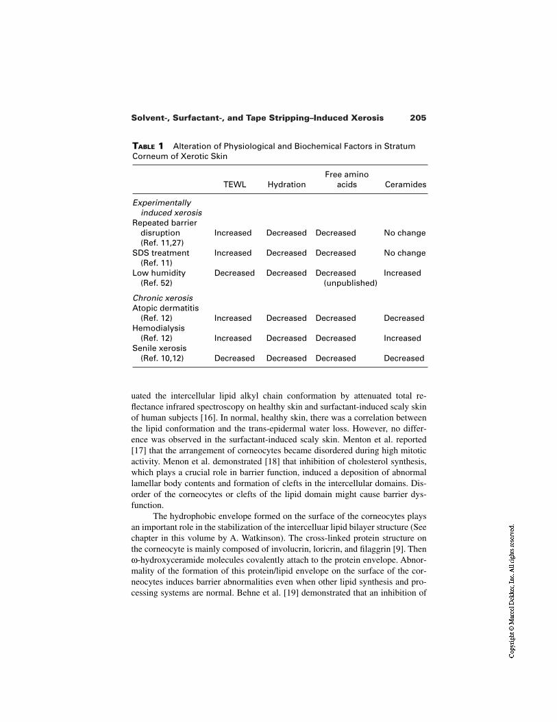
205Solvent-, Surfactant-, and Tape Stripping–Induced Xerosis
TABLE 1 Alteration of Physiological and Biochemical Factors in StratumCorneum of Xerotic Skin
TEWL HydrationFree amino
acids Ceramides
Experimentallyinduced xerosis
Repeated barrierdisruption(Ref. 11,27)
Increased Decreased Decreased No change
SDS treatment (Ref. 11)
Increased Decreased Decreased No change
Low humidity (Ref. 52)
Decreased Decreased Decreased(unpublished)
Increased
Chronic xerosisAtopic dermatitis
(Ref. 12) Increased Decreased Decreased DecreasedHemodialysis
(Ref. 12) Increased Decreased Decreased IncreasedSenile xerosis
(Ref. 10,12) Decreased Decreased Decreased Decreased
uated the intercellular lipid alkyl chain conformation by attenuated total re-flectance infrared spectroscopy on healthy skin and surfactant-induced scaly skinof human subjects [16]. In normal, healthy skin, there was a correlation betweenthe lipid conformation and the trans-epidermal water loss. However, no differ-ence was observed in the surfactant-induced scaly skin. Menton et al. reported[17] that the arrangement of corneocytes became disordered during high mitoticactivity. Menon et al. demonstrated [18] that inhibition of cholesterol synthesis,which plays a crucial role in barrier function, induced a deposition of abnormallamellar body contents and formation of clefts in the intercellular domains. Dis-order of the corneocytes or clefts of the lipid domain might cause barrier dys-function.
The hydrophobic envelope formed on the surface of the corneocytes playsan important role in the stabilization of the intercelluar lipid bilayer structure (Seechapter in this volume by A. Watkinson). The cross-linked protein structure onthe corneocyte is mainly composed of involucrin, loricrin, and filaggrin [9]. Thenω-hydroxyceramide molecules covalently attach to the protein envelope. Abnor-mality of the formation of this protein/lipid envelope on the surface of the cor-neocytes induces barrier abnormalities even when other lipid synthesis and pro-cessing systems are normal. Behne et al. [19] demonstrated that an inhibition of
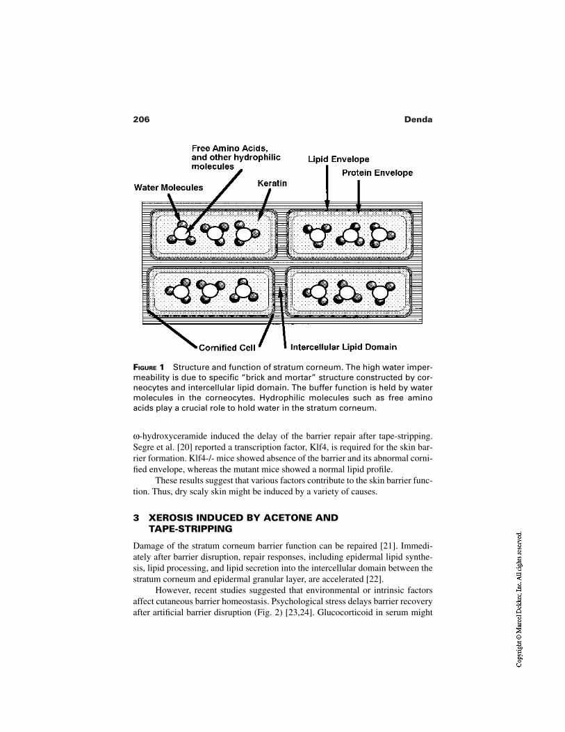
206 Denda
FIGURE 1 Structure and function of stratum corneum. The high water imper-meability is due to specific “brick and mortar” structure constructed by cor-neocytes and intercellular lipid domain. The buffer function is held by watermolecules in the corneocytes. Hydrophilic molecules such as free aminoacids play a crucial role to hold water in the stratum corneum.
ω-hydroxyceramide induced the delay of the barrier repair after tape-stripping.Segre et al. [20] reported a transcription factor, Klf4, is required for the skin bar-rier formation. Klf4-/- mice showed absence of the barrier and its abnormal corni-fied envelope, whereas the mutant mice showed a normal lipid profile.
These results suggest that various factors contribute to the skin barrier func-tion. Thus, dry scaly skin might be induced by a variety of causes.
3 XEROSIS INDUCED BY ACETONE AND TAPE-STRIPPING
Damage of the stratum corneum barrier function can be repaired [21]. Immedi-ately after barrier disruption, repair responses, including epidermal lipid synthe-sis, lipid processing, and lipid secretion into the intercellular domain between thestratum corneum and epidermal granular layer, are accelerated [22].
However, recent studies suggested that environmental or intrinsic factorsaffect cutaneous barrier homeostasis. Psychological stress delays barrier recoveryafter artificial barrier disruption (Fig. 2) [23,24]. Glucocorticoid in serum might
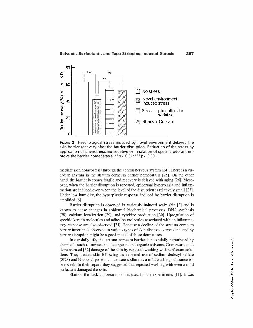
207Solvent-, Surfactant-, and Tape Stripping–Induced Xerosis
FIGURE 2 Psychological stress induced by novel environment delayed theskin barrier recovery after the barrier disruption. Reduction of the stress byapplication of phenotheiazine sedative or inhalation of specific odorant im-prove the barrier homeostasis. **p < 0.01; ***p < 0.001.
mediate skin homeostasis through the central nervous system [24]. There is a cir-cadian rhythm in the stratum corneum barrier homeostasis [25]. On the otherhand, the barrier becomes fragile and recovery is delayed with aging [26]. More-over, when the barrier disruption is repeated, epidermal hyperplasia and inflam-mation are induced even when the level of the disruption is relatively small [27].Under low humidity, the hyperplastic response induced by barrier disruption isamplified [6].
Barrier disruption is observed in variously induced scaly skin [3] and isknown to cause changes in epidermal biochemical processes, DNA synthesis[28], calcium localization [29], and cytokine production [30]. Upregulation ofspecific keratin molecules and adhesion molecules associated with an inflamma-tory response are also observed [31]. Because a decline of the stratum corneumbarrier function is observed in various types of skin diseases, xerosis induced bybarrier disruption might be a good model of those dermatoses.
In our daily life, the stratum corneum barrier is potentially perturbated bychemicals such as surfactants, detergents, and organic solvents. Gruneward et al.demonstrated [32] damage of the skin by repeated washing with surfactant solu-tions. They treated skin following the repeated use of sodium dodecyl sulfate(SDS) and N-cocoyl protein condensate sodium as a mild washing substance forone week. In their report, they suggested that repeated washing with even a mildsurfactant damaged the skin.
Skin on the back or forearm skin is used for the experiments [11]. It was
208 Denda
easier to induce scaly skin on the back than on the forearm. After stripping thestratum corneum on the back nine times with adhesive cellophane tape, theTEWL value was over 10 mg/cm2/hr, and most of the stratum corneum was re-moved. But to induce dry scaly skin on the forearm, stripping up to 30–50 timeswas needed. One week after the treatment, TEWL increased, skin surface con-ductance decreased, and the cell area in the stratum corneum also decreased. Theskin surface became scaly and flaky. Abnormal scaling is observed on the surfaceof the skin after tape-stripping. These phenomena are commonly observed in nat-ural dry skin, such as in atopic dermatitis and psoriasis.
Acetone treatment is also used for barrier disruption [33]. Compared totape-stripping, this treatment breaks the stratum corneum barrier homogeneously.On the other hand, it takes a longer period of time to break the barrier than bytape-stripping. Thus, the barrier disruption by acetone treatment is more usefulfor studies using hairless mice than human subjects because the mouse has a thinstratum corneum.
Treatment with surfactants is another way to break the barrier [32]. The ef-ficacy varies with each surfactant. Yang et al. suggested [34] some kind of anion-ic surfactant such as sodium dodecyl sulfate, affecting not only the stratumcorneum barrier, but also the nucleous layer of the epidermis. Fartasch demon-strated [35] that the topical application of SDS caused damage to the nucleatedcells of the epidermis and that acetone treatment disrupted the lipid structure onlyin the stratum corneum. Some surfactants induce an inflammatory response of theepidermis. These effects of surfactants will be described in the next section.
The degree of epidermal hyperplasia correlated with the level and durationof barrier disruption [27]. Using hairless mice, we investigated the effects of re-peated barrier disruption [27]. Not only epidermal hyperplasia, but also cutaneousinflammation was observed with a longer and higher level of repeated barrier dis-ruption by tape-stripping and acetone treatment. Flank skin of hairless mice is of-ten used for the study, but ears of other types of hairy mice can also be used forthe study. In our previous study [27], ear skin of ICR mice showed more obviousinflammation after repeated barrier disruption than that of flank skin of hairlessmice. Since neither the increase in epidermal cytokine production nor the de-scribed changes in cutaneous pathology were prevented by occlusion, this modelshould not be attributed to increased water loss, but rather to epidermal injury re-sulting in the production and release of epidermal cytokines.
The xerosis induced by repeated barrier disruption would be a very usefulmodel for the dry scaly skin induced by environmental factors such as detergentsor organic solvents. However, although repeated barrier disruption induces in-flammation, epidermal hyperplasia, and abnormal keratinization, there are sever-al histological differences between this model and psoriasis. Gerritsen et al. re-ported [36] the absence of some characteristic features of psoriasis in the dry skininduced by repeated tape-stripping. They also demonstrated the difference of fi-
209Solvent-, Surfactant-, and Tape Stripping–Induced Xerosis
laggrin expression between the model system and psoriasis. The different featuresof these chronic skin diseases and the model system should be investigated forfurther understanding of the diseases.
4 SURFACTANT-INDUCED XEROSIS
In our daily life, surfactants, i.e., detergents, are a potential cause of dermatitis[4,5]. Thus, the dry skin induced by surfactants has been studied not only as amodel system of dry skin, but also for clinical study of skin trouble in our dailylife. As described, the surfactant could damage not only stratum corneum barrierfunction, but also other skin properties.
The effect of the surfactant on skin is dependent on the type of the surfac-tant. Wilhelm et al. demonstrated the irritation potential of anionic surfactants[37]. They evaluated the effects of sodium salts of n-alkyl sulfates with variouscarbon chain lengths on TEWL and found the maximum response on the C12analog. In this report, they suggested that the mechanisms responsible for the hy-dration of the stratum corneum are related to the irritation properties of the sur-factants. Leveque et al. also suggested [38] the occurrence of the hyperhydrationof the stratum corneum consecutive to the inflammation process. They demon-strated that the increase of TEWL was induced by SDS without removal of lipidsin the stratum corneum. Sodium dodecyl sulfate might influence not only stratumcorneum barrier function, but also the nucleated layer of the epidermis and/ordermal system associated with inflammation [38]. A previous study revealed nocorrelation between the level of epidermal hyperplasia and TEWL increase on theSDS-irritated skin [39]. Ruissen et al. demonstrated [40] different effects of vari-ous types of detergents on keratinocyte culture and human intact skin. As a hy-perproliferative/inflammatory marker, they monitored SKALP, a protease in-hibitor that is found in hyperproliferative skin. In their in vitro system, anionicSDS induced SKALP expression, SDS also induced upregulation of involucrinand downregulation of cytokeratin 1 expression, which are associated with epi-dermal inflammation and hyperplasia. On the other hand, a cationic detergent,cetyltrimethylammonium bromide (CTAB), and nonionic detergents, Nonident P-40 and TritonX-100, did not induce the expression of the proliferative markersobserved by the SDS treatment. Different detergents showed different features ofcytotoxicity of human keratinocyte. CTAB, Triton X-100, and Nonident-P40showed strong, SDS showed moderate, and Tween-20 showed no cytotoxicity.Thus, cytotoxicity was not correlated with the potential of epidermal prolifera-tion. They also compared the induction of erythema and skin barrier disruption bydifferent detergents. In both parameters, SDS showed the most obvious effects;Triton X-100 showed the smallest; and CTAB showed a moderate effect on hu-man skin. Other reports demonstrated an induction of intercellular adhesion mol-ecule-1 (ICAM-1) [41] or vascular endothelial growth factor (VEGF) [42] by
210 Denda
SDS treatment. These results suggest that anionic detergents such as SDS have ahigh potential to induce epidermal hyperplasia or inflammation. Thus, SDS hasoften been used in dry scaly skin model systems.
In our previous study [16], we used human forearm skin or back skin. Theforearm skin was treated with a 5% aqueous solution of SDS and an occlusivedressing applied. After the treatment, we washed the surfactant solution with wa-ter and then we continuously measured TEWL, skin surface conductance, andlipid morphology in the stratum corneum by ATR-IR for 14 days (Fig. 3). Thelipid morphology in the stratum corneum was disordered by the treatment, but re-covered to normal within a couple of days. On the other hand, both TEWL andskin surface conductance were abnormal even 2 weeks after the SDS treatment.In the case of a single application of the barrier disruption by tape-stripping andacetone treatment, these parameters recovered to normal within a couple of days.Thus, the occlusive dressing of the surfactant affected skin not only on the stra-tum corneum, but also the nucleated layer of the epidermis and dermis. Potential-ly this method damages the skin too much. One must pay attention to the concen-tration of the surfactant solution and the period of the occlusive dressing. Theskin damage varies with the individual. The subsequent occlusion substantiallyincreases the irritant response of the skin to repeated short-term SDS treatment.
5 OTHER SUBSTANCES WHICH COULD INDUCE XEROSIS
In addition to surfactants, several chemical substances also induce dry scaly skin.Chiba et al. demonstrated [43] that topical application of squalene-monohy-droperoxide, a product of UV-peroxidated squalene, induced dry skin on hairlessmice. In this model, they showed hyperkeratosis and epidermal proliferation. Per-oxidized lipid by UV irradiation might be a cause of xerotic skin.
Sato et al. [44] demonstrated that cholesterol sulfate inhibited both trypsintype and chymotrypsin type protease and suggested the inhibition of these prote-sases reduced degradation of desmosomes, which play a crucial role in the adhe-sion of corneocytes and, as a consequence, abnormal scales were induced. Abnor-mality of cholesterol sulfate processing also induced serious skin abnormalities[45]. The content of cholesterol sulfate in total lipids is 5% in the healthy epider-mis and about 1% in the stratum corneum. Steroid sulfatase catalyzes the desulfa-tion of cholesterol sulfate to cholesterol. In recessive X-linked ichthyosis, whichdisplays a large amount of abnormal scales, the level of cholesterol sulfate in thestratun corneum was increased 10-fold because of the absence of steroid sulfa-tase. Moreover, Nemes et al. [46] reported another potential negative role of cho-lesterol sulfate in the stratum corneum. Involucrin cross-linking and involucrinesterification with ω-hydroxyceramides are crucial for cornified envelope forma-tion. They demonstrated that both reactions were inhibited by cholesterol sulfate.
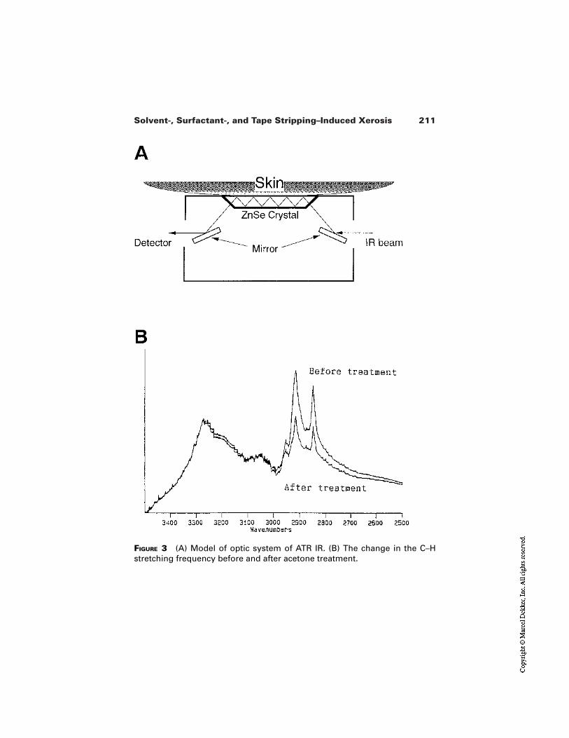
211Solvent-, Surfactant-, and Tape Stripping–Induced Xerosis
FIGURE 3 (A) Model of optic system of ATR IR. (B) The change in the C–Hstretching frequency before and after acetone treatment.
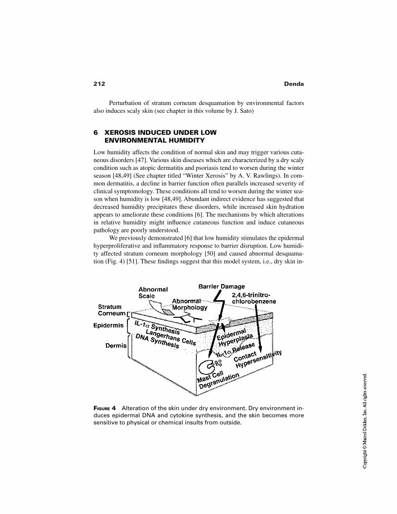
212 Denda
FIGURE 4 Alteration of the skin under dry environment. Dry environment in-duces epidermal DNA and cytokine synthesis, and the skin becomes moresensitive to physical or chemical insults from outside.
Perturbation of stratum corneum desquamation by environmental factorsalso induces scaly skin (see chapter in this volume by J. Sato)
6 XEROSIS INDUCED UNDER LOWENVIRONMENTAL HUMIDITY
Low humidity affects the condition of normal skin and may trigger various cuta-neous disorders [47]. Various skin diseases which are characterized by a dry scalycondition such as atopic dermatitis and psoriasis tend to worsen during the winterseason [48,49] (See chapter titled “Winter Xerosis” by A. V. Rawlings). In com-mon dermatitis, a decline in barrier function often parallels increased severity ofclinical symptomology. These conditions all tend to worsen during the winter sea-son when humidity is low [48,49]. Abundant indirect evidence has suggested thatdecreased humidity precipitates these disorders, while increased skin hydrationappears to ameliorate these conditions [6]. The mechanisms by which alterationsin relative humidity might influence cutaneous function and induce cutaneouspathology are poorly understood.
We previously demonstrated [6] that low humidity stimulates the epidermalhyperproliferative and inflammatory response to barrier disruption. Low humidi-ty affected stratum corneum morphology [50] and caused abnormal desquama-tion (Fig. 4) [51]. These findings suggest that this model system, i.e., dry skin in-
213Solvent-, Surfactant-, and Tape Stripping–Induced Xerosis
duced by low humidity, is also an important model for clinical research of skindiseases associated with skin surface dryness.
In these studies we used hairless mice [6,50–52]. Before the experiments,the mice were caged separately for at least 4 days. These cages were kept in aroom with temperature maintained at 22–25°C and relative humidity (RH) of40–70%. Then the mice were kept separately in 7.2-L cages in which the relativehumidity was maintained at either 10% (low humidity) with dry air or 80% (highhumidity) with humid air. The temperature was 22–25°C with fresh air circulated100 times per hour, and the animals were kept out of the direct stream of air. Thelevel of NH3 was always below 1 ppm.
Under low humidity, epidermal DNA synthesis increased within 12 hr [53].Abnormal scaling and increase of stratum corneum thickness were also observedwithin 2–3 days [51]. Obvious epidermal hyperplasia and mast cell degranulationwere observed 48 hr after the treatment of flank skin with acetone in the animalsthat had been kept in low humidity for 48 hr [6]. Contact hypersensitivity to2,4,6-trinitrochlorobenzene also increased after exposure to low humidity for 2days [54]. An immunohistochemical study showed that the amount of interleukin1α (IL-1α) in the epidermis was higher in animals kept in low humidity than inthose kept in high humidity [7]. The release of IL-1α from skin immediately aftertape-stripping was significantly higher in the animals kept in low humidity thankept in high humidity. Moreover, epidermal IL-1α mRNA increased significantlyin the animals kept in low humidity for 24 hr. These studies provide evidence thatchanges in environmental humidity contribute to the seasonal exacerbation/ame-lioration of cutaneous disorders such as atopic dermatitis and psoriasis, diseaseswhich are characterized by a defective barrier, epidermal hyperplasia, and inflam-mation. Because these responses were prevented by occlusion with a plasticmembrane, petrolatum, and humectant [6], this dry skin model is a good model toevaluate the clinical methods to treat skin problems.
7 NEW ROUTES TO TREAT XEROSIS
As described, repeated barrier disruption induces epidermal hyperplasia and in-flammation. Even slight damage of the barrier resulted in epidermal hyperplasiaunder low humidity. We previously reported [55,56] several methods to acceler-ate skin barrier repair by regulation of nonlipid factors such as enzymes and ions.We also demonstrated that the acceleration of the barrier repair improved thoseskin conditions [55]. Thus, studies on the biochemical and biophysical functionsassociated with the epidermal barrier homeostasis should be important for clinicaldermatology (Table 2).
Damaged barrier function can be restored by topical application of a water-impermeable substance such as petrolatum [57]. In this case, the petrolatum staysin the stratum corneum and forms a water-impermeable membrane. However,
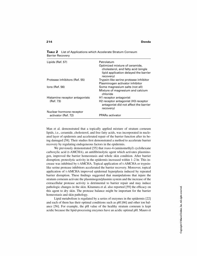
214 Denda
TABLE 2 List of Applications which Accelerate Stratum Corneum Barrier Recovery
Lipids (Ref. 57) PetrolatumOptimized mixture of ceramide,
cholesterol, and fatty acid (singlelipid application delayed the barrierrecovery)
Protease inhibitors (Ref. 55) Trypsin-like serine protease inhibitorPlasminogen activator inhibitor
Ions (Ref. 56) Some magnesium salts (not all) Mixture of magnesium and calcium
chlorideHistamine receptor antagonists
(Ref. 73)H1 receptor antagonist H2 receptor antagonist (H3 receptor
antagonist did not affect the barrier recovery)
Nuclear hormone receptoractivator (Ref. 72) PPARα activator
Man et al. demonstrated that a topically applied mixture of stratum corneumlipids, i.e., ceramide, cholesterol, and free fatty acids, was incorporated in nucle-ated layer of epidermis and accelerated repair of the barrier function after its be-ing damaged [58]. Their studies first demonstrated a method to accelerate barrierrecovery by regulating endogeneous factors in the epidermis.
We previously demonstrated [55] that trans-4-(aminomethyl) cyclohexanecarboxylic acid (t-AMCHA), an antifibrinolytic agent which activates plasmino-gen, improved the barrier homeostasis and whole skin condition. After barrierdisruption; proteolytic activity in the epidermis increased within 1–2 hr. This in-crease was inhibited by t-AMCHA. Topical application of t-AMCHA or trypsin-like serine protease inhibitors accelerated the barrier recovery. Moreover, topicalapplication of t-AMCHA improved epidermal hyperplasia induced by repeatedbarrier disruption. These findings suggested that manipulations that injure thestratum corneum activate the plasminogen/plasmin system and the increase of theextracellular protease activity is detrimental to barrier repair and may inducepathologic changes in the skin. Kitamura et al. also reported [59] the efficacy onthis agent to dry skin. The protease balance might be important for the barrierhomeostasis and skin pathology.
Lipid metabolism is regulated by a series of enzymes in the epidermis [22]and each of them has their optimal conditions such as pH [66] and other ion bal-ance [56]. For example, the pH value of the healthy stratum corneum is keptacidic because the lipid-processing enzymes have an acidic optimal pH. Mauro et
215Solvent-, Surfactant-, and Tape Stripping–Induced Xerosis
al. [66] demonstrated that topical application of basic buffer after barrier disrup-tion delayed the repair process because the basic condition perturbates the lipidprocessing.
Other ions such as calcium and magnesium [56,61] also play importantroles in lipid metabolism in the epidermis. We demonstrated that the topical ap-plication of calcium or potassium reduced barrier repair, and magnesium and amixture of calcium and magnesium salts accelerated the repair process [56]. Top-ical application of 10 mM magnesium chloride, magnesium sulfate, and magne-sium lactate aqueous solution accelerated barrier repair. Application of magne-sium bis(dihydrogen phosphate) or magnesium chloride in PBS solution did notaffect the barrier recovery rate. Application of 10 mM calcium chloride aqueoussolution delayed the barrier repair, but a mixture of calcium chloride and magne-sium chloride accelerated barrier recovery when the calcium-to-magnesium mo-lar ration was lower than 1. Application of the mixture also improved the condi-tion of dry scaly skin induced by SDS treatment (Fig. 5). These results suggest animportant role for these ions in barrier homeostasis.
We demonstrated a heterogeneous distribution of calcium, magnesium, andpotassium in the human epidermis [62]. Both calcium and magnesium were lo-calized in the glanular layer, while potassium was localized in the spinous layer.Immediately after the barrier function, this distribution disappeared. Calciumplays various roles in the formation of the stratum corneum barrier [61]. It in-duces terminal differentiation [63], carnified envelope formation, and epidermallipid synthesis [64]. Menon et al. demonstrated that alteration of the calcium gra-dient affects the exocytosis of the lamellar body at the interface between the stra-tum corneum and epidermal granular layer [65]. Vicanova demonstrated [66] theimprovement of the barrier function in the reconstructed human epidermis by thenormalization of epidermal calcium distribution. The heterogeneous field whichis formed by ions such as calcium, magnesium, and potassium might be crucialfor the terminal differentiation and the barrier formation in the epidermis. Rabplays an important role in the exocytosis and endocytosis after modification withhydrophobic molecules [67]. Magnesium is required for the activity of Rab-ger-anylgeranyl transferase, which modifies Rab [68]. For barrier formation, exocyto-sis of the lamellar body is an important process. Previous studies have indicatedthat Rab is modified by Rab-geranylgeranyl transferase during the terminal dif-ferentiation of the epidermis [69]. Regulation of ion gradation in the epidermismight be important to improve barrier homeostasis and skin pathology.
Feingold and coworkers demonstrated an important role of nuclear hor-mone receptor on epidermal differentiation and stratum corneum barrier forma-tion. Activation of PPARα by farnesol also stimulated the differentiation of epi-dermal keratinocytes [70,71]. And in carnified envelope formating, involucrinand transglutaminase protein mRNA levels were also increased by the activationof PPARα [72]. Interestingly, DNA synthesis was inhibited by the treatment [72].
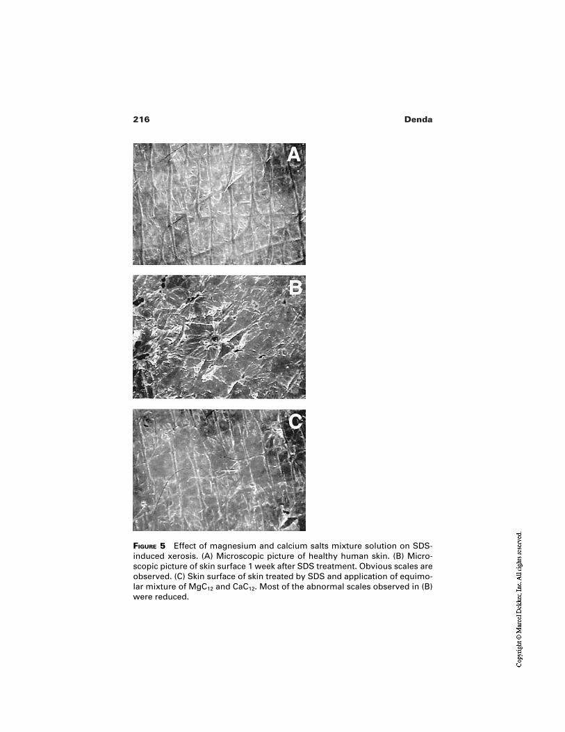
216 Denda
FIGURE 5 Effect of magnesium and calcium salts mixture solution on SDS-induced xerosis. (A) Microscopic picture of healthy human skin. (B) Micro-scopic picture of skin surface 1 week after SDS treatment. Obvious scales areobserved. (C) Skin surface of skin treated by SDS and application of equimo-lar mixture of MgC12 and CaC12. Most of the abnormal scales observed in (B)were reduced.
217Solvent-, Surfactant-, and Tape Stripping–Induced Xerosis
They also showed that topical application of PPARα activators accelerated thebarrier recovery after tape-stripping or acetone treatment and prevented the epi-dermal hyperplasia induced by repeated barrier disruption [72]. Regulation of thenuclear hormone receptor would open a new possibility for improvement of thecutaneous barrier.
Recently, Ashida et al. presented the relationship between histamine recep-tor and skin barrier function [73]. Three different types of histamine receptors,H1, H2, and H3, have been reported. First, topical application of histamine H1and H2 receptor antagonists accelerated the barrier repair. Histamine itself, H2 re-ceptor agonist, and histamine releaser delayed the barrier repair. Histamine H3 re-ceptor antagonist and agonist did not affect the barrier recovery rate. We demon-strated that topical application of the H1 and H2 receptor antagonists preventedthe epidermal hyperplasia induced by barrier disruption under low humidity. Themechanism of the relationship between the histamine receptors and the barrier re-pair process has not yet been elucidated.
As described, psychological stress [23,24] and aging [26] disrupt the barri-er homeostasis. The delay of barrier repair induced by psychological stress wasprevented by application of a sedative drug or inhalation of specific odorantswhich had a sedative effect [74]. The delay of barrier recovery with aging was im-proved by topical application of cholesterol [75] or mevalonic acid [76], becausethe delay of the aged skin was caused by a decrease of cholesterol synthesis. Re-moval of each cause of the barrier abnormality is the basic idea to improve thebarrier homeostasis. Thus, studies on the relationship between the barrier homeo-stasis and the physiology of the whole body system is important in clinical der-matology.
Regulation of epidermal lipid metabolism by eliminating other causativefactors might be effective to improve skin pathology. Because it can improve theendogeneous homeostatic process, occlusion or moisturization with artificial ma-terial could improve the skin condition. However, such treatment potentially per-turbs the homeostasis of the skin. On the other hand, recovery of the original, en-dogeneous skin function by acceleration of its homeostatic process results innatural healthy skin without side effects. Methods to accelerate barrier repairmight open new possibilities for future skin care systems.
8 CONCLUSION
In modern life, various environmental factors might induce xerotic skin. Skin sur-face dryness is caused by various factors. Decrease of free amino acid in the stra-tum corneum is commonly observed in different types of chronic and experimen-tally induced dry skin. Barrier abnormality is also often observed in xerotic skin,and improvement of the barrier homeostasis is effective to improve the wholeskin condition. The series of experimentally induced xerosis presented here are
218 Denda
useful for further understanding of the mechanistic study of xerosis. These mod-els should be important to develop a new strategy to treat xerosis.
REFERENCES
1. Sauer GC, Hall JC. Manual of Skin Diseases. 7th ed. Philadelphia Lippincott-Raven,1996.
2. Tagami H, Yoshikuni K. Interrelationship between water-barrier and reservoir func-tions of pathologic stratum corneum. Arch Dermatol 1985; 121:642–645.
3. Black D, Diridollou S, Lagarde JM, Gall Y. Skin care products for normal, dry andgreasy skin. In: Baran R, Maibach HI, eds. Textbook of Cosmetic Dermatology, 2 ed.London: Martin Dunitz, 1998; 125–150.
4. Kishi R, Harabuchi I, Katakura Y, Ikeda T, Miyake H. Neurobehavioral effects onchronic occupational exposure to organic solvents among Japanese industrialpainters. Environ Res 1993; 62:303–313.
5. Imabayashi Y. Skin surface lipids and sweat of hand in girl students who complainedof impaired skin on the hand by using household detergents. Fukuoka Igaku Zasshi1990; 81:359–369.
6. Denda M, Sato J, Tsuchiya T, Elias PM, Feingold KR. Low humidity stimulates epi-dermal DNA synthesis and amplifies the hyperproliferative response to barrier dis-ruption: implication of seasonal exacerbations of inflammatory dermatoses. J InvestDermatol 1998; 111:873–878.
7. Denda M. Influence of dry environment on epidermal function. J Dermatol Sci 2000;24:522–528.
8. Elias PM, Menon GK. Structural and lipid biochemical correlates of the epidermalpermeability barrier. In: Elias PM, ed. Advances in Lipid Research. Vol. 24. SkinLipids. San Diego: Academic Press, 1991; 1–26.
9. Nemes Z, Steinert PM. Bricks and mortar of the epidermal barrier. Exp Mol Med1999; 31:5–19.
10. Horii I, Obata M, Tagami H. Stratum corneum hydration and amino acid content inxerotic skin. Br J Dermatol 1989; 121:587–592.
11. Denda M, Hori J, Koyama J, Yoshida S, Namba R, Takahashi M, Horii I, YamamotoA. Stratum corneum sphingolipids and free amino acids in experimentally inducedscaly skin. Arch Dermatol Res 1992; 284:363–367.
12. Takahashi M, Ikezawa Z. Dry skin in atopic dermatitis and patients on hemodialysis.In: Loden M, Maibach HI, eds. Dry Skin and Moisturizers: Chemistry and Function.Boca Raton: CRC Press, 2000; 135–146.
13. Tanaka M, Okada M, Zhen YX, Inamura N, Kitano T, Shirai S, Sakamoto K, Inamu-ra T, Tagami H. Decreased hydration state of the stratum corneum and reducedamino acid content of the skin surface in patients with seasonal allergic rhinitis. Br JDermatol 1998; 139:618–621.
14. Imokawa G, Hattori M. A possible function of structural lipids in the water-holdingproperties of the stratum corneum. J Invest Dermatol 1985; 84:282–284.
15. Cornwell PA, Barry BW, Stoddart CP, Bouwstra JA. Wide-angle x-ray diffraction ofhuman stratum corneum: effects of hydration and terpene enhancer treatment. JPharm Pharmacol 1994; 46:938–950.
219Solvent-, Surfactant-, and Tape Stripping–Induced Xerosis
16. Denda M, Koyama J, Namba R, Horii I I. Stratum corneum lipid morphology andtransepidermal water loss in normal skin and surfactant-induced scaly skin. ArchDermatol Res 1994; 286:41–46.
17. Menton DN, Eisen AZ. Structural organization of the stratum corneum in certainscaling disorders of the skin. J Invest dermatol 1971; 57:295–307.
18. Menon GK, Feingold KR, Man MQ, Schande M, Elias PM. Structural basis for thebarrier abnormality following inhibition of HMG CoA reductase in murine epider-mis. J Invest Dermatol 1992; 98:209–219.
19. Behne M, Uchida Y, Seki T, de Montellano PO, Elias PM, Holleran WM. Omega-hydroxyceramides are required for corneocyte lipid envelope (CLE) formation andnormal epidermal permeability barrier function. J Invest Dermatol 2000;114:185–192.
20. Segre JA, Bauer C, Fuchs E. Klf4 is a transcription factor required for establishingthe barrier function of the skin. Nat Genet 1999; 22:356–360.
21. Elias PM, Holleran WM, Menon GK, Ghadially R, Williams ML, Feingold KR. Nor-mal mechanisms and pathophysiology of epidermal permeability barrier homeosta-sis. Curr Opin Dermatol 1993; 231–237.
22. Elias PM, Feingold KR. Lipids and the epidermal water barrier: metabolism, regula-tion, and pathophysiology. Semin Dermatol 1992; 11:176–182.
23. Denda M, Tsuchiya T, Hosoi J, Koyama J. Immobilization-induced and crowded en-vironment–induced stress delay barrier recovery in murine skin. Br J Dermatol 1998;138:780–785.
24. Denda M, Tsuchiya T, Elias PM, Feingold KR. Stress alters cutaneous permeabilitybarrier homeostasis. Am J Physiol 2000; 278:R367–R372.
25. Denda M, Tsuchiya T. Barrier recovery rate varies time-dependently in human skin.Br J Dermatol 2000; 142:881–884.
26. Ghadially R, Brown BE, Sequeira-Martin SM, Feingold KR, Elias PM. The agedepidermal permeability barrier. J Clin Invest 1995; 95:2281–2290.
27. Denda M, Wood LC, Emami S, Calhoun C, Brown BE, Elias PM, Feingold KR.Theepidermal hyperplasia associated with repeated barrier disruption by acetonetreatment or tape stripping cannot be attributed to increased water loss. Arch Derma-tol Res 1996; 288:230–238.
28. Proksch E, Feingold KR, Man MQ, Elias PM. Barrier function regulates epidermalDNA synthesis. J Clin Invest 1991; 87:1668–1673.
29. Menon GK, Elias PM, Lee SH, Feingold KR. Localization of calcium in murine epi-dermis following disruption and repair of the permeability barrier. Cell Tis Res 1992;270:503–512.
30. Wood LC, Jackson SM, Elias PM, Grunfeld C, Feingold KR. Cutaneous barrier per-turbation stimulates cytokine production in the epidermis of mice. J Clin Invest1992; 90:482–487.
31. Nickoloff BJ, Naidu Y. Perturbation of epidermal barrier function correlates with ini-tiation of cytokine cascade in human skin. J Am Acad Dermatol 1994; 30:535–546.
32. Gruneward AM, Gloor M, Gehring W, Kleesz P. Damage to the skin by repetitivewashing. Contact Dermatitis 1995; 32:225–232.
33. Denda M, Brown BE, Elias PM, Feingold KR. Epidermal injury stimulates prenyla-tion in the epidermis of hairless mice. Arch Dermatol Res 1997; 289:104–110.
34. Yang L, Man MQ, Taljebini M, Elias PM, Feingold KR. Topical stratum corneum
220 Denda
lipids accelerate barrier repair tape stripping, solvent treatment and some but not alltypes of detergent treatment. Br J Dermatol 1995; 133:679–685.
35. Fartasch M. Ultrastructure of the epidermal barrier after irritation. Microsc Res Tech1997; 37:193–199.
36. Gerritsen MJP, van Erp PEJ, van Vlijmen-Willems IMJJ, Lenders LTM, van deKerkhof PCM. Repeated tape stripping of normal skin: a histological assessment andcomparison with events seen in psoriasis. Arch Dermatol Res 1994; 286:455–461.
37. Wilhelm KP, Cua AB, Wolff HH, Maibach HI. Surfactant-induced stratum corneumhydration in vivo: prediction of the irritation potential of anionic surfactants. J InvestDermatol 1993; 101:310–315.
38. Leveque JL, de Rigal J, Saint-Leger D, Billy D. How does sodium lauryl sulfate al-ter the skin barrier function in man? A multiparametric approach. Skin Pharmacol1993; 6:111–115.
39. Welzel J, Metker C, Wolff H, Wilhelm KP. SLS-irritated human skin shows no cor-relation between degree of proliferation and TEWL increase. Arch Dermatol Res1998; 290:615–620.
40. Ruissen F, Le M, Carroll JM, Valk PGM, Schalkwijk J. Differential effects of deter-gents on keratinocyte gene expression. J Invest Dermatol 1998; 110:358–363.
41. Driesch P, Fartasch M, Huner A, Ponec M. Expression of integrin receptors andICAM-1 on keratinocytes in vivo and in an in vitro reconstructed epidermis: effect ofsodium dodecyl sulphate. Arch Dermatol Res 1995; 287:249–253.
42. Palacio S, Schmitt D, Viac J. Contact allergens and sodium lauryl sulphate upregu-late vascular endothelial growth factor in normal keratinocytes. Br J Dermatol 1997;137:540–544.
43. Chiba K, Sone T, Kawakami K, Onoue M. Skin roughness and wrinkle formation in-duced by repeated application of squalene-monohydroperoxide to the hairlessmouse. Exp Dermatol 1999; 8:471–479.
44. Sato J, Denda M, Nakanishi J, Nomura J, Koyama J. Cholesterol sulfate inhibits pro-teases that are involved in desquamation of stratum corneum. J Invest Dermatol1998; 111:189–193.
45. Zettersten E, Man MQ, Sato J, Denda M, Farrell A, Ghadially R, Williams ML, Fein-gold KR, Elias PM. Recessive X-linked ichthyosis: role of cholesterol-sulfate accu-mulation in the barrier abnormality. J Invest Dermatol 1998; 111:784–790.
46. Nemes Z, Demeny M, Marekov LN, Fesus L, Steinert PM. Cholesterol 3-sulfate interferes with cornified envelope assembly by diverting transglutaminase 1 activityfrom the formation of cross-links and esters to the hydrolysis of glutamine. J BiolChem 2000; 275:2636–2646.
47. Rycroft PJG, Smith WDL. Low humidity occupational dermatoses. Contact Der-matitis 1980; 6:488–492.
48. Wilkinson JD, Rycroft RJ. Contact Dermatitis. In: Champion RH, Burton JL, Ebling FJG, eds. Textbook of Dermatology, 5th ed. London: Blackwell Scientific,1992:614–615.
49. Sauer GC, Hall JC. Seasonal skin diseasses. In: Sauer GC, Hall JC, eds. Manual ofSkin Diseases. Philadelphia: Lippincott-Raven, 1996:23–28.
50. Sato J, Yanai M, Hirao T, Denda M. Water content and thickness of stratum corneumcontribute to skin surface morphology. Arch Dermatol Res 2000; 292:412–417.
221Solvent-, Surfactant-, and Tape Stripping–Induced Xerosis
51. Sato J, Denda M, Nakanihi J, Koyama J. Dry conditions affect desquamation of stra-tum corneum in vivo. J Dermatol Sci 1998; 18:163–169.
52. Denda M, Sato J, Masuda Y, Tsuchiya T, Koyama J, Kuramoto M, Elias PM, Fein-gold KR. Exposure to a dry environment enhances epidermal permeability barrierfunction. J Invest Dermatol 1998; 111:858–863.
53. Sato J, Denda M, Ashida Y, Koyama J. Loss of water from the stratum corneuminduces epidermal DNA synthesis in hairless mice. Arch Dermatol Res 1998;290:634–637.
54. Hosoi J, Hariya T, Denda M, Tsuchiya T. Regulation of the cutaneous allergic reac-tion by humidity. Contact Dermatitis 2000; 42:81–84.
55. Denda M, Kitamura K, Elias PM, Feingold KR. trans-4-(Aminomethyl) cyclohexa-ne carboxylic acid (T-AMCHA), an anti-fibrinolytic agent, accelerates barrier recov-ery and prevents the epidermal hyperplasia induced by epidermal injury in hairlessmice and humans. J Invest Dermatol 1997; 109:84–90.
56. Denda M, Katagiri C, Hirao T, Maruyama N, Takahashi M. Some magnesium saltsand a mixture of magnesium and calcium salts accelerate skin barrier recovery. ArchDermatol Res 1999; 291:560–563.
57. Man MQ, Brown BE, Wu-Pong S, Feingold KR, Elias PM. Exogenous nonphysio-logic vs physiologic lipids. Arch Dermatol 1995; 131:809–816.
58. Man MQ, Feingold KR, Thornfeld CR, Elias PM. Optimization of physiologic lipidmixtures for barrier repair. J Invest Dermatol 1996; 106:1096–1101.
59. Kitamura K, Yamada K, Ito A, Fukuda M. Research on the mechanism by which dryskin occurs and the development of an effective compound for its treatment. J SocCosmet Chem 1995; 29:133–145.
60. Mauro T, Grayson S, Gao WN, Man MQ, Kriehuber E, Behne M, Feingold KR, EliasPM. Barrier recovery is impeded at neutral pH, independent of ionic effects: impli-cations for extracellular lipid processing. Arch Dermatol Res 1998; 290:215–222.
61. Lee SH, Elias PM, Proksch E, Menon GK, Man MQ, Feingold KR. Calcium andpotassium are important regulators of barrier homeostasis in murine epidermis. JClin Invest 1992; 89:530–538.
62. Denda M, Hosoi J, Ashida Y. Visual imaging of ion distribution in human epidermis.Biochem Biophys Res Commun 2000; 272:134–137.
63. Watt FM. Terminal differentiation of epidermal keratinocytes. Curr Opin Cell Biol1989; 1:1107–1115.
64. Watanabe R, Wu K, Paul P, Marks DL, Kobayashi T, Pittelkow MR, Pagano RE. Up-regulation of glucosylceramide synthase expression and activity during human ker-atinocyte differentiation. J Biol Chem 1998; 273:9651–9655.
65. Menon GK, Price LF, Bommannan B, Elias PM, Feingold KR. Selective obliterationof the epidermal calcium gradient leads to enhanced lamellar body secretion. J InvestDermatol 1994; 102:789–795.
66. Vicanova J, Boelsma E, Mommas AM, Kempenaar JA, Forslind B, Pallon J, Egel-rund T, Koerten HK, Ponec M. Normalization of epidermal calcium distribution pro-file in reconstructed human epidermis is related to improvement of terminal differen-tiation and stratum corneum barrier formation. J Invest Dermatol 1998; 111:97–106.
67. Novick P, Brennward P. Friends and family: the role of the Rab GTPases in vesiculartraffic. Cell 1993; 75:597–601.
222 Denda
68. Seabra MC, Goldstein JL, Sudhof TC, Brown MS. Rab geranylgeranyl transferase. JBiol Chem 1992; 267:14497–14503.
69. Song HJ, Rossi A, Ceci R, Kim IG, Anzano MA, Jang SI, DeLaurenzi V, SteinertPM. The genes encoding geranylgeranyl transferase a-subunit and transglutaminaseI are very closely linked but not functionally related in terminally differentiating ker-atinocytes. Biochem Biophys Res Commun 1997; 235:10–14.
70. Hanley K, Komuves LG, Ng DC, Schoonjans K, He SS, Lau P, Bikle DD, WilliamsML, Elias PM, Auwerx J, Feingold KR. Farnesol stimulates differentiation in epi-dermal keratinocytes via PPARα. J Biol Chem 2000; 275:11484–11491.
71. Hanley K, Jiang Y, He SS, Friedman M, Elias PM, Bikle DD, Williams ML, Fein-gold KR. Keratinocyte differentiation is stimulated by activators of the nuclear hor-mone receptor PPARα. J Invest Dermatol 1998; 111:368–375.
72. Feingold KR. Role of nuclear hormone receptors in regulating epidermal diffrentia-tion. Program and Preprints of Annual Scientific Seminar. Soc Cosmet Chem 1999;30–31.
73. Ashida Y, Denda M, Hirao T. Histamine H1 and H2 receptor antagonists accelerateskin barrier repair and prevent epidermal hyperplasia induced by barrier disruptionin a dry environment. J Invest Dermatol 2001; 116:261–265.
74. Denda M, Tsuchiya T, Shoji K, Tanida M. Odorant inhalation affects skin barrierhomeostasis in mice and humans. Br J Dermatol 2000; 142:1007–1010.
75. Ghadially R, Brown BE, Hanley K, Reed JT, Feingold KR, Elias PM. Decreased epi-dermal lipid synthesis accounts for altered barrier function in aged mice. J InvestDermatol 1996; 106:1064–1069.
76. Haratake A, Ikenaga K, Katoh N, Uchiwa H, Hirano S, Yasuho H. Topical mevalonicacid stimulates de novo cholesterol synthesis and epidermal permeability barrierhomeostasis in aged mice. J Invest Dermatol 2000; 114:247–252.
12Clinical Effects of Emollients on Skin
Joachim FluhrUniversity of Pavia, San Matteo, Pavia, Italy andUniversity of California, San Francisco, San Francisco, California
Walter M. HolleranUniversity of California, San Francisco, San Francisco, California
Enzo BerardescaUniversity of Pavia, San Matteo, Pavia, Italy
1 INTRODUCTION
Emollients play a central role in topical treatments in dermatology, with their pro-tective and curative effects being of renewed interest. Different therapeutic func-tions of dermatological emollients are well recognized, and a review of such hasrecently been published [1]. Research has shown that the composition of emol-lients is of great importance for disease treatment. For example, the more chronicthe cutaneous disease is regarded, the higher the emollient lipid content shouldbe, as treatment efficacy is improved with the use of adequate emollients. How-ever, the specific role of emollients in drug delivery systems for different skin dis-eases has not yet been studied in detail. In this chapter a number of features ofemollients will be presented, including emollient composition, classification, androle in therapeutics; potential mechanisms of emollient function; and the role(s)of emollients in epidermal barrier function and specific diseases. Recent studieshave shown that the use of an appropriate emollient for the treatment of specific
223
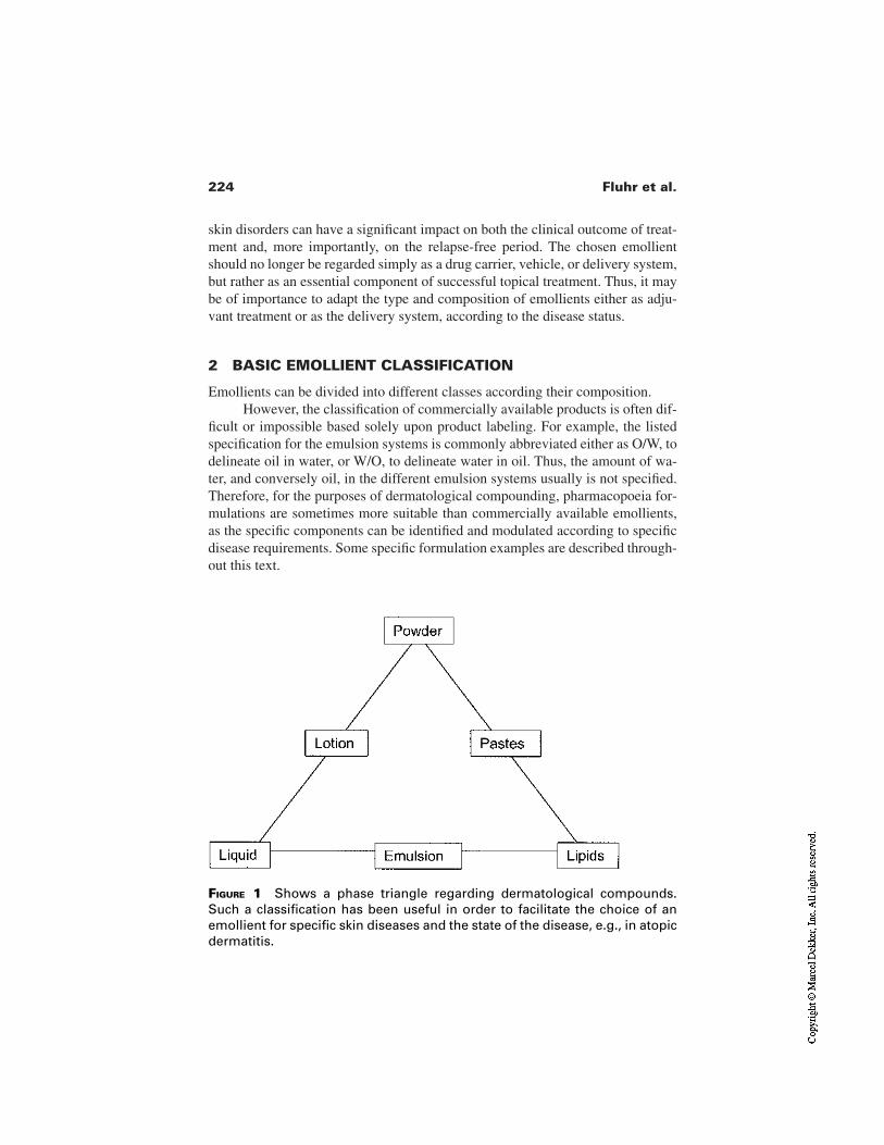
224 Fluhr et al.
skin disorders can have a significant impact on both the clinical outcome of treat-ment and, more importantly, on the relapse-free period. The chosen emollientshould no longer be regarded simply as a drug carrier, vehicle, or delivery system,but rather as an essential component of successful topical treatment. Thus, it maybe of importance to adapt the type and composition of emollients either as adju-vant treatment or as the delivery system, according to the disease status.
2 BASIC EMOLLIENT CLASSIFICATION
Emollients can be divided into different classes according their composition.However, the classification of commercially available products is often dif-
ficult or impossible based solely upon product labeling. For example, the listedspecification for the emulsion systems is commonly abbreviated either as O/W, todelineate oil in water, or W/O, to delineate water in oil. Thus, the amount of wa-ter, and conversely oil, in the different emulsion systems usually is not specified.Therefore, for the purposes of dermatological compounding, pharmacopoeia for-mulations are sometimes more suitable than commercially available emollients,as the specific components can be identified and modulated according to specificdisease requirements. Some specific formulation examples are described through-out this text.
FIGURE 1 Shows a phase triangle regarding dermatological compounds.Such a classification has been useful in order to facilitate the choice of anemollient for specific skin diseases and the state of the disease, e.g., in atopicdermatitis.
225Clinical Effects of Emollients on Skin
2.1 Hydrogel Emollients
The first class of emollients, the hydrogels, itself can be divided into two groups:(1) surface-active hydrogels that produce a thin film at the surface and (2) car-bomer-gels, which penetrate or act in deeper parts of the skin [1]. In general, thepenetration rate of either type of hydrogel can be enhanced increasing the amountof isopropanol in the composition. However, the carbomer-hydrogels are rarelyused as dermatological therapeutics, as they deliver the active compounds indeeper parts as, e.g., in heparin-containing sports-gels. For preservation reasonsthe carbomer-hydrogels also contain larger amounts of ethanol, isopropanol, orpreservatives. Different amounts and types of polyethyleneglycol characterize anadditional group of hydrogel emollients. These emollients are especially usefulfor antiseptic and antifungal preparations.
2.2 Oil-in-Water Emollients
Emollients usually are presented in the form of lotions (O/W emulsions) orcreams which are characterized by a hydrophilic external phase. As such, O/Wemulsions are the most frequently used emollient type for commercial topicaldermatologics. They have excellent absorption qualities and are readily formulat-ed into cosmetically elegant products. Due to their relatively high water content,the O/W emollients exert a cooling effect as free water is liberated following top-ical application.
2.3 Water-in-Oil Emollients
Conversely, W/O emulsions are characterized by a lipophilic external phase. Inthese preparations, the lipid phase consists primarily of petrolatum and/or paraf-fin oil to which other lipid fractions may or may not be added. The more lipid-richformulations are used in chronic dermatological disease in the nonacute phase,skin conditions where a lack of skin hydration and skin plasticity as well as an in-creased scaling can be observed, e.g., in eczematous skin conditions.
2.4 Amphiphilic Emollients
Amphiphilic creams, which contain both W/O and O/W characteristics, can bemixed with either lipophilic or hydrophilic (e.g., aqueous) compounds and maybe useful for a wider range of formulations. An example of a common am-phiphilic creme is given in the German Pharmacopoeia (Deutscher ArzneimittelCodex; DAC) as follows [2]:
Base cream DAC
Glycerol monostearate 60 4.0Cetyl alcohol 6.0
226 Fluhr et al.
Mid-chain triglycerides 7.5White petrolatum 25.5Macrogol-1000-glycerol monostearate 7.0Propylene glycol 10.0Purified water 40.0
3 SPECIFIC EFFECTS OF EMOLLIENTS
Emollients exert a number of effects in and on the skin, including skin hydration,skin cooling, and lipidization. As mentioned, the relative cooling effect of emol-lients can be attributed to the amount of water and/or alcohol in the emulsion sys-tem(s). This effect is more pronounced when an aqueous or alcohol phase is pres-ent within the external phase of the formulation, e.g., in lotions, hydrogels, orO/W emulsions. However, relatively nonstable W/O emulsions, like cold creams,also can exert mild cooling effect(s) when applied topically to the skin [3].
Emollients also are well known to influence the hydration of the stratumcorneum, for which at least three different mechanism have been proposed:
1. The emollient can exert a direct hydrating effect by liberating waterfrom the formulation itself [4]. In short-term applications, this hydrat-ing effect is more pronounced with formulations containing a high per-centage of water when compared with lipid-rich and low-water-con-taining preparations [5]. As expected, the hydrating effect of O/Wsystems in short-term applications depends primarily on the water con-tent of the formulation [3], since only the presence of unbound waterinsures hydration of the stratum corneum. In contrast, long-term appli-cations of either W/O or O/W emulsions with different water contentrevealed hydration of the stratum corneum with the W/O but not withthe O/W emulsions [6]. Thus, although a W/O emulsion may be cos-metically less acceptable, such a formulation can be expected toachieve better stratum corneum hydration, especially with prolongeduse.
2. The occlusive effect of the formulation can influence stratum corneumhydration, especially in long-term applications. A standard model forthis occlusion effect is petrolatum [5], for which the highest occlusiveeffect was detected [3]. Water-in-oil emulsions with low water contenthave occlusive effects similar to petrolatum, while W/O emulsionswith high water content have occlusive properties similar to O/Wemulsions [3]. Interestingly, even O/W formulations with high watercontent can exert an occlusive effect after the unbound water evapo-rates. However, the occlusive effects of each of these formulations arenot always desirable. For example, in atopic dermatitis, where a for-
227Clinical Effects of Emollients on Skin
mulation with high lipid content is desired, an occlusive effect may en-hance discomfort and induce itching response. The occlusive effectsalso may enhance drug penetration, an effect that may or may not bedesired.
3. A third mechanism by which emollients influence skin hydration is ev-ident when highly hygroscopic compounds like glycerol are applied.By absorbing water either from the emollient itself, from surface water,or from water evaporation, these agents then are able to increase stra-tum corneum hydration [7,8].
In addition, emollients can exert a lipidizing or “greasing” effect that is ofgreat importance in skin conditions were patients express discomfort due tocracked or rigid skin. It has been suggested that this lipidization effect be of ma-jor importance to the plasticity of the skin [9]. Moreover, lipid-rich formulationsimprove skin distensibility, while creams and gels with lower lipid content have amore pronounced effect on skin hydration, as previously discussed [9]. The emol-lient greasing effect appears to be limited to the application period and is not along-term effect.
Finally, it should be noted that compounds with distinct dielectric constantshave been shown to influence the electrophysical properties of the stratumcorneum as measured by capacitance- or conductance-based instruments [10].Thus, it is plausible that topically applied moisturizing creams might be a sourceof false-positive results using these instruments [11]. Although a good correlationbetween capacitance values and water content of the tested creams has beendemonstrated [11], a sufficient time lag following application of compoundsshould be allotted before any such measurements are registered.
4 BARRIER PROTECTION AND BARRIER RECOVERY
A number of factors are involved with determining the effectiveness of emol-lients to protect skin barrier. Commonly used barrier creams, which are eitherW/O emulsions or emollients with lipophilic character, are claimed to protectagainst hydrophilic irritants. On the other hand, barrier creams that are O/Wemulsion systems, or that act like hydrophilic systems, are thought to protectagainst lipophilic irritants. Dermatological skin protection (especially in workconditions) is based on different product groups in situations where barrier orprotective creams are employed. For example, pre-exposure skin care includesthe use of O/W and W/O emulsions, tannery substances, zinc oxide, talcum, per-fluorpolyethers, chelating agents, and UV protectors [12]. However, cleansingproducts and postexposure skin care are two other important components of skinprotection. Postexposure skin care is based on emollients, moisturizers, humec-tants, and lipid-rich formulations. Although claimed protective effects have been
228 Fluhr et al.
shown using specific test conditions, double-blind, placebo-controlled, random-ized trials are still lacking, especially under conditions that approximate realworkplace situations [12]. In fact, cumulative stress tests with repetitive applica-tion of irritants appear to be the best conditions for approximating work condi-tions [13–19].
The distinction between skin protection and skin care is not always clear.For example, in nurses a barrier cream was compared with its emollient vehiclefor effects on clinical improvement. Interestingly, both clinical skin status andstratum corneum hydration improved significantly in each treatment group, with-out evidence of a difference between the emollient and the barrier cream groups[20]. Thus, the emollient or vehicle alone often shows a significant improvementof the clinical skin conditions as well as the stratum corneum hydration. ThusBerndt and colleagues proposed that a strict distinction between skin care andskin protection products should not be maintained [20]. Correct instructions forthe consumer should be stressed with regard to regular and frequent applicationof a protection product in order to be effective [21]. In addition, a recent studydiscussed whether claims could be made with respect to protective and preventiveproperties of topically applied body lotions and barrier creams [22]. In this par-ticular study, enhanced stratum corneum hydration, improved barrier function, aswell as a faster barrier recovery were reported after sodium lauryl sulfate (SLS)barrier disruption [22].
Exposure to tensides represents a common potential workplace irritant.Protection against tensides seems to be more effective with lipid-enriched emol-lients, such as W/O emulsions [23,24]. In contrast, Held and colleagues showedthat a 4-week pretreatment of normal skin with W/O emollients increases suscep-tibility to detergents (sodium lauryl sulfate) [25]. Thus the long-term applicationof barrier creams in working conditions where detergents are present should becarefully evaluated.
Clinical observations have established that skin irritants are more harmfulin dry skin conditions. Thus emollients often are used to increase the water con-tent of the stratum corneum as a preventive measure [26]. Moisturizer-containingemollients prevent irritant skin reactions induced by detergents and may also ac-celerate regeneration of barrier function in irritated skin [27]. Emollients withmoisturizing properties usually contain either singly or in combination humec-tants, such as urocanic acid, ammonia, lactic acid, pyrrolidone carboxylic acid,urea, citrate, glycerol, sorbitol, and hydroxyacids. These agents belong to a groupcalled natural moisturizing factors (NMF), or moisturizers. Their common prop-erties include the increase of hydration and the enhancement of water-binding ca-pacity in the upper stratum corneum. Reduced NMF content in the stratumcorneum can diminish water-absorption capacity and may result in perturbationof corneodesmolysis leading to hyperkeratosis [28].
229Clinical Effects of Emollients on Skin
Emollients with barrier properties also can prevent certain types of epider-mal damage. For example, Fartasch and colleagues have shown that alterations ofthe lower part of stratum corneum and damage to the nucleated layers of the epi-dermis are induced by sodium lauryl sulfate [29]. In this model, formation oflamellar lipid membrane structures was disturbed in the lower stratum corneum.In contrast, the upper stratum corneum showed intact intercellular lipid bilayers.The barrier disruption effect of SLS was prevented by the application of a barriercream (discussed in more detail subsequently), with diminished sodium laurylsulfate penetration as the likely mechanism [29].
4.1 Emollients in Atopic Dermatitis
The utility of emollients in the treatment of atopic dermatitis is well recognized.In atopic dermatitis stratum corneum, hydration and water-binding capacity arereduced [30–32], and impaired barrier function is readily observed in involvedskin [30,33]. These patients also are more prone to develop an irritant contactdermatitis [34] and show less pronounced so-called hardening effect than healthycontrol patients, i.e., with repeated skin barrier disruption, the barrier deteriora-tion stopped after a certain time, suggesting that the stratum corneum accommo-dates the barrier insults [35]. In atopic dermatitis stratum corneum, the content ofbarrier lipids is reduced, most prominently that of ceramide 1 and ceramide 3[33,36]. This reduction of ceramide levels may result form the overaction of asphingomyelin deacylase enzyme [37,38]. However, sebaceous gland activity, therole of which remains unknown, also is reduced in these patients [39]. Moreover,in atopic dermatitis stratum corneum the membrane-coating granules, or lamellarbodies, are incompletely extruded and organized. This, along with the alteredstratum corneum lipid content, may partly explain the impaired barrier functionin these patients [40].
Thus, ideal emollients for patients with atopic dermatitis should
Improve barrier functionHave protective propertiesImprove stratum corneum hydrationHave an antibacterial active compound (e.g., Staph. aureus)
These demands can be met by emollients
Showing W/O emulsions properties with a high water content [24]Containing a moisturizer (e.g., glycerol, urea) [7,41]Having a physiological lipid mixture [42–44]Containing an antiseptically active compound (e.g., triclosan) [45,46]
In this regard, urea-containing emollients were able to improve skin barrier func-
230 Fluhr et al.
*Beiersdorf, Hamburg, Germany.†Hansen & Rosenthal, Germany: mineral oil 77.9%, polyglyceryl-3-isostearate 10.0%, isopropylpalmitate 8.0%, polyethylene 4.1%.§Spirig AG, Egerkingen, Switzerland: containing 4% urea and Triclosan.
tion and to reduce skin susceptibility to irritants within patients suffering fromatopic dermatitis [41]. Evening primrose oil (20%) in a W/O emulsion (but not inan amphiphilic emulsion system) also improved barrier function and stratumcorneum hydration in atopic patients [47].
Some examples of useful formulations for water-rich W/O emollients forpatients with atopic dermatitis are as follows [1]:
W/O emulsion I
Urea 5.0Glycerol 85% 10.0Triclosan 3.0Eucerinum W/O emollient* ad 100.0
W/O emulsion II
Urea 5.0Glycerol 85% 10.0Triclosan 3.0Pionier KWH Pharm† 30.0Citric acid anhydr. 0.07Magnesium sulfate heptahydrate 0.5Purified water ad 100.0
W/O emulsion III
Glycerol 85% 22.0Triclosan 4.4Excipial U Lipolotio§ ad 220.0
These emollients have shown good stability for more than 3 months [1] and canbe used for compounding other active ingredients like evening primrose oil orpale sulfonated shale oil (in W/O emulsion I). As a note of caution, formulationsintended for facial use should avoid urea due to its irritative potency and potential[48].
For patients showing allergic reactions to constituents of emollients [49](e.g., patients with a long history of leg ulcers [50]), fragrances, lanolin, cetyl-
231Clinical Effects of Emollients on Skin
stearyle alcohol, and parabens should be avoided, if possible. In such instances,the following emollient can be recommended [1]:
Cold cream
Cytylwaxester 11.75Beeswax 13.5Paraffin oil 63.25Purified water 11.50
5 ROLE OF LANOLIN IN EMOLLIENTS
Lanolin, or wool wax, is the secretion product of sebaceous glands of sheep andserves to impregnate and protect the wool fibres. Commercially available lanolinis commonly a mixture of wool wax (65–75%), water (20–30%), and paraffin (upto 15%). Wool wax contains mainly sterol esters, ester waxes, hydroxyesters, andlanolin alcohols (6–12%), with about 40% of the esters containing α-hydrox-yesters. However, the exact number of different esters is yet unknown.
Moreover, the sensitization potential of lanolin remains still under discus-sion [51]. Clark has proposed a reduction or purification of lanolin alcohols in or-der to minimize the risk of sensitization [52,53]. Interestingly, an epidemiologicalstudy revealed that lanolin-induced allergies occur in less than 0.001% of indi-viduals, i.e., less than ten per million [54], while a recent studies showed higherpercentage of positive test reactions in children with atopic dermatitis (1.7 and4.4%) [55,56]. The population of the pediatric studies [50,51] was much more se-lected than the population in the epidemiological study [50]. Kligman suggeststhat most of the lanolin sensitization cases represent false-positive results, andthus wool wax–containing products can be considered safe [51].
Lanolin has known moisturizing properties [57], similar to petrolatum, withlong-term effects that can be detected up to 14 days after termination of treatment[57]. The hydrating effect of lanolin has been shown using electrophysical mea-surements [58,59]. While lanolin penetrates into the stratum corneum, it remainsconcentrated in the upper layers [60]. Compared with vehicle, topically appliedlanolin accelerated barrier recovery following an acute barrier disruption withacetone [61]; 3% lanolin significantly reduced trans-epidermal water loss both 45min and 4 hr following disruption. Moreover, the immediate effect of lanolin onbarrier recovery was pronounced. In fact, the positive barrier effect of lanolin wascomparable to that of an optimized ratio of stratum corneum lipids, i.e., a barrierformulation (to be discussed in more detail) including cholesterol, ceramides, es-sential fatty acids, and nonessential fatty acids in a ratio 3:1:1:1 [42,44,61]. Final-ly, skin roughness is positively influenced by formulations containing lanolin in a
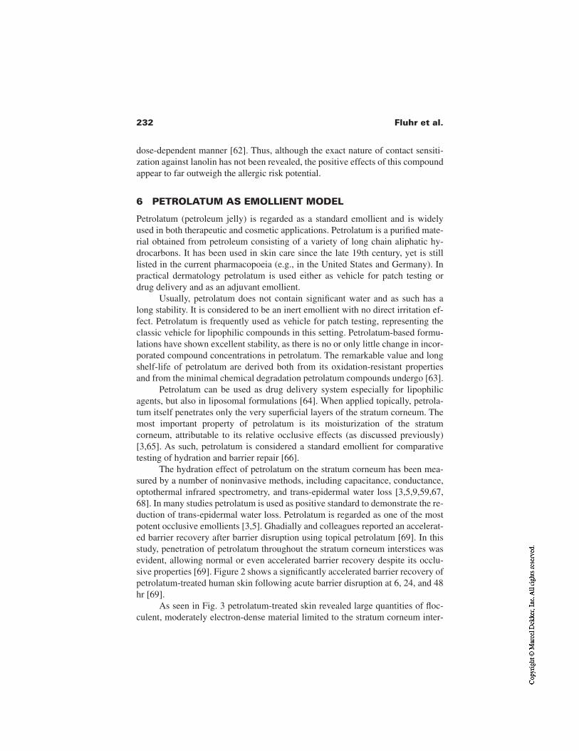
232 Fluhr et al.
dose-dependent manner [62]. Thus, although the exact nature of contact sensiti-zation against lanolin has not been revealed, the positive effects of this compoundappear to far outweigh the allergic risk potential.
6 PETROLATUM AS EMOLLIENT MODEL
Petrolatum (petroleum jelly) is regarded as a standard emollient and is widelyused in both therapeutic and cosmetic applications. Petrolatum is a purified mate-rial obtained from petroleum consisting of a variety of long chain aliphatic hy-drocarbons. It has been used in skin care since the late 19th century, yet is stilllisted in the current pharmacopoeia (e.g., in the United States and Germany). Inpractical dermatology petrolatum is used either as vehicle for patch testing ordrug delivery and as an adjuvant emollient.
Usually, petrolatum does not contain significant water and as such has along stability. It is considered to be an inert emollient with no direct irritation ef-fect. Petrolatum is frequently used as vehicle for patch testing, representing theclassic vehicle for lipophilic compounds in this setting. Petrolatum-based formu-lations have shown excellent stability, as there is no or only little change in incor-porated compound concentrations in petrolatum. The remarkable value and longshelf-life of petrolatum are derived both from its oxidation-resistant propertiesand from the minimal chemical degradation petrolatum compounds undergo [63].
Petrolatum can be used as drug delivery system especially for lipophilicagents, but also in liposomal formulations [64]. When applied topically, petrola-tum itself penetrates only the very superficial layers of the stratum corneum. Themost important property of petrolatum is its moisturization of the stratumcorneum, attributable to its relative occlusive effects (as discussed previously)[3,65]. As such, petrolatum is considered a standard emollient for comparativetesting of hydration and barrier repair [66].
The hydration effect of petrolatum on the stratum corneum has been mea-sured by a number of noninvasive methods, including capacitance, conductance,optothermal infrared spectrometry, and trans-epidermal water loss [3,5,9,59,67,68]. In many studies petrolatum is used as positive standard to demonstrate the re-duction of trans-epidermal water loss. Petrolatum is regarded as one of the mostpotent occlusive emollients [3,5]. Ghadially and colleagues reported an accelerat-ed barrier recovery after barrier disruption using topical petrolatum [69]. In thisstudy, penetration of petrolatum throughout the stratum corneum interstices wasevident, allowing normal or even accelerated barrier recovery despite its occlu-sive properties [69]. Figure 2 shows a significantly accelerated barrier recovery ofpetrolatum-treated human skin following acute barrier disruption at 6, 24, and 48hr [69].
As seen in Fig. 3 petrolatum-treated skin revealed large quantities of floc-culent, moderately electron-dense material limited to the stratum corneum inter-
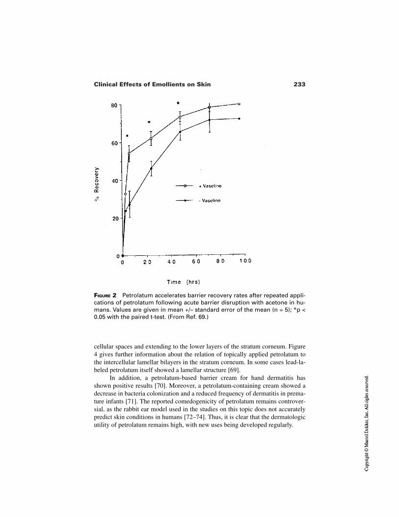
233Clinical Effects of Emollients on Skin
FIGURE 2 Petrolatum accelerates barrier recovery rates after repeated appli-cations of petrolatum following acute barrier disruption with acetone in hu-mans. Values are given in mean +/– standard error of the mean (n = 5); *p <0.05 with the paired t-test. (From Ref. 69.)
cellular spaces and extending to the lower layers of the stratum corneum. Figure4 gives further information about the relation of topically applied petrolatum tothe intercellular lamellar bilayers in the stratum corneum. In some cases lead-la-beled petrolatum itself showed a lamellar structure [69].
In addition, a petrolatum-based barrier cream for hand dermatitis hasshown positive results [70]. Moreover, a petrolatum-containing cream showed adecrease in bacteria colonization and a reduced frequency of dermatitis in prema-ture infants [71]. The reported comedogenicity of petrolatum remains controver-sial, as the rabbit ear model used in the studies on this topic does not accuratelypredict skin conditions in humans [72–74]. Thus, it is clear that the dermatologicutility of petrolatum remains high, with new uses being developed regularly.
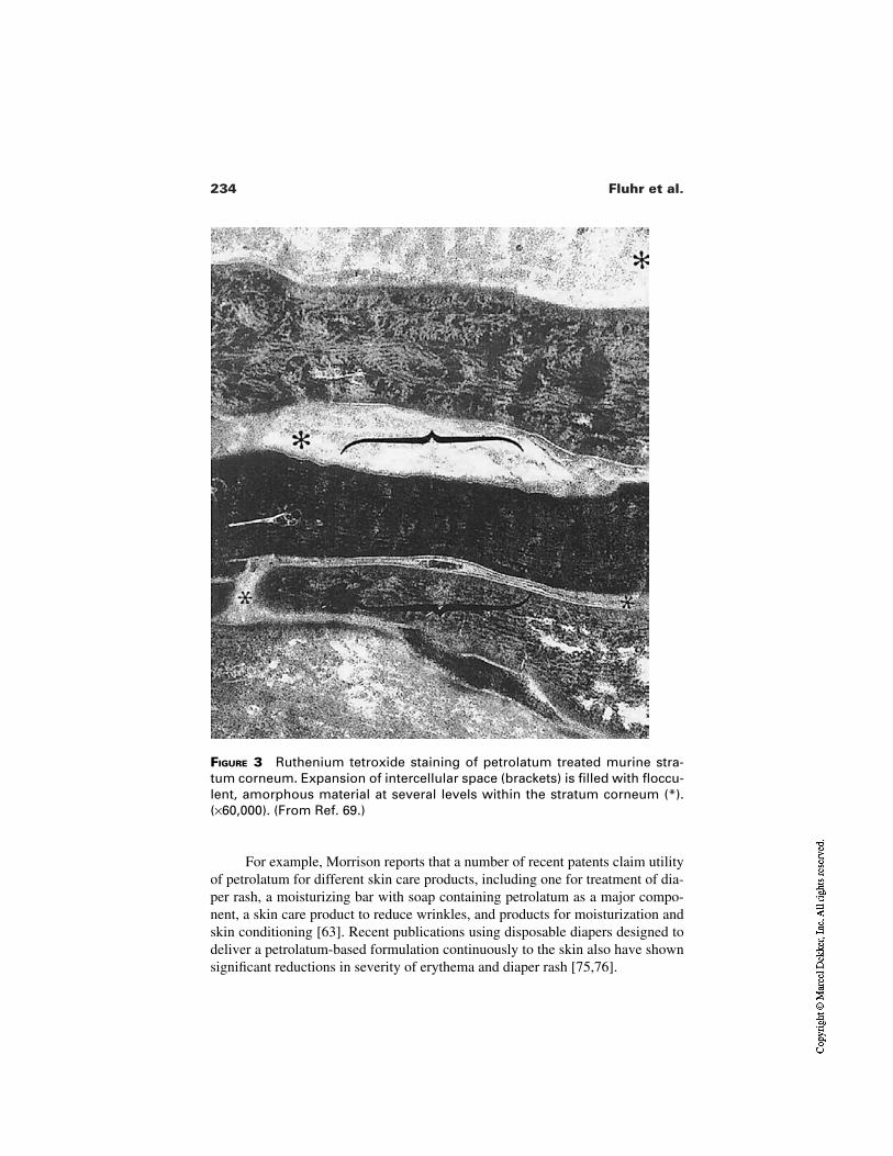
234 Fluhr et al.
FIGURE 3 Ruthenium tetroxide staining of petrolatum treated murine stra-tum corneum. Expansion of intercellular space (brackets) is filled with floccu-lent, amorphous material at several levels within the stratum corneum (*).(×60,000). (From Ref. 69.)
For example, Morrison reports that a number of recent patents claim utilityof petrolatum for different skin care products, including one for treatment of dia-per rash, a moisturizing bar with soap containing petrolatum as a major compo-nent, a skin care product to reduce wrinkles, and products for moisturization andskin conditioning [63]. Recent publications using disposable diapers designed todeliver a petrolatum-based formulation continuously to the skin also have shownsignificant reductions in severity of erythema and diaper rash [75,76].
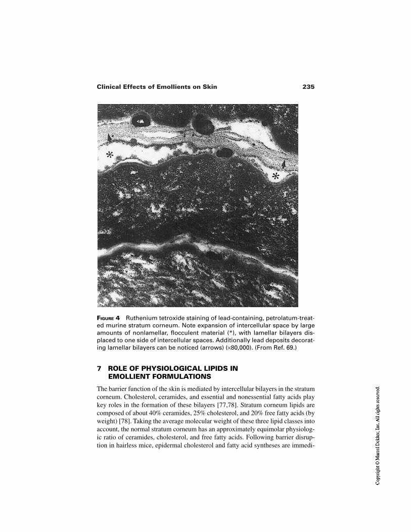
235Clinical Effects of Emollients on Skin
FIGURE 4 Ruthenium tetroxide staining of lead-containing, petrolatum-treat-ed murine stratum corneum. Note expansion of intercellular space by largeamounts of nonlamellar, flocculent material (*), with lamellar bilayers dis-placed to one side of intercellular spaces. Additionally lead deposits decorat-ing lamellar bilayers can be noticed (arrows) (×80,000). (From Ref. 69.)
7 ROLE OF PHYSIOLOGICAL LIPIDS IN EMOLLIENT FORMULATIONS
The barrier function of the skin is mediated by intercellular bilayers in the stratumcorneum. Cholesterol, ceramides, and essential and nonessential fatty acids playkey roles in the formation of these bilayers [77,78]. Stratum corneum lipids arecomposed of about 40% ceramides, 25% cholesterol, and 20% free fatty acids (byweight) [78]. Taking the average molecular weight of these three lipid classes intoaccount, the normal stratum corneum has an approximately equimolar physiolog-ic ratio of ceramides, cholesterol, and free fatty acids. Following barrier disrup-tion in hairless mice, epidermal cholesterol and fatty acid syntheses are immedi-
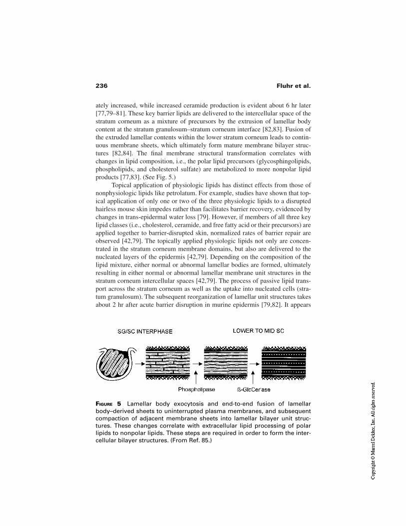
236 Fluhr et al.
ately increased, while increased ceramide production is evident about 6 hr later[77,79–81]. These key barrier lipids are delivered to the intercellular space of thestratum corneum as a mixture of precursors by the extrusion of lamellar bodycontent at the stratum granulosum–stratum corneum interface [82,83]. Fusion ofthe extruded lamellar contents within the lower stratum corneum leads to contin-uous membrane sheets, which ultimately form mature membrane bilayer struc-tures [82,84]. The final membrane structural transformation correlates withchanges in lipid composition, i.e., the polar lipid precursors (glycosphingolipids,phospholipids, and cholesterol sulfate) are metabolized to more nonpolar lipidproducts [77,83]. (See Fig. 5.)
Topical application of physiologic lipids has distinct effects from those ofnonphysiologic lipids like petrolatum. For example, studies have shown that top-ical application of only one or two of the three physiologic lipids to a disruptedhairless mouse skin impedes rather than facilitates barrier recovery, evidenced bychanges in trans-epidermal water loss [79]. However, if members of all three keylipid classes (i.e., cholesterol, ceramide, and free fatty acid or their precursors) areapplied together to barrier-disrupted skin, normalized rates of barrier repair areobserved [42,79]. The topically applied physiologic lipids not only are concen-trated in the stratum corneum membrane domains, but also are delivered to thenucleated layers of the epidermis [42,79]. Depending on the composition of thelipid mixture, either normal or abnormal lamellar bodies are formed, ultimatelyresulting in either normal or abnormal lamellar membrane unit structures in thestratum corneum intercellular spaces [42,79]. The process of passive lipid trans-port across the stratum corneum as well as the uptake into nucleated cells (stra-tum granulosum). The subsequent reorganization of lamellar unit structures takesabout 2 hr after acute barrier disruption in murine epidermis [79,82]. It appears
FIGURE 5 Lamellar body exocytosis and end-to-end fusion of lamellarbody–derived sheets to uninterrupted plasma membranes, and subsequentcompaction of adjacent membrane sheets into lamellar bilayer unit struc-tures. These changes correlate with extracellular lipid processing of polarlipids to nonpolar lipids. These steps are required in order to form the inter-cellular bilayer structures. (From Ref. 85.)
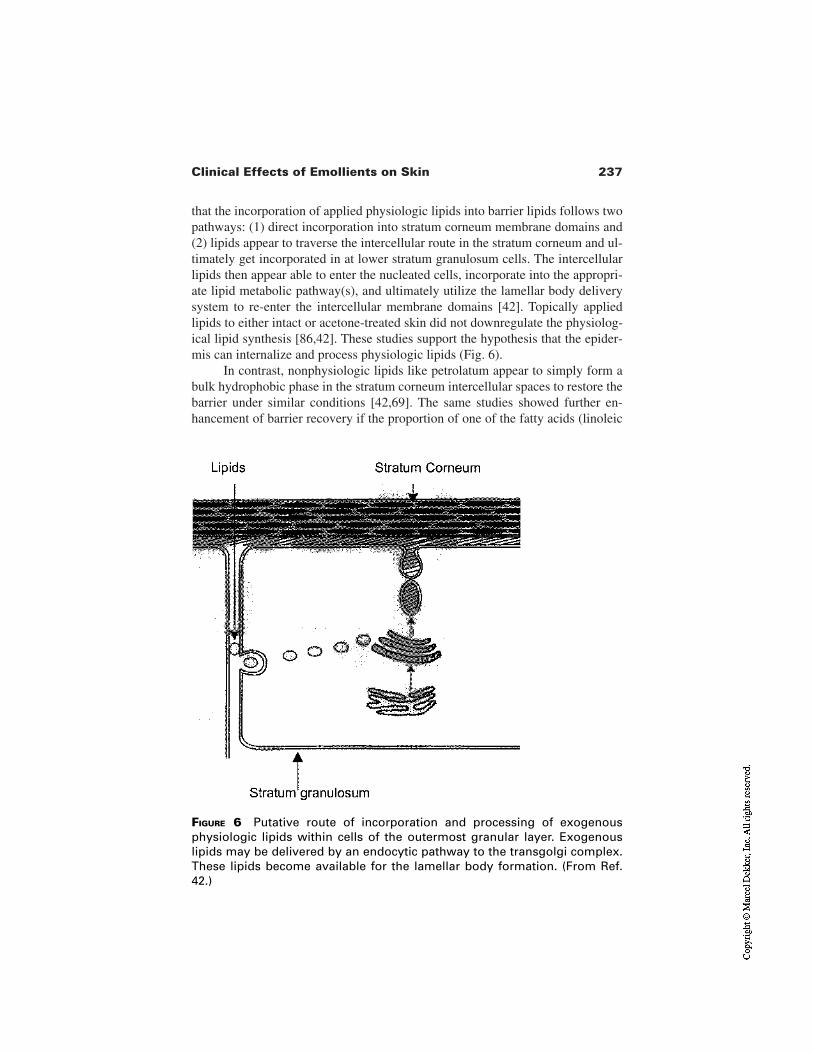
237Clinical Effects of Emollients on Skin
that the incorporation of applied physiologic lipids into barrier lipids follows twopathways: (1) direct incorporation into stratum corneum membrane domains and(2) lipids appear to traverse the intercellular route in the stratum corneum and ul-timately get incorporated in at lower stratum granulosum cells. The intercellularlipids then appear able to enter the nucleated cells, incorporate into the appropri-ate lipid metabolic pathway(s), and ultimately utilize the lamellar body deliverysystem to re-enter the intercellular membrane domains [42]. Topically appliedlipids to either intact or acetone-treated skin did not downregulate the physiolog-ical lipid synthesis [86,42]. These studies support the hypothesis that the epider-mis can internalize and process physiologic lipids (Fig. 6).
In contrast, nonphysiologic lipids like petrolatum appear to simply form abulk hydrophobic phase in the stratum corneum intercellular spaces to restore thebarrier under similar conditions [42,69]. The same studies showed further en-hancement of barrier recovery if the proportion of one of the fatty acids (linoleic
FIGURE 6 Putative route of incorporation and processing of exogenousphysiologic lipids within cells of the outermost granular layer. Exogenouslipids may be delivered by an endocytic pathway to the transgolgi complex.These lipids become available for the lamellar body formation. (From Ref.42.)
238 Fluhr et al.
acid, palmitic acid, or stearic acid) or the other key species was augmented tothreefold in a four-component system, i.e. consisting of fatty acid, ceramide, cho-lesterol, and essential fatty acids in a 3:1:1:1 ratio [43]. Interestingly, the structur-al requirements of this lipid mixture are not restricted to essential fatty acids, find-ings that were confirmed in similarly disrupted human barrier [43]. Alsointeresting, acylceramides applied as a single agent delayed barrier recovery.However, acylceramides in a mixture with cholesterol (optimum ratio of 1:5:1 or1:2) also revealed accelerated barrier recovery after acute barrier disruption [43].Moreover, in another study stratum corneum hydration (measured by conduc-tance) was increased 4 hr after topical application of cholesterol/acylceramide/petrolatum/glycerol-containing vehicle (propylene glycol/ethanol) [87]. Thesefindings were confirmed as accelerated barrier repair was noted using a similarformulation after tape-stripping, solvent treatment, and some types of detergenttreatment [88]. Specifically, topical application of the physiologic lipids choles-terol, ceramide, palmitate, and linoleate in the ratio of 4.3:2.3:1:1.08 showed en-hanced barrier recovery. However, it must be noted that in barrier repair versushydration studies, correlations between moisturizing properties and barrier repairmechanism of applied lipid mixtures are not always evident. Actually, the besthydrating lipid composition is often different from the optimal barrier repair for-mulation and vice versa [87].
8 CONCLUSIONS
In this chapter different roles of emollients have been presented, including emol-lient classification, with some examples of compounded emollients and specificeffects of emollients with special focus on barrier protection and barrier recovery.An extended discussion was presented on the special functions of lanolin, petro-latum, and physiological lipids in emollient formulation. Choosing the appropri-ate emollient for the treatment of different skin diseases may have a high impacton the clinical outcome of a treatment and even more on the relapse-free period.The chosen emollient should no longer be regarded simply as a drug carrier or ve-hicle or drug delivery system but as an essential part of topical treatment. Thus itmay be of importance to adapt type and composition of the emollient according tothe evolution of the disease either as an adjuvant treatment or as drug deliverysystem, especially in dermatological prescriptions. This review may help in a bet-ter understanding of specific function(s) of emollients and enable an evidence-based use of different emollient types and compositions.
REFERENCES
1. Gloor M, Thoma K, Fluhr J. Dermatologische Externatherapie. Berlin: Springer-Ver-lag, 2000.

239Clinical Effects of Emollients on Skin
2. Deutscher Arzneimittel-Codex. Eschborn: Govi Verlag, 1999.3. Lehmann L, Gloor M, Schlierbach S, Gehring W. Stabilität und Okklusivität von Ex-
ternagrundlagen auf der Haut. Z Hautkr 1997; 872:585–590.4. Blichmann CW, Serup J, Winther A. Effects of single application of a moisturizer:
evaporation of emulsion water, skin surface temperature, electrical conductance,electrical capacitance, and skin surface (emulsion) lipids. Acta Derm Venereol 1989;69:327–330.
5. Loden M. The increase in skin hydration after application of emollients with differ-ent amounts of lipids. Acta Derm Venereol 1992; 72:327–330.
6. Fluhr JW, Vrzak G, Gloor M. Hydratisierender und die Steroidpenetration modi-fizierender Effekt von Harnstoff und Glycerin in Abhängigkeit von der verwendetenGrundlage. Z Hautkr 1997; 73:210–214.
7. Fluhr JW, Gloor M, Lehmann L, Lazzerini S, Distante F, Berardesca E. Glycerol ac-celerates recovery of barrier function in vivo. Acta Derm Venereol 1999; 79:418–421.
8. Batt M, Fairhust E. Haydration of the stratum corneum. J Cosmet Sci 1986; 8.9. Jemec GB, Wulf HC. Correlation between the greasiness and the plasticizing effect
of moisturizers. Acta Derm Venereol 1999; 79:115–117.10. Fluhr JW, Gloor M, Lazzerini S, Kleesz P, Grieshaber R, Berardesca E. Comparative
study of five instruments measuring stratum corneum hydration (Corneometer CM820 and CM 825, Skicon 200, Nova DPM 90003, DermLab). Part I. In vitro. SkinRes Technol 1999; 5:161–170.
11. Jemec GB, Na R, Wulf HC. The inherent capacitance of moisturising creams: asource of false positive results? Skin Pharmacol Appl Skin Physiol 2000; 13:182–187.
12. Wigger-Alberti W, Elsner P. Barrier creams and emollients. In: Kanerva L, Elsner P,Wahlberg JE, Maibach HI, eds. Handbook of Occupational Dermatology. Berlin:Springer, 2000:490–496.
13. Grunewald AM, Gloor M, Gehring W, Kleesz P. Damage to the skin by repetitivewashing. Contact Dermatitis 1995; 32:225–232.
14. Wigger-Alberti W, Hinnen U, Elsner P. Predictive testing of metalworking fluids: acomparison of 2 cumulative human irritation models and correlation with epidemio-logical data. Contact Dermatitis 1997; 36:14–20.
15. Wigger-Alberti W, Rougier A, Richard A, Elsner P. Efficacy of protective creams ina modified repeated irritation test. Methodological aspects. Acta Derm Venereol1998; 78:270–273.
16. Wigger-Alberti W, Krebs A, Elsner P. Experimental irritant contact dermatitis due tocumulative epicutaneous exposure to sodium lauryl sulphate and toluene: single andconcurrent application. Br J Dermatol 2000; 143:551–556.
17. Frosch PJ, Kurte A. Efficacy of skin barrier creams. IV. The repetitive irritation test(RIT) with a set of 4 standard irritants. Contact Dermatitis 1994; 31:161–168.
18. Frosch PJ, Kurte A, Pilz B. Efficacy of skin barrier creams. III. The repetitive irrita-tion test (RIT) in humans. Contact Dermatitis 1993; 29:113–118.
19. Frosch PJ, Schulze-Dirks A, Hoffmann M, Axthelm I, Kurte A. Efficacy of skin bar-rier creams. I. The repetitive irritation test (RIT) in the guinea pig. Contact Dermati-tis 1993; 28:94–100.
240 Fluhr et al.
20. Berndt U, Wigger-Alberti W, Gabard B, Elsner P. Efficacy of a barrier cream and itsvehicle as protective measures against occupational irritant contact dermatitis. Con-tact Dermatitis 2000; 42:77–80.
21. Wigger-Alberti W, Maraffio B, Wernli M, Elsner P. Self-application of a protectivecream. Pitfalls of occupational skin protection. Arch Dermatol 1997; 133:861–864.
22. de Paepe K, Derde MP, Roseeuw D, Rogiers V. Claim substantiation and efficiencyof hydrating body lotions and protective creams. Contact Dermatitis 2000; 42:227–234.
23. Grunewald AM, Gloor M, Gehring W, Kleesz P. Efficacy of barrier creams. CurrProbl Dermatol 1995; 23:187–197.
24. Bettinger J, Gloor M, Gehring W. Influence of a pretreatment with emulsions on thedehydrating effect of the skin by surfactants. Int J Cosm Sci 1994; 16:53–60.
25. Held E, Sveinsdottir S, Agner T. Effect of long-term use of moisturizer on skin hy-dration, barrier function and susceptibility to irritants. Acta Derm Venereol 1999;79:49–51.
26. Zhai H, Maibach HI. Moisturizers in preventing irritant contact dermatitis: anoverview. Contact Dermatitis 1998; 38:241–244.
27. Ramsing DW, Agner T. Preventive and therapeutic effects of a moisturizer. An ex-perimental study of human skin. Acta Derm Venereol 1997; 77:335–337.
28. Harding CR, Watkinson A, Rawlings AV, Scott IR. Dry skin, moisturization and cor-neodesmolysis. Int J Cosmet Sci 2000; 22:21–52.
29. Fartasch M, Schnetz E, Diepgen TL. Characterization of detergent-induced barrieralterations—effect of barrier cream on irritation. J Invest Dermatol Symp Proc 1998;3:121–127.
30. Loden M, Olsson H, Axell T, Linde YW. Friction, capacitance and transepidermalwater loss (TEWL) in dry atopic and normal skin. Br J Dermatol 1992; 126:137–141.
31. Thune P. Evaluation of the hydration and the water-holding capacity in atopic skinand so-called dry skin. Acta Derm Venereol 1989; 144(Suppl):133–135.
32. Berardesca E, Fideli D, Borroni G, Rabbiosi G, Maibach H. In vivo hydration andwater-retention capacity of stratum corneum in clinically uninvolved skin in atopicand psoriatic patients. Acta Derm Venereol 1990; 70:400–404.
33. Di Nardo A, Wertz P, Giannetti A, Seidenari S. Ceramide and cholesterol composi-tion of the skin of patients with atopic dermatitis. Acta Derm Venereol 1998;78:27–30.
34. Gehring W, Gloor M, Kleesz P. Predictive washing test for evaluation of individualeczema risk. Contact Dermatitis 1998; 39:8–13.
35. Grunewald AM, Gloor M, Kleesz P. Barrier recompensation mechanisms. Curr ProblDermatol 1996; 25:206–213.
36. Imokawa G, Abe A, Jin K, Higaki Y, Kawashima M, Hidano A. Decreased level ofceramides in stratum corneum of atopic dermatitis: an etiologic factor in atopic dryskin? J Invest Dermatol 1991; 96:523–526.
37. Hara J, Higuchi K, Okamoto R, Kawashima M, Imokawa G. High expression ofsphingomyelin deacylase is an important determinant of ceramide deficiency leadingto barrier disruption in atopic dermatitis. J Invest Dermatol 2000; 115:406–413.
38. Higuchi K, Hara J, Okamoto R, Kawashima M, Imokawa G. The skin of atopic der-
241Clinical Effects of Emollients on Skin
matitis patients contains a novel enzyme, glucosylceramide sphingomyelin deacy-lase, which cleaves the N-acyl linkage of sphingomyelin and glucosylceramide.Biochem J 2000; 350:747–756.
39. Wirth H, Gloor M, Stoika D. Sebaceous glands in uninvolved skin of patients suffer-ing from atopic dermatitis. Arch Dermatol Res 1981; 270:167–169.
40. Fartasch M, Diepgen TL. The barrier function in atopic dry skin. Disturbance ofmembrane-coating granule exocytosis and formation of epidermal lipids? Acta DermVenereol 1992; 176(Suppl):26–31.
41. Loden M, Andersson AC, Lindberg M. Improvement in skin barrier function in pa-tients with atopic dermatitis after treatment with a moisturizing cream (Canoderm).Br J Dermatol 1999; 140:264–267.
42. Mao-Qiang M, Brown BE, Wu-Pong S, Feingold KR, Elias PM. Exogenous non-physiologic vs physiologic lipids. Divergent mechanisms for correction of perme-ability barrier dysfunction. Arch Dermatol 1995; 131:809–816.
43. Man MM, Feingold KR, Thornfeldt CR, Elias PM. Optimization of physiologicallipid mixtures for barrier repair. J Invest Dermatol 1996; 106:1096–1101.
44. Man MQ, Feingold KR, Elias PM. Exogenous lipids influence permeability barrierrecovery in acetone-treated murine skin. Arch Dermatol 1993; 129:728–738.
45. Gehring W, Forssman T, Jost G, Gloor M. Die keimreduzierende Wirkung vonErythromycin und Triclosan bei atopischer Dermatitis. Akt Dermatol 1996; 22:28–31.
46. Sugimoto K, Kuroki H, Kanazawa M, Kurosaki T, Abe H, Takahashi Y, Ishiwada N,Nezu Y, Hoshioka A, Toba T. New successful treatment with disinfectant for atopicdermatitis. Dermatology 1997; 195(Suppl 2):62–68.
47. Gehring W, Bopp R, Rippke F, Gloor M. Effect of topically applied evening prim-rose oil on epidermal barrier function in atopic dermatitis as a function of vehicle.Arzneimittelforschung 1999; 49:635–642.
48. Agner T. An experimental study of irritant effects of urea in different vehicles. ActaDerm Venereol 1992; 177(Suppl):44–46.
49. Gallenkemper G, Rabe E, Bauer R. Contact sensitization in chronic venous insuffi-ciency: modern wound dressings. Contact Dermatitis 1998; 38:274–278.
50. Pasche-Koo F, Piletta PA, Hunziker N, Hauser C. High sensitization rate to emulsi-fiers in patients with chronic leg ulcers. Contact Dermatitis 1994; 31:226–228.
51. Kligman AM. The myth of lanolin allergy: lanolin is not a contact sensitizer. In: TheLanolin Book. Hamburg: Beiersdorf, 1999:161–175.
52. Clark EW, Cronin E, Wilkinson DS. Lanolin with reduced sensitising potential: apreliminary report. Contact Dermatitis 1977; 3:69–73.
53. Clark EW. The history and evolution of lanolin. In: The Lanolin Book. Hamburg:Beiersdorf, 1999:15–50.
54. Clark EW. Estimation of general incidence of specific lanolin allergy. J Soc CosmetChem 1975; 26:323–335.
55. Dotterud LK, Falk ES. Contact allergy in relation to hand eczema and atopic dis-eases in north Norwegian schoolchildren. Acta Paediatr 1995; 84:402–406.
56. Giordano-Labadie F, Rance F, Pellegrin F, Bazex J, Dutau G, Schwarze HP. Fre-quency of contact allergy in children with atopic dermatitis: results of a prospectivestudy of 137 cases. Contact Dermatitis 1999; 40:192–195.
242 Fluhr et al.
57. Kligman A. Regression method for assessing the efficacy of moisturizers. CosmetToil 1978;93:27.
58. Moss J. The effect of three moisturisers on skin surface hydration. Skin Res Technol1996; 2:32.
59. Petersen EN. The hydrating effect of a cream and white petrolatum measured by op-tothermal infrared spectrometry in vivo. Acta Derm Venereol 1991; 71:373–376.
60. Clark EW. Short term penetration of lanolin into human stratum corneum. J Soc Cos-met Chem 1992; 43:219.
61. Elias P, Mao-Quiang M, Thornfeldt CR, Feingold KR. The epidermal permeabilitybarrier: effects of physiologic and non-physiologic lipids. In: The Lanolin Book.Hamburg: Beiersdorf, 1999:253–278.
62. Sauermann G, Schreiner V. The skin caring effect of topical products containinglanolin alcohols. In: The Lanolin Book. Hamburg: Beiersdorf, 1999:217–235.
63. Morrison DS. Petrolatum. In: Lodén M, Maibach HI, eds. Dry Skin and Moisturiz-ers. Boca Raton: CRC, 2000:251–257.
64. Foldvari M. Effect of vehicle on topical liposomal drug delivery: petrolatum bases. JMicroencapsul 1996; 13:589–600.
65. Lazar AP, Lazar P. Dry skin, water, and lubrication. Dermatol Clin 1991; 9:45–51.66. O’Goshi KI, Tabata N, Sato Y, Tagami H. Comparative study of the efficacy of vari-
ous moisturizers on the skin of the ASR miniature swine. Skin Pharmacol Appl SkinPhysiol 2000; 13:120–127.
67. Loden M, Lindberg M. The influence of a single application of different moisturizerson the skin capacitance. Acta Derm Venereol 1991; 71:79–82.
68. Pellacani G, Belletti B, Seidenari S. Evaluation of the short-term effects of skin careproducts: a comparison between capacitance values and echographic parameters ofepidermal hydration. Curr Probl Dermatol 1998; 26:177–182.
69. Ghadially R, Halkier-Sorensen L, Elias PM. Effects of petrolatum on stratumcorneum structure and function. J Am Acad Dermatol 1992; 26:387–396.
70. Schleicher SM, Milstein HJ, Ilowite R, Meyer P. Response of hand dermatitis to anew skin barrier-protectant cream. Cutis 1998; 61:233–234.
71. Nopper AJ, Horii KA, Sookdeo-Drost S, Wang TH, Mancini AJ, Lane AT. Topi-cal ointment therapy benefits premature infants [see comments]. J Pediatr 1996;128:660–669.
72. Zimmermann R. [The rabbit ear model as comedogenic test. 3. Histologic and en-zyme histochemical studies of the follicle and sebaceous gland epithelium]. Derma-tol Monatsschr 1990; 176:55–61.
73. Fulton JE Jr, Pay SR, Fulton JED. Comedogenicity of current therapeutic products,cosmetics, and ingredients in the rabbit ear. J Am Acad Dermatol 1984; 10:96–105.
74. Frank SB. Is the rabbit ear model, in its present state, prophetic of acnegenicity? JAm Acad Dermatol 1982; 6:373–377.
75. Odio MR, O’Connor RJ, Sarbaugh F, Baldwin S. Continuous topical administrationof a petrolatum formulation by a novel disposable diaper. 2. Effect on skin condition.Dermatology 2000; 200:238–243.
76. Odio MR, O’Connor RJ, Sarbaugh F, Baldwin S. Continuous topical administrationof a petrolatum formulation by a novel disposable diaper. 1. Effect on skin surfacemicrotopography. Dermatology 2000; 200:232–237.
243Clinical Effects of Emollients on Skin
77. Elias PM, Feingold KR. Lipids and the epidermal water barrier: metabolism, regula-tion, and pathophysiology. Semin Dermatol 1992; 11:176–182.
78. Schurer NY, Elias PM. The biochemistry and function of stratum corneum lipids.Adv Lipid Res 1991; 24:27–56.
79. Mao-Qiang M, Elias PM, Feingold KR. Fatty acids are required for epidermal per-meability barrier function. J Clin Invest 1993; 92:791–798.
80. Holleran WM, Man MQ, Gao WN, Menon GK, Elias PM, Feingold KR. Sphin-golipids are required for mammalian epidermal barrier function. Inhibition of sphin-golipid synthesis delays barrier recovery after acute perturbation. J Clin Invest 1991;88:1338–1345.
81. Feingold KR, Man MQ, Menon GK, Cho SS, Brown BE, Elias PM. Cholesterol syn-thesis is required for cutaneous barrier function in mice. J Clin Invest 1990;86:1738–1745.
82. Menon GK, Feingold KR, Elias PM. Lamellar body secretory response to barrierdisruption. J Invest Dermatol 1992; 98:279–289.
83. Elias PM, Menon GK. Structural and lipid biochemical correlates of the epidermalpermeability barrier. Adv Lipid Res 1991; 24:1–26.
84. Fartasch M, Bassukas ID, Diepgen TL. Structural relationship between epidermallipid lamellae, lamellar bodies and desmosomes in human epidermis: an ultrastruc-tural study. Br J Dermatol 1993; 128:1–9.
85. Elias PM. Stratum corneum architecture, metabolic activity and interactivity withsubjacent cell layers. Exp Dermatol 1996; 5:191–201.
86. Menon GK, Feingold KR, Moser AH, Brown BE, Elias PM. De novo sterologenesisin the skin. II. Regulation by cutaneous barrier requirements. J Lipid Res 1985;26:418–427.
87. Thornfeldt C. Critical and optimal molar ratios of key lipids. In: Lodén M, MaibachHI, eds. Dry Skin and Moisturizers. Boca Raton: CRC, 2000.
88. Yang L, Mao-Qiang M, Taljebini M, Elias PM, Feingold KR. Topical stratumcorneum lipids accelerate barrier repair after tape stripping, solvent treatment andsome but not all types of detergent treatment. Br J Dermatol 1995; 133:679–685.

13Humectants
Anthony V. RawlingsUnilever Research, Port Sunlight Laboratory, Bebington, Wirral, United Kingdom
Clive R. Harding and Allan WatkinsonUnilever Research, Colworth Laboratory, Sharnbrook, Bedford,United Kingdom
Prem Chandar and Ian R. ScottUnilever Research, Edgewater Laboratory, Edgewater, New Jersey
1 INTRODUCTION
Dry skin is a complex phenomenon in which the skin can feel rough, tight, anditchy and visibly look “dry” due to the appearance of macroscopic flakes or scaleon the skin surface [1]. To understand dry skin, we must first understand theprocesses taking place both in the epidermis and the stratum corneum, but partic-ularly within the superficial layers of the stratum corneum because this is ulti-mately where dry skin is manifest. Dry skin results from a perturbation in theprocess of desquamation, the progressive degradation of the cohesive forcesbinding the corneocytes of the stratum corneum [2]. In healthy skin, desquama-tion is a carefully regulated process in which the surface corneocytes are shed incareful balance with the underlying formation of new corneocytes at the stratumgranulosum/corneum boundary, without compromising the overall integrity ofthis critical tissue. Desquamation is not only responsible for maintaining stratum
245
246 Rawlings et al.
corneum thickness, but by ensuring a continual turnover of corneocytes it alsoprotects against the ever-present damaging effects of the environment. The majorevent occurring in desquamation is the controlled degradation of the cor-neodesmosomes, rivet-like protein complexes that, by linking neighboring cor-neocytes, represent the principal cohesive element of the tissue. Less well-under-stood changes in lipid organization and phase behavior also play a role infacilitating this process. Under normal conditions, the corneodesmosomes are de-graded by proteases, located in the stratum corneum intercellular space, whichhydrolyze the binding regions of these structures. (For reviews see Refs. 3 and 4.)
Fundamentally, dry skin occurs when the desquamation process is per-turbed and the peripheral corneodesmosomes are not degraded, resulting in an ac-cumulation of cohesive rafts of corneocytes on the skin’s surface [5]. These accu-mulations of surface corneocytes are manifest as dry flaky scale. Probably themajor extrinsic factor involved in perturbation of the desquamation process is re-duced humidity, although low temperature and UV damage can also precipitatethis condition [6]. Low environmental humidity increases the desiccation stresson the outermost layers of the stratum corneum, leading to a reduction in watercontent. Since the desquamatory enzymes require water for functionality, the re-duced water content then leads to a decrease in the activity of these hydrolytic en-zymes, perturbed corneodesmosomal hydrolysis resulting in skin scale [7].
Due to the crucial importance of water to the desquamatory process, themost effective treatment for common dry skin ailments is moisturization, a factinitially demonstrated by Irwin Blank in the 1950s [8]. His demonstration that thelow moisture content of the skin was the primary factor in causing the dry skincondition was the beginning of moisturization research as we know it.
2 WATER AND THE STRATUM CORNEUM
As the main barrier tissue against the environment, the major role of the stratumcorneum is to prevent water loss. This is primarily achieved by the network of or-ganized lipid lamellae surrounding the corneocytes, which are highly effective atreducing water flux through this tissue, although without question the physicalpresence of the multiple layers of corneocytes play a significant role [9]. Exceptduring water immersion or with total skin occlusion, we are constantly losing wa-ter to the atmosphere. However in desiccating conditions, where the external hu-midity is less than 100%, water will be continuously transferred outward and belost from the skin. This is referred to as trans-epidermal water loss (TEWL) and isgeneral regarded as a measure of the barrier competency [10,11]. This water lossfrom the surface of the stratum corneum is replenished from the hydrated tissuesbelow, resulting in a water flux across the skin [12].
Paradoxically, the stratum corneum must not only be an efficient barrier towater loss, but must itself retain water to allow the hydrolytic events essential for
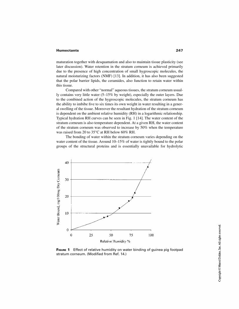
247Humectants
FIGURE 1 Effect of relative humidity on water binding of guinea pig footpadstratum corneum. (Modified from Ref. 14.)
maturation together with desquamation and also to maintain tissue plasticity (seelater discussion). Water retention in the stratum corneum is achieved primarilydue to the presence of high concentration of small hygroscopic molecules, thenatural moisturizing factors (NMF) [13]. In addition, it has also been suggestedthat the polar barrier lipids, the ceramides, also function to retain water withinthis tissue.
Compared with other “normal” aqueous tissues, the stratum corneum usual-ly contains very little water (5–15% by weight), especially the outer layers. Dueto the combined action of the hygroscopic molecules, the stratum corneum hasthe ability to imbibe five to six times its own weight in water resulting in a gener-al swelling of the tissue. Moreover the resultant hydration of the stratum corneumis dependent on the ambient relative humidity (RH) in a logarithmic relationship.Typical hydration RH curves can be seen in Fig. 1 [14]. The water content of thestratum corneum is also temperature dependent. At a given RH, the water contentof the stratum corneum was observed to increase by 50% when the temperaturewas raised from 20 to 35°C at RH below 60% RH.
The bonding of water within the stratum corneum varies depending on thewater content of the tissue. Around 10–15% of water is tightly bound to the polargroups of the structural proteins and is essentially unavailable for hydrolytic
248 Rawlings et al.
processes. As the water content increases, the water is less tightly bound; andabove about 40% it essentially acts as free bulk water [15].
3 MOISTURIZERS AND HUMECTANTS
It is not surprising, regarding the importance of water to the stratum corneummaturation process that moisturization is still the most effective treatment avail-able for common dry skin. However, despite the importance of water to thedesquamatory process and tissue plasticity, topically applied water is in fact apoor treatment for dry skin. It can initially alleviate the signs of dry skin; howev-er, the effect of water in any moisturizing product only provides short-term reliefof dry skin (minutes) as it quickly evaporates from the skin’s surface and does notaddress the underlying problem of impaired enzyme activity.
As exogenous water alone is insufficient to moisturize the skin and correctthe aberrant desquamatory process, cosmetic treatments to alleviate the conditionare required, i.e., moisturizers.
Classically, three approaches can be used to moisturize the stratumcorneum: (1) emolliency (to mask the rough scaly condition); (2) occlusion (to re-duce water loss from the skin); or (3) humectancy (to help retain water in theskin). The last two routes work by retaining water in the stratum corneum, whichwould be naturally lost from the body by TEWL. The humectancy route can alsoattract water to the skin from the outside but only under high humidity conditions.In reality modern commercial moisturizers act by a combination of these effects,normalizing the water content of the upper layers of the stratum corneum and, byincreasing enzyme activity, lead to an improvement in the natural exfoliationprocess and a smoother moisturized skin surface [7]. In this chapter we review thefunction of humectants and how, by helping to retain water, they affect the biolo-gy of the stratum corneum.
Humectants are water-soluble organic compounds, typically polyhydric al-cohols (polyols) that can imbibe water. Indeed, these can be thought of as the cos-metic equivalents of the NMF. The most common is glycerol, but other examplesinclude sorbitol, propylene glycerol, butylene glycol, urea, sodium lactate, andsodium pyrollidone carboxylic acid (PCA) (the last three being intrinsic moistur-izers produced within the stratum corneum).
The efficacy of humectants can be determined using a simple hygroscopicmeasurement. The humectant is equilibrated in an atmosphere of defined constantrelative humidity and weighed, and the final weight is compared to the value forthe dry weight of the humectant, determined after treatment with phosphoric ox-ide (Table 1). Alternatively, a more realistic method of determining humectancyinvolves treating samples of stratum corneum with the agent and then incubatingunder defined humidities and determining water uptake [16]. In both of these as-says, the sodium and ammonium salts of PCA and lactic acid are the most effec-
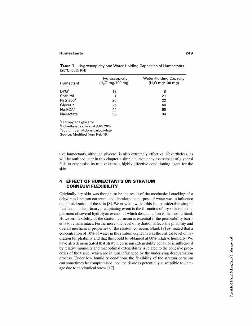
249Humectants
TABLE 1 Hygroscopicity and Water-Holding Capacities of Humectants(25°C, 50% RH)
HumectantHygroscopicity
(H2O mg/100 mg)Water-Holding Capacity
(H2O mg/100 mg)
DPG1 12 8Sorbitol 1 21PEG 2002 20 22Glycerin 25 40Na-PCA3 44 60Na-lactate 56 84
1Dipropylene glycerol2Polyethylene glycerol (MW 200)3Sodium pyrrolidone carboxylateSource: Modified from Ref. 16.
tive humectants, although glycerol is also extremely effective. Nevertheless, aswill be outlined later in this chapter a simple humectancy assessment of glycerolfails to emphasise its true value as a highly effective conditioning agent for theskin.
4 EFFECT OF HUMECTANTS ON STRATUMCORNEUM FLEXIBILITY
Originally dry skin was thought to be the result of the mechanical cracking of adehydrated stratum corneum, and therefore the purpose of water was to influencethe plasticization of the skin [8]. We now know that this is a considerable simpli-fication, and the primary precipitating event in the formation of dry skin is the im-pairment of several hydrolytic events, of which desquamation is the most critical.However, flexibility of the stratum corneum is essential if the permeability barri-er is to remain intact. Furthermore, the level of hydration affects the pliability andoverall mechanical properties of the stratum corneum. Blank [8] estimated that aconcentration of 10% of water in the stratum corneum was the critical level of hy-dration for pliability and that this could be obtained at 60% relative humidity. Wehave also demonstrated that stratum corneum extensibility behavior is influencedby relative humidity and that optimal extensibility is related to the cohesive prop-erties of the tissue, which are in turn influenced by the underlying desquamationprocess. Under low humidity conditions the flexibility of the stratum corneumcan sometimes be compromised, and the tissue is potentially susceptible to dam-age due to mechanical stress [17].
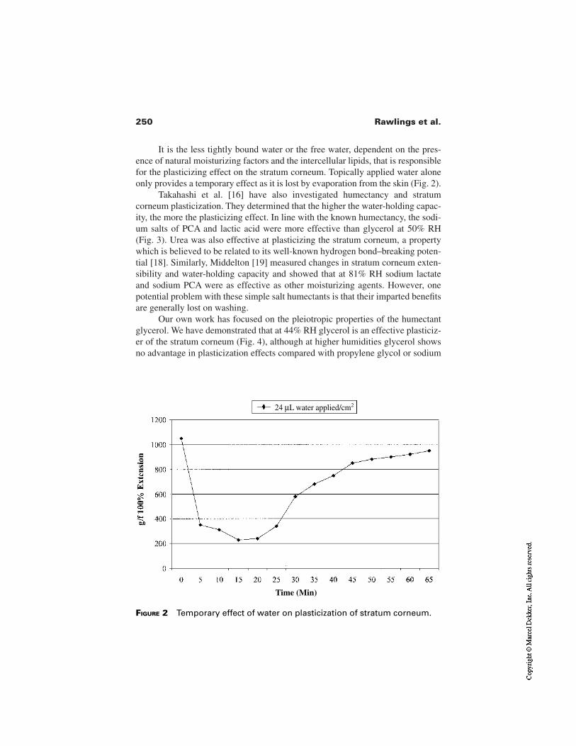
250 Rawlings et al.
It is the less tightly bound water or the free water, dependent on the pres-ence of natural moisturizing factors and the intercellular lipids, that is responsiblefor the plasticizing effect on the stratum corneum. Topically applied water aloneonly provides a temporary effect as it is lost by evaporation from the skin (Fig. 2).
Takahashi et al. [16] have also investigated humectancy and stratumcorneum plasticization. They determined that the higher the water-holding capac-ity, the more the plasticizing effect. In line with the known humectancy, the sodi-um salts of PCA and lactic acid were more effective than glycerol at 50% RH(Fig. 3). Urea was also effective at plasticizing the stratum corneum, a propertywhich is believed to be related to its well-known hydrogen bond–breaking poten-tial [18]. Similarly, Middelton [19] measured changes in stratum corneum exten-sibility and water-holding capacity and showed that at 81% RH sodium lactateand sodium PCA were as effective as other moisturizing agents. However, onepotential problem with these simple salt humectants is that their imparted benefitsare generally lost on washing.
Our own work has focused on the pleiotropic properties of the humectantglycerol. We have demonstrated that at 44% RH glycerol is an effective plasticiz-er of the stratum corneum (Fig. 4), although at higher humidities glycerol showsno advantage in plasticization effects compared with propylene glycol or sodium
FIGURE 2 Temporary effect of water on plasticization of stratum corneum.
24 µL water applied/cm2
Time (Min)
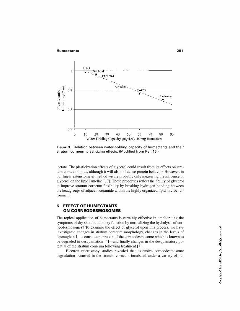
251Humectants
FIGURE 3 Relation between water-holding capacity of humectants and theirstratum corneum plasticizing effects. (Modified from Ref. 16.)
lactate. The plasticization effects of glycerol could result from its effects on stra-tum corneum lipids, although it will also influence protein behavior. However, inour linear extensiometer method we are probably only measuring the influence ofglycerol on the lipid lamellae [17]. These properties reflect the ability of glycerolto improve stratum corneum flexibility by breaking hydrogen bonding betweenthe headgroups of adjacent ceramide within the highly organized lipid microenvi-ronment.
5 EFFECT OF HUMECTANTS ON CORNEODESMOSOMES
The topical application of humectants is certainly effective in ameliorating thesymptoms of dry skin, but do they function by normalizing the hydrolysis of cor-neodesmosomes? To examine the effect of glycerol upon this process, we haveinvestigated changes in stratum corneum morphology, changes in the levels ofdesmoglein 1—a constituent protein of the corneodesmosome which is known tobe degraded in desquamation [4]—and finally changes in the desquamatory po-tential of the stratum corneum following treatment [7].
Electron microscopy studies revealed that extensive corneodesmosomedegradation occurred in the stratum corneum incubated under a variety of hu-
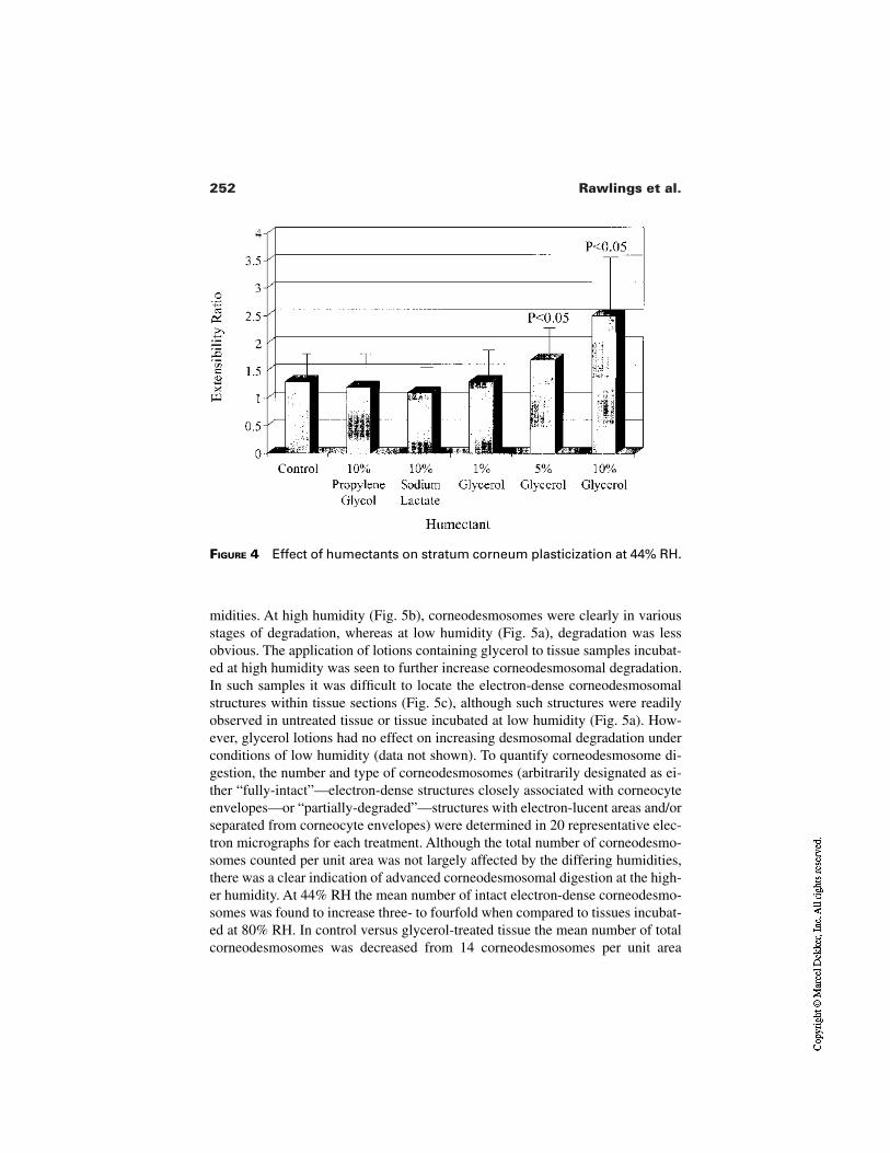
252 Rawlings et al.
FIGURE 4 Effect of humectants on stratum corneum plasticization at 44% RH.
midities. At high humidity (Fig. 5b), corneodesmosomes were clearly in variousstages of degradation, whereas at low humidity (Fig. 5a), degradation was lessobvious. The application of lotions containing glycerol to tissue samples incubat-ed at high humidity was seen to further increase corneodesmosomal degradation.In such samples it was difficult to locate the electron-dense corneodesmosomalstructures within tissue sections (Fig. 5c), although such structures were readilyobserved in untreated tissue or tissue incubated at low humidity (Fig. 5a). How-ever, glycerol lotions had no effect on increasing desmosomal degradation underconditions of low humidity (data not shown). To quantify corneodesmosome di-gestion, the number and type of corneodesmosomes (arbitrarily designated as ei-ther “fully-intact”—electron-dense structures closely associated with corneocyteenvelopes—or “partially-degraded”—structures with electron-lucent areas and/orseparated from corneocyte envelopes) were determined in 20 representative elec-tron micrographs for each treatment. Although the total number of corneodesmo-somes counted per unit area was not largely affected by the differing humidities,there was a clear indication of advanced corneodesmosomal digestion at the high-er humidity. At 44% RH the mean number of intact electron-dense corneodesmo-somes was found to increase three- to fourfold when compared to tissues incubat-ed at 80% RH. In control versus glycerol-treated tissue the mean number of totalcorneodesmosomes was decreased from 14 corneodesmosomes per unit area
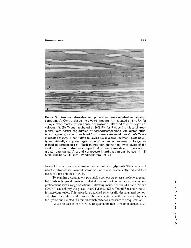
253Humectants
FIGURE 5 Osmium tetroxide– and potassium ferrocyanide–fixed stratumcorneum. (A) Control tissue, no glycerol treatment, incubated at 44% RH for7 days. Note intact electron-dense desmosomes attached to corneocyte en-velopes (*). (B) Tissue incubated at 80% RH for 7 days (no glycerol treat-ment). Note partial degradation of corneodesmosomes, vacuolated struc-tures beginning to be dissociated from corneocyte envelopes (*). (C) Tissueincubated at 80% RH for 7 days following 5% glycerol treatment. Note pauci-ty and virtually complete degradation of corneodesmosomes no longer at-tached to corneocytes (*). Each micrograph shows the lower levels of thestratum corneum (stratum compactum) where corneodesmosomes are ingreater abundance. Areas of corneocyte interdigitation can be seen in (B)(×200,000; bar = 0.05 mm). (Modified from Ref. 7.)
(control tissue) to 4 corneodesmosomes per unit area (glycerol). The numbers ofintact electron-dense corneodesmosomes were also dramatically reduced to amean of 1 per unit area (Fig. 6).
To examine desquamatory potential, a corneocyte release model was estab-lished where biopsied skin was incubated at a variety of humidities with or withoutpretreatment with a range of lotions. Following incubation for 24 hr at 20°C and80% RH, each biopsy was placed into 0.1M Tris-HCl buffer, pH 8.0, and vortexedin microfuge tubes. This procedure detached functionally desquamated corneo-cytes from the surface of the biopsy. The corneocytes were then recovered by cen-trifugation and counted in a microhaematometer as a measure of desquamation.
As can be seen from Fig. 7, the desquamation rates for skin incubated at 80
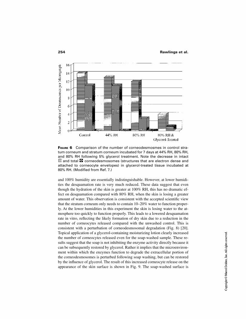
254 Rawlings et al.
FIGURE 6 Comparison of the number of corneodesmosmes in control stra-tum corneum and stratum corneum incubated for 7 days at 44% RH, 80% RH,and 80% RH following 5% glycerol treatment. Note the decrease in intact
and total corneodesmosomes (structures that are electron dense andattached to corneocyte envelopes) in glycerol-treated tissue incubated at80% RH. (Modified from Ref. 7.)
and 100% humidity are essentially indistinguishable. However, at lower humidi-ties the desquamation rate is very much reduced. These data suggest that eventhough the hydration of the skin is greater at 100% RH, this has no dramatic ef-fect on desquamation compared with 80% RH, when the skin is losing a greateramount of water. This observation is consistent with the accepted scientific viewthat the stratum corneum only needs to contain 10–20% water to function proper-ly. At the lower humidities in this experiment the skin is losing water to the at-mosphere too quickly to function properly. This leads to a lowered desquamationrate in vitro, reflecting the likely formation of dry skin due to a reduction in thenumber of corneocytes released compared with the unwashed control. This isconsistent with a perturbation of corneodesmosomal degradation (Fig. 8) [20].Topical application of a glycerol-containing moisturizing lotion clearly increasedthe number of corneocytes released even for the soap-washed sample. These re-sults suggest that the soap is not inhibiting the enzyme activity directly because itcan be subsequently restored by glycerol. Rather it implies that the microenviron-ment within which the enzymes function to degrade the extracellular portion ofthe corneodesmosomes is perturbed following soap washing, but can be restoredby the influence of glycerol. The result of this increased corneocyte release on theappearance of the skin surface is shown in Fig. 9. The soap-washed surface is
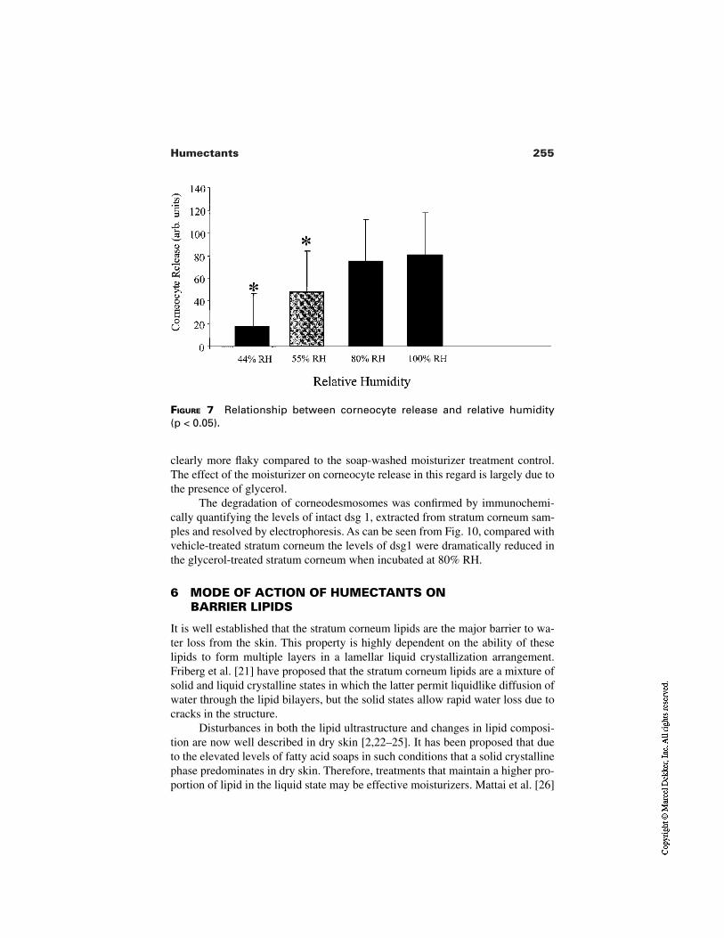
255Humectants
FIGURE 7 Relationship between corneocyte release and relative humidity (p < 0.05).
clearly more flaky compared to the soap-washed moisturizer treatment control.The effect of the moisturizer on corneocyte release in this regard is largely due tothe presence of glycerol.
The degradation of corneodesmosomes was confirmed by immunochemi-cally quantifying the levels of intact dsg 1, extracted from stratum corneum sam-ples and resolved by electrophoresis. As can be seen from Fig. 10, compared withvehicle-treated stratum corneum the levels of dsg1 were dramatically reduced inthe glycerol-treated stratum corneum when incubated at 80% RH.
6 MODE OF ACTION OF HUMECTANTS ON BARRIER LIPIDS
It is well established that the stratum corneum lipids are the major barrier to wa-ter loss from the skin. This property is highly dependent on the ability of theselipids to form multiple layers in a lamellar liquid crystallization arrangement.Friberg et al. [21] have proposed that the stratum corneum lipids are a mixture ofsolid and liquid crystalline states in which the latter permit liquidlike diffusion ofwater through the lipid bilayers, but the solid states allow rapid water loss due tocracks in the structure.
Disturbances in both the lipid ultrastructure and changes in lipid composi-tion are now well described in dry skin [2,22–25]. It has been proposed that dueto the elevated levels of fatty acid soaps in such conditions that a solid crystallinephase predominates in dry skin. Therefore, treatments that maintain a higher pro-portion of lipid in the liquid state may be effective moisturizers. Mattai et al. [26]
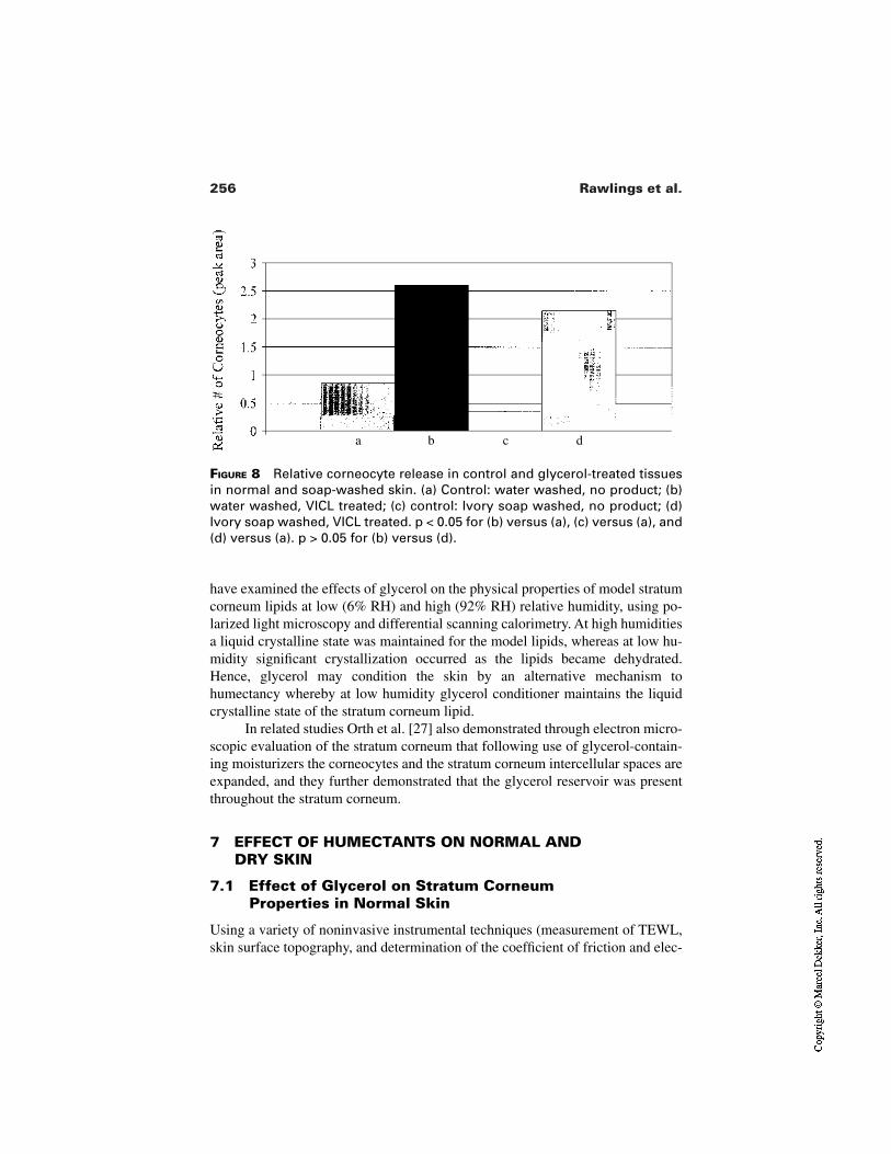
256 Rawlings et al.
FIGURE 8 Relative corneocyte release in control and glycerol-treated tissuesin normal and soap-washed skin. (a) Control: water washed, no product; (b)water washed, VICL treated; (c) control: Ivory soap washed, no product; (d)Ivory soap washed, VICL treated. p < 0.05 for (b) versus (a), (c) versus (a), and(d) versus (a). p > 0.05 for (b) versus (d).
a b c d
have examined the effects of glycerol on the physical properties of model stratumcorneum lipids at low (6% RH) and high (92% RH) relative humidity, using po-larized light microscopy and differential scanning calorimetry. At high humiditiesa liquid crystalline state was maintained for the model lipids, whereas at low hu-midity significant crystallization occurred as the lipids became dehydrated.Hence, glycerol may condition the skin by an alternative mechanism tohumectancy whereby at low humidity glycerol conditioner maintains the liquidcrystalline state of the stratum corneum lipid.
In related studies Orth et al. [27] also demonstrated through electron micro-scopic evaluation of the stratum corneum that following use of glycerol-contain-ing moisturizers the corneocytes and the stratum corneum intercellular spaces areexpanded, and they further demonstrated that the glycerol reservoir was presentthroughout the stratum corneum.
7 EFFECT OF HUMECTANTS ON NORMAL AND DRY SKIN
7.1 Effect of Glycerol on Stratum CorneumProperties in Normal Skin
Using a variety of noninvasive instrumental techniques (measurement of TEWL,skin surface topography, and determination of the coefficient of friction and elec-
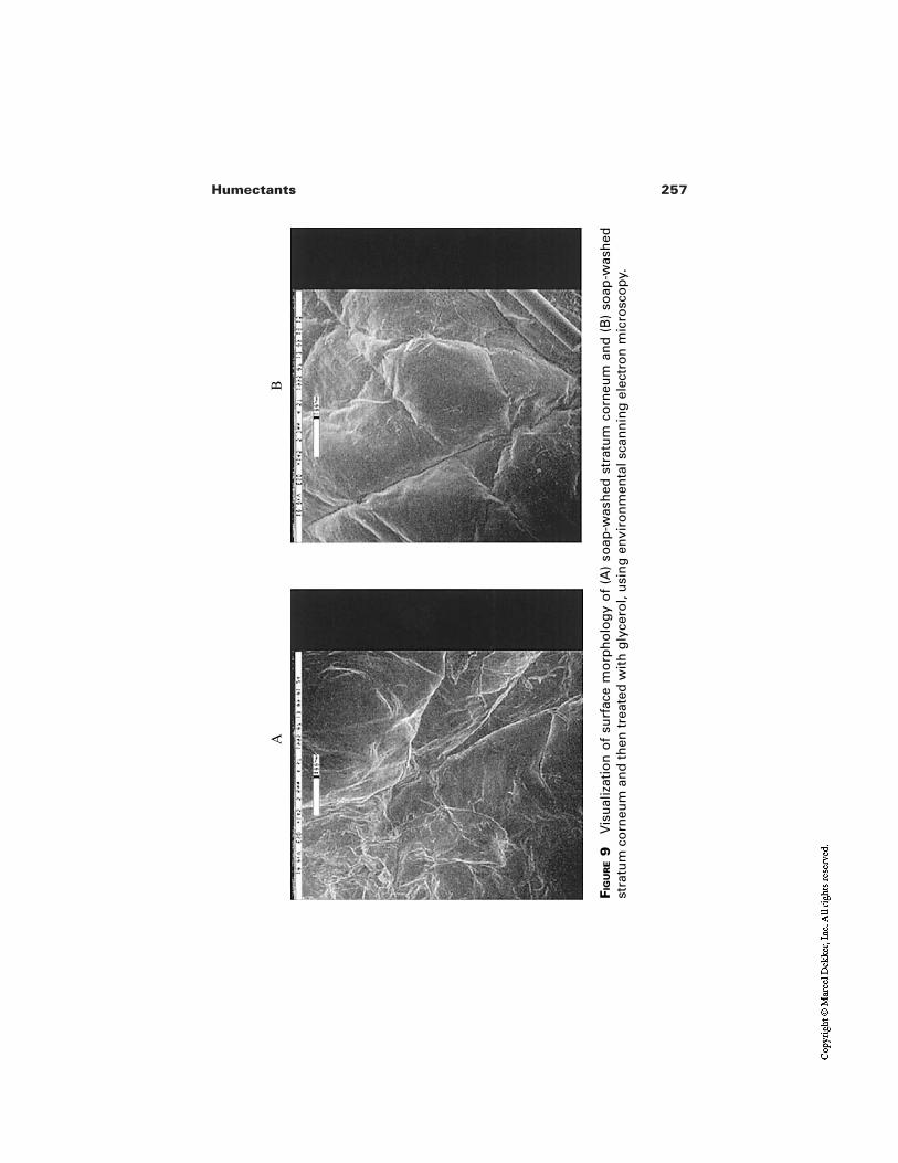
257Humectants
FIG
UR
E9
Vis
ual
izat
ion
of
surf
ace
mo
rph
olo
gy
of
(A)
soap
-was
hed
str
atu
m c
orn
eum
an
d (
B)
soap
-was
hed
stra
tum
co
rneu
m a
nd
th
en t
reat
ed w
ith
gly
cero
l, u
sin
g e
nvi
ron
men
tal s
can
nin
g e
lect
ron
mic
rosc
op
y.
AB
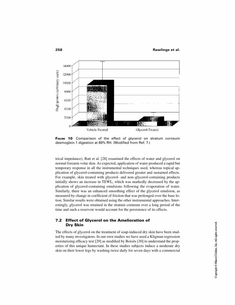
258 Rawlings et al.
FIGURE 10 Comparison of the effect of glycerol on stratum corneumdesmoglein 1 digestion at 80% RH. (Modified from Ref. 7.)
trical impedance), Batt et al. [28] examined the effects of water and glycerol onnormal forearm volar skin. As expected, application of water produced a rapid buttemporary response in all the instrumental techniques used, whereas topical ap-plication of glycerol-containing products delivered greater and sustained effects.For example, skin treated with glycerol- and non–glycerol-containing productsinitially shows an increase in TEWL, which was markedly decreased by the ap-plication of glycerol-containing emulsions following the evaporation of water.Similarly, there was an enhanced smoothing effect of the glycerol emulsion, asmeasured by change in coefficient of friction that was prolonged over the base lo-tion. Similar results were obtained using the other instrumental approaches. Inter-estingly, glycerol was retained in the stratum corneum over a long period of thetime and such a reservoir would account for the persistence of its effects.
7.2 Effect of Glycerol on the Amelioration of Dry Skin
The effects of glycerol on the treatment of soap-induced dry skin have been stud-ied by many investigators. In our own studies we have used a Kligman regressionmoisturizing efficacy test [29] as modified by Boisits [30] to understand the prop-erties of this unique humectant. In these studies subjects induce a moderate dryskin on their lower legs by washing twice daily for seven days with a commercial
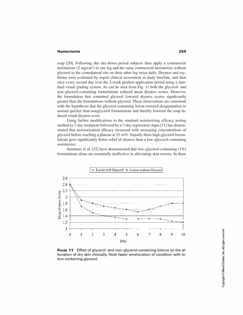
259Humectants
soap [20]. Following this dry-down period subjects then apply a commercialmoisturizer (2 mg/cm2) to one leg and the same commercial moisturizer withoutglycerol to the contralateral site on their other leg twice daily. Dryness and ery-thema were evaluated by expert clinical assessment at study baseline, and thenonce every second day over the 2-week product application period using a stan-dard visual grading system. As can be seen from Fig. 11 both the glycerol- andnon–glycerol-containing formulations reduced mean dryness scores. However,the formulation that contained glycerol lowered dryness scores significantlygreater than the formulations without glycerol. These observations are consistentwith the hypothesis that the glycerol-containing lotion restored desquamation tonormal quicker than nonglycerol formulations and thereby lowered the soap in-duced visual dryness score.
Using further modifications to the standard moisturizing efficacy testingmethod (a 7-day treatment followed by a 7-day regression) Appa [31] has demon-strated that moisturization efficacy increased with increasing concentrations ofglycerol before reaching a plateau at 25 wt%. Equally these high-glycerol formu-lations gave significantly better relief of dryness than a low–glycerol-containingmoisturizer.
Summers et al. [32] have demonstrated that low–glycerol-containing (1%)formulations alone are essentially ineffective in alleviating skin xerosis. In these
FIGURE 11 Effect of glycerol- and non–glycerol-containing lotions on the al-leviation of dry skin clinically. Note faster amelioration of condition with lo-tion containing glycerol.
Lotion without Glycerol
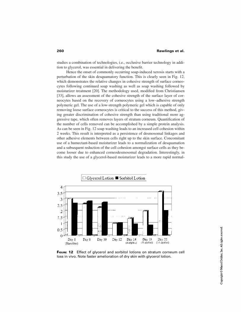
260 Rawlings et al.
studies a combination of technologies, i.e., occlusive barrier technology in addi-tion to glycerol, was essential in delivering the benefit.
Hence the onset of commonly occurring soap-induced xerosis starts with aperturbation of the skin desquamatory function. This is clearly seen in Fig. 12,which demonstrates the relative changes in cohesive strength of surface corneo-cytes following continued soap washing as well as soap washing followed bymoisturizer treatment [20]. The methodology used, modified from Christiansen[33], allows an assessment of the cohesive strength of the surface layer of cor-neocytes based on the recovery of corneocytes using a low–adhesive strengthpolymeric gel. The use of a low-strength polymeric gel which is capable of onlyremoving loose surface corneocytes is critical to the success of this method, giv-ing greater discrimination of cohesive strength than using traditional more ag-gressive tape, which often removes layers of stratum corneum. Quantification ofthe number of cells removed can be accomplished by a simple protein analysis.As can be seen in Fig. 12 soap washing leads to an increased cell cohesion within2 weeks. This result is interpreted as a persistence of desmosomal linkages andother adhesive elements between cells right up to the skin surface. Concomitantuse of a humectant-based moisturizer leads to a normalization of desquamationand a subsequent reduction of the cell cohesion amongst surface cells as they be-come looser due to enhanced corneodesmosomal degradation. Interestingly, inthis study the use of a glycerol-based moisturizer leads to a more rapid normal-
FIGURE 12 Effect of glycerol and sorbitol lotions on stratum corneum cellloss in vivo. Note faster amelioration of dry skin with glycerol lotion.
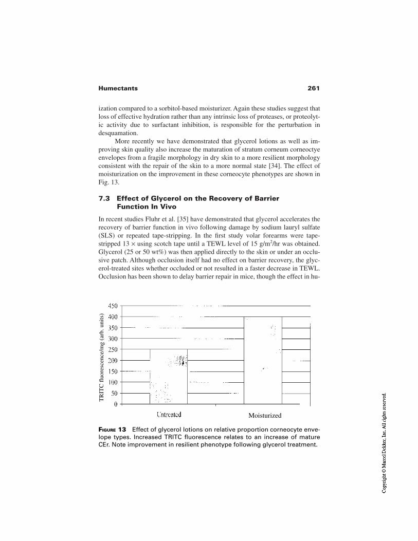
261Humectants
ization compared to a sorbitol-based moisturizer. Again these studies suggest thatloss of effective hydration rather than any intrinsic loss of proteases, or proteolyt-ic activity due to surfactant inhibition, is responsible for the perturbation indesquamation.
More recently we have demonstrated that glycerol lotions as well as im-proving skin quality also increase the maturation of stratum corneum corneoctyeenvelopes from a fragile morphology in dry skin to a more resilient morphologyconsistent with the repair of the skin to a more normal state [34]. The effect ofmoisturization on the improvement in these corneocyte phenotypes are shown inFig. 13.
7.3 Effect of Glycerol on the Recovery of BarrierFunction In Vivo
In recent studies Fluhr et al. [35] have demonstrated that glycerol accelerates therecovery of barrier function in vivo following damage by sodium lauryl sulfate(SLS) or repeated tape-stripping. In the first study volar forearms were tape-stripped 13 × using scotch tape until a TEWL level of 15 g/m2/hr was obtained.Glycerol (25 or 50 wt%) was then applied directly to the skin or under an occlu-sive patch. Although occlusion itself had no effect on barrier recovery, the glyc-erol-treated sites whether occluded or not resulted in a faster decrease in TEWL.Occlusion has been shown to delay barrier repair in mice, though the effect in hu-
FIGURE 13 Effect of glycerol lotions on relative proportion corneocyte enve-lope types. Increased TRITC fluorescence relates to an increase of matureCEr. Note improvement in resilient phenotype following glycerol treatment.
Moisturized
TR
ITC
fluo
resc
ence
/mg
(arb
. uni
ts)
262 Rawlings et al.
mans is inconsistent and equivocal. In this study occlusion alone had no effect onbarrier repair, but glycerol application under occlusion enhanced the moisturizingeffect. In the second study, stratum corneum barrier function was perturbed bywashing with 0.2% SLS with a foam roller, and creams containing glycerol wereapplied to forearms. After 3 days of treatment the use of the glycerol formulationswas associated with lower TEWL and elevated capacitance. Significant improve-ment in moisturization was observed at 7 days. However, for the effects onTEWL, significant differences were not observed until after 2 weeks. These re-sults suggest that glycerol creates a stimulus for barrier repair. The effect waslong lasting, persisting for up to 7 days after the end of the treatment. Therefore,glycerol can also be regarded as a barrier-enhancing and stabilizing agent.Whether other polyols have this effect is unknown.
7.4 Effect of Other Humectants on Dry Skin
7.4.1 PCA
A vast amount of work has been performed examining the effects of PCA and itssalts in vitro, but there has been little work in vivo except one study of Middletonand Roberts [36], demonstrating that PCA lotion were more effective at treatingdry skin compared to a placebo lotion. As discussed previously [13], the precisemode of action of PCA and other low molecular weight free amino acids and theirderivatives remains unclear.
7.4.2 Urea
Urea is a natural component of the stratum corneum moisturizing factor and it iscommonly used in skin care. It has been proven to be an excellent skin humidifi-er and descaling agent, and it has been used effectively in hand creams for overhalf a century. Clinical studies have shown urea to be more effective than petrole-um jelly and salicylic acid. Urea lotions have also been shown to reduce TEWL,increase skin capacitance, and reduce skin irritation. There is also some evidenceurea may improve epidermal lipid biosynthesis and improve barrier performance[37].
7.4.3 Lactic Acid
Like glycerol, α-hydroxyacids (AHAs), and particularly the salts of lactic acid,have pleiotropic effects on the skin. The anti-aging effects will be reviewed byJohnson [38]. However lactic acid salts are well established as agents that canameliorate the signs and symptoms of dry skin clinically [39–41]. Alpha-hydrox-yacids, like glycerol, can promote desquamation in vivo. The precise mode of ac-tion is not well understood, but ultrastructural studies have demonstrated a cor-neodesmolytic activity, particularly in the stratum dysjunctum layers of thestratum corneum [42]. Equally important these agents also prevent the reappear-
263Humectants
ance of dry skin when directly compared with lactic acid–free products [39]. Likeurea, lactic acid has been shown to improve the barrier properties of the stratumcorneum as exemplified by decreased TEWL and lower irritant reactions after ex-posure to SLS [43]. Studies from our own lab have indicated that this reflects anunderlying stimulation of ceramide synthesis that serves to compensate for theloss of these critical barrier components in dry skin [44].
8 CONCLUDING COMMENTS
The term “moisturizer” was first coined to describe products that provided hydra-tion for the skin thereby keeping it soft, supple, and smooth. However, wateralone is not a panacea for dry skin, and indeed water can be damaging to the skin.Although water in a cosmetic product can have a short-term effect on the proper-ties of the stratum corneum, it has become apparent that moisturizers deliver theirprimary benefits by maximizing the retention of water within the stratumcorneum. This water then has the dual effect of providing the required hydrationof the many hydrolyases involved in stratum corneum maturation and desquama-tion or enhancing stratum corneum flexibility.
As is evident from this synopsis, not all humectants are equivalent in theirability to improve skin condition, and we have seen that the mode of action ofhumectants at the molecular level may reflect an influence on an intracellular oran intercellular event within the stratum corneum. Within the range of humectantsused in dry skin products, glycerol appears unique in providing more benefit tothis tissue than can be explained by simple humectancy. This range of propertiesis not shared by other polyols, which nevertheless still remain common humec-tant ingredients in many formulations. Glycerol is capable of plasticizing the stra-tum corneum, manipulating the lyotropic nature of the lamellar lipids and therebypromoting the enzyme-mediated lysis of corneodesmosomes within the extracel-lular matrix. This latter finding indicates that glycerol is a true corneodesmolyticagent and enhances desquamation effectively to ameliorate dry and scaly skin.Understanding the mechanism by which this remarkable molecule can influencebarrier repair remains to be elucidated.
REFERENCES
1. Pierard GE. What does dry skin mean? Int J Dermatol 1987; 23:167–168.2. Rawlings AV, Watkinson A, Rogers J, Mayo AM, Hope J, Scott IR. Abnormalities in
stratum corneum structure, lipid composition and desmosome degradation in soap-induced winter xerosis. J Cosmet Chem 1994; 45:203–220.
3. Rawlings AV, Scott IR, Harding CR, Bowser PA. Stratum corneum moisturisation atthe molecular level. J Invest Dermatol 1994; 103:731–740.
4. Harding CR, Watkinson A, Scott IR, Rawlings AV. Dry skin, moisturisation and cor-neodesmolysis. Int J Cosmet Sci 2000; 22:21–52.
264 Rawlings et al.
5. Rawlings AV, Harding CR, Watkinson A, Scott IR. Dry and xerotic skin conditions.In: Leyden J, Rawlings AV, eds. Skin Moisturization. New York: Marcel Dekker (inpress).
6. Suzuki Y, Koyama J, Moro O, Horii I, Kukuchi K, Tanida M, Tagami H. Br J Der-matol 1996; 134:460–464.
7. Rawlings A, Harding CR, Watkinson A, Banks J, Ackerman C, Sabin R. The effectof glycerol and humidity on desmosome degradation in stratum corneum. Arch Der-matol Res 1995; 287:457–464.
8. Blank IH. Further observations on factors which influence the water content of thestratum corneum. J Invest Dermatol 1953; 21:259–271.
9. Lindberg M, Forslind B. The skin as a barrier. In: Loden M, Maibach HI, eds. DrySkin and Moisturizers. Boca Raton: CRC Press, 2000:27–37.
10. Scheuplen PJ, Blank IH. Permeability of the skin. Physiol Rev 1971; 51:702–747.11. Idson B. Water and the skin. J Soc Cosmet Chem 1973; 24:197–212.12. Idson B. Biophysical factors in skin penetration. J Soc Cosmet Chem 1971;
22:615–34.13. Harding CR, Scott IR. Natural moisturising factor. In: Leyden J, Rawlings AV, eds.
Skin Moisturization. New York: Marcel Dekker (in press).14. Middleton JC. The mechanism of water binding in the stratum corneum. Br J Der-
matol 1968; 80:437–450.15. Potts RO. Stratum corneum hydration: experimental techniques and interpretations
of results. J Soc Cosmet Chem 1986; 37:9–33.16. Takahashi M, Yamada M, Machida Y. A new method to evaluate the softening effects
of cosmetic ingredients on the skin. J Soc Cosmet Chem 1984; 35:171–181.17. Rawlings AV, Watkinson A, Harding CR, Ackerman C, Banks J, Hope J, Scott IR.
Changes in stratum corneum lipid and desmosome structure together with water bar-rier function during mechanical stress. J Soc Cosmet Chem 1995; 46:141–151.
18. Takahashi M, Kawasaki K, Tanaka M, Ohta S, Tsuda Y. The mechanism of stratumcorneum plasticisation with water. In Marks R, Pine PA, Bioengineering of the Skin.eds Lancaster: MTP Press, 1981 67–73.
19. Middleton JD. Development of a skin cream designed to reduce dry and flaky skin. JSoc Cosmet Chem 1974; 25:519–534.
20. Chander P, Harding CR, Watkinson A, Banks J, Sabin R, Hoyberg K, Rawlings AV.Superiority of glycerol containing moisturisers on desquamation and desmosomehydrolysis. J Invest Dermatol 1996; 106:919.
21. Friberg SE, Kyali I, Rhein LD. Direct role of linoleic acid in barrier function. Effectof linoleic acid on the crystalline structure of oleic acid/oleate model stratumcorneum lipid. J Disp Sci Technol 1990; 11:31–47.
22. Saint-Leger D, Francois AM, Leveque JL, Stuudesmeuyer T, Kligman AM, GroveGL. Stratum corneum lipids in winter xerosis. Dermatologica 1989; 178:151–155.
23. Fulmer AW, Kramer GJ. Stratum corneum abnormalities in surfactant induced dryscaly skin. J Invest Dermatol 1989; 80:598–602.
24. Warner RR, Boissy YL. Effect of moisturising products on the structure of lipids inthe outer stratum corneum of human. In: Loden M, Maibach HI, eds. Dry Skin andMoisturizers. Boca Raton: CRC Press, 2000: 349–372.
25. Berry N, Charmeil C, Goujon C, Silvy A, Girard P, Corcuff C. Moisturiser. A clini-
265Humectants
cal, biometrological and ultrastructural study of xerotic skin. Int J Cosmet Sci 1999;21:241–252.
26. Mattai J, Froebe CL, Rhein LD, Simion A, Ohlmeyer DT, Friberg SE. Preventationof model stratum corneum lipid phase transitions in vitro by cosmetic additives. Dif-ferential scanning calorimetry optical microscopy and water evaporation studies. JSoc Cosmet Chem 1993; 44:89–100.
27. Orth DS, Appa Y, Contard P, Siegel E, Donnelly TA, Rheins LA. Effect of high glyc-erin therapeutic moisturizers on the ultrastructure of the stratum corneum. Annualmeeting of ADD 1995.
28. Batt MD, Davis WB, Fairhurst WA, Gerrard WA, Ridge BD. Changes in the physicalproperties of the stratum corneum following treatment with glycerol. J Soc CosmetChem 1988; 39:367–381.
29. Kligman A. Regression method for assessing the efficacy of moisturizers. CosmetToil 1978; 93:27–35.
30. Boisits EK, Nole GE, Cheney MC. The refined regression method. J Cutan AgingCosmet Dermatol 1989; 1:155–163.
31. Orth D, Appa Y. Glycerine: a natural ingredient for moisturising skin. In: Loden M,Maibach HI, eds. Dry Skin and Moisturizers. Boca Raton: CRC Press,2000:213–228.
32. Summers RS, Summers B, Chander P, Fernberg C, Gursky R, Rawlings AV. The ef-fect of lipids, with and without humectant, on skin xerosis. J Soc Cosmet Chem1996; 47:27–39.
33. Christensen MS, Nacht S, Kantor SL, Gans EH. A method for measuring desquama-tion and its use for assessing the effects of some common exfoliants. J Invest Der-matol 1978; 71:289–294.
34. Harding CR, Rawlings AV, Long S, Richardson J, Rogers J, Zhang Z, Bush A. Thecornified cell envelope: an important marker of stratum corneum maturation inhealthy and dry skin. In: Lal M, Lifford PJ, Waik VM, Prakash V, eds. Supramolecu-lar and Celloidal Structures in Biomaterials and Biosubstrates. Proceedings of the5th Roycal Society–Unilever Indo-UK forum in materials science and engineering.1999:386–405.
35. Fluhr JW, Gloor M, Lehmann L, Lazzerini S, Distante F, Berardesca E. Glycerol ac-celerates the recovery of barrier function in vivo. Acta Derm Venereol 1999;79:418–421.
36. Middleton JD, Roberts ME. Effect of a skin cream containing the sodium slat of py-rollidone carboxylic acid on dry and flaky skin. J Soc Cosmet Chem 1978;29:201–205.
37. Loden M. Urea. In: Loden M, Maibach HI, eds. Dry Skin and Moisturizers. BocaRaton: CRC Press, 2000:243–257.
38. Johnson AW. Hydroxyacids. In: Leyden J, Rawlings AV, eds. Skin Moisturization.New York: Marcel Dekker (in press).
39. Bagatell FK, Smoot W. Observations on a lactate containing emollient cream. Cutis1976; 18:591.
40. Dahl MV, Dahl AC. 12% lactate lotion for the treatment of xerosis. Arch Dermatol1983; 119:27.
41. Wehr R, Krochmal L, Bagatell F, Ragsdale W. A controlled 2 center study of lactate
266 Rawlings et al.
12% lotion and a petrolatum based cream in patients with xerosis. Cutis 1986;23:205.
42. Leveque S. Salicylic acid and derivatives: which roles. In: Leyden J, Rawlings AV,eds. Skin Moisturization. New York Marcel Dekker (in press).
43. Berardesca E, Distante F, Vignol G, Oresajo C, Green B. Alpha-hydroxy acids mod-ulate stratum corneum barrier function. Br J Dermatol 1997; 137:934.
44. Rawlings AV, Davies A, Carlomusto M, Pillai S, Zhang K, Kosturko R, Verdejo P,Feinberg C, Nguyen L, Chandar P. Keratinocyte ceramide synthesis, effect of lacticacid isomers on straturm corneum lipid levels and stratum corneum barrier function.Arch Dermatol Res 1996; 288:383.
14Ceramides as Natural Moisturizing Factorsand Their Efficacy in Dry Skin
Genji ImokawaKao Biological Science Laboratories, Haga, Tochigi, Japan
1 INTRODUCTION
The flexibility of the stratum corneum (SC) plays an important role in keepingthe skin supple and in giving it a radiant appearance. Water present within the SCis essential for maintaining the flexibility of the SC, and is constitutively regu-lated by the water-holding capacity of the SC. Much evidence suggests that wa-ter-soluble materials, such as free amino acids, organic acids, urea, and inorgan-ic ions determine the water-holding properties of the SC; these materials havebeen termed natural moisturizing factors [1]. Based upon this theory, many mois-turizers have been designed and developed in the cosmetic field. Well-known re-movers of lipids, such as organic solvents, despite their poor ability to removewater-soluble materials, induce dry skin, which is characterized by a reduction inthe water-holding function of the SC. Thus, we hypothesized that structurallipids, mainly comprised of ceramides, play a significant role in the water-hold-ing potential of the SC. In this chapter, we introduce a new mechanism underly-ing the water-holding properties of the SC and elucidate the role of ceramides asnatural moisturizing factors and their efficacy in the clinical treatment of dryskin.
267
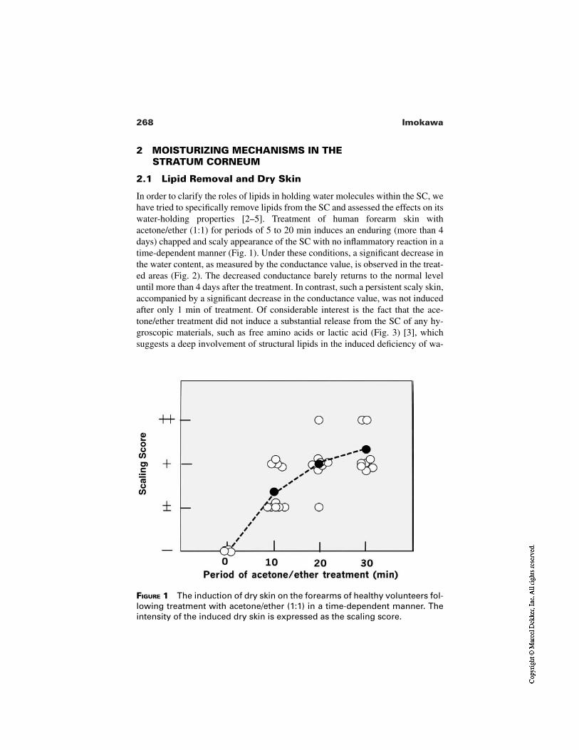
268 Imokawa
FIGURE 1 The induction of dry skin on the forearms of healthy volunteers fol-lowing treatment with acetone/ether (1:1) in a time-dependent manner. Theintensity of the induced dry skin is expressed as the scaling score.
Sca
ling
Sco
re2 MOISTURIZING MECHANISMS IN THE
STRATUM CORNEUM
2.1 Lipid Removal and Dry Skin
In order to clarify the roles of lipids in holding water molecules within the SC, wehave tried to specifically remove lipids from the SC and assessed the effects on itswater-holding properties [2–5]. Treatment of human forearm skin withacetone/ether (1:1) for periods of 5 to 20 min induces an enduring (more than 4days) chapped and scaly appearance of the SC with no inflammatory reaction in atime-dependent manner (Fig. 1). Under these conditions, a significant decrease inthe water content, as measured by the conductance value, is observed in the treat-ed areas (Fig. 2). The decreased conductance barely returns to the normal leveluntil more than 4 days after the treatment. In contrast, such a persistent scaly skin,accompanied by a significant decrease in the conductance value, was not inducedafter only 1 min of treatment. Of considerable interest is the fact that the ace-tone/ether treatment did not induce a substantial release from the SC of any hy-groscopic materials, such as free amino acids or lactic acid (Fig. 3) [3], whichsuggests a deep involvement of structural lipids in the induced deficiency of wa-
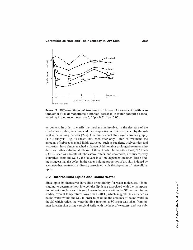
269Ceramides as NMF and Their Efficacy in Dry Skin
FIGURE 2 Different times of treatment of human forearm skin with ace-tone/ether (1:1) demonstrates a marked decrease in water content as mea-sured by impedance meter. n = 6; **p < 0.01; *p < 0.05.
ter content. In order to clarify the mechanisms involved in the decrease of theconductance value, we compared the composition of lipids extracted by the sol-vent after varying periods [2–5]. One-dimensional thin-layer chromatography(TLC) analysis (Fig. 4) shows that, even after only 1 min of treatment, theamounts of sebaceous gland lipids extracted, such as squalene, triglycerides, andwax esters, have almost reached a plateau. Additional or prolonged treatments in-duce no further substantial release of those lipids. On the other hand, SC lipids(SCLs), such as cholesterol, cholesterol esters, and ceramides, are successivelysolubilized from the SC by the solvent in a time-dependent manner. These find-ings suggest that the defect in the water-holding properties of dry skin induced byacetone/ether treatment is directly associated with the depletion of intercellularlipids.
2.2 Intercellular Lipids and Bound Water
Since lipids by themselves have little or no affinity for water molecules, it is in-triguing to determine how intercellular lipids are associated with the incorpora-tion of water molecules. It is well known that water within the SC does not freezereadily, even at temperatures lower than –40°C, which suggests its existence asbound water within the SC. In order to examine the amounts of bound water inthe SC which reflect the water-holding function, a SC sheet was taken from hu-man forearm skin using a surgical knife with the help of tweezers, and was sub-
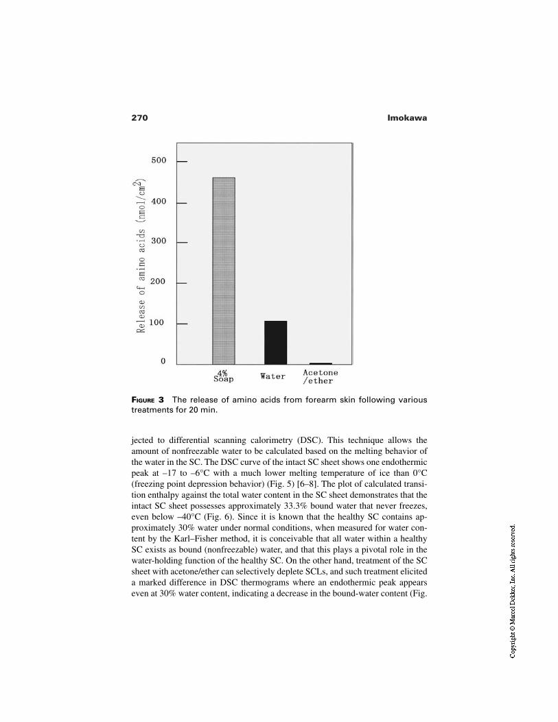
270 Imokawa
FIGURE 3 The release of amino acids from forearm skin following varioustreatments for 20 min.
jected to differential scanning calorimetry (DSC). This technique allows theamount of nonfreezable water to be calculated based on the melting behavior ofthe water in the SC. The DSC curve of the intact SC sheet shows one endothermicpeak at –17 to –6°C with a much lower melting temperature of ice than 0°C(freezing point depression behavior) (Fig. 5) [6–8]. The plot of calculated transi-tion enthalpy against the total water content in the SC sheet demonstrates that theintact SC sheet possesses approximately 33.3% bound water that never freezes,even below –40°C (Fig. 6). Since it is known that the healthy SC contains ap-proximately 30% water under normal conditions, when measured for water con-tent by the Karl–Fisher method, it is conceivable that all water within a healthySC exists as bound (nonfreezable) water, and that this plays a pivotal role in thewater-holding function of the healthy SC. On the other hand, treatment of the SCsheet with acetone/ether can selectively deplete SCLs, and such treatment eliciteda marked difference in DSC thermograms where an endothermic peak appearseven at 30% water content, indicating a decrease in the bound-water content (Fig.
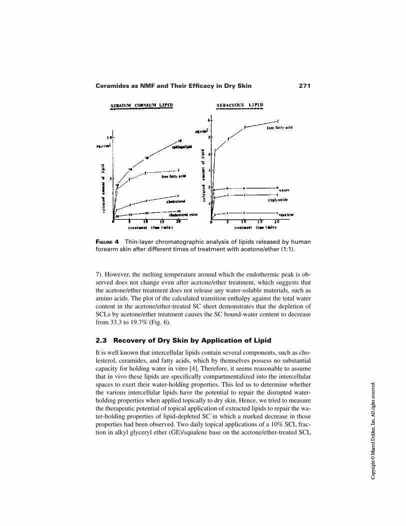
271Ceramides as NMF and Their Efficacy in Dry Skin
FIGURE 4 Thin-layer chromatographic analysis of lipids released by humanforearm skin after different times of treatment with acetone/ether (1:1).
7). However, the melting temperature around which the endothermic peak is ob-served does not change even after acetone/ether treatment, which suggests thatthe acetone/ether treatment does not release any water-soluble materials, such asamino acids. The plot of the calculated transition enthalpy against the total watercontent in the acetone/ether-treated SC sheet demonstrates that the depletion ofSCLs by acetone/ether treatment causes the SC bound-water content to decreasefrom 33.3 to 19.7% (Fig. 6).
2.3 Recovery of Dry Skin by Application of Lipid
It is well known that intercellular lipids contain several components, such as cho-lesterol, ceramides, and fatty acids, which by themselves possess no substantialcapacity for holding water in vitro [4]. Therefore, it seems reasonable to assumethat in vivo these lipids are specifically compartmentalized into the intercellularspaces to exert their water-holding properties. This led us to determine whetherthe various intercellular lipids have the potential to repair the disrupted water-holding properties when applied topically to dry skin. Hence, we tried to measurethe therapeutic potential of topical application of extracted lipids to repair the wa-ter-holding properties of lipid-depleted SC in which a marked decrease in thoseproperties had been observed. Two daily topical applications of a 10% SCL frac-tion in alkyl glyceryl ether (GE)/squalene base on the acetone/ether-treated SCL
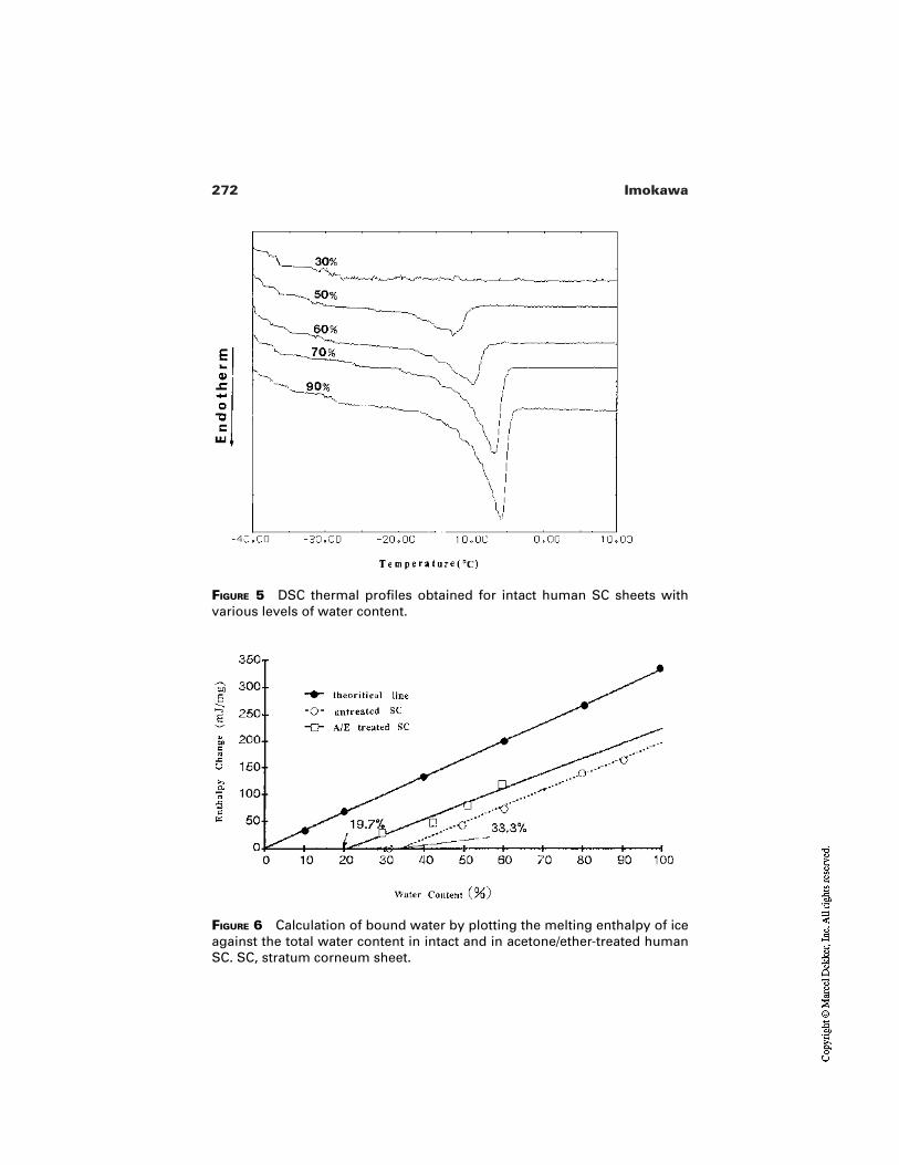
272 Imokawa
FIGURE 5 DSC thermal profiles obtained for intact human SC sheets withvarious levels of water content.
FIGURE 6 Calculation of bound water by plotting the melting enthalpy of iceagainst the total water content in intact and in acetone/ether-treated humanSC. SC, stratum corneum sheet.
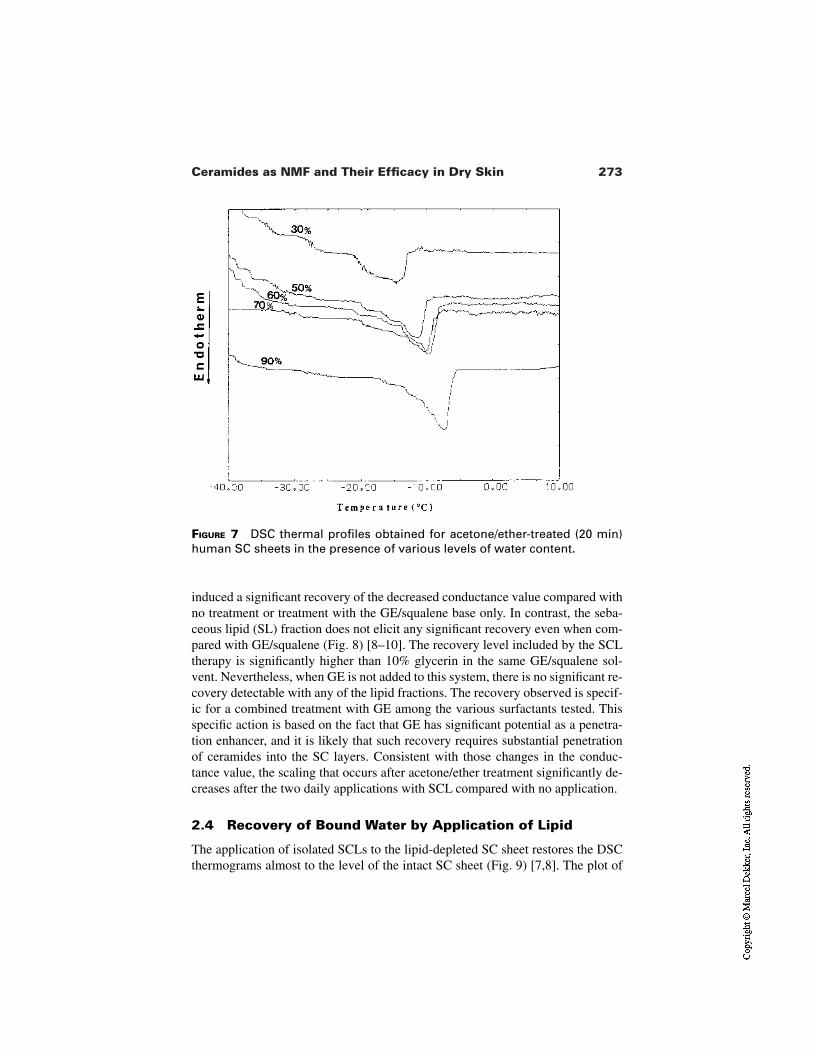
273Ceramides as NMF and Their Efficacy in Dry Skin
FIGURE 7 DSC thermal profiles obtained for acetone/ether-treated (20 min)human SC sheets in the presence of various levels of water content.
induced a significant recovery of the decreased conductance value compared withno treatment or treatment with the GE/squalene base only. In contrast, the seba-ceous lipid (SL) fraction does not elicit any significant recovery even when com-pared with GE/squalene (Fig. 8) [8–10]. The recovery level included by the SCLtherapy is significantly higher than 10% glycerin in the same GE/squalene sol-vent. Nevertheless, when GE is not added to this system, there is no significant re-covery detectable with any of the lipid fractions. The recovery observed is specif-ic for a combined treatment with GE among the various surfactants tested. Thisspecific action is based on the fact that GE has significant potential as a penetra-tion enhancer, and it is likely that such recovery requires substantial penetrationof ceramides into the SC layers. Consistent with those changes in the conduc-tance value, the scaling that occurs after acetone/ether treatment significantly de-creases after the two daily applications with SCL compared with no application.
2.4 Recovery of Bound Water by Application of Lipid
The application of isolated SCLs to the lipid-depleted SC sheet restores the DSCthermograms almost to the level of the intact SC sheet (Fig. 9) [7,8]. The plot of
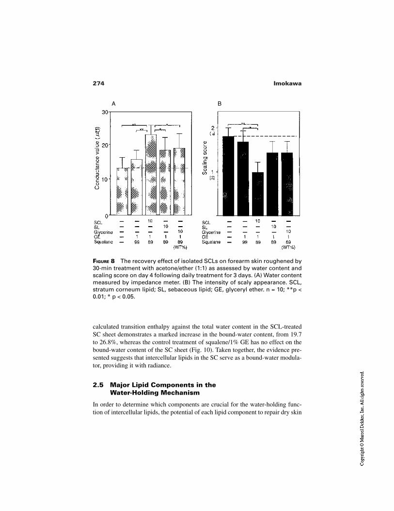
274 Imokawa
FIGURE 8 The recovery effect of isolated SCLs on forearm skin roughened by30-min treatment with acetone/ether (1:1) as assessed by water content andscaling score on day 4 following daily treatment for 3 days. (A) Water contentmeasured by impedance meter. (B) The intensity of scaly appearance. SCL,stratum corneum lipid; SL, sebaceous lipid; GE, glyceryl ether. n = 10; **p <0.01; * p < 0.05.
A B
calculated transition enthalpy against the total water content in the SCL-treatedSC sheet demonstrates a marked increase in the bound-water content, from 19.7to 26.8%, whereas the control treatment of squalene/1% GE has no effect on thebound-water content of the SC sheet (Fig. 10). Taken together, the evidence pre-sented suggests that intercellular lipids in the SC serve as a bound-water modula-tor, providing it with radiance.
2.5 Major Lipid Components in the Water-Holding Mechanism
In order to determine which components are crucial for the water-holding func-tion of intercellular lipids, the potential of each lipid component to repair dry skin
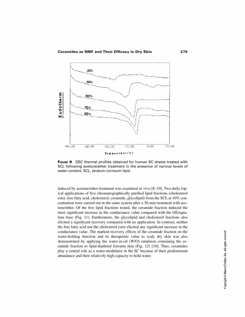
275Ceramides as NMF and Their Efficacy in Dry Skin
FIGURE 9 DSC thermal profiles obtained for human SC sheets treated withSCL following acetone/ether treatment in the presence of various levels ofwater content. SCL, stratum corneum lipid.
induced by acetone/ether treatment was examined in vivo [8–10]. Two daily top-ical applications of five chromatographically purified lipid fractions (cholesterolester, free fatty acid, cholesterol, ceramide, glycolipid) from the SCL at 10% con-centration were carried out in the same system after a 30-min treatment with ace-tone/ether. Of the five lipid fractions tested, the ceramide fraction induced themost significant increase in the conductance value compared with the GE/squa-lene base (Fig. 11). Furthermore, the glycolipid and cholesterol fractions alsoelicited a significant recovery compared with no application. In contrast, neitherthe free fatty acid nor the cholesterol ester elicited any significant increase in theconductance value. The marked recovery effects of the ceramide fraction on thewater-holding function and its therapeutic value to scaly dry skin was alsodemonstrated by applying the water-in-oil (W/O) emulsion containing the ce-ramide fraction to lipid-depleted forearm skin (Fig. 12) [10]. Thus, ceramidesplay a central role as a water-modulator in the SC because of their predominantabundance and their relatively high capacity to hold water.
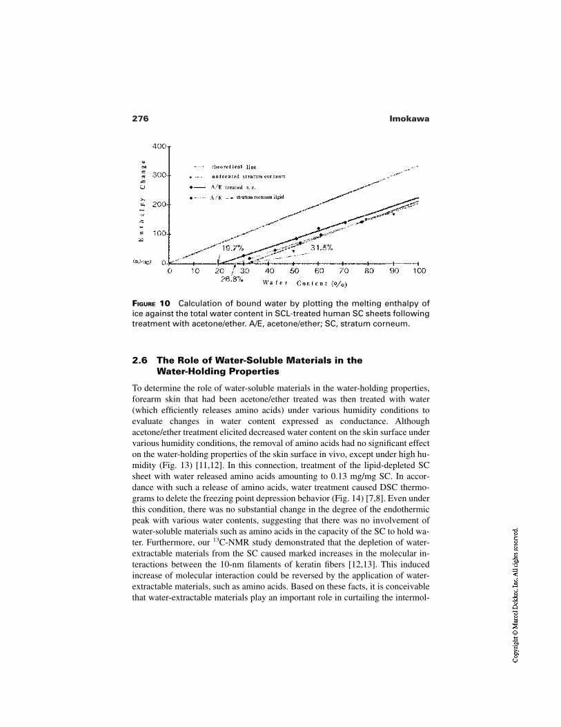
276 Imokawa
FIGURE 10 Calculation of bound water by plotting the melting enthalpy ofice against the total water content in SCL-treated human SC sheets followingtreatment with acetone/ether. A/E, acetone/ether; SC, stratum corneum.
2.6 The Role of Water-Soluble Materials in theWater-Holding Properties
To determine the role of water-soluble materials in the water-holding properties,forearm skin that had been acetone/ether treated was then treated with water(which efficiently releases amino acids) under various humidity conditions toevaluate changes in water content expressed as conductance. Althoughacetone/ether treatment elicited decreased water content on the skin surface undervarious humidity conditions, the removal of amino acids had no significant effecton the water-holding properties of the skin surface in vivo, except under high hu-midity (Fig. 13) [11,12]. In this connection, treatment of the lipid-depleted SCsheet with water released amino acids amounting to 0.13 mg/mg SC. In accor-dance with such a release of amino acids, water treatment caused DSC thermo-grams to delete the freezing point depression behavior (Fig. 14) [7,8]. Even underthis condition, there was no substantial change in the degree of the endothermicpeak with various water contents, suggesting that there was no involvement ofwater-soluble materials such as amino acids in the capacity of the SC to hold wa-ter. Furthermore, our 13C-NMR study demonstrated that the depletion of water-extractable materials from the SC caused marked increases in the molecular in-teractions between the 10-nm filaments of keratin fibers [12,13]. This inducedincrease of molecular interaction could be reversed by the application of water-extractable materials, such as amino acids. Based on these facts, it is conceivablethat water-extractable materials play an important role in curtailing the intermol-
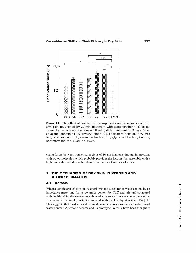
277Ceramides as NMF and Their Efficacy in Dry Skin
FIGURE 11 The effect of isolated SCL components on the recovery of fore-arm skin roughened by 30-min treatment with acetone/ether (1:1) as as-sessed by water content on day 4 following daily treatment for 3 days. Base:squalene (containing 1% glyceryl ether); CE, cholesterol fraction; FFA, freefatty acid fraction; CER, ceramide fraction; GL, glycolipid fraction; Control,nontreatment. **p < 0.01; *p < 0.05.
Co
nd
uct
ance
val
ue
(µΩ
)Ω
ecular forces between nonhelical regions of 10-nm filaments through interactionswith water molecules, which probably provides the keratin fiber assembly with ahigh molecular mobility rather than the retention of water molecules.
3 THE MECHANISM OF DRY SKIN IN XEROSIS ANDATOPIC DERMATITIS
3.1 Xerosis
When a xerotic area of skin on the cheek was measured for its water content by animpedance meter and for its ceramide content by TLC analysis and comparedwith healthy skin, the xerotic area showed a decrease in water content as well asa decrease in ceramide content compared with the healthy skin (Fig. 15) [14].This suggests that the decreased ceramide content is responsible for the decreasedwater content. Asteatotic eczema and its prototype, xerosis, have been thought to
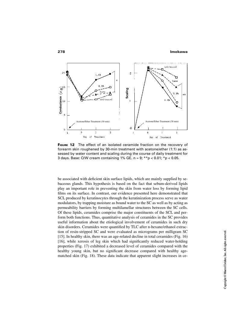
278 Imokawa
FIGURE 12 The effect of an isolated ceramide fraction on the recovery offorearm skin roughened by 30-min treatment with acetone/ether (1:1) as as-sessed by water content and scaling during the course of daily treatment for3 days. Base: O/W cream containing 1% GE. n = 9; **p < 0.01; *p < 0.05.
Co
nd
uct
ance
Acetone/Ether Treatment (30 min) Acetone/Ether Treatment (30 min)
be associated with deficient skin surface lipids, which are mainly supplied by se-baceous glands. This hypothesis is based on the fact that sebum-derived lipidsplay an important role in preventing the skin from water loss by forming lipidfilms on its surface. In contrast, our evidence presented here demonstrated thatSCL produced by keratinocytes through the keratinization process serve as watermodulators, by trapping moisture as bound water to the SC as well as by acting aspermeability barriers by forming multilamellar structures between the SC cells.Of these lipids, ceramides comprise the major constituents of the SCL and per-form both functions. Thus, quantitative analysis of ceramides in the SC providesuseful information about the etiological involvement of ceramides in such dryskin disorders. Ceramides were quantified by TLC after n-hexane/ethanol extrac-tion of resin-stripped SC and were evaluated as micrograms per milligram SC[15]. In healthy skin, there was an age-related decline in total ceramides (Fig. 16)[16], while xerosis of leg skin which had significantly reduced water-holdingproperties (Fig. 17) exhibited a decreased level of ceramides compared with thehealthy young skin, but no significant decrease compared with healthy age-matched skin (Fig. 18). These data indicate that apparent slight increases in ce-
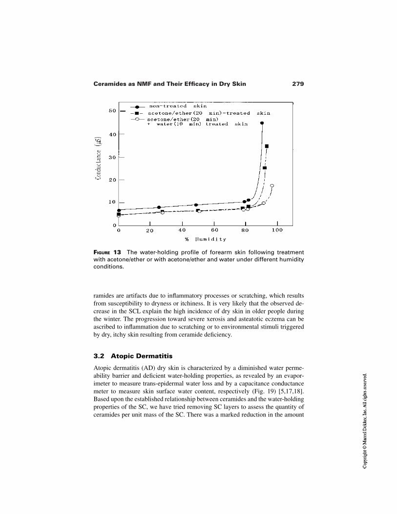
279Ceramides as NMF and Their Efficacy in Dry Skin
FIGURE 13 The water-holding profile of forearm skin following treatmentwith acetone/ether or with acetone/ether and water under different humidityconditions.
ramides are artifacts due to inflammatory processes or scratching, which resultsfrom susceptibility to dryness or itchiness. It is very likely that the observed de-crease in the SCL explain the high incidence of dry skin in older people duringthe winter. The progression toward severe xerosis and asteatotic eczema can beascribed to inflammation due to scratching or to environmental stimuli triggeredby dry, itchy skin resulting from ceramide deficiency.
3.2 Atopic Dermatitis
Atopic dermatitis (AD) dry skin is characterized by a diminished water perme-ability barrier and deficient water-holding properties, as revealed by an evapor-imeter to measure trans-epidermal water loss and by a capacitance conductancemeter to measure skin surface water content, respectively (Fig. 19) [5,17,18].Based upon the established relationship between ceramides and the water-holdingproperties of the SC, we have tried removing SC layers to assess the quantity ofceramides per unit mass of the SC. There was a marked reduction in the amount
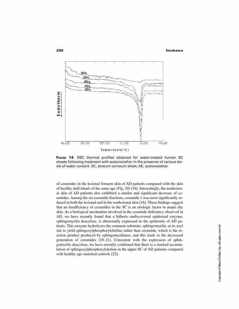
280 Imokawa
FIGURE 14 DSC thermal profiles obtained for water-treated human SCsheets following treatment with acetone/ether in the presence of various lev-els of water content. SC, stratum corneum sheet; AE, acetone/ether.
of ceramides in the lesional forearm skin of AD patients compared with the skinof healthy individuals of the same age (Fig. 20) [16]. Interestingly, the nonlesion-al skin of AD patients also exhibited a similar and significant decrease of ce-ramides. Among the six ceramide fractions, ceramide 1 was most significantly re-duced in both the lesional and in the nonlesional skin [16]. These findings suggestthat an insufficiency of ceramides in the SC is an etiologic factor in atopic dryskin. As a biological mechanism involved in the ceramide deficiency observed inAD, we have recently found that a hitherto undiscovered epidermal enzyme,sphingomyelin deacylase, is abnormally expressed in the epidermis of AD pa-tients. This enzyme hydrolyzes the common substrate, sphingomyelin, at its acylsite to yield sphingosylphosphorylcholine rather than ceramide, which is the re-action product produced by sphingomyelinase, and this leads to the decreasedgeneration of ceramides [19–21]. Consistent with the expression of sphin-gomyelin deacylase, we have recently confirmed that there is a marked accumu-lation of sphingosylphosphorylcholine in the upper SC of AD patients comparedwith healthy age-matched controls [22].
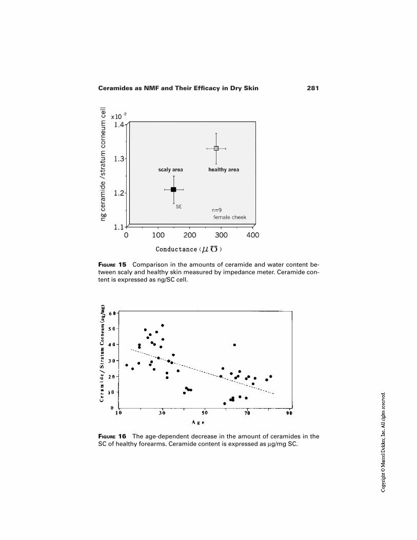
281Ceramides as NMF and Their Efficacy in Dry Skin
FIGURE 15 Comparison in the amounts of ceramide and water content be-tween scaly and healthy skin measured by impedance meter. Ceramide con-tent is expressed as ng/SC cell.
FIGURE 16 The age-dependent decrease in the amount of ceramides in theSC of healthy forearms. Ceramide content is expressed as µg/mg SC.
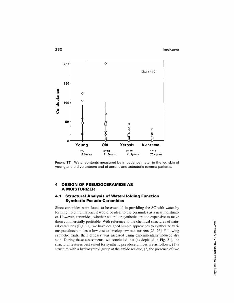
282 Imokawa
FIGURE 17 Water contents measured by impedance meter in the leg skin ofyoung and old volunteers and of xerotic and asteatotic eczema patients.
4 DESIGN OF PSEUDOCERAMIDE AS A MOISTURIZER
4.1 Structural Analysis of Water-Holding FunctionSynthetic Pseudo-Ceramides
Since ceramides were found to be essential in providing the SC with water byforming lipid multilayers, it would be ideal to use ceramides as a new moisturiz-er. However, ceramides, whether natural or synthetic, are too expensive to makethem commercially profitable. With reference to the chemical structures of natu-ral ceramides (Fig. 21), we have designed simple approaches to synthesize vari-ous pseudoceramides at low cost to develop new moisturizers [23–26]. Followingsynthetic trials, their efficacy was assessed using experimentally induced dryskin. During these assessments, we concluded that (as depicted in Fig. 21), thestructural features best suited for synthetic pseudoceramides are as follows: (1) astructure with a hydroxyethyl group at the amide residue, (2) the presence of two
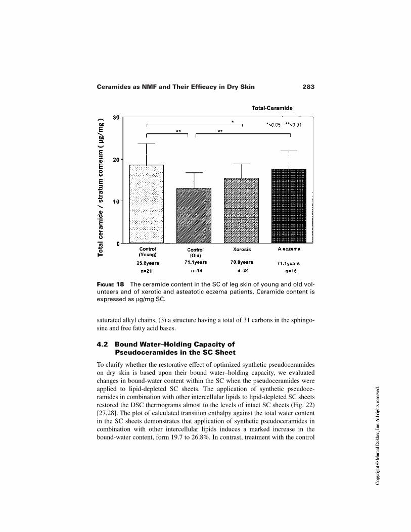
283Ceramides as NMF and Their Efficacy in Dry Skin
FIGURE 18 The ceramide content in the SC of leg skin of young and old vol-unteers and of xerotic and asteatotic eczema patients. Ceramide content isexpressed as µg/mg SC.
saturated alkyl chains, (3) a structure having a total of 31 carbons in the sphingo-sine and free fatty acid bases.
4.2 Bound Water–Holding Capacity ofPseudoceramides in the SC Sheet
To clarify whether the restorative effect of optimized synthetic pseudoceramideson dry skin is based upon their bound water–holding capacity, we evaluatedchanges in bound-water content within the SC when the pseudoceramides wereapplied to lipid-depleted SC sheets. The application of synthetic pseudoce-ramides in combination with other intercellular lipids to lipid-depleted SC sheetsrestored the DSC thermograms almost to the levels of intact SC sheets (Fig. 22)[27,28]. The plot of calculated transition enthalpy against the total water contentin the SC sheets demonstrates that application of synthetic pseudoceramides incombination with other intercellular lipids induces a marked increase in thebound-water content, form 19.7 to 26.8%. In contrast, treatment with the control
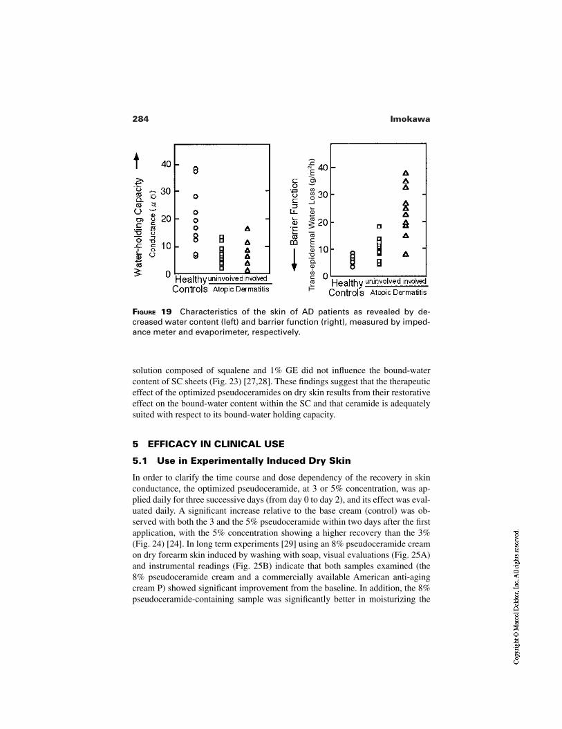
284 Imokawa
FIGURE 19 Characteristics of the skin of AD patients as revealed by de-creased water content (left) and barrier function (right), measured by imped-ance meter and evaporimeter, respectively.
Tran
s-ep
ider
mal
Wat
er L
oss
(g
/m2h
)
solution composed of squalene and 1% GE did not influence the bound-watercontent of SC sheets (Fig. 23) [27,28]. These findings suggest that the therapeuticeffect of the optimized pseudoceramides on dry skin results from their restorativeeffect on the bound-water content within the SC and that ceramide is adequatelysuited with respect to its bound-water holding capacity.
5 EFFICACY IN CLINICAL USE
5.1 Use in Experimentally Induced Dry Skin
In order to clarify the time course and dose dependency of the recovery in skinconductance, the optimized pseudoceramide, at 3 or 5% concentration, was ap-plied daily for three successive days (from day 0 to day 2), and its effect was eval-uated daily. A significant increase relative to the base cream (control) was ob-served with both the 3 and the 5% pseudoceramide within two days after the firstapplication, with the 5% concentration showing a higher recovery than the 3%(Fig. 24) [24]. In long term experiments [29] using an 8% pseudoceramide creamon dry forearm skin induced by washing with soap, visual evaluations (Fig. 25A)and instrumental readings (Fig. 25B) indicate that both samples examined (the8% pseudoceramide cream and a commercially available American anti-agingcream P) showed significant improvement from the baseline. In addition, the 8%pseudoceramide-containing sample was significantly better in moisturizing the
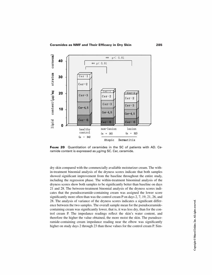
285Ceramides as NMF and Their Efficacy in Dry Skin
FIGURE 20 Quantitation of ceramides in the SC of patients with AD. Ce-ramide content is expressed as µg/mg SC. Cer, ceramide.
dry skin compared with the commercially available moisturizer cream. The with-in-treatment binomial analysis of the dryness scores indicate that both samplesshowed significant improvement from the baseline throughout the entire study,including the regression phase. The within-treatment binominal analysis of thedryness scores show both samples to be significantly better than baseline on days21 and 28. The between-treatment binomial analysis of the dryness scores indi-cates that the pseudoceramide-containing cream was assigned the lower scoresignificantly more often than was the control cream P on days 2, 7, 19, 21, 26, and28. The analysis of variance of the dryness scores indicates a significant differ-ence between the two samples. The overall sample mean for the pseudoceramide-containing cream was significantly lower, that is, it was less dry, than for the con-trol cream P. The impedance readings reflect the skin’s water content, andtherefore the higher the value obtained, the more moist the skin. The pseudoce-ramide-containing cream impedance reading near the elbow was significantlyhigher on study days 2 through 23 than those values for the control cream P. Sim-
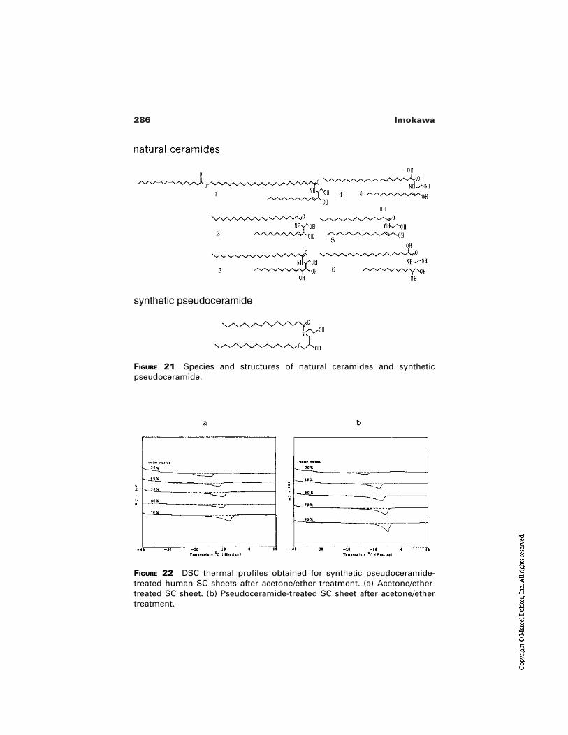
286 Imokawa
FIGURE 21 Species and structures of natural ceramides and syntheticpseudoceramide.
synthetic pseudoceramide
FIGURE 22 DSC thermal profiles obtained for synthetic pseudoceramide-treated human SC sheets after acetone/ether treatment. (a) Acetone/ether-treated SC sheet. (b) Pseudoceramide-treated SC sheet after acetone/ethertreatment.
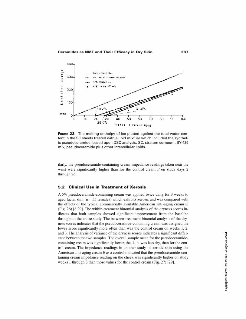
287Ceramides as NMF and Their Efficacy in Dry Skin
FIGURE 23 The melting enthalpy of ice plotted against the total water con-tent in the SC sheets treated with a lipid mixture which included the synthet-ic pseudoceramide, based upon DSC analysis. SC, stratum corneum, SY-425mix, pseudoceramide plus other intercellular lipids.
ilarly, the pseudoceramide-containing cream impedance readings taken near thewrist were significantly higher than for the control cream P on study days 2through 26.
5.2 Clinical Use in Treatment of Xerosis
A 5% pseudoceramide-containing cream was applied twice daily for 3 weeks toaged facial skin (n = 35 females) which exhibits xerosis and was compared withthe effects of the typical commercially available American anti-aging cream G(Fig. 26) [8,29]. The within-treatment binomial analysis of the dryness scores in-dicates that both samples showed significant improvement from the baselinethroughout the entire study. The between-treatment binomial analysis of the dry-ness scores indicates that the pseudoceramide-containing cream was assigned thelower score significantly more often than was the control cream on weeks 1, 2,and 3. The analysis of variance of the dryness scores indicates a significant differ-ence between the two samples. The overall sample mean for the pseudoceramide-containing cream was significantly lower, that is, it was less dry, than for the con-trol cream. The impedance readings in another study of xerotic skin using theAmerican anti-aging cream E as a control indicated that the pseudoceramide-con-taining cream impedance reading on the cheek was significantly higher on studyweeks 1 through 3 than those values for the control cream (Fig. 27) [29].
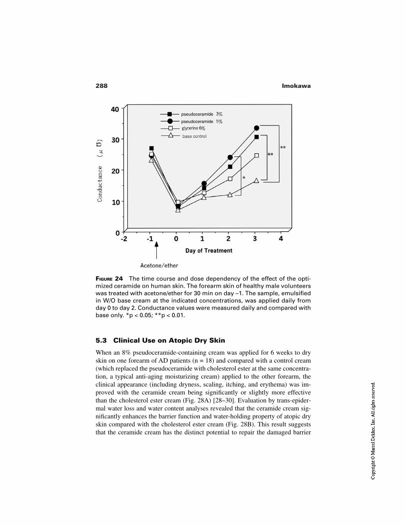
288 Imokawa
FIGURE 24 The time course and dose dependency of the effect of the opti-mized ceramide on human skin. The forearm skin of healthy male volunteerswas treated with acetone/ether for 30 min on day –1. The sample, emulsifiedin W/O base cream at the indicated concentrations, was applied daily fromday 0 to day 2. Conductance values were measured daily and compared withbase only. *p < 0.05; **p < 0.01.
pseudoceramide
pseudoceramide
5.3 Clinical Use on Atopic Dry Skin
When an 8% pseudoceramide-containing cream was applied for 6 weeks to dryskin on one forearm of AD patients (n = 18) and compared with a control cream(which replaced the pseudoceramide with cholesterol ester at the same concentra-tion, a typical anti-aging moisturizing cream) applied to the other forearm, theclinical appearance (including dryness, scaling, itching, and erythema) was im-proved with the ceramide cream being significantly or slightly more effectivethan the cholesterol ester cream (Fig. 28A) [28–30]. Evaluation by trans-epider-mal water loss and water content analyses revealed that the ceramide cream sig-nificantly enhances the barrier function and water-holding property of atopic dryskin compared with the cholesterol ester cream (Fig. 28B). This result suggeststhat the ceramide cream has the distinct potential to repair the damaged barrier
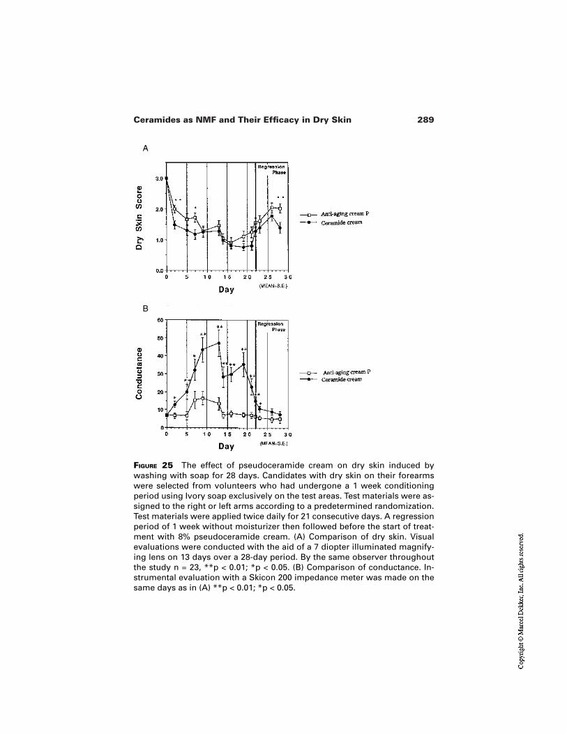
289Ceramides as NMF and Their Efficacy in Dry Skin
FIGURE 25 The effect of pseudoceramide cream on dry skin induced bywashing with soap for 28 days. Candidates with dry skin on their forearmswere selected from volunteers who had undergone a 1 week conditioningperiod using Ivory soap exclusively on the test areas. Test materials were as-signed to the right or left arms according to a predetermined randomization.Test materials were applied twice daily for 21 consecutive days. A regressionperiod of 1 week without moisturizer then followed before the start of treat-ment with 8% pseudoceramide cream. (A) Comparison of dry skin. Visualevaluations were conducted with the aid of a 7 diopter illuminated magnify-ing lens on 13 days over a 28-day period. By the same observer throughoutthe study n = 23, **p < 0.01; *p < 0.05. (B) Comparison of conductance. In-strumental evaluation with a Skicon 200 impedance meter was made on thesame days as in (A) **p < 0.01; *p < 0.05.
B
A
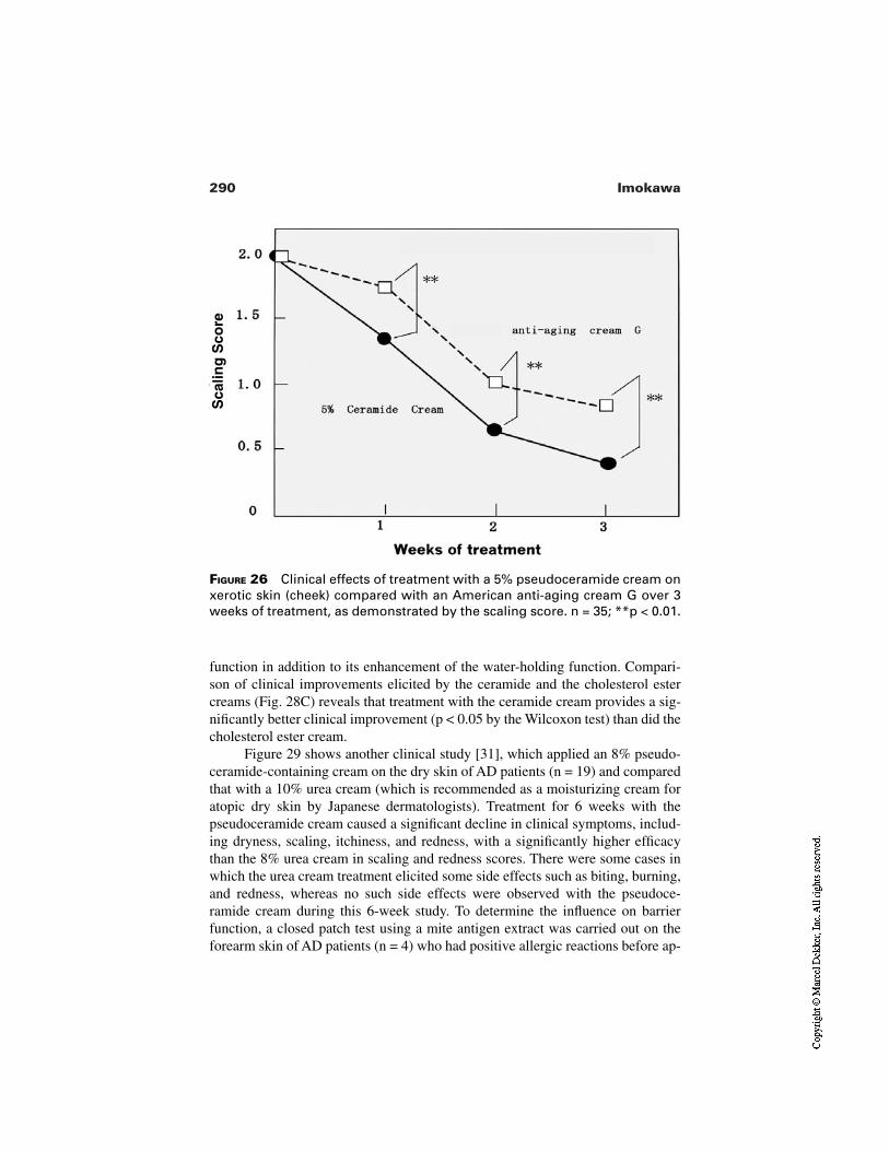
290 Imokawa
FIGURE 26 Clinical effects of treatment with a 5% pseudoceramide cream onxerotic skin (cheek) compared with an American anti-aging cream G over 3weeks of treatment, as demonstrated by the scaling score. n = 35; **p < 0.01.
Sca
ling
Sco
re
function in addition to its enhancement of the water-holding function. Compari-son of clinical improvements elicited by the ceramide and the cholesterol estercreams (Fig. 28C) reveals that treatment with the ceramide cream provides a sig-nificantly better clinical improvement (p < 0.05 by the Wilcoxon test) than did thecholesterol ester cream.
Figure 29 shows another clinical study [31], which applied an 8% pseudo-ceramide-containing cream on the dry skin of AD patients (n = 19) and comparedthat with a 10% urea cream (which is recommended as a moisturizing cream foratopic dry skin by Japanese dermatologists). Treatment for 6 weeks with thepseudoceramide cream caused a significant decline in clinical symptoms, includ-ing dryness, scaling, itchiness, and redness, with a significantly higher efficacythan the 8% urea cream in scaling and redness scores. There were some cases inwhich the urea cream treatment elicited some side effects such as biting, burning,and redness, whereas no such side effects were observed with the pseudoce-ramide cream during this 6-week study. To determine the influence on barrierfunction, a closed patch test using a mite antigen extract was carried out on theforearm skin of AD patients (n = 4) who had positive allergic reactions before ap-
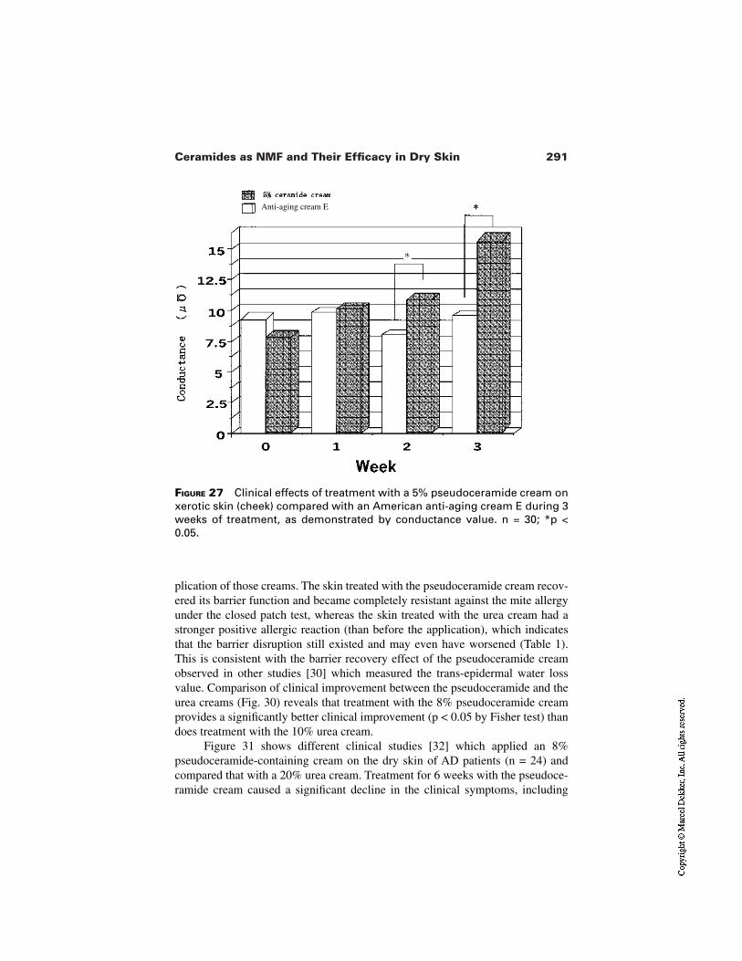
291Ceramides as NMF and Their Efficacy in Dry Skin
FIGURE 27 Clinical effects of treatment with a 5% pseudoceramide cream onxerotic skin (cheek) compared with an American anti-aging cream E during 3weeks of treatment, as demonstrated by conductance value. n = 30; *p <0.05.
Anti-aging cream E
plication of those creams. The skin treated with the pseudoceramide cream recov-ered its barrier function and became completely resistant against the mite allergyunder the closed patch test, whereas the skin treated with the urea cream had astronger positive allergic reaction (than before the application), which indicatesthat the barrier disruption still existed and may even have worsened (Table 1).This is consistent with the barrier recovery effect of the pseudoceramide creamobserved in other studies [30] which measured the trans-epidermal water lossvalue. Comparison of clinical improvement between the pseudoceramide and theurea creams (Fig. 30) reveals that treatment with the 8% pseudoceramide creamprovides a significantly better clinical improvement (p < 0.05 by Fisher test) thandoes treatment with the 10% urea cream.
Figure 31 shows different clinical studies [32] which applied an 8%pseudoceramide-containing cream on the dry skin of AD patients (n = 24) andcompared that with a 20% urea cream. Treatment for 6 weeks with the pseudoce-ramide cream caused a significant decline in the clinical symptoms, including
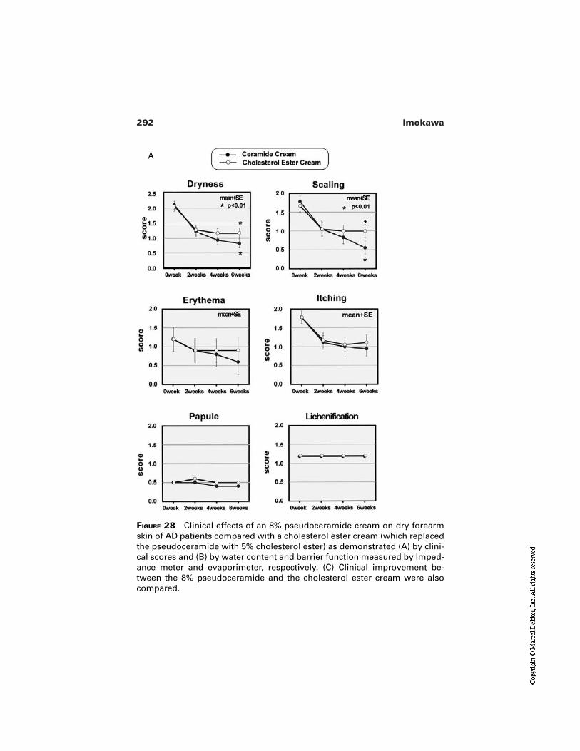
292 Imokawa
FIGURE 28 Clinical effects of an 8% pseudoceramide cream on dry forearmskin of AD patients compared with a cholesterol ester cream (which replacedthe pseudoceramide with 5% cholesterol ester) as demonstrated (A) by clini-cal scores and (B) by water content and barrier function measured by Imped-ance meter and evaporimeter, respectively. (C) Clinical improvement be-tween the 8% pseudoceramide and the cholesterol ester cream were alsocompared.
A
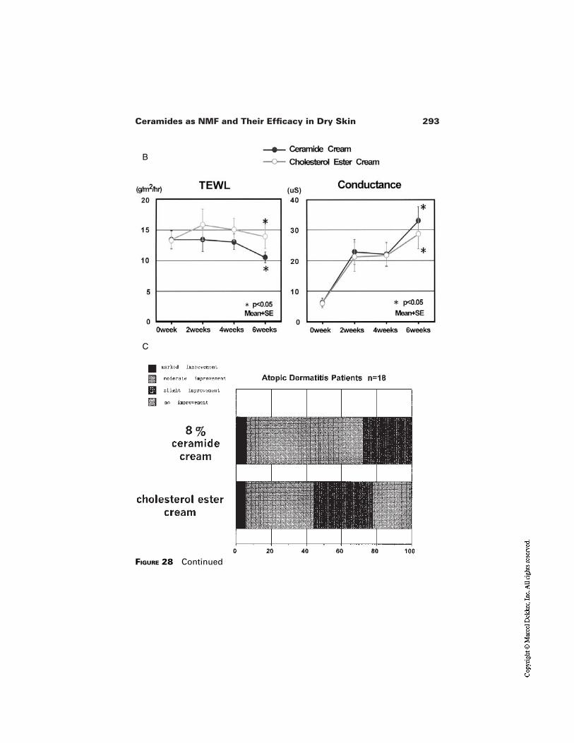
293Ceramides as NMF and Their Efficacy in Dry Skin
FIGURE 28 Continued
B
C
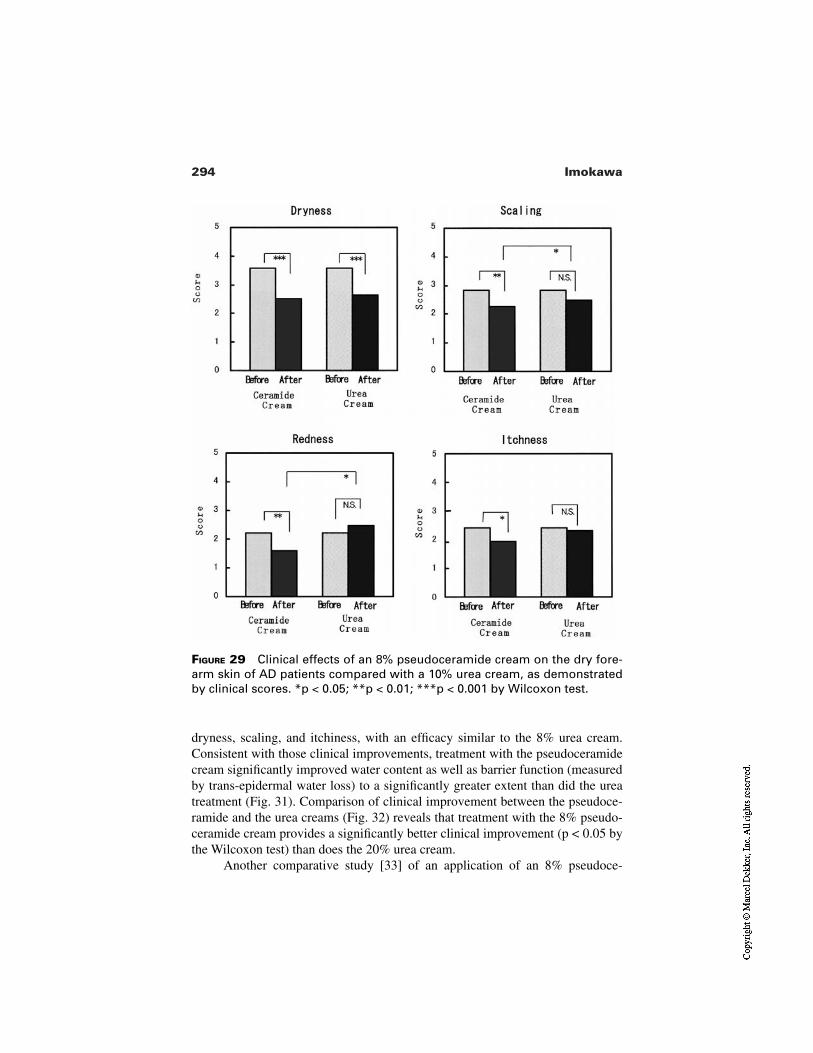
294 Imokawa
FIGURE 29 Clinical effects of an 8% pseudoceramide cream on the dry fore-arm skin of AD patients compared with a 10% urea cream, as demonstratedby clinical scores. *p < 0.05; **p < 0.01; ***p < 0.001 by Wilcoxon test.
dryness, scaling, and itchiness, with an efficacy similar to the 8% urea cream.Consistent with those clinical improvements, treatment with the pseudoceramidecream significantly improved water content as well as barrier function (measuredby trans-epidermal water loss) to a significantly greater extent than did the ureatreatment (Fig. 31). Comparison of clinical improvement between the pseudoce-ramide and the urea creams (Fig. 32) reveals that treatment with the 8% pseudo-ceramide cream provides a significantly better clinical improvement (p < 0.05 bythe Wilcoxon test) than does the 20% urea cream.
Another comparative study [33] of an application of an 8% pseudoce-
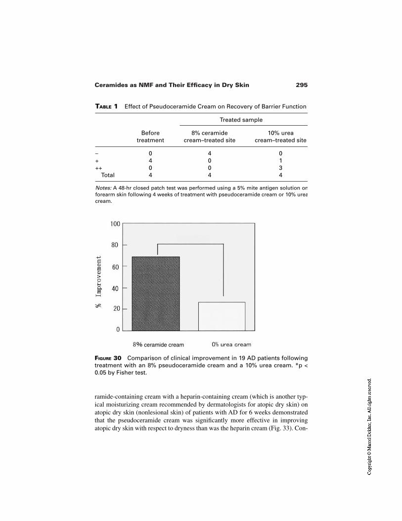
295Ceramides as NMF and Their Efficacy in Dry Skin
TABLE 1 Effect of Pseudoceramide Cream on Recovery of Barrier Function
Treated sample
Beforetreatment
8% ceramidecream–treated site
10% urea cream–treated site
– 0 4 0+ 4 0 1++ 0 0 3
Total 4 4 4
Notes: A 48-hr closed patch test was performed using a 5% mite antigen solution onforearm skin following 4 weeks of treatment with pseudoceramide cream or 10% ureacream.
FIGURE 30 Comparison of clinical improvement in 19 AD patients followingtreatment with an 8% pseudoceramide cream and a 10% urea cream. *p <0.05 by Fisher test.
ramide-containing cream with a heparin-containing cream (which is another typ-ical moisturizing cream recommended by dermatologists for atopic dry skin) onatopic dry skin (nonlesional skin) of patients with AD for 6 weeks demonstratedthat the pseudoceramide cream was significantly more effective in improvingatopic dry skin with respect to dryness than was the heparin cream (Fig. 33). Con-
ceramide cream
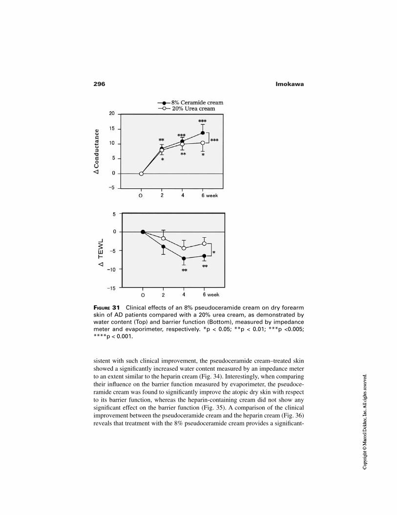
296 Imokawa
FIGURE 31 Clinical effects of an 8% pseudoceramide cream on dry forearmskin of AD patients compared with a 20% urea cream, as demonstrated bywater content (Top) and barrier function (Bottom), measured by impedancemeter and evaporimeter, respectively. *p < 0.05; **p < 0.01; ***p <0.005;****p < 0.001.
sistent with such clinical improvement, the pseudoceramide cream–treated skinshowed a significantly increased water content measured by an impedance meterto an extent similar to the heparin cream (Fig. 34). Interestingly, when comparingtheir influence on the barrier function measured by evaporimeter, the pseudoce-ramide cream was found to significantly improve the atopic dry skin with respectto its barrier function, whereas the heparin-containing cream did not show anysignificant effect on the barrier function (Fig. 35). A comparison of the clinicalimprovement between the pseudoceramide cream and the heparin cream (Fig. 36)reveals that treatment with the 8% pseudoceramide cream provides a significant-
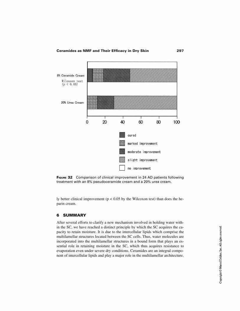
297Ceramides as NMF and Their Efficacy in Dry Skin
FIGURE 32 Comparison of clinical improvement in 24 AD patients followingtreatment with an 8% pseudoceramide cream and a 20% urea cream.
ly better clinical improvement (p < 0.05 by the Wilcoxon test) than does the he-parin cream.
6 SUMMARY
After several efforts to clarify a new mechanism involved in holding water with-in the SC, we have reached a distinct principle by which the SC acquires the ca-pacity to retain moisture. It is due to the intercellular lipids which comprise themultilamellar structures located between the SC cells. Thus, water molecules areincorporated into the multilamellar structures in a bound form that plays an es-sential role in retaining moisture in the SC, which thus acquires resistance toevaporation even under severe dry conditions. Ceramides are an integral compo-nent of intercellular lipids and play a major role in the multilamellar architecture.
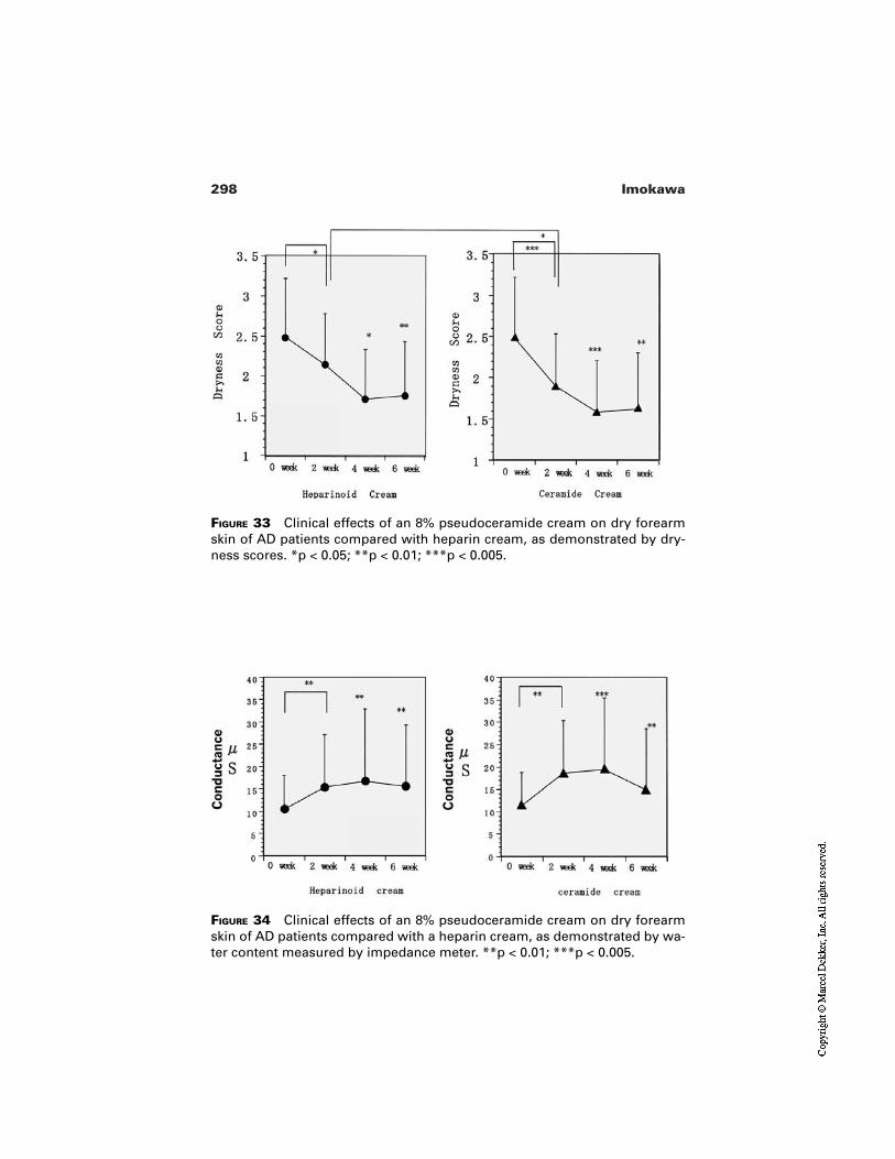
298 Imokawa
FIGURE 33 Clinical effects of an 8% pseudoceramide cream on dry forearmskin of AD patients compared with heparin cream, as demonstrated by dry-ness scores. *p < 0.05; **p < 0.01; ***p < 0.005.
FIGURE 34 Clinical effects of an 8% pseudoceramide cream on dry forearmskin of AD patients compared with a heparin cream, as demonstrated by wa-ter content measured by impedance meter. **p < 0.01; ***p < 0.005.
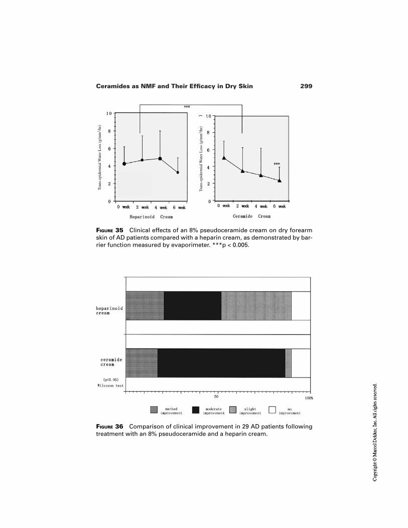
299Ceramides as NMF and Their Efficacy in Dry Skin
FIGURE 35 Clinical effects of an 8% pseudoceramide cream on dry forearmskin of AD patients compared with a heparin cream, as demonstrated by bar-rier function measured by evaporimeter. ***p < 0.005.
FIGURE 36 Comparison of clinical improvement in 29 AD patients followingtreatment with an 8% pseudoceramide and a heparin cream.
Tra
ns-e
pide
rmia
l Wat
er L
oss
(g/m
m2 /h
r)
Tra
ns-e
pide
rmia
l Wat
er L
oss
(g/m
m2 /h
r)
300 Imokawa
With the idea that ceramides are an ideal moisturizer when available, we have at-tempted to synthesize various pseudoceramides which exert water-holding prop-erties similar to those of natural ceramides. Since ceramide deficiencies are ob-served in the SC in several dry skin symptoms and since they play an essentialrole in eliciting dry skin, it seems likely that the application of pseudoceramidesto dry skin is an ideal approach for therapy. Several clinical trials using pseudo-ceramide-containing creams have demonstrated that their use is highly effectivein preventing and treating dry skin conditions.
REFERENCES
1. Jacobi OT. About the mechanisms of moisture regulation in the horny layer of theskin. Pro Sci Sect Good Assoc 1959; 31:22–24.
2. Imokawa G, Hattori M. A possible function of structural lipid in the water-holdingproperties of the stratum corneum. J Invest Dermatol 1985; 84:282–284.
3. Imokawa G. Stratum corneum moisturizing effect and intercellular lipids. FragranceJ 1987; 15:35–41.
4. Imokawa G. Intercellular Lipids of the Stratum Corneum. Gendai HifukagakuTaikei. Nakayama Publishing, 1990; 43–53.
5. Imokawa G. Properties and function of the stratum corneum lipids and its measure-ment. Rinshouhifuka 1990; 44:583–588.
6. Imokawa G, Akasaki S, Kuno O, Zama M, Kawai M, Minematsu Y, Hattori M,Yoshizuka N, Kawamata A, Yano S, Takaishi N. Function of lipids on human skin. JDis Sci Tech 1989; 10:617–641.
7. Imokawa G, Kuno H, Kawai M. Stratum corneum lipids serve as a bound-watermodulator. J. Invest. Dermatol. 1991; 96:845–851.
8. Imokawa G. Water and the stratum corneum. In: Elsner P, Berardesca E, MaibachHI, eds. Bioengineering of Skin. Vitro and in Vivo Models. Vol 1. CRC Press, 3.1993:23–47.
9. Imokawa G, Akasaki S, Hattori M, Yoshizuka N. Selective recovery of deranged wa-ter-holding properties by stratum corneum lipids. J. Invest. Dermatol. 1986; 87:758–761.
10. Akasaki S, Minematsu Y, Yoshizuka N, Imokawa G. The role of intercellular lipidsin the water-holding properties of the stratum corneum: recovery effect on experi-mentally induced dry skin. Jap. J. Dermatol. 1988; 98:40–51.
11. Imokawa G. Role of stratum corneum components in their moisturizing function.Fragrance J 2000; 17:27–39.
12. Imokawa G. Atopic dermatitis: dry skin mechanism. Japan Pediatr Dermatol 1997;16:87–99.
13. Jokura T, Ishikawa Y, Tokuda H, Imokawa G. Molecular analysis of elastic proper-ties of the stratum corneum by solid-state 13C-nuclear magnetic resonance spec-troscopy. J Invest Dermatol 1995; 104:806–812.
14. Imokawa G. Stratum corneum intercellular lipids: function and association with dryskin disorders. Rinsho Hifuka 1993; 35:1147–1161.
15. Akimoto K, Yoshikawa N, Higaki Y, Kawashima M, Imokawa G. Quantitative
301Ceramides as NMF and Their Efficacy in Dry Skin
analysis of stratum corneum lipids in xerosis and asteatotic eczema. J Dermatol(Tokyo) 1993; 20:1–6.
16. Imokawa G. Abe A, Kumi J, Higaki Y, Kawashima M, Hidano A. Decreased level ofceramides in stratum corneum of atopic dermatitis: an etiologic factor in atopic dryskin? J. Invest. Dermatol. 1991; 96:523–526.
17. Imokawa G. Atopic dermatitis and abnormality in sphingolipid metabolisms. Thelipid 1996; 7:428–423.
18. Imokawa G, Kawashima M. Atopic dermatitis and the stratum corneum lipids.Tokyo Tanabe Quarterly 1997; 42:41–52.
19. Murata Y, Ogata J, Higaki Y, Kawashima M, Yada Y, Higuchi K, Tsuchiya T,Kawaminami S, Imokawa G. Abnormal expression of sphingomyelin acylase inatopic dermatitis: an etiologic factor for ceramide deficiency? J. Invest Dermatol.1996; 106:1242–1249.
20. Hara J, Higuchi K, Okamoto R, Kawashima M, Imokawa G. High expression ofsphingomyelin deacylase is an important determinant of ceramide deficiency leadingto barrier disruption in atopic dermatitis. J Invest. Dermatol. 2000; 115:406–413.
21. Higuchi K, Hara J, Okamoto R, Kawashima M, Imokawa G. The skin of atopic der-matitis patients contains a novel enzyme, glucosylceramide sphingomyelin deacy-lase, which cleaves the N-acyl linkage of sphingomyelin and glucosylceramide.Biochem. J. 2000; 350:747–756.
22. Okamoto R, Hara J, Kawashima M, Takagi Y, Imokawa G. Quantitative analysis ofsphingosylphosphorylcholine in the stratum corneum of atopic dermatitis patients[abstract]. J Dermatol Sci 1999; 21:221.
23. Imokawa G, Akasaki S, Zama M, Minematsu Y, Kawamata A, Yano Y, Takaishi N.Selective recovery of deranged water-holding properties in the stratum corneum bysynthesized pseudo-ceramide derivatives. Proc Jpn Soc Invest Dermatol 1988;12:126–127.
24. Imokawa G, Akasaki S, Kawamata A, Yano S, Takaishi N. Water-retaining functionin the stratum corneum and its recovery properties by synthetic pseudo-ceramides. JSoc Cosmet Chem 1989; 40:273–285.
25. Imokawa G. Function of epidermal sphingolipids and their application. Fragrance J1990; 4:26–34.
26. Imokawa G. Stratum corneum intercellular lipids: their function and application.Hifu to Biyo 1991; 23:3806–3818.
27. Imokawa G. Structure and function of intercellular lipids in the stratum corneum.Yukagaku 1995; 44:751–766.
28. Imokawa G. Moisturizers used in dermatology fields. J Clin Dermatol 1998;56:87–96.
29. Imokawa G. Skin moisturizers: development and clinical use of ceramides. In: Lo-den M, ed. Dry Skin and Moisturizers. CRC Press, 1999:269–299.
30. Koizumi K, Noguchi K, Imokawa G, Etou T, Nakagawa H, Isibashi H. Clinical ef-fects of synthetic pseudo-ceramides on the dry skin of atopic dermatitis patients [ab-stract]. Jpn J Dermatoallergol 1993; 2:66.
31. Mizutani J, Takahashi M, Shimizu M, Kariya N, Sato H, Imokawa G. Usage ofpseudoceramide cream in atopic dry skin in comparison with 10% urea cream.Nishihihon Hifuka 2001; 63:457–461.
302 Imokawa
32. Yamanaka M, Ishikawa O, Takahashi M, Sato H, Imokawa G. Usage of pseudoce-ramide cream in atopic dry skin in comparison with 20% urea cream. Hifu 2001;43:341–347.
33. Nakamura T, Honma D, Katusragi T, Sakai H, Hashimoto Y, Iizuka H. Usage ofpseudoceramide cream in atopic dry skin in comparison with heparinoid cream.Nishinihon Hifu 1999; 61:671–681.
15Phosphatidylcholine and Skin Hydration
Miklos GhyczyNattermann Phospholipid GmbH, Cologne, Germany
Vladimir VacataUniversity of Bonn, Bonn, Germany
Phosphatidylcholine (PC) is the most abundant phospholipid in animal cells. Itpossesses an intrinsic hydration force, and its metabolites are essential osmopro-tectants. Phosphatidylcholine composed of saturated fatty acids (hydrogenatedPC; HPC) possesses physical properties which are comparable with those of thecomponents of the skin permeability barrier. When applied to skin, HPC is takenup by the stratum corneum (SC); it interacts with lipids of the permeability barri-er, but it does not cause any irritation. Phosphatidylcholine, HPC, and theirmetabolites display preventive efficacy in pathological states caused by the redoximbalance and the ensuing genesis of free radicals. This phenomenon is taken ad-vantage of in the drug formulations where PC ameliorates certain side effects ofdrugs. In human skin challenged by sodium lauryl sulfate (SLS), HPC increasesskin hydration, but does not exhibit any effect on the trans-epidermal water loss(TEWL). In addition, HPC has the ability to counteract the inflammatory effectsof SLS. And HPC is an industrially available, easy-to-handle and well-definedsubstance produced according to the cGMP standards. The favorable biologicaleffects inspire a new approach to the development of topical formulations for thetreatment and prevention of frequent skin problems connected with dry skin andthe ensuing pathological states.
303
304 Ghyczy and Vacata
1 INTRODUCTION
Phospholipids, as components of lecithins, are the emulsifiers with perhaps thelongest history in cosmetics and dermatology. Even the oldest cosmetic protocolsrecommend egg yolk (which contains 10% phospholipids) as a universal emulsi-fier. In the past 10 years, some 700 articles and patents have been publishedand/or registered concerning the use of phospholipids in skin products. However,despite the growing interest in this field, the mode of action and the function ofphospholipids in skin are still subjects of critical discussion.
The data acquired during the past decade on skin hydration, skin barrier,and PC itself may help to explain and justify the long-lasting interest in phospho-lipids and particularly in PC and its applications. The new findings disclose thecentral role of skin hydration for healthy skin. There are two factors which con-trol the water content in stratum corneum—the intracellular occlusion and the in-tercellular humectancy [1]. It was also shown that PC with saturated fatty acids(INCI definition: hydrogenated lecithin) possesses thermodynamic and structure-forming properties similar to those of the SC lipids [2]. Hydrogenated PC wasfound to be capable of penetrating into the SC lipid barrier, though at significant-ly lower rates than PC with unsaturated fatty acids [3,4]. Because skin containsphospholipases D and A, PC may also serve as a source of osmoprotectants suchas choline, betaine, and glycerylphosphatidylcholine. In medicine PC is used asdrug substance, and findings in related fields suggest that the mode of its action isbased on redox regulation and prevention of formation of free radicals and the en-suing oxidative stress [5]. All these findings suggest that topically applied HPCmay have the potential to control skin hydration and prevent the pathologicalstates of dry skin.
2 CONTROL OF SKIN HYDRATION
It is now generally recognized that sufficient water content of SC is the basic pre-requisite to healthy skin. This is based on the recent acknowledgment that the SChomeostasis depends on the activity of several enzymes for which stringentlycontrolled water content is essential [6]. This water content is a function of twoprinciples. The first is the permeability barrier, which controls the translocation ofwater from the lower to the to the upper layer of the SC and subsequently to theenvironment. The structure responsible for the control is a stack of continuouslipid bilayers in a gel state. Perturbation of this barrier by a diet deficient in linole-ic acid or by an external insult such as extensive skin cleansing results in an in-creased TEWL. The second principle consists of the natural moisturizing factors(NMF) located in the corneocytes. The concentration of NMF in the corneocytesis controlled by NMF synthesis, which itself is feedback-controlled by the watercontent as well as by external noxae and aging [1].
305Phosphatidylcholine and Skin Hydration
In view of this knowledge the prevention of and the cure for dry skin syn-drome by a topical product should strive for a normalization of both the intercel-lular occlusion by mitigating the lipid barrier damage and the hypo-osmolarity inthe corneocytes.
3 PHOSPHATIDYLCHOLINE, PHOSPHOLIPIDS, AND LECITHIN
Phospholipids belong to the most versatile and to a large extent still enigmaticbiomolecules. Not only do they form barriers between biological compartmentsand the environment, but they also provide the matrix for the all-important chem-ical reactions of photosynthesis and energy conversion. The metabolites of phos-pholipids are involved in the regulation of cell volume (the osmoprotectant func-tion), in the signaling systems of cells and organs, as well as in the control ofredox reactions. The underlying mechanisms of many of these functions, the re-dox reactions in particular, are still poorly understood [5].
This may help to explain why PC is used in so many diverse applications,e.g., as an excipient in cosmetics and drug formulations, as an active substancewith distinct pharmacological efficacies in dietetics, and as an emulsifier in drugsand food.
Phospholipids can be categorized by their chemical structure. Because ex-cellent reviews on this subject are available, only the aspects relevant for the sub-ject of this chapter will be given here [7,8].
The chemical structure of phospholipids can differ both in the hydrophilicheadgroups and the hydrophobic fatty acids which are esterified to the backboneglycerol moiety of the molecule. Figure 1 and Table 1 outline the general formu-las of phospholipids and the variations in composition of their fatty acids and hy-drophilic headgroups. Additionally, the melting point temperature of the fattyacids is given. The most important headgroups of phospholipids are choline,ethanolamine, inositol, serine, and glycerol. The fatty acids of phospholipids canbe either saturated or unsaturated, with chain lengths of mainly 14, 16, and 18carbons. Biological membranes of humans always consist of mixtures of phos-pholipids, but the most abundant and ever-present phospholipid is PC, most com-monly with unsaturated fatty acids and, to a lesser extent, also with saturated fat-ty acids (HPC). There are at least two exceptions to this last rule. The firstexception is the membrane of the lung–air interface, in which the most abundantphospholipid is HPC with two palmitic acids. A membrane formed of PC withsuch saturated fatty acids is more rigid, and it is often referred to as crystalline, orbeing in a gel state. The different composition of the membrane at the lung–air in-terface is a consequence of a different requirement put on the membrane that sep-arates the aqueous and gaseous phases, as compared to most other biologicalmembranes, which separate two aqueous phases. The second exception to the rule
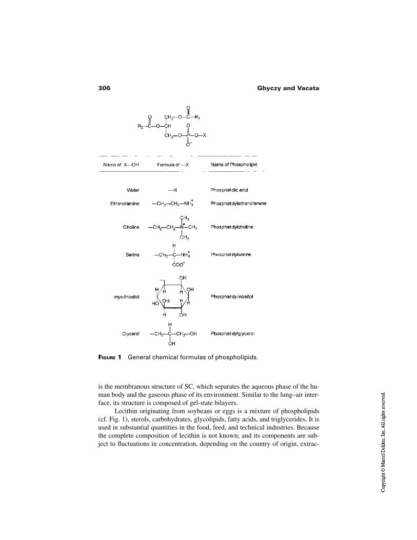
306 Ghyczy and Vacata
FIGURE 1 General chemical formulas of phospholipids.
is the membranous structure of SC, which separates the aqueous phase of the hu-man body and the gaseous phase of its environment. Similar to the lung–air inter-face, its structure is composed of gel-state bilayers.
Lecithin originating from soybeans or eggs is a mixture of phospholipids(cf. Fig. 1), sterols, carbohydrates, glycolipids, fatty acids, and triglycerides. It isused in substantial quantities in the food, feed, and technical industries. Becausethe complete composition of lecithin is not known; and its components are sub-ject to fluctuations in concentration, depending on the country of origin, extrac-
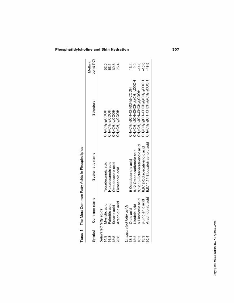
307Phosphatidylcholine and Skin Hydration
TA
BLE
1T
he
Mo
st C
om
mo
n F
atty
Aci
ds
in P
ho
sph
olip
ids
Sym
bo
lC
om
mo
n n
ame
Sys
tem
atic
nam
eS
tru
ctu
reM
elti
ng
po
int
(°C
)
Sat
ura
ted
fat
ty a
cid
s14
:8M
yris
tic
acid
Tetr
adec
ano
ic a
cid
CH
3(C
H2) 1
2C
OO
H52
,016
:8P
alm
itic
aci
dH
exad
ecan
oic
aci
dC
H3(C
H2) 1
4C
OO
H63
,118
:8S
tear
ic a
cid
Oct
adec
ano
ic a
cid
CH
3(C
H2) 1
6C
OO
H69
,620
:8A
rach
idic
aci
dE
ico
san
oic
aci
dC
H3(C
H2) 1
8C
OO
H75
,4
Un
satu
rate
d f
atty
aci
ds
18:1
Ole
ic a
cid
9-O
ctad
ecen
oic
aci
dC
H3(C
H2) 7
(CH
=CH
)(C
H2) 7
CO
OH
13,4
18:2
Lin
ole
ic a
cid
9,12
-Oct
adec
adie
no
ic a
cid
CH
3(C
H2) 7
(CH
=CH
CH
2) 2
(CH
2) 3
CO
OH
–9,0
18:3
α-Li
no
len
ic a
cid
9,12
,15-
Oct
adec
atri
eno
ic a
cid
CH
3(C
H2) 7
(CH
=CH
CH
2) 3
CO
OH
–11,
018
:3γ-
Lin
ole
nic
aci
d6,
9,12
-Oct
adec
atri
eno
ic a
cid
CH
3(C
H2) 4
(CH
=CH
CH
2) 3
(CH
2) 3
CO
OH
–10,
020
:4A
rach
ido
nic
aci
d5,
8,11
,14-
Eic
osa
tetr
aen
oic
aci
dC
H3(C
H2) 3
(CH
=CH
CH
2) 4
(CH
2) 2
CO
OH
–49,
5
308 Ghyczy and Vacata
tion process, storage conditions, and product age, lecithin itself cannot be recom-mended for use in modern cosmetic products.
Unfortunately, the term lecithin is often used rather loosely and imprecise-ly. Particularly in the English literature it is often used synonymously for PCand/or the complex mixture of lecithin as just outlined. This often leads to confu-sion and to incompatible findings achieved with PC, phospholipid mixtures, andlecithin.
It is also important to note that the only phospholipids documented in ac-cordance with the requirements of cosmetic and drug applications are pure PCand fractions with a high content of PC, either in unsaturated or hydrogenatedform, originating from soybeans.
An important factor of the use of lecithin and phospholipids in topical for-mulations is the presence of phosphatidylethanolamine (PE). This phospholipidpossesses a primary amino group (cf. Fig. 1) which may react with aldehydes, ke-tones, and carbohydrates present in the topical formulation or in the skin. Such achemical reaction may have two effects. It may cause a time-dependent deteriora-tion of components such as perfumes and preservatives. Or, if the amino group ofPE does interact with biomolecules which are components of the skin, it may leadto unpredicted pathological states. Because every lecithin and most phospholipidproducts contain PE, this aspect should be considered when designing a new for-mulation.
3.1 Phosphatidylcholine and Water
Phosphatidylcholine is hygroscopic—one molecule of PC binds approximately20 molecules of H2O. Each molecule of PC permeating into SC will drag along20 molecules of water. Also, in contrast to other phospholipids present in lecithin,PC (and to a lesser extent also PE) is the only phospholipid with an intrinsic hy-dration force [9]. The water-binding capacity of PC is thus independent of thepresence of ions. It can be assumed that if PC is taken up by the SC, the watercontent and the water-binding capacity of the SC will be elevated by virtue of theintrinsic hydration force of the PC taken up. Because in a disrupted permeationbarrier the flow of ions from the inside to the outside of skin has a messengerfunction [10], the ion-independent hydration force of PC may be of importance inthe treatment of damaged skin.
In the skin, PC is metabolized by the enzymes phospholipase A (PLA) andphospholipase D (PLD) into choline, betaine, and glycerylphosphatidylcholine(GPC). These metabolites belong to the group of biomolecules called osmopro-tectants, also known as osmolytes, compatible osmolytes, and/or (most precisely)counteracting osmolytes. These molecules play an essential role in the control ofvolumes of animal cells; they bind and keep water in the cell, but owing to theirhydrophilicity they are not able to penetrate into the membranes and transportwater across them.
309Phosphatidylcholine and Skin Hydration
The major organic osmoprotectants in animal cells can be divided into threegroups. The first group comprises substances which contain a quaternary nitrogenmoiety with three methyl groups and a positive charge. Glycerylphosphoryl-choline and betaine belong to this group. The second group consists of carbohy-drates such as sorbitol and inositol, and the third one of certain amino acids andtheir derivatives.
The presence of osmoprotectants in the skin is particularly important inview of the flow of ions between the inside and the outside of the skin. It is obvi-ous that a curative and/or preventive treatment of the skin with osmoprotectantscan only be successful if these molecules are capable of penetration into the skin.
Phosphatidylcholine and HPC are pro-osmoprotectants that do penetrateinto the skin, where they become precursors and a source of osmoprotectants. Incontrast to these penetration-capable precursors, the osmoprotectants themselvesare very hydrophilic and therefore are not capable of penetrating into the skin.
3.2 Phosphatidylcholine in Skin Treatment
Biological bilayers are permeation barriers which allow the formation of com-partments in a human organism. The composition of these bilayers is given by thetask they have to perform. The fluid-state membranes of cells and organelles sep-arate different aqueous phases and provide means for the generation of chemicaland electrical gradients. In the lung the barrier separates the aqueous and thegaseous phases, allowing an active gas exchange. In the skin it also separates theaqueous and the gaseous phases, but in addition it deals with biological, chemi-cal, and mechanical stresses.
These varying tasks are reflected in the different compositions of these bi-layers. The composition changes from the fluid-state phospholipids (mainly PC)in cell membranes to the gel-state PC in the lung and to the highly crystalline andhydrophobic structure of the skin, as imparted by ceramides. The ceramides ofthe latter membrane are the product of reprocessing of phospholipids and otherlipids in a deeper layer of the skin, the stratum granulosum. In this specific trans-formation, the fluid, hydrophilic vesicle–forming phospholipids get converted tothe hydrophobic, gel-state and sheet-forming ceramides. Such properties are nec-essary for the formation of a permeability barrier with a continuous, highly crys-talline bilayer structure.
Phosphatidylcholine is available in the form of two distinct chemical enti-ties. The first is native soybean PC, which contains approximately 70% linoleicacid, other unsaturated fatty acids, and only 15 to 16% of saturated fatty acids.Because of its fatty acid composition, this PC quality has a transition temperatureof around 0°C. In water it spontaneously forms fluid-state membranes and lipo-somes. It is extensively used in skin treatment as (1) a penetration enhancer[3,4,11] and (2) as a source of linoleic acid, e.g., in the treatment of acne andgreasy skin [12,13]. The second entity is PC containing saturated fatty acids only
310 Ghyczy and Vacata
(i.e., HPC), which is used in skin treatment with the aim of strengthening or sub-stituting the permeation barrier [14].
3.3 Phosphatidylcholine with Saturated Fatty Acids
In contrast to the fluid-state PC, the hydrogenated soybean phosphatidylcholine(HPC) contains approximately 85% stearic acid, 14% palmitic acid, and 1% oth-er fatty acids (Fig. 2). These fatty acids have a high melting point (cf. Table 1),and the transition temperature of HPC is therefore approximately 55°C.
The INCI declaration of HPC is hydrogenated lecithin, a misleading termbecause it does not express the fact that this product is a well-defined substance. Inour terminology, HPC is a chemical entity which forms bilayers with a gel-to-fluidtransition temperature of 50–55°C, as compared to 50°C of the SC lipids [15]. Asan excipient, HPC is used in drug formulations because it ameliorates side effectsof drugs such as amphotericin B and is well tolerated by humans. It possesses ther-modynamic properties similar to those of skin ceramides [2]. It is produced on anindustrial scale according to the cGMP requirements. Its handling is simple andwell documented. These factors suggest that for the preventive and curative treat-ment of dry skin, HPC could be a good industrial alternative to ceramides.
4 BIOLOGICAL FINDINGS WITH HPC
4.1 Interaction of HPC with SC Lipids
Blume et al. [16] performed differential scanning calorimetry and 2H-NMR ex-periments on a dispersion of a SC model lipid mixture consisting of 40% ce-ramides, 25% cholesterol, 25% palmitic acid, and 10% cholesterol sulfate. In wa-ter at 37°C this lipid mixture formed lamellar gel-state structures which werecomparable to those of the skin permeability barrier. These lamellar sheets inter-acted with the dispersions made of (1) a fluid-state PC fraction and (2) HPC byexchange of monomers through the water phase. For the fluid-state PC the inter-action was complete in 2 hr; for HPC in 24 hr. The interaction was dependent onthe lipid concentration.
These results indicate that the two PC dispersions tested have different ki-netics of interaction with the skin, and also that their effect on skin homeostasis ispossibly dose dependent. One can conclude that HPC does not penetrate asdeeply into the skin as the fluid-state PC.
4.2 Uptake of HPC by Skin
The different kinetics of the interaction between the native skin lipids and the flu-id-state PC and/or HPC was determined by several in vivo and in vitro penetra-tion studies.
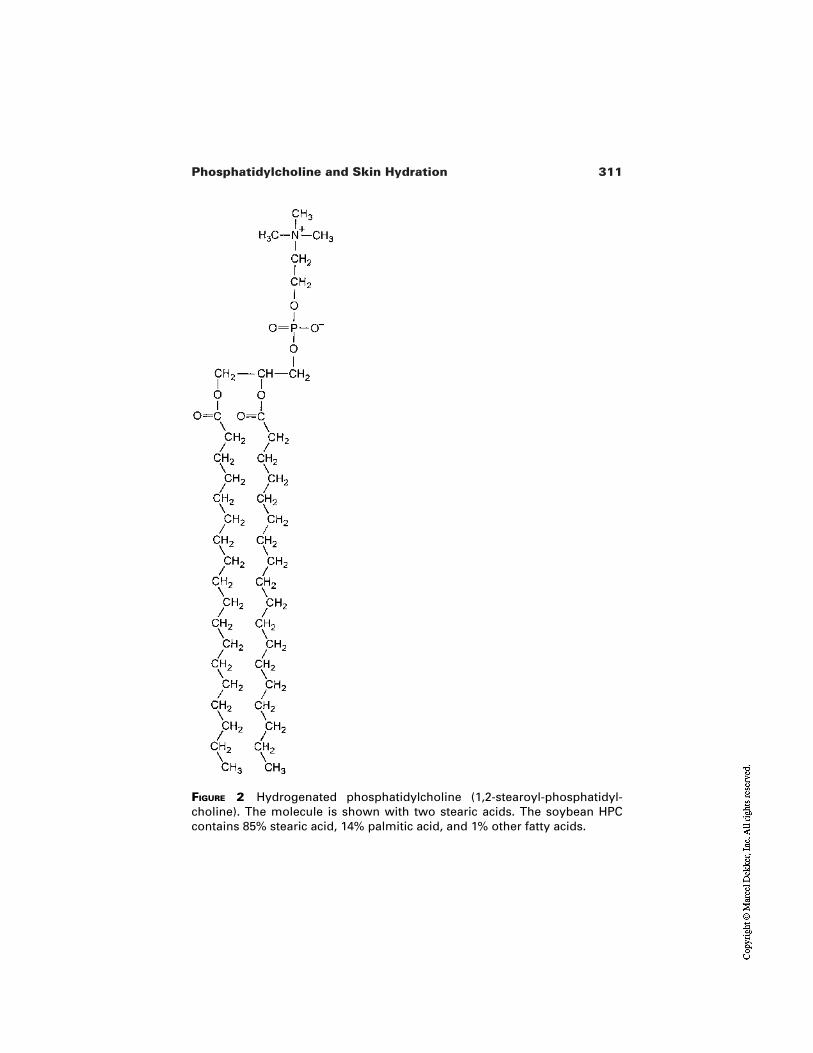
311Phosphatidylcholine and Skin Hydration
FIGURE 2 Hydrogenated phosphatidylcholine (1,2-stearoyl-phosphatidyl-choline). The molecule is shown with two stearic acids. The soybean HPCcontains 85% stearic acid, 14% palmitic acid, and 1% other fatty acids.
312 Ghyczy and Vacata
Van Kuijk et al. [17] showed that in vivo the fluorescent-labeled liposomesmade from fluid-state PC penetrate significantly deeper into the rat skin thanthose composed of HPC. The same results were obtained for analogous in vitrostudies on human skin [18]. The uptake was higher under nonocclusive condi-tions, indicating the existence of an as yet unrecognized role of water in the skinpenetration of the formulations containing PC.
Kirjavainen et al. [4] compared the distribution of drugs formulated withfluid-state PC and/or HPC in the skin in vitro. The HPC-based liposomes re-mained in the SC and were thus not able to enhance the transdermal drug pene-tration.
Fahr et al. [3] visualized penetration of liposomes with encapsulated fluores-cent dye carboxyfluorescein into the human abdomen skin. Figure 3 shows that,compared to the liposomes made from HPC, those composed of fluid-state PC aretaken up by the skin more readily, permeate it faster, and penetrate beyond the SC.
These findings suggest that HPC, and most probably also the accompany-ing water, is taken up by the SC but not by the deeper layers of the skin. In addi-tion, HPC does not seem to perturb the lipid barrier to the extent that it would en-hance the uptake of substances by the dermis.
4.3 Tolerance of HPC by Sensitive Skin
Damaging the permeability barrier is considered to be the first step in the processof irritation of the skin by chemical or physical noxae [19]. The consequence ofthis damage is the increased synthesis of cytokines and lipids as well as an in-creased level of TEWL [20,21]. The visual or sensory symptoms perceptible bythe test persons and the investigators are scaling and erythema. These effectswere used to determine and quantify the effects of different emulsifiers on the testpersons as compared to those of HPC [22].
The results of this study (Fig. 4) lend themselves to the following interpre-tation: (1) the emulsifiers which are not related to the lipids of the permeabilitybarrier have the highest irritation potential; (2) the sugar-containing substanceswhich are related to the biological membranes have a lower irritation potential;and (3) HPC (substance most closely related to the permeability barrier lipids)displays no irritation potential at all.
4.4 Effect of HPC on Skin Hydration
Sodium lauryl sulfate challenge of the human skin in vivo is generally recognizedas the most convincing method for imitating dry skin. We have chosen thismethod to evaluate the potential of HPC in the control of skin hydration. Thisclinical study was carried out by Gehring et al. [23] at the Dermatology Depart-ment of the University of Karlsruhe, Germany, in cooperation with the authors,and has been submitted for publication.
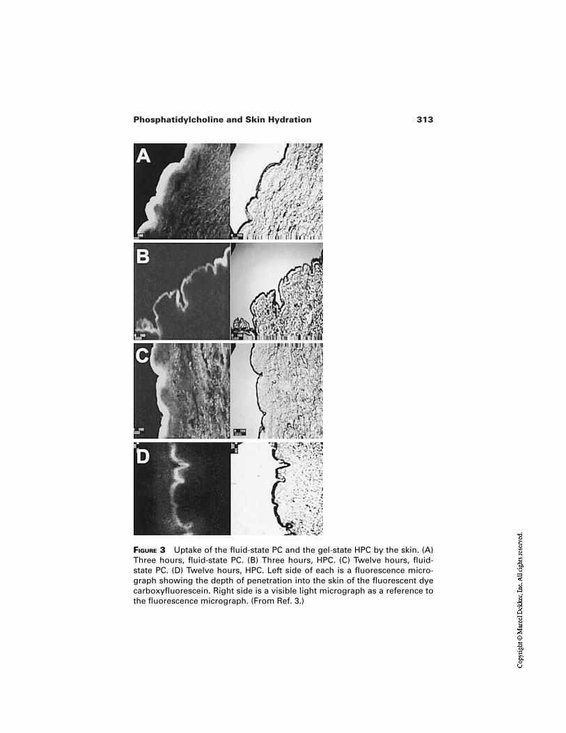
313Phosphatidylcholine and Skin Hydration
FIGURE 3 Uptake of the fluid-state PC and the gel-state HPC by the skin. (A)Three hours, fluid-state PC. (B) Three hours, HPC. (C) Twelve hours, fluid-state PC. (D) Twelve hours, HPC. Left side of each is a fluorescence micro-graph showing the depth of penetration into the skin of the fluorescent dyecarboxyfluorescein. Right side is a visible light micrograph as a reference tothe fluorescence micrograph. (From Ref. 3.)
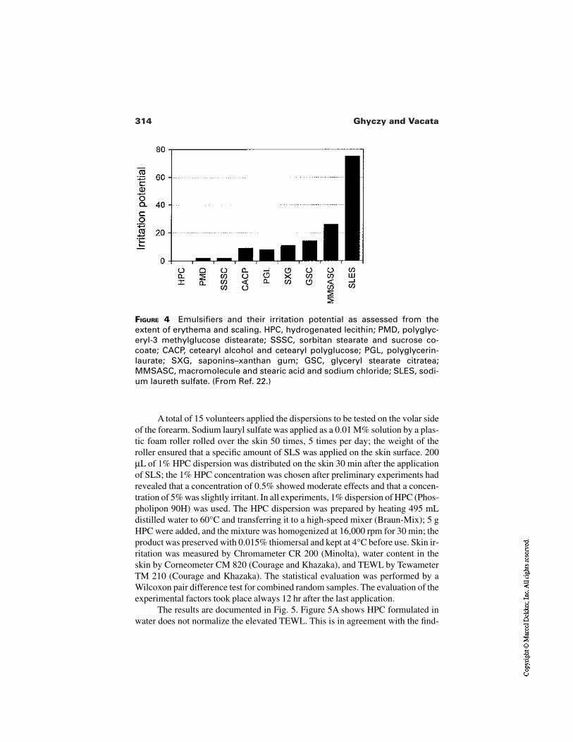
314 Ghyczy and Vacata
FIGURE 4 Emulsifiers and their irritation potential as assessed from theextent of erythema and scaling. HPC, hydrogenated lecithin; PMD, polyglyc-eryl-3 methylglucose distearate; SSSC, sorbitan stearate and sucrose co-coate; CACP, cetearyl alcohol and cetearyl polyglucose; PGL, polyglycerin-laurate; SXG, saponins–xanthan gum; GSC, glyceryl stearate citratea;MMSASC, macromolecule and stearic acid and sodium chloride; SLES, sodi-um laureth sulfate. (From Ref. 22.)
A total of 15 volunteers applied the dispersions to be tested on the volar sideof the forearm. Sodium lauryl sulfate was applied as a 0.01 M% solution by a plas-tic foam roller rolled over the skin 50 times, 5 times per day; the weight of theroller ensured that a specific amount of SLS was applied on the skin surface. 200µL of 1% HPC dispersion was distributed on the skin 30 min after the applicationof SLS; the 1% HPC concentration was chosen after preliminary experiments hadrevealed that a concentration of 0.5% showed moderate effects and that a concen-tration of 5% was slightly irritant. In all experiments, 1% dispersion of HPC (Phos-pholipon 90H) was used. The HPC dispersion was prepared by heating 495 mLdistilled water to 60°C and transferring it to a high-speed mixer (Braun-Mix); 5 gHPC were added, and the mixture was homogenized at 16,000 rpm for 30 min; theproduct was preserved with 0.015% thiomersal and kept at 4°C before use. Skin ir-ritation was measured by Chromameter CR 200 (Minolta), water content in theskin by Corneometer CM 820 (Courage and Khazaka), and TEWL by TewameterTM 210 (Courage and Khazaka). The statistical evaluation was performed by aWilcoxon pair difference test for combined random samples. The evaluation of theexperimental factors took place always 12 hr after the last application.
The results are documented in Fig. 5. Figure 5A shows HPC formulated inwater does not normalize the elevated TEWL. This is in agreement with the find-
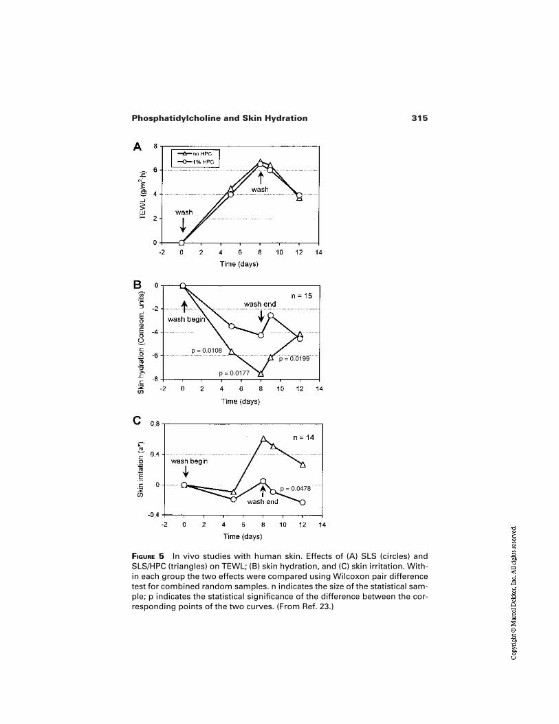
315Phosphatidylcholine and Skin Hydration
FIGURE 5 In vivo studies with human skin. Effects of (A) SLS (circles) andSLS/HPC (triangles) on TEWL; (B) skin hydration, and (C) skin irritation. With-in each group the two effects were compared using Wilcoxon pair differencetest for combined random samples. n indicates the size of the statistical sam-ple; p indicates the statistical significance of the difference between the cor-responding points of the two curves. (From Ref. 23.)
p = 0.0108
p = 0.0177
p = 0.0199
p = 0.0478
316 Ghyczy and Vacata
ings of other laboratories that the application of products containing only phos-pholipids [24] or only ceramides [25] to a perturbed skin is insufficient for thenormalization of TEWL. Figure 5B shows that the water content of SC in thechallenged skin gets significantly elevated upon the treatment with HPC. Figure5C shows that HPC normalized the cytokine-transmitted inflammatory responseof the perturbed skin.
4.5 Effect of HPC in Topical Formulations onHydration of Healthy Skin
This cosmetic study was conducted for Kuhs GmbH by Derma Consult GmbH(Bonn, Germany). The aim of the study was to compare effects of HPC in a cos-metic formulation on healthy skin with those of three commercial oil-in-water(O/W) emulsions. The study has not been published yet, but it is available fromKuhs GmbH (Leichlingen, Germany; [email protected]).
A total of five preparations were tested:
1. HPC-containing DMS formulation 1 (Aqua, carprylic/capric triglyc-eride, pentylene glycol, hydrogenated lecithin (HPC 4%), butyrosper-mum parkii, glycerin, squalene, 0.0066% ceramide 3)
2. HPC-containing DMS formulation 2 (Aqua, carprylic/capric triglyc-eride, pentylene glycol, hydrogenated lecithin (HPC 2%), butyrosper-mum parkii, glycerin, squalene, sodium carbomer, xantham gum,0.0033% Ceramide 3)
3. Commercial product 1 (Aqua, caprylic/capric triglyceride, hydrogenat-ed coco-glycerides, polyglyceryl-3 methylglucose distearate, glycerylstearate, setyl alcohol, stearyl alcohol, phenoxyethanol, cyclome-thicone, glyceryl polymethacrylate, imidazolidinyl urea, aluminumstarch octenyl succinate, propylene glycol)
4. Commercial product 2 (Aqua, paraffinum liquidum, dicaprylyl ether,cyclomethicone, glyceryl stearate SE, isohexadecane, butyrospermumparkii, sodium acrylatees copolymer, phenoxyethanol, cetyl phosphate,imidazolidinyl urea, PPG-1 Trideceth-6, sodium hydroxide, PEG-8, to-copherol, ascorbyl palmitate, ascorbic acid, citric acid)
5. Commercial product 3 (Aqua, paraffinum liquidum, sorbitol, dicapry-lyl ether, cyclomethicone, glyceryl stearate SE, isohexadecane, toco-pheryl acetate, butryrospermum parkii, sodium acrylates copolymer,panthenol, phenoxyethanol, cetyl phosphate, hexylene glycol, imida-zolidinyl urea, PPG-1 Trideceth-6, retinyl palmitate, arachis hypogaea,fructose, glucose, sodium hydroxide, PEG-8, dextrin, sucrose, urea, to-copherol, alanine, ascorbyl palmitate, aspartic acid, glutamic acid,hexyl nicotinate, ascorbic acid, citric acid)
317Phosphatidylcholine and Skin Hydration
The test was performed on 20 healthy female volunteers (25 to 40 years old), whoapplied the formulations to be tested on the volar side of the forearm. The prod-ucts were applied twice daily over a period of 28 days. The time of evaluationwas 8 hr, 1 and 3 days after the last application. Skin hydration was measuredwith Corneometer CM 825 (Courage and Khazaka), skin roughness with Skin Vi-siometer (image analysis on silicon base, Courage and Khazaka) and skin firm-ness with Cutometer SEM 474 (Courage and Khazaka).
The results of this comparative study on healthy skin are summarized inFig. 6. They indicate the following.
1. Both of the formulations containing HPC show similar efficacy; onecan therefore presume that the highest beneficial effect is achieved at2% HPC.
2. The superior effects of the DMS formulations, especially the longer-lasting effects after the application was terminated, are most likelybased on the presence of HPC. This conclusion is supported by thepenetration property of HPC as well as by the elevation of skin hydra-tion as indicated in the case of the SLS-damaged skin. Because inhealthy skin the barrier is not damaged, these findings strongly indicatethat the elevated skin hydration could be a function of the intrinsic hy-dration force of HPC and the osmoprotectant efficacy of its metabo-lites, due to which HPC brings in and retains water in the permeabilitybarrier. No other components of topical formulations possess suchproperties.
Because the commercial products do not include substances with the func-tionality of HPC outlined, the superior efficacy of the DMS formulations is un-derstandable. This difference further indicates the relevance of HPC for the con-trol of skin hydration also in the healthy undamaged skin.
5 DISCUSSION
The functionalities of HPC explained in this chapter support and expand ourknowledge of the mechanism and the means of skin hydration control.
It was shown that SC takes up HPC. The prerequisite for this uptake is theinteraction of HPC with the permeability barrier. In vitro, this interaction does notperturb the SC lipid structure. This explains the lack of any irritant capacity ofHPC on healthy human skin in vivo. In contrast to the interaction of HPC with theskin, nearly all of the commercial emulsifiers dissolve the SC lipid structure invitro, and in vivo most of them display an irritation capacity. In SLS-perturbedskin, the elevated TEWL is an indicator of the disintegration of the permeabilitybarrier. In our experiments, HPC formulated in water does not normalize the ele-vated TEWL. This is in accordance with the findings of other laboratories sug-
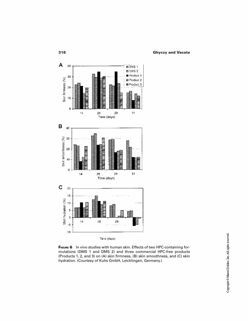
318 Ghyczy and Vacata
FIGURE 6 In vivo studies with human skin. Effects of two HPC-containing for-mulations (DMS 1 and DMS 2) and three commercial HPC-free products(Products 1, 2, and 3) on (A) skin firmness, (B) skin smoothness, and (C) skinhydration. (Courtesy of Kuhs GmbH, Leichlingen, Germany.)
319Phosphatidylcholine and Skin Hydration
gesting that the bilayer-forming substances, such as phospholipids [24] or ce-ramides [25], have to be formulated with other lipids which do not form bilayerson their own, but which will insert into existing bilayers, thus providing a form ofrepair to a disrupted barrier. On the other hand the water content of the SC inSLS-perturbed skin becomes significantly elevated if the skin is treated with HPCconcomitantly with and after the SLS treatment. This indicates that HPC func-tions as a transport vehicle and storage for water, thus substituting to some degreethe SLS-depleted NMF. Because of the intrinsic hydration force of HPC and theosmoprotectant properties of the HPC metabolites, such as betaine and glyc-erylphosphatidylcholine, and because HPC is taken up in the SC, these findingsare not surprising.
The repetitive application of SLS causes not only a disruption of the per-meability barrier, but also the release of cytokines, which are the indicators of in-flammation. In our experiments, HPC normalized the inflammatory response ofthe skin to the SLS treatment. This result is supported by previous findings re-garding the anti-inflammatory potential of PC in the cases of UV irritation [26]and efflorescences in acne [12]. Another supportive argument could be the pro-posed mode of protective effects of PC and its metabolites during a redox imbal-ance [5].
The finding that HPC has an anti-inflammatory efficacy but no normalizingeffects on TEWL is in contrast to the common belief that the cytokine-releasinginflammatory effect of SLS is based on barrier disruption, and that a barrier repairleads to normalization of the inflamed skin [27].
Further experiments are necessary for the evaluation of the full anti-inflam-matory potential of HPC because it is conceivable that due to the uptake of HPCin SC, the penetration rate of SLS through SC increases, which is a contrary effectto the amelioration of irritation by HPC.
In order to evaluate the full potential of HPC in skin hydration control weneed to perform experiments with formulations containing HPC and lipids whichare related to endogenous SC lipids. The positive effects of such lipid combina-tions on SLS-induced dry skin [24] should suggest the optimal composition ofsuch formulations.
REFERENCES
1. Harding CR, Watkinson A, Rawlings AV, Scott IR. Dry skin, moisturization and cor-neodesmolysis. Int J Cosmet Sci 2000; 22:21–52.
2. Pechtold LA, Abraham W, Potts RO. Characterization of the stratum corneum lipidmatrix using fluorescence spectroscopy. J Invest Dermatol Symp Proc 1998;3:105–109.
3. Fahr A, Schäfer U, Verma DD, Blume G. Skin penetration enhancement of sub-stances by a novel type of liposomes. SÖFW-Journal 2000; 126:49–53.
320 Ghyczy and Vacata
4. Kirjavainen M, Monkkonen J, Saukkosaari M, Valjakka-Koskela R, Kiesvaara J,Urtti A. Phospholipids affect stratum corneum lipid bilayer fluidity and drug parti-tioning into the bilayers. J Contr Release 1999; 58:207–214.
5. Ghyczy M, Boros M. Electrophilic methyl groups present in the diet amelioratephysiological states induced by reductive and oxidative stress. Hypothesis. Br J Nutr2001; 85:409–414.
6. Rawlings AV, Scott IR, Harding CR, Bowser PA. Stratum corneum moisturization atthe molecular level. J Invest Dermatol 1994; 103:731–741.
7. A Wendel. Lecithin. In: Kroschwitz JI, Howe-Grant M, eds. Encyclopedia of Chem-ical Technology. Vol. 15. New York: Wiley, 1995:192–210.
8. Silvius JR. Structure and nomenclature. In: Cevc G, ed. Phospholipids Handbook.New York: Marcel Dekker, 1993:1–22.
9. Israelachvili J. Intermolecular and Surface Forces. London: Academic Press,1992:395–421.
10. Lee SH, Elias PM, Proksch E, Menon GK, Mao-Quiang M, Feingold KR. Calciumand potassium are important regulators of barrier homeostasis in murine epidermis. JClin Invest 1992; 89:530–538.
11. Gehring W, Ghyczy M, Gareiss J, Gloor M. The influence on skin penetration bydithranol formulated in phospholipids solutions and phospholipid liposomes. Eur JPharm Biopharm 1995; 41:140–141.
12. Ghyczy M, Nissen HP, Blitz H. The treatment of acne vulgaris by phosphatidyl-choline from soybeans, with a high content of linoleic acid. J Appl Cosmetol 1996;14:137–145.
13. Morganti P, Randazzo SD, Giardina A, Bruno C, Vincenti M, Tiberi L. Effect ofphosphatidylcholine linoleic acid–rich and glycolic acid in Acne vulgaris. J ApplCosmetol 1997; 15:21–32.
14. DMS-Concentrate: A new Hydrolipid System Similar to the Skin Lipid Structure.Langenfeld: Kuhs GmbH & Co., 1995.
15. Bonte F, Pinguet P, Saunois A, Meybeck A, Beugin S, Ollivon M, Lesieur S. Ther-motropic phase behavior of in vivo extracted human stratum corneum lipids. Lipids1997; 32:653–660.
16. Blume A, Jansen M, Ghyczy M, Gareiss J. Interaction of phospholipid liposomeswith lipid model mixtures for stratum corneum lipids. Int J Pharm 1993;99:219–228.
17. van Kuijk-Meuwissen ME, Mougin L, Junginger GE, Bouwstra JA. Application ofvesicles to rat skin in vivo: a confocal laser scanning microscopy study. J Contr Re-lease 1998; 56:189–196.
18. van Kuijk-Meuwissen ME, Junginger GE, Bouwstra JA. Interactions between lipo-somes and human skin in vitro, a confocal laser scanning microscopy study. BiochimBiophys Acta 1998; 1371:31–39.
19. Tsai JC, Feingold KR, Crumrine D, Wood LC, Grunfeld C, Elias PM. Permeabilitybarrier disruption alters the localization and expression of TNF alpha/protein in theepidermis. Arch Dermatol Res 1994; 286:242–248.
20. Fartasch M. Ultrastructure of the epidermal barrier after irritation. Microsc Res Tech1997; 37:193–199.
21. Wood LC, Jackson SM, Elias PM, Grunfeld C, Feingold KR. Cutaneous barrier per-
321Phosphatidylcholine and Skin Hydration
turbation stimulates cytokine production in the epidermis of mice. J Clin Invest1992; 90:482–487.
22. Kutz G, Biehl P, Waldmann-Laue M, Jackwerth B. Zur Auswahl von O/W-Emulga-toren für den Einsatz in Hautpflegeprodukten bei sensibler Haut. SÖFW-Journal1997; 123:145–149.
23. Gehring W, Ghyczy M, Vacata V, Gloor M. Effect of HPC on SLS-treated skin (sub-mitted).
24. Summers RS, Summers B, Chandar P, Feinberg C, Gursky R, Rawlings AV. The ef-fect of lipids, with and without humectant, on skin xerosis. J Soc Cosmet Chem1996; 47:27–39.
25. Man MQ, Feingold KR, Elias PM. Exogenous lipids influence permeability barrierrecovery in acetone-treated murine skin. Arch Dermatol 1993; 129:728–738.
26. Thiele B, Ghyczy M, Lunow C, Teichert GM, Wolff GH. Influence of phospholipidliposomes (PLL) on UVB-induced erythema formation. Arch Dermatol Res 1993;285:428–431.
27. Nickoloff BJ, Naidu Y. Perturbation of epidermal barrier function correlates with ini-tiation of cytokine cascade in human skin. J Am Acad Dermatol 1994; 30:535–546.

16Hydroxyacids
Anthony W. JohnsonUnilever Home and Personal Care North America, Trumbull, Connecticut
1 INTRODUCTION
Although there are many hydroxy acids, the focus of this chapter is on alpha-hy-droxyacids, or AHAs as they have come to be known universally since their ex-plosive takeover of the cosmetic facial moisturizer market in the early 1990s[1–3]. More recently, other classes of hydroxyacids have been used in skin careproducts [4], but at the time of writing (2001) AHAs stand alone as the only hy-droxyacids supported by placebo-controlled clinical testing. In fact it is one par-ticular AHA, glycolic acid, that was used in the first AHA facial moisturizers andremains the most common form today. As detailed herein, glycolic acid and otherAHAs do more than moisturize. They are able to reduce wrinkles, eliminate finelines, improve skin surface texture, and lessen some of the other changes associ-ated with photodamaged skin. However, the AHA story starts long before thespectacular appearance of glycolic acid “anti-aging” skin creams in 1992. Some25 years earlier, another alpha-hydroxyacid, lactic acid, was identified as a com-ponent of the skin’s natural moisturizing factor (NMF) and introduced as a mois-turizing ingredient in creams and lotions to treat and prevent dry skin, particular-ly winter dry skin on the hands, legs, and body [5]. At about the same time VanScott and Yu reported that alphahydroxy acids as a class were effective for treat-ing ichthyosis and other disorders of keratinization [6]. The new information
323
324 Johnson
about AHAs prompted increased interest in glycolic acid by dermatologists forchemical peel procedures. Glycolic acid solutions (50–70% concentration with-out neutralization) were found to be very effective as acid peels, easier to use andwithout side effects compared to other peeling agents [7].
The extensive use of AHAs in the last 10 years has transformed the cosmet-ic market place and changed consumer expectations. Women have seen that AHAmoisturizers can provide skin improvements dramatically better than previousskin care creams. There has been much discussion about the biological effectsand cosmetic efficacy of AHA products over the last 10 years [8–10], with themajority of publications being editorial and review articles rather than originalscientific papers. This has resulted in a merging of fact and speculation relating toAHA skin actions and benefits.
Alpha-hydroxyacids have a variety of different actions on skin dependingon their pH and concentration. These effects are detailed in the sections that fol-low. In simple terms, the salt forms of AHAs are effective humectant moisturiz-ers, whereas the acid forms go beyond moisturizing to correct the dysfunction un-derlying dry skin and to enhance the normal processes of the epidermis. There isan important distinction between the strong acid solutions used as chemical peelsby dermatologists and the mild buffered preparations available to consumers aseveryday cosmetic creams and lotions. The former work by damaging skin, andthe latter are designed to provide benefits without any adverse effect.
Alpha-hydroxyacids appear to have multiple actions on the stratumcorneum and living epidermis depending on concentration and pH. But given thatthe primary function of the epidermis is to produce a healthy and effective outerprotective layer, it is not surprising that the main benefits of AHAs are manifest asenhancements of stratum corneum quality (its look, touch, feel, and effectivenessas a protective barrier). Another property of AHAs in their buffered acid form is atendency to induce sensory irritation (burn, sting, or tingling) in susceptible indi-viduals, at concentrations which are not overtly irritating, i.e., do not induce aninflammatory reaction [11]. The sensory irritant effect is pH/concentration relatedand reduces as pH is increased (i.e., as the proportion of free acid is reduced).
Failure to take account of pH and concentration has led to a good deal ofquestioning and miscommunication about the benefits, safety, and effectivenessof AHA products. It is surprising how many otherwise sound scientific publica-tions do not specify the pH of AHA preparations under study. It is therefore ap-propriate to start this chapter with a brief consideration of pH and some of theother basic facts about AHAs.
2 CHEMISTRY AND BIOCHEMISTRY OF ALPHA-HYDROXYACIDS
Hydroxyacids are common and essential constituents of all living cells. Simplehydroxyacids are mainly involved in the breakdown of fats and carbohydrates to
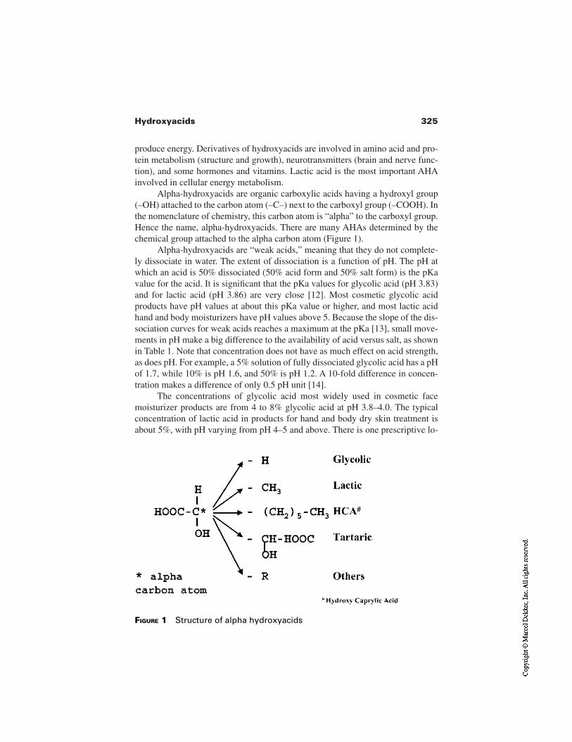
325Hydroxyacids
produce energy. Derivatives of hydroxyacids are involved in amino acid and pro-tein metabolism (structure and growth), neurotransmitters (brain and nerve func-tion), and some hormones and vitamins. Lactic acid is the most important AHAinvolved in cellular energy metabolism.
Alpha-hydroxyacids are organic carboxylic acids having a hydroxyl group(–OH) attached to the carbon atom (–C–) next to the carboxyl group (–COOH). Inthe nomenclature of chemistry, this carbon atom is “alpha” to the carboxyl group.Hence the name, alpha-hydroxyacids. There are many AHAs determined by thechemical group attached to the alpha carbon atom (Figure 1).
Alpha-hydroxyacids are “weak acids,” meaning that they do not complete-ly dissociate in water. The extent of dissociation is a function of pH. The pH atwhich an acid is 50% dissociated (50% acid form and 50% salt form) is the pKavalue for the acid. It is significant that the pKa values for glycolic acid (pH 3.83)and for lactic acid (pH 3.86) are very close [12]. Most cosmetic glycolic acidproducts have pH values at about this pKa value or higher, and most lactic acidhand and body moisturizers have pH values above 5. Because the slope of the dis-sociation curves for weak acids reaches a maximum at the pKa [13], small move-ments in pH make a big difference to the availability of acid versus salt, as shownin Table 1. Note that concentration does not have as much effect on acid strength,as does pH. For example, a 5% solution of fully dissociated glycolic acid has a pHof 1.7, while 10% is pH 1.6, and 50% is pH 1.2. A 10-fold difference in concen-tration makes a difference of only 0.5 pH unit [14].
The concentrations of glycolic acid most widely used in cosmetic facemoisturizer products are from 4 to 8% glycolic acid at pH 3.8–4.0. The typicalconcentration of lactic acid in products for hand and body dry skin treatment isabout 5%, with pH varying from pH 4–5 and above. There is one prescriptive lo-
FIGURE 1 Structure of alpha hydroxyacids
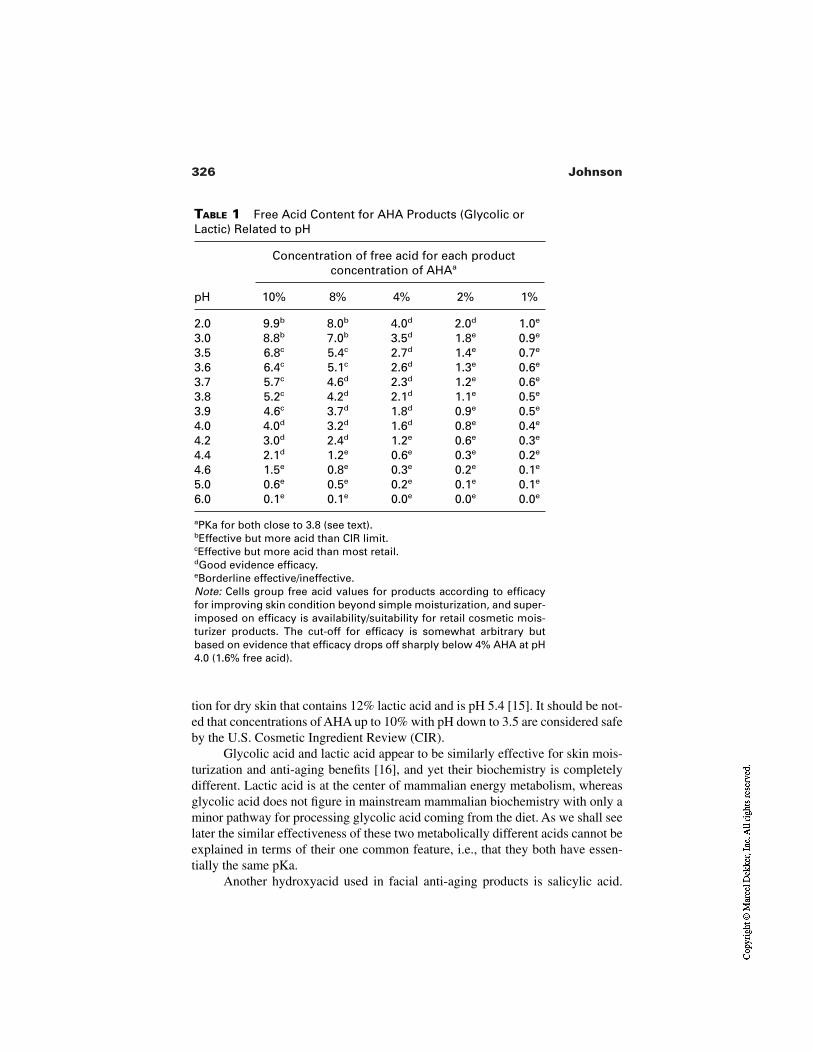
326 Johnson
TABLE 1 Free Acid Content for AHA Products (Glycolic orLactic) Related to pH
Concentration of free acid for each productconcentration of AHAa
pH 10% 8% 4% 2% 1%
2.0 9.9b 8.0b 4.0d 2.0d 1.0e
3.0 8.8b 7.0b 3.5d 1.8e 0.9e
3.5 6.8c 5.4c 2.7d 1.4e 0.7e
3.6 6.4c 5.1c 2.6d 1.3e 0.6e
3.7 5.7c 4.6d 2.3d 1.2e 0.6e
3.8 5.2c 4.2d 2.1d 1.1e 0.5e
3.9 4.6c 3.7d 1.8d 0.9e 0.5e
4.0 4.0d 3.2d 1.6d 0.8e 0.4e
4.2 3.0d 2.4d 1.2e 0.6e 0.3e
4.4 2.1d 1.2e 0.6e 0.3e 0.2e
4.6 1.5e 0.8e 0.3e 0.2e 0.1e
5.0 0.6e 0.5e 0.2e 0.1e 0.1e
6.0 0.1e 0.1e 0.0e 0.0e 0.0e
aPKa for both close to 3.8 (see text).bEffective but more acid than CIR limit.cEffective but more acid than most retail.dGood evidence efficacy.eBorderline effective/ineffective.Note: Cells group free acid values for products according to efficacyfor improving skin condition beyond simple moisturization, and super-imposed on efficacy is availability/suitability for retail cosmetic mois-turizer products. The cut-off for efficacy is somewhat arbitrary butbased on evidence that efficacy drops off sharply below 4% AHA at pH4.0 (1.6% free acid).
tion for dry skin that contains 12% lactic acid and is pH 5.4 [15]. It should be not-ed that concentrations of AHA up to 10% with pH down to 3.5 are considered safeby the U.S. Cosmetic Ingredient Review (CIR).
Glycolic acid and lactic acid appear to be similarly effective for skin mois-turization and anti-aging benefits [16], and yet their biochemistry is completelydifferent. Lactic acid is at the center of mammalian energy metabolism, whereasglycolic acid does not figure in mainstream mammalian biochemistry with only aminor pathway for processing glycolic acid coming from the diet. As we shall seelater the similar effectiveness of these two metabolically different acids cannot beexplained in terms of their one common feature, i.e., that they both have essen-tially the same pKa.
Another hydroxyacid used in facial anti-aging products is salicylic acid.
327Hydroxyacids
This is an aromatic hydroxyacid that has long been used as a keratolytic agent inthe treatment of warts and other hyperkeratotic conditions [17] and in OTCpreparations for the treatment of mild acne [18]. Unlike AHAs, salicylic acid hasnot been used as a moisturizing ingredient in products for treating dry skin, but itdoes seem to have cosmetic benefits for skin photodamage and has been promot-ed as an alternative to AHAs [19]. The pKa of salicylic acid is 2.97 [20], nearly 10times more acid than the pKa of glycolic and lactic acids. Because of its potentialfor irritation at higher concentrations, salicylic acid is mostly used at concentra-tions of 1.5% or less in cosmetic skin creams. Salicylic acid has been described asa beta-hydroxyacid (BHA) by the cosmetic industry, but described correctly it isan aromatic ortho-hydroxyacid.
The alpha-hydroxyacids are sometimes called fruit acids because of theirabundance in common fruits (citric acid in citrus fruits, malic acid in apples, tar-taric acid in grapes). Ironically, the two most widely used AHAs are not majorcomponents of fruits; glycolic acid is a constituent of sugar cane juice and lacticacid occurs most abundantly in sour milk [12].
3 SKIN BENEFITS OF HYDROXYACIDS
There have been several distinct phases in the development of AHAs for cosmet-ic skin care products.
1. 1970 onward: use of lactic acid in products to treat and prevent dryskin (initially in the United States and Europe, extending worldwide).
2. 1992 onward: alongside lactic acid moisturizers, use of glycolic acid totreat facial photodamage (initially in the United States, extendingworld wide).
3. Mid-1990s onward: alongside glycolic and lactic acid products, use ofother hydroxy acids including BHA, tri-hydroxyacids (THAs), poly-hydroxyacids (PHAs), combinations of these, and ascorbic acid (vita-min C), in a search for systems to improve on glycolic acid.
4. In addition to their cosmetic skin care use, AHAs, saw increasing usefrom the 1970s onward, specifically glycolic acid, for chemical peels.This was both as conventional peels and as combination therapieswhere patients use “cosmetic” AHA preparations at home and visit thedermatologist at regular intervals for supplementary glycolic acid lightpeels [21].
The focus for the first 20 years of AHA application was for moisturizing productsto treat and prevent dry skin. The typical pH of lactate-containing dry skin creamsand lotions has been above the pKa of lactic acid, usually by a pH unit or more.Therefore, the lactate in these products has been more in the salt form (ammoni-um, sodium, and potassium salts are most common) than as free acid. The pub-lished data suggest that lactate dry skin products vary in effectiveness and are
328 Johnson
generally only a little more effective for controlling dry skin than products basedon the main alternatives, humectants such as glycerol, sorbitol, urea, and glycols.However, it is difficult to draw conclusions about the different ingredients be-cause most of the clinical comparisons of these products have been with commer-cial products that contain different concentrations of humectant in different emul-sion bases [22]. Over the last 30 years, most of the leading dry skin moisturizingproducts have been based on these alternative moisturizing ingredients more thanlactic acid. [23].
The second wave of AHA products, from 1992 onward, were the facialmoisturizers promoted as wrinkle reduction creams and lotions, or so-called anti-aging creams. The evidence presented here leaves little doubt that these productsare effective for reducing the visible signs of photodamaged skin and that thisproduces a younger and healthier look to the skin. Typically, there is a marked im-provement in the first week or so that can be attributed to direct effects on thestratum corneum (hydration and exfoliation). This is followed by a slow, progres-sive further improvement over several months with fine lines and wrinkles be-coming less evident and overall complexion achieving a brighter and more evencolor tone. Initially, anti-aging AHA products were available in two strengths ofglycolic acid, 8 and 4% glycolic acid and a pH 3.8–4.0. These products wereproven effective in a placebo-controlled clinical trial (see Section 8). Their suc-cess was followed by scores of other products that were mostly highly priceditems sold in prestige and specialist channels of trade. Some of these productswere effective (i.e., they contained AHA, usually glycolic acid, at effective con-centrations/pH), but many were probably not. The effective products have stoodthe test of time and remain on the market in 2001. Many of the others have disap-peared or been reformulated with new ingredients and new claims.
The third phase of AHA products, which continues today, has two compo-nents. One was the extension of the original AHAs, glycolic and lactic acid, intoa broader range of products addressing a wider range of everyday skin problems(see subsequent sections). The other was the introduction of “new” hydroxy acidsand related ingredients, emerging from the research and exploration of cosmeticmanufacturers and the raw material supply industries. The search for ingredientsand combinations superior to the original AHA products for skin improvementcontinues.
The review in this chapter follows the chronology of AHA discovery andapplication in cosmetic products, a chronology that starts with research conduct-ed in the late 1960s and published in the early 1970s.
4 BENEFITS FOR ICHTHYOSIS
The 1974 publication by Van Scott and Yu [6], indicating that hydroxyacids as aclass have skin therapeutic activity beyond simple moisturization, was a major
329Hydroxyacids
landmark in the AHA story. In a small but very elegant and revealing study, VanScott and Yu demonstrated unequivocally that alpha-hydroxyacids have a re-markable therapeutic action on ichthyotic skin conditions. Ichthyosis describes anumber of similarly appearing hereditary skin conditions in which there is ab-normal keratinization, leading to an accumulation of heavy scale at the skin sur-face [24]. Ichthyotic skin has a characteristic fish skin–like appearance (Gr.Ichthys, fish). When Van Scott did his pioneering work over 25 years ago, thecause of ichthyoses, as for most skin conditions, was unknown beyond specula-tion based on symptoms and histological findings. Treatment of the ichthyoseswas empirical, involving heavy application of moisturizing creams and oint-ments containing keratolytic agents such as salicylic acid and urea. These prepa-rations provided only modest relief of symptoms. Modern molecular biology hasrevealed the genetic defects and associated metabolic disturbances underlyingthe different ichthyoses [25]. Recessive X-linked ichthyosis is caused by a defi-ciency of the enzyme steroid sulfatase [26,27]; epidermolytic ichthyoses arecaused by defects in keratin proteins [28]; and lamellar ichthyosis is due to de-fects in transglutaminase cross-linking of proteins in the upper epidermis[29,30].
In their 1974 study [6], Van Scott and Yu studied the effect of some sixty orso low molecular weight organic mono- and di-acids, fatty acids, amino acids,aromatic acids including salicylic acid, urea, and analogs. These materials wereapplied to circular areas on the arms of patients with severe ichthyosis at concen-trations of 5 or 10%. Up to six solutions were tested on each arm with twice dai-ly application for 2 weeks. After only 4 days, some preparations cleared all thehyperkeratotic scale from the skin surface. Twelve solutions, all AHAs, were veryeffective for restoring normal looking skin. Forty other materials had little or noeffect and the remainder, including salicylic acid (10%) and urea (5%), had aslight effect. The effective hydroxyacids were citric acid, ethyl pyruvic acid, gly-colic acid, gluconic acid, 3-hydroxybutyric acid, lactic acid, malic acid, methylpyruvic acid, 2-hydroxy-isobutyric acid, pyruvic acid, tartaric acid, and tartronicacid. Van Scott and Yu went on to show that 2% preparations of effective hy-droxyacids used therapeutically could clear the visible ichthyotic condition in 2weeks. Histological evaluation of treated and adjacent untreated sites indicated anabrupt removal of the abnormal stratum corneum rather that a slow dissolutionfrom the surface as occurs with keratolytic agents. They also observed greatly re-duce epidermal thickening, indicating a physiological effect of AHAs and notsimply superficial exfoliation. Based on preliminary additional observations, VanScott and Yu predicted AHAs would benefit other dermatological conditions in-volving disorders of keratinization. This has proved to be the case with clearevidence of improvement for acne [31], hyperkeratotic skin [32,33], pseudo-folliculitis barbae, i.e., ingrown hair [34], skin photodamage [35,36], and ker-atoses [37].
330 Johnson
5 LACTIC ACID MOISTURIZATION, PLASTICIZATION,AND NMF
In the same year that Van Scott published his seminal paper on hydroxyacid nor-malization of aberrant keratinization, Middleton published results on the favor-able effects of sodium lactate and more particularly lactic acid for the treatment ofcutaneous dryness and flaking [5]. It had been known since the classic experi-ments of Blank [38] that water bound in the stratum corneum was critically im-portant for maintaining softness and flexibility of the skin surface. Subsequentstudies had shown that hygroscopic substances occurring naturally in the stratumcorneum keep it hydrated. In 1968, Middleton published an insightful paper onthe mechanism of water binding in the stratum corneum [39]. He showed that iso-lated corneum can take up and lose water by osmosis, and that powdering thecorneum allows water to extract the water-soluble substances without a prior sol-vent extraction. He suggested that water-soluble substances are retained withinthe corneum by a lipid-containing semipermiable membrane system within thecell walls which allows hygroscopic substances to take up water by osmosis andprotects them from washout when the intact corneum is immersed in water. Heproposed that damage to the cell walls would allow water to extract the hygro-scopic water-binding substances from the cells. We now know that this mecha-nism is the essence of water retention within the stratum corneum and the causeof dryness that arises when cells are damaged by solvents and surfactants [40].Using his method for measuring the extensibility of isolated strips of stratumcorneum, Middleton showed that water held by hygroscopic substances is re-sponsible for most of the extensibility of stratum corneum.
In 1973, Middleton reported results of continuing work to define the water-binding properties of humectants and the relationship between stratum corneumextensibility and hydration [41]. He showed that the increase in stratum corneumextensibility after application of humectants was related to the water content ofthe tissue, and if the water was removed by exposure to low humidity, the in-crease in extensibility was lost. Sodium lactate behaved like other humectants inthis respect, but lactic acid had an additional action. The increase in extensibilityafter application of lactic acid solution persisted after the water was removed.This effect was attributed to a direct plasticization of the stratum corneum proteinby direct interaction of the lactic acid molecule. This plasticizing effect was notseen when lactic acid was applied as the sodium salt. Alderson and coworkers lat-er showed that longer chain analogs of lactic acid had a similar plasticizing effect,which reached a maximum with the eight–carbon chain 2-hydroxy caprylic acid[42]. They also confirmed that this effect required the free acid and was reducedwhen pH is raised from 3 to 4 (pKa of lactic acid is pH 3.86).
Lactic acid is one of the main constituents of the natural moisturizing factorof the stratum corneum [43]. The NMF is usually considered a natural humectant
331Hydroxyacids
system, very hygroscopic, and able to absorb and hold water even at low relativehumidity. However, the direct plasticizing action of lactic acid may make a con-tribution to the physiological role of NMF, although at the pH of skin, approxi-mately pH 5.5, lactic acid is mostly present as the sodium salt. Urea, another mainconstituent of the NMF, also has a direct plasticizing action on corneocyte protein[44] and is unaffected by pH.
6 AHA AS A TREATMENT FOR DRY SKIN
Although 1974 saw the milestone publications of Van Scott and Middleton thatclearly demonstrated efficacy for alpha-hydroxyacids beyond simple hydration, itwould be another 20 years before AHAs exploded on the U.S. cosmetic skin caremarketplace and established a new multimillion anti-aging product category. Inthe intervening years, there was much development of lactic acid as a treatmentfor dry, flaky skin.
In his 1974 publication [5], Middleton showed that skin creams containinglactic acid were effective for reducing dry and flaky skin. In fact, these studiescompared the relative effectiveness of lactic acid and sodium lactate and identi-fied two different mechanisms for acid and salt forms. He showed that lactic acidbinds to stratum corneum and has a direct plasticizing effect, and sodium lactate,which does not bind to stratum corneum but absorbs into corneocytes, acts by ahygroscopic water-holding effect. Middleton conducted two clinical studies inwhich 100 women used a placebo hand cream for 2 weeks, a 10% lactic acid handcream (pH 4.0) for 2 weeks and a 10% sodium lactate hand cream for 2 weeks.Both creams were more effective than the hand cream base (an oil-in-water emul-sion). In the first study, under relatively mild UK winter conditions, the lactic acidand sodium lactate creams were equally effective. In the second study, carried outunder colder drier UK winter conditions, the lactic acid cream was significantlymore effective (p < 0.05) than the sodium lactate cream. Middleton went on toshow that a hand cream containing 5% lactic acid was as effective as 10% lacticacid cream.
There is a clear relationship between pH and primary irritation potential forlactic acid [13] with irritancy increasing rapidly below the pKa value (pH 3.86).Primary irritation potential should not be confused with the burn/sting sensory ir-ritation that is characteristic of AHAs, and lactic acid in particular [11]. Middle-ton formulated his lactic acid hand creams at pH 4.0 to avoid irritation. At thispH, a little above the pKa for lactic acid, the creams would have contained a mix-ture of lactic acid and sodium lactate, approximately 40% acid and 60% salt.Thus, Middleton’s clinical studies demonstrate that a combination of hygroscop-ic humectant plus protein-binding plasticizer is a more effective treatment for dryskin than humectant alone. This important insight appears to have been over-looked because dry skin lotions developed over the next 20 years (see) were al-
332 Johnson
most exclusively based on hygroscopic humectants like glycerol or lactic acidused at a pH that would render it mostly the nonplasticizing humectant sodiumsalt.
The main humectant ingredients in over-the-counter (OTC) lotions for dryskin have been summarized [23] and these are discussed in detail elsewhere inthis book. It is interesting to note that lactic acid and other hydroxyacids were lit-tle used in the main commercial products for prevention and treatment of dry skinup to the mid 1990s. One mass-market product contained 5% lactic acid, but atthe product pH of 5.5 most of the lactate would be present as the sodium salt [45].Middleton’s earlier work showed that lactic acid binding to stratum corneum pro-tein decreases as pH increases, with no detectable absorption above pH 5.0. Since1995, presumably stimulated by the extensive use of AHAs in anti-aging creamsand lotions, more dry skin products have been formulated with lactic acid, usual-ly in addition to glycerol or other humectants.
Although there was only limited application of AHAs in commercial prod-ucts in the 1980s, one notable development was a prescription lotion containing12% ammonium lactate. This product was for treatment of dry, scaly skin (xero-sis) and ichthyosis vulgaris. Several clinical studies show that this lotion is moreeffective than mass-market dry skin products, but not dramatically so. Dahl andDahl reported a double-blind clinical trial where 12% ammonium lactate lotion(AML) was compared with a 5% lactic acid lotion and a nonlactate emollient lo-tion [22]. During the 3-week treatment phase of the study, all three products wereequally effective in their ability to reduce the severity of xerosis. However, during“regression,” the period after treatment was discontinued, subjects using 12%AML showed a slower return of dry skin condition than those using the other twoproducts. Wehr et al. showed that 12% AML was a little more effective that anemollient, petrolatum-based dry skin cream in a 3-week, double-blind, pairedcomparison, dry skin regression study with 73 subjects [46]. After 1 week oftreatment there was not a significant difference between the two products, but byweeks 2 and 3, there was a significant advantage for the 12% AML, which waseven more evident during the regression period (Table 2).
Buxman et al. showed 12% AML was more effective than vehicle andpetrolatum for the treatment of ichthyosis [47]. Rogers et al. compared 12% AMLwith a 5% lactic acid lotion and saw a small but significant benefit for 12% AML.As in the previous studies, the advantage for 12% AML was most evident duringregression [48]. At the pH of 12% AML product (pH 5.4) lactate is present most-ly as ammonium salt. Although the increased effectiveness of 12% AML in thesestudies could be attributed simply to a higher concentration of humectant than thecomparator products, there is the additional evidence of a regression benefit thatindicates 12% AML does more than simply enhance the water content of the stra-tum corneum. Others have reported effects for 12% AML which go beyond sim-ple moisturization. Lavker et al. found that topical ammonium lactate (using 12%
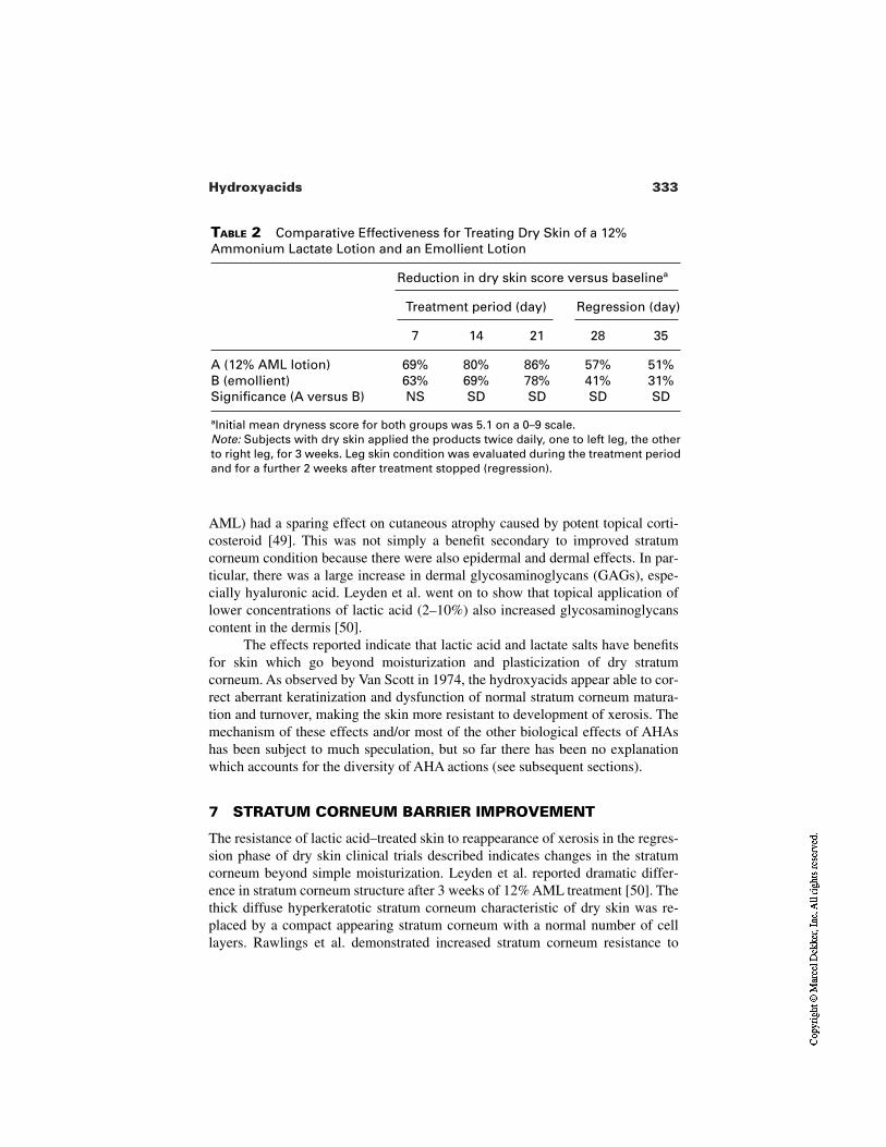
333Hydroxyacids
TABLE 2 Comparative Effectiveness for Treating Dry Skin of a 12%Ammonium Lactate Lotion and an Emollient Lotion
Reduction in dry skin score versus baselinea
Treatment period (day) Regression (day)
7 14 21 28 35
A (12% AML lotion) 69% 80% 86% 57% 51%B (emollient) 63% 69% 78% 41% 31%Significance (A versus B) NS SD SD SD SD
aInitial mean dryness score for both groups was 5.1 on a 0–9 scale.Note: Subjects with dry skin applied the products twice daily, one to left leg, the otherto right leg, for 3 weeks. Leg skin condition was evaluated during the treatment periodand for a further 2 weeks after treatment stopped (regression).
AML) had a sparing effect on cutaneous atrophy caused by potent topical corti-costeroid [49]. This was not simply a benefit secondary to improved stratumcorneum condition because there were also epidermal and dermal effects. In par-ticular, there was a large increase in dermal glycosaminoglycans (GAGs), espe-cially hyaluronic acid. Leyden et al. went on to show that topical application oflower concentrations of lactic acid (2–10%) also increased glycosaminoglycanscontent in the dermis [50].
The effects reported indicate that lactic acid and lactate salts have benefitsfor skin which go beyond moisturization and plasticization of dry stratumcorneum. As observed by Van Scott in 1974, the hydroxyacids appear able to cor-rect aberrant keratinization and dysfunction of normal stratum corneum matura-tion and turnover, making the skin more resistant to development of xerosis. Themechanism of these effects and/or most of the other biological effects of AHAshas been subject to much speculation, but so far there has been no explanationwhich accounts for the diversity of AHA actions (see subsequent sections).
7 STRATUM CORNEUM BARRIER IMPROVEMENT
The resistance of lactic acid–treated skin to reappearance of xerosis in the regres-sion phase of dry skin clinical trials described indicates changes in the stratumcorneum beyond simple moisturization. Leyden et al. reported dramatic differ-ence in stratum corneum structure after 3 weeks of 12% AML treatment [50]. Thethick diffuse hyperkeratotic stratum corneum characteristic of dry skin was re-placed by a compact appearing stratum corneum with a normal number of celllayers. Rawlings et al. demonstrated increased stratum corneum resistance to
334 Johnson
sodium lauryl sulfate (SLS) irritation after 4 weeks of treatment with a lotion con-taining 4% lactic acid at pH 3.7–4.0 [51]. After twice daily application of lotionfor 4 weeks, trans-epidermal water loss (TEWL) was measured on treated and ve-hicle control sites. These sites were then patched with 0.25% SLS for 24 hr andTEWL remeasured. The increase in TEWL caused by SLS irritation was signifi-cantly less on lactic acid–treated skin, indicating increased resistance of the stra-tum corneum. Lipid analysis of tape strips taken from lactic acid and control sitesshowed an increase in ceramide levels in lactic acid treatment sites and specifi-cally an improvement in Ceramide 1 linoleate/oleate ratio. In follow-up studiesRawlings and his coworkers demonstrated that L-lactic acid was more effectivethan DL-lactic acid for improving stratum corneum barrier resistance to irritants.D-Lactic acid was ineffective [52]. They concluded that lactic acid, particularlythe L isomer stimulates ceramide biosynthesis, leading to increased stratumcorneum ceramide levels and a superior lipid barrier that is more resistant to irri-tants and development of xerosis.
8 AHA BENEFITS FOR AGED AND PHOTODAMAGED SKIN
It was a publication by Van Scott and Yu in 1989 [53] demonstrating anti-wrinkleeffects of AHAs at home use concentrations that set the stage for the biggest rev-olution in the skin care marketplace in decades if not for all time. It was wellknown that acid peels would rejuvenate photodamaged skin, but here for the firsttime was evidence that twice-daily application of skin cream containing a poten-tially cosmetic concentration of glycolic acid (5–10%) for 3–10 months could re-duce facial wrinkles. This was a prospective study without specific controls andwith light glycolic acid peels as supplementary treatments every 1 to 6 weeks. Bystrict clinical criteria the evidence for anti-wrinkle effects of glycolic acid at po-tentially cosmetic concentrations was not conclusive. However, the evidence wascompelling. The fountain of youth had been a dream of the cosmetic consumer, ahope beyond realistic expectation, and here was a rational, scientific publicationshowing it might be possible after all. One man in the cosmetic industry recog-nized the significance of this publication and took action that led to the launch ofAnew Creams by Avon Products Inc. in 1992 (personal communication). Avon’sAnew cream contained 8% glycolic acid at pH 4.0. The product was a huge suc-cess with consumers. They saw facial skin benefits not previously experiencedwith regular facial moisturizing products. Other companies followed suit andwithin 2 years AHAs became both a new category in the skin care marketplaceand also the hottest property in town [54]. Ten years further on, AHA productscontinue to be the star of the cosmetic skin care marketplace. There are chal-lengers and pretenders, but so far no other cosmetic ingredient has produced theoverwhelming consumer response and the clear-cut clinical evidence seen with
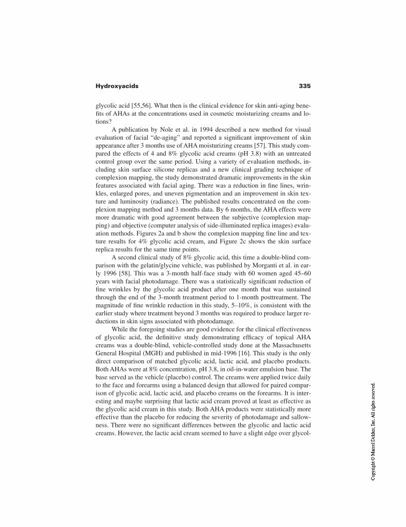
335Hydroxyacids
glycolic acid [55,56]. What then is the clinical evidence for skin anti-aging bene-fits of AHAs at the concentrations used in cosmetic moisturizing creams and lo-tions?
A publication by Nole et al. in 1994 described a new method for visualevaluation of facial “de-aging” and reported a significant improvement of skinappearance after 3 months use of AHA moisturizing creams [57]. This study com-pared the effects of 4 and 8% glycolic acid creams (pH 3.8) with an untreatedcontrol group over the same period. Using a variety of evaluation methods, in-cluding skin surface silicone replicas and a new clinical grading technique ofcomplexion mapping, the study demonstrated dramatic improvements in the skinfeatures associated with facial aging. There was a reduction in fine lines, wrin-kles, enlarged pores, and uneven pigmentation and an improvement in skin tex-ture and luminosity (radiance). The published results concentrated on the com-plexion mapping method and 3 months data. By 6 months, the AHA effects weremore dramatic with good agreement between the subjective (complexion map-ping) and objective (computer analysis of side-illuminated replica images) evalu-ation methods. Figures 2a and b show the complexion mapping fine line and tex-ture results for 4% glycolic acid cream, and Figure 2c shows the skin surfacereplica results for the same time points.
A second clinical study of 8% glycolic acid, this time a double-blind com-parison with the gelatin/glycine vehicle, was published by Morganti et al. in ear-ly 1996 [58]. This was a 3-month half-face study with 60 women aged 45–60years with facial photodamage. There was a statistically significant reduction offine wrinkles by the glycolic acid product after one month that was sustainedthrough the end of the 3-month treatment period to 1-month posttreatment. Themagnitude of fine wrinkle reduction in this study, 5–10%, is consistent with theearlier study where treatment beyond 3 months was required to produce larger re-ductions in skin signs associated with photodamage.
While the foregoing studies are good evidence for the clinical effectivenessof glycolic acid, the definitive study demonstrating efficacy of topical AHAcreams was a double-blind, vehicle-controlled study done at the MassachusettsGeneral Hospital (MGH) and published in mid-1996 [16]. This study is the onlydirect comparison of matched glycolic acid, lactic acid, and placebo products.Both AHAs were at 8% concentration, pH 3.8, in oil-in-water emulsion base. Thebase served as the vehicle (placebo) control. The creams were applied twice dailyto the face and forearms using a balanced design that allowed for paired compar-ison of glycolic acid, lactic acid, and placebo creams on the forearms. It is inter-esting and maybe surprising that lactic acid cream proved at least as effective asthe glycolic acid cream in this study. Both AHA products were statistically moreeffective than the placebo for reducing the severity of photodamage and sallow-ness. There were no significant differences between the glycolic and lactic acidcreams. However, the lactic acid cream seemed to have a slight edge over glycol-
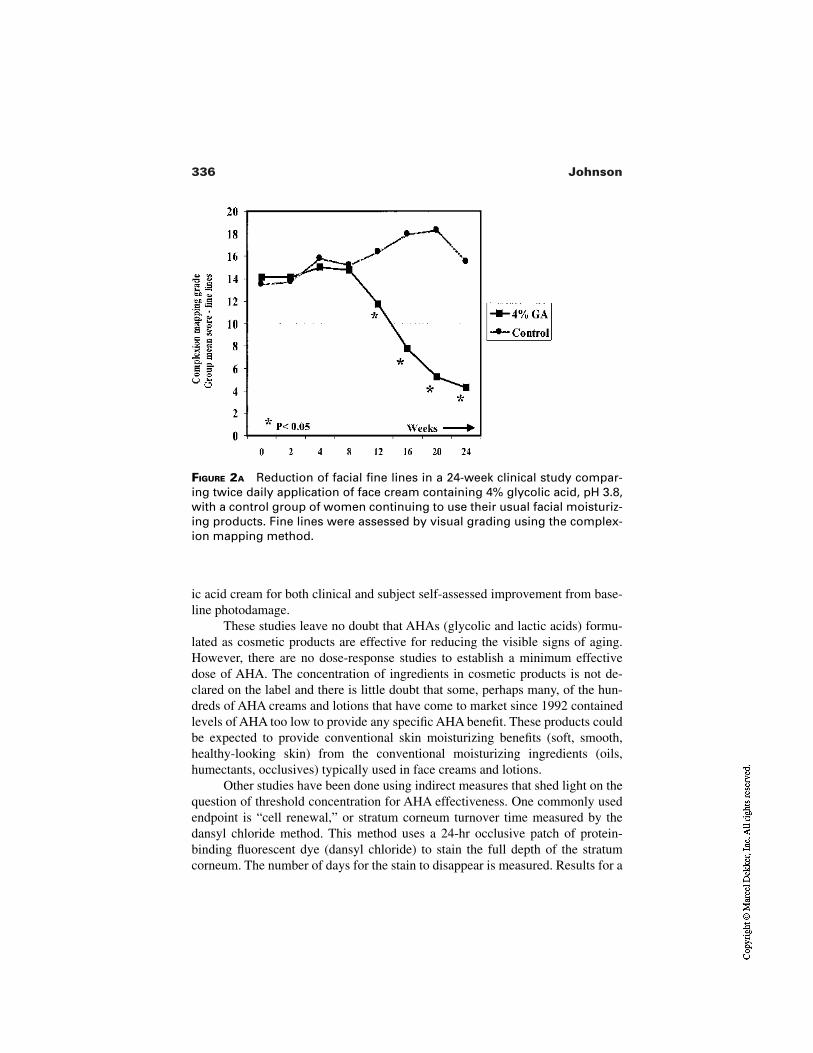
336 Johnson
ic acid cream for both clinical and subject self-assessed improvement from base-line photodamage.
These studies leave no doubt that AHAs (glycolic and lactic acids) formu-lated as cosmetic products are effective for reducing the visible signs of aging.However, there are no dose-response studies to establish a minimum effectivedose of AHA. The concentration of ingredients in cosmetic products is not de-clared on the label and there is little doubt that some, perhaps many, of the hun-dreds of AHA creams and lotions that have come to market since 1992 containedlevels of AHA too low to provide any specific AHA benefit. These products couldbe expected to provide conventional skin moisturizing benefits (soft, smooth,healthy-looking skin) from the conventional moisturizing ingredients (oils,humectants, occlusives) typically used in face creams and lotions.
Other studies have been done using indirect measures that shed light on thequestion of threshold concentration for AHA effectiveness. One commonly usedendpoint is “cell renewal,” or stratum corneum turnover time measured by thedansyl chloride method. This method uses a 24-hr occlusive patch of protein-binding fluorescent dye (dansyl chloride) to stain the full depth of the stratumcorneum. The number of days for the stain to disappear is measured. Results for a
FIGURE 2A Reduction of facial fine lines in a 24-week clinical study compar-ing twice daily application of face cream containing 4% glycolic acid, pH 3.8,with a control group of women continuing to use their usual facial moisturiz-ing products. Fine lines were assessed by visual grading using the complex-ion mapping method.
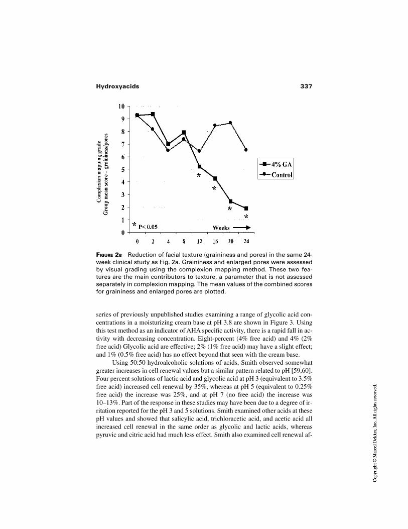
337Hydroxyacids
FIGURE 2B Reduction of facial texture (graininess and pores) in the same 24-week clinical study as Fig. 2a. Graininess and enlarged pores were assessedby visual grading using the complexion mapping method. These two fea-tures are the main contributors to texture, a parameter that is not assessedseparately in complexion mapping. The mean values of the combined scoresfor graininess and enlarged pores are plotted.
series of previously unpublished studies examining a range of glycolic acid con-centrations in a moisturizing cream base at pH 3.8 are shown in Figure 3. Usingthis test method as an indicator of AHA specific activity, there is a rapid fall in ac-tivity with decreasing concentration. Eight-percent (4% free acid) and 4% (2%free acid) Glycolic acid are effective; 2% (1% free acid) may have a slight effect;and 1% (0.5% free acid) has no effect beyond that seen with the cream base.
Using 50:50 hydroalcoholic solutions of acids, Smith observed somewhatgreater increases in cell renewal values but a similar pattern related to pH [59,60].Four percent solutions of lactic acid and glycolic acid at pH 3 (equivalent to 3.5%free acid) increased cell renewal by 35%, whereas at pH 5 (equivalent to 0.25%free acid) the increase was 25%, and at pH 7 (no free acid) the increase was10–13%. Part of the response in these studies may have been due to a degree of ir-ritation reported for the pH 3 and 5 solutions. Smith examined other acids at thesepH values and showed that salicylic acid, trichloracetic acid, and acetic acid allincreased cell renewal in the same order as glycolic and lactic acids, whereaspyruvic and citric acid had much less effect. Smith also examined cell renewal af-
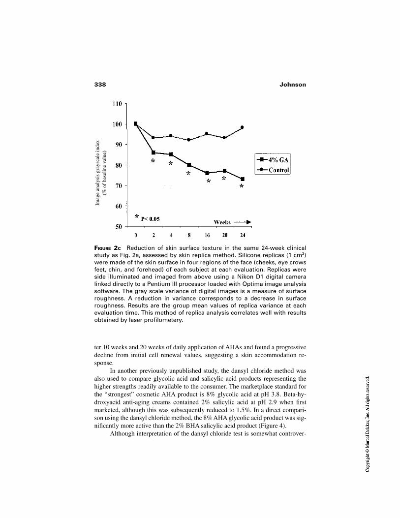
338 Johnson
FIGURE 2C Reduction of skin surface texture in the same 24-week clinicalstudy as Fig. 2a, assessed by skin replica method. Silicone replicas (1 cm2)were made of the skin surface in four regions of the face (cheeks, eye crowsfeet, chin, and forehead) of each subject at each evaluation. Replicas wereside illuminated and imaged from above using a Nikon D1 digital cameralinked directly to a Pentium III processor loaded with Optima image analysissoftware. The gray scale variance of digital images is a measure of surfaceroughness. A reduction in variance corresponds to a decrease in surfaceroughness. Results are the group mean values of replica variance at eachevaluation time. This method of replica analysis correlates well with resultsobtained by laser profilometery.
Imag
e an
alys
is g
rays
cale
inde
x(%
of
base
line
valu
e)
ter 10 weeks and 20 weeks of daily application of AHAs and found a progressivedecline from initial cell renewal values, suggesting a skin accommodation re-sponse.
In another previously unpublished study, the dansyl chloride method wasalso used to compare glycolic acid and salicylic acid products representing thehigher strengths readily available to the consumer. The marketplace standard forthe “strongest” cosmetic AHA product is 8% glycolic acid at pH 3.8. Beta-hy-droxyacid anti-aging creams contained 2% salicylic acid at pH 2.9 when firstmarketed, although this was subsequently reduced to 1.5%. In a direct compari-son using the dansyl chloride method, the 8% AHA glycolic acid product was sig-nificantly more active than the 2% BHA salicylic acid product (Figure 4).
Although interpretation of the dansyl chloride test is somewhat controver-
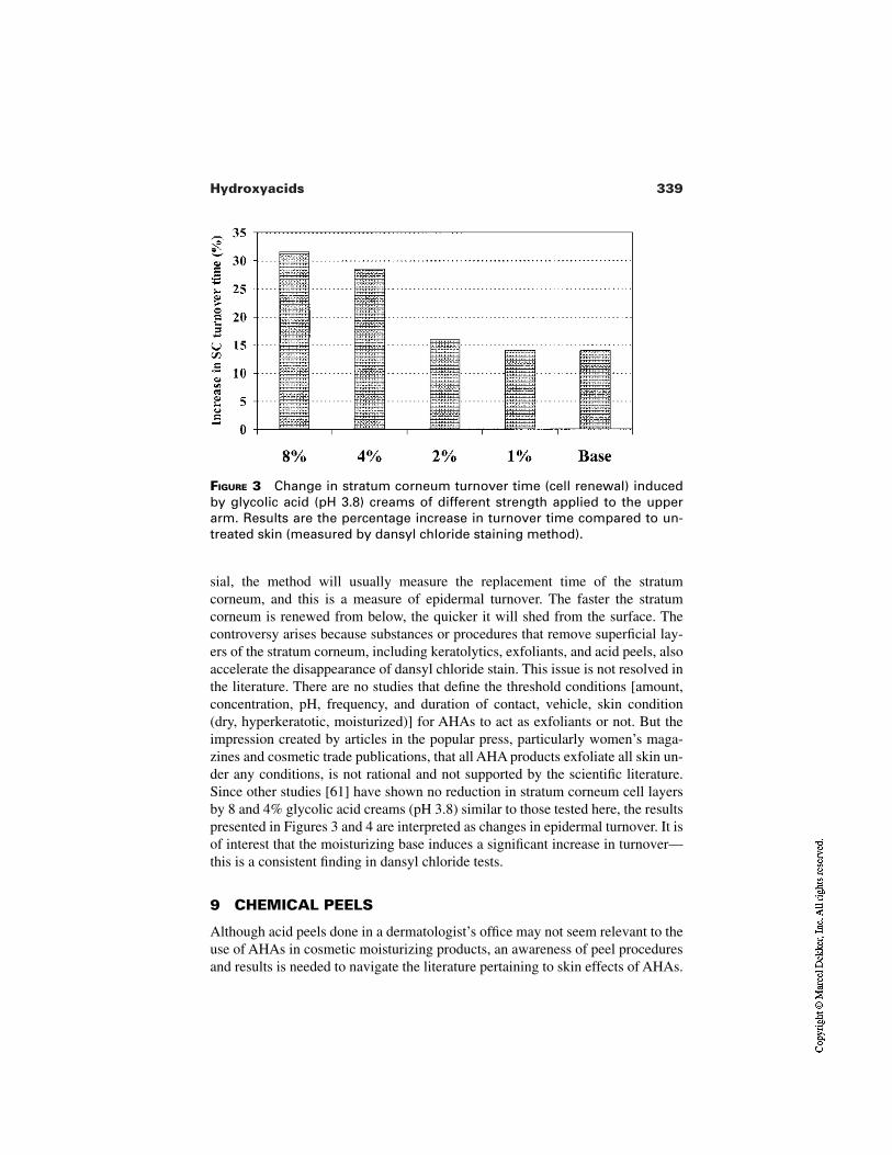
339Hydroxyacids
FIGURE 3 Change in stratum corneum turnover time (cell renewal) inducedby glycolic acid (pH 3.8) creams of different strength applied to the upperarm. Results are the percentage increase in turnover time compared to un-treated skin (measured by dansyl chloride staining method).
sial, the method will usually measure the replacement time of the stratumcorneum, and this is a measure of epidermal turnover. The faster the stratumcorneum is renewed from below, the quicker it will shed from the surface. Thecontroversy arises because substances or procedures that remove superficial lay-ers of the stratum corneum, including keratolytics, exfoliants, and acid peels, alsoaccelerate the disappearance of dansyl chloride stain. This issue is not resolved inthe literature. There are no studies that define the threshold conditions [amount,concentration, pH, frequency, and duration of contact, vehicle, skin condition(dry, hyperkeratotic, moisturized)] for AHAs to act as exfoliants or not. But theimpression created by articles in the popular press, particularly women’s maga-zines and cosmetic trade publications, that all AHA products exfoliate all skin un-der any conditions, is not rational and not supported by the scientific literature.Since other studies [61] have shown no reduction in stratum corneum cell layersby 8 and 4% glycolic acid creams (pH 3.8) similar to those tested here, the resultspresented in Figures 3 and 4 are interpreted as changes in epidermal turnover. It isof interest that the moisturizing base induces a significant increase in turnover—this is a consistent finding in dansyl chloride tests.
9 CHEMICAL PEELS
Although acid peels done in a dermatologist’s office may not seem relevant to theuse of AHAs in cosmetic moisturizing products, an awareness of peel proceduresand results is needed to navigate the literature pertaining to skin effects of AHAs.
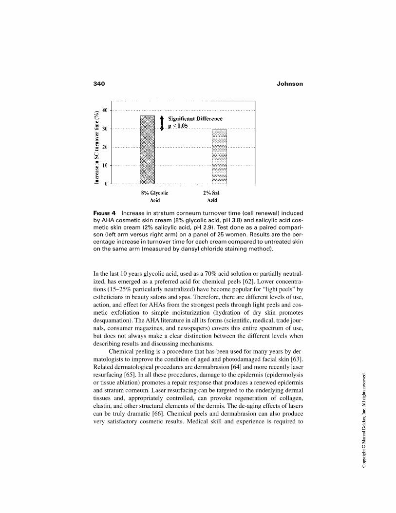
340 Johnson
FIGURE 4 Increase in stratum corneum turnover time (cell renewal) inducedby AHA cosmetic skin cream (8% glycolic acid, pH 3.8) and salicylic acid cos-metic skin cream (2% salicylic acid, pH 2.9). Test done as a paired compari-son (left arm versus right arm) on a panel of 25 women. Results are the per-centage increase in turnover time for each cream compared to untreated skinon the same arm (measured by dansyl chloride staining method).
In the last 10 years glycolic acid, used as a 70% acid solution or partially neutral-ized, has emerged as a preferred acid for chemical peels [62]. Lower concentra-tions (15–25% particularly neutralized) have become popular for “light peels” byestheticians in beauty salons and spas. Therefore, there are different levels of use,action, and effect for AHAs from the strongest peels through light peels and cos-metic exfoliation to simple moisturization (hydration of dry skin promotesdesquamation). The AHA literature in all its forms (scientific, medical, trade jour-nals, consumer magazines, and newspapers) covers this entire spectrum of use,but does not always make a clear distinction between the different levels whendescribing results and discussing mechanisms.
Chemical peeling is a procedure that has been used for many years by der-matologists to improve the condition of aged and photodamaged facial skin [63].Related dermatological procedures are dermabrasion [64] and more recently laserresurfacing [65]. In all these procedures, damage to the epidermis (epidermolysisor tissue ablation) promotes a repair response that produces a renewed epidermisand stratum corneum. Laser resurfacing can be targeted to the underlying dermaltissues and, appropriately controlled, can provoke regeneration of collagen,elastin, and other structural elements of the dermis. The de-aging effects of laserscan be truly dramatic [66]. Chemical peels and dermabrasion can also producevery satisfactory cosmetic results. Medical skill and experience is required to
341Hydroxyacids
control the skin contact time of concentrated glycolic acid during peels. The lightpeels done by estheticians also involve timed application of glycolic acid to theskin followed by a neutralizing rinse, but there is much less risk with the lowerAHA concentrations used for light peels. It seems plausible that chemical peelswith glycolic acid provide some element of the AHA benefit seen at nondamag-ing, nonirritating cosmetic concentrations and pH. However, it is less clear, andarguably unlikely, that any of the benefit of cosmetic concentrations of glycolicacid is due to an element of acid damage. But so far there have been no controlledstudies to investigate these relationships. Because peel procedures and cosmeticproducts work in their individual fields of application, there has been no impera-tive to investigate the relationship of mechanisms involved, even though it hasbecome a common practice to combine the two procedures [67].
10 OTHER BENEFITS OF AHA
Alpha-hydroxyacids are used in skin moisturizer products primarily for moistur-ization and skin anti-aging benefits, but as indicated these compounds have abroader range of beneficial effects for skin. Most anti-aging products for the faceuse glycolic acid and most hand and body moisturizer products use lactic acid.This is probably a reflection of cost. The raw material cost of glycolic acid isabout four times that of lactic acid. Face care products sell at higher prices thanhand and body products, and therefore glycolic acid is “affordable” for face prod-ucts but too expensive for mass-market general use/hand and body products.There are other AHAs and, after the first flush of glycolic acid anti-aging creamsand lotions, products containing combinations of glycolic and other AHAs ap-peared in the marketplace. As there is no convincing evidence for a significantperformance advantage for the combinations it can be assumed that the main pur-pose was marketing-driven product differentiation in a crowded marketplace. Al-though there have been no head-to-head studies to compare AHA combinationswith glycolic acid or lactic acid at equivalent acid strength, there are claims thatcombination products are effective for both control of dry skin and improvementof photodamaged skin (anti-aging). The Van Scott group showed that a blend ofAHA and polyhydroxy acids (PHAs) was effective for improving the symptomsof xerosis, epidermolytic hyperkeratosis, and ichthyosis [68]. Another publicationfrom Van Scott is useful for identifying and characterizing the broader range ofAHAs and their effects on skin [14].
There are several publications indicating AHAs, and glycolic acid in partic-ular, are helpful for treating acne. It may be used at high concentration (70%) as achemical peel or daily at cosmetic strength (5–10%) where it acts to reduce cohe-sion of follicular corneocytes, helping to dislodge comedones and prevent theirformation [37]. Glycolic acid is a less effective treatment for acne than tretinoin
342 Johnson
and acts by a different mechanism, but the combination of glycolic acid andtretinoin is more effective than either agent alone [69–71]. Alternative combina-tion treatments using light glycolic acid peels (20–35%), daily 15% glycolic acidgel, and 4% gluconolactone cleanser, alone or in addition to prescription medica-tions, have proved effective for dealing with recalcitrant papulopustular acne[72]. A similar mixed regimen was effective for treating rosacea, a commonchronic inflammatory disorder of the face characterized by erythema and telang-iectasia that is often accompanied by outbreaks of acnelike papules and pustules[73].
Cosmetic strength glycolic acid (8%, pH 4) is very effective for another fol-licular skin problem, razor bumps or pseudofolliculitis barbae (PFB), a foreignbody inflammatory reaction to ingrown facial hair. The condition arises whenshaved hairs curl and penetrate the skin near the follicle opening as they growback after shaving. The problem afflicts over 50% of black males and a high pro-portion of black females who need to shave around the jaw line. Two weeks useof 8% glycolic acid cream reduces the number of papules and pustules by morethan half, and continuous treatment usually provides satisfactory control of theproblem [34].
Some types of sun-induced age spots, seborrheic keratoses, and actinic ker-atoses involve a dysfunction of normal epidermal differentiation and maturation,whereas others, the solar lentigines, reflect proliferation and hyperactivity ofmelanocytes. These lesions can be removed using peel concentrations of AHA,but daily application of cosmetic strength AHA is also effective and usually pre-ferred [53]. In keeping with the ability of AHAs to correct abnormal keratiniza-tion, AHAs are more effective for reducing/eliminating keratoses than lentigines.A combination of glycolic acid with hydroquinone or kojic acid has been report-ed more effective than hydroquinone or kojic acid alone for treating melasma andother hyperpigmentation conditions [74]. It seems clear that AHAs do have theability to reduce skin hyperpigmentation. However, as described subsequently,AHAs also cause a small increase in skin photosensitivity, which could be ex-pected to promote pigmentation (tanning). A recent publication by Tsai andMaibach confirms that AHA (10% glycolic acid, pH 3.5) does promote UVB-in-duced skin tanning [75]. In this 3-week study, glycolic acid had no effect (did notreduce) pre-existing tan.
Although contrary to the several sunburn studies considered for the CIR re-view, there is a 1996 report that topical glycolic acid is photoprotective and exertsan anti-inflammatory action [76]. The anti-inflammatory conclusion is based onthe finding that skin sites irradiated with 3 times the minimal erythemal doseshowed a marked reduction in erythema when treated postirradiation with 12%ammonium lactate at pH 4.2. The photoprotective conclusion is based on findingan attenuation of UVB effect equivalent to SPF 2–4 on skin sites treated daily for3 weeks with an AHA cleanser and AHA lotion both containing 8% glycolic acid
343Hydroxyacids
at pH 3.25. Similar results were obtained in an earlier study by the same author[77].
11 SKIN COMPATIBILITY OF AHAS
There has never been any real concern about the skin compatibility of AHA prod-ucts sold as dry skin moisturizers since the early 1970s. But when AHAs were firstintroduced in facial moisturizers in 1992 there were complaints of irritation thatfueled a controversy that still continues. The reality is that AHAs induce sensoryirritation (chemosensory irritability) in susceptible individuals [78]. Alpha-hy-droxyacid stinging is a transient effect usually with no clinical signs of irritation,although a mild transient erythema may appear in unusually susceptible individu-als. The effect is related to the susceptibility of the individual and the strength ofthe AHA. Some consumers regard a slight stinging as a positive sign. Those withsensitive skin often reject AHA products. Prior to the appearance of AHAs in facialproducts, cosmetic moisturizers were almost universally bland emollient prepara-tions that helped to hydrate dry superficial regions of the stratum corneum. Con-sumers using AHA products for the first time and experiencing burning and sting-ing sensations were concerned. This was outside their normal experience andexpectation. Some consumers assumed there was something wrong with the prod-ucts. The lack of familiarity with AHA products coupled with a strong desire to trythe products because of the anti-wrinkle benefits promised, encouraged wide-spread trial by consumers and an associated burst of complaints. This caught theattention of the media and became the issue of the week for investigative journal-ists for the next several months. Alpha-hydroxyacids were so new that factual in-formation was hard to find and anecdotal comments and opinions became the cur-rency of the day. Nearly a decade later, AHAs are far more widespread and popularthan ever before. The stinging issue is recognized. There has been much work bythe cosmetic industry to develop products that retain the benefits of AHAs withoutthe sensory irritation, but so far with only modest success [79,80].
Good skin compatibility and absence of irritation is a consistent observa-tion in all the clinical studies of cosmetic strength AHAs referenced in this chap-ter. Indeed, some of the studies with facial peel strength AHAs show no irritationon forearm and body parts less sensitive than the face. In Ditre’s study, subjectswere able to apply lotions containing 25% glycolic, lactic, and citric acid at pH3.5 (17% free acid) to the forearm twice daily for 6 months. There was no irrita-tion but a most dramatic improvement in the condition of the photodamaged armskin [36]. Results of a 14-day cumulative irritation test by DiNardo provide an-other indication that AHAs are not primary irritants at pH values +/–0.5 pH unitsof the pKa value [81]. In this study, 12% ammonium lactate, pH 4.4, demonstrat-ed an irritation score of 30 out of a possible 882 points (cumulative over 14 days).Eight percent glycolic acid, pH 4.4, demonstrated a score of 1 out of 882 points.
344 Johnson
12 MECHANISMS OF ACTION
It will be clear from this discussion that AHAs have multiple effects on skin re-lated to pH and concentration. It might be imagined that the popularity of AHAsfor both everyday and medical skin care would have stimulated intensive researchto understand the biochemical and physiological mechanisms of AHA action. Infact the few serious scientific studies that have been done have focused mostly onthe end benefits of AHAs: what they do, much more than how they do it. As indi-cated, the published research on AHAs has significant limitations, as follows:
1. A majority of publications do not state the pH of the preparations ex-amined.
2. Publications that discuss mechanisms, particularly general articles andreviews, tend not to make a distinction between cosmetic and dermato-logical (chemical peel) levels of action/effect.
3. Many publications that discuss AHA mechanisms simply reiterate the1974 suggestions of Van Scott and Yu, which these authors recognizedas speculative at the time they made them.
Histological studies show that AHAs, unlike keratolytics that eliminate cor-neocytes from the skin surface inward, diminish corneocyte adhesion in the low-er layers of the stratum corneum (stratum compactum). Van Scott and Yu specu-lated that this effect was due to AHA interference with intercorneocyte ionicbonding by inhibiting enzymes that add phosphate and sulfate groups to the cor-neocyte envelope [33]. A study by Ditre [36] provided indirect evidence that theAHA effect might be due to modification of desmosomal (protein) links betweencorneocytes. After 6 months of daily application to the forearm of 25% AHA (gly-colic, lactic, or citric acid, all at pH 3.5), electron microscopic examination of theepidermis revealed fewer desmosomes connecting basal cells in skin sectionsfrom AHA-treated sites than from placebo-treated control sites. A more recentstudy by Fartasch et al. [82] applying glycolic acid cream (4%, pH 3.8) to thevolar forearm twice daily for 3 weeks provides a somewhat different picture. Inthis study, electron microscopic examination of the stratum corneum revealed nochanges or indications of reduced cohesion in the stratum compactum. In themore superficial layers of the stratum corneum (stratum disjunctum), wheredesmosome degradation is initiated as part of the normal process of desquama-tion, there was enhanced degradation of desmosomes (corneosomes) in AHA-treated sites compared to control sites. Also, in contrast to the Ditre study, therewas no change detected in the epidermis. Comparing the Ditre and Fartasch re-sults, it appears that AHAs may exert actions at different levels of the stratumcorneum depending on the strength of AHA applied to skin. The strength used byFartasch is a cosmetic strength that is effective. The strength used by Ditre, al-
345Hydroxyacids
though apparently not irritating when applied daily to the forearm for severalmonths, is a much higher concentration than used in cosmetic products.
Research in the 1990s revealed some of the biochemical mechanisms in-volved in cellular adhesions and degradation of these adhesions during desqua-mation [83,84]. At points of adhesion, including desmosomes, cells are connect-ed by transmembrane proteins called cadherins. Calcium bound to cadherinsprotects the structures from enzymic proteolysis. Combining these insights withclinical and experimental data published for AHAs, Wang proposed “a theory forthe mechanism of action of AHAs applied to the skin” [85]. The essence of thistheory is that AHAs remove calcium from cadherins by chelation, resulting inproteolysis and enhanced desquamation. Wang also suggested that lowering epi-dermal calcium levels promotes cell proliferation and retards differentiation ofkeratinocytes. Wang’s theory appears to be a possible explanation for effects ofAHAs on the stratum corneum, but it does not explain the specific enhancementof corneocyte adhesion by alpha-acetoxyacids acids [86]. Nor does it explain themetabolic effects of AHAs, such as the sparing effect on steroid-induced cuta-neous atrophy [49], increased epidermal ceramide synthesis [52], increased trans-glutaminase expression in dermal dendrocytes [87], increased dermal and epider-mal hyaluronic acid [88], and increased collagen deposition in the papillarydermis [81]. Like any theory, Wang’s proposed mechanism of AHA action mustbe put to the test by appropriate experimentation, particularly as the theory reliesin part on calcium-induced changes observed using in vitro systems.
The diversity of metabolic changes induced by AHAs could reflect multiplemechanisms of action or, as most would argue, an effect on a fundamental cellu-lar target that initiates a procession of consequential actions and reactions. It is es-tablished that perturbation of the stratum corneum barrier produces a cascade ofstimulatory and inhibitory cytokines [89] that would be expected to migrate to theepidermis and dermis and exert a host of effects, either directly or by impactingother cell regulatory pathways. Is cytokine activation and release the mechanismof the AHA effect on skin? I don’t know and the literature to date (2001) does notprovide the answer.
13 AHA SAFETY: THE COSMETIC INGREDIENT REVIEW
Because of escalating use of AHAs in cosmetic skin care products in the early1990s and the U.S. Food and Drug Administration (FDA) concern to ensure con-sumer safety was thoroughly evaluated, the Cosmetics, Toiletries and FragranceAssociation (CTFA) asked the Cosmetic Ingredient Review to review AHAs,principally glycolic and lactic acids, their salts, and simple esters. The CIR is acomprehensive program for independent review of the safety of cosmetic ingre-
346 Johnson
dients in the United States. Although CIR is supported by industry funding it isstaffed and operated independently. All decisions regarding safety of cosmeticingredients are made by the CIR Expert Panel, a panel composed of seven phy-sicians and scientists with expertise in dermatology, toxicology, pathology,carcinogenicity, and biochemistry. In addition, there are three nonvoting mem-bers of the CIR panel representing industry (CTFA), consumers (the Consum-er Federation of America), and government (FDA Office of Cosmetics andColors).
The CIR compiled a scientific literature review of published data on AHAsplus unpublished industry data made available to CIR via CTFA. There were over350 studies covering all aspects of safety of glycolic acid and lactic acid [90]. TheCIR Expert Panel discussed the safety data and their particular areas of concern atCIR public meetings in 1995 and again in 1996. The CIR acknowledged that inmost respects there were no concerns about the safety of AHAs [91]. There wasmuch data from which to conclude that AHAs are not mutagenic or carcinogenic,are not reproductive or developmental toxins, and are not skin sensitizers. Theareas CIR identified for particular consideration were (1) the irritation potential of AHAs and (2) the exfoliating effect of AHAs that could potentially enhancepenetration of other ingredients and/or increase the sensitivity of skin to solarUVR.
With respect to irritation, the CIR Expert Panel concluded that there weredata indicating acceptable limits for concentration/pH of AHAs for leave-on skinproducts. The panel also concluded that there were studies indicating no need forconcern about use of AHA enhancing the penetration of other chemicals. Theynoted that AHAs themselves do penetrate skin readily, but because of low sys-temic toxicity this was not a concern. However, the expert panel stated that theevidence did point to a small and variable increase in sun sensitivity after use ofAHA products. They noted that there were studies providing contradictory evi-dence on the effect of AHA on minimal erythemal dose. One study suggestedMED was increased (reduced UV sensitivity) after use of AHAs, but a secondstudy showed a reduction in UVR dose required to produce skin reddening. Theaverage reduction in MED (increased UV sensitivity) was 13%, but some indi-viduals showed greater than 50% reduction. Additional studies were carried outusing sunburn cell (SBC) production as the endpoint for indicating UVR penetra-tion of skin. After 4 days of application of standard AHA preparation (10% gly-colic acid at pH 3.5) there was no significant increase in SBC production follow-ing UVR challenge. However, after 12 weeks of AHA treatment there was a smallbut significant increase in SBC formation after UVR challenge. The increase innumber of SBCs after a 12-week AHA pretreatment was equivalent to increasingUV exposure of untreated skin by the equivalent of 1.6 MED. Therefore, additionof a sunscreen with an SPF 2 to an AHA product containing 10% glycolic acid atpH 3.5 would be sufficient to eliminate the AHA-induced increase in UVR sensi-
347Hydroxyacids
tivity. In fact, the most widely available commercial AHA products have a con-centration of 8% glycolic acid and a pH of 3.8. This product strength providesless than half the free acid contained in the standard AHA preparation used in theSBC studies (approximately 4% free acid for 8% glycolic acid, pH 3.8, comparedto 8.7% free acid for 10% glycolic acid, pH 3.5). The 1996 recommendations ofthe CIR Expert Panel were based on the SBC results showing a relatively smallAHA-induced increase in sun sensitivity that could be eliminated by addition oflow SPF to AHA creams and lotions. The Expert Panel concluded that AHAs aresafe for use in cosmetic products at concentrations equal or less than 10%, at a fi-nal formulation pH equal to or greater than 3.5, when formulated to avoid in-creasing sun sensitivity or when directions for use include the daily use of sunprotection. A recent publication [61] confirms that commercial AHA products (8and 4% glycolic acid at pH 3.8) containing SPF 4 sunscreen do not increase sunsensitivity over 6 months of twice daily application to forearms. More recentstudies by the FDA add further support for the 1996 CIR recommendation. TheFDA did additional studies to examine AHA effect on the sensitivity of skin toUVR, using MED, SBCs, and thiamine dimer formation as endpoints for indicat-ing UVR penetration of skin [92]. The first study used a 10%, pH 3.5, glycolicacid solution applied 6 days per week for 4 weeks followed by UVR challenge,and a second UVR challenge 1 week after the last AHA application. Results indi-cated a small AHA-induced reduction in MED and small increase in SBC forma-tion after 4 weeks of treatment. One week after AHA treatment was stopped,AHA-induced changes were no longer evident. In a second study, there was nosignificant increase in thiamine dimer formation after 4 weeks treatment with thesame AHA solution. The CIR Expert Panel reviewed the FDA results and report-ed at the February 2000 CIR meeting their conclusion that the new results sup-ported the 1996 panel conclusion [93].
14 FUTURE TRENDS
Research over 30 years has revealed that AHAs, most particularly lactic acid andglycolic acid, have many mostly beneficial actions on skin. The mechanisms ofaction are less clear. Explanations for some actions are more a statement of effecton a process (e.g., AHAs reduce corneocyte adhesion) than true mechanistic de-scriptions of the physiological, biochemical, and molecular biological causes forthe observed effects. In the future, with accelerating advances in diagnostic andanalytical techniques, such as DNA arrays to probe subtle changes in gene ex-pression and cellular biochemistry, we can expect the actual mechanisms of AHAeffects on skin will be worked out. It seems unlikely that the future will see dis-coveries of major new effects of AHAs on skin. Instead, clarification of mecha-nisms of action should enable more effective targeting and optimal use of AHAsfor the indications of skin benefit already identified.
348 Johnson
REFERENCES
1. Jackson EM. AHA-type products proliferate in 1993. Cosmet Dermatol 1993;6(12):22–26.
2. Branna T. The skin care market. Happi 1991; 29(5):49–64.3. Jackson EM. Update on AHA-containing products. Cosmet Dermatol 1994;
7(1):29–30.4. Draelos ZD. Sorting out all your hydroxy acid options. Skin Aging 1998;
6(4):45–47.5. Middleton JD. Development of a skin cream designed to reduce dry and flaky skin. J
Soc Cosmet Chem 1974; 25:519–534.6. Van Scott EJ, Yu RJ. Control of keratinization with α-hydroxy acids and related
compounds. Arch Dermatol 1974; 110:586–590.7. Elson ML. The utilization of glycolic acid in photoaging. Cosmet Dermatol 1992;
5(1):12–15.8. Jackson EM. Supporting advertising claims for AHA products. Cosmet Dermatol
1996; 9(5):40–47.9. Jackson EM. AHAs: what’s wrong with this picture? J Toxicol Cut Ocular Toxicol
1997; 16(4):203–205.10. Jackson EM. Do AHA products really work? Cosmet Dermatol October 1994; (sup-
pl):21–22.11. Frosch PJ, Kligman AM. A method for appraising the stinging capacity of topically
applied substances. J Soc Cosmet Chem 1977; 28:197–209.12. Rosan AM. The chemistry of alpha-hydroxy acids. Cosmet Dermatol October 1994;
(suppl):4–9.13. Johnson AW, Nole GE, Rozen MG, DiNardo JC. Skin tolerance of AHAs: a compar-
ison of lactic and glycolic acids and the role of pH. Cosmet Dermatol 1997;10(2):38–45.
14. Yu RJ, Van Scott EJ. Alpha-hydroxy acids: science and therapeutic use. Cosmet Der-matol October 1994; (suppl):12–20.
15. Greaves MW. Alpha hydroxy acid derivative for relieving dry itching skin. CosmetToil 1990; 105(10):61–64.
16. Stiller MJ, Bartolone J, Stern R, Smith S, Kollias N, Gillies R, Drake LA. Topical8% glycolic acid and 8% L-lactic acid creams for the treatment of photodamagedskin. Arch Dermatol 1996; 132:631–636.
17. Nook TH. In vivo measurement of the keratolytic effect of salicylic acid in threeointment formulations. Br J Dermatol 1987; 117:243–247.
18. Leyden JJ, Shalita AR. Rational therapy for acne vulgaris: an update on topical treat-ment. J Am Acad Dermatol 1986; 4:507–515.
19. Kligman AM. Salicylic acid: an alternative to alpha hydroxy acids. J Geriatr Derma-tol 1997; 5(3):128–131.
20. Draelos ZD. Hydroxy acids for the treatment of aging skin. J Geriatr Dermatol 1997;5(5):236–240.
21. Bergfeld W, Tung R, Vidimos A, Vellanki L, Remzi B, Stanton-Hicks U. Improvingthe cosmetic appearance of photoaged skin with glycolic acid. J Am Acad Dermatol1977; 36:1011–1013.
349Hydroxyacids
22. Dahl MV, Dahl AC. 12% lactate lotion for the treatment of xerosis. Arch Dermatol1983; 119:27–30.
23. Johnson AW. Dry skin: recent advances in research and therapy. J Retail Pharmacy1994; 4(suppl):S1–S8.
24. Hanifin JM, Rajka G. Diagnostic features of atopic dermatitis. Acta Derm Verereol(Stockholm) 1980; 92(suppl):44–47.
25. Roop D. Defects in the barrier. Science 1995; 267:474–475.26. Williams ML. Lipids in normal and pathological desquamation. Adv Lipid Res
1991; 24:211–262.27. Morita E, Katoh O, Shinoda S, Hiragun T, Tanada T, Kameyoshi Y, Yamamoto S. A
novel point mutation in the steroid sulphatase gene in X-linked ichthyosis. J InvestDermatol 1997; 109:244–245.
28. Rothnagel JA, Roop DR. Analysis, diagnosis, and molecular genetics of keratin dis-orders. Curr Opin Dermatol 1995; 2:211–218.
29. Huber M, Rettler I, Bernasconi K, Frenk E, Lavrijsen SPM, Ponec M, Bon A, Lau-tenschlager S, Schorderet DF, Hohl D. Mutations of keratinocyte transglutaminase inlamellar ichthyosis. Science 1995; 267:525–528.
30. Bale SJ, Compton JG, Russell LJ, DiGiovanna JJ. Genetic heterogeneity in lamellarichthyosis. J Invest Dermatol 1996; 107 (Letters to the Editor):140–141.
31. Petratos MA. Drug therapies and adjunctive uses of alphahydroxy and polyhydroxyacids. Cutis 2000; 66:107–111.
32. Siskin SB, Quinlan PJ, Finkelstein MS, Marlucci M, Maglietta TG, Gibson JR. Theeffects of ammonium lactate 12% lotion versus no therapy in the treatment of dryskin of the heels. Int J Dermatol 1993; 32:905–907.
33. Van Scott EJ, Yu RJ. Hyperkeratinization, corneocyte cohesion, and alpha hydroxyacids. J Am Acad Dermatol 1984; 11:867–879.
34. Perricone NV. Treatment of pseudofolliculitis barbae with topical glycolic acid: a re-port of two studies. Cutis 1993; 52:233–235.
35. Smith WP. Epidermal and dermal effects of topical lactic acid. J Am Acad Dermatol1996; 35:388–391.
36. Ditre CM, Griffin TD, Murphy GF, Sueki H, Telegan B, Johnson WC, Yu R, VanScott EJ. Effects of alpha-hydroxy acids on photoaged skin: a pilot clinical, histolog-ic, and ultrastructural study. J Am Acad Dermatol 1996; 34:187–195.
37. Van Scott EJ, Yu RJ. Alpha hydroxyacids: therapeutic potentials. Can J Dermatol1989; 1(5):108–112.
38. Blank IH. Factors which influence the water content of the stratum corneum. J InvestDermatol 1952; 18:433–440.
39. Middleton JD. The mechanism of water binding in stratum corneum. Br J Dermatol1968; 80:437.
40. Harding CR, Watkinson A, Rawlings AV. Dry skin, moisturization and cor-neodesmolysis. Int J Cosmet Sci 2000; 22:21–52.
41. Middleton JD. The influence of temperature and humidity on stratum corneum andits relation to skin chapping. J Soc Cosmet Chem 1973; 24:239.
42. Alderson SG, Barratt MD, Black JG. Effect of 2-hydroxyacids on guinea-pig foot-pad stratum corneum: mechanical properties and binding studies. Int J Cosm Sci1984; 6:91–100.
350 Johnson
43. Harding CR, Bartolone J, Rawlings AV. Effects of natural moisturizing factor andlactic acid isomers on skin function. In: Loden M, Maibach HI, eds. Dry skin andMoisturizers: Chemistry and Function. Boca Raton: CRC Press, 2000:229–241.
44. Takahashi M, Kawasaki K, Tanaka M, Ohra S, Tsuda Y. The mechanism of stratumcorneum plasticization with water. In: Marks R, Pine PA, eds. Bioengineering andthe Skin. Lancaster: MTP Press, 1981:67–73.
45. Yu RJ, Van Scott EJ. Bioavilability of alpha-hydroxy acids in topical formulations.Cosmet Dermatol 1996; 9:54–62.
46. Wehr R, Krochmal L, Bagatell F, Radsdale W. A controlled two-center study of lac-tate 12 percent lotion and a petrolatum-based cream in patients with xerosis. Cutis1986; 37:205–209.
47. Buxman M, Hickman H, Ragsdale W, et al. Therapeutic activity of lactate 12% lo-tion in the treatment of ichthyosis: active versus vehicle and active versus a petrola-tum cream. J Am Acad Dermatol 1986; 15:1253–1258.
48. Rogers RS III, Callen J, Wehr R, Krochmal L. Comparative efficacy of 12% ammo-nium lactate lotion and 5% lactic acid lotion in the treatment of moderate to severexerosis. J Am Acad Dermatol 1989; 21:714–716.
49. Lavker RM, Kaidbey K, Leyden J. Effects of topical ammonium lactate on cuta-neous atrophy from a potent topical corticosteroid. J Am Acad Dermatol 1992;26:535–544.
50. Leyden JJ, Lavker RM, Grove G, Kaidbey K. Alpha hydroxy acids are more thanmoisturizers. J Geriatr Dermatol 1995; 3(suppl):33A–37A.
51. Rawlings AV, Conti A, Feinberg C, Van Dyk K, Nicoll G. Improvements in stratumcorneum ceramide level and barrier function following treatment with alpha hy-droxy acids. Poster exhibit, American Academy of Dermatology, San Francisco,1994.
52. Rawlings AV, Davies A, Carlomusto M, Pillai S, Zhang K, Kosturko R, Verdejo P,Feinberg C, Nguyen L, Chandar P. Effect of lactic acid isomers on keratinocyte ce-ramide synthesis, stratum corneum lipid levels and stratum corneum barrier func-tion. Arch Dermatol Res 1996; 288:383–390.
53. Van Scott EJ, Yu RJ. Alpha hydroxy acids: procedures for use in clinical practice.Cutis 1989; 43:222–228.
54. Smith W. AHAs or Retin A—the dermatological view of actives. Proceedings Ad-vanced Technology Conference, Paris, 1995:3–45.
55. Draelos ZD. Therapeutic moisturizers. Dermatol Clinics 2000; 18:597–606.56. Ghadially R. Do anti-aging creams really work? Cosmet Dermatol 2000;
13(12):13–17.57. Nole G, Edgerly S, Johnson A, Znaiden A. Global face assessment—a clinical eval-
uation method. Cosmet Toil 1994; 109(7):69–72.58. Morganti P, Randazzo SD, Bruno C. Alpha hydroxy acids in the cosmetic treatment
of photo-induced skin aging. J Appl Cosmetol 1996; 14:1–8.59. Smith WP. Hydroxy acids and skin aging. Soap Cosmet Chem Specialities 1993;
69(9):54–76.60. Smith WP. Hydroxy acids and skin aging. Cosmet Toil 1994; 109(9):41–48.61. Johnson AW, Stoudermayer TS, Kligman AM. Application of 4% and 8% glycolic
acid to human skin in commercial skin creams formulated to CIR guidelines does not
351Hydroxyacids
thin the stratum corneum or increase sensitivity to UVR. J Cosmet Sci 2000;51:343–349.
62. Ditre CM, Nini KT, Vagley RT. Practical use of glycolic acid as a chemical peelingagent. J Geriatr Dermatol 1996; 4(suppl):2B–7B.
63. Elson ML. The Art of chemical peeling. Cosmet Dermatol, October 1994; (sup-pl):24–28.
64. Benedetto AV, Griffin TD, Benedetto EA, Humeniuk HM. Dermabrasion: therapyand prophylaxis of the photoaged face. J Am Acad Dermatol 1963; 27:439–447.
65. Ratner D, Tse Y, Marchell N, Goldman MP, Fitzpatrick RE, Fader DJ. Cutaneouslaser resurfacing. J Am Acad Dermatol 1999; 41:365–389.
66. Guttman C. Branching into cosmetic procedures no stretch. Dermatol Times 1999;20(2):1.
67. Murad H, Shamban AT. Various combinations of peeling agents improve results.Cosmet Dermatol, 1994; (suppl):29–32.
68. Kempers S, Katz HI, Wildnauer R, Green B. An evaluation of the effect of an alphahydroxy acid-blend skin cream in the cosmetic improvement of symptoms of moder-ate to severe xerosis, epidermolytic hyperkeratosis, and ichthyosis. Cutis 1998;61:347–350.
69. Hermitte R. Aged skin, retinoids and alpha hydroxy acids. Cosmet Toil 1992;107(7):63–66.
70. Kligman A. Results of a pilot study evaluating the compatibility of topical tretinoinin combination with glycolic acid. Cosmet Dermatol 1993; 6(10):28–32.
71. Elson ML. Differential effects of glycolic acid and tretinoin in acne vulgaris. CosmetDermatol 1992; 5(12):36–40.
72. Briden ME, Kakita LS, Petratos MA, Rendon-Pellerano MI. Treatment of acne withglycolic acid. J Geriatr Dermatol 1996; 4(suppl):22B–27B.
73. Briden ME, Rendon-Pellerano MI. Treatment of rosacea with glycolic acid. J GeriatrDermatol 1996; 4(suppl):17B–21B.
74. Garcia A, Fulton JE Jr. The combination of glycolic acid and hydroquinone or kojicacid for the treatment of melasma and related conditions. Dermatol Surg 1996;22:443–447.
75. Tsai T, Paul BH, Jee S, Maibach HI. Effects of glycolic acid on light-induced skinpigmentation in Asian and Caucasian subjects. J Am Acad Dermatol 2000;43:238–243.
76. Perricone NV, DiNardo JC. Photoprotective and anti-inflammatory effects of topicalglycolic acid. Dermatol Surg 1996; 22:435–437.
77. Perricone NV. An alpha hydroxy acid acts as an antioxidant. J Geriatr Dermatol1993; 1(2):101–104.
78. Christensen M, Kligman AM. An improved procedure for conducting lactic acidstinging tests on facial skin J Soc Cosmet Chem 1996; 47:1–11.
79. Cosmederm-7 anti-irritant “substantially reduces” AHA irritation. The Rose Sheet.F-D-C Reports, Washington, D.C., June 16, 1997.
80. Morganti P, Randazzo SD, Fabrizl G, Bruno. C. Decreasing the stinging capacity andimproving the antiaging activity of AHAs. J Appl Cosmetol 1996; 14:79–91.
81. DiNardo JC, Grove GL, Moy LS. 12% ammonium lactate versus 8% glycolic acid. JGeriatr Dermatol 1995; 3(5):144–147.
352 Johnson
82. Fartasch M, Teal J, Menon GK. Mode of action of glycolic acid on human stratumcorneum: ultrastructural and functional evaluation of the epidermal barrier. ArchDermatol Res 1997; 289:404–409.
83. Burge S. Cohesion in the epidermis. Br J Dermatol 1994;131:153–159.84. Harding CR, Watkinson A, Rawlings AV, Scott IR. Dry skin, moisturization and cor-
neodesmolysis. Int J Cosmet Sci 2000; 22:21–52.85. Wang X. A theory for the mechanism of action of the alpha-hydroxy acids applied to
the skin. Medical Hypotheses 1999; 53:380–382.86. Van Scott EJ, Yu RJ. Actions of alpha hydroxy acids on skin compartments. J Geriatr
Dermatol 1995; 3(suppl):19A–24A.87. Griffin TD, Murphy GF, Sueki H, Telegan B, Johnson WC, Ditre CM, Yu RJ, Van
Scott EJ. Increased factor XIIIa transglutaminase expression in dermal dendrocytesafter treatment with alpha hydroxy acids: potential physiological significance. J AmAcad Dermatol 1996; 34:196–203.
88. Bernstein EF, Uitto J. Connective tissue alterations in photoaged skin and the effectsof alpha hydroxy acids. J Geriatr Dermatol 1995; 3(suppl):7A–18A.
89. Nickoloff BJ, Naider Y. Perturbation of epidermal barrier function correlates withinitiation of cytokine cascade in human skin. J Am Acad Dermatol 1994;30:535–546.
90. Cosmetic Ingredient Review. Scientific Literature Review on Glycolic and LacticAcids, Their Common Salts, and Their Simple Esters. Washington, D.C.: CosmeticIngredient Review, April 7, 1995.
91. Cosmetic Ingredient Review. Final report on the safety assessment of glycolic acid,ammonium, calcium, potassium, and sodium glycolates, methyl, ethyl, propyl, andbutyl glycolates, and lactic acid, ammonium, calcium, potassium, sodium, and TEA-lactates, methyl, ethyl, isopropyl, and butyl lactates, and lauryl, myristyl, and ceryllactates. Int J Toxicol 1998; 17(suppl 1):1–242.
92. The Rose Sheet. F-D-C Reports, Washington, D.C., September 13, 1999, p. 3.93. The Rose Sheet. F-D-C Reports, Washington, D.C., February 28, 2000, p. 3.
17Salicylic Acid and Derivatives
Jean Luc Lévêque and Didier Saint-LégerL’Oréal, Clichy, France
To the trees from the Salix and Spiraea spp, for so many vanished pains . . .
1 INTRODUCTION
From the bark of trees to the human stratum corneum, salicylic acid (SA) has fol-lowed a unique, now legendary, trajectory. Early and pragmatically recognized bydermatological masters as a valuable help in disorders of hyperkeratinization, SAprogressively became a standard. As a russian doll, from therapy to research, SAoffered successive and intricate developments, being both a therapeutic agent anda tool of research as well. More recently, since it is well tolerated by the humanskin, SA logically entered the cosmetic field as a major skin care agent. The ra-tionale of its introduction in this application, with other molecules such as α-hy-droxyacids (AHAs), retinoic acid, etc., is mostly grounded in their so-called ker-atolytic action, a property that leads to skin softening which, in turn, improve theaspect (hue, color) of the consumer’s skin.
Within this domain, new and recent findings have shown that by itself andas a mother molecule SA still shows promise with regard to its derived products.
The aim of this chapter is, through its properties and those of one of its de-rivatives, to illustrate how such an ancient product may be “self-rejuvenating.”
353
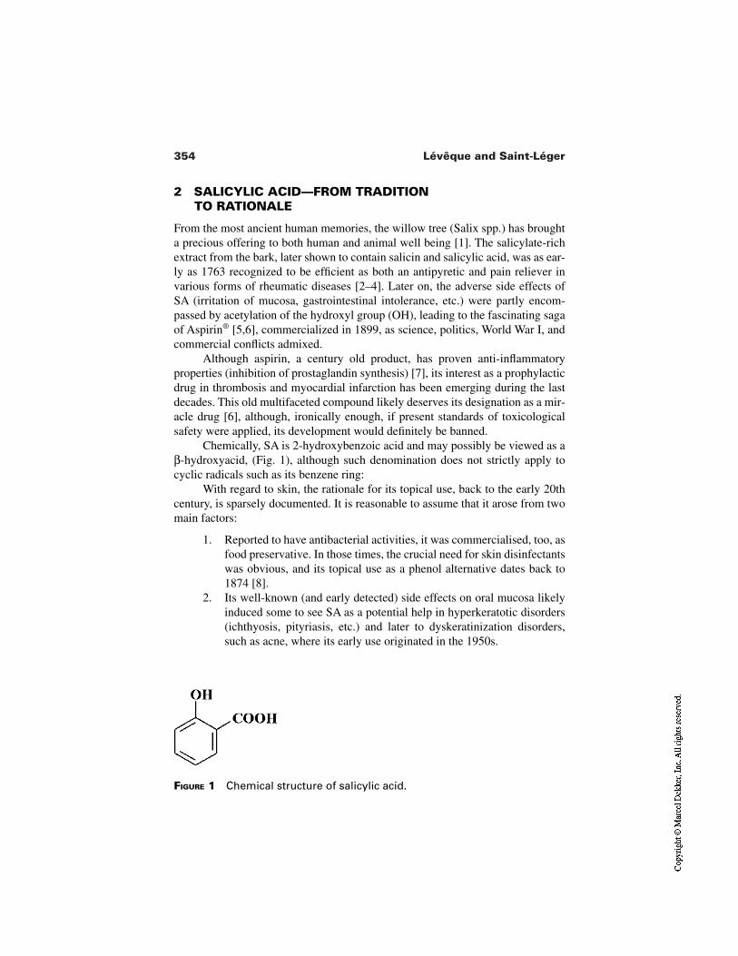
354 Lévêque and Saint-Léger
FIGURE 1 Chemical structure of salicylic acid.
2 SALICYLIC ACID—FROM TRADITION TO RATIONALE
From the most ancient human memories, the willow tree (Salix spp.) has broughta precious offering to both human and animal well being [1]. The salicylate-richextract from the bark, later shown to contain salicin and salicylic acid, was as ear-ly as 1763 recognized to be efficient as both an antipyretic and pain reliever invarious forms of rheumatic diseases [2–4]. Later on, the adverse side effects ofSA (irritation of mucosa, gastrointestinal intolerance, etc.) were partly encom-passed by acetylation of the hydroxyl group (OH), leading to the fascinating sagaof Aspirin® [5,6], commercialized in 1899, as science, politics, World War I, andcommercial conflicts admixed.
Although aspirin, a century old product, has proven anti-inflammatoryproperties (inhibition of prostaglandin synthesis) [7], its interest as a prophylacticdrug in thrombosis and myocardial infarction has been emerging during the lastdecades. This old multifaceted compound likely deserves its designation as a mir-acle drug [6], although, ironically enough, if present standards of toxicologicalsafety were applied, its development would definitely be banned.
Chemically, SA is 2-hydroxybenzoic acid and may possibly be viewed as aβ-hydroxyacid, (Fig. 1), although such denomination does not strictly apply tocyclic radicals such as its benzene ring:
With regard to skin, the rationale for its topical use, back to the early 20thcentury, is sparsely documented. It is reasonable to assume that it arose from twomain factors:
1. Reported to have antibacterial activities, it was commercialised, too, asfood preservative. In those times, the crucial need for skin disinfectantswas obvious, and its topical use as a phenol alternative dates back to1874 [8].
2. Its well-known (and early detected) side effects on oral mucosa likelyinduced some to see SA as a potential help in hyperkeratotic disorders(ichthyosis, pityriasis, etc.) and later to dyskeratinization disorders,such as acne, where its early use originated in the 1950s.
355Salicylic Acid and Derivatives
2.1 Dermatological and Cosmetical Properties
For decades, SA has certainly been one of the most commonly used compoundsamong both dermatological and cosmetical arsenals. A 1975 review [9] conferredto topical SA a mosaic of actions according to dosages (0.3 to 5% and above), i.e.,germistatic, acidogenic, photoprotective, anti-eczematous, and the so-called ker-atolytic effect. Presently, both scientific works and routine practice have, in fact,largely focused interest on the latter action [10–13] for three main reasons:
1. The first four actions were of a very modest amplitude as compared toother candidates (true germicides, powerful sunscreens, etc.).
2. Clinically, its progressive (and empirical) use in common hyperkera-totic conditions showed clear benefits, coupled with an acceptable tol-erance.
3. The intense development of bioengineering methods during the lastthree decades allowed a precise quantification of its “keratolytic” prop-erty when applied to skin. Salicylic acid rapidly became, too, an im-portant tool for researchers involved in exfoliative cytology or trans-cutaneous penetration studies [14,15].
It is now a common statement that keratolytic and derived effects are unjustified.Neither SA nor AHA lead to breakage of keratin chains as this invented term sug-gested [16].
In fact, SA appears to be a clear disrupter agent of the horny cell junctions.Desmosome or Corneodesmosome attachments between adjacent cells mostlyensure the latter, maintaining stratum corneum cohesion. These cellular “snapfasteners” of glycoprotein structures are progressively degraded through (endo-genic) enzymatic attacks, leading to a loosening of the cell junctions and conse-quent monocellular desquamation, in the normal situation. Our group has recent-ly shown that desmosomes-like organizations are the precise privileged sites ofaction of SA, leading to their degradation [17]. As suggested, “desmolysis”would be a much better term for such action.
In hyperkeratotic conditions (xerosis, acne, warts), where the SC appearsthick, cracking, fluffy, or badly organized, topical SA restores within a few weeksa normalized and thinner horny organization [18,19]. This action is coupled to thefollowing criteria:
Form. Salicylic acid is sparingly hydrosoluble. It is, most of the time, intro-duced in lipophilic ointments or alcoholic lotions. In most cases, SA isintroduced as a free form, i.e., at a spontaneous acid pH, the free statesof both OH and COOH groups seemingly a prerequisite for desmolyticaction.
Dose dependence. Desmolysis is dose dependent, ranging from 2 to 3% inthe minor cases of xerosis, to 17% (coupled to lactic acid) for wart treat-
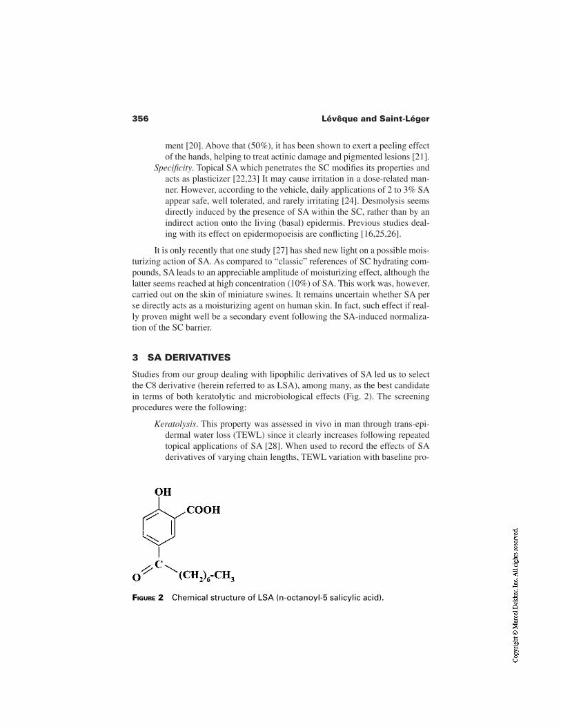
356 Lévêque and Saint-Léger
ment [20]. Above that (50%), it has been shown to exert a peeling effectof the hands, helping to treat actinic damage and pigmented lesions [21].
Specificity. Topical SA which penetrates the SC modifies its properties andacts as plasticizer [22,23] It may cause irritation in a dose-related man-ner. However, according to the vehicle, daily applications of 2 to 3% SAappear safe, well tolerated, and rarely irritating [24]. Desmolysis seemsdirectly induced by the presence of SA within the SC, rather than by anindirect action onto the living (basal) epidermis. Previous studies deal-ing with its effect on epidermopoeisis are conflicting [16,25,26].
It is only recently that one study [27] has shed new light on a possible mois-turizing action of SA. As compared to “classic” references of SC hydrating com-pounds, SA leads to an appreciable amplitude of moisturizing effect, although thelatter seems reached at high concentration (10%) of SA. This work was, however,carried out on the skin of miniature swines. It remains uncertain whether SA perse directly acts as a moisturizing agent on human skin. In fact, such effect if real-ly proven might well be a secondary event following the SA-induced normaliza-tion of the SC barrier.
3 SA DERIVATIVES
Studies from our group dealing with lipophilic derivatives of SA led us to selectthe C8 derivative (herein referred to as LSA), among many, as the best candidatein terms of both keratolytic and microbiological effects (Fig. 2). The screeningprocedures were the following:
Keratolysis. This property was assessed in vivo in man through trans-epi-dermal water loss (TEWL) since it clearly increases following repeatedtopical applications of SA [28]. When used to record the effects of SAderivatives of varying chain lengths, TEWL variation with baseline pro-
FIGURE 2 Chemical structure of LSA (n-octanoyl-5 salicylic acid).
357Salicylic Acid and Derivatives
gressively increases with increasing length (from C4 to C8), peaks atC8, and declines afterward. As compared to SA, LSA leads to compara-ble variation in TEWL for a reduced concentration (w/w), about threetimes lower than that of SA [26]. Expressed as molar ratios, this in-creased activity is even more in favor of LSA since it has a much highermolecular weight (264 versus 138). According to these expressions,LSA therefore shows for a “keratolytic” property an intrinsic potency ofthree to six times that of SA.
Microbiological data. MIC’s of the various derivatives on different strainsof the resident human skin flora appear lower with longer chains (C8,C10, and above) as compared to shorter ones (C2, C4, and C6) [29].Based on such in vitro findings, a better efficacy is therefore given priv-ilege to chains greater than C8.
These two citeria, coupled to safety data profiles, led us to investigate further thecosmetological interest of LSA, by comparing it to previously well-recognizedkeratolytic agents such as SA, glycolic acid (GA), lactic acid (LA), and retinoicacid (RA).
3.1 Comparative Effects of the KeratolyticCompounds on Human Epidermis
3.1.1 Effects on Stratum Corneum in Vitro
Samples from either strippings or biopsies from healthy human volunteers weresubmitted to the actions of LSA (1 and 5%), LA, and SA (3 and 15%), all intro-duced in the same propylene glycol vehicle and further processed by transmissionelectronic microscopy (TEM) coupled to freeze fracture (FF) according to classi-cal technical procedures [17].
The results clearly show that corneodesmosomes (CDs) are the main targetsof these three agents. Keratin filaments remain intact in all samples.
However, some differences between the respective effects of these com-pounds may be outlined. Salicylic and lactic acid seem to act uniformly into theoverall thickness of the stratum corneum, whereas LSA appears to limit its actionto the superior third, a location of the SC where corneodesmosomes have previ-ously been modified/degraded by proteolytic enzymes. The nature of the modifi-cations of the CDs seems different, too; LSA appears to act on their whole struc-ture, completely detaching them from one full side, while LA and SA seem tofractionate these CDs. With regard to SA, the glycoproteins, which cross themembrane, appear strongly denatured. These changes are illustrated in Figs. 3and 4.
According to the authors of these works, the different modes of actions ofthese agents are closely related to their intrinsic lipophilic property. The latter
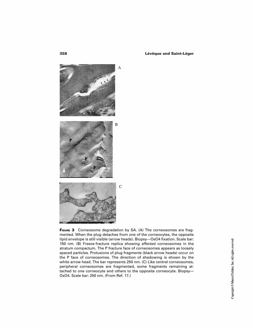
358 Lévêque and Saint-Léger
FIGURE 3 Corneosome degradation by SA. (A) The corneosomes are frag-mented. When the plug detaches from one of the corneocytes, the oppositelipid envelope is still visible (arrow heads). Biopsy—OsO4 fixation. Scale bar:150 nm. (B) Freeze-fracture replica showing affected corneosomes in thestratum compactum. The P fracture face of corneosomes appears as looselyspaced particles. Protusions of plug fragments (black arrow heads) occur onthe P face of corneosomes. The direction of shadowing is shown by thewhite arrow head. The bar represents 250 nm. (C) Like central corneosomes,peripheral corneosomes are fragmented, some fragments remaining at-tached to one corneocyte and others to the opposite corneocyte. Biopsy—OsO4. Scale bar: 250 nm. (From Ref. 17.)
A
B
C
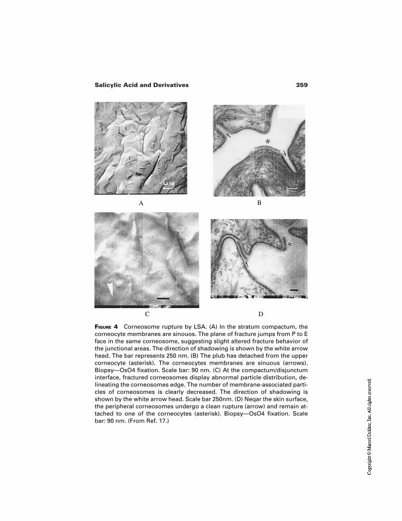
359Salicylic Acid and Derivatives
FIGURE 4 Corneosome rupture by LSA. (A) In the stratum compactum, thecorneocyte membranes are sinouos. The plane of fracture jumps from P to Eface in the same corneosome, suggesting slight altered fracture behavior ofthe junctional areas. The direction of shadowing is shown by the white arrowhead. The bar represents 250 nm. (B) The plub has detached from the uppercorneocyte (asterisk). The corneocytes membranes are sinuous (arrows).Biopsy—OsO4 fixation. Scale bar: 90 nm. (C) At the compactum/disjunctuminterface, fractured corneosomes display abnormal particle distribution, de-lineating the corneosomes edge. The number of membrane-associated parti-cles of corneosomes is clearly decreased. The direction of shadowing isshown by the white arrow head. Scale bar 250nm. (D) Neqar the skin surface,the peripheral corneosomes undergo a clean rupture (arrow) and remain at-tached to one of the corneocytes (asterisk). Biopsy—OsO4 fixation. Scalebar: 90 nm. (From Ref. 17.)
C D
A B
360 Lévêque and Saint-Léger
would, in turn, “orientate” their action either on the membrane (LSA) of the CDsor on their proteins/glycoproteins for the least lipophilic agents (SA, LA).
3.1.2 Effects on Epidermal Renewal in Vivo
It has been shown that on human photoaged skin repeated topical applications ofretinoic acid lead to clear changes in the epidermal turnover, even for as short aperiod as 4 weeks [23]. Following a similar protocol, Pierard et al. compared invivo on man the respective activities of SA (5%) and LSA (1.5%) versus RA(0.025%) [30]. The study was carried out for 4 weeks in a double-blind proce-dure, and the three agents were compared to their respective vehicles. On biop-sies, automated histometry measurements, immunohistology allowed the record-ing of changes in cell proliferation (Ki67), cell differentiation (K), and activity ofthe papillary dermis as well.
The skin sites treated by vehicle and SA do not differ from a nontreatedcontrol site in any aspect. However, both sites treated by RA and LSA revealedsignificant increases in the thickness of the viable epidermis and its renewal rate.In the papillary dermis, dendrocytes FXIII positive were shown significantly acti-vated and of higher amplitude in the RA-treated site than that of the LSA.
Another study of a comparable methodology (double-blind, vehicle-con-trolled) compared on three groups of human volunteers (eight per group) the ef-fects of RA (0.05%), LSA (2%), and GA (10%, pH 3.5), each product being ap-plied onto one forearm for two months, the other receiving the vehicle (commonto the three agents) alone [31].
In addition to standard histometric and immunohistologic measurements,classical histology (H&E, Luna, Hale, Fontana–Masson, and PAS-Giemsa stains)was undertaken.
This study, in conflict with previously published data, failed to detect anymodification brought by GA. This study confirms, however, the RA and LSA ef-fects that were initially found in the Pierard study [23]. Additionally, classical his-tology revealed a partial correction of epidermal atrophy, atypia, and dysplasia.Again, this study noted higher amplitude of effect brought by RA than by LSA.
As far as melanins are concerned, both products show comparable activity;the large and dense melanosome conglomerates present on the basal layer be-come dispersed into small units.
These studies illustrate the effectiveness of this lipophilic derivative of SAin the skin care of photoaged skins and its superiority vis a vis SA, LA, and GA inthese precise experimental conditions.
3.2 LSA-Treated Skin and Cosmetic Implications
The usual cosmetic concepts (skin smoothness, firmness, hue, glow) can hardlybe reduced to the measurements of one or two objective cutaneous parameters.
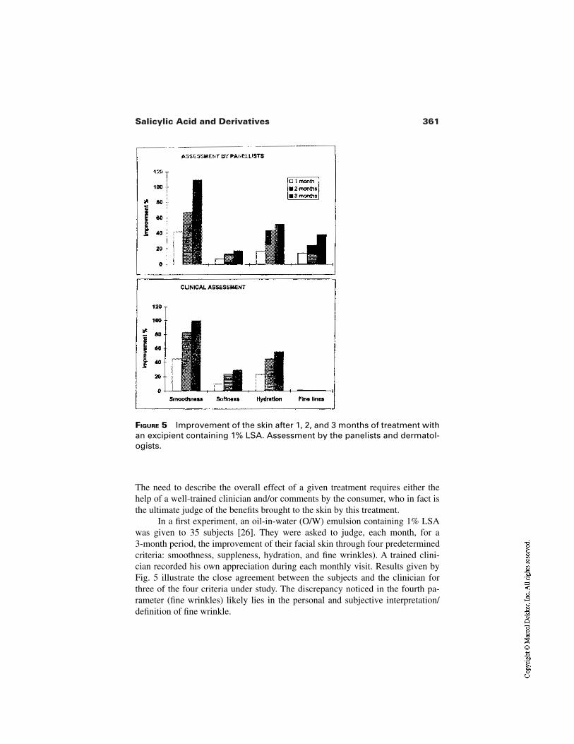
361Salicylic Acid and Derivatives
FIGURE 5 Improvement of the skin after 1, 2, and 3 months of treatment withan excipient containing 1% LSA. Assessment by the panelists and dermatol-ogists.
The need to describe the overall effect of a given treatment requires either thehelp of a well-trained clinician and/or comments by the consumer, who in fact isthe ultimate judge of the benefits brought to the skin by this treatment.
In a first experiment, an oil-in-water (O/W) emulsion containing 1% LSAwas given to 35 subjects [26]. They were asked to judge, each month, for a 3-month period, the improvement of their facial skin through four predeterminedcriteria: smoothness, suppleness, hydration, and fine wrinkles). A trained clini-cian recorded his own appreciation during each monthly visit. Results given byFig. 5 illustrate the close agreement between the subjects and the clinician forthree of the four criteria under study. The discrepancy noticed in the fourth pa-rameter (fine wrinkles) likely lies in the personal and subjective interpretation/definition of fine wrinkle.
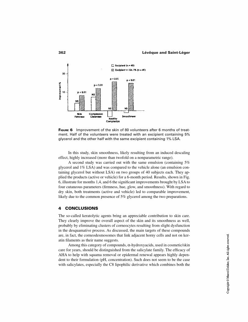
362 Lévêque and Saint-Léger
FIGURE 6 Improvement of the skin of 80 volunteers after 6 months of treat-ment. Half of the volunteers were treated with an excipient containing 5%glycerol and the other half with the same excipient containing 1% LSA.
In this study, skin smoothness, likely resulting from an induced descalingeffect, highly increased (more than twofold on a nonparametric range).
A second study was carried out with the same emulsion (containing 5%glycerol and 1% LSA) and was compared to the vehicle alone (an emulsion con-taining glycerol but without LSA) on two groups of 40 subjects each. They ap-plied the products (active or vehicle) for a 6-month period. Results, shown in Fig.6, illustrate for months 1,4, and 6 the significant improvements brought by LSA tofour cutaneous parameters (firmness, hue, glow, and smoothness). With regard todry skin, both treatments (active and vehicle) led to comparable improvement,likely due to the common presence of 5% glycerol among the two preparations.
4 CONCLUSIONS
The so-called keratolytic agents bring an appreciable contribution to skin care.They clearly improve the overall aspect of the skin and its smoothness as well,probably by eliminating clusters of corneocytes resulting from slight dysfunctionin the desquamative process. As discussed, the main targets of these compoundsare, in fact, the corneodesmosomes that link adjacent horny cells and not on ker-atin filaments as their name suggests.
Among this category of compounds, α-hydroxyacids, used in cosmetic/skincare for years, should be distinguished from the salicylate family. The efficacy ofAHA to help with squama removal or epidermal renewal appears highly depen-dent to their formulation (pH, concentration). Such does not seem to be the casewith salicylates, especially the C8 lipophilic derivative which combines both the
363Salicylic Acid and Derivatives
effects of a slight boost of cell renewal and a dispersing of the basal melanin ag-gregates, leading to a more uniform and clearer aspect to the skin. The results re-viewed here highlight the LSA as a promising active agent for skin care where aslight stimulation of the epidermal turnover is desirable.
REFERENCES
1. Stone E. An account of the success of the bark of the willow in the cure of agues.Philos Trans R Soc Lond (Biol) 1763; 53:195–200.
2. Stricker F. Aus der Traubschen Klinik. Über die Resultate der behandlung der poly-arthritis rhumatica mit salicylsäure. Berl Klin Woschr 1876;13:1–15.
3. Gross M, Greenberg LA. The salicylates: a critical bibliographic review. NewHaven, CT: Hillhouse Press, 1948:8.
4. Hedner T, Everts B. The early clinical history of salicylates in rheumatology andpain. Clin Rheumatol 1998; 17:17–25.
5. Kubnert N. Hundert Jahre Aspirin. Chemie 1999; 3364:213–220.6. Jourdier S. A miracle drug. Chemistry in Britain 1999; Feb:33–35.7. Vane JR. Inhibition of prostaglandin synthesis as a mechanism of action for aspirin-
like drugs. Nature 1971; 231:232–235.8. Kolbe H. Ueber eine neue Darstellungmethode und einige bemerkenswerte Eigen-
schaften der Salicylsäure. Arch Pharm 1874; 5–45.9. Weirich EG. Dermatopharmacology of salicylic acid. Dermatologica 1975;
151:268–273.10. Huber C, Christophers E. “Keratolytic” effect of salicylic acid. Arch Derm Res 1977;
257:293–297.11. Kligman LH, Kligman AM. The effect on rhino mouse skin of agents which influ-
ence keratinization and exfoliation. J Invest Dermatol 1979; 73:354–358.12. Roberts DL, Marshall R, Marks R. Detection of the action of salicylic acid on the
normal sratum corneum. Br J Dermatol 1980; 103:191–196.13. Loden M, Boström P, Kneczke M. Distribution and keratolytic effect of salicylic acid
and urea in human skin. Skin Pharmacol 1995; 8:173–178.14. Elias PM, Cooper ER, Korc A, Brown BE. Percutaneous transport in relation to stra-
tum corneum structure and lipid composition. J Invest Dermatol 1981; 76:297–301.15. Harada K, Murakami T, Yata N, Yamamoto S. Role of intercellular lipids in stratum
corneum in the percutaneous permeation of drugs. J Invest Dermatol 1992;99:278–282.
16. Davies M, Marks R. Studies on the effect of salicylic acid on normal skin. Br J Der-matol 1976; 95:187–192.
17. Corcuff P, Fiat F, Gracia AM, Lévêque JL. Hydroxyacid induced desquamation ofthe human stratum corneum: a comparative ultrastructural study. Proc 19th IFSCCCongress 1996; 3:85–94.
18. Mark R, Davies M, Cattel A. An explanation for the keratolytic effect of salicylicacid. J Invest Dermamol Abstracts 1975; April:283.
19. Nook TH. In vivo measurement of the keratolytic effect of salicylic acid in threeointment formulations. Br J Dermatol 1987; 117:243–245.
364 Lévêque and Saint-Léger
20. Lawson EE, Edwards H, Barry BW, Williams AC. Interaction of salicylic acid withverrucae assessed by FT raman spectroscopy. Journal of Drug Targeting 1998;5(5):343–351.
21. Swineheart JM. Salicylic acid ointment peeling of the hands and forearms. J Derma-tol Surg Oncol 1992; 18:495–498.
22. Rasseneur L, De Rigal J, Lévêque JL. Influence des différents constituants de lacouche cornée sur la mesure de son élasticité. Int J Cosmet Sci 1982; 4:247–260.
23. Pierard-Franchimont C, Goffin V, Pierard GE. Modulation of human stratumcorneum properties by salicylic and all-trans retinoic acid. Skin Pharmacol 1998;11:266–272.
24. Davis DA, Krasu AL, Thomson GA, Olerich M, Odio MR. Percutaneous absorptionof salicylic acid after repeated (14 days) in vivo administration to normal, acnegenicor aged human skin. J Pharmaceut Sci 1997; 86(8):896–899.
25. Weirich EG, Longauer JK, Kirkwod AH. Effect of topical salicylic acid on animalepidermopoiesis. Dermatologica 1978; 156:89–96.
26. Lévêque JL, Corcuff P, Gonnord G, Montastier C, Renault B, Bazin R, Pierard GE,Poelman MC. Mechanism of action of a lipophilic derivative of salicylic acid on nor-mal skin. Skin Res Technol 1995; 1:115–122.
27. Goshi KI, Tabata N, Sato Y. Comparative study of the efficacy of various moisturiz-ers on the skin of the ASR miniature swine. Skin Pharmacol Appl Skin Physiol 2000;13:120–127.
28. Guillaume JC, De Rigal J, Lévêque JL, Dubertret L, Touraine L. Etude comparée dela perte insensible d’eau et de la pénétration cutanée des corticoides. Dermatologica1981; 162:380–390.
29. International patent L’Oreal no. 850.69.53.30. Pierard GE, Nikkels-Tassoudji N, Arrese JE, et al. Dermo-epidermal stimulation
elicited by a lipohydroxyacid: a comparison with salicylic acid and all-trans retinoicacid. Dermatology 1997; 194:398–401.
31. Pierard GE, Kligman AM, Stoudemayer TJ, et al. Comparative effects of retinoicacid, glycolic acid and a lipophilic derivative of salicylic acid on photoaged epider-mis. Dermatology 1999; 199:50–53.
18The Efficacy, Stability, and Safety of TopicallyApplied Protease in Treating Xerotic Skin
David J. Pocalyko and Prem ChandarUnilever Research, Edgewater Laboratory, Edgewater, New Jersey
Clive R. Harding, Lynn Blaikie, andAllan WatkinsonUnilever Research, Colworth Laboratory, Sharnbrook, Bedford,United Kingdom
Anthony V. RawlingsUnilever Research, Port Sunlight Laboratory, Bebington, Wirral,United Kingdom
1 INTRODUCTION
Abnormal desquamation arises from an inability to effectively degrade the mo-lecular components that provide cohesive force and thereby maintain tissue in-tegrity. In such conditions corneocytes do not detach as single cells, but are shedin large clusters forming visible scales. The degree of scaling varies from severein genetically determined disorders such as ichthyoses (which are also associatedwith increased thickness of the stratum corneum) to “cosmetic” dry skin. The ap-pearance of the latter form of dry skin is a common feature in the population andis usually associated with extrinsic damage (e.g., surfactants, UV irradiation) and
365
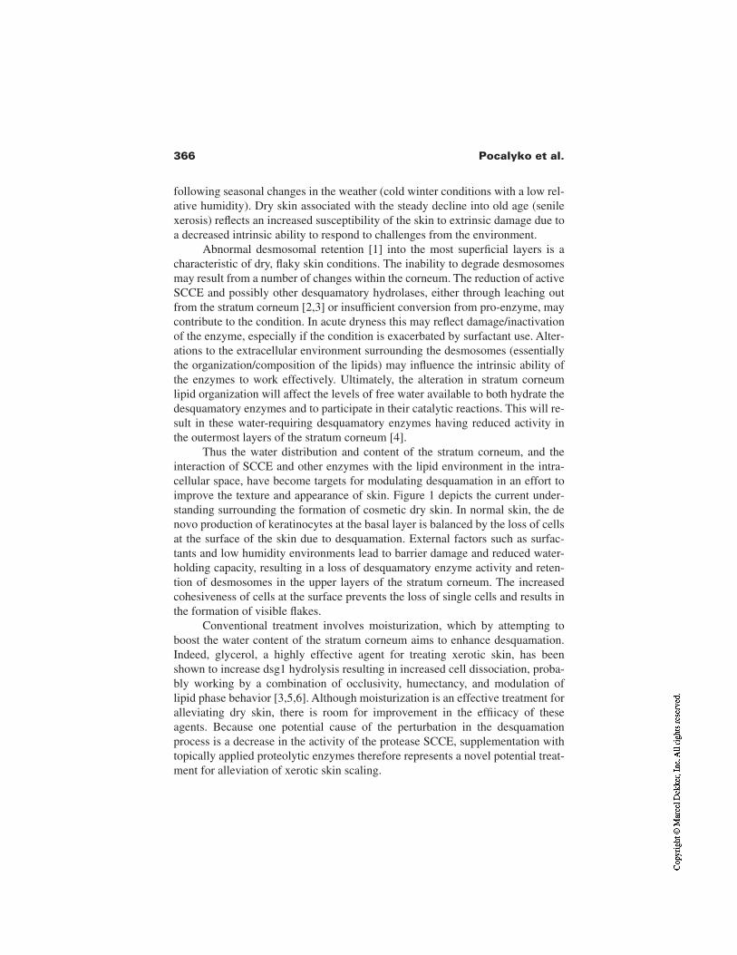
366 Pocalyko et al.
following seasonal changes in the weather (cold winter conditions with a low rel-ative humidity). Dry skin associated with the steady decline into old age (senilexerosis) reflects an increased susceptibility of the skin to extrinsic damage due toa decreased intrinsic ability to respond to challenges from the environment.
Abnormal desmosomal retention [1] into the most superficial layers is acharacteristic of dry, flaky skin conditions. The inability to degrade desmosomesmay result from a number of changes within the corneum. The reduction of activeSCCE and possibly other desquamatory hydrolases, either through leaching outfrom the stratum corneum [2,3] or insufficient conversion from pro-enzyme, maycontribute to the condition. In acute dryness this may reflect damage/inactivationof the enzyme, especially if the condition is exacerbated by surfactant use. Alter-ations to the extracellular environment surrounding the desmosomes (essentiallythe organization/composition of the lipids) may influence the intrinsic ability ofthe enzymes to work effectively. Ultimately, the alteration in stratum corneumlipid organization will affect the levels of free water available to both hydrate thedesquamatory enzymes and to participate in their catalytic reactions. This will re-sult in these water-requiring desquamatory enzymes having reduced activity inthe outermost layers of the stratum corneum [4].
Thus the water distribution and content of the stratum corneum, and theinteraction of SCCE and other enzymes with the lipid environment in the intra-cellular space, have become targets for modulating desquamation in an effort toimprove the texture and appearance of skin. Figure 1 depicts the current under-standing surrounding the formation of cosmetic dry skin. In normal skin, the denovo production of keratinocytes at the basal layer is balanced by the loss of cellsat the surface of the skin due to desquamation. External factors such as surfac-tants and low humidity environments lead to barrier damage and reduced water-holding capacity, resulting in a loss of desquamatory enzyme activity and reten-tion of desmosomes in the upper layers of the stratum corneum. The increasedcohesiveness of cells at the surface prevents the loss of single cells and results inthe formation of visible flakes.
Conventional treatment involves moisturization, which by attempting toboost the water content of the stratum corneum aims to enhance desquamation.Indeed, glycerol, a highly effective agent for treating xerotic skin, has beenshown to increase dsg1 hydrolysis resulting in increased cell dissociation, proba-bly working by a combination of occlusivity, humectancy, and modulation oflipid phase behavior [3,5,6]. Although moisturization is an effective treatment foralleviating dry skin, there is room for improvement in the effiicacy of theseagents. Because one potential cause of the perturbation in the desquamationprocess is a decrease in the activity of the protease SCCE, supplementation withtopically applied proteolytic enzymes therefore represents a novel potential treat-ment for alleviation of xerotic skin scaling.
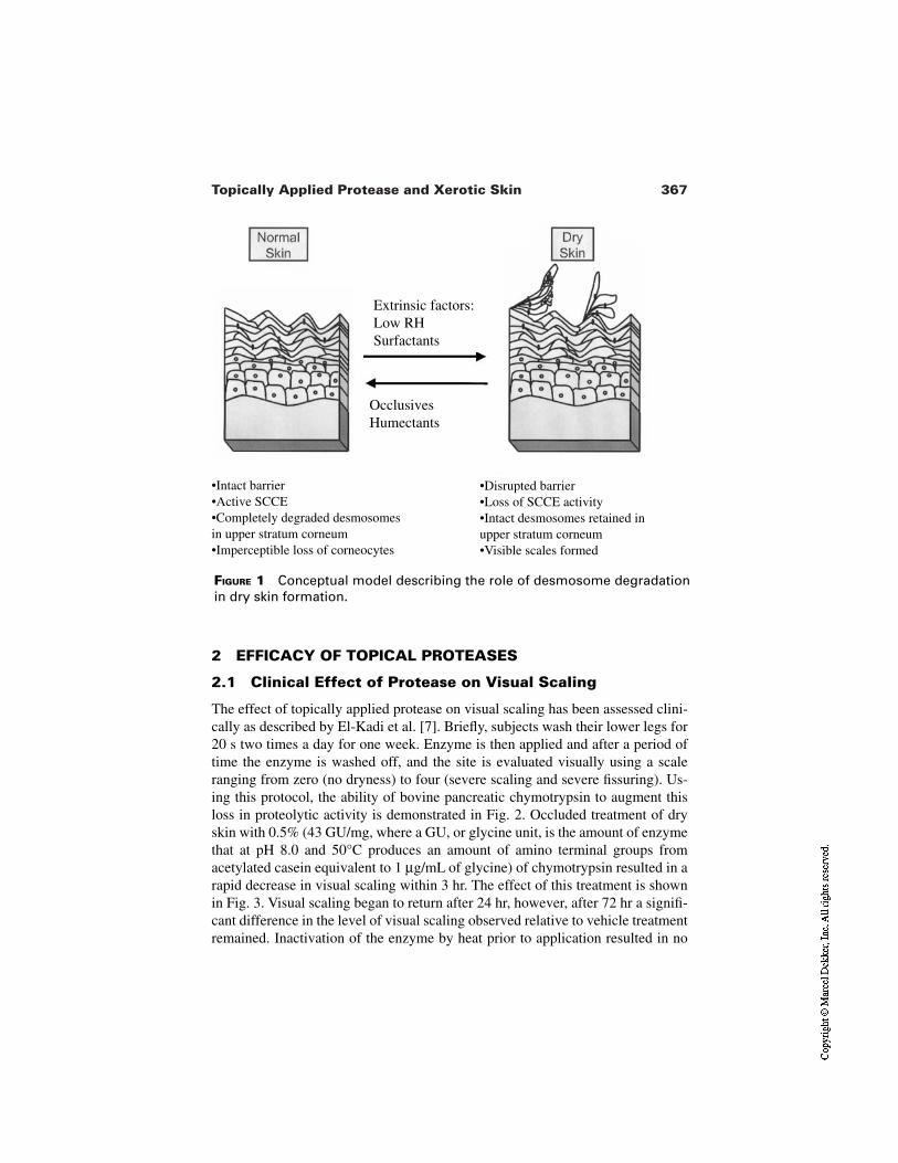
367Topically Applied Protease and Xerotic Skin
FIGURE 1 Conceptual model describing the role of desmosome degradationin dry skin formation.
2 EFFICACY OF TOPICAL PROTEASES
2.1 Clinical Effect of Protease on Visual Scaling
The effect of topically applied protease on visual scaling has been assessed clini-cally as described by El-Kadi et al. [7]. Briefly, subjects wash their lower legs for20 s two times a day for one week. Enzyme is then applied and after a period oftime the enzyme is washed off, and the site is evaluated visually using a scaleranging from zero (no dryness) to four (severe scaling and severe fissuring). Us-ing this protocol, the ability of bovine pancreatic chymotrypsin to augment thisloss in proteolytic activity is demonstrated in Fig. 2. Occluded treatment of dryskin with 0.5% (43 GU/mg, where a GU, or glycine unit, is the amount of enzymethat at pH 8.0 and 50°C produces an amount of amino terminal groups fromacetylated casein equivalent to 1 µg/mL of glycine) of chymotrypsin resulted in arapid decrease in visual scaling within 3 hr. The effect of this treatment is shownin Fig. 3. Visual scaling began to return after 24 hr, however, after 72 hr a signifi-cant difference in the level of visual scaling observed relative to vehicle treatmentremained. Inactivation of the enzyme by heat prior to application resulted in no
Extrinsic factors:Low RHSurfactants
OcclusivesHumectants
•Intact barrier•Active SCCE•Completely degraded desmosomesin upper stratum corneum•Imperceptible loss of corneocytes
•Disrupted barrier•Loss of SCCE activity•Intact desmosomes retained inupper stratum corneum•Visible scales formed
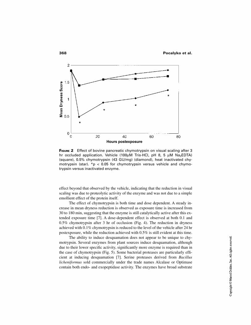
368 Pocalyko et al.
FIGURE 2 Effect of bovine pancreatic chymotrypsin on visual scaling after 3hr occluded application. Vehicle (100µM Tris-HCl, pH 8, 5 µM Na2EDTA)(square), 0.5% chymotrypsin (43 GU/mg) (diamond), heat inactivated chy-motrypsin (star). *p < 0.05 for chymotrypsin versus vehicle and chymo-trypsin versus inactivated enzyme.
Hours postexposure
effect beyond that observed by the vehicle, indicating that the reduction in visualscaling was due to proteolytic activity of the enzyme and was not due to a simpleemollient effect of the protein itself.
The effect of chymotrypsin is both time and dose dependent. A steady in-crease in mean dryness reduction is observed as exposure time is increased from30 to 180 min, suggesting that the enzyme is still catalytically active after this ex-tended exposure time [7]. A dose-dependent effect is observed at both 0.1 and0.5% chymotrypsin after 3 hr of occlusion (Fig. 4). The reduction in drynessachieved with 0.1% chymotrypsin is reduced to the level of the vehicle after 24 hrpostexposure, while the reduction achieved with 0.5% is still evident at this time.
The ability to induce desquamation does not appear to be unique to chy-motrypsin. Several enzymes from plant sources induce desquamation, althoughdue to their lower specific activity, significantly more enzyme is required than inthe case of chymotrypsin (Fig. 5). Some bacterial proteases are particularly effi-cient at inducing desquamation [7]. Serine proteases derived from Bacilluslicheniformus sold commercially under the trade names Alcalase or Optimasecontain both endo- and exopeptidase activity. The enzymes have broad substrate
369Topically Applied Protease and Xerotic Skin
FIGURE 3 Effect of bovine pancreatic chymotrypsin in 100 µM Tris-HCl, pH 8,5 µM Na2EDTA after 3 hr occluded application. (A) Before treatment. (B) Aftertreatment.
A
B
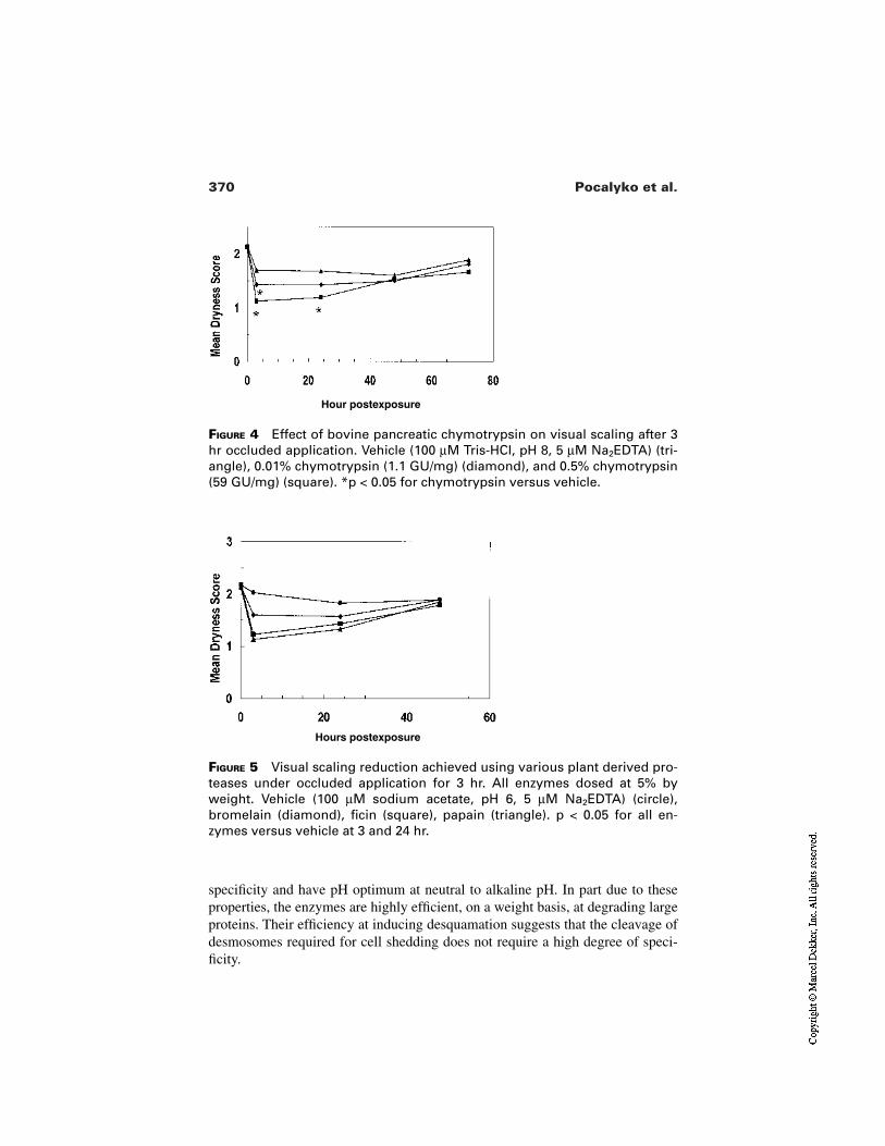
370 Pocalyko et al.
FIGURE 4 Effect of bovine pancreatic chymotrypsin on visual scaling after 3hr occluded application. Vehicle (100 µM Tris-HCl, pH 8, 5 µM Na2EDTA) (tri-angle), 0.01% chymotrypsin (1.1 GU/mg) (diamond), and 0.5% chymotrypsin(59 GU/mg) (square). *p < 0.05 for chymotrypsin versus vehicle.
Hour postexposure
FIGURE 5 Visual scaling reduction achieved using various plant derived pro-teases under occluded application for 3 hr. All enzymes dosed at 5% byweight. Vehicle (100 µM sodium acetate, pH 6, 5 µM Na2EDTA) (circle),bromelain (diamond), ficin (square), papain (triangle). p < 0.05 for all en-zymes versus vehicle at 3 and 24 hr.
Hours postexposure
specificity and have pH optimum at neutral to alkaline pH. In part due to theseproperties, the enzymes are highly efficient, on a weight basis, at degrading largeproteins. Their efficiency at inducing desquamation suggests that the cleavage ofdesmosomes required for cell shedding does not require a high degree of speci-ficity.
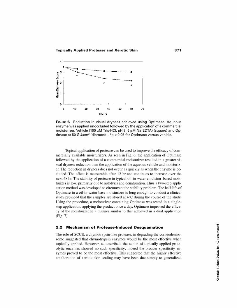
371Topically Applied Protease and Xerotic Skin
FIGURE 6 Reduction in visual dryness achieved using Optimase. Aqueousenzyme was applied unoccluded followed by the application of a commercialmoisturizer. Vehicle (100 µM Tris-HCl, pH 8, 5 µM Na2EDTA) (square) and Op-timase at 50 GU/cm2 (diamond). *p < 0.05 for Optimase versus vehicle.
Topical application of protease can be used to improve the efficacy of com-mercially available moisturizers. As seen in Fig. 6, the application of Optimasefollowed by the application of a commercial moisturizer resulted in a greater vi-sual dryness reduction than the application of the aqueous vehicle and moisturiz-er. The reduction in dryness does not occur as quickly as when the enzyme is oc-cluded. The effect is measurable after 12 hr and continues to increase over thenext 48 hr. The stability of protease in typical oil-in-water emulsion–based mois-turizes is low, primarily due to autolysis and denaturation. Thus a two-step appli-cation method was developed to circumvent the stability problem. The half-life ofOptimase in a oil-in-water base moisturizer is long enough to conduct a clinicalstudy provided that the samples are stored at 4°C during the course of the study.Using the procedure, a moisturizer containing Optimase was tested in a single-step application, applying the product once a day. Optimase improved the effica-cy of the moisturizer in a manner similar to that achieved in a dual application(Fig. 7).
2.2 Mechanism of Protease-Induced Desquamation
The role of SCCE, a chymotrypsin-like protease, in degrading the corneodesmo-some suggested that chymotrypsin enzymes would be the most effective whentopically applied. However, as described, the action of topically applied prote-olytic enzymes showed no such specificity; indeed the broader specificity en-zymes proved to be the most effective. This suggested that the highly effectiveamelioration of xerotic skin scaling may have been due simply to generalized
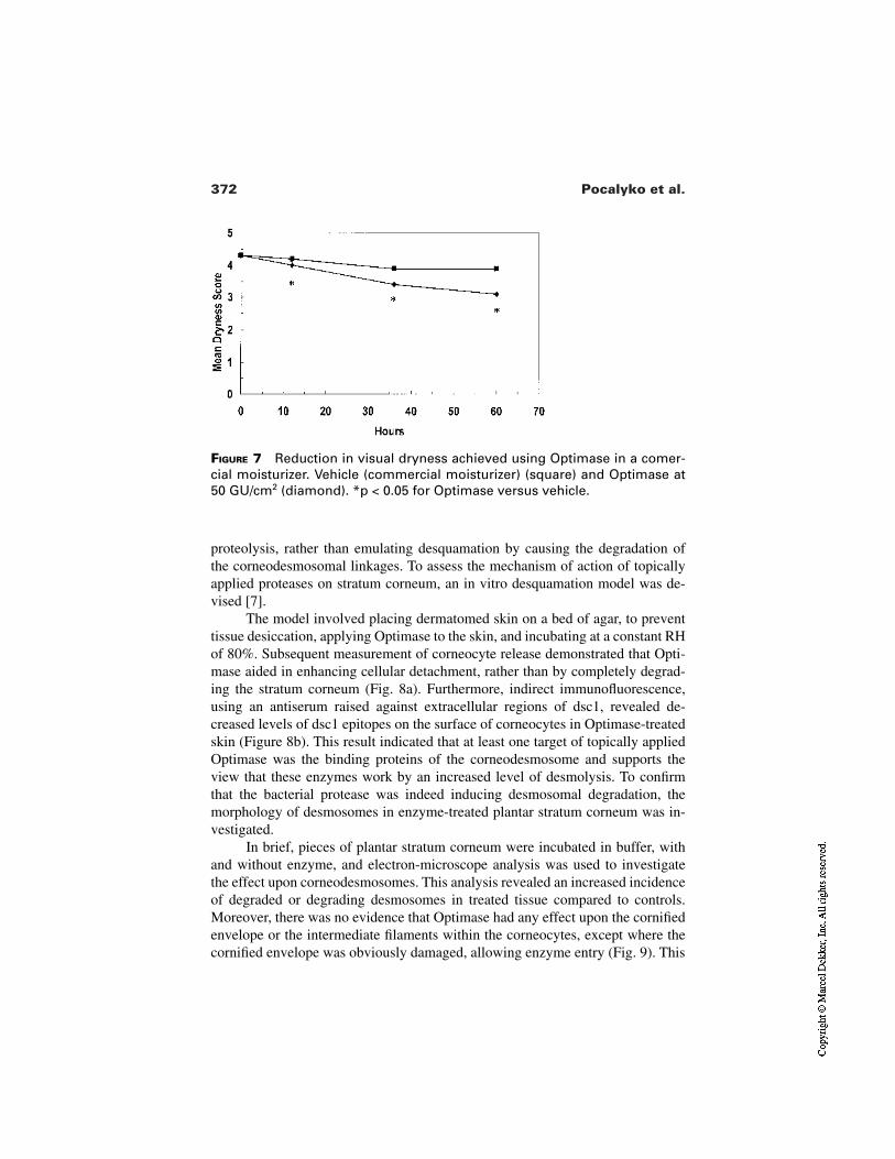
372 Pocalyko et al.
FIGURE 7 Reduction in visual dryness achieved using Optimase in a comer-cial moisturizer. Vehicle (commercial moisturizer) (square) and Optimase at50 GU/cm2 (diamond). *p < 0.05 for Optimase versus vehicle.
proteolysis, rather than emulating desquamation by causing the degradation ofthe corneodesmosomal linkages. To assess the mechanism of action of topicallyapplied proteases on stratum corneum, an in vitro desquamation model was de-vised [7].
The model involved placing dermatomed skin on a bed of agar, to preventtissue desiccation, applying Optimase to the skin, and incubating at a constant RHof 80%. Subsequent measurement of corneocyte release demonstrated that Opti-mase aided in enhancing cellular detachment, rather than by completely degrad-ing the stratum corneum (Fig. 8a). Furthermore, indirect immunofluorescence,using an antiserum raised against extracellular regions of dsc1, revealed de-creased levels of dsc1 epitopes on the surface of corneocytes in Optimase-treatedskin (Figure 8b). This result indicated that at least one target of topically appliedOptimase was the binding proteins of the corneodesmosome and supports theview that these enzymes work by an increased level of desmolysis. To confirmthat the bacterial protease was indeed inducing desmosomal degradation, themorphology of desmosomes in enzyme-treated plantar stratum corneum was in-vestigated.
In brief, pieces of plantar stratum corneum were incubated in buffer, withand without enzyme, and electron-microscope analysis was used to investigatethe effect upon corneodesmosomes. This analysis revealed an increased incidenceof degraded or degrading desmosomes in treated tissue compared to controls.Moreover, there was no evidence that Optimase had any effect upon the cornifiedenvelope or the intermediate filaments within the corneocytes, except where thecornified envelope was obviously damaged, allowing enzyme entry (Fig. 9). This
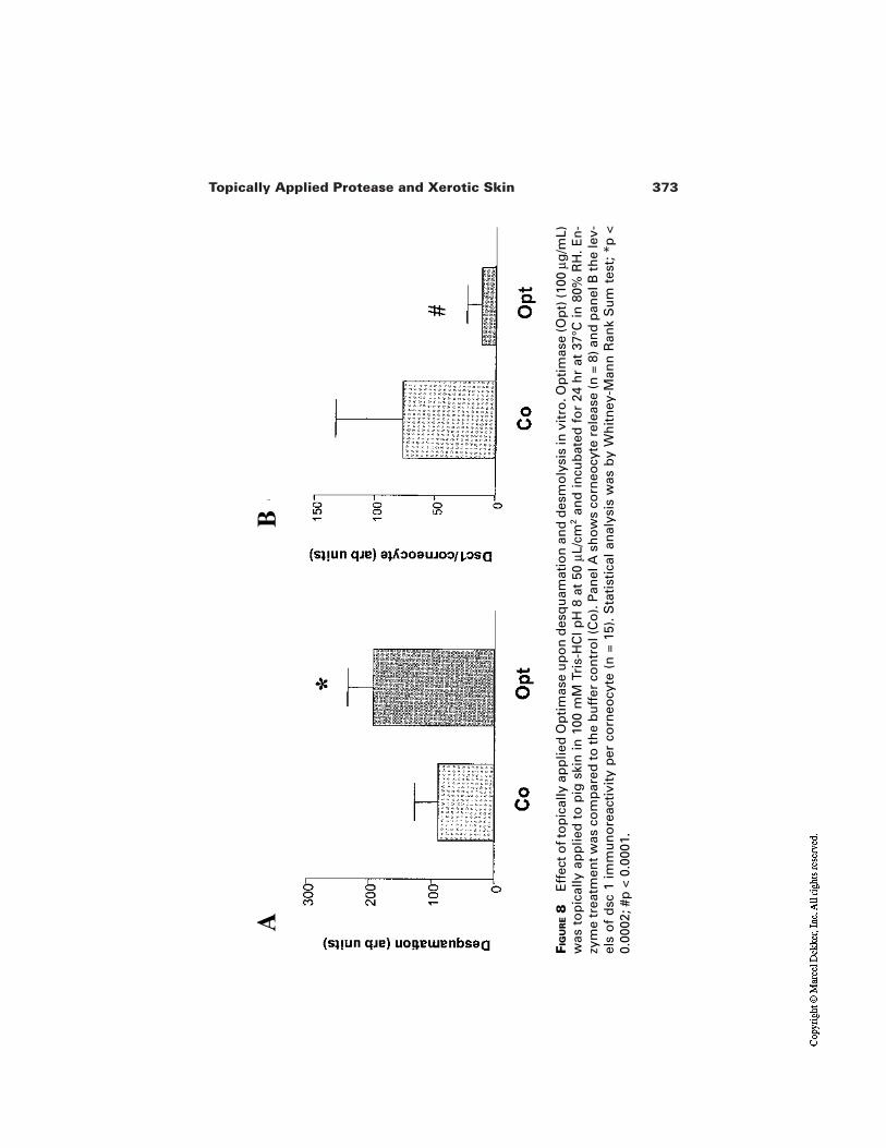
373Topically Applied Protease and Xerotic Skin
FIG
UR
E8
Eff
ect o
f to
pic
ally
ap
plie
d O
pti
mas
e u
po
n d
esq
uam
atio
n a
nd
des
mo
lysi
s in
vit
ro. O
pti
mas
e (O
pt)
(100
µg
/mL)
was
to
pic
ally
ap
plie
d t
o p
ig s
kin
in 1
00 m
M T
ris-
HC
l pH
8 a
t 50
µL/
cm2
and
incu
bat
ed f
or
24 h
r at
37°
C in
80%
RH
. En
-zy
me
trea
tmen
t was
co
mp
ared
to th
e b
uff
er c
on
tro
l (C
o).
Pan
el A
sh
ow
s co
rneo
cyte
rel
ease
(n =
8) a
nd
pan
el B
the
lev-
els
of
dsc
1 i
mm
un
ore
acti
vity
per
co
rneo
cyte
(n
= 1
5).
Sta
tist
ical
an
alys
is w
as b
y W
hit
ney
-Man
n R
ank
Su
m t
est;
*p
<0.
0002
; #p
< 0
.000
1.
BA
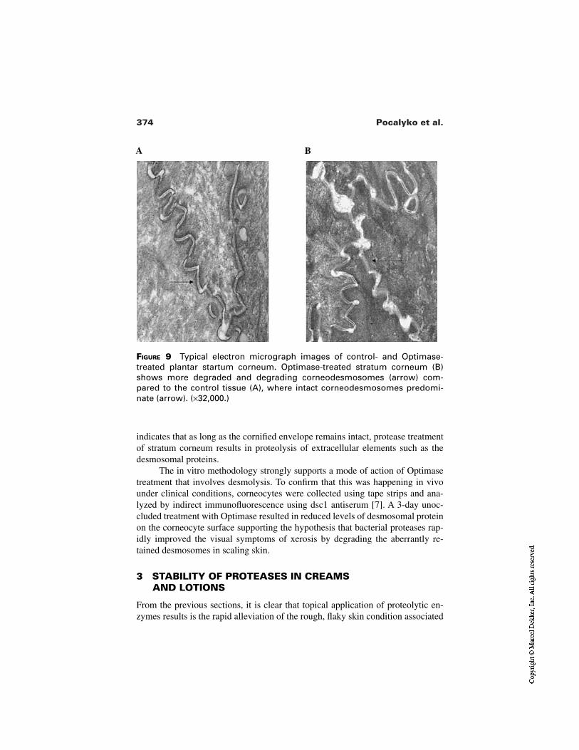
374 Pocalyko et al.
FIGURE 9 Typical electron micrograph images of control- and Optimase-treated plantar startum corneum. Optimase-treated stratum corneum (B)shows more degraded and degrading corneodesmosomes (arrow) com-pared to the control tissue (A), where intact corneodesmosomes predomi-nate (arrow). (×32,000.)
A B
indicates that as long as the cornified envelope remains intact, protease treatmentof stratum corneum results in proteolysis of extracellular elements such as thedesmosomal proteins.
The in vitro methodology strongly supports a mode of action of Optimasetreatment that involves desmolysis. To confirm that this was happening in vivounder clinical conditions, corneocytes were collected using tape strips and ana-lyzed by indirect immunofluorescence using dsc1 antiserum [7]. A 3-day unoc-cluded treatment with Optimase resulted in reduced levels of desmosomal proteinon the corneocyte surface supporting the hypothesis that bacterial proteases rap-idly improved the visual symptoms of xerosis by degrading the aberrantly re-tained desmosomes in scaling skin.
3 STABILITY OF PROTEASES IN CREAMS AND LOTIONS
From the previous sections, it is clear that topical application of proteolytic en-zymes results is the rapid alleviation of the rough, flaky skin condition associated
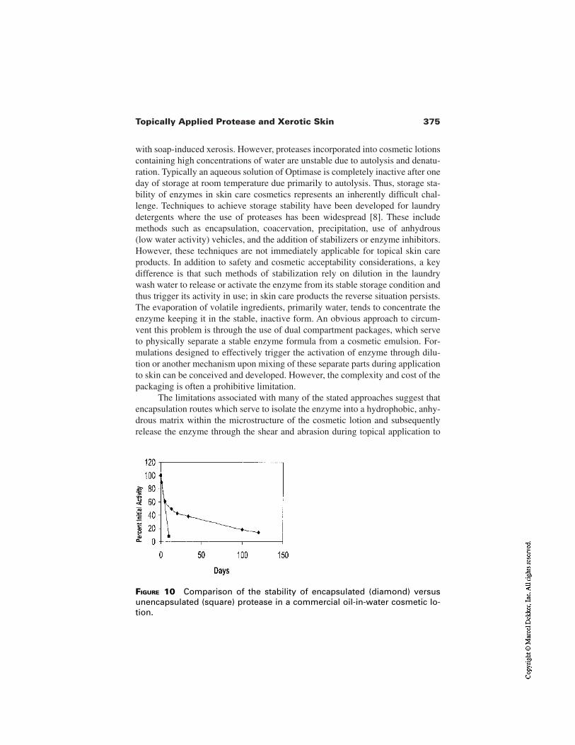
375Topically Applied Protease and Xerotic Skin
with soap-induced xerosis. However, proteases incorporated into cosmetic lotionscontaining high concentrations of water are unstable due to autolysis and denatu-ration. Typically an aqueous solution of Optimase is completely inactive after oneday of storage at room temperature due primarily to autolysis. Thus, storage sta-bility of enzymes in skin care cosmetics represents an inherently difficult chal-lenge. Techniques to achieve storage stability have been developed for laundrydetergents where the use of proteases has been widespread [8]. These includemethods such as encapsulation, coacervation, precipitation, use of anhydrous(low water activity) vehicles, and the addition of stabilizers or enzyme inhibitors.However, these techniques are not immediately applicable for topical skin careproducts. In addition to safety and cosmetic acceptability considerations, a keydifference is that such methods of stabilization rely on dilution in the laundrywash water to release or activate the enzyme from its stable storage condition andthus trigger its activity in use; in skin care products the reverse situation persists.The evaporation of volatile ingredients, primarily water, tends to concentrate theenzyme keeping it in the stable, inactive form. An obvious approach to circum-vent this problem is through the use of dual compartment packages, which serveto physically separate a stable enzyme formula from a cosmetic emulsion. For-mulations designed to effectively trigger the activation of enzyme through dilu-tion or another mechanism upon mixing of these separate parts during applicationto skin can be conceived and developed. However, the complexity and cost of thepackaging is often a prohibitive limitation.
The limitations associated with many of the stated approaches suggest thatencapsulation routes which serve to isolate the enzyme into a hydrophobic, anhy-drous matrix within the microstructure of the cosmetic lotion and subsequentlyrelease the enzyme through the shear and abrasion during topical application to
FIGURE 10 Comparison of the stability of encapsulated (diamond) versusunencapsulated (square) protease in a commercial oil-in-water cosmetic lo-tion.
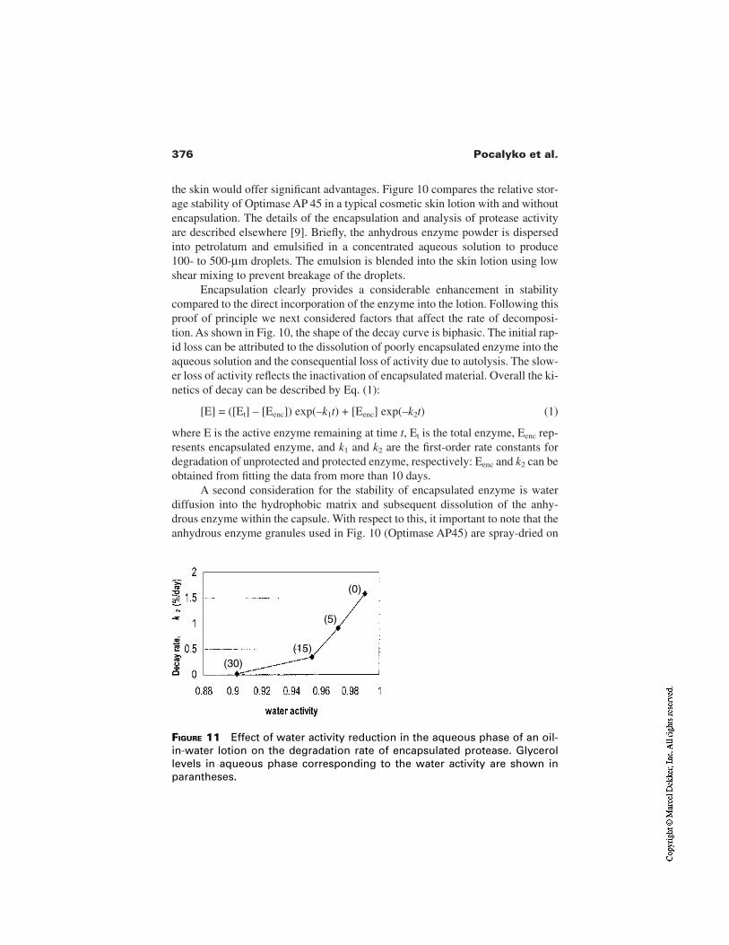
376 Pocalyko et al.
the skin would offer significant advantages. Figure 10 compares the relative stor-age stability of Optimase AP 45 in a typical cosmetic skin lotion with and withoutencapsulation. The details of the encapsulation and analysis of protease activityare described elsewhere [9]. Briefly, the anhydrous enzyme powder is dispersedinto petrolatum and emulsified in a concentrated aqueous solution to produce100- to 500-µm droplets. The emulsion is blended into the skin lotion using lowshear mixing to prevent breakage of the droplets.
Encapsulation clearly provides a considerable enhancement in stabilitycompared to the direct incorporation of the enzyme into the lotion. Following thisproof of principle we next considered factors that affect the rate of decomposi-tion. As shown in Fig. 10, the shape of the decay curve is biphasic. The initial rap-id loss can be attributed to the dissolution of poorly encapsulated enzyme into theaqueous solution and the consequential loss of activity due to autolysis. The slow-er loss of activity reflects the inactivation of encapsulated material. Overall the ki-netics of decay can be described by Eq. (1):
[E] = ([Et] – [Eenc]) exp(–k1t) + [Eenc] exp(–k2t) (1)
where E is the active enzyme remaining at time t, Et is the total enzyme, Eenc rep-resents encapsulated enzyme, and k1 and k2 are the first-order rate constants fordegradation of unprotected and protected enzyme, respectively: Eenc and k2 can beobtained from fitting the data from more than 10 days.
A second consideration for the stability of encapsulated enzyme is waterdiffusion into the hydrophobic matrix and subsequent dissolution of the anhy-drous enzyme within the capsule. With respect to this, it important to note that theanhydrous enzyme granules used in Fig. 10 (Optimase AP45) are spray-dried on
FIGURE 11 Effect of water activity reduction in the aqueous phase of an oil-in-water lotion on the degradation rate of encapsulated protease. Glycerollevels in aqueous phase corresponding to the water activity are shown inparantheses.
(30)(15)
(5)
(0)
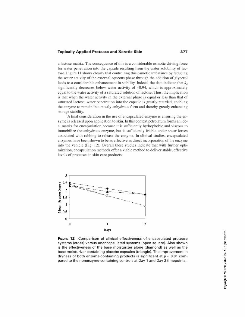
377Topically Applied Protease and Xerotic Skin
a lactose matrix. The consequence of this is a considerable osmotic driving forcefor water penetration into the capsule resulting from the water solubility of lac-tose. Figure 11 shows clearly that controlling this osmotic imbalance by reducingthe water activity of the external aqueous phase through the addition of glycerolleads to a considerable enhancement in stability. Indeed, the data indicate that k2
significantly decreases below water activity of ∼0.94, which is approximatelyequal to the water activity of a saturated solution of lactose. Thus, the implicationis that when the water activity in the external phase is equal or less than that ofsaturated lactose, water penetration into the capsule is greatly retarded, enablingthe enzyme to remain in a mostly anhydrous form and thereby greatly enhancingstorage stability.
A final consideration in the use of encapsulated enzyme is ensuring the en-zyme is released upon application to skin. In this context petrolatum forms an ide-al matrix for encapsulation because it is sufficiently hydrophobic and viscous toimmobilize the anhydrous enzyme, but is sufficiently friable under shear forcesassociated with rubbing to release the enzyme. In clinical studies, encapsulatedenzymes have been shown to be as effective as direct incorporation of the enzymeinto the vehicle (Fig. 12). Overall these studies indicate that with further opti-mization, encapsulation methods offer a viable method to deliver stable, effectivelevels of proteases in skin care products.
FIGURE 12 Comparison of clinical effectiveness of encapsulated proteasesystems (cross) versus unencapsulated systems (open square). Also shownis the effectiveness of the base moisturizer alone (diamond) as well as thebase moisturizer containing placebo capsules (triangle). The improvement indryness of both enzyme-containing products is significant at p < 0.01 com-pared to the nonenzyme-containing controls at Day 1 and Day 2 timepoints.
378 Pocalyko et al.
4 SAFETY ISSUES ASSOCIATED WITH THE USE OFENZYMES IN SKIN CARE PRODUCTS
4.1 Potential Health Effects Associated with the Useof Enzymes
The primary toxicological hazard presented by the use of enzymes in any appli-cation is that they are potentially allergenic. The capacity of proteins, includingenzymes, to cause sensitisation of the respiratory tract and thus to have the poten-tial to induce asthma is well known [10–13], and enzymes are recognized occu-pational allergens [14]. Respiratory sensitization is an immediate IgE anti-body–mediated Type I hypersensitivity response regulated by TH2 lymphocytesand associated cytokines [15,16]. The principal safety concern for the use of en-zymes in any application is therefore the potential for the induction of Type I sen-sitization and most particularly respiratory sensitization.
Another concern with the use of enzymes is the possibility of direct reac-tions when enzymes are topically applied to the skin. Skin sensitization, referredto clinically as allergic contact dermatitis, is a cell-mediated Type IV delayed hy-persensitivity reaction. The delayed contact hypersensitivity reaction is immuno-logically based and is dependent on TH1 lymphocytes [15,16]. To behave as askin sensitizer a substance must first penetrate the stratum corneum, partition intothe epidermis, and react with endogenous proteins to form a hapten–carrier con-jugate. Such substances will therefore normally be of low molecular weight (nor-mally <400 D), e.g., metals (nickel), plant extracts [the poison ivy/oak family(pentadecyl catechols)], dyes [17–19]. Proteins (and thus enzymes) are not impli-cated as a cause of delayed contact hypersensitivity and this is supported by theexperience in the detergent industry where there have been no reported cases ofdelayed contact hypersensitivity due to enzymes. However, there are other typesof dermatitis in man, including irritant contact dermatitis (ICD) [20]. While pro-teins are not commonly associated with ICD, this can occur with proteolytic en-zymes, presumably as a direct consequence of barrier damage [21], and this maybe a factor in the accommodation of skin barrier penetration in the much lesscommon protein contact dermatitis. Protein contact dermatitis is a type of contacturticaria [22]. Although the majority of contact urticaria is nonimmunological, asmall proportion is caused by a Type I hypersensitivity reaction mediated by spe-cific IgE antibody [23]. Typically immunological protein contact urticaria is ex-pressed on skin where the barrier function has been compromised through wetwork and contact with surfactants. This condition was recognized largely in theoccupational context of food preparation—common causes are (shell) fish andvegetables—and has a substantial skin irritation component [24]. However, it isnot known whether repeated contact of foreign protein with intact skin will leadto the formation of specific IgE antibody. Although occupational protein contactdermatitis is not uncommon, the frequency of reported cases caused by enzymesis limited relative to exposure [25,26]. While in these cases the reaction is elicit-
379Topically Applied Protease and Xerotic Skin
ed via the skin, it is not possible to exclude the possibility that sensitization wasinduced via another route. Exposure to foreign proteins (enzymes) can occur viaseveral routes: respiratory, mucosal, and skin (particularly if damaged/compro-mised, e.g., in eczema) and although the role of dermal contact in the induction ofType I sensitization is unclear, immunological protein contact urticaria can occurin a sensitized individual after skin exposure. The route of exposure which in-duces the production of specific IgE antibody may not be the same as that bywhich a clinical response is elicited. In the case of enzymes the most likely routeof primary sensitization is via the respiratory tract.
Although enzymes are recognized as potential occupational respiratory al-lergens [10,14], consumers have used them safely in laundry detergent productsfor many years. With the exception of a few isolated cases of allergic reactions inthe consumer population when enzymes were first introduced into products andwere extremely dusty, there have to date been no reported cases of the inductionof Type I hypersensitivity. This is most probably due to the extremely low levelsof enzyme exposure during consumer use of enzyme-containing laundry deter-gent products (estimated as <0.067 ng/m3) [27,28]. However, there have been re-ported incidences in the consumer population of allergic reactions to enzymeswhen used in other applications. Allergic reactions to papain have been reporteddue to its use in contact lens cleaning solution [29,30] and cosmetics [31]. Thehistory of the safe use of enzymes in laundry products has prompted the con-sideration of the use of these enzymes in other applications, including personalproducts. Given the recognized potential of enzymes as occupational respiratoryallergens and the reported cases of allergy from other consumer uses of enzyme-containing products, a careful risk assessment of any new use scenario is vital.The main concern is possibility inducing Type I hypersensitivity, primarily respi-ratory sensitization, because the fate of the enzyme following use in topical orpersonal cleansing applications is uncertain. The potential for inhalation of air-borne or deposited enzyme in the home of the consumer, due to loss from the skinsurface following desquamation or by aerosolization during washing as well asabsorption via mucosal and dermal exposure, is therefore a major concern. Thepossible risks of the induction of Type I hypersensitivity reactions associated withthe use of enzymes in leave-on products have therefore been assessed in severalstudies.
4.2 Safety Studies on the Use of Enzymes inPersonal Care Products
A study to assess the levels of aerosolized enzyme generated during showeringusing an enzyme-containing personal cleansing product was carried out and thiswas followed by a 6-month pilot use test with concurrent monitoring for the de-velopment of specific IgE antibody [32]. Levels of airborne enzyme were moni-tored during showering using prototype soap bars containing different concentra-
380 Pocalyko et al.
tions (0.2, 0.4, and 2.8%) of a Bacillus subtilis–derived protease. The levels of en-zyme measured in the shower cubicle increased proportionately with the concen-tration of enzyme in the product (mean values 11.4, 15.7, and 183 ng/m3). Re-peated experiments using the 0.2% level confirmed the measurements (meanvalues 5.7–11.8 ng/m3). The enzyme levels in the room outside the shower cubi-cle were also measured and found to be approximately 2.5-fold less than the lev-el in the shower cubicle itself. In the follow-up 6-month use test, volunteers witha reported history of seasonal allergic rhinitis substituted test (with 0.2% enzyme)or control soap bars for their normal products. Prior to the start of the test and attwo monthly intervals, skin prick tests (SPT) using the enzyme and appropriatecontrols were carried out on the volunteers. All SPT analysis was negative up tothe 4-month assessment point. However, after 6 months 4 out of the 61 individu-als in the test group gave a positive SPT response to the enzyme, and all four pos-itive results were confirmed by the identification of specific IgE antibody usingserological analysis. Although none of these individuals reported clinical symp-toms, the potential to develop symptoms with extended use cannot be ruled out.
When used in the manufacture of laundry detergent products, occupationalairborne exposure levels for the Bacillus subtilis–derived protease used in thesoap bar study (and other enzymes of similar antigenic potency) have been limit-ed to 15–20 ng/m3. In this controlled occupational environment, data show thatclinical symptoms are prevented and the rate of sensitization is minimized[33,34]. The assumption was therefore that because the frequency, duration, andmagnitude of the exposure to enzyme from the soap bar application was muchless than that observed in the occupational environment, the use would be safe.However, the induction of specific IgE antibody in several of the study partici-pants emphasizes the need for caution when developing a risk assessment for anyproposed new enzyme use application. The possibility that Type I hypersensitivi-ty can be induced through routes other than via inhalation, e.g., dermal or mucos-al, is uncertain. As discussed previously, it is known that dermal contact with pro-teins (e.g., latex proteins) [35] can elicit IgE antibody–mediated allergic reactionsthus confirming the ability of proteins to penetrate the skin barrier under certainconditions. Although the primary route of induction is thought to be via the respi-ratory tract, the mechanisms are not fully understood, and the dermal and mucos-al routes may be contributory. Other studies, including the assessment of enzyme-containing laundry bars for hand laundry and personal cleansing over a 6- to18-month period with no induction of IgE antibody [36,37], indicate that it is un-likely that the dermal route alone was responsible for the sensitizations observedin the soap bar study.
Further studies have investigated the use of enzymes in topically appliedleave-on products and have confirmed the potential for the generation of inhal-able enzyme. The important consideration here was not skin penetration, butrather the fate of the enzyme lost from the skin surface. In a study to assess theairborne levels of enzyme generated following the use of a topically applied skin
381Topically Applied Protease and Xerotic Skin
cream, groups of five volunteers applied a skin cream with (∼0.03%) or without a Bacillus-derived proteolytic enzyme twice daily for 12 weeks [38]. At 2- to 4-week intervals during the 12-week use phase and for 6 weeks after use, the pan-ellists’ bedroom air was sampled before and after vacuuming and changing thebed linen. No airborne enzyme was detected before vacuuming and changing oflinen in any of the groups. No enzyme was detected in the control group follow-ing vacuuming and changing the bed linen; however, enzyme was detected on anumber of occasions in the low-dose (0.5 g cream/application) group but with noobvious pattern. In the high-dose group (3 g cream/application) there was withcontinued exposure clear evidence of a trend to increasing airborne enzyme levels(up to 29 ng/m3) that dropped back to nondetectable levels 6 weeks after the ap-plications were discontinued. Although no specific IgE antibody was detected inany of the small number of individuals participating in this study, this is mostprobably due to the relatively short duration of the exposure period. Specific IgEantibody is usually induced after 6–12 months of exposure, as illustrated in thesoap bar study [32], the papain in contact lens solution case [29], and the exten-sive experience from occupational exposure to enzymes within the detergent in-dustry. The possibility of the induction of a specific IgE antibody response withprolonged exposure should not be ignored.
The potential for the deposition of enzyme on the skin and aerosolizationafter showering following the use of an enzyme-containing leave-on productwere investigated in another study [39]. This study demonstrated that immuno-re-active enzyme could remain on the skin (100–400 ng/cm2) for at least 24 hr fol-lowing application of a leave-on vehicle containing less than 0.02% of a prote-olytic enzyme. The study also showed that the “reservoir” of residual enzyme onthe skin could become aerosolized during showering. The leave-on vehicle con-taining <0.02% enzyme was applied by six volunteers to the whole body and af-ter 12 hr the levels of airborne enzyme were measured during showering, and lev-els of up to 1.13 ng/m3 were detected.
4.3 Potential for the Future Use of Enzymes inPersonal Care Products
While the exposure patterns to enzymes in the occupational environment are verydifferent to those arising from the consumer use of enzyme-containing personalproducts, the levels of airborne enzyme demonstrated in various studies[32,38,39] are orders of magnitude greater than estimated in the consumer use oflaundry detergent products [28]. This gives cause for concern and means that arisk assessment for topically applied or personal wash products containing en-zyme must include as a primary consideration the potential for respiratory sensi-tization arising from the inhalation route of exposure. In all the cases presentedhere, the risk assessment conclusion would be that the proposed use of the en-zyme would be unacceptable in the absence of further detailed evaluation.
382 Pocalyko et al.
The conduct of thorough risk assessments, including detailed evaluation ofpotential sources and routes of exposure, when developing safety evaluations forenzymes in new applications is paramount. As part of this process, it is importantto understand the levels of exposure which might induce an immunological (spe-cific IgE antibody) response under normal use conditions, i.e., it is necessary tounderstand something of the dose response relationship for the enzyme/applica-tion in question. Although evidence indicates clinical skin problems associatedwith the use of enzymes are limited, any risk assessment should also include anassessment of the possible effects of repeated skin exposure to enzyme onchanges in skin structure and function. Enzymes are implicated in irritant der-matitis, and compromised skin is recognized as a critical factor in protein contacturticaria and may also be a factor in the induction of specific IgE antibody andthus potentially respiratory sensitization. Appropriately designed clinical evalua-tions are therefore required. The duration of the clinical study is critical, with thepotential development of an IgE antibody response requiring 6–24 months. Prop-er statistical analysis must be carried out to ensure the calculation of any risk inthe consumer population. Postmarket surveillance should also be considered fol-lowing any introduction of a new enzyme-containing product, and all complaintsshould be carefully evaluated, including a medical assessment if required.
5 SUMMARY
Considerable evidence has been presented in this chapter that supports the hy-pothesis that a reduction in proteolytic activity within the stratum corneum and asubsequent retention of desmosomes in the upper layers of stratum corneum con-tribute to the visible scaling associated with xerotic skin. Topical application ofprotease has been shown to rapidly reduce this scaling leading to a significant re-duction in the visible signs of dryness. From our research, broad specificity pro-teases appear to be most efficient at producing this effect. The benefit of proteasecan be achieved from a conventional moisturizer, and routes to stabilize the en-zyme using encapsulation can be envisioned. Concerns regarding the safety ofsuch products have been investigated and indicate that until the technologies ex-ist to address the safety of enzymes, for example, modification to reduce the in-herent allergenicity, they are probably not appropriate for use in topical or per-sonal cleansing products. Consequently, further research will be required tocommercialize this promising technology.
REFERENCES
1. Rawlings AV, Watkinson A, Rogers J, Mayo A, Scott IR. Abnormalities in stratumcorneum structure, lipid composition and desmosome degradation in soap-inducedwinter xerosis. J Soc Cosmet Chem 1994; 45:203–220.
383Topically Applied Protease and Xerotic Skin
2. Watkinson A, Smith C, Coan P, Wiedow O. The role of pro-SCCE and SCCE indesquamation. 21st IFSCC Congress, Berlin, 2000.
3. Harding CR, Watkinson A, Rawlings AV, Scott IR. Dry skin, moisturization and cor-neodesmolysis. Int J Cosmet Sci 2000; 22:21–52.
4. Watkinson A, Rogers JS, Harding CR. Water activity: the critical factor controllingSCCE and desquamation J Invest Dermatol 1999; 112:573.
5. Froebe CL, Simion FA, Ohlemeyer H, Rhein LD, Mattai J, Cagan RH, Friberg SE.Prevention of stratum corneum lipid phase transition by glycerol—an alternativemechanism for skin moisturisation. J Soc Cosmet Chem 1990; 41:51–65.
6. Long S, Banks J, Watkinson A, Harding C, Rawlings AV. Desmocollin 1: a keymarker for desmosome processing in the stratum corneum. J Invest Dermatol 1996;106:397.
7. El-Kadi KN, Rawlings AV, Feinberg C, Watkinson A, Nunn CC, Battaglia A, Chan-dar P, Richardson N, Pocalyko DJ. Broad specificity alkaline proteases efficiently re-duce the visual scaling associated with soap-induced winter xerosis. Arch DermatolRes (in press).
8. Hawkins J, Chadwick P, Messenger ET. Method for preparing stabilized enzyme dis-persion. US patent 5,198,353.
9. Chandar P, Richardson NK, Battaglia A, Cicciari KJ, El-Kadi KN. Oil-in-water cos-metic emulsions containing stabilized protease. US patent 5,811,112.
10. Flindt MLH. Pulmonary disease due to inhalation of derivatives of Bacillus subtiliscontaining enzyme. Lancet 1969; 1:1177–1181.
11. Venables KM, Tee RD, Hawkins ER, Gordon DJ, Wale CJ, Farrer NM Lam TH,Baxter PJ, Taylor AJN. Laboratory animal allergy in a pharmaceutical company. Br JIndustr Med 1988; 45:660–666.
12. Douglas JDM, McSharry C, Blaikie L, Morrow T, Miles S, Franklin D. Occupation-al asthma caused by automated salmon processing. Lancet 1995; 346:737–740.
13. Quirce S, Diéz-Goméz ML, Eiras P, Cuevas M, Baz G, Losada E. Inhalant allergy toegg yolk and egg white proteins. Clin Exper Allergy 1998; 28:478–485.
14. Pepys J, Haregreave FE, Longbottom JL, Faux JA. Allergic reactions of the lungs toenzymes of Bacillus subtilis. Lancet 1969; 1:1181–1184.
15. Dearman RJ, Basketter DA, Coleman JW, Kimber I. The cellular and molecular ba-sis for divergent allergic responses to chemicals. Chem-Biol Interactions 1992;84:1–10.
16. Kimber I. Cytokines and regulation of allergic sensitisation to chemicals. Toxicology1994; 93:1–11.
17. Cronin E. Contact Dermatitis. New York: Churchill Livingstone, 1980.18. Fisher AA. Contact Dermatitis. Philidelphia: Lea and Febiger, 1986.19. Rycroft RJG, Menne T, Frosch PJ, Benezra C. Textbook of Contact Dermatitis.
Berlin: Springer-Verlag, 1992.20. van der Valk PGM, Maibach HI. The Irritant Contact Dermatitis Syndrome. Boca
Raton: CRC Press, 1996.21. Zachariae H, Thomsen K, Rasmussen OG. Occupational contact dermatitis. Acta
Dermatovener 1973; 53:145–148.22. Hjorth N, Roed-Petersen J. Occupational contact dermatitis in food handlers. Con-
tact Dermatitis 1976; 2:28–42.
384 Pocalyko et al.
23. Shafer T, Ring J. Epidemiology of contact urticaria. In: Burr ML, ed. Epidemiologyof Contact Allergy. Vol. 31. Basal: Karger Press, 1993:49.
24. Cronin E. Dermatitis of the hands in caterers. Contact Dermatitis 1987; 17:265–269.25. Kanerva L, Vanhanen M, Tupasela O. Occupational contact urticaria from cellulase
enzyme. Contact Dermatitis 1998; 38:176–177.26. Kanerva L, Vanhanen M. Occupational protein contact dermatitis from glucosamy-
lase. Contact Dermatitis 1999; 41:171–173.27. Belin L, Hoborn J, Falsen E, Andre J. Enzyme sensitisation in consumers of enzyme-
containing washing powder. Lancet 1970; 2:1153–1157.28. Hendricks MH. Measurement of laundry product dust levels and characteristics in
consumer use. J Am Oil Chem Soc 1970; 47:207–211.29. Berbstein DI, Gallagher JS, Grad M, Bernstein IL. Local ocular anaphylaxis to pa-
pain enzyme contained in a contact lens cleansing solution. J Allergy Clin Immunol1984; 74:258–260.
30. Fisher AA. Allergic reactions to contact lens solutions. Cutis 1985; 36:209–211.31. Niinimaki A, Reijula K, Pirila T, Koistinen AM. Papain-induced rhinoconjunctivitis
in a cosmetologist. J Allergy Clin Immunol 1993; 92:492–493.32. Kelling CK, Bartolo RG, Ertel KD, Smith LA, Watson DD, Sarlo K. Safety assess-
ment of enzyme-containing personal cleansing products: exposure characterisationand development of IgE antibody to enzymes after a 6-month use test. J Allergy ClinImmunol 1998; 101:179–187.
33. Juniper CP, Roberts DM. Enzyme asthma: fourteen years clinical experience of a re-cently prescribed disease. J Soc Occup Med 1984; 34:127–132.
34. Gaines WG. Occupational health experience manufacturing multiple enzymes deter-gents and methods to control enzyme exposures. The Toxicology Forum 1994;143–147.
35. Hamann CP. Natural rubber latex protein sensitivity in review. Am J Contact Derm1993; 4:4–21.
36. Sarlo K., Cormier E, MacKenzie D, Scott L. Lack of Type-I sensitisation to laundryenzymes among consumers in the Phillipines: results of an 18-month clinical study.J Allergy Clin Immunol 1996; 97(1):749.
37. Cormier E, Sarlo K, Scott L, Vasunia K, Smith M, Payne N, MacKenzie D. Lack ofsensitisation to laundry enzymes among consumers in the Phillipines. J Allergy ClinImmunol 1997; 99(1):321.
38. Blaikie L, Richold M, Whittle E, Lawrence RS, Keech S, Basketter DA. Airborneexposure from topically applied protein (proteolytic enzyme). Hum Exp Toxicol1999; 18:528.
39. Johnson G, Innis JD, Mills KJ, Bielen F, Date RF, Weisgerber D, Sarlo K. Safety as-sessment for a leave-on personal care product containing a protease enzyme. HumExp Toxicol 1999; 18:527.
19Enzymes in Cleansers
Takuji MasunagaKosé Corporation, Tokyo, Japan
1 INTRODUCTION
The essential function of skin care cosmetics is to maintain beautiful and healthyskin. Skin care cosmetics, including creams, lotions, facial packs, cleansers, andastringents, have specific purposes and effects. The main purpose of a skincleanser is to clean the skin surface by removing dirt to promote beautiful andhealthy skin. Any dirt remaining on the skin surface or plugged in the piloseba-ceous orifices is easily oxidized or degraded by oxygen and microorganisms,leading to skin trouble, including inflammation and acne vulgaris.
Dirt on the skin surface consists of sloughed corneocytes, sebum, sweatremnants, products from bacteria on the skin surface, and environmental pollu-tants. Keratin is a main component of corneocytes, which are continuallysloughed from the skin surface. Stratum corneum is formed by terminal differen-tiation of epidermal keratinocytes, and both epidermal proliferation and kera-tinization increase in response to daily life ultraviolet exposure [1] and dry envi-ronmental conditions [2]. As a result of inappropriate keratinization, the skintends to become rough or dry and can lead to scarring. Sebum is excreted from pi-losebaceous orifices and spreads on the skin surface. The lipid on the skin surfaceis easily oxidized and converted to lipid peroxidation. Such peroxidized lipid cancause skin irritation, leading to skin damage. Acne vulgaris can result from sebumbecoming plugged in the pilosebaceous orifices. Certain kinds of bacterial prod-
385
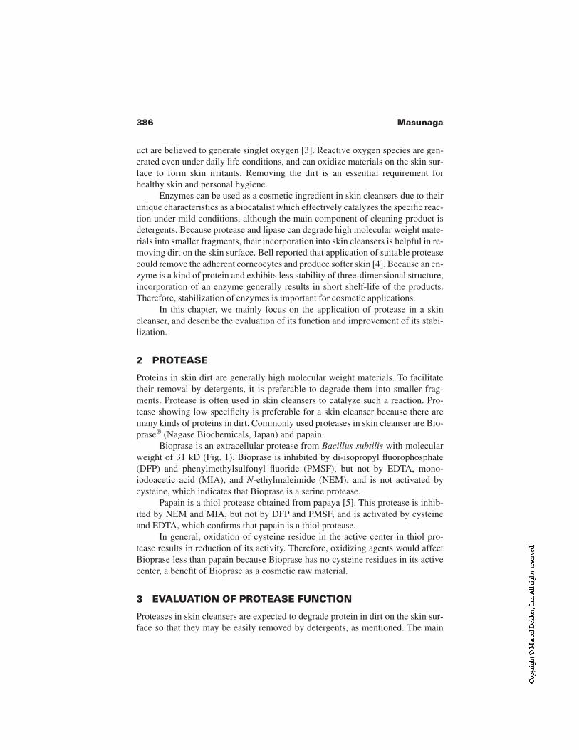
386 Masunaga
uct are believed to generate singlet oxygen [3]. Reactive oxygen species are gen-erated even under daily life conditions, and can oxidize materials on the skin sur-face to form skin irritants. Removing the dirt is an essential requirement forhealthy skin and personal hygiene.
Enzymes can be used as a cosmetic ingredient in skin cleansers due to theirunique characteristics as a biocatalist which effectively catalyzes the specific reac-tion under mild conditions, although the main component of cleaning product isdetergents. Because protease and lipase can degrade high molecular weight mate-rials into smaller fragments, their incorporation into skin cleansers is helpful in re-moving dirt on the skin surface. Bell reported that application of suitable proteasecould remove the adherent corneocytes and produce softer skin [4]. Because an en-zyme is a kind of protein and exhibits less stability of three-dimensional structure,incorporation of an enzyme generally results in short shelf-life of the products.Therefore, stabilization of enzymes is important for cosmetic applications.
In this chapter, we mainly focus on the application of protease in a skincleanser, and describe the evaluation of its function and improvement of its stabi-lization.
2 PROTEASE
Proteins in skin dirt are generally high molecular weight materials. To facilitatetheir removal by detergents, it is preferable to degrade them into smaller frag-ments. Protease is often used in skin cleansers to catalyze such a reaction. Pro-tease showing low specificity is preferable for a skin cleanser because there aremany kinds of proteins in dirt. Commonly used proteases in skin cleanser are Bio-prase® (Nagase Biochemicals, Japan) and papain.
Bioprase is an extracellular protease from Bacillus subtilis with molecularweight of 31 kD (Fig. 1). Bioprase is inhibited by di-isopropyl fluorophosphate(DFP) and phenylmethylsulfonyl fluoride (PMSF), but not by EDTA, mono-iodoacetic acid (MIA), and N-ethylmaleimide (NEM), and is not activated bycysteine, which indicates that Bioprase is a serine protease.
Papain is a thiol protease obtained from papaya [5]. This protease is inhib-ited by NEM and MIA, but not by DFP and PMSF, and is activated by cysteineand EDTA, which confirms that papain is a thiol protease.
In general, oxidation of cysteine residue in the active center in thiol pro-tease results in reduction of its activity. Therefore, oxidizing agents would affectBioprase less than papain because Bioprase has no cysteine residues in its activecenter, a benefit of Bioprase as a cosmetic raw material.
3 EVALUATION OF PROTEASE FUNCTION
Proteases in skin cleansers are expected to degrade protein in dirt on the skin sur-face so that they may be easily removed by detergents, as mentioned. The main
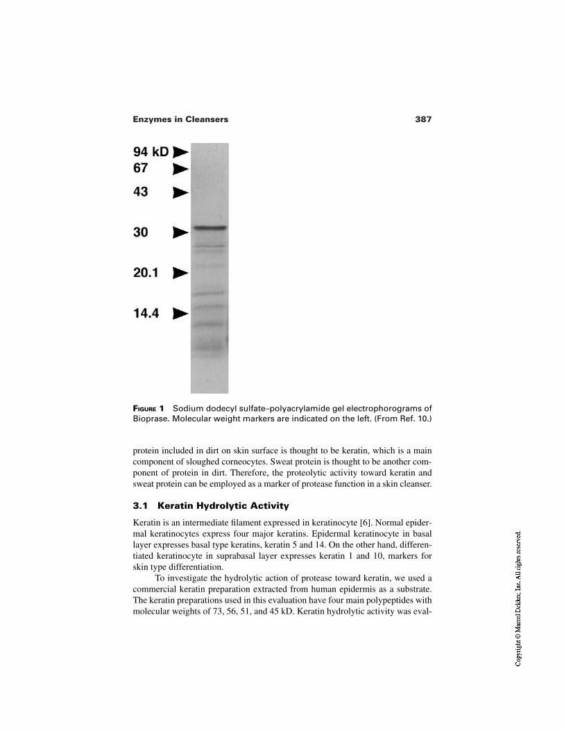
387Enzymes in Cleansers
FIGURE 1 Sodium dodecyl sulfate–polyacrylamide gel electrophorograms ofBioprase. Molecular weight markers are indicated on the left. (From Ref. 10.)
protein included in dirt on skin surface is thought to be keratin, which is a maincomponent of sloughed corneocytes. Sweat protein is thought to be another com-ponent of protein in dirt. Therefore, the proteolytic activity toward keratin andsweat protein can be employed as a marker of protease function in a skin cleanser.
3.1 Keratin Hydrolytic Activity
Keratin is an intermediate filament expressed in keratinocyte [6]. Normal epider-mal keratinocytes express four major keratins. Epidermal keratinocyte in basallayer expresses basal type keratins, keratin 5 and 14. On the other hand, differen-tiated keratinocyte in suprabasal layer expresses keratin 1 and 10, markers forskin type differentiation.
To investigate the hydrolytic action of protease toward keratin, we used acommercial keratin preparation extracted from human epidermis as a substrate.The keratin preparations used in this evaluation have four main polypeptides withmolecular weights of 73, 56, 51, and 45 kD. Keratin hydrolytic activity was eval-
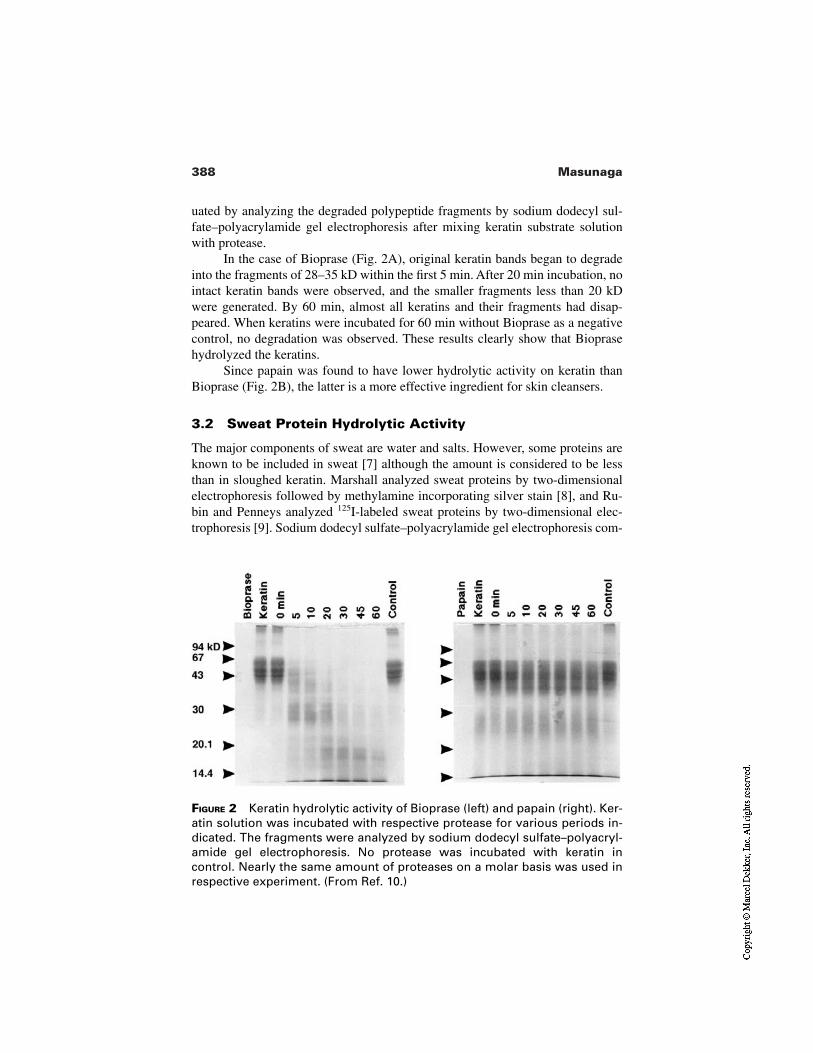
388 Masunaga
FIGURE 2 Keratin hydrolytic activity of Bioprase (left) and papain (right). Ker-atin solution was incubated with respective protease for various periods in-dicated. The fragments were analyzed by sodium dodecyl sulfate–polyacryl-amide gel electrophoresis. No protease was incubated with keratin incontrol. Nearly the same amount of proteases on a molar basis was used inrespective experiment. (From Ref. 10.)
uated by analyzing the degraded polypeptide fragments by sodium dodecyl sul-fate–polyacrylamide gel electrophoresis after mixing keratin substrate solutionwith protease.
In the case of Bioprase (Fig. 2A), original keratin bands began to degradeinto the fragments of 28–35 kD within the first 5 min. After 20 min incubation, nointact keratin bands were observed, and the smaller fragments less than 20 kDwere generated. By 60 min, almost all keratins and their fragments had disap-peared. When keratins were incubated for 60 min without Bioprase as a negativecontrol, no degradation was observed. These results clearly show that Bioprasehydrolyzed the keratins.
Since papain was found to have lower hydrolytic activity on keratin thanBioprase (Fig. 2B), the latter is a more effective ingredient for skin cleansers.
3.2 Sweat Protein Hydrolytic Activity
The major components of sweat are water and salts. However, some proteins areknown to be included in sweat [7] although the amount is considered to be lessthan in sloughed keratin. Marshall analyzed sweat proteins by two-dimensionalelectrophoresis followed by methylamine incorporating silver stain [8], and Ru-bin and Penneys analyzed 125I-labeled sweat proteins by two-dimensional elec-trophoresis [9]. Sodium dodecyl sulfate–polyacrylamide gel electrophoresis com-
389Enzymes in Cleansers
bined with ordinary silver staining technique is a convenient and highly sensitiveway to analyze sweat proteins as a substrate for proteases in skin cleansers. Thesweat proteins can be collected from the facial skin after exercise by using gauze.The details are described elsewhere [10].
As shown in Fig. 3, Bioprase can hydrolyze the sweat proteins of ranges21–26 and 32–41 kD within 60 min. The results indicate that enzyme in skincleanser is effective in removing the dirt originating from sweat proteins.
3.3 Experimental Conditions
In the assay of keratin and sweat protein hydrolytic activity described, the em-ployed incubation time of 60 min is very long compared with the time typicallyspent washing the face. It is not clear how much protein is degraded in actual facewashing. However, complete degradation of the protein in dirt is not necessarybecause detergent, but not protease, is the main component in removing dirt. Theamount of protease in the formula should be decided by considering not onlyfunctionality, but also safety, cost, and so on.
FIGURE 3 Sweat protein hydrolytic activity of Bioprase. Sweat protein wasincubated with Bioprase for 60 min. The fragments were analyzed by sodiumdodecyl sulfate–polyacrylamide gel electrophoresis. The bands were re-vealed by silver staining. (From Ref. 10.)
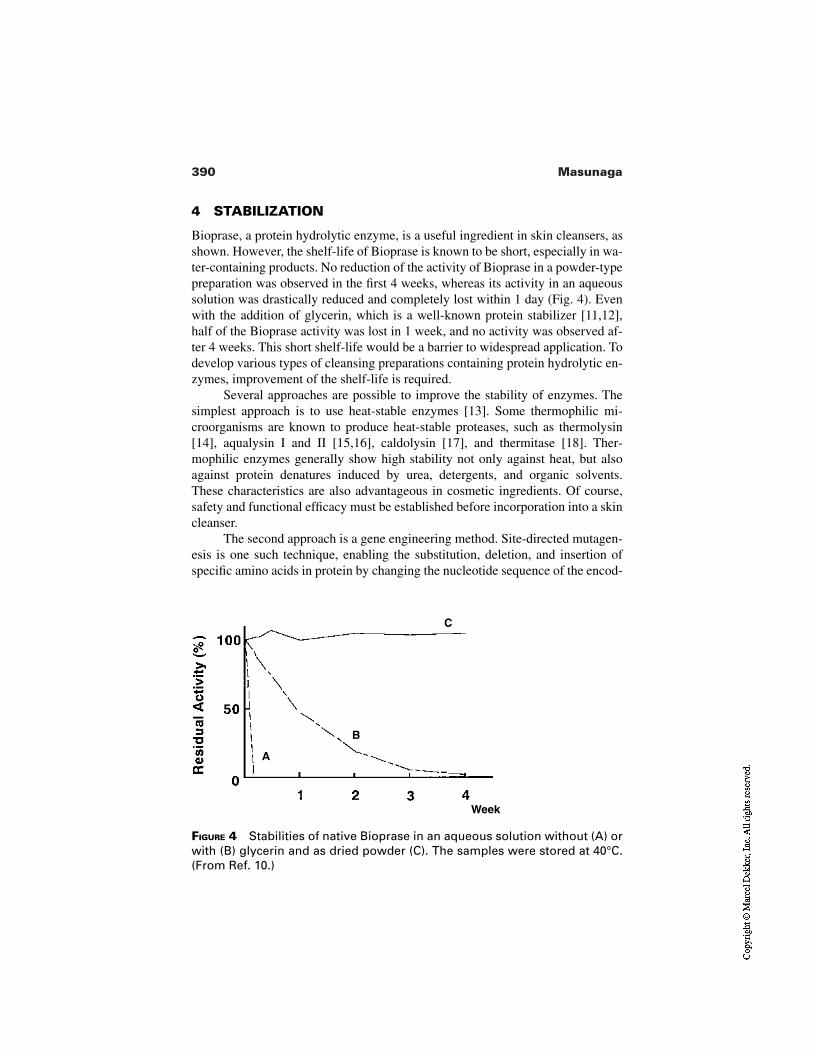
390 Masunaga
FIGURE 4 Stabilities of native Bioprase in an aqueous solution without (A) orwith (B) glycerin and as dried powder (C). The samples were stored at 40°C.(From Ref. 10.)
A
B
C
Week
4 STABILIZATION
Bioprase, a protein hydrolytic enzyme, is a useful ingredient in skin cleansers, asshown. However, the shelf-life of Bioprase is known to be short, especially in wa-ter-containing products. No reduction of the activity of Bioprase in a powder-typepreparation was observed in the first 4 weeks, whereas its activity in an aqueoussolution was drastically reduced and completely lost within 1 day (Fig. 4). Evenwith the addition of glycerin, which is a well-known protein stabilizer [11,12],half of the Bioprase activity was lost in 1 week, and no activity was observed af-ter 4 weeks. This short shelf-life would be a barrier to widespread application. Todevelop various types of cleansing preparations containing protein hydrolytic en-zymes, improvement of the shelf-life is required.
Several approaches are possible to improve the stability of enzymes. Thesimplest approach is to use heat-stable enzymes [13]. Some thermophilic mi-croorganisms are known to produce heat-stable proteases, such as thermolysin[14], aqualysin I and II [15,16], caldolysin [17], and thermitase [18]. Ther-mophilic enzymes generally show high stability not only against heat, but alsoagainst protein denatures induced by urea, detergents, and organic solvents.These characteristics are also advantageous in cosmetic ingredients. Of course,safety and functional efficacy must be established before incorporation into a skincleanser.
The second approach is a gene engineering method. Site-directed mutagen-esis is one such technique, enabling the substitution, deletion, and insertion ofspecific amino acids in protein by changing the nucleotide sequence of the encod-
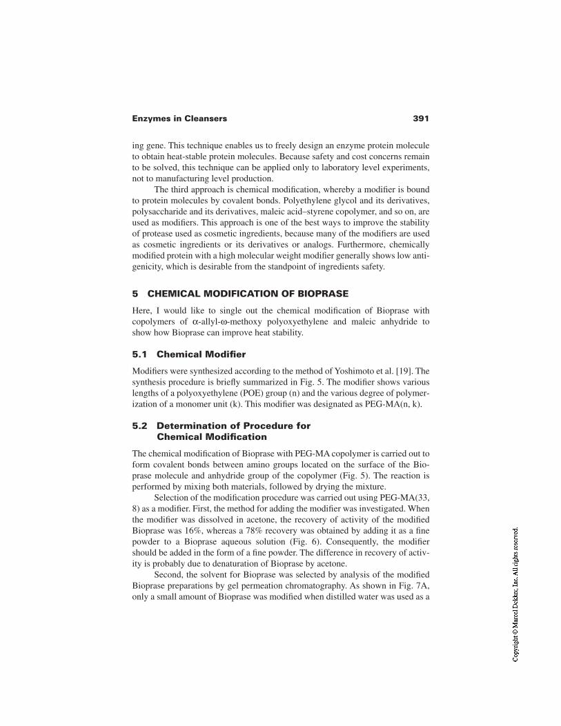
391Enzymes in Cleansers
ing gene. This technique enables us to freely design an enzyme protein moleculeto obtain heat-stable protein molecules. Because safety and cost concerns remainto be solved, this technique can be applied only to laboratory level experiments,not to manufacturing level production.
The third approach is chemical modification, whereby a modifier is boundto protein molecules by covalent bonds. Polyethylene glycol and its derivatives,polysaccharide and its derivatives, maleic acid–styrene copolymer, and so on, areused as modifiers. This approach is one of the best ways to improve the stabilityof protease used as cosmetic ingredients, because many of the modifiers are usedas cosmetic ingredients or its derivatives or analogs. Furthermore, chemicallymodified protein with a high molecular weight modifier generally shows low anti-genicity, which is desirable from the standpoint of ingredients safety.
5 CHEMICAL MODIFICATION OF BIOPRASE
Here, I would like to single out the chemical modification of Bioprase withcopolymers of α-allyl-ω-methoxy polyoxyethylene and maleic anhydride toshow how Bioprase can improve heat stability.
5.1 Chemical Modifier
Modifiers were synthesized according to the method of Yoshimoto et al. [19]. Thesynthesis procedure is briefly summarized in Fig. 5. The modifier shows variouslengths of a polyoxyethylene (POE) group (n) and the various degree of polymer-ization of a monomer unit (k). This modifier was designated as PEG-MA(n, k).
5.2 Determination of Procedure for Chemical Modification
The chemical modification of Bioprase with PEG-MA copolymer is carried out toform covalent bonds between amino groups located on the surface of the Bio-prase molecule and anhydride group of the copolymer (Fig. 5). The reaction isperformed by mixing both materials, followed by drying the mixture.
Selection of the modification procedure was carried out using PEG-MA(33,8) as a modifier. First, the method for adding the modifier was investigated. Whenthe modifier was dissolved in acetone, the recovery of activity of the modifiedBioprase was 16%, whereas a 78% recovery was obtained by adding it as a finepowder to a Bioprase aqueous solution (Fig. 6). Consequently, the modifiershould be added in the form of a fine powder. The difference in recovery of activ-ity is probably due to denaturation of Bioprase by acetone.
Second, the solvent for Bioprase was selected by analysis of the modifiedBioprase preparations by gel permeation chromatography. As shown in Fig. 7A,only a small amount of Bioprase was modified when distilled water was used as a
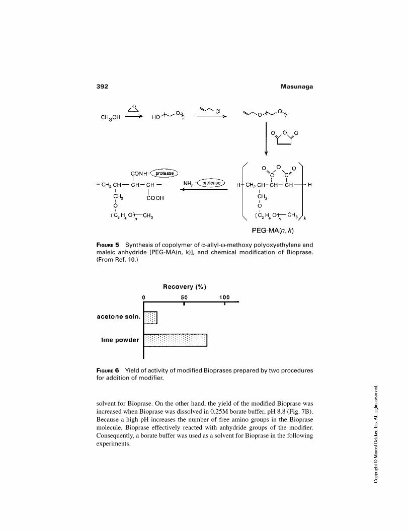
392 Masunaga
FIGURE 5 Synthesis of copolymer of α-allyl-ω-methoxy polyoxyethylene andmaleic anhydride [PEG-MA(n, k)], and chemical modification of Bioprase.(From Ref. 10.)
FIGURE 6 Yield of activity of modified Bioprases prepared by two proceduresfor addition of modifier.
solvent for Bioprase. On the other hand, the yield of the modified Bioprase wasincreased when Bioprase was dissolved in 0.25M borate buffer, pH 8.8 (Fig. 7B).Because a high pH increases the number of free amino groups in the Bioprasemolecule, Bioprase effectively reacted with anhydride groups of the modifier.Consequently, a borate buffer was used as a solvent for Bioprase in the followingexperiments.
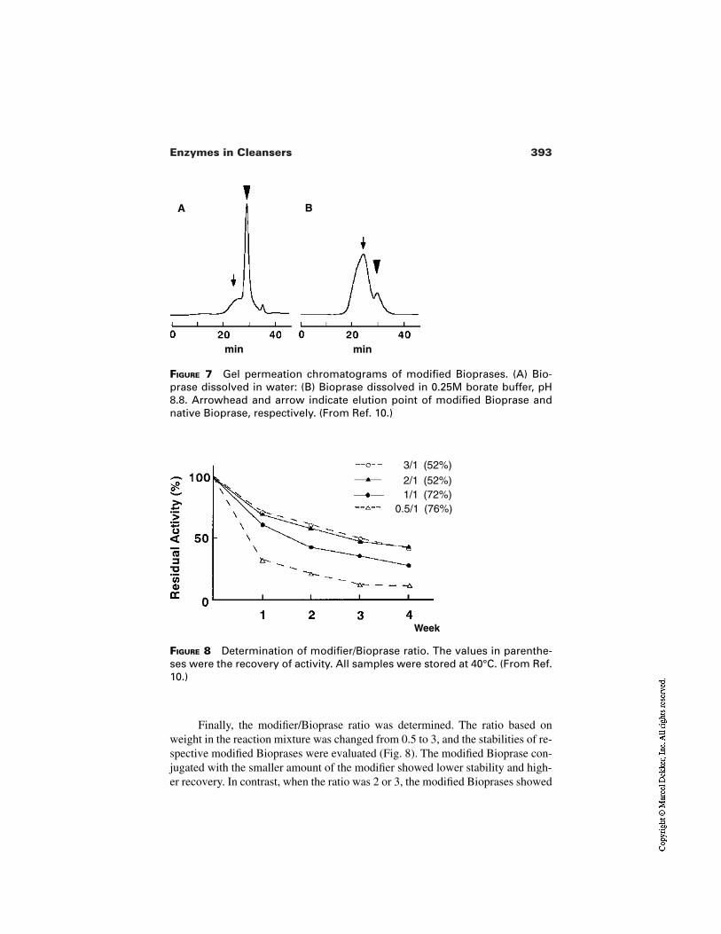
393Enzymes in Cleansers
FIGURE 7 Gel permeation chromatograms of modified Bioprases. (A) Bio-prase dissolved in water: (B) Bioprase dissolved in 0.25M borate buffer, pH8.8. Arrowhead and arrow indicate elution point of modified Bioprase andnative Bioprase, respectively. (From Ref. 10.)
A B
minmin
FIGURE 8 Determination of modifier/Bioprase ratio. The values in parenthe-ses were the recovery of activity. All samples were stored at 40°C. (From Ref.10.)
Week
3/1 (52%)2/1 (52%)1/1 (72%)
0.5/1 (76%)
Finally, the modifier/Bioprase ratio was determined. The ratio based onweight in the reaction mixture was changed from 0.5 to 3, and the stabilities of re-spective modified Bioprases were evaluated (Fig. 8). The modified Bioprase con-jugated with the smaller amount of the modifier showed lower stability and high-er recovery. In contrast, when the ratio was 2 or 3, the modified Bioprases showed
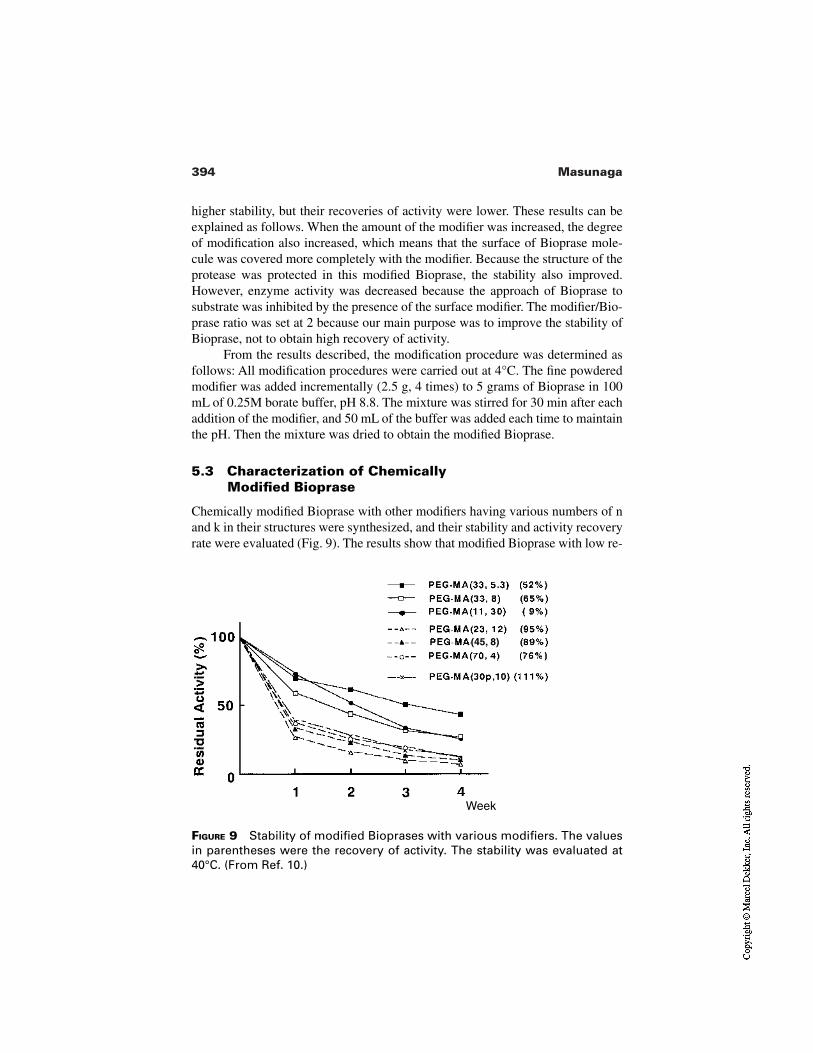
394 Masunaga
FIGURE 9 Stability of modified Bioprases with various modifiers. The valuesin parentheses were the recovery of activity. The stability was evaluated at40°C. (From Ref. 10.)
Week
(45, 8)
higher stability, but their recoveries of activity were lower. These results can beexplained as follows. When the amount of the modifier was increased, the degreeof modification also increased, which means that the surface of Bioprase mole-cule was covered more completely with the modifier. Because the structure of theprotease was protected in this modified Bioprase, the stability also improved.However, enzyme activity was decreased because the approach of Bioprase tosubstrate was inhibited by the presence of the surface modifier. The modifier/Bio-prase ratio was set at 2 because our main purpose was to improve the stability ofBioprase, not to obtain high recovery of activity.
From the results described, the modification procedure was determined asfollows: All modification procedures were carried out at 4°C. The fine powderedmodifier was added incrementally (2.5 g, 4 times) to 5 grams of Bioprase in 100mL of 0.25M borate buffer, pH 8.8. The mixture was stirred for 30 min after eachaddition of the modifier, and 50 mL of the buffer was added each time to maintainthe pH. Then the mixture was dried to obtain the modified Bioprase.
5.3 Characterization of Chemically Modified Bioprase
Chemically modified Bioprase with other modifiers having various numbers of nand k in their structures were synthesized, and their stability and activity recoveryrate were evaluated (Fig. 9). The results show that modified Bioprase with low re-
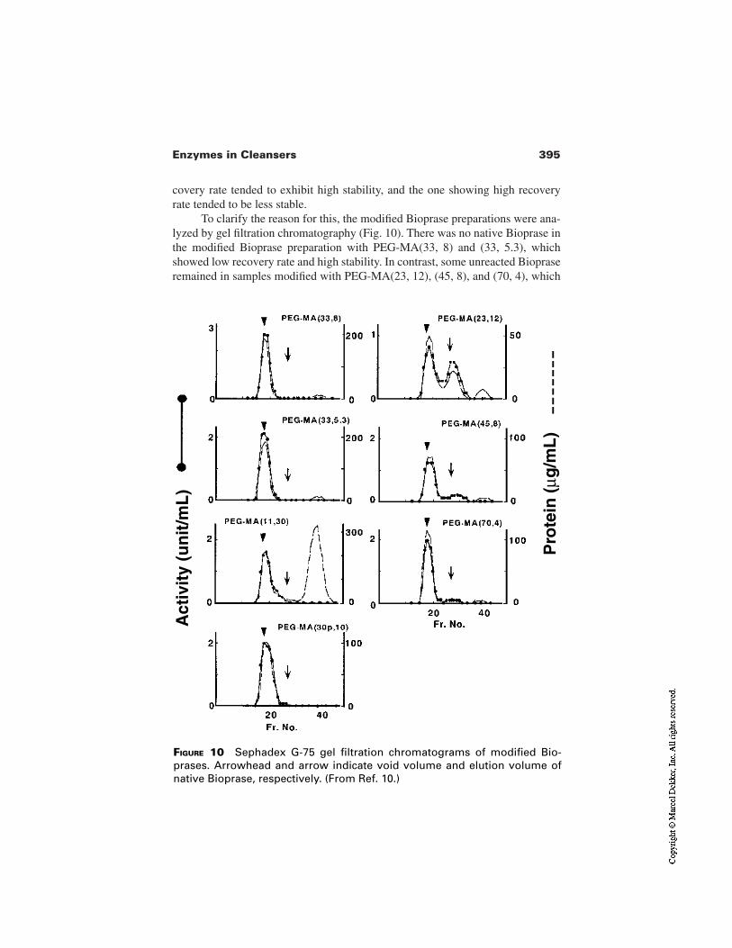
395Enzymes in Cleansers
covery rate tended to exhibit high stability, and the one showing high recoveryrate tended to be less stable.
To clarify the reason for this, the modified Bioprase preparations were ana-lyzed by gel filtration chromatography (Fig. 10). There was no native Bioprase inthe modified Bioprase preparation with PEG-MA(33, 8) and (33, 5.3), whichshowed low recovery rate and high stability. In contrast, some unreacted Biopraseremained in samples modified with PEG-MA(23, 12), (45, 8), and (70, 4), which
FIGURE 10 Sephadex G-75 gel filtration chromatograms of modified Bio-prases. Arrowhead and arrow indicate void volume and elution volume ofnative Bioprase, respectively. (From Ref. 10.)
Act
ivit
y (u
nit
/mL
)
Pro
tein
(µg
/mL
)
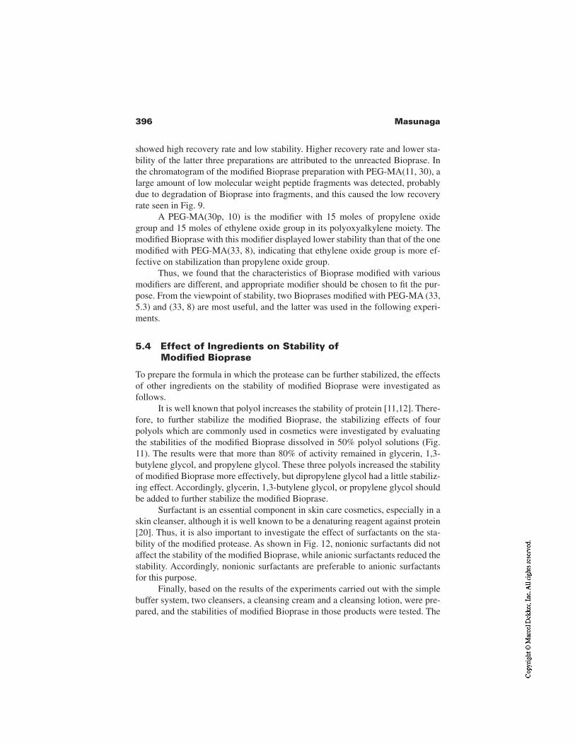
396 Masunaga
showed high recovery rate and low stability. Higher recovery rate and lower sta-bility of the latter three preparations are attributed to the unreacted Bioprase. Inthe chromatogram of the modified Bioprase preparation with PEG-MA(11, 30), alarge amount of low molecular weight peptide fragments was detected, probablydue to degradation of Bioprase into fragments, and this caused the low recoveryrate seen in Fig. 9.
A PEG-MA(30p, 10) is the modifier with 15 moles of propylene oxidegroup and 15 moles of ethylene oxide group in its polyoxyalkylene moiety. Themodified Bioprase with this modifier displayed lower stability than that of the onemodified with PEG-MA(33, 8), indicating that ethylene oxide group is more ef-fective on stabilization than propylene oxide group.
Thus, we found that the characteristics of Bioprase modified with variousmodifiers are different, and appropriate modifier should be chosen to fit the pur-pose. From the viewpoint of stability, two Bioprases modified with PEG-MA (33,5.3) and (33, 8) are most useful, and the latter was used in the following experi-ments.
5.4 Effect of Ingredients on Stability of Modified Bioprase
To prepare the formula in which the protease can be further stabilized, the effectsof other ingredients on the stability of modified Bioprase were investigated asfollows.
It is well known that polyol increases the stability of protein [11,12]. There-fore, to further stabilize the modified Bioprase, the stabilizing effects of fourpolyols which are commonly used in cosmetics were investigated by evaluatingthe stabilities of the modified Bioprase dissolved in 50% polyol solutions (Fig.11). The results were that more than 80% of activity remained in glycerin, 1,3-butylene glycol, and propylene glycol. These three polyols increased the stabilityof modified Bioprase more effectively, but dipropylene glycol had a little stabiliz-ing effect. Accordingly, glycerin, 1,3-butylene glycol, or propylene glycol shouldbe added to further stabilize the modified Bioprase.
Surfactant is an essential component in skin care cosmetics, especially in askin cleanser, although it is well known to be a denaturing reagent against protein[20]. Thus, it is also important to investigate the effect of surfactants on the sta-bility of the modified protease. As shown in Fig. 12, nonionic surfactants did notaffect the stability of the modified Bioprase, while anionic surfactants reduced thestability. Accordingly, nonionic surfactants are preferable to anionic surfactantsfor this purpose.
Finally, based on the results of the experiments carried out with the simplebuffer system, two cleansers, a cleansing cream and a cleansing lotion, were pre-pared, and the stabilities of modified Bioprase in those products were tested. The
397Enzymes in Cleansers
FIGURE 11 Increase of the stability of modified Bioprase by addition of poly-ols. Stability of modified Bioprase with PEG-MA(33, 8) was evaluated at 40°C.Control experiment was carried out in the absence of any polyols (From Ref.10.)
Week
Propylene Glycol
Dipropylene Glycol
residual activities of modified Bioprase in these products were over 50% in 1week, while almost all activities of the native Bioprase in both preparations werelost within 1 week. This result is consistent with the results obtained with a sim-ple buffer system, and shows that the modified Bioprase is more stable than thenative Bioprase in a product preparation as well as in a simple buffer system.
5.5 Activity of Modified Bioprase
To confirm whether the modified Bioprase with improved stability also main-tained the protein hydrolytic function, the keratin and sweat protein hydrolyticactivities were measured (Fig. 13). Modified Bioprase could degrade the keratins,although it displayed a slightly lower keratin hydrolytic activity than the nativeone, due to steric hindrance by the modifier located on the surface of Bioprasemolecule. However, the activity displayed is sufficient for a skin cleanser becausealmost all of the main keratin bands disappeared. In contrast to the slightly re-duced keratin hydrolytic activity, almost no reduction in the hydrolytic activitytoward sweat proteins was observed, as shown in Fig. 13B. It was presumed thatthe surface modifier did not hinder the approach of the modified Bioprase tosweat proteins, which were lower molecular weight proteins than keratins. Theseexperiments confirm that the modified Bioprase possesses sufficient hydrolyticactivity toward protein in dirt on the skin surface, confirming the usefulness ofmodified Bioprase as an ingredient in skin cleansers.
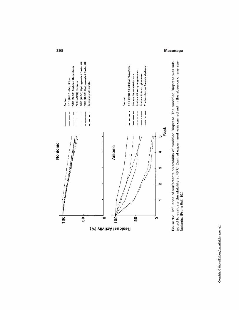
398 Masunaga
FIG
UR
E1
2In
flu
ence
of
surf
acta
nts
on
sta
bili
ty o
f m
od
ified
Bio
pra
se. T
he
mo
difi
ed B
iop
rase
was
su
b-
ject
ed t
o e
valu
ate
the
stab
ility
at
40°C
. C
on
tro
l ex
per
imen
t w
as c
arri
ed o
ut
in t
he
abse
nce
of
any
sur-
fact
ants
. (Fr
om
Ref
. 10.
)
Wee
k
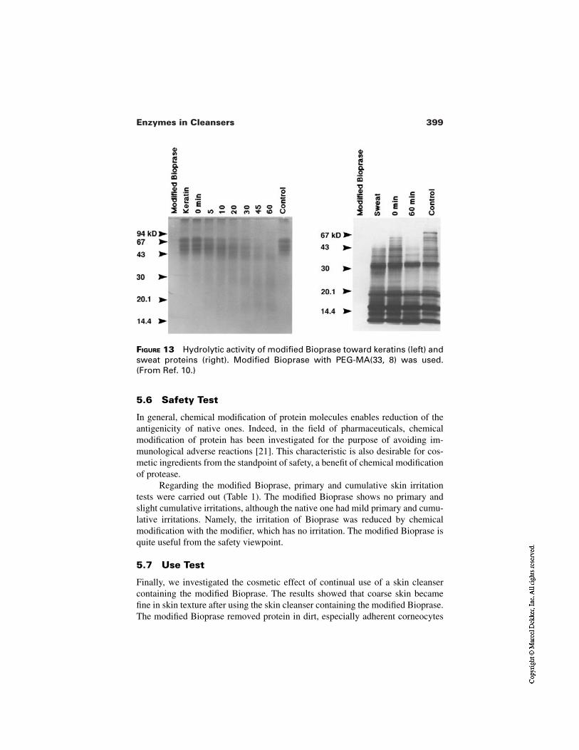
399Enzymes in Cleansers
FIGURE 13 Hydrolytic activity of modified Bioprase toward keratins (left) andsweat proteins (right). Modified Bioprase with PEG-MA(33, 8) was used.(From Ref. 10.)
5.6 Safety Test
In general, chemical modification of protein molecules enables reduction of theantigenicity of native ones. Indeed, in the field of pharmaceuticals, chemicalmodification of protein has been investigated for the purpose of avoiding im-munological adverse reactions [21]. This characteristic is also desirable for cos-metic ingredients from the standpoint of safety, a benefit of chemical modificationof protease.
Regarding the modified Bioprase, primary and cumulative skin irritationtests were carried out (Table 1). The modified Bioprase shows no primary andslight cumulative irritations, although the native one had mild primary and cumu-lative irritations. Namely, the irritation of Bioprase was reduced by chemicalmodification with the modifier, which has no irritation. The modified Bioprase isquite useful from the safety viewpoint.
5.7 Use Test
Finally, we investigated the cosmetic effect of continual use of a skin cleansercontaining the modified Bioprase. The results showed that coarse skin becamefine in skin texture after using the skin cleanser containing the modified Bioprase.The modified Bioprase removed protein in dirt, especially adherent corneocytes
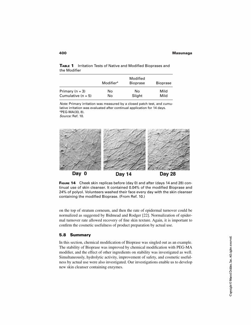
400 Masunaga
TABLE 1 Irritation Tests of Native and Modified Bioprases andthe Modifier
ModifieraModifiedBioprase Bioprase
Primary (n = 3) No No MildCumulative (n = 5) No Slight Mild
Note: Primary irritation was measured by a closed patch test, and cumu-lative irritation was evaluated after continual application for 14 days.aPEG-MA(33, 8).Source: Ref. 10.
FIGURE 14 Cheek skin replicas before (day 0) and after (days 14 and 28) con-tinual use of skin cleanser. It contained 0.04% of the modified Bioprase and24% of polyol. Volunteers washed their face every day with the skin cleansercontaining the modified Bioprase. (From Ref. 10.)
on the top of stratum corneum, and then the rate of epidermal turnover could benormalized as suggested by Bidmead and Rodger [22]. Normalization of epider-mal turnover rate allowed recovery of fine skin texture. Again, it is important toconfirm the cosmetic usefulness of product preparation by actual use.
5.8 Summary
In this section, chemical modification of Bioprase was singled out as an example.The stability of Bioprase was improved by chemical modification with PEG-MAmodifier, and the effect of other ingredients on stability was investigated as well.Simultaneously, hydrolytic activity, improvement of safety, and cosmetic useful-ness by actual use were also investigated. Our investigations enable us to developnew skin cleanser containing enzymes.
401Enzymes in Cleansers
Ohta et al. reported on the chemical modification of esperase, a kind of pro-tease [23]. Esperase was chemically modified with dextran to improve stabilityand to reduce skin sensitization potential. They successfully obtained the dex-tran–esperase conjugate, investigated the influence of cosmetic ingredients forfurther stabilization in cosmetic formulations, and finally prepared the commer-cial product. Their strategy and the results obtained were similar to those de-scribed in this paper.
6 OTHER ENZYMES
In this chapter, we have focused on the protease as an enzyme in skin cleanser.However, besides protease, some other enzymes are proposed as an effective in-gredient of skin cleanser.
Lipase is one such useful enzyme. This enzyme hydrolyzes triglyceride todiglyceride, monoglyceride, and finally to free fatty acids. On the skin surface,especially on the face, sebum is one of the main components of the dirt, and lipidis oxidized, generating lipid peroxidation which can lead to skin trouble. The ma-jor component of sebum is triglyceride, which is not easily washed out by deter-gent compared with diglyceride, monoglyceride, or free fatty acid. The incorpo-ration of lipase into skin cleanser can improve its efficacy in washing out fatty dirton the skin surface.
Lysozyme, usually extracted from egg white, is a bacteriolyzing enzymethrough the cleavage of polysaccharide of plasma membrane of bacteria. Its ac-tivity can be measured by the tubidimetric assay of Micrococcus lysodeikticus. Ina skin cleanser, this enzyme could impart antibacterial and anti-inflammatory ef-fects.
Superoxide dismutase (SOD) is an enzyme quenching the reactive oxygenspecies; SOD catalyzes the dismutation of superoxide anion (O2
–) into oxygenand hydrogen peroxide to protect cells against toxic reactive oxygen species. Be-cause superoxide anion can react with various materials, including protein andlipid, and such an oxidized material may act as a skin irritant, it is important toprevent the oxidation of the dirt on skin surface and to maintain personal skin hy-giene.
7 FUTURE
Enzymes are protein, which means that they have low stability but unique action.Therefore, stabilization of enzymes is key to their incorporation into skincleansers or other cosmetic products. In the near future, it will be possible to in-clude the various enzymes into a skin cleanser due to the progress in enzyme sta-bilization itself, pharmaceutical technology, formulation of cleansers, and manu-facturing methods. The development of new cosmetic products, including skin
402 Masunaga
cleansers containing enzymes, will depend on the progress of all these aspects ofcosmetic science.
REFERENCES
1. Asano H, Masunaga T, Takemoto Y, Kawada A, Kominami E. Influence of consecu-tive irradiation of low dose UVB on the mice epidermis. Proceedings of the 20th An-nual Meeting of the Japanese Society of Photomedicine and Photobiology, Ku-mamoto, Japan, 1999, pp. 89–90.
2. Denda M, Sato J, Tsuchiya T, Elias PM, Feingold KR. Low humidity stimulates epi-dermal DNA synthesis and amplifies the hyperproliferative response to barrier dis-ruption: implication for seasonal exacerbations of inflammatory dermatoses. J InvestDermatol 1998; 111:873–878.
3. Arakane K, Ryu A, Hayashi C, Masunaga T, Shinmoto K, Mashiko S, Nagano T, Hi-robe M. Singlet oxygen (1∆g) generation from coproporphyrin in Propionibacteriumacnes on irradiation. Biochem Biophys Res Commun 1996; 223:578–582.
4. Bell KW. Enzymes in cosmetics. Am Cosmet Perf 1972; 87:39–44.5. Arnon R. Papain. In: Perlmann GE, Lorand L, eds. Methods in Enzymology. New
York: Academic Press, 1970:226–244.6. Sun T-T, Tseng SCG, Huang AJ-W, Cooper D, Schermer A, Lynch MH, Weiss R,
Eichner R. Monoclonal antibody studies of mammalian epithelial keratins: a review.Ann NY Acad Sci 1985; 455:307–329.
7. Jenkinson DM, Mabon RM, Manson W. Sweat proteins. Br J Dermatol 1974;90:175–181.
8. Marshall T. Analysis of human sweat proteins by two-dimensional electrophoresisand ultrasensitive silver staining. Anal Biochem 1984; 139:506–509.
9. Rubin RW, Penneys NS. Subpicogram analysis of sweat proteins using two-dimen-sional polyacrylamide gel electrophoresis. Anal Biochem 1983; 131:520–524.
10. Masunaga T, Yasukohchi T, Hirobe M, Arakane K, Adachi K. The protease as acleansing agent and its stabilization by chemical modification. J Soc Cosmet ChemJpn 1993; 27:276–288.
11. Gekko K, Ito H. Competing solvent effects of polyols and guanidine hydrochlorideon protein stability. J Biochem 1990; 107:572–577.
12. Arakawa T, Kita Y, Carpenter JF. Protein–solvent interactions in pharmaceutical for-mulations. Pharm Res 1991; 8:285–291.
13. Cowan D, Daniel R, Morgan H. Thermophilic proteases: properties and potential ap-plications. Trends Biotech 1985; 3:68–72.
14. Matsubara H. Purification and assay of thermolysin. In: Perlmann GE, Lorand L,eds. Methods in Enzymology. New York: Academic Press, 1970:642–651.
15. Matsuzawa H, Hamaoki M, Ohta T. Production of thermophilic extracellular pro-teases (aqualysins I and II) by Thermus aquticus YT-1, an extreme thermophile.Agric Biol Chem 1983; 47:25–28.
16. Matsuzawa H, Tokugawa K, Hamaoki M, Mizoguchi M, Taguchi H, Terada I, KwonS-T, Ohta T. Purification and characterization of aqualysin I (a thermophilic alkaline
403Enzymes in Cleansers
serine protease) produced by Thermus aquaticus YT-1. Eur J Biochem 1988;171:441–447.
17. Cowan DA, Daniel RM. Purification and some properties of an extracellular protease(caldolysin) from an extreme thermophile. Biochim Biophys Acta 1982; 705:293–305.
18. Frömmel C, Höhne WE. Influence of calcium binding on the thermal stability of‘thermitase’, a serine protease from Thermoactinomyces vulgaris. Biochim BiophysActa 1981; 670:25–31.
19. Yoshimoto T, Ritani A, Ohwada K, Takahashi K, Kodera Y, Matsushima A, Saito Y,Inada Y. Polyethylene glycol derivative-modified cholesterol oxidase soluble and ac-tive in benzene. Biochem Biophys Res Commun 1987; 148:876–882.
20. Schomaecker R, Robinson BH, Fletcher PDI. Interaction of enzymes with surfac-tants in aqueous solution and in water-in-oil microemulsions. J Chem Soc FaradayTrans I 1988; 84:4203–4212.
21. Yoshimoto T, Nishimura H, Saito Y, Sakurai K, Kamisaki Y, Wada H, Sako M, Tsu-jino G, Inada Y. Characterization of polyethylene glycol–modified L-asparaginasefrom Escherichia coli and its application to therapy of leukemia. Jpn J Cancer Res(Gann) 1986; 77:1264–1270.
22. Bidmead MC, Rodger MN. The effect of enzymes on stratum corneum. J Soc Cos-met Chem 1973; 24:493–500.
23. Ohta M, Goto A, Mori K, Fukunaga S, Nakayama H, Fujino Y. A dextran–proteaseconjugate for cosmetic use. A stabilized dextran–protease conjugate leads to cleaner,smoother, more attractive skin. Cosmet Toil 1996; 111(6):79–88.

20Moisturizing Cleansers
Kavssery P. Ananthapadmanabhan, Kumar Subramanyan, and Gail B. RattingerUnilever Research, Edgewater Laboratory, Edgewater, New Jersey
1 INTRODUCTION
Historically, the primary purpose of cleansing has been to achieve cleanliness andfreshness by removing oily soils from face and body. Hygienic benefits of cleans-ing have also been recognized for a very long time. While soap-like materials forcleansing have been around as early as 2500 BC [1], soap itself is believed tohave been invented sometime around 600–300 BC [2]. The first industrial typemanufacturing of soap in an individually wrapped and branded bar form was in1884 in England [2]. The desire for cleanliness and freshness coupled with thesensory pleasures and health benefits has driven the growth of soap in the 20thcentury [3]. Thus, deodorant soaps grew from a desire for health and hygiene ben-efits. The beauty segment, on the other hand, grew from a desire for beautiful skincoupled with the sensory pleasures of cleansing using cleansing bars of differentcolors, fragrances, and shapes [3].
With increasing use of soaps, awareness of soap-induced skin irritation,itching, dry skin, and other potential effects also increased. This led to an in-creased desire on the part of the consumer to have mild cleansing bars. Introduc-tion of synthetic detergents into the cleansing arena in 1948 made it possible todevelop cleansing bars that were demonstrably milder than soaps [3]. These bars
405
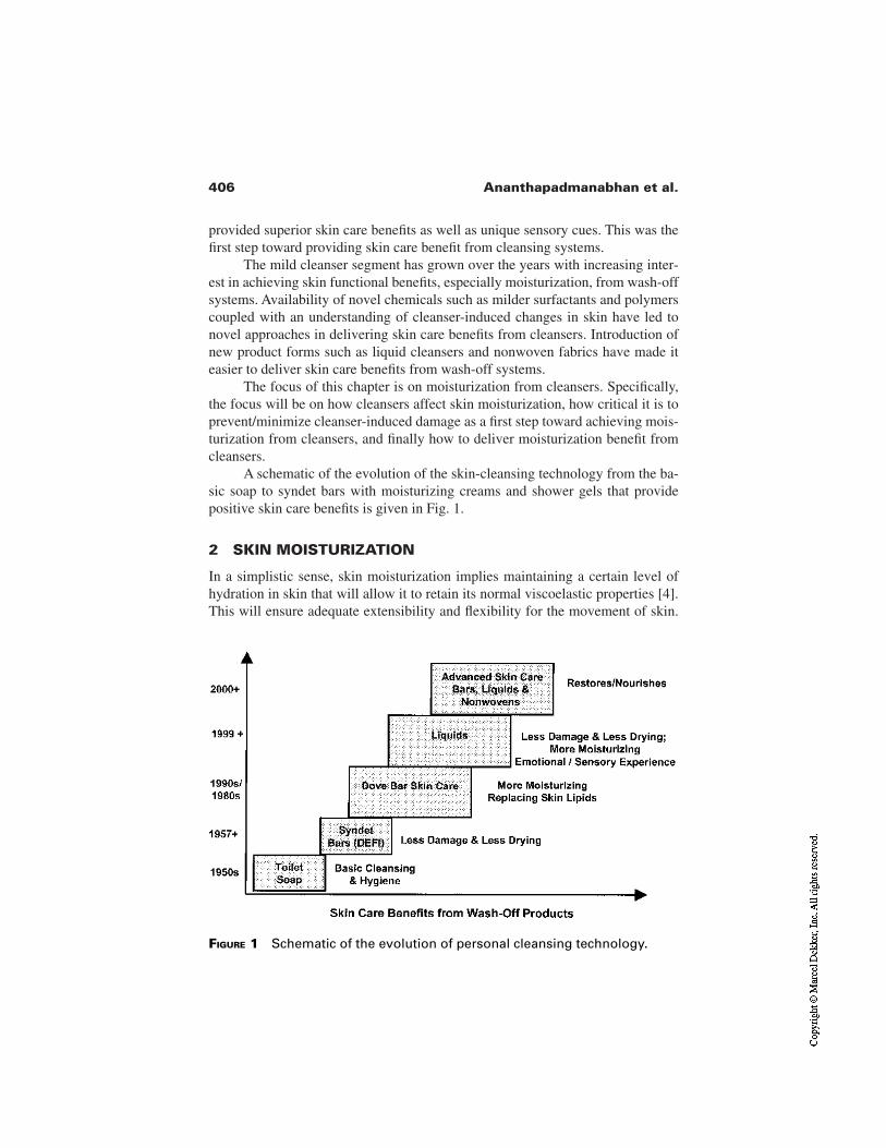
406 Ananthapadmanabhan et al.
provided superior skin care benefits as well as unique sensory cues. This was thefirst step toward providing skin care benefit from cleansing systems.
The mild cleanser segment has grown over the years with increasing inter-est in achieving skin functional benefits, especially moisturization, from wash-offsystems. Availability of novel chemicals such as milder surfactants and polymerscoupled with an understanding of cleanser-induced changes in skin have led tonovel approaches in delivering skin care benefits from cleansers. Introduction ofnew product forms such as liquid cleansers and nonwoven fabrics have made iteasier to deliver skin care benefits from wash-off systems.
The focus of this chapter is on moisturization from cleansers. Specifically,the focus will be on how cleansers affect skin moisturization, how critical it is toprevent/minimize cleanser-induced damage as a first step toward achieving mois-turization from cleansers, and finally how to deliver moisturization benefit fromcleansers.
A schematic of the evolution of the skin-cleansing technology from the ba-sic soap to syndet bars with moisturizing creams and shower gels that providepositive skin care benefits is given in Fig. 1.
2 SKIN MOISTURIZATION
In a simplistic sense, skin moisturization implies maintaining a certain level ofhydration in skin that will allow it to retain its normal viscoelastic properties [4].This will ensure adequate extensibility and flexibility for the movement of skin.
FIGURE 1 Schematic of the evolution of personal cleansing technology.
407Moisturizing Cleansers
Absence of moisturization, on the other hand, is a state of skin that can manifestin a variety of forms including a sensation of after-wash tightness (AWT), lack offlexibility/extensibility, visible dryness (skin whitening), skin roughness, scaling,cracking, and ultimately irritation in the form of visible erythema, and itching.Thus there are emotional, tactile, and visual manifestations of absence of skinmoisturization.
Stratum corneum upper layers contain about 15% water, 65–75% proteins,and 10–15% lipids [5]. Most of the water in the corneum is present as waterbound to the proteins. Water level changes markedly with depth in the corneum,increasing to levels as much as 40% at the innermost level [6]. Water content ofthe corneum can vary markedly with changes in the relative humidity [6], waterbinding capacity of corneum proteins, concentration of natural moisturizing fac-tors (NMFs), and the integrity of the barrier lipids [7]. The NMFs in the corneuminclude short chain amino acids (40%), pyrrolidone carboxylic acid (12%), lac-tate (12%), urea, (7%), and Na/Ca/K/Mg phosphate/chloride (18.5%) [8]. Stra-tum corneum lipids, in addition to being a water barrier, also play an importantrole in maintaining its elasticity. Moisturization technology in the leave-on skincare area usually involves a combination of actives such as humectants thatincrease water-holding capacity of the corneum, emollients that form a lightlubricant coating, and occlusives that provide an external water barrier film onskin. Thus, a moisuturizer provides several positive benefits to improve the skinhydration and alter the unpleasant tactile and visual manifestations of skindryness.
Cleansers are designed to remove oily soils, dirt, sweat, and sebum fromskin. This is achieved through the use of surfactants that aid in the uplifting of dirtand solubilization of oily soils including sebum. In addition to removal of un-wanted materials from skin, the cleansing process also helps the normal exfolia-tion process by removing the dead skin cells, leading to rejuvenation of the skin.These beneficial effects can, however, be accompanied with other interactionswith the corneum that can be deleterious to skin. For example, cleanser surfac-tants can bind to stratum corneum proteins leading to a reduction in their abilityto bind and hold water [9,10]. Surfactants can also cause dissolution of fluid skinlipids during cleansing [11] or alterations to the lipid layers by adsorption and in-tercalation into lipid layers [12–14]. In addition, washing with cleansers can alsolead to a reduction in the level of NMFs in skin [15,16]. All these factors that re-duce the water content of skin can lead to changes in the viscoelastic properties ofskin and this in turn can manifest as after-wash tightening of skin [17]. Continueduse of such cleansers can lead to dry skin, barrier damage, and erythema. Thus, ina simplistic sense, while cleansers have the potential to negatively impact the hy-dration and viscoelastic properties of skin, a moisturizer is expected to improvesuch conditions. In the case of cleansers, minimizing the damaging/deleteriouseffects leading to dry skin and erythema is the first step toward moisturization.
408 Ananthapadmanabhan et al.
Most cleansers currently available in the marketplace make claims of mois-turization based on minimizing damage. Limited ones have begun to combine theminimizing damage concept with positive skin benefits by depositing moisturiz-ing agents on skin. This is clearly in the direction of positive skin moisturization.Several excellent reviews have appeared recently on mechanistic aspects ofcleanser-induced damage to stratum corneum [4,18–22] and therefore this chap-ter will have a limited discussion on the damage itself and focus more on currentapproaches to minimizing the deleterious effects of cleansers and providing mois-turization benefits.
3 EFFECTS OF CLEANSERS ON STRATUM CORNEUM
3.1 Stratum Corneum Structure
Stratum corneum, the upper most layer of the skin, is a nonliving layer consistingof proteins, lipids, and water. The proteins and lipids in the corneum are orga-nized in a brick and mortar–like structure with protein (corneocyte) bricks em-bedded in a lipid mortar phase [23]. Available data seem to support the notion oftwo types of lipids, specifically, relatively fluid lipids as well as covalently bond-ed relatively rigid lipids in the matrix [24,25]. Available ESR and DSC data seemto support the presence of complex lipid domains in the stratum corneum [26].Corneocytes with a keratin envelope and NMFs (e.g., pyrrolidone carboxylicacid, urea, lactic acid, short chain amino acids) within them have the potential tobind water molecules to keep the skin moisturized [27]. Corneocytes in differentlayers are also linked to each other through membrane protein links referred to asdesmosomes [28]. As a part of the normal desquamation process, desmosomesare degraded by protease enzymes present in the upper layers of skin [29]. Inhealthy skin, the corneum is renewed about every two weeks and this process ofdesquamation requires a certain level of hydration of the skin.
3.2 Surfactant Interactions with Stratum Corneum Proteins
Interactions of cleanser surfactants with stratum corneum proteins and model pro-teins have been studied extensively in the past [30–38]. Typically, anionic surfac-tants, because of their excellent foam and lather characteristics, find use as pri-mary surfactants in cleansers. Liquid cleansers often have a combination ofanionic and amphoteric surfactants. Nonionic surfactants also find limited appli-cation, mostly in combination with anionic or amphoteric surfactants. In general,the tendency of surfactants to interact with proteins follow the following order:anionic surfactants > amphhoteric surfactants > nonionic surfactants. Typical an-ionic surfactants used in cleansers include soaps (salts of fatty acids), syntheticsurfactants such as alkyl ether sulfates, alkyl acyl isethionates, alkyl phosphates,
409Moisturizing Cleansers
and alkyl sulfonates. The binding tendency of anionic surfactants to proteins fol-low the order sodium lauryl sulfate (SLS) = sodium laurate >> monoalkyl phos-phate (MAP) > sodium cocoyl isethionate [19,37]. Recently, Imokawa has shownthat in studies using Yucatan microswine in vivo, the binding of surfactants uponexposure at 100 mM level for 30 min at 30°C follow the order soap > SLS >>MAP > sodium cocoyl isethionate (SCI) > triethanolamine N-lauroyl β-amino-propionate (LBA) [19]. In general, for a given chain length surfactant, the largerthe headgroup size, the lower is its binding to proteins. Thus, ethoxylated alkylsulfates tend to have lower binding to keratin compared to the correspondingalkyl sulfates [31]. For a given surfactant headgroup, there is an optimum chainlength for maximum binding, and this governed by a balance between surfactantsolubility in the aqueous phase and its surface activity [30,38]. This optimum formost surfactants at room temperature is around C12. Even though the higher chainlength surfactants have higher surface activity, because of their limited solubilitytheir binding is limited. At higher temperature, increased solubility of higherchain length surfactants can increase their binding.
Surfactants that tend to bind strongly to corneum proteins, in general, havea higher potential to cause significant protein denaturation leading to barrier dam-age, erythema, and itching. Some of the common approaches to lowering the ten-dency of anionic surfactants to bind to proteins are (1) increase in the size ofhead/polar group of the surfactant [31,39] and (2) use anionic surfactants withamphoteric or nonionic surfactants [40]. A typical example of modulating the ac-tivity of sodium lauryl ether sulfate (SLES) by adding an amphoteric surfactant,cocoamido propylbetaine (CAPB) is shown in Fig. 2 in terms of the solubility ofa corn protein, zein. The ability of a surfactant to dissolve zein has been used as ameasure of its irritation potential [41]. Thus, in this example, there exists a certainratio of SLES to CAPB where the dissolution of zein is minimum and this coin-cides with the minimum in the critical micelle concentration (CMC) of thesemixed surfactants. The commonly accepted hypothesis for the reduced binding ofthe anionic surfactant in the presence of amphoteric or nonionic co-surfactant isthe competition between binding and co-micellization for monomers that tend tofavor co-micellization in the presence of lower CMC surfactants. Thus, at a givenlevel of anionic surfactant, addition of lower CMC surfactant whether it is non-ionic, amphoteric, or even anionic can result in reduced irritation [18]. These ef-fects can often be synergistic since co-micellization is often synergistic, resultingin CMC values that are lower than the CMC of either surfactant.
Surfactant binding to protein can significantly affect the water-binding and-holding capacity of proteins. Several studies show that surfactant solutions causeswelling of the corneum [42] and this effect saturates at about the CMC of thesurfactant, suggesting that the swelling is controlled by surfactant monomers[42]. The swelling itself is due to increased water uptake resulting from an in-crease in the net negative charge of the protein because of surfactant binding.
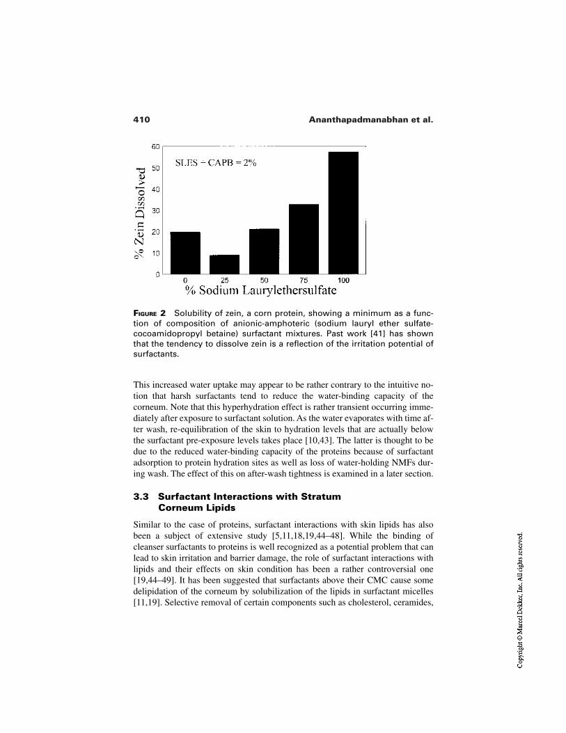
410 Ananthapadmanabhan et al.
FIGURE 2 Solubility of zein, a corn protein, showing a minimum as a func-tion of composition of anionic-amphoteric (sodium lauryl ether sulfate-cocoamidopropyl betaine) surfactant mixtures. Past work [41] has shownthat the tendency to dissolve zein is a reflection of the irritation potential ofsurfactants.
This increased water uptake may appear to be rather contrary to the intuitive no-tion that harsh surfactants tend to reduce the water-binding capacity of thecorneum. Note that this hyperhydration effect is rather transient occurring imme-diately after exposure to surfactant solution. As the water evaporates with time af-ter wash, re-equilibration of the skin to hydration levels that are actually belowthe surfactant pre-exposure levels takes place [10,43]. The latter is thought to bedue to the reduced water-binding capacity of the proteins because of surfactantadsorption to protein hydration sites as well as loss of water-holding NMFs dur-ing wash. The effect of this on after-wash tightness is examined in a later section.
3.3 Surfactant Interactions with Stratum Corneum Lipids
Similar to the case of proteins, surfactant interactions with skin lipids has alsobeen a subject of extensive study [5,11,18,19,44–48]. While the binding ofcleanser surfactants to proteins is well recognized as a potential problem that canlead to skin irritation and barrier damage, the role of surfactant interactions withlipids and their effects on skin condition has been a rather controversial one[19,44–49]. It has been suggested that surfactants above their CMC cause somedelipidation of the corneum by solubilization of the lipids in surfactant micelles[11,19]. Selective removal of certain components such as cholesterol, ceramides,
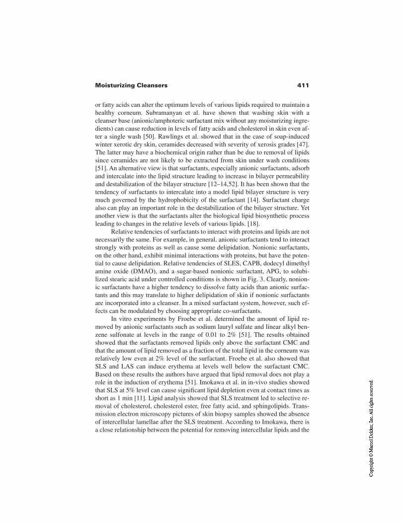
411Moisturizing Cleansers
or fatty acids can alter the optimum levels of various lipids required to maintain ahealthy corneum. Subramanyan et al. have shown that washing skin with acleanser base (anionic/amphoteric surfactant mix without any moisturizing ingre-dients) can cause reduction in levels of fatty acids and cholesterol in skin even af-ter a single wash [50]. Rawlings et al. showed that in the case of soap-inducedwinter xerotic dry skin, ceramides decreased with severity of xerosis grades [47].The latter may have a biochemical origin rather than be due to removal of lipidssince ceramides are not likely to be extracted from skin under wash conditions[51]. An alternative view is that surfactants, especially anionic surfactants, adsorband intercalate into the lipid structure leading to increase in bilayer permeabilityand destabilization of the bilayer structure [12–14,52]. It has been shown that thetendency of surfactants to intercalate into a model lipid bilayer structure is verymuch governed by the hydrophobicity of the surfactant [14]. Surfactant chargealso can play an important role in the destabilization of the bilayer structure. Yetanother view is that the surfactants alter the biological lipid biosynthetic processleading to changes in the relative levels of various lipids. [18].
Relative tendencies of surfactants to interact with proteins and lipids are notnecessarily the same. For example, in general, anionic surfactants tend to interactstrongly with proteins as well as cause some delipidation. Nonionic surfactants,on the other hand, exhibit minimal interactions with proteins, but have the poten-tial to cause delipidation. Relative tendencies of SLES, CAPB, dodecyl dimethylamine oxide (DMAO), and a sugar-based nonionic surfactant, APG, to solubi-lized stearic acid under controlled conditions is shown in Fig. 3. Clearly, nonion-ic surfactants have a higher tendency to dissolve fatty acids than anionic surfac-tants and this may translate to higher delipidation of skin if nonionic surfactantsare incorporated into a cleanser. In a mixed surfactant system, however, such ef-fects can be modulated by choosing appropriate co-surfactants.
In vitro experiments by Froebe et al. determined the amount of lipid re-moved by anionic surfactants such as sodium lauryl sulfate and linear alkyl ben-zene sulfonate at levels in the range of 0.01 to 2% [51]. The results obtainedshowed that the surfactants removed lipids only above the surfactant CMC andthat the amount of lipid removed as a fraction of the total lipid in the corneum wasrelatively low even at 2% level of the surfactant. Froebe et al. also showed thatSLS and LAS can induce erythema at levels well below the surfactant CMC.Based on these results the authors have argued that lipid removal does not play arole in the induction of erythema [51]. Imokawa et al. in in-vivo studies showedthat SLS at 5% level can cause significant lipid depletion even at contact times asshort as 1 min [11]. Lipid analysis showed that SLS treatment led to selective re-moval of cholesterol, cholesterol ester, free fatty acid, and sphingolipids. Trans-mission electron microscopy pictures of skin biopsy samples showed the absenceof intercellular lamellae after the SLS treatment. According to Imokawa, there isa close relationship between the potential for removing intercellular lipids and the
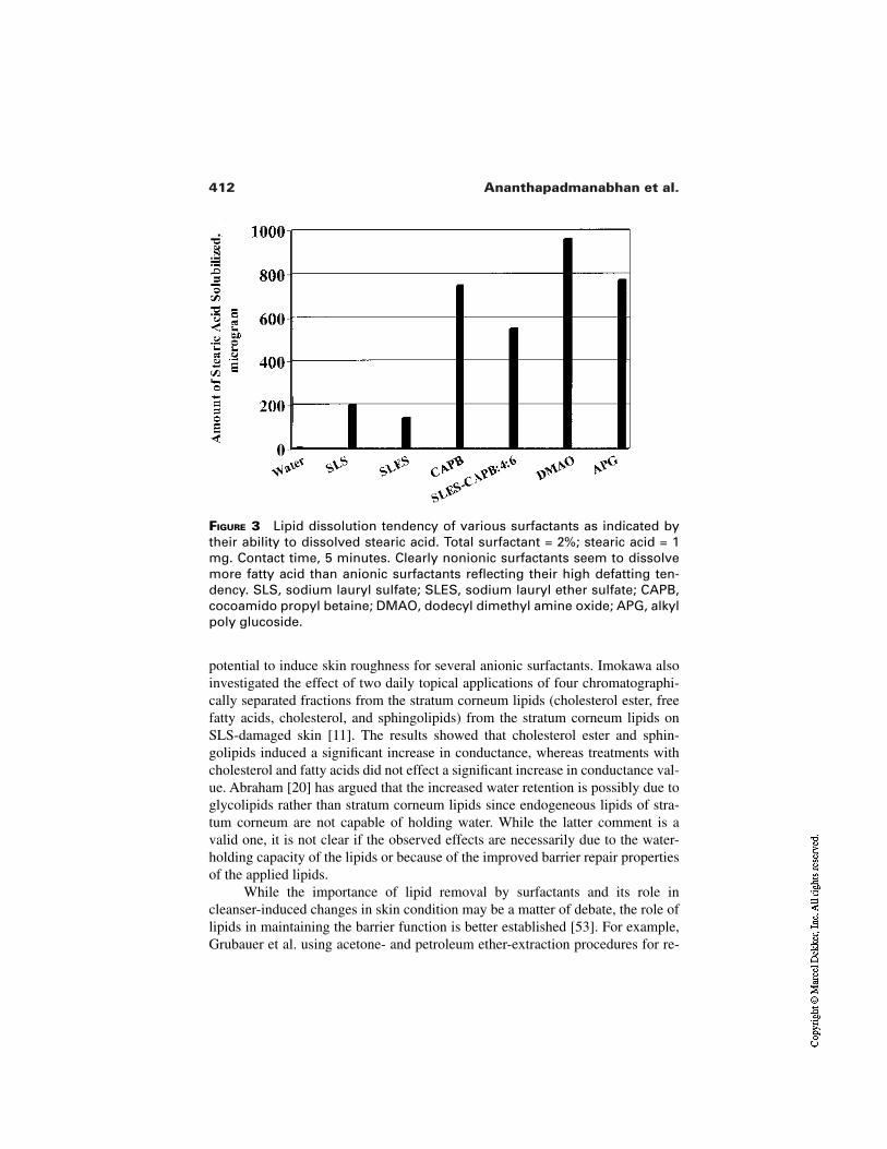
412 Ananthapadmanabhan et al.
potential to induce skin roughness for several anionic surfactants. Imokawa alsoinvestigated the effect of two daily topical applications of four chromatographi-cally separated fractions from the stratum corneum lipids (cholesterol ester, freefatty acids, cholesterol, and sphingolipids) from the stratum corneum lipids onSLS-damaged skin [11]. The results showed that cholesterol ester and sphin-golipids induced a significant increase in conductance, whereas treatments withcholesterol and fatty acids did not effect a significant increase in conductance val-ue. Abraham [20] has argued that the increased water retention is possibly due toglycolipids rather than stratum corneum lipids since endogeneous lipids of stra-tum corneum are not capable of holding water. While the latter comment is avalid one, it is not clear if the observed effects are necessarily due to the water-holding capacity of the lipids or because of the improved barrier repair propertiesof the applied lipids.
While the importance of lipid removal by surfactants and its role incleanser-induced changes in skin condition may be a matter of debate, the role oflipids in maintaining the barrier function is better established [53]. For example,Grubauer et al. using acetone- and petroleum ether-extraction procedures for re-
FIGURE 3 Lipid dissolution tendency of various surfactants as indicated bytheir ability to dissolved stearic acid. Total surfactant = 2%; stearic acid = 1mg. Contact time, 5 minutes. Clearly nonionic surfactants seem to dissolvemore fatty acid than anionic surfactants reflecting their high defatting ten-dency. SLS, sodium lauryl sulfate; SLES, sodium lauryl ether sulfate; CAPB,cocoamido propyl betaine; DMAO, dodecyl dimethyl amine oxide; APG, alkylpoly glucoside.
413Moisturizing Cleansers
moving lipids from hairless mice skin showed that there exists a linear relation-ship between total lipid content of the corneum and the stratum barrier function[53]. Based on the trans-epidermal water loss (TEWL) differences between ace-tone- and petroleum ether–extracted sites, the authors have hypothesized thatwhile the total lipid content is important, removal of sphingolipids and freesterols lead to a more pronounced level of barrier breakdown.
3.4 Clinical Manifestations of Cleanser-InducedEffects on Skin
It is clear from the preceding discussion that several factors contribute tocleanser-induced skin damage. In addition to cleansers, factors such as age, ge-netic conditions, nutrition, weather, and other environmental factors also influ-ence skin condition. A schematic diagram of factors that can lead to skin damageis shown in Fig. 4. In general, a combination of these factors can lead to increaseddamage, and the use of harsh cleansers will aggravate the situation even further.The emphasis here will be on cleanser-induced damage.
3.4.1 After-Wash Tightness
Harsh cleansers such as soaps induce perceivable skin tightness compared to mildsyndet surfactant-based cleansers [54]. Factors that cause skin tightness, a sensa-
FIGURE 4 Schematic diagram of factors that contribute to skin dryness, irri-tation, and itching. While any one of the factors such as cleanser, weather,poor nutrition, genetic factors, or UV damage can lead to skin problems, acombination can significantly increase the potential for skin problems.
Aberrant Desquamation
414 Ananthapadmanabhan et al.
tion that manifests about 5 to 10 min after wash with a cleanser, have been linkedto stresses created in skin because of rapid evaporation of water from surface lay-ers. As mentioned earlier, treatment with harsh surfactants can actually lead tohyperhydration immediately after wash, followed by rapid evaporation of waterto equilibrium values that are below the presurfactant treatment levels [19]. Thishyperhydration coupled with lower equilibrium hydration levels sets up a higherrate of evaporation, and this creates a differential stress in the upper layers lead-ing to AWT. The hyperhydration itself is possibly due to surfactant-induced cor-neocyte swelling, which in turn is linked to surfactant binding to proteins. The re-duction in equilibrium levels of water in skin, on the other hand, is possibly dueto loss of NMFs as well as reduction in water-binding capacity of keratinous pro-teins. Results reported in the literature seem to indicate that the tendency to causeskin tightness parallels both lipid removal as well as binding to proteins [19]. Thecorrelation between tightness and NMF removal appears to be rather weak com-pared to surfactant binding to proteins [19]. According to Imokawa, skin lipid re-moval enhances the tightness but is not essential for tightness [19].
3.4.2 Skin Dryness, Scaling, and Roughness
It is well recognized that harsh cleansers such as soaps can induce dry skin lead-ing to scaly rough skin. Note, however, that irritation is not a prerequisite for skindryness [22]. In fact, some of the lipid solvents such as alcohols, acetone, [55]and even some nonionic surfactants that cause minimal or no irritation can causesignificant dry skin. Thus there may be a link between lipid removal and dry skin.These effects may be much more acute during winter months and low humidityconditions. This is not unreasonable since changes in skin elasticity at tempera-tures below the glass transition temperature of skin lipids make the corneum morevulnerable to chapping/cracking, leading to barrier breakdown. Similarly, waterbeing an excellent plasticizer of skin, under low humidity conditions, glass tran-sition temperature of skin decreases markedly, making the corneum more suscep-tible to cracking. Thus a combination of harsh cleanser use, cold temperatures,and low humidity make the conditions ideal for dry skin.
Increase in visible skin dryness has been found to exhibit a positive correla-tion with surface hydration, but not necessarily with an increase in TEWL. Thisclearly suggests that significant barrier breakdown is not a requirement for skindryness. Continued increase in dryness to values above a certain level may, how-ever, lead to cracking and chapping leading to a barrier breakdown and eventual-ly to irritation.
3.4.3 Skin Irritation
Harsh surfactants that can cause significant barrier damage have the potential tocause skin irritation, erythema, and itching. Erythema and itching are basicallyinflammatory responses to penetration of a foreign substance such as surfactant.
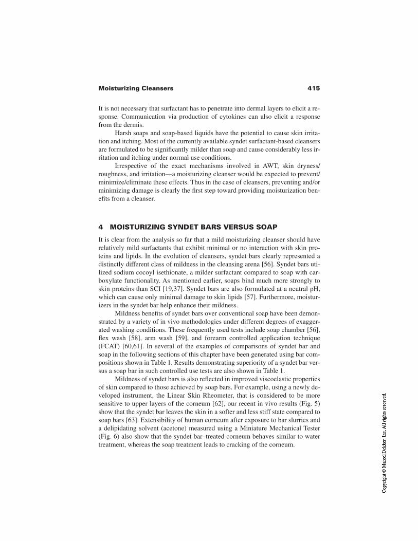
415Moisturizing Cleansers
It is not necessary that surfactant has to penetrate into dermal layers to elicit a re-sponse. Communication via production of cytokines can also elicit a responsefrom the dermis.
Harsh soaps and soap-based liquids have the potential to cause skin irrita-tion and itching. Most of the currently available syndet surfactant-based cleansersare formulated to be significantly milder than soap and cause considerably less ir-ritation and itching under normal use conditions.
Irrespective of the exact mechanisms involved in AWT, skin dryness/roughness, and irritation—a moisturizing cleanser would be expected to prevent/minimize/eliminate these effects. Thus in the case of cleansers, preventing and/orminimizing damage is clearly the first step toward providing moisturization ben-efits from a cleanser.
4 MOISTURIZING SYNDET BARS VERSUS SOAP
It is clear from the analysis so far that a mild moisturizing cleanser should haverelatively mild surfactants that exhibit minimal or no interaction with skin pro-teins and lipids. In the evolution of cleansers, syndet bars clearly represented adistinctly different class of mildness in the cleansing arena [56]. Syndet bars uti-lized sodium cocoyl isethionate, a milder surfactant compared to soap with car-boxylate functionality. As mentioned earlier, soaps bind much more strongly toskin proteins than SCI [19,37]. Syndet bars are also formulated at a neutral pH,which can cause only minimal damage to skin lipids [57]. Furthermore, moistur-izers in the syndet bar help enhance their mildness.
Mildness benefits of syndet bars over conventional soap have been demon-strated by a variety of in vivo methodologies under different degrees of exagger-ated washing conditions. These frequently used tests include soap chamber [56],flex wash [58], arm wash [59], and forearm controlled application technique(FCAT) [60,61]. In several of the examples of comparisons of syndet bar andsoap in the following sections of this chapter have been generated using bar com-positions shown in Table 1. Results demonstrating superiority of a syndet bar ver-sus a soap bar in such controlled use tests are also shown in Table 1.
Mildness of syndet bars is also reflected in improved viscoelastic propertiesof skin compared to those achieved by soap bars. For example, using a newly de-veloped instrument, the Linear Skin Rheometer, that is considered to be moresensitive to upper layers of the corneum [62], our recent in vivo results (Fig. 5)show that the syndet bar leaves the skin in a softer and less stiff state compared tosoap bars [63]. Extensibility of human corneum after exposure to bar slurries anda delipidating solvent (acetone) measured using a Miniature Mechanical Tester(Fig. 6) also show that the syndet bar–treated corneum behaves similar to watertreatment, whereas the soap treatment leads to cracking of the corneum.
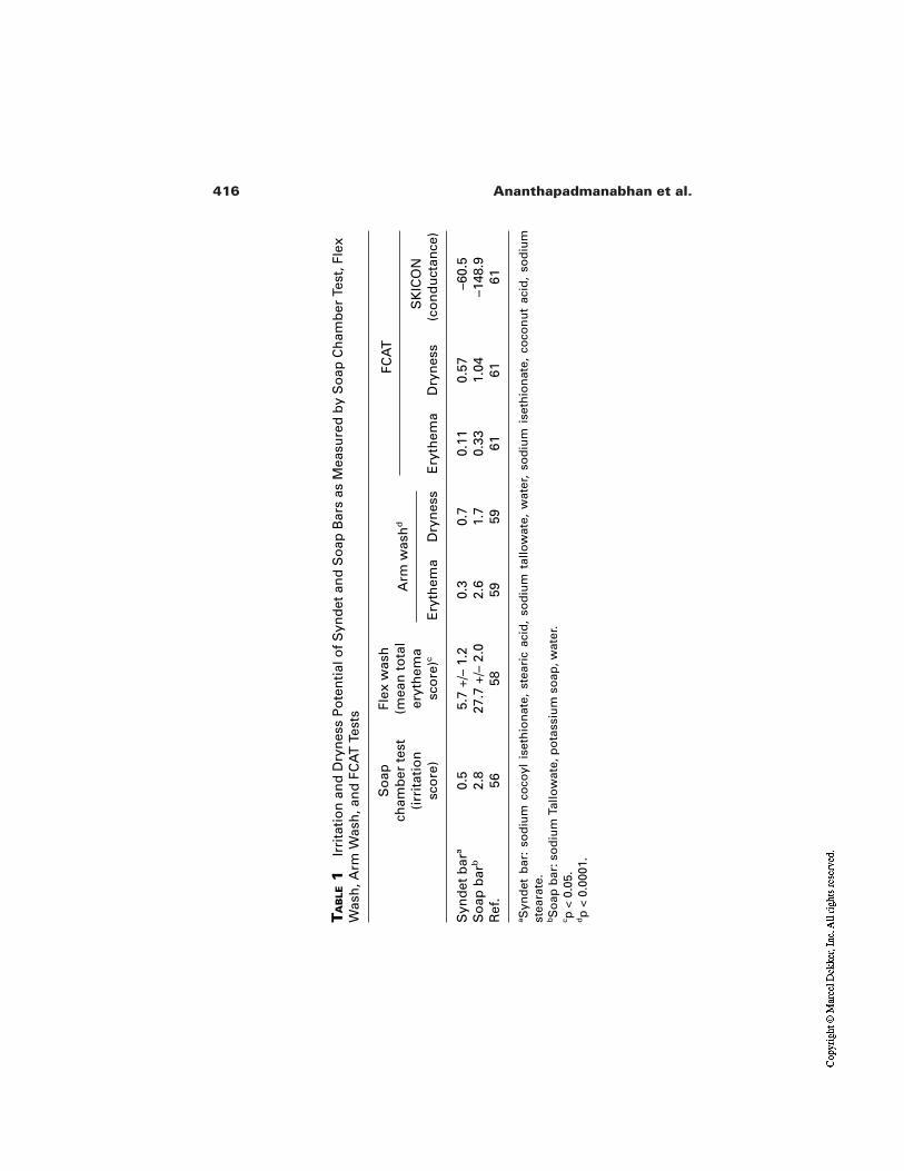
416 Ananthapadmanabhan et al.
TA
BLE
1Ir
rita
tio
n a
nd
Dry
nes
s P
ote
nti
al o
f S
ynd
et a
nd
So
ap B
ars
as M
easu
red
by
So
ap C
ham
ber
Tes
t, F
lex
Was
h, A
rm W
ash
, an
d F
CA
T T
ests
FCA
T
sco
re)
sco
re)c
Ery
them
aD
ryn
ess
Ery
them
aD
ryn
ess
(co
nd
uct
ance
)
Syn
det
bar
a0.
55.
7 +/
– 1.
20.
30.
70.
110.
57–6
0.5
So
ap b
arb
2.8
27.7
+/–
2.0
2.6
1.7
0.33
1.04
–148
.9R
ef.
5658
5959
6161
61
aS
ynd
et b
ar:
sod
ium
co
coyl
ise
thio
nat
e, s
tear
ic a
cid
, so
diu
m t
allo
wat
e, w
ater
, so
diu
m i
seth
ion
ate,
co
con
ut
acid
, so
diu
mst
eara
te.
bS
oap
bar
: so
diu
m T
allo
wat
e, p
ota
ssiu
m s
oap
, wat
er.
c p <
0.0
5.dp
< 0
.000
1.
So
apch
amb
er t
est
Flex
was
h(m
ean
to
tal
(irr
itat
ion
er
yth
ema
Arm
was
hd
SK
ICO
N
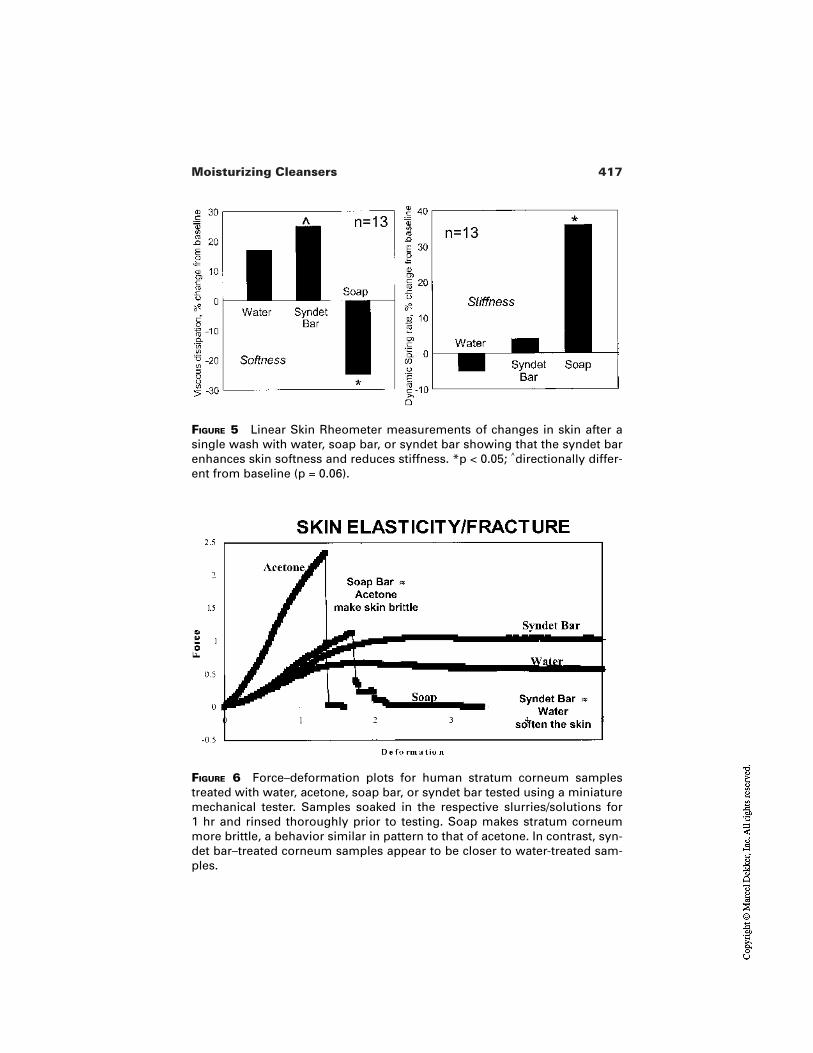
417Moisturizing Cleansers
FIGURE 5 Linear Skin Rheometer measurements of changes in skin after asingle wash with water, soap bar, or syndet bar showing that the syndet barenhances skin softness and reduces stiffness. *p < 0.05; ^directionally differ-ent from baseline (p = 0.06).
FIGURE 6 Force–deformation plots for human stratum corneum samplestreated with water, acetone, soap bar, or syndet bar tested using a miniaturemechanical tester. Samples soaked in the respective slurries/solutions for 1 hr and rinsed thoroughly prior to testing. Soap makes stratum corneummore brittle, a behavior similar in pattern to that of acetone. In contrast, syn-det bar–treated corneum samples appear to be closer to water-treated sam-ples.
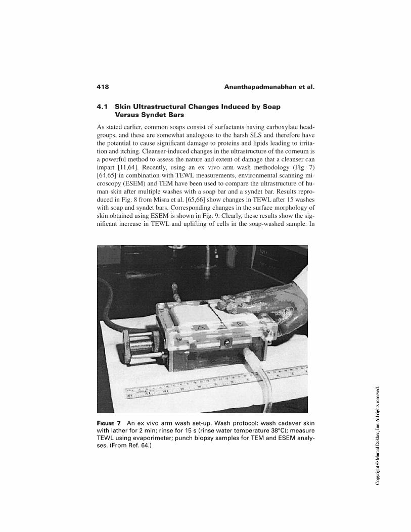
418 Ananthapadmanabhan et al.
4.1 Skin Ultrastructural Changes Induced by SoapVersus Syndet Bars
As stated earlier, common soaps consist of surfactants having carboxylate head-groups, and these are somewhat analogous to the harsh SLS and therefore havethe potential to cause significant damage to proteins and lipids leading to irrita-tion and itching. Cleanser-induced changes in the ultrastructure of the corneum isa powerful method to assess the nature and extent of damage that a cleanser canimpart [11,64]. Recently, using an ex vivo arm wash methodology (Fig. 7)[64,65] in combination with TEWL measurements, environmental scanning mi-croscopy (ESEM) and TEM have been used to compare the ultrastructure of hu-man skin after multiple washes with a soap bar and a syndet bar. Results repro-duced in Fig. 8 from Misra et al. [65,66] show changes in TEWL after 15 washeswith soap and syndet bars. Corresponding changes in the surface morphology ofskin obtained using ESEM is shown in Fig. 9. Clearly, these results show the sig-nificant increase in TEWL and uplifting of cells in the soap-washed sample. In
FIGURE 7 An ex vivo arm wash set-up. Wash protocol: wash cadaver skinwith lather for 2 min; rinse for 15 s (rinse water temperature 38°C); measureTEWL using evaporimeter; punch biopsy samples for TEM and ESEM analy-ses. (From Ref. 64.)
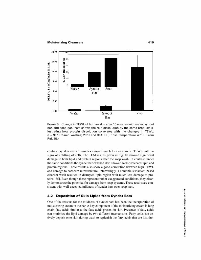
419Moisturizing Cleansers
FIGURE 8 Change in TEWL of human skin after 15 washes with water, syndetbar, and soap bar. Inset shows the zein dissolution by the same products il-lustrating how protein dissolution correlates with the changes in TEWL. n = 9; 15 2-min washes; 25°C and 30% RH; rinse temperature 40°C. (From Ref. 65.)
contrast, syndet-washed samples showed much less increase in TEWL with nosigns of uplifting of cells. The TEM results given in Fig. 10 showed significantdamage to both lipid and protein regions after the soap wash. In contrast, underthe same conditions the syndet bar–washed skin showed well-preserved lipid andprotein regions. These results also show a good correlation between high TEWLand damage to corneum ultrastructure. Interestingly, a nonionic surfactant-basedcleanser wash resulted in disrupted lipid region with much less damage to pro-teins [65]. Even though these represent rather exaggerated conditions, they clear-ly demonstrate the potential for damage from soap systems. These results are con-sistent with well-accepted mildness of syndet bars over soap bars.
4.2 Deposition of Skin Lipids from Syndet Bars
One of the reasons for the mildness of syndet bars has been the incorporation ofmoisturizing cream in the bar. A key component of the moisturizing cream is longchain fatty acids similar to the fatty acids present in skin. Presence of fatty acidscan minimize the lipid damage by two different mechanisms. Fatty acids can ac-tively deposit onto skin during wash to replenish the fatty acids that are lost dur-
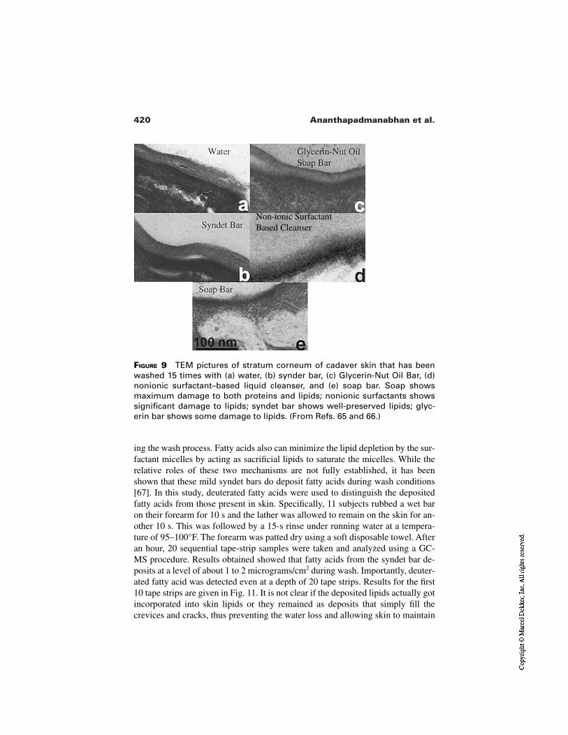
420 Ananthapadmanabhan et al.
FIGURE 9 TEM pictures of stratum corneum of cadaver skin that has beenwashed 15 times with (a) water, (b) synder bar, (c) Glycerin-Nut Oil Bar, (d)nonionic surfactant–based liquid cleanser, and (e) soap bar. Soap showsmaximum damage to both proteins and lipids; nonionic surfactants showssignificant damage to lipids; syndet bar shows well-preserved lipids; glyc-erin bar shows some damage to lipids. (From Refs. 65 and 66.)
ing the wash process. Fatty acids also can minimize the lipid depletion by the sur-factant micelles by acting as sacrificial lipids to saturate the micelles. While therelative roles of these two mechanisms are not fully established, it has beenshown that these mild syndet bars do deposit fatty acids during wash conditions[67]. In this study, deuterated fatty acids were used to distinguish the depositedfatty acids from those present in skin. Specifically, 11 subjects rubbed a wet baron their forearm for 10 s and the lather was allowed to remain on the skin for an-other 10 s. This was followed by a 15-s rinse under running water at a tempera-ture of 95–100°F. The forearm was patted dry using a soft disposable towel. Afteran hour, 20 sequential tape-strip samples were taken and analyzed using a GC-MS procedure. Results obtained showed that fatty acids from the syndet bar de-posits at a level of about 1 to 2 micrograms/cm2 during wash. Importantly, deuter-ated fatty acid was detected even at a depth of 20 tape strips. Results for the first10 tape strips are given in Fig. 11. It is not clear if the deposited lipids actually gotincorporated into skin lipids or they remained as deposits that simply fill thecrevices and cracks, thus preventing the water loss and allowing skin to maintain
Non-ionic SurfactantBased Cleanser
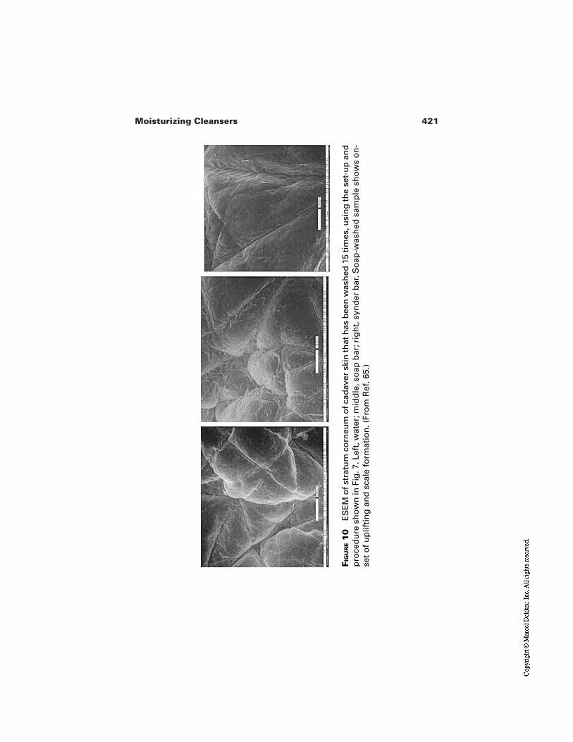
421Moisturizing Cleansers
FIG
UR
E1
0E
SE
M o
f str
atu
m c
orn
eum
of c
adav
er s
kin
that
has
bee
n w
ash
ed 1
5 ti
mes
, usi
ng
the
set-
up
an
dp
roce
du
re s
ho
wn
in F
ig. 7
. Lef
t, w
ater
; mid
dle
, so
ap b
ar; r
igh
t, s
ynd
er b
ar. S
oap
-was
hed
sam
ple
sh
ow
s o
n-
set
of
up
lifti
ng
an
d s
cale
fo
rmat
ion
. (Fr
om
Ref
. 65.
)
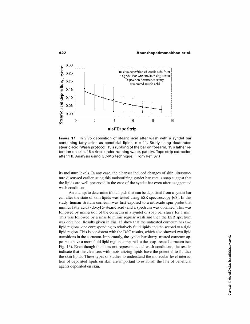
422 Ananthapadmanabhan et al.
FIGURE 11 In vivo deposition of stearic acid after wash with a syndet barcontaining fatty acids as beneficial lipids. n = 11. Study using deuteratedstearic acid. Wash protocol: 15 s rubbing of the bar on forearm, 15 s lather re-tention on skin, 15 s rinse under running water, pat dry. Tape strip extractionafter 1 h. Analysis using GC-MS technique. (From Ref. 67.)
its moisture levels. In any case, the cleanser induced changes of skin ultrastruc-ture discussed earlier using this moisturizing syndet bar versus soap suggest thatthe lipids are well preserved in the case of the syndet bar even after exaggeratedwash conditions.
An attempt to determine if the lipids that can be deposited from a syndet barcan alter the state of skin lipids was tested using ESR spectroscopy [68]. In thisstudy, human stratum corneum was first exposed to a nitroxide spin probe thatmimics fatty acids (doxyl 5-stearic acid) and a spectrum was obtained. This wasfollowed by immersion of the corneum in a syndet or soap bar slurry for 1 min.This was followed by a rinse to mimic regular wash and then the ESR spectrumwas obtained. Results given in Fig. 12 show that the untreated corneum has twolipid regions, one corresponding to relatively fluid lipids and the second to a rigidlipid region. This is consistent with the DSC results, which also showed two lipidtransitions in the corneum. Importantly, the syndet bar slurry–treated corneum ap-pears to have a more fluid lipid region compared to the soap-treated corneum (seeFig. 13). Even though this does not represent actual wash conditions, the resultsindicate that the cleansers with moisturizing lipids have the potential to fluidizethe skin lipids. These types of studies to understand the molecular level interac-tion of deposited lipids on skin are important to establish the fate of beneficialagents deposited on skin.
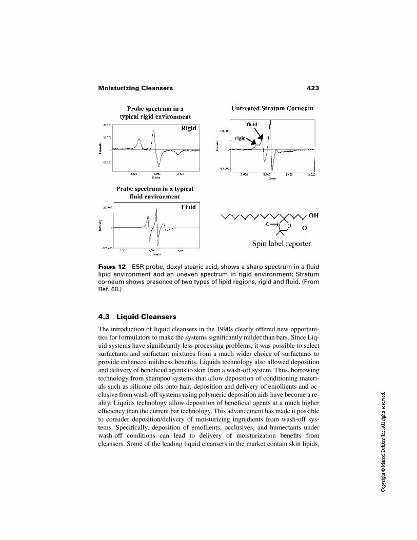
423Moisturizing Cleansers
FIGURE 12 ESR probe, doxyl stearic acid, shows a sharp spectrum in a fluidlipid environment and an uneven spectrum in rigid environment; Stratumcorneum shows presence of two types of lipid regions, rigid and fluid. (FromRef. 68.)
4.3 Liquid Cleansers
The introduction of liquid cleansers in the 1990s clearly offered new opportuni-ties for formulators to make the systems significantly milder than bars. Since Liq-uid systems have significantly less processing problems, it was possible to selectsurfactants and surfactant mixtures from a much wider choice of surfactants toprovide enhanced mildness benefits. Liquids technology also allowed depositionand delivery of beneficial agents to skin from a wash-off system. Thus, borrowingtechnology from shampoo systems that allow deposition of conditioning materi-als such as silicone oils onto hair, deposition and delivery of emollients and oc-clusive from wash-off systems using polymeric deposition aids have become a re-ality. Liquids technology allow deposition of beneficial agents at a much higherefficiency than the current bar technology. This advancement has made it possibleto consider deposition/delivery of moisturizing ingredients from wash-off sys-tems. Specifically, deposition of emollients, occlusives, and humectants underwash-off conditions can lead to delivery of moisturization benefits fromcleansers. Some of the leading liquid cleansers in the market contain skin lipids,
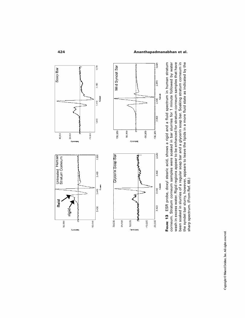
424 Ananthapadmanabhan et al.
FIG
UR
E1
3E
SR
pro
be,
do
xyl
stea
ric
acid
, sh
ow
s a
rig
id a
nd
a fl
uid
sp
ectr
um
in
hu
man
str
atu
mco
rneu
m.
Str
atu
m c
orn
eum
sam
ple
s w
ere
soak
ed i
n b
ar s
lurr
ies
for
1 m
inu
te f
ollo
wed
by
wat
erw
ash
in e
xces
s w
ater
. Rig
id r
egio
ns
app
ear
to b
e en
han
ced
fo
r st
ratu
m c
orn
eum
sam
ple
s th
at h
ave
bee
n s
oak
ed in
slu
rrie
s o
f a
reg
ula
r so
ap b
ar a
nd
a g
lyce
rin
so
ap b
ar. S
oak
ing
str
atu
m c
orn
eum
inth
e sy
nd
et b
ar s
lurr
y, h
ow
ever
, ap
pea
rs t
o le
ave
the
lipid
s in
a m
ore
flu
id s
tate
as
ind
icat
ed b
y th
esh
arp
sp
ectr
um
. (Fr
om
Ref
. 68.
)
Unt
reat
ed
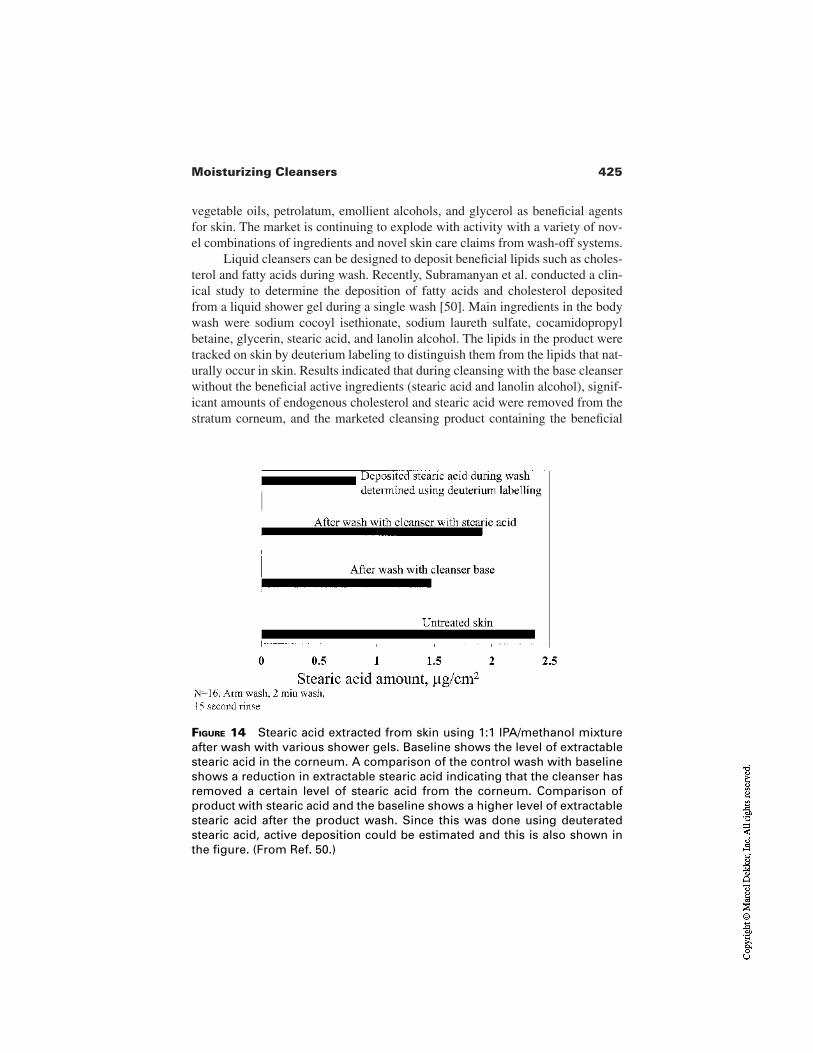
425Moisturizing Cleansers
vegetable oils, petrolatum, emollient alcohols, and glycerol as beneficial agentsfor skin. The market is continuing to explode with activity with a variety of nov-el combinations of ingredients and novel skin care claims from wash-off systems.
Liquid cleansers can be designed to deposit beneficial lipids such as choles-terol and fatty acids during wash. Recently, Subramanyan et al. conducted a clin-ical study to determine the deposition of fatty acids and cholesterol depositedfrom a liquid shower gel during a single wash [50]. Main ingredients in the bodywash were sodium cocoyl isethionate, sodium laureth sulfate, cocamidopropylbetaine, glycerin, stearic acid, and lanolin alcohol. The lipids in the product weretracked on skin by deuterium labeling to distinguish them from the lipids that nat-urally occur in skin. Results indicated that during cleansing with the base cleanserwithout the beneficial active ingredients (stearic acid and lanolin alcohol), signif-icant amounts of endogenous cholesterol and stearic acid were removed from thestratum corneum, and the marketed cleansing product containing the beneficial
FIGURE 14 Stearic acid extracted from skin using 1:1 IPA/methanol mixtureafter wash with various shower gels. Baseline shows the level of extractablestearic acid in the corneum. A comparison of the control wash with baselineshows a reduction in extractable stearic acid indicating that the cleanser hasremoved a certain level of stearic acid from the corneum. Comparison ofproduct with stearic acid and the baseline shows a higher level of extractablestearic acid after the product wash. Since this was done using deuteratedstearic acid, active deposition could be estimated and this is also shown inthe figure. (From Ref. 50.)
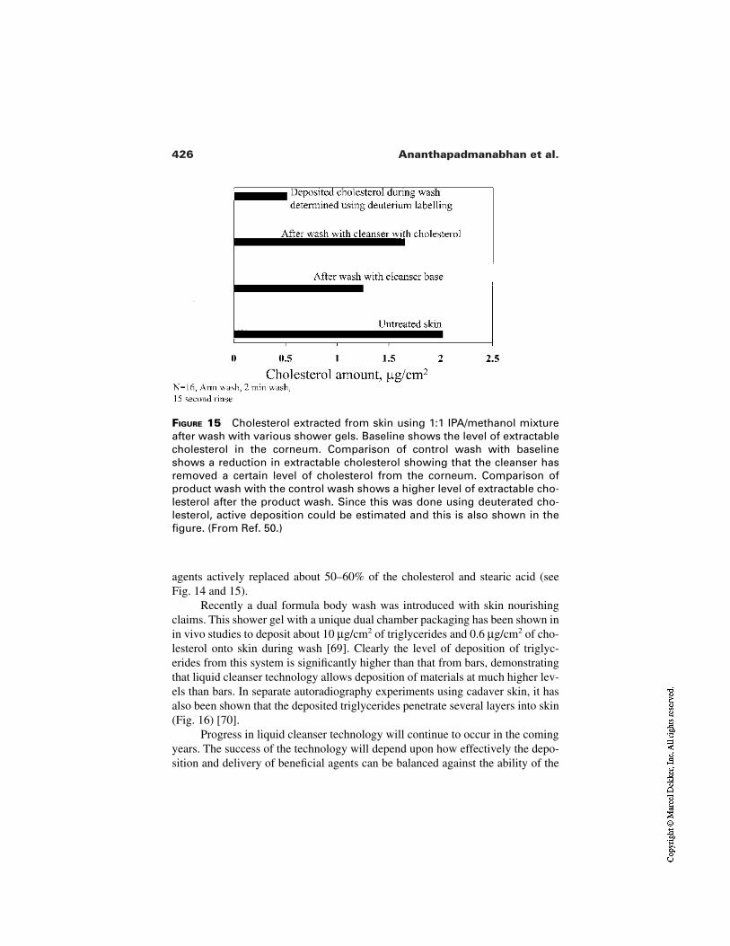
426 Ananthapadmanabhan et al.
FIGURE 15 Cholesterol extracted from skin using 1:1 IPA/methanol mixtureafter wash with various shower gels. Baseline shows the level of extractablecholesterol in the corneum. Comparison of control wash with baselineshows a reduction in extractable cholesterol showing that the cleanser hasremoved a certain level of cholesterol from the corneum. Comparison ofproduct wash with the control wash shows a higher level of extractable cho-lesterol after the product wash. Since this was done using deuterated cho-lesterol, active deposition could be estimated and this is also shown in thefigure. (From Ref. 50.)
agents actively replaced about 50–60% of the cholesterol and stearic acid (seeFig. 14 and 15).
Recently a dual formula body wash was introduced with skin nourishingclaims. This shower gel with a unique dual chamber packaging has been shown inin vivo studies to deposit about 10 µg/cm2 of triglycerides and 0.6 µg/cm2 of cho-lesterol onto skin during wash [69]. Clearly the level of deposition of triglyc-erides from this system is significantly higher than that from bars, demonstratingthat liquid cleanser technology allows deposition of materials at much higher lev-els than bars. In separate autoradiography experiments using cadaver skin, it hasalso been shown that the deposited triglycerides penetrate several layers into skin(Fig. 16) [70].
Progress in liquid cleanser technology will continue to occur in the comingyears. The success of the technology will depend upon how effectively the depo-sition and delivery of beneficial agents can be balanced against the ability of the
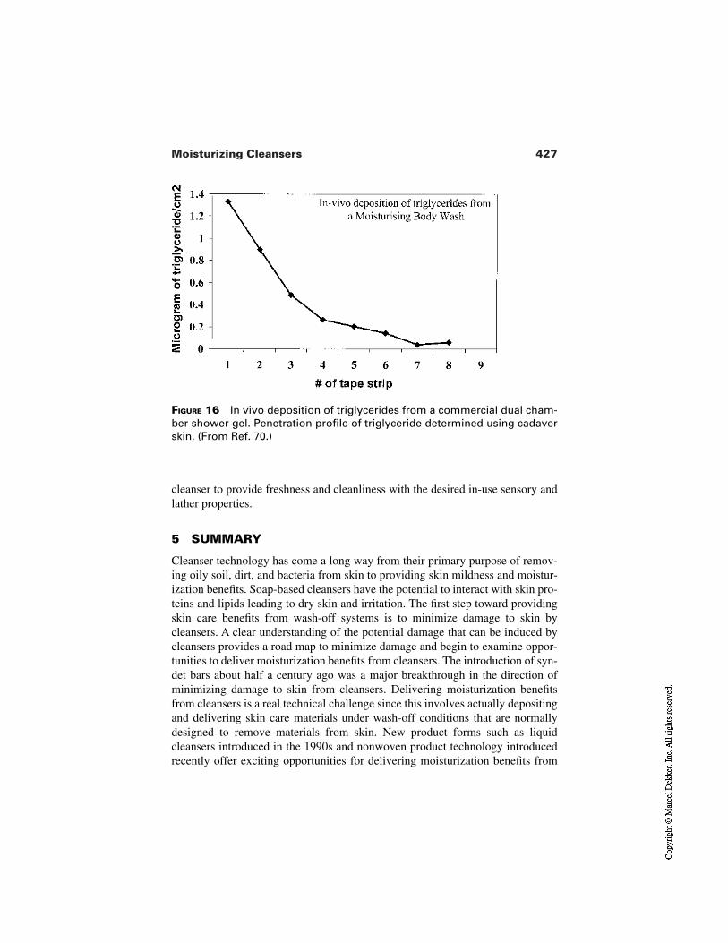
427Moisturizing Cleansers
FIGURE 16 In vivo deposition of triglycerides from a commercial dual cham-ber shower gel. Penetration profile of triglyceride determined using cadaverskin. (From Ref. 70.)
cleanser to provide freshness and cleanliness with the desired in-use sensory andlather properties.
5 SUMMARY
Cleanser technology has come a long way from their primary purpose of remov-ing oily soil, dirt, and bacteria from skin to providing skin mildness and moistur-ization benefits. Soap-based cleansers have the potential to interact with skin pro-teins and lipids leading to dry skin and irritation. The first step toward providingskin care benefits from wash-off systems is to minimize damage to skin bycleansers. A clear understanding of the potential damage that can be induced bycleansers provides a road map to minimize damage and begin to examine oppor-tunities to deliver moisturization benefits from cleansers. The introduction of syn-det bars about half a century ago was a major breakthrough in the direction ofminimizing damage to skin from cleansers. Delivering moisturization benefitsfrom cleansers is a real technical challenge since this involves actually depositingand delivering skin care materials under wash-off conditions that are normallydesigned to remove materials from skin. New product forms such as liquidcleansers introduced in the 1990s and nonwoven product technology introducedrecently offer exciting opportunities for delivering moisturization benefits from
428 Ananthapadmanabhan et al.
wash-off systems. Skin cleansing products that contain emollients, occlusives,humectants, and skin nutrients have begun to appear in the marketplace already.This trend of providing skin care benefits from wash-off systems will continue tobe an area of active research resulting in novel product forms and technologies inthe coming years.
Acknowledgments
The authors would like to thank Drs. S. Mukherjee, X. Lei, and N.J. Turro forgranting permission to use their unpublished data on ESR spectroscopy ofcorneum treated with cleanser solutions. We would also like to thank Drs. M.Misra and M. Aronson for their helpful discussions and Unilever Research for al-lowing us to publish this work.
REFERENCES
1. Spitz L. In: Spitz L, ed. Soap Technology for the 1990’s. Champaign, IL: Am OilChem Soc 1990:1–47.
2. Stanislaus IVS, Meerbott PB. In: American Soap Makers Guide. New York: HenryCarey Baird, 1928:914–919.
3. Murahata RI, Aronson MP, Sharko PT, Greene AP. In: Rieger MM, Rhein LD, eds.Surfactants in Cosmetics. New York: Marcel Dekker, 1997:307–330.
4. Matthies W. Dermatological observations. In: Gloxhuber C, Kunstler K, eds. Anion-ic Surfactants. New York: Marcel Dekker, 1992:291–329.
5. Warner RR, Lilly NA. Correlation of water content with ultrastructure in the stratumcorneum. In: Elsner P, Berardesca E, Maibach HI, eds. Bioengineering of the Skin:Water and the Stratum Corneum. Boca Raton: CRC Press, 1994:3–12.
6. Leveque J. Water–keratin interactions. In: Elsner P, Berardesca E, Maibach HI, eds.Bioengineering of the Skin: Water and the Stratum Corneum. Boca Raton: CRCPress, 1994:13–22.
7. Yamamura T, Tezuka T. The water holding capacity of the stratum corneum mea-sured by 1H-NMR. J Invest Dermatol 1989; 93(1):160–164.
8. Loden M, Lindberg M. Product testing—testing moisturizers. In: Elsner P, Be-rardesca E, Maibach HI, eds. Bioengineering of the Skin: Water and the StratumCorneum. Boca Raton: CRC Press, 1994:275–289.
9. Imokawa G, Sumura K, Katsumi M. Study on skin roughness caused by surfactants.II. Correlation between protein denaturation and skin roughness. J Am Oil Chem Soc1975; 52:484.
10. Wihelm KP, Wolff HH, Maibach HI. Effects of surfactants on skin hydration. In: Els-ner P, Berardesca E, Maibach HI, eds. Bioengineering of the Skin: Water and theStratum Corneum. Boca Raton: CRC Press, 1994:257–274.
11. Imokawa G, Akasaki S, Minematsu Y, Kawai M. Importance of intercellular lipids inwater-retention properties of the stratum corneum: induction and recovery study ofsurfactant dry skin. Arch Dermatol Res 1989; 281:45–51.
12. Inoue I, Miyakawa K, Shimozawa K. Interaction of surfactants with vesicle mem-
429Moisturizing Cleansers
brane of dipalmitoylphosphatidylcholine—effect on gel-to-liquid crystalline phasetransition of lipid bilayer. Chem Phys Lipids 1986; 42:261–270.
13. Lichtenberg D, Robson J, Dennis EA. Characterization of the lipid bilayers by sur-factants. Biochim Biophys Acta 1985; 821(3):470–478.
14. de la Maza A, Coderch L, Lopez O, Baucells J, Parra JL. Permeability changescaused by surfactants in liposomes that model the stratum corneum lipid composi-tion. J Am Oil Chem Soc 1997; 74(1):1–8.
15. Middleton JD. The mechanism of action of surfactants on the water binding proper-ties of isolated stratum corneum. J Soc Cosmet Chem 1969; 20:399–412.
16. Prottey C, Ferguson T. Factors which determine the skin irritation potential of soapsand detergents. J Soc Cosmet Chem 1975; 26:29–46.
17. Kawai M, Imokawa G. The induction of skin tightness by surfactants. J Soc CosmetChem 1984; 35:147–156.
18. Rhein LD. In vitro interactions: biochemical and biophysical effects of surfactantson skin. In: Rieger MM, Rhein LD, eds. Surfactants in Cosmetics. New York: Mar-cel Dekker, 1997:397–425.
19. Imokawa G. Surfactant mildness. In: Rieger MM, Rhein LD, eds. Surfactants inCosmetics. New York: Marcel Dekker, 1997:427–471.
20. Abraham W. Surfactant effects on skin repair. In: Rieger MM, Rhein LD, eds. Sur-factants in Cosmetics. New York: Marcel Dekker, 1997:473–487.
21. Rizvi PY, Morrison BM. Bioengineering techniques for investigating the effects ofsurfactants on skin. In: Rieger MM, Rhein LD, eds. Surfactants in Cosmetics. NewYork: Marcel Dekker, 1997:489–499.
22. Simion FA. Human in vivo methods for assessing the irritation potential of cleansingsystems. In: Rieger MM, Rhein LD, eds. Surfactants in Cosmetics. New York: Mar-cel Dekker, 1997:519–532.
23. Elias PM. Epidermal lipids, barrier function, and desquamation. J Invest Dermatol1983; 80:44S–49S.
24. Swartzendruber DC, Wertz PW, Madison KC, Downing DT. Evidence that the cor-neocyte has a chemically bound lipid envelope. J Invest Dermatol 1987; 88(6):709–713.
25. Madison KC, Swartzendruber DC, Wertz PW, Elias PM. Presence of intact intercel-lular lipid lamellae in the upper layers of the stratum corneum. J Invest Dermatol1987; 88(6):714–718.
26. Rehfeld SJ, Plachy WZ, Williams ML, Elias PM. Calorimetric and electron spin res-onance examination of lipid phase transition in human stratum corneum: molecularbasis for normal cohesion and abnormal desquamation in recessive X-linkedichthyosis. J Invest Dermatol 1988; 91:499–505.
27. Middleton JD. The mechanism of action of surfactants on the water binding proper-ties of isolated stratum corneum. J Soc Cosmet Chem 1969; 20:399–412.
28. Chapman SJ, Walsh A. Desquamation, corneosomes and desquamation. Anultrastructural study of adult pig epidermis. Arch Dermatol Res 1990; 282:304–310.
29. Egelrud T, Lundstrom A. The dependence of detergent induced cell dissociation innon-palmo-plantar stratum corneum on endogenous proteolysis. J Invest Dermatol1992; 90:456–459.
430 Ananthapadmanabhan et al.
30. Imokawa G, Mishima J. Cumulative effects of surfactants on cutaneous horny lay-ers—adsorption onto human keratin layers in vivo. Contact Dermatitis 1979;5(6):357–366.
31. Faucher JA, Goddard ED. Interaction of keratinous proteins with sodium lauryl sul-fate: sorption. J Soc Cosmet Chem 1978; 29:323–338.
32. Breuer MM. The interaction between surfactants and keratinous tissues. J Soc Cos-met Chem 1979; 30:41–64.
33. Dominguez JG, Parra JL, Infante RM, Pelejero F, Balaguer F, Sastre TA. New ap-proach to the theory of adsorption and permeability of surfactants on keratinic pro-teins: specific behavior of certain hydrophobic chains. J Soc Cosmet Chem 1977;28:165–182.
34. Conrads A, Zahn H. A study of interaction of sodium dodecyl sulfate with proteins ofhuman stratum corneum. Int J Cosmet Sci 1976; 9:29–46.
35. Schwuger MJ, Bartnik FG. Interaction of anionic surfactants with proteins, enzymes,and membranes. In: Gloxhuber C, ed. Anionic Surfactants, Surfactant Science Se-ries, Vol. 10. New York: Marcel Dekker, 1980:1–49.
36. Ananthapadmanabhan KP. Protein–surfactant interactions. In: Goddard ED, Anan-thapadmanabhan KP, eds. Interaction of Surfactants with Polymers and Proteins.Boca Raton: CRC Press, 1993:319–366.
37. Ananthapadmanabhan KP. Binding of surfactants to stratum corneum. J Soc CosmetChem 1996; 47:185–200.
38. Rhein LD, Simion FA. Surfactant interactions with skin. In: Bender M, ed. Interfa-cial Phenomena in Biological Systems. Surfactant Science Series. Vol. 39. NewYork: Marcel Dekker, 1991:33–49.
39. Pierard GE, Goffin V, Pierard-Franchimont C. Corneosufametry: a predictive assess-ment of the interaction of personal care cleansing products with human stratumcorneum. Dermatology 1994; 189:152–156.
40. Dominguez JG, Balaguer F, Parra JL, Pelejero CM. The inhibitory effect of someamphoteric surfactants on the irritation potential of alkyl sulfates. Int J Cosmet Soc1981; 3(2):57–68.
41. Gotte E. Skin compatibility of tensides measured by their capacity for dissolvingzein In: Proceedings of the 4th International Congress on Surface Active Substances,Brussels; 1964, pp. 83–90.
42. Rhein LD, Robbins CR, Kernee K, Cantore R. Surfactant structure effects onswelling of isolated human stratum corneum. J Soc Cosmet Chem 1986; 37:125–139.
43. Wilhelm KP, Cua AB, Wolff HH, Maibach HI. Predicting surfactant induced stratumcorneum hydration in vivo: prediction of the irritation potential of anionic surfac-tants. J Invest Dermatol 1994; 101:310–315.
44. Fulmer AW, Kramer GJ. Stratum corneum lipid abnormalities in surfactant-induceddry scaly skin. J Invest Dermatol 1986; 86:598–602.
45. Fartasch M, Diepgen TL, Hornstein OP. Morphological changes of epidermal lipidlayers of stratum corneum in sodium lauryl sulfate induced dry skin: a functional andultrastructural study. J Invest Dermatol 1991; 96(4):617.
46. Rieger M. Skin lipids and their importance to cosmetic science. Cosmet Toil 1987;102(7):36–49.
431Moisturizing Cleansers
47. Rawlings AW, Watkinson A, Rogers J, Mayo HJ, Scott IR. Abnormalities in stratumcorneum structure, lipid composition, and desmosome degradationin soap-inducedwinter zerosis. J Soc Cosmet Chem 1994; 45:203–220.
48. Imokawa G, Akasaki S, Hattori M, Yoshizuka N. Selective recovery of deranged wa-ter-holding properties by stratum corneum lipids. J Invest Dermatol 1986; 187:758–761.
49. Leveque JL, DeRigal J, Legere DS, Billy D. How does sodium lauryl sulfate alter theskin barrier function in man—a multiparametric approach. Skin Pharmacol 1993;6(2):111–115.
50. Subramanyan K, Wong J, Ananthapadmanabhan K, Pereira A. Deposition of lipidsfrom personal wash cleansers. Poster presentation at the IFSCC conference, Berlin,Sep 2000.
51. Froebe CL, Simion FA, Rhein LD, Cagan RH, Kligman A. Stratum corneum lipid re-moval by surfactants: relation to in vivo irritation. Dermatologica 1990; 181:277–283.
52. Downing DT, Abraham W, Wegner KK, Willman KW, Marshal JL. Partition of sodi-um dodecyl sulfate into stratum corneum lipid liposomes. Arch Dermatol Res 1993;285(3):151–157.
53. Grubauer G, Feingold KR, Harris RM, Elias PM. Lipid content and lipid type as de-terminants of the epidermal permeability barrier. J Lipid Res 1989; 30:89–96.
54. Simion FA, Rhein LD, Morrison BM, Scala DD, Salko DM, Kligman AM, GroveGL. Self-perceived sensory responses to soap and synthetic detergent bars correlatewith clinical signs of irritation. J Am Acad Dermatol 1995; 32:205–211.
55. Imokawa G, Hattori M. A possible function of structural lipids in the water-holdingproperties of the stratum corneum. J Invest Dermatol 1985; 84(4):282–284.
56. Frosch PJ, Kligman AM. The soap chamber test. J Am Acad Dermatol 1979;1(1):35–41.
57. Murahata RI, Aronson MP. The relationship between solution pH and clinical irritan-cy for carboxylic acid-based personal washing products. J Soc Cosmet Chem 1994;45:239–246.
58. Strube DA, Koontz SW, Murahata RI, Theiler RI, The flex wash test: a method forevaluating the mildness of personal washing products. J Soc Cosmet Chem 1989;40:297–306.
59. Sharko PT, Murahata RI, Leyden JL, Grove GL. Arm wash with instrumental evalu-ation—a sensitive technique for differentiating the irritation potential of personalwashing products. J Dermoclinical Eval Soc 1991; 2:19–27.
60. Ertel KD, Keswick BH, Bryant PB. A forearm controlled application technique forestimating the relative mildness of personal cleansing products. J Soc Cosmet Chem1995; 46:67–76.
61. Azri-Meehan S, Edison B, Borowski D. Clinical evaluation of soap bars. UnileverResearch US, Nov 1998.
62. Matts PJ, Goodyer E. A new instrument to measure the mechanical properties of hu-man stratum corneum in vivo. J Cosmet Sci 1998; 49:321–333.
63. Subramanyan K, Bautista B, Mok W, Meyers L. Unpublished results, Unilever Re-search, Sep 2000.
64. Ananthapadmanabhan KP, Prowell S, Hoyberg K, Misra M, Spaltro S, Mukherjee S,
432 Ananthapadmanabhan et al.
Aronson MP. Cleanser induced structural changes in human stratum corneum. Proc4th Congr Eur Acad Dermatol Venereol, Brussels, 1995, p. 143.
65. Misra M, Ananthapadmanabhan KP, Hoyberg K, Gursky RP, Prowell S, AronsonMP. Correlation between surfactant-induced ultrastructural changes in epidermis andtransepidermal water loss. J Soc Cosmet Chem 1997; 48:219–234.
66. Misra M, Ananthapadmanabhan KP. Quantitative analysis of surfactant induced ul-trastructural changes in skin lipids. In: Lal M, Lillford PJ, Naik VM, Prakash V, eds.Supramolecular and Colloidal Structures in Biomaterials and Biosubstrates. Proc.5th Royal Soc Unilever-Indo-UK Forum in Materials Science and Engineering, Jan10–14, 1999, pp. 183–196.
67. Yu K, Hargiss L, Wong JK, Anathapadmanabhan KP. In-vivo deposition of stearicacid from syndet bars: a clinical study using deuterated stearic acid. Unpublished re-sults, Unilever Research US, 1995.
68. Mukherjee S, Lei X, Turro NJ, ESR study of cleanser induced changes in humanstratum corneum. Unpublished results, Unilever Research US, 1995.
69. Naser M, Atlas J, Chang E, Meyers L, Morgan L, Velez S, In-vivo deposition of cho-lesterol and triglycerides from dual chamber body wash. Unpublished results,Unilever Research US, 1998.
70. Subramanyan K, Prowell S, Ananthapadmanabhan KP. Unpublished results,Unilever Research US, 1998.
21Consumer Testing Methods
Steven S. Braddon and Gwendolyn S. JarrettUnilever Home and Personal Care North America, Trumbull, Connecticut
Alejandra M. MuñozInternational Resources for Insights and Solutions, Mountainside, New Jersey
1 INTRODUCTION
Moisturizers are big business. Consumers spend millions of dollars each year insearch of efficacious products. All one needs to do is scan the monthly “women’smagazines” to understand that moisturizers of all types—hand and body lotions,facial moisturizers, and body washes—are being advertised in record numbers.Approximately 80% of women in the United States use a hand and body lotionregularly. Products offer benefits ranging from dry skin relief to youthful, lesswrinkled, and firmer skin. Formulations include such ingredients as α- and β-hy-droxyacids (AHA and BHA), retinols, and seaweed extract. Vitamins are added asantioxidants and firming agents. It is common knowledge that herbs, long usedfor healing purposes, are finding new life in skin preparations [1]. Herbal extracts,considered by some as good for the inside, are now being used on the outside aswell. Foods have also found their way into moisturizers—soya, whey protein,oats, sugar, cucumbers, and green tea are but a few of these ingredients. It is pos-
433
434 Braddon et al.
sible to purchase moisturizers that are fragranced or unfragranced in every colorimaginable.
Products are sold not only in the more traditional locales of supermarketsand drug, discount, and department stores, but also in mall specialty shops whichprovide an endless supply of moisturizing lotions, creams, and body washes of-fering exotic fragrances in expensive packaging. The internet, the latest shoppingmedium, is another source of supply for the consumer’s insatiable quest for newand different products. To understand more about what will entice the consumerto actually buy a product, it is up to the testing professional to ascertain their likesand dislikes.
In the fast-paced environment of the personal care industry, who to test,what to test, and how to test are conundrums posed daily by many marketingbrand managers. Start-up operations and smaller businesses, some with limited orno research dollars, may rely on the intuition of a company’s president, marketingteam member, or development chemist as the deciding factor in selecting productformulas, colors, or fragrances. At more established companies those types ofquestions are normally directed to the in-house experts: market research and re-search guidance testing departments.
Market research is traditionally conducted within a marketing departmentwhich, in turn, usually operates within the corporate headquarters. It exploressuch areas as brand awareness, trial and repeat purchase, category segmentation,habits and attitude studies, advertising effectiveness, and large-scale performanceand acceptance tests for current brands and potential new products. Within the re-search and development department (R&D), with which the authors are most fa-miliar, research guidance testing seeks insight into products and prototypesthrough the analysis of early-in consumer evaluations and the assessments oftrained descriptive panels. Their main responsibility is to help guide the develop-ment chemist’s efforts to “build” products that will delight consumers and inducethem to become loyal brand users. Some companies rely more heavily on the in-sights derived from trained panels because their judgments are free of bias. Oth-ers lean toward tests with naive consumers because they are the ultimate pur-chasers of a product and because the cost of trained panels can be prohibitive.
This chapter provides a comprehensive overview of moisturizer testingmethodologies currently utilized in the personal care industry for the reader withlittle or no experience in the field as well as a review for the professional. On thefollowing pages the authors first provide a brief history of moisturizer use. Al-though commercially sold product is a relatively new invention of the 20th centu-ry, there is evidence that moisturizer-like products were in use from the earliesttimes [2]. While no testing was conducted on those early products, there exists to-day a number of options for extensive consumer, sensory, and expert testing ofmoisturizers.
Before testing can begin it is crucial to identify meaningful terms for boththe consumer and expert to evaluate a product. Terminology is important because
435Consumer Testing Methods
it provides the professional with a common lexicon for deciphering consumerfeedback. It is important to clearly understand the objectives of a study, for with-out explicitly defined goals it is impossible to choose appropriate test methods.The various methodologies discussed will comprise discrimination tests, descrip-tive panels conducted by expert evaluators, and naive consumer testing. Dataanalysis is then examined as a way of understanding consumer responses. Thechapter ends with a discussion of irritation issues and a number of testing appli-cations such as claim substantiations.
2 HISTORY OF MOISTURIZERS
Concern about the appearance of skin predates modern society. Just how far backin time cosmetics originated is somewhat surprising. Cosmetics and perfumeshave been found at ancient burial sites in Egypt [3], and the use of homemademixtures to moisturize and rejuvenate dry and aging skin have been documentedin early Greece and Rome [4,5].
2.1 Egypt
Viewing the powerful art that remains on the walls of ancient Egyptian burialsites presents a picture of a highly refined, painted culture. The early Egyptianswere not only interested in color cosmetics; home-worked products for skin mois-turizing and anti-aging also abounded. Stylish Egyptian women applied a productcalled Coan Quince Cream for silky complexions [6], and anthropological litera-ture shows that women in Greece and Rome were doing much the same thing.
2.2 Greece and Rome
Some Greek women followed a routine of spreading a poultice of bread and milkon their faces before retiring at night “to repair the effects of time as a cause ofcutaneous aging” [4]. Often referred to as the father of medicine, the Greek physi-cian Hippocrates, in his discourse on “Considerations of treatment of wounds,”speaks of using honey on the face, arguing that it “assures a fresh and jovial look”[4]. Roman women were doing likewise. Masks prepared from “breadcrumbssoaked in milk, or rye flour with honey” [7] were used at night and usually re-moved in the morning. Some women wore them all day, cleansing their facesonly to run an important errand [7]. Also, some Roman women applied the dregsfrom the bottom of wine vats looking for the same results that moisturizing prod-ucts claim today: soft and smooth skin [7]. Still others attempted to maintain theirskin with milk baths [7]. Thomas Spelios, writing in a historical review of cos-metics, states that Galen of Pergamon, the renowned physician, is credited withdeveloping cold cream sometime around 200 AD and that “the product was usedwidely by Roman women as a beauty aid for aged and dry skin . . .” [8].
436 Braddon et al.
Times have not changed much despite the passage of several thousandyears. Women of today might be surprised to learn that while their face productsare touted as new and revolutionary, some of the concoctions used so long ago bytheir Greek and Roman sisters are similar to the currently popular moisturizingcreams and lotions formulated with AHA. What a surprise to find the roots of ourexpensive moisturizers in the mixtures of Roman and Greek housewives!
2.3 Anglo-Saxons and the English
Hundreds of years later, the Anglo-Saxons developed moisturizing products theyused specifically for their hands [5]. A recipe remains for a “hand cream” that in-cluded lily of the valley, old lard, and wine. The lily of the valley and lard werepounded together, heated with the wine, and worked into a mixture. Living in thedamp and cold apparently caused many cases of dry, chapped hands.
During Elizabethan times lotions and ointments were prepared with “ass’smilk, hog lard, honey and beeswax with added embellishments of cherries, rosepetals and herbs” [5]. Elizabeth I is purported to have made many of her own cos-metics including a forehead cream with “a compound of posset curd” to removewrinkles and a skin lotion from a concoction of “egg white, powdered egg shells,alum, borax and white poppy seeds” [5].
For the next several hundred years the majority of moisturizer-like prepara-tions were made at home for personal use. During the 1800s, there was no cos-metic industry as we know it today. Some of the following account of the indus-try’s growth comes from a recent excellent book entitled, Hope In a Jar: TheMaking of America’s Beauty Culture [9]. During the 19th century cosmetics re-ferred to creams and lotions that protected the face. These were the precursors ofmodern day facial moisturizers. Women basically had three choices during thisperiod: home preparation, local druggist, and, somewhat less common, overseassources and larger wholesale drug suppliers.
2.4 Twentieth Century
By the turn of the century “the era of soap and water, and the modest applicationof home-made face creams, was certainly at an end” [5]. At the same time, one ofthe first hand lotions to be introduced was Jergens Benzoin and Almond LotionCompound, later to be known as Jergens Lotion. Initially the only mass-producedmoisturizers were cold cream and vanishing cream [5]. Cold cream was used as a cleanser and moisturizer to restore dry, flaky skin. Vanishing cream, an emol-lient, served as a make-up base and also protected facial skin against moistureloss. The now legendary figure Helena Rubinstein opened her first beauty salon in London in 1908 selling a wide array of creams and lotions. She was followedby Elizabeth Arden, who in 1910 also opened a salon in New York. Both soldproducts claiming to provide the same benefits that today’s women desire and
437Consumer Testing Methods
will pay so dearly for—youthful complexions. It was at this stage that womenwere less interested in what could be purchased from their pharmacist than frombeauty salons. As products became more effective an increased demand arose forbetter products; coupled with this, women were exerting a growing indepen-dence. By the end of World War I, cosmetics were readily available in Wool-worths and other large department stores. The transition from “class to mass”took little time. By the 1930s the beauty industry was in full swing. Advertise-ments in the popular ladies magazines of the times such as Ladies Home Journaland Good Housekeeping promoted face products that offered smooth, soft skin.The introduction of television provided yet another opportunity for industry toreach the masses by promoting the latest skin care preparations. Manufacturersspent millions of dollars each year extolling the benefits of their latest moisturiz-ing products.
Moisturizers of all types are literally used by millions of people around theglobe every day. They are looking for many of the same benefits our ancestors didthousands of years ago. Only now, they have an endless array of choices withproducts that have been tested and retested among the correct user groups. As weenter the 21st century the buzzword is innovation. The keys to the new productsof tomorrow are ingredients that work better and faster to provide healthy look-ing, moisturized skin.
3 MOISTURIZING TERMINOLOGY
3.1 Naive Consumers and Trained Sensory Panelists
To provide direction for product development, moisturizer terms or attributesmust have actionable meanings. In other words, a pattern of ratings or scoresmust be convertible into one or more courses of action for the moisturizer formu-lation. When consumers rate products on attributes such as “silky” or “greasy”their definitions are culture dependent and are also affected by the product setwith which they are familiar. The terms are fuzzy. There are core meanings plusmany shadings or nuances radiating out from the center of the definition. Productdevelopment formulators learn with experience that certain changes in a moistur-izer systematically move consumer ratings, even though no two participants in alarge study may define the attributes identically.
In contrast, moisturizer terms when used by descriptive sensory panelshave meanings defined through lengthy training with reference standards and rat-ing scales [10,11]. “Spreadibility” has a technical and precise meaning that is notcombined with other terms in the mind of the expert. Panelists strive to performratings as human machines, generating replicable sensory “signatures” for prod-ucts, as well as providing detailed information in order for chemists to know thesubtle differences that changes in a formula can make. The descriptive results are
438 Braddon et al.
nonhedonic, that is, without reference to liking or skin benefit. A more detailedtreatment of descriptive sensory panels and their unique terminology can befound in Sec. 4.2.2.
3.2 Hedonic Terminology
Like foods, moisturizers have a dual identity. They are experienced as functionalas well as hedonic/“pleasure-giving” substances. It is clear that some lotions arepositioned as more functional or therapeutic than others. Terminology or descrip-tors should then in turn emphasize the nuances of efficacy and skin healing. Con-versely, the more hedonically focused or experiential lotions require a more de-tailed treatment of pleasure or emotive qualities. Traditionally, hedonic surveyitems such as overall liking or purchase intent are not seen as the purview of thetrained sensory professional because of the “possibility of bias and resultant errorin predicting consumer preferences” [12]. Enjoying a product and functioning asa dispassionate sensory machine are incompatible. As a result, sensory languagerelated to experiential qualities is not nearly as developed as, say, texture or skin-feel properties. Fortunately, a new research discipline called hedonic psychologymay in time rectify this by bringing attention to the experiential and improvingour understanding of what makes some moisturizers pleasurable. In their editedcollection launching the field, Kahnemann et al. [13] target psychology in gener-al. They argue that pleasure and well-being are ignored topics. Consumer behav-ior is not addressed specifically. But they present some provocative findings thatmay in time find their way into consumer psychology to expand our understand-ing of the dimensions of liking and preference. One important inference fromtheir work is that retrospective assessments of one’s state of happiness or unhap-piness (as measured by the standard post-product use questionnaire) are not as ac-curate as the moment-by-moment feelings experienced in real time as a product isused. This suggests a greater use of diaries or handheld microcassette recorders tocapture what consumers feel. In the same volume, Stone et al. [14] argue for theuse of “ecological momentary assessment,” which entails the frequent probing ofa consumer’s feelings and mood states during the day as a product is used. Theysuggest data entry into portable computers but note that the feasibility dependsupon a generous research budget.
4 TESTING
4.1 Study Objectives
Requests for consumer and sensory studies are made every day by marketing andformulation chemists in research and development eager to learn what consumersthink of their latest products. In today’s work environment, deadlines and budgetsare invariably tight. A common reaction by many testing professionals is to con-duct a test as quickly as possible, presumably meeting client needs without asking
439Consumer Testing Methods
what the goal or objective is. However, the first reaction to any request should beto review the available information concerning all aspects of the test product sothat the appropriate test can be designed. In today’s fast-paced, cooperative busi-ness environment, it is important for the testing professional to be included as ear-ly as possible in discussions on development projects to create clearly defined ob-jectives before consumer testing begins. Cooperation, good communicationamong team members, and the determination of correct methodology to meet testobjectives are critical to achieve success. To think of consumer testing as a ser-vice group only to be involved when executing studies is a critical error becauseit does not allow for a program of systematic testing to be developed. It encour-ages a scattershot approach that is not an efficient testing strategy. The days ofworking in a vacuum are long gone. Snap judgments and general assumptions area waste of time and may prove costly if studies are fielded without the total un-derstanding and agreement of team members. Product testing has become a veryexpensive business. Budgets necessary to conduct the myriad studies required tosupport business goals have increased dramatically from the early days of solelygoing desk to desk asking for employee opinions. Budgets are wisely used whenobjectives are tailored to the phase of the development cycle.
Objectives for studies in the early development phase tend to be morebroadly defined and focused on general product assessments such as overall ac-ceptability and presence/absence of gross negatives (irritation). Later studies re-quire more specific details about product characteristics and user groups. For ex-ample, “does product x produce a lower stinging (irritation) rate than product y”among women, 35–59 years old. Later studies may also address other importantissues including performance relative to concept or a competitive product, orclaim substantiations.
4.2 Methodology
Product testing methodologies used in the evaluation of skin moisturizers arebuilt on a foundation established by decades of research in the food industry,about which so much has been written, especially in the area of sensory evalua-tion (see Ref. 15 for a historical review). Some texts now focus exclusively on theapplication of these well-established principles to cosmetics [16,17]. The selec-tion of the appropriate test method for the evaluation of moisturizers depends onthe development process phase. Generally speaking, discrimination testing pre-cedes expert panels and naive consumer assessments.
4.2.1 Discrimination Testing
Discrimination testing is a sensory method used to determine if two products aredistinguishable or not. In general, there are two types of discrimination tests: over-all and attribute tests. In the overall discrimination tests, panelists consider all sen-sory characteristics in making a judgment. Therefore, the response reflects overall
440 Braddon et al.
differences and similarities between products. In contrast, the more focused attrib-ute discrimination tests zero in on a person’s ability to detect differences betweenproducts on a particular attribute, for instance, the difference between two prod-ucts in speed of rub-in. In most projects, the overall discrimination tests are used todetermine if, overall, two products are similar or different. Attribute tests are usedonly when there is a specific interest in an attribute. Caution must be taken whendesigning and interpreting attribute tests. The researcher has to be careful in choos-ing the attribute, since panelists will be asked to concentrate on only that one at-tribute. In addition, results have to be analyzed carefully. If two products are notfound to be different in one attribute test (e.g., greasiness), it should not be impliedthat they are not different overall. There may be other attributes (e.g., fragrance in-tensity, shininess) that may differentiate the two products.
The two most popular discrimination methods are the triangle and duo-triotests. In the standard triangle test, two of the samples are the same and one is dif-ferent. The task is to select the odd product. In a duo-trio test, one product is iden-tified as the reference. The participant is asked to pick which of the remaining twosamples is the same as the reference. Other methods include the two-out-of-fivetest, A–not A test, difference from control test, etc. [18]. The selection of the testdepends on the desired type of response [nominal data (yes/no) or degree of dif-ference], limitations on the number of product applications/evaluations to reach ajudgment, etc.
When designing and interpreting discrimination tests, the researcher needsto assess if the interest is in finding a difference or similarity between products. Inthe past, all discrimination tests were handled as difference tests. One has to beaware that in projects such as ingredient and process substitution, discriminationtests should be handled as similarity tests (i.e., to protect against committing TypeII error—incorrectly declaring that samples are indistinguishable when in factthey are different) [18].
Discrimination tests are classified as analytical/laboratory tests. Tradition-ally, panelists/discriminators are used to participate in these tests [11,18]. Ascreening process should be followed either prior to participation or once pan-elists have participated in series of tests to insure that panelists participating inthese tests are discriminators [11]. There are some companies that have usednaive consumers as judges for discrimination tests. However, there are many crit-icisms within the sensory community as to the use of consumers in discriminationtests. This debate will continue until research proves that consumers can reliablyparticipate in these tests.
4.2.2 Descriptive Panels
Definitions and Importance. Moisturizers are frequently tested by de-scriptive/attribute panels to characterize their appearance, fragrance, and skin-
441Consumer Testing Methods
feel characteristics. Descriptive analysis is one of the most complex and involvedsensory tests used in the evaluation of personal care products. This technique isused by a trained panel to qualitatively and quantitatively characterize the per-ceived sensory attributes of a product (i.e., evaluate the intensity of perceived at-tributes in the product) [11,19]. In the case of moisturizers, the evaluated sensorydimensions include appearance, fragrance, and skin-feel characteristics. Descrip-tive analysis results provide information not obtained though other methods. Forexample, descriptive tests provide technical and specific information on per-ceived attributes and their intensities, free of (or minimally influenced by) psy-chological errors (halo effect, stimulus error, etc.) and personal preferences.
History and Current Skin-feel Descriptive Methods. All current skin-feeldescriptive evaluations of moisturizers and other personal care products are basedon the modified texture profile method [20]. In this method, the concepts of thefood texture profile method [21,22] were adapted to the evaluation of skin careproducts. Schwartz [20] classified the main stages of evaluation of skin care prod-ucts as pick-up (the removal of the product from the container), rub-out (the ap-plication of the products to the skin), and after-feel (the evaluation of the effect ofthe product on the skin). This pioneering work was adapted by all professionalsworking in skin-feel evaluations for their specific applications. A milestone in de-scriptive skin-feel evaluation occurred when the standard practice for descriptiveskin-feel analysis of creams and lotions was published by the ASTM committeeE18 on sensory evaluation [23]. The techniques published in the ASTM standardare also based on the modified texture profile method for skin care product evalu-ations [20]. Currently most personal care products companies base their descrip-tive skin-feel evaluations on this methodology. Table 1 details many of the senso-ry attributes that should be considered in moisturizer evaluation.
Panels. The evaluation of product attributes and intensities requires theuse of a panel, or group of trained individuals. Names given to panels involved indescriptive/attribute evaluations include descriptive, expert, attribute, and experi-enced panels.
Descriptive and expert panels. Used interchangeably by some professionalsto describe the same panel type, a descriptive or expert panel is a groupof individuals who have undergone a formal and rigorous training[24–27]. This panel evaluates products following common establishedprocedures. The term “descriptive panel” is preferred over “expert pan-el.” The latter often connotes those professionals who are experts onspecific products who have acquired their expertise through their contin-uous exposure to products and product evaluations. Frequently these ex-perts work alone/independently and have not participated in a grouptraining.
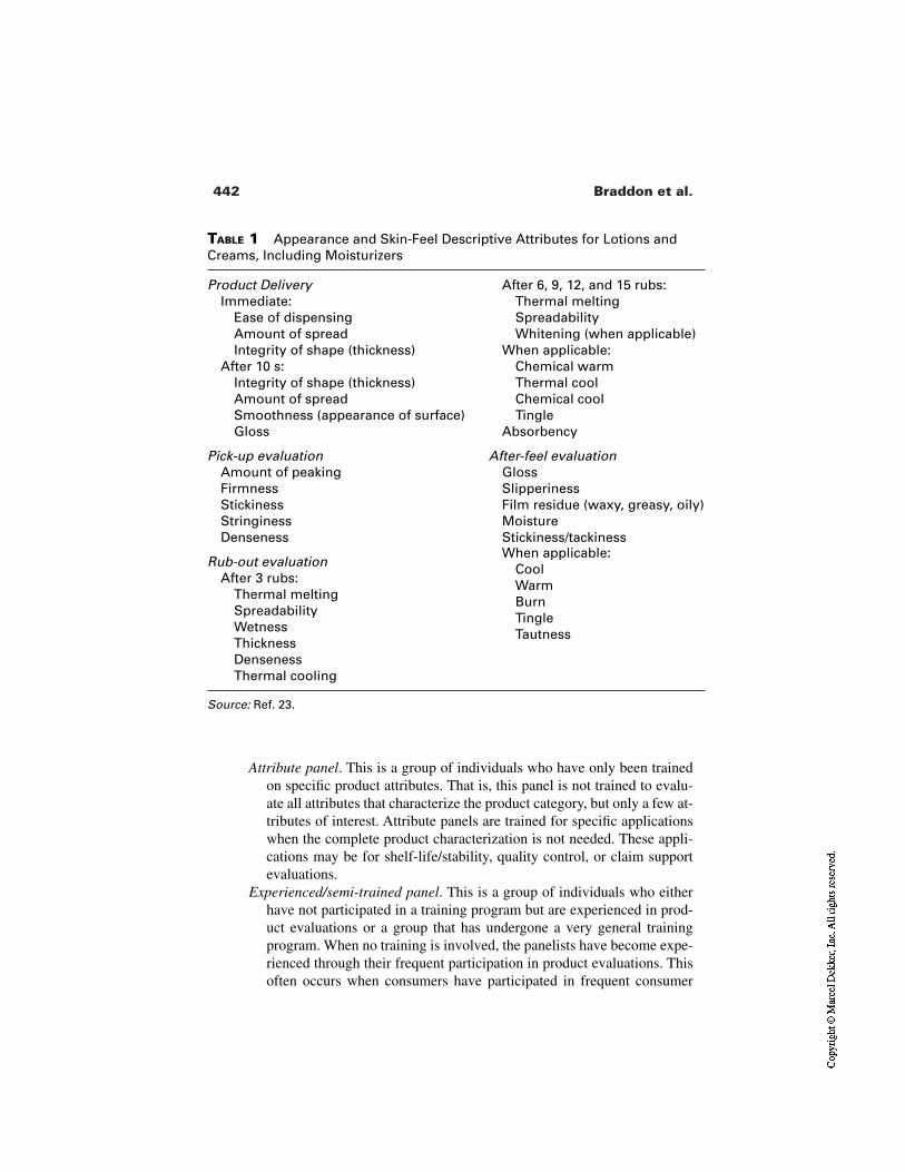
442 Braddon et al.
TABLE 1 Appearance and Skin-Feel Descriptive Attributes for Lotions andCreams, Including Moisturizers
Product Delivery After 6, 9, 12, and 15 rubs:Immediate: Thermal melting
Ease of dispensing SpreadabilityAmount of spread Whitening (when applicable)Integrity of shape (thickness) When applicable:
After 10 s: Chemical warmIntegrity of shape (thickness) Thermal coolAmount of spread Chemical coolSmoothness (appearance of surface) TingleGloss Absorbency
Pick-up evaluation After-feel evaluationAmount of peaking GlossFirmness SlipperinessStickiness Film residue (waxy, greasy, oily)Stringiness MoistureDenseness Stickiness/tackiness
Rub-out evaluationAfter 3 rubs:
Thermal meltingSpreadabilityWetnessThicknessDensenessThermal cooling
When applicable:CoolWarmBurnTingleTautness
Source: Ref. 23.
Attribute panel. This is a group of individuals who have only been trainedon specific product attributes. That is, this panel is not trained to evalu-ate all attributes that characterize the product category, but only a few at-tributes of interest. Attribute panels are trained for specific applicationswhen the complete product characterization is not needed. These appli-cations may be for shelf-life/stability, quality control, or claim supportevaluations.
Experienced/semi-trained panel. This is a group of individuals who eitherhave not participated in a training program but are experienced in prod-uct evaluations or a group that has undergone a very general trainingprogram. When no training is involved, the panelists have become expe-rienced through their frequent participation in product evaluations. Thisoften occurs when consumers have participated in frequent consumer
443Consumer Testing Methods
product evaluations and become experienced through this frequent prod-uct assessment. Experienced/semi-trained panels should only be usedfor product screening purposes, not for formal and important productevaluations. Frequently, experienced panels participate in a formal train-ing program and become trained descriptive panels. This training is gen-erally shorter and simpler than a training with naive panelists, since theexperienced panelists have acquired considerable product experience.
4.2.3 Descriptive Characterization of Moisturizers
Appearance and Skin-Feel Attributes. When experts evaluate moisturiz-ers, they divide the characteristics into four categories that depend on the timecourse of the lotion application event. Table 1 shows these categories, which are
Product delivery (e.g., ease of dispensing, amount of spread)Pick-up evaluation (e.g., firmness, stickiness, denseness)Rub-out evaluation (e.g., wetness, spreadability)After-feel characteristics (e.g., gloss, residual film, tautness)
See ASTM Standard Practice E 1490–92 [23] for a detailed description of proce-dures and attributes.
Fragrance Attributes. The fragrance evaluations for moisturizers arecompleted in two ways. The fragrance submissions are evaluated by themselves(i.e., not in the product) or in the product. When evaluated by themselves, the fra-grances are evaluated in glass containers. When the fragrance is evaluated in theproduct, it is applied and the fragrance is sniffed on the skin. The panel can betrained to evaluate basic fragrance/odor attributes or complete fragrance profiles.When a panel is trained on basic fragrance notes, the attributes may include over-all fragrance, overall base odors, and off-odors. Panelists may be asked to de-scribe the character of the fragrance. This information is only qualitative.
A panel can be trained to recognize and score specific fragrance characters.The evaluations can be general and address main fragrance components (e.g., flo-ral, fruity) or address specific notes within each category (e.g., rose, carnation,white flower, violet). Table 2 shows an example of fragrance/odor categories thata panel can be trained on [28].
Establishing a Descriptive Panel. The establishment of a descriptive ca-pability for the evaluation of moisturizers requires management support, thebuilding or procurement of testing facilities, and the recruitment/selection andtraining of the panel. Many sources [10,11,24–27] provide thorough coverage ofhow to set up and train a descriptive panel.
Applications and Uses of Descriptive/Attribute Data. The main applica-tions of this type of data are
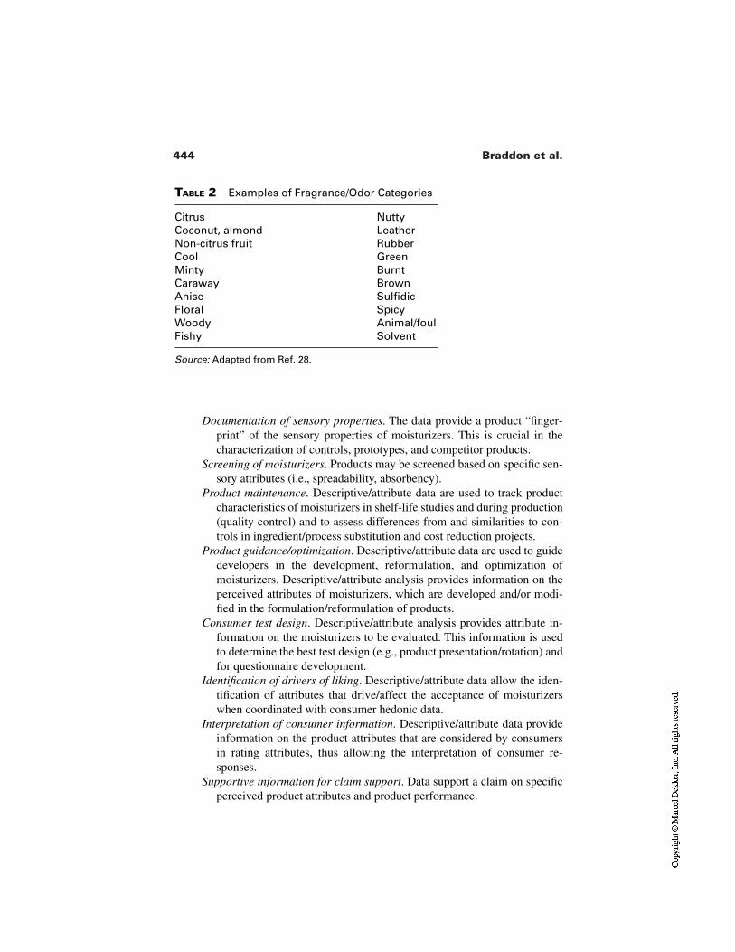
444 Braddon et al.
TABLE 2 Examples of Fragrance/Odor Categories
Citrus NuttyCoconut, almond LeatherNon-citrus fruit RubberCool GreenMinty BurntCaraway BrownAnise SulfidicFloral SpicyWoody Animal/foulFishy Solvent
Source: Adapted from Ref. 28.
Documentation of sensory properties. The data provide a product “finger-print” of the sensory properties of moisturizers. This is crucial in thecharacterization of controls, prototypes, and competitor products.
Screening of moisturizers. Products may be screened based on specific sen-sory attributes (i.e., spreadability, absorbency).
Product maintenance. Descriptive/attribute data are used to track productcharacteristics of moisturizers in shelf-life studies and during production(quality control) and to assess differences from and similarities to con-trols in ingredient/process substitution and cost reduction projects.
Product guidance/optimization. Descriptive/attribute data are used to guidedevelopers in the development, reformulation, and optimization ofmoisturizers. Descriptive/attribute analysis provides information on theperceived attributes of moisturizers, which are developed and/or modi-fied in the formulation/reformulation of products.
Consumer test design. Descriptive/attribute analysis provides attribute in-formation on the moisturizers to be evaluated. This information is usedto determine the best test design (e.g., product presentation/rotation) andfor questionnaire development.
Identification of drivers of liking. Descriptive/attribute data allow the iden-tification of attributes that drive/affect the acceptance of moisturizerswhen coordinated with consumer hedonic data.
Interpretation of consumer information. Descriptive/attribute data provideinformation on the product attributes that are considered by consumersin rating attributes, thus allowing the interpretation of consumer re-sponses.
Supportive information for claim support. Data support a claim on specificperceived product attributes and product performance.
445Consumer Testing Methods
Upon completion of the experts’ efforts the next step is evaluation using naiveconsumers.
4.2.4 Naive Consumer Tests
Testing with consumers falls into two categories: qualitative and quantitative. Inqualitative studies, inferences are drawn from patterns in the ideas or opinions of-fered by consumers. In quantitative evaluation, recommendations are based onstatistical inferences from product ratings. Results from consumer testing ofmoisturizers are only as good as the recruitment and screening process used to se-lect study participants.
For naive consumer testing, the counterpart to expert training is the carefulscreening for the appropriate moisturizer user. The complete set of questions usedto qualify or disqualify a study participant is termed a screener. Typically, a screen-er probes demographics, habits, and attitudes; personality or cognitive characteris-tics; lifestyle; and routine background items such as whether one works for a mar-ket research firm or has taken part in a consumer study in the past six months.
When consumers qualify for a study, it is a good practice to reconfirm thatthey qualify by asking key screening questions at the time products are picked up.It is disconcerting but common for the question “what is your one most frequent-ly used brand of facial moisturizer” to be answered inconsistently from one weekto the next.
Qualitative Testing. Qualitative research provides a forum for people toexpress in their own words what moisturizers mean to them as well as what effectthey have on dry skin. All the emotion, logic, free association, and irrationalthought that may be behind the checkmark in the answer box is revealed by qual-itative research.
FOCUS GROUPS PRINCIPLES. The focus group is the best known technique forgaining qualitative insight into consumer attitudes. It can be used to generateproduct ideas, new product benefits, weaknesses in current products, as well as tounderstand how people think about brand categories, advertising, product usagehabits, product concepts, and much more. There is no substitute for real discus-sions with people. Qualitative research typically precedes quantitative especiallywhen the project is about new products. However, an effective project oftenwinds back and forth between the two types of research, each benefiting the oth-er. Focus groups confirm and expand on the statistically derived conclusions ofsurvey research. They also help to sharpen surveys by highlighting the terminolo-gy actually used by people discussing products.
Focus group leaders should reconfirm the qualifications of participants onkey criteria before the sessions begin. Some people should be kept on-hand as“alternates” by over-recruiting the group. That is, invite more people than thenumber of available seats in case some potential participants need to be disquali-fied at the last minute.
446 Braddon et al.
Guidelines for the conduct of effective qualitative research are readilyfound [29–31]. Many are basic rules of civil conduct. Examples are talk in turns,listen to what it said by others, allow people to hold their own opinions.
Focus groups are conducted with 8–10 participants per group. Sessions last90–120 minutes. The moderator follows a discussion guide preapproved by theproject team. Discussions that are even more focused can be achieved with one-on-ones, so-called because only a moderator and one participant are present. One-on-ones can remove the influence of a dominating or bullying focus group mem-ber. Other variations that fall between full focus groups and one-on-ones arepossible and have unique benefits. For example, two-on-ones (two participants,one moderator) might enable a panelist and a friend to discuss their differences inshopping strategy or how they hear about interesting new products from one an-other.
When properly conducted, qualitative research can provide a wealth of in-formation to help guide a project. It can be used to clarify and understand theterms that people actually use to describe moisturizers and their effects on theskin, to understand how people categorize products in a given segment, and, mosttypically for the r&d environment, to learn what people think of one or more pro-totypes. On this last point, since prototypes often go through several phases of re-finement before launch, focus groups can be a powerful tool when they include aniterative component. Inviting the same people back to evaluate and discuss suc-cessive improvements can show what it takes, in microcosm, to win over an audi-ence. Caution though should be exercised here. Generalizing to the populationfrom a small sample of people in a qualitative setting is not appropriate.
INNOVATIVE TECHNIQUES. The insight and creativity derived from a focusgroup is a joint product of the participants and the techniques the moderator usesto engage the imagination. To recruit qualified research participants requires morethan screening for the appropriate category users. Personality and cognitive skillsfactor in also. There are screening tools for finding people who like to think [32]or who are open to novel products [33,34]. Less formal methods are also possible.Lists of creative people can be compiled by a nomination process, that is, regu-larly asking study participants to identify friends who they view as creative,trend-setting, off-beat, etc. The use of projective techniques and metaphor [35]are tools the moderator should employ to make the two hours enjoyable and en-tertaining to the participants as well as productive for the project team. An exam-ple of such a technique is “brainwriting” [36]. The moderator asks people to writedown a new product idea or improvement. The piece of paper gets passed to thenext person who builds on the idea and so on around the room.
OBSERVATIONAL STUDIES. While focus groups and one-on-one interviewsfree people from the constraints of a product survey, they still have limitations.The focus group or testing facility, as comfortable as it may be, extracts peopleaway from the natural context in which products are used. The consumer re-
447Consumer Testing Methods
searcher is thus cut off from seeing how and where the product fits in with otherproducts and routines. The solution is to talk to people in their home and observethem as they use health and beauty products such as moisturizers. This option fitswithin the growing movement in consumer research to utilize ethnographic tech-niques to more fully understand people and their products. Most associated withthe field of anthropology, ethnography involves “participating, overtly or covert-ly, in people’s daily lives for an extended period of time, watching what happens,listening to what is said, asking questions—in fact, collecting whatever data areavailable to throw light on the issues that are the focus of the research” [37]. Theresultant account, which can be captured on video, may be as realistic as astraightforward documentary or quite impressionistic and personal [38]. Eitherway, the ethnographic approach endeavors to create “thick descriptions” [39]which capture the particulars of the habit or product regimen in all its richness.The application of moisturizers may be embedded in a series of personal care andcleansing events which give it deeper meaning. As well, the physical product it-self may be stored in ways that illuminate the range of cognitive categories it fallswithin. It is hard to glean this information without the ethnographic tools.
PHOTOGRAPHIC TECHNIQUES. Unfortunately for the consumer researcher on atight budget or timetable, observational research may not often be feasible. Luck-ily, the advent of inexpensive disposable cameras has been a boon. Moisturizerusers can be given the cameras and asked to photograph the physical layout oftheir products on the shelf or vanity as well as to document product usage with thehelp of a willing family member stationed behind the lens. These pictures can bebrought to a focus group, photocopied, discussed, and organized. While there is arisk that the photographs may be “posed” or self-selected, the consumer should beencouraged to “flesh out” the story behind the image to reveal what was left out,if anything. These photographs could also be circulated around the focus group inbrainwriting fashion [36].
THE INTERNET. Various projections put U.S. on-line consumer spending in2000 at $38–61 billion [40]. As a tool for both quantitative and qualitative inves-tigations of consumer behavior, the internet is having a similarly enormous im-pact though the field is still in its infancy, and there have been difficulties assimi-lating on-line research into traditional market research organizations [41].Regardless, it is clear how the internet is being harnessed to consumer researchobjectives. Some of the uses include
Consumers are being encouraged to visit a company or product website.There, they register, complete a personal profile, and agree to participatein future testing/surveys. A database can thus be created from which atest sample can be drawn for the testing of moisturizer products or con-cepts.
Personal care companies contract with an outside agency that can create an
448 Braddon et al.
on-line chat about product or concepts between a moderator and regis-tered consumers who fit the relevant screening criteria.
Consumer researchers create and post a survey to be completed on-line byregistered members.
There are many clear plusses for internet research: feedback is immediate; nar-rowly defined niche markets can be more easily tapped; and it offers participantsthe freedom to complete surveys when they want—at 3 am if they desire! Nega-tives are the lingering uncertainty about who exactly is completing the survey,whether it is being completed independently, and the projectability of the opin-ions of the on-line audience to the target of, say, mass market hand and body lo-tion users. Clearly, the growth of on-line accounts points to a time when internetusers will more closely reflect national demographics, rather than a limited, moreeducated, upscale audience. Once qualitative investigations are complete, proj-ects normally progress to quantitative research.
Quantitative Test Methods. One of the advantages of the quantitativestudy is the completion of a standardized product questionnaire by all respon-dents to reach statistically based conclusions. Aside from an open-ended questionsuch as “what did you like or dislike about the product?” the questionnaire con-tains primarily closed-ended items using terminology and rating scales selectedby the researcher.
Table 3 shows some quantitative designs and several variants on the se-quential option. These are defined by how many surveys are used, when they aregiven, and what sorts of questions they contain. For example, in the paired pref-erence procedure, two products are used but there is no survey until after the sec-ond usage period. The questions are entirely about preference for one product orthe other.
The choice of a monadic or sequential test design is more than just a deci-sion about how products are assigned to consumers. Each design actually mirrorsa real-world encounter with products. The compatibility of the test objective withthat encounter should be considered when selecting a design. The monadic designis more reflective of normal product use. People rarely use two different brandssimultaneously, in alternation, or in quick succession for purposes of comparison.Monadic presentation is also aligned with an encounter with a dramatic “break-through” type product. There is no regular brand on the shelf and none is provid-ed as context in the study. The sequential design, in contrast, is aligned with aproduct substitution, upgrade, or brand switching experience. For example, sup-pose a company plans to substitute a new, improved, or less expensive moisturiz-er for a currently marketed product. The shopping experience of the loyal con-sumer of that brand will, at some point, be the use of the current variant followedby its replacement when the new variant is purchased. The sequential monadicdesign reflects or recreates that shopping experience. Half the participants use the
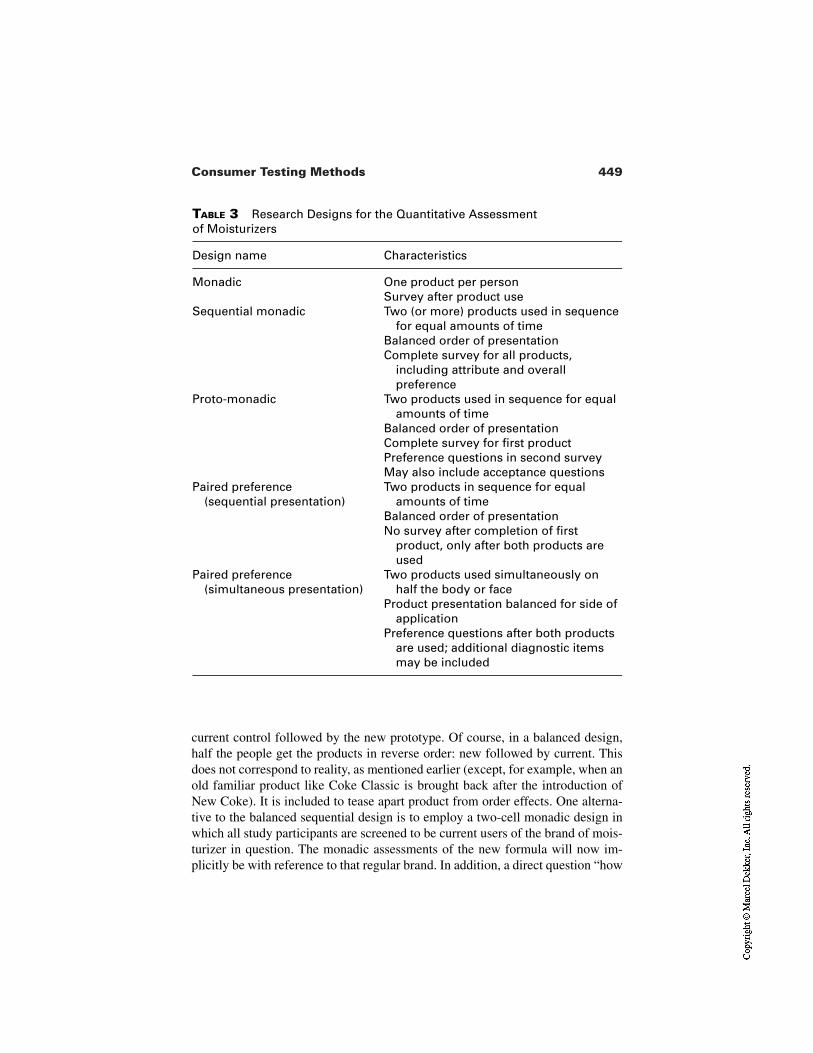
449Consumer Testing Methods
TABLE 3 Research Designs for the Quantitative Assessment of Moisturizers
Design name Characteristics
Monadic One product per personSurvey after product use
Sequential monadic Two (or more) products used in sequence for equal amounts of time
Balanced order of presentationComplete survey for all products,
including attribute and overall preference
Proto-monadic Two products used in sequence for equal amounts of time
Balanced order of presentationComplete survey for first productPreference questions in second surveyMay also include acceptance questions
Paired preference (sequential presentation)
Two products in sequence for equal amounts of time
Balanced order of presentationNo survey after completion of first
product, only after both products are used
Paired preference (simultaneous presentation)
Two products used simultaneously on half the body or face
Product presentation balanced for side of application
Preference questions after both products are used; additional diagnostic items may be included
current control followed by the new prototype. Of course, in a balanced design,half the people get the products in reverse order: new followed by current. Thisdoes not correspond to reality, as mentioned earlier (except, for example, when anold familiar product like Coke Classic is brought back after the introduction ofNew Coke). It is included to tease apart product from order effects. One alterna-tive to the balanced sequential design is to employ a two-cell monadic design inwhich all study participants are screened to be current users of the brand of mois-turizer in question. The monadic assessments of the new formula will now im-plicitly be with reference to that regular brand. In addition, a direct question “how
450 Braddon et al.
does the product you used compare with your regular brand?” may also be in-cluded.
TESTING LOCATIONS. Generally speaking, quantitative testing is conductedeither in the consumers’ homes or at a testing facility, called a central location. In-home testing most approximates real life extended use. In contrast, the central lo-cation test evaluates a product often used only once, but most importantly allowsthe investigator the opportunity to observe product use. Frequently, the researchquestion does not require extended use by the consumer. In these circumstances,the central location test is all that is needed to answer the research question.
QUESTIONNAIRE DESIGN. Regardless of where the test is conducted, the qual-ity of the information collected hinges on the development of a suitable question-naire. Much has been written on this topic (see, for example, Ref. 42), but a goodquestionnaire should comprise the following kinds of questions:
Hedonic. Overall, how much do you like this product? How much do youlike this product for _____ (attribute)?
Open ended. What, if anything, is there about this product that you like?Attributes. Ratings of attributes on a 5- or 7-point “excellent-to-poor” scale,
for example, rub-in, silkiness, greasiness, softness, smoothness.Agree/disagree items. Is a fragrance for someone just like me? Is a moistur-
izer for a contemporary woman?Directional/intensity. Using a “just about right” scale, for example, “too
thin,” “just about right,” “too thick’’; or a unipolar intensity scale such as“not at all greasy” to “much too greasy.”
Comparison to regular brand. A scale that typically includes “much better”and “much worse” at the extremes and “equal to” in the middle.
Uniqueness. A scale anchored by “similar to all others” at one end and“similar to no others” at the other end.
Preference Which product did you prefer overall? (Only appropriate ifmore than one product is being tested.)
When developing the questionnaire the researcher has to be careful to in-clude terms that consumers understand and scale rating points that make sense in-tuitively to the average consumer. Questionnaires should also be formatted forclarity and ease of use by the consumer.
IMPROVING TEST SENSITIVITY. One factor in the decision to select one re-search design over another is the test’s ability to detect product differences. Forexample, when a cost reduction is being considered, the happy outcome is thatconsumers not notice the difference in esthetics, efficacy, fragrance, etc. It is eas-ier to “prove” product parity if people are not given sufficient opportunity to de-tect the difference, as when the product usage period is too short. In contrast,when difference detection is desired, extending the usage period or employing thehalf-body procedure is recommended. The half-body technique improves sensi-
451Consumer Testing Methods
tivity because both products are experienced nearly simultaneously. Product A isapplied to the left side and product B to the right for half the sample, and the oth-er half receive the reverse order. Whatever differences there are can be perceivedwithout the distorting effects of the passage of time or the presence of interveningevents that are part of a sequential design by definition.
COMPLIANCE. The chances of obtaining reliable data are enhanced whenmeasures are taken to check that participants follow procedures and understanddirections. For example, the half-body procedure places some added complianceburdens on the research participant, especially in a home-use test. Participantsmust be instructed to keep the products separate or not to mix the products. Yetpeople may not easily comply with a cross-handed product application instruction(put product A in right hand and apply to left side of body; do reverse for productB) when using products at home. The technique should be demonstrated beforeproducts are taken home or the first application occasion should be in the testingfacility under the observation of a project team member. Such controls are typicalof a central location test (CLT), where the entire study takes place at an indepen-dent test facility rather than at home (home-use test; HUT). A CLT is monitoredby a member of the project team or a briefed employee of the CLT agency. Theyensure that the participant is attending to the products as instructed by being pres-ent during product usage.
Compliance in another sense is somewhat easier to monitor. This is use-upcompliance which is accomplished by a product weight check. Did the consumeruse the moisturizer twice a day for four weeks? The product should be weighedbefore and after a home usage period to ascertain that the samples were actuallyused sufficiently. This requires that product developers provide to consumer re-searchers a “reasonable” dose value. Calculations will then establish a lowerbound for product use-up. People who used less than that amount should be dis-carded from the data analysis.
SEASONALITY. The time of year and regional climate affects the efficacy ofmoisturizers. If the usage and purchase frequency of hand lotions drops in thesummer, tests relating to therapeutic benefits, such as relief of dry, chapped skin,should be delayed until the winter months or perhaps relocated to a dry climate.Testing in the wrong season limits the ability to see product differences.
4.3 Data Analysis
Sensory and consumer responses collected in moisturizer studies are analyzedstatistically to enable separation of random from real treatment effects.
4.3.1 Summary Statistics and Graphical Representation
Prior to applying any statistical tests, it is advisable to summarize data and com-plete some simple diagnostic and/or graphical analyses to assess the nature of the
452 Braddon et al.
data [43]. Basic summary statistics for continuous data may include sample size,average, ranges, standard deviation and other deviation measures, skewness mea-sures, interquartile range, etc. Graphical representations are used to observe cer-tain data characteristics of interest. The researcher may choose graphs such asstem leaf, boxplot, data distributions (number or percent of observations/scores),bar, radar/spiderplot, and other charts to display data.
These summary statistics and graphs provide simple and revealing informa-tion of the data to be analyzed. The outcome of some statistical tests may be an-ticipated through this assessment. In addition, these simple summaries and graphsprovide preliminary information on the data to help decide on the statisticalanalysis to apply. For example, a simple distribution graph displaying the per-centage of observations across a scale indicates if the data distribution is uni- ormultimodal. Data that are not unimodal should not be analyzed through paramet-ric statistics.
4.3.2 Common Statistical Analyses for Sensory andConsumer Data
This topic is covered extensively in the literature. The books describe the charac-teristics of the basic statistical tests used to analyze sensory and consumer data[44,45]. Sensory/consumer research publications cover the topic specifically forthe data collected in sensory/consumer studies of consumer products, includingmoisturizers [11,18,46,47].
Analysis for Treatment Effects and Panelist Performance. The analysiscompleted on the sensory and consumer data from moisturizers and other con-sumer products may have two objectives: to study treatment effects or to studypanelist performance. Routinely, upon completion of a consumer, descriptive/at-tribute, or discrimination test, data analyses are completed to reach conclusionsabout product or sample treatment effects [11]. For example, analyses on treat-ment effects may address questions such as Are two moisturizers, control andproduct A, equally liked? or Is one significantly better liked over the other? orDoes one moisturizer spread significantly easier than another? or Is the competi-tor’s moisturizer and product X perceived significantly differently when appliedon the skin? All these questions deal with product or attribute differences/similar-ities. The statistical tests for each data type [18,19,48–50] address these questionsrelated to treatment effects (or product attributes).
Practitioners who work with trained or semi-trained panelists to evaluateattributes and attribute intensities do need to analyze the data with an additionalobjective. This objective is to learn about panelist performance. These analysesare conducted on a routine basis to monitor panelists, assure that their responsesare valid and reliable, and ensure that sound product conclusions are obtained.Panelist monitoring analyses investigate ability of panelists to (1) find significant
453Consumer Testing Methods
differences if differences exist, (2) replicate their own judgments, (3) agree withthe rest of the panel as far as the direction of product differences, and (4) matchintensity references (if used). Different statistical procedures for panel monitor-ing have been and continue to be developed [51–54].
5 APPLICATIONS
Sensory and consumer testing methods have many applications. It would be im-possible to cover all applications and with the detail each application deserves. Asummary of the main applications are discussed.
5.1 Product Matching
Product matching projects are very common in any industry. Testing conductedfor these projects assesses if a prototype matches a control. This control can bethe competition, the “gold standard,” or the current product. Product matchingprojects can be completed either through discrimination or descriptive tests. Dis-crimination tests indicate if, overall, products are sufficiently similar or different.Descriptive tests will, in addition, provide information on the attributes that dif-ferentiate products, if they are found to be different.
Discrimination tests are conducted if the interest is only in overall differ-ence/similarity (as the first step), when there are a few products to test, and whenenough panelists are available. Similarity tests should be conducted for matchingprojects, which require a large number of panelists [55]. The testing of fragrancesin moisturizers is commonly completed using discrimination tests.
The testing of a match for skin-feel characteristics of moisturizers is mostcommonly conducted using descriptive tests. Since skin-feel properties requiretime intensity or assessment at different stages (e.g., immediate and 5- and 10-minute evaluations, etc.), discrimination tests may not be as useful.
5.2 Assessment of Differences and Similarities
There are many projects where the differences and similarities among moisturiz-ers need to be determined. These evaluations are conducted when comparing thecompany’s current product to competition, a new formula, different prototypesthat encompass new packaging materials, ingredients, process variables, etc.
Ultimately, companies are interested in determining the differences (or lackthereof) in liking between and among products. Frequently, a company may con-duct a consumer study to test the differences and similarities between the controland test samples/prototypes as perceived by consumers without completing anyother tests. Although this test strategy is sound and followed by many companies,it may not be the most efficient approach.
454 Braddon et al.
The use of sensory tests (e.g., discrimination and descriptive tests) prior toa consumer test represents a more efficient testing strategy, since the former areusually less expensive and can have a faster turnaround time. The rationale in us-ing such tests first is to assess the differences and similarities in perception, thenmake a decision if an ensuing consumer test is needed. In addition, the discrimi-native or descriptive tests provide information on the products to be tested andthus aid in designing a better consumer study (e.g., in guiding decisions on sam-ple presentation strategy, attributes in the consumer questionnaire, etc.).
For example, a descriptive test is conducted first if the objective is to con-firm that a perceivable difference exists between a new formula and the control(or a new formula and the competition) and to characterize the difference(s). Ifthe descriptive tests show that there are no differences or not large enough differ-ence(s), a recommendation is given to reformulate the product before conductinga more expensive and involved consumer test. If a trained panel does not find adifference, a consumer will not find the difference nor indicate a difference in lik-ing or preference. If the descriptive panel finds a difference, the consumer test canbe conducted to explore how consumers react to the perceived difference(s). Thissequence of testing represents an effective test strategy. In addition, the completedata set—the descriptive information and the consumer reaction to the differ-ences—can then be put together to interpret data, make decisions, and providerecommendations.
5.3 Research Guidance and Optimization
Sensory and consumer test methods are widely used for research guidance in thedevelopment of new products. Formulators/researchers are interested in studyingthe effect of certain variables (e.g., ingredients, technologies, packaging, andprocesses) on the product’s perceived properties and consumer responses. Ulti-mately, the goal is to develop the best liked product in the category or within thelimits of the raw materials and technologies used. Formulators/researchers needto obtain guidance in case products need to be reformulated. This guidance is pro-vided either using the consumer diagnostic information or the descriptive/attrib-ute information linked with the consumer information (see Sec. 5.5).
Research guidance can be provided in two ways: (1) through individualsample evaluation (one-at-a-time approach) or (2) through designed experiments,where a set of products is produced to represent variables and ranges of interest.From the experimental point of view the first type of study is called a nonde-signed experiment or one-way treatment structure study; the latter is called a de-sign experiment, treatment structure, or optimization study [56,57].
5.3.1 Nondesigned Experiments
Nondesigned experiments are very common in industry and are used to test a se-ries of products either in a sensory or consumer study. The products are a set of
455Consumer Testing Methods
qualitatively distinct objects, having no particular association among themselves.In such tests, the products tested may include the company’s products, a few com-mercial products, and one or more prototypes. There is no association amongthemselves in the formula, package, performance, etc.
These experiments are favored by those companies that require the testingof a series of products with fast turnaround and do not want to or cannot invest thetime, effort, and funds to systematically produce samples following an experi-mental design and do not want to or cannot test the larger number of productsgenerated by designed experimental plans. The design and execution of thesetests is relatively simple, since samples are produced, purchased, or collected andthen tested.
Once the test is completed, results are analyzed through statistical proce-dures which indicate if the products are different and how they are different in theresponse measured (e.g., liking, oiliness, glossiness). No inferences can be madeon the effect of one variable or concentration on the response measured. Often, ifanalytical data (e.g., sensory or any instrumental measure) are collected, they arerelated to liking or any other consumer response (e.g., greasiness). A relationshipcan be build between the consumer response (e.g., liking or greasiness) and thedescriptively perceived attributes (e.g., greasy, oily, waxy) or the concentration ofa certain variable (if measured or known for all samples). Even though these rela-tionships can be built, the interpretation of results may not be clear, since vari-ables may be confounded and not cover the complete intensity ranges [58].
5.3.2 Designed Experiments
A most effective way to study the effect of variables (or factors) on a response isto use experimental designs to produce the set of samples/prototypes that repre-sent specific variables and levels. These prototypes are then tested (e.g., in a de-scriptive or consumer study), and the results can be analyzed to accurately inferthe effect of such variables on the response (e.g., liking), on the way the responsevaries as a function of the variable studied (if and how the response increases ordecreases with increasing concentrations/levels of the variables), and on an opti-mal (if any) combination of variables to achieve the lowest or highest response[57].
For example, an optimization study for moisturizers may include two vari-ables such as the amount of aloe and sunflower seed oil. An experimental plan isthen used to determine the samples that need to be produced to represent specificcombinations and concentrations of these variables. The prototypes are then test-ed (e.g., in a consumer study). Results (e.g., liking) are analyzed statistically vis-a-vis the nature of the experimental design. Models can be developed and resultscan be displayed in graphs or plots that show the relationship of the response vari-able (e.g., liking) as a function of the concentration and combination of the twovariables studied.
456 Braddon et al.
Common plans or treatment structures used in consumer product tests arefactorial treatment structures and response surface structures (RSM) [56]. Manydesigned approaches and optimization studies have been predominantly pub-lished in the food industry [56–61]
5.4 Claim Support
Clinical claims are addressed in the next chapter. Here, the focus is on consumer-based claims. The application of sensory and consumer methods is essential inclaim support projects. Advertising claims most often address perceived productattributes and/or differences as perceived by consumers or trained panelists.Therefore, no sensory related claim can be substantiated without the use of thesemethods. This area is of particular importance to a company not only becauseproper claim support testing provides the basis for sound advertising claims, butalso for the legal implication that may result from unsupported claims. Beingsuch a critical application of sensory/consumer methods, the ASTM committeeE18 on sensory evaluation formed a task group to document the design and im-plementation of sound sensory and consumer testing practices geared to validateclaims addressing sensory properties [62]. The most important step in a claimsupport study is to delineate an explicit statement of what the claim will be. Thisstatement will in fact dictate the type of testing strategy to follow. Most claimsupport studies are completed with consumers, even if they deal with perceivedattributes (e.g., “our product leaves your skin feeling softer than brand X”). How-ever, much claims support testing is completed using discriminative/laboratorytests. When these tests are used, they are intended to provide more objective dataregarding perceived attributes without regard for personal preference.
5.4.1 Consumer-Based Claim Support Testing
Consumer-Based Claims. Consumer-based claims can be tested stating ahedonic/liking or a perceived attribute responses. These claims can be compara-tive and noncomparative. In addition, comparative claims can be parity or superi-ority claims [62]. Examples of each of these claims follow:
Comparative/parity. Our product is as well liked as product A. Our productis as moisturizing as product C.
Comparative/superiority. Our product is preferred over any other product.Our product leaves your skin looking more radiant than product D.
Noncomparative. Our product leaves your skin feeling soft. Our product isgentle to your skin.
Consumer Studies for Claim Support. Consumer studies designed to sup-port claims need to be carefully designed and executed. Special attention needs tobe paid to sample size [57], product selection, and questionnaire design.
457Consumer Testing Methods
5.4.2 Discriminative/Laboratory-Based Claim Support Testing
The two discriminative/laboratory tests used in claim support are discrimination(e.g., duo-trio, attribute difference tests) and descriptive tests [62]. Discrimina-tion tests are used when information needs to be collected regarding the overall(or attribute) difference/similarity between products. Usually, claim support stud-ies use discrimination tests to obtain additional back-up information, but not ex-clusively to support a claim. Descriptive tests are more commonly used, sincethey address attribute perception.
Descriptive-Based Claims. Descriptive-based claims have the followingcharacteristics compared to consumer-based claims: (1) they do not address pref-erence and acceptance/liking and (2) they address attribute perception by a high-ly trained panel. The attributes are not consumer terms (gentle, silky, moisturiz-ing, radiant), but technical and descriptive, as described in Sec. 3.1 (oily, greasy,spreadable, sticky, dense, taut). Frequently, descriptive information is collected inclaim support studies only as supportive information for the claim. The concur-rent consumer study may address the advertisement claim per se. However, theclaim becomes much stronger if the consumer results are supported by the de-scriptive information.
Examples of comparative and noncomparative claims based on descriptiveinformation include
Comparative/parity. Our product is as absorbent as product C.Comparative/superiority. Our product is less greasy than product D.Noncomparative. Our product leaves a cool sensation on your skin.
Descriptive Studies for Claim Support. As with consumer tests, specialattention has to be paid to the test design and test parameters of a descriptivestudy for claim support. The product selection and acquisition needs to be care-fully completed. Unique aspects of descriptive studies to be controlled are
Trained panel. Descriptive evaluations for claim support have to be com-pleted by a highly trained and calibrated panel.
Test design and ballot development. The attributes evaluated only have toaddress those attributes related to the claim. All descriptive attributes donot need to be measured. The sample preparation and presentation needsto be carefully controlled.
Test execution and replications. The evaluations are completed followingstrict and controlled protocols. If appropriate, references are reviewedprior to the study to calibrate the panel. No claim support evaluationshould be conducted without an assurance that the panel is well trainedand calibrated. In addition, the study needs to be replicated to be able toconfirm adequate panelist performance.
458 Braddon et al.
5.5 Category Appraisals
A category appraisal or review is one of the most fascinating projects conductedby a consumer products company. In these projects, a product category is evalu-ated to assess differences and similarities among products in the category. Thestudy usually includes the main commercial products within the category, thecompany’s products, and sometimes prototypes.
Category appraisals are conducted (1) only with consumer data (traditionalcategory reviews) or (2) both with consumer and analytical data (e.g., descriptiveand instrumental) as in multifaceted category research studies [53]. In general,the main benefit of the individual category studies is the documentation of the dif-ferences and similarities among products which yield an understanding of the cat-egory. The consumer category study provides information on the differences andsimilarities among products in liking and consumer-perceived attributes [63]. Thelaboratory category study provides information on the differences and similaritiesamong products in the properties measured. If the laboratory data are descriptive,these results show the differences and similarities in perceived attributes by atrained panel. There are many benefits and applications when studies in both cat-egories are completed. The most important applications are [64]
1. The determination of drivers for liking (in descriptive terms) to pro-vide actionable and clear direction to researchers in the formulationand reformulation of acceptable products within the category.
2. The interpretation and understanding of consumer responses.3. The establishment of a system in which laboratory data can be used to
infer consumer responses.
5.6 Irritation
Some facial moisturizers, especially those containing AHA, produce skin irrita-tion or an “adverse reaction.” Typical descriptive terminology includes tingling,stinging, burning, rashing, pulling, tightness, and itchiness. Consumer researchprojects are frequently motivated by a need to know whether the obtained irrita-tion rate is below a pre-established action standard level or whether an ingredientchange is effective at reducing irritation levels.
5.6.1 Measurement Issues
Asking about and accurately measuring irritation is a delicate issue. On the onehand, simply raising the issue by including irritation items on a survey may cluethe consumer into certain expectations of the project and create an inflated rate ofsuch comments via a self-fulfilling prophecy type effect. On the other hand, in-cluding relevant questions or warning of possible irritation reactions provides acontext for understanding the product. A respondent is told that some level of ir-
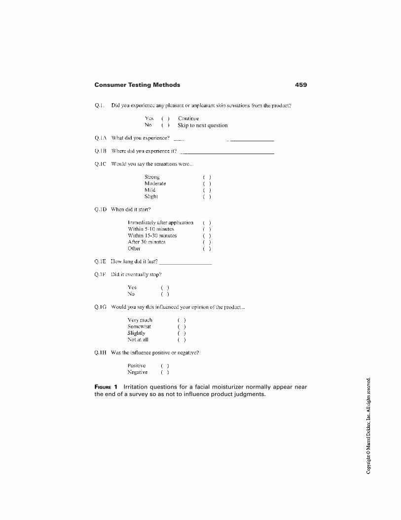
459Consumer Testing Methods
FIGURE 1 Irritation questions for a facial moisturizer normally appear nearthe end of a survey so as not to influence product judgments.
Skip to next question
460 Braddon et al.
ritation is part and parcel of the moisturizer’s mode of action. The level of report-ed irritation may, in this case, be under-reported because, given a rationale for it,the consumer discounts the experience. To create a perfectly balanced positionbetween these concerns may not be possible. In practice, the consumer’s cogni-tive machinery is activated in complex ways by the presentation of a product con-cept, by the naming of the product (e.g., “age-defying” or “pore-refining”), andeven by the courtesy instructions and warnings that ethically accompany moistur-izers that may irritate the skin, for instance, “You may experience a brief burningor tingling. Discontinue use, if severe.” All one should ask of the questionnaire isthat it be probing and free of bias. One option is to have balanced wording in thequestionnaire so that specific product expectations are not divulged. For example,asking Did you experience any pleasant or unpleasant sensations after using theproduct for two weeks? is balanced and nonbiasing, whereas Did you experienceany unpleasant sensations after using the product for two weeks? suggests thatthis sort of reaction is expected. One full set of possible irritation questions is pre-sented in Fig. 1.
It is important to realize that unpleasant sensory stimulation does not nec-essarily correlate with disliking for a product. Questions Q.1G and H in Fig. 1look at this possibility. Brief stinging may be a signal of a product’s efficacy, i.e.,that it is working to smooth or exfoliate the skin. As such, the unpleasantness maynot negatively influence purchase intent.
6 CONCLUSION
The authors have attempted to present a selective overview of current methodsand issues. Moisturizers can no longer be formulated in a laboratory vacuum.Companies large and small can not afford to dismiss or ignore the voice of theconsumer in the product development process. More and more emphasis is beingplaced on understanding the consumer as a person and not as a marketing object(J. Kastenholz, personal communication, 2000). The evaluation of moisturizershas come a long way since the early days of informal product appraisals. Empha-sis should be placed on studying the consumer in their normal environment.While more traditional methods (e.g., trained panels, home-use tests, mall inter-cept) will continue to be effective, new approaches should be explored.
Acknowledgment
We would like to thank M. Davis and C. Wesolowski for their research assistance.
REFERENCES
1. Grievson M, Barber J, Hunting A. Natural Ingredients in Cosmetics: Based on aSymposium Organized by the Society of Cosmetic Scientists. Weymouth, England:Michelle Press, 1989.
461Consumer Testing Methods
2. DeNavarre MG. Oils and fats: the historical cosmetics. J Am Oil Chem Soc 1978;55.
3. McDonough EG. Truth About Cosmetics. New York: The Drug and Cosmetics In-dustry, 1937:12.
4. Fernandez Rodriguez MC, Selles Flores E. The splendour of the cosmetology in theancient Greece. Cosmetic News 1998; XXI.
5. Gunn F. The Artificial Face: A History of Cosmetics. New York: Hippocrene Books,1983.
6. Angeloglou M. A History of Make Up. London: Macmillian, 1970:22.7. Fernandez Rodriguez MC. The beginnings of esthetic and cosmetological care in an-
cient Rome. Cosmetic News 1999; XXII.8. Spelios T. An Informal History Of Cosmetics II. Drug Cosmet Ind 1983; 133:42.9. Peiss K. Hope In A Jar: The Making of America’s Beauty Culture. New York: Henry
Holt and Company, 1998.10. American Society for Testing and Materials. Guidelines for the Selection and Train-
ing of Sensory Panel Members. ASTM Special Technical Publication 758. Philadel-phia: ASTM, 1981.
11. Stone H, Sidel JL. Sensory Evaluation Practices. 2nd ed. New York: Academic Press,1993.
12. American Society for Testing and Materials. Manual on Sensory Testing Methods.ASTM Special Technical Publication 434. Philadelphia: ASTM, 1981:6.
13. Kahneman D, Diener E, Schwarz N, eds. Well-Being: The Foundations of HedonicPsychology. New York: Russell Sage Foundation, 1999.
14. Stone AA, Shiffman SS, DeVries MW. Ecological Momentary Assessment. In: Kah-neman D, Diener E, Schwarz N, eds. Well-Being: The Foundations of Hedonic Psy-chology. New York: Russell Sage Foundation, 1999.
15. Schutz HG. Evolution of the sensory science discipline. Food Technol 1998;52(8):42–46.
16. Moskowitz HR. Cosmetic Product Testing: A Modern Psychophysical Approach.New York: Marcel Dekker, 1984.
17. Moskowitz HR. Cosmetic Testing and Evaluation of Personal Care Products. NewYork: Marcel Dekker, 1996.
18. Meilgaard M, Civille GV, Carr BT. Sensory Evaluation Techniques. 2d ed. Boca Ra-ton: CRC Press, 1991.
19. Lawless HT, Heymann H. Sensory Evaluation of Food. New York: Chapman andHall/International Thomson Publishing, 1998.
20. Schwartz NO. Adaptation of the sensory texture profile method to skin care products.J Texture Studies 1975; 6:33–42.
21. Brandt MA, Skinner EZ, Coleman JA. The texture profile method. J Food Sci 1963;28:404–409.
22. Szczesniak AS. Classification of textural characteristics. J Food Sci 1963;28:385–389.
23. American Society for Testing and Materials. ASTM practice for descriptive skinfeelanalysis of cream and lotions (E 1490–92). In: Annual Book of ASTM Standards.Philadelphia: ASTM, 1999.
24. Keane P. The flavor profile. In: ASTM Manual Series MNL 13. Manual on Descrip-tive Analysis Testing. Philadelphia: ASTM, 1992.
462 Braddon et al.
25. Stone H. Quantitative descriptive analysis (QDA). In: ASTM Manual Series MNL13. Manual on Descriptive Analysis Testing. Philadelphia: ASTM, 1992.
26. Muñoz M, Civille GV. The spectrum descriptive analysis method. In: ASTM Manu-al Series MNL 13. Manual on Descriptive Analysis Testing. Philadelphia: ASTM,1992.
27. Muñoz AM, Szczesniak AS, Einstein MA, Schwartz NO. The texture profile. In:ASTM Manual Series MNL 13. Manual on Descriptive Analysis Testing. Philadel-phia: ASTM, 1992.
28. Jeltema MA, Southwick EW. Evaluation and applications of odor profiling. J Senso-ry Studies 1986; 1:123–136.
29. Hayes TJ. The flexible focus group: designing and implementing effective and cre-ative research. In: Wu LS, ed. Product Testing with Consumers for Research Guid-ance. Philadelphia: ASTM, 1989:77–84.
30. Hawkins DI, Best RJ, Coney KA. Consumer Behavior: Building Market Strategy.7th ed. Boston: Irwin/McGraw-Hill, 1998.
31. Morgan DL, Krueger RA. The Focus Group Kit. Thousand Oaks, CA: Sage Publica-tions, 1998.
32. Cacioppo JT, Petty RE. The need for cognition. J Personality Social Psychol 1982;42:116–131.
33. Lynn M, Harris J. The desire for unique consumer products: a new individual differ-ence scale. Psychology and Marketing 1997; 14:601–616.
34. Goldsmith RE, Hofacker CF. Measuring consumer innovativeness. J Acad Market-ing Sci 1991; 19:209–221.
35. Coulter RH, Zaltman G. Using the Zaltman metaphor technique to understand brandimage. In: Allen CT, John DR, eds. Advances in Consumer Research XXI. Provo,UT: Association for Consumer Research, 1994:501–507.
36. Mattimore BW. 99% Inspiration: Tips, Tales, and Techniques for Liberating YourBusiness Creativity. New York: American Management Association, 1994.
37. Hammersley M, Atkinson P. Ethnography: Principles in Practice. 2d ed. London:Routledge, 1995:1.
38. Joy A. Interpretations of ethnographic writing in consumer behavior. In: Belk RW,ed. Highways and Buyways: Naturalistic Research from the Consumer BehaviorOdyssey. Provo, UT: Association for Consumer Research, 1991:216–233.
39. Geertz C. The Interpretation of Cultures. New York: Basic Books, 1973.40. Weber N. E-commerce projections: which to believe? Home Furnishing Network
2000; 74(21):15.41. Gonier DE. Factionalization imperils market research groups. Advertising Age 2000;
71(25):40.42. Sudman S, Bradburn NM. Asking Questions. San Francisco: Jossey-Bass Publishers,
1982.43. Richmond SB. Statistical Analysis. New York: The Ronald Press Company, 1964.44. Dixon WJ, Massey FJ Jr. Introduction to Statistical Analysis. New York: McGraw-
Hill, 1969.45. Snedecor GW, Cochran WG. Statistical Methods. 7th ed. Iowa State University
Press, 1980.46. Gacula MC, Singh J. Statistical Methods in Food and Consumer Research. Florida:
Academic Press, 1984.
463Consumer Testing Methods
47. Powers JJ. Descriptive methods of analysis. In: Sensory Analysis of Foods. PiggottJR, ed. Essex, England: Elsevier Science, 1988.
48. Neter J, Wasserman W. Applied Linear Statistical Models. Illinois: Richard D. Irwin1974.
49. Hollander M, Wolfe D. Nonparametric Statistical Methods. New York: John Willey& Sons, 1973.
50. American Society for Testing and Materials. ASTM standard test method for unipo-lar magnitude estimation of sensory attributes (E 1697–95). In: Annual Book ofASTM Standards. Philadelphia: ASTM, 1999.
51. Sinesio F, Risvik E, Rodbotten M. Evaluation of panelist performance in descriptiveprofiling of rancid sausage: a multivariate study. J Sensory Studies 1990; 5:33–52.
52. Lundahl DS, McDaniel MR. Use of contrasts for the evaluation of panel consistency.J Sensory Studies 1990; 5:265–277.
53. Naes T, Solheim R. Detection and interpretation of variation within and between as-sessors in sensory profiling. J Sensory Studies 1991; 6:159–177.
54. Schlich P. GRAPES: a method and a SAS® program for graphical representations ofassessor performances. J Sensory Studies 1994; 9:157–169.
55. American Society for Testing and Materials. ASTM standard test method for senso-ry analysis—triangle test (E 1885–97). In: Annual Book of ASTM Standards.Philadelphia: ASTM, 1999.
56. Montgomery DC. Design and Analysis of Experiments. New York: John Willey &Sons, 1976.
57. Gacula MC. Design and Analysis of Sensory Optimization. Trumbull, CT: Food andNutrition Press, 1993.
58. Muñoz AM, Chambers E IV. Relating sensory measurements to consumer accep-tance of meat products. Food Technol 1993; 47(11):128–131.
59. Giovanni M. Response surface methodology and product optimization. Food Tech-nol 1983; 37(11):41–45.
60. Henika RG. Use of response surface methodology in sensory evaluation. Food Tech-nol 1982; 36(11):96–101.
61. Galvez FCF, Resurreccion AVA. Optimization of processing of peanut beverages. JSensory Studies 1990; 5:1–17.
62. American Society for Testing and Materials. ASTM standard guide for sensory claimsubstantiation (E 1958–98). In: Annual Book of ASTM Standards. Philadelphia:ASTM, 1999.
63. Moskowitz H. Food Concepts and Products. Just in Time Development. Trumbull,CT: Food and Nutrition Press, 1994.
64. Muñoz AM, Chambers E IV, Hummer S. A multifaceted category research study:how to understand a product category and its consumer responses. J Sensory Studies1996; 11:261–294.

22Clinical Testing of Moisturizers
Gregory NoleUnilever Home and Personal Care North America, Trumbull, Connecticut
“A moisturizer is a topically applied substance that overcomes the signs andsymptoms of dry skin” [1]. When Dr. Kligman wrote that definition in 1978,moisturizers were generally either emulsions or oleaginous mixtures. As a result,products were generally designed around two primary mechanisms of action,humectancy and/or occlusion. In both cases, as Dr. Kligman’s definition shows,moisturizing products of that time focused on treatment of symptoms, that is, theappearance and feel of dry skin.
Today, the amelioration of symptoms is still the primary benefit to the con-sumer, however, many if not most products attempt to go beyond simplehumectancy and occlusion to treat the underlying causes of dryness. Products to-day often deliver skin lipids, natural moisturizing factors (NMF), essential fattyacids (EFA), or other components of healthy skin that are deficient in dry skin.Moreover, who would have predicted 20 years ago that even some cleanserswould begin to move toward delivering low level moisturization benefits.
These changes in product technology have had significant impact on therole and means of clinical testing of moisturizing products. No longer can productinvestigators be content with measuring the end benefit to the skin, they must nowfollow the example of researchers in trying to understand what the product is do-ing to skin. With a wide array of clinical tools and techniques at their disposal, thechoice of measurement and interpretation of the measurements is of course criti-
465
466 Nole
cal to good experimental design. But bioinstruments and skin assays are just thetools. Equally important in good clinical design is the protocol, that is, the com-plete description of the experiment in robust scientific terms.
1 HISTORY OF DRY SKIN TESTING
1.1 Early Method for Describing Skin Barrier Properties
Clinical testing is conducted for the purposes of research, technology evaluation,or product claim support. In the earliest days the work was principally for re-search. The origins of modern dry skin understanding rightly belong to Dr. IrwinBlank when he described the mechanism of water content in skin [2,3]. Hedemonstrated that water, not oils, were necessary for maintaining skin plasticity.He described the problem in terms of regulating the barrier function. Dryness wasviewed as a function of water loss rate from the surface to the environment versusreplacement rate from the lower layers. In this early research, Blank was the firstto describe barrier integrity by measuring the transpiration rate of water throughexcised human skin in a diffusion cell. He measured the water loss rate of normal,damaged, and treated skin and demonstrated that this rate was a function and di-rect measure of skin barrier quality.
Since that time, trans-epidermal water loss (TEWL) has been an acceptedmeans of describing skin condition. Where Blank’s experiments were conductedin vitro, methods to measure water loss in vivo were later being devised [4,5]. Be-fore the development of the modern evaporimeter, TEWL was measured by pass-ing a stream of dry nitrogen over skin through a closed cup that was in contactwith the skin surface and measuring moisture pick-up with a dew point hygrome-ter. For the product investigator, evaporimetry not only described skin barrierquality, but could be used objectively to compare the performance of emollientlotions and creams [6]. However, TEWL alone says little about the momentaryhydration state and is not appropriate for comparing product humectancy. Evenwith modern instrumentation, TEWL measurements can be confounded by thepresence of surface moisture or an alteration in skin condition [7]. For example,an increase in TEWL could indicate an increase in surface moisture levels orcould be the result of skin damage. Unless testing conditions are well controlled,TEWL alone could lead to ambiguous results which call for further investigation.
1.2 Biomechanical Developments
The 1960s brought further understanding of the relationship of water content inskin and its effect on physical skin properties. This led to development of in vitroanalytical and biomechanical methods for describing skin conditions beyond va-
467Clinical Testing of Moisturizers
por transpiration rate [8–11]. Gravemetric approaches measured the amount ofwater loosely held in the skin that could be feely lost to the atmosphere under lowhumidity. Differential scanning calorimetry (DSC) was used to describe the waterthat was tightly bound within the cellular matrix in an attempt to better describethe underlying hydration state. Tensile testing was used to describe elastic proper-ties of skin in order to relate skin plasticity to hydration state. All of these ap-proaches provided opportunity for the product investigator to further understandproduct efficacy, but as in vitro methods, they could not demonstrate the end ben-efit of products in actual usage.
1.3 Regression Testing
Through most of the 1970s the underlying principle of lotion product testing wasthat there is a correlation between skin mechanical properties and the effects ofmoisturizer treatment. In 1978, Dr. Kligman challenged this assumption. Hepointed out that while physical properties of skin can vary, their true relationshipto dry skin is unclear. The lack of real understanding of dry skin is summarized inhis assertion (or more likely frustration) that “we cannot even say that dry skin isdry; that is, lacks water” [1]. For the clinician, this raises a serious question ofwhat to measure. Kligman’s proposal, in this seminal paper, was the dry skin re-gression test.
The thinking behind the regression method was derived from an under-standing of “cosmetic dry skin.” Kligman recognized that classical signs of dry-ness such as skin flaking can be effectively masked with emollient lotions thatgive the appearance of moisturized skin but do not provide a fundamental im-provement in skin quality. He observed that while treatments continued, dry skinmay look and even feel better, but as soon as treatment was stopped, the skinquickly reverted or regressed back to its original state. Kligman’s view was that itwas this rate of regression that was the true measure of moisturizer efficacy.
The original regression method was a 4- to 6-week process separated intodistinct treatment and regression phases. The studies were conducted on legs in apaired comparison design. Treatments were applied twice daily, Monday throughFriday for 3 weeks with no treatments occurring over the weekends. Because vi-sual assessments were made on Monday mornings (three days after the last treat-ment) they better reflected the true condition of the skin. After 3 weeks, treat-ments were discontinued altogether, and during this regression phase the skin wasassessed on Mondays and Thursdays until baseline was reached, which could take2 weeks or longer. Assessments involved visual grading of the test sites on a four-point scale for the overall appearance (or lack) of dryness. A quarter century later,with the many advances in our knowledge of dry skin and instrumental analysis,the principles of the regression method remain sound and continue to be the back-bone of modern dry skin testing programs.
468 Nole
1.4 Bioinstrumental Methods
The original regression method used visual observation of skin as the primarymeasurement. About the time this method was proposed numerous bioinstrumen-tal methods were just beginning to emerge [13–18]. These various techniques,many described elsewhere in this volume, were developed to quantify skin hydra-tion (electrical hygromety), texture (profilometry), elasticity (dynamometry andballistometry), optical properties (spectroscopy and photography), and otherphysical states (squamometry), just to name a few. These methods were often de-veloped for research into the fundamental properties of skin, and the emphasis inthe literature was on what these measurements told about skin. For the product in-vestigator they provided new opportunities for describing product effects; howev-er, there was insufficient guidance in how the instrument should be used in thecontext of a consumer product clinical trial. While these instruments are excellenttools, there is an ever-present danger of seeing them as the test method rather thanas tools within a test method.
1.5 Regression Testing Revisited
Following publication of the regression method there was a growing body of lit-erature on instrumental methods but a lack of writings on clinical evaluation. Yetthere was evidence that researchers were using the principles of the regressionmethod as framework for bioinstrumental evaluations [19]. Bioinstruments pro-vided finer resolution than a four-point visual scale, and this improved the abilityto clearly separate the strictly visual (cosmetic) effects from fundamental changesto skin condition.
For product investigation, the need for a well-structured test design inwhich to use these new measurement techniques became clear. Because skinmoisturization can be achieved through various routes (occlusion, humectancy,dryness prevention, and healthy skin repair), study details and the selection of themeasurement tools must be made appropriately. Further, as measurements be-came more sophisticated and the demands for product discrimination increased,some refinements (but not changes!) to the original regression method were need-ed to meet the increased requirements [20]. These refinements proposed basic pa-rameters such as weather and panelist recruitment criteria as well as defining pro-cedures such as for product assignment and application to ensure unambiguousresults for evaluating consumer products. The robustness of the regressionmethod remains evident in clinical testing where it is almost no longer a consid-eration but simply treated as a de facto approach for product evaluation [21,22].
1.6 The Miniregression Test
For the product investigator, bioinstrumental measures within a well-structuredtest provided a sensitive measurement of product performance. Within these mul-
469Clinical Testing of Moisturizers
tiweek trials, it was recognized that methods such as hygrometery had potential topredict after one week the overall product performance seen after three weeks.This led to the development of a more rapid predictive product evaluation testcalled the Miniregression [23]. In this test, all of the principles of the original re-gression method test are retained, just shortened. Treatment is for 5 days, fol-lowed by a treatment-free weekend and then a 7-day regression period. Using in-strumental measures instead of visual, the miniregression method focuses onpredicting the functional benefit (increased hydration state) rather than visual(cosmetic) benefit of treatment. This method provides value to the investigatornot just for quickly screening products, but also for demonstrating the early treat-ment benefits of products on skin.
1.7 “Nutrition Protocol” Regression Test
A novel approach took the standard regression test and turned it inside-out in or-der to better evaluate the quality of skin following treatment [24]. Referred to asthe nutrition protocol, it starts with a 4-week pretreatment phase which allows fullstratum corneum turnover of the treated skin site. With fully treated skin and nofurther product use, the test phase focuses principally on a 3-week regression.The healthier the skin quality, as a result of pretreatment, the greater should be thepersistence of “good” skin during the regression period.
1.8 Short-Term Alternative to Regression Testing
While a well- and widely established test, the regression method and its variationsare by no means the only acceptable approaches to product evaluation. Regres-sion testing in all its forms always involves the use of multiple product applica-tions over time. However, rapid and sensitive bioinstrumental measures such ashygrometry have allowed the development of methods to look at the immediatehydrating effects of even a single lotion application. These immediate hydrationtests are typically used to discriminate product performance within 4 hr of appli-cation though they can be extended [25,26]. With controlled application and re-strictions on subject activities, the tests almost always involve a multiple productdesign, typically evaluating up to six sites simultaneously, usually on lower legs,which allows for highly sensitive, direct within-subject interproduct compar-isons.
Note that while appropriate for showing moisturization effects, short-termprotocols involving moisturemeters are not appropriate for predicting longer-termbehavior. For example, occlusive materials such as petrolatum show no immedi-ate benefit based on a moisturemeter yet are highly effective in the longer term.
Short-term testing is best limited to describing the immediate effect of atreatment. Bioinstruments to look at elasticity, surface roughness, and other skinproperties can also be used in a single-use test to understand how quickly and ef-
470 Nole
fectively a particular treatment delivers relief to the user. In fact, a thorough un-derstanding of immediate effects can be as important to the product investigatoras determining the long-term benefit because when consumers begin to feel theirskin getting dry and tight, they want immediate relief.
1.9 Testing Moisturizers Today and Tomorrow
Today the product investigator has a wide array of tools, both visual and instru-mental measurement methods, and well-designed long- and short-term protocolsto fully understand the effects of moisturizer treatment on skin. These techniqueshave been used by researchers and product manufacturers to develop a solid un-derstanding of lotion technology. While the growing array of bioinstrumentaltools complements the more traditional visual evaluation methods, the need for awell-designed, structured framework for using any measurement technique isfundamental.
We are already seeing today that the delivery of moisture is beginning tocome from a most unlikely source, body cleansers. As the range of moisturizingproducts expands, it creates a need for a broader range of clinical methods [27].As we look forward, the future in leave-on products bodes new delivery mecha-nisms, biomimemics, and new forms [28]. However, in the end, to be accepted asa true moisturizer, new technologies will still need to deliver immediate and sus-tained relief and the well-developed methods for measuring short- and long-termeffects will still apply.
2 BASIC ELEMENTS OF CLINICAL DESIGN
The more objective and controlled the clinical trial, the more detached it becomesfrom the normal-use consumer experience. The competing interests of clinicalcontrol and consumer relevance cannot be easily reconciled. No single method isall-encompassing. Taking a broad view of testing, one finds a spectrum of meth-ods, from loosely controlled panel tests to the rigorously controlled clinical trials.Moreover, dry skin has many aspects, from how it looks to how it feels to how itfunctions, all of which further complicate the testing choices. Determining theideal course for testing is not always easy. Clearly the variety of available bioin-strumental methods can play an important role in dry skin testing; however, in-struments tend to give a unidimensional picture for a multidimensional problem.Thus the answer is often a stepwise approach to product evaluation [29,30].
In developing a clinical testing plan, the product investigator must balancethe requirements of each experiment against the objectives and conclusions to bedrawn from the results. Before the details of a clinical trial can be determined, thebroad requirements of the trial must first be established. Discussed herein are
471Clinical Testing of Moisturizers
three basic questions that underlie the study design. Only once the fundamentalsare established can then the details of the protocol can be considered, and theseare discussed in the next section.
2.1 Setting the Size and Scope of the Trial
Depending on the stage of the research or product development program, trials ofdifferent size and scope are entirely appropriate. In the early stages of a program,preliminary or pilot trials are often used as a cost-effective and rapid means ofgetting indicative information as a guide toward further study and full clinical tri-als. Sometimes referred to as preclinicals, these trials tend to be short with a min-imal number of subjects and are typically used as screening tools or as predictorsof product effects.
In preparing a study proposal, it is necessary to understand the value andlimitations of pilot trials but also to approach such trials with the same thoughtfuldiligence as larger clinical trials. A danger in labeling trials as preliminary is thatthey can be perceived as informal, resulting in an unacceptable degree of laxity.Preliminary trials should never be approached as “quick and dirty,” but must re-ceive the same detailed consideration as large-scale trials. When done properly,preliminary trials provide scientifically valid supporting evidence and incremen-tally add to the knowledge base. The data may not necessarily be sufficient tostand entirely on their own, but should constitute a solid first step.
Pilot trials must not be confused with small trials. The mere fact that a testis small does not mean that it is preliminary. Pilot tests should be defined on thebasis of statistical risk, not just complexity. All studies carry with them some riskof error, and preliminary studies carry higher levels of this risk. To illustrate thedifference between a pilot trial and small trial, consider two proposals: a 6-weekregression test with only six subjects per cell versus a 1-hr hygrometer trial on100 subjects. The regression trial would likely be set up as a pilot trial becauseexpectations for the statistical power is low (i.e., high risk of error due to smallsample size) despite the complexity of conducting a 6-week trial. Depending onobjectives, the simpler hygrometer study may or may not be approached as a pi-lot trial. If the objective were to record the before/after effects of a lotion, it canpass as a definitive clinical study. But if the same trial were being used surrogatefor a 3-week home-use test, then it would be considered preliminary because itwas being used to make predictions well beyond the scope of its data.
So the first element in designing a clinical trial is to establish the size andscope. How much risk can you accept in order to save time, money, or other re-sources? How comfortable will you be making a decision based on the outcome?How does this trial fit in with next steps? How does this trial fit in with the time-line of your research program? With the study objectives in mind, the trial mustbe developed appropriately, but to do so consideration must first be given to
472 Nole
specifically determining what is appropriate and necessary to meet those objec-tives.
2.2 Selecting the Measurements
Closely linked with setting the scope of the trial is selection of test methods. Thechoice of measurement method is ultimately what will define the study. After allis done, it is the output of these measurements, the numeric data, that is compiled,analyzed, and pored over in order to describe the effects of skin treatment.
Based on a review of product claims, it has been shown that moisturizerbenefits can be divided into three categories: what moisturizers do for the skin,how the skin looks, and how the skin feels [31]. Clinical tests can follow this pathusing a three-prong approach. This multimeasurement approach offers strong ad-vantages as each method has its own strengths and weaknesses (Table 1). Moreimportantly, the multiprong approach allows the conclusions based on onemethod to be corroborated and reinforced by the others. These three classes ofevaluation methods are discussed below and further in Sec. 3.
Instrumental methods are ideal at providing objective data to individual as-pects of what the product does for skin. They can provide quantificationof invisible aspects such as moisture content within the skin, and a bat-tery of instrumental measurements helps draw a detailed picture of phys-iological changes in skin. They are, nevertheless, unidimensional, whichcan lead to an incomplete view of product performance. Instrumentsalso can be highly sensitive to small differences suggesting skin benefitsthat are neither consumer relevant nor perceivable.
With expert assessment, a qualified human judge integrates many aspectsinto a measurement of the visual appearance of skin. Normal healthyskin is visually apparent, and this is the ultimate goal of a dry skin treat-ment. Any deviation from normal/healthy appearance can be captured inthe evaluation. Visual grading is traditionally the cornerstone of dry skinmeasurement.
Subject self-assessment within the context of the clinical trial gives quanti-tative measure to the perception of skin look and feel. Self-assessmentmeasurements have two roles: they provide a means of measuring sen-sory attributes that cannot be measured instrumentally and they demon-strate whether the instrumentally measured changes are resulting inmeaningful and perceivable benefits to the user.
Clearly, the selection of measurement(s) must be made appropriately forthe products being evaluated. Moreover, it is axiomatic that they are made in thecontext of the study objectives. No measurement is perfect; all have cost and re-source requirements associated with them, and all have certain limitations.Choice of measurement must balance the different study needs, and compromise
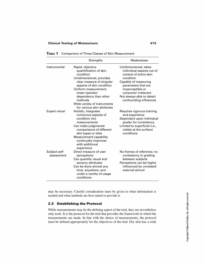
473Clinical Testing of Moisturizers
TABLE 1 Comparison of Three Classes of Skin Measurement
Strengths Weaknesses
Instrumental Rapid, objective quantification of skin condition
Unidimensional, provides clear measure of singular aspects of skin condition
Uniform measurement; lower operator dependency than other methods
Wide variety of instruments for various skin attributes
Unidimensional, takes individual aspects out of context of entire skin condition
Capable of measuring parameters that are imperceptible or consumer irrelevant
Not always able to detect confounding influences
Expert visual Holistic, integrates numerous aspects of condition into measurements
Can make judgmental comparisons of different skin types or sites
Measurement capability continually improves with additional experience
Requires rigorous training and experience
Dependent upon individual grader for consistency
Limited to superficial (i.e., visible at the surface) conditions
Subject self-assessment
Direct measure of user perceptions
Can quantify visual and sensory attributes
Can be done almost any time, anywhere, and under a variety of usage conditions
No frames of reference; no consistency in grading between subjects
Perceptions can be highly influenced by unrelated external stimuli
may be necessary. Careful consideration must be given to what information isneeded and what methods are best suited to provide it.
2.3 Establishing the Protocol
While measurements may be the defining aspect of the trial, they are neverthelessonly tools. It is the protocol for the trial that provides the framework in which themeasurements are made. In line with the choice of measurements, the protocolmust be defined appropriately for the objectives of the trial. Dry skin has a wide
474 Nole
range of etiologies, and dry skin treatment can be achieved through a variety ofroutes such as occlusion, humectancy, preventing dryness, and healthy skin repair[29,32]. Thus, the difficulty facing the product investigator is that there is no sin-gle all-encompassing clinical approach that addresses all of the aspects of productperformance.
For the evaluation of leave-on moisturizing lotions, common clinical test-ing approaches can be divided into two groups: single application trials and mul-tiple application trials, each of which can be further divided into trials for short-or long-term effects (Table 2). Each has its own distinct purpose, which must beconsidered in developing a clinical testing program.
Single application tests assess the physical changes in skin due to the prod-uct and predict the physiological changes with repeated use. In short-term moisturemeter hydration tests, measurements can be made withinseconds of applying lotion. This is appropriate for determining the im-mediate, even instantaneous effect of a lotion on skin but is extremelyweak for predicting long-term repeat use benefits. The entire study usu-ally takes place under controlled indoor conditions to eliminate environ-ment effects. This test can be used to compare the overall moisture de-livery of competing products.
The longer-term moisturemeter hydration test measures the persistence ofthe immediate physical changes to skin. It is often used to demonstrate“all-day” or “24-hour” effects. Due to the length of time involved, ac-commodation for panelist comfort must be considered; however, pan-elist activities can add variability to the measurement. This test can beused to compare the long-lasting moisturization ability of competingproducts.
Multiple application tests assess the true physiological changes in skin overtime. The advantage of tests with a 3-week or longer treatment phase is that it al-lows time for complete stratum corneum turnover.
A test of 1 to 2 weeks with no regression shows the rate of benefit, that is,the rate of healing of dry skin. Traced over time, it is a strong predictorof overall benefit, though the skin may not achieve a level of completerecovery. This test can be used to compare speed of benefit betweencompeting products.
The miniregression test shows the rate of healing and better predicts theoverall benefit by also showing the persistence in regression. This testcan be used to compare speed of benefit and predict the quality of thatbenefit between competing products.
The Kligman regression test shows the speed of benefit as well as the over-all benefit of treating skin through a complete cycle of stratum corneumturnover. Further, it separates the cosmetic (superficial) benefit from
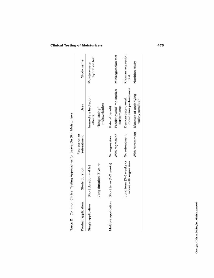
475Clinical Testing of Moisturizers
TA
BLE
2C
om
mo
n C
linic
al T
esti
ng
Ap
pro
ach
es f
or
Leav
e-O
n S
kin
Mo
istu
rize
rs
Pro
du
ct a
pp
licat
ion
Stu
dy
du
rati
on
Reg
ress
ion
or
retr
eatm
ent
Use
sS
tud
y n
ame
Sin
gle
ap
plic
atio
nS
ho
rt d
ura
tio
n (
<4 h
r)Im
med
iate
hyd
rati
on
ef
fect
sM
ois
ture
met
erh
ydra
tio
n t
est
Mu
ltip
le a
pp
licat
ion
Sh
ort
ter
m (
1–2
wee
ks)
No
reg
ress
ion
Rat
e o
f b
enefi
t
Lon
g d
ura
tio
n (
8–24
hr)
“lo
ng
last
ing
” m
ois
turi
zati
on
Wit
h r
egre
ssio
nP
red
ict
ove
rall
mo
istu
rize
r p
erfo
rman
ceM
inir
egre
ssio
n t
est
Lon
g t
erm
(3–
6 w
eeks
or
mo
re)
wit
h r
egre
ssio
nN
o r
etre
atm
ent
Dem
on
stra
te o
vera
ll m
ois
turi
zer
per
form
ance
Klig
man
reg
ress
ion
te
st
Wit
h r
etre
atm
ent
Mea
sure
of
un
der
lyin
g
hea
lth
y co
nd
itio
nN
utr
itio
n s
tud
y
476 Nole
substantive benefits by measuring how well the fully treated skin per-sists in regression. This test can be used to directly compare the overalleffectiveness of competing products to treat dry skin and improve skinquality.
The nutrition protocol ignores speed of benefit to focus on the quality of theskin due to product use. More than just measuring persistence of benefitin regression, it measures the resistance of skin to external assault as in-dicator of the underlying skin health condition. This test can be used tocompare the overall effectiveness of competing products to restore skinhealth.
The selection of measurement tools and general framework for the studyprovide the necessary structure for a well-designed protocol. But regardless of theapproach taken, numerous aspects must still be considered which not only definewhat must be done, but ensure that the results are scientifically sound. There is nosingle right way to conduct a clinical trial, though there are always opportunitiesfor improvements. (It is the nature of scientists to provide critical review andidentify what could be done better.) General protocols such as the regression testprovide well-established principles, but the specifics of the trial must be given in-dividual consideration in the detailed protocol. The product investigator actuallyhas wide latitude to tailor a study to particular needs and must demonstrate soundrationale for decisions.
3 DETAILED CONSIDERATIONS OF CLINICAL DESIGN
To fully elucidate the effects of a moisturizer on skin requires careful planning,and every aspect of the study must be considered. These considerations go far be-yond the mechanics of the test and measurement tools. Some considerations thatcan impact on a study’s outcome have been well described in the literature, suchas the effect of weather, environment, temperature, and humidity on skin mea-surements [32–37]. General descriptions of how to design and conduct trials areharder to come by, although some excellent references can be found[38–41].However, the real details of how a study is conducted are often the result of yearsof experience by the investigating lab. These details become part of the operatingroutine and are often taken for granted as their standard, good practice. Whilesome of these details may not be formally documented in the study protocol, theyare extremely important for a well-designed and well-conducted trial.
Following is a reference list of points to consider when developing a proto-col or conducting a clinical trial. This list is not nearly exhaustive, but is fairly ex-tensive. Thinking through these points will place detail on the general protocolframework and help create an unimpeachable study design. The points are dis-
477Clinical Testing of Moisturizers
cussed with respect to the clinical evaluation of dry skin and are relevant to allmanner of dry skin trials from the 2-hr hydration trial to the 6-week full regres-sion trial.
3.1 General Factors
The first level of detail is to define the structure of the trial. Following are someoperational parameters that define the framework for the protocol.
3.1.1 Statement of the Test Objective
An obvious and necessary part of any study, a well-written objective will clearlyarticulate what is to be achieved from the trial in a just few words. This objectivestatement is the first stake in the ground and every aspect of trial must be mea-sured against this statement. Therefore it must be the first consideration for thedetailed protocol and should be written specifically to the desired test. Broad orgenerically written objectives should be avoided.
3.1.2 Body Site
Where on the body to focus treatment is not a trivial question. Legs, forearms,hands, and face are the most commonly used sites and each has its strengths andweaknesses. Site selection must balance the study objectives with the practicali-ties of the clinical method. Most dry skin clinical trials are conducted on legs,hands, and forearms and to a lesser extent on faces. Table 3 provides a compari-son of strengths and weaknesses of each site. Other areas such as feet, knees, andelbows are occasionally used when there is specific interest.
3.1.3 Number of Cells and Cell Size
Statistical analysis can predict the minimum panel size required to show an ex-pected level of change where the level of variability is known. However, datavariability are not always known. Moreover, very large samples can sometimesbe used to demonstrate statistical significance on small differences that are nei-ther consumer perceivable nor relevant. Another chapter in this book providesthorough discussion of statistics and relevant cell size.
The product investigator who operates in the real world must think beyondjust the theoretical considerations and must also consider the practical aspects ofpanel logistics. For example, how long each measurement takes will limit howmany panelists can be assessed in a given period of time. Equally important isclinician fatigue that will occur with repetitive measurements. Both of these is-sues have direct bearing on sample size and the number of cells that can be run byexerting very real practical constraints. For reference, regression tests have typi-cally ranged from 10 to 20 subjects per cell, while hydration tests typically range
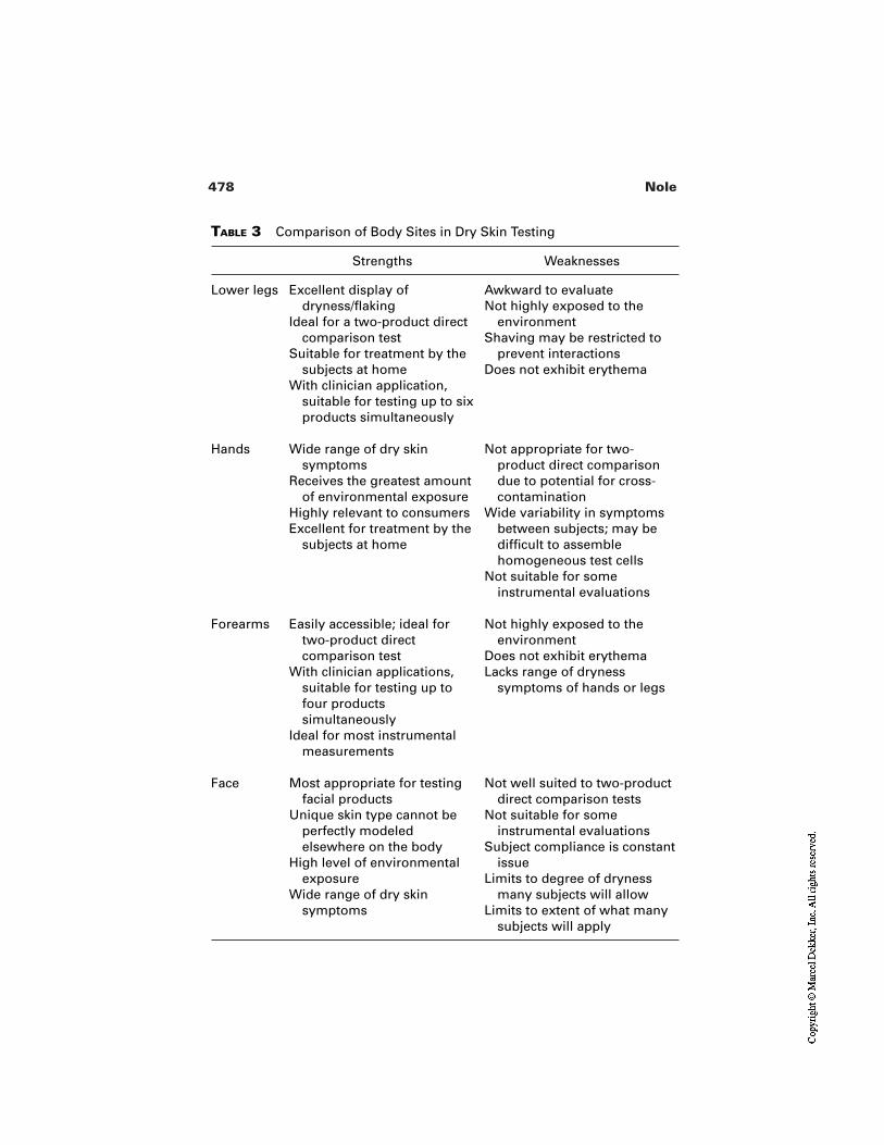
478 Nole
TABLE 3 Comparison of Body Sites in Dry Skin Testing
Strengths Weaknesses
Lower legs Excellent display of dryness/flaking
Ideal for a two-product direct comparison test
Suitable for treatment by the subjects at home
With clinician application, suitable for testing up to six products simultaneously
Awkward to evaluateNot highly exposed to the
environmentShaving may be restricted to
prevent interactionsDoes not exhibit erythema
Hands Wide range of dry skin symptoms
Receives the greatest amount of environmental exposure
Highly relevant to consumersExcellent for treatment by the
subjects at home
Not appropriate for two-product direct comparison due to potential for cross-contamination
Wide variability in symptoms between subjects; may be difficult to assemble homogeneous test cells
Not suitable for some instrumental evaluations
Forearms Easily accessible; ideal for two-product direct comparison test
With clinician applications, suitable for testing up to four products simultaneously
Ideal for most instrumental measurements
Not highly exposed to the environment
Does not exhibit erythemaLacks range of dryness
symptoms of hands or legs
Face Most appropriate for testing facial products
Unique skin type cannot be perfectly modeled elsewhere on the body
High level of environmental exposure
Wide range of dry skin symptoms
Not well suited to two-product direct comparison tests
Not suitable for some instrumental evaluations
Subject compliance is constant issue
Limits to degree of dryness many subjects will allow
Limits to extent of what many subjects will apply
479Clinical Testing of Moisturizers
from 20 to 30 per cell. In the end, actual study size must be established within thecontext of what can effectively be handled by the lab.
3.1.4 Control Products
Control cells generally come in three varieties:
Positive or benchmark product: a product with a known effect againstwhich other products can be compared.
Negative or placebo product: a nonperforming product or a vehicle product(test product minus “active” ingredients) to monitor background “noise”which can be factored out of the test results
Untreated: baseline skin left on its own.
Typically a hydration test will include an untreated cell as the baseline reference.Base vehicles are often used in a two-product direct comparative leg regression.In a regression test, benchmark products are often included as a common point ofcomparison across trials.
3.1.5 Number of Products per Subject
Hand trials are usually limited to one product per subject due to the propensity forcross-contamination. In multiple application tests where home applications areperformed, no more than two products per person should be considered in a legtest (left leg/right leg) and only with great effort made to insure there is no poten-tial for cross-over or left/right confusion. Clinician controlled application testscan comfortably accommodate up to six sites on the legs (three on each leg) orfour on the forearm (two on each arm).
3.1.6 Test Duration
Study duration depends first on the study objective. Measuring the “immediatemoisturization” can be accomplished in under 2 hr, but demonstrating persistenceof the moisture will require a longer time. Skin hydration tests are commonlyconducted over a 4-hr period. However, if a specific claim such as “lasts all day”is sought, then test duration must be extended appropriately. To see beyond hy-dration effects, the first indications of physiological improvement usually requiremultiple applications over a few days and full recovery may require up to severalweeks.
While scientific requirements should be the primary factor in deciding howlong to run a study, environmental issues, panelist concerns, and logistics mustalso all be considered. As discussed next, unpredictable weather conditions are asignificant risk in multiweek trials. Longer studies also have more difficulties inmaintaining panelist interest/compliance and are inherently more complex to or-ganize and more costly to complete. Even with single-day trials, the logistics oforganizing panelists for a 4-hr study is much different than for an 8- or 12-hr
480 Nole
study. The requirements for test duration must be considered in context of realworld constraints.
3.1.7 Test Period/Environment: Weather, Season, and Location
Weather plays a significant role in the induction and maintenance of dry skin andtherefore is an overarching force in dry skin evaluation. The relative effectivenessof moisturizing products can appear very different when evaluated in the dead ofwinter versus early spring. For a regression test, it is not possible to evaluate dryskin under anything other than dry skin conditions; however, depending on theproduct, very harsh conditions are not always required. For example, a hot drydesert condition (such as 90+°F with average RH consistently under 40%) can in-duce skin dryness, albeit without the severe cracking and chapping of cold dryconditions. This may be ideal for testing a light, everyday moisturizer, but for thatheavy occlusive skin protectant more severe cold winter conditions may bepreferable, where average temperatures are consistently below freezing.
In trials of a few hours duration, weather and season are less of a factor. Asa model for predicting lotion performance, dry skin can be induced with soap for2–7 days before the test, and on the day of the trial subjects can be maintained in-doors in a controlled dry environment (such as 70°F, 40% RH) for the duration ofthe test.
In selecting the location and the time of the year, consider what type ofbenefit your product will deliver, how much dryness is needed at baseline, andhow weather conditions will affect performance. If the available conditions donot make sense for your study objectives, be prepared to wait for appropriate con-ditions or seek an alternative location where conditions are favorable.
For multiweek studies, consistency of weather conditions can be as impor-tant as the average temperature and humidity. A sudden shift in weather, such as aperiod of warm rainy weather, can completely overwhelm product effects falsify-ing conclusions about product benefits. It is a good practice to define the accept-able range of weather criteria and be prepared to discontinue a study if the criteriaare adversely exceeded.
3.2 Subject Recruitment
The practical issues around subject recruitment are often overlooked. An ade-quate supply of bodies is not enough. The following points will help insure theright panelists are available and that there is no misunderstanding with them overrequirements.
3.2.1 Panelist Criteria
Good clinical practice requires that characteristics of test subject are clearly de-fined both in terms of what to seek (inclusion) and what to avoid (exclusion). The
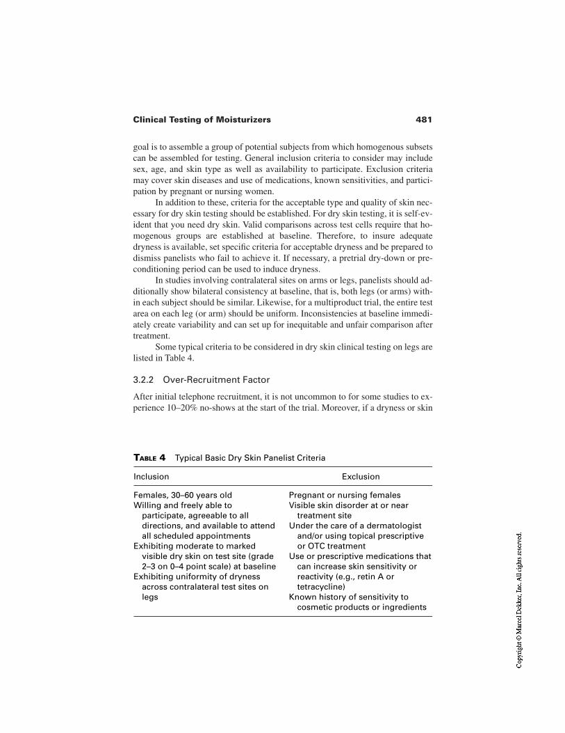
481Clinical Testing of Moisturizers
goal is to assemble a group of potential subjects from which homogenous subsetscan be assembled for testing. General inclusion criteria to consider may includesex, age, and skin type as well as availability to participate. Exclusion criteriamay cover skin diseases and use of medications, known sensitivities, and partici-pation by pregnant or nursing women.
In addition to these, criteria for the acceptable type and quality of skin nec-essary for dry skin testing should be established. For dry skin testing, it is self-ev-ident that you need dry skin. Valid comparisons across test cells require that ho-mogenous groups are established at baseline. Therefore, to insure adequatedryness is available, set specific criteria for acceptable dryness and be prepared todismiss panelists who fail to achieve it. If necessary, a pretrial dry-down or pre-conditioning period can be used to induce dryness.
In studies involving contralateral sites on arms or legs, panelists should ad-ditionally show bilateral consistency at baseline, that is, both legs (or arms) with-in each subject should be similar. Likewise, for a multiproduct trial, the entire testarea on each leg (or arm) should be uniform. Inconsistencies at baseline immedi-ately create variability and can set up for inequitable and unfair comparison aftertreatment.
Some typical criteria to be considered in dry skin clinical testing on legs arelisted in Table 4.
3.2.2 Over-Recruitment Factor
After initial telephone recruitment, it is not uncommon to for some studies to ex-perience 10–20% no-shows at the start of the trial. Moreover, if a dryness or skin
TABLE 4 Typical Basic Dry Skin Panelist Criteria
Inclusion Exclusion
Females, 30–60 years oldWilling and freely able to
participate, agreeable to all directions, and available to attend all scheduled appointments
Exhibiting moderate to marked visible dry skin on test site (grade 2–3 on 0–4 point scale) at baseline
Exhibiting uniformity of dryness across contralateral test sites on legs
Pregnant or nursing femalesVisible skin disorder at or near
treatment siteUnder the care of a dermatologist
and/or using topical prescriptive or OTC treatment
Use or prescriptive medications that can increase skin sensitivity or reactivity (e.g., retin A or tetracycline)
Known history of sensitivity to cosmetic products or ingredients
482 Nole
condition criteria is established, losing a certain percentage of panelists at base-line must be expected. To prepare for this, studies should be over-recruited by anappropriate factor, perhaps as much as 50% if criteria are very restrictive. It is al-ways better to start a panel with an excess of acceptable panelist than to have toconsider changing criteria because insufficient panelists are available.
3.2.3 Panelist Restrictions
During recruitment, well in advance of the start of the trial, it is important thatsubjects fully understand the personal restrictions to be placed on them, that is,the products that they cannot use and activities they must stop. The restrictionsshould also be fully spelled out in the panelist paperwork. This will help insurecompliance and avoid misunderstandings later. Similarly, in multivisit trials, pan-elist must commit to the observation schedule and understand the penalties fornot following it.
3.3 Grading/Measurements
As discussed earlier, the three-prong approach of expert assessment, instrumentalassessment, and self-assessment allows the results of one method to be corrobo-rated and reinforced by the others. Within these three categories, the choice oftechniques must be rational and appropriate to the objectives. More measurementis not always better. Data from measurements that are irrelevant to the objectiveor, worse, inappropriate to the objective can be a distraction and waste resources.Quality data will flow from a well-conceived set of measurements.
3.3.1 Expert Visual Grading
Dry skin condition is typically graded in terms of its main symptoms, flakiness(scaling), roughness, and erythema, though it is also common to use a single as-sessment of overall dryness. Because expert grading is only as good as the expertgrader, experience and training is critical. The use of photographic reference stan-dards and/or descriptive texts to define grading levels is strongly recommended[42,43]. Because visual grading is subjective, all expert grading scales should bedescribed in the protocol with the precise language used to delineate scale points.It is the ability to discriminate each point unambiguously that defines the limits ofsensitivity.
Even the most experienced graders are not free from potential bias. To en-sure complete objectivity of the visual grading procedures, visual assessors mustbe completely independent of all other aspects of the trial and must be blind to theproducts being tested and the allocation of cells. Grading must also be performedabsolutely, that is, how it appears at that moment. There should be no reference toprior data for comparison.
483Clinical Testing of Moisturizers
3.3.2 Bioinstruments
The many instruments which are available to quantify a wide range of physicalattribute are discussed in detail elsewhere in this volume. For the study designer,the selection of instrumental measures must be relevant to the objectives andshould be aligned with expert grading. Procedures for bioinstrumental readingsmust be defined in the protocol, for example, that three replicate readings are be-ing taken and averaged for each measurement. Also, in that skin can be influencedby transient environmental conditions, the protocol should specify the acceptabletest conditions for instruments.
3.3.3 Self assessment
In clinical testing, self-assessment is not the same as in consumer panels. Whereconsumer panels are interested in likes and dislikes of product attributes, self-as-sessment clinical grading is concerned with how skin looks and feels to the user.Clinical self-assessment demonstrates whether the changes in skin condition areresulting in meaningful and perceivable benefits to the subject. It also allowsmeasurement of a much richer set of attributes than can be achieved with eithervisual or instrumental evaluations both in terms of descriptive attributes (such asroughness, redness, chapping, and cracking) and in terms of sensory attributesthat cannot be otherwise measured (such as soreness, itching, tight feel).
Self-assessment scales are commonly of two types. The first is analog, orline scale, where subjects place a mark anywhere along a continuous line betweentwo endpoints. With this type of scale the relative distance from the end point issimply measured and recorded. The alternative is a fixed interval scale, wheresubjects select a relative grade of increasing severity, usually a series of bubbledots. In either case, the language used to define the anchor points must be clearand unambiguous (Table 5).
Self-assessment within a clinical trial can not replace consumer testing. Therelatively small sample size and controlled structure of the clinical trial are inap-propriate for measuring consumer habits and attitudes. Within a clinical trial, self-assessment should remain focused on the perception of skin condition (physicaland sensory) and the changes in condition due to product use.
3.3.4 Order of Measurements
Instrumental probes that contact the skin can alter the test sites. It is also difficultto keep the subjects from having an awareness of the various evaluations, even ifthey are not fully understood, and this can bias their own perceptions of skin con-dition. Therefore the order of measurements should be thought through in termsof how each could affect the next. It is generally good to allow subjects to com-plete their self-evaluation first, with visual expert assessments next, followed byany instrumental measures in order of increasing invasiveness.
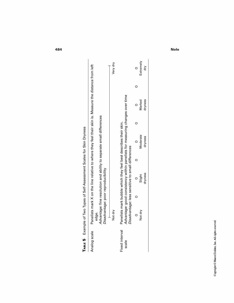
484 Nole
TA
BLE
5E
xam
ple
of
Two
Typ
es o
f S
elf-
Ass
essm
ent
Sca
les
for
Ski
n D
ryn
ess
An
alo
g s
cale
Pan
elis
ts m
ark
X o
n t
he
line
rela
tive
to
wh
ere
they
fee
l th
eir
skin
is. M
easu
re t
he
dis
tan
ce f
rom
left
ed
ge.
Ad
van
tag
e:fi
ne
reso
luti
on
an
d a
bili
ty t
o s
epar
ate
smal
l dif
fere
nce
sD
isad
van
tag
e:p
oo
r re
pro
du
cib
ility
No
t d
ryVe
ry d
ry
Fixe
d in
terv
al
Pan
elis
ts m
ark
bu
bb
le w
hic
h t
hey
fee
l bes
t d
escr
ibes
th
eir
skin
.A
dva
nta
ge:
go
od
co
nsi
sten
cy w
ith
in p
anel
ists
fo
r m
easu
rin
g c
han
ges
ove
r ti
me
Dis
adva
nta
ge:
less
sen
siti
ve t
o s
mal
l dif
fere
nce
s
ON
ot
dry
OO
Slig
ht
dry
nes
s
OO
Mo
der
ate
dry
nes
s
OO
Mar
ked
dry
nes
s
OO
Ext
rem
ely
dry
scal
e
485Clinical Testing of Moisturizers
3.4 Observation Events
Observation is the essence of a clinical trial. When all is done, the output of thestudy is the collection of observation data. Exactly how, when, and under whatconditions the measurements are taken must be well documented in the protocol.
3.4.1 Equilibration Period
Transient activities and environmental conditions can temporarily affect thephysical properties of skin. For example, the face can become flushed coming in-side on a brisk day or perspiration can increase with nervousness. Subjects needto accommodate to the testing environment prior to measurements being made. Aquiet period of 15–30 min is generally accepted as sufficient to equilibrate to thetesting conditions and allow the subject to reach a relaxed physiological state.
3.4.2 Blinding Procedures
Double-blind procedures are necessary to prevent product knowledge by subjectsand evaluators that could bias study results. Because they are such an importantpart of good clinical practice, the specific blinding procedures should be definedin the protocol. A double-blind trial includes both product and assignment blind-ness, that is, all products are in coded plain white containers and evaluators areunaware of which subjects are using which products.
3.4.3 Frequency of Observations
A study with only two observations, baseline and end point, leaves much to be de-sired. A schedule of interim observations provides valuable insight into how theskin reacts to the product over time, which allows a time course of improvementto be plotted. A sensible time response profile is a very good indication of a validstudy. For a multiweek test, a schedule of Monday/Thursday or Monday/Wednes-day/Friday is typical. Daily observations over several weeks are not usually nec-essary, except perhaps at the very start of the treatment or regression phases. Insingle application trials, readings at 30- to 90-min intervals are common, with atleast two interim observations between baseline and the end point.
3.4.4 Observation Schedule
Diurnal rhythms can have a significant physiological effect with potential impli-cations for clinical measurements. In addition to natural cycles, the time gap sincemorning shower or last product application should be consistent among subjects.If it is not possible to ensure uniformity, having each subject evaluated at thesame time at each observation and having subjects from each study cell uniform-ly distributed throughout the observation day can at least minimize the diurnaleffects.
486 Nole
3.4.5 Condition of Skin Immediately Prior to Measurement
Skin reflects not just the effect of treatment, but the transient effect of any activi-ty prior to measurement. For example, baseline hygrometer measurements can bedepressed immediately following a wash cycle or the visual appearance of dry-ness can be affected by the presence of residual product still on the skin. Becauseactivities immediately prior to measurement can affect the measurement, the pro-tocol should describe how skin is to be prepared. For example, in hygrometerstudies, a standard mild wash procedure can be implemented across all subjects30 min prior to the baseline observations. For regression studies, daily treatmenton observation days can be delayed until after observations are complete.
3.4.6 Number of Independent Judges
Despite all efforts to ensure any two judges work similarly, there is always poten-tial for inconsistencies between them to occur. It is more important that judges re-main consistent within themselves (intrajudge consistency) than between otherjudges (interjudge consistency). A practice to avoid is one judge being responsi-ble for half of the study and another judge being responsible for the balance, orsome other such configuration. In any trial, a single primary judge responsible forall observations throughout the study is ideal.
3.4.7 Observation Schedule: Timing and Traffic Flow
Machines never tire, but people certainly do. It is difficult to maintain the samemental acuity for assessing the 99th panelist as the 9th panelist. A rule of thumbwould be to determine the typical amount of time it takes to complete an observa-tion, then allow 50% more time. This will prevent the pace from becoming toohectic. Short breaks should also be scheduled every hour to prevent fatigue. Pan-elists too will become fatigued if they are endlessly probed by a succession of in-struments. When setting up a schedule, consider what is reasonable for subjectsas well. The pace and break schedule will define the throughput, that is, the num-ber of panelists per hour, that can be evaluated; the throughput in turn defines thetotal number of subjects that can be evaluated in a day. The total subjects that canbe evaluated is often the maximum limit for participants in your trial.
The daily observation schedule should be described in the protocol. In a re-gression test, throughput of 10–20 subjects per hour is reasonable, depending onexperience and efficiency of the clinicians. In a hydration test, throughput of 30subjects per hour can be achieved.
In a trial where multiple measurements are being collected by different cli-nicians, a panelist checklist is recommended. The panelist follows the list in se-quence to be sure the correct order is maintained, and as each measurement iscompleted it is initialed by the clinician. Before the subject is dismissed, a check-list is reviewed to make sure everything has been completed.
487Clinical Testing of Moisturizers
3.4.8 Lighting and Environmental Conditions
For all observations—visual, self-assessment or instrumental—consistency ofconditions is very important both from one observation to the next and from onepanelist to the next. Visual evaluations are best done under fixed lights and awayfrom windows where sunlight changes throughout the day. A 2x magnifying ring-lamp is commonly used for dry skin visual evaluation. Instrumental measuresbenefit from having subjects equilibrate to a standard indoor temperature and hu-midity. For single-use tests, an air conditioned lab with good control is generallyacceptable; for more rigorous control, a humidity/temperature control room isrecommended.
3.4.9 Bathing and Shaving Restrictions
In studies over several weeks, it is recommended to standardize the bathing habitsof subjects, for example, by issuing a standard mild soap bar and requiring allbathing be done in the morning, within 30 min of product application. Shavingduring leg trials poses a particular challenge as shaving can exacerbate the ap-pearance of dryness. A uniform schedule of how and when to shave should be in-cluded in the panelist instruction.
3.5 Sample Administration
In any trial, the product is usually the only thing subjects are concerned with. Inclinician applied studies, subject interaction with the product is minimal. But intrials with home applications, the product is the most prominent source of infor-mation disclosed to subjects. In such trials, products must be presented withoutbias and should be adequately blinded. Label contents and product handlingshould be defined in the protocol. The following points are relevant only to clini-cal studies involving at-home application.
3.5.1 Uniform Packaging
Ideally all products would be presented in identical plain, coded containers. Ifproduct forms are different (creams versus lotions) the different packaging shouldbe nondescript. Pump packages are preferred as they insure a more uniformdosage.
3.5.2 Descriptive Label Contents
Labels should contain absolute minimal descriptive information about the prod-uct. Three pieces of information are sufficient: study number, a cryptic productcode, and a unique bottle number or subject identification. Good product codesare randomly assigned letter/number combinations. Bottle numbers can be se-
488 Nole
quentially assigned independently of product codes to aid in traceability of theproducts.
3.5.3 Warnings/Restrictions
If necessary, warning and caution statements must be included on the package. Itis good practice to include a contact phone number in case of emergency on thelabel. Additional information may be provided in an accompanying sheet.
3.5.4 Weight Record and Sample Disposition
It is good practice to preweigh all bottles and reweigh at each observation. Usingthe bottle numbers, products can be traced to the individual panelists, and non-compliance (subjects applying too little or too much product) can be identified.Clinical test products must always be returned by the end of the trial, even if re-turned empty.
3.5.5 Retained Samples
It is always a good practice to retain one to three additional samples of each testmaterial in the test package for reference during and after the study.
3.6 Pretreatment Phase
To evaluate the effectiveness of a dry skin treatment requires some degree of dryskin at baseline. The greater the initial level of dryness, the greater the potentialfor demonstrating effects. In the real world, few people allow their skin to reachmarked or severe levels of dryness without taking action, therefore some type ofpretreatment may be needed. In designing the dry skin protocol it is necessary todefine the acceptability criteria for dryness, and if a pretreatment phase is needed,then the means for achieving baseline dyrness must likewise be defined.
3.6.1 Product Use/Exclusion
The generally accepted means of achieving sufficient baseline dryness is with amoisturizer weaning period, that is, where all cream/lotion use is stopped. For legstudies, the use of moisturizing cleansers or moisturizing leg shaving productsmust likewise be discontinued. During this phase subjects should be suppliedwith a commercial mild cleansing bar (such as Dove) for general bathing.
3.6.2 Length of Preconditioning Phase
Inducing dryness too harshly can lead to irritation. Superficial dryness for singleapplication studies can usually be achieved in 24–72 hr even under moderate dryskin weather. Moderate to marked dryness for regression tests typically requires3–7 days under more severe dry skin conditions.
489Clinical Testing of Moisturizers
3.6.3 Soap Washing Procedure
If weather conditions are severe, adequate dryness will occur naturally with lo-tion weaning. To enhance the drying process when necessary, a routine of two orthree times daily washing can be included. A dry-down wash procedure is morestringent than an ordinary wash. For example;
Lather bar in wet hand 10 rotations in 10 seconds.Apply lather to wet leg (or arm) and lather test site for 30 seconds.Rinse site thoroughly with water.Lightly pat dry.
Note that overwashing, especially with plain soap, must be avoided as it can gobeyond flaky dry to a condition where surface squames are removed and skinlipids are hyperdepleted.
3.7 Product Treatment Phase
Whether the product is applied once or repeatedly, by clinician or by subjects athome, questions of how the products are applied, how much is used, where it isused, when it is used, and when it is to be avoided must all be answered. In a con-trolled clinical trial, the goal is to eliminate the variability of consumer use bydefining every aspect of the treatment phase so that all subjects across all productgroups are doing the exact same thing. Only by ensuring consistency across thegroups can valid comparison of the groups be made.
3.7.1 Usage Rate
The rule set down by the FDA in the 1970s for sunscreen testing was to apply auniform film of lotion at a rate of 2 mg/cm2 [44]. This standard has been widelyaccepted for any lotion treatment, as it is in fact a reasonable amount. Higher ap-plication levels lead to excess on the skin which is unpleasant to the subject andwill likely get rubbed off anyway.
For clinician-applied studies, test sites of 15–30 cm2 are typically markedon the arm or leg. Treatment amounts are precisely dispensed using a pipette andrubbed in uniformly within the demarcated area with a gloved finger.
Because trials with home application cannot achieve this level of precision,detailed instructions to the panelists are required. For example, a pump which dis-penses 0.5 g per press can be used as a convenient means of dispensing a standarddose. An instruction to apply one pump of product to the lower outer leg and rub-bing in thoroughly is acceptably close to the desired application rate and, at thevery least, is a uniform application procedure across test groups. In general, 0.5 gof a facial product is adequate for treating the entire face, while 1 g of a hand lo-tion is needed to treat both hands.
490 Nole
3.7.2 Frequency of Application
The original regression method called for twice daily application. At this rate,skin generally improves slowly over a week or two. While more frequent applica-tion would appear to allow skin to improve more quickly, much of the additional“benefit” would be cosmetic only and it could obscure the differences in real per-formance between products. Moreover, twice daily is a very easy routine forhome-based panelists to follow: once when subjects get up in the morning andagain before going to bed at night.
3.7.3 How Long to Use Product
For regression testing, 1 week is minimum to begin to see true physiological ef-fects (i.e., discernable from mere cosmetic effects). In 3 weeks the skin hasachieved full stratum corneum turnover so the entire skin layer at that point is theresult of the treatment product. Two weeks is a middling approach that gives aperformance prediction very near the 3-week results.
3.7.4 Weekend Treatments
The original regression test discontinued product over the weekends in order tostrip away the cosmetic benefits and view more clearly the substantive benefits todry skin. This procedure has its merit particularly where daily applications areconducted by a clinician. However, for home-use trials, simplicity of routine iskey and it may be better to continue the same daily regimen over the weekends.
3.7.5 Observation Day Treatments
During regression tests, to avoid the cosmetic effect of residual lotion on skin,there should never be a product application prior to observations. The recommen-dation is to apply (as usual) the night before, then shower as normal on the day ofthe observation to wash away residual product. The daily treatment resumes im-mediately after the observation session is completed.
3.7.6 Panelist Instructions
Panelist instructions can never be clear enough or simple enough. The treatmentprocedure should be described as plainly as possible. On the first day of the trial,subjects must read these instructions and review them with the clinician. If possi-ble, the first application should take place at the test center under supervision. Abrief review of the instructions at each observations is recommended.
3.8 Regression
The regression phase is the signature component of the regression test. To proper-ly evaluate the rate of return of dry skin, as much consideration must be given to
491Clinical Testing of Moisturizers
what panelists do not do during this phase as was given to what they did duringtreatment phase.
3.8.1 What Products to Allow/Exclude
All treatment during this phase is discontinued. Panelists continue to refrain fromusing their normal lotion products and must continue to use only the prescribedwash products in the prescribed manner.
3.8.2 Length of Regression
Generally the regression phase is set up as a fixed term of 1–2 weeks after the fi-nal treatment. Seven days is sufficient to establish a rate of dry skin return thoughskin may not reach baseline. A 2-week regression allows skin to approach theoriginal baseline level of dryness. In the nutrition protocol, regression can be ex-tended to 3 weeks.
3.8.3 Observation Schedule
The difference between a cosmetic and substantive product should show up im-mediately after treatment is discontinued, where the healthier skin shows greaterpersistence. Therefore, a daily or every-other-day observation schedule is usedduring the first half of the regression phase.
4 EXAMPLE PROTOCOLS
From this discussion of points to consider when developing a protocol, it is evi-dent that there is no single right way to conduct a trial. It is a generally poor prac-tice to take a standard protocol and use it without thinking through the listedpoints to insure that the proposed study design is appropriate for your productsand objectives.
Having said that, some guidance can be helpful. Table 6 provides a side-by-side listing of typical protocol specifications for four common approaches to dry skin. This table provides quick reference for some details of proceduresand provides easy comparison of the size and scope of each procedure. Keep inmind that these should only be used for guidance; the fine points of your own fi-nal protocol must flow from your objectives and your consideration of the studydetails.
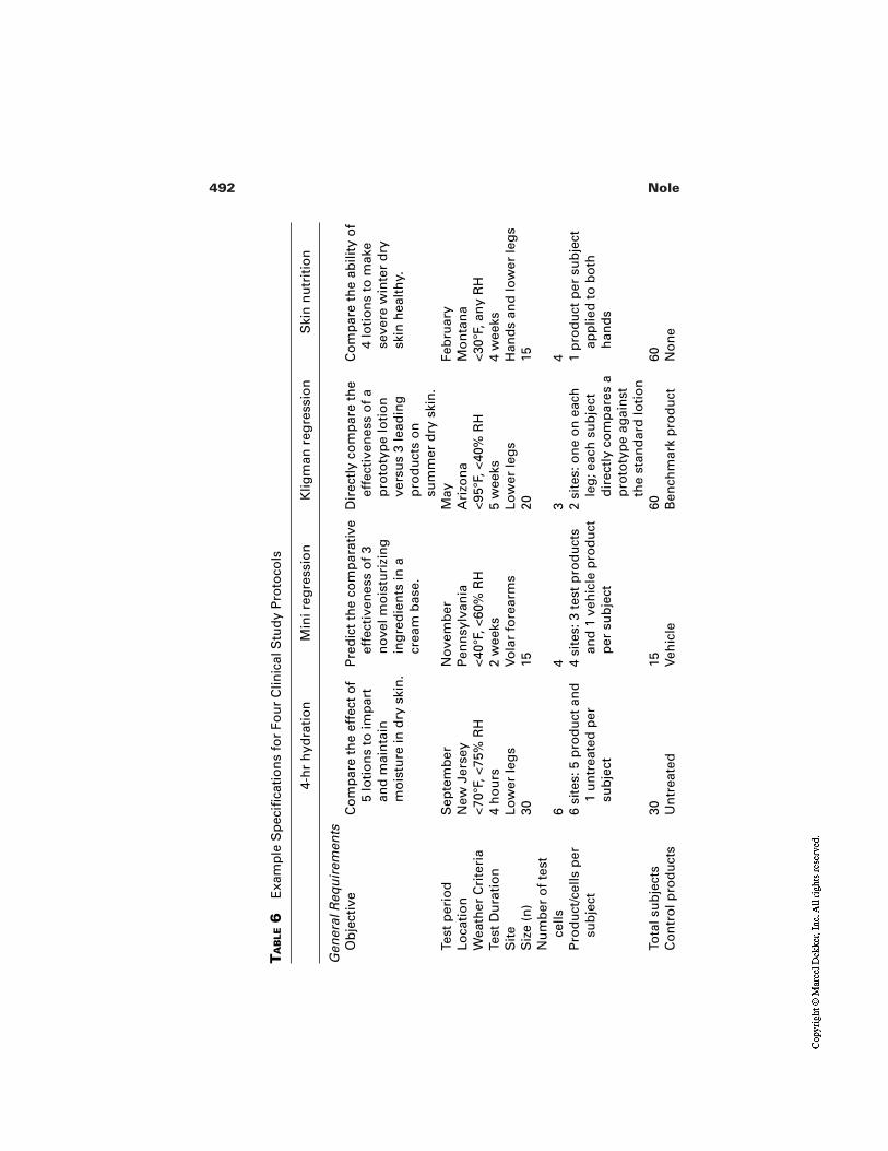
492 Nole
TA
BLE
6E
xam
ple
Sp
ecifi
cati
on
s fo
r Fo
ur
Clin
ical
Stu
dy
Pro
toco
ls
4-h
r h
ydra
tio
nM
ini r
egre
ssio
nK
ligm
an r
egre
ssio
nS
kin
nu
trit
ion
Gen
eral
Req
uir
emen
tsO
bje
ctiv
eC
om
par
e th
e ef
fect
of
5 lo
tio
ns
to im
par
tan
d m
ain
tain
mo
istu
re in
dry
ski
n.
Pre
dic
t th
e co
mp
arat
ive
effe
ctiv
enes
s o
f 3
no
vel m
ois
turi
zin
gin
gre
die
nts
in a
crea
m b
ase.
Dir
ectl
y co
mp
are
the
effe
ctiv
enes
s o
f a
pro
toty
pe
loti
on
vers
us
3 le
adin
gp
rod
uct
s o
nsu
mm
er d
ry s
kin
.
Co
mp
are
the
abili
ty o
f4
loti
on
s to
mak
ese
vere
win
ter
dry
skin
hea
lth
y.
Test
per
iod
Sep
tem
ber
No
vem
ber
May
Feb
ruar
yLo
cati
on
New
Jer
sey
Pen
nsy
lvan
iaA
rizo
na
Mo
nta
na
Wea
ther
Cri
teri
a<7
0°F,
<75
% R
H<4
0°F,
<60
% R
H<9
5°F,
<40
% R
H<3
0°F,
an
y R
HTe
st D
ura
tio
n4
ho
urs
2 w
eeks
5 w
eeks
4 w
eeks
Sit
eLo
wer
leg
sVo
lar
fore
arm
sLo
wer
leg
sH
and
s an
d lo
wer
leg
sS
ize
(n)
3015
2015
Nu
mb
er o
f te
st
cells
64
34
Pro
du
ct/c
ells
per
su
bje
ct6
site
s: 5
pro
du
ct a
nd
1 u
ntr
eate
d p
ersu
bje
ct
4 si
tes:
3 t
est
pro
du
cts
and
1 v
ehic
le p
rod
uct
per
su
bje
ct
2 si
tes:
on
e o
n e
ach
leg
; eac
h s
ub
ject
dir
ectl
y co
mp
ares
ap
roto
typ
e ag
ain
stth
e st
and
ard
loti
on
1 p
rod
uct
per
su
bje
ctap
plie
d t
o b
oth
han
ds
Tota
l su
bje
cts
3015
6060
Co
ntr
ol p
rod
uct
sU
ntr
eate
dVe
hic
leB
ench
mar
k p
rod
uct
No
ne
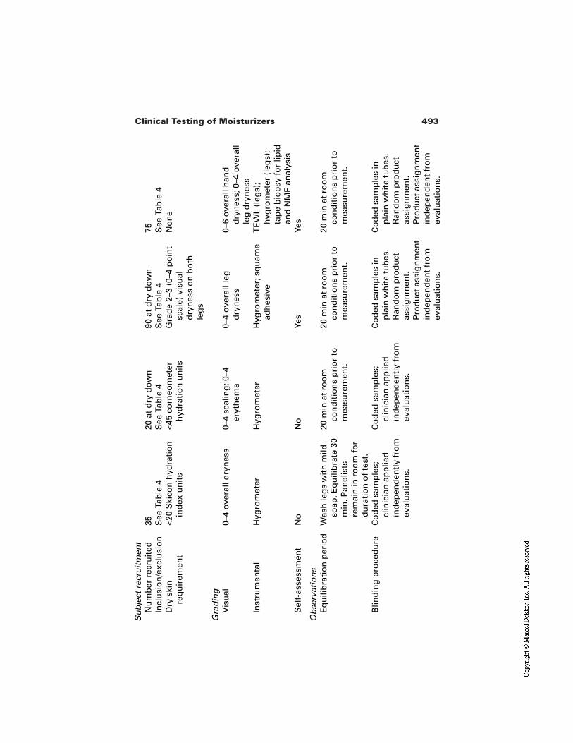
493Clinical Testing of Moisturizers
Su
bje
ct r
ecru
itm
ent
Nu
mb
er r
ecru
ited
3520
at
dry
do
wn
90 a
t d
ry d
ow
n75
Incl
usi
on
/exc
lusi
on
See
Tab
le 4
See
Tab
le 4
See
Tab
le 4
See
Tab
le 4
Dry
ski
n
req
uir
emen
t<2
0 S
kico
n h
ydra
tio
nin
dex
un
its
<45
corn
eom
eter
hyd
rati
on
un
its
Gra
de
2–3
(0–4
po
int
scal
e) v
isu
ald
ryn
ess
on
bo
thle
gs
No
ne
Gra
din
gV
isu
al0–
4 o
vera
ll d
ryn
ess
0–4
scal
ing
; 0–4
eryt
hem
a0–
4 o
vera
ll le
gd
ryn
ess
0–6
ove
rall
han
dd
ryn
ess;
0–4
ove
rall
leg
dry
nes
sIn
stru
men
tal
Hyg
rom
eter
Hyg
rom
eter
Hyg
rom
eter
; sq
uam
ead
hes
ive
TE
WL
(leg
s);
hyg
rom
eter
(le
gs)
;ta
pe
bio
psy
fo
r lip
idan
d N
MF
anal
ysis
Sel
f-as
sess
men
tN
oN
oYe
sYe
s
Ob
serv
atio
ns
Eq
uili
bra
tio
n p
erio
dW
ash
leg
s w
ith
mild
soap
. Eq
uili
bra
te 3
0m
in. P
anel
ists
rem
ain
in r
oo
m f
or
du
rati
on
of
test
.
20 m
in a
t ro
om
con
dit
ion
s p
rio
r to
mea
sure
men
t.
20 m
in a
t ro
om
con
dit
ion
s p
rio
r to
mea
sure
men
t.
20 m
in a
t ro
om
con
dit
ion
s p
rio
r to
mea
sure
men
t.
Blin
din
g p
roce
du
reC
od
ed s
amp
les;
clin
icia
n a
pp
lied
ind
epen
den
tly
fro
mev
alu
atio
ns.
Co
ded
sam
ple
s;cl
inic
ian
ap
plie
din
dep
end
entl
y fr
om
eval
uat
ion
s.
Co
ded
sam
ple
s in
pla
in w
hit
e tu
bes
.R
and
om
pro
du
ctas
sig
nm
ent.
Pro
du
ct a
ssig
nm
ent
ind
epen
den
t fr
om
eval
uat
ion
s.
Co
ded
sam
ple
s in
pla
in w
hit
e tu
bes
.R
and
om
pro
du
ctas
sig
nm
ent.
Pro
du
ct a
ssig
nm
ent
ind
epen
den
t fr
om
eval
uat
ion
s.
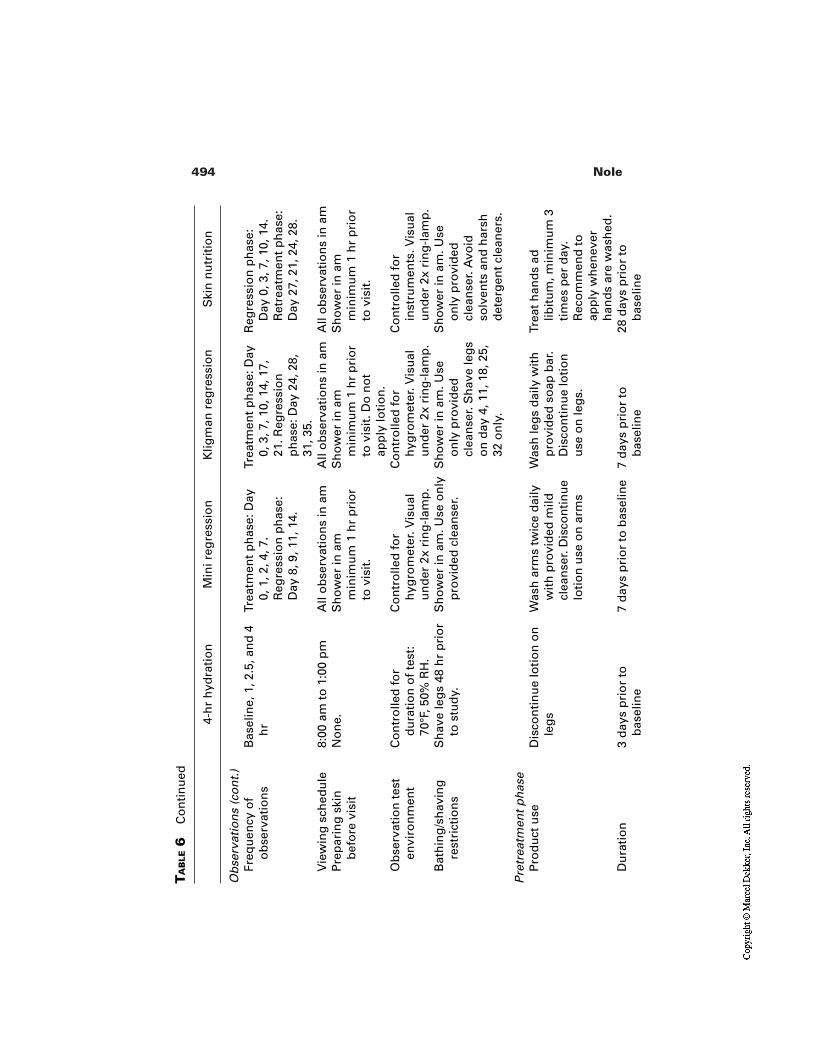
494 NoleT
AB
LE6
Co
nti
nu
ed
4-h
r h
ydra
tio
nM
ini r
egre
ssio
nK
ligm
an r
egre
ssio
nS
kin
nu
trit
ion
Ob
serv
atio
ns
(co
nt.
)Fr
equ
ency
of
ob
serv
atio
ns
Bas
elin
e, 1
, 2.5
, an
d 4
hr
Trea
tmen
t p
has
e: D
ay0,
1, 2
, 4, 7
.R
egre
ssio
n p
has
e:D
ay 8
, 9, 1
1, 1
4.
Trea
tmen
t p
has
e: D
ay0,
3, 7
, 10,
14,
17,
21. R
egre
ssio
np
has
e: D
ay 2
4, 2
8,31
, 35.
Reg
ress
ion
ph
ase:
Day
0, 3
, 7, 1
0, 1
4.R
etre
atm
ent
ph
ase:
Day
27,
21,
24,
28.
Vie
win
g s
ched
ule
8:00
am
to
1:0
0 p
mA
ll o
bse
rvat
ion
s in
am
All
ob
serv
atio
ns
in a
mA
ll o
bse
rvat
ion
s in
am
Pre
par
ing
ski
n
bef
ore
vis
itN
on
e.S
ho
wer
in a
mm
inim
um
1 h
r p
rio
rto
vis
it.
Sh
ow
er in
am
min
imu
m 1
hr
pri
or
to v
isit
. Do
no
tap
ply
loti
on
.
Sh
ow
er in
am
min
imu
m 1
hr
pri
or
to v
isit
.
Ob
serv
atio
n t
est
envi
ron
men
tC
on
tro
lled
fo
rd
ura
tio
n o
f te
st:
70°F
, 50%
RH
.
Co
ntr
olle
d f
or
hyg
rom
eter
. Vis
ual
un
der
2x
rin
g-l
amp
.
Co
ntr
olle
d f
or
hyg
rom
eter
. Vis
ual
un
der
2x
rin
g-l
amp
.
Co
ntr
olle
d f
or
inst
rum
ents
. Vis
ual
un
der
2x
rin
g-l
amp
.B
ath
ing
/sh
avin
gre
stri
ctio
ns
Sh
ave
leg
s 48
hr
pri
or
to s
tud
y.S
ho
wer
in a
m. U
se o
nly
pro
vid
ed c
lean
ser.
Sh
ow
er in
am
. Use
on
ly p
rovi
ded
clea
nse
r. S
hav
e le
gs
on
day
4, 1
1, 1
8, 2
5,32
on
ly.
Sh
ow
er in
am
. Use
on
ly p
rovi
ded
clea
nse
r. A
void
solv
ents
an
d h
arsh
det
erg
ent
clea
ner
s.
Pre
trea
tmen
t p
has
eP
rod
uct
use
Dis
con
tin
ue
loti
on
on
leg
sW
ash
arm
s tw
ice
dai
lyw
ith
pro
vid
ed m
ildcl
ean
ser.
Dis
con
tin
ue
loti
on
use
on
arm
s
Was
h le
gs
dai
ly w
ith
pro
vid
ed s
oap
bar
.D
isco
nti
nu
e lo
tio
nu
se o
n le
gs.
Trea
t h
and
s ad
libit
um
, min
imu
m 3
tim
es p
er d
ay.
Rec
om
men
d t
oap
ply
wh
enev
erh
and
s ar
e w
ash
ed.
Du
rati
on
3 d
ays
pri
or
tob
asel
ine
7 d
ays
pri
or
to b
asel
ine
7 d
ays
pri
or
tob
asel
ine
28 d
ays
pri
or
tob
asel
ine
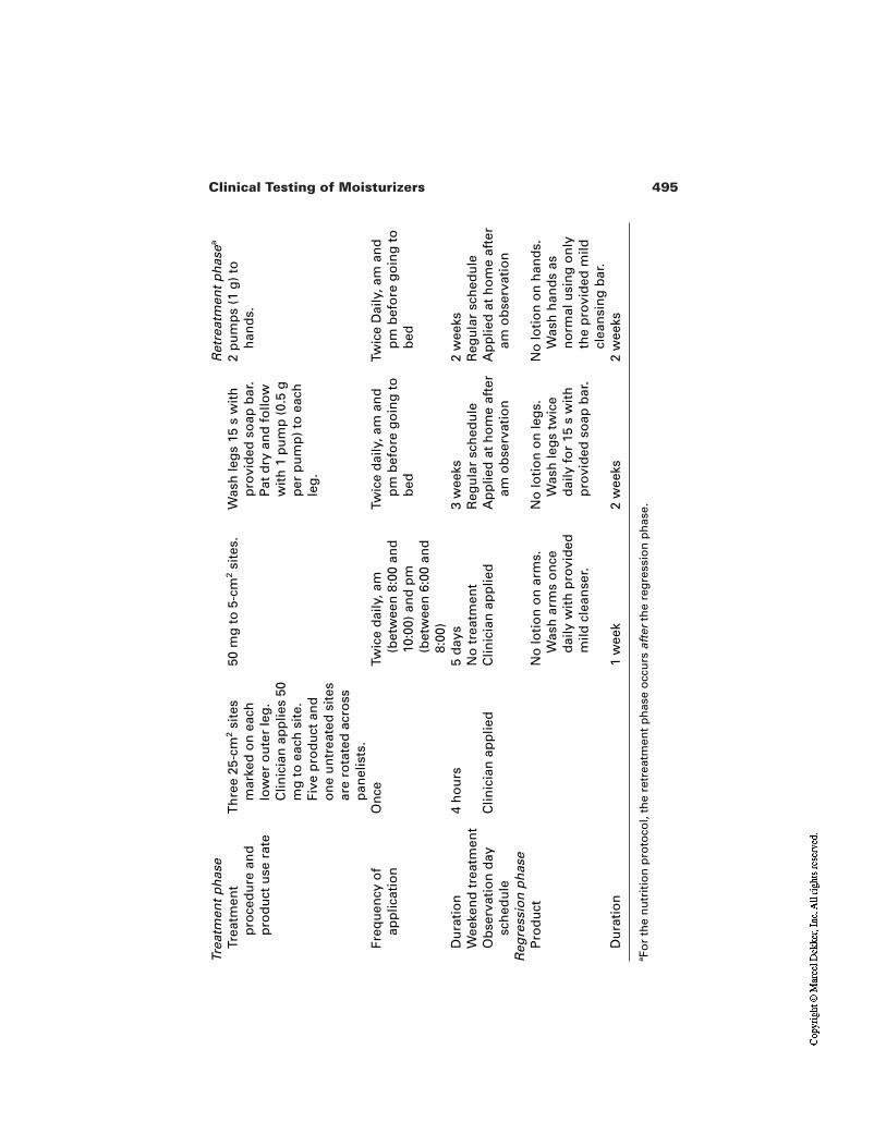
495Clinical Testing of Moisturizers
Trea
tmen
t p
has
eR
etre
atm
ent
ph
asea
Trea
tmen
t p
roce
du
re a
nd
p
rod
uct
use
rat
e
Th
ree
25-c
m2
site
sm
arke
d o
n e
ach
low
er o
ute
r le
g.
Clin
icia
n a
pp
lies
50m
g t
o e
ach
sit
e.Fi
ve p
rod
uct
an
do
ne
un
trea
ted
sit
esar
e ro
tate
d a
cro
ssp
anel
ists
.
50 m
g t
o 5
-cm
2si
tes.
Was
h le
gs
15 s
wit
hp
rovi
ded
so
ap b
ar.
Pat
dry
an
d f
ollo
ww
ith
1 p
um
p (
0.5
gp
er p
um
p)
to e
ach
leg
.
2 p
um
ps
(1 g
) to
han
ds.
Freq
uen
cy o
f ap
plic
atio
nO
nce
Twic
e d
aily
, am
(bet
wee
n 8
:00
and
10:0
0) a
nd
pm
(bet
wee
n 6
:00
and
8:00
)
Twic
e d
aily
, am
an
dp
m b
efo
re g
oin
g t
ob
ed
Twic
e D
aily
, am
an
dp
m b
efo
re g
oin
g t
ob
ed
Du
rati
on
4 h
ou
rs5
day
s3
wee
ks2
wee
ksW
eeke
nd
tre
atm
ent
No
tre
atm
ent
Reg
ula
r sc
hed
ule
Reg
ula
r sc
hed
ule
Ob
serv
atio
n d
ay
sch
edu
leC
linic
ian
ap
plie
dC
linic
ian
ap
plie
dA
pp
lied
at
ho
me
afte
ram
ob
serv
atio
nA
pp
lied
at
ho
me
afte
ram
ob
serv
atio
nR
egre
ssio
n p
has
eP
rod
uct
No
loti
on
on
arm
s.W
ash
arm
s o
nce
dai
ly w
ith
pro
vid
edm
ild c
lean
ser.
No
loti
on
on
leg
s.W
ash
leg
s tw
ice
dai
ly f
or
15 s
wit
hp
rovi
ded
so
ap b
ar.
No
loti
on
on
han
ds.
Was
h h
and
s as
no
rmal
usi
ng
on
lyth
e p
rovi
ded
mild
clea
nsi
ng
bar
.D
ura
tio
n1
wee
k2
wee
ks2
wee
ks
aFo
r th
e n
utr
itio
n p
roto
col,
the
retr
eatm
ent
ph
ase
occ
urs
aft
erth
e re
gre
ssio
n p
has
e.
496 Nole
REFERENCES
1. Kligman A. Regression method for assessing the efficacy of moisturizers. CosmetToil 1978; 93:27–35.
2. Blank I. Factors which influence the water content of the stratum corneum. J InvestDermatol 1952; 18:433–440.
3. Blank I. Further observatons on factors which influence the water content of the stra-tum corneum; J Invest Dermatol 1953; 21:259–271.
4. Spruit D, Malten KE. Epidermal water barrier formation after stripping of normalskin. J Invest Dermatol 1965; 45(1):6–14.
5. Berube G, Messinger M, Burdick M. Measurement in vivo of transepidermal mois-ture loss. J Soc Cosmet Chem 1971; 22:361–368.
6. Berube G, Burdick M. Transepidermal water loss. II. The significance of the usethickness of topical substances. J Soc Cosmet Chem 1973; 25:397–406.
7. Rietschel R. A method to evaluate skin moisturizers in vivo. J Invest Dermatol 1978;70:152–155.
8. Wildauer R, Bathwell J, Douglass A. Stratum corneum biomechanical properties. JInvest Dermatol 1971; 56:72–78.
9. Rieger M, Deem D. Skin moisturizers. I. Methods for measuring water regain, me-chanical properties and transepidermal moisture loss of stratum corneum. J Soc Cos-met Chem 1974; 25:239–253.
10. Rieger M, Deem D. Skin moisturizers. II. The effects of cosmetic ingredients on hu-man stratum corneum. J Soc Cosmet Chem 1974; 25:253–262.
11. Quattrone A, Laden K. Physical techniques for assessing skin moisturization. J SocCosmet Chem 1976; 27:607–623.
12. Christenson M, Hargens C, Nacht S, Gans E. Viscoelastic properties of intact humanskin: instrumentation hydration effects and the contribution of the stratum corneum.J Invest Dermatol 1977; 69:282–286.
13. Cook T. Profilometry of skin: a useful tool for the substantiation of cosmetic effica-cy. J Soc Cosmet Chem 1980; 31:339–358.
14. Tagami H. Electrical measurement of the water content of the skin surface. CosmetToil 1982; 97:39–47.
15. Leveque J. Physical methods for skin investigation. Int J Dermatol 1983; 22:368.16. Nole G, Boisits E, Thaman L. The Salter complex impedance device as an instru-
ment to measure the hydration level of the stratum corneum in vivo. Bioeng Skin1988; 4(4):285–296.
17. Grove G, Grove M. Objective methods for assessing skin surface topography nonin-vasively. In: Leveque J, ed. Cutaneous Investigation in Health and Disease. NewYork: Marcel Dekker, 1988:1–32.
18. Grove G. Noninvasive methods for assessing moisturizers. In: Waggoner W, ed.Clinical Safety and Efficacy Testing of Cosmetics. New York: Marcel Dekker,1990:121–147.
19. Prall J, Theiler R, Bowser P, Walsh M. The effectiveness of cosmetic products in al-leviating a range of skin dryness conditions as determined by clinical and instrumen-tal techniques. Int J Cosmet Sci 1986; 8:159–174.
20. Boisits E, Nole G, Cheney M; The refined regression method; J Cutan Aging CosmetDermatol 1989; Vol 1(3):155–163.
497Clinical Testing of Moisturizers
21. Moss J. The effect of 3 moisturizers on skin surface hydration. Skin Res Technol1996; 2:32–36.
22. Loden M. Biophysical methods of providing objective documentation of the effectsof moisturizing creams. Skin Res Technol 1995; 1:101–108.
23. Grove G. Skin surface hydration changes during a mini-regression test as measuredin vivo by electrical conductivity. Current Therap Res 1992; 52(4):556–561.
24. Rawlings A. Effect of lactic acid isomers on keratinocyte ceramide synthesis. ArchDerm Res 1996; 288:383–390.
25. Batt M, Davis W, Fairhurst E, Gerrard W. Changes in the physical properties of thestratum corneum following treatment with glycerol. J Soc Cosmet Chem 1988;39:367–381.
26. Grove G. The effect of moisturizers on skin surface hydration as measured in vivo byelectrical conductivity; Current Therap Res; Vol 50:712–718.
27. Ertel K, Neumann P, Hartwig P, Rains G, Keswick B. Leg wash protocol to assessthe skin moisturization potential of personal cleansing products. Int J Cosmet Sci1999; 21:383–397.
28. Barker M. Moisturizers of tomorrow. Toxicol Cutan Ocular Toxicol 1992;11(3):257–262.
29. McCook J, Berube G. Evaluation of hand and body lotions: correlation of objectiveand subjective responses. J Soc Cosmet Chem 1982; 33:372.
30. Boisits E. The evaluation of moisturizing products. Cosmet Toil 1986; 101:31–39.
31. Jackson E. Moisturizers of today. Toxicol Cutan Ocular Toxicol 1992; 11(3):173–184.
32. Hannon W, Maibach H. Efficacy of moisturizers assessed through bioengineeringtechniques. In: Cosmetology for Special Locations. pp. 246–269.
33. Rogiers V, Derde M, Verleye G, Roseeuw D. Standardized conditions needed forskin surface hydration measurements. Cosmet Toil 1990; 105:73–82.
34. Black D, DelPoza A, Lagarde J, Gall Y. Seasonal variability in the biophysical prop-erties of stratum corneum from different anatomical sites. Skin Res Technol 2000;5:70–76.
35. Goh C. Seasonal variations and environmental influences on the skin. In: Serup J, Je-mec G, eds. Handbook of Non-Invasive Methods and the Skin. Boca Raton: CRCPress, 1995:27–30.
36. Prall J. Instrumental evaluation of the effects of cosmetic products on skin sur-faces with particular reference to smoothness. J Soc Cosmet Chem 1973; 24:693–707.
37. Farinelli N, Berardesca E. The skin integument: variation relative to sex, age, raceand body region. In: Serup J, Jemec G, eds. Handbook of Non-Invasive Methods andthe Skin. Boca Raton: CRC Press, 1995:22–26.
38. Barlow T. Measuring skin hydration. Cosmet Toil 1999; 114(12):47–53.39. Salter D. Study design. In: Loden M, Maibach H, eds. Dry Skin and Moisturizers:
Chemistry and Function. Boca Raton: CRC Press, 2000:373–378.40. Serup J. Prescription for a Bioengineering study: strategy, standards and definitions.
In: Serup J, Jemec G, eds. Handbook of Non-Invasive Methods and the Skin. BocaRaton: CRC Press, 1995:17–21.
498 Nole
41. Spilker B. Guide to Clinical Trials. Philadelphia: Lippincott-Raven, 1996.42. Serup J. EEMCO guidance for the assessment of dry skin (xerosis) and ichthyosis:
clinical scoring systems. Skin Res Technol 1995; 1:109–114.43. Seitz JC, Rizer RL, Spencer TS. Photographic standardization of dry skin. J Soc
Cosmet Chem 1984; 35:423–437.44. U.S. Food and Drug Administration. Sunscreen drug products for over-the-counter
human use. Tentative Final Monograph; Proposed rule. 21 CFR 352.
23Noninvasive Instrumental Methods forAssessing Moisturizers
Gary L. Grove, Charles Zerweck,and Elizabeth PierceKGL Skin Study Center, Broomall, Pennsylvania
1 INTRODUCTION
Although the term moisturizer is in widespread use by both lay people and med-ical professionals, it is a neologism of the cosmetics industry and lacks a precisedefinition. It was conveniently coined by Madison Avenue to describe a treatmentfor dry skin, which remains one of the most frequent everyday skin problems.Unfortunately despite the fact that dry skin is a troublesome, disquieting problemfor many millions of people, surprisingly little is known about its pathogenesis.We do know that dry skin is not a single entity, but rather a family of disordersthat can originate in a variety of ways [1–4].
One type is the temporary dry skin condition which is the inevitable resultof any kind of skin damage whether physical or chemical. This is a stereotypicalresponse to injury and reflects a repair process in which new skin surface cells areformed at a greatly accelerated rate. This accelerated epidermopoiesis, which typ-ically occurs after sunburn, chemical irritation, abrasion, or detergent damage,causes the skin to be dry and scaly, for example. As pointed out by Marenus [5]modern moisturizers often contain soothing agents to reduce chronic low levelirritation.
499
500 Grove et al.
We also know that some of the more severe dry skin conditions are geneti-cally determined. Indeed, the biochemical defect in X-linked ichthyosis has beenprecisely identified as a lack of a specific enzyme, steroid sulfatase, which is es-sential for proper utilization of cholesterol in the keratinization process [6]. Otherforms of dry skin are associated with an underlying disease such as psoriasis,atopic dermatitis, and diabetes or with nutritional problems.
By far the most general form of dry skin is that which is the primary con-cern of the typical moisturizer user and the focus of this chapter. For a thoroughconsideration of such common dry skin problems, one should read the excellentreview by Chernosky [7].
Although it is certainly a gross oversimplification, historically, a reducedwater content of the stratum corneum has been thought to be the key causativefactor in skin dryness. There are two reasons for suggesting this. First, considerthat dry skin problems are heavily influenced by weather conditions. Dry skin isfar worse in the winter months when low relative humidity is often accompaniedby low temperatures, strong icy winds, and dry overheated homes. The dry, hotclimate of the desert is also apt to provoke dry skin. The critical factor, as shownby the thoughtful analysis provided by Gaul and Underwood [8], is the absolutemoisture content of the air. Since the exposed stratum corneum will establish ahydration gradient in equilibrium with the surrounding air, any drop in the rela-tive humidity of the environment will lead to a corresponding decrease in skinsurface moisture levels. As shown by Middleton and Allen [9] the suppleness ofthe stratum corneum is closely related to its temperature and its water content.This means that such dry skin is relatively inflexible and inelastic under the pre-viously mentioned adverse conditions. As a result, it will crack and fissure in or-der to accommodate body movements, producing one of the more characteristicsigns of dry skin.
The second reason for believing that water content is a key factor in dryskin is a series of classic experiments performed by Blank [10,11]. He demon-strated very convincingly in the early 1950s that skin softening was most effec-tively achieved with water. Even long-term soaking of cadaver skin samples invarious oils failed to produce a comparable degree of softening as a brief expo-sure to just water alone. Since that time, dermatologists and cosmetic chemistshave emphasized that water is the principal plasticizer of the skin and critical inrelieving the signs and symptoms of dry skin. Even if the therapy is based on an-hydrous lipids, their effect on water content of the stratum corneum is still be-lieved to be the keystone for an effective moisturizer.
Thus, it is not surprising that traditional moisturizers are formulated to in-crease the moisture content of the stratum corneum in two ways:
1. Occlusion. Coating the skin with oils such as petrolatum will retard theloss of surface water, thus increasing the moisture content of the un-derlying stratum corneum.
501Methods for Assessing Moisturizers
2. By humectants. Agents such as glycerin, sodium lactate, and PCA,which draw and strongly bind water, are added to a formulation andthus help trap water in the skin surface.
Of course, one should recognize that water alone can effectively eliminate all vi-sual signs of dryness, although only temporarily. In this context, there are no in-effective cosmetic moisturizers, and the major difficulty facing investigators inthis area is not to be unduly influenced by these transient effects. We must alsoheed the views of Prall et al. [12] as well as Wehr and Krochmal [13], who havefound very little conclusive evidence to support a causative role of reduced watercontent with skin flakiness. They feel, and we concur, that the majority of mois-turizers work by merely “sticking” loosened squames back onto the surface,thereby altering light scatter, causing the skin surface to appear more transparent,and hence less dry for a short period of time. We agree with Kligman [1,2] thatsuch products should be classified as being “cosmetic” moisturizers in contrast to“therapeutic” moisturizers, which in fact modify the dry skin process rather thansimply conceal it.
With these points in mind, let us review some of the clinical and instrumen-tal methods that have developed to quantify skin dryness and thus allow us toevaluate moisturizers. Although Marenus [5] has provided some justification forextending the claims being made for modern moisturizers to include anti-aging,in this chapter we are going to limit ourselves to the more traditional claims of re-lieving the sings and symptoms of common dry skin.
2 EXPERIMENTAL DESIGNS FOR CLINICAL STUDIES
2.1 The Regression Method
By far the most widely used clinical method for assessing the efficacy of moistur-izers is the regression method first proposed by Kligman [1,2] and later refined byBoisits, Nole, and Cheyney [14]. Such an approach can be used to identify prod-ucts that are therapeutic moisturizers, which really modify the dry skin process,as opposed to cosmetic moisturizers, which merely conceal it temporarily. Themost noteworthy change is our preference for middle-aged suburban housewivesover young college co-eds as panelists. Although dry skin occurs across all agegroups, we have found the housewives to have more consistent levels of drynessand, most importantly, to be far more reliable in complying with the requirementsof the protocol, which include refraining from the use of any products exceptthose provided during the study. We have also found that more mature volunteersare quite capable of self-administering the treatments whether using a controlleddosage or ad libitum application of the products. Nevertheless, we still prefer tohave panelists report to our research center on a daily basis for at least their morn-ing applications.
502 Grove et al.
In recruiting volunteers for these types of clinical studies we screen forthose individuals who have a history of dry skin problems of sufficient severity torequire routine use of moisturizers during the dry skin season. We have found itbest to enroll approximately twice as many subjects as needed and require themto refrain from the use of all products on the test sites for a 2-week pretrial period.Since many of our studies are run on the lateral aspects of the leg, we also imposerestrictions on the scheduling and manner in which leg hair is removed. Restric-tions are also placed on the use of such products as Buf-Pufs® (3M, St. Paul)loofa sponges, and bath oil beads.
At the end of this 2-week pretrial period, we select from these candidatesthe best subjects who have appreciable signs of dryness of equivalent magnitudeon the paired test sites. This pretrial period also gives us a chance to determine thereliability of the panelists and their willingness to fully comply with the require-ments of the experimental design, especially the restrictions.
Once a panel of 20 suitable subjects is selected, they are treated twice dailywith the appropriate test products on each weekday for the next 2 weeks. In con-trolled dosing experiments, both applications are made by a technician, who ap-plies liberal amounts of product with moderate hand pressure for a period of 30 sper leg. Disposable gloves are worn to prevent cross contamination of test prod-ucts. Of course, at-home studies with ad libitum applications by the paneliststhemselves could also be conducted provided that the panelists are reliable andproperly instructed.
The test sites are evaluated on the first two Mondays (days 7 and 14), andthe study is terminated if improvement is negligible on the second Monday. Oth-erwise, follow-up observations are made on a daily basis every weekday for thenext 2 weeks, or until the original level of dryness appears. During this regressionperiod, the application of test products is discontinued, but all restrictions as tohow the test sites are to be treated are maintained. Typical results based on theclassic 6-week Kligman leg regression study schedule, which illustrates the be-havior of a “cosmetic” moisturizer and a very effective “therapeutic” moisturizer,are shown in Fig. 1.
This basic design can be followed regardless of whether the test sites arethe legs, arms, elbows, face, or other sites. The clinical expression of dry skindoes vary from site to site, and the grading scale should be adjusted accordinglyto reflect these differences. One effective way to do this is to utilize a comparativerating scheme in which the grader is forced to choose which of the paired sites isbetter and indicate by how much better using the terms slight, moderate, or dra-matic to describe the degree of difference. Another equally valid approach is touse a well-characterized scoring scheme that has a sufficient number of grades (6or more) to allow good resolution between products. We are very much in favorof the creation of standardized grading schemes which are accepted industry-wide such as EEMCO (European Expert Group on Efficacy Measurements of
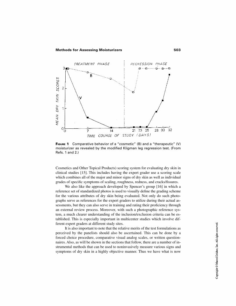
503Methods for Assessing Moisturizers
FIGURE 1 Comparative behavior of a “cosmetic” (B) and a “therapeutic” (V)moisturizer as revealed by the modified Kligman leg regression test. (FromRefs. 1 and 2.)
Cosmetics and Other Topical Products) scoring system for evaluating dry skin inclinical studies [15]. This includes having the expert grader use a scoring scalewhich combines all of the major and minor signs of dry skin as well as individualgrades of specific symptoms of scaling, roughness, redness, and cracks/fissures.
We also like the approach developed by Spencer’s group [16] in which areference set of standardized photos is used to visually define the grading schemefor the various attributes of dry skin being evaluated. Not only do such photo-graphs serve as references for the expert graders to utilize during their actual as-sessments, but they can also serve in training and rating their proficiency throughan external review process. Moreover, with such a photographic reference sys-tem, a much clearer understanding of the inclusion/exclusion criteria can be es-tablished. This is especially important in multicenter studies which involve dif-ferent expert graders at different study sites.
It is also important to note that the relative merits of the test formulations asperceived by the panelists should also be ascertained. This can be done by aforced choice procedure, comparative visual analog scales, or written question-naires. Also, as will be shown in the sections that follow, there are a number of in-strumental methods that can be used to noninvasively measure various signs andsymptoms of dry skin in a highly objective manner. Thus we have what is now
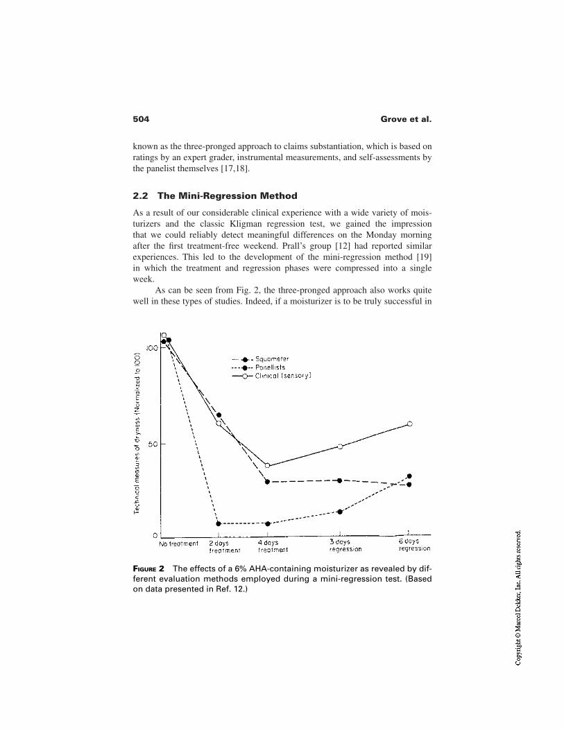
504 Grove et al.
known as the three-pronged approach to claims substantiation, which is based onratings by an expert grader, instrumental measurements, and self-assessments bythe panelist themselves [17,18].
2.2 The Mini-Regression Method
As a result of our considerable clinical experience with a wide variety of mois-turizers and the classic Kligman regression test, we gained the impression that we could reliably detect meaningful differences on the Monday morningafter the first treatment-free weekend. Prall’s group [12] had reported similarexperiences. This led to the development of the mini-regression method [19] in which the treatment and regression phases were compressed into a single week.
As can be seen from Fig. 2, the three-pronged approach also works quitewell in these types of studies. Indeed, if a moisturizer is to be truly successful in
FIGURE 2 The effects of a 6% AHA-containing moisturizer as revealed by dif-ferent evaluation methods employed during a mini-regression test. (Basedon data presented in Ref. 12.)
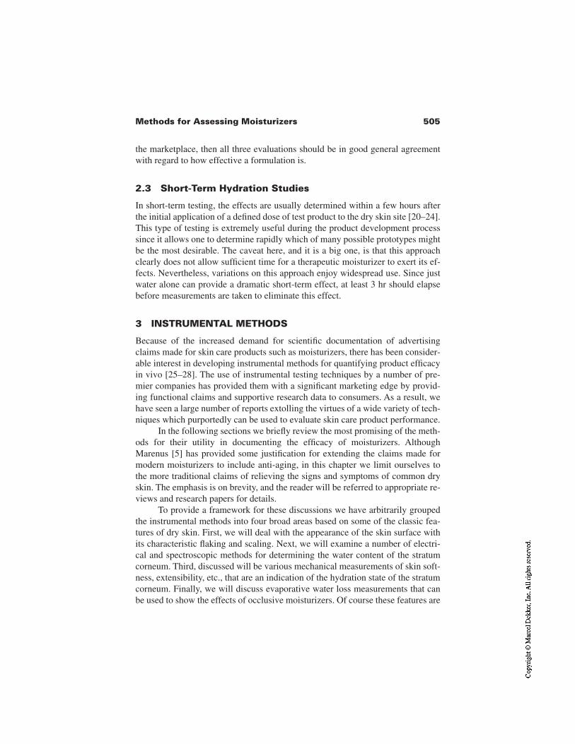
505Methods for Assessing Moisturizers
the marketplace, then all three evaluations should be in good general agreementwith regard to how effective a formulation is.
2.3 Short-Term Hydration Studies
In short-term testing, the effects are usually determined within a few hours afterthe initial application of a defined dose of test product to the dry skin site [20–24].This type of testing is extremely useful during the product development processsince it allows one to determine rapidly which of many possible prototypes mightbe the most desirable. The caveat here, and it is a big one, is that this approachclearly does not allow sufficient time for a therapeutic moisturizer to exert its ef-fects. Nevertheless, variations on this approach enjoy widespread use. Since justwater alone can provide a dramatic short-term effect, at least 3 hr should elapsebefore measurements are taken to eliminate this effect.
3 INSTRUMENTAL METHODS
Because of the increased demand for scientific documentation of advertisingclaims made for skin care products such as moisturizers, there has been consider-able interest in developing instrumental methods for quantifying product efficacyin vivo [25–28]. The use of instrumental testing techniques by a number of pre-mier companies has provided them with a significant marketing edge by provid-ing functional claims and supportive research data to consumers. As a result, wehave seen a large number of reports extolling the virtues of a wide variety of tech-niques which purportedly can be used to evaluate skin care product performance.
In the following sections we briefly review the most promising of the meth-ods for their utility in documenting the efficacy of moisturizers. AlthoughMarenus [5] has provided some justification for extending the claims made formodern moisturizers to include anti-aging, in this chapter we limit ourselves tothe more traditional claims of relieving the signs and symptoms of common dryskin. The emphasis is on brevity, and the reader will be referred to appropriate re-views and research papers for details.
To provide a framework for these discussions we have arbitrarily groupedthe instrumental methods into four broad areas based on some of the classic fea-tures of dry skin. First, we will deal with the appearance of the skin surface withits characteristic flaking and scaling. Next, we will examine a number of electri-cal and spectroscopic methods for determining the water content of the stratumcorneum. Third, discussed will be various mechanical measurements of skin soft-ness, extensibility, etc., that are an indication of the hydration state of the stratumcorneum. Finally, we will discuss evaporative water loss measurements that canbe used to show the effects of occlusive moisturizers. Of course these features are
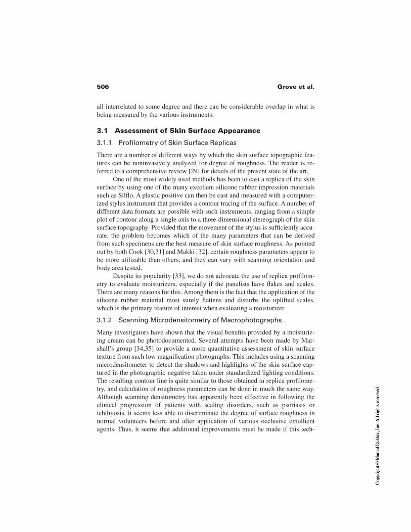
506 Grove et al.
all interrelated to some degree and there can be considerable overlap in what isbeing measured by the various instruments.
3.1 Assessment of Skin Surface Appearance
3.1.1 Profilometry of Skin Surface Replicas
There are a number of different ways by which the skin surface topographic fea-tures can be noninvasively analyzed for degree of roughness. The reader is re-ferred to a comprehensive review [29] for details of the present state of the art.
One of the most widely used methods has been to cast a replica of the skinsurface by using one of the many excellent silicone rubber impression materialssuch as Silflo. A plastic positive can then be cast and measured with a computer-ized stylus instrument that provides a contour tracing of the surface. A number ofdifferent data formats are possible with such instruments, ranging from a simpleplot of contour along a single axis to a three-dimensional stereograph of the skinsurface topography. Provided that the movement of the stylus is sufficiently accu-rate, the problem becomes which of the many parameters that can be derivedfrom such specimens are the best measure of skin surface roughness. As pointedout by both Cook [30,31] and Makki [32], certain roughness parameters appear tobe more utilizable than others, and they can vary with scanning orientation andbody area tested.
Despite its popularity [33], we do not advocate the use of replica profilom-etry to evaluate moisturizers, especially if the panelists have flakes and scales.There are many reasons for this. Among them is the fact that the application of thesilicone rubber material most surely flattens and disturbs the uplifted scales,which is the primary feature of interest when evaluating a moisturizer.
3.1.2 Scanning Microdensitometry of Macrophotographs
Many investigators have shown that the visual benefits provided by a moisturiz-ing cream can be photodocumented. Several attempts have been made by Mar-shall’s group [34,35] to provide a more quantitative assessment of skin surfacetexture from such low magnification photographs. This includes using a scanningmicrodensitometer to detect the shadows and highlights of the skin surface cap-tured in the photographic negative taken under standardized lighting conditions.The resulting contour line is quite similar to those obtained in replica profilome-try, and calculation of roughness parameters can be done in much the same way.Although scanning densitometry has apparently been effective in following theclinical progression of patients with scaling disorders, such as psoriasis orichthyosis, it seems less able to discriminate the degree of surface roughness innormal volunteers before and after application of various occlusive emollientagents. Thus, it seems that additional improvements must be made if this tech-
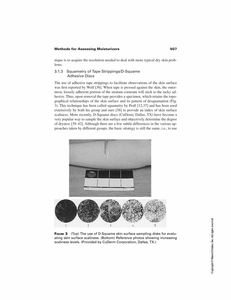
507Methods for Assessing Moisturizers
nique is to acquire the resolution needed to deal with more typical dry skin prob-lems.
3.1.3 Squametry of Tape Strippings/D-Squame Adhesive Discs
The use of adhesive tape strippings to facilitate observations of the skin surfacewas first reported by Wolf [36]. When tape is pressed against the skin, the outer-most, loosely adherent portion of the stratum corneum will stick to the tacky ad-hesive. Thus, upon removal the tape provides a specimen, which retains the topo-graphical relationships of the skin surface and its pattern of desquamation (Fig.3). This technique has been called squametry by Prall [12,37] and has been usedextensively by both his group and ours [38] to provide an index of skin surfacescaliness. More recently, D-Squame discs (CuDerm; Dallas, TX) have become avery popular way to sample the skin surface and objectively determine the degreeof dryness [39–42]. Although there are a few subtle differences in the various ap-proaches taken by different groups, the basic strategy is still the same, i.e., to use
FIGURE 3 (Top) The use of D-Squame skin surface sampling disks for evalu-ating skin surface scaliness. (Bottom) Reference photos showing increasingscaliness levels. (Provided by CuDerm Corporation, Dallas, TX.)
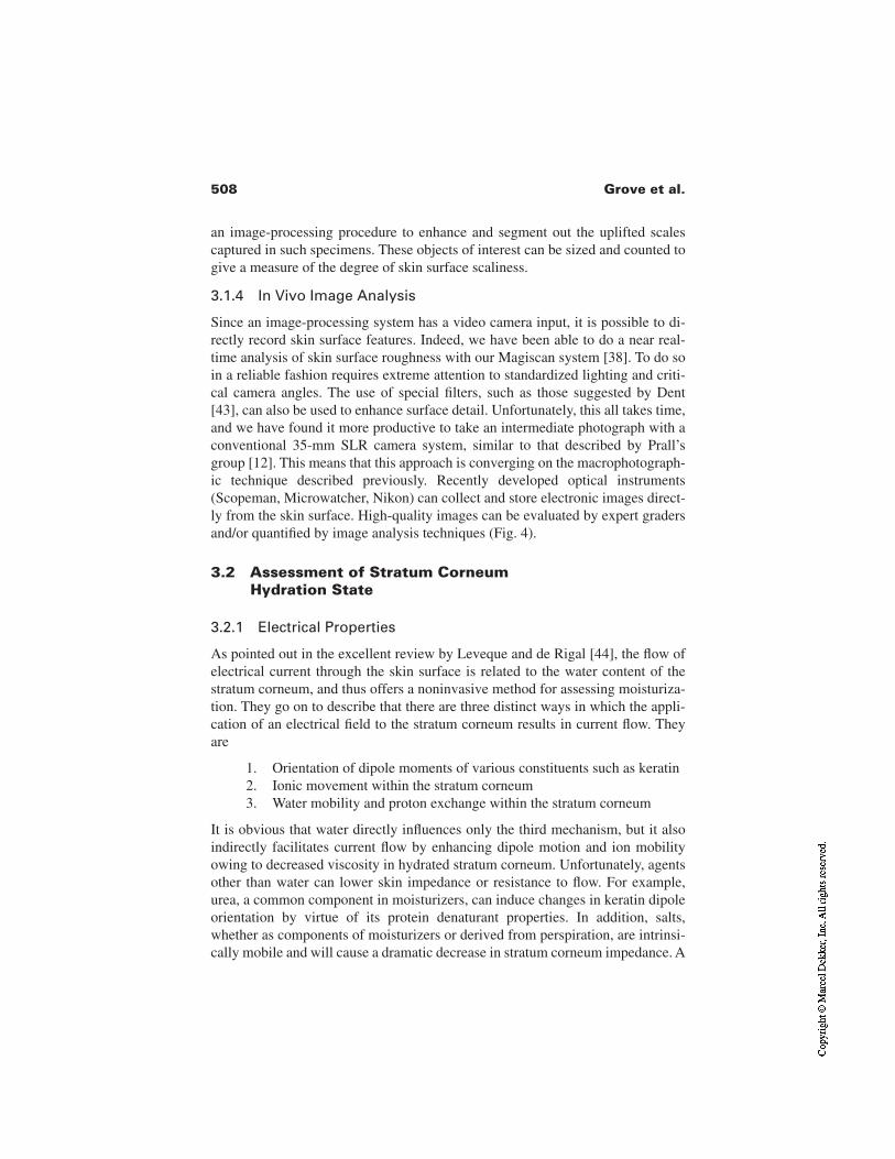
508 Grove et al.
an image-processing procedure to enhance and segment out the uplifted scalescaptured in such specimens. These objects of interest can be sized and counted togive a measure of the degree of skin surface scaliness.
3.1.4 In Vivo Image Analysis
Since an image-processing system has a video camera input, it is possible to di-rectly record skin surface features. Indeed, we have been able to do a near real-time analysis of skin surface roughness with our Magiscan system [38]. To do soin a reliable fashion requires extreme attention to standardized lighting and criti-cal camera angles. The use of special filters, such as those suggested by Dent[43], can also be used to enhance surface detail. Unfortunately, this all takes time,and we have found it more productive to take an intermediate photograph with aconventional 35-mm SLR camera system, similar to that described by Prall’sgroup [12]. This means that this approach is converging on the macrophotograph-ic technique described previously. Recently developed optical instruments(Scopeman, Microwatcher, Nikon) can collect and store electronic images direct-ly from the skin surface. High-quality images can be evaluated by expert gradersand/or quantified by image analysis techniques (Fig. 4).
3.2 Assessment of Stratum Corneum Hydration State
3.2.1 Electrical Properties
As pointed out in the excellent review by Leveque and de Rigal [44], the flow ofelectrical current through the skin surface is related to the water content of thestratum corneum, and thus offers a noninvasive method for assessing moisturiza-tion. They go on to describe that there are three distinct ways in which the appli-cation of an electrical field to the stratum corneum results in current flow. Theyare
1. Orientation of dipole moments of various constituents such as keratin2. Ionic movement within the stratum corneum3. Water mobility and proton exchange within the stratum corneum
It is obvious that water directly influences only the third mechanism, but it alsoindirectly facilitates current flow by enhancing dipole motion and ion mobilityowing to decreased viscosity in hydrated stratum corneum. Unfortunately, agentsother than water can lower skin impedance or resistance to flow. For example,urea, a common component in moisturizers, can induce changes in keratin dipoleorientation by virtue of its protein denaturant properties. In addition, salts,whether as components of moisturizers or derived from perspiration, are intrinsi-cally mobile and will cause a dramatic decrease in stratum corneum impedance. A
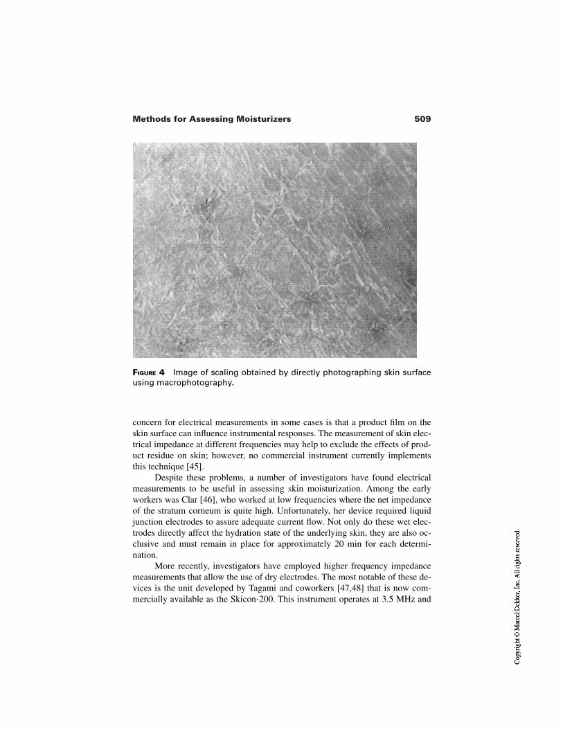
509Methods for Assessing Moisturizers
FIGURE 4 Image of scaling obtained by directly photographing skin surfaceusing macrophotography.
concern for electrical measurements in some cases is that a product film on theskin surface can influence instrumental responses. The measurement of skin elec-trical impedance at different frequencies may help to exclude the effects of prod-uct residue on skin; however, no commercial instrument currently implementsthis technique [45].
Despite these problems, a number of investigators have found electricalmeasurements to be useful in assessing skin moisturization. Among the earlyworkers was Clar [46], who worked at low frequencies where the net impedanceof the stratum corneum is quite high. Unfortunately, her device required liquidjunction electrodes to assure adequate current flow. Not only do these wet elec-trodes directly affect the hydration state of the underlying skin, they are also oc-clusive and must remain in place for approximately 20 min for each determi-nation.
More recently, investigators have employed higher frequency impedancemeasurements that allow the use of dry electrodes. The most notable of these de-vices is the unit developed by Tagami and coworkers [47,48] that is now com-mercially available as the Skicon-200. This instrument operates at 3.5 MHz and
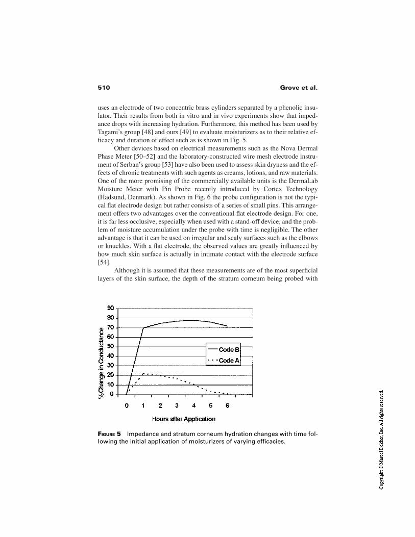
510 Grove et al.
uses an electrode of two concentric brass cylinders separated by a phenolic insu-lator. Their results from both in vitro and in vivo experiments show that imped-ance drops with increasing hydration. Furthermore, this method has been used byTagami’s group [48] and ours [49] to evaluate moisturizers as to their relative ef-ficacy and duration of effect such as is shown in Fig. 5.
Other devices based on electrical measurements such as the Nova DermalPhase Meter [50–52] and the laboratory-constructed wire mesh electrode instru-ment of Serban’s group [53] have also been used to assess skin dryness and the ef-fects of chronic treatments with such agents as creams, lotions, and raw materials.One of the more promising of the commercially available units is the DermaLabMoisture Meter with Pin Probe recently introduced by Cortex Technology(Hadsund, Denmark). As shown in Fig. 6 the probe configuration is not the typi-cal flat electrode design but rather consists of a series of small pins. This arrange-ment offers two advantages over the conventional flat electrode design. For one,it is far less occlusive, especially when used with a stand-off device, and the prob-lem of moisture accumulation under the probe with time is negligible. The otheradvantage is that it can be used on irregular and scaly surfaces such as the elbowsor knuckles. With a flat electrode, the observed values are greatly influenced byhow much skin surface is actually in intimate contact with the electrode surface[54].
Although it is assumed that these measurements are of the most superficiallayers of the skin surface, the depth of the stratum corneum being probed with
FIGURE 5 Impedance and stratum corneum hydration changes with time fol-lowing the initial application of moisturizers of varying efficacies.
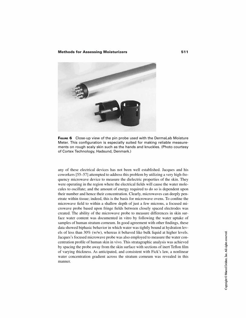
511Methods for Assessing Moisturizers
FIGURE 6 Close-up view of the pin probe used with the DermaLab MoistureMeter. This configuration is especially suited for making reliable measure-ments on rough scaly skin such as the hands and knuckles. (Photo courtesyof Cortex Technology, Hadsund, Denmark.)
any of these electrical devices has not been well established. Jacques and hiscoworkers [55–57] attempted to address this problem by utilizing a very high-fre-quency microwave device to measure the dielectric properties of the skin. Theywere operating in the region where the electrical fields will cause the water mole-cules to oscillate; and the amount of energy required to do so is dependent upontheir number and hence their concentration. Clearly, microwaves can deeply pen-etrate within tissue; indeed, this is the basis for microwave ovens. To confine themicrowave field to within a shallow depth of just a few microns, a focused mi-crowave probe based upon fringe fields between closely spaced electrodes wascreated. The ability of the microwave probe to measure differences in skin sur-face water content was documented in vitro by following the water uptake ofsamples of human stratum corneum. In good agreement with other findings, thesedata showed biphasic behavior in which water was tightly bound at hydration lev-els of less than 30% (w/w), whereas it behaved like bulk liquid at higher levels.Jacques’s focused microwave probe was also employed to measure the water con-centration profile of human skin in vivo. This stratagraphic analysis was achievedby spacing the probe away from the skin surface with sections of inert Teflon filmof varying thickness. As anticipated, and consistent with Fick’s law, a nonlinearwater concentration gradient across the stratum corneum was revealed in thismanner.
512 Grove et al.
3.2.2 Spectroscopic Methods
Photoacoustic Spectroscopy. Another technique that offers the potentialfor quantitatively measuring the water concentration profile is photoacousticspectroscopy (PAS). For a review of the clinical applications of this PAS method,the reader is referred to several excellent reviews [58–61]. Briefly, PAS measuresthe thermal properties of the stratum corneum by modulating optical radiationwith a beam chopper to create periodic heat waves within this compartment.These heat waves propagate through the stratum corneum, giving rise to periodicpressure waves that can be detected at the skin surface by an extremely sensitivemicrophone. The distance a heat wave can travel before being dissipated is relat-ed directly to the thermal diffusivity of the tissue and inversely to the modulationfrequency. This means that by altering the modulation frequency of the optical ra-diation, one can probe the thermal properties of the stratum corneum as a functionof depth. Although such measurements are experimentally difficult and time con-suming, the effects of hydration-induced changes in the human stratum corneumwater gradient have been evaluated both in vitro [62] and in vivo [63,64]. The re-sults revealed that the stratum corneum is nonhomogeneous, and that the outer-most surface layers dehydrate more rapidly than underlying tissue. The biggestproblem with the PAS technique is that the probe is occlusive and water can buildup during the consuming experiments leading to questionable results.
Infrared Spectroscopy. Attenuated total reflection infrared spectroscopyhas been used by a number of investigators [65–68] to noninvasively examine thephysicochemical interactions of skin and topically applied products. The basicpremise of this approach is that the infrared (IR) absorbance spectrum of watercan be, at least in principle, uniquely identified and effectively separated from theIR absorbance spectrum of everything else in the skin plus topical product sys-tem. Once this is achieved, the water content can be quantitatively determinedfrom the intensity of IR absorbance in that particular region.
In early applications of this technique, the O-H stretching band from 3200to 3600 cm was utilized to assess skin hydration [69]. More recent workers[70–72] have used the ratio of the amide I band at 1645 cm to the amide II bandat 1545 cm to evaluate moisturizer efficacy. The idea here is that the amide I bandis due to both protein and water absorbencies, whereas the amide II band is due toprotein alone. Unfortunately, as pointed out by Potts [73], it seems that both theamide I and II bands of keratin, the major protein component of the stratumcorneum (SC), change with hydration, thus removing the possibility of an ab-solute determination. Moreover, many moisturizers contain absorbencies whichoverlap with and therefore influence the intensity of the amide I band. Thus thereare a number of difficulties inherent in quantitative evaluation of stratumcorneum water content from amide I/II intensity ratios. Potts [74] is a strong ad-vocate of the use of spectroscopy and feels that these problems can be overcome
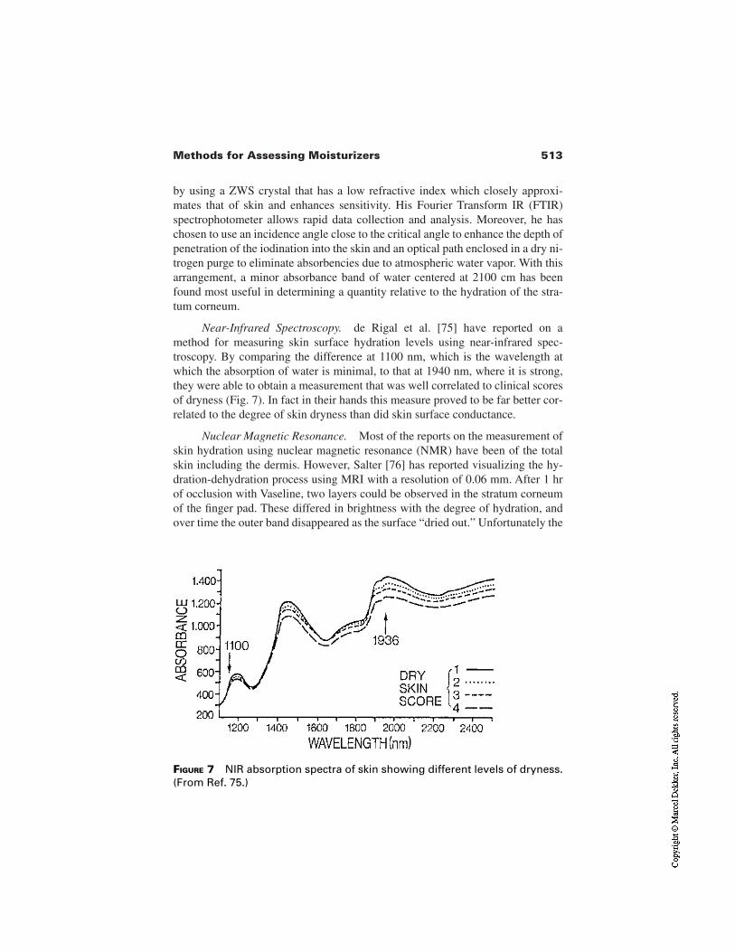
513Methods for Assessing Moisturizers
by using a ZWS crystal that has a low refractive index which closely approxi-mates that of skin and enhances sensitivity. His Fourier Transform IR (FTIR)spectrophotometer allows rapid data collection and analysis. Moreover, he haschosen to use an incidence angle close to the critical angle to enhance the depth ofpenetration of the iodination into the skin and an optical path enclosed in a dry ni-trogen purge to eliminate absorbencies due to atmospheric water vapor. With thisarrangement, a minor absorbance band of water centered at 2100 cm has beenfound most useful in determining a quantity relative to the hydration of the stra-tum corneum.
Near-Infrared Spectroscopy. de Rigal et al. [75] have reported on amethod for measuring skin surface hydration levels using near-infrared spec-troscopy. By comparing the difference at 1100 nm, which is the wavelength atwhich the absorption of water is minimal, to that at 1940 nm, where it is strong,they were able to obtain a measurement that was well correlated to clinical scoresof dryness (Fig. 7). In fact in their hands this measure proved to be far better cor-related to the degree of skin dryness than did skin surface conductance.
Nuclear Magnetic Resonance. Most of the reports on the measurement ofskin hydration using nuclear magnetic resonance (NMR) have been of the totalskin including the dermis. However, Salter [76] has reported visualizing the hy-dration-dehydration process using MRI with a resolution of 0.06 mm. After 1 hrof occlusion with Vaseline, two layers could be observed in the stratum corneumof the finger pad. These differed in brightness with the degree of hydration, andover time the outer band disappeared as the surface “dried out.” Unfortunately the
FIGURE 7 NIR absorption spectra of skin showing different levels of dryness.(From Ref. 75.)
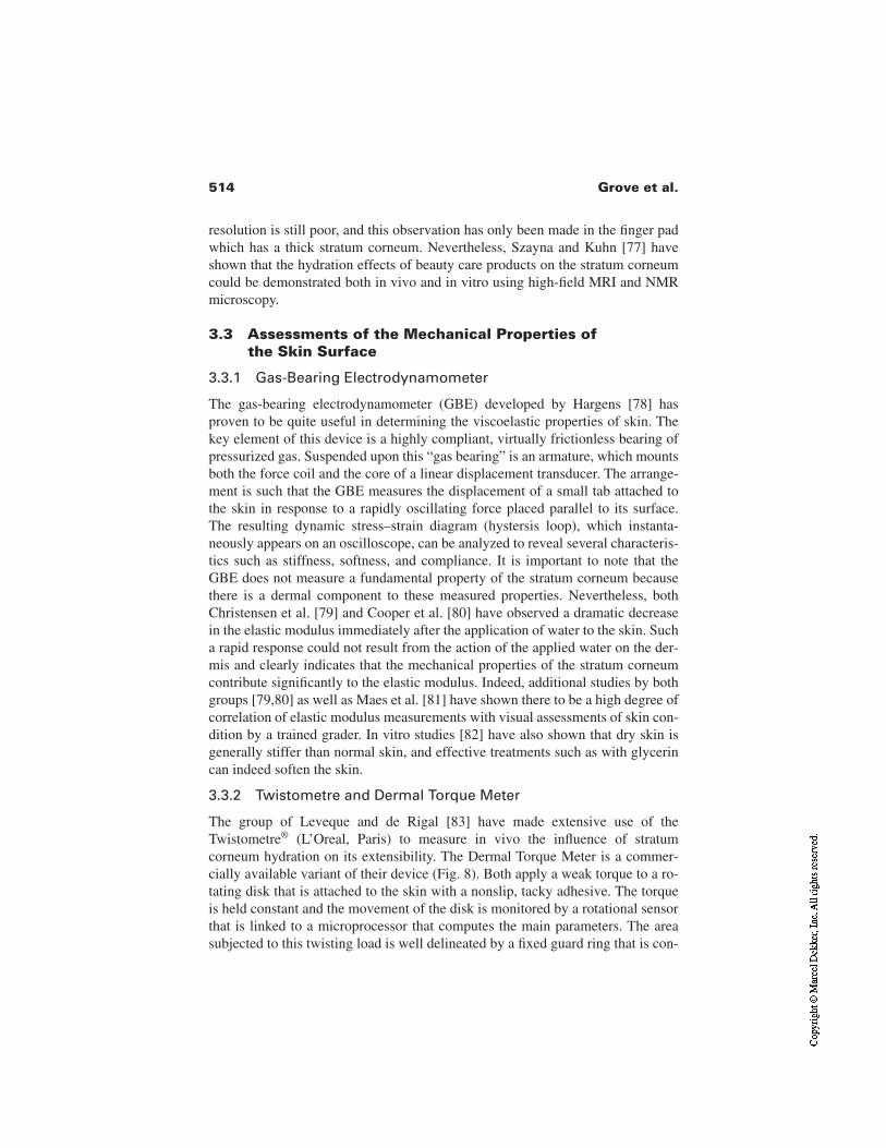
514 Grove et al.
resolution is still poor, and this observation has only been made in the finger padwhich has a thick stratum corneum. Nevertheless, Szayna and Kuhn [77] haveshown that the hydration effects of beauty care products on the stratum corneumcould be demonstrated both in vivo and in vitro using high-field MRI and NMRmicroscopy.
3.3 Assessments of the Mechanical Properties ofthe Skin Surface
3.3.1 Gas-Bearing Electrodynamometer
The gas-bearing electrodynamometer (GBE) developed by Hargens [78] hasproven to be quite useful in determining the viscoelastic properties of skin. Thekey element of this device is a highly compliant, virtually frictionless bearing ofpressurized gas. Suspended upon this “gas bearing” is an armature, which mountsboth the force coil and the core of a linear displacement transducer. The arrange-ment is such that the GBE measures the displacement of a small tab attached tothe skin in response to a rapidly oscillating force placed parallel to its surface.The resulting dynamic stress–strain diagram (hystersis loop), which instanta-neously appears on an oscilloscope, can be analyzed to reveal several characteris-tics such as stiffness, softness, and compliance. It is important to note that theGBE does not measure a fundamental property of the stratum corneum becausethere is a dermal component to these measured properties. Nevertheless, bothChristensen et al. [79] and Cooper et al. [80] have observed a dramatic decreasein the elastic modulus immediately after the application of water to the skin. Sucha rapid response could not result from the action of the applied water on the der-mis and clearly indicates that the mechanical properties of the stratum corneumcontribute significantly to the elastic modulus. Indeed, additional studies by bothgroups [79,80] as well as Maes et al. [81] have shown there to be a high degree ofcorrelation of elastic modulus measurements with visual assessments of skin con-dition by a trained grader. In vitro studies [82] have also shown that dry skin isgenerally stiffer than normal skin, and effective treatments such as with glycerincan indeed soften the skin.
3.3.2 Twistometre and Dermal Torque Meter
The group of Leveque and de Rigal [83] have made extensive use of theTwistometre® (L’Oreal, Paris) to measure in vivo the influence of stratumcorneum hydration on its extensibility. The Dermal Torque Meter is a commer-cially available variant of their device (Fig. 8). Both apply a weak torque to a ro-tating disk that is attached to the skin with a nonslip, tacky adhesive. The torqueis held constant and the movement of the disk is monitored by a rotational sensorthat is linked to a microprocessor that computes the main parameters. The areasubjected to this twisting load is well delineated by a fixed guard ring that is con-
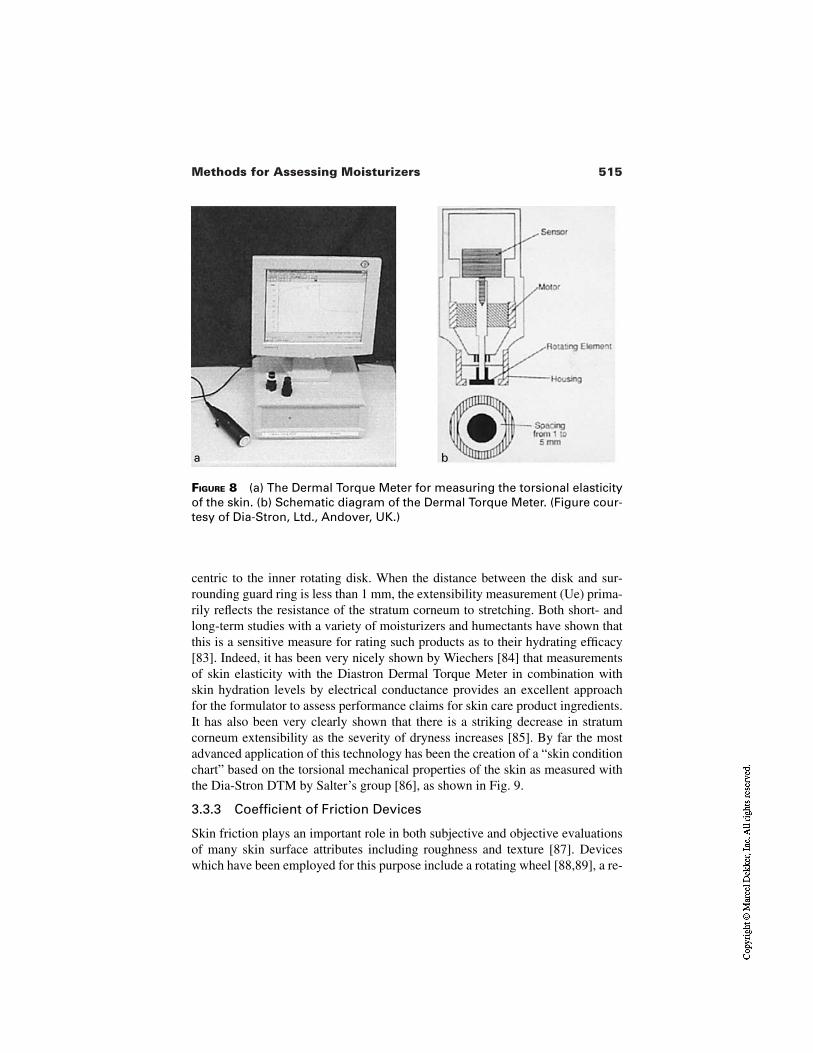
515Methods for Assessing Moisturizers
FIGURE 8 (a) The Dermal Torque Meter for measuring the torsional elasticityof the skin. (b) Schematic diagram of the Dermal Torque Meter. (Figure cour-tesy of Dia-Stron, Ltd., Andover, UK.)
a b
centric to the inner rotating disk. When the distance between the disk and sur-rounding guard ring is less than 1 mm, the extensibility measurement (Ue) prima-rily reflects the resistance of the stratum corneum to stretching. Both short- andlong-term studies with a variety of moisturizers and humectants have shown thatthis is a sensitive measure for rating such products as to their hydrating efficacy[83]. Indeed, it has been very nicely shown by Wiechers [84] that measurementsof skin elasticity with the Diastron Dermal Torque Meter in combination withskin hydration levels by electrical conductance provides an excellent approachfor the formulator to assess performance claims for skin care product ingredients.It has also been very clearly shown that there is a striking decrease in stratumcorneum extensibility as the severity of dryness increases [85]. By far the mostadvanced application of this technology has been the creation of a “skin conditionchart” based on the torsional mechanical properties of the skin as measured withthe Dia-Stron DTM by Salter’s group [86], as shown in Fig. 9.
3.3.3 Coefficient of Friction Devices
Skin friction plays an important role in both subjective and objective evaluationsof many skin surface attributes including roughness and texture [87]. Deviceswhich have been employed for this purpose include a rotating wheel [88,89], a re-
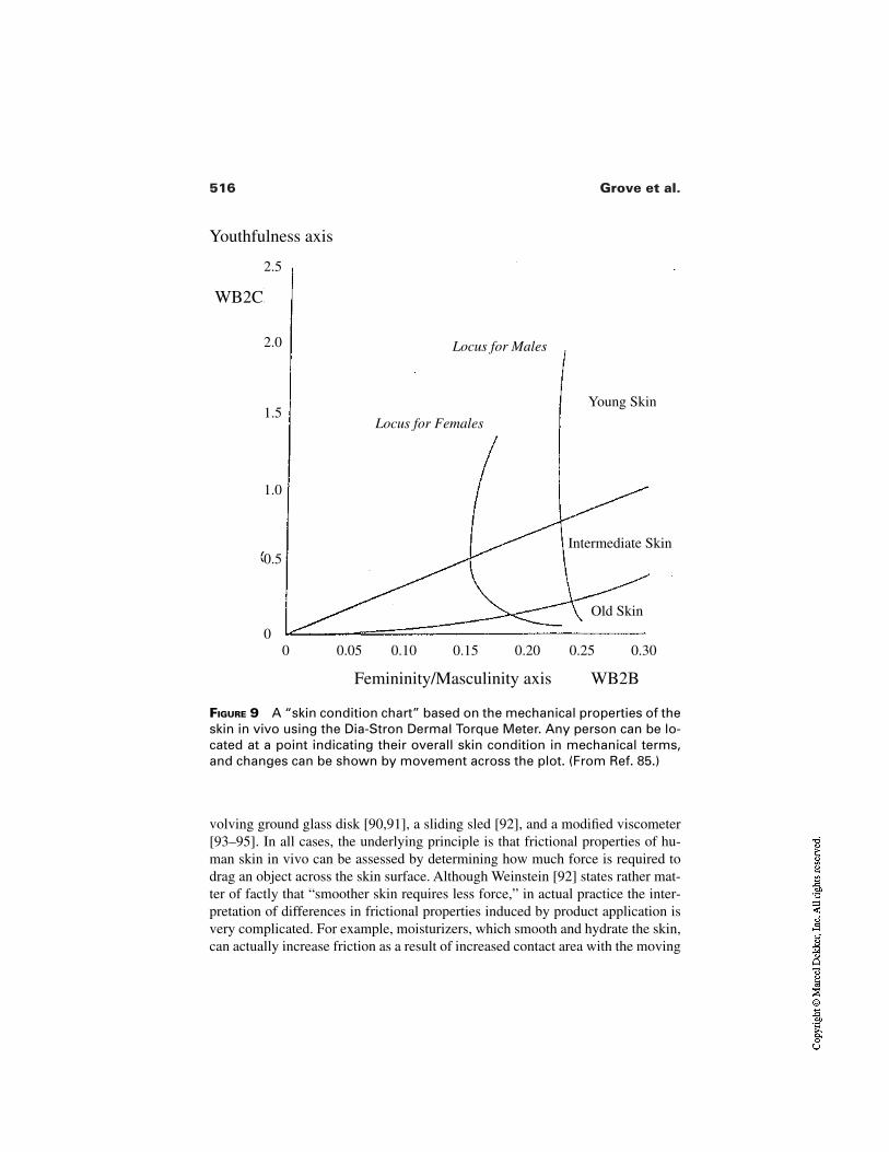
516 Grove et al.
FIGURE 9 A “skin condition chart” based on the mechanical properties of theskin in vivo using the Dia-Stron Dermal Torque Meter. Any person can be lo-cated at a point indicating their overall skin condition in mechanical terms,and changes can be shown by movement across the plot. (From Ref. 85.)
Youthfulness axis
Femininity/Masculinity axis WB2B
0 0.05 0.10 0.15 0.20 0.25 0.30
2.5
2.0
1.5
1.0
0.5
0
WB2C
Locus for Males
Locus for Females
Young Skin
Intermediate Skin
Old Skin
volving ground glass disk [90,91], a sliding sled [92], and a modified viscometer[93–95]. In all cases, the underlying principle is that frictional properties of hu-man skin in vivo can be assessed by determining how much force is required todrag an object across the skin surface. Although Weinstein [92] states rather mat-ter of factly that “smoother skin requires less force,” in actual practice the inter-pretation of differences in frictional properties induced by product application isvery complicated. For example, moisturizers, which smooth and hydrate the skin,can actually increase friction as a result of increased contact area with the moving
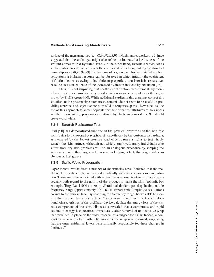
517Methods for Assessing Moisturizers
surface of the measuring device [88,90,92,95,96]. Nacht and coworkers [97] havesuggested that these changes might also reflect an increased adhesiveness of thestratum corneum in a hydrated state. On the other hand, materials which act assurface lubricants do indeed lower the coefficient of friction, making the skin feelmore slippery [88,96,98,99]. In the case of a greasy occlusive material such aspetrolatum, a biphasic response can be observed in which initially the coefficientof friction decreases owing to its lubricant properties, then later it increases overbaseline as a consequence of the increased hydration induced by occlusion [96].
Thus, it is not surprising that coefficient of friction measurements by them-selves sometimes correlate very poorly with sensory scores of smoothness, asshown by Prall’s group [90]. While additional studies in this area may correct thissituation, at the present time such measurements do not seem to be useful in pro-viding a precise and objective measure of skin roughness per se. Nevertheless, theuse of this approach to screen topicals for their after-feel attributes of greasinessand their moisturizing properties as outlined by Nacht and coworkers [97] shouldprove worthwhile.
3.3.4 Scratch Resistance Test
Prall [90] has demonstrated that one of the physical properties of the skin thatcontributes to the overall perception of smoothness by the customer is hardness,as measured by the lowest pressure load which causes a stylus to just visiblyscratch the skin surface. Although not widely employed, many individuals whosuffer from dry skin problems will do an analogous procedure by scraping theskin surface with their fingernail to reveal underlying defects that might not be soobvious at first glance.
3.3.5 Sonic Wave Propagation
Experimental results from a number of laboratories have indicated that the me-chanical properties of the skin vary dramatically with the stratum corneum hydra-tion. These are often associated with subjective assessments of moisturization, es-pecially with regard to the ability of the product to make the skin feel soft. Forexample, Torgalkar [100] utilized a vibrational device operating in the audiblefrequency range (approximately 700 Hz) to impart small amplitude oscillationsnormal to the skin surface. By scanning the frequency range, he was able to mea-sure the resonant frequency of these “ripple waves” and from the known vibra-tional characteristics of the oscillator device calculate the energy loss of the vis-cous component of the skin. His results revealed that a continuous and rapiddecline in energy loss occurred immediately after removal of an occlusive wrapthat remained in place on the volar forearm of a subject for 14 hr. Indeed, a con-stant value was reached within 10 min after the wrap was removed, suggestingthat the outer epidermal layers were primarily responsible for these changes in“softness.’’
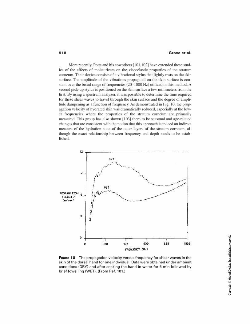
518 Grove et al.
More recently, Potts and his coworkers [101,102] have extended these stud-ies of the effects of moisturizers on the viscoelastic properties of the stratumcorneum. Their device consists of a vibrational stylus that lightly rests on the skinsurface. The amplitude of the vibrations propagated on the skin surface is con-stant over the broad range of frequencies (20–1000 Hz) utilized in this method. Asecond pick-up stylus is positioned on the skin surface a few millimeters from thefirst. By using a spectrum analyzer, it was possible to determine the time requiredfor these shear waves to travel through the skin surface and the degree of ampli-tude dampening as a function of frequency. As demonstrated in Fig. 10, the prop-agation velocity of hydrated skin was dramatically reduced, especially at the low-er frequencies where the properties of the stratum corneum are primarilymeasured. This group has also shown [103] there to be seasonal and age-relatedchanges that are consistent with the notion that this approach is indeed an indirectmeasure of the hydration state of the outer layers of the stratum corneum, al-though the exact relationship between frequency and depth needs to be estab-lished.
FIGURE 10 The propagation velocity versus frequency for shear waves in theskin of the dorsal hand for one individual. Data were obtained under ambientconditions (DRY) and after soaking the hand in water for 5 min followed bybrief towelling (WET). (From Ref. 101.)
519Methods for Assessing Moisturizers
Dahlgren and Elsnau [104] have utilized a similar sonic velocity techniqueto assess the relative efficacy of moisturizers. Their results have shown that topi-cally applied moisturizers do indeed decrease the sonic propagation velocity inskin and that this decrease is highly correlated with subjective assessments ofmoisturization. More importantly, the instrumental changes were noted after onlya few days of use, whereas the subjective assessments took several weeks, indi-cating that sonic velocity can be a predictive measure.
3.4 Measurement of Stratum Corneum Barrier Function
The development of various methods for measuring trans-epidermal water loss(TEWL) have been comprehensively reviewed [105–108]. Leveque et al. [109]have shown that there is at least a trend toward higher than normal water loss val-ues with dry skin. In more severe cases, where the skin is cracked and fissured,water loss rates are clearly elevated. One would expect that the greater the occlu-sivity of a product, the greater the reduction in TEWL upon its application, andthis is certainly the case as shown by several studies [110–114]. With the com-mercially available instruments such as the computerized DermaLab TEWLProbe (Fig. 11) these types of assessments are straightforward and easy to accom-plish [115,116]. Some care should be taken to ensure that the amount of productapplied is relevant to the intended use conditions, as a thick film can give an un-realistically high value [117], and that the ambient temperatures are low so thatsweating is not a factor [118]. In short-term experiments, it is important that suf-
FIGURE 11 (a) The DermaLab TEWL Probe for measuring evaporative waterloss from the skin surface. (b) Schematic diagram of the probe showing thepaired RH and temperature sensors at fixed distance above the skin surfacehoused in an open chamber. (Figure courtesy of cyberDERM, Inc., Media,PA.)
a b
520 Grove et al.
ficient time be given for the volatiles to escape from the newly applied formula-tion as some of these can harm the sensors of the TEWL probe [119].
In dealing with moisturizers that are based on humectants, the situation ismuch more complex. Very early studies by Powers and Fox [120] demonstratedthat trans-epidermal water loss was increased, not decreased, when treated witheffective moisturizers. Rietschel [121,122] has confirmed these findings, espe-cially with regard to the behavior of products containing glycerin. It is known thatthe diffusion coefficient increases with increased hydration of the stratumcorneum [123–125], and thus water vapor will move through more readily. Thismeans that more water is available to the outermost layers of the stratumcorneum, thus leading to relief of the signs and symptoms of dry skin. Unfortu-nately, damaging products such as detergents will disrupt the barrier properties ofthe stratum corneum and also lead to increased flux and elevations in trans-epi-dermal water loss values. Of course, this is an undesirable consequence of prod-uct usage.
This means that there is a complex relationship between water content andflux in the stratum corneum that depends upon skin condition, which has beenbeautifully graphically summarized by Loden and Lindberg [20,21] as shown inFig. 12.
4 SUMMARY
The methods for assessing the moisturizing efficacy of skin care products rangefrom relying on the subjective assessments made by the panelists of their ownskin to highly objective computer-assisted instrumental methods. No matter howaccurate and precise an objective measurement may be, the question of relevancemust always be considered. The measurement must ultimately correlate with theclinical perception of dry skin. At the moment, no one instrumental method canreplace the experienced grader in rating dry skin. Devices invariably measure asingle attribute, whereas the human brain integrates multiple inputs secured fromvision and touch. By employing a battery of instrumental methods we can hope togain a much fuller understanding of what defects are responsible for the dry skinproblem, but this should be done in conjunction with, not instead of, the moreclassic clinical studies. Indeed, we strongly advocate that a three-pronged ap-proach based on panelist self-appraisal, expert grader evaluations, and relevantinstrumental measurements be utilized for adequately substantiating product per-formance.
Each of these methods has its own advantages, disadvantages, and idiosyn-cracies. We recommend the use of multiple instruments in measuring the effectsof moisturizing products on skin to characterize the broad spectrum of their ef-fects. We would also encourage the adoption of standard operating procedures ap-propriate for the study design. Key methodological considerations include sever-
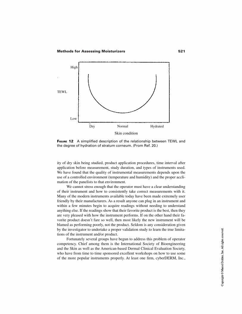
521Methods for Assessing Moisturizers
FIGURE 12 A simplified description of the relationship between TEWL andthe degree of hydration of stratum corneum. (From Ref. 20.)
Dry
Skin condition
Low
HydratedNormal
High
TEWL
ity of dry skin being studied, product application procedures, time interval afterapplication before measurement, study duration, and types of instruments used.We have found that the quality of instrumental measurements depends upon theuse of a controlled environment (temperature and humidity) and the proper accli-mation of the panelists to that environment.
We cannot stress enough that the operator must have a clear understandingof their instrument and how to consistently take correct measurements with it.Many of the modern instruments available today have been made extremely userfriendly by their manufacturers. As a result anyone can plug in an instrument andwithin a few minutes begin to acquire readings without needing to understandanything else. If the readings show that their favorite product is the best, then theyare very pleased with how the instrument performs. If on the other hand their fa-vorite product doesn’t fare so well, then most likely the new instrument will beblamed as performing poorly, not the product. Seldom is any consideration givenby the investigator to undertake a proper validation study to learn the true limita-tions of the instrument and/or product.
Fortunately several groups have begun to address this problem of operatorcompetency. Chief among them is the International Society of Bioengineeringand the Skin as well as the American-based Dermal Clinical Evaluation Society,who have from time to time sponsored excellent workshops on how to use someof the more popular instruments properly. At least one firm, cyberDERM, Inc.,
522 Grove et al.
has established a formal training program including educational course materialsand proficiency exams that lead to certification of the operator according to facto-ry authorized standards.
REFERENCES
1. Kligman AM. Regression method for assessing the efficacy of moisturizers. CosmetToil 1978; 93:27–35.
2. Kligman AM, Lavker RM, Grove GL, Stoudemayer T. Some aspects of dry skinand its treatment. In: Kligman AM, Leyden JJ, eds. Safety and Efficacy of TopicalDrugs and Cosmetics. New York: Grune and Stratton, 1982:221–238.
3. Pierard GE. What does “dry skin” mean? Int J Dermatol 1987; 26:167–168.4. Rieger MM. Skin, water and moisturization. Cosmet Toil 1989; 104:41–50.5. Marenus KD. Skin conditioning benefits of moisturizing products. In: Aust LB, ed.
Cosmetic Claims Substantiation. Melville, NY: Estee Lauder Research Laborato-ries, 1998:97–113.
6. Shapiro IJ, Weiss R, Webster D. X-linked ichthyoses due to sterol sulfatase defi-ciency. Lancet 1970; 1:70–72.
7. Chernosky ME. Clinical aspects of dry skin. J Soc Cosmet Chem 1976;27:365–376.
8. Gaul LE, Underwood GB. Relation of dew point and barometric pressure to chap-ping of skin. J Invest Dermatol 1951; 18:9–12.
9. Middleton JD, Allen BM. The influence of temperature and humidity on stratumcorneum and its relation to skin chapping. J Soc Cosmet Chem 1973; 24:239–243.
10. Blank IH. Factors which influence the water content of the stratum corneum. J In-vest Dermatol 1952; 18:433–440.
11. Blank IH. Further observations on factors which influence the water content of thestratum corneum. J Invest Dermatol 1953; 21:259–271.
12. Prall JK, Theiler RF, Bowser PA, Walsh M. The effectiveness of cosmetic productsin alleviating a range of skin dryness conditions as determined by clinical and in-strumental techniques. Int J Cosmet Sci 1986; 8:159–174.
13. Wehr RF, Krochmal L. Considerations in selecting a moisturizer on the water distri-bution in human stratum corneum. Cutis 1987; 39(VI):512–515.
14. Boisits EK, Nole GE, Cheyney MC. The refined regression method. J Cutan AgingCosmet Dermatol 1989; 1(III):155–163.
15. Serup J. EEMCO guidance for the assessment of dry skin (xerosis) and ichthyosis:clinical scoring systems. Skin Res Technol 1995; 1:109–114.
16. Seitz J, Rizer R, Spencer T. Photographic standardization of dry skin. J Soc CosmetChem 1984; 35:423.
17. Grove GL. Techniques for substantiating skin care product claims. In: KligmanAM, Leyden JJ, eds. Safety and Efficacy of Topical Drugs and Cosmetics. NewYork: Grune and Stratton, 1982:157–176.
18. Grove GL. Design of studies to measure skin care product performance. BioengSkin 1987; 3:359–373.
19. Grove GL. Skin surface hydration changes during a mini leg regression test as mea-sured in vivo by electrical conductivity. Curr Ther Res 1992; 52 40:1–6.
523Methods for Assessing Moisturizers
20. Loden M, Lindberg M. Product testing—testing of moisturizers. In: Elsner P, Wil-helm K-P, Maibach HI, eds. Bioengineering of the Skin: Water and the StratumCorneum. Boca Raton: CRC Press, 1994:275–289.
21. Loden M, Lindberg M. The influence of a single application of different moisturiz-ers on the skin capacitance. Acta Derm Venerol (Stockholm) 1991; 71:79.
22. Serup J. A three-hour test for rapid comparison of the effects of moisturizers and ac-tive constituents (urea). Measurements of hydration, scaling and skin surface li-pidization by non-invasive techniques. Acta Derm Venereol (Stockholm) 1992;(Suppl 177):29.
23. Blichmann CW, Serup J, Winther A. Effects of a single application of a moisturizer:evaporation of emulsion water, skin surface temperature, electrical conductance,electrical capacitance and skin surface (emulsion) lipids. Acta Derm Venerol(Stockholm) 1989; 70:400.
24. Grove GL. The effects of moisturizers on skin surface hydration as measured invivo by electrical conductivity. Curr Ther Res 1991; 50 5:712–719.
25. Leveque JL. Physical methods for skin investigation. Int J Dermatol 1983;22:368–375.
26. Kajs TM, Gartstein V. Review of the instrumental assessment of skin: effects ofcleansing products. J Soc Cosmet Chem 1991; 42:249–271.
27. Takahaski M. Recent progress in skin bioengineering and its application to evalua-tion of cosmetics. SÖFW J 2000; 126:6–18.
28. Grove GL. Non-invasive methods for assessing moisturizers. In: Waggoner WC,ed. Clinical Safety and Efficacy Testing of Cosmetics. New York: Marcel Dekker,1990:121–148.
29. Grove GL, Grove MJ. Objective methods for assessing skin surface topographynoninvasively. In: Leveque JL, ed. Cutaneous Investigation in Health and Disease.New York: Marcel Dekker, 1988:1–32.
30. Cook TH. Profilometry of skin: a useful tool for the substantiation of cosmetic effi-cacy. J Soc Cosmet Chem 1980; 31:339–359.
31. Cook TH, Craft TJ, Brunelle RL, Norris F, Griffin WA. Quantification of the skin’stopography by skin profilometry. Int J Cosmet Sci 1982; 4:195–205.
32. Makki S, Agache P, Mignot J, Zahouani H. Statistical analysis and three dimension-al representation of the human skin surface. J Soc Cosmet Chem 1984; 35:311–325.
33. Dorogi PL, Zielinski M. Assessment of skin conditions using profilometry. CosmetToil 1989; 104(3):39–44.
34. Marshall R, Marks R. Assessment of skin surface by scanning densitometry ofmacrophotography. Clin Exp Dermatol 1983; 8:121–127.
35. Marshall R, Marks R. Quantification of skin surface texture of macrophotographyand computer aided scanning densitometry. J Audiov Media Med 1983; 6:98–103.
36. Wolf J. Das oberflachen relief der menschlichen Haut. Z Mikrosk Anat Forsch1940; 47:351.
37. Prall JK. Scaliness of human skin. Arch Biochem Cosmetol 1966; 9:27–43.38. Grove GL. Dermatological applications of the Magiscan image analyzing comput-
er. In: Marks R, Payne PA, eds. Bioengineering and the Skin. Lancaster, England:MTP Press, 1981:173–182.
39. Pierard GE, Pierard-Franchimont C, Saint Leger D, Kligman AM. Squamometry:
524 Grove et al.
the assessment of xerosis by cyanoacrylate surface biopsies and colorimetry of D-Squame adhesive disks. J Soc Cosmet Chem 1992; 43:292–305.
40. Miller DL. D-Squame adhesive disks. In: Wilhelm K-P, Elsner P, Berardesca E,Maibach HI, eds. Bioengineering of the skin: skin surface imaging and analysis.Boca Raton: CRC Press, 1997:39–46.
41. Schatz H, Altmeyer PJ, Kligman AM. Dry skin and scaling evaluated by D-squames and image analyses. In: Serup J, Jemec GBE, eds. Handbook of non-in-vasive methods and the skin. Boca Raton: CRC Press, 1995:153–157.
42. Serup J, Winther A, Blichmann C. A simple method for the study of scale patternand effect of a moisturizer—qualitative and quantitative evaluation by D-Squametape in comparison with parameters of epidermal hydration. Clin Exp Dermatol1989; 14:227–282.
43. Dent RV. The photographic aspects. J Lab Clin Med 1983; 26:1852–1862.44. Leveque JL, de Rigal J. Impedance methods for studying skin moisturization. J Soc
Cosmet Chem 1983; 34:419–428.45. Salter DC. Examination of stratum corneum hydration state by electrical methods.
Curr Probl Dermatol 1998; 26:38–47.46. Clar EJ, Her CP, Sturelle CG. Skin impedance and moisturization. J Soc Cosmet
Chem 1975; 26:337–353.47. Tagami H, Ohi M, Iwatsuki K, Kanamaru Y, Ichijo B. Evaluation of the skin surface
hydration in vivo by electrical measurement. J Invest Dermatol 1980; 75:500–507.48. Tagami H. Impedance measurement for evaluation of the hydration state of the skin
surface. In: Leveque JL, ed. Cutaneous Investigation in Health and Disease: Nonin-vasive Methods and Instrumentation. New York: Marcel Dekker, 1989:79–111.
49. Wortzman M, Grove GL. Assessment of long-lasting moisturizers by skin surfaceelectrical hydrometry. Proceedings of 7th International Symposium of Bioengineer-ing and the Skin, Milwaukee, WI, June 16–18, 1988.
50. Kohli R. Impedance measurements for the noninvasive monitoring of skin hydra-tion-A reassessment. Int J Pharm 1985; 26:275–287.
51. Spencer TS, Anderson PJ, Seitz JC. Use of a phase angle meter to measure producteffects on the skin surface. Bioeng Skin 1986; 2:153.
52. Gabard B, Treffel P. Hardware and measuring principle: the NOVA DPM 9003. In:Elsner P, Berardesca E, Maibach HI, eds. Bioengineering of the Skin: Water and theStratum Corneum. Boca Raton: CRC Press, 1994:177–195.
53. Serban GP, Henry SM, Cotty VF, Cohen GL, Riveley JA. Electronic technique forthe in vivo assessment of skin dryness and the effect of chronic treatment with a lo-tion on the water barrier function of dry skin. J Soc Cosmet Chem 1983;34:383–389.
54. Berardesca E. EEMCO guidance for the assessment of stratum corneum hydration:electrical methods. Skin Res Technol 1997; 3:126–132.
55. Jacques SL. A linear measurement of the water content of the stratum corneum ofhuman skin using a microwave probe. IEEE Eng Med Biol Soc Conf 1979;180–182.
56. Jacques SL, Maibach HI, Susskind C. Water content in stratum corneum measuredby focused microwave probe: normal and psoriatic. Bioeng Skin Newsletter 1981;3:118.
525Methods for Assessing Moisturizers
57. Murahara RI, Hing SAO, Maibach HI, Roheim JR. The use of a microwave probeto evaluate the hydration of human stratum corneum in vivo. Bioeng Skin 1986;2:235–247.
58. Rosencwaig A. Photoacoustic spectroscopy of biological materials. Science 1973;181:657–658.
59. Rosencwaig A. Potential clinical applications of photoacoustics. Clin Chem 1982;28:1878–1881.
60. Cahen D, Bults G, Garty H, Malkin S. Photoacoustics in life sciences. J BiochemBiophys Methods 1980; 3:293–310.
61. Campbell SD, Yee SS, Afromwitz MA. Applications of photoacoustic spectroscopyto problems in dermatological research. IEEE Transactions in Biomedical Engi-neering 1979; BME-28 26:220–227.
62. Pines E, Cunningham T. Dermatological photoacoustic spectroscopy. In: Marks R,Payne PA, eds. Bioengineering and the Skin. Lancaster, England: MTP Press,1981:283–290.
63. Simon I, Emslie AG, Apt CM, Blank IH, Anderson RR. Determination of in vivowater concentration profile in human stratum corneum by a photoacoustic method.In: Marks R, Payne PA, eds. Bioengineering and the Skin. Lancaster, England:MTP Press, 1981:187–195.
64. Rosencwaig A, Pines E. A photoacoustic study of newborn rat stratum corneum.Biochim Biophys Acta 1977; 493:10–23.
65. Scheuplein RJ. A survey of some fundamental aspects of the absorption and reflec-tion of light by tissue. J Soc Cosmet Chem 1964; 11:111–112.
66. Puttnam NA, Baxter BH. Spectroscopic studies of skin in situ by attenuated total re-flectance. J Soc Cosmet Chem 1967; 18:469–472.
67. Puttnam NA. Attenuated total reflectance studies of the skin. J Soc Cosmet Chem1978; 23:209–226.
68. Comaish S. Infrared studies of human skin in vivo by multiple internal reflection.Br J Dermatol 1968; 80:552–528.
69. Baier RE. Noninvasive, rapid characterization of human skin chemistry in situ. JSoc Cosmet Chem 1978; 29:283–306.
70. Osberghaus R, Gloxhuber H, Braig S. Hydagen-F, ein neuer Hautfeuchitigkeitsre-gulator Methoden und Ergebnisse des Wirkungsnachweises. J Soc Cosmet Chem1979; 29:133–146.
71. Triebskorn A, Gloor M, Greiner F. Comparative investigation of the water contentof the stratum corneum, using different methods of measurement. Dermatologica1983; 167:64–69.
72. Stanfield JW, Kyriakopolour A. Substantivity of alpha keri bath oil® (WestwoodPharmaceuticals Inc., Buffalo, NY). Proceedings of 4th International Symposium inBioengineering and the Skin, Bescanson, France, 1983.
73. Potts RO, Guzek DB, Harris RH, McKie JE. A noninvasive, in vivo technique toquantitatively measure the water concentration of the stratum corneum using atten-uated total reflection infrared spectroscopy. Arch Dermatol Res 1985;277:489–495.
74. Potts RO. Stratum corneum hydration: experimental techniques and interpretationsof results. J Soc Cosmet Chem 1986; 37:9–33.
526 Grove et al.
75. de Rigal J, Losch MT, Bazin R, Camus C, Sturelle C, Descamps V. Near infraredspectroscopy: a new approach to the characterization of dry skin. Proceedings ofIFSCC International Congress, Yokohama, Japan 1992, pp. 1131–1146.
76. Ablett S, Burdett NG, Carpenter TA, Hall LD, Salter DC. Short echo time MRI en-ables visualization of the natural state of human stratum corneum water in vivo.Magn Reson Imaging 1996; 14(4):357–360.
77. Szayna M, Kuhn W. In vivo and in vitro investigations of hydration effects of beau-ty care products by high-filed MRI and NMR microscopy. J Eur Acad DermatolVenereol 1998; 11(2):112–128.
78. Hargens CW. The gas-bearing electrodynamometer (GBE) applied to measuringmechanical changes in skin and other tissues. In: Marks R, Payne P, eds. Bioengi-neering and the Skin. Lancaster, England: MTP Press, 1981:113–122.
79. Christensen MS, Hargens CW, Nacht S, Gans EH. Viscoelastic properties of intacthuman skin: instrumentation, hydration effects, and the contribution of the stratumcorneum. J Invest Dermatol 1977; 69:282–286.
80. Cooper ER, Missel PJ, Hannon DP, Albright GB. Mechanical properties of dry, nor-mal, and glycerol-treated skin as measured by the gas-bearing electrodynamometer.J Soc Cosmet Chem 1985; 36:335–348.
81. Maes D, Short J, Turek BA, Reinstein JA. In vivo measuring of skin softness usingthe gas bearing electrodynamometer. Int J Cosmet Sci 1983; 5:189–200.
82. Missel PJ, Bowman WD, Benzinger MJ, Albright GB. An in vitro method for skinpreservation to study the influences of relative humidity and treatment on stratumcorneum elasticity. Bioeng Skin 1986; 2:203–214.
83. Leveque JL, de Rigal J. In vivo measurements of the stratum corneum elasticity.Bioeng Skin 1985; 1:13–23.
84. Wiechers JW. A supplier’s contribution to performance testing of personal care in-gredients. Proceedings of In Cosmetics, Kongresszentrum Süd, Düsseldorf, 1997,pp. 206–228.
85. Leveque JL, Grove G, de Rigal J, Corcuff P, Kligman AM, Saint Leger D. Bio-physical characterization of dry facial skin. J Soc Cosmet Chem 1987; 82:171–177.
86. Salter DC, McArthur HC, Crosse JE, Dickens AD. Skin mechanics measured invivo using torsion: a new and accurate model more sensitive to age, sex, and mois-turizing treatment. Int J Cosmet Sci 1993; 15:200–218.
87. Wolfram LJ. Friction of skin. J Soc Cosmet Chem 1983; 34:465–476.88. Comaish S, Bottoms E. The skin and friction: deviation from Amonton’s laws, and
the effects of hydration and lubrication. Br J Dermatol 1971; 84:37.89. Comaish S, Harborow PRH, Hofman DA. A hand-held friction meter. Br J Derma-
tol 1973; 89:33.90. Prall JK. Instrumental evaluation of the effects of cosmetic products on skin sur-
faces with particular reference to smoothness. J Soc Cosmet Chem 1973; 24:693–707.
91. Highley DR, Coomey M, DenBeste M, Wolfram LJ. Frictional properties of skin. JInvest Dermatol 1977; 69:303.
92. Weinstein S. New methods for the in-vivo assessment of skin smoothness and skinsoftness. J Soc Cosmet Chem 1978; 29:99–115.
527Methods for Assessing Moisturizers
93. Gerrad ED, Stimpson IM. A versatile friction meter based on a viscometer. LabPract 1984; 33:82–83.
94. Gerrard ED, Stimpson IM. The effect of treatment on skin friction coefficient invivo. Bioeng Skin 1985; 1:229.
95. Naylor PFD. The skin surface and friction. Br J Dermatol 1955; 67:239.96. El-Shimi AF. In vivo skin friction measurements. J Soc Cosmet Chem 1977; 28:37.97. Nacht S, Close J, Yeung D, Gans EH. Skin friction coefficient: changes induced by
skin hydration and emollient application and correlation with perceived skin feel. JSoc Cosmet Chem 1981; 32:55.
98. Cussler EL, Zlotnick SJ, Shaw MC. Texture perceived with fingers. Percept Psy-chophys 1977; 21:504.
99. Appeldoorn JK, Barnett G. Frictional aspects of emollience. Proc Sci Sect ToilGoods Assoc 1963; 40:28.
100. Torgalkar AM. A resonance frequency technique to determine the energy absorbedin stratum corneum, in vivo. In: Marks R, Payne PA, eds. Bioengineering and theSkin. Lancaster, England: MTP Press, 1981:55–65.
101. Potts RO. In vivo measurement of water content of the stratum corneum using in-frared spectroscopy: a review. Cosmet Toil 1985; 100(10):27–31.
102. Potts RO. Stratum corneum hydration: experimental techniques and interpretationsof results. J Soc Cosmet Chem 1988; 37:9–33.
103. Potts RO, Buras EM, Chrisman DA. Changes with age in the moisture content. J In-vest Dermatol 1984; 82:97–100.
104. Dahlgren RM, Elsnau WH. Measurement of skin condition by sonic velocity. J SocCosmet Chem 1984; 35:1–20.
105. Idson B. In vivo measurement of transdermal water loss. J Soc Cosmet Chem 1976;29:573–580.
106. Nilsson GE, Oberg PA. Measurement of evaporative water loss: methods and clini-cal applications. In: Rolfe P, ed. Non-Invasive Physiological Measurements. NewYork: Academic Press, 1979:279–311.
107. Grice KA. Transepidermal water loss and transepidermal water loss in pathologicalskin. In: Jarrett A, ed. The Physiology and Pathophysiology of the Skin. London:Academic Press, 1980:2115–2146, 2147–2155.
108. Miller DL, Brown AM, Artz EJ. Indirect measures of epidermal water loss. In:Marks R, Payne PA, eds. Bioengineering and the Skin. Lancaster, England: MTPPress, 1980:161–171.
109. Leveque JL, Garson JC, de Rigal J. Transepidermal water loss from dry and normalskin. J Soc Cosmet Chem 1979; 30:333–343.
110. Weldon AE, Monteith JL. Performance of a skin Evaporimeter. Med Biol Eng Com-put 1980; 18:201.
111. Spruit D. Interference of some substances with water vapor loss from human skin.Am Perfumer Cosmet 1971; 86:27–32.
112. Baker H. Experimental studies on the influence of vehicles on percutaneous absorp-tion. J Soc Cosmet Chem 1969; 20:239–252.
113. Weil I, Princen HM. Diffusion therapy analysis of transepidermal water lossthrough occlusive films. J Soc Cosmet Chem 1977; 28:481–484.
114. Seitz JC, Spencer TS. The use of capacitative evaporimetry to measure the effects
528 Grove et al.
of topical ingredients on transepidermal water loss (TEWL). J Invest Dermatol1982; 78:351.
115. Grove GL, Grove MJ, Zerweck C, Pierce E. Computerized evaporimetry using theDermaLab TEWL probe. Skin Res Technol 1999; 5:9–13.
116. Grove GL, Grove MJ, Zerweck C, Pierce E. Comparative metrology of the evapor-imeter and the DermaLab TEWL probe. Skin Res Technol 1999; 5:1–8.
117. Berube GR, Berick M. Transepidermal moisture loss. II. The significance of the usethickness of topical substances. J Soc Cosmet Chem 1974; 25:397–406.
118. Thiele FAJ, Hemels HGWM, Malten KE. Skin temperature and water loss by skin.Trans St John’s Hosp Dermatol Soc 1972; 58:218–223.
119. Morrison BM. ServoMed Evaporimeter: precautions when evaluating the effect ofskin care products on barrier function. J Soc Cosmet Chem 1986; 37:351.
120. Powers DH, Fox CA. A study of the effect of cosmetic ingredients, creams and lo-tions on the rate of moisture loss from the skin. Proc Sci Sect Toil Goods Assoc1957; 28:21–26.
121. Rietschel RL. A method to evaluate skin moisturizers in vivo. J Invest Dermatol1978; 70:152–155.
122. Rietschel RL. A skin moisturization essay. J Soc Cosmet Chem 1979; 30:360–373.123. Buettner KJ. The moisture of human skin as affected by water transfer. J Soc Cos-
met Chem 1965; 16:133–143.124. Blank IH, Moloney J, Emslie AG, Simon I, Apt C. The diffusion of water across the
stratum corneum as a function of its water content. J Invest Dermatol 1984;82:188–194.
125. Wu M, Yee DJ, Sullivan ME. Effect of a skin moisturizer on the water distributionon human stratum corneum. J Invest Dermatol 1983; 81:446–448.
24Laboratory-Based Ex Vivo Assessment ofStratum Corneum Function
Claudine Piérard-Franchimont,Veroniqué Goffin, and Gérald E. PiérardUniversity Medical Center Sart Tilman, Liège, Belgium
Marc PayeColgate-Palmolive, Milmort, Belgium
1 INTRODUCTION
The normal stratum corneum (SC) is composed of orderly interdigitating stacksof corneocytes coated by layers of intercellular lipids. The protein-enriched cor-neocytes are filled with a dense array of disulfide cross-linked keratin filamentsbound to filaggrin. Permeating through this matrix are low molecular weight wa-ter-soluble molecules forming the natural moisturizing factor (NMF), whichlargely derives from the enzymatic degradation of filaggrin. The NMF avidly andeffectively binds water. The resulting osmotic pressure inside the cells does notlead to their disintegration because corneocytes are made of so strong a cross-linked protein matrix surrounded by a thick cornified envelope, formed itselffrom highly cross-linked isopeptide bonded proteins. The lipid-enriched intercel-lular matrix provides a rate-limiting barrier to water evaporation from the skinsurface and to the transport of other chemicals across the skin. With failure of thebarrier, xerosis develops and may evolve to flaky and scaly presentations (Fig. 1).This represents an abnormal process of desquamation. In fact, the SC is not sim-
529
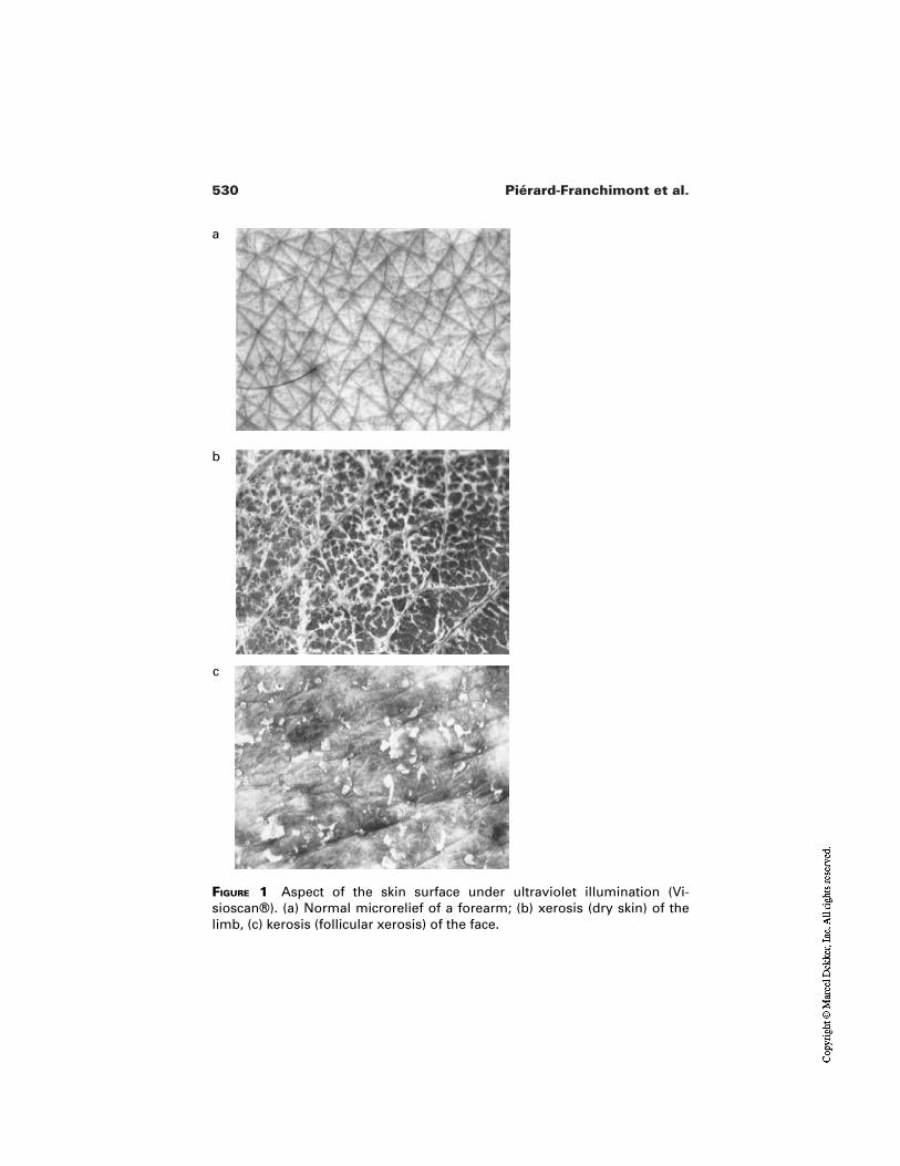
530 Piérard-Franchimont et al.
FIGURE 1 Aspect of the skin surface under ultraviolet illumination (Vi-sioscan®). (a) Normal microrelief of a forearm; (b) xerosis (dry skin) of thelimb, (c) kerosis (follicular xerosis) of the face.
a
b
c
531Ex Vivo Assessment of Stratum Corneum
ply a collection of dead cells, but rather represents a dynamic and metabolicallyactive tissue interacting with subjacent cell layers that reacts to various environ-mental threats [1,2].
The integrity of the SC depends upon the intercellular cohesion provided bythe corneodesmosomes, also called corneosomes [3]. There is ample evidencethat regional differences are present on the body with respect of the structure, co-hesion, thickness, and functions of the SC. Overall a critical level of hydration isrequired to allow the SC to maintain its flexibility and orderly desquamation [3].Enzymatic degradation of corneodesmosomes by proteases along with glycosi-dases is inhibited at low environmental humidities [4–6]. Filaggrin hydrolysisgiving rise to the intracellular NMF pool is similarly critically influenced by wa-ter activity in the SC. However, water is not the single modulator of SC func-tions. The nature and amount in intercellular lipids are also important. The re-newal rate and thickness of the SC and the presence of parakeratotic cells are alsokey features. Laboratory-based in vitro methods may help in studying these pa-rameters.
2 METHODS FOR SC HARVESTING
Half a century ago, the ex vivo usage of SC was pioneered to investigate its func-tions outside of the body influences. Stratum corneum was removed from footcallus and further dehydrated to demonstrate the effect of water upon flexibilityof the samples [7]. This model proved to be useful and innovative. However, cal-lus is not representative of SC from other body sites in terms of structure, thick-ness, and functions.
Powdered human SC is also a product made from foot callus which is cutinto small pieces and ground with dry ice to form a powder. Sieving brings uni-formity in particle size. The material retains some physical and chemical charac-teristics of human stratum corneum [8].
Other techniques of SC collection have been described from other bodysites. They are described hereafter.
2.1 Separation of SC from Excised Skin Samples
Skin samples can be collected from cadaver, on surgery and from slaughterhouseanimals. Different methods have been used to isolate the SC from the underlyingtissues. The mechanical method involved repetitive stretching of the skin [9].This procedure has only a historical value because it is nowadays rarely applieddue to significant alteration of the SC.
Heat treatment of the skin samples is another approach [10]. Placing thesample for 1 min in a water bath at 60°C allows easy subsequent removal of theSC with a grip. Minor variants in temperature and incubation time have been de-
532 Piérard-Franchimont et al.
scribed [11]. This treatment is likely to alter the SC structure and functions. How-ever, permeability properties of the membrane appear unaffected by the proce-dure [11]. Thickness was found to range between 10–15 µm. Due to the absenceof chemical treatment, this method is still used by some research groups.
Exposure of skin for 1 to 3 hr to ammonia vapor in a dessicator jar followedby water immersion allows the epidermis to be separated from the dermis [12].Epidermal cells are then gently scraped off to leave the SC with a thicknessaround 25 µm.
Enzymatic treatment using trypsin digestion of the living tissues is still an-other approach [13]. The skin sample is incubated several hours at 37°C orovernight at 4°C in a buffered 0.5 or 1% trypsin solution, after which the SC canbe easily peeled off. Trypsin is then inhibited using soybean trypsin inhibitor, andthe SC is abundantly rinsed in water. Other enzymes like dispase can be used sim-ilarly. However, an enzymatic technique should be reserved for some specificaims when it has been demonstrated that the treatment is unlikely to interfere withthe SC function under investigation.
2.2 SC Harvesting from Human Volunteers
An SC sheet can be collected from the forearm using a surgical knife and a tweez-er. This method preserves the SC from any chemical or mechanical alteration andis often preferred to trypsin treatment for hydration studies [14]. Suction blistersunder negative pressure (2 atmospheres) is applied on a small area of the volarforearm of the volunteers using a vacuum pump for about 1 to 2 hr [15]. When theblister is formed, its roof is cut off and the living epidermis rubbed away with acotton swab moistened in saline solution. The SC may be full thickness, com-posed of 14 to 17 layers.
Cyanoacrylate skin surface stripping is an ancillary method used to collecta few layers of the outer portion of the SC [16–19]. Corneocytes are tightly boundto the plastic or glass support used for the harvesting process. Horny casts withinthe upper part of the hair follicles are also ripped off [20,21].
The stripping of corneocytes from the skin surface using casual adhesivetapes [22] is nowadays often replaced by calibrated strippings with adhesive-coated discs (SACD). This method removes fragments of the upmost outer layerof corneocytes with better reproducibility. Cells collected represent those whosebond with the underlying SC layers is weaker than with the bond of the adhesivepresent on the collecting disc [18,19,23–25]. The pressure applied to the discshould be carefully controlled [18,19,25]. The contact time between the disc andthe skin affects the data [19,25,26]. There may be some seasonal influence on theamount of SC collected [27].
The detergent scrub method removes individual corneocytes from the stra-tum disjunctum [28]. A buffered solution of 0.05% Triton X-100 is placed in a
533Ex Vivo Assessment of Stratum Corneum
glass cylinder held firmly on the skin. The skin surface is rubbed or scrubbed for30–60 s. The wash fluid contains individual corneocytes.
Forced desquamation using a motorized scrub apparatus with controlledapplication force onto the SC represents a significant improvement of the method[29]. Still another device generates an air current by a turbine, and a woollen padserves as a friction element to remove corneocytes [30].
In many experiments, the amount of SC harvested is critical for the inter-pretation of data. In fact, it may represent the primary parameter to be evaluatedafter an in vivo intervention. It may also affect significantly other outcomes inpurely ex vivo experiments. Weighing samples is difficult to perform accuratelydue to the variability in the SC dessication according to the ambiant temperature,relative humidity (RH), and air movement. Other methods include chemicalquantification [31], optical spectroscopy [32], and squamometry [18,19,25]. Thelatter method consists of a colorimetric assessment (Chroma C*) after stainingthe SC with a solution of toluidine blue and basic fuschin at pH 3.4.
3 SC AND XENOBIOTICS
Many xenobiotics are absorbed to, stored in, and/or transported across the SC.The SC binding/partitioning of chemicals correlates well with percutaneous ab-sorption. These biological features can be studied ex vivo on harvested humanSC. Water represents a particular example of such interactions.
3.1 Chemical Partitioning Inside the SC
Many experiments have been conducted to predict chemical partitioning into theSC in vitro. Most were based on quantitative structure–activity relationships(QSARs) or on related chemicals to determine the partitioning process.
Human SC has been used as an in vitro model to explore percutaneous ab-sorption and risk of dermal exposure [33,34]. The traditional method of prepara-tion uses physicochemical and enzymatic processes to separate the SC fromwhole skin. However, it is time consuming and, in some cases, difficult to controlthe size and thickness of a sheet of SC.
Powdered human SC can be used to study the partitioning process of di-verse compounds and to determine which decontaminant might be able to removehazardous chemicals from human skin [35,36].
3.2 In Vitro Adsorption on SC
Isolated SC can be used in vitro to investigate the adsorption of various chemicals[37–41]. This is especially important in the case of products which have to be
534 Piérard-Franchimont et al.
rinsed off like body cleansing products. During the development of cosmetics andtoiletries with skin moisturizing claims, or at least aiming to respect the skin sur-face moisture, the SC adsorption tests can be used to optimize the formulation intwo ways, namely, by minimizing the adsorption of anionic surfactants and/or bymaximizing the adsorption of emollients or humectants.
The residual anionic surfactants bound to the skin surface induce a percep-tion of rough and dry skin to the consumer, independently of a loss of moisture[42]. Understanding and controlling surfactant adsorption to the SC is thus a keystep toward consumer acceptance of cleansing products claiming skin surface hy-dration. In such a study, SC isolated by trypsin from hairless guinea pig and hu-man cadaver skin can be used for testing different radiolabeled anionic surfac-tants [41]. After a defined incubation period, the amount of surfactant bound tothe SC is determined by scintillation counting and corrected for the weight of SC.The adsorption of several surfactants to SC can be compared, and the effect ofconcentration, temperature, and pH of the solutions is conveniently assessed.
Humectants are ingredients which adsorb onto the SC and hold water in itlike a sponge. Examples are sodium lactate, urea, glycerol, amino acids, pyrolli-done carboxylic acid, and peptides. Emollients participate in increasing the SChydration by forming an occlusive coating between the SC and the environmentto avoid the evaporation of moisture from the SC. In both cases, the delivery ofmolecules to the SC is a key step which can be investigated in vitro using isolat-ed SC and revelation techniques depending on the ingredient to be detected andon the level of quantification which is required [38–40].
3.3 Skin Permeation
The absorption of chemicals through skin can be assessed in vivo and in vitro.With the current efforts to suppress tests on animals, in vitro tests on excised skinhave become more and more popular for percutaneous absorption studies [43].For water-soluble compounds the absorption rate-limiting barrier is the SC, whilefor lipophilic compounds, the living epidermis is the major barrier to absorption.
Depending on the compounds to be tested, skin permeation studies can beperformed on full or split skin thickness. When the interest is mainly in investi-gating the passive barrier function of the SC to substances applied to its externalside, the split thickness model is preferred. The skin piece, checked for integrity,is placed between two compartments. The chamber beneath the skin serves as acontainer for a receptor fluid, while the compartment above the skin serves to re-ceive the topical preparation. Many parameters need to be carefully controlled,including the temperature of the receptor fluid, the hydration level of the skin, thevehicle of the test compound and its application mode, and an adequate mixing ofthe receptor fluid. Other models using dynamic flux also exist. However, these
535Ex Vivo Assessment of Stratum Corneum
skin permeation tests are essentially used in toxicological studies, rather than forSC hydration investigations, and the reader is referred to specific literature for amore detailed description [43,44].
3.4 Predicting Surfactant-Induced Skin Irritationwith the SC Swelling Test
In the original swelling test, square (2 × 2 cm) pieces of guinea pig SC wereweighed and incubated in the appropriate test solutions and reweighed after theincubation to estimate the extent of swelling of the sample [45]. A revised methodused SC collected by heat or trypsin treatment of cadaver skin samples [46]. Sim-ilar results were obtained by both procedures. After isolation and drying, the SCis cut crosswise to the longitudinal axis of the cadaver into strips 0.5 cm wide and4 cm long. At each end of the strips, plastic tabs are glued resulting in final ex-posed dimensions of 0.5 × 2.5 cm, and the membranes are hung by one end. Themembranes are then soaked into the solutions of surfactant to be tested and re-main overnight in a refrigerator. Usually five to six strips are used as replicates.Swelling of the SC is estimated by measuring the change in length of the longaxis of the strips and the weight.
The more irritant the test solution, the more the swelling of the SC (Table1). Variants to the technique were described with SC from isolated pig skin [47]and different incubation conditions (30 min at 40°C). Swelling is evaluated bygravimetry. Nowadays, the SC sheets are often replaced by commercially avail-able dried collagen membranes [48,49]. This procedure is faster, does not sufferfrom the lack of reproducibility due to the SC preparation, and provides resultssimilar to the original SC swelling test.
Using these methods, a large series of anionic surfactants were tested andshowed excellent predictability of their skin irritation potential [43,46,48,49].Nonionic surfactants do not induce SC swelling. Relative to anionic surfactants,they are usually very mild and this could be considered as an acceptable predic-tion of their skin irritation, with however a degree of uncertainty. In contrast, am-photeric and cationic surfactants show no swelling or even swelling inhibition, al-though several of them are clearly irritants for the skin. The SC swelling test isthus not appropriate for those classes of surfactants in single solutions. In morecomplex systems including surfactants from different classes, the presence ofnonionic, amphoteric, or cationic surfactant is able to improve the skin compati-bility of anionic surfactants; such an effect can be easily predicted through SC orcollagen swelling tests, which have proved their usefulness in those circum-stances [50].
As a predictor of the skin mildness/irritation potential of surfactant-basedcompositions, the SC swelling test should be considered when developing a prod-
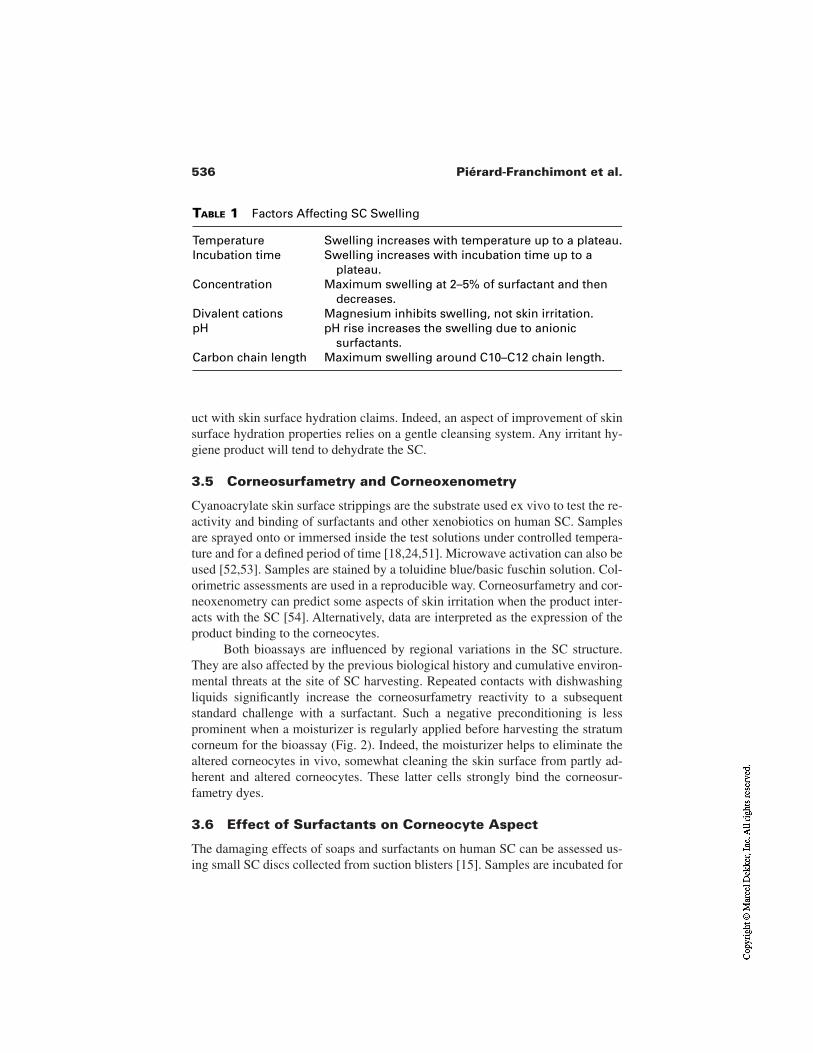
536 Piérard-Franchimont et al.
uct with skin surface hydration claims. Indeed, an aspect of improvement of skinsurface hydration properties relies on a gentle cleansing system. Any irritant hy-giene product will tend to dehydrate the SC.
3.5 Corneosurfametry and Corneoxenometry
Cyanoacrylate skin surface strippings are the substrate used ex vivo to test the re-activity and binding of surfactants and other xenobiotics on human SC. Samplesare sprayed onto or immersed inside the test solutions under controlled tempera-ture and for a defined period of time [18,24,51]. Microwave activation can also beused [52,53]. Samples are stained by a toluidine blue/basic fuschin solution. Col-orimetric assessments are used in a reproducible way. Corneosurfametry and cor-neoxenometry can predict some aspects of skin irritation when the product inter-acts with the SC [54]. Alternatively, data are interpreted as the expression of theproduct binding to the corneocytes.
Both bioassays are influenced by regional variations in the SC structure.They are also affected by the previous biological history and cumulative environ-mental threats at the site of SC harvesting. Repeated contacts with dishwashingliquids significantly increase the corneosurfametry reactivity to a subsequentstandard challenge with a surfactant. Such a negative preconditioning is lessprominent when a moisturizer is regularly applied before harvesting the stratumcorneum for the bioassay (Fig. 2). Indeed, the moisturizer helps to eliminate thealtered corneocytes in vivo, somewhat cleaning the skin surface from partly ad-herent and altered corneocytes. These latter cells strongly bind the corneosur-fametry dyes.
3.6 Effect of Surfactants on Corneocyte Aspect
The damaging effects of soaps and surfactants on human SC can be assessed us-ing small SC discs collected from suction blisters [15]. Samples are incubated for
TABLE 1 Factors Affecting SC Swelling
Temperature Swelling increases with temperature up to a plateau.Incubation time Swelling increases with incubation time up to a
plateau.Concentration Maximum swelling at 2–5% of surfactant and then
decreases.Divalent cations Magnesium inhibits swelling, not skin irritation.pH pH rise increases the swelling due to anionic
surfactants.Carbon chain length Maximum swelling around C10–C12 chain length.
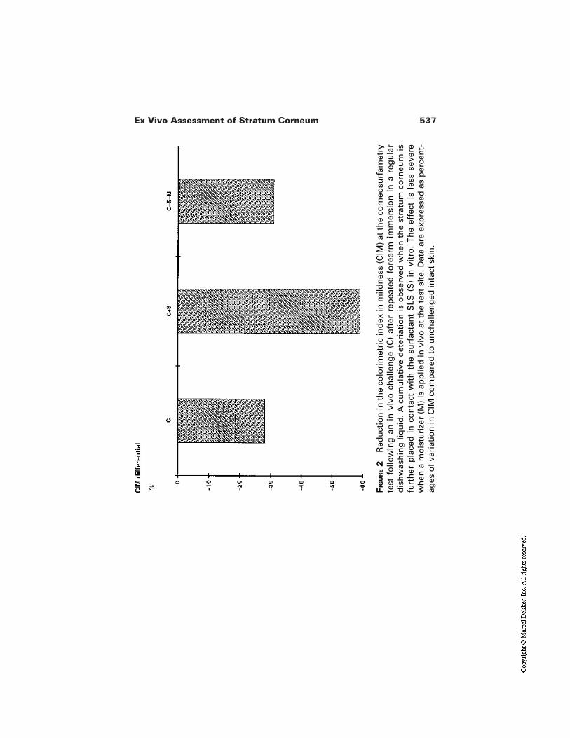
537Ex Vivo Assessment of Stratum Corneum
FIG
UR
E2
Red
uct
ion
in t
he
colo
rim
etri
c in
dex
in m
ildn
ess
(CIM
) at
th
e co
rneo
surf
amet
ryte
st f
ollo
win
g a
n i
n v
ivo
ch
alle
ng
e (C
) af
ter
rep
eate
d f
ore
arm
im
mer
sio
n i
n a
reg
ula
rd
ish
was
hin
g l
iqu
id.
A c
um
ula
tive
det
eria
tio
n i
s o
bse
rved
wh
en t
he
stra
tum
co
rneu
m i
sfu
rth
er p
lace
d i
n c
on
tact
wit
h t
he
surf
acta
nt
SLS
(S
) in
vit
ro.
Th
e ef
fect
is
less
sev
ere
wh
en a
mo
istu
rize
r (M
) is
ap
plie
d in
viv
o a
t th
e te
st s
ite.
Dat
a ar
e ex
pre
ssed
as
per
cen
t-ag
es o
f va
riat
ion
in C
IM c
om
par
ed t
o u
nch
alle
ng
ed in
tact
ski
n.
538 Piérard-Franchimont et al.
6 hr at 60°C in solutions of surfactants or soaps. Cytological alternations of cor-neocytes can be evaluated through different parameters: the number of corneo-cytes released from the SC with time, their size (swelling), and their changes inshape. This method allows a classification of the tested soaps according to theirirritancy potential. Nonionic and cationic surfactants do not appear significantlydifferent from water, although some cationic surfactants are skin irritants. Anion-ic surfactants release more corneocytes, but induce less swelling and changes.This test is a useful screening tool to determine the irritation potential of soaps. Italso provides insights in the understanding of the interaction of surfactants withthe SC.
Stratum corneum harvested by SACD can also be used to assess the cor-neocyte integrity after an in vivo challenge. The method is called squamometry S[25]. Samples are stained by a toluidine blue/basic fuschin solution at a pH about3.4. The color (Chroma C*) is measured by reflectance colorimetry. In addition,the microscopic assessment brings information about the shape and intercellularcohesion of corneocytes [18,25,55].
4 SC AND WATER
4.1 Prediction of Water Uptake by SC
Several groups have run ex vivo studies to investigate water uptake by SC. Themoisture content of the SC is essentially determined by gravimetry [7] and spec-trometric techniques [56]. In the first case, samples of SC were prepared bytrypsin treatment from human cadavers, dried, cut into small pieces, and weighed.The SC pieces were placed at equilibrium at constant temperature, but variableRH, when the samples were reweighed [9]. The gain of weight, relative to the wa-ter uptake from the environment, was calculated. Up to 50–60% RH, water up-take is moderate, while further increase in RH up to 90–95% leads to exponentialwater uptake. Ambient temperature seemed to affect the uptake of water in thelow humidity range, while not above 50–60% RH, suggesting two differentmechanisms. Gravimetric techniques were also used to determine the effect offormulations on SC hydration [57].
Spectrometric methods used to measure skin hydration of SC include in-frared [58], Raman [59,60], and nuclear magnetic resonance (NMR) [61] assess-ments. Such methods not only provide a quantification of the water content in theSC, but also bring information on the interaction and mobility between the watermolecules with SC proteins. Among these assessment methods, only NMR is ableto quantitate the hydration profile of the various SC layers and to provide infor-mation on the concentration gradient of water within the SC [62]. Proton NMRcan determine in the same samples both the total amount and the bound nonfreez-ing water by recording the spectra below 0°C [61].
539Ex Vivo Assessment of Stratum Corneum
Altogether, the data collected in vitro from the gravimetric and spectromet-ric technique demonstrate that at low RH water molecules quickly adsord to theSC and are tightly bound to the SC proteins. Increasing the ambient RH leads to aslowdown the uptake of water by the SC and make it less temperature dependent.Water also becomes less tightly bound. At very high RH, part of water is in a liq-uidlike state [54].
4.2 SACD and SC Hydration
In some instances xerosis, flakiness, and scaliness are related to desiccation of thesuperficial part of the SC [2,6]. Assessments of SACD samples can be used toquantify such a physiological ailment [18,49,63–65]. Several methods of quan-tification have been proposed. Measuring light transmission through the samples[63] and weighing them are either not validated or difficult to handle properly. Inparticular, data are significantly affected by RH after SACD has been harvested.Image analysis appears more reliable although technically difficult to handle be-cause a sophisticated softwave program is mandatory to cope with both area andthickness of scales [64,65]. Squamometry X (for xerosis) entails staining SACDusing a toluidine blue/basic fuschin solution and subsequently measuring the col-or (Chroma C*) using reflectance colorimetry [18,19,25]. The effect of moisturiz-ers can be elegantly assessed by this method (Fig. 3). In fact, moisturizers exhib-it an indirect corneodesmolytic effect. As a result the amount of corneocytesharvested by SACD is abated. Time to recurrence after stopping the moisturizingtreatment evaluates the lingering effect of the product.
4.3 Water Diffusion Through SC
When water evaporation through SC is studied on human volunteers, some envi-ronmental variables and the volunteer physiology need to be rigorously con-trolled [66]. Temperature, humidity, air flux, psychologic status, sweating, foodconsumption, and water hygiene procedure can affect the so-called trans-epi-dermal water loss (TEWL). Measurements often yield high interindividual vari-ability.
Using isolated SC in vitro allows one to better standardize the variabilityfactors linked to the individuals. Furthermore, due to the small size of the sam-ples, they can be placed in test chambers with well-controlled ambient conditions[67]. In those studies the SC membrane is mounted, external face side up, abovea water-containing reservoir. Water is maintained at a constant and defined tem-perature and separated from the SC by a small vapor space. Water diffusionthrough the SC membrane can be evaluated by measuring the weight loss fromthe water reservoir [7], the water uptake in the environment [68], the flux of ra-diolabeled water through the SC [69,70], or the water vapor gradient at the exter-nal side of the SC [67]. Also in the in vitro tests, several parameters need to be
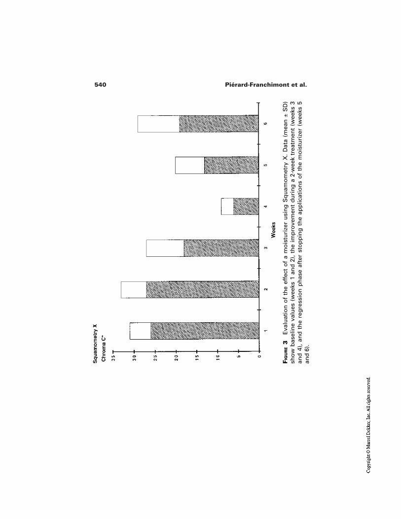
540 Piérard-Franchimont et al.
FIG
UR
E3
Eva
luat
ion
of
the
effe
ct o
f a
mo
istu
rize
r u
sin
g S
qu
amo
met
ry X
. D
ata
(mea
n ±
SD
)sh
ow
bas
elin
e va
lues
(w
eeks
1 a
nd
2),
th
e im
pro
vem
ent
du
rin
g a
2-w
eek
trea
tmen
t (w
eeks
3an
d 4
), a
nd
th
e re
gre
ssio
n p
has
e af
ter
sto
pp
ing
th
e ap
plic
atio
ns
of
the
mo
istu
rize
r (w
eeks
5an
d 6
).
541Ex Vivo Assessment of Stratum Corneum
well controlled. Changes in RH affect the SC water content and hence the waterdiffusion rate through the SC [71]. The thickness of the SC also affects the waterdiffusion [72]. Studies using SC from different body sites showed huge differ-ences in water flux, with up to 20-fold differences between abdominal and scrotalSC [73]. Any treatment of the skin before isolating the SC can also have signifi-cant consequences on water diffusion, mainly if those treatments cause SC lipidextraction or disorganization [73] or if they cause any mechanical damage to theSC.
Well-controlled in vitro studies using a computer-based Evaporimeter® sys-tem yield a threefold higher precision in TEWL in vitro measurement comparedto human studies [67]. With such a precision, the role of the corneocyte envelopeand intercellular lipids were shown on the skin barrier function. In addition, theeffect of treatment with anionic surfactants, skin permeation enhancers, and highCaCl2 concentrations were demonstrated.
5 EFFECT OF TREATMENTS ON SC LIPIDS USING INVITRO TESTS
Lipids play a prominent role in the water-holding capacity of the SC. Such a func-tion has been demonstrated in vitro on isolated SC by differential scanningcalorimetry (DSC) [74,75] and ultrastructural studies [14,76]. Briefly, the piece ofSC is dried and then hydrated in excess. The total amount of water in the SC is de-termined by the gain of weight after rehydration. For DSC measurements, the SCsamples are first freezed to –40°C and then progressively heated at a constant,slow rate up to 20°C. The melting behavior of the water contained within the SCis recorded. Unbound water in the SC causes an endothermic peak between –17 to–6°C. When the SC is hydrated to less than 33.3%, no peak can be observed inthis area. This is the approximate amount of bound water in intact SC which nev-er freezes, even at –40°C, and thus has no endothermic peak when reheating. Thedepletion in SC lipids by acetone/ether treatment causes the SC bound-water con-tent to markedly decrease. Electron microscopic observations reveal the removalor alteration of intercellular lamellae. In contrast, application of a mixture of SClipids to the acetone/ether treated SC is able to re-increase the bound water con-tent in the SC and to refill the intercellular spaces induced by the treatment withmultiple lamellae [14]. These studies demonstrate the usefulness of in vitro in-vestigations on isolated SC to understand and control the moisture of the SC. Us-ing an in vitro model of SC lipids, glycerol helped to maintain the SC lipids undera liquid crystalline state, even at low relative humidity [77]. Such an effect partic-ipates in keeping SC well moisturized. The same model was used to investigatethe effect of several cosmetic additives in preventing lipid phase transition in vit-ro [78].
In vitro studies have also been performed to determine how much and
542 Piérard-Franchimont et al.
which lipids could be extracted from the SC by surfactants. The effects of sodiumlauryl sulfate (SLS) and linear alkyl benzene sulfonate (LAS) were studied onisolated SC [79]. The extracted lipids were analyzed by high performance thinlayer chromatography (HPTCL) after elimination of the surfactants. Both surfac-tants at various concentrations removed lipids only above the critical micelle con-centration (CMC). Even at high concentrations, only very small amounts of SClipids were extracted. Such in vitro extractions of lipids from SC can also be usedfor rapid screening of lipid biochemical abnormalities of scaling skin disorders[80]. Extraction and analysis of lipids by HPTLC can also be performed oncyanoacrylate skin surface. However, this method may look more tedious as it isnecessary to completely remove glue residues from the extracted lipids before thechromatographic analysis can be performed.
6 SC AND MOISTURIZER DEVELOPMENT
As described, many methods are available to harvest human SC and to study itsinteraction with xenobiotics in vitro. Some of these approaches can be used formoisturizer development. The study design may involve a spontaneously devel-oped xerosis or scaly skin. A similar aspect can also be induced by differentmeans including barrier disruption by organic solvents, occlusive surfactantdressing, and decreased environmental dew point. The effect of a moisturizer canbe performed in a second step in vitro. In these instances, squamometry is one ofthe cheapest, most rapid, and most reliable objective in vitro tests.
REFERENCES
1. Elias PM. Stratum corneum architecture, metabolic activity and interactivity withsubjacent cell layers. Ext Dermatol 1996; 5:191–201.
2. Piérard GE, Goffin V, Hermanns-Lê T, Piérard-Franchimont C. Corneocyte desqua-mation. Int J Mol Med 2000; 6:217–221.
3. Chapman SJ, Walsh A. Desmosomes, corneosomes and desquamation. An ultrastruc-tural study of adult pig epidermis. Arch Dermatol Res 1990; 282:304–310.
4. Bernard D, Camus C, Nguyen QL, Serre G. Proteolysis of corneodesmosomal pro-teins in winter xerosis. J Invest Dermatol 1995; 105:176.
5. Rawlings AV, Harding CR, Watkinson A, Banks J, Ackerman C, Sabin R. The effectof glycerol and humidity on desmosome degradation in stratum corneum. Arch Der-matol Res 1995; 287:457–464.
6. Harding CR, Watkinson AW, Rawlings AV, Scott IR. Dry skin, moisturization andcorneodesmolysis. Int J Cosmet Sci 2000; 22:21–52.
7. Blank IH. Factors which influence the water content of the stratum corneum. J InvestDermatol 1952; 18:433–440.
8. Wester RC, Mobayen M, Maibach HI. In vivo and in vitro absorption and binding topowdered stratum corneum as methods to evaluate skin absorption of environmental

543Ex Vivo Assessment of Stratum Corneum
chemical contaminants from ground and surface water. J Toxicol Environ Health1987; 21:367.
9. Anderson RL, Cassidy JM, Hansen JR, Yellin W. Hydration of the stratum corneum.Biopolymers 1973; 12:2789–2802.
10. Kligman AM, Christophers E. Preparation of isolated sheets of human stratumcorneum. Arch Dermatol 1963; 88:70–73.
11. Scheuplein RJ. Mechanism of percutaneous adsorption. I. Routes of penetration andthe influence of solubility. J Invest Dermatol 1965; 45:334–346.
12. Faucher JA, Goddard ED. Sorption of a cationic polymer by stratum corneum. J SocCosmet Chem 1976; 27:543–553.
13. Lampe MA, Burlingame AL, Whitney JA, Williams ML, Brown BE, Roitman E,Elias PM. Human stratum corneum lipids: characterization and regional variations. JLipid Res 1983; 24:120–130.
14. Imokawa G, Kuno H, Kawai M. Stratum corneum lipids serve as a bound-watermodulator. J Invest Dermatol 1991; 96:845–851.
15. Shukuwa T, Kligman AM, Stoudemayer TJ. A new model for assessing the damag-ing effects of soaps and surfactants on human stratum corneum. Acta Derm Venereol1997; 77:29–34.
16. Marks R, Dawber RPR. Skin surface biopsy: an improved technique for the exami-nation of the horny layer. Br J Dermatol 1971; 84:117–123.
17. Piérard-Franchimont C, Piérard GE. Skin surface stripping in diagnosing and moni-toring inflammatory, xerotic and neoplastic diseases. Ped Dermatol 1985;2:180–184.
18. Piérard GE, Piérard-Franchimont C. Drug and cosmetics evaluation with skin strip-pings. In: Maibach H, ed. Dermatologic Research Techniques. Boca Raton: CRCPress, 1996:133–148.
19. Piérard GE, Masson P, Rodrigues I, Berardesca E, Lévêque JL, Loden M, Rogiers V,Sauermann G, Serup J. EEMCO guidance for the assessment of dry skin (xerosis)and ichthyosis: evaluation by stratum corneum strippings. Skin Res Technol 1996;2:3–11.
20. Piérard GE, Piérard-Franchimont C, Goffin V. Digital image analysis of microcome-dones. Dermatology 1995; 190:99–103.
21. Piérard-Franchimont C, Piérard GE. Comedogenicity. In: Elsner P, Merk HF,Maibach HI, eds. Cosmetics: Controlled Efficacy Studies and Regulations. Berlin:Springer, 1999:268–274.
22. Haidl G, Plewig G. Exfoliative cytology of stratum corneum and the effects of topi-cal retinoids on the physical properties of corneocytes. J Soc Cosmet Chem 1988;39:53.
23. Piérard GE, Piérard-Franchimont C, Saint Léger D, Kligman AM. Squamometry: theassessment of xerosis by colorimetry of D-Squame adhesive discs. J Soc CosmetChem 1992; 47:297–305.
24. Piérard GE, Goffin V, Piérard-Franchimont C. Squamometry and corneosurfametryin rating interactions of cleansing products with stratum corneum. J Soc CosmetChem 1994; 45:269–277.
25. Piérared-Franchimont C, Henry F, Piérard GE. The SACD method and the XLRSsquamometry tests revisited. Int J Cosmet Sci 2000; 22:437–446.
544 Piérard-Franchimont et al.
26. Tokumara F, Ohyama K, Fujisana H, Suzuki M, Nukatsuka H. Time-dependentchanges in dermal peeling force of adhesive tapes. Skin Res Technol 1999; 5:33–36.
27. Tokumara F, Ohyama K, Fujisawa H, Nukatsuka H. Seasonal variation in adhesivetape stripping of the skin. Skin Res Technol 1999; 5:208–212.
28. McGinley KJ, Marples RR, Plewig G. A method for visualizing and quantitating thedesquamating portion of the human stratum corneum. J Invest Dermatol 1969;53:107.
29. Roberts D, Marks R. The determination of regional and age variations in the rate ofdesquamation: a comparison of four techniques. J Invest Dermatol 1980; 74:13.
30. Corcuff P, Chatenay F, Saint Léger D. Hair–skin relationships: a new approach todesquamation, Bioeng Skin 1985; 1:133.
31. Dreher F, Arens A, Hostynek JJ, Mudumba S, Ademola J, Maibach HI. Colorimetricmethod for quantifying human stratum corneum removed by adhesive tape-strip-ping. Acta Derm Venereol 1998; 78:186–189.
32. Weigmann HJ, Lademann J, Meffert H, Schaefer H, Sterry W. Determination of thehorny layer profile by tape stripping in combination with optical spectroscopy in thevisible range as a prerequisite to quantify percutaneous absorption. Skin PharmacolAppl Skin Physiol 1999; 12:34–45.
33. Surber C, Wilhelm KP, Hori M, Maibach HI, Guy RH. Optimization of topical ther-apy: partitioning of drugs into stratum corneum. Pharmceut Res 1990; 12:1320.
34. Potts PO, Guy RH. Predicting skin permeability. Pharmaceut Res 1992; 9:663.35. Wester RC, Maibach HI, Sedik L, Melendres J, Wade M. In vivo and in vitro percu-
taneous absorption and skin decontamination of arsenic from water and soil. FundamAppl Toxicol 1993; 20:336–340.
36. Hui X, Wester RC, Magee PS, Maibach HI. Partitionng of chemicals from water intopowdered human stratum corneum (callus)—a model study. In Vitro Toxicol 1995;8:159–163.
37. Faucher JA, Goddard ED. Interaction of keratinous substrates with sodium laurylsulfate. I. Sorption. J Soc Cosmet Chem 1978; 29:323–337.
38. Goddard ED, Leung PS. Protection of skin by cationic cellulosics: in-vitro testingmethods. Cosmet Toil 1982; 97:55–69.
39. Turowski A, Adelmann-Grill BC. Substantivity to hair and skin of 125I-labelled col-lagen hydrolysates undeer application simulating conditions. Int J Cosmet Sci 1985;7:71–84.
40. Goddard ED, Harris WC. Adsorption of polymers and lipids on stratum corneummembranes as measured by ESCA. J Soc Cosmet Chem 1987; 38:295–306.
41. Ananthapadmanabhan KP, Yu KK, Meyers CL, Aronson MP. Binding of surfactantsto stratum corneum. J Soc Cosmet Chem 1996; 47:185–200.
42. Kawai M, Imokawa G. The induction of skin tightness by surfactants. J Soc CosmetChem 1984; 35:147–156.
43. Bronaugh RL, Collier SW. In vitro methods for measuring skin permeation. CosmetToil 1990; 105:86–93.
44. Diembeck W, Beck H, Benech-Kieffer F, Courtellemont P, Dupuis J, Lovell W, PayeM, Spengler J, Steiling W. Test guidelines for in vitro assessment of dermal absorp-tion and percutaneous penetration of cosmetic products. Food Chem Toxicol 1999;37:191–205.
545Ex Vivo Assessment of Stratum Corneum
45. Putterman GJ, Wolfram NF, Laden K. The effect of detergents on swelling of stratumcorneum. J Soc Cosmet Chem 1977; 28:521–532.
46. Robbins CR, Fernee KM. Some observations on the swelling of human epidermalmembrane. J Soc Cosmet Chem 1983; 34:21–34.
47. Zeidler U. Physico-chemical in vitro methods for the determination of the skin com-patibility of surfactants. J Soc Cosmet Chem (Japan) 1986; 20:17–26.
48. Blake-Haskins JC, Scala D, Rhein LD. Predicting surfactant irritation from theswelling response of a collagen film. J Soc Cosmet Chem 1986; 37:199–210.
49. Goffin V, Paye M, Piérard GE. Comparison of in vitro predictive tests for irritationinduced by anionic surfactant. Contact Dermatitis 1995; 33:38–41.
50. Rhein LD, Simion FA. Surfactant interactions with skin. In: Bender M, ed. Interfa-cial Phenomena in Biological Systems. New York: Marcel Dekker, 1991:33–49.
51. Henry F, Goffin V, Maibach H, Piérard GE. Regional differences in stratum corneumreactivity to surfactants: quantitative assessment using the corneosurfametry bioas-say. Contact Dermatitis 1997; 37:271–275.
52. Goffin V, Piérard-Franchimont C, Piérard GE. Microwave corneosurfametry. Aminute assessment of the mildness of surfactant-containing products. Skin Res Tech-nol 1997; 3:242–244.
53. Goffin V, Piérard GE. Microwave corneosurfametry and the short-duration dansylchloride extraction test for rating concentrated irritant surfactants. Dermatology2001; 202:46–48.
54. Goffin V, Henry F, Piérard-Franchimont C, Piérard GE. Penetration enhancers as-sessed by corneoxenometry. Skin Pharmacol Appl Skin Physiol 2000; 13:280–284.
55. Paye M, Cartiaux Y. Squamometry: a tool to move from exaggerated to more andmore realistic application conditions for comparing the skin compatibility of surfac-tant-based products. Int J Cosmet Sci 1999; 21:59–68.
56. Potts RO. Stratum corneum hydration: experimental techniques and interpretationsof results. J Soc Cosmet Chem 1986; 37:9–33.
57. Gehring W, Fluhr J, Gloor M. Influence of vitamin E acetate on stratum corneum hy-dration. Arzneimittel-Forschung 1998; 48:772–775.
58. Potts RO, Guzek DB, Harriss RR, McKie JE. A non invasive, in vivo, technique toquantitatively measure water concentration of the stratum corneum using attenuatedtotal-reflectance infrared spectroscopy. Arch Dermatol Res 1985; 277:489–495.
59. Williams AC, Lawson EE, Edwards HGM, Barry BW. In vitro–in vivo correlation ofFT-Raman spectra of human stratum corneum. Pharm Res 1997; 14:S454.
60. Caspers PJ, Lucassen GW, Wolthuis R, Bruining HA, Puppels GJ. In vitro and invivo Raman spectroscopy of human skin. Biospectroscopy 1998; 4:S31–S39.
61. Gilard V, Malet-Martino M, Riviere M, Gournay A, Navarro R. Measurement of to-tal water and bound water contents in human stratum corneum by in vitro proton nu-clear magnetic resonance spectroscopy. Int J Cosmet Sci 1998; 20:117–125.
62. Querleux B, Richard S, Bittoun J, Jolivet O, Idy-Peretti I, Bazin R, Lévêque JL. Invivo hydration profile in skin layers by high-resolution magnetic resonance imaging.Skin Pharmacol 1994; 7:210–216.
63. Serup J, Winther A, Blichmann C. A simple method for the study of scale pattern andeffects of a moisturizer—qualitative and quantitative evaluation by D-squame tapecompared with parameters of hydration. Clin Exp Dermatol 1989; 14:277–282.
546 Piérard-Franchimont et al.
64. el Gammal C, Pagnoni A, Kligman AM, el Gammal S. A model to assess the effica-cy of moisturizers—the quantification of soap-induced xerosis by image analysis ofadhesive-coated discs (D-squames®). Clin Exp Dermatol 1996; 21:338–343.
65. Lagarde JM, Black D, Gall Y, Del Pozo A. Image analysis of scaly skin usingDsquame samples: technical and physiological validation. Int J Cosmet Sci 2000;22:53–65.
66. Rogiers V, EEMCO group. EEMCO guidance for the assessment of the transepider-mal water loss (TEWL) in cosmetic sciences. Skin Pharmacol Appl Skin Physiol2001; 14:117–128.
67. Norlen L, Engblom J, Andersson M, Forslin B. A new computer-based evaporimetersystem for rapid and precise measurements of water diffusion through stratumcorneum in vitro. J Invest Dermatol 1999; 113:533–540.
68. Smith G, Fischer RW, Blank IH. The epidermal barrier. A comparison between scro-tal and abdominal skin. J Invest Dermatol 1961; 36:337–342.
69. Blank IH, Moloney J, Emslie AG, Simon I, Apt C. The diffusion of water across thestratum corneum as a function of its water content. J Invest Dermatol 1984;82:188–194.
70. Potts RO, Francoeur ML. The influence of stratum corneum morphology on waterpermeability. J Invest Dermatol 1991; 96:495–499.
71. El-Shimi AF, Princen HM. Water vapor sorption and desorption behavior of somekeratins. Colloid Polym Sci 1978; 256:105–114.
72. Blank IH. Further observations on factors which influence the water content of thestratum corneum. J Invest Dermatol 1953; 21:259–271.
73. Smith WP, Christensen MS, Nacht S, Gans EH. Effect of lipids on the aggregationand permeability of human stratum corneum. J Invest Dermatol 1982; 78:7–11.
74. Golden GM, Guzek DB, Harris RR, McKie JE, Potts RO. Lipid thermotropic transi-tions in human stratum corneum. J Invest Dermatol 1986; 86:255–259.
75. Golden GM, Guzek DB, Kennedy AH, McKie JE, Potts RO. Stratum corneum lipidphase transitions and water barrier properties. Biochemistry 1987; 26:2382–2388.
76. Imokawa G. In vitro and in vivo models. In: Elsner P, Berardesca E, Maibach HI,eds. Bioengineering of the Skin: Water and the Stratum Corneum. New York: Marceldekker, 1994:23–47.
77. Froebe CL, Simion FA, Olmeyer H, Rhein LD, Mattai J, Cagan RH, Friberg SE. Pre-vention of stratum corneum lipid phase transition in vitro by glycerol—an alternativemechanism for skin moisturization. J Soc Cosmet Chem 1990; 41:51–65.
78. Mattai J, Froebe CL, Rhein LD, Simion FA, Ohlmeyer H, Su DT, Friberg SE. Pre-vention of model stratum corneum lipid phase transitions in vitro by cosmetic addi-tives—differential scanning calorimetry, optical microscopy, and water evaporationstudies. J Soc Cosmet Chem 1993; 44:89–100.
79. Froebe CL, Simion FA, Rhein LD, Cagan RH, Kligman A. Stratum corneum lipid re-moval by surfactants: relation to in vivo irritation. Dermatologica 1990; 181:277–283.
80. Melnik BC, Hollman J, Erler E, Verhoeven B, Plewig G. Microanalytical screeningof all major stratum corneum lipids by sequential high-performance thin-layer chro-matography. J Invest Dermatol 1989; 92:231–234.
25Formulation of Skin Moisturizers
Steve BartonThe Boots Company, Nottingham, United Kingdom
1 INTRODUCTION
Earlier chapters in this treatise on skin moisturizers have provided detailed analy-sis of skin physiology and function or described conditions resulting from pertur-bations in these. Other chapters have discussed some common moisturizer ingre-dients and means of assessing their efficacy. This chapter attempts to bring all thisinto perspective for those wishing to understand how the knowledge may be usedto develop and manufacture a skin moisturizer.
This cannot hope to be a comprehensive directory on the art of formulatinga moisturizer; the range of raw materials alone is prohibitive. For formularies anddetails of raw material nomenclature and function the reader can access specifictexts [1–3]. Here I will identify the major issues to be considered in this processof moisturizer product development.
Some assumptions are required. The first of these is recognition that thechallenge is both technologically and commercially driven. This chapter will fo-cus on the technology but always with a view to commercial factors, the principalof which is consumer need. This gives rise to the second assumption; no attemptwill be made to artificially separate “cosmetic skin care” from “therapeutic skincare” since these can be seen to derive from slightly different consumer needs,but with often very different regulatory constraints. The third, related, assumptionis recognition of the fact that any topical product will have a physiological action.
547
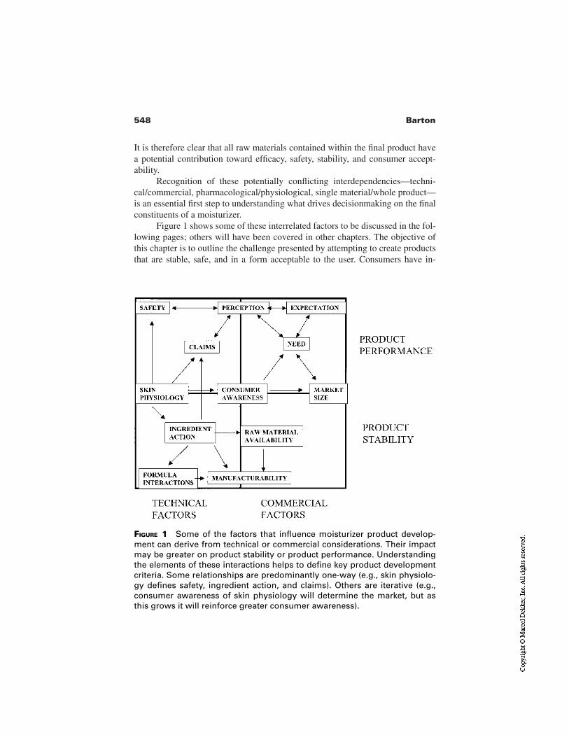
548 Barton
It is therefore clear that all raw materials contained within the final product havea potential contribution toward efficacy, safety, stability, and consumer accept-ability.
Recognition of these potentially conflicting interdependencies—techni-cal/commercial, pharmacological/physiological, single material/whole product—is an essential first step to understanding what drives decisionmaking on the finalconstituents of a moisturizer.
Figure 1 shows some of these interrelated factors to be discussed in the fol-lowing pages; others will have been covered in other chapters. The objective ofthis chapter is to outline the challenge presented by attempting to create productsthat are stable, safe, and in a form acceptable to the user. Consumers have in-
FIGURE 1 Some of the factors that influence moisturizer product develop-ment can derive from technical or commercial considerations. Their impactmay be greater on product stability or product performance. Understandingthe elements of these interactions helps to define key product developmentcriteria. Some relationships are predominantly one-way (e.g., skin physiolo-gy defines safety, ingredient action, and claims). Others are iterative (e.g.,consumer awareness of skin physiology will determine the market, but asthis grows it will reinforce greater consumer awareness).
549Formulation of Skin Moisturizers
creasingly high expectations of these performance criteria with the consequencethat “acceptable” is being replaced by “esthetically pleasing and efficacious.’’
2 BACKGROUND
The term skin moisturizer is a misleading anachronism from a technical or scien-tific viewpoint, since the means of achieving “moisturized” skin may have littleto do with adding water. The benefit perceived by the person using the moisturiz-er will depend on several factors, including skin type, product type, and expectedbenefit, and again may not necessarily relate to moisture. Alongside this there hasbeen constant growth in the added benefits delivered by a moisturizer. This hashad the effect of changing the meaning of moisturizer to become a class of skincare products in their own right. Whilst this may seem to create an initial difficul-ty in deciding how to formulate a moisturizer, it can help focus on the key attrib-utes the target consumer requires (see Table 1).
This is the first stage of developing a product, to be assessed alongsidemany other factors including total market size, anticipated market share, price,competitor products/benchmarks, and any promotional or advertizing require-ments. Consideration of these factors will help a product developer to focus onthe technical options and cost constraints required to deliver the targeted benefits.
Table 1 also introduces specific considerations, worthy of more detailedanalysis, which may help to provide a comprehensive background to developinga moisturizer. These can be loosely termed demographics—skin type, age, gen-der, ethnicity, and intention to purchase (see this volume, chapter by Johnson, formore detail). Such information will help describe the target user and requiredproduct function of a given moisturizer—conventional moisturizer, specialistmoisturizer, or a product with a secondary moisturizing action. These insightswill help define the technical delivery options. Table 1 summarizes some of thesefactors and Fig. 2 shows one way of defining where a product is positioned, thushelping to identify the technological options available to satisfy consumer expec-tations.
2.1 Technological Influences
The earlier chapters within this book explore more fully the many biological fac-tors that undoubtedly drive the development of moisturizers. Not only do thesefactors suggest options for achieving improved skin condition, they also influencehow this improvement can be assessed.
Understanding the role of the skin’s own moisturizing components alsopresents options, e.g., should we directly address the “deficiency” by replacementor allow the skin to replenish its own integrity by use of a protective product?
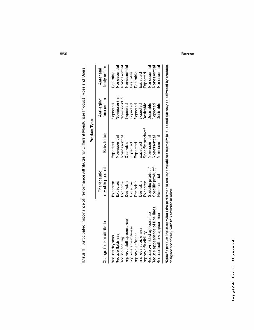
550 Barton
TA
BLE
1A
nti
cip
ated
Imp
ort
ance
of
Per
form
ance
Att
rib
ute
s fo
r D
iffe
ren
t M
ois
turi
zer
Pro
du
ct T
ypes
an
d U
sers
Pro
du
ct T
ype
Ch
ang
e to
ski
n a
ttri
bu
teT
her
apeu
tic
dry
ski
n p
rod
uct
Bab
y lo
tio
nA
nti
-ag
ing
face
cre
amA
nte
nat
alb
od
y cr
eam
Red
uce
dry
nes
sE
xpec
ted
Exp
ecte
dE
xpec
ted
Des
irab
leR
edu
ce fl
akin
ess
Exp
ecte
dN
on
esse
nti
alN
on
esse
nti
alN
on
esse
nti
alR
edu
ce s
calin
gE
xpec
ted
No
nes
sen
tial
No
nes
sen
tial
No
nes
sen
tial
Imp
rove
du
ll ap
pea
ran
ceD
esir
able
Des
irab
leE
xpec
ted
No
nes
sen
tial
Imp
rove
sm
oo
thn
ess
Exp
ecte
dE
xpec
ted
Exp
ecte
dD
esir
able
Imp
rove
so
ftn
ess
Des
irab
leE
xpec
ted
Exp
ecte
dD
esir
able
Imp
rove
su
pp
len
ess
Des
irab
leE
xpec
ted
Exp
ecte
dE
xpec
ted
Imp
rove
flex
ibili
tyE
xpec
ted
Sp
ecifi
c p
rod
uct
aD
esir
able
Exp
ecte
dR
edu
ce w
rin
kled
ap
pea
ran
ceS
pec
ific
pro
du
cta
No
nes
sen
tial
Des
irab
leN
on
esse
nti
alR
edu
ce a
pp
eara
nce
of
fin
e lin
esS
pec
ific
pro
du
cta
No
nes
sen
tial
Exp
ecte
dN
on
esse
nti
alR
edu
ce le
ath
ery
app
eara
nce
No
nes
sen
tial
No
nes
sen
tial
Des
irab
leN
on
esse
nti
al
aS
pec
ific
pro
du
ct in
dic
ates
wh
ere
the
per
form
ance
att
rib
ute
wo
uld
no
t no
rmal
ly b
e ex
pec
ted
bu
t may
be
del
iver
ed b
y p
rod
uct
sd
esig
ned
sp
ecifi
cally
wit
h t
his
att
rib
ute
in m
ind
.
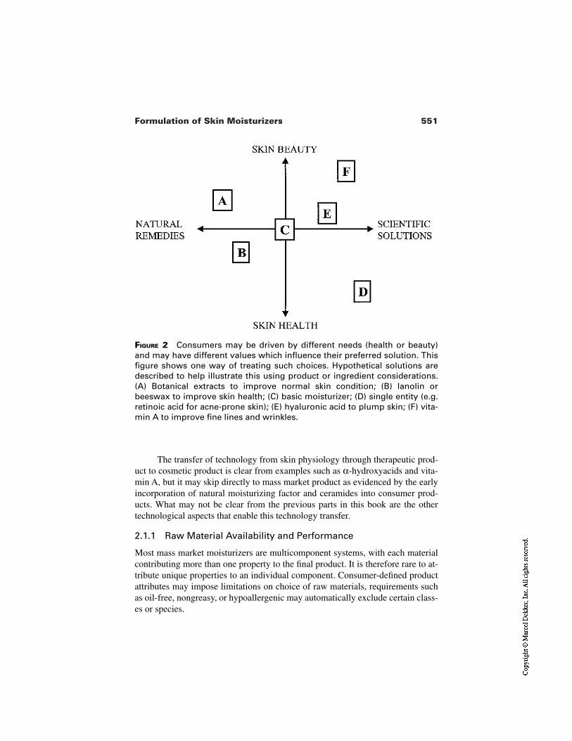
551Formulation of Skin Moisturizers
FIGURE 2 Consumers may be driven by different needs (health or beauty)and may have different values which influence their preferred solution. Thisfigure shows one way of treating such choices. Hypothetical solutions aredescribed to help illustrate this using product or ingredient considerations.(A) Botanical extracts to improve normal skin condition; (B) lanolin orbeeswax to improve skin health; (C) basic moisturizer; (D) single entity (e.g.retinoic acid for acne-prone skin); (E) hyaluronic acid to plump skin; (F) vita-min A to improve fine lines and wrinkles.
The transfer of technology from skin physiology through therapeutic prod-uct to cosmetic product is clear from examples such as α-hydroxyacids and vita-min A, but it may skip directly to mass market product as evidenced by the earlyincorporation of natural moisturizing factor and ceramides into consumer prod-ucts. What may not be clear from the previous parts in this book are the othertechnological aspects that enable this technology transfer.
2.1.1 Raw Material Availability and Performance
Most mass market moisturizers are multicomponent systems, with each materialcontributing more than one property to the final product. It is therefore rare to at-tribute unique properties to an individual component. Consumer-defined productattributes may impose limitations on choice of raw materials, requirements suchas oil-free, nongreasy, or hypoallergenic may automatically exclude certain class-es or species.
552 Barton
Specific comments on useful raw materials are given later in this chapter.However, some general comments on raw materials are worthwhile.
Identification of a useful raw material (e.g., ceramide) is only the first stagein providing a means of modifying stratum corneum. Producing a material incommercial quantities, to an acceptably reproducible standard, in a form that maybe easily and safely used in the manufacture of moisturizers represents a majorchallenge. Commercial and regulatory considerations will influence whether amaterial is suitable for a therapeutic or mass market product.
The essential fatty acid linoleic acid has been shown to improve dry skin inessential fatty acid deficiency, and the natural source (sunflower seed oil) wasalso shown to be beneficial [4].
Other sources such as evening primrose oil have also been shown to pro-vide skin benefits, but cost, esthetic, and stability considerations dictate that massmarket products use limited proportions of these materials.
Natural raw materials or blends of raw materials are thus an important andattractive source of moisturizing ingredients. Natural sources present other chal-lenges, some of which are dealt with elsewhere (see this volume, chapters byFlower, Young). The key technical factors include stability (including resistanceto microbial degradation), variability in identifiable “active” constituents, consis-tency of supply, and removal of unwanted components.
In the case of plant extracts where skin benefits are attributable to particularcomponents (e.g., chamomile extract) the method of extraction, species, cultivar,time, and place of harvest may also be considerations a product developer wishesto take into account. Where the desirable element within the plant is not readilyavailable, the extraction process may help to provide a more usable form withoutcompromizing benefit. The saponification of plant structures is an example wherethis occurs. Insoluble components can be made available as modified waxes oroils. Alternatively, useful materials can be left in the unsaponifiable fraction. Thismay have the added advantage of reducing the bulk of the material, hence makingit easier to handle and formulate into products. An alternative approach to usingnatural materials is to use synthetic processes to provide “nature identical” mole-cules (e.g., lactic acid/panthenol). In such cases the proportion of different iso-mers may or may not be an important consideration. The purity and reproduciblequality of synthetic sources may outweigh any cost disadvantage.
Many of the basic raw materials for moisturizers originate from the petrole-um industry. Cost, bulk availability, and reproducible quality were influential intheir introduction, though undoubtedly the coincidental growth in affluence andthe petrochemical and cosmetics industries also played a part. Materials such aswhite soft paraffin and light liquid paraffin (mineral oil) remain relatively cheap,standard components for many moisturizing products.
Other important petrochemical byproducts are emulsifiers. These are notonly important in themselves, in allowing stable oil and water emulsions to be
553Formulation of Skin Moisturizers
formulated, but also form the major tool for modifying other raw materials suchas vegetable oils or waxes to allow their easy incorporation into the moisturizer.Such modification of raw materials may have several purposes. Changing the sol-ubility, fluidity, or sensory properties are important, but a secondary aim may beto improve the bulk handling properties.
Where novel raw materials have been identified there are additional consid-erations of safety and registration or patenting. Whilst the latter may not seem atechnical issue it may place constraints on the timing of testing. Placing the in-vention in the public domain before the patent application is accepted may con-stitute “prior art.’’
2.1.2 Manufacturability
Raw material bulk handling has already been recognized as an important factor inthe previous section. The level of human exposure to a raw material in a manu-facturing environment will be different to that anticipated in a product, thus as-sessment of safe levels under both conditions is important.
Such raw material factors will be considered alongside the processes usedto manufacture the bulk. Size of batch, heating/cooling requirements, energy re-quired for dispersion, water content, transfer to pack, pack material and size, willall be considerations in the choice of final formulation. It is also clear that pro-cessing itself is an important component of the final formula since viscosity, clar-ity, and appearance are all dependent on achieving reproducible processing.These factors are considered in more detail later in this chapter.
2.1.3 Performance Testing
The practicalities of performance testing have been discussed elsewhere (see thisvolume, chapters by Jarret, Grove, Pierard). In formulating a moisturizing prod-uct the claims, regulatory requirements, and performance expectations will influ-ence which raw materials and testing methods are to be used and will thus affectthe total time and cost of new product development.
2.2 “Extraneous” Constraints
In starting out to develop a new moisturizer there are several other nontechnicalissues which will play a part. These are dealt with in detail elsewhere (see thisvolume, chapters by Flower, Young). For a given raw material, different marketswill impose various conditions upon what may or may not be used from the pointof view of safety and claims. Additionally, its use in the various product cate-gories and limitations on level of incorporation into a product will impose con-straints on the product developer. Patent searching is also an important early stepin the product development process—what is freely used in one market may beprotected in another.
554 Barton
Various guidelines will exist for good manufacturing practice and goodclinical practice for testing efficacy. There are also existing guidelines for claimssubstantiation testing, e.g., COLIPA guidelines [5]. Thought will need to be givento the promotional activity supporting the postlaunch marketing. Various geo-graphic regions will have their own advertizing standards guidelines which needto be borne in mind in order to help decide, at an early stage, how the productbenefits are to be communicated. Outside of these formal systems there will alsoexist an awareness of attitudes, concerns, and product performance requirementswithin the healthcare professionals community, particularly dermatologists.
3 DESIGNING MOISTURIZERS: BASIC PRINCIPLES
Moisturizing products are to be applied to the skin and left there to replace lostmoisture to improve hydration, protect from drying, and improve the various out-ward signs of dryness such as scaling or flaking and generally smooth the skin(see Table 1). Bringing together all the materials to achieve the desired result(s)requires an understanding of their benefits to the skin and physicochemical be-havior if the three key criteria of safety, stability, and esthetics are to be achieved.
3.1 Cosmetic Emulsions
The delivery options open to a product developer may seem straightforward, butgiven that most mass market moisturizing products are emulsions, one could beforgiven for asking why this is the case. Table 2 summarizes some of the reasonsfor choosing an emulsion. In most cases the emulsion will comprise oil and water.
Emulsions are multiphase systems where one phase (the continuous phase)contains droplets of the other (dispersed) phase(s). Commonly skin care emul-sions are two-phase systems where the relative volume of the continuous phase isusually, but not necessarily, greater than the dispersed phase. Droplet size is usu-ally, but not necessarily, large enough to interfere with the path of light and thusemulsions will be at least opalescent and usually white. Special cases exist wherethe particle size is so small that the liquid is clear (microemulsions) or where theemulsion structure becomes a liquid crystalline matrix (ringing gels). Three-phase systems can also exist, but all these systems tend to follow the basic princi-ples described herein.
The theoretical approach to emulsions is helpful in formulating personalcare products, but the underlying principles are often based on highly simplifiedsystems. Experience and intelligent use of these principles rather than exact sci-ence underlies much of the skill of the formulator. To formulate cost-effectivemoisturizers the basic challenge is to overcome the problem that oil and water areimmiscible. It is thermodynamically preferable for the two phases to exist in dis-crete layers, minimizing the interfacial tension.
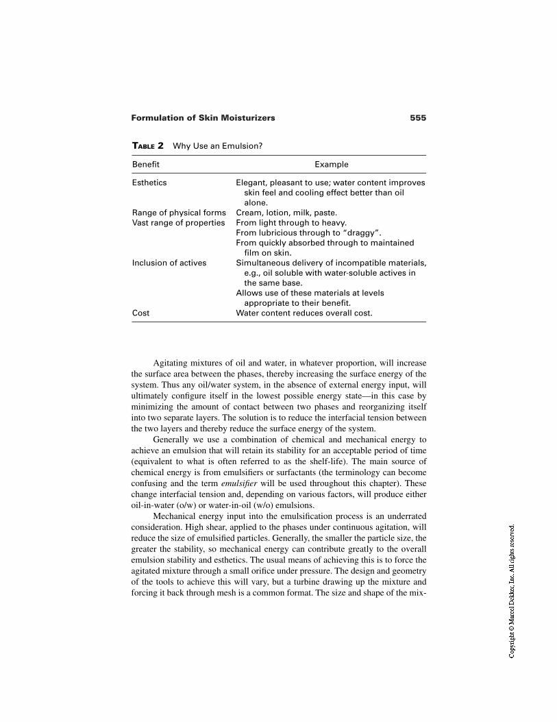
555Formulation of Skin Moisturizers
TABLE 2 Why Use an Emulsion?
Benefit Example
Esthetics Elegant, pleasant to use; water content improvesskin feel and cooling effect better than oilalone.
Range of physical forms Cream, lotion, milk, paste.Vast range of properties From light through to heavy.
From lubricious through to “draggy”.From quickly absorbed through to maintained
film on skin.Inclusion of actives Simultaneous delivery of incompatible materials,
e.g., oil soluble with water-soluble actives inthe same base.
Allows use of these materials at levelsappropriate to their benefit.
Cost Water content reduces overall cost.
Agitating mixtures of oil and water, in whatever proportion, will increasethe surface area between the phases, thereby increasing the surface energy of thesystem. Thus any oil/water system, in the absence of external energy input, willultimately configure itself in the lowest possible energy state—in this case byminimizing the amount of contact between two phases and reorganizing itselfinto two separate layers. The solution is to reduce the interfacial tension betweenthe two layers and thereby reduce the surface energy of the system.
Generally we use a combination of chemical and mechanical energy toachieve an emulsion that will retain its stability for an acceptable period of time(equivalent to what is often referred to as the shelf-life). The main source ofchemical energy is from emulsifiers or surfactants (the terminology can becomeconfusing and the term emulsifier will be used throughout this chapter). Thesechange interfacial tension and, depending on various factors, will produce eitheroil-in-water (o/w) or water-in-oil (w/o) emulsions.
Mechanical energy input into the emulsification process is an underratedconsideration. High shear, applied to the phases under continuous agitation, willreduce the size of emulsified particles. Generally, the smaller the particle size, thegreater the stability, so mechanical energy can contribute greatly to the overallemulsion stability and esthetics. The usual means of achieving this is to force theagitated mixture through a small orifice under pressure. The design and geometryof the tools to achieve this will vary, but a turbine drawing up the mixture andforcing it back through mesh is a common format. The size and shape of the mix-
556 Barton
ing vessel, the speed of mixing, and the rate of heating and cooling will also playa role since heat energy is conventionally another means of facilitating emulsifi-cation.
Alternatives to simple emulsions have been prepared and used in personalcare products: multiple phase emulsions (e.g., oil-in-water-in-oil), liquid crystalemulsions, etc. Silicone oils can also form part or all of one of the phases of anemulsion. Space does not permit a full description of the differences, but energyrequirements, particle size, and different manufacturing conditions required areamongst the considerations a product developer would consider when choosingone of these special types of emulsion. The principal considerations to be de-scribed will be similar for these specialized cases.
The process of emulsification also finds use in cleansers (see this volume,chapter by Simion) where oil droplets are suspended in an aqueous environmentthereby removing oil-bound dirt. Emulsifiers are amphipathic molecules that actby partitioning themselves at the boundary between the oil and water phases.Since conventional emulsions comprise water and oil it is their relative prefer-ence for, or solubility in, water and oil that dictates the characteristics of anemulsifier. The partitioning between the phases will also be influenced by thephysicochemical characteristics of the two phases—for example, the meltingpoint of the combined oil phase and the salt concentration of the water phase,each of which, separately but indirectly, may contribute to the effectiveness ofemulsification.
The factors responsible for determining the physical format of the emulsion(lotion, cream, etc.) include emulsifier selection, physicochemical nature of oilphase components, relative phase volume, and droplet particle size. The mechan-ical energy applied to the system can also influence greatly the droplet size andtherefore the whole outcome.
One clear source is energy during manufacture, however secondary sourcesworthy of consideration from a stability and esthetics standpoint are pumpingfrom manufacturing vessel into pack and dispensing from pack during use.
3.1.1 Emulsifier Types
There are various types of emulsifier described by their chemical nature. The ear-liest used chemical emulsifiers were probably soaps. These have a charged, hencehydrophilic, group and a fatty, hence hydrophobic, portion. These early anionicemulsifiers—named on the basis of the nature of the ion providing the polar orhydrophilic region—demonstrate some of the problems of turning emulsificationinto an exact science.
Soaps from tallow and coconut have very different proportions of fatty acidspecies. Even within coconut soaps the proportion of particular carbon chainlengths will vary between batches and the source of coconut oil starting materials.This variation will affect the physicochemical behavior at the oil–water interface,
557Formulation of Skin Moisturizers
since the oil partitioning will depend upon fatty chain length, and thus there willbe a range of emulsifying performance resulting from the chemical heterogeneityof the starting raw material. Refinement of the starting material can produce morereliable performance but at a higher cost. Crude material, e.g., hydrogenated veg-etable oils can be “cut” to produce mixed fatty alcohols, e.g., cetearyl alcohol,and further refined to produce purer materials, e.g., polyglyceryl-3 oleate. Thisprinciple applies as much to emulsifiers as it does to other oil phase ingredientscommonly used in formulating moisturizers.
Many original skin care emulsions were produced by in situ neutralizationof stearic acid with sodium hydroxide. The resulting soap acts as an emulsifier.There are problems with such systems, particularly their pH and susceptibility toheavy metal salts. In particular their aggressiveness on skin has tended to rulethem out of favor moreso in leave-on moisturizers than wash-off cleansers (seethis volume chapter by Simion). More sophisticated anionic systems do howeverstill find use in emulsions. Modified coconut oils still form the basis of the sulfateand ether sulfate anionic emulsifiers (such as sodium lauryl sulfate and sodiumlauryl ether sulfate). The substitution of the fatty chain using an ether group re-duces the hydrophobic nature of the fatty chain, hence the emulsifier, and pro-duces an emulsion system less aggressive to the skin [6].
Nonionic emulsifiers are commonly used in personal care moisturizers, andas the name implies they have no charge. Instead their amphipathic nature derivesfrom their long carbon chains imparting hydrophobic nature (e.g., fatty acids) orhydrophilic nature (e.g., alcohols). Unsaturated carbon chains are easily substitut-ed with ethylene oxide. This process of ethoxylation can be carried out to varyingdegrees on the same starting material to give a range of different amphipathic be-haviors. When combined with varying lengths and branching of the carbon chain,this has produced a vast array of different molecules; many of which have be-come the workhorses for skin care formulators.
Cationic emulsifiers also exist and do have some use in skin care formula-tions. Many of those used also demonstrate some antimicrobial activity; this maybe considered of benefit. Cationic emulsifiers do not have the same problem asanionics with metal salts, but may be prone to instability at high pH and negativeion concentration. They also have a property that can be either detrimental or ben-eficial. The skin surface has a net negative charge, and cationic emulsifiers maythus bind to the substrate.
Polymeric emulsifiers are also common in moisturizer formulations. Exam-ples include those based on silicone or polyacrylic acids. These polymers distrib-ute themselves along the oil/water interface with side groups of lipophilic and hy-drophilic nature inserted into their respectively preferred phase.
It is not uncommon to find mixed types of emulsifier in a given emulsion.Indeed many raw material suppliers combine anionic and nonionic emulsifiers asa blend for commercial purposes. The skill of the formulator will be to recognize
558 Barton
where one of the particular properties mentioned is required, understand the con-sequences and compromises this confers on the emulsion, and act accordingly.
3.1.2 Emulsifier Choice
This is not straightforward but has been simplified by the introduction, around 50years ago, of the HLB system. The system accompanied the increased use of non-ionic emulsifiers where it finds greatest use. The HLB number (hydrophile/lipo-phile balance) represents the balance of the size and strength of the hydrophilic(or polar) and lipophilic (or nonpolar) groups within the emulsifier molecules.This can be visualized in terms of how the emulsifier molecule will position itselfat the oil–water interface (Fig. 3).
The more polar one part of the molecule is (often referred to as the head-group), the more it will insert itself into the water domain of an emulsion. Con-versely, the more nonpolar the other part (or the tailgroup), the more it will bepart of the oil phase. The relative polarity will not only affect the final positionof the emulsifier within the interface, but also the stability and fluidity of thisboundary.
The HLB system uses this relative preference on a scale from 1 to 20 andprovides most emulsifiers, particularly nonionic emulsifiers, with such a number.The usefulness of the HLB system is that it is additive. Thus combining equalproportions of emulsifier of different HLB (e.g., 5 and 15) will produce a systemof average HLB (i.e., 10). This property of the HLB system can be used to calcu-late the most appropriate emulsifier for an oil phase of unknown requirements.Simple experimental determinations can be carried out, using a range of emulsifi-er mixtures with a known range of HLB, to show the HLB value(s) that result ingood emulsification. An appropriate emulsifier, or blend of emulsifiers, can thenbe chosen having the equivalent HLB number. In practice the task is not alwaysthat simple, however the HLB system provides a useful guide. The HLB valuesare normally published in suppliers’ literature and are therefore often readilyavailable.
The factors that influence HLB include the chemistry, stereochemistry, andpurity for each emulsifier and their miscibility with other oil and water phasecomponents (rarely do we wish to produce an emulsion of a single oil with purewater). These factors can usefully be taken into account alongside the HLB. It isalso necessary to point out that HLB determinations will produce two conclu-sions, one for oil-in-water the other for water-in-oil systems.
The usefulness of the HLB system, the range of physical forms, and relativemildness (compared to anionics and cationics) have made nonionic emulsifiersthe commonest choice.
Milder anionics have become more available and more appropriate forleave-on products and blends of nonionics and anionics are useful starting pointsfor emulsions, combining the advantages of both emulsifier types. However non-
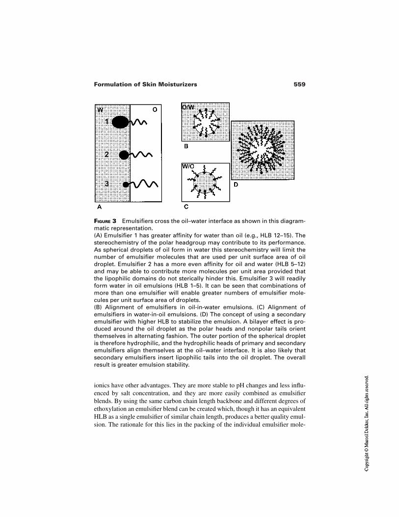
559Formulation of Skin Moisturizers
FIGURE 3 Emulsifiers cross the oil–water interface as shown in this diagram-matic representation.(A) Emulsifier 1 has greater affinity for water than oil (e.g., HLB 12–15). Thestereochemistry of the polar headgroup may contribute to its performance.As spherical droplets of oil form in water this stereochemistry will limit thenumber of emulsifier molecules that are used per unit surface area of oildroplet. Emulsifier 2 has a more even affinity for oil and water (HLB 5–12)and may be able to contribute more molecules per unit area provided thatthe lipophilic domains do not sterically hinder this. Emulsifier 3 will readilyform water in oil emulsions (HLB 1–5). It can be seen that combinations ofmore than one emulsifier will enable greater numbers of emulsifier mole-cules per unit surface area of droplets.(B) Alignment of emulsifiers in oil-in-water emulsions. (C) Alignment ofemulsifiers in water-in-oil emulsions. (D) The concept of using a secondaryemulsifier with higher HLB to stabilize the emulsion. A bilayer effect is pro-duced around the oil droplet as the polar heads and nonpolar tails orientthemselves in alternating fashion. The outer portion of the spherical dropletis therefore hydrophilic, and the hydrophilic heads of primary and secondaryemulsifiers align themselves at the oil–water interface. It is also likely thatsecondary emulsifiers insert lipophilic tails into the oil droplet. The overallresult is greater emulsion stability.
ionics have other advantages. They are more stable to pH changes and less influ-enced by salt concentration, and they are more easily combined as emulsifierblends. By using the same carbon chain length backbone and different degrees ofethoxylation an emulsifier blend can be created which, though it has an equivalentHLB as a single emulsifier of similar chain length, produces a better quality emul-sion. The rationale for this lies in the packing of the individual emulsifier mole-
560 Barton
cules into the oil–water interface; single species will compete for space, whereasmixed species will take up complementary spatial distribution (see Fig. 3).
3.1.3 Stability
Stability is a term used in the cosmetics and toiletries industry to describe how theproduct may be expected to respond to storage and use after manufacture, pack-ing, and temporary storage in warehouse or point of sale. As with many of the is-sues discussed in this particular chapter there are no absolutes; much will be de-termined by assessing risk on the basis of experience and laboratory simulation.Cosmetic emulsions are in thermodynamic equilibrium and changes in this statusquo can destabilize the emulsion structure in a number of ways.
To help understand why we use some ingredients in a moisturizer it can beuseful to look at what might go wrong. A formulator, having understood the com-promises mentioned, will think ahead to some of these possibilities and modifythe formula appropriately. Table 3 is a brief and incomplete summary of some ofthe factors. It is designed to show how the factors affecting emulsion stability areinterlinked. We can rarely pinpoint a single variable and change it to effect a de-sired end. For example, increasing the viscosity (actually the rheology, since thesystem is in equilibrium) of the continuous phase using a thickener will reducethe effects of dispersed phase Brownian motion.
However, the use of such additives (e.g., waxes for continuous oil phase orpolymers for a continuous water phase) will also increase the specific gravity ofthe continuous phase, change the interfacial behavior at the water–oil interfaceand thus affect the fluidity of this layer.
The overall effects on the emulsion may be beneficial, but simultaneouslychanging other emulsion components or processing parameters will render it dif-ficult to know which emulsion characteristic has been changed and how, hencethe inexact science!
Coalescence resulting from fusion of two droplets of the dispersed phasemay be minimized by the changes mentioned (through limiting the effects ofBrownian motion), but the void space (between the dispersed phase particles) andthe particle size are important contributory factors. Changing the relative volumeand/or the emulsifier can effect changes to these emulsion attributes. Concentra-tion of emulsifier is also important in maintaining the phase separation at the in-terface, and minimizing the possible confluence of droplets if they do come intocontact. Increasing the concentration of a nonionic emulsifier in this way to over-come the risk of coalescence may however create other problems—flocculation,for example. Options for overcoming this eventuality may include substitutingsome anionic for nonionic emulsifier. Here again the concept of emulsifier blendshelps to achieve the desired end (see Fig. 3).
Where dispersed phase droplets become extremely tightly packed, phaseinversion can occur, e.g., oil-in-water emulsion changes to water-in-oil. There
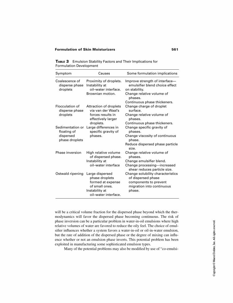
561Formulation of Skin Moisturizers
TABLE 3 Emulsion Stability Factors and Their Implications for Formulation Development
Symptom Causes Some formulation implications
Coalescence ofdisperse phasedroplets
Proximity of droplets.Instability at
oil–water interface.Brownian motion.
Improve strength of interface—emulsifier blend choice effect
on stability.Change relative volume of
phases.Continuous phase thickeners.
Flocculation ofdisperse phasedroplets
Attraction of dropletsvia van der Waal’sforces results ineffectively largerdroplets.
Change charge of droplet surface.
Change relative volume of phases.
Continuous phase thickeners.Sedimentation or
floating ofdispersedphase droplets
Large differences inspecific gravity ofphases.
Change specific gravity of phases.
Change viscosity of continuous phase.
Reduce dispersed phase particle size.
Phase inversion High relative volume of dispersed phase.
Instability at oil–water interface
Change relative volume of phases.
Change emulsifier blend.Change processing—increased
shear reduces particle size.Ostwald ripening Large dispersed
phase droplets formed at expense of small ones.
Instability at oil–water interface.
Change solubility characteristicsof dispersed phasecomponents to preventmigration into continuousphase.
will be a critical volume fraction for the dispersed phase beyond which the ther-modynamics will favor the dispersed phase becoming continuous. The risk ofphase inversion can be a particular problem in water-in-oil emulsions where highrelative volumes of water are favored to reduce the oily feel. The choice of emul-sifier influences whether a system favors a water-in-oil or oil-in-water emulsion,but the rate of addition of the dispersed phase or the degree of mixing can influ-ence whether or not an emulsion phase inverts. This potential problem has beenexploited in manufacturing some sophisticated emulsion types.
Many of the potential problems may also be modified by use of “co-emulsi-
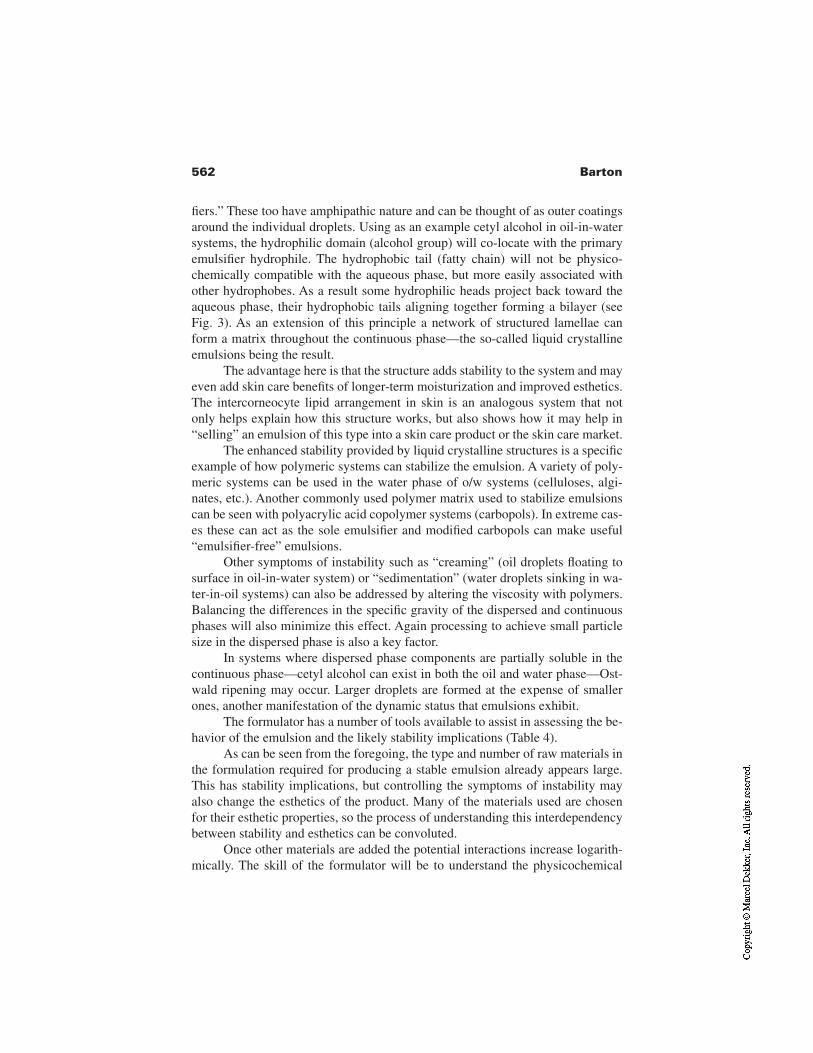
562 Barton
fiers.” These too have amphipathic nature and can be thought of as outer coatingsaround the individual droplets. Using as an example cetyl alcohol in oil-in-watersystems, the hydrophilic domain (alcohol group) will co-locate with the primaryemulsifier hydrophile. The hydrophobic tail (fatty chain) will not be physico-chemically compatible with the aqueous phase, but more easily associated withother hydrophobes. As a result some hydrophilic heads project back toward theaqueous phase, their hydrophobic tails aligning together forming a bilayer (seeFig. 3). As an extension of this principle a network of structured lamellae canform a matrix throughout the continuous phase—the so-called liquid crystallineemulsions being the result.
The advantage here is that the structure adds stability to the system and mayeven add skin care benefits of longer-term moisturization and improved esthetics.The intercorneocyte lipid arrangement in skin is an analogous system that notonly helps explain how this structure works, but also shows how it may help in“selling” an emulsion of this type into a skin care product or the skin care market.
The enhanced stability provided by liquid crystalline structures is a specificexample of how polymeric systems can stabilize the emulsion. A variety of poly-meric systems can be used in the water phase of o/w systems (celluloses, algi-nates, etc.). Another commonly used polymer matrix used to stabilize emulsionscan be seen with polyacrylic acid copolymer systems (carbopols). In extreme cas-es these can act as the sole emulsifier and modified carbopols can make useful“emulsifier-free” emulsions.
Other symptoms of instability such as “creaming” (oil droplets floating tosurface in oil-in-water system) or “sedimentation” (water droplets sinking in wa-ter-in-oil systems) can also be addressed by altering the viscosity with polymers.Balancing the differences in the specific gravity of the dispersed and continuousphases will also minimize this effect. Again processing to achieve small particlesize in the dispersed phase is also a key factor.
In systems where dispersed phase components are partially soluble in thecontinuous phase—cetyl alcohol can exist in both the oil and water phase—Ost-wald ripening may occur. Larger droplets are formed at the expense of smallerones, another manifestation of the dynamic status that emulsions exhibit.
The formulator has a number of tools available to assist in assessing the be-havior of the emulsion and the likely stability implications (Table 4).
As can be seen from the foregoing, the type and number of raw materials inthe formulation required for producing a stable emulsion already appears large.This has stability implications, but controlling the symptoms of instability mayalso change the esthetics of the product. Many of the materials used are chosenfor their esthetic properties, so the process of understanding this interdependencybetween stability and esthetics can be convoluted.
Once other materials are added the potential interactions increase logarith-mically. The skill of the formulator will be to understand the physicochemical
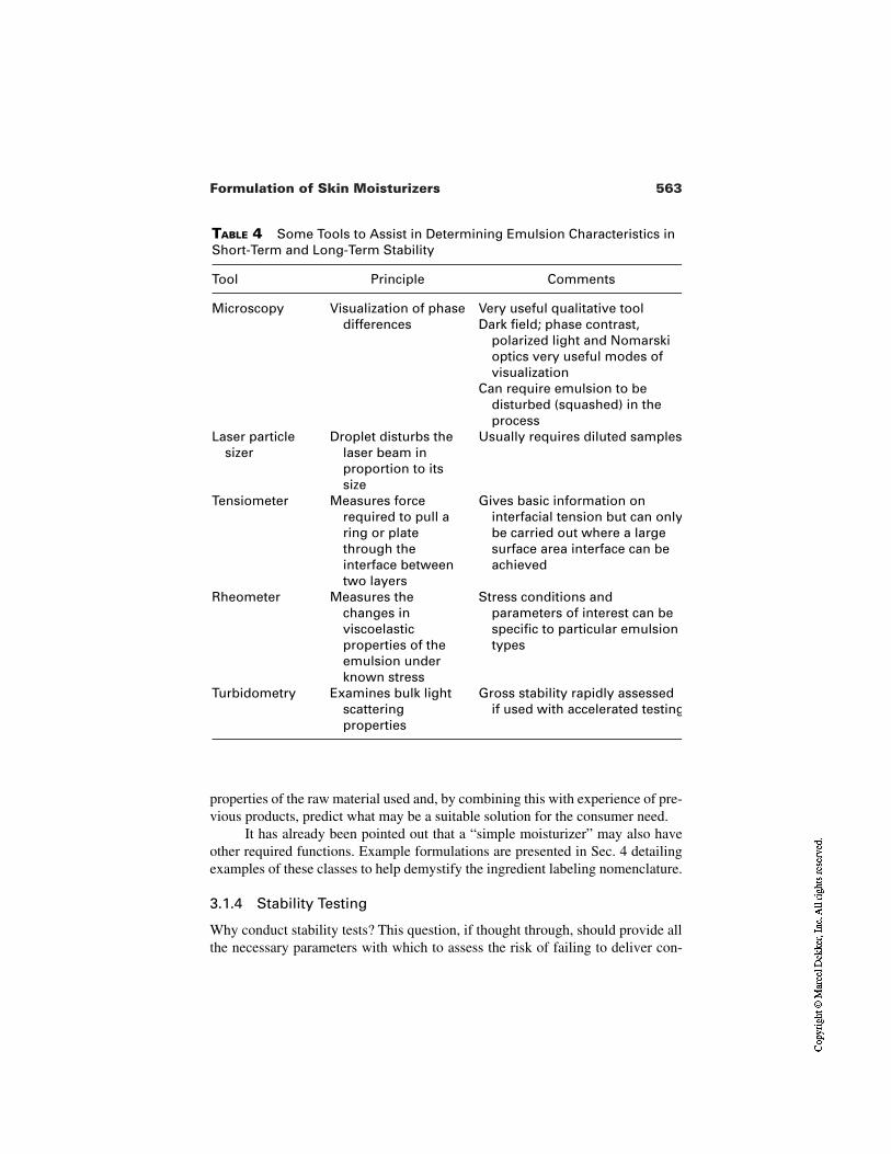
563Formulation of Skin Moisturizers
TABLE 4 Some Tools to Assist in Determining Emulsion Characteristics inShort-Term and Long-Term Stability
Tool Principle Comments
Microscopy Visualization of phasedifferences
Very useful qualitative toolDark field; phase contrast,
polarized light and Nomarski optics very useful modes of visualization
Can require emulsion to be disturbed (squashed) in the process
Laser particlesizer
Droplet disturbs thelaser beam inproportion to itssize
Usually requires diluted samples
Tensiometer Measures forcerequired to pull aring or platethrough theinterface betweentwo layers
Gives basic information oninterfacial tension but can onlybe carried out where a largesurface area interface can beachieved
Rheometer Measures thechanges inviscoelasticproperties of theemulsion underknown stress
Stress conditions andparameters of interest can bespecific to particular emulsiontypes
Turbidometry Examines bulk lightscatteringproperties
Gross stability rapidly assessedif used with accelerated testing
properties of the raw material used and, by combining this with experience of pre-vious products, predict what may be a suitable solution for the consumer need.
It has already been pointed out that a “simple moisturizer” may also haveother required functions. Example formulations are presented in Sec. 4 detailingexamples of these classes to help demystify the ingredient labeling nomenclature.
3.1.4 Stability Testing
Why conduct stability tests? This question, if thought through, should provide allthe necessary parameters with which to assess the risk of failing to deliver con-
564 Barton
sumer expectation. The consumer wants a safe, effective product. What maymake it unsafe or ineffective? The consumer wants a product that will be pleasur-able to use. What may detract from this pleasurable experience? The consumerwants to be able to use it in the course of their daily routine. What is that likely tobe and how will the product respond? Many of these questions are common to thegeneral principles underlying formulation of a successful product.
The formulator will test the initial batch(es) of the product to assess fit withconsumer expectation; what happens once the product is opened or stored forlong periods?
Physicochemical and Organoleptic Stability. As detailed in Table 4 thereare a number of physicochemical characteristics that can help determine stability.Viscosity and microscopic appearance are commonly used as laboratory tools inthis way. Rapid change in viscosity over time will suggest a change in the emul-sion structure. Both increased and decreased viscosity can have implications forstability and each may affect the delivery from the pack.
Microscopy can be helpful in understanding the reasons underlying thechange in stability. Emulsion structure and homogeneity of droplet in terms ofsize and structure can be visualized. Emulsification of complex lipid mixtures canresult in droplets containing different lipid phases; their stability can be assessedfrom how well defined and how consistent these phases are within and betweenindividual lipid droplets. Crystallization of water-soluble or oil-soluble compo-nents can be seen using polarizing light microscopy, and the special case of liquidcrystalline emulsions can be assessed for consistency and appearance.
Many of the other criteria for stability assessment focus on the sensory as-pects—odor, color, and viscosity, in particular. Changes in these are likely to benoticed by the customer and have an effect on the perceived as well as the realperformance. Objective characteristics such as pH and viscosity are easy torecord; sensory properties often require reference to a standard sample, usuallythe one stored at room temperature.
Amongst other factors that may change, the pH of the product will be themost often tested, and changes here may have safety implications. This can onlybe assessed in an oil-in-water emulsion, and pH changes in a water-in-oil formu-lation could affect the overall product stability.
One of the requirements during the initial formulation work will be to rap-idly assess potential stability problems. Exposure to elevated temperatures (60°C)is often used as an indicator, since many of the potential problems are a conse-quence of the thermodynamics of emulsification. Temperature cycling can also beused in a similar way to stress the potential formulation, but the method finds usein longer-term testing for anticipated changes that may result from warehousestorage, travel, and export to other countries.
Many of the stability characteristics will be dictated by the pack. Whilst
565Formulation of Skin Moisturizers
glass is fairly inert, many of the other commonly used plastics can impose con-straints on formulation raw materials. Some esters used in formulations can solu-bilize certain pack types. The resulting effect varies from softening and deforma-tion of the pack to reduction of permeability of the pack material. The latterchange will reduce the physical barrier allowing gaseous exchange to occur andeven result in microbial spoilage. Testing in the appropriate pack is therefore es-sential.
Clear packs also impose some constraints. Colored products can fade in aclear pack and inclusion of a sunscreen in the product or pack required may re-solve the problem. Testing to mimic the exposure to light in the shop window orbathroom window is therefore another means of ensuring that the esthetics of theproduct do not change on storage.
Microbial Stability. The most important of the potential risks in the safetycategory is microbial contamination. Sources of contamination occur throughoutthe supply chain—raw material, raw material storage and processing, packaging,final product storage, and product use and storage by the consumer. There may beparticular considerations that influence the risk analysis. For instance baby mois-turizer products for the diaper area will be exposed to fecal contamination.Eczema sufferers have increased risk of skin staphylococcal contamination.Botanical extracts for incorporation in skin moisturizers may contain microbialcontamination at source.
Particular parts of the machinery used to process the product may havereservoirs susceptible to contamination or prove difficult to clean in betweenmanufacturing batches, pumps in particular. The pack in which the product is pre-sented also plays an important part. Whilst pump packs and squeeze bottles pre-vent the introduction of contamination from the fingers or outside world, the“suck-back” from a squeeze bottle could siphon contamination back in and pro-vide a false sense of security. Once the product has been used a number of times,there is a large air space and condensation on the side of pack can create an envi-ronment for microbial growth.
The response to all of these risks is to incorporate a degree of preservation.Incorporating preservatives or using barrier methods (e.g., pump pack; single-dose format) are two common strategies but these should be as protection againstaccidental contamination postmanufacture. Primary risk reduction should alwayscome from good manufacturing practice—avoidance of contamination in the firstplace. The implication for the product developer is that no raw material or processused should increase the risk of microbial contamination. Use of freeze-dried oralcoholic or glycolic rather than aqueous plant extracts is one level of risk limita-tion.
The processes used for testing microbial stability are termed challenge test-ing. Here inoculation of the product with known organisms is carried out and
566 Barton
their subsequent survival monitored. The protocols used and the criteria for pass-ing and failing may be laid down in prescribed monographs [7]. Such testing willbe conducted on samples from first laboratory or small-scale batches as well as onthese samples after storage for a given period. Full microbial counting after man-ufacture and before release for sale may also be used to assess the quality of thefactory batch prior to delivery.
3.2 Processing and Packaging
Whilst the focus of this chapter is the formulation aspects of moisturizers, pro-cessing and packaging cannot be excluded as they impose limitations and createopportunities for the formulator.
Skin care moisturizers are produced in large quantities worldwide. Manydifferent batch sizes, processes, and plant specifications exist, and economics of-ten dictate the role played by human operator or automated machine. Small-scaleproduction runs up to 1000 L can be managed by small enterprises without muchsophistication. Where large-scale volumes permit, continuous processing can beachieved, but this is probably the exception. Similarly, dispensing lotions into 30-mL narrow-necked bottles will impose different economic constraints thanfilling 500-g tubs with a thick cream. Whichever the case, raw material handling,water purity, and plant hygiene are of prime concern.
The technical and formulation constraints imposed by different manufac-turing conditions are a key part of formulation development and can commandconsiderable resource. The key issues that require control will be speed of heatingand cooling, speed of addition of one phase to another, shear, duration, and ener-gy input during mixing. Some of the other constraints imposed will be addition ofvolatiles (e.g., fragrance) or heat-sensitive raw materials (e.g., vitamins, botanicalextracts).
Where oils are added to water there is the likelihood of crystallization ofsome high–melting point components if the water temperature differs too greatlyfrom the oil. Water phase thickeners also pose potential problems since thoroughdispersion is required to prevent the formation of gelatinous lumps; hydration ofthe external layer can occur, preventing adequate hydration of the central mass.Overagitation can introduce air into the mixture and with some emulsifier levelsand types, this aeration can be retained all the way through into the pack.
Another area of potential risk is incorporation of mineral particulates suchas talc, zinc oxide, or titanium dioxide. These create a high surface area and re-quire adequate “wetting” if incorporated in water phase or dispersion into oilphase. The effect on the overall emulsification requirements will not be simplyassessed via HLB calculations.
In some cases processing conditions can be the vital element in producing
567Formulation of Skin Moisturizers
the desired product and here, or where other factors dictate, small-scale trial or pi-lot batches may be desirable.
4 FORMULATION EXAMPLES
A number of example formulations are presented and ingredients discussed to il-lustrate the issues highlighted earlier in the chapter. The formulations will also bereferred to later in the chapter when discussing performance aspects.
4.1 Basic Moisture Lotion
Formula I (Table 5) is a basic moisturizing lotion, which requires that some ofthe oil phase will remain fluid at room temperature. Shorter chain esters are morefluid contributing to this property in this formulation. Mineral oils are also use-ful here but with added benefits of stability (lower risk of rancidity than esters)and lower bulk cost. However the esthetics of a solely mineral oil formula wouldnot be very good and incorporating esters improves the skin-feel without reduc-ing lubricity. Cetyl alcohol helps stabilize the emulsion and provides some vis-cosity. Its higher molecular weight fatty chain length and relative hydrophobici-ty contribute to this action. The ratio of oil to water will be around 1:3 and thiswill influence the overall viscosity. Silicone oils usually count toward the oilphase, and the type and concentration of silicone oil will depend on the intendedproduct use. Higher levels of barrier can be achieved with higher molecularweight silicones.
Water phase thickeners not only provide added stability, but also increasethe bulk or body of the lotion. Such viscosity modifiers are also important whenconsidering how the product is dispensed. Pumping through nozzles creates highshear forces. Some rheology modifiers tolerate these better than others and theformulator must choose the correct combinations for this and “rub-in” proper-ties. In the case of alginates or carbomers the effects of salts also require consid-eration.
Salts present on the skin have a thinning effect on these systems and thiscan be put to use to achieve “quick break” effects on application. Glycerin is anall-purpose moisturizing ingredient that is cost effective and widely availablefrom a variety of sources. The level of incorporation and claims associated withglycerin depend on the target market. In some markets urea might be another ex-pected moisturizing ingredient though its mode of action would be different. Ureaalso introduces stability constraints—pH and preservative and thickener compat-ibility being examples.
Since pump delivery has the added benefit of reducing the risk of spoilagevia microbial contamination, this can be a popular pack choice for baby lotions.
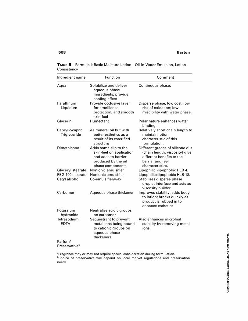
568 Barton
TABLE 5 Formula I: Basic Moisture Lotion—Oil-in-Water Emulsion, LotionConsistency
Ingredient name Function Comment
Aqua Solubilize and deliveraqueous phaseingredients; providecooling effect
Continuous phase.
ParaffinumLiquidum
Provide occlusive layerfor emollience,protection, and smoothskin-feel
Disperse phase; low cost; lowrisk of oxidation; lowmiscibility with water phase.
Glycerin Humectant Polar nature enhances waterbinding.
Caprylic/capricTriglyceride
As mineral oil but withbetter esthetics as aresult of its esterifiedstructure
Relatively short chain length tomaintain lotioncharacteristic of thisformulation.
Dimethicone Adds some slip to theskin-feel on applicationand adds to barrierproduced by the oilphase components
Different grades of silicone oils(chain length, viscosity) givedifferent benefits to thebarrier and feelcharacteristics.
Glyceryl stearate Nonionic emulsifier Lipophilic>lipophobic HLB 4.PEG 100 stearate Nonionic emulsifier Lipophilic<lipophobic HLB 18.Cetyl alcohol Co-emulsifier/wax Stabilizes disperse phase
droplet interface and acts asviscosity builder.
Carbomer Aqueous phase thickener Improves stability; adds bodyto lotion; breaks quickly asproduct is rubbed in toenhance esthetics.
Potassiumhydroxide
Neutralize acidic groupson carbomer
TetrasodiumEDTA
Sequestrant to preventmetal ions being boundto cationic groups onaqueous phasethickeners
Also enhances microbialstability by removing metalions.
Parfuma
Preservativeb
aFragrance may or may not require special consideration during formulation.bChoice of preservative will depend on local market regulations and preservationneeds.
569Formulation of Skin Moisturizers
4.2 Water-in-Oil Cream
Whilst water-in-oil creams (Formula II, Table 6) may be expected to have highproportions of oil, this is not axiomatic. Use of a combination of low HLB non-ionic and silicone emulsifier systems to obtain highly stable small droplet sizewater phase can contribute to an increased water content. The rich feel can comefrom the high oil loading, and the emulsifier combination enhances this by pro-viding a shear-thickening effect in use. This is counteracted by silicone fluid help-ing provide final rub-in characteristic. In the case of this formulation theC12–C15 ester makes up the bulk of the oil phase. This material has good solubi-lizing properties for other oil phase components and has lower cost and greaterpredictability and quality compared with other oil phase components. The lanolinand bees wax add structure. The ratio of these structuring waxes and the fluid es-ter will help define the final viscosity. The glycerin in the water phase is “trapped”within the lipid matrix and when applied to skin may be released more slowly.
4.3 Anhydrous Emollient Cream
Where cost and efficacy permit, such anhydrous systems as Formula III (Table 7)can deliver moisturized skin via emollience and occlusion. Lanolin performs thisdual role well but in the case of this formulation the cost is reduced by blendingpetroleum based oils and waxes, silicones and carnauba wax (also a commonstructural wax ingredient for lipsticks). Dispersion is aided by stearic acid. Mi-crobial spoilage is unlikely in the absence of water but rancidity of lanolin andother oils can be prevented by the inclusion of anti-oxidants such as butylated hy-droxy toluene.
4.4 Basic Dry Skin Cream
A basic dry skin cream is described by Formula IV (Table 8). Where products fortherapeutic use are required, there are limitations imposed by regulatory bodieson permitted raw materials.
Here the bulk of the oil phase is from mineral oil/wax to achieve a suitableconsistency occlusive film that rubs in well without being too greasy. Lanolin isadded for its emollient properties.
The combination of PEG-20 stearate and cetearyl alcohol is an example of acommercially available mixed emulsifier system. This one known as Polawax™ iscommonly used in therapeutic skin care products. It provides very thick but notparticularly elegant emulsions. Oil phase components have well-documented tol-erance and pharmacopoeia specifications. Other ingredients may be included fortherapeutic purposes—urea, lactic acid introduced into the water phase; additionaloil phase components rich in linoleic acid (borage oil, evening primrose oil, etc.).Anti-oxidant may be included for formulation stability rather than skin benefit.
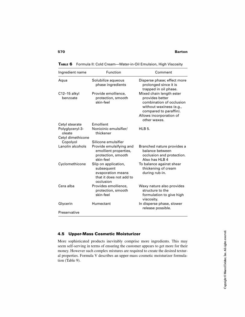
570 Barton
TABLE 6 Formula II: Cold Cream—Water-in-Oil Emulsion, High Viscosity
Ingredient name Function Comment
Aqua Solubilize aqueousphase ingredients
Disperse phase; effect moreprolonged since it istrapped in oil phase.
C12–15 alkylbenzoate
Provide emollience,protection, smoothskin-feel
Mixed chain length ester provides better combination of occlusion without waxiness (e.g., compared to paraffin).
Allows incorporation of other waxes.
Cetyl stearate EmollientPolyglyceryl-3-
oleateNonioinic emulsifier/
thickenerHLB 5.
Cetyl dimethiconeCopolyol Silicone emulsifier
Lanolin alcohols Provide emulsifying andemollient properties,protection, smoothskin-feel
Branched nature provides abalance betweenocclusion and protection.Also has HLB 4
Cyclomethicone Slip on application,subsequentevaporation meansthat it does not add toocclusion
To balance against shearthickening of creamduring rub-in.
Cera alba Provides emollience,protection, smoothskin-feel
Waxy nature also providesstructure to theformulation to give highviscosity.
Glycerin Humectant In disperse phase, slowerrelease possible.
Preservative
4.5 Upper-Mass Cosmetic Moisturizer
More sophisticated products inevitably comprise more ingredients. This mayseem self-serving in terms of ensuring the customer appears to get more for theirmoney. However such complex mixtures are required to create the desired textur-al properties. Formula V describes an upper-mass cosmetic moisturizer formula-tion (Table 9).
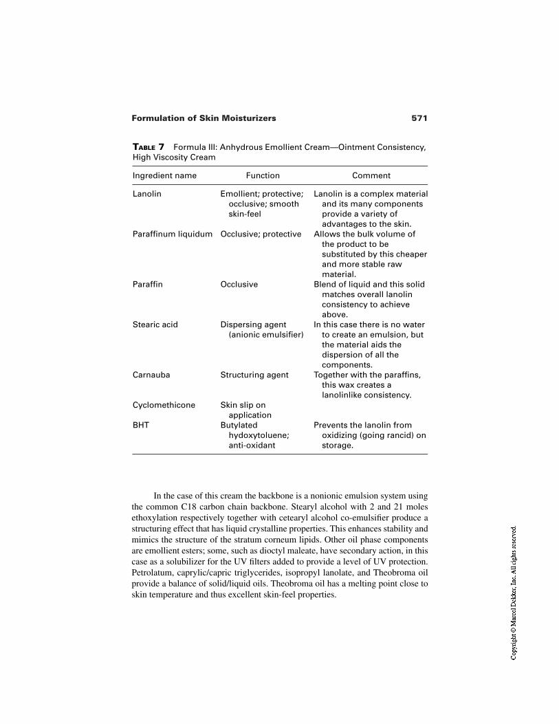
571Formulation of Skin Moisturizers
TABLE 7 Formula III: Anhydrous Emollient Cream—Ointment Consistency,High Viscosity Cream
Ingredient name Function Comment
Lanolin Emollient; protective;occlusive; smoothskin-feel
Lanolin is a complex materialand its many componentsprovide a variety ofadvantages to the skin.
Paraffinum liquidum Occlusive; protective Allows the bulk volume ofthe product to besubstituted by this cheaperand more stable rawmaterial.
Paraffin Occlusive Blend of liquid and this solidmatches overall lanolinconsistency to achieveabove.
Stearic acid Dispersing agent(anionic emulsifier)
In this case there is no waterto create an emulsion, butthe material aids thedispersion of all thecomponents.
Carnauba Structuring agent Together with the paraffins,this wax creates alanolinlike consistency.
Cyclomethicone Skin slip onapplication
BHT Butylatedhydoxytoluene;anti-oxidant
Prevents the lanolin fromoxidizing (going rancid) onstorage.
In the case of this cream the backbone is a nonionic emulsion system usingthe common C18 carbon chain backbone. Stearyl alcohol with 2 and 21 molesethoxylation respectively together with cetearyl alcohol co-emulsifier produce astructuring effect that has liquid crystalline properties. This enhances stability andmimics the structure of the stratum corneum lipids. Other oil phase componentsare emollient esters; some, such as dioctyl maleate, have secondary action, in thiscase as a solubilizer for the UV filters added to provide a level of UV protection.Petrolatum, caprylic/capric triglycerides, isopropyl lanolate, and Theobroma oilprovide a balance of solid/liquid oils. Theobroma oil has a melting point close toskin temperature and thus excellent skin-feel properties.
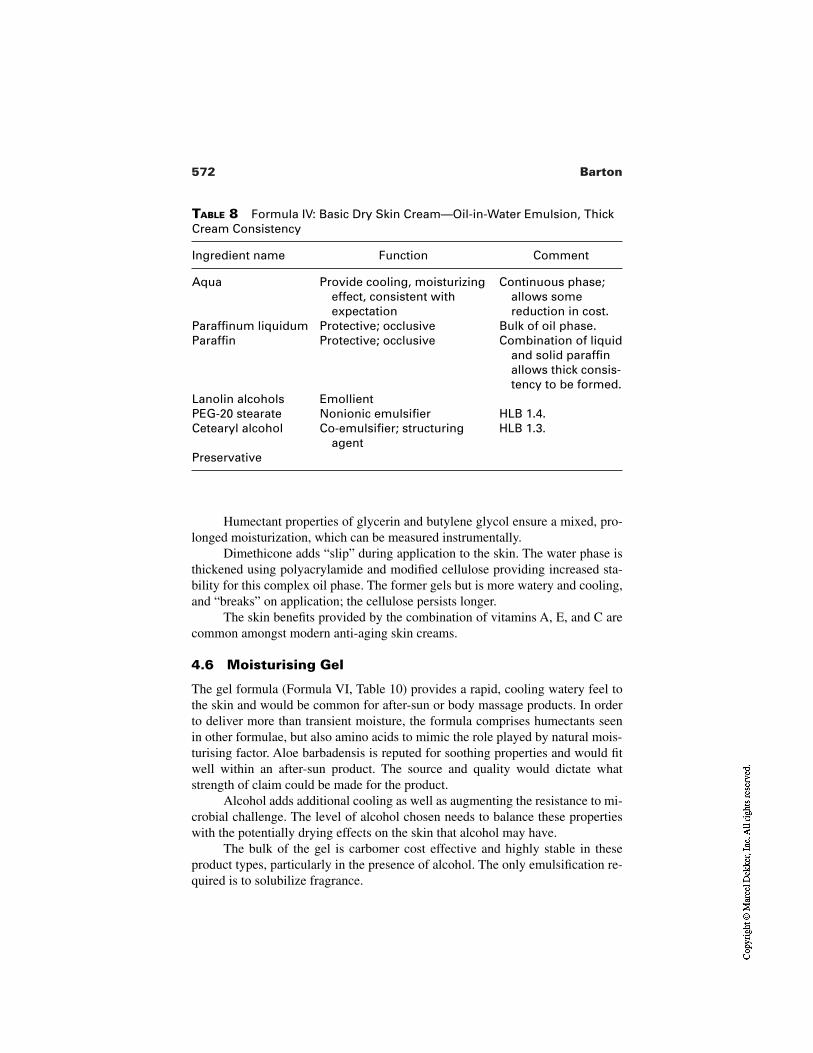
572 Barton
TABLE 8 Formula IV: Basic Dry Skin Cream—Oil-in-Water Emulsion, ThickCream Consistency
Ingredient name Function Comment
Aqua Provide cooling, moisturizingeffect, consistent withexpectation
Continuous phase;allows somereduction in cost.
Paraffinum liquidum Protective; occlusive Bulk of oil phase.Paraffin Protective; occlusive Combination of liquid
and solid paraffinallows thick consis-tency to be formed.
Lanolin alcohols EmollientPEG-20 stearate Nonionic emulsifier HLB 1.4.Cetearyl alcohol Co-emulsifier; structuring
agentHLB 1.3.
Preservative
Humectant properties of glycerin and butylene glycol ensure a mixed, pro-longed moisturization, which can be measured instrumentally.
Dimethicone adds “slip” during application to the skin. The water phase isthickened using polyacrylamide and modified cellulose providing increased sta-bility for this complex oil phase. The former gels but is more watery and cooling,and “breaks” on application; the cellulose persists longer.
The skin benefits provided by the combination of vitamins A, E, and C arecommon amongst modern anti-aging skin creams.
4.6 Moisturising Gel
The gel formula (Formula VI, Table 10) provides a rapid, cooling watery feel tothe skin and would be common for after-sun or body massage products. In orderto deliver more than transient moisture, the formula comprises humectants seenin other formulae, but also amino acids to mimic the role played by natural mois-turising factor. Aloe barbadensis is reputed for soothing properties and would fitwell within an after-sun product. The source and quality would dictate whatstrength of claim could be made for the product.
Alcohol adds additional cooling as well as augmenting the resistance to mi-crobial challenge. The level of alcohol chosen needs to balance these propertieswith the potentially drying effects on the skin that alcohol may have.
The bulk of the gel is carbomer cost effective and highly stable in theseproduct types, particularly in the presence of alcohol. The only emulsification re-quired is to solubilize fragrance.
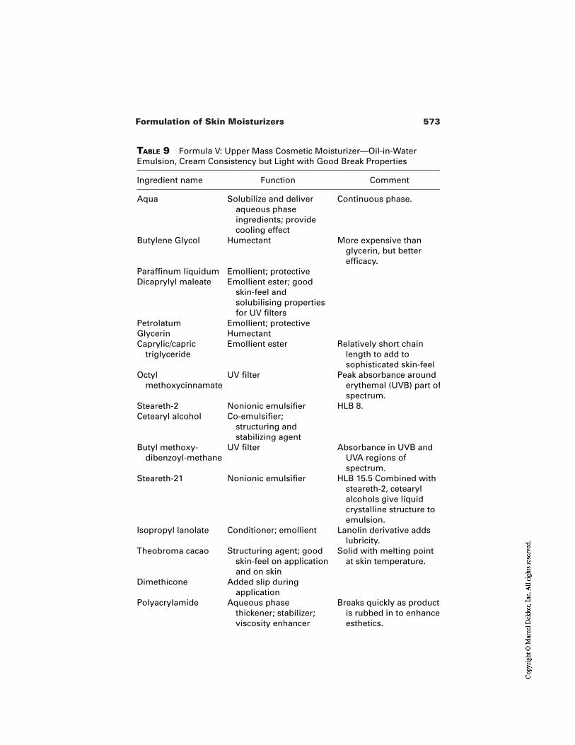
573Formulation of Skin Moisturizers
TABLE 9 Formula V: Upper Mass Cosmetic Moisturizer—Oil-in-WaterEmulsion, Cream Consistency but Light with Good Break Properties
Ingredient name Function Comment
Aqua Solubilize and deliveraqueous phaseingredients; providecooling effect
Continuous phase.
Butylene Glycol Humectant More expensive thanglycerin, but betterefficacy.
Paraffinum liquidum Emollient; protectiveDicaprylyl maleate Emollient ester; good
skin-feel andsolubilising propertiesfor UV filters
Petrolatum Emollient; protectiveGlycerin HumectantCaprylic/capric
triglycerideEmollient ester Relatively short chain
length to add tosophisticated skin-feel
Octylmethoxycinnamate
UV filter Peak absorbance arounderythemal (UVB) part ofspectrum.
Steareth-2 Nonionic emulsifier HLB 8.Cetearyl alcohol Co-emulsifier;
structuring andstabilizing agent
Butyl methoxy-dibenzoyl-methane
UV filter Absorbance in UVB andUVA regions ofspectrum.
Steareth-21 Nonionic emulsifier HLB 15.5 Combined withsteareth-2, cetearylalcohols give liquidcrystalline structure toemulsion.
Isopropyl lanolate Conditioner; emollient Lanolin derivative addslubricity.
Theobroma cacao Structuring agent; goodskin-feel on applicationand on skin
Solid with melting pointat skin temperature.
Dimethicone Added slip duringapplication
Polyacrylamide Aqueous phasethickener; stabilizer;viscosity enhancer
Breaks quickly as productis rubbed in to enhanceesthetics.
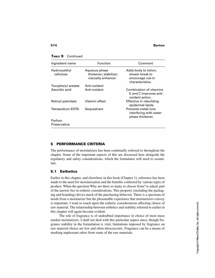
574 Barton
TABLE 9 Continued
Ingredient name Function Comment
Hydroxyethylcellulose
Aqueous phasethickener; stabilizer;viscosity enhancer
Adds body to lotion;slower break toencourage rub-incharacteristics.
Tocopheryl acetate Anti-oxidantAscorbic acid Anti-oxidant Combination of vitamins
E and C improves anti-oxidant action.
Retinyl palmitate Vitamin effect Effective in rebuildingepidermal lipids.
Tetrasodium EDTA Sequestrant Prevents metal ionsinterfering with waterphase thickener.
ParfumPreservative
5 PERFORMANCE CRITERIA
The performance of moisturizers has been continually referred to throughout thechapter. Some of the important aspects of this are discussed here alongside theregulatory and safety considerations, which the formulator will need to assimi-late.
5.1 Esthetics
Earlier in this chapter, and elsewhere in this book (Chapter 1), reference has beenmade to the need for moisturization and the benefits conferred by various types ofproduct. When the question Why are there so many to choose from? is asked, partof the answer lies in esthetic considerations. This property (including the packag-ing and branding) drives much of the purchasing behavior. There is a spectrum ofneeds from a moisturizer but the pleasurable experience that moisturizers conveyis important. I want to touch upon the esthetic considerations affecting choice ofraw material. The relationship between esthetics and stability referred to earlier inthis chapter will again become evident.
The role of fragrance is of undoubted importance in choice of most massmarket moisturizers. I shall not deal with this particular aspect since, though fra-grance stability in the formulation is vital, limitations imposed by fragrance onraw material choice are few and often idiosyncratic. Fragrance can be a means ofmasking unpleasant odors from some of the raw materials.
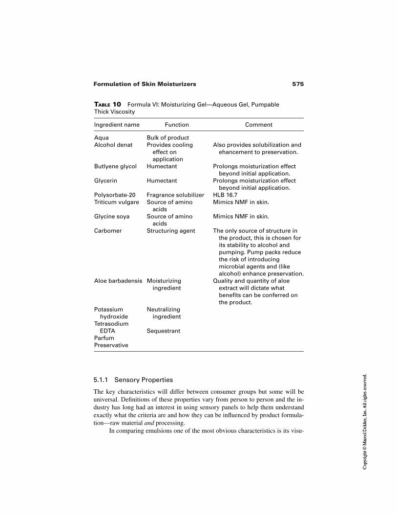
575Formulation of Skin Moisturizers
TABLE 10 Formula VI: Moisturizing Gel—Aqueous Gel, Pumpable Thick Viscosity
Ingredient name Function Comment
Aqua Bulk of productAlcohol denat Provides cooling
effect onapplication
Also provides solubilization andehancement to preservation.
Butlyene glycol Humectant Prolongs moisturization effectbeyond initial application.
Glycerin Humectant Prolongs moisturization effectbeyond initial application.
Polysorbate-20 Fragrance solubilizer HLB 16.7Triticum vulgare Source of amino
acidsMimics NMF in skin.
Glycine soya Source of aminoacids
Mimics NMF in skin.
Carbomer Structuring agent The only source of structure inthe product, this is chosen forits stability to alcohol andpumping. Pump packs reducethe risk of introducingmicrobial agents and (likealcohol) enhance preservation.
Aloe barbadensis Moisturizingingredient
Quality and quantity of aloeextract will dictate whatbenefits can be conferred onthe product.
Potassiumhydroxide
Neutralizingingredient
TetrasodiumEDTA Sequestrant
ParfumPreservative
5.1.1 Sensory Properties
The key characteristics will differ between consumer groups but some will beuniversal. Definitions of these properties vary from person to person and the in-dustry has long had an interest in using sensory panels to help them understandexactly what the criteria are and how they can be influenced by product formula-tion—raw material and processing.
In comparing emulsions one of the most obvious characteristics is its visu-
576 Barton
al impact. Bright, white, shiny emulsions are achieved with good processing andsmall particle size. Earlier in the chapter the role of emulsifiers has been dis-cussed. Some stabilizers, such as higher molecular weight waxes, can inducesome graininess and crystallike appearance, especially if heating and cooling hasnot been well controlled. Use of water phase thickeners and stabilizers can inducea watery whiteness. A high degree of order, as exhibited in liquid crystallineemulsions, can confer a translucent appearance.
In order to understand these properties expert panels can be used. Thesecomprise persons with a high degree of sensory perception. By recognizing theskill and developing it by suitable training the panel can detect and reproduciblydescribe in a quantitative manner many of the attributes that consumers demandof the product. In doing so they are acting in a similar way to instrumental analy-sis, providing numeric information with which to compare many different proper-ties from a given set of products. Ultimately the measures are comparative, andthe use of benchmarks is essential to make any comparison between sets of data.
Sensory testing can only be a test under ideal conditions, and relating this toin-use conditions is another essential part of understanding product esthetics.Factors that must be borne in mind include the variations in skin site (e.g., faceversus body). Even on the face the underlying bone and musculature will varywithin individuals and with other factors such as age. Thus formulations targetedat older facial skin types will first need to consider the sensory properties pre-ferred by the target group prior to testing these using expert sensory panels. Thefinal stage will be to confirm the result in a large number of the target population.
Results from product comparisons using sensory analysis will provide anunderstanding of the qualitative differences between products (see Fig. 4). Con-verting these findings into quantitative changes in the raw material components ofthe formulation will require skill and experience from the formulator. Like manyother factors in formulation technology, changing one component rarely results ina single change in one product characteristic. As an example of this I shall brieflytouch on some of the sensory properties that may be exhibited in a product andshow how they may be altered.
Rub-in. This property can be described as the ease with which a productdisappears on application. It will be a function of several things includ-ing initial viscosity, viscosity change under shear, compatibility of oilphase with the skin, the amount of water, and whether the water is thecontinuous or dispersed phase. Improving rub-in of Formula I (Table 5)could be achieved in several ways. Increasing the aqueous phase thick-ener concentration whilst reducing the cetyl alcohol will maintain theviscosity and decrease the rub-in. Increasing the ratio of caprylic/caprictriglyceride to liquid paraffin in Formula I, or increasing the ratio ofC12–15 alkyl benzoate to cetyl stearate and or lanolin alcohols in For-
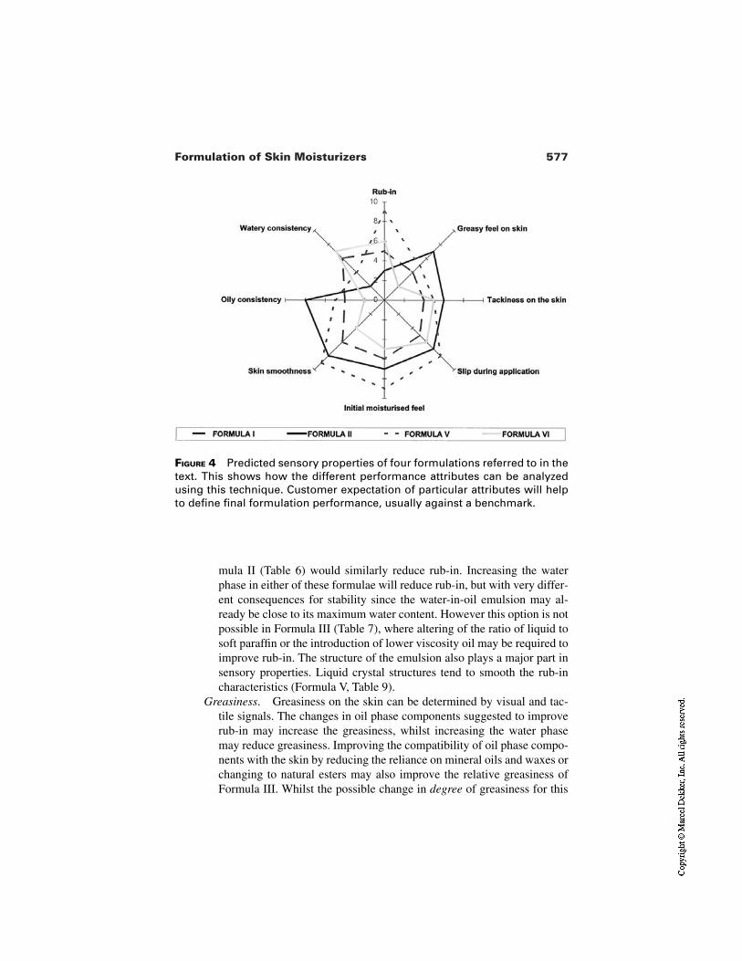
577Formulation of Skin Moisturizers
FIGURE 4 Predicted sensory properties of four formulations referred to in thetext. This shows how the different performance attributes can be analyzedusing this technique. Customer expectation of particular attributes will helpto define final formulation performance, usually against a benchmark.
mula II (Table 6) would similarly reduce rub-in. Increasing the waterphase in either of these formulae will reduce rub-in, but with very differ-ent consequences for stability since the water-in-oil emulsion may al-ready be close to its maximum water content. However this option is notpossible in Formula III (Table 7), where altering of the ratio of liquid tosoft paraffin or the introduction of lower viscosity oil may be required toimprove rub-in. The structure of the emulsion also plays a major part insensory properties. Liquid crystal structures tend to smooth the rub-incharacteristics (Formula V, Table 9).
Greasiness. Greasiness on the skin can be determined by visual and tac-tile signals. The changes in oil phase components suggested to improverub-in may increase the greasiness, whilst increasing the water phasemay reduce greasiness. Improving the compatibility of oil phase compo-nents with the skin by reducing the reliance on mineral oils and waxes orchanging to natural esters may also improve the relative greasiness ofFormula III. Whilst the possible change in degree of greasiness for this
578 Barton
formula may appear to be small in comparison to the lotion in Formula I,sensory analysis (comparing like with like) may show where improve-ments have been achieved. Smaller particle size o/w emulsions will alsohave reduced perception of greasiness.
Tackiness. Although self-explanatory, this property can manifest itselfduring or after application to the skin and can stem from oil or waterphase components. Tacky oils include lanolins, cetyl and cetearyl alco-hols, and high viscosity modified silicones. Water phase componentssuch as proteins, panthenol, and some aqueous phase thickeners willproduce a tacky feeling if used in too high a concentration. Thus theranges in which these can be used to improve stability are limited bytheir adverse effects on sensory properties. Anhydrous systems such asFormula III will have an inherent tackiness during application; thiscould be reduced by changing the ratios of higher and lower meltingpoint components or increasing the level of silicone. However “oil-free”moisturizers are not without tackiness. Polyols and proteins added toprovide substantive moisturization (beyond the transient addition of wa-ter to stratum corneum) can confer some tacky after-feel to the skin ifused at inappropriate levels.
Slip. This expresses how the product glides across the skin. In some waysthis is related to “break” (see following). The lubricity of a product canbe highly pleasurable and an indicator of the “premium” position of aproduct. Likewise a moisturizer for compromised skin will require a de-gree of slip in order to prevent putting a dry or friable skin under unduestress during application. Esters are among the best sources of slip in aformulation, but mineral oils and silicones are also important. Wheremassage is an important attribute of the product in-use (e.g., ante-natalbody creams), slip might need to be balanced against provision of someresistance to encourage more rub-in (e.g., shear thickening properties ofFormula II, Table 6).
Break. This can be seen as the initial slip and is more a descriptor of theformulation than its action on the skin. A thick, gel-like cream may actu-ally contain relatively low oil phase and very quickly break down underthe stress of rubbing in. Such systems need to be stable to other forms ofstress (e.g., pumping), and the presence of salts on the skin helps to de-structure the polymer matrix and achieve the break. Such products whenplaced on the skin without any rubbing can sometimes be seen to meltand flow across the skin as the gel structure breaks down. Certain emul-sifiers can also impart this property.
Moisturized after-feel. This is obviously a highly important factor for amoisturizer. However there are other sensory characteristics contribut-ing to this, e.g., softness, smoothness, suppleness. To understand their
579Formulation of Skin Moisturizers
relative importance, further breakdown of the sensory properties may berequired if the consumer group targeted actually wants a specific mois-turizing effect. However for basic moisturizers satisfying moisturizedafter-feel may be sufficient. The formulation choices will depend in parton which of these different needs are to be fulfilled.
It is important to point out that these sensory descriptors and any others thatmay be generated for a particular product are of no inherent value without an un-derstanding of their contribution to consumer expectation or demand. Thus a highlevel of tackiness may be a negative influence in formulating a premium moistur-izer, but neutral or of no importance for a therapeutic dry skin cream. It is also im-portant to understand the role of fragrance and minimize its impact in comparingproducts by using unfragranced samples or masking the sensory panel’s sense ofsmell in some way. Understanding the relationship between sensory characteris-tics and consumer expectation can be difficult for innovative products, but for ex-isting product types the use of a benchmark will help define the key parametersthat will drive choice of formulation.
It is also clear that there are an enormous number of raw material combina-tions that will achieve a required sensory profile. Thus different combinationsmay provide sensory properties so similar that they are beyond perception.
5.1.2 Rheology
The process of rubbing a product into the skin involves placing the product undershear, and the sensory performance of products under these conditions are key toour understanding of the consumer acceptability. For this reason product rheolo-gy is studied in an attempt to produce objective instrumental data on product be-havior during shear. This has the added benefit of assisting understanding of prod-uct performance during processing, packing, and delivery.
Under increased shear, a proportional linear increase in the induced stress(Newtonian flow) is rarely seen in emulsions. Usually the response will exhibitsome form of curve describing viscoelastic behavior. Where increasing shear ratecauses an initially steep but then plateau response in stress, the flow is pseudo-plastic. This would be typical for a rich cream that resisted initial massage butthen flowed into the skin. When increasing shear rate produces an initially lowshear stress response which then rises exponentially, the flow is dilatant. Thiswould be typical for a lotion which flowed but then thickened as the water disap-peared into the skin and the mixture became thicker in texture.
5.1.3 Ensuring Continued Product Use
From a therapeutic or commercial standpoint this is an important factor since aproduct will be best used if it is pleasant, easily absorbed, and well tolerated inuse. For therapeutic moisturizers the skin-feel during application may be less im-
580 Barton
portant than the skin texture after application, and skin comfort will probablyoverride both. Skin texture will be important for users of cosmetic products butless important than elegance on application and skin residue. The role of raw ma-terials in providing these sensory properties is vital, as is to a lesser extent howthe product is processed prior to sale.
In assessing the compliance factors and the trade-offs a consumer maymake in approving a product, the formulator will need to use a number of meth-ods. The sophistication of the method will vary depending on the cost of getting itwrong. Large-scale consumer testing using identified target consumers will bringbetter information but is not a useful screening method for candidate formula-tions. There exists an array of methods that can help the formulator refine and/orselect the final product. In many of the cases it is the qualities of the formulationthat are under test rather than the total product (pack, design, claims, etc.).
5.2 Regulatory Influence
These issues are covered elsewhere in this book, but it is worth noting their im-portance in driving choice in formulation components, testing, and delivery. Theproduct must fulfill consumer expectation not only from a legal standpoint, butalso from commercial necessity. Understanding the formulation constraints of(say) α-hydroxyacids is as important from a technical standpoint as a legal one ifsafety and efficacy are to be delivered.
5.2.1 Interaction of Product with the Skin
It is clear that combining the complexity of skin biochemistry, in particular that ofthe stratum corneum, with the complex chemistry of moisturizer formulation, inparticular emulsification, produces a system with little in the way of predictivebehavior. Deleterious effects of raw materials can be detected in simple systems,but once formulated the picture becomes less clear. The product developer has thetask of assessing raw material performance (including safety) from a number ofsources, including previous experience. In light of this, limitations on new rawmaterial introduction and product innovation need to be balanced against issuesof ethics and safety.
The formulator must design for safety (and efficacy a close second!) fromthe bottom up. Some authors have suggested that long-term use of emulsions willadversely affect the skin [8], and routine testing of skin tolerance should ideallybe achieved using more than a single use. It has also been suggested that emulsi-fier choice contributes to the skin intolerance often attributed to other formulationcomponents such as fragrance or preservative [9].
Whilst the relative risk here is probably low, future developments in tech-nology and consumer expectation of moisturizer formulations will undoubtedlyinvolve continued tailoring of the emulsifiers to the skin.
581Formulation of Skin Moisturizers
Elsewhere in this chapter the need for preservatives is discussed. There isincreasing interest in the prevalence of skin hypersensitivity to particular preser-vatives. This is also the case for sunscreens and fragrances. The product develop-er will be aware of these issues and prior to inclusion in a formulation will assessthe rationale for incorporating any raw material with a likely sensitization risk.Consideration will be given to target customer (e.g., infants, people with sensitiveskin), conditions of use (where on skin, how often, etc.), use rate (daily, weekly),and geographical market (European, global). Product testing will also help to in-form a final assessment of the risk.
5.2.2 Interaction with the Skin and Ensuring Claims Substantiation
In the context of regulatory influence it is also true that the cosmetics industry hasother challenges. The increasing desire to control products appears to be drivenfrom several angles. One suggests that since cosmetic products are “trivial,” therisk/benefit analysis should err toward greater control of safety. More recently theincreased understanding of skin physiology has led to the belief that cosmeticscan have beneficial effects on skin physiology and products should reflect thesebenefits in claims on the packaging. This has again resulted in a belief that controlalong the lines of drug status or “cosmeceuticals” is desirable (see Chapters 16and 28).
The challenge for the product developer is to ensure that the claims remainwithin the legal definition of a cosmetic. Ensuring that the product actually deliv-ers a claim is also vital, since claims are increasingly seen as the means of prod-uct differentiation. This is especially important where ingredients referred to onthe label are used to justify or help deliver claims. Whilst this is a legislative issuethe formulator (and marketer) should consider this high on the list of productspecification, since not delivering a claim can be commercially disastrous. Indus-try guidelines on claims do exist but since the market moves very quickly it isimpossible to define a particular test to support a given claim. The EuropeanGuidelines propose an approach to claims substantiation rather than prescribedmethods [5].
It is important to return to the fact that skin moisturizers now encompassmany products that do more than moisturize (see Chapter 1). Skin lightening,anti-aging, sun protection, and cell renewal are all claimed to be delivered from adaily moisturizer. Much of the rest of this book is dedicated to understanding theunderlying skin physiology and some of the means of influencing this. Here I willbriefly touch on issues that exercise the formulator’s thoughts in designing mois-turizers with “actives.’’
The formulator is often first confronted by data from suppliers of candidateraw materials. In previous times such data may have been from animal studies,which were sometimes performed irrespective of ethical factors and were of lim-
582 Barton
ited value in scientific interpretation. Today data from in vitro tests is frequentlyprovided.
The formulator needs to understand the limitations of the test(s), the influ-ence of formulations used in these tests, particularly where cells grown in aque-ous media are used, and the relevance to human in vivo exposure. The develop-ment of three-dimensional, fully differentiated human epidermal skin models ofreproducible quality has gone some way to assisting the formulation scientist inthis task [10].
With these factors taken into account, there is often a residual question tobe answered. How much of raw material X do I need to achieve the desired ef-fect? The response is to work through the available data in order to choose a lev-el of ingredient that is both safe and efficacious prior to testing the final product inhuman volunteers to assess if a claim is measurable and perceivable. (See thisvolume Chapters by Jaret; Grove; Pierard).
5.2.3 Fitting Performance to Skin Physiology
This aim would appear to be sensible in order to maintain the safety and perfor-mance expectations of the consumer. However, even where a case appearsstraightforward, there can be complex issues that influence the formulator’s finaldecision. The case of UV protection in daily moisturizers is an example.
The influence of ultraviolet light exposure from the sun on premature skinaging (photoaging) is well accepted though compounded by other factors [11].The role of short wavelength ultraviolet light (UVB) in burning the skin is alsoaccepted, but the relationship between frequency and intensity of burning andpremature skin aging has not been defined. What is clear is that those who haveoutdoor occupations show higher degrees of photoaging than those with indooroccupations.
The role of longer wavelength ultraviolet light (UVA) in burning is approx-imately 1000 times less than UVB, but there is still damage caused by increasedexposure to UVA alone. This has been termed aging damage, though both UVBand UVA are involved.
Sun protection factors (SPF) can be derived for products using acceptedand standardized human test methods to deliver protection against burning. How-ever the protection is not complete; even SPF 60 products allow 1.7% of the burn-ing UV rays to get through to the skin. The raw materials used as UV filters in theformulation rarely act like a neutral density filter. Therefore the burning UVBwavelengths may be absorbed, protecting against burning without absorbing thelonger wavelengths to the same efficiency. Therefore there may be a risk of over-exposing the skin to the longer wavelengths by use of these products. This has ledto the use of other measures of assessing sun protection against longer wave-length ultraviolet (UVA) penetration.
583Formulation of Skin Moisturizers
The consumer need for SPF numbers is protection against burning, and theproduct is usually applied when deliberate, recreational exposure is anticipated,e.g., whilst at the beach. Daily moisturizers are not used globally as protectionagainst burning, and only in markets where the occasional, accidental exposure toUV constitutes a threat of burning is the SPF number really relevant.
Where the consumer need is protection against premature aging the role ofthe SPF number has become the consumer short-hand. In the absence of anythingbetter, the message has become “sun protection is important therefore SPF num-bers are important.’’
This message has been given great support by dermatologists and the beau-ty press, and as a consequence we find ourselves using SPF numbers (possibly in-appropriately) to help deliver a consumer need. We cannot guarantee what levelof SPF is appropriate since we do not have the direct supporting evidence, butSPF 15 has become the recommendation for protection against UV in daily skincare based on the opinion of a number of experts. Discussion of the value of thisnumber and the basis for this recommendation has been overtaken by events.
Perhaps more importantly there are other considerations for the formulator.A once-per-day application of a moisturizer claiming SPF 15 would require that itdeliver this level of protection throughout the day. Beach use products normallyinclude advice on pack to reapply ever hour or two. In terms of consumer expec-tation, the package wording needs consideration. Neither is it easy to formulatean SPF 15 moisturizer with high quality esthetics; the UV filter loading requiredproduces heavy and greasy skin-feel. From a safety standpoint, there is also theissue of exposure to UV filters. The risk of contact sensitization has been raisedthough not quantified. The safety assessments made on UV filters used exposuresbased on infrequent holiday use rather than daily exposure via a moisturizer. Thuseven with an active with a known action, physiological endpoint, and standardtest methods, there are difficulties for the formulator to overcome.
6 SUMMARY
Formulation of a moisturizer requires an understanding of technical and commer-cial factors and the restrictions that they can impose. As discussed in Chapter 1,many new moisturizers launched on the market do not survive. The overall suc-cess can depend on such factors as promotion and professional support, whichthough they are outside the developer’s control should be part of the early plan-ning. Understanding how the various factors interact is as important as under-standing the physicochemical interactions within the formulation. Delivering astable, safe, and esthetically pleasing product that meets consumer expectation isthe aim of all product developers. This chapter has identified some of the techni-cal elements that go toward supporting successful product development.
584 Barton
ACKNOWLEDGMENTS
The author would like to thank Ed Owen, Claire Wilson, Patrick Love, and NickiLenton for their help in preparing this manuscript.
REFERENCES
1. Hunting ALL. A Formulary of Cosmetic Preparations. Vol. 2: Creams, Lotions andMilks. Micelle Press, 1993.
2. Cosmetic Bench Reference. Cosmet Toil 2001; 115(13).3. Wenninger JA, Canterberry RC, McEwen GN, eds. International Cosmetic Ingredi-
ent Dictionary and Handbook. 8th ed. The Cosmetic, Toiletries and Fragrance Asso-ciation, 2000.
4. Prottey C, Hastop PJ, Press M. Correction of the cutaneous manifestations of essen-tial fatty acid deficiency in man by application of sunflower seed oil to the skin. J In-vest Dermatol 1975; 64:228–234.
5. Guidelines for the Evaluation of the Efficacy of Cosmetic Products. 2nd ed. The Eu-ropean Cosmetic, Toiletry and Perfumery Association, 2001.
6. Hubbard AW, Moore LJ, Clothier RH, Sulley H, Rollin KA. Use of in-vitro method-ology to predict the irritancy potential of surfactants. Toxic In-Vitro 1994; 8(4):689–691.
7. Efficacy of anti-microbial preservation. Sec. 5.1.3. In: European Pharmacopoeia. 3rded. Supplement 2001. European Directorate for Quality of Medicines Within Coun-cil of Europe, Strasbourg.
8. Held E, Sveinsdottir S, Agner T. Effect of long term use of moisturisation on skinhydration, barrier function and susceptibility to irritants. Acta Derm Venereol 1999;79(1):49–51.
9. Maes D, Declercq L, Muizzuddin N, Fthenakis C, McKeever MA, Collins D, Mam-mone T, Dicanio D, Marenus K. Optimisation of safety testing for better product de-velopment. Proceedings of 21st IFSCC Congress, Berlin, 2000, pp. 321–327.
10. Rognet R, Tessoneaud E, Gagne C, Teissler MH, Cohen C, Leclaire J. Use of stan-dardised reconstructed epidermis kit to assess in-vitro the tolerance and efficacy ofcosmetics. Int J Cosmet Sci 2000; 22(6):409–419.
11. Malvy DJM, Guinot C, Preziosi P, Valliant L, Tenenhans M, Galan P, Hercberg S,Tschachler E. Epidemiologic determinants of skin photo ageing: baseline data of theSU.VI.MAX cohort. J Am Acad Dermatol 2000; 42(1):47–55.
26Formulation and Assessment of Moisturizing Cleansers
David C. Story and Frederick Anthony SimionThe Andrew Jergens Company, Cincinnati, Ohio
1 INTRODUCTION
Very few types of cleansing products have the ability to truly moisturize the skin,either by enhancing the residual moisture content of the stratum corneum, remov-ing dry skin flakes, or increasing skin elasticity. Many cleansing products claim tobe, or are trademarked as, moisturizing cleansers but during typical use they donot deliver these benefits. The objectives of this chapter are to provide an expla-nation of what constitutes a moisturizing cleanser in today’s market and outlinemethods of measuring the benefits.
The most common and economical form for cleansing is bar soap. Soap isthe comparative standard for all personal cleansers. If a product is milder, moremoisturizing, less drying, etc., it is always milder than soap, more moisturizingthan soap, and less drying than soap.
Simplistically, soap is the sodium and/or potassium salt of a fatty acid.When sodium potassium soaps are used with conventional water, the divalent andtrivalent metallic ions will exchange with the Na+ or K+ ion to form multivalentsalts that are insoluble. The multivalent metal soaps readily precipitate on the skinand any other available surface. These insoluble soaps are referred to as lime
585
586 Story and Simion
soaps or soap scum. This phenomenon is important since it affects the sensoryproperties of cleansing products. The scum deposition is not observed with thesynthetic detergents used in moisturizing cleansers.
The pH of toilet soaps is approximately 10 [1]. This high pH and its cleans-ing properties promote the loss of water and water-binding materials from thestratum corneum. These characteristics establish the basic precepts for moisturiz-ing cleansers. By substituting different surfactants for all or part of the soap andlowering the product pH closer to physiologic values, the product becomes lessdisruptive to the skin barrier, and therefore less residual water is lost. The firstproducts to apply these precepts were combination bars that used a mixture ofsodium soap and synthetic detergents. These products were shown to be less dry-ing than conventional soap bars and subsequently became thought of as moistur-izing cleansers. The most common synthetic detergent added to bars is isethion-ate.
2 FORMULATION
Isethionates were one of the first surfactants used in the moisturizing category.This class of surfactants is a monovalent metallic salt of an alkyl carboxyethanesulfonate. Their properties make them ideal for solid cleansers. They possess ahigh melting point and are stable when added to soap bars. Isethionates are effec-tive dispersants for lime soaps and thus decrease the surface deposition of soapscum. Soap bars containing isethionate are stable at a pH of 7.
The solid cleansing products have now given way to the liquid cleansers,which are less drying than the combination bars and allow for a high degree offlexibility in formulation and the delivery of new benefits. The liquid cleansersare essentially the same formulations as today’s hair shampoos. The approach forformulating moisturizing cleansers will be detailed in the following sections.
2.1 Ingredients
2.1.1 Surfactants
The surfactants are classified usually by the inherent charge associated with thesurface-active moiety of the molecule. The surfactant classes are anionic (nega-tive charge), cationic (positive charge), zwitterionic (both positive and negativecharges), and nonionic (no charge).
Primary. The products are usually assembled with a mixture of detergenttypes. The primary detergents are almost always anionic. The alkyl sulfates maybe included as the primary detergent for their lather characteristics and durabilityin the presence of soil loads. They are inexpensive but are much too irritating tothe skin to be the only surfactant in a cleansing product. The linear alkylbenzenesulfonates have also been used as the primary detergents. Although as a class they
587Moisturizing Cleansers
are milder than the alkyl sulfates, they are considered skin irritants. The mostprevalent anionic surfactant used in moisturizing products today is the alkyl ethersulfates. This class of detergents usually has a C12 alkyl group (lauryl) and 2 to 3moles of ethylene oxide (ethoxylation) to form the ether linkage of the molecule.If lower degrees of ethoxylation are used, the surfactant will produce better foam-ing characteristics and have lower water solubility, higher propensity for irritatingor drying skin, and higher Krafft temperatures. The 2–3 moles of ethoxylationseem to be the best balance at cleansing, foaming, and mildness. Longer carbonchains will produce some lather but at a much lower volumes and more densewhen compared to the C12 alkyl chain.
Other anionic surfactants have been used as the primary cleanser but usual-ly have some disadvantage when compared to sodium laureth sulfates, e.g.,monoalkylphosphates, ammonium isethionates, and lauryl or cocoyl sarcosinates.Primary surfactants can be combined to achieve the ideal balance between mild-ness and lather characteristics (volume and durability). The primary surfactantsare commercially available with various counterions. The counterion can affectthe solubility, formula compatibility, and overall mildness. Na+, K+, and Mg2+ arethe more common counterions seen in today’s moisturizing liquid cleansing prod-ucts. The active concentration for the primary surfactants is typically in a range of8–20 w/w%.
Co-Surfactants. The co-surfactants are added to moisturizing liquidcleansers for a multitude of reasons. If co-surfactants were used as the primarydetergent, they would be inferior by consumer standards for lather volume anddensity. Consumers typically would rate the products as diluted, weak, and ineffi-cient. When the co-surfactant is combined with the primary anionic detergent, theformulation will produce results that increase consumer acceptance and are lessdestructive to the stratum corneum barrier.
The usual co-surfactants are anionic or zwitterionic. In some specializedproducts, nonionics will be used to decrease the harshness of the cleanser to skinand eyes. The nonionic co-surfactants are generally found in products marketedfor children or babies. Adding nonionics to liquid moisturizing cleansers is achallenge since they suppress lather volume and durability. In many moisturizingliquid cleansing compositions, two or three co-surfactants will be used. The zwit-terionics are more prevalent due to their various properties. Zwitterionics dimin-ish the harshness of the primary surfactants and have a propensity to adsorb to theskin, thus providing some conditioning benefit. The most common zwitterionicsare imidazoline, amino acid, and betaine derivatives. The inherent charge of thezwitterion surfactant molecule is pH dependent. Adjusting the pH to an acidic orbasic range can control the character of the composition. The amphoteric (orzwitterionic) detergents will as a rule produce a better quality of lather at basic pHsince they assume a negative charge (anionic). When the pH of the formula is
588 Story and Simion
acidic, the amphoteric surfactant becomes cationic (positively charged) and pro-duces a conditioning effect since it is now substantitive to skin or hair. It is welldocumented that irritation and eye stinging can be significantly reduced by theaddition of amphoterics to conventional cleansing systems [2].
The newest class of co-surfactants is the polyglucosides. This group of de-tergents is formed from the reaction of glucose and fatty alcohols. The polygluco-sides are mild and have a low potential for irritation. They are nonionic in charac-ter and possess better foam properties than the amphoteric class of detergents.They are not well adsorbed by skin and therefore have no conditioning effect. Theapplication of polyglucosides is detailed in the referenced patent [3].
One of the mildest anionic co-surfactant classes is the sodium alkyl sulfos-uccinates or alkyl ether sulfosuccinates. Sulfosuccinates are restricted to a narrowformula pH of 4 to 6 because they are prone to base catalyzed hydrolysis.
The amino acid–derived co-surfactants include the sarcosinates, propi-onates, glutamates, and taurates. The mildness and conditioning effects of thesedetergents are well documented. They are often included in specialized hairshampoos and moisturizing body cleansers.
The co-surfactants have the significant ability to influence the consumersensory properties of moisturizing cleansers. In general applications, the total ac-tive concentration for co-surfactants can range from as low as 2 w/w% and ashigh as 15 w/w%. The use concentration in moisturizing formulas is frequentlylimited by product economics.
2.1.2 Conditioning/Moisturizing Agents
This diverse group of materials provides the distinctive features of marketedmoisturizing cleansers. The discussion in this section has been limited to the morecommon ingredients. These agents are used for their effects on skin, whether theywould be physiological, sensory, or a combination of the two.
The deposition of these ingredients on skin is a true dichotomy since the in-tent of cleansing is to remove foreign material from the target surface. Promotingthe deposition of the conditioning or moisturizing agent while selectively remov-ing dirt is a significant challenge for the product formulator. The deposition of theselected materials must be highly efficient since only 5 to 15 g of product are usedduring the cleansing treatment. Considering, on average, an adult has 2 m2 ofskin, this calculates to be 0.75 mg cleansing product per square centimeter. Sincethe conditioning or moisturizing ingredient is usually less than 10% of the cleans-ing composition, and if 20% of this material would be adsorbed, the amount ofmaterial remaining on the skin is less than 15 µg/cm2. Another factor which can,and usually does, interfere with the deposition of these beneficial materials is theuse of an accessory, e.g., a washcloth, a polyethylene puff or pouf, or even asponge. Often the target material deposits or adsorbs to the accessory and very lit-tle is delivered to the skin. Finally, the most significant obstacle to skin deposition
589Moisturizing Cleansers
is the constant elution of the adsorbed agent by the rinse water. It becomes moreapparent that delivering an effective amount of the conditioner or moisturizer tothe skin is not a simple task and presents a true challenge to the technical formu-lator.
Lipids and Oils. If moisturizing or conditioning the skin were simplyadding a lipid or oil to the cleansing product, then this discussion would be brief.Simply adding these materials to a cleansing formula usually results in a productfailure. The lipophilic ingredients suppress the lathering characteristics and oftenlead to phase separation of the cleansing product. There must be a balance be-tween the detergent and conditioning ingredients in the formula. Ethoxylation ofthe oil or lipid will facilitate its incorporation into the formula, but if the materialis too soluble or too dispersible in water, it can easily be rinsed from the skin. An-other concern about the inclusion of an oil or lipid in the cleansing formula is thatthe lipophilic material may enhance the solubility of the skin lipids and facilitatetheir removal during treatment. There are commercial hard-surface cleaningproducts that have successfully applied the synergy of oil and detergents. Obvi-ously, skin is not a hardsurface and this is not a desired outcome for a moisturiz-ing cleansing product.
Another technology that is used to incorporate the lipid in the cleansingcomposition is structured surfactants. Albright and Wilson developed this tech-nology in the 1980s. A simple analogy of the technology is an onion [4]. Theonion is composed of multiple layers, in this case surfactant layers, and sand-wiched between the surfactants is the lipid ingredient. When this material is ap-plied during cleansing, the structure collapses when diluted (rinsing) and the lipidis deposited on the skin. The technology provides a significant lipid payload toskin.
The cleansing accessory must also be included in this discussion. With therapid commercialization of the polyethylene body pouf or sponge, cleansing for-mulas no longer needed high levels of surfactants. The open weave of the acces-sory provided a high number of nucleation sites for bubble or lather generation, inother words the device mechanically compensates for low lather volumes andslow lather-building systems. This allowed formulators to use higher percentagesof lipophilic ingredients and maintain consumer acceptability of the productwhen used with the accessory.
There are many examples of oils and lipids used in marketed moisturizingcleansers. Glycerides from vegetable oils, e.g., capric, cacprylic, and soybean,which are usually unsaturated or short carbon chains, are often added as the mois-turizing ingredient. Giret discloses the utility of some of these oils [5]. The unsat-urated or branched carbon chains have lower melting points and tend be liquid orsemisolid at room temperature, which is desirable for ease of incorporation intothe formula and product stability. Hydrocarbons, such as petrolatum or paraffin,
590 Story and Simion
have been used successfully in key moisturizing cleansing brands. The usual uselevel of these materials is 3–5 w/w%, but some products may contain as much as8–10 w/w%.
Polymers. This discussion will include synthetic and natural polymerssince both are used as conditioners in moisturizing cleansers. The majority of thepolymers has a cationic net charge and is substantive to skin. The molecularcharge is usually from a quaternized structure in which N+ is the cationic moiety.These large chains attach to the negative sites on the skin and create the sensoryeffect of smoothness and softness. Many of the polymers have a cellulosic or re-peating sugar backbone. The polymers must be hydrated, to facilitate their elec-trostatic attachment to the skin. The conditioning effect is transient since thepolymers eventually will dehydrate and be sloughed from the skin’s surface. Af-ter the polymers are fully hydrated, they are tolerant to the anionic detergentswithout flocculation or precipitation. This phenomenon will be discussed in moredetail later in the chapter. A few examples of these polymers are polyquaternium-10, polyquaternium-7, polyquaternium-11, and chitosan. The active polymer con-centration normally found in cleansing products is 0.1–1.0 w/w%.
Proteins. The original use of proteins in moisturizing cleansing productswas found in marketed hair shampoos. The proteins, which can be animal or plantderived, are usually hydrolyzed or chemically modified to improve their physicalproperties, such as odor, color, and their affinity for skin or hair. This discussionof proteins also includes polypeptides since these ingredients all have similarcharacteristics. They are somewhat substantive to areas of the skin where someprotein denaturation has occurred. These ingredients may also be cationic and at-tracted to the skin via the electrostatic forces. Proteins exhibit a sensory effectsimilar to the polymers, i.e., skin smoothness and softness. Their effect is alsotransient since these materials will also wear from the skin. The concern of usingproteins, especially animal derived, is the potential for developing sensitivity tothe material. The use level of proteins is relatively small due to material cost andthe adverse influence they can impart to the quality of the marketed cleanser. Theactive protein concentration is usually restricted to less than 1 w/w%.
Silicones. Silicones have been used as a conditioning or moisturizingagent in cleansing products for over a decade. One of the first applications of sil-icones in a cleansing product was reported by Bolich et al. [6]. He disclosed howto suspend the silicone gums in a detergent system (shampoo). Upon rinsing (di-lution) the silicone suspension is destabilized and the gum is uniformly depositedon the hair or skin. One of the inherent properties of silicones is to efficientlyspread and form single layers on a surface. The expectation would be the same forskin. The silicone spreads evenly on the skin surface providing a sensory effectand possibly forming a barrier to reduce water loss. The sensory effect is a
591Moisturizing Cleansers
smooth, powdery feeling on the skin. The challenge with using silicones is thefact they are known to be one of the most efficient antifoam materials available.In a personal cleansing product the lack of lather will make the product’s perfor-mance completely unacceptable. The most common approach for adding sili-cones to cleansing compositions is to suspend the material separately and then in-corporate this suspension into the detergent phase. The mixing of the two phasesuses minimal shear, which allows them to form a uniform macrosuspension, with-out physical interaction. The preferred range of silicone in a moisturizing cleans-ing product is 1–5 w/w% active.
2.1.3 Preservatives and Fragrances
These two classes of ingredients are not necessary for cleansing or moisturiza-tion, but are required to prevent spoilage and to drive consumer acceptance of theproduct. A variety of preservatives are available that have different types of actionretarding spoilage of the formula. Microbial spoilage is usually controlled with aformaldehyde donor, such as DMDM hydantoin, imidazolidinyl urea, diazo-lidinyl urea, methylchloroisothiazolinone and methylisothiazolinone. Other typesof microbicides that are common to toiletries include iodopropynyl butylcarba-mate, methylparaben, propylparaben phenoxyethanol, benzyl alcohol, and etha-nol. Often times chelating agents and anti-oxidants are added to prevent oxidationand color change of the product or to enhance the effectiveness of the microbi-cides. These materials are highly effective at low levels in cleansing products.The phenoxyethanol is usually 1 w/w% or lower and ethanol is usually more than10 w/w%. The other preservatives are usually no more than 0.2 w/w%. It is al-ways desirable to use the minimum effective level of these ingredients since someconsumers may react to the preservatives (hypersensitivity).
Fragrances are an essential component to most cleansing products. The ob-vious benefit of a fragrance is to impart a pleasant odor to the product so that dur-ing use it creates a positive emotional experience. Fragrances can be designed tobe retentive to the skin, which will maintain the pleasant odor character for an ex-tended period of time. Even though fragrances do not usually have a physical orchemical effect on the moisturizing cleanser, they can be the single most impor-tant point of differentiation for the product. The formulator carefully selects thefragrance based on several factors:
The inherent odor of the formulationThe product concept and emotional messageConsumer satisfaction and approval of the product’s aroma (in the package
and during use)
Oftentimes the fragrance will determine whether the product is a market successor failure, regardless of other formula benefits.
592 Story and Simion
2.1.4 Viscosity Modifiers
A broad definition for the viscosity modifiers would be a single ingredient (orgroup of ingredients) that will increase or decrease the apparent viscosity whenadded to a liquid cleanser. Since these cleansers are complex mixtures of surfac-tants, electrolytes, and conditioning or moisturizing ingredients, the effects ofviscosity modifiers are highly unpredictable. These materials almost always havesignificant interaction with the available water in the formulation. They oftenhave the ability to increase or reduce the water solubility of the other ingredientsin the composition. If the viscosity modifier increases the water solubility (hy-drotrope) of other formula ingredients, frequently the viscosity will decrease. Ifthe modifier decreases the water solubility of materials in the composition, theviscosity of the formula increases. If the modifying material has impact on the surfactant dynamics (structure), it also will affect the apparent viscosity of theproduct. As a generality, if more organization occurs in the detergent network,then characteristically the viscosity will increase; and if the network is disrupted,the viscosity decreases.
Polyols are well known for their hydrotropic actions in detergent mixturesand are one of the most effective materials for reducing the viscosity of liquidcleansers. Short chain alcohols, e.g., methanol, ethanol, propanol, etc., will alsoact as a hydrotrope and effectively decrease viscosity. Electrolytes that are poly-valent often will reduce viscosity since they can disrupt the dynamics of the sur-factant network. From the formulator’s perspective reducing the viscosity is notdifficult and usually is a minor modification to the composition.
Thickening liquid cleansers is more challenging than reducing the productviscosity. The simple approach is to modify the electrical properties of surfactantnetwork by adding NaCl to the formula. The formulator can construct a curve ofadded salt versus apparent viscosity of the formula. Typically, by adding a smallpercentage of NaCl to the composition, the viscosity will increase. The elec-trolyte influences the surfactant network by encouraging the multilamellar unitsto build upon themselves [7] and have less interaction with external ingredients.The NaCl may also reduce the water solubility of other ingredients in the compo-sition and, as explained previously, lead to higher viscosity in the formulation.Since NaCl is easily added to the liquid cleansers and is very economical, it typi-cally is the formulator’s first choice for increasing the product viscosity.
The next major class of viscosity-building agents is the polymers. Acrylatesand cellulosics are the most common polymers used to increase viscosity. Theacrylates require a basic pH for maximum efficiency and impact on the productviscosity. The acrylates are somewhat sensitive to the inherent electrolyte in theformula. This may prevent the polymer chain from unfolding, and subsequentlythe polymer does not create its typical network. This phenomenon precludes anysignificant increase in viscosity. The combination acrylates are less sensitive tothe electrolyte contaminants. These copolymers, such as acrylates/steareth-20 ita-
593Moisturizing Cleansers
conate copolymer, tend to increase viscosity regardless of the electrolyte present.The cellulosics are equally efficient as the acrylate copolymers. Cellulosics suchas methylcellulose, hydroxyethylcellulose, and hyrdoxypropyl methylcellulose,are not excessively sensitive to electrolyte after hydration and usually will buildthe product viscosity. The advantage of the cellulosics is that they are neutral(have no charge) and therefore are more compatible with multiple classes of sur-factants and conditioners. The cationic polymers may add to the product viscosi-ty, but this is a secondary benefit to their conditioning properties.
The alkanolamides are one of the most common classes of viscosity build-ing agents. They are easily incorporated into liquid cleansing compositions andenhance the lather properties of the composition. They synergistically build theproduct viscosity with small amounts of electrolyte. The alkanolamides, whichincludes the monoethanolamides and diethanolamides, are insoluble in water butdispersible. This property allows them to build within the surfactant network,which leads to higher viscosity. This alkanolamide–surfactant network increasesthe compatibility with the air interface, which leads to more stable and denserfoams or lather. Although the alkanolamides are highly effective, there are someconcerns associated with their purity and the free amine present in the material.Free diethanolamine is considered to be a precursor to nitrosoamines, which areknown carcinogens. Industry has responded to this concern by manufacturing su-peramides, which have less than 1% free amine. This has not eliminated all con-cern and the issue remains unsettled. The monoethanolamides are of less concernsince the free monoethanolamine does not form the precursor that is required togenerate the nitrosoamines. In spite of the apprehension associated with alka-nolamides, their use has not declined in liquid cleansing products.
Another type of thickener that acts as the alkanolamides do is the high mo-lecular weight, ethoxylated fatty acid esters. Several examples are the PEG-150pentaerythrityl tetrastearate, PEG-120 methyl glucose dioleate, and PEG-18 glyc-eryl oleate/cocoate. These large molecules are water dispersible, with some smalldegree of solubility and contribute to the product viscosity by promoting the sur-factant network structure.
There are numerous materials that may not be included in a classical dis-cussion of thickeners but are used by formulators to increase product viscosity.Many times these viscosity-building materials are formula specific, and their ef-fects are due exclusively to their interaction with that formula. The one suchgroup of materials is the amine oxides. These ingredients significantly enhancelather formation and density and often contribute to the product viscosity. Thereare several concerns with using amine oxides in personal care products. Sinceamine oxides have a propensity to form the nitrosoamines precursor, these mate-rials must be extremely pure and be stabilized to prevent conversion to the ni-trosoamines. The first commercial materials were known to have free amine con-taminants, which caused human reactions. The materials no longer have thesehigh levels of impurities but the concerns associated with nitrosoamine formation
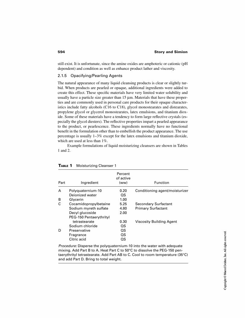
594 Story and Simion
TABLE 1 Moisturizing Cleanser 1
Part Ingredient
Percentof active
(ww) Function
A Polyquaternium-10 0.20 Conditioning agent/moisturizerDeionized water QS
B Glycerin 1.00C Cocamidopropylbetaine 5.25 Secondary Surfactant
Sodium myreth sulfate 4.80 Primary SurfactantDecyl glucoside 2.00PEG-150 Pentaerythrityl
tetrastearate 0.30 Viscosity Building AgentSodium chloride QS
D Preservative QSFragrance QSCitric acid QS
Procedure: Disperse the polyquaternium-10 into the water with adequatemixing. Add Part B to A. Heat Part C to 50°C to dissolve the PEG-150 pen-taerythrityl tetrastearate. Add Part AB to C. Cool to room temperature (35°C)and add Part D. Bring to total weight.
still exist. It is unfortunate, since the amine oxides are amphoteric or cationic (pHdependent) and condition as well as enhance product lather and viscosity.
2.1.5 Opacifying/Pearling Agents
The natural appearance of many liquid cleansing products is clear or slightly tur-bid. When products are pearled or opaque, additional ingredients were added tocreate this effect. These specific materials have very limited water solubility andusually have a particle size greater than 15 µm. Materials that have these proper-ties and are commonly used in personal care products for their opaque character-istics include fatty alcohols (C16 to C18), glycol monostearates and distearates,propylene glycol or glycerol monostearates, latex emulsions, and titanium diox-ide. Some of these materials have a tendency to form large reflective crystals (es-pecially the glycol diesters). The reflective properties impart a pearled appearanceto the product, or pearlescence. These ingredients normally have no functionalbenefit in the formulation other than to embellish the product appearance. The usepercentage is usually 1–3% except for the latex emulsions and titanium dioxide,which are used at less than 1%.
Example formulations of liquid moisturizing cleansers are shown in Tables1 and 2.
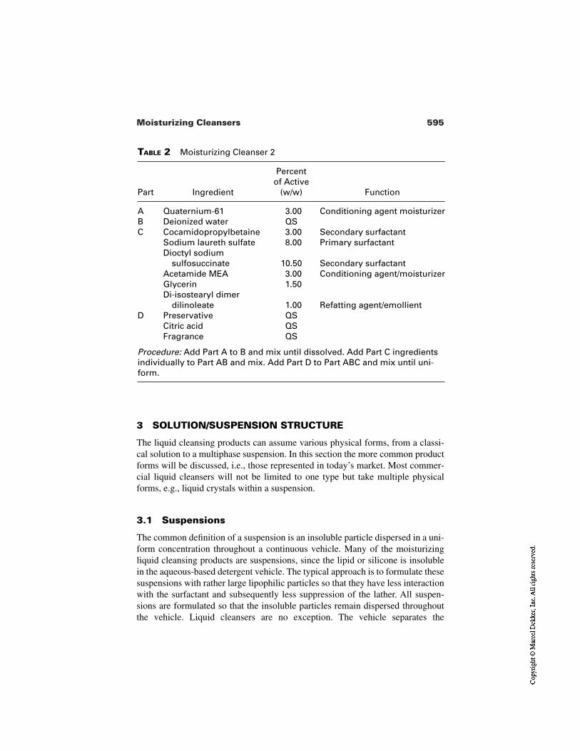
595Moisturizing Cleansers
TABLE 2 Moisturizing Cleanser 2
Part Ingredient
Percentof Active
(w/w) Function
A Quaternium-61 3.00 Conditioning agent moisturizerB Deionized water QSC Cocamidopropylbetaine 3.00 Secondary surfactant
Sodium laureth sulfate 8.00 Primary surfactantDioctyl sodium
sulfosuccinate 10.50 Secondary surfactantAcetamide MEA 3.00 Conditioning agent/moisturizerGlycerin 1.50Di-isostearyl dimer
dilinoleate 1.00 Refatting agent/emollientD Preservative QS
Citric acid QSFragrance QS
Procedure: Add Part A to B and mix until dissolved. Add Part C ingredientsindividually to Part AB and mix. Add Part D to Part ABC and mix until uni-form.
3 SOLUTION/SUSPENSION STRUCTURE
The liquid cleansing products can assume various physical forms, from a classi-cal solution to a multiphase suspension. In this section the more common productforms will be discussed, i.e., those represented in today’s market. Most commer-cial liquid cleansers will not be limited to one type but take multiple physicalforms, e.g., liquid crystals within a suspension.
3.1 Suspensions
The common definition of a suspension is an insoluble particle dispersed in a uni-form concentration throughout a continuous vehicle. Many of the moisturizingliquid cleansing products are suspensions, since the lipid or silicone is insolublein the aqueous-based detergent vehicle. The typical approach is to formulate thesesuspensions with rather large lipophilic particles so that they have less interactionwith the surfactant and subsequently less suppression of the lather. All suspen-sions are formulated so that the insoluble particles remain dispersed throughoutthe vehicle. Liquid cleansers are no exception. The vehicle separates the
596 Story and Simion
macrolipophilic particles from each other, and the inherent viscosity of the vehi-cle prevents the settling or coalescence (fusing) of these particles. The shelf-lifeof the composition is a primary concern for the formulator, since suspensions arethermodynamically unstable. The intent is to have the product remain stablethroughout its life cycle. Most of the suspensions are unstable by design. The sus-pension while in a concentrated state is stable. During use, the consumer appliesthe product to their skin and then rinses. The dilution associated with rinsingdestabilizes the suspension and the moisturizing ingredient is delivered to theskin (or hair). Suspensions allow the formulator to target and control the deliveryof the moisturizing ingredient. Although this is somewhat simplistic, it is a cost-effective delivery system for liquid moisturizing products.
3.2 Solutions
Very few moisturizing liquid products would conform to the ideal definition of asolution. The ideal solution is one in which there is no change in the properties ofthe components, other than dilution, when they are mixed to form the solution [8].The less complicated product forms would fall into this class. These are usuallysimple mixtures of detergents with an ethoxylated refatting ingredient. Theethoxylation allows the moisturizing ingredient to be solubilized in the aqueousdetergents. When the product is used for cleansing, a small amount of the refat-ting ingredient will deposit on the skin and remain throughout the rinse (theoreti-cally). Those formulators experienced in the art usually find this product form tobe inefficient for any significant delivery of a moisturizing or conditioning mate-rial. A great majority of the refatting agent is lost during rinsing.
3.3 Coacervates/Colloids
Coacervates refers to the polymer-rich phase that occurs when dilute solutionsseparate into a solvent phase practically free of polymer and a viscous liquidphase that contains almost all of the polymer still with a significant amount of sol-vent. How is this phenomenon relevant to moisturizing cleansers? Coacervationis another method for delivery of conditioning ingredients to the skin during thedynamics of cleansing and rinsing. When more than one polymer is in solutionand then incorporated into the detergent phase of the formulation coacervates canform which build the product viscosity and temporarily stabilize the mixture [9].When this mixture is applied to the skin during cleansing, the product is diluted.As it undergoes infinite dilution during rinsing the coacervate structure is elimi-nated allowing the polymer to readily deposit on the skin. Often during the mix-ing of polymer phases coacervation is undetectable, except for a slight increase inthe formulation viscosity.
597Moisturizing Cleansers
Colloidal solutions are another common form for moisturizing liquidcleansers. These are not true solutions and often exhibit a Faraday–Tyndall effect[10]. This is the effect when a strong light beam passing through a colloidal solu-tion causes the colloidal particles to scatter the light to form a cone, usually bluein color. Although the mixture appears to be clear, it is a dispersion with particlesof size less than 5µm. Since many of the conditioning polymers are cationic andhave a limited, if any, compatibility with the anionic detergents, it is likely theyform colloidal solutions. The presence of colloidal solution can be confirmed bytesting for the Faraday–Tyndall effect. The colloidal solution upon dilution willexhibit characteristics of a true solution. The cationic polymers will have a high-er affinity for the skin than the anionic solution. The product then delivers its con-ditioning effect during rinsing.
It is very difficult to determine which phenomenon is responsible for themoisturizer/conditioner delivery but from the formulator’s perspective, it is notnecessary, since the consumer benefit is still realized.
3.4 Liquid Crystals
There are three types of liquid crystal phases relevant to detergents. They are one-dimensional periodicity, which is lamellar; two-dimensional periodicity, hexago-nal; and three-dimensional periodicity, cubic. These liquid crystal phases are im-portant since they have very different rheological properties. The hexagonalphase is the most viscous and is often preferred by the formulator [11]. Thehexagonal liquid crystal structure allows the suspension of noncompatible mate-rials into the aqueous vehicle. The liquid crystalline structure is very stable untilthe concentration of the surfactant is altered. The surfactant concentration ischanged when the product is used in the cleansing and rinsing process. When thecleansing composition is applied, the suspending properties are lost and the non-compatible polymer or lipophile is readily available for deposition on the skin.
The knowledge of liquid crystals and their stability has been extensivelyapplied to emulsion products for many years. The liquid crystal formation fromsurfactants is now being applied in a similar manner to temporarily stabilize oilsthat would be commonly used in emulsions. The delivery dynamics are differentbut the end result is similar.
4 STRATEGIES FOR FORMULATING MOISTURIZING CLEANSERS
There are two approaches to enhancing skin condition. First is not to damage theskin. The second is to deliver moisturizing agents that improve its condition.These approaches are not mutually exclusive and can be used simultaneously.
598 Story and Simion
4.1 Reducing Skin Damage
Skin irritation can take many forms. This includes primary irritation (erythemaand edema), skin dryness and roughness, as well as sensory irritation. It is likelythat these phenomena are caused by different mechanisms—different interactionsbetween the skin and surfactants. Therefore it is important to develop formula-tions that minimize the different interactions thereby reducing all forms of irri-tation.
4.1.1 Primary Irritation
Prolonged and repeated surfactant exposure to the skin can produce erythema(redness) and edema (swelling). The relative ability of the surfactants to causeprimary irritation has been well documented especially for anionic and cationicsurfactants. For anionic surfactants, primary irritation potential reaches its maxi-mum for C12 surfactants [12]. Either reducing or increasing the alkyl chainlength reduces primary irritation. Other factors that can reduce irritation of thesurfactant molecule are
Increasing the degree of ethoxylationIncreasing the size of the hydrophilic headgroupReducing the charge density of the hydrophilic headgroup
4.1.2 Mechanisms Inducing Primary Irritation by Anionic Surfactants
The key step by which anionic surfactants start the irritation process is the inter-action with the skin surface. The two main factors in this process are (1) the num-ber of surfactant monomers available and (2) their ability to bind to the skin’s sur-face. In examining the dose response of surfactants to swell the stratum corneum,it is apparent that both parameters increase it until the critical micelle concentra-tion (CMC) is reached [13,14]. Further increases in surfactant concentration donot cause additional swelling. As the concentration of surfactant monomer alsoceases to increase at the CMC, it has been hypothesized that the surfactantmonomers are key contributors to irritation. This hypothesis is supported by thealkyl ether sulfates. Increasing the degree of ethoxylation reduces both theirCMC and their primary irritation potential [14].
There are alternative models that account for many of all these effects. Forinstance, the ability of surfactants to bind to the stratum corneum surface and tocause the denaturation of the skin, are important steps.
4.1.3 Surfactant Binding to Skin Causes Primary Irritationand Skin Dryness
Imokawa and his colleagues have proposed that surfactant binding to the stratumcorneum surface has an important effect on its potential to cause dryness and
599Moisturizing Cleansers
roughness [15]. It may also play an important role in primary irritation. Theymodeled normal use of surfactant solutions by exposing the skin to a surfactantsolution for 10 min, once a day, for four consecutive days. A trained evaluator as-sessed the extent of skin roughness. A rank order for the surfactants’ ability to in-duce roughness in vivo was determined:
SLS > LAS > AOS > SLES-3EO > C12(EO)7
There were two in vitro parameters that correlated with this in vivo order:
The ability of the surfactants to bind to the stratum corneumThe ability of the surfactants to denature bovine serum albumin
Its is known that the first step of the denaturing interaction of surfactants with thebovine serum albumin is ionic absorption onto the protein [16]. This initial denat-uration appears to have two important effects. First, it is the basis of superhydra-tion, the ability of surfactants to cause rapid swelling of the stratum corneum.Wilhelm et al. demonstrated that even a short, 10-min exposure to a surfactant so-lution will cause a significantly greater uptake of water than exposure to a buffersolution [12]. Although this swelling is temporary and reverses when the solutionis removed, the stratum corneum does not go back to its original condition. Thesmall hydrophilic molecules in the stratum corneum, known as natural moisturiz-ing factors, are probably leached out. Conversely, the damaging surfactants thatbind to the stratum corneum proteins probably remain in place to further damagethe skin. Wilhelm’s data also showed a correlation between the superhydrationcaused by alkyl sulfates of different chain lengths and their primary irritation po-tential. This observation is probably the basis of using the in vitro stratumcorneum and protein film swelling assays to predict irritation potential of anionicsurfactants and cleansers.
Further support for the binding models of irritation comes from the work ofWarren et al. [17]. They were able to show that increasing water hardness, espe-cially in the rinse, increased soap binding and soap-induced skin irritation. This isprobably caused by the formation of insoluble calcium soaps, scum, on the skin’ssurface. Irritation caused by anionic surfactant based bars was less influenced bywater hardness. The calcium salts of these surfactants are more soluble than that ofsoap.
This mechanism of irritation is probably more relevant for cationic surfac-tants. Dodecyl trimethyl ammonium chloride (DTAC) does not swell the stratumcorneum or protein film, yet in occlusive patching it can be as irritating as SLS[18,19].
4.1.4 Percutaneous Penetration of Surfactants Has LittleEffect on Irritation Potential
Ironically, the ability of surfactants to penetrate the stratum corneum barrier doesnot appear to have much effect on their irritation potential. Sodium lauryl sulfate
600 Story and Simion
penetrates more slowly than its ethoxylated analog [SLES-3EO], which in turnpenetrates more slowly down its unsulfated analog laureth-3EO [C12–(EO)3][20,21]. The primary irritation potential is in the reverse order.
However it is known that SLS will significantly increase in the percuta-neous penetration of other molecules such as water and hydrocortisone. Thisprobably relates to its ability to damage the stratum corneum [22].
4.1.5 Strategies for Enhancing Formulation Mildness
There are three approaches to enhance formulation mildness:
Use of mild surfactantsUse of interactive surfactantsAddition of polymers
Use of Mild Surfactants. The substitution of a harsh surfactant by amilder one will reduce the overall irritation potential of the product. However,mildness is not the only parameter that needs to be optimized; the ability to pro-duce lather/foam is also critical for consumer acceptability. Unfortunately, manymild surfactants do not lather as well as their harsher analogs.
Use of Interactive Surfactants. The irritation of harsh anionic surfactantscan be reduced by the addition of a secondary milder surfactant, even though thelevel of the more irritating ingredient remains unchanged. This was demonstratedby Rhein et al., who added increasing levels of SLES-7EO to a fixed concentra-tion of SLS [23]. As a result, the primary irritation potential was reduced. Thereare two explanations for this behavior. First, the formation of mixed micelles(SLS + SLES-7EO). This mixture has a lower CMC than SLS alone, so the con-centration of free SLS monomers available to induce irritation is reduced. A sec-ond explanation is the competitive binding between the two molecules. Addingadditional SLES reduces the amount of SLS bound to the stratum corneum sur-face and therefore the amount of irritation. There are insufficient data to deter-mine which model is correct.
Similar interactions have been observed in vitro and in vivo using other sur-factants. Each of the models are supported by different systems. Faucher andGoddard showed that the nonionic surfactant (Tergitol 15-S-9) reduces SLS bind-ing to hair by modifying its solution properties [24]. Adding increasing levels ofTergitol to the SLS reduces the CMC, meaning that there is less SLS monomeravailable for binding. As little Tergitol binds to hair, this argues that this is the so-lution properties, rather than competitive binding that has the primary effect onSLS binding.
Conversely Dominguez et al. showed that betaine and SLS compete forbinding sites on skin callus [25]. This competition is affected by the pH and thealkyl chain length of the betaine. Such interactions can reduce irritation in vivo.
601Moisturizing Cleansers
A second paper by Dominguez et al. showed that mixtures of betaine and SLScaused less ocular irritation than either surfactant alone [26]. However it is oftensuggested that the interaction between SLS and betaine is a charge neutraliza-tion, especially at pH 6 and below, where betaine will carry a positive charge.This observation is similar to that of Rhein et al., who showed that amine oxides,which carry a partial positive charge, can also reduce the irritation potential ofSLS [13].
Recently McFadden et al. demonstrated that the cationic surfactant dodecyltrimethyl ammonium chloride (DTAC) can reduce SLS-induced irritation evenwhen it is added after the SLS application [27]. This suggests that the formationof a “pseudononionic” complex via charge neutralization can reduce irritation po-tential.
Addition of Cationic Polymers. Another example of charge neutralizationis the interaction of anionic surfactants with cationic polymers. Cationic poly-mers are frequently used in body washes and hair shampoos as conditioningagents and are readily deposited on keratinous surfaces. They have been demon-strated to reduce irritation and dryness caused by anionic surfactants. Certainly, insolution they will complex with anionic surfactants and may carry them to theskin. However as they are in the form of pseudononionic complexes, the surfac-tants will be relatively mild.
4.1.6 Deposition of Moisturizing Ingredients on the Skin
It has long been recognized that cleansing systems can be used to deliver usefulamounts of ingredients to the skin. These include antidandruff shampoos, anti-bacterial agents from bar and liquid soaps, and silicone and hydrocarbon hair con-ditioners from two-in-one shampoos. There is no reason why a skin conditionercannot be delivered to the skin in an analogous way. One of the challenges of de-livering a moisturizer to the skin from a cleanser is the detergency of the cleans-ing base. Indeed, much of the conditioning agents will be rinsed away. Parkhanishowed that this was a function of the surfactant’s structure as well as its concen-tration [28].
An approach to increasing retention of the moisturizing agent on the skinduring washing and rinsing is to increase the affinity between the two. For zincpyrithione, an antidandruff agent, this means reducing the pH to 5 so the mole-cules bears a positive charge. This enables the pyrithione ion to bind to the nega-tively charged sites on the skin and hair.
The linkage does not have to be direct. Story et al. showed that the cationicpolymer polyquaterium 6 can be used as a bridge between the anionic condition-er sulfated castor oil and the skin [29]. This significantly increases the condition-er’s substantivity.
602 Story and Simion
5 ASSESSMENT OF MOISTURIZING POTENTIAL OF CLEANSERS
5.1 Consumer Testing
Moisturizing cleansers are consumer personal care products, and ultimately theirbenefits should be recognizable to consumers. If they do not deliver against theirpromise, that is, to leave the skin feeling and looking moisturized, or at least lessdry, they will not be successful in the marketplace. Therefore it is important to as-sess whether potential consumers recognize the product’s benefits in normal useconditions. Such data from a properly designed test can be used for claims sup-port as well as to assure the scientists developing the product that it successfullydelivers the benefits promised. The review of how to design and implement suchconsumer studies is beyond the scope of this chapter.
5.2 Clinical Testing Overview
Previously, most evaluations of cleanser effects have been to assess primary irri-tation or drying potential. Such studies start with the skin in good condition, andthe extent by which parameters such as erythema and dryness worsen is evaluat-ed. However, moisturization potential carries the implication that it is improvingskin condition. Therefore a different experimental design is required. Such stud-ies should incorporate aspects of moisturizer efficacy testing especially with re-gard to (1) starting with dry skin, to enable improvement to be observed and (2)the use of moisturizer end points such as assessments of skin dryness and skin hy-dration, together with (3) an application method that reflects how cleansing prod-ucts are used.
Ideally the application method should not greatly affect, especially de-crease, the degree of dry skin. Thus the method initially described by Lukakovicet al. in 1988 is probably more appropriate than methods that involve rubbing forlonger periods, e.g., the flex wash or the volar forearm wash test [30]. For the lat-ter studies, the prolonged rubbing has the potential to remove skin flakes, and asa result the methods will lose sensitivity [31,32].
5.2.1 Experimental Design
In order to demonstrate that the cleanser delivers a benefit to the skin, the skinmust start out in poor condition. As with moisturizer efficacy studies, the skinshould be dry at baseline (dryness score of 2 or more on a 0–4 scale). The testshould be run on a body site that readily shows skin dryness. This includes thelower legs or the dorsal aspect of the forearms. The lower leg, in particular, hassufficient area to enable multiple products (and a no-product control) to be testedsimultaneously. Using a within-subject design enables potentially large person-
603Moisturizing Cleansers
to-person variations to be eliminated. There are three main ways to produce dryskin:
Cold weather frequently, occurring during the winter, can often producedryness.
As people age they exhibit more dry skin, especially at the extremities.Washing the test area with a drying cleanser will induce dryness.
Combining the first and second methods is probably the best approach. Relying on the weather alone can be risky, as a few warm humid days will significantly re-duce the level of dryness observed. Giving the panelists a drying soap bar for reg-ular cleansing has two great disadvantages. First, the soap bar may interfere withthe effects of the moisturizing cleanser. Then, consumers usually do not use twocleansing products on the same body sites. Second, Ertel et al. suggested that arti-ficially drying out the skin with a cleanser reduces subsequent responses comparedwith naturally dry skin [33]. The basis of this is unclear, but it contrasts with theincreased irritation response observed when subclinically or mildly irritated skin is re-exposed to an irritant. There are several hypotheses for the different behaviorof dry skin, but all reflect that we have studied dryness much less than irritation.
5.2.2 Measuring the Clinical Effects of Products on the Skin
Based on the approaches used to assess moisturizer efficacy, the two main param-eters to assess the moisturizing potential of cleansing products are skin drynessand skin hydration. It is always advisable to use multiple methods for assessingefficacy, as each individual method has potential shortcomings. The use of a bas-ket of methods will yield a fuller assessment of skin condition.
Skin Dryness. Traditionally skin dryness has been evaluated by a trainedobserver using an ordinal scale. However, this approach has two major problems.It is very dependent on the evaluator, and great care must be taken to ensure re-producibility between evaluators, studies, and different testing laboratories. Forthis, a standardized photographic scale is very helpful. Second, there are manyfactors that can reduce the appearance of dryness without there being any benefitto the skin. These include short-term humidity and occlusive lotions that mattethe dry skin flakes down without removing them. These problems can be over-come by using a sticky tape to sample the skin’s surface, e.g., DeSquame® tape(CuDerm Inc., Dallas, TX). The tape is pressed onto the skin’s surface and thenremoved. The greater the scaling, the more skin flakes are removed by the tape.These can be quantified by using an analog scale or by image analysis. The tapewill remove the flakes even if they are matted down or obscured by warm, humidweather. This was demonstrated in a single-wash study (see Fig. 1). After a base-line assessment, dry skin on the dorsal forearm was washed once by the methodof Lukakovic et at. [30]. The skin condition was reassessed three hours later. Re-
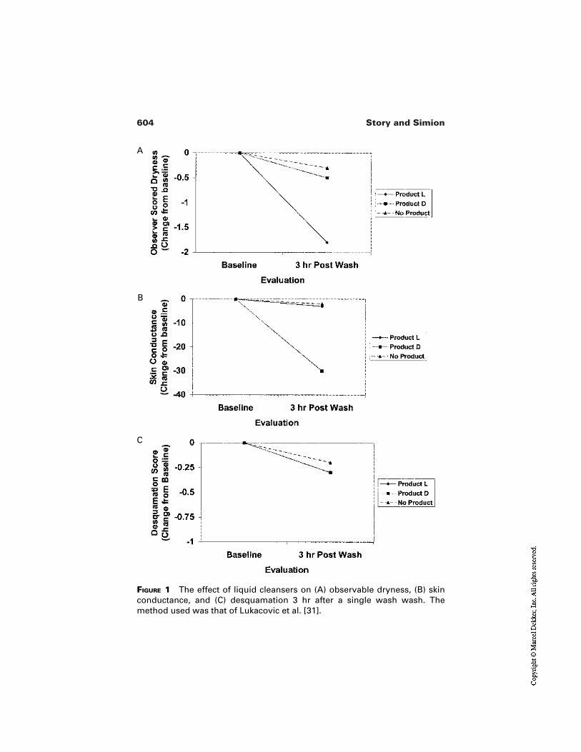
604 Story and Simion
FIGURE 1 The effect of liquid cleansers on (A) observable dryness, (B) skinconductance, and (C) desquamation 3 hr after a single wash wash. Themethod used was that of Lukacovic et al. [31].
A
B
C
605Moisturizing Cleansers
sults show that Product L reduces observable dryness more than product D or noproduct. However the DeSquame tape indicates that flakes are still present, justmasked. This conclusion is supported by the conductance readings, which had notincreased.
Conductance and Capacitance. Conductance and/or capacitance are fre-quently used to measure skin hydration. This approach has been supported empir-ically by Morrison and Scala, who showed a strong correlation between observ-able dryness and reduction in skin conductance (measured by a Skicon 200) andcapacitance (measured by a dermal phase meter) [34]. There are two reasons thatexplain how skin conductance may measure dryness. First, as the skin becomesdrier, the concentration of water in the stratum corneum is reduced. Since water isa good conductor compared with the more hydrophobic stratum corneum, a re-duction in water activity will reduce conductance. Another possible mechanismby which dryness reduces conductance is that as scales develop, air pockets areformed in the damaged stratum corneum. Since air is a poor conductor, this scal-ing also results in reduced conductance. Clearly these two mechanisms are notmutually exclusive and may occur simultaneously.
It should be stressed that residues left on the skin’s surface may modifyconductance in the absence of dryness. For instance, petrolatum, silicones, andmineral oil are good insulators and can reduce conductance even as they moistur-ize the skin. Conductance data should be evaluated based on the product’s com-position and with an understanding of which ingredients may remain on the skinafter rinsing.
Stained DeSquames. Staining skin flakes on the DeSquame tape with hy-drophilic dyes can give a measure of the integrity or degree of damage. From irri-tation testing, Pierard and his colleagues demonstrated that even mild insults willdamage the stratum corneum surface and cause an increase in dye uptake [35].This is readily quantified using a color meter such as a Minolta Chromameter.Damage that is not readily observable to the naked eye can be detected by thismethod. This approach has been extended to moisturizer efficacy testing. Simionhas shown that glycerin-based moisturizers are good at removing dry damagedskin from the surface, revealing undamaged corneocytes below [36]. As yet, thismethod has not been used to assess the efficacy of moisturizing cleansers.
5.3 Irritation Testing
Skin dryness and primary irritation are frequently separate phenomena and can beinduced by different mechanisms. It has been demonstrated that dryness from re-peated hand washing can be induced in the absence of erythema [37]. However,dryness is frequently produced as a sequala to primary irritation as the skin beginsto repair itself [38]. Therefore it is important to assess irritation potential of
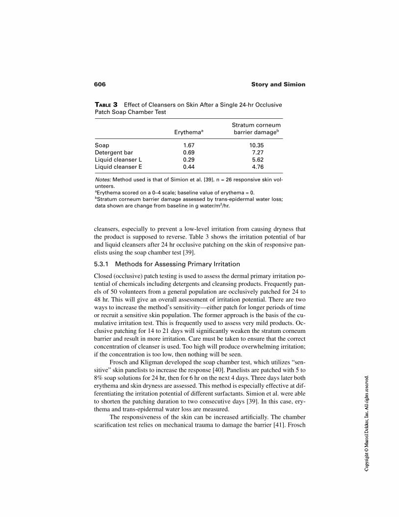
606 Story and Simion
TABLE 3 Effect of Cleansers on Skin After a Single 24-hr OcclusivePatch Soap Chamber Test
ErythemaaStratum corneum barrier damageb
Soap 1.67 10.35Detergent bar 0.69 7.27Liquid cleanser L 0.29 5.62Liquid cleanser E 0.44 4.76
Notes: Method used is that of Simion et al. [39]. n = 26 responsive skin vol-unteers.aErythema scored on a 0–4 scale; baseline value of erythema = 0.bStratum corneum barrier damage assessed by trans-epidermal water loss;data shown are change from baseline in g water/m2/hr.
cleansers, especially to prevent a low-level irritation from causing dryness thatthe product is supposed to reverse. Table 3 shows the irritation potential of barand liquid cleansers after 24 hr occlusive patching on the skin of responsive pan-elists using the soap chamber test [39].
5.3.1 Methods for Assessing Primary Irritation
Closed (occlusive) patch testing is used to assess the dermal primary irritation po-tential of chemicals including detergents and cleansing products. Frequently pan-els of 50 volunteers from a general population are occlusively patched for 24 to48 hr. This will give an overall assessment of irritation potential. There are twoways to increase the method’s sensitivity—either patch for longer periods of timeor recruit a sensitive skin population. The former approach is the basis of the cu-mulative irritation test. This is frequently used to assess very mild products. Oc-clusive patching for 14 to 21 days will significantly weaken the stratum corneumbarrier and result in more irritation. Care must be taken to ensure that the correctconcentration of cleanser is used. Too high will produce overwhelming irritation;if the concentration is too low, then nothing will be seen.
Frosch and Kligman developed the soap chamber test, which utilizes “sen-sitive” skin panelists to increase the response [40]. Panelists are patched with 5 to8% soap solutions for 24 hr, then for 6 hr on the next 4 days. Three days later botherythema and skin dryness are assessed. This method is especially effective at dif-ferentiating the irritation potential of different surfactants. Simion et al. were ableto shorten the patching duration to two consecutive days [39]. In this case, ery-thema and trans-epidermal water loss are measured.
The responsiveness of the skin can be increased artificially. The chamberscarification test relies on mechanical trauma to damage the barrier [41]. Frosch
607Moisturizing Cleansers
and Kligman showed that the responsiveness to hydrophilic material was greatlyincreased. For instance, the threshold for SLS to produce irritation was reducedfiftyfold.
6 SUMMARY
For centuries, soap has been the primary product used for personal cleansing. Al-though it is an effective cleanser, it can also leave the skin dry and irritated. Thedevelopment of synthetic surfactants in the 20th century has enabled the formula-tion of milder cleansers that do not damage the skin as much as soap. This resultsin consumers experiencing less dryness and irritation. However, these cleanserscan go beyond reducing skin damage and deliver benefits to the skin. This can bedone from soap bars, e.g., through delivery of triclocarban from antibacterialsoap, but is more effective from liquid cleansers. Unlike a bar such as soap, whichis mainly detergents, or cocoyl isethionate, liquid cleansers have a much lowersurfactant load. Thus their ability to wash away beneficial ingredients is reduced.However, they still are able to adequately cleanse most consumers that can affordtheir higher price, as these individuals usually do not need all the cleansing po-tential that a bar car deliver.
This chapter described the use of the different components required for aliquid cleanser, whose formulation is more complex than that of a bar of soap.Constituents include primary and secondary surfactants for cleansing and lather-ing. A foam booster is frequently added as well. Thickeners, opacifiers, color, andfragrance are needed to optimize the esthetic acceptability of the product to con-sumers. Finally, preservatives are required to prevent microbial growth in theaqueous base.
Beyond having a milder product, beneficial ingredients can be delivered tothe skin. These include antibacterial ingredients such as triclosan, triclocarban,and antidandruff agents such as zinc pyrithione. Cationic polymeric skin condi-tioners will bind to anionic sites on the skin and be retained after rinsing. Thesepolymers can leave the skin feeling softer and smoother. However, it must bestressed that the vast proportion of any beneficial agent is rinsed away due to thedetergency of the surfactant system. To effectively deliver greater benefits, espe-cially when using effective but expensive ingredients, requires new methods ofdelivering the agents and preventing them from washing away during rinsing.However, for the foreseeable future, this approach will remain less effective atdelivering beneficial agents than leave-on products such as lotions. For example,an anti-aging cleanser may not dry and roughen the skin as much as soap, so thefine lines associated with dryness are less evident. However, delivery of sun-screens, retinol, or other materials that prevent damage boost the skin’s moisturecontent or stimulate the skin’s self-repair mechanism is still done more effective-ly with a lotion.
608 Story and Simion
There are two approaches to measuring the clinical effects of moisturizingcleansers on the skin. The first is to ensure that the cleansing system causes mini-mal damage to the skin, for which traditional irritation testing is used. Second isto measure moisturizing efficacy, which requires a paradigm shift toward thetechniques used for assessing lotion efficacy. Irritation assessments such as closedpatch tests and exaggerated arm washes are used to assess a product’s ability toincrease irritation from a negligible starting point. In contrast, to measure theproduct’s ability to deliver a benefit such as moisturization, the skin must startwith a deficiency, i.e., it must be dry. Then the methods used to assess leave-onmoisturizers, such as conductance and DeSquame tape, can be used to assess themoisturizing cleanser.
REFERENCES
1. Gloxhuber C, Künstler K. Anionic Surfactants: Biochemistry, Toxicology, Dermatol-ogy. 2nd ed. Vol. 43, Surfactant Science Series. New York: Marcel Dekker,1992:212.
2. Bluestein BR, Hilton CL. Amphoteric Surfactants. Vol. 12, Surfactant Science Se-ries. New York: Marcel Dekker, 1982:145–150.
3. Ramon LA. U.S. patent 4,663,069.4. Phillips BM, Akred BJ. U.S. patent 5,039,451.5. Giret MJ, Langlois A, Duke RP. U.S. patent 5,409,640.6. Bolich RE Jr., Williams TB. U.S. patent 4,788,006.7. Ogino K, Abe M. Mixed Surfactant Systems. Vol. 46, Surfactant Science Series.
New York: Marcel Dekker, 1993:235–247.8. Martin AN, Swarbrick J, Cammarata A. Physical Pharmacy. 2d ed. Philadelphia: Lea
& Febiger, 1969:149.9. Gennaro AR. Remington’s Pharmaceutical Sciences. 18th ed. Easton, PA: Mack
Publishing, 1990:292.10. Martin AN, Swarbrick J, Cammarata A. Physical Pharmacy. 2d ed. Philadelphia: Lea
& Febiger, 1969:449.11. Ogino K, Abe M. Mixed Surfactant Systems. Vol. 46, Surfactant Science Series.
New York: Marcel Dekker, 1993:248–251.12. Wilhelm KP, Cua AB, Wolff HH, Maibach HI. Surfactant-induced stratum corneum
hydration in vivo: prediction of the irritation potential of anionic surfactants. J InvestDermitol 1993; 101:310–315.
13. Rhein LD, Robbins CR, Fernee K, Cantore R. Surfactant structure effects onswelling of isolated stratum corneum. J Soc Cosmet Chem 1986; 37:125–139.
14. Blake-Haskins JC, Scala DD, Rhein LD, Robbins CR. Predicting surfactant irritationfrom the swelling response of a collagen film. J Soc Cosmet Chem 1986;37:199–210.
15. Imokawa G, Sumura K, Katsumi M. A correlation between adsorption of surfactantonto callus and skin roughness caused by the surfactant. J Jpn Oil Chem Soc 1974;23:17–23.
609Moisturizing Cleansers
16. Cooper ER, Berner B. Vol. 16, Surfactant Science Series. Rieger MM, ed. New York:Marcel Dekker, 1985:195–208.
17. Warren R, Ertel KD, Bartolo RG, Levine MJ, Bryant PB, Wong LF. The influence ofhard water (calcium) and surfactants on irritant contact dermatitis. Contact Dermati-tis. 1996; 36:337–343.
18. Imokawa G, Takeuchi T. Surfactants and skin roughness. Cosmet Toil 1976;91:32–46.
19. Cutler RA, Droebek HP. Toxicology of Cationic Surfactants in Cationic Surfactants.Jungerman E, ed. New York: Marcel Dekker, 1970:527–616.
20. Howes D. The percutaneous absorption of some anionic surfactants. J Soc CosmetChem 1975; 26:47–63.
21. Prottey C, Ferguson T. Factors which determine the skin irritation potential of soapand detergents. J Soc Cosmet Chem 1975; 26:29–46.
22. Wilhelm KP, Surber C, Maibach HI. Effect of sodium lauryl sulfate–induced skin ir-ritation on in vivo percutaneous penetration of four drug. J Invest Dermatol 1991;97:927–932.
23. Rhein LD, Simion FA, Hill RL, Cagan RH. Matti J, Maibach HI. Human cutaneousresponse to a mixed surfactant system: role of solution phenomena in controllingsurfactant irritation. Dermatologica 1990; 180:18–23.
24. Faucher JA, Goddard ED. Interaction of keratinous substrates with sodium laurylsulfate. I. Sorption. J Soc Cosmet Chem 1978; 29:323–337.
25. Dominguez JG, Balaguer F, Parra JL, Pelejero CM. The inhibitory effects of someamphoteric surfactants on the irritation potential of alkylsulfates. Int J Cosmet Sci1981; 3:57–68.
26. Dominguez JG, Parra JL, Infante MR, Pelejero CM, Balaguer F, Sautre T. A new ap-proach to the theory of adsorption and permeability of surfactants on keratinic pro-teins: the specific behavior of certain hydrophobic chains. J Soc Cosmet Chem 1977;28:165–182.
27. McFadden JP, Holloway DB, Whittle EG, Basketter DA. Benzalkonium chlorideneutralizes the irritant effects of sodium dodecyl sulfate. Contact Dermatitis 2000;43:264–266.
28. Parkhani N. The effect of surfactants on lipid deposition from liquid cleansers to theskin. Master’s Thesis, University of Cincinnati, 1995.
29. Story DC, Gott RE, Asbury MT, Phifer K, Simion FA. U.S. patent 6,024,952.30. Lukacovic MF, Dunlap FE, Michails SF, Visscher MO, Watson DD. Forearm wash
test to evaluate the clinical mildness of cleansing products. J Soc Cosmet Chem1988; 39:355–366.
31. Strube DD, Koontz SW, Murahata RI, Theiler RF. The flex wash test: a method forevaluating the mildness of personal washing products. J Soc Cosmet Chem 1989;40:297–306.
32. Sharko PT, Murahata RI, Leyden JJ, Grove GL. Arm wash with instrumental evalu-ation—a sensitive technique for differentiating irritation potential of personal wash-ing products. J Derm Clin Eval Soc 1991; 2:19–27.
33. Ertel KD, Neumann PB, Hartwig PM, Rains GY, Keswick BH. Leg wash protocol toassess the skin moisturization potential of personal cleansing products. Int J CosmetSci 1999; 21:383–387.
610 Story and Simion
34. Morrison BM, Scala DD. Comparison of instrumental measurements of skin hydra-tion. J Toxicol Cut Ocular Toxicol 1999; 15:305–314.
35. Pierard GE, Goffin V, Pierard-Franchimont C. Corneosurfametry: a predictive as-sessment of the interaction of personal care products with human stratum corneum.Dermatology 1994; 189:152–156.
36. Simion FA. Use of stained sticky tape to enhance assessment of the moisturizationefficacy of lotions. 59th American Academy of Dermatology Annual Meeting, Wash-ington, D.C., 2001.
37 Simion FA, Babulak SW, Morrison BM, Rhein LD, Scala DD. Experimental methodsoap induced dryness in the absence of erythema. 50th American Academy of Der-matology Annual Meeting, Dallas, TX, 1991.
38. Wilhelm KP, Freitag G, Wolff HH. Surfactant-induced skin irritation and skin repair:evaluation of a cumulative human irritation model by non-invasive techniques. J AmAcad Dermatol 1994; 31:981–987.
39. Simion FA, Rhein LD, Grove GL, Wojtowski J, Cagan RH, Scala DD. Sequential or-der of skin responses to surfactants in a soap chamber test. Contact Dermatitis 1991;25:242–249.
40. Frosch PJ, Kligman AM. The soap chamber test: a new method for assessing irritan-cy of soaps. J Am Acad Dermatol 1979; 1:35–41.
41. Frosch PJ, Kligman AM. The chamber scarification test for irritancy. Contact Der-matitis 1976; 2:314–324.
27Safety Assessment of Cosmetic Products
Christopher FlowerThe Cosmetic, Toiletry, and Perfumery Association, London, United Kingdom
1 INTRODUCTION
It is clearly not possible in a short chapter such as this to cover in detail safety as-sessment programs to comply with the legislative environment surrounding cos-metic products in each of the major markets of the world. Neither is it possible toprovide in-depth instruction on toxicological testing methods and interpretationof data. Such a program would, and indeed should, become outdated as scientificmethods evolve. In fact, during the writing of this chapter, the Organisation forEconomic Co-operation and Development (OECD) agreed to delete the LD50 testfrom its list of official protocols [1]. The shortcomings of this test have beenknown to toxicologists for a long time, but getting agreement for it to be delistedhas also taken a long time. Instead, therefore, the intention of this chapter will beto provide something of the philosophy of cosmetic product safety to guide ratherthan train from scratch the safety assessor and to help colleagues understand theneeds of the safety assessor.
Although the European Community Cosmetics Directive (76/768/EEC) isseen as a model for legislation by an increasing number of countries in the world,significant markets operate under quite different regimes. The nonalignment ofdifferent definitions of what constitutes a cosmetic complicates the situation,
611
612 Flower
with some borderline products being regulated quite differently in different mar-kets around the world depending on their final classification in each country.Underlying each regime, though, is the basic demand that cosmetic products,however defined, should be safe for their intended purpose and not cause harmto the consumer. The regimes run from those which, like the United States, re-strict very few ingredients to those, like Japan until recently, where only per-mitted ingredients may be used in cosmetic products. The EU regime is some-what midway between the two. There are lists of prohibited and restrictedsubstances and several positive lists too. Any other substance may be used as aningredient subject to an overriding safety requirement. It is this basic safety re-quirement and how a cosmetic is assessed for compliance that is the subject ofthis chapter.
The successful cosmetic product will be used by large numbers of people,repeatedly and for a considerable time. Its mode of use will be governed partly bycustom or past experience and partly by the manufacturer’s instructions. Rarelywill use be under professional supervision although that may be the case some-times. Yet experience shows this situation is satisfactory; by and large, cosmeticproducts do not lead to personal injury or worse [2], and today they are amongstthe safest products in the major markets of the world to which general consumersare exposed.
This has not always been the case. The historical use of substances such aswhite lead, arsenic, and belladonna for cosmetic purposes is well documented andeven today some traditional cosmetics are still available which might not pass theassessment process being described here.
That is not to say cosmetic products are not subject to controls or regula-tions. On the contrary, in all member states of the European Union, cosmeticproducts are closely regulated by their own specific directive, the Cosmetics Di-rective [or, to give it its full title, Council Directive of 27 July 1976 on the ap-proximation of the laws of the Member States relating to cosmetic products(76/768/EEC)]. The Cosmetics Directive is a safety directive. This is made clearin the recitals where it is stated, “Whereas the main objective of these laws is thesafeguarding of public health.” To this end, the Cosmetics Directive first defineswhat is a cosmetic product (Article 1) and then immediately requires of them thatthey should be safe (Article 2). Both the degree of safety required and the cir-cumstances to be considered are indicated in this article. Furthermore, the direc-tive requires that each cosmetic product should be subject to a safety assessmentto ensure compliance with Article 2.
Thus, cosmetic products are defined in European Community law, and eachcosmetic has to undergo a safety assessment prior to marketing to ensure compli-ance with the safety requirement. Guidelines have been written elsewhere to helpin this regard [3,4].
613Safety Assessment of Cosmetic Products
2 THE SAFETY REQUIREMENT
Article 2 of the Cosmetics Directive states, “A cosmetic product put on the mar-ket within the Community must not cause damage to human health when appliedunder normal or reasonably foreseeable conditions of use, taking account, in par-ticular, of the product’s presentation, its labelling, any instructions for use anddisposal as well as any other indication or information provided by the manufac-turer or his authorised agent or by any person responsible for placing the producton the Community market. The provision of such warnings shall not, in anyevent, exempt any person from compliance with the other requirements laid downin this Directive.’’
The wording of Article 2 is very important. It requires that a cosmetic prod-uct must not cause damage to human health, but does not require that a cosmeticmust not provoke any adverse reaction. Thus, absolute safety is not required—in-deed could not be achieved—but a degree of safety is required, and the appropri-ate degree is defined as a freedom from damage to human health.
Compliance with the safety requirement is a matter of professional judge-ment and will be considered in greater depth later, but it will involve the evalua-tion of factors such as severity of possible adverse reactions, their duration andreversibility, and their prevalence or likelihood in the normal population. Reac-tions judged to be mild, readily reversible, or rare tend to support the safety of aproduct, whereas responses judged to be severe, long-lasting, or common wouldnot.
Article 2 also requires that the product be “safe” under normal or reason-ably foreseeable conditions of use. There is no requirement for a product to besafe under all circumstances; misuse and deliberate abuse, therefore, need not beconsidered. However, particular care needs to be given as to what is reasonablyforeseeable and to the value warning labels might have.
For example, a shampoo might cause some discomfort should it get into theeyes. A warning advising against allowing that to happen and what action to takein the event is quite reasonable. However, because shampoo getting in to the eyesin normal use is reasonably foreseeable, any adverse reactions that occur shouldnot be so severe as to constitute damage to human health.
Finally, the safety requirement is not qualified but is absolute. In otherwords, it is not possible to weigh the risks against the benefits, as one may do witha pharmaceutical for example. No matter what benefits may be provided by a cos-metic, the cosmetic product must not cause damage to human health. This is,therefore, a very stringent requirement.
Compliance with the safety requirement of Article 2 is through the conductof an “assessment of the safety of human health of the finished product. To thatend the manufacturer shall take into consideration the general toxicological pro-file of the ingredient, its chemical structure and its level of exposure” [Article
614 Flower
7a(d)]. Although the foregoing relates specifically to the situation in the memberstates of the European Community, the principles expounded will also apply tosafety assessments conducted under other regulatory regimes.
The process of safety assessment is a multistep one which comprises anumber of stages:
Hazard identification. This step essentially collects all the adverse datapertaining to the product and its constituent ingredients to identify thehazards.
Risk characterisation. This step compares the hazard data with the antic-ipated exposure conditions to determine which hazards could constituterisks to human health. In essence, this step identifies the relevant hazardsfor further consideration.
Risk evaluation. This step is a further refinement of risk characterisationin which the potential risks are quantified and gauged against knownrisks. This step requires detailed information regarding actual exposure.
Safety assessment. In the final step, the safety assessor must judgewhether the potential risks are deemed acceptable and therefore whetherthe product is safe to market.
Before going through each of these steps in detail, it is appropriate to considerpractical aspects of importance to the safety assessment process. This is informa-tion that is essential if the safety assessor is to be in a position to perform an ade-quate assessment.
2.1 Assess the Right Product
Companies often have complex development programs and many alternative for-mulations may be under simultaneous evaluation prior to deciding which candi-date is to be selected for the market. Product brand or variant names may changeduring the course of development and a means of tracking these changes must bein place. If each formula is assigned a unique reference number, then a clear andunambiguous link can be established between the marketed product and its safetyassessment regardless of other changes to its name.
Then, if each adjustment to a formula, no matter how slight, generates anew unique formula reference number, a new review or safety assessment is au-tomatically triggered. Without this trigger, there is a risk that formulae may be re-peatedly modified until the original assessment becomes remote from the market-ed product. It is not acceptable that a product formula should be changed withoutreference to the safety assessor. Manufacturers should be aware that the safety as-sessor cannot be held responsible for the safety of a formula changed without hisor her knowledge, and manufacturers should be aware that it is they and not thesafety assessor who runs the risk of prosecution under the Cosmetic Directive.
615Safety Assessment of Cosmetic Products
2.2 Know the Formula
The complete formula must be available to the safety assessor, including thechemical name, INCI name, and any in-house name by which each ingredient isknown.
Since most cosmetic ingredients are available in various grades or qualities,the specification for each as actually used must be available. Some raw materialsare presented as mixtures, sometimes with additives to enhance stability or forpreservation. The specifications for these ingredients should show such additives.
Where necessary, the safety assessor may determine which grades are to beused by defining appropriate quality criteria. The manufacturer should ensuresubstitution is not possible without the approval of the safety assessor. Productmade from ingredients that do not comply with a defined quality would not nec-essarily be covered by the safety assessment.
2.3 Exact Level of Ingredients
The final level of each specific ingredient must be known when they are addedfrom different sources. Some ingredients will be added in variable amounts, q.s.,or quantum satis; pH and viscosity adjusters are typical. The likely ranges andmaximum limits for these must be specified. Some ingredients may react duringmanufacture and reaction products must be identified; others are added as pre-mixes and their constituents must be known.
Safety assessors must be given details of the method of manufacture and adescription and the specification of the final product. They must also understandwhat changes take place during manufacture to know to what the consumer willbe exposed when using the product.
2.4 Product Stability
The product must be stable and an assurance of this is required by the safety as-sessor; written assurance of stability or actual reports of stability tests should beprovided. In addition, the safety assessor may wish to see analytical data on a typ-ical product, particularly regarding levels of “active” ingredients or undesirabledegradation products or contamination substances both before, during, and afterstorage testing. It is important to take account of storage conditions likely in useand consider whether that may affect product stability and safety.
2.5 Microbiological Quality
The finished product must have an acceptable microbiological specification. Thesafety assessor needs to know this and must be sure that the product meets its
616 Flower
specification. Again, reports of microbiological testing or written assurance ofmicrobiological quality may be required by the safety assessor.
Stability testing and microbiology are specialist fields in their own right.Although large companies will probably have expertise in house, smaller con-cerns may have to employ contractors. In either case, the safety assessor needs tobe satisfied that the product is microbiologically and chemically stable through-out the anticipated shelf-life of the product, bearing in mind normal storage con-ditions in the anticipated market. Consideration must be given to the capacity ofthe preservative system to cope with normal contamination once the product hasbeen opened and in use.
2.6 Packaging, Instructions, and Labeling
Safety assessors should see the proposed packaging to consider what impact thatmight have on safety. For example, child-resistant closures may be appropriate insome cases or the maximum pack size might need to be limited in other situa-tions. They should also see instructions for use and any warning labels to be in-cluded, and must be able to influence these where safety is affected. In particular,compliance with any mandatory warnings needs to be checked, a task that may behandled by the safety assessor.
A copy of the INCI ingredient declaration should be supplied and linked tothe formula as a check to ensure no ingredients are omitted.
2.7 Efficacy
Although efficacy and claims support are not generally the province of the safetyassessor, in certain circumstances (suncare products are good examples) efficacydoes become a safety issue. Copies of efficacy study reports should be availableto the safety assessor where there may be a safety aspect, but the safety assessoris not required to determine efficacy per se nor to endorse product claims.
If human trials have been carried out, any adverse effects must be reportedto the safety assessor as this constitutes relevant information. Copies of trial re-ports must be made available.
2.8 Adverse Effects Reports
The safety assessor must have access to reports of adverse effects notified to thecompany on this or similar products; ideally, the number of such events should berelated to the number of product units sold. Such reports can be tabulated period-ically to provide a continuous record, but a system of highlighting severe or par-ticularly unusual events should be considered.
617Safety Assessment of Cosmetic Products
2.9 Toxicity Data
The safety assessor must have access to toxicity and safety data on each of the in-gredients, on the final product or similar products where available and shouldhave knowledge of possible ingredient interactions. These data would includematerial safety data sheets from ingredient suppliers and letters of safety assur-ance from fragrance suppliers confirming the fragrance conforms to the code ofpractice of the fragrance industry [5] as well as conventional toxicological data,in vitro as well as in vivo, and human volunteer studies. In addition, authoritativesources, such as the Cosmetic Ingredient Review in the United States, should beconsulted [6].
2.10 Exposure Estimation
Before commencing a safety assessment proper, it is essential to know the cate-gory of cosmetic product that is to be assessed and how, in general terms, it is tobe used: e.g., whether it is for hair care or skin care, whether it is to be left in situor rinsed off, whether ingestion or inhalation are likely, whether it is diluted be-fore or during use, and so on. Later, it will be necessary to refine this general un-derstanding to gain an accurate exposure assessment and to consider also how theproduct may be misused, perhaps accidentally, and whether there is potential fordeliberate abuse.
Specifically, therefore, the following information is required:
Type of cosmetic productIntended mode of useQuantity used each timeFrequency of applicationDuration of contactSite of contactArea of contactUnintentional contactNature of consumers and numbersOther factors (e.g., exposure to sunlight, potential for abuse, interaction
with other products)
2.11 Hazard Identification
The first step in the safety assessment of cosmetics, as with any other consumerproduct, has to be that of hazard identification. First and foremost, you have toknow what hazards might be associated with each of the ingredients in the prod-uct, and not just the ingredients but also any contaminants and possible degrada-
618 Flower
tion products. Later, the safety assessor must also consider possible reaction prod-ucts and ingredient interactions, either with one another or with the packaging.
Hazard identification is primarily achieved through conventional toxicolog-ical testing both in vivo and, to an increasing extent, in vitro. Much data will beobtained from the literature and from toxicology databases as well as from ingre-dient suppliers. The quality of the data needs to be considered, not just the appar-ent quantity [7]; it is the interpretation of the significance of the data in terms ofits applicability to the use of the ingredient in a cosmetic product that is the pri-mary role of the toxicologist/safety assessor. For this reason, original reports andpublications should be consulted whenever possible rather than relying solely onreported data. Particular attention should be given to the actual substance testedand its description to gauge whether the data are relevant to the ingredient inquestion.
Toxicity tests are frequently defined in terms of their endpoints and, forconvenience, that is how they are considered here. Thus, for each ingredient, oneneeds to consider
Acute toxicityIrritancy to skin, eyes, and mucous membranesSensitization and photosensitizationSubchronic or repeated exposure toxicityMutagenicity and genotoxicityLong-term toxicity and carcinogenicityPhotomutagenicity, photogenotoxicity, and photocarcinogenicityToxicokinetics (absorption, distribution, metabolism, and excretion)Human data
2.11.1 Acute Toxicity
Some estimate of the acute toxicity of a substance to be used as an ingredient in acosmetic product must be available. This does not automatically mean an LD50
test, although historically the LD50 was often used for such a purpose. What is re-quired is evidence that the ingredient will not be acutely toxic at a relevant expo-sure level and by an appropriate route of exposure. With the imminent deletion ofthe LD50 test from the OECD guidelines, more information and less severe stud-ies should become the norm in the absence of in vitro alternatives.
Normally, pre-existing data are often limited to gavage studies in rodentsup to a dose limit (typically 2 g/kg or a dose volume of 10 mL/kg). Oral dosingmay be appropriate for an ingredient likely to be ingested, but such studies re-quire careful interpretation before extrapolating the results to dermal exposurescenarios. Consideration must be given to the effects of dermal absorption [8,9],metabolism by the skin, and to differences in body compartment distribution re-
619Safety Assessment of Cosmetic Products
sulting from dermal exposure, as opposed to absorption from the gastrointestinaltract with its potential for first-pass hepatic metabolism and other differenceslikely in distribution and excretion. The all-important target tissue concentrationof the active toxic principle may be very different depending on the route of ex-posure.
2.11.2 Local Tolerance—Skin, Eye, and Mucous Membrane Irritancy
Estimation of the irritancy of the substance to both skin and eyes may be appro-priate depending on the type of end product use envisaged and the concentrationto be used. Without care, because of use of excessively high doses or concentra-tions or the use of a sensitive or reactive model (such as the rabbit eye), data fromlocal tolerance studies may prove misleading by suggesting the existence of ahazard which is unlikely to be encountered in practice. This, however, will be re-considered in the next sections, those of risk characterization and risk evaluation,when such hazards will be identified as unlikely or irrelevant risks.
Ingredients to be used in specialized cosmetic products, such as those fororal care, intimate hygiene, or on infants, may require data on their mucous mem-brane compatibility. Whilst conjunctival reactions seen from ocular test resultscan be useful, they can only serve as a guide in the absence of clear benchmarks.
Again, frequently such local tolerance data are pre-existing, having beengenerated on the basic substance as required under chemicals legislation. Prob-lems arise when the data are only generated for high doses or high concentrationsand there has been no attempt to characterize the dose–response relationship.
2.11.3 Sensitization and Photosensitization
Sensitization and, increasingly, photosensitization data are required if the sensi-tizing potential of a product is to be estimated. Various in vitro tests have been de-veloped over the years to model aspects of the sensitization process, but they arenot yet able to replace in vivo studies completely at the present time. Neverthe-less, there have been reductions in the severity of the in vivo procedures [10],with the murine local lymph node test [11] now being accepted as a fully validat-ed alternative to the more stressful guinea pig maximization methods.
One crucial factor for skin sensitization is that exposure be considered interms of dose of substance per unit area of skin, rather than as simple concentra-tion in the vehicle. It is important to remember this when considering the finalproduct safety assessment. For example, a spray-on cologne based on a volatilesolvent such as alcohol contains a given concentration of any ingredient; as thesolvent evaporates from the skin, the concentration of ingredient in the remainingproduct rises, but the dose of the ingredient per unit area of skin remains un-changed.
620 Flower
Photosensitization (and phototoxicity) data are becoming ever more impor-tant when considering ingredients which will be used in products such as sun-screens [12,13]. Evidence that a substance is capable of absorbing UV lightshould be taken as a trigger for considering the need for phototoxicity and photo-sensitization data.
2.11.4 Repeat Dose or Subchronic Toxicity
These studies provide valuable information on any toxic effects which occur fol-lowing repeated exposure to a substance. Investigations normally involve postmortem examination of all major organ systems as well as numerous in vivo in-vestigations. As with acute studies, both the dose levels used and the routes of ex-posure must be taken into account when extrapolating the findings to a cosmeticproduct safety assessment.
Ideally, the results of a repeated exposure study would allow the identifica-tion of doses which could be termed the no effect level (NEL), no adverse effectlevel (NAEL), no observed effect level (NOEL), or no observed adverse effectlevel (NOAEL). The differences in meaning of these terms needs to be under-stood and their accurate use checked.
Strictly, since one cannot guarantee that all possible effects which mighthave occurred in a study were actually detectable, any reference to effects shouldbe qualified as relating to observed effects only. The NOEL would be the highestdose at which no effects due to treatment could be detected; such animals would,to all intents and purposes, be identical to animals sham-treated with vehicle ascontrols. Where effects were noticed, there is then a judgement as to whether theeffects were adverse or not. Thus, the NOAEL is the highest dose level at whichany changes observed are judged not to be adverse ones. The NOAEL may be thesame dose level as the NOEL, or may be higher.
In either case, these estimates of doses which appear not to cause harm areused as markers when attempting to establish safety margins for human expo-sures. Safety margins are introduced in toxicology to make allowance for uncer-tainty; just because a given dose was without toxic effect in one study on onespecies does not mean to say that same dose would always be equally benign. In-dividuals within a species vary in many ways, including in their susceptibility toa putative toxin. Similarly, whole species may differ from one another in suscep-tibility. Because it is rare for the most susceptible and the most resistant memberswithin a species to differ by more than tenfold, a safety factor of 10 is applied totake account of this. In the same way, the difference between the most and theleast susceptible species is rarely greater than tenfold, so an additional factor of10 is applied here too.
Thus, if, by chance, a measure of toxicity had been established in a resis-tance species the application of a 100-fold safety factor would take account of the
621Safety Assessment of Cosmetic Products
most sensitive member of the most sensitive species (as man is assumed to be).For example, if a dose of 100 mg/kg was “safe” in an animal study (i.e., producedno observed adverse effects), applying a 100-fold safety factor means that 1 mg/kg should be “safe” in man.
2.11.5 Mutagenicity and Genotoxcity
Mutagenicity is the capacity to induce mutation, which is a permanent change inthe amount or structure of the genetic material of an organism that may result in aheritable change in the characteristics, or phenotype, of the organism. Mutationsmay involve single genes, blocks of genes, or whole chromosomes, and theprocesses include point mutations (changes to a single base or the addition ordeletion of a base), clastogenicity, or chromosome aberrations (gaps, breaks, ortranslocations) as well as aneuploidy (changes in chromosome numbers).
Genotoxicity is the specific adverse effect upon the genome of living cellsthat may be expressed as a mutagenic or carcinogenic effect. Interaction by achemical or its metabolites may be with the DNA directly or to the apparatuswhich regulates the fidelity of the genome. Interference with the process of chro-mosome segregation during meiotic or mitotic cell division can lead to aneu-ploidy without there necessarily being mutation of the DNA itself.
In vitro tests exist to detect all three mutation endpoints, namely, gene mu-tation, clastogenicity, and aneuploidy, but there is currently no single, validatedtest that can provide information on them all. A range of tests will be required andthese may involve a variety of organisms, from bacteria and yeasts through cul-tured mammalian cells to whole mammal studies. In general, testing strategy fol-lows a hierarchical approach whereby absence of activity in an in vitro study istaken as encouraging, but a positive effect is not proof of a potential human haz-ard, only that further investigation is warranted. The underlying principle is to seewhether activity seen in vitro can be expressed in vivo. If it cannot, that is takenas an indication that in vitro result may be given less emphasis.
Considerable expertise in conducting the tests themselves and in under-standing the underlying mechanisms is essential if the results are to be interpret-ed correctly.
2.11.6 Long-Term Toxicity and Carcinogenicity
Increasingly, questions are asked about the potential adverse effects of long-termexposure to substances and whether those effects may include carcinogenicity.Where pre-existing data are available, again the quality of those data and their rel-evance to the proposed use of the substance must be taken into account. Howev-er, the need for studies to generate such data should be carefully weighed againstthe value of the information they could usefully provide. It is here that informa-tion on the likely exposure to and fate of the substance is most valuable.
622 Flower
Many examples exist of substances that are toxic or carcinogenic at veryhigh doses and for which clear thresholds can be demonstrated below which tox-icity or carcinogenicity are not manifest, or which exert their toxicity throughmechanisms known not to be relevant for man. Nevertheless, since exposure tocosmetic ingredients is generally of the long-term and low level kind, a consider-ation needs to be given to the potential for subtle, long-term changes in fieldssuch as immunotoxicity and hormonal toxicology. Identification of thresholds islikely to become more relevant.
2.11.7 Photomutagenicity, Photogenotoxicity, and Photocarcinogenicity
Information relating to these endpoints is needed when the proposed conditionsof use would include prolonged exposure to sunlight, as would be the case withthe UV filters present in sun protection cosmetics. Since the absorbed energy ofthe filtered UV is not destroyed, some consideration is required as to how this en-ergy is handled by the filter.
The absorbed energy could, theoretically, be re-emitted as visible light orsound, but in practice it is more likely to be emitted as heat or as an energetic par-ticle, such as a free radical. The skin protective benefit comes from the re-emis-sion taking place near the external surfaces of the skin where the nearby tissueswhich form the primary target (stratum corneum and upper epidermis) are subjectto constant renewal, so avoiding the accumulation of subcritical toxic insultswhich could otherwise develop into overt toxicity. Without a sun protection prod-uct, UV rays penetrate deeper into the skin and are able to interact directly withviable cells.
2.11.8 Toxicokinetics
Toxicokinetics is the effect the body has on the substance and refers to the ab-sorption, distribution, metabolism, and excretion of the substance in question.Knowledge of the mathematics of toxicokinetics allows an estimate to be made ofthe concentration and duration profile of the substance, or its metabolites, at thetissue site of interest, and thereby a better assessment of the likelihood of a hazardis gained.
For example, a substance which is either very poorly absorbed through theskin or is extensively and rapidly metabolized there is most unlikely to have thesame effects following topical application as it would have after ingestion or fol-lowing gavage dosing.
2.11.9 Human Data
Human data are extremely valuable to the safety assessor of cosmetic productssince they are obtained from the intended target species. Ethical considerations
623Safety Assessment of Cosmetic Products
relating to the use of human volunteers must be evaluated carefully [14], but hu-man data may come from a variety of sources:
Market experience. A history of safe use of the substance in similar prod-ucts for a substantial period of time is a valuable benchmark againstwhich to judge the proposed product.
Human volunteer studies. A variety of studies in which volunteers are de-liberately exposed to the substance in question or to a formulated prod-uct again provides good background data. These may be in-use or pref-erence trials of the market research kind, efficacy trials under clinicalconditions, or clinical tests to determine kinetic data [15,16].
Accidents and industrial exposures. The literature may record incidents inwhich humans have been exposed to the substance and provide descrip-tions of the consequences. The details surrounding such incidents are ofvital importance if their relevance to cosmetic use is to be assessed ac-curately.
Epidemiology studies. Epidemiology studies require particular care in theirinterpretation. By their very nature they are inherently subject to biasand the influence of confounding variables. Although epidemiologystudies may indicate an association or link between two factors, by itselfsuch a link, even if statistically significant, is not necessarily of biologi-cal significance and should not be taken as indicative of a causal rela-tionship. Even if the results of one study are replicated in others, eachmay have suffered from the same bias or failed to have controlled thesame confounding factors.
When reviewing epidemiology studies as part of a safety assessment pro-gram, the original reports should certainly be evaluated by an expert inthis field if the risk of being misled is to be avoided.
In spite of this, epidemiology data have a very real part to play in the safetyassessment of cosmetic products both prospectively, when a review ofthe consequences of past exposure aids future safety predictions, andretrospectively, in establishing that past exposures have not led to dam-age to human health. Since most human exposure to cosmetic ingredi-ents outside the industrial setting is to low doses but for prolonged peri-ods, epidemiological methods are likely to become ever more pertinent.
At this stage, data will have been accumulated on each of the ingredientsand, to some extent, on the finished product itself or on comparable products.These data will be incomplete; that is inevitable. Even so, the safety assessormust now consider whether there is enough data to proceed or whether vital in-formation is missing and must be made available first.
Moving on at this time does not preclude a return to this point in the futureto obtain further data to clarify issues or questions as they arise. Figure 1 provides
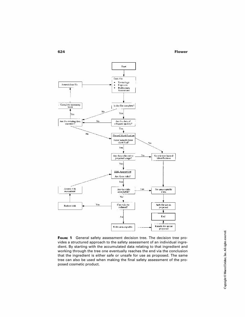
624 Flower
FIGURE 1 General safety assessment decision tree. The decision tree pro-vides a structured approach to the safety assessment of an individual ingre-dient. By starting with the accumulated data relating to that ingredient andworking through the tree one eventually reaches the end via the conclusionthat the ingredient is either safe or unsafe for use as proposed. The sametree can also be used when making the final safety assessment of the pro-posed cosmetic product.
625Safety Assessment of Cosmetic Products
a decision tree applicable to evaluating individual substances and, by analogy,completed products by guiding the safety assessor through the sequence of ques-tions in a logical manner.
If vital information on potential hazards are not available, effort to uncoverthem must be made. Previous literature searches could be extended and addition-al suppliers questioned. Should vital data not exist, then no further progress on es-tablishing the safety of a product is possible until those data are generated. Theviability of the project as a whole will need to be reassessed, taking account of thetime and cost involved in any proposed testing and the risk that the data generat-ed will not actually support the safety of the product. The possibility of generat-ing adverse data must be faced in a realistic manner.
In addition, if the data can only be obtained by animal testing, the client orcompany policy on that issue must be fully understood and followed, as must thelegislation applicable in the territory concerned. This chapter is not the appropri-ate place for a discussion of the question of animal testing [17,18]. Instead, if ananimal study is the only way of obtaining vital data which are not available byany other means and if company policy (and legal issues) allow such studies, theymust be carried out to the highest scientific standards and with full regard to thethree Rs of reduction, refinement, and replacement. Guidelines on the testing ofsubstances specifically for use as cosmetic ingredients are available [19,20].
Reduction, of course, means using the fewest animals consistent with thescientific objective being pursued, but an adequate number is still re-quired. If too few are used such that a poor study is carried out, in effectall those animals will have been wasted. Therefore, experimental designand statistical interpretation are vital factors to be considered and shouldbe fully evaluated before any study is commissioned.
Refinement means that the procedure adopted should produce the leaststress, discomfort, or interference with the animals’ normal functioning,physiology, and well being as possible, consistent with aims and purposeof the study. Remember that a stressed animal will rarely respond nor-mally and abnormal responses can compromise the integrity of thestudy. In any study, good science must be the goal, for good science iscompatible with the three Rs; bad science is not.
Replacement can mean more than simply replacing an animal study with analternative not involving animals. By extension, it can also mean replac-ing the particular ingredient with another for which testing is not re-quired or it may mean replacing animal test data with analogy, interpre-tation, or calculation to see whether the data are, in fact, needed.Replacement presents an opportunity to recheck the need for an animalstudy before it commences. However, the absence of animal test data hasbeen used to criticize the use of some ingredients and as an opportunityto challenge industry in the past.
626 Flower
2.12 Risk Characterization
This is the second main step of the safety assessment process and it is the onewhere all the hazard data on each of the ingredients are given a review with theaim of categorizing them in the light of information relating to likely exposure. Inessence, this is the first step in converting hazard identification into risk assess-ment. At this stage the safety assessor needs to be familiar with the mode of useof the cosmetic product.
Hazards that are only manifest under conditions quite unrelated to the an-ticipated usage can be put to one side. For example, lung damage following in-halation is a hazard that presents an unlikely risk if that ingredient cannot be in-haled, such as when used in a skin lotion. Similarly, liver enzyme inductionfollowing prolonged ingestion is a hazard that would be unlikely to present a riskfrom most topical cosmetics. In each case, the rationale for categorizing each haz-ard from each ingredient as an unlikely risk should be recorded.
Conversely, hazards that might related to actual usage conditions need to beidentified for priority consideration. For example, severe irritation or corrosivityassociated with an ingredient for a leave-on cosmetic needs to be noted, as doesinhalation toxicity with an ingredient of an aerosol. At this time, there should beno attempt to quantify the risk; the purpose of risk characterization is to identifyfor further assessment hazards that may pose risks.
There will be a number of ingredients which present hazards whose signif-icance is not clear at this stage and need to be assigned to a third, intermediatecategory for further consideration, for example, toxicity following ingestion of asubstance destined for use in an oral care product or lip product.
Risk characterization therefore results in a list of potential high risks wherean evaluation of the actual risk should begin, a list of moderate or unknown risksthat need to be evaluated next, and a third category or low of unlikely risks thatcan be double-checked last. Taken together, they begin to build up a picture ofwhere any risks to human health may lie with the product and, inevitably, allow adegree of assessment as to the final level of confidence in the product safety. Thisthen leads into the third step, risk evaluation.
2.13 Risk Evaluation
Although the distinction between risk characterization and risk evaluation is, insome respects, artificial, nevertheless it does represent a shift of emphasis fromdeciding whether the identified hazards present risks in general terms (and inwhat priority should they be handled) to a more specific quantification of the in-dividual risks as they apply to the product in question.
In this step, each high priority hazard is considered in the context of the useof that substance in the product. The chance or likelihood that any particular haz-
627Safety Assessment of Cosmetic Products
ard would manifest itself as harmful needs to be weighed in terms of likely sever-ity of adverse effect and in the frequency or likely prevalence of adverse effect. Inturn, these will depend on exposure (concentration of the substance in the prod-uct, quantity of product used at each application, and frequency of use) and num-bers of people exposed.
It is at the risk evaluation stage that the safety assessor needs to know like-ly consumer exposure, in terms of quantity of product used and concentration ofsubstance in the product. Also to be considered at this stage is whether the cos-metic product is intended for any special category of consumers (e.g., productsfor infants or products intended for people with sensitive skin) as this may affectlikely exposure patterns or possible susceptibility of the consumer to adverse re-actions.
2.14 Safety Assessment
This is the final step in the process of determining whether a particular cosmeticproduct is likely to be safe for release to the market. The final decision is rarelyeasy and rarely straightforward.
Of course, there may be occasions when the product being assessed is but aminor variation on a well-known product with a long history of safety in use. Ap-proval is readily granted and the certainty factor is high.
Alternatively, the product under investigation may be judged to presentclear and unacceptable risks to human health and, again, a decision is readily tak-en with a high certainty factor, but this time it is a decision not to market.
However, in the majority of cases, the decision will only be made after areappraisal of the data to hand and a reassessment of the risks involved. The ob-jective is to reduce the uncertainty involved in making the decision and to in-crease personal confidence. Remember, the underlying basis of the decision is apositive approval saying the product is safe to market and it is that decision thatmust be justifiable.
What then are the actual steps that need to be completed to justify the mar-keting decision? First, the hazards from each ingredient will have been listed, therelevant ones identified and those of little or no relevance placed to one side, witha note of justification. Second, the likelihood of each hazard being manifest willhave been judged based on the human exposure, in turn based on the concentra-tion of the ingredient, the quantity and frequency of use, the mode of use, and theparticular characteristics of the intended consumers and their numbers.
By this time, only those risks judged likely to be of relevance to the con-sumer remain to be assessed. This judgment cannot be performed in isolation; itmust be gauged against established criteria, yardsticks, or benchmarks. The expe-rienced safety assessor working within an established product field will haveready access to such benchmarks from historical records which will justify the ac-
628 Flower
ceptance of certain levels of risk. For example, the acceptability of a new sham-poo can be judged quite accurately by comparison of its formula with that of es-tablished products.
If no such benchmark is apparent, then other ways are needed to achieve aresolution. For example, if an ingredient is found to contain variable amounts ofan impurity shown to be a genotoxic carcinogen and no substitute is available,can that ingredient be used and, if so, what impurity limits should be set?
In such a circumstance, one would look to quantifying the risk over a life-time of exposure and working back from a value deemed acceptable (the bench-mark, in this case, is therefore the acceptable lifetime risk figure) to set a limit forthe level of that impurity in the final product. If the impurity level can be main-tained below the limit, the product would have an acceptable safety assessment,but if the impurity level exceeds the limit, the product must be rejected.
Such calculations are relatively common in the assessment of exposure totoxic substances in an industrial setting or in assessing the consequences of expo-sure to toxic substances present in the environment. Therefore, the safety assessorof cosmetic products must have an awareness of toxicology in its broadest senseif support is to be sought from related fields.
Having established that neither the ingredients nor their impurities consti-tute an unacceptable risk to human health in the proposed product, there needs tobe a final re-evaluation of the finished product. The safety assessor needs to checkthat the combination of impurities from different sources does not become exces-sive or that exposure to two or more substances in combination might not com-promise safety. Finally, there should be a simple logic check in which the safetyassessor reflects upon the decision made and the level of confidence. Are theredoubts? Can they be expressed and dealt with? Is the labeling adequate or exces-sive? Only when all these voices have been answered can the safety assessor saythe assessment stage has been completed.
Having decided that the proposed cosmetic is safe for marketing to the pub-lic, the safety assessor must communicate this information to colleagues and mustalso ensure any caveats are understood. The simplest way is to issue a signed doc-ument stating exactly which formula has been assessed, when, and by whom; theoutcome of that assessment; and most importantly any restrictions that are to beapplied.
For example, the safety assessor may approve the product only if the levelof a certain impurity is controlled either in one of the raw materials or in the fin-ished product itself, or only if a specific warning phrase is present on the pack (inaddition to any mandatory warnings), or only if the pack has a child-resistant clo-sure, etc.
The safety assessor may, if necessary, issue a provisional approval for lim-ited release to the market, with full approval only being granted following an
629Safety Assessment of Cosmetic Products
evaluation of the feedback from the market. Postmarketing surveillance is dis-cussed later.
An outline of a suitable document is provided as Fig. 2. The safety assessorshould include a copy of such a document with a full report of the safety assess-ment. Whilst the report need not include copies of all scientific papers and studyreports consulted, these should be referenced and stored in a library or archive.The report should be a logical argument of the process by which the assessorreached the conclusion that the cosmetic product was safe to market. In particular,it should refer to the ingredients and their toxicological profile and to any interac-tions or combined effects likely from the finished product.
3 POSTMARKETING SURVEILLANCE
The EU Cosmetics Directive requires a manufacturer to maintain a record of ad-verse reactions to a cosmetic product. Such records, whether or not they are re-quired by law, can provide essential data for the safety assessor. In essence, theyprovide evidence indicating whether the decision to market was correct.
Adverse reactions are generally held to be those that
Involve an identified individualAre directly related to a specific productAre verified by an appropriate professional (doctor, dentist, nurse, etc.)
Unless all three conditions are met, it is not possible to be sure that an adverse re-action actually occurred or that the cause was the product in question.
The background level of adverse reactions to cosmetic products is low, withthere being typically only one reaction for every several hundred thousand oreven million units sold. Should the frequency of adverse reactions rise, the safetyassessor should be informed of this fact and of the nature of those reactions. Suchfigures could be indicative of a previously unsuspected problem arising or theycould mean nothing more than a chance cluster of events.
The rarity of adverse reactions to cosmetics is both testimony to the safetyof this class of consumer product and an indicator of the problem facing the safe-ty assessor; even an unacceptable level of adverse events of, say, 1 in 10,000 isstill a very rare event. Animal studies, in vitro studies, and even human volunteerstudies are statistically quite incapable of detecting so low an incidence, muchless determining the difference between two similar products, unless exposureconditions are exaggerated to the point of being provocative. That is the reasonany studies are carried out under exaggerated conditions, so that a comparisoncan be made between the proposed product and a benchmark control. Postmar-keting surveillance provides the evidence to justify the decision made to proceedto market.
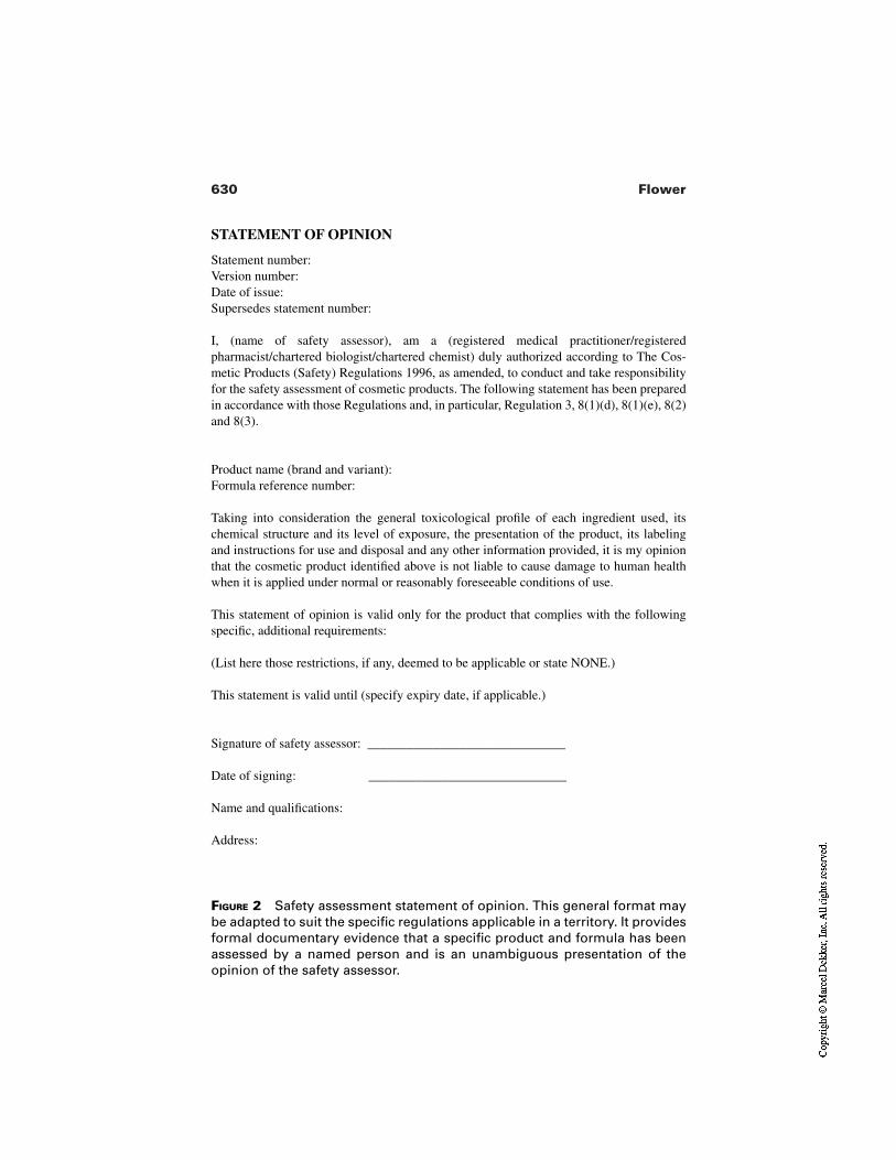
630 Flower
FIGURE 2 Safety assessment statement of opinion. This general format maybe adapted to suit the specific regulations applicable in a territory. It providesformal documentary evidence that a specific product and formula has beenassessed by a named person and is an unambiguous presentation of theopinion of the safety assessor.
STATEMENT OF OPINION
Statement number:Version number:Date of issue:Supersedes statement number:
I, (name of safety assessor), am a (registered medical practitioner/registeredpharmacist/chartered biologist/chartered chemist) duly authorized according to The Cos-metic Products (Safety) Regulations 1996, as amended, to conduct and take responsibilityfor the safety assessment of cosmetic products. The following statement has been preparedin accordance with those Regulations and, in particular, Regulation 3, 8(1)(d), 8(1)(e), 8(2)and 8(3).
Product name (brand and variant):Formula reference number:
Taking into consideration the general toxicological profile of each ingredient used, itschemical structure and its level of exposure, the presentation of the product, its labelingand instructions for use and disposal and any other information provided, it is my opinionthat the cosmetic product identified above is not liable to cause damage to human healthwhen it is applied under normal or reasonably foreseeable conditions of use.
This statement of opinion is valid only for the product that complies with the followingspecific, additional requirements:
(List here those restrictions, if any, deemed to be applicable or state NONE.)
This statement is valid until (specify expiry date, if applicable.)
Signature of safety assessor: ______________________________
Date of signing: ______________________________
Name and qualifications:
Address:
631Safety Assessment of Cosmetic Products
Although the frequency of true or verifiable adverse reactions to cosmeticproducts is low, most manufacturers receive a number of customer complaints.Those that allege adverse reactions, particularly if the allegations are of severe orpotentially long-lasting effects, need to be handled sympathetically by a special-ist from within the company. Although many of these allegations may turn out tobe groundless, some may become verified as true reactions. In any event, the pat-tern of complaints must be monitored and the data made available to the safetyassessor. How they are handled will also reflect the public image projected by thecompany.
A higher level of customer complaints does not of itself indicate a safety is-sue, but it could be indicative of poor consumer acceptance. This is clearly of im-portance to the manufacturer. A transient rise in complaints frequently follows thelaunch of a new product or relaunch of a previous one. However, if the increasefails to fall back to the level expected from past experience, the nature of the com-plaints should be investigated. Remedial action may be warranted.
4 CRISIS MANAGEMENT
In the worst circumstances, a sudden rise in the number or severity of complaintscould indicate a serious fault in a batch of product, with possible implications forhuman health. Alternatively, a new hazard may become apparent and require re-medial action. Manufacturers should have a plan prepared to manage such a cri-sis, and the safety assessor should be part of the crisis management team. Hisknowledge and expertise will be vital in helping to decide whether the problemmight even require product recall and stock uplift.
5 SUMMARY
This chapter has tried to show not only what is involved is the safety assessmentof cosmetic products and how to go about the process itself, but also to indicatethe central role a safety assessor can play in a cosmetic company. That role canextend far beyond a simple “pass or fail” statement on the safety of a proposednew product.
Safety assessors should be involved in project development planning and inensuring that adequate time is allowed for the safety assessment. They should beinvolved in product formula development to advise of possible ingredient safetyissues throughout the development. They should be involved with marketing andadvertizing and with product packaging and labeling, etc. Finally, they should beinvolved with customer relations and with crisis management. All of these sup-plement their primary role of actually assessing product safety prior to launch. Ifutilized fully, the safety assessor can avert many problems early in product devel-opment and so directly contribute to effective use of time and resources.
632 Flower
It has been said that anyone can become a toxicologist in two easy lessons,each of which takes ten years. To that I would add that the safety assessment ofcosmetic products also requires experience.
REFERENCES
1. Organisation for Economic Co-operation and Development. OECD Guidelines forTesting of Chemicals. Section 4, Health Effects. Paris: OECD.
2. de Groot AC. Adverse Reactions to Cosmetics. Groningen, The Netherlands: StateUniversity of Gronongen, 1988.
3. Colipa. Guidelines for the safety assessment of a cosmetic product. Brussels: Colipa,1997.
4. SCCNFP. Opinion concerning the revision of annex 7 of the notes for guidance forthe safety assessment of the finished cosmetic product. Brussels: European Commis-sion, 24 October 2000. http://europa.eu.int/comm/food/fs/sc/sccp/out 129_en.html
5. International Fragrance Association. Code of Practice. 33rd amendment. Geneva:IFRA, 1999.
6. Cosmetic Ingredient Review. 2000 CIR Compendium. Washington, D.C.: CIR,2000.
7. SCCNFP. Opinion of the SCCNFP concerning basic requirements for toxicologicaldossiers to be evaluated by the SCCNFP. Brussels: European Commission, 17 Feb-ruary 2000. http://europa.eu.int/comm/food/fs/sc/sccp/out111_en.html
8. SCCNFP. Opinion on in vitro methods to assess percutaneous absorption of cosmet-ic ingredients. Brussels: European Commission, 20 January 1999. http://europa.eu.int/comm/food/fs/sc/sccp/out48_en.html
9. SCCNFP. Opinion concerning basic criteria for the in vitro assessment of percuta-neous absorption of cosmetic ingredients. Brussels: European Commission, 23 June1999. http://europa.eu.int/comm/food/fs/sc/sccp/out86_en.html
10. SCCNFP. Opinion concerning the predictive testing of potentially cutaneous sensi-tising mixtures of ingredients. Brussels: European Commission, 17 February, 2000.http://europa.eu.int/comm/food/fs/sc/sccp/out102_en.html
11. National Institute of Environmental Health Sciences. The murine local lymph nodeassay: a test method for assessing the allergic contact dermatitis potential of chemi-cals/compounds. NIH publication No. 99-4494. Bethesda: NIEHS, 1999.
12. European Commission. Commission Directive 2000/33/EC of 25 April 2000 adapt-ing to technical progress for the 27th time Council Directive 67/548/EEC on the ap-proximation of laws, regulations and administrative provisions relating to the classi-fication, packaging and labelling of dangerous substances. In vitro tests for:B40—skin corrosion and for B41—phototoxicity, in vitro 3T3 NRU phototoxicitytest. Official Journal of the European Communities, 2000; L136:90–107.
13. SCCNFP. Opinion on the in vitro methods to assess phototoxicity in the safety eval-uation of cosmetic ingredients or mixtures of ingredients. Brussels: European Com-mission, 25 November 1998. http://europa.eu.int/comm/food/fs/sc/sccp/out46_en.html
633Safety Assessment of Cosmetic Products
14. SCCNFP. Opinion—guidelines on the use of human volunteers in the testing of po-tentially cutaneous irritant cosmetic ingredients or mixtures of ingredients. Brussels:European Commission, 25 November 1998. http://europa.eu.int/comm/food/fs/sc/sccp/out45_en.html
15. SCCNFP. Opinion concerning guidelines on the use of human volunteers in compat-ibility testing of finished cosmetic products. Brussels: European Commission, 23June 1999. http://europa.eu.int/comm/food/fs/sc/sccp/out87_en.html
16. SCCNFP. Opinion concerning basic criteria of the protocols for the skin compatibil-ity testing of potentially cutaneous irritant ingredients or mixtures of ingredients onhuman volunteers. Brussels: European Commission, 18 December 1999, SCC-NFP/0245/99/final. http://europa.eu.int/comm/food/fs/sc/sccp/out101_en.pdf
17. SCCNFP. Opinion concerning the present development and validation of adequatealternative methodologies to the use of animals in safety testing of cosmetics. Brus-sels: European Commission, 23 June 1999. http://europa.eu.int/comm/food/fs/sc/sccp/out84_en.html
18. SCCNFP. Opinion on the use of alternative methods to animal testing in the safetyevaluation of cosmetic ingredients (with three annexes) and the updating of notes ofguidance for testing of cosmetic ingredients for their safety evaluation (with two an-nexes). Brussels: European Commission, 20 January 1999. http://europa.eu.int/comm/food/fs/sc/sccp/out49_en.html
19. SCCNFP. Notes of guidance for testing of cosmetic ingredients for their safety eval-uation. 3d revision. Brussels: European Commission, 23 June 1999. SCCNFP/0119/99/final. http://europa.eu.int/comm/food/fs/sc/sccp/out12_en.pdf
20. SCCNFP. Notes of guidance for testing of cosmetic ingredients for their safety eval-uation. Brussels: European Commission, 24 October 2000, SCCNFP/0321/00.

28Regulatory Assessment of Cosmetic Products
Simon YoungUnilever Research, Port Sunlight Laboratory, Bebington, Wirral, United Kingdom
1 INTRODUCTION
This chapter is not intended to offer the reader an extended view of the history ortheory of the regulation of skin moisturization products. It is intended to focus onthe regulations which affect the development and marketing of such products inEurope, offer a practical guide to ensuring that products comply with these regu-lations, and give a very brief overview of cosmetic regulations in the UnitedStates and Japan.
2 CONTROL OF COSMETIC PRODUCTS IN THEEUROPEAN COMMUNITY
The Cosmetic Products Directive (76/768/EC [1] as amended) has two mainaims, first, to ensure consumer safety and, second, to create a single Europeanmarket for cosmetic products to enable free trade. The requirement for productsafety is enshrined in Article 2 of the directive and is dealt with extensively by an-other chapter of this book. Free trade is enabled by common rules on what consti-tutes a cosmetic product, ingredients, labeling and data requirements which havebeen enacted in member state legislation in response to the directive.
635
636 Young
2.1 Definition of a Cosmetic Product in the EU
Article 1 of the Cosmetic Products Directive gives the following definition of acosmetic
A cosmetic product shall mean any substance or preparation intended tobe placed in contact with the various external parts of the human body(epidermis, hair system, nails, lips and external genital organs) or withthe teeth and the mucous membranes of the oral cavity with a view ex-clusively or mainly to cleaning them, perfuming them, changing theirappearance and/or correcting body odours and/or protecting them orkeeping them in good condition.
Further clarification of what constitutes a cosmetic product is given in Annex I ofthe Directive. This is an “illustrative list” and is not exhaustive. It was put in placewhen the directive was first produced to give clarity as to what kinds of productsshould be considered to be included within the scope of cosmetic products. Thislist includes product types, such as antiperspirants and anti-dandruff shampoosthat are regulated in other markets either as medicines or in a class (or classes) be-tween cosmetics and medicines. Examples of these systems are over-the-counter(OTC) drugs in the United States, quasi-drugs (Japan), specially-controlled cos-metics (Thailand) and Risk Category 2 cosmetics (Brazil). Products fall into thesecategories either by virtue of making specific functional claims or by containingnominated functional ingredients. The level of safety and efficacy support re-quired for these intermediate classes is generally between that of a cosmetic andthat of a drug.
2.1.2 Borderline Products in the European Union
There is an overlap between the definitions of cosmetic products and medicinalproducts within European regulations. Current EC regulations that cover the bor-derline area are the Cosmetics Directive (76/768/EC as amended) and the Medi-cines Directive (65/65/EC [2] as amended). These provide definitions of cosmet-ic and medicine as follows:
A cosmetic is
Any substance or preparation intended to be placed in contact with theexternal parts of the human body . . . or with the teeth and the mucousmembranes of the oral cavity with a view exclusively or mainly forcleaning them, perfuming them, changing their appearance and/or cor-recting body odours and/or protecting or keeping them in good condi-tion.
A medicinal product is
Any substance or combination of substances presented for treating orpreventing disease in human beings or animals or Any substance or
637Regulatory Assessment of Cosmetic Products
combination of substances which may be administered to human beingsor animals with a view to making a medical diagnosis or to restoring,correcting or modifying physiological functions in human beings or ani-mals.
When the Cosmetics Directive was enacted in 1976, the Council of Europewas aware of the possibility of an overlap between the definition of a cosmeticand a medicine and that some products could fall under both regimes. In an at-tempt to avoid this, the Council made it clear in the preamble to the CosmeticsDirective that it intended to draw a dividing line between medicines and cosmet-ics. The recital reads
Whereas this Directive relates only to cosmetic products and not to phar-maceutical specialities and medicinal products; whereas for this purposeit is necessary to define the scope of the Directive by delimiting the fieldof cosmetics from that of pharmaceuticals; whereas this delimitation fol-lows in particular from the detailed definition of cosmetic productswhich refers both to their areas of application and to the purposes of use;whereas this Directive is not applicable to products that fall under thedefinition of cosmetic products but are exclusively intended to protectfrom disease.
In order to understand the thought processes behind the words it does help to lookat the way that the original 1976 definition was modified in 1993 [3].
Original:
Any substance or preparation intended for placing in contact with theexternal parts of the human body . . . or with the teeth and the mucousmembranes of the oral cavity with a view exclusively or principally tocleaning them,1 perfuming them2 or protecting them3 in order to keepthem in good condition,4 change their appearance5 or correct bodyodour.6
Current
Any substance or preparation intended to be placed in contact with theexternal parts of the human body . . . or with the teeth and the mucousmembranes of the oral cavity with a view exclusively or mainly forcleaning them,7 perfuming them,8 changing their appearance9 and/orcorrecting body odours10 and/or protecting11 or keeping them in goodcondition.12
By removing the words “in order to” and replacing the three functions [1–3] andthree objectives [4–6] by six individual purposes [7–12], the 1993 definition re-moves several legal anomalies including the one that effectively excluded all dec-orative products from being cosmetics.
638 Young
It should be noted that the phrase “exclusively or principally” has beenchanged to “exclusively or mainly” reinforcing the fact that the regulators recog-nize that cosmetic products may have functions other than those six individuallylisted.
While the Medicinal Products Directive defines a products presented fortreating or preventing a disease as a medicine, the Cosmetics Directive impliesthat a product intended for external application with a view exclusively or mainlyfor one of the six cosmetic purposes could remain a cosmetic provided that it isnot exclusively or mainly intended to protect from disease. This has been inter-preted in most member states of Europe as meaning that a cosmetic product couldhave a secondary, minor therapeutic function provided that its prime purpose wasa cosmetic one. An example of this is the marketing of sensitive teeth toothpastesas cosmetic products in all EC member states with the exception of the UnitedKingdom. Here there is clearly some overlap between the two definitions. Cos-metics sometimes claim to prevent disease, e.g., use of a fluoride toothpaste re-duces the incidence of caries and use of a sensitive teeth toothpaste reduces theperception of dentinal sensitivity. The cosmetics directive states that cosmeticproducts must deliver “exclusively or mainly” the types of cosmetic benefits list-ed in the definition. However, the word “mainly” means that a cosmetic productcan make a therapeutic claim as long as this is secondary to the cosmetic claim.This is supported by the preamble in the Cosmetics Directive that “This directiveis not applicable to cosmetic products . . . exclusively intended to protect fromdisease.” In the case of a toothpaste the primary cosmetic function is to clean and keep the teeth in good condition, the secondary function is to reduce theincidence of caries. If the product contains fluoride at a cosmetically acceptablelevel (<1500 ppm), it is classified as a cosmetic. If a similar product had no clean-ing function or contained fluoride at >1500 ppm, it would be classified as a med-icine.
It can also be argued that cosmetics often “restore, correct or modify phys-iological function.” Indeed it would be difficult to identify any product applied tothe skin that had no effect at all on physiological function. Again this is an area ofoverlap between the definitions of a cosmetic and a drug. The decision as towhether the product should be regulated as a cosmetic or not is made by each na-tional authority in the light of a number of factors. For example, will the average-ly well-informed consumer think that they are buying a medicine? This will belargely driven by the claims made in the context of the product and its presenta-tion as a whole. Does the product claim to treat or prevent a disease or to interferewith the normal operation of a physiological function of the human body. Otherfactors that will be taken into account by the competent authorities in making thisdecision will include
Medicinal implications of the product name, e.g., UlcerOut.Medicinal presentation of the product, e.g., as a tablet, or in a package typ-
639Regulatory Assessment of Cosmetic Products
ically used for medicines, or sold in pharmacies next to medicinal prod-ucts.
Medicinal implications of data made available to the public by the marketingcompany which reports a therapeutic effect. This is true whether it is giv-en directly through advertisements and helplines or indirectly via a thirdparty, e.g., in a newspaper article to which the marketer has contributed.
Medicinal implications of specifically marketing the product at particularsections of the population with, or vulnerable to, a specific adverse con-dition.
Each country in Europe independently controls what claims are permittedfor cosmetic products in their own market. There is no central EU list of permit-ted and banned claims for cosmetics and there is no central EU organization re-sponsible for deciding whether a claim is cosmetic or medicinal for the followingreasons:
Culture, historical treatment of products, habits, and attitudes vary fromcountry to country. The acceptability of claims in each country must beassessed by local nationals for local nationals.
Claims are made up of words which communicate a subtle message to con-sumers in the context of their own language and culture. Translation of acommon list into all of the languages of the EU would lead to differ-ences in meaning.
Any list of claims would require constant revision and agreement at techni-cal, bureaucratic, and political levels between all 15 countries to covernew, innovative product types and claims. This would create an enor-mous workload and lead to long delays to new product/claim introduc-tion.
Industry can be very creative in developing new products and wording newclaims to avoid rigid positive and negative lists.
The current approach allows for rapid classification of completely newproduct types and claims using the rationale and tools laid down by theEU as interpreted by local regulators.
Confirmation of this of this national approach was given by the EuropeanCourt of Justice (ECJ) in 1988. The Medicines Directive defines a medicinalproduct partly by its action on disease. However, it does not actually define whatconstitutes a disease. When this was examined by the ECJ they decided that
It is for the national authorities to determine, subject to judicial review,whether or not, having regard to its composition, the risk which its pro-longed consumption may entail or its side-effects and, more generally,all of its characteristics, a product presented as counteracting certainconditions or sensations, such as hunger, heaviness in the legs, tirednessor itching constitutes a medicinal product.
640 Young
The case of Delattre was referred to the European Court of Justice for considera-tion and decision on what constituted a medicinal product (Case C-369/88[1991]ECR 1487). The Court ruled that
A product may be regarded as being presented as a medicine if its formand the manner in which it is packaged render it sufficiently similar to amedicinal product and, in particular, if on its packaging and in the infor-mation provided with it reference is made to research by pharmaceuticallaboratories, to methods or substances developed by medical practition-ers or even to certain testimonials from medical practitioners commend-ing the qualities of the product. A statement that the product is not med-ical is persuasive evidence which the national court may take intoconsideration but is not, in itself, conclusive.
One of the few borderline cases which has come to the European Court ofJustice for resolution was that of Upjohn vs. Farzoo (Case C-112/89 [1991] ECRI-1703) in which Farzoo was marketing Upjohn’s minoxidil-based hair growthproduct Regaine® in the Netherlands as a cosmetic product. Upjohn claimed thatthis was against EC law as the product was a medicine. In its judgement the ECJdecided for Upjohn, rejecting the claim of Farzoo that a medicine could only bedefined in relation to the notion of illness. The ECJ also clarified that the defini-tion of a medicine that Regaine fell under was the second definition, i.e., “restor-ing, correcting or modifying physiological functions . . . (65/65/EC).” The keysections of the judgement are as follows:
21. With regard to what must be understood by “to restore, correct ormodify physiological function” . . . this expression must be under-stood in a manner which is sufficiently wide so as to include all sub-stances which may have an effect on the function of the body.
22. However this criterion does not include substances which while hav-ing an influence on the human body, as for instance certain cosmetics,do not have a significant effect on the metabolism which thereforestrictly speaking do not modify the condition of its function.
23. It is necessary for the national judge to proceed case by case on thenecessary categorization taking account of the pharmacological prop-erties of the product and the consideration of its methods of use, theextent of its distribution, and the knowledge of its customers.
2.2 The Sixth Amendment
In 1993 major modifications were made to the Cosmetics Directive by the sixthamendment Council Directive (93/35/EEC4), the most significant of which ad-dressed
641Regulatory Assessment of Cosmetic Products
Ingredient labelingProduct information requirementsImplications of animal testing
2.2.1 Ingredient labeling
Article 6 of the Cosmetics Directive lists labeling requirements for cosmeticproducts. The sixth amendment introduced a requirement for a label on the outerpackage to carry a list of all ingredients. Prior to the sixth amendment, the onlyingredients which had to be designated on the label were those mandated withinthe annexes of the directive.
Article 6(1)(g) of the amended directive requires that all ingredients are la-beled by descending order of weight at the time that they were added. Once thelist reaches those ingredients added at less than 1%, it does not need to be inweight order.
The nomenclature used should be the common names adopted by the Euro-pean Commission. Article 5a of the Cosmetics Directive established an inventoryof these names. Since the publication of the original inventory of cosmetic ingre-dients in June 1996 there have been approximately 1500 additions and 900changes made. The Scientific Committee on Cosmetics and Non-Food Products(SCCNFP) published the first update of the inventory, adopted 28 June 2000, onthe internet at http://europa.eu.int/comm/food/fs/sc/sccp/out123_en.pdf
International Nomenclature of Cosmetic Ingredients (INCI) names for nov-el ingredients can be easily obtained by submission of a package of chemico-physical data on the substance to the INCI Committee with a suggested INCIname. To address concerns of trade secrecy Commission Directive 95/17/EC es-tablished a provision that enables manufactures to request a code number insteadof an INCI name for ingredient listing. Once granted, confidentiality codes maybe used for up to 5 years and may be extended for a further 3 years.
Exceptions from these general ingredient labeling rules are as follows:
Ingredients added for the purposes of flavor or fragrance do not need to benamed but can be covered by the terms “aroma” or “parfum.’’
Raw material impurities do not have to be listed, neither do subsidiary tech-nical materials such as solvents or perfume carriers used in strictly nec-essary quantities.
For color cosmetics, the label can mention all of the colors in the productrange.
2.2.2 Product Information
Probably the greatest change to the Cosmetics Directive introduced by the sixthamendment was the requirement for every party responsible for placing a cos-metic product on the market in the European Community to maintain certain data
642 Young
readily accessible to government authorities. These data must be kept readilyavailable at an address specified on the product label. The information required isas follows:
The qualitative and quantitative composition of the product (in the case ofperfume compositions and perfumes, the name and code number of thecomposition and the identity of the supplier).
The physicochemical and microbiological specifications of the raw materi-als and the finished product and the purity and microbiological contentof the cosmetic product.
The method of manufacture complying with good manufacturing practicelaid down by Community law or, failing that, laid down by the law of themember state concerned. The person responsible for manufacture or firstimportation into the Community must possess an appropriate level ofprofessional qualification or experience in accordance with the legisla-tion and practice of the member state which is the place of manufactureor first importation.
An assessment of the safety for human health of the finished product.The name and address of the qualified person/people responsible for the
safety assessment. They must hold a diploma as defined in Article 1 ofDirective 89/48/EC in the filed of pharmacy, toxicology, dermatology,medicine, or a similar discipline.
Existing data on undesirable effects on human health resulting from use ofthe cosmetic product.
Proof of the effect claimed for the cosmetic product, where justified by thenature of the effect or product.
This information is the property of the company but may be viewed by the com-petent authority designated by the government in each member state. The datapackage has to be readily accessible to the competent authority. It is generally ac-cepted that 24–72 hours is considered an adequate response time.
2.2.3 Implications of Animal Testing
The use of animals for safety and efficacy testing is an emotive issue in Europewhich has ethical, political, and technical aspects, and it is right that the use of an-imals in testing of cosmetic products and ingredients is subject to scrutiny. In reg-ulatory terms, Article 2 of the Cosmetics Directive demands that a cosmetic prod-uct put on the market within the Community must not cause damage to humanhealth when applied under normal or reasonably foreseeable conditions of use.Article 7 of the directive then states that “. . . the manufacturer shall take into con-sideration the general toxicological profile of the (cosmetic) ingredient, its chem-ical structure and its level of exposure.’’
643Regulatory Assessment of Cosmetic Products
Although there are no set methodologies imposed by the cosmetics direc-tive to enable this safety assessment to be performed, there is some guidance fromthe Commission’s Scientific Committee on Cosmetics and Non-Food Products.(“See Notes for Guidance on the Safety Assessment of Cosmetic Ingredients,”SCCNFP, 1997). Some animal-based toxicological tests, used to ensure the safe-ty of pharmaceuticals, agrochemicals, dangerous substances, etc., are recom-mended. These test methods are detailed in The Organisation of Economic Co-operation and Development (OECD) publications and have been incorporatedinto EU legislation as Annex V test methods for the Dangerous Substances Direc-tive [5].
In 1993, the sixth amendment addressed concerns regarding the use of ani-mal testing by the cosmetics and associated industries by including the clause“Member States . . . shall prohibit the marketing of cosmetic products containingingredients or combinations of ingredients tested on animals after 1 January 1998in order to meet the requirements of this Directive.” This amendment also ac-knowledged that prohibition depended on the development of satisfactory, vali-dated, nonanimal alternative tests, and made provision for a postponement for notless than 2 years if the alternative methods were not available. Following a reviewof progress toward validation of nonanimal methodologies carried out in 1997 themarketing ban was postponed until 30 June 2000, with provision for a further re-assessment of progress by 1 January 2000.
2.3 Control of Ingredients
When assessing an ingredient for use in a cosmetic formulation to be marketedwithin the European Community there are two principal pieces of regulation to beconsidered. First, the Dangerous Substances Directive and, second, the Cosmet-ics Directive.
2.3.1 The Dangerous Substances Directive
The aim of the Dangerous Substances Directive (67/548/EC [5] as amended) is toprotect people and the environment from the possible harmful effects of chemicalsubstances and to create a single market in new substances across the EU. It aimsto reinforce the latter by ensuring that chemical notification requirements areidentical in all 15 member states and that there is mutual recognition of notifica-tions, i.e., a notification accepted in one member state is valid for all of them.
This regulation was issued in the form of a European Directive and hasbeen enacted into national legislation by member states. Each member state hasdesignated its own competent authority which has the responsibility of runningthe system. In the United Kingdom the competent authority is the Health andSafety Executive and the Department of the Environment, acting jointly.
644 Young
From the perspective of the Dangerous Substances Directive (DSD), poten-tial ingredients for cosmetic products for the European market are considered tobe one of the following:
Existing chemical substancesNotified chemical substancesNew chemical substances
If the ingredient is either existing or notified, then no further action will be re-quired with reference to the DPD by the cosmetic formulator. If the ingredient isnew, then it has to be notified before use in a cosmetic product unless covered byone of the exemptions described here in Section 2.4.5.
In terms of structures, the DSD has three important components, a closedinventory [6], an open inventory [7], and a notification process.
The European Inventory of Existing Chemical Substances (EINECS).This is a list of over 100,000 substances which were on the European market be-tween 1 January 1971 and 18 September 1981. Substances on this list are consid-ered to be “existing” as opposed to “new.” The list is “closed” in that no sub-stances can be added to it. The EINECS was published in the Official Journal ofthe European Community on 15 June 1990 (Vol. C146A). Substances on this listcarry a number of the format 2XX-XXX-X or 3XX-XXX-X.
The European List of Notified (New) Chemical Substances (ELINCS).Since 1981 any substance placed on the European market which was not includ-ed in EINECS has had to be notified to one of the member state competentauthorities. The process is described briefly in the next subsection. The success-ful outcome being that the new substance is added to the ELINCS list and be-comes a notified substance. These are given an ELINCS number in the format4XX-XXX-X.
Notification Process. The notification process involves submission of apackage of physicochemical characterization, toxicology, and ecotoxicology datato the competent authority. The nature and level of data required are proportionalto the annual and cumulative tonnage of the material to be placed on the market inthe EU and are defined in the regulations. Receipt of this data submission is for-mally acknowledged by the competent authority, and unless the notifier is con-tacted within 30 or 60 days (dependent upon the level of the notification) of thisacknowledgement then the material may be placed on the market. It is stronglyadvised that discussions with the competent authorities on the scope of the datasubmission are held during the planning phase of a notification.
Practical Implications. From a practical point of view then, the first stepis to determine whether the ingredient of interest already exists or has been noti-fied. This can often be done by the supplier of the material. If this is not possible,
645Regulatory Assessment of Cosmetic Products
there can be difficulties due to the differences in chemical nomenclature systemsin use by chemical suppliers around the world. Identification of the chemical ab-stracts system (CAS) number of the material is extremely helpful as a commondenominator which is used within the published EINECS/ELINCS documenta-tion. When searching EINECS it should also be noted that not all of the entries inEINECS are for specific chemicals; many substances of plant and origin are de-scribed in more general terms.
Electronic versions of the EINECS, including annually updated copies ofthe ELINCS, are also available commercially and reduce search times consider-ably. If the ingredients of interest are already included in one of the inventories,then there is no need to notify them.
Exemptions from the Dangerous Substances Directive. In terms of thecosmetics industry in Europe there are two important exemptions from the notifi-cation requirements of the regulations.
COSMETIC PRODUCTS. New ingredients which are placed on the EU only aspart of cosmetic products are exempt from notification. In practical terms thismeans that cosmetic products containing a new chemical substance may be im-ported from outside the EU without notification of that substance. This exemptiondoes not cover new chemical substances which are manufactured by a chemicalsupplier within the EU for use only in cosmetic products, as the transfer betweenthe separate legal entities of the supplier and the cosmetic company is covered bythe definition of “placing the ingredient on the market.”
SUBSTANCES NO LONGER POLYMERS. Polymers are treated differently from othermaterials by these regulations and are not subject to the same notification require-ments. The definition of a polymer in the original regulation (67/548/EC) wasmodified in 1992 by the seventh amendment to the DSD (92/32/EC [8]), and somesubstances which were covered (and hence exempt from notification) by the 1967definition of a polymer were outside the 1992 definition of a polymer given in theamending directive. Any of the materials that were inside the 1967 definition, out-side the 1992 definition, and were placed on the EC market between 1 January1971 and 1 November 1993 are considered to be exempt from notification.
SUBSTANCES TREATED AS HAVING ALREADY BEEN NOTIFIED. Three categories of no-tifiable chemical substances of importance to the cosmetics industry can be treat-ed as having already been notified under the requirements of the DSD. These aretechnically complex and subject to interpretation by the competent authorities. Itis strongly recommended that assistance and clarification are sought from the lo-cal competent authority when operating in these areas.
POLYMERS. Polymers (as defined by the directive) which meet the followingconditions do not require notification:
Those which are produced by polymerization of EINECS-listed substancesonly.
646 Young
Those which are produced by polymerization of EINECS-listed substancesonly, with postpolymerization reaction with another EINECS-listed sub-stance.
Those which contain less than 2% (in bonded form) of a non–EINECS-list-ed substance. This includes incorporation of the non-EINECS listed sub-stance in the initial polymerization or by postpolymerization reaction. Inthis case, notification may not be required if it is decided by the compe-tent authority that the polymer is identical with an existing polymer pro-duced with EINECS-listed substance(s) that is already available on theEU market.
NEW CHEMICAL SUBSTANCES FOR SCIENTIFIC RESEARCH AND DEVELOPMENT. Sub-stances intended specifically for scientific research and development in quantitiesless than 100 kg per annum do not need to be notified to the competent authority.It should be noted that the interpretation of the scope of this exemption may varybetween competent authorities, and certain information may have to be providedto the competent authority and/or maintained in-house.
NEW CHEMICAL SUBSTANCES FOR PROCESS-ORIENTATED RESEARCH AND DEVELOPMENT.Notification is not required for substances used for these purposes. However, cer-tain information, up to that for a reduced notification at the 100 kg per annum lev-el, does have to be provided to the competent authority. Again, it should be notedthat the interpretation of the scope of this exemption may vary between compe-tent authorities.
2.4 The Cosmetics Products Directive
Once a potential ingredient has been assessed against the Dangerous SubstancesDirective the next regulatory check is to ascertain whether it will be permitted foruse in a cosmetic product to be placed on the market in the European Union. TheCosmetics Directive (76/768/EEC as amended) controls the use of ingredientspermitted to be used in cosmetic products to be placed on the EC market bymeans of a negative list, a restricted list, and three positive lists. These lists areregularly updated by means of Adapting Directives which reflect advances intechnical progress.
2.4.1 Annex II (The Negative List)
Annex II is a single list of over 400 substances which must not form part of thecomposition of cosmetic products to be placed on the EC market. It should benoted that cosmetic products may be placed on the EC market if they containtraces of these materials provided that their presence is technically unavoidable ingood manufacturing practice and that they conform with the safety requirementsfor cosmetic products laid down in Article 2 of the Directive. Annex II includes
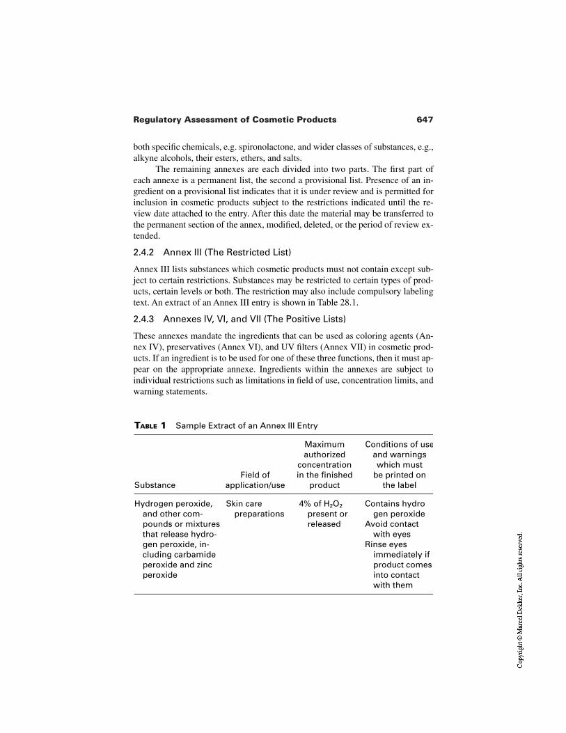
647Regulatory Assessment of Cosmetic Products
TABLE 1 Sample Extract of an Annex III Entry
SubstanceField of
application/use
Maximumauthorized
concentrationin the finished
product
Conditions of useand warningswhich must
be printed on the label
Hydrogen peroxide,and other com-pounds or mixturesthat release hydro-gen peroxide, in-cluding carbamideperoxide and zincperoxide
Skin carepreparations
4% of H2O2
present orreleased
Contains hydrogen peroxide
Avoid contact with eyes
Rinse eyes immediately if product comes into contact with them
both specific chemicals, e.g. spironolactone, and wider classes of substances, e.g.,alkyne alcohols, their esters, ethers, and salts.
The remaining annexes are each divided into two parts. The first part ofeach annexe is a permanent list, the second a provisional list. Presence of an in-gredient on a provisional list indicates that it is under review and is permitted forinclusion in cosmetic products subject to the restrictions indicated until the re-view date attached to the entry. After this date the material may be transferred tothe permanent section of the annex, modified, deleted, or the period of review ex-tended.
2.4.2 Annex III (The Restricted List)
Annex III lists substances which cosmetic products must not contain except sub-ject to certain restrictions. Substances may be restricted to certain types of prod-ucts, certain levels or both. The restriction may also include compulsory labelingtext. An extract of an Annex III entry is shown in Table 28.1.
2.4.3 Annexes IV, VI, and VII (The Positive Lists)
These annexes mandate the ingredients that can be used as coloring agents (An-nex IV), preservatives (Annex VI), and UV filters (Annex VII) in cosmetic prod-ucts. If an ingredient is to be used for one of these three functions, then it must ap-pear on the appropriate annexe. Ingredients within the annexes are subject toindividual restrictions such as limitations in field of use, concentration limits, andwarning statements.
648 Young
2.4.4 Changes to the Annexes
There is a process in place within the European Commission for updating the in-gredient annexes to reflect progress in technical knowledge. Scientific support tothis process is given by a body of independent academic experts known as theScientific Committee on Cosmetics and Non-Food Products intended for Con-sumers. Activity can be initiated by member state governments or by industry viathe European Cosmetics Industry Trade Association (COLIPA).
2.5 Impact on Selection of Potential Ingredients
The logic flows used when assessing a potential ingredient against the annexes ofthe cosmetics directive are as follows:
A substance may be used as an ingredient in a cosmetic product for purpos-es other than as a colorant, preservative, or UV filter provided that it isnot banned by inclusion in annexe II or restricted for intended purposeby annexe III and that the product is safe.
A substance may only be used as an ingredient in a cosmetic product as acolorant, preservative, or UV filter if it appears in the appropriate annexeand the product is safe.
3 OVERVIEW OF CONTROL OF COSMETICPRODUCTS IN THE UNITED STATES
Cosmetics marketed in the United States are regulated under the federal Food,Drug and Cosmetic Act (FD&C Act) [9] and the Fair Packaging and Labeling Act(FPLA) [9].
The FD&C Act defines cosmetics as articles intended to be applied to thehuman body for cleansing, beautifying, promoting attractiveness, or altering theappearance without affecting the body’s structure or functions. Included in thisdefinition are moisturizing skin creams and lotions and any material intended foruse as a component of a cosmetic product. As in the European Union there is nopremarketing approval process for cosmetic products, and the person placing theproduct on the market carries responsibility for the safety of the product.
With the exception of color additives and a few prohibited ingredients, anyraw material may be used as a cosmetic ingredient without prior approval. Thelaw requires that color additives used in food, drugs, and cosmetics must be test-ed for safety and approved by the FDA for their intended uses. The color addi-tives approved for use in cosmetics are listed at 21 CFR 73, 74, and 82 [9]. Theuse of the following ingredients is either restricted or prohibited in cosmetics:bithionol, mercury compounds, vinyl chloride, halogenated salicylanilides, zirco-nium complexes in aerosol cosmetics, chloroform, methylene chloride, chloroflu-orocarbon propellants, and hexa-chlorophene.
649Regulatory Assessment of Cosmetic Products
4 OVERVIEW OF CONTROL OF COSMETICPRODUCTS IN JAPAN
Products which are typically considered to be cosmetics in Europe are regulatedunder two separate systems in Japan. Product claims which the authorities con-sider to be more active are classified as “quasi-drugs’’. In terms of skin productsthese include those skin lotions making specific claims in areas such as chappingand roughness, prevention of razor burn, keeping the skin healthy, and supplyingthe skin with moisture. In order to make such a claim, the product must contain aquasi-drug active which has been approved for that specific class of product andmust only make claims in the area defined within the quasi-drug regulations [12].It should be noted that once preapproved by the authorities for marketing, quasi-drugs can be sold freely through retail outlets.
Control of general cosmetic products was deregulated in Japan from 1 April2001, moving away from the previous system which imposed tight restrictions onwhich ingredients could be used for each type of product toward a system whereinternationally accepted ingredients can be used. Major changes to the regulatorysystem include abolition of the premarket approval system, the adoption of ingre-dient labeling, and the implementation of positive lists for UV filters, colors, andpreservatives. Although implementation is still at an early stage, most cosmeticingredients will be freely available for use in Japan. These new regulations coverordinary skin lotions making general skin moisturization claims. Details of thesenew cosmetic regulations which contain ingredient lists, rules for prior approvalof products containing ingredients not on the lists, and new labeling requirementshave been published by the Japanese authorities [13].
5 SUMMARY
Products making simple skin moisturization claims only are generally classifiedas cosmetics around the world. Regulations controlling cosmetics are currently ina state of evolution across the world, moving generally in the direction of a har-monized system based upon the general principles of regulation in the EuropeanUnion as follows:
A standard definition of cosmetic products which clearly delineates themfrom drugs and foods
An illustrative list of product types, e.g., creams, face masks, toilet soaps,perfumes, etc.
No borderline categories between cosmetics and other product typesResponsibility for product safety clearly with the manufacturer and/or
marketerClear restriction of certain ingredientsStandardization of cosmetic Good Manufacturing Practice
650 Young
Appropriate labeling to ensure safe use of productsUse of in-market control to verify fraud or negligenceStandardization of minimum labeling requirementsIngredient labeling using INCIScientifically valid system for restricting and banning ingredients for use in
cosmetic productsSystem for regular updating of restrictions
Good progress against these principles is already being made in manycountries around the world and every step toward harmonization facilitates sup-ply to the consumer of safer and more innovative cosmetic products. However,the challenge presented to industry by these rapid changes to industry is to stayabreast of the current situation, predict short-term changes, and work with regula-tors during regulatory transition to assure that products meet the regulations in alltarget markets.
REFERENCES
1. Council Directive 76/768/EEC of 27 July 1976 on the approximation of the laws ofthe Member States relating to cosmetic products. Official Journal L 262, 27/09/1976P. 0169.
2. Council Directive 65/65/EEC of 26 January 1965 on the approximation of provisionslaid down by law, regulation or administrative action relating to proprietary medici-nal products. Official Journal B 022, 09/02/1965, pp. 0369–0373.
3. Council Directive 93/35/EEC of 14 June 1993 amending for the sixth time Directive76/768/EEC on the approximation of the laws of the member states relating to cos-metic products. Official Journal L 151, 23/06/1993, pp. 0032–0037.
4. Council Directive 93/35/EEC of 14 June 1993 amending for the sixth time Directive76/768/EEC on the approximation of the laws of the Member States relating to cos-metic products. Official Journal L 151, 23/06/1993, pp. 0032–0037.
5. Council Directive 67/548/EEC of 27 June 1967 on the approximation of laws, regu-lations and administrative provisions relating to the classification, packaging and la-belling of dangerous substances. Official Journal B 196, 16/08/1967, pp. 0001–0005.
6. EINECS Volumes I and II. Notice number 90/C 146A/01. Official Journal C146A,Volume 33, 15 June 1990.
7. Third Publication of ELINGS. Notice numbers 93/C 130/01 and 93/C 130/02. Offi-cial Journal C130, Volume 36, 10 May 1993.
8. Council Directive 92/32/EEC of 30 April 1992 amending for the seventh time Direc-tive 67/548/EEC on the approximation of the laws, regulations and administrativeprovisions relating to the classification, packaging and labelling of dangerous sub-stances. Official Journal L 154, 05/06/1992, pp. 0001–0029.
9. Code of Federal Regulations, Title 21.10. PAB Notification No. 44, February 8, 1961; No. 287, July 17, 1961; and No. 470,
November 18, 1961.11. Pharmaceutical Publication No. 990, MHW Ordinances 125, 330, and 331, Medical
Safety Bureau of the Ministry of Health and Welfare, September 29, 2000
ABT, 46Accidents, 623Acetone
xerosis, 206–209Acetone/ether
ceramide, 278dry skin, 268–269SCLs, 277water-holding, 279
Acne, 385glycolic acid, 341–342SA, 355
Acrylates, 592copolymers, 593
Actinic keratoses, 342Acylceramide. See Ceramide 1Adverse effects reports
safety assessment, 616Advertising, 17–18, 437Africa
skin care market, 5
After-feel, 578–579After wash tightness (AWT)
cleansers, 413–414Aging, 12–13
barrier homeostasis, 217ceramide, 281recessive X-linked ichthyosis, 195and sensitive skin, 151stratum corneum, 125vs. TEWL, 152
AHAs. See Alpha-hydroxy acidsAlcalase, 368Alkanolamides, 593Alkylbenzene sulfonates, 586–587Alkyl ether sulfates, 587Alkyl ether sulfosuccinates, 588Alkyl sulfates, 586Allergic contact dermatitis
topical protease, 378Allergy
topical protease, 378
651
Index
652 Index
Alpha-hydroxy acids (AHAs), 19, 89,262–263, 323–347
biochemistry, 324–327chemical peels, 339–341chemistry, 324–327dry skin, 331–333free acid content, 326future trends, 347ichthyosis, 328–329mechanism of action, 344–345photodamaged skin, 334–339, 341safety, 345–347skin benefits, 327–328skin compatibility, 343stratum corneum, 333–334
Amino acids, 270Aminobenzotriazole (ABT), 46AML, 332–333Ammonium isethionates, 587Ammonium lactate lotion (AML),
332–333Amphiphilic emollients, 225–227Anew Creams, 334Anglo-Saxons
moisturizers, 436Anhydrous emollient cream
formula, 569, 571Anionic surfactants
primary irritation, 598stratum corneum proteins, 407–408
Antenatal body creamperformance attributes, 550
Anti-aging creamvs. pseudoceramide, 290–291
Anti-aging face creamperformance attributes, 550
Anti-aging products, 18–19Antibiotics
epidermolytic hyperkeratosis, 196Antiproteinase
defense, 135Antiseptics
epidermolytic hyperkeratosis, 196Aqualysin, 390Arden, Elizabeth, 436
Arm washex vivo set up, 418
Asthmatopical protease, 378
Atopic dermatitis, 165–175, 226–227ceramides, 167–169, 172–173cholesterol, 171dry skin mechanism, 279–281emollients, 229–231
formulations, 230free fatty acids, 170–171infant, 166management, 173–175skin characteristics, 284stratum corneum lipids, 166–173
Atopic dry skinpseudoceramide, 288–297
Atopics, 20Attribute panels, 442Attribute questions, 450Autosomal dominant ichthyosis, 89AWT
cleansers, 413–414
Baby lotionsperformance attributes, 550
Bacillus licheniformus, 368–369Bacillus subtilus, 386
protease, 380–381Balsam of Peru
and blood flow, 148–150Bar soap, 585Beta-glucosylcerebrosidase, 41–42, 46Betaine, 308Bioinstruments
grading, 483Bioprase, 386
chemical modification, 391–401activity, 397characterization, 394–396procedure determination, 391–394safety test, 399stability, 396–397use test, 399–401
gene engineering, 390–392
653Index
irritation tests, 400keratin hydrolytic activity, 388sodium dodecyl sulfate-
polyacrylamide gelelectrophorograms, 387
stabilization, 390–391sweat protein hydrolytic activity, 389
Blank, Irwin, 466Blinding procedures, 485Blood flow
and balsam of Peru, 148–150detector, 148
Body care, 12Body cream
antenatalperformance attributes, 550
Body moisturizers, 19–21Body parts, 12Body wash
dual formula, 426Bound water
calculation, 276lipids, 273–274stratum corneum, 269–271
Break, 578Brownian motion, 560Butylene glycol, 572
Calcipotriollamellar ichthyosis, 196
Calcium, 215–216Caldolysin, 390Canola oil
atopic dermatitis, 174–175Capacitance, 605CAPB, 409, 411Capillary blood flow detector, 148–150Carcinogenicity, 621–622Catalog sales, 4Cationic polymers
formulation mildness, 601Caucasian skin, 67CE. See Cornified envelopeCell renewal, 335–336
Cellulite, 20Cellulosics, 592–593Celtic ancestry, 160Central location test (CLT), 451Ceramidase, 131Ceramide, 7
acetone/ether, 278aging, 281atopic dermatitis, 167–169, 172–173dose dependency, 288efficacy, 267–300emollients, 235–238fractions, 170hydrolysis, 131impedance meter, 281nomenclature, 169–170physical properties, 47–50roles, 43, 47stratum corneum, 31–55, 269structure, 286xerosis, 278–279
Ceramide 1, 43, 168atopic dermatitis, 173
Ceramide 4, 169Ceramide 5, 169Ceramide glucosyltransferase (CGT), 46CER FB, 43Cetyltrimethylammonium bromide
(CTAB), 209CGT, 46Chamomile extracts, 552Chemical peels, 339–341China
skin care market, 5Cholesterol, 7, 43, 50–51
atopic dermatitis, 171biochemistry, 50–51emollients, 235–238liquid cleansers, 425–426phase behavior, 171–172recessive X-linked ichthyosis, 195skin barrier, 54stratum corneum, 269structure, 168
Cholesterol ceramide ratio, 54
[Bioprase]
654 Index
Cholesterol ester creamvs. pseudoceramide cream,
292–293Cholesterol sulfate, 53, 189, 210
desquamation, 86–87Cholesteryl esters, 52Choline, 308Chronic plaque psoriasis, 185Chymotrypsin
visual scaling, 368–369Chymotrypsin-like serine protease,
83–84CIR, 345–346Claim support, 456–458
descriptive studies, 457–458Classic lamellar ichthyosis, 190–192
stratum corneum, 191CLE, 38, 40, 47Cleansers, 556, 585–608
AWT, 413–414bars, 23–24body wash
dual formula, 426clinical testing, 602–604
experimental design, 602–603measuring effects, 603–605
consumer testing, 602dry skin, 17enzymes, 385–402formulation, 586–594
ingredients, 586–594mildness, 600–601moisturizing potential assessment,
602–607solution/suspension structure,
595–597strategies, 597–601
function, 407irritation, 414–415
testing, 605–607liquid, 423–427, 592, 594–595moisturizing, 6–7, 23–25, 405–428proteases, 386scaling, 414skin damage, 8, 407
skin dryness, scaling, roughness, 414skin effects
clinical manifestations, 413–415skin irritation, 414–415skin moisture, 6–7stratum corneum, 16, 407–415
interactions, 407–410structure, 407
surfactants, 8TEWL, 419types, 8
Cleansing, 6–7bars, 23–24liquids, 24–25wipes, 25
Climate, 11Climate therapy
recessive X-linked ichthyosis, 195Closed (occlusive) patch testing, 606CLT, 451Coacervates, 596Coalescence, 560Cocoamido propylbetaine (CAPB), 409,
411Coconut soaps, 556–557Cocoyl sarcosinates, 587Coefficient of friction devices,
515–517Cold cream
formula, 231, 570Colloids, 596–597Communications
mass, 5–6Conditioning agents, 588–591Conductance, 605Congenital ichthyosiform erythroderma,
192stratum corneum, 193
Consumerchoices, 6expectations, 2
Consumer-based claims, 456–457Consumer data
statistical analyses, 452–453
[Cleansers]
655Index
Consumer product marketplace, 6Consumer testing, 433–460
applications, 453–460category appraisals, 458claim support, 456–458data analysis, 451–453irritation, 458–460methodology, 439–451objectives, 438–439research guidance, 454–456
Corneocyte lipid envelope (CLE), 38, 40,47
Corneocytes, 7, 61–62, 96, 385vs. CE, 108cleanser damage, 8desquamation, 162extracellular domains, 40–43glycerol, 256humidity, 255hydrophobic envelope, 205morphology, 95–96surfactants, 536–538
Corneodesmosomes, 125degradation, 126humectants, 251–255
Corneosomes, 531Corneosurfametry, 536Corneoxenometry, 536Cornified envelope (CE), 95–112
biochemistry, 96–102contrast microscopy, 102vs. corneocytes, 108formation, 97–99glycerol, 261maturation, 102–106skin condition, 106–111
Corticosteroidsatopic dermatitis, 175
Cosmeceutical, 22–23Cosmetic consultants, 3Cosmetic emulsions
benefits, 555design, 554–566
Cosmetic Ingredient HandbookCTFA, 13
Cosmetic Ingredient Review (CIR),345–346
Cosmetic moisturizers, 21Cosmetic products
defined, 635–640vs. medicinal products, 636–640nomenclature, 641regulatory assessment, 635–650safety assessment, 611–632
Cosmetics Toiletries and FragranceAssociation (CTFA), 345–346
Cosmetic Ingredient Handbook, 13Co-surfactants, 587–588Covalently-bound lipid, 99–102Creaming, 562Creams
Anew, 334antenatal body
performance attributes, 550anti-aging
performance attributes, 550vs. pseudoceramide, 290–291
formulaanhydrous emollient, 569, 571cold, 231, 570dry skin, 569, 572water-in-oil, 569
handAnglo-Saxons, 436
heparin-containing, 294–295petrolatum-based carrier
dermatitis, 233pseudoceramide, 292–297recessive X-linked ichthyosis, 195topical protease
stability, 374–377urea, 294–297
CREP, 97Crisis management, 631CTAB, 209CTFA, 345–346
Cosmetic Ingredient Handbook, 13Cutaneous barrier homeostasis,
206–209
656 Index
Cyanoacrylate skinsurface stripping, 532surface strippings, 536
Cystatin-alpha, 97Cystine-rich envelope protein (CREP),
97
Dangerous Substances Directive(67/548/EC), 643–646
exemptions, 645–646Dansyl chloride method, 336–339Deep sea fishing, 12Delayed contact hypersensitivity
topical protease, 378DEPE, 50DermaLab Moisture Meter, 510–511DermaLab TWEL Probe, 519–520Dermal Torque Meter, 514–515Dermatitis
handpetrolatum-based carrier cream,
233Dermatology
skin moisturizers, 21–22Dermis, 32
photodamage, 158–159Descriptive-based claims, 457–458Descriptive panels, 440–443
defined, 440–441establishing, 443history, 441
Descriptive tests, 453Designed experiments, 455–456Desmosomes, 89
degradation, 367glycerol, 254
Desquamation, 7abnormal
cause, 365cholesterol sulfate, 86–87stratum corneum enzymes, 81–91water, 87–89
DeSquamesstained, 605
Detergent scrub, 532–533Diastron Dermal Torque Meter, 515
Dielaidoyl phosphatidylethanolamine(DEPE), 50
Differential scanning calorimetry (DSC),51, 270–273, 275, 280, 467, 541
Difpoptosis, 40Dimethicone, 572Dipalmitoyl phosphatidylcholine
(DPPC), 51Direct sale, 4Discrimination tests, 439–440, 453Discriminative/laboratory-based claim
support testing, 457–458Distributor own brands (DOBs), 3DMAO, 411DOBs, 3Dodecyl dimethyl amine oxide (DMAO),
411Dodecyl trimethyl ammonium chloride
(DTAC), 599Doppler capillary blood flow detector,
148–150DPPC, 51Drying factors, 16Dry skin, 119–139
acetone/ether, 268–269AHAs, 331–333atopic
pseudoceramide, 288–297clinical condition, 120–124cream
formula, 569, 572genetics, 500glycerol, 258–262humectants, 256–258intercellular lipids, 271–273lactic acid, 262–263lipid removal, 268–269newborn infants, 138PCA, 262photodamage, 155–163photographs, 121–123products
performance attributes, 550stratum corneum
biology, 124–139corneocyte envelopes, 136–137
657Index
corneodesmosomal protein, 131enzymes, 131–136lipid biochemistry, 127–131morphology, 124–127
testingbioinstrumental methods, 468biomechanical developments,
466–467history, 466–470
ultraviolet radiation, 138–139urea, 262water content, 500
DSC, 51, 270–275, 280, 467, 541D-Squame adhesive discs, 507–508DTAC, 599Dual formula body wash, 426Duo-trio tests, 440Dustbin hypothesis, 65
Eastern Europeskin care market, 5
EEMCO (European Expert Group ofEfficacy Measurements ofCosmetics and Other TopicalProducts), 501
Efficacysafety assessment, 616
Egyptmoisturizers, 435
EINECS, 644Elafin, 97Electrodynamometer
skin moisturization testing, 147ELINCS, 644Emollient cream
anhydrousformula, 569, 571
Emollients, 9–10, 13–15amphiphilic, 225–227atopic dermatitis, 229–231barrier protection, 227–231ceramide, 235–238cholesterol, 235–238classification, 224–226clinical effects, 223–238
epidermolytic hyperkeratosis, 196fatty acids, 235–238hydrating effect, 226hydrogel, 225lanolin, 231–232occlusive effect, 226–227oil-in-water, 225physiological lipids, 235–238recessive X-linked ichthyosis, 195water-in-oil, 225
formulations, 230Emotional factors, 11Emulsifiers, 552–553
selection, 558–560types, 556–558
Emulsions, 10characteristics, 563stability factors, 561visual impact, 575–576
Englishmoisturizers, 436
Environment, 11, 12, 16moisturizer clinical testing, 480–481,
487Envoplakin, 97, 99Enzymes
cleansers, 385–402Epidemiology studies
safety assessment, 623Epidermis
keratolytic compoundsin vivo, 359–360
photodamage, 157–158, 158sterologenesis, 43strata, 32–33
Epidermolytic hyperkeratosis, 192–194,341
treatment, 196Epidermopoiesis, 499Equilibration period, 485Erythrodermic psoriasis, 184Erythrodermic stratum corneum, 183ESR spectroscopy, 422–423Esthetics, 10
moisturizers, 574–580
[Dry skin] [Emollients]
658 Index
Ether. See Acetone/etherEthnic skin, 11European community
cosmetic product control, 635–649European Community Cosmetics
Directive (76/768/EEC),611–632, 635–640
annexes, 646–648cosmetic product defined,
635–640safety requirement, 613–629
correct product, 614–625sixth amendment, 640–643
European Expert Group of EfficacyMeasurements of Cosmetics andOther Topical Products, 501
European Inventory of ExistingChemical Substances (EINECS),644
European List of Notified (New)Chemical Substances (ELINCS),644
Evening primrose oil, 552Experienced/semi-trained panel,
442–443Expert assessment, 472Expert panels, 441Expert visual grading, 482Exposure estimation
safety assessment, 617Eye irritation, 619
Face creamanti-aging
performance attributes, 550Facial care, 12
products, 3value, 2
Facial moisturizers, 18–19Fair Packaging and Labeling Act
(FLPA), 648Fair skin, 160Faraday-Tyndall effect, 597Fatty acids, 7, 47, 420, 552
emollients, 235–238esters, 593
free, 51–52phospholipids, 307
Fatty acyl co carboxylase, 43FCAT, 415FDA, 345Filaggrin, 65, 97, 103, 105
oral epithelia, 70–72proteolysis, 69–70
activation, 72–74FITR spectrophotometer, 513FLPA, 648Focus groups, 445–446Food, 3Food and Drug Administration (FDA),
345Food Drug and Cosmetic Act, 648Foods, 433Forearm controlled application technique
(FCAT), 415Formula identification
safety assessment, 615Fountain of youth, 5Fourier Transform IR (FITR)
spectrophotometer, 513Fragrances, 443–444, 574, 591Free fatty acids, 51–52
atopic dermatitis, 170–171
Galen, 435Gas bearing electrodynamometer
skin moisturization testing, 147Gas-bearing electrodynamometer (GBE),
514Gaucher’s disease, 46GBE, 514Gelatin/glycine
vs. glycolic acid, 335Genetic counseling
recessive X-linked ichthyosis, 195Genotoxicity, 621Global skin care industry
value, 2Glucosylcerebrosidase, 41–42, 46Glutamates, 588Glutamine, 65
[Fatty acids]
659Index
Glycerides, 589Glycerin, 572Glycerin bars, 24Glycerol, 249
barrier functionin vivo, 261–262
CE, 261corneocytes, 256desmosomes, 254dry skin, 258–262pleiotropic properties, 250–251stratum corneum, 256–258xerosis, 259–260
Glycerylphosphatidylcholine (GPC), 308Glycolic acid, 89, 335–336
acne, 341–342vs. gelatin/glycine, 335PFB, 342vs. salicylic acid, 338
Glycosidases, 135GPC, 308Greasiness, 577–578Greece
moisturizers, 435–436
Handcream
Anglo-Saxons, 436dermatitis
petrolatum-based carrier cream, 233moisturizers, 19–21
Hazard identification, 614, 617–625Heat-stable enzymes, 390Hedonic questions, 450Hedonic terminology, 438Heparin-containing cream
vs. pseudoceramide cream, 294–295Herbs, 433Hippocrates, 435Histamine receptor, 217HLB, 558–560HMG Co A reductase, 43Home use test (HST), 451Housewives, 501HPC. See Hydrogenated
phosphatidylcholine
HST, 451Human data
safety assessment, 622–625Human volunteer studies
safety assessment, 623Humectants, 13–15, 245–263, 501,
534barrier lipids, 255–256corneodesmosomes, 251–255effects, 256–258efficacy, 248–249hygroscopic measurement, 248–249and moisturizers, 248–249stratum corneum, 249–251
plasticization, 252Humidity
corneocytes, 255stratum corneum, 16xerosis, 212–213
Hydrocarbons, 589–590Hydrogel emollients, 225Hydrogenated phosphatidylcholine
(HPC), 303, 310chemical structure, 311composition, 305–306hydration, 312–316SC lipids, 310sensitive skin tolerance, 312skin uptake, 310–312topical
normal skin, 316–317Hydrophile/lipophile balance (HLB),
558–560Hydroxyethylcellulose, 593Hydroxypropyl methylcellulose, 593
ICDtopical protease, 378
Ichthyosiform erythrodermacongenital, 192
Ichthyosis, 187–196AHAs, 328–329autosomal dominant, 89classic lamellar, 190–192lamellar, 137, 189–192
prenatal diagnosis, 196
660 Index
PHA, 341X-linked, 500
Ichthyosis vulgaris, 187–188treatment, 194
IgE antibody, 379Image analysis
in vivo, 508Industrial exposures, 623Infants
atopic dermatitis, 166newborn
dry skin, 138Information, 4
reliable sources, 26–27Infrared spectroscopy, 512–513Ingredients
European Community CosmeticsDirective (76/768/EEC), 643–646
safety assessment, 615Instructions
safety assessment, 616Instrumental methods, 472Integument, 53–55Intercellular lipids
dry skin, 271–273stratum corneum, 269–271
Internet, 4, 26, 434consumer testing, 447–448
Involucrin, 96, 100, 210–211Irritant contact dermatitis (ICD)
topical protease, 378Irritation, 619
anionic surfactants, 598cleansers, 414–415
testing, 605–607consumer testing, 458–460surfactant penetration, 599–600surfactants, 598–599
prediction, 535–537Irritation tests
Bioprase, 400Isethionate, 24Isotretinoin
recessive X-linked ichthyosis, 195
Japancosmetic product control, 648–649skin care market, 5
Jergen’s lotion, 436Journals
peer reviewed, 27
Keratin, 385Keratin/filaggrin interaction, 69–70Keratinocytes, 32, 96
cultures, 46photodamage, 157–158
Keratohyalin granules, 35, 65Keratolinin, 97Keratolytic compounds
comparative effects, 357–362epidermis
in vivo, 359–360stratum corneum
in vivo, 357–359Keratolytics, 355–356
recessive X-linked ichthyosis, 195Kligman, Albert, 23Kligman regression test, 474–475
Label, 1–2Labeling
European Community CosmeticsDirective (76/768/EEC), 641
safety assessment, 616Lactic acid, 19, 248–249, 330–331,
335–336dry skin, 262–263NMF, 330–331recessive X-linked ichthyosis, 195
Lamellarichthyosis
treatment, 195–196Lamellar bodies, 35, 38Lamellar ichthyosis, 137, 189–192
prenatal diagnosis, 196treatment, 195–196
Langerhans cells, 32Lanolin
emollients, 231–232long term effects, 231
[Ichthyosis]

661Index
LAS, 542Laser Doppler capillary blood flow
detector, 148–150Laser particle sizer, 563Laser resurfacing, 340–341Laundry bars
enzyme containing, 380Lauryl sarcosinates, 587Lecithin, 305–310
terminology, 308Liarazole
lamellar ichthyosis, 196recessive X-linked ichthyosis, 195
Lineactant, 51Linear alkyl benzene sulfonate (LAS),
542Linear Skin Rheometer, 415, 417Linoleic acid, 552Lipase, 401Lipids, 589–590
bound water, 273–274covalently-bound, 99–102dry skin, 268–269, 271–273liquid cleansers, 423–427permeability barrier
humectants, 255–256physical properties, 47–53
stratum corneum, 44–45, 127–131,204–205, 269–271
water-holding mechanism, 274–275Liquid cleansers
formula, 594–595lipid deposition, 423–427moisturizing, 24–25thickening, 592
Liquid crystals, 597emulsions, 556
Liquid detergents, 24–25Local tolerance, 619Lotions
ammonium lactate, 332–333Jergen’s, 436moisture
formulation, 567–568performance attributes
antenatal, 550
baby, 550topical protease
stability, 374–377LSA
chemical structure, 356cosmetic implications, 360–362
Lysozyme, 401
Macrophotographsscanning microdensitometry, 506–507
Magazines, 26Magnesium, 215–216Mail order, 4Mammalian skin
histology, 32–40Manufacturability, 553Markets
experiencesafety assessment, 623
research, 434segmentation, 5skin care, 5–6, 27–28
Mass communications, 5–6Mass market products, 3Mature markets
skin care, 5Mechanoreceptors, 32Media, 26–27Medicinal products
vs. cosmetic products, 636–640Melanins, 360Melanocytes, 32
photodamage, 158Menopause
sensitive skin, 151Merkel cells, 32Message, 26–27Methylcellulose, 593Microbial stability, 565–566
safety assessment, 615–616Microscopy, 563, 564–565Mineral oil, 552Miniature Mechanical Tester, 415Mini-regression testing, 468, 474,
504–505
[Lotions]
662 Index
Misinformation, 26Mitochondrial DNA (mt DNA), 163Moisture
cleansers, 6–7retention, 7
Moisture lotionformulation, 567–568
Moisturemeter hydration tests, 474Moisturization
definition, 146moisturizing cleansers, 406–408natural mechanisms, 7–8testing, 147and TEWL, 145–153
Moisturizers, 9–17, 248–249Anglo-Saxons, 436assessment
short-term hydration studies, 505body, 19–21clinical testing, 465–495
body site, 477cells, 477–479clinical design, 470–475control products, 479duration, 479–480environment, 480–481, 487example protocols, 491–495grading, 482–484measurements, 472–473objective, 477observation events, 485–487pretreatment phase, 488–490product treatment phase, 489–490protocol, 473–476regression, 490–491sample administration, 487–488scope, 471–472subjects, 480–482
continued use, 579–580cosmetic, 21customization, 10dermatology, 21–22descriptive characterization,
443–445development
stratum corneum, 542
differencesassessment, 453–454
Egypt, 435English, 436esthetics, 574–580facial, 18–19factors influencing, 16–17formulation, 547–583
design, 554–567example, 567–573extraneous constraints, 553–554performance criteria, 574–583technological influences,
549–553Greece, 435–436hand, 19–21historical development, 435–437and humectants, 248–249instrumental assessment, 505–520
skin surface appearance, 506–508skin surface mechanical properties,
514–519stratum corneum barrier function,
519–520stratum corneum hydration state,
508–514marketplace, 1–28noninvasive assessment, 499–522
clinical studies, 501–505packaging, 566–567processing, 566–567regulatory categories, 22Rome, 435–436sensory characteristics, 575–579similarities
assessment, 453–454skin
dermatology, 21–22sun protection, 25
skin interaction, 580–582skin physiology, 582–583sun protection, 25terminology, 437–438upper mass cosmetic
formula, 570–574
[Moisturizers]
663Index
Moisturizing, 6–7cleansers, 23–25, 405–428
liquids, 24–25skin moisturization, 406–408wipes, 25
gel formula, 572, 575ingredients
skin deposition, 601Monadic test design, 448–449Monoalkylphosphates, 587Motta system, 170Mt DNA, 163Mucous membrane irritation, 619Multiple phase emulsions, 556Mutagenicity, 621
NAEL, 620Naive consumer tests, 445–451Natural moisturizing factor (NMF), 7,
62–63function, 529generation, 74–75lactic acid, 330–331mechanism of action, 64origin, 64–68stratum corneum, 137–139
Near-infrared spectroscopy, 513Negative list
European Community CosmeticsDirective (76/768/EEC), 646–647
NEL, 620Netherton syndrome, 43Newborn infants
dry skin, 138Newtonian flow, 579NMF. See Natural moisturizing factorNMR, 513–514No adverse effect level (NAEL), 620NOAEL, 620No effect level (NEL), 620NOEL, 620Nondesigned experiments, 455Nonident P-40, 209Nonionic emulsifiers, 557No observed adverse effect level
(NOAEL), 620
No observed effect level (NOEL), 620
Normal skincare, 6–7humectants, 256–258stratum corneum
biology, 124–139corneocyte envelopes, 136–137corneodesmosomal protein, 131enzymes, 131–136lipid biochemistry, 127–131morphology, 124–127
topical HPC, 316–317Nova Dermal Phase Meter, 510Nuclear hormone receptor, 215Nuclear magnetic resonance (NMR),
513–514Nursing, 12Nutrition protocol regression test, 469,
475
Observational studies, 446–447Occlusion, 501Occlusives, 9, 13–15Occupation, 12OECD, 611Oil-in-water emollients, 225Oil-in-water emulsion
LSA, 360–361Oil-in-water-in-oil, 556Oils, 589–590Opacifying agents, 594Open ended questions, 450Optimase, 368, 371–373
stability, 375Oral epithelia
filaggrin, 70–72Organization for Economic Cooperation
and Development (OECD), 611Organoleptic stability
testing, 564–565
Packagingmoisturizers, 566–567safety assessment, 616
Pack label, 1–2
664 Index
Palmo-plantar stratum corneum, 109–110Panelists
performanceanalysis, 452–453
restriction, 482selection, 480–481
Papain, 386allergic reaction, 379
Paraffin, 552, 589–590PAS, 512PC. See PhosphatidylcholinePCA, 63, 250
dry skin, 262PE
chemical formulas, 306, 308Pearling agents, 594Peer reviewed journals, 27PEG-MA copolymer, 391–396Perception
product performance, 10–11Performance testing, 553Periplakin, 97Permeability barrier, 162
cholesterol, 54lipids
humectants, 255–256physical properties, 47–53
plastic crystal model, 53–55properties
description, 466recessive X-linked ichthyosis, 188stratum corneum, 519–520structural morphology, 54–55
Personal caremarket segments, 2–3products, 2
growth, 2Personal cleansing technology
evolution, 406Petrolatum, 231–234, 237–238, 377
barrier recovery, 233stratum corneum, 232, 234–235xerosis, 213–214
Petrolatum-based carrier creamdermatitis
hand, 233
Petroleum, 589–590products, 552
Petroleum jelly. See PetrolatumPFB
glycolic acid, 342PHAs, 341Phosphatidic acid
chemical formulas, 306Phosphatidylcholine (PC), 50–51
chemical formulas, 306saturated fatty acids, 310skin hydration, 303–319skin treatment, 309–310water, 308–309
Phosphatidylethanolamine (PE)chemical formulas, 306, 308
Phosphatidylglycerolchemical formulas, 306
Phosphatidylinositolchemical formulas, 306
Phosphatidylserinechemical formulas, 306
Phospholipase A (PLA), 308Phospholipase D (PLD), 308Phospholipids, 304, 305–310
chemical structure, 305–306fatty acids, 307
Photoacoustic spectroscopy (PAS), 512Photoaging, 582
clinical manifestations, 156Photocarcinogenicity, 622Photodamage
AHAs, 334–339, 341dermis, 158–159dry skin, 155–163effects, 156–159epidermis, 157–158stratum corneum, 157visual analog scale, 160–161vulnerable phenotypes, 160
Photogenotoxicity, 622Photographic techniques, 447Photomutagenicity, 622Photosensitization, 619–620Physicochemical stability
testing, 564–565
665Index
Physiological lipidsemollients, 235–238
Phytosphingosine, 47Pilot trials, 471PLA, 308Plant extracts, 552Plastic crystal model
permeability barrier, 53–55PLD, 308Polyglucosides, 588Polyhydroxy acids (PHAs), 341Polymeric emulsifiers, 557Polymers, 590Polyols, 592Positive list
European Community CosmeticsDirective (76/768/EEC), 646–647
Postmarketing surveillance, 629–631Potassium, 215Preference questions, 450Prenatal diagnosis
lamellar ichthyosis, 196recessive X-linked ichthyosis, 195
Preservatives, 591Prestige products, 3–4Primary irritation
anionic surfactants, 598surfactants, 598–599
Processingmoisturizers, 566–567
Productscycle, 17–18information
European Community CosmeticsDirective (76/768/EEC), 641–642
matching, 453performance
perception, 10–11stability
safety assessment, 615Profilaggrin, 68–69, 138, 187–188Profilometry
skin surface replicas, 506Projective techniques, 446Propionates, 588
Propylene glycolrecessive X-linked ichthyosis, 195
Protease-induced desquamationmechanism, 371–374
Proteases, 83–86cleansers, 386function, 386–389keratin hydrolytic activity, 387–388sweat protein hydrolytic activity,
388–389topical
efficacy, 367–374future use, 381–382health effects, 378–379safety, 378–382safety studies, 379–381stability, 374–377visual scaling, 367–371xerosis, 365–382
Proteins, 590Pseudoceramide
vs. anti-aging cream, 290–291atopic dry skin, 288–297clinical efficacy, 284–297design, 282–284experimentally induced dry skin,
284–287SC sheets
bound water-holding capacity,283–284
structure, 286water-holding function, 282–283xerosis, 287
Pseudoceramide creamvs. cholesterol ester cream, 292–293vs. heparin-containing cream, 294–295vs. urea cream, 294–297
Pseudofolliculitis barbae (PFB)glycolic acid, 342
Psoriasis, 181–187barrier function, 186chronic plaque, 185erythrodermic, 184histology, 182
Psychological stress, 206–207barrier homeostasis, 217
666 Index
Pyrrolidone carboxylic acid (PCA), 63dry skin, 262
QSARs, 533Qualitative testing, 445–448Quantitative structure-activity
relationships (QSARs), 533Quantitative testing, 448–451
compliance, 451improving sensitivity, 450–451locations, 450questionnaire design, 450research designs, 449seasonality, 451
Reactive oxygen species, 386Recessive X-linked ichthyosis, 89,
188–189, 210age, 195genetic counseling, 195histology, 188permeability barrier, 188prenatal diagnosis, 195stratum corneum, 189treatment, 195
Reduction, 625Refinement, 625Regression testing, 467–468, 501–504
short term alternative, 469–470Regulatory influence, 580–583Repeat dose, 620–621Replacement, 625Respiratory sensitization
topical protease, 378Restricted list
European Community CosmeticsDirective (76/768/EEC), 646–647
Retinoidsepidermolytic hyperkeratosis, 196lamellar ichthyosis, 195–196
Rheology, 579Rheometer, 563Risk
characterization, 614, 625evaluation, 614, 625–626
Romemoisturizers, 435–436
Rub-in, 576–577Rubinstein, Helena, 436Ruthenium tetroxide, 172, 181
SA, 353–363anti-aging products, 326–327discovery, 354
SACD, 532Safety assessment, 614
decision tree, 624Salicylic acid
chemical structure, 354cosmetical properties, 355–356derivatives, 356–362dermatological properties, 355–356dose dependence, 355–356form, 355vs. glycolic acid, 338historical use, 354keratolysis, 356–357microbiological data, 357specificity, 356topical use, 354
Salicylic acid (SA), 353–363anti-aging products, 326–327discovery, 354
Salts, 567Sarcosinates, 588Saturated fatty acids
phosphatidylcholine, 310Scaffold protein, 72Scaling
cleansers, 414Scanning microdensitometry
macrophotographs, 506–507SCCE, 132–136, 366, 371–372SCCNFP
cosmetic nomenclature, 641Scientific Committee on Cosmetics and
Non-Food Products (SCCNFP)cosmetic nomenclature, 641
SC lipids (SCLs), 269acetone/ether, 277HPC, 310
667Index
SCLs, 269acetone/ether, 277HPC, 310
Scratch resistance test, 517Scum, 586SDS, 207, 209Sebaceous gland
photodamage, 158Seborrheic keratoses, 342Sebum, 385Secretory leukocyte protease inhibitor
(SLPI), 136Sedimentation, 562Self-assessed skin sensitivity
and TEWL, 145–153Self assessment, 472, 483–484Senile xerosis, 366Sensitive skin
and age, 151definition, 146menopause, 151and moisturization, 145–153properties, 147–150and psychological stress,
150–151testing, 146TEWL, 145–153tolerance
HPC, 312Sensitization, 619Sensory characteristics
moisturizers, 575–579Sensory data
statistical analyses, 452–453Sequential test design, 448–449Serine palmitoyl transferase, 43Serine proteases, 368–369
chymotrypsin-like, 83–84Serine residues
phosphorylation, 65Short-term hydration studies
moisturizer assessment, 505Shower gel, 426Signatures, 437Silicone oils, 556Silicones, 590–591
Single gel phase modelpermeability barrier, 53–55
SKALP, 136Skin. See also Dry skin; Normal skin;
Sensitive skinCaucasian, 67cyanoacrylate, 532, 536damage
reduction, 598–601ethnic, 11fair, 160mammalian
histology, 32–40measurement
classes, 472–473permeation, 534–535sensitization
topical protease, 378type, 11xerotic, 119–139
Skin barriercholesterol, 54
Skin caremarket
regional variation, 5–6trends, 27–28
vs. skin protection, 228Skin condition
CE, 106–111Skin deposition
moisturizing ingredients, 601Skin-derived antileukoprotease
(SKALP), 136Skin-feel descriptive methods, 441–442Skin hydration
PC, 303–319Skin interaction
moisturizers, 580–582Skin lipids deposition
Syndet bars, 419–422Skin moisturization. See MoisturizationSkin moisturizers. See also Moisturizers
dermatology, 21–22sun protection, 25
Skin physiologymoisturizers, 582–583
668 Index
Skin prick tests (SPT), 380Skin protection
vs. skin care, 228Skin sensitivity. See Sensitive skinSkin surface replicas
profilometry, 506Skin treatment
PC, 309–310Skin ultrastructural changes
soap vs. Syndet bars, 418–419Skin uptake
HPC, 310–312SLES, 409–410, 411Slip, 578SLPI, 136SLS, 261–262, 303, 314, 542Small proline-rich proteins (SPRs),
96–97Snap fasteners, 355Soap, 556
bars, 8–9, 585coconut, 556–557induced xerosis, 260stratum corneum, 125vs. Syndet bars, 415–427
skin ultrastructural changes,418–419
SOD, 401Sodium alkyl sulfosuccinates, 588Sodium dodecyl sulfate (SDS), 207,
209Sodium lactate, 250, 330Sodium laureth sulfates, 587Sodium lauryl ether sulfate (SLES),
409–410, 411Sodium lauryl sulfate (SLS), 261–262,
303, 314, 542Sodium pyrrolidone carboxylic acid
(PCA), 250Solutions, 596Sonic wave propagation, 517–519South America
skin care market, 5Spectroscopy
ESR, 422–423infrared, 512–513
near-infrared, 513photoacoustic, 512
SPF, 25, 582Sphingolipids, 47Sphingomyelin deacylase, 280Spinous layer, 33Splayed chain, 47Spreadibility, 437S protein, 131SPRs, 96–97SPT, 380Squalene-monohydroperoxide
xerosis, 210Squametry
tape strippings, 507–508Stability, 560–563
testing, 563–566Stained DeSquames, 605Statistics
graphical representation, 451–452Stearic acid
Syndet bar, 422Steroids
topicalatopic dermatitis, 175
Steroid sulfatase, 500Stinging test
skin moisturization, 148, 149Store brands, 3, 20–21Stratum basale, 33Stratum compactum, 344Stratum corneum, 95
AHAs, 333–334barrier function, 519–520biology, 124–139bound water, 269–271ceramides, 31–55chemical partitioning, 533classic lamellar ichthyosis, 191cleansers, 16, 407–415, 420–421composition, 407, 529congenital ichthyosiform
erythroderma, 193corneocyte envelopes, 136–137corneodesmosomal protein, 131
[Spectroscopy]
669Index
electrodynamometer, 147enzymes, 131–136
desquamation, 81–91erythrodermic, 183function, 204–206, 206glycerol, 256–258harvesting from human volunteers,
532–533humectants, 249–251
plasticization, 252humidity, 16hydration
SACD, 539hydration state assessment, 508–514
electrical properties, 508–510spectroscopic methods, 512–514
intercellular lipids, 269–271keratolytic compounds
in vivo, 357–359laboratory ex vivo assessment,
529–542harvesting, 531–533xenobiotics, 533–538
lipids, 44–45, 204–205atopic dermatitis, 166–173biochemistry, 127–131structure, 168surfactant interactions, 410–413in vitro tests, 541–542
moisturization, 61–76, 248–249moisturizer development, 542moisturizing mechanisms, 268–277morphology, 124–127NMF, 137–139outermost layer, 61palmo-plantar, 109–110petrolatum, 232, 234–235photodamage, 157prospects, 90–91proteins
surfactant interactions, 407–410recessive X-linked ichthyosis, 189separation from excised skin samples,
531–532structure, 7, 206
swellingfactors affecting, 536
ultrastructure, 82, 180ultraviolet radiation, 161–163in vitro adsorption, 533–534water, 16, 246–248
balance, 62diffusion, 539–541humidity, 247plasticization, 250uptake, 538–541
wind, 16xerosis, 204–206
Stratum corneum chymotryptic enzyme(SCCE), 132–136, 366, 371–372
Stratum corneum swelling test, 535–537Stratum disjunctum, 344Stratum granulosum, 35Stratum spinosum, 33Stress
psychological, 206–207barrier homeostasis, 217and skin sensitivity, 150–151
Strippingvs. TEWL, 148, 149
Strippings with adhesive-coated discs(SACD), 532
Structured surfactants, 589Subramanyan, 425Sunflower seed oil, 552Sun-induced skin spots, 342Sun protection
skin moisturizers, 25Sun protection factor (SPF), 25, 582Sunscreens, 25Supermarkets, 3Superoxide dismutase (SOD), 401Surfactants, 396, 586–588
anionicprimary irritation, 598stratum corneum proteins, 407–408
cleansers, 8corneocytes, 536–538formulation mildness, 600–601
[Stratum corneum] [Stratum corneum]
670 Index
penetrationirritation, 599–600
primary irritation, 598–599structured, 589xerosis, 209–210
Suspensions, 595–596Sweat proteins
sodium dodecyl sulfate-polyacrylamide gelelectrophorograms, 389
Syndet barsskin lipids deposition, 419–422vs. soap, 415–427
skin ultrastructural changes,418–419
Tackiness, 578Talc, 566Tallow soaps, 556Tape strippings
squametry, 507–508xerosis, 206–209
Taurates, 588Temperature
dry skin, 16–17Tensides, 228Tensiometer, 563Terminology
moisturizers, 437–438Tetramethylrhodamine isothiocyanate
(TRITC), 103–105TGases, 97, 100–102, 136
enzymes, 107schematic, 98type 1, 186
Theobroma oil, 571Thermitase, 390Thermolysin, 390Thin-layer chromatography (TLC), 269,
271Tissue matrix metalloproteinase
inhibitors (TIMPs), 136Titanium dioxide, 566TLC, 269, 271
Toilet soapspH, 586
Toxicityacute, 618–619long-term, 621–622safety assessment, 617subchronic, 620–621
Toxicokinetics, 622Tranglutaminase (TGase), 136Trans-4-(aminomethyl) cyclohexane
carboxylic acid (t-AMCHA)xerosis, 214
Trans-epidermal water loss (TEWL), 7,43, 145, 246
vs. age, 152cleansers, 419early measurement, 466measurement, 519–520sensitive skin, 152vs. stripping, 149
Transglutaminases (TGases), 97,100–102
enzymes, 107schematic, 98type 1, 186
Travel, 12Tretinoin
epidermolytic hyperkeratosis, 196Triangle tests, 440TRITC, 103–105TritonX-100, 209Trypsin-like protease, 84–86Turbidometry, 563TWEL. See Trans-epidermal water lossTwistometre, 514–515
UCA, 63Ultraviolet A, 156–157, 582
stratum corneum, 161–163Ultraviolet B, 156, 582
stratum corneum, 161–163Ultraviolet C, 156Ultraviolet radiation, 155–163
dry skin, 17, 138–139stratum corneum, 161–163
[Surfactants]
671Index
United Statesskin care market, 5
Upper mass cosmetic moisturizerformula, 570–574
Upper mass market, 4Urea
dry skin, 262recessive X-linked ichthyosis, 195
Urea creamvs. pseudoceramide cream, 294–297
Urocanic acid (UCA), 63
Value brands, 20–21Viscosity modifiers, 592–594Visual analog scale
photodamage, 160–161Visual grading
expert, 482Visual scaling
chymotrypsin, 368–369topical protease, 367–371
Vitamins, 433
WartsSA, 355
Waterbalance
stratum corneum, 62bound, 269–276content
dry skin, 500desquamation, 87–89diffusion
stratum corneum, 539–541phosphatidylcholine, 308–309stratum corneum, 16, 246–248,
538–541plasticization, 250
Water-holding mechanismacetone/ether, 279lipid components, 274–275
Water-in-oil creamformula, 569
Water-in-oil emollients, 225formulations, 230
Water phase thickeners, 567Water-soluble materials
water-holding properties, 276–277Western Europe
skin care market, 5Willow tree, 354Wind
stratum corneum, 16Winter itch, 119–120Wool wax. See Lanolin
Xerosis, 341acetone, 206–209barrier disruption, 208–209ceramide, 278–279dry skin mechanism, 277–279glycerol, 259–260humidity, 212–213pseudoceramide, 287SA, 355senile, 366soap-induced, 260squalene-monohydroperoxide, 210stratum corneum, 204–206
biochemical factors, 205surfactants, 208, 209–210tape-stripping, 206–209topical protease, 365–382treatment, 213–217
Xerotic skin, 119–139electron microscopy, 127stratum corneum
lipid biochemistry, 129–130X-linked ichthyosis, 500
Zinc oxide, 566


Related Documents