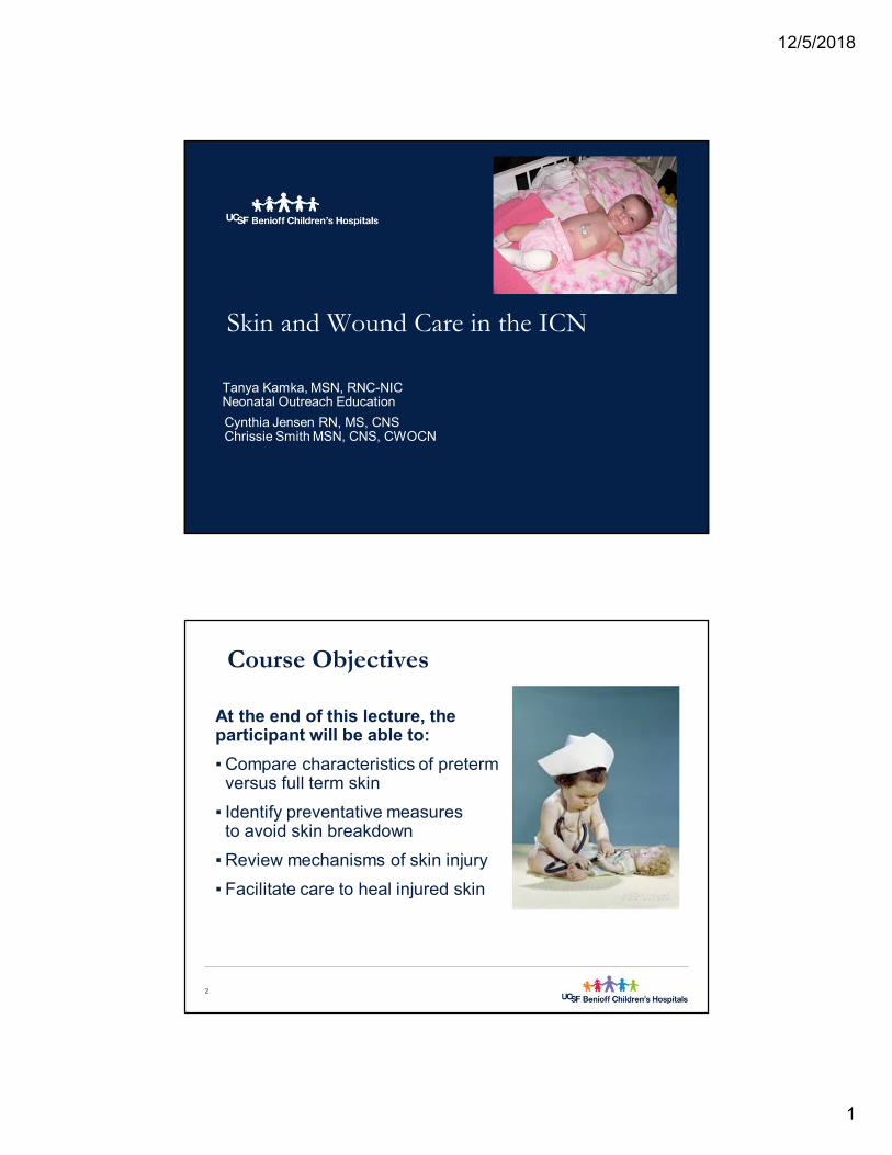
12/5/2018 1 Skin and Wound Care in the ICN Tanya Kamka, MSN, RNC-NIC Neonatal Outreach Education Cynthia Jensen RN, MS, CNS Chrissie Smith MSN, CNS, CWOCN Course Objectives At the end of this lecture, the participant will be able to: ▪ Compare characteristics of preterm versus full term skin ▪ Identify preventative measures to avoid skin breakdown ▪ Review mechanisms of skin injury ▪ Facilitate care to heal injured skin 2

Welcome message from author
This document is posted to help you gain knowledge. Please leave a comment to let me know what you think about it! Share it to your friends and learn new things together.
Transcript
12/5/2018
1
Skin and Wound Care in the ICN
Tanya Kamka, MSN, RNC-NICNeonatal Outreach EducationCynthia Jensen RN, MS, CNSChrissie Smith MSN, CNS, CWOCN
Course Objectives
At the end of this lecture, the participant will be able to:▪ Compare characteristics of preterm
versus full term skin ▪ Identify preventative measures
to avoid skin breakdown▪ Review mechanisms of skin injury ▪ Facilitate care to heal injured skin
2

12/5/2018
2
Skin and the Sick Newborn in the NICU
The Skin as a Neurodevelopmental Interface
Hoath, S. (2001). The Skin as a Neurodevelopmental Interface. Neoreviews, 2(12), 292e-301. http://dx.doi.org/10.1542/neo.2-12-e292Studio, T. (2014). Brain and Skin. Beautyphile. Retrieved 12 November 2015, from http://www.msbeautyphile.com/brain-skin-something-common/
4

12/5/2018
3
Functions of the SkinLargest organ of the body…
• Protective barrier
• Temperature regulation
• Active role in immune system
• Sensory
5
Sites.google.com,. (2015). Skin System Gabi - Body Systems Werner 2014. Retrieved 12 November 2015, from https://sites.google.com/a/jeffcoschools.us/body-systems-werner-2014/home/skin-system-gabi
6Visscher, M. (2009). Update on the Use of Topical Agents in Neonates. Newborn And Infant Nursing Reviews, 9(1), 31-47. http://dx.doi.org/10.1053/j.nainr.2008.12.010
12/5/2018
4
The Epidermis
• Outermost layer• Main function is a barrier
– Prevents absorption of fluid in utero– Prevents dessication – Prevents absorption of toxins and
microorganisms• Retains water and heat
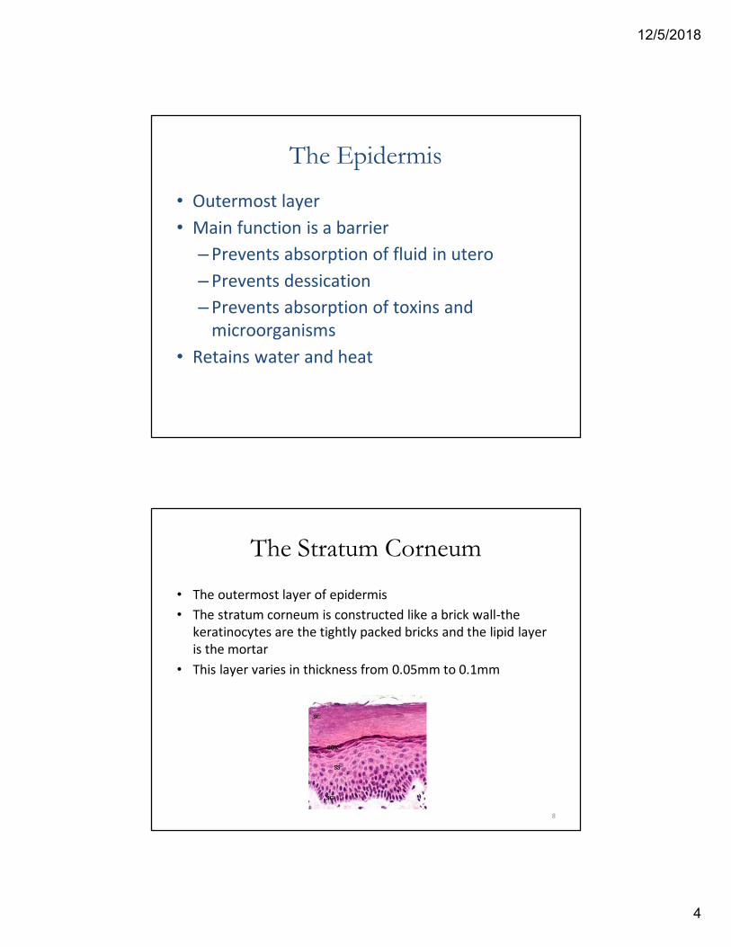
The Stratum Corneum
• The outermost layer of epidermis• The stratum corneum is constructed like a brick wall-the
keratinocytes are the tightly packed bricks and the lipid layer is the mortar
• This layer varies in thickness from 0.05mm to 0.1mm
8
12/5/2018
5
The Dermis
• Lies directly under the epidermis• Anchored to the dermis by the basal layer• It is a closely woven layer of fibrous protein
imbedded with collagen and elastin fibers. This gives the dermis it’s elasticity and tensile strength
• Also contains mast cells, inflammatory cells, blood and lymph vessels and cutaneous nerves
• Development of this layer begins at about 11 weeks gestation
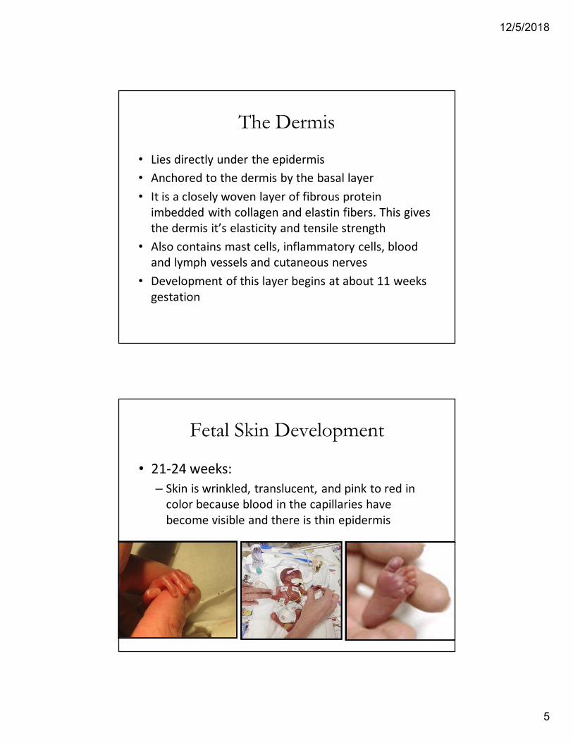
Fetal Skin Development
• 21-24 weeks:– Skin is wrinkled, translucent, and pink to red in
color because blood in the capillaries have become visible and there is thin epidermis

12/5/2018
6
Fetal Skin Development• 26-29 weeks:
– Subcutaneous fat and collagen begins to be deposited and starts to smooth out the many wrinkles
– Sweat glands form
• 30-34 weeks:– Skin is pink and smooth– Fingernails reach fingertips– Lanugo begins to shed
Fetal Skin Development
• 35 to 38 weeks:– Fetus begins to plump up– Skin is white or bluish pink in caucasian
infants

12/5/2018
7
Variations in Neonatal Skin▪ Underdevelopment of Stratum
Corneum, thinner skin. • Infants born at 22-25 weeks may
require 5-7 weeks to develop functional layer
▪ Decreased cohesion between epidermis and dermis
▪ Increased susceptibility to infection, toxicity from topically applied substances • Topical administration can equal IV
administration
▪ Excessive evaporative heat and fluid losses
13
Risk Factors for Skin Breakdown in Infants Include:▪ Gestational age <32 weeks
▪ Poor nutrition
▪ Edema
▪ Immobility
▪ Need for IV therapies
▪ Hypotension, and need for vasopressors
▪ Presence of medical devices: tubes, drains and monitoring equipment
▪ Surgical wounds
▪ Ostomies
14 Medscape,. (2015). Neonatal Skin: Back to Nature?. Retrieved 12 November 2015, from http://www.medscape.org/viewarticle/519767

12/5/2018
8
Unique Properties of Preterm Skin:▪Skin accounts for 3% of adult body weight▪ In preterm infants it accounts for 13% of total body weight
▪The skin of a premature neonate is 40-60% thinner than adult skin
15
Unique Properties of Preterm Skin:
• Increased skin permeability in the preemie therefore:– Increased risk of infection from pathogens– Increased risk of toxicity from topically applied
products– Increased TEWL (trans-epidermal water loss)
through skin due to insufficient layers of Stratum Corneum to keep water in
– Increased risk of irritation/epidermal stripping

12/5/2018
9
Unique Properties of Preterm Skin:
• Heat loss:– Thin skin , decreased subcutaneous fat stores and
immature thermoregulatory system= cold baby• Skin pH:
– Initial skin pH in newborns is about 6.34 which gradually decreases to about 4.95 within 4 days
– Skin pH less that 5 provides bacteriocidal protection against pathogens
– For this reason, only pH neutral products should be used for the initial bath or water only
Skin Care Goals▪ Maintain skin integrity & promote normal skin development
▪ Prevent injury
• Mechanical
‒ Pressure, Shear, Skin stripping, Trauma
• Thermal
‒ From monitoring devices (SpO2, Tcom)
• Chemical
‒ Irritants, Incontinence, Extravasation injury
• Other injury:
‒ Infection, Vascular Compromise, Congenital Skin Conditions
18

12/5/2018
10
Bathing
19
Bathing▪ Immediate bathing not necessary unless for
infectious reasons
▪ Only bathe when maintaining stable temperature for several hours
▪ Use warm sterile water on babies with breakdown or pH balanced soap and water
• Premature infants <32 weeks, only water for first 2 weeks
▪ Use soft cloth or cotton balls
▪ Following baths should be no more than q 48hrs
20

12/5/2018
11
The Acid Mantle▪At birth the skin pH is more alkaline at about 6.34▪The skin pH eventually drops of the first days to 4.95▪The skin’s acid mantle provides a state of equilibrium for the skin’s normal bacterial flora which provides protection against invading pathogenic organisms including fungus
▪Frequent bathing disrupts the acid mantle and increases risk of infection
21
Bathing: What the Evidence Shows
▪Quinn, D., Newton, N., & Piecuch, R. (2005). Effect of less frequent bathing on premature infant skin. Journal of Obstetric, Gynecologic, and Neonatal Nursing : JOGNN / NAACOG, 34(6), 741-746
Every 4th day bathing of premature infants appears to be safe
22

12/5/2018
12
Vernix-Don’t Rub it Off, Rub in IN!
23
Vernix: The Literature▪Protection against infection▪ Decreased skin permeability and transepidermal water loss (TEWL)
▪Skin cleansing▪ Moisturization of the skin surface▪ pH development▪ Wound healing▪ Temperature regulation
24
12/5/2018
13
Skin Care Goals▪ Maintain skin integrity & promote normal skin development▪ Prevent injury
• Mechanical ‒ Pressure, Shear, Skin stripping, Trauma
• Thermal ‒ From monitoring devices (SpO2, Tcom)
• Chemical‒ Irritants, Incontinence, Extravasation injury
• Other injury:‒ Infection, Vascular Compromise, Congenital Skin
Conditions
25
26
12/5/2018
14
▪ A localized damage to the skin and/or underlying soft tissue • usually over a bony prominence
• or related to a medical or other device.
• The injury can present as intact skin or an open ulcer and may be painful.
▪ Occurs as a result of intense and/or prolonged pressure or pressure in combination with shear.
▪ The tolerance of soft tissue for pressure and shear may also be affected by microclimate, nutrition, perfusion, co-morbidities and condition of the soft tissue.
‒ 4/2016 Revised definitions by NPUAP
27
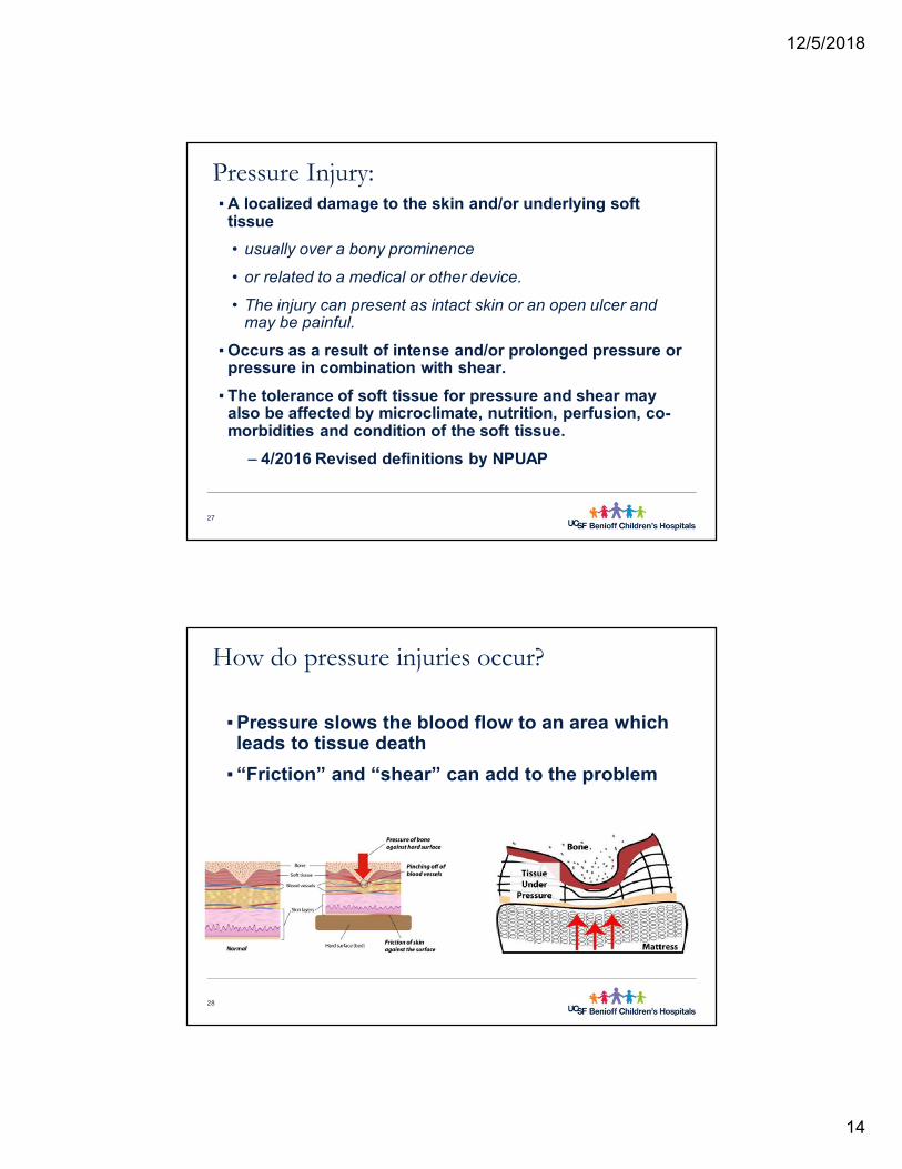
Pressure Injury:
▪ Pressure slows the blood flow to an area which leads to tissue death
▪ “Friction” and “shear” can add to the problem
28
How do pressure injuries occur?
12/5/2018
15
▪ Pressure injuries are painful
▪ Pressure injury incidence is a nursing quality indicator
▪ Medicare and Medicaid have designated pressure injuries as “Never Events”
▪ No reimbursement for treatment of some pressure injuries
▪ Cost to heal a single full thickness pressure injury can be as high as $150,000
▪ Skin injury may contribute to complications
• Increased risk of infection
• Functional abnormalities
• Permanent scarring
• Often patient’s report pressure injury to be the most painful
• Longer hospital stay
29
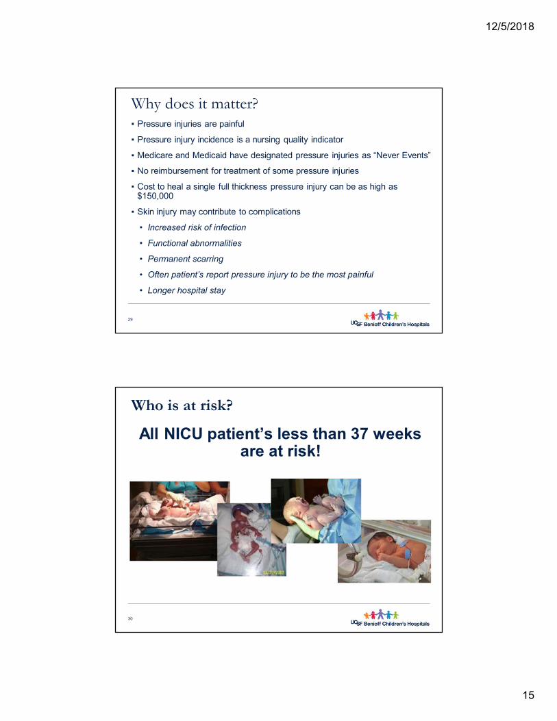
Why does it matter?
All NICU patient’s less than 37 weeks are at risk!
30
Who is at risk?

12/5/2018
16
▪ Skin Assessment:
• On admission, transfer, and after any procedure lasting >3 hours, then every shift
• Inspect the skin Q shift for signs of pressure injury, especially non-blanchable erythema
• Assess skin near and under medical devices at least every shift (splint, catheter, tube, brace)
• If order written to not reposition or remove a device, discuss concern with ordering provider. If order remains report to unit CNS immediately.
• When inspecting darkly pigmented skin, look for changes in skin tone, temperature, and tissue consistency compared to adjacent skin
▪ Skin Inspection
• Spread buttocks/cheeks, assess occiput
31
32

12/5/2018
17
33
Where do you suspect pressure injuries to occur?
Pulse Oximetry Application Tips: • Select correct sensor• Correctly apply sensor:
‒ Star (emitter) to the sky
‒ Black square (detector) directly opposite star
‒ Apply gently, assure adherence, avoid tourniquet type fit
‒ Sensor cord direction can be either toward or away from the patient
• Choose best application site based on clinical assessment of your patient ‒ Apply sensor to patient first then attach to cable—“Sensor
detects light, not life”
• Meticulous Assessment
34

12/5/2018
18
NIPPV: Incidence of skin breakdown
▪ Skin breakdown “… even after only a few hours of ventilation, is a frequent complication, ranging from 2-23%”.
▪ “In one study, where patients were continuously ventilated with a face mask for more than 48 hours, this percentage reached 70%”.
Scott K. Epstein, MD. Respiratory Care, January 2009 Vol 54 No 1. 2 Gregoretti et al. Evaluation of patient skin breakdown and comfort with a new face mask for non-invasive ventilation: a multi-center study. Inten Care Med 2002; 28:278-284.
35
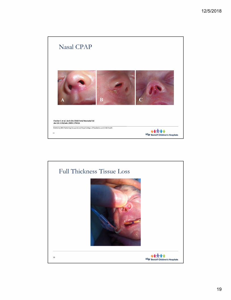
Nasal CPAP
36
12/5/2018
19
Nasal CPAP
37
Full Thickness Tissue Loss
38
12/5/2018
20
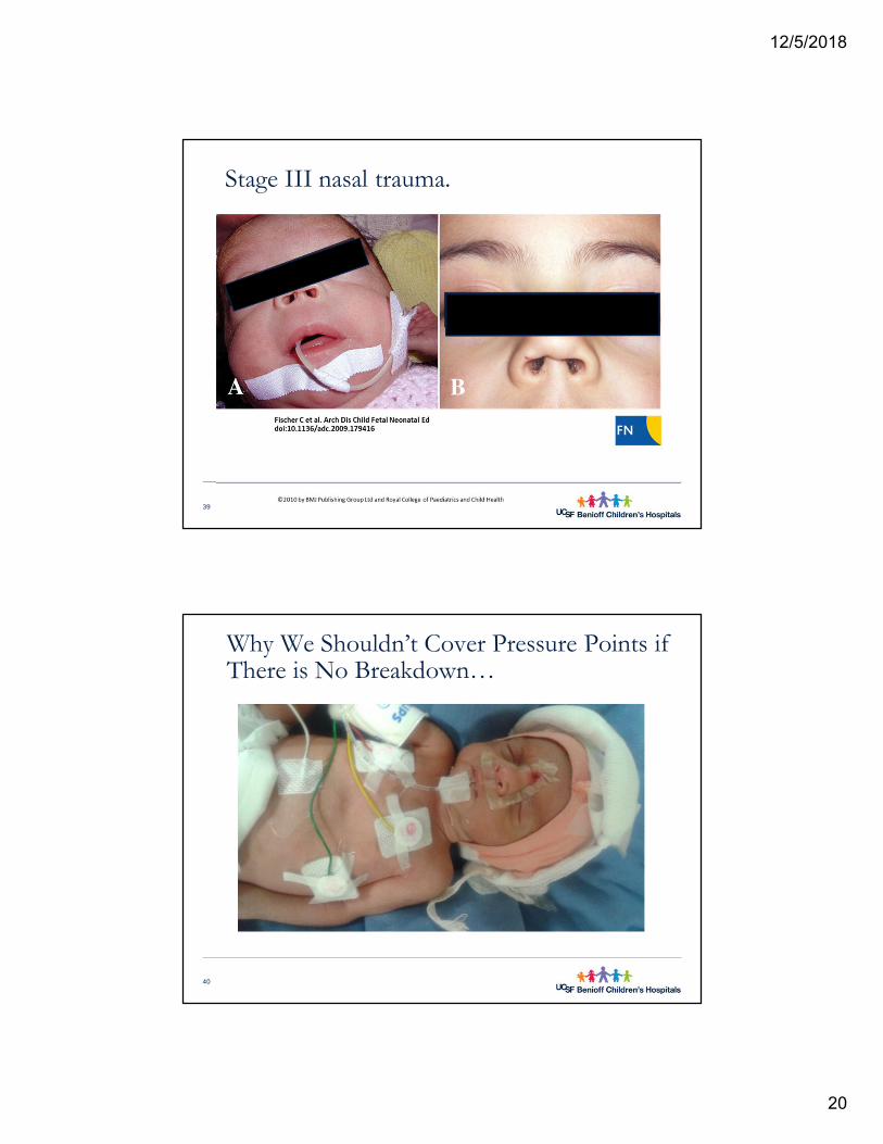
Stage III nasal trauma.
39
Why We Shouldn’t Cover Pressure Points if There is No Breakdown…
40

12/5/2018
21
Blanching with Excessive Pressure
41
“Snubbing”
42

12/5/2018
22
43
NG tubes
Pressure Ulcer Prevention
▪Frequent assessment ▪Minimize pressure from medical devices▪Specialty mattresses for infants at risk
44

12/5/2018
23
Optimal Use of Specialty Mattresses :▪More than one layer between baby and mattress decrease effectiveness
▪One layer can be:• Mattress Cover• A Pillowcase • One thin blanket
45
Skin Care Goals▪ Maintain skin integrity & promote normal skin development▪ Prevent injury
• Mechanical ‒ Pressure, Shear, Skin stripping, Trauma
• Thermal ‒ From monitoring devices (SpO2, Tcom)
• Chemical‒ Irritants, Incontinence, Extravasation injury
• Other injury:‒ Infection, Vascular Compromise, Congenital Skin
Conditions
46

12/5/2018
24
Mechanical Injury: Skin Stripping and Trauma
47
Fragility of Epidermal/Dermal Connection
▪ Diminished cohesion between dermis and epidermis places premature infant at higher risk for injury
48
12/5/2018
25
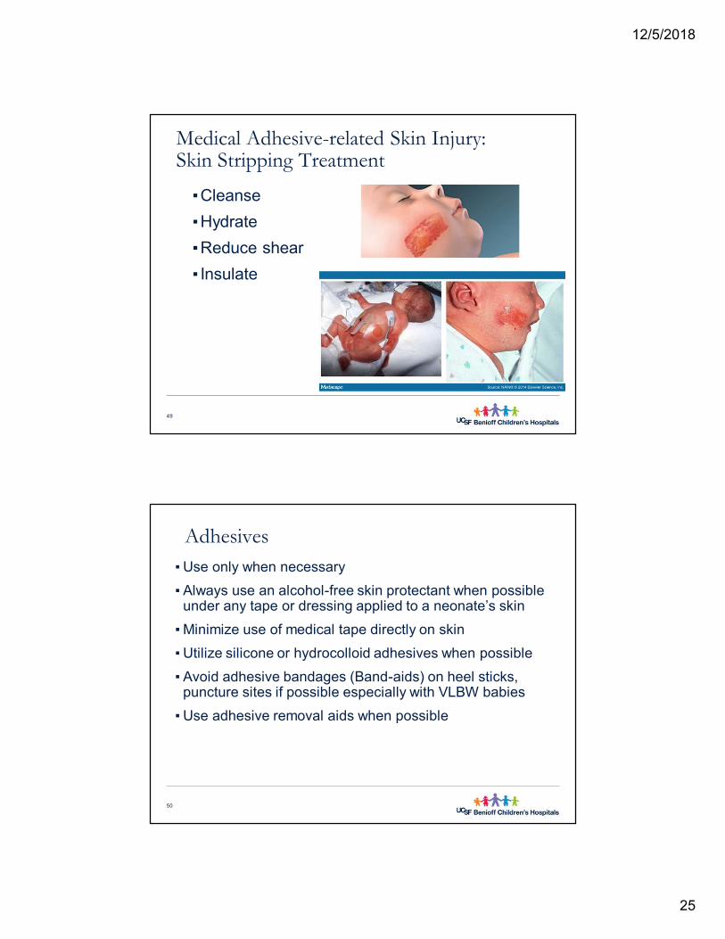
Medical Adhesive-related Skin Injury:Skin Stripping Treatment
▪Cleanse ▪Hydrate▪Reduce shear▪ Insulate
49
Adhesives▪ Use only when necessary▪ Always use an alcohol-free skin protectant when possible
under any tape or dressing applied to a neonate’s skin▪ Minimize use of medical tape directly on skin▪ Utilize silicone or hydrocolloid adhesives when possible▪ Avoid adhesive bandages (Band-aids) on heel sticks,
puncture sites if possible especially with VLBW babies▪ Use adhesive removal aids when possible
50
12/5/2018
26
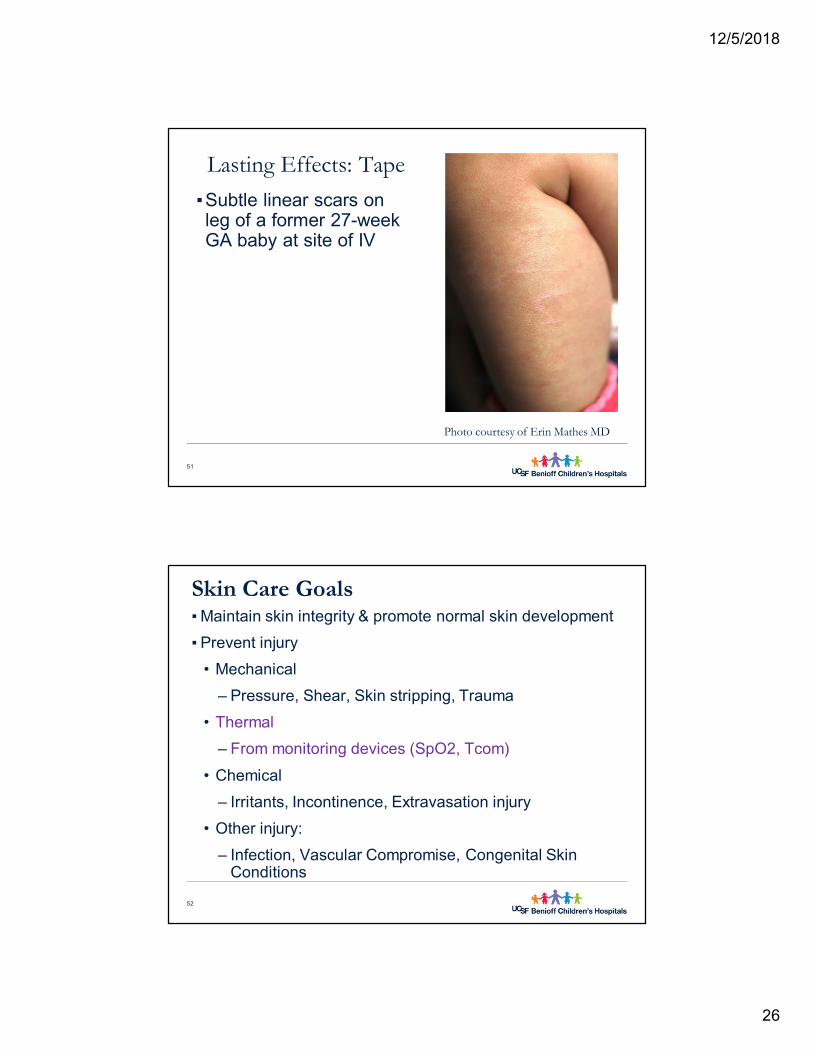
Lasting Effects: Tape▪Subtle linear scars on leg of a former 27-week GA baby at site of IV
51
Photo courtesy of Erin Mathes MD
Skin Care Goals▪ Maintain skin integrity & promote normal skin development▪ Prevent injury
• Mechanical ‒ Pressure, Shear, Skin stripping, Trauma
• Thermal ‒ From monitoring devices (SpO2, Tcom)
• Chemical‒ Irritants, Incontinence, Extravasation injury
• Other injury:‒ Infection, Vascular Compromise, Congenital Skin
Conditions
52

12/5/2018
27
Mechanical Injury: Thermal
53
Skin Care Goals▪ Maintain skin integrity & promote normal skin development▪ Prevent injury
• Mechanical ‒ Pressure, Shear, Skin stripping, Trauma
• Thermal ‒ From monitoring devices (SpO2, Tcom)
• Chemical‒ Irritants, Incontinence, Extravasation injury
• Other injury:‒ Infection, Vascular Compromise, Congenital Skin
Conditions
54

12/5/2018
28
Mechanical Injury: Chemical
▪ Irritants & Toxins▪ Incontinence, Diaper Dermatitis▪Extravasation injury
55
Lessons Learned: Permeability ▪ “CYANOSIS IN NEWBORN BABIES CAUSED BY ANILINE-DYE POISONING”
▪Robertson, A. F. (2003). Reflections on errors in neonatology: IN. the "hands-off" years, 1920 to 1950. Journal of Perinatology : Official Journal of the California Perinatal Association, 23(1), 48-55.
▪Routine Hexachlorophene Bathing▪Povidone Iodine
56

12/5/2018
29
Bacitracin: Things to Consider▪ Bacitracin has been noted as one of the 12 most frequent
allergens causing a positive patch test reaction in patients ages 8–92 years• Draelos, Z. D., Rizer, R. L., & Trookman, N. S. (2011). A
comparison of postprocedural wound care treatments: Do antibiotic-based ointments improve outcomes? Journal of the American Academy of Dermatology, 64(3 Suppl), S23-9.
57
Disinfectants ▪ Disinfecting skin surfaces before invasive procedures
reduces the risk of infection and contamination▪ Disinfectant solution should be chosen based on careful
evaluation of safety for preterm and term infants ▪ Any disinfectant applied to the skin should be cleansed off
after the procedure
58

12/5/2018
30
Chlorhexidine Gluconate (CHG)▪ At UCSF use of 2% CHG in 70% Isopropyl Alcohol may be used
when infant meets all 3 of the following criteria:
• >27 weeks
• >1000
• >7days
▪ Based on:
• Garland JS, Alex CP, Mueller CD, et al. A randomized trial comparing povidone-iodine to a chlorhexidine gluconate-impregnated dressing for prevention of central venous catheter infections in neonates. Pediatrics. 2001;107:1431–1436.
59
Skin Disinfectants: The Evidence▪ Why do we have criteria for CHG?
▪ Four of 36 (11%) infants < 1000 grams exposed to 2% aqueous chlorhexidine developed severe skin irritation (all had erythema and one progressed to breakdown with exudates). The study used 2% chlorhexidine for all central & arterial catheters and PIVs for infants <1000 grams and<14 days and 1% chlorhexidine in ethanol for all other IVs. (Anderson 2005)
60

12/5/2018
31
CHG Considerations ▪ It is critical that CHG be allowed to air dry for 30 seconds so that isopropyl alcohol can evaporate
▪Failure to allow drying has been associated with chemical burns
61
Povidone Iodine▪ Povidone Iodine should be used to disinfect skin for
preterm infants who do not meet criteria for CHG▪ Apply Povidone Iodine in an outward circular motion and
allowed to dry per manufacturer recommendation▪ Clean off completely with Saline Wipe or sterile water after
use to prevent chemical irritation and absorption
62

12/5/2018
32
Povidone Iodine Considerations
63
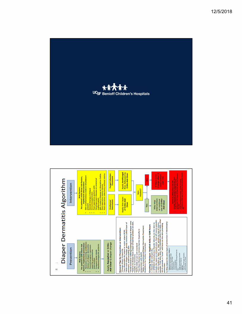
Incontinence, Diaper Dermatitis & Moisture Management
64

12/5/2018
33
Moisture Management…
65
• Urine is composed of 95% H2O, 5% organic solutes, primary urea
• Normal skin has a pH of 5.4-5.0 (acid environment) this has an antibacterial effect limiting pathogenic organisms.
• Urinary urea decomposes on the skin to form ammonium hydroxide which is an alkaline substance and raises the skins pH, which favors bacterial proliferation.
• Feces degrade the skin barrier function
General Diaper Care▪ Avoid friction or rubbing when cleaning
• to Desitin or not to Desitin…?▪ Use superabsorbent diapers and change frequently▪ Avoid scented wipes ▪ Identify infants at risk for diaper dermatitis
• Hyperbili babies, infants with neurogenic bowel/bladder, “short gut” syndrome, antibiotic therapy
▪ For infants at risk, apply Petrolatum/Aquaphor or Zinc ointment with every soiled diaper cleaning and changing to provide a protective barrier
66

12/5/2018
34
DD Topical Applications:Positive recommendations
▪Zinc oxide AWHONN, 2007; Baldwin, et al., 2001;Hoggarth et al., 2005;Lund et al., 1999; Nield & Kamat, 2007;Wananukul et al., 2006
▪Petrolatum AWHONN, 2007; Atherton, 2001; Hoggarth et al., 2005; Lund et al., 1999;Odio et al., 2000
▪Frequent Diaper Changes: AWHONN, 2007; Atherton, 2004; Borkowski, 2004;Kazaks & Lane, 2000; Nield & Kamat, 2007
67
DD-Negative Recommendations▪Open to Air Lund et al., 1999▪Antibacterial Products AWHONN, 2007; Lund et al., 1999
▪Powder Darmstadt & Dinulos, 2000; Farrington, 1992
68
12/5/2018
35
Diaper Care
69
Dressing change frequency
Indications for use
PRN or Q diaper change
• Diaper dermatitis prevention
• Mild redness without breakdown
• “frosting” a cupcake
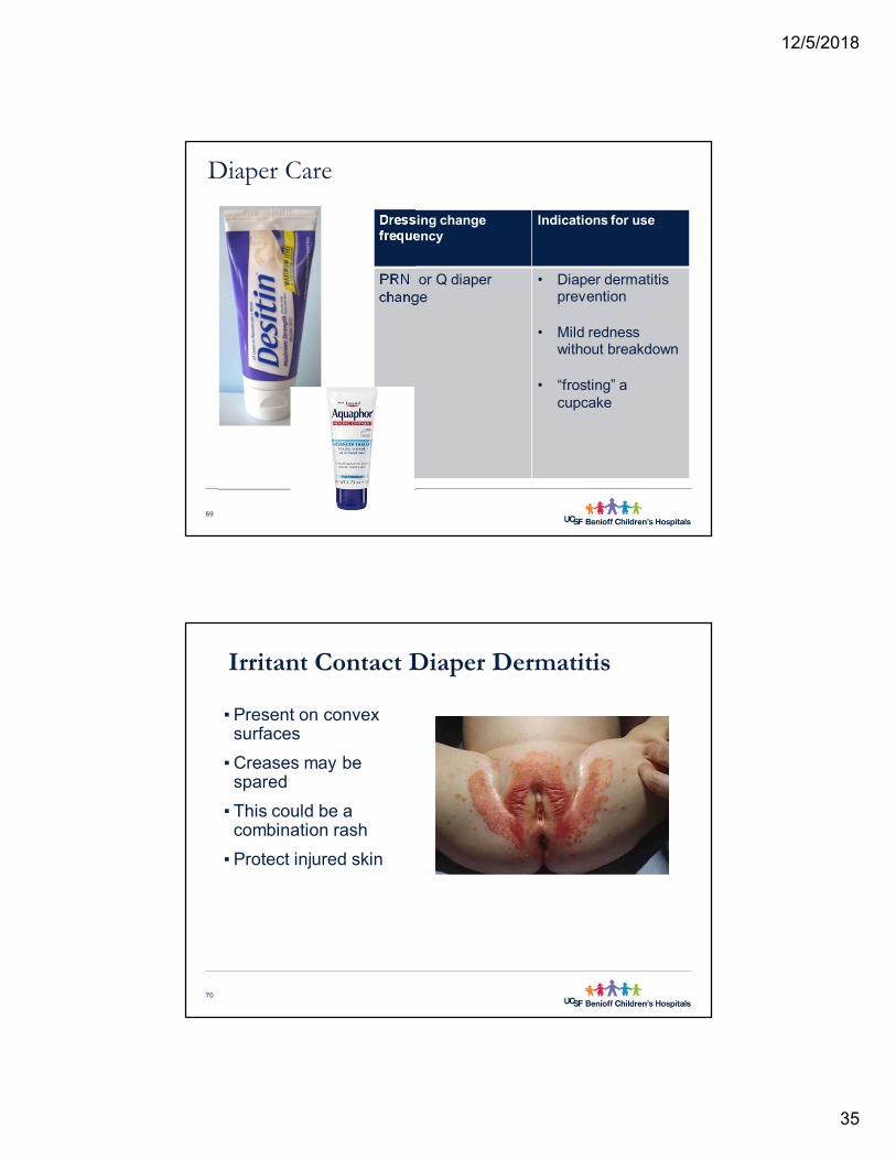
Irritant Contact Diaper Dermatitis
▪ Present on convex surfaces
▪ Creases may be spared
▪ This could be a combination rash
▪ Protect injured skin
70

12/5/2018
36
71
Candida Rash (Yeast)▪ Assess for Candida and
treat if needed ▪ Notice creases are involved▪ Satellite pustules are
present▪ May occur simultaneously
with oral thrush▪ Frequent diaper changes
needed▪ “Open to air” may be
required
72

12/5/2018
37
73
DD with Ulcerations▪Although “open to air” approach may help temporarily, contact with urine or stool will reinjure skin
▪This baby would benefit from barrier paste
74

12/5/2018
38
75
76

12/5/2018
39
DD: Things to Consider▪Prevention is key▪Follow diaper dermatitis care plan▪Try plan for 24-48 hours to see if it is working▪For problem cases consult expert
77
Take home points…▪ The skin of a premature infant, is essentially,
wounded
▪ Prevention of further injury for all infants is essential
▪ Bathe minimally, using pH-neutral cleanser when appropriate
▪ Assess for pressure injury and utilize pressure reducing devices
▪ Utilize diaper dermatitis protocol, assessing 24-48 hours and involving CNS if needed
▪ Minimize adhesives
▪ Selective use of topical antiseptics, with removal after application
78
12/5/2018
40
References▪ Association of Women’ Health, Obstetric and Neonatal Nurses: Evidence based clinical practice
guideline: neonatal skin care, ed 2, Washington, DC, 2007, The Association.
▪ Baharestani MM: An overview of neonatal and pediatric wound care knowledge and considerations, Ostomy Wound Management 53:34, 2007.
▪ Eichenfield, LF, Frieden, IJ, Mathes, EF. 2015. Textbook of Neonatal Dermatology. Philadelphia: WB Saunders.
▪ Garland JS, Alex CP, Mueller CD, et al. A randomized trial comparing povidone-iodine to a chlorhexidine gluconate-impregnated dressing for prevention of central venous catheter infections in neonates. Pediatrics. 2001;107:1431–1436.
▪ Heimall, L. M., Storey, B., Stellar, J. J., & Davis, K. F. (2012). Beginning at the bottom: Evidence-based care of diaper dermatitis. MCN. The American Journal of Maternal Child Nursing, 37(1), 10-16
▪ Quinn, D., Newton, N., & Piecuch, R. (2005). Effect of less frequent bathing on premature infant skin. Journal of Obstetric, Gynecologic, and Neonatal Nursing : JOGNN / NAACOG, 34(6), 741-746.
▪ Robertson, A. F. (2003). Reflections on errors in neonatology: I. the "hands-off" years, 1920 to 1950. Journal of Perinatology : Official Journal of the California Perinatal Association, 23(1), 48-55.
79
Questions?
Thank you!!
80
12/5/2018
41
82
Related Documents