Skeletal Tuberculosis (Part-2) Dr. Sunil Arora Junior Resident Deptt. of Chest &TB Govt. Medical College, Patiala. 1 1

Skeletal Tuberculosis (Part-2) Dr. Sunil Arora J unior Resident Deptt. of Chest &TB Govt. Medical College, Patiala. 1 1.
Dec 21, 2015
Welcome message from author
This document is posted to help you gain knowledge. Please leave a comment to let me know what you think about it! Share it to your friends and learn new things together.
Transcript
Skeletal Tuberculosis
(Part-2)
Dr. Sunil Arora Junior Resident
Deptt. of Chest &TB Govt. Medical College, Patiala.
11
IntroductionSkeletal TB accounts for 10 to 35% of cases of
extrapulmonary tuberculosis. Although spine is the
commonest site (constituting about 50%) of skeletal
TB. The other bones and joints can also be involved.
Skeletal TB generally occurs due to haematogenous
spread from a primary focus.
2

Classification TUBERCULAR ARTHRITIS- 30% HIP JOINT KNEE WRIST JOINT SACROILIAC JOINT ANKLE JOINT AND FOOT SHOULDER JOINT TUBERCULAR OSTEOMYELITIS- 19% LONG TUBULAR BONES SHORT TUBULAR BONES FLAT BONES- RIBS,STERNUM, SCAPULA, PELVIS TENOSYNOVITIS/BURSITIS- 1%
3

Pathogenesis• It produces similar response as in lungs i.e.
Chronic granulomatous inflammation.The disease process can start either in bone or in the synovial membrane.
• Active focus forms in the metaphysis (in children) or epiphysis (in adults) and the inflammation extends peripherally along the shaft to reach the subperiosteal space.
• The inflammatory exudate may extend outward through the soft tissue to form cold abscess and sinuses.The affected bone may undergo fracture.
4
• Metaphyseal infection reaches the joint through subperiosteal space by penetrating the capsular attachment.In adults,the inflammation can spread up to the subchondral area and enter the joint at the periphery where synovium joins the cartilage which leads to the loosening the attachment of articular cartilage and joint displacement.
5
• Sometimes the synovium is infected first and the bone is infected secondarily.It is usually in the form of low-grade synovitis with thickening of the synovial membrane and leading to the formation of pannus.Eventually,the articular cartilage is destroyed,joint gets distended with pus,which may burst out to form a cold abscess or discharging sinus.The joint may also get subluxated due to the laxity of the joint capsule and ligaments.
• Fibrous ankylosis is a common outcome of healed tuberculosis of the joints except in spine where bony ankylosis follows more often.
6
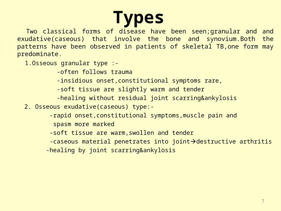
Types Two classical forms of disease have been seen;granular and and exudative(caseous)
that involve the bone and synovium.Both the patterns have been observed in patients of skeletal TB,one form may predominate.
1.Osseous granular type :-
-often follows trauma
-insidious onset,constitutional symptoms rare,
-soft tissue are slightly warm and tender
-healing without residual joint scarring&ankylosis
2. Osseous exudative(caseous) type:-
-rapid onset,constitutional symptoms,muscle pain and
spasm more marked
-soft tissue are warm,swollen and tender
-caseous material penetrates into jointdestructive arthritis
-healing by joint scarring&ankylosis
7

Clinical Features TB should be included in the differential diagnosis of any
slow onset of disease of musculo-skeletal system particularly in TB prevalent countries like India.Symptoms can be:-
Constitutional symptoms:–• The patient may be apathetic and pale• Loss of weight and appetite• Low grade fever(especially in the afternoons)• Night sweats,tachycardia These constitutional symptoms may be present
before the definite symptoms pertaining to specific bone/joint involved.
8
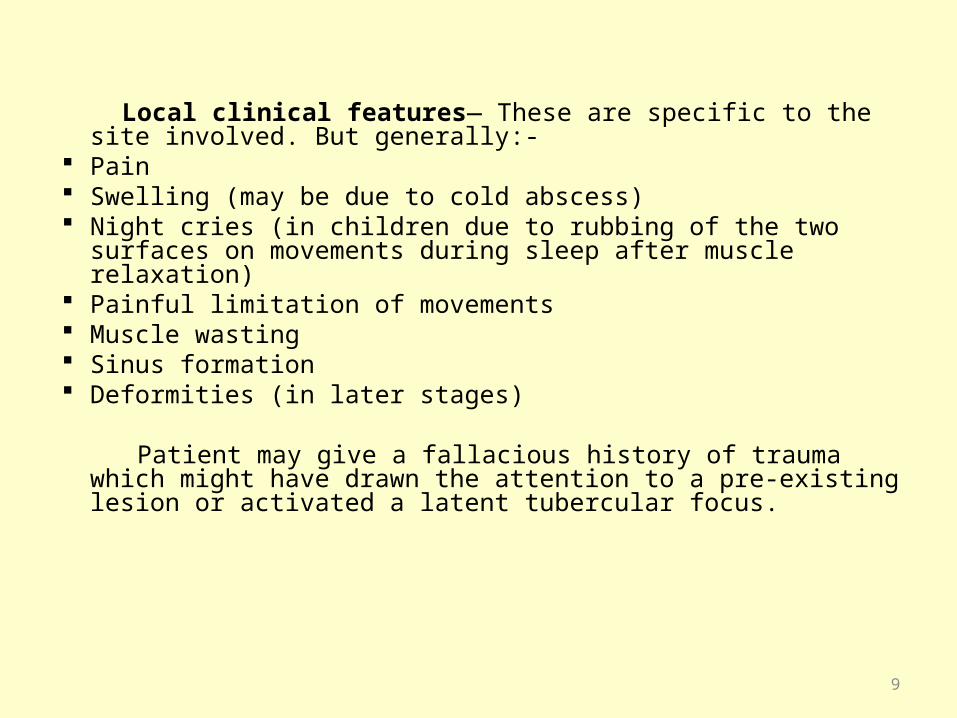
Local clinical features— These are specific to the site involved. But generally:-
Pain Swelling (may be due to cold abscess) Night cries (in children due to rubbing of the two surfaces on
movements during sleep after muscle relaxation) Painful limitation of movements Muscle wasting Sinus formation Deformities (in later stages)
Patient may give a fallacious history of trauma which might have drawn the attention to a pre-existing lesion or activated a latent tubercular focus.
9
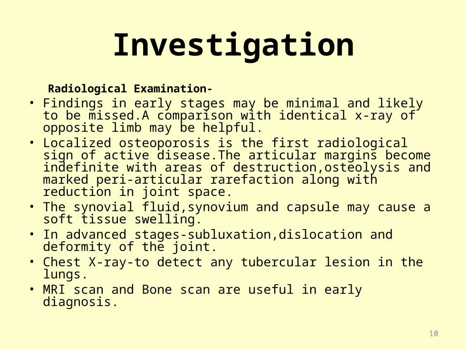
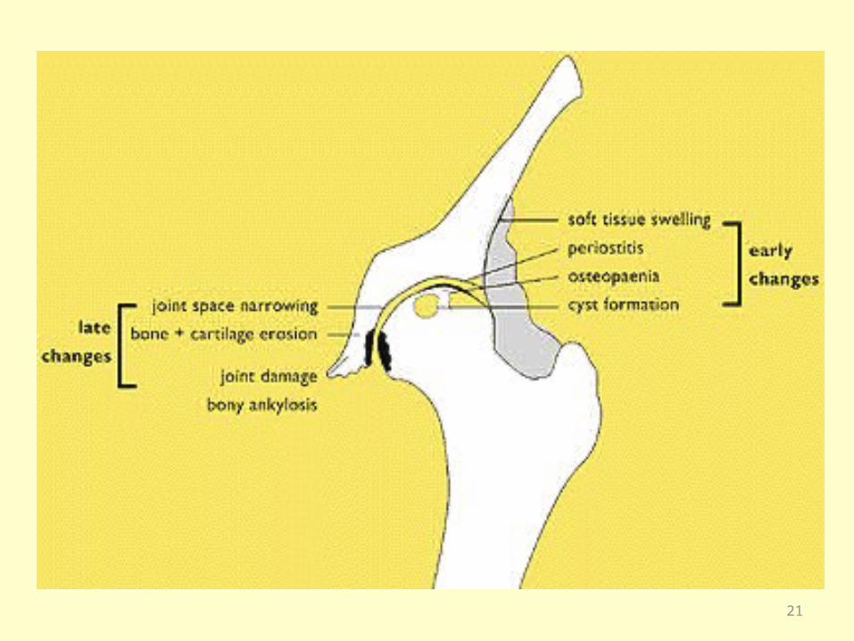
Investigation Radiological Examination- • Findings in early stages may be minimal and likely to be
missed.A comparison with identical x-ray of opposite limb may be helpful.
• Localized osteoporosis is the first radiological sign of active disease.The articular margins become indefinite with areas of destruction,osteolysis and marked peri-articular rarefaction along with reduction in joint space.
• The synovial fluid,synovium and capsule may cause a soft tissue swelling.
• In advanced stages-subluxation,dislocation and deformity of the joint.
• Chest X-ray-to detect any tubercular lesion in the lungs.• MRI scan and Bone scan are useful in early diagnosis.
10

Other investigations• Haemogram – It may show anemia, lymphocytic
leucocytosis,high ESR• Mx test – useful in children.• Synovial fluid aspiration – -ADA Levels(Non TB septic arthritis patients have been
reportedly higher than other types of inflammatory arthritis but not as high as TB arthritis).
- Culture.• Aspiration of cold abscess- Histopathological
examination,Smear for AFB & culture.• Biopsy- in doubtful diagnosis,may be from
synovium,bone.• FNAC from lymph node
11
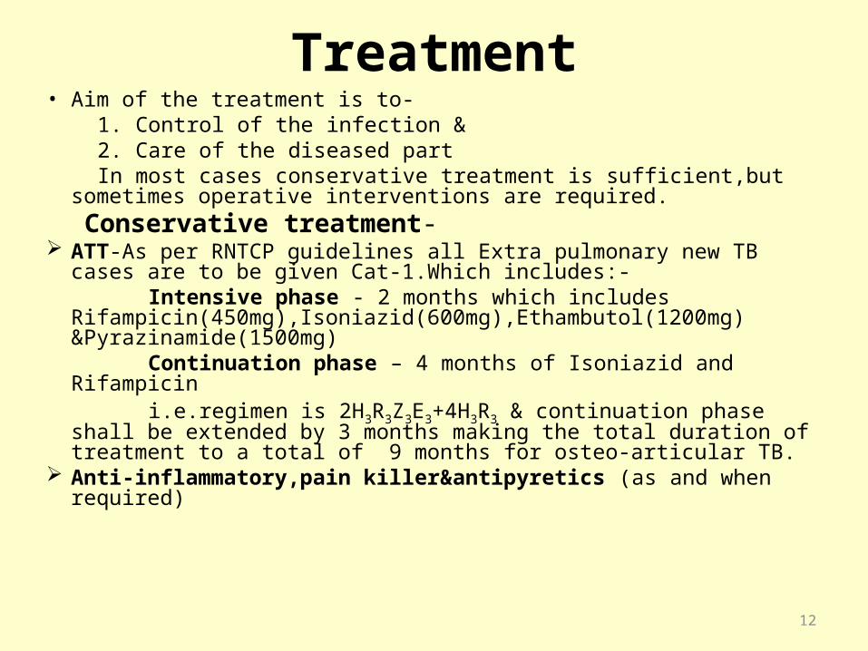
Treatment• Aim of the treatment is to- 1. Control of the infection & 2. Care of the diseased part In most cases conservative treatment is sufficient,but sometimes
operative interventions are required. Conservative treatment- ATT-As per RNTCP guidelines all Extra pulmonary new TB cases
are to be given Cat-1.Which includes:- Intensive phase - 2 months which includes
Rifampicin(450mg),Isoniazid(600mg),Ethambutol(1200mg) &Pyrazinamide(1500mg)
Continuation phase – 4 months of Isoniazid and Rifampicin i.e.regimen is 2H3R3Z3E3+4H3R3 & continuation phase shall be
extended by 3 months making the total duration of treatment to a total of 9 months for osteo-articular TB.
Anti-inflammatory,pain killer&antipyretics (as and when required)
12
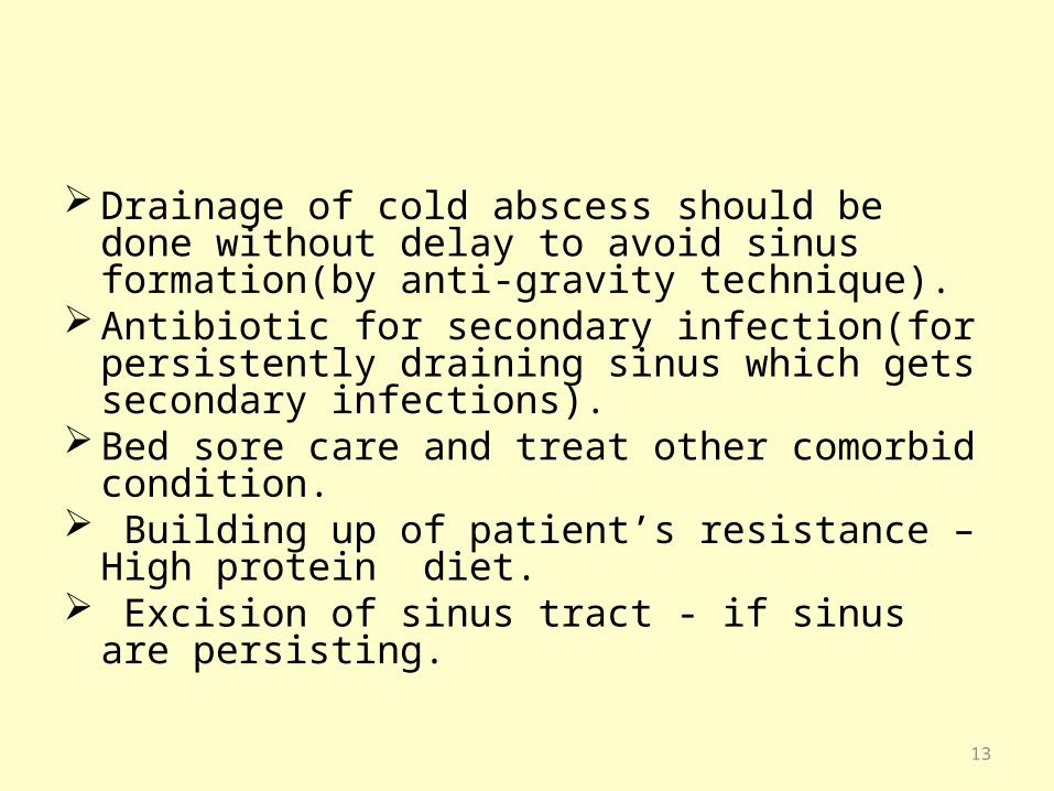
Drainage of cold abscess should be done without delay to avoid sinus formation(by anti-gravity technique).
Antibiotic for secondary infection(for persistently draining sinus which gets secondary infections).
Bed sore care and treat other comorbid condition.
Building up of patient’s resistance – High protein diet.
Excision of sinus tract - if sinus are persisting.
13
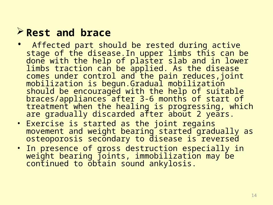
Rest and brace• Affected part should be rested during active stage of the
disease.In upper limbs this can be done with the help of plaster slab and in lower limbs traction can be applied. As the disease comes under control and the pain reduces,joint mobilization is begun.Gradual mobilization should be encouraged with the help of suitable braces/appliances after 3-6 months of start of treatment when the healing is progressing, which are gradually discarded after about 2 years.
• Exercise is started as the joint regains movement and weight bearing started gradually as osteoporosis secondary to disease is reversed
• In presence of gross destruction especially in weight bearing joints, immobilization may be continued to obtain sound ankylosis.
14
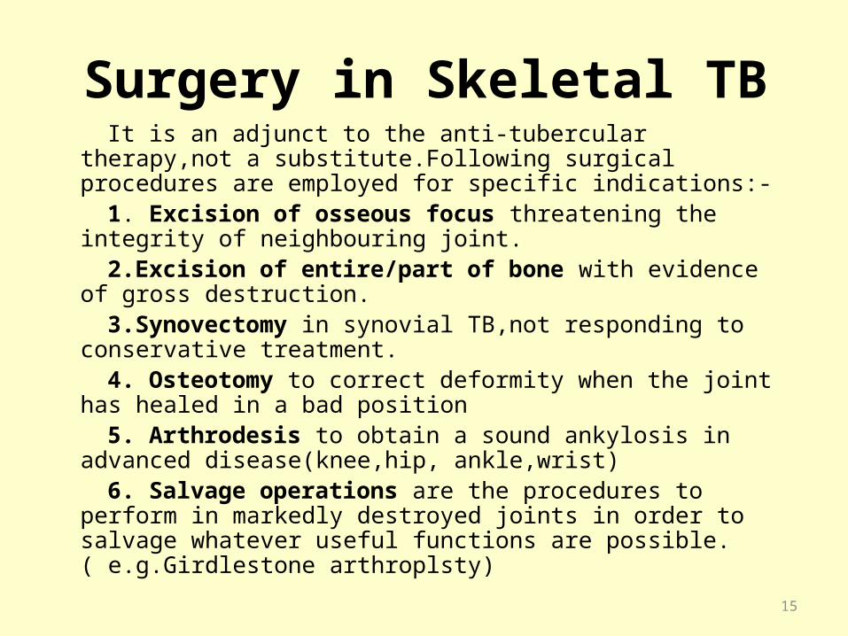
Surgery in Skeletal TB It is an adjunct to the anti-tubercular therapy,not a
substitute.Following surgical procedures are employed for specific indications:-
1. Excision of osseous focus threatening the integrity of neighbouring joint.
2.Excision of entire/part of bone with evidence of gross destruction.
3.Synovectomy in synovial TB,not responding to conservative treatment.
4. Osteotomy to correct deformity when the joint has healed in a bad position
5. Arthrodesis to obtain a sound ankylosis in advanced disease(knee,hip, ankle,wrist)
6. Salvage operations are the procedures to perform in markedly destroyed joints in order to salvage whatever useful functions are possible.( e.g.Girdlestone arthroplsty)
15
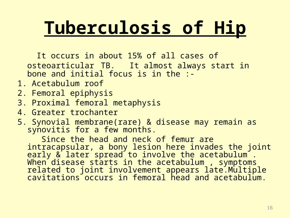
Tuberculosis of Hip
It occurs in about 15% of all cases of osteoarticular TB. It almost always start in bone and initial focus is in the :-
1. Acetabulum roof2. Femoral epiphysis3. Proximal femoral metaphysis4. Greater trochanter5. Synovial membrane(rare) & disease may remain as synovitis
for a few months. Since the head and neck of femur are intracapsular, a bony
lesion here invades the joint early & later spread to involve the acetabulum . When disease starts in the acetabulum , symptoms related to joint involvement appears late.Multiple cavitations occurs in femoral head and acetabulum.
16

Stages of TB of Hip Classical untreated TB of hip passes through following 3 stages
Stage 1 (stage of synovitis) –Intrasynovial effusion and exudate distending the joint capsule demanding the hip to be in a position of maximum capacity i.e. position of flexion, abduction &external rotation. As the pelvis tilts to compensate for abduction and flexion deformities so the affected limb appears longer. This is a stage of apparent lengthening.
Stage 2 (stage of Arthritis)- The capsule and articular cartilage is involved leading to spasm of powerful muscle. The hip joint assumes a position of flexion, adduction & internal rotation. The flexion & adduction may be concealed by compensatory tilt, the affected limb appears shorter i.e. stage of apparent shortening.
Stage 3 (stage of Erosion)-The capsule is further destroyed, along with advanced destruction of cartilage and the head and/or acetabulum is eroded. The attitude of limb is flexion , adduction and internal rotation.There is true shortening of the limb because of the actual destruction of bone.The destroyed head may come to lie proximally and posteriorlyWandering acetabulum or instead protrusio acetabuli can develop with destruction of medial wall of acetabulum17
EXAMINATION It should be carried out with the patient undressed. Gait – Lameness is one of the first sign. In the early stage, it is
because of the stiffness and deformity of the hip. Because of the flexion deformity, patient stands with compensatory exaggerated lumbar lordosis.Later, the limp is exaggerated by pain, so that,the patient hastens to take the weight of the affected side(painful or antalgic gait).
Muscle wasting of thigh and gluteal muscles. Swelling due to cold abscess-Sometimes joint space is filled
with caseous material and it may track down to the path of least resistance resulting in cold abscesses in:-
1.Inguinal region 2.Femoral triangle 3.ischiorectal fossa
4.Adductor compartment of thigh 5. Greater trochanter
18

Discharging sinuses in the groin or around the greater trochanter. There may be puckered scars from healed sinuses.
Deformity – Minimal deformities are compensated by pelvic tilt. Commonly it is flexion, adduction & internal rotation.
Shortening – Generally true shortening except in stage 1 ( which is apparent lengthening ).
Movements –Limitations of active and passive movements in all directions. If no movement at al
( Bony ankylosis ) Abnormal positioning of head-In a dislocated
hip,head can be felt in gluteal region.
19
Investigation
1.Radiological Examination- X-rays AP &lateral view.2.Other investigations- Includes blood investigation,Mx,Synovial fluid
examination, biopsy.
MRI scan and Bone scan are useful in early diagnosis
20
21
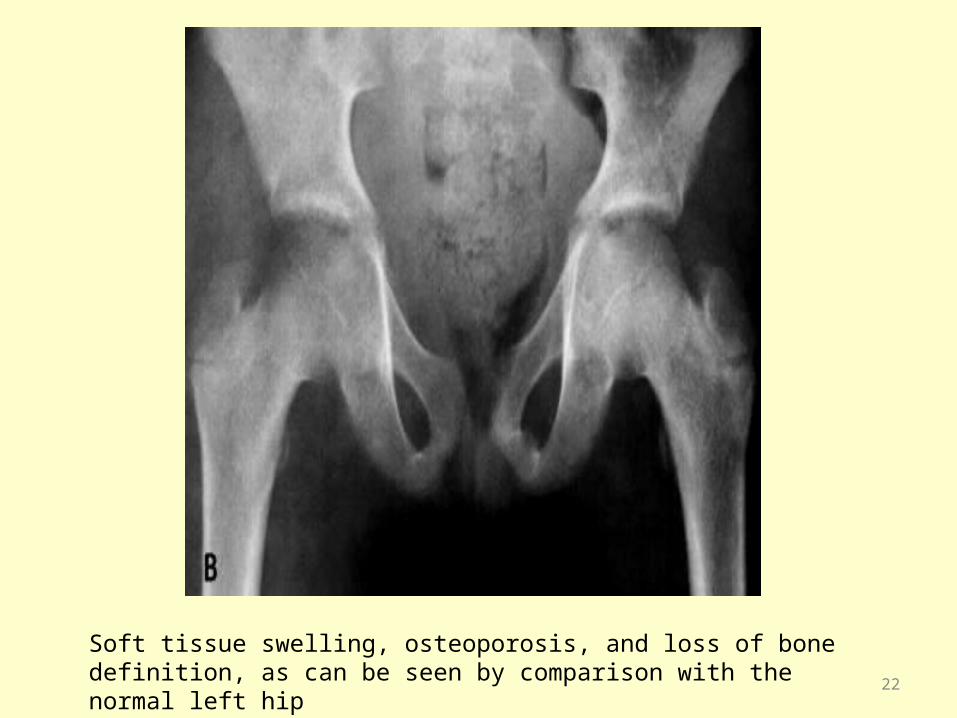
Soft tissue swelling, osteoporosis, and loss of bone definition, as can be seen by comparison with the normal left hip
22
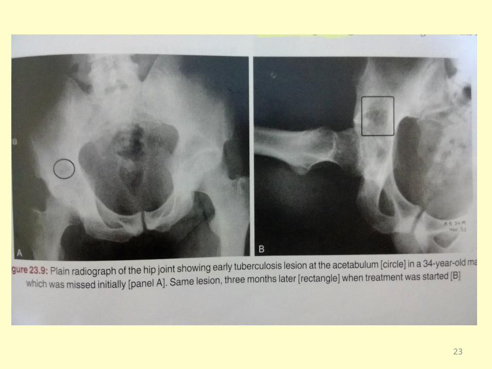
23

Radiological Types Seven different types of radiological appearances in advance stage of TB hip
joint are as:- Normal type – Disease is mainly synovial, may be cysts in femoral neck,
head or acetabulum, but no gross destruction and joint space is normal. Perthes type – Generally seen under 5 years of age,Femoral head is
sclerotic and it is difficult to differentiate from Perthes disease. Dislocating type – Subluxation or dislocation of femoral head occurs due to
capsular laxity and synovial hypertrophy(not due to accumulation of pus). Wandering acetabulum-There occurs destruction of acetabulum of its
superior (weight bearing part)&femoral head shifts proximally on the ilium. Atrophic type – Femoral head is irregular and joint space is narrow.
Seen exclusively in adults.
Protrusioacetabuli-medialization of the medial wall of the acetabulum. Mortar and Pastle type – Destruction causes enlargement and deepening
of acetabulum and femoral head shifts medially..
24
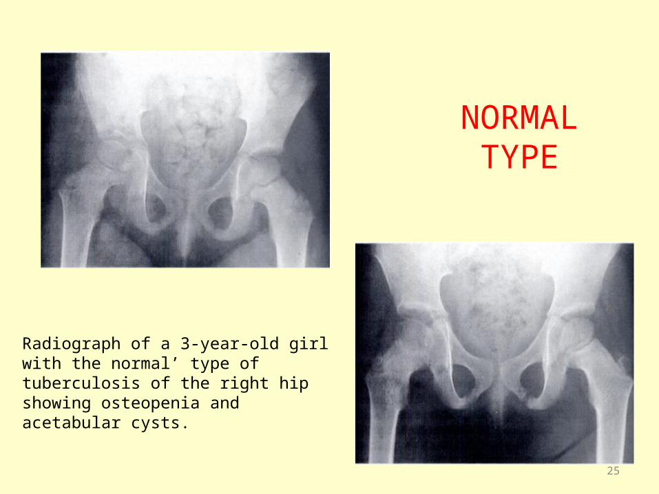
NORMAL TYPE
Radiograph of a 3-year-old girl with the normal’ type of tuberculosis of the right hip showing osteopenia and acetabular cysts.
25

PERTHES TYPE
Left femoral epiphysis is flattened absence of metaphyseal changes and presence of juxta-articular osteopenia favour TB over perthes disease.
26
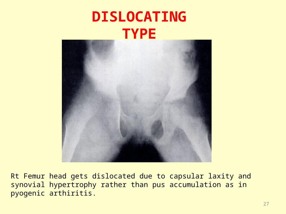
DISLOCATING TYPE
Rt Femur head gets dislocated due to capsular laxity and synovial hypertrophy rather than pus accumulation as in pyogenic arthiritis.
27
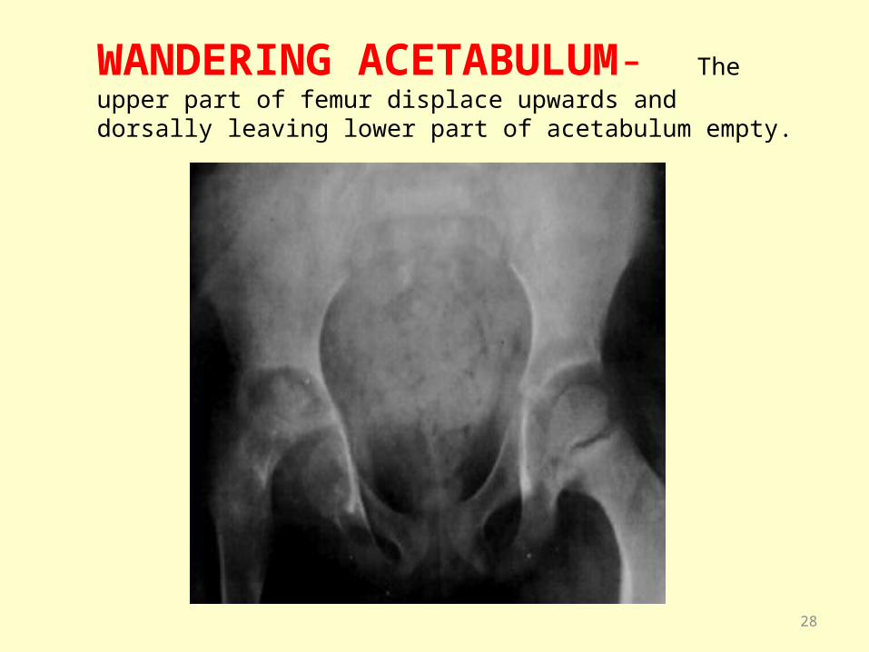
WANDERING ACETABULUM- The upper part of femur displace upwards and dorsally leaving lower part of acetabulum empty.
28
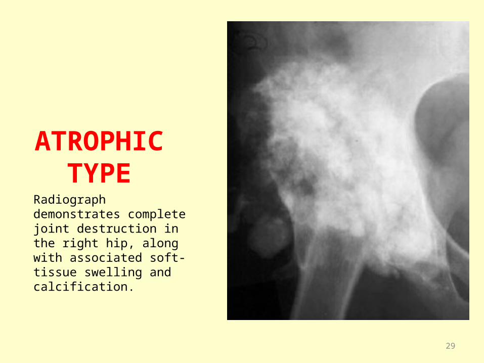
ATROPHIC TYPE
Radiograph demonstrates complete joint destruction in the right hip, along with associated soft-tissue swelling and calcification.
29

MORTAR AND PESTLE TYPE : femoral head and neck are grossly destroyed, collapsed and contained in a large acetabulum
30

Differential Diagnosis TB of hip is the commonest cause of pain in the hip in
children in TB prevalent countries.The following differential diagnosis should be considered:
a) Rheumatoid arthritis- In rheumatoid arthritis B/L symmetrical, more small joint involvement,h/o remissions &the joint space is uniformly reduced,
b) Inguinal LAN or Psoas Abscess – Patients with these extra articular diseases often present with the flexion deformity of hip because of spasm of iliopsoas. All movements of the hip expect extension are pain free.
c) Pyogenic arthritis- It can be differentiated as:-
31
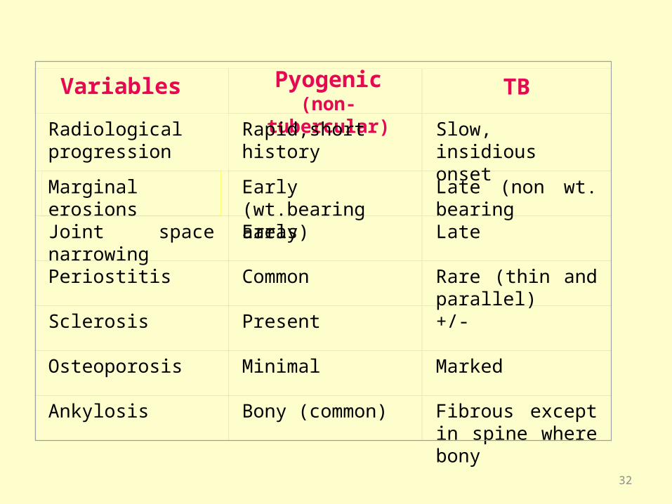
Variables Pyogenic(non-tubercular)
TB
Radiological progression
Rapid,short history Slow, insidious onset
Marginal erosions Early (wt.bearing areas)
Late (non wt. bearing
Joint space narrowing
Early Late
Periostitis Common Rare (thin and parallel)
Sclerosis Present +/-
Osteoporosis Minimal Marked
Ankylosis Bony (common) Fibrous except in spine where bony
32

d) Congenital dislocation of hip – Limp is painless, generally detected at birth. Telescopy test is positive and X-ray are decisive.
e) Congenital coxavara – Painless limp, abduction and internal rotation are limited.Adduction and external rotation maybe increased. X-ray usually confirms the diagnosis.
f) Perthe’s Disease –Seen in age group of 5-10 years, associated with minimal limitation of movements,mainly abduction and internal rotation. Typical X-ray changes are out of proportions to the physical findings. The joint space may be widened (unlike TB). Absence of metaphyseal changes and presence of juxta articular osteopenia favours TB
g) Osteoarthritis – occurs in older individuals.
- Hip movements are limited in all
directions but only terminally
. - Associated pain and crepitus .
h) PVNS - lack of osteopenia,heavy hemosiderin deposits causes prominent hypointensity on MRI
33
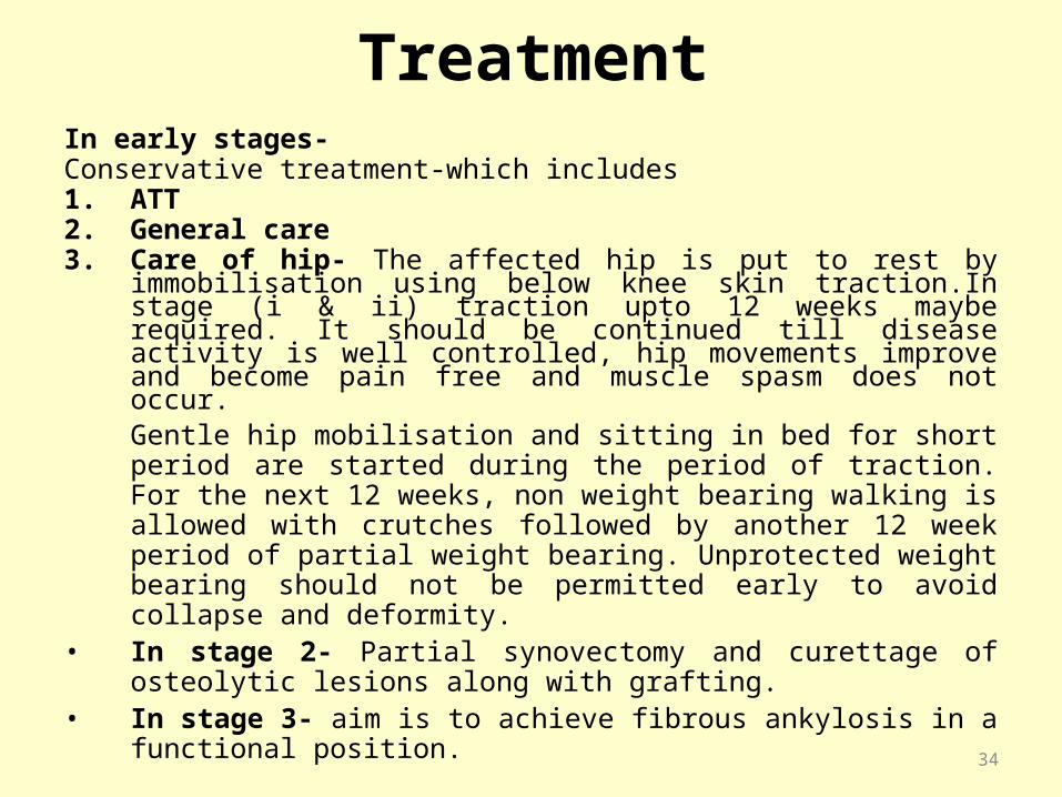
TreatmentIn early stages- Conservative treatment-which includes1. ATT2. General care3. Care of hip- The affected hip is put to rest by immobilisation
using below knee skin traction.In stage (i & ii) traction upto 12 weeks maybe required. It should be continued till disease activity is well controlled, hip movements improve and become pain free and muscle spasm does not occur.
Gentle hip mobilisation and sitting in bed for short period are started during the period of traction. For the next 12 weeks, non weight bearing walking is allowed with crutches followed by another 12 week period of partial weight bearing. Unprotected weight bearing should not be permitted early to avoid collapse and deformity.
• In stage 2- Partial synovectomy and curettage of osteolytic lesions along with grafting.
• In stage 3- aim is to achieve fibrous ankylosis in a functional position.
34
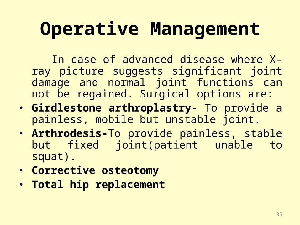
Operative Management
In case of advanced disease where X-ray picture suggests significant joint damage and normal joint functions can not be regained. Surgical options are:
• Girdlestone arthroplastry- To provide a painless, mobile but unstable joint.
• Arthrodesis-To provide painless, stable but fixed joint(patient unable to squat).
• Corrective osteotomy• Total hip replacement
35
Tuberculosis of Knee Joint
• It is the 3rd most common site for osteoarticular TB accounting for about 10 % of cases of osteoatricular TB.
• Focus of infection may be from:-
1.Synovium(MC)
2.Subchondral cancellous bone.
3.Juxta-articular osseous focus in the epiphysis.
4.Physeal plate.
5.Metaphysis.
6.Patella(rare)
36

Examination Pain and swelling in the knee- Gradual onset and later
increases and knee takes the attitude of flexion. There is inflammatory exudates in the joint with supra patellar fullness and filling of fossa on either side of patellar tendon.
The synovium and capsule becomes palpably thickened and tender”Doughy swelling”
Movements-In synovial disease,may be terminally restricted movements,but as arthritis sets in there is pain accompanied by muscle spasm.
• Muscle atrophy of thigh muscles(quadriceps)• Cold abscess either arround the knee or in the calf• Sinus formation• Deformity-In the early cases mild flexion deformity and later
triple deformity due to spasm and contracture of biceps femoris and tensor fascia lata.It is:-
--Flexion of knee --Posteriolateral subluxation of tibia& --Lateral rotation and abduction of the leg 37
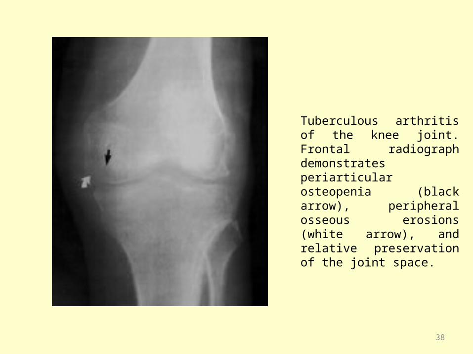
Tuberculous arthritis of the knee joint. Frontal radiograph demonstrates periarticular osteopenia (black arrow), peripheral osseous erosions (white arrow), and relative preservation of the joint space.
38
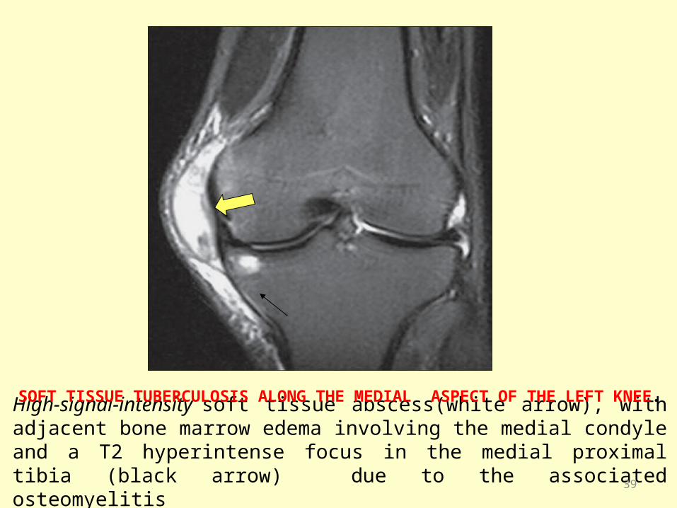
High-signal-intensity soft tissue abscess(white arrow), with adjacent bone marrow edema involving the medial condyle and a T2 hyperintense focus in the medial proximal tibia (black arrow) due to the associated osteomyelitis
SOFT TISSUE TUBERCULOSIS ALONG THE MEDIAL ASPECT OF THE LEFT KNEE.
39
Differential diagnosis
• Meniscal tear and synovitis due to trauma-generally young persons engaged in sports,h/o classic twisting injury of the knee, recurrent episodes of pain and sudden locking and unlocking of knee.
• Rheumatic arthritis(children)—in children,h/o sore throat,migratory polyarthritis,cardiac involvement.
• Rheumatoid arthritis(adults)• Subacute pyogenic infection• Villonodular synovitis.
40
Management
• Aim is to achieve a painless mobile joint.But it is possible in early stages.In later stages,some amount of pain and stiffness persists in spite of treatment.
• General care• Local care-In early stages the treatment is usually
conservative with antitubercular drugs and immobilization in a below knee skin traction or an above-knee POP cast till the disease is quiescent after which the patient can be mobilized.
41
Operative Treatment
• Synovectomy: may be required in cases of purely synovial TB,which is not responding to conservative treatment or doubtfull treatment.
• Joint debridement: the pus is drained,synovium excised and all cavities curetted.
• Arthrodesis: In advanced stage with triple subluxation and cartilage destruction.One such method is Charnley’s compression arthrodesis
42
TB of Ankle and Foot• Accounts for less than 5% of osteoarticular TB.• Tarsals and the ankle joint are usually involved together
due to intercommunicating synovial channels.• Most commonly starts as synovitis.Usually it presents as
synovial disease or extra-synovial soft tissue disease associated with bony focus.
• MC affected bone is calcaneum followed by talus,1st metatarsal,navicular and medial 2 cuneiform bones.
• Multiple lesions,abscess and sinus formation are common in adults but synovial disease is more common in children.
• Clinical and radiological features are same as of other joints.
• Synovial biopsy may be needed for the confirmation of diagnosis
43
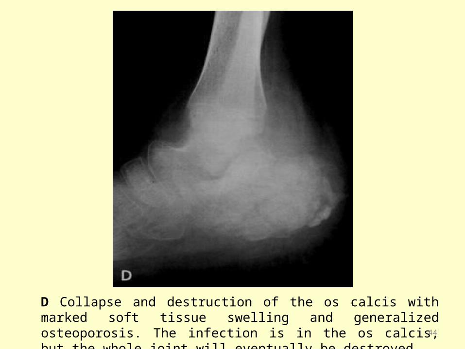
D Collapse and destruction of the os calcis with marked soft tissue swelling and generalized osteoporosis. The infection is in the os calcis, but the whole joint will eventually be destroyed 44

Sagittal T1-weighted MRI demonstrates hypointense periarticular effusions (black arrows) with bone erosion of the talus and tibia (white arrow).
Tuberculous arthritis of the ankle joint
45

Management• Antitubercular drugs along with pain killer(if
required)&genaral care. • POP cast to give rest: in 10 degree equines position.• An extensive,long standing disease requires arthrotomy
and synovectomy.• An isolates bony lesion should be curetted and large
cavities should be packed with bone grafts.• In extensive joint destruction,arthrodesis of ankle or
subtalar joint alone or together.
46
TB of Shoulder Joint
• Rare,only 1-2% of all osteoarticular cases.• Disease originates in head of humerous,
glenoid.Synovial variety is rare..• Common variety is dry and atrophic type—Caries sicca..• In children lesion may start in metaphyseal region of
humerus and affects longitudinal growth.• Joint involvement is early with painful limitation of all
movements esp.abduction and external rotation.• Swelling,abscess and sinus formation are rare.
47
• In early stages,some swelling,thickening and tenderness in soft tissue around the joint is present.
• Marked wasting of deltoid and supraspinatus musle may occur along with axillary lymphadenopathy.
• D/D include frozen shoulder in early stage of TB shoulder joint and Rheumatiod arthritis in late stage(Marked soft tissue swelling and effusion occurs in Rh.arthritis)
• In doubtful cases synovial biopsy can be sent for microbiological and histopathilogical examination.
48

Caries sicca. There is erosion and destruction of humoral head and glenoid cavity with soft tissue swelling, along with fibrotic opacities..
49
Management
• Genarally shoulder joint TB responds well to antituberculosis treatment.
• In addition to general treatment for TB,shoulder joint is immobilized in Saluting position to encourage ankylosis of gleno-humeral articulation in functional position.
• If ankylosis is painful, disease is uncontrolled or reccurance,arthodesis is carried out
50

Tuberculosis of elbow joint2% or less of all osteoarticular TB, adults are more affected.Osseus focus is in the olecranon>coronoid> lower end of humerus or upper end of radius. Synovial disease is uncommon.Swelling is appreciated at the back of elbow on both sides of olecranon & triceps tendon.The principle underlying the management are similar to TB of other synovial joints.In all stages of disease,elbow should be immobilized in an above elbow plaster cast in 90 degree flexion and mid prone position and when the acute stage is under control, intermittent active & assisted exercise is carried out. Surgical debridement with or without excision may be required for advance disease and surgical excision of a juxta – articular osseus lesion is indicated to prevent extension of disease to joint. Arthrodesis of elbow is rarely required for heavy manual work
51

Lateral view of elbow joint involved by TB showing lytic lesion in olecranon [arrow] and loss of joint space with flexion deformity
52
Tuberculosis of Wrist Joint Rare & mainly seen in adults. Most common site of initial focus are distal radius &
capitate and less often in synovium and Infection spreads to involve intercarpal&wrist joint,flexor and extensor tendon sheath. Abscess & sinus formation are common.
Onset in insidious, exacerbation of pain by movements occur early &soft tissue swelling, limitation of flexor & extensor movement occurs late.
53
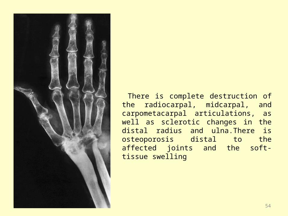
There is complete destruction of the radiocarpal, midcarpal, and carpometacarpal articulations, as well as sclerotic changes in the distal radius and ulna.There is osteoporosis distal to the affected joints and the soft-tissue swelling
54
Management TB of wrist joint is quite likely to be misdiagnosed so It is
essential to confirm the diagnosis with synovial biopsy to differentiate it from mono articular rheumatoid arthritis.
On early diagnosis, patient should be treated with antitubercular treatment & anti inflammatory drugs.
Prolonged splintage with wrist in 10 degree dorsiflexion and in mid prone position is always required.
Heavy physical work is avoided for 18-24 months. Synovectomy & curettage of bony lesions are indicated
in non responding patients. In advanced destruction, arthodesis is the treatment of
choice.
55
TB of Sacroiliac Joint
• It is a pure synovial joint,involves in around 2-5% of osteoarticular TB.More common in adults.(M:F=2:5)
• Lesion may start in sacrum,ilium or in synovium.Infection can also extend from ipsilateral hip joint or lumbosacral area of spine.
• Association with TB spine,hip,viscera are frequent.• Abscess formation is common and it may be present
dorsally or inside the pelvis.• Sinus formation also occurs.
56
• Insidious in onset or may follow trauma/pregnancy.• Pain is the main symptom, which may be referred to
groin/sciatic nerve distribution.Which is worsened on lying in supine or on affected side Sitting on buttock on affected side is painful,whearas on opposite side pain is relieved.
• Sudden jerks,coughing,sneezing,stress on involved SI joint increases pain.
• Rectal examination is important to detect intrapelvic abscess.
• CT and MRI are helpful in detecting early joint erosion,cavitation and abscess.
57
Management• Difficult to diagnose as presentation is atypical.• ATT and general care along with rest&
mobilization when the acute symptoms subsides.
• Less responsive to ATT,so surgery is generally required.
• Surgical options are debridement of joint,freshening of joint margins and adequate grafting to achieve arthrodesis.Post-operative bed rest is required for 3 months followed by gradual mobilization in L-S belt.
58
Other Rare Involvements 1. Sternoclavicular 2. Acromioclavicular 3. Symphysis Pubis 4. Ilium,Ischium and Ischiopubic ramus 5. Sternum and Ribs 6. Scapula 7. Clavicle 8. TB Tenosynovitis& TB Bursitis
59

TB Osteomyelitis• TB rarely affect the shaft of long bones. It is usually a low
grade subacute osteomyelitis.Common presenting features are pain,swelling,tenderness,regional LAN,abscess and sinus formation.
• An imp.radiological feature is that the bone lysis is out of proportion to the new bone formation(unlike pyogenic osteomyelitis).
• Biopsy for histopathological and micribiological investigation may help in diagnosis.
• Short bones like phalanges(Spina ventosa),metacarpals,metatarsals are involved in children.
• Disease process starts in medullary cavity diphysis sequestration.
• Patient presents with gradual and painless swelling of one of the phalanges.
60
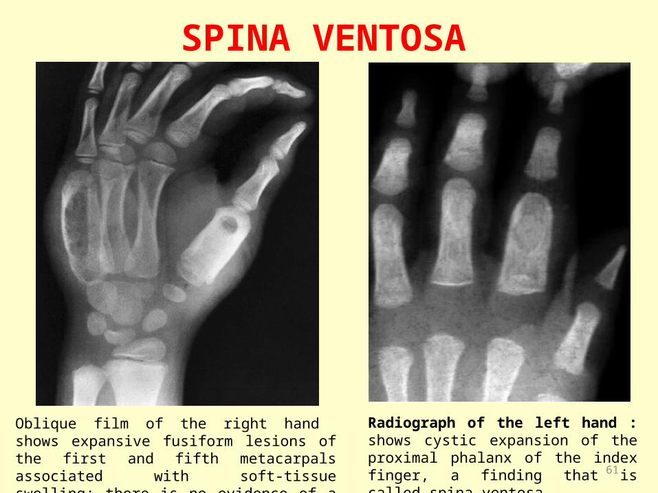
SPINA VENTOSA
Oblique film of the right hand shows expansive fusiform lesions of the first and fifth metacarpals associated with soft-tissue swelling; there is no evidence of a periosteal reaction
Radiograph of the left hand : shows cystic expansion of the proximal phalanx of the index finger, a finding that is called spina ventosa. 61

• FDG-PET{Flourine flouro deoxyglucose positron emission tomography} has been found to be useful in localizing TB disease in inaccessible or obscure sites.
• Response to Antituberculosis drugs is good.• Refractory cases or in presence of abscess around the
bone may require excision of granulation tissue and infected bone.
• Additional antibiotic for secondary infection through sinuses.
62

THANK YOU63
Related Documents


