bioengineering Review Skeletal Muscle Tissue Engineering: Biomaterials-Based Strategies for the Treatment of Volumetric Muscle Loss Meagan E. Carnes and George D. Pins * Department of Biomedical Engineering, Worcester Polytechnic Institute, 100 Institute Rd., Worcester, MA 01609, USA; [email protected] * Correspondence: [email protected] Received: 24 June 2020; Accepted: 28 July 2020; Published: 31 July 2020 Abstract: Millions of Americans suffer from skeletal muscle injuries annually that can result in volumetric muscle loss (VML), where extensive musculoskeletal damage and tissue loss result in permanent functional deficits. In the case of small-scale injury skeletal muscle is capable of endogenous regeneration through activation of resident satellite cells (SCs). However, this is greatly reduced in VML injuries, which remove native biophysical and biochemical signaling cues and hinder the damaged tissue’s ability to direct regeneration. The current clinical treatment for VML is autologous tissue transfer, but graft failure and scar tissue formation leave patients with limited functional recovery. Tissue engineering of instructive biomaterial scaffolds offers a promising approach for treating VML injuries. Herein, we review the strategic engineering of biophysical and biochemical cues in current scaffold designs that aid in restoring function to these preclinical VML injuries. We also discuss the successes and limitations of the three main biomaterial-based strategies to treat VML injuries: acellular scaffolds, cell-delivery scaffolds, and in vitro tissue engineered constructs. Finally, we examine several innovative approaches to enhancing the design of the next generation of engineered scaffolds to improve the functional regeneration of skeletal muscle following VML injuries. Keywords: biomaterials; tissue engineering; volumetric muscle loss; skeletal muscle regeneration 1. Clinical Need: Volumetric Muscle Loss A total of 65.8 million Americans suffer from musculoskeletal injuries annually, with treatment costs exceeding 176 billion dollars [1–5]. Although these injuries are not commonly life threatening, they profoundly impact the quality of life of patients. Musculoskeletal conditions are highly debilitating, comprising the second highest global volume of years lived with disability [6]. It is estimated that these injuries result in an additional 326 billion dollars annually in lost productivity [7]. Severe musculoskeletal injuries can lead to volumetric muscle loss (VML), where extensive musculoskeletal damage and tissue loss result in permanent loss of function [8,9]. VML injuries can result from sports injuries, surgical resection, and traumatic events such as car accidents and combat injury. In particular, musculoskeletal injuries sustained in combat present a unique challenge because they lead to the highest number of disabled war fighters and have the largest disability costs [10]. While the rate of combat mortality for U.S. Warfighters has dropped significantly since World War II, there has been a marked increase in the number of soldiers who suffer from extraordinary injuries, such as blast injuries, which impart extensive damage to the head, neck and extremities [11]. A total of 54% of all soldiers wounded on the battlefield suffer from at least one musculoskeletal extremity injury, with 53% of these injuries involving soft tissue damage [8,12]. Combat-related extremity injuries cause the greatest number of disabled soldiers [10]. Injured soldiers incur an average of 4.2 wounds, Bioengineering 2020, 7, 85; doi:10.3390/bioengineering7030085 www.mdpi.com/journal/bioengineering

Welcome message from author
This document is posted to help you gain knowledge. Please leave a comment to let me know what you think about it! Share it to your friends and learn new things together.
Transcript
bioengineering
Review
Skeletal Muscle Tissue Engineering:Biomaterials-Based Strategies for the Treatmentof Volumetric Muscle Loss
Meagan E. Carnes and George D. Pins *
Department of Biomedical Engineering, Worcester Polytechnic Institute, 100 Institute Rd.,Worcester, MA 01609, USA; [email protected]* Correspondence: [email protected]
Received: 24 June 2020; Accepted: 28 July 2020; Published: 31 July 2020�����������������
Abstract: Millions of Americans suffer from skeletal muscle injuries annually that can result involumetric muscle loss (VML), where extensive musculoskeletal damage and tissue loss resultin permanent functional deficits. In the case of small-scale injury skeletal muscle is capable ofendogenous regeneration through activation of resident satellite cells (SCs). However, this isgreatly reduced in VML injuries, which remove native biophysical and biochemical signaling cuesand hinder the damaged tissue’s ability to direct regeneration. The current clinical treatment forVML is autologous tissue transfer, but graft failure and scar tissue formation leave patients withlimited functional recovery. Tissue engineering of instructive biomaterial scaffolds offers a promisingapproach for treating VML injuries. Herein, we review the strategic engineering of biophysicaland biochemical cues in current scaffold designs that aid in restoring function to these preclinical VMLinjuries. We also discuss the successes and limitations of the three main biomaterial-based strategies totreat VML injuries: acellular scaffolds, cell-delivery scaffolds, and in vitro tissue engineered constructs.Finally, we examine several innovative approaches to enhancing the design of the next generation ofengineered scaffolds to improve the functional regeneration of skeletal muscle following VML injuries.
Keywords: biomaterials; tissue engineering; volumetric muscle loss; skeletal muscle regeneration
1. Clinical Need: Volumetric Muscle Loss
A total of 65.8 million Americans suffer from musculoskeletal injuries annually, with treatmentcosts exceeding 176 billion dollars [1–5]. Although these injuries are not commonly life threatening, theyprofoundly impact the quality of life of patients. Musculoskeletal conditions are highly debilitating,comprising the second highest global volume of years lived with disability [6]. It is estimated thatthese injuries result in an additional 326 billion dollars annually in lost productivity [7].
Severe musculoskeletal injuries can lead to volumetric muscle loss (VML), where extensivemusculoskeletal damage and tissue loss result in permanent loss of function [8,9]. VML injuries canresult from sports injuries, surgical resection, and traumatic events such as car accidents and combatinjury. In particular, musculoskeletal injuries sustained in combat present a unique challenge becausethey lead to the highest number of disabled war fighters and have the largest disability costs [10].While the rate of combat mortality for U.S. Warfighters has dropped significantly since World War II,there has been a marked increase in the number of soldiers who suffer from extraordinary injuries,such as blast injuries, which impart extensive damage to the head, neck and extremities [11]. A totalof 54% of all soldiers wounded on the battlefield suffer from at least one musculoskeletal extremityinjury, with 53% of these injuries involving soft tissue damage [8,12]. Combat-related extremity injuriescause the greatest number of disabled soldiers [10]. Injured soldiers incur an average of 4.2 wounds,
Bioengineering 2020, 7, 85; doi:10.3390/bioengineering7030085 www.mdpi.com/journal/bioengineering
Bioengineering 2020, 7, 85 2 of 39
making extremity injuries the primary cause for hospitalization and evacuation from theater [10].VML injuries also result in significant long-term disability that does not improve over time [13,14].These extremity wounds also represent the largest projected disability costs of combat injuries [10,15].The projected lifetime disability costs of a soldier with VML is $341,200 per individual [14]. Extremityinjuries account for 69% of resource utilization, making them not only the most common injuriesbut also some of the most expensive to treat [15].
Due to the complex and large-scale nature of VML injuries, current treatment options remainlimited and have substantial disadvantages. In the case of small-scale injuries or strains, muscle iscapable of endogenous regeneration and complete functional restoration. However, this ability is abatedin VML, where the native biophysical and biochemical signaling cues are no longer present to facilitateregeneration. These injuries are concomitant with denervation and the destruction of native vasculature,further limiting regeneration. Currently physical therapy is the only targeted treatment for VMLinjuries, and it has shown limited success in improving muscle strength [16–18]. The current standardof care for VML is autologous tissue transfer, where a muscle flap is excised from an undamaged muscleand grafted into the injury site [19–22]. This procedure is commonly referred to as a free functionalmuscle transfer (FFMT). While FFMT has been moderately successful in salvaging limbs and restoringsome muscle function, muscle flaps remain unable to completely restore muscle function [22–25].This procedure is also complicated and time consuming to perform and requires the expertise of skilledorthopedic and microvascular surgeons, which may limit its widespread use [19,26]. Additionally,a high instance of muscle flap procedures result in complications such as infection, graft failure,and donor site morbidity due to tissue necrosis [21,22,27,28]. Often a revisionary surgery or amputationof the affected limb is required [21,22,27,28]. Thus, a clinical need exists for the development ofan alternative treatment that will restore function in VML injuries.
2. Skeletal Muscle Anatomy
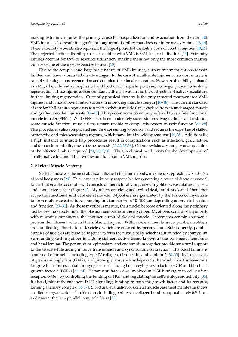
Skeletal muscle is the most abundant tissue in the human body, making up approximately 40–45%of total body mass [29]. This tissue is primarily responsible for generating a series of discrete uniaxialforces that enable locomotion. It consists of hierarchically organized myofibers, vasculature, nerves,and connective tissue (Figure 1). Myofibers are elongated, cylindrical, multi-nucleated fibers thatact as the functional unit of skeletal muscle. Myofibers are generated by the fusion of myoblaststo form multi-nucleated tubes, ranging in diameter from 10–100 µm depending on muscle locationand function [29–31]. As these myofibers mature, their nuclei become oriented along the peripheryjust below the sarcolemma, the plasma membrane of the myofiber. Myofibers consist of myofibrilswith repeating sarcomeres, the contractile unit of skeletal muscle. Sarcomeres contain contractileproteins thin filament actin and thick filament myosin. Within skeletal muscle tissue, parallel myofibersare bundled together to form fascicles, which are encased by perimysium. Subsequently, parallelbundles of fascicles are bundled together to form the muscle belly, which is surrounded by epimysium.Surrounding each myofiber is endomysial connective tissue known as the basement membraneand basal lamina. The perimysium, epimysium, and endomysium together provide structural supportto the tissue while aiding in force transmission and synchronous contraction. The basal lamina iscomposed of proteins including type IV collagen, fibronectin, and laminin-2 [32,33]. It also consistsof glycosaminoglycans (GAGs) and proteoglycans, such as heparan sulfate, which act as reservoirsfor growth factors essential for myogenesis, including hepatocyte growth factor (HGF) and fibroblastgrowth factor 2 (FGF2) [32–34]. Heparan sulfate is also involved in HGF binding to its cell surfacereceptor, c-Met, by controlling the binding of HGF and regulating the cell’s mitogenic activity [35].It also significantly enhances FGF2 signaling, binding to both the growth factor and its receptor,forming a ternary complex [36,37]. Structural evaluation of skeletal muscle basement membrane showsan aligned organization of architecture, including perimysial collagen bundles approximately 0.5–1 µmin diameter that run parallel to muscle fibers [33].

Bioengineering 2020, 7, 85 3 of 39Bioengineering 2020, 7, x FOR PEER REVIEW 3 of 40
Figure 1. Skeletal muscle anatomy. Skeletal muscle is a highly aligned tissue with a hierarchically organized, cable-like structure.
Just below the basal lamina and above the sarcolemma is where satellite cells (SCs), muscle-specific resident stem cells, are located [38]. In healthy skeletal muscle, SCs typically account for only about 2–7% of the total myonuclei [39]. They are identified by the expression of transcription factor paired box 7 (Pax7) and have been found to be necessary for skeletal muscle regeneration [40–43]. Upon injury, SCs leave their quiescent state and become activated to enter the cell cycle [44]. They proliferate and differentiate to form multi-nucleated myotubes, which mature to form myofibers. SCs are also capable self-renewing by maintaining a stem-like population [45]. A more detailed explanation of the role of SCs in skeletal muscle regeneration will be explored in Section 3.
To allow for voluntary locomotion, skeletal muscle is highly innervated. Motor neurons extend from the central nervous system and branch extensively throughout the muscle tissue to contact individual myofibers at a neuromuscular junction (NMJ) (Figure 2). The NMJ is the site at which an action potential from the motor neuron is converted to a muscle contraction. Contraction is initiated by acetylcholine release from the presynaptic axon, which subsequently binds to the myofiber and depolarizes the membrane. Membrane depolarization results in an action potential which travels down the length of the myofiber and initiates the release of calcium ions. Calcium binding within the myofibril results in an actin/myosin-mediated power stroke and muscle contraction. To meet its high metabolic demands, skeletal muscle tissue is also highly vascularized. An organized branching structure with capillary networks running parallel to the myofibers allow for optimal nutrient and oxygen exchange (Figure 2). Capillary networks in skeletal muscle are dense, with approximately 600 capillaries/mm [2,46]. This results in 40 μm distance between capillaries, and thus a 20 μm distance for oxygen diffusion [46].
Figure 2. Anatomy of skeletal muscle vasculature and neuromuscular junctions. Arterioles, venules, and neurons run adjacent and parallel to myofibers.
3. Skeletal Muscle Regeneration
After acute injury, endogenous repair of skeletal muscle follows a highly coordinated regenerative process involving three separate but overlapping phases: destruction/inflammatory, repair, and remodeling (Figure 3A–E).
Figure 1. Skeletal muscle anatomy. Skeletal muscle is a highly aligned tissue with a hierarchicallyorganized, cable-like structure.
Just below the basal lamina and above the sarcolemma is where satellite cells (SCs), muscle-specificresident stem cells, are located [38]. In healthy skeletal muscle, SCs typically account for only about2–7% of the total myonuclei [39]. They are identified by the expression of transcription factor pairedbox 7 (Pax7) and have been found to be necessary for skeletal muscle regeneration [40–43]. Uponinjury, SCs leave their quiescent state and become activated to enter the cell cycle [44]. They proliferateand differentiate to form multi-nucleated myotubes, which mature to form myofibers. SCs are alsocapable self-renewing by maintaining a stem-like population [45]. A more detailed explanation ofthe role of SCs in skeletal muscle regeneration will be explored in Section 3.
To allow for voluntary locomotion, skeletal muscle is highly innervated. Motor neuronsextend from the central nervous system and branch extensively throughout the muscle tissue tocontact individual myofibers at a neuromuscular junction (NMJ) (Figure 2). The NMJ is the siteat which an action potential from the motor neuron is converted to a muscle contraction. Contraction isinitiated by acetylcholine release from the presynaptic axon, which subsequently binds to the myofiberand depolarizes the membrane. Membrane depolarization results in an action potential which travelsdown the length of the myofiber and initiates the release of calcium ions. Calcium binding withinthe myofibril results in an actin/myosin-mediated power stroke and muscle contraction. To meet itshigh metabolic demands, skeletal muscle tissue is also highly vascularized. An organized branchingstructure with capillary networks running parallel to the myofibers allow for optimal nutrientand oxygen exchange (Figure 2). Capillary networks in skeletal muscle are dense, with approximately600 capillaries/mm [2,46]. This results in 40 µm distance between capillaries, and thus a 20 µm distancefor oxygen diffusion [46].
Bioengineering 2020, 7, x FOR PEER REVIEW 3 of 40
Figure 1. Skeletal muscle anatomy. Skeletal muscle is a highly aligned tissue with a hierarchically organized, cable-like structure.
Just below the basal lamina and above the sarcolemma is where satellite cells (SCs), muscle-specific resident stem cells, are located [38]. In healthy skeletal muscle, SCs typically account for only about 2–7% of the total myonuclei [39]. They are identified by the expression of transcription factor paired box 7 (Pax7) and have been found to be necessary for skeletal muscle regeneration [40–43]. Upon injury, SCs leave their quiescent state and become activated to enter the cell cycle [44]. They proliferate and differentiate to form multi-nucleated myotubes, which mature to form myofibers. SCs are also capable self-renewing by maintaining a stem-like population [45]. A more detailed explanation of the role of SCs in skeletal muscle regeneration will be explored in Section 3.
To allow for voluntary locomotion, skeletal muscle is highly innervated. Motor neurons extend from the central nervous system and branch extensively throughout the muscle tissue to contact individual myofibers at a neuromuscular junction (NMJ) (Figure 2). The NMJ is the site at which an action potential from the motor neuron is converted to a muscle contraction. Contraction is initiated by acetylcholine release from the presynaptic axon, which subsequently binds to the myofiber and depolarizes the membrane. Membrane depolarization results in an action potential which travels down the length of the myofiber and initiates the release of calcium ions. Calcium binding within the myofibril results in an actin/myosin-mediated power stroke and muscle contraction. To meet its high metabolic demands, skeletal muscle tissue is also highly vascularized. An organized branching structure with capillary networks running parallel to the myofibers allow for optimal nutrient and oxygen exchange (Figure 2). Capillary networks in skeletal muscle are dense, with approximately 600 capillaries/mm [2,46]. This results in 40 μm distance between capillaries, and thus a 20 μm distance for oxygen diffusion [46].
Figure 2. Anatomy of skeletal muscle vasculature and neuromuscular junctions. Arterioles, venules, and neurons run adjacent and parallel to myofibers.
3. Skeletal Muscle Regeneration
After acute injury, endogenous repair of skeletal muscle follows a highly coordinated regenerative process involving three separate but overlapping phases: destruction/inflammatory, repair, and remodeling (Figure 3A–E).
Figure 2. Anatomy of skeletal muscle vasculature and neuromuscular junctions. Arterioles, venules,and neurons run adjacent and parallel to myofibers.
Bioengineering 2020, 7, 85 4 of 39
3. Skeletal Muscle Regeneration
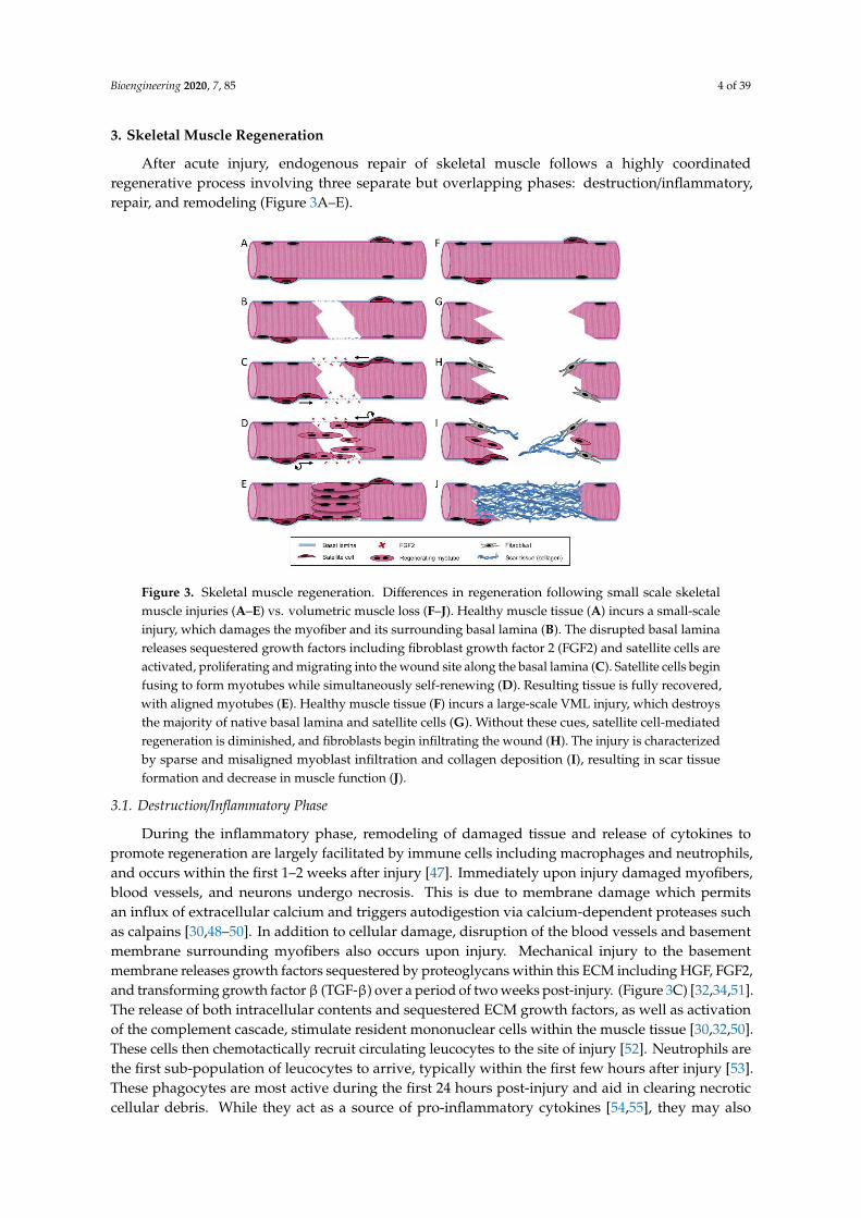
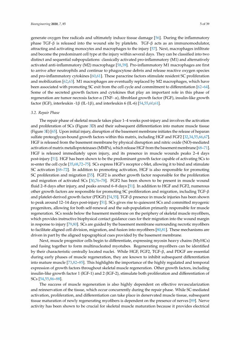
After acute injury, endogenous repair of skeletal muscle follows a highly coordinatedregenerative process involving three separate but overlapping phases: destruction/inflammatory,repair, and remodeling (Figure 3A–E).Bioengineering 2020, 7, x FOR PEER REVIEW 4 of 40
Figure 3. Skeletal muscle regeneration. Differences in regeneration following small scale skeletal
muscle injuries (A–E) vs. volumetric muscle loss (F–J). Healthy muscle tissue (A) incurs a small-scale
injury, which damages the myofiber and its surrounding basal lamina (B). The disrupted basal lamina
releases sequestered growth factors including fibroblast growth factor 2 (FGF2) and satellite cells are
activated, proliferating and migrating into the wound site along the basal lamina (C). Satellite cells
begin fusing to form myotubes while simultaneously self-renewing (D). Resulting tissue is fully
recovered, with aligned myotubes (E). Healthy muscle tissue (F) incurs a large-scale VML injury,
which destroys the majority of native basal lamina and satellite cells (G). Without these cues, satellite
cell-mediated regeneration is diminished, and fibroblasts begin infiltrating the wound (H). The injury
is characterized by sparse and misaligned myoblast infiltration and collagen deposition (I), resulting
in scar tissue formation and decrease in muscle function (J).
3.1. Destruction/Inflammatory Phase
During the inflammatory phase, remodeling of damaged tissue and release of cytokines to
promote regeneration are largely facilitated by immune cells including macrophages and
neutrophils, and occurs within the first 1–2 weeks after injury [47]. Immediately upon injury
damaged myofibers, blood vessels, and neurons undergo necrosis. This is due to membrane damage
which permits an influx of extracellular calcium and triggers autodigestion via calcium-dependent
proteases such as calpains [30,48–50]. In addition to cellular damage, disruption of the blood vessels
and basement membrane surrounding myofibers also occurs upon injury. Mechanical injury to the
basement membrane releases growth factors sequestered by proteoglycans within this ECM
including HGF, FGF2, and transforming growth factor β (TGF-β) over a period of two weeks post-
injury. (Figure 3C) [32,34,51]. The release of both intracellular contents and sequestered ECM growth
factors, as well as activation of the complement cascade, stimulate resident mononuclear cells within
the muscle tissue [30,32,50]. These cells then chemotactically recruit circulating leucocytes to the site
of injury [52]. Neutrophils are the first sub-population of leucocytes to arrive, typically within the
first few hours after injury [53]. These phagocytes are most active during the first 24 hours post-injury
and aid in clearing necrotic cellular debris. While they act as a source of pro-inflammatory cytokines
[54,55], they may also generate oxygen free radicals and ultimately induce tissue damage [56]. During
the inflammatory phase TGF-β is released into the wound site by platelets. TGF-β acts as an
immunomodulator, attracting and activating monocytes and macrophages to the injury [57]. Next,
macrophages infiltrate and become the predominant cell type at the injury within several days. They
can be classified into two distinct and sequential subpopulations: classically activated pro-
inflammatory (M1) and alternatively activated anti-inflammatory (M2) macrophage [58,59]. Pro-
Figure 3. Skeletal muscle regeneration. Differences in regeneration following small scale skeletalmuscle injuries (A–E) vs. volumetric muscle loss (F–J). Healthy muscle tissue (A) incurs a small-scaleinjury, which damages the myofiber and its surrounding basal lamina (B). The disrupted basal laminareleases sequestered growth factors including fibroblast growth factor 2 (FGF2) and satellite cells areactivated, proliferating and migrating into the wound site along the basal lamina (C). Satellite cells beginfusing to form myotubes while simultaneously self-renewing (D). Resulting tissue is fully recovered,with aligned myotubes (E). Healthy muscle tissue (F) incurs a large-scale VML injury, which destroysthe majority of native basal lamina and satellite cells (G). Without these cues, satellite cell-mediatedregeneration is diminished, and fibroblasts begin infiltrating the wound (H). The injury is characterizedby sparse and misaligned myoblast infiltration and collagen deposition (I), resulting in scar tissueformation and decrease in muscle function (J).
3.1. Destruction/Inflammatory Phase
During the inflammatory phase, remodeling of damaged tissue and release of cytokines topromote regeneration are largely facilitated by immune cells including macrophages and neutrophils,and occurs within the first 1–2 weeks after injury [47]. Immediately upon injury damaged myofibers,blood vessels, and neurons undergo necrosis. This is due to membrane damage which permitsan influx of extracellular calcium and triggers autodigestion via calcium-dependent proteases suchas calpains [30,48–50]. In addition to cellular damage, disruption of the blood vessels and basementmembrane surrounding myofibers also occurs upon injury. Mechanical injury to the basementmembrane releases growth factors sequestered by proteoglycans within this ECM including HGF, FGF2,and transforming growth factorβ (TGF-β) over a period of two weeks post-injury. (Figure 3C) [32,34,51].The release of both intracellular contents and sequestered ECM growth factors, as well as activationof the complement cascade, stimulate resident mononuclear cells within the muscle tissue [30,32,50].These cells then chemotactically recruit circulating leucocytes to the site of injury [52]. Neutrophils arethe first sub-population of leucocytes to arrive, typically within the first few hours after injury [53].These phagocytes are most active during the first 24 hours post-injury and aid in clearing necroticcellular debris. While they act as a source of pro-inflammatory cytokines [54,55], they may also
Bioengineering 2020, 7, 85 5 of 39
generate oxygen free radicals and ultimately induce tissue damage [56]. During the inflammatoryphase TGF-β is released into the wound site by platelets. TGF-β acts as an immunomodulator,attracting and activating monocytes and macrophages to the injury [57]. Next, macrophages infiltrateand become the predominant cell type at the injury within several days. They can be classified into twodistinct and sequential subpopulations: classically activated pro-inflammatory (M1) and alternativelyactivated anti-inflammatory (M2) macrophage [58,59]. Pro-inflammatory M1 macrophages are firstto arrive after neutrophils and continue to phagocytose debris and release reactive oxygen speciesand pro-inflammatory cytokines [60,61]. These paracrine factors stimulate resident SC proliferationand mobilization [62,63]. M1 macrophages are eventually replaced by M2 macrophages, which havebeen associated with promoting SC exit from the cell cycle and commitment to differentiation [62–64].Some of the secreted growth factors and cytokines that play an important role in this phase ofregeneration are tumor necrosis factor-α (TNF- α), fibroblast growth factor (FGF), insulin-like growthfactor (IGF), interleukin -1β (IL-1β), and interleukin 6 (IL-6) [54,55,60,61].
3.2. Repair Phase
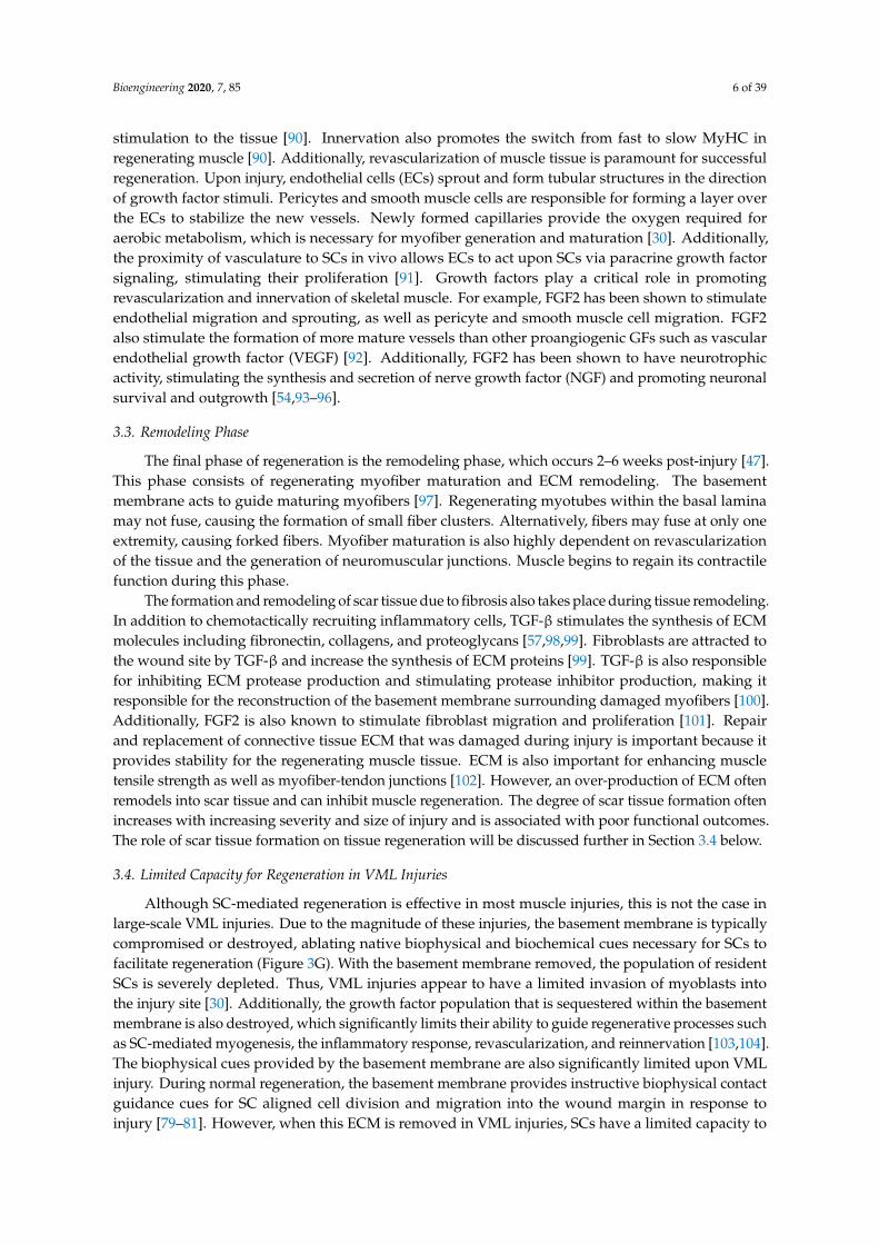
The repair phase of skeletal muscle takes place 1–4 weeks post-injury and involves the activationand proliferation of SCs (Figure 3D) and their subsequent differentiation into mature muscle tissue(Figure 3E) [65]. Upon initial injury, disruption of the basement membrane initiates the release of heparansulfate proteoglycan-bound growth factors within this matrix, including HGF and FGF2 [32,34,55,66,67].HGF is released from the basement membrane by physical disruption and nitric oxide (NO)-mediatedactivation of matrix metalloproteinases (MMPs), which release HGF from the basement membrane [68–71].HGF is released immediately upon injury, and its presence in muscle wounds peaks 2–4 dayspost-injury [51]. HGF has been shown to be the predominant growth factor capable of activating SCs tore-enter the cell cycle [35,68,72–75]. SCs express HGF’s receptor c-Met, allowing it to bind and stimulateSC activation [68–72]. In addition to promoting activation, HGF is also responsible for promotingSC proliferation and migration [55]. FGF2 is another growth factor responsible for the proliferationand migration of activated SCs [30,76–78]. FGF2 has been shown to be present in muscle woundfluid 2–8 days after injury, and peaks around 6–8 days [51]. In addition to HGF and FGF2, numerousother growth factors are responsible for promoting SC proliferation and migration, including TGF-βand platelet-derived growth factor (PDGF) [54,55]. TGF-β presence in muscle injuries has been shownto peak around 12–14 days post-injury [51]. SCs gives rise to quiescent SCs and committed myogenicprogenitors, allowing for both self-renewal and the sub-population primarily responsible for muscleregeneration. SCs reside below the basement membrane on the periphery of skeletal muscle myofibers,which provides instructive biophysical contact guidance cues for their migration into the wound marginin response to injury [79,80]. SCs are guided by the basement membrane surrounding necrotic myofibersto facilitate aligned cell division, migration, and fusion into myofibers [80,81]. These mechanisms aredriven in part by the aligned topographical cues provided by the basement membrane.
Next, muscle progenitor cells begin to differentiate, expressing myosin heavy chains (MyHCs)and fusing together to form multinucleated myotubes. Regenerating myofibers can be identifiedby their characteristic centrally located nuclei. While HGF, FGF2, TGF-β, and PDGF are essentialduring early phases of muscle regeneration, they are known to inhibit subsequent differentiationinto mature muscle [73,82–85]. This highlights the importance of the highly regulated and temporalexpression of growth factors throughout skeletal muscle regeneration. Other growth factors, includinginsulin-like growth factor 1 (IGF-1) and 2 (IGF-2), stimulate both proliferation and differentiation ofSCs [54,55,86–88].
The success of muscle regeneration is also highly dependent on effective revascularizationand reinnervation of the tissue, which occur concurrently during the repair phase. While SC-mediatedactivation, proliferation, and differentiation can take place in denervated muscle tissue, subsequenttissue maturation of newly regenerating myofibers is dependent on the presence of nerves [89]. Nerveactivity has been shown to be crucial for skeletal muscle maturation because it provides electrical
Bioengineering 2020, 7, 85 6 of 39
stimulation to the tissue [90]. Innervation also promotes the switch from fast to slow MyHC inregenerating muscle [90]. Additionally, revascularization of muscle tissue is paramount for successfulregeneration. Upon injury, endothelial cells (ECs) sprout and form tubular structures in the directionof growth factor stimuli. Pericytes and smooth muscle cells are responsible for forming a layer overthe ECs to stabilize the new vessels. Newly formed capillaries provide the oxygen required foraerobic metabolism, which is necessary for myofiber generation and maturation [30]. Additionally,the proximity of vasculature to SCs in vivo allows ECs to act upon SCs via paracrine growth factorsignaling, stimulating their proliferation [91]. Growth factors play a critical role in promotingrevascularization and innervation of skeletal muscle. For example, FGF2 has been shown to stimulateendothelial migration and sprouting, as well as pericyte and smooth muscle cell migration. FGF2also stimulate the formation of more mature vessels than other proangiogenic GFs such as vascularendothelial growth factor (VEGF) [92]. Additionally, FGF2 has been shown to have neurotrophicactivity, stimulating the synthesis and secretion of nerve growth factor (NGF) and promoting neuronalsurvival and outgrowth [54,93–96].
3.3. Remodeling Phase
The final phase of regeneration is the remodeling phase, which occurs 2–6 weeks post-injury [47].This phase consists of regenerating myofiber maturation and ECM remodeling. The basementmembrane acts to guide maturing myofibers [97]. Regenerating myotubes within the basal laminamay not fuse, causing the formation of small fiber clusters. Alternatively, fibers may fuse at only oneextremity, causing forked fibers. Myofiber maturation is also highly dependent on revascularizationof the tissue and the generation of neuromuscular junctions. Muscle begins to regain its contractilefunction during this phase.
The formation and remodeling of scar tissue due to fibrosis also takes place during tissue remodeling.In addition to chemotactically recruiting inflammatory cells, TGF-β stimulates the synthesis of ECMmolecules including fibronectin, collagens, and proteoglycans [57,98,99]. Fibroblasts are attracted tothe wound site by TGF-β and increase the synthesis of ECM proteins [99]. TGF-β is also responsiblefor inhibiting ECM protease production and stimulating protease inhibitor production, making itresponsible for the reconstruction of the basement membrane surrounding damaged myofibers [100].Additionally, FGF2 is also known to stimulate fibroblast migration and proliferation [101]. Repairand replacement of connective tissue ECM that was damaged during injury is important because itprovides stability for the regenerating muscle tissue. ECM is also important for enhancing muscletensile strength as well as myofiber-tendon junctions [102]. However, an over-production of ECM oftenremodels into scar tissue and can inhibit muscle regeneration. The degree of scar tissue formation oftenincreases with increasing severity and size of injury and is associated with poor functional outcomes.The role of scar tissue formation on tissue regeneration will be discussed further in Section 3.4 below.
3.4. Limited Capacity for Regeneration in VML Injuries
Although SC-mediated regeneration is effective in most muscle injuries, this is not the case inlarge-scale VML injuries. Due to the magnitude of these injuries, the basement membrane is typicallycompromised or destroyed, ablating native biophysical and biochemical cues necessary for SCs tofacilitate regeneration (Figure 3G). With the basement membrane removed, the population of residentSCs is severely depleted. Thus, VML injuries appear to have a limited invasion of myoblasts intothe injury site [30]. Additionally, the growth factor population that is sequestered within the basementmembrane is also destroyed, which significantly limits their ability to guide regenerative processes suchas SC-mediated myogenesis, the inflammatory response, revascularization, and reinnervation [103,104].The biophysical cues provided by the basement membrane are also significantly limited upon VMLinjury. During normal regeneration, the basement membrane provides instructive biophysical contactguidance cues for SC aligned cell division and migration into the wound margin in response toinjury [79–81]. However, when this ECM is removed in VML injuries, SCs have a limited capacity to
Bioengineering 2020, 7, 85 7 of 39
migrate into the wound and undergo aligned cell division and myotube fusion. Lateral migrationof SCs outside the basement membrane is more likely to occur in VML injuries, where the basementmembrane has been disrupted [105]. In addition to limited contact guidance cues and signaling, theselarge-scale injuries also have a lack of mechanical support [106].
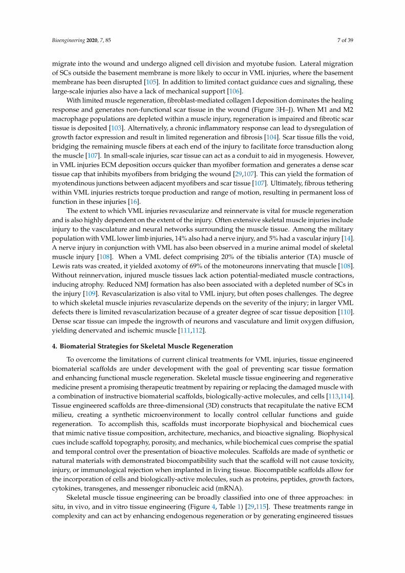
With limited muscle regeneration, fibroblast-mediated collagen I deposition dominates the healingresponse and generates non-functional scar tissue in the wound (Figure 3H–J). When M1 and M2macrophage populations are depleted within a muscle injury, regeneration is impaired and fibrotic scartissue is deposited [103]. Alternatively, a chronic inflammatory response can lead to dysregulation ofgrowth factor expression and result in limited regeneration and fibrosis [104]. Scar tissue fills the void,bridging the remaining muscle fibers at each end of the injury to facilitate force transduction alongthe muscle [107]. In small-scale injuries, scar tissue can act as a conduit to aid in myogenesis. However,in VML injuries ECM deposition occurs quicker than myofiber formation and generates a dense scartissue cap that inhibits myofibers from bridging the wound [29,107]. This can yield the formation ofmyotendinous junctions between adjacent myofibers and scar tissue [107]. Ultimately, fibrous tetheringwithin VML injuries restricts torque production and range of motion, resulting in permanent loss offunction in these injuries [16].
The extent to which VML injuries revascularize and reinnervate is vital for muscle regenerationand is also highly dependent on the extent of the injury. Often extensive skeletal muscle injuries includeinjury to the vasculature and neural networks surrounding the muscle tissue. Among the militarypopulation with VML lower limb injuries, 14% also had a nerve injury, and 5% had a vascular injury [14].A nerve injury in conjunction with VML has also been observed in a murine animal model of skeletalmuscle injury [108]. When a VML defect comprising 20% of the tibialis anterior (TA) muscle ofLewis rats was created, it yielded axotomy of 69% of the motoneurons innervating that muscle [108].Without reinnervation, injured muscle tissues lack action potential-mediated muscle contractions,inducing atrophy. Reduced NMJ formation has also been associated with a depleted number of SCs inthe injury [109]. Revascularization is also vital to VML injury, but often poses challenges. The degreeto which skeletal muscle injuries revascularize depends on the severity of the injury; in larger VMLdefects there is limited revascularization because of a greater degree of scar tissue deposition [110].Dense scar tissue can impede the ingrowth of neurons and vasculature and limit oxygen diffusion,yielding denervated and ischemic muscle [111,112].
4. Biomaterial Strategies for Skeletal Muscle Regeneration
To overcome the limitations of current clinical treatments for VML injuries, tissue engineeredbiomaterial scaffolds are under development with the goal of preventing scar tissue formationand enhancing functional muscle regeneration. Skeletal muscle tissue engineering and regenerativemedicine present a promising therapeutic treatment by repairing or replacing the damaged muscle witha combination of instructive biomaterial scaffolds, biologically-active molecules, and cells [113,114].Tissue engineered scaffolds are three-dimensional (3D) constructs that recapitulate the native ECMmilieu, creating a synthetic microenvironment to locally control cellular functions and guideregeneration. To accomplish this, scaffolds must incorporate biophysical and biochemical cuesthat mimic native tissue composition, architecture, mechanics, and bioactive signaling. Biophysicalcues include scaffold topography, porosity, and mechanics, while biochemical cues comprise the spatialand temporal control over the presentation of bioactive molecules. Scaffolds are made of synthetic ornatural materials with demonstrated biocompatibility such that the scaffold will not cause toxicity,injury, or immunological rejection when implanted in living tissue. Biocompatible scaffolds allow forthe incorporation of cells and biologically-active molecules, such as proteins, peptides, growth factors,cytokines, transgenes, and messenger ribonucleic acid (mRNA).
Skeletal muscle tissue engineering can be broadly classified into one of three approaches: insitu, in vivo, and in vitro tissue engineering (Figure 4, Table 1) [29,115]. These treatments range incomplexity and can act by enhancing endogenous regeneration or by generating engineered tissues
Bioengineering 2020, 7, 85 8 of 39
to replace damaged muscle. In situ tissue engineering involves the implantation of an acellularbiomaterial scaffold into the injury that can direct endogenous regeneration. Strategic engineeringof biophysical and biochemical cues allows the scaffold to instruct host cell recruitment, activation,proliferation, and differentiation. Slightly more complex, in vivo tissue engineering involves seedinginstructive biomaterial scaffolds with cells immediately prior to transplantation, where they can thenparticipate in regeneration. While this approach limits the manipulation of cells prior to transplantationand preserves their efficacy, it can leave them susceptible to low viability, retention, and immunerejection [29,116,117]. Finally, in vitro tissue engineering involves the development and implantationof a functional tissue engineered construct. This is achieved by combining scaffolds, biological factors,and cells and culturing these constructs in vitro until the cells differentiate into contractile myofibers.Differentiation is often achieved through a combination of biochemical cues, mechanical stimulation,and electrical stimulation. While in vitro tissue engineered constructs have greater functionality prior toimplantation than those developed through in situ and in vivo techniques, they have several significantdrawbacks. While they display some functionality, these contractile forces are often significantlylower than what is seen in native muscle tissue [106]. Additionally, due to oxygen and nutrientdiffusion limitations these constructs are often size-limited or require the development of a complexvascular network to support extended cell viability. Herein we review past and current skeletalmuscle tissue engineering strategies, with a focus on the use of instructive biomaterial scaffolds.This review will not cover scaffold-free approaches to treating VML, such as rehabilitation regimes,autologous grafts, or minced muscle grafts. It will also not include cellular, drug, gene, or growthfactor injections unless they are delivered using a biomaterial carrier. Additionally, this review willfocus exclusively on the treatment of muscle injuries and not include treatments for genetic diseasessuch as Duchenne muscular dystrophy. While this review focuses on VML resection injuries, it alsoevaluates muscle injuries induced from critical limb ischemia (CLI), crush, and myotoxin injuries,which present a different pathophysiology and capacity for functional recovery than VML injuries [118].Preclinical and clinical in situ, in vivo, and tissue engineering strategies will be reviewed, with a focuson the biophysical and biochemical cues of these scaffolds that guide regeneration.
Bioengineering 2020, 7, x FOR PEER REVIEW 8 of 40
biological factors, and cells and culturing these constructs in vitro until the cells differentiate into contractile myofibers. Differentiation is often achieved through a combination of biochemical cues, mechanical stimulation, and electrical stimulation. While in vitro tissue engineered constructs have greater functionality prior to implantation than those developed through in situ and in vivo techniques, they have several significant drawbacks. While they display some functionality, these contractile forces are often significantly lower than what is seen in native muscle tissue [106]. Additionally, due to oxygen and nutrient diffusion limitations these constructs are often size-limited or require the development of a complex vascular network to support extended cell viability. Herein we review past and current skeletal muscle tissue engineering strategies, with a focus on the use of instructive biomaterial scaffolds. This review will not cover scaffold-free approaches to treating VML, such as rehabilitation regimes, autologous grafts, or minced muscle grafts. It will also not include cellular, drug, gene, or growth factor injections unless they are delivered using a biomaterial carrier. Additionally, this review will focus exclusively on the treatment of muscle injuries and not include treatments for genetic diseases such as Duchenne muscular dystrophy. While this review focuses on VML resection injuries, it also evaluates muscle injuries induced from critical limb ischemia (CLI), crush, and myotoxin injuries, which present a different pathophysiology and capacity for functional recovery than VML injuries [118]. Preclinical and clinical in situ, in vivo, and tissue engineering strategies will be reviewed, with a focus on the biophysical and biochemical cues of these scaffolds that guide regeneration.
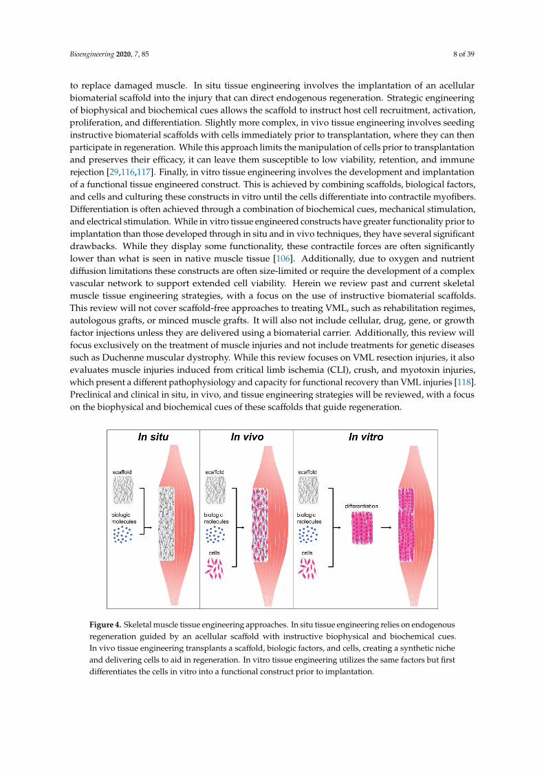
Figure 4. Skeletal muscle tissue engineering approaches. In situ tissue engineering relies on endogenous regeneration guided by an acellular scaffold with instructive biophysical and biochemical cues. In vivo tissue engineering transplants a scaffold, biologic factors, and cells, creating a synthetic niche and delivering cells to aid in regeneration. In vitro tissue engineering utilizes the same factors but first differentiates the cells in vitro into a functional construct prior to implantation.
Table 1. Comparison of tissue engineering approaches. Based off Table 1 in [119].
In Situ In Vivo In Vitro Off-the-shelf availability Likely Possible Not possible
Scalability Easier Difficult Most difficult Ease of clinical translation Easier Complex Complex
Cost-effectiveness More Less Least Disease modeling No No Yes
Drug screening modeling No No Yes
Figure 4. Skeletal muscle tissue engineering approaches. In situ tissue engineering relies on endogenousregeneration guided by an acellular scaffold with instructive biophysical and biochemical cues.In vivo tissue engineering transplants a scaffold, biologic factors, and cells, creating a synthetic nicheand delivering cells to aid in regeneration. In vitro tissue engineering utilizes the same factors but firstdifferentiates the cells in vitro into a functional construct prior to implantation.
Bioengineering 2020, 7, 85 9 of 39
Table 1. Comparison of tissue engineering approaches. Based off Table 1 in [119].
In Situ In Vivo In Vitro
Off-the-shelf availability Likely Possible Not possibleScalability Easier Difficult Most difficult
Ease of clinical translation Easier Complex ComplexCost-effectiveness More Less LeastDisease modeling No No Yes
Drug screening modeling No No Yes
4.1. In Situ Strategies: Acellular Scaffolds to Promote Endogenous Regeneration
In situ tissue engineering utilizes acellular biomaterial scaffolds to direct endogenous regeneration.Tissue engineered scaffolds employ biophysical and biochemical cues to recapitulate native ECMstructure, creating a synthetic microenvironment to locally control host cellular functions and guideregeneration. Implantation of an acellular scaffold offers unique advantages over cell-basedstrategies, including faster and simpler fabrication and storage. By not delivering cells the culturetime for these scaffolds is eliminated, yielding faster fabrication, streamlined delivery workflowsand operations, the potential for long term storage and off-the-shelf capabilities. Additionally, thesefactors result in the production of scaffolds that often have lower regulatory barriers and a quickerpath to commercialization (Table 1) [120]. For this strategy to be effective, strategic engineering ofthe biomaterial scaffold is paramount to develop a synthetic niche capable of directing endogenousregeneration. This section will focus on the strategies utilized for skeletal muscle scaffold development,with a focus on biomaterial selection and biophysical and biochemical cues used to modulatethese materials.
4.1.1. Biomaterial Selection: Synthetic, Natural, and Hybrid Polymers
Most biomaterials fall into one of two classes, synthetic or natural, which both offer distinctadvantages and drawbacks. Synthetic scaffolds are easy, consistent, and inexpensive to fabricateand can be manufactured to have detailed geometries down to the nanoscale. They can also beengineered to have precise and tunable degradation profiles and mechanical properties. Conjugationof biomolecules such as growth factors is also possible, and their release from the scaffolds can befinely tuned by altering conjugation strategies or scaffold degradation rates. Synthetic polymers offeradditional benefits, such as the ability to be electrically conductive [121–123]. Synthetic scaffolds thathave been used for skeletal muscle tissue engineering include poly(ε-caprolactone) (PCL), poly(glycolicacid) (PGA), polylactic acid (PLA, PLLA), and copolymer poly-lactic-co-glycolic acid (PLG, PLGA),polyurethane (PU), polyethylene glycol (PEG), and polypropylene (PP). However, use of syntheticbiomaterials comes at a cost. These polymers are often associated with low cell attachment, limitingtheir use without functionalization of a natural biopolymer. Additionally, synthetic scaffolds havelimited biocompatibility and have been shown to elicit a pro-inflammatory immune response uponimplantation [116].
In contrast, natural polymers are highly biocompatible and contain native signaling cues which aidin promoting cellular attachment, proliferation, and differentiation. They also contain native functionalgroups suitable for growth factor conjugation and are naturally degraded upon implantation. Naturalbiopolymers include collagen, fibrin, alginate, laminin, silk fibroin, hyaluronic acid (HA), decellularizedECM, chitosan, keratin, and gelatin. While biopolymer scaffold porosity, topography, and mechanicscan be modified, there is less precision and tunability than with synthetic scaffolds. Biopolymers are alsosubject to inherent biologic variability due to material sourcing. Both synthetic and natural biomaterialscan be made into an ideal scaffold through strategic incorporation of biophysical and biochemical cuesdesigned to create a synthetic microenvironment conducive to skeletal muscle tissue regeneration.
Bioengineering 2020, 7, 85 10 of 39
4.1.2. Biophysical Cues
An ideal biomaterial scaffold should match native tissue mechanical properties, degrade at a ratethat matches the rate of new tissue regeneration, and contain 3D topographical features and porosityto direct cellular alignment and allow for cellular infiltration. These features can be accomplishedthrough the incorporation of instructive biophysical cues (Figure 5). Scaffold strength and stiffnessshould be optimized to match native tissue mechanics to create a synthetic niche that exposes cells tophysiologically relevant forces. Myoblasts respond to mechanical stimuli through mechanotranduction,informing their proliferation, adhesion, and differentiation. Substrate stiffness does not affectthe propensity for myoblasts to assemble into myotubes, but it was shown to have an important roleon the development of myosin/actin striations [124]. Myoblasts cultured on substrates with a modulusof 12 kPa, which matches the elasticity of native resting muscle tissue, were found to have significantlyincreased striations, indicating a more functional and mature cellular phenotype [124]. Scaffoldbiodegradation is another biophysical cue that must match the kinetics of skeletal muscle regenerationand new tissue ingrowth [125]. Rapid degradation can lead to voids within the tissue and compromisedregeneration, while slow degradation can invoke a chronic inflammatory response, scar tissuedeposition, and encapsulation [126]. Both scaffold mechanics and degradation are commonly controlledby material choice and crosslinking. Hydrogels, sponges, fibers, composites and devitalized ECM areamong the most commonly exploited conformations for biomaterial scaffolds. These systems are allable to create a 3D environment that provides suitable porosity, topographical cues, and mechanicalproperties to support tissue regeneration.
Bioengineering 2020, 7, x FOR PEER REVIEW 10 of 40
with a modulus of 12 kPa, which matches the elasticity of native resting muscle tissue, were found to have significantly increased striations, indicating a more functional and mature cellular phenotype [124]. Scaffold biodegradation is another biophysical cue that must match the kinetics of skeletal muscle regeneration and new tissue ingrowth [125]. Rapid degradation can lead to voids within the tissue and compromised regeneration, while slow degradation can invoke a chronic inflammatory response, scar tissue deposition, and encapsulation [126]. Both scaffold mechanics and degradation are commonly controlled by material choice and crosslinking. Hydrogels, sponges, fibers, composites and devitalized ECM are among the most commonly exploited conformations for biomaterial scaffolds. These systems are all able to create a 3D environment that provides suitable porosity, topographical cues, and mechanical properties to support tissue regeneration.
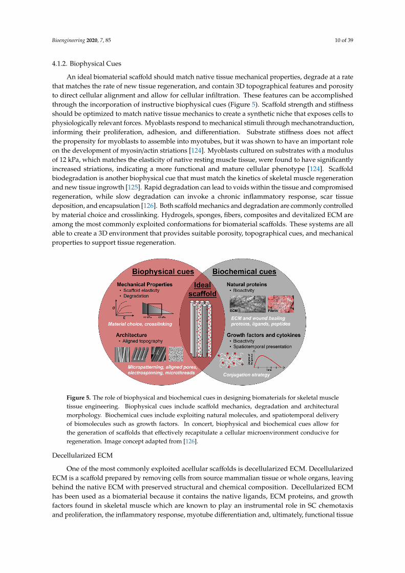
Figure 5. The role of biophysical and biochemical cues in designing biomaterials for skeletal muscle tissue engineering. Biophysical cues include scaffold mechanics, degradation and architectural morphology. Biochemical cues include exploiting natural molecules, and spatiotemporal delivery of biomolecules such as growth factors. In concert, biophysical and biochemical cues allow for the generation of scaffolds that effectively recapitulate a cellular microenvironment conducive for regeneration. Image concept adapted from [126].
Decellularized ECM
One of the most commonly exploited acellular scaffolds is decellularized ECM. Decellularized ECM is a scaffold prepared by removing cells from source mammalian tissue or whole organs, leaving behind the native ECM with preserved structural and chemical composition. Decellularized ECM has been used as a biomaterial because it contains the native ligands, ECM proteins, and growth factors found in skeletal muscle which are known to play an instrumental role in SC chemotaxis and proliferation, the inflammatory response, myotube differentiation and, ultimately, functional tissue regeneration [63,127–129]. These ECM molecules include collagens, laminin, fibronectin, heparin sulfate, chondroitin sulfate, HA, VEGF, FGF2, and TGF-β [130–133]. The goal for these scaffolds is that upon implantation into a muscle injury, they will become infiltrated by immune cells that degrade the scaffold, releasing native growth factors and ECM proteins that promote host cell infiltration [63]. Decellularized ECM is harvested from a variety of source tissues, including dermis, skeletal muscle, small intestinal submucosa (SIS), and urinary bladder matrix (UBM) [127,128]. Different source tissues provide varying structural organization and chemical composition [134]. The process by and extent to which ECM is decellularized can also yield varying physical and chemical properties, and contribute to the varying degrees of remodeling seen after implantation [135–137].
Figure 5. The role of biophysical and biochemical cues in designing biomaterials for skeletal muscletissue engineering. Biophysical cues include scaffold mechanics, degradation and architecturalmorphology. Biochemical cues include exploiting natural molecules, and spatiotemporal deliveryof biomolecules such as growth factors. In concert, biophysical and biochemical cues allow forthe generation of scaffolds that effectively recapitulate a cellular microenvironment conducive forregeneration. Image concept adapted from [126].
Decellularized ECM
One of the most commonly exploited acellular scaffolds is decellularized ECM. DecellularizedECM is a scaffold prepared by removing cells from source mammalian tissue or whole organs, leavingbehind the native ECM with preserved structural and chemical composition. Decellularized ECMhas been used as a biomaterial because it contains the native ligands, ECM proteins, and growthfactors found in skeletal muscle which are known to play an instrumental role in SC chemotaxisand proliferation, the inflammatory response, myotube differentiation and, ultimately, functional tissue
Bioengineering 2020, 7, 85 11 of 39
regeneration [63,127–129]. These ECM molecules include collagens, laminin, fibronectin, heparinsulfate, chondroitin sulfate, HA, VEGF, FGF2, and TGF-β [130–133]. The goal for these scaffolds is thatupon implantation into a muscle injury, they will become infiltrated by immune cells that degradethe scaffold, releasing native growth factors and ECM proteins that promote host cell infiltration [63].Decellularized ECM is harvested from a variety of source tissues, including dermis, skeletal muscle,small intestinal submucosa (SIS), and urinary bladder matrix (UBM) [127,128]. Different source tissuesprovide varying structural organization and chemical composition [134]. The process by and extent towhich ECM is decellularized can also yield varying physical and chemical properties, and contribute tothe varying degrees of remodeling seen after implantation [135–137]. This may explain the conflictingreports in the literature of decellularized ECM inflammatory response, induction of fibrosis, and abilityto promote muscle fiber regeneration upon implantation.
Early studies investigating the use of xenogeneic SIS and homologous muscle ECM found thatimplanted scaffolds promoted a strong angiogenic response despite variable contractile functionand the persistent deposition of collagenous connective tissue [138–140]. In one study, Badylak’sgroup observed that upon implantation into rodent partial resection models, both SIS and muscleECM scaffolds induced constructive remodeling characterized by robust mononuclear cell infiltrationand myogenesis, although no contractile analysis was performed to evaluate muscle function [134,141].Corona et al. implanted a syngeneic muscle ECM scaffold into a partially resected rat TA muscle and sawrecovery of one third of the original force deficit was restored after two months post-injury, despitehistological analysis showing overwhelming fibrosis at the implantation site [142]. The authorshypothesize that the ECM scaffold prevented muscle fiber damage and acted as a structuralreinforcement to transmit forces across the injury. Another study evaluated a syngeneic muscleECM scaffold to treat a rodent TA VML defect model and found that at two weeks post-implantation,the scaffold elicited a pro-inflammatory response with a large quantity of macrophages surroundingthe implant [143]. At eight weeks after treatment little to no myosin+ muscle fibers were present,and increased collagen 1 was observed. Upon stimulation, TA muscles treated with muscle ECMdemonstrated a 17% increase in torque compared to those with an injury and no treatment. More recentstudies utilizing a UBM ECM scaffold to treat rodent and porcine VML injuries found that injuriestreated with scaffolds demonstrated limited myogenesis, fibrotic deposition, and chronic functionaldeficits at terminal timepoints [144,145]. Discrepancies in the existing literature regarding whetheror not ECM scaffolds promote constructive remodeling in preclinical models may be due in part todifferences in ECM sources, decellularization protocols, differences in anatomy, and the severity ofpreclinical VML models.
Decellularized ECM is the first tissue engineered scaffold to be clinically evaluated for treatingVML injury. A 2010 case study treated a persistent combat-induced quadriceps injury 3.5 yearspost-injury with an acellular porcine SIS ECM scaffold and subsequent physical therapy [17]. At 16weeks post-operatively a modest improvement in isokinetic muscle function was demonstrated, as wellas new soft tissue observed via computed tomography (CT) at 36 weeks post-operatively. A 2014study by Sicari et al. evaluated the use of UBM ECM in five male patients with extremity VML injuriesincurred at least six months prior that maintained a minimum of 25% functional deficit comparedto the contralateral uninjured limb [120]. At 6 months post-surgery magnetic resonance imaging(MRI) and histological evaluation of biopsies both showed the presence of vascularization and islandsof muscle cells. Furthermore, three of the five patients showed a 20% functional improvement ofthe affected limb. A follow-up study of eight patients with VML, including the five from the previousstudy [120], used electrodiagnostic evaluations with nerve conduction studies (NCS) and needleelectromyography (EMG) to demonstrate restoration of nerve tissue as it relates to variable functionaloutcomes in ECM-treated VML injuries [146]. The study found that five of the eight patientstreated with ECM scaffolds demonstrated improvements in electrical activity evaluated throughNCS and EMG as well as improved muscle strength, compared to the pretreatment condition. Mostrecently, a 13-patient study was conducted to evaluate the ability of ECM bioscaffolds and physical
Bioengineering 2020, 7, 85 12 of 39
therapy to improve force production, range-of-motion, and function in a range of VML extremityinjuries [147], which also included five patients from the previous study [120]. Patients demonstratedan average improvement of 37.3% in strength 27.1% in range-of-motion tasks and 271.8% in functionaltask performance at six months post-operatively. The authors acknowledged that debridement ofscar tissue during surgery and the effect of mechanical transduction via the ECM scaffold may havecontributed to these modest functional gains. They also observed the formation of muscle tissueat the injury site through histology of biopsied tissue and MRI or CT imaging. Despite some promisingresults, the clinical use of decellularized ECM remains limited due to variable outcomes amongpatients and the limited understanding of the mechanisms by which these acellular scaffolds mediatemuscle regeneration.
Hydrogels, Sponges, and Meshes
Hydrogels, sponges, and meshes are used as alternative acellular scaffolds to decellularizedECM because they allow for more precise control of the scaffold material, mechanics, degradation,and porosity. Porosity is a critical biophysical cue to control in tissue engineered scaffolds because ofits role in permitting cellular infiltration and oxygen and nutrient diffusion. Porosity is commonlyachieved through the use of hydrogels, sponges, and fibrous meshes. Pore sizes typically range from 10to 500 µm, and larger macropores typically permit greater cell viability and migration [148]. Injectablein situ polymerizing collagen and decellularized ECM hydrogel scaffolds were evaluated for theirability to treat critical limb ischemia (CLI) in a rat hindlimb ischemia model [149]. They found thatdecellularized ECM hydrogels promoted increased the number of MyoD+ cells recruited to the injuryand blood vessel density compared to the collagen hydrogel. More recent work delivering a laminin111-enriched fibrin hydrogel to a murine VML defect demonstrated an infiltration of macrophagesand ECs into the hydrogels at two weeks post-injury, but did not report increases in peak isometrictorque at four weeks post-injury compared to the untreated negative control [150]. Sponge-basedscaffolds can be generated by freeze-drying polymer solutions or hydrogels, creating a porousmicrostructure. Freeze-dried collagen sponges implanted into a partial resection of the vastus lateralismuscle in a rabbit model qualitatively demonstrated less scar tissue formation and a greater number,diameter, and length of myofibers at 24 weeks post-injury compared to the untreated control [151].Lyophilized, highly porous sponges made of gelatin, collagen, and laminin 111 and crosslinkedwith 1-ethyl-3-(3-dimethyl aminopropyl) carbodiimide (EDC) have also been evaluated in a 10%resection of the gastrocnemius-soleus complex of mice [152]. At two weeks post-injury proteinlysates from sponge-treated muscles showed significantly higher expression of MyoD and desmincompared to untreated muscles, suggesting an increase in myogenic activity at the injury site dueto scaffold-mediated regeneration. In addition to hydrogels and sponges, fibrous meshes are alsohighly porous scaffolds that permit cellular infiltration. Fibrous meshes of PLLA with an averagefiber diameter of 150 µm and pore size of 50–100 µm demonstrated host cell infiltration in rat TAVML defects. Histological evaluation of TA muscles at one, two, three, and four weeks demonstratedan influx of Von Willebrand factor (vWF)+ ECs and Pax7+ muscle progenitor cells into the fibrous meshscaffolds over time [153]. While acellular hydrogels, sponges, and meshes permit cellular infiltrationupon injury into VML defects, these scaffolds have not demonstrated aligned myofibers or significantgains in muscle function. This may be due in part to the lack of instructive topographical alignmentcues that these scaffolds provide due to their isotropic nature. This has motivated a thrust of skeletalmuscle tissue engineering research that focuses on the development of anisotropic scaffolds withaligned architectural features.
Aligned Scaffolds
Incorporation of instructive biophysical cues such as anisotropic surface topography is a commonlyexploited technique to promote cell alignment in skeletal muscle tissue engineering. Myoblastalignment is an essential step towards myotube formation, which is guided in vivo by ECM structure
Bioengineering 2020, 7, 85 13 of 39
and micron-scale grooves between adjacent muscle fibers. Many strategies have been explored to createscaffolds with anisotropic surface topography, including patterned substrates [154–161]. electrospunfibers [122,123,155,162–165], microthreads [166–168], and aligned pores [169–171]. A review describesthese methods in detail and their ability to align and differentiate myoblasts in vitro, as this falls outsideof the scope of this review [172]. These methods are also commonly employed to generate in vitrotissue engineered skeletal muscle, which will be discussed in Section 4.3. Nakayama and colleaguesevaluated the therapeutic benefit of aligned nanofibrillar collagen scaffolds and rehabilitative exerciseon the treatment of VML [173]. Ablated murine TA muscles were treated with either aligned orrandomly oriented collagen nanofibers, and animals were randomly assigned to either voluntary cagerunning or no rehabilitation regime during recovery. After 21 days post-treatment, muscle treatedwith both random and aligned nanofibers exhibited significantly higher myofiber cross sectional areathan those left untreated or treated with a decellularized ECM scaffold. Additionally, they notedsignificantly higher perfused vascular density in muscles treated with the aligned nanofibers comparedwith those treated with the randomly-oriented nanofibers. This study warrants further investigation ofanisotropically aligned scaffolds. Use of these scaffolds in conjunction with biochemical cues and cellswill be discussed in later sections of this review.
4.1.3. Biochemical Cues
Biochemical cues are often strategically incorporated into scaffolds for skeletal muscle tissueengineering as a method to further regulate cellular functions including survival, attachment,proliferation, migration, and differentiation into myotubes. These include biologically active moleculessuch as proteins, peptides, growth factors, cytokines, transgenes, and messenger ribonucleic acid(mRNA). Strategic design of biomaterial-based scaffolds must take place to protect these moleculesfrom degradation, maintain their native conformation, and preserve their bioactivity. Biomaterialscaffolds should also be designed to carefully control spatiotemporal presentation, biomolecule releasekinetics, and local concentrations. For example, genetic substances can be delivered via viral ornon-viral vectors, such as liposomes and synthetic particles, engineered to translocate into the cellor nucleus.
Growth Factors and Cytokines
Growth factors and cytokines are among some of the most commonly investigated biologicmolecules to treat skeletal muscle injuries due to their instrumental role in facilitating native regeneration(reviewed in Section 3) [54]. Early clinical trials using bolus injections of growth factors such as VEGFand FGF2 to treat cardiovascular disease had limited success [174–176]. This is likely attributed tothe bolus delivery method, which provided initial supraphysiological growth factor concentrationsfollowed by rapid degradation, preventing sustained presentation of these factors for the necessarytime frame [177,178]. Delivering growth factors via biomaterial carriers prevents their denaturationand mediates their release, which is controlled by scaffold degradation and/or diffusion throughthe matrix. They are often incorporated into biomaterials through physical entrapment, ionicinteractions, or covalent coupling [126,178]. Engineering these scaffolds to control growth factorrelease kinetics allows for the delivery of optimized concentrations, localized delivery, and increasedtherapeutic efficiency.
Several researchers have investigated the ability of growth factor-loaded scaffolds to promoteskeletal muscle regeneration in ischemic and VML injury models [153,179–190]. VEGF-loaded hydrogelscaffolds have been investigated for their ability to promote angiogenesis in hindlimb injuries [179–182].Silva et al. confirmed that VEGF delivered to a TA ischemic murine injury via alginate hydrogel waspresent at physiologically relevant levels for up to 15 days post-injection, compared to only three daysafter delivery of VEGF via bolus injection [179]. Sustained delivery of VEGF via alginate hydrogelsresulted in significantly higher blood vessel density and tissue perfusion compared to no treatmentand bolus VEGF delivery. Another study evaluated the sustained delivery of VEGF from alginate
Bioengineering 2020, 7, 85 14 of 39
hydrogels for the treatment of an ischemic murine TA injury [180]. After seven days post-injury,VEGF delivery resulted in 50% innervated motor end plates compared to 5% in the blank alginate gel,likely due to the significant increase in glial-derived neurotrophic factor (GDNF) and NGF expressionlevels compared to uninjured muscle. Hydrogel-mediated VEGF also resulted in significantly highernumber of vessels (CD31+) and mature vessels (smooth muscle actin+) after 14 days compared toblank hydrogels. To generate a more controlled release of VEGF from alginate hydrogels, Lee et al.encapsulated VEGF within PLGA microspheres, creating a sustained release of VEGF over threeweeks [181]. These composite scaffolds generated significantly higher platelet endothelial cell adhesionmolecules (PECAM) expression than bolus VEGF or VEGF-loaded alginate hydrogels, indicatingthe formation of functional microvessels. Another study evaluated the ability of VEGF-coated collagenmatrices to stimulate repair in a rabbit lower leg osteotomy with soft tissue contusion injury, and foundVEGF scaffolds resulted in 73% recovery of muscle strength compared to 53% recovery of no treatmentcontrol group after 30 days post-injury [182].
IGF-1 has also been used to treat VML injuries because of its important role in stimulatingmyoblast survival, proliferation, and differentiation [153,183]. Hammers et al. found that a controlledrelease of IGF-1 from (PEG)ylated fibrin gels implanted into a murine hindlimb ischemia injurystimulated significantly higher force production 14 days post-injury compared to bolus IGF-1 and blankhydrogel treatments [183]. Other researchers implanted IGF-1-loaded gelatin porous sponges into TAmuscle of rats and after two weeks found a four-fold increase in the number of Pax7+ infiltrated cellsand significantly greater number of muscle fibers compared to control blank hydrogels [153].
Another commonly utilized growth factor for promoting skeletal muscle regeneration isFGF2 [153,184–186]. In a rabbit hind limb ischemia injury model, FGF2-loaded gelatin hydrogelshad significantly greater tissue blood flow, number of arterioles, and vascular density four weeksafter treatment [184]. Other researchers have delivered sustained FGF2 release from ionic gelatinhydrogels covalently crosslinked with PLG, which yielded significantly greater capillary density(CD31+) and blood flow compared to bolus FGF2 injection at eight weeks after ischemic hindlimbinjury [185]. Murine TA muscles implanted with FGF2-loaded gelatin sponges found a significantincrease in the number of Pax7+ infiltrated cells and significantly greater number of muscle fiberscompared to control blank hydrogels [153].
Our laboratory delivered HGF adsorbed to EDC-crosslinked fibrin microthread scaffolds ina murine VML defect [187]. Surgical resection of 30 mg of the TA muscle yielded approximately 50%reduction in force immediately after injury, and was immediately filled with fibrin microthreads thatwere EDC crosslinked and passively adsorbed with 40 ng/mL of HGF. At 60 days post-injury, thistreatment resulted in an increase of over 200% of twitch and tetanic force production, compared tothe 150% and 130% increases seen from treatment with fibrin microthreads or fibrin gel with no HGFdelivery. HGF fibrin microthreads resulted in a significantly higher recovered force production thaninjuries that received no treatment or treatment with a fibrin hydrogel alone. This work has beenrecently acknowledged as the only study of growth factor-based repair that evaluated functionalrecovery in an appropriate VML injury model [191]. Histological analysis showed myofibers adjacentto implanted EDC crosslinked microthreads, indicating that the aligned microthread architecture likelyguides myofiber ingrowth and alignment [187]. This motivates the future development of scaffoldswith biophysical cues such as aligned topography in conjunction with the delivery of biochemicalcues such as growth factors. Overall, the delivery of a single myogenic or angiogenic growth factor toskeletal muscle injuries yields improvements in regeneration, but it remains unclear which growthfactors, concentrations, and delivery strategies yield the best results, and warrants further investigation.
Toward the goal of recapitulating in vivo regeneration, several studies also assessed the synergisticpresentation of multiple growth factors. While delivery of a single growth factor has shown promisingresults for promoting skeletal muscle regeneration and angiogenesis, this strategy represents a drasticallysimplified version of the complex, spatiotemporal presentation of multiple factors during regeneration.Through the development of more complex biomaterials systems, the release kinetics of multiple factors
Bioengineering 2020, 7, 85 15 of 39
in a spatiotemporal manner that mimics in vivo presentation and concentration will likely enhanceregenerative outcomes [126,192]. The first preclinical work evaluating delivery of multiple growthfactors for skeletal muscle repair investigated the co-stimulatory effect of IGF-1 and VEGF delivered toischemic rodent hindlimbs via an injectable alginate hydrogel [188]. Despite the limited control overrelease kinetics that this scaffold provided, the co-delivery of 3 µg each of these factors from the alginatehydrogel stimulated significantly higher blood perfusion seven weeks after ligation compared tothe blank hydrogel and bolus IGF-1/VEGF treatments. Injuries treated with the IGF-1/VEGF hydrogelalso had significantly larger myofiber diameters and number of centrally located nuclei comparedto blank gels, which are hallmarks of regenerating muscle. Another study evaluated the therapeuticpotential of stromal cell-derived factor-1 α (SDF-1α) alone or in combination with IGF-1 to treatan ischemic skeletal muscle injury in rodents [189]. Co-delivery of these factors in a PEGylated fibrinhydrogel yielded significant improvements in revascularization (CD31+ cells/fiber) and functionalrecovery (maximum tetanic force production) at 14 days post-injury compared to treatment with blankhydrogel, which was not achieved by the delivery of SDF-1α alone. Matsui et al. investigated gelatinhydrogel granules to deliver FGF2 alongside a mix of growth factors isolated from platelet-activatedplatelet rich plasma (PRP), which included PDGF, VEGF, TGF- β, and FGF2 [190]. One week afterimplantation into a murine hindlimb ischemic injury the combination treatment yielded a significantlyhigher number of blood vessels compared to no treatment, which was not achieved by delivery ofFGF2 or the PRP-isolated growth factor mixture alone. The combination treatment also significantlyenhanced blood reperfusion compared to hydrogels with just FGF2 or PRP-isolated growth factors.As biomaterial scaffolds advance to allow for more precise spatiotemporal delivery of multiple growthfactors, this strategy will better mimic the complex in vivo temporal presentation during regenerationand will likely yield greater therapeutic, functional outcomes.
Genetic Substances
Another thrust of research focuses on the delivery of genetic material including cDNA and mRNA.Gene therapy strategies transfer genetic material into host cells to treat genetic diseases or injury.This treatment often utilizes engineering of viral and non-viral vectors to safely and efficiently transfergenetic material into cell nuclei. These strategies include adenovirus, adeno-associated virus, retrovirus,lentivirus, liposomes, synthetic particles, and polymer-based scaffolds, and display varying degrees ofimmunogenicity and transfection efficiency. These approaches are typically limited to injections thatdo not control vector spatiotemporal presentation. To make gene therapy treatments more effective,researchers recently utilized biomaterial-based delivery systems to provide localized and sustaineddelivery of these therapies. Biomaterial scaffolds can help deliver genetic cargo by preserving geneticstructures, protecting them from nuclease-mediated degradation and controlling their release fromthe scaffold. By modulating scaffold properties such as molecular weight, porosity, or crosslinking,a localized and sustained release of genetic material can be mediated through diffusion or scaffolddegradation [193,194]. This delivery strategy has the potential to increase transfection efficiencyand expression, ultimately improving the therapeutic effectiveness of these treatments.
One study evaluated the delivery of adenoviral vectors and plasmids encoding FGF2 and FGF6transgenes delivered in collagen-gelatin scaffolds to excisional quadriceps defects in rats [195]. At 21days after injury they found that treatment with FGF2 transgenes increased the arteriole density by11-fold and myotube marker CD56 expression 20-fold compared to controls. They also note thatthe delivery of recombinant FGF2 protein was unable to produce equivalent responses, highlightingthe benefit of a gene delivery strategy for treating skeletal muscle injury. Another gene therapystudy delivered plasmid FGF4 cDNA within a gelatin hydrogel scaffold to treat hindlimb ischemiain rabbits [196]. The hydrogel preserved the plasmid structure allowing for improved transfectionefficiency compared to naked FGF4 gene. Ischemic injuries treated with hydrogel-FGF4 had significantlyless severe tissue damage and more pronounced vascular responsiveness to adenosine at four weekscompared to injuries treated with a naked FGF4 gene.
Bioengineering 2020, 7, 85 16 of 39
An alternative strategy to the delivery of plasmids and viruses is mRNA-based delivery.This method is advantageous because it does not present the risks of genome integration or insertionalmutagenesis that other gene therapy strategies have, but it can be limited based on the conventionaldelivery method of direct solution-based injection. Biomaterial-based delivery of mRNA allows forlocalized and controlled release of cargo. Recently, Zaitseva et al. evaluated the delivery of modifiedmRNA encoding HGF through crosslinked nanofibrillar collagen scaffolds to a murine skeletal muscleinjury [197]. After 20% ablation of the TA muscle, scaffolds were implanted to fill the defect void. At twoweeks after implantation, capillary density (CD31+) was observed in injuries treated with the HGFmRNA than by control scaffolds loaded with firefly luciferase-mRNA. As the use of biomaterial-baseddelivery of genetic substances continues to be implemented, it is likely that this strategy will continueto be investigated for skeletal muscle regeneration. However, cost, immunogenicity, and safety remainconcerns regarding this relatively new approach [114].
Small Molecules
Small molecules are another class of biologic molecules that have been investigated for skeletalmuscle regeneration. These organic compounds are typically under 800 Da in size which allowsthem to readily diffuse across the cell membrane, unlike macromolecules [198]. Small molecules canbe produced using synthetic chemistry, allowing for diverse structures and lower manufacturingcosts compared to larger molecules such as recombinant growth factors. They work by regulatingbiological targets such as receptors or enzymes to modulate specific cellular effects. These effectscan be reversed and temporally controlled to allow for rapid activation or inhibition. However,small molecules are often delivered orally or via direct injection and the risk of off-target eventswhere proteins with similar conformations are affected can be problematic [198]. To address this,biomaterial-based delivery of small molecules to localize treatment to target tissues is something thatis recently being explored. One study investigated the use of a PLGA thin film to deliver FTY720 toa full thickness VML defect in the spinotrapezius muscle of mice [199]. FTY720 is a small moleculeagonist of sphingosine-1-phosphate (S1P), a bioactive signaling lipid produced by red blood cells,platelets, and ECs [199]. Receptors of S1P are highly expressed on anti-inflammatory monocytesand have been shown to leverage pro-regenerative effects [200]. They demonstrated that, three daysafter injury, localized delivery of FTY720 had a higher percentage of anti-inflammatory monocyteand macrophage infiltration into the injury compared to defects treated with the PLGA film alone [199].They also found significantly higher blood vessel density (CD31+) in the defect region in mice treatedwith FTY720 compared to those treated with a blank scaffold control. Another group also foundangiogenic effects with the biomaterial-based delivery of small molecule CEP03, derived from CEP(ω-[2-carboxyethyl]pyrrole) protein adducts [201]. CEP protein adducts are a product of lipid oxidationand accumulate during inflammation and wound healing, but as-is face limitations of protein deliveryincluding high cost and manufacturing challenges. CEP03 was encapsulated in Matrigel, an ECMgenerated by carcinoma cells in vitro, injected intramuscularly into the gastrocnemius muscle ofmice with hind limb ischemia. Fourteen days after treatment with CEP03, a significant increasein relative mean perfusion ratio (ischemic/unoperated leg) and microvessel density (CD31+) wasobserved compared with injuries treated with Matrigel alone. Ongoing work developing biomaterialsto spatiotemporally deliver small molecules has the potential to increase their therapeutic potentialand make this a more commonly investigated approach to treating skeletal muscle injury.
4.2. In Vivo Strategies: Utilizing Biomaterials to Improve Cell Delivery
While acellular in situ tissue engineering strategies reviewed in Section 4.1 have demonstratedsuccess in promoting endogenous skeletal muscle regeneration through recruitment of host cellpopulations, there are conflicting studies described in the literature that question whether thisstrategy will facilitate full functional recovery after the large-scale injuries incurred from VML [63,202].Corona et al. hypothesized that restricted host SC infiltration into the VML defect is primary limitation
Bioengineering 2020, 7, 85 17 of 39
to the success of this strategy, as they noted a low number of Pax7+ SCs within a defect treatedwith an acellular ECM scaffold [202]. Thus, one of the most commonly explored approaches forskeletal muscle regeneration involves the delivery of cells. While early research focused on the directintramuscular injection of cell suspensions, this simplistic delivery technique has several significantlimitations including poor retention, survival, and immune rejection of transplanted cells [29,203].However, the recent development of biomaterial-based delivery systems with strategic biophysicaland biochemical signaling cues creates a synthetic microenvironment conducive for cell survivaland engraftment upon transplantation. The first study to evaluate the efficiency of SCs deliveredvia a polymer scaffold found 3-fold higher engraftment of cells into host muscle compared to thosedelivered via conventional direct injection [204]. Biomaterial delivery systems may also regulatecell fate, stemness and, ultimately, participation in regeneration. In vivo tissue engineering involvesseeding instructive biomaterial scaffolds with cells immediately prior to transplantation, wherethey can then provide host cells with paracrine signaling and/or participate in regeneration [116].By limiting the culture time and manipulation of cells prior to their transplantation, their efficacy canbe preserved [29].
4.2.1. Cell Source
A range of cell types have been utilized for skeletal muscle tissue engineering including SCs,myoblasts, muscle-derived precursor cells (MDPCs), mesenchymal stem cells (MSCs), perivascular stemcells (PSCs), adipose-derived stem cells (ASCs), and induced pluripotent stem cells (iPSCs) [205–207].SCs are a common cell type utilized for tissue engineering because of their essential role as the residentprogenitor cell in native tissue regeneration. However, population heterogeneity and purification remainchallenges and sources of variability regarding their functional regenerative potential. Additionally,expansion of SCs in vitro results in senescence and loss of proliferative capability, limiting theirclinical utility [208]. Thus, there is a tradeoff where increased SC manipulation yields a morepurified population but with generally lower regenerative potential [126]. Other muscle-derivedcell populations including myoblasts and MDPCs expressing various stem cell markers have alsodemonstrated regenerative potential, but pose similar challenges regarding cell engraftment, survival,and immunologic rejection [205]. MSCs, including ASCs and PSCs, are also commonly used becausethey are easily isolated from bone marrow, fat, and umbilical cord tissues, among others [205]. MSCsare pluripotent and give rise to mesodermal tissues including skeletal muscle as well as ectodermaland endodermal tissues. This may permit MSCs to simultaneously participate in peripheral nerverepair and angiogenesis which both play critical roles in skeletal muscle repair. ASCs are a particularlyattractive cell type because they are isolated with a simple, high yield procedure and can be expandedquickly, differentiate into myotubes, and evade the host immune system to prevent rejection [206].These various cell types used for skeletal muscle tissue engineering have been reviewed in detailpreviously [205–207]. Another important concern is whether cellular therapies are derived fromautologous or allogeneic sources. Autologous cells present minimal immunogenicity concerns, whileallogeneic and xenogeneic cells may require immunosuppressive treatment that potentially limitsimportant native responses that direct skeletal muscle regeneration [209].
4.2.2. Hydrogel-Based Delivery
Hydrogels are one of the most commonly implemented scaffolds for cell delivery-based strategiesbecause cells can be easily mixed into precursor solutions prior to polymerization, and they canbe cast directly into the injury, filling irregularly shaped voids. Researchers have used hydrogelscaffolds to deliver myoblasts [210,211], MDPCs [212–214], minced muscle grafts [215,216], MSCs [217],ASCs [218,219], and a combination of cell types [220,221]. Beier et al. injected 1 million male primarymyoblasts suspended in a fibrin matrix into a female rodent anterior gracilis muscle defect and foundmale nuclei within host female muscle fibers two weeks after delivery [211]. Other studies deliveredMDPCs in fibrin [212,214] and HA [213] hydrogels to partially resected rodent TA muscles, and found
Bioengineering 2020, 7, 85 18 of 39
that treatment supported reduced fibrosis [212], increased myogenesis, and vascularization [212]and partial restoration of muscle contractile function when delivered progressively via multipleinjections over time [214]. Rossi et al. evaluated the delivery of either SCs or MDPCs via a HAhydrogel and found that SCs improved the number of new myofibers and resulted in a significantlyhigher tetanic force generated compared to delivery of MDPCs alone [213]. Other researchers utilizedcollagen [215,220] and laminin-111 supplemented HA [216] hydrogels as a vehicle to deliver mincedmuscle grafts to rodent TA VML injuries [215,216]. Ward et al. found that 50% less minced muscle grafttissue was needed to regain the same functional improvement when delivered in a collagen hydrogelcompared to the 100% minced muscle graft [215]. Another study delivering 50% minced muscle graftswith a laminin-111 supplemented HA hydrogel found a 42% improvement in peak tetanic torqueproduction after eight weeks compared to unrepaired limbs. However, this was not a significantimprovement compared to delivery of the 50% minced muscle grafts alone. It is not clear if a scaffold isnecessary for the delivery of minced muscle grafts, as these cells remain largely contained by theirnative ECM that remains partially intact.
Several researchers utilized ASCs for skeletal muscle tissue engineering [218,219]. Aurora et al.delivered human ASCs encapsulated in a composite scaffold of PEGylated platelet free plasma hydrogelwith porcine ECM to a rodent TA VML injury [218]. Two weeks after injury the composite scaffoldwith ASCs resulted in the transplanted cells localized to the injury site and a significantly highervessel density compared to treatment with acellular composite scaffolds. Another study deliveringASCs in a collagen hydrogel to a similar TA defect found injuries treated with ASC-loaded hydrogelshad significantly higher blood flow restoration compared to the hydrogel alone treatment after eightweeks post-injury [219]. It is clear that ASCs encapsulated within hydrogel scaffolds are a promisingtechnology for promoting angiogenesis in VML defects.
Finally, the effect of co-delivery of multiple cell types has also been evaluated [220,221].One group used composite scaffolds of PLLA/PLGA sponges and fibrin hydrogel to co-deliverECs and fibroblasts to a defect created in the linea-alba of male nude mice, and 10 days after injuryfound more neovascularization and significantly higher perfusion than treatment with acellular fibrinhydrogels [221]. While hydrogel delivery systems are easy to deliver and aid in localized cell retention,they are limited due to their weak mechanics and lack of aligned topographical cues necessary topromote functional muscle regeneration.
4.2.3. Decellularized ECM-Based Delivery
Another commonly investigated scaffold for in vivo tissue engineering strategies is decellularizedECM [222–229]. Early work studied the delivery of acellular or autologous myoblast-seeded decellularizedmuscle ECM to the obliqui abdominis muscle of rats [223]. They found that myoblast-seeded scaffoldsdemonstrated abundant blood vessels after two months, while acellular scaffolds were completelyreplaced by scar tissue. Another study used decellularized muscle ECM to deliver male SC-derivedmyoblasts to a full-thickness abdominal wall defect in female rats and found the persistence of malemyoblasts in the implanted patches after nine months [224]. This work suggests that decellularized ECMscaffolds may facilitate the retention of transplanted cells at the injury site. Quarta et al. evaluated whetherMDPCs (expressing bioluminescent reporter protein luciferase) encapsulated in decellularized muscleECM enhanced skeletal muscle regeneration [227]. The researchers discovered that injecting a MDPCsuspension directly into ECM scaffolds resulted in inefficient retention within the scaffold, and insteadincorporated MDPCs into an ECM hydrogel and microinjected the cell-seeded hydrogel into the bulkdecellularized ECM scaffold. MDPCs were delivered alone or in combination with muscle residentcells, a heterogeneous population of fibro-adipogenic progenitors, macrophages, and ECs, to a TA VMLinjury in mice. ECM scaffolds that co-delivered MDPCs and a muscle resident cell population yieldedapproximately 40 times higher bioluminescence than scaffolds that delivered MDPCs alone. This suggeststhat the heterogeneous resident cell population supports MDPC viability within the implanted scaffolds.Additionally, injuries treated with ECM scaffolds co-seeded with MDPCs and muscle resident cells
Bioengineering 2020, 7, 85 19 of 39
demonstrated active and passive length-tension relationships that were equivalent to control uninjuredmuscle [228]. Injuries treated with co-seeded scaffolds also exhibited less fibrosis compared to untreatedinjuries and treatment with scaffolds alone.
Other researchers utilized decellularized ECM scaffolds to deliver minced muscleautografts [225,226]. An allogeneic decellularized muscle ECM scaffold was used to deliver mincedmuscle autograft tissue to a rat lower-limb VML injury [225]. After 12 weeks, treatment with mincedmuscle-loaded ECM scaffolds significantly increased contractile force recovery compared to untreatedinjury controls. Treated injuries also demonstrated increased MyoD expression and less fibrous scartissue formation than those left untreated. Goldman et al. created VML defects in the TA of ratsand used a UBM ECM scaffold to deliver minced muscle grafts at a dose of 50% of the total defectmass [226]. At eight weeks post-injury, this treatment resulted in a 28.2% significant increase in peakisometric torque compared to the no repair control, whereas treatment with an acellular UBM ECMscaffold did not result in a significant improvement. These studies indicate that decellularized ECMmay serve as an effective method to deliver and retain minced muscle tissue within VML defects.
Finally, decellularized ECM has also been used to deliver MSCs [222,229]. Merritt et al. implanteddecellularized muscle ECM into a full-thickness defect in the lateral gastrocnemius of Lewis rats [222].Seven days after implantation, bone marrow-derived MSCs were injected into the implanted ECM. After42 days an 85% functional recovery was observed in muscles treated with MSC-loaded ECM scaffoldswith respect to the contralateral uninjured muscle. However, this functional improvement was notsignificantly greater than treatment with the acellular ECM scaffold. Muscles treated with MSC-seededscaffolds had significantly more blood vessels and myofibers than those treated with acellular ECMscaffolds. Another study used human umbilical cord MSCs delivered in a porcine decellularizedcardiac ECM because the researchers hypothesized that both components could promote macrophagepolarization toward a pro-regenerative M2 phenotype [229]. Rodent TA defects were treated withMSCs, ECM scaffolds, or MSC-loaded ECM scaffolds. Muscle injuries treated with MSC-loaded ECMscaffolds had the highest recorded isometric torque at four and eight weeks post-injury compared totreatment with MSCs alone and ECM scaffolds alone. At two weeks post-implantation, the MSC-loadedECM treatment group had a significantly higher population of CD206+ macrophages, indicative ofpro-regenerative M2 polarization. Finally, histological evaluation of treated muscle found treatmentwith MSC-loaded ECM had higher new muscle fiber regeneration and lower collagen depositioncompared to MSCs alone or ECM scaffolds alone. Taken together, varying degrees of success have beendemonstrated by using decellularized ECM scaffolds as cell delivery vehicles and may be attributedto differences in ECM sources and decellularization protocols as well as differences in the anatomyand severity of preclinical VML models.
4.2.4. Microfiber-Based Delivery
While hydrogel, sponge, and decellularized ECM scaffolds have effectively delivered cells toVML injuries, these methods lack aligned topographical cues that have been shown to direct cellularprocesses that promote improved functional skeletal muscle regeneration. While the use of anisotropictopography has been widely adopted to generate aligned myotubes in vitro, few studies have usedaligned scaffolds as a cell delivery vehicle to treat in vivo VML defects [230]. Page et al. investigatedthe ability of an aligned fibrin microthread scaffold seeded with human MDPCs to restore function ina mouse TA partial resection VML injury [230]. Tissue sections explanted two days and two weekspost-implantation stained with human nuclear antigen revealed that implanted cells had migratedinto the host tissue. After 30 days, the percent of collagen was significantly reduced and musclearea was significantly higher in muscles treated with MDPC-seeded fibrin microthreads comparedto injuries that received no treatment. After 120 days post-treatment, mice receiving treatmentwith MDPC-loaded microthreads had significant improvements in tetanic force generation uponelectrical stimulation compared to untreated injuries. This study suggests that fibrin microthreadsin combination with MDPCs are a promising scaffold for treating VML defects. A recent study
Bioengineering 2020, 7, 85 20 of 39
performed a meta-analysis of 44 studies that evaluated quantitative functional capacity after treatmentof VML injuries by using a random-effects model to evaluate the effect size, which indicates treatmenteffectiveness [231]. The findings from the Page et al. study had the third highest effect size of the 44studies that met the inclusion criteria, meaning it resulted in one of the highest improvements infunctional capacity. This further validates that an aligned microfiber-based scaffold in conjunctionwith MDPCs is a promising therapeutic treatment for VML and should be further explored.
4.2.5. Growth Factor-Loaded Scaffolds
As discussed previously (Section 4.1.3.), growth factors are among the most commonly investigatedtherapeutic molecules for treating skeletal muscle defects due to their role in orchestrating nativeregeneration [54]. Incorporation of growth factors within scaffolds allows for control over their releasekinetics, localized delivery and concentration, and therapeutic effectiveness. Delivering cells withina growth factor-loaded biomaterial scaffold creates a synthetic niche that more accurately mimicsECM and its native signaling molecules. This growth factor-enriched microenvironment may preventcultured myoblasts from undergoing apoptosis, losing their myogenic potential, and having lowtransplantation efficiency, which are the major bottlenecks of traditional cell injection-based approaches.Incorporation of growth factors within cell delivery scaffolds may also promote transplanted cellactivation, proliferation, migration, and differentiation. Hagiwara et al. implanted FGF2 and greenfluorescent protein (GFP)+ myoblast-loaded gelatin hydrogel microspheres into a rat thigh muscleinjury [232]. After four weeks, muscle injuries treated with FGF2 and myoblast-loaded scaffolds hadsignificantly higher GFP expression compared to the delivery of myoblasts alone or in combinationwith blank microspheres. Furthermore, this co-treatment strategy yielded significantly higherexpression of myogenin and reduced expression of MyoD1, suggesting that this approach promotesmyoblast differentiation.
Studies also investigated the delivery of cells from biomaterial scaffolds containing multiple growthfactors [233–238]. Complex biomaterials systems that release multiple factors in a spatiotemporal mannermimicking in vivo GF presentation will create a more conducive cellular microenvironment and likelyenhance regenerative outcomes [126,192]. Several studies specifically investigated the delivery of IGF-1and VEGF in tandem [233–235]. One study evaluated the co-stimulatory effect of IGF-1 and VEGFby delivering these factors, along with GFP+ primary myoblasts, in an alginate scaffold [233]. Aftermyotoxin injury to the murine TA muscle and subsequent hindlimb ischemia, scaffolds were implantedinto the injuries. Antibody staining against GFP revealed that after three days of treatment mice implantedwith myoblast seeded scaffolds releasing IGF-1/VEGF had a 25.5-fold increase in GFP fibers/mm2 overbolus injection of GFP+ myoblasts. Scaffolds delivering myoblasts and IGF-1/VEGF also demonstratedthe highest recovery of normalized tetanic force compared to all other experimental conditionsevaluated. Co-delivery of IGF-1 and VEGF with myoblasts also appeared to have an angiogenic effecton treated muscle injuries, improving blood perfusion after six weeks, compared to those treated withacellular IGF-1/VEGF-loaded scaffolds. Another group used a shape-memory alginate scaffold withIGF-1/VEGF and autologous MDPCs to treat myotoxin and ischemic injuries in TA muscles of mice [234].Scaffolds delivering IGF-1/VEGF and MDPCs improved transplanted cell engraftment and contractilefunction after six weeks, with the treatment promoting 90% recovery of the tetanic contractile forcesrelative to unoperated controls. Another study by Pumberger et al. sought to increase the paracrinesignaling of transplanted MSCs by incorporating IGF-1 and VEGF into implanted porous alginatecryogels [235]. They hypothesized that stimulation with IGF-1/VEGF would increase MSC paracrinesecretion and induce muscle regeneration without the need for transplanted cells to engraft into the hosttissue and directly participate in this process. A crush injury to a rat left soleus muscle was induced,followed by implantation of the MSC-loaded scaffold adjacent to the injured muscle. After 56 dayspost-injury a significant increase in tetanic contraction and muscle fiber density was observed in injuriestreated with MSC-loaded scaffolds with IGF-1/VEGF compared to the blank alginate cryogel control
Bioengineering 2020, 7, 85 21 of 39
treatment. Incorporation of both IGF-1 and VEGF into biomaterial scaffolds clearly demonstrates theirco-stimulatory effect on cell engraftment, angiogenesis, and functional muscle regeneration [233–235].
Other researchers evaluated FGF2 in tandem with HGF [236] or IGF-1 [237,238]. Hill et al.implanted alginate scaffolds containing SCs, HGF, and FGF2 into murine TA laceration injuries toassess their ability to promote muscle regeneration [236]. Myoblasts delivered on scaffolds releasingHGF/FGF2 had notably higher engraftment into the regenerating muscle compared to myoblastsdelivered via bolus injection or acellular HGF/FGF2 loaded scaffolds. Two studies from the Christ labrecently evaluated the co-stimulatory effect of keratin hydrogels loaded with MDPCs, FGF2, and IGF-1for the treatment of VML defects in rat TA [237] and mouse latissimus dorsi (LD) VML defects [238].Eight weeks after implantation into TA VML defects, acellular keratin hydrogels incorporated withIGF-1 was the only treatment group that significantly improved functional recovery compared to injurieswith no repair [237]. Overall, injuries treated with acellular scaffolds demonstrated greater functionalimprovements compared to MDPC-seeded scaffolds. A similar result was observed when these scaffoldswere implanted in the mouse LD injury [238]. Acellular keratin hydrogels loaded with FGF2/IGF-1enabled significantly improved recovery of contractile force compared to the same FGF2/IGF-1 loadedscaffold seeded with MDPCs. From these studies, it is clear that FGF2 in combination with HGF orIGF-1 has a co-stimulatory effect in treating muscle injuries, but it remains unclear whether the additionof cells is necessary to further enhance skeletal muscle regeneration. Additional optimization ofbiomaterial carriers, growth factor incorporation strategies, and cell loading methods are necessary tomaximize the functional regenerative potential of in vivo tissue engineering strategies.
4.2.6. Genetically Modified Cells
Another recent strategy to enhance muscle regeneration in VML injuries is the delivery of scaffoldsloaded with transfected myoblasts that upregulate myogenic or angiogenic gene expression. While genetherapy is commonly employed, it can be limited by low efficiency and transient transgene expression.These limitations are often due to delivery modality. In addition to utilizing biomaterial-based strategiesto more efficiently deliver genetic payloads (reviewed in Section 4.1.3.), researchers investigatedthe delivery of transfected cells encapsulated within biomaterial scaffolds [239–242]. Researcherstransfected myoblasts [240,241], mesoangioblasts [239], and ASCs [242] to express growth factorsinvolved in muscle regeneration including VEGF [241,242], SDF-1, FGF2 [240], and placental growthfactor (PLGF) [239]. Fuoco et al. transduced mesoangioblasts with a lentivector encoding PLGFand encapsulated them within a photopolymerizable PEG-fibrinogen hydrogel [239]. This cell-loadedhydrogel was implanted to replace an 80% ablation of the TA in mice. MyHC+ muscle fiber numberand size increased with time after injury through six months when muscle fibers appeared fullymature. Grip and tread mill tests revealed that mice whose injuries were treated with transfectedmyoblasts expressing PLGF demonstrated strength and running resistances almost equivalent touninjured mice. Another group transfected myoblasts with plasmid vectors carrying human FGF2cDNA [240]. Myoblasts overexpressing FGF2 were encapsulated in an alginate scaffold and implantedinto a crush injury of the rat soleus muscle. After four days post-injury, injuries treated with FGF2overexpressing myoblasts had a significantly higher number of proliferating cells, lower number ofapoptotic cells, and two-fold increase in microvessel formation compared to control animals receivingmyoblasts overexpressing luciferase. Despite this, functional analysis at 14 days post-injury did notshow a significant increase in muscle force as a result of treatment with FGF2 overexpressing myoblasts.This may be due to evaluating contractile function only two weeks after treatment. Shevchenko et al.transduced human ASCs with recombinant adeno-associated virus encoding human VEGF, encapsulatedthem in Matrigel, and injected them into the TA, gastrocnemius, and biceps femoris muscles of micethat underwent ischemic injury [242]. They assessed the viability of transplanted cells seven days afterintramuscular injection and found the presence of transplanted cells throughout the muscle tissue. After12 days, injuries treated with VEGF overexpressing ASCs reached 80–90% higher blood perfusion thanthose treated with non-genetically modified ASCs. Zhou and colleagues transfected myoblasts with
Bioengineering 2020, 7, 85 22 of 39
VEGF or SDF-1using a lipofectamine transfection reagent [241]. These transfected cells were seeded ontocollagen scaffolds and then implanted into rat muscle defects. Implanted conditions include collagenscaffolds and cells transfected with VEGF, SDF-1, or a combination of cells transfected with eitherVEGF or SDF-1. At two weeks post-injury, microvessel density of the injured tissue was significantlyhigher in the muscles treated with both VEGF and SDF-1 transfected cells compared to just VEGF,SDF-1, or non-transfected cells. This indicates the synergistic effect of delivering cells transfected withboth VEGF and SDF-1. Future work to address challenges including immunogenicity, transfectionefficiency, and expression may help expand the use of this promising strategy. Additionally, furtheroptimization of biomaterial carriers to provide a suitable synthetic microenvironment to support cellviability and engraftment will be critical areas of investigation.
4.3. In Vitro Strategies: Developing Mature Tissue Constructs Prior to Implantation
While in vivo tissue engineering strategies reviewed in Section 4.2 have resulted in functionalregeneration of skeletal muscle defects, this approach has some distinct limitations. Despite reducingthe manipulation of cells prior to transplantation and preserving their efficacy, this approach stillleaves cells susceptible to low viability, retention, and immune rejection [29,116,117]. In vitro tissueengineering also involves cell delivery, but instead through the development and implantationof a functional tissue engineered muscle graft (TEMG). This is achieved by combining scaffolds,biological factors, and cells, creating cultured constructs in vitro that permit cell differentiation intomyofibers. Differentiation is often achieved through a combination of biophysical and biochemicalcues as well as mechanical and electrical stimulation. Biomaterial scaffolds with strategic biophysicaland biochemical cues create a synthetic microenvironment conducive to cell expansion, differentiation,and ultimately enhanced tissue regeneration. TEMG have the added benefit of being suitable forin vitro disease modeling and drug screening [243,244].
While in vitro TEMG have greater functionality prior to implantation than constructs developedthrough in situ and in vivo tissue engineering strategies, they have several notable limitations. AlthoughTEMGs can generate contractile force, it is often significantly lower than that observed in nativemuscle tissue [106]. Additionally, oxygen and nutrient diffusion limitations within these constructsoften limit their size or require the development of a complex vascular network to support extendedcell viability. Prolonged in vitro culture times make TEMG clinical scalability, cost-effectiveness,off-the-shelf availability, and regulatory compliance more challenging than in situ and in vivo tissueengineering approaches. Implanted TEMGs have resulted in varied degrees of skeletal muscleregeneration, which is likely due to vast assortment of scaffold biomaterials, cell types, culture timeand conditions, construct sizes, stimulation regimes, custom bioreactors, and preclinical modelsevaluated [49].
4.3.1. Aligned Scaffolds
One of the most facile methods of generating functional TEMG constructs is by utilizing scaffoldswith anisotropic surface features to guide aligned myoblast orientation and differentiation. Strategiesto generate aligned features are discussed in detail in Section 4.1.2. TEMG constructs have primarilyutilized fibrous scaffolds [245–247] and aligned pores [170]. The Grayson lab developed hydrogelmicrofibers by electrospinning co-extruded solutions of fibrinogen and sodium alginate, wherethe latter acts as a sacrificial structure that when dissolved creates a porous fibrin microfiber [49,245].They generated TEMGs by seeding these hydrogel microfibers with ASCs [245] or myoblasts [246]and evaluated their ability to regenerate murine VML defects. ASC seeded fibrin microfiber bundleswere implanted into a fully resected TA and extensor digitorum longus (EDL) muscle injury [245].Injuries treated with ASC seeded TEMGs demonstrated minimal fibrosis and significantly more MHC+
cells compared to those treated with acellular fibrin microfiber scaffolds. TEMGs made of fibrinmicrofibers and myoblasts were also evaluated in a separate study for their ability to participatein skeletal muscle regeneration after a partial ablation of the murine TA [246]. At four weeks
Bioengineering 2020, 7, 85 23 of 39
post-injury measurements of maximal isometric torque indicated that defects treated with myoblastTEMGs and acellular fibrin microfiber scaffolds demonstrated complete functional recovery, whileuntreated defects demonstrated a 30% torque deficit compared to uninjured muscles. Another groupgenerated TEMGs by co-culturing myoblasts and ECs on aligned nanofibrillar collagen scaffolds [247].In vitro, co-cultured aligned nanofibrillar scaffolds generated constructs with longer myotubes, moresynchronized contractility, and higher secretion of angiogenic cytokines compared to randomly orientedscaffolds. After nine days of in vitro culture, aligned scaffolds co-seeded with myoblasts and ECs wereimplanted into a murine partial TA resection injury. Organized myofibers and increased vascularperfusion were observed in muscles treated with aligned TEMGs compared to muscles that receivedTEMGs with randomly-oriented scaffolds. Finally, Kroehne et al. used a controlled freeze-dryingprocess of collagen gels to create aligned pores 20–50 µm in width [170]. When seeded with myoblasts,the anisotropic pore structure enabled aligned myotube formation in vitro. Upon implantation intoa murine TA and EDL resection injury, the TEMG generated force when electrically stimulated. Becauseof its simplicity and effectiveness, aligned scaffolds continue to be a commonly investigated method ofgenerating TEMGs with aligned myotubes, contractility, and the ability to promote skeletal muscleregeneration in vivo [170,245–247].
4.3.2. Mechanical and Electrical Stimulation
Another commonly employed method for generating mature and contractile TEMGs is the use ofexternal stimuli such as electrical or mechanical stimulation. These methods create culture conditionsthat mimic in vivo stimuli and initiate the activation of intracellular signaling pathways involved inskeletal muscle development [248]. Mechanical stimulation regimes mimic uniaxial strains experiencedduring daily movement and exercise, while electrical stimulation induces muscle contraction and mimicsmotor neuron electrical synapses. It is well known that immobility (lack of mechanical stimulation)and denervation (lack of electrical stimulation) can cause muscle atrophy [249,250]. Thus, researchersutilized mechanical and electrical stimulation to promote myoblast fusion, alignment, hypertrophy,and contractile TEMGs in vitro [251–257]. A range of uniaxial mechanical stimulation regimes havebeen applied to TEMGs, including static and cyclic stretch. Static stretching (or tension culture)provides a single strain at the beginning of a culture period, while cyclic stretching applies periodicstrain pulses to TEMGs. Researchers have evaluated different frequencies, amplitudes, and “exercise”durations of cyclic stretch. Similar variables are modified in the development of cyclic electricalstimulation regimes. It is important to note the important role that bioreactors play in the applicationof these stimulation regimes. Bioreactors are often custom built to control culture parameters as wellas provide controlled and precise application of external stimuli.
Electrical stimulation has been used to generate TEMGs that promote myofiber formation whenimplanted in vivo [251]. Serena et al. seeded MDPCs in a porous collagen sponge and used stainlesssteel electrodes to apply a cyclic electrical stimulation regime (33 mHz square wave with 70 mM/cmimpulses) starting three days after cell seeding [251]. When MDPC seeded collagen constructs wereallowed to mature in vitro for seven days, the investigators found that TEMGs that received electricalstimulation had significantly higher expression of MyoD and desmin compared to unstimulatedcontrol TEMGs. After one week, TEMGs were implanted into the TA of syngeneic mice. Tendays after treatment, TEMGs were excised and found to contain desmin+ cells and new myofiberformation. While electrical stimulation is a commonly exploited technique for generating maturemuscle tissue in vitro, more studies evaluating the ability of these constructs to facilitate functionalmuscle regeneration upon in vivo implantation must be conducted.
Cyclic mechanical conditioning is also commonly utilized to generate mature TEMGs prior toimplantation into VML injuries [252–257]. Moon et al. seeded decellularized ECM with MDPCsand used a linear motor to create cyclic uniaxial 10% strain, which stretched the constructs threetimes/min for the first five minutes of every hour [252]. They found that one week of mechanicalpreconditioning generated TEMGs with cellular maturation and organization. Preconditioned
Bioengineering 2020, 7, 85 24 of 39
TEMGs were implanted subcutaneously into the LD of mice. Histological analysis revealed thatpreconditioned TEMGs demonstrate cellular alignment after one week of implantation. After fourweeks, preconditioned TEMGs generated specific force that was 1% of native tissue upon stimulatedtetanic contraction, where TEMGs with no preconditioning did not generate detectable contractileforce. Machingal et al. used this same mechanical pre-conditioning regime to stimulate their TEMGs ofdecellularized UBM seeded with MDPCs in a custom bioreactor for one week [253]. TEMGs were thenimplanted into a 50% resection injury of the LD muscle in mice. After two months, muscles treatedwith mechanically pre-conditioned TEMGs generated a maximum tetanic force representing 72% ofnative LD muscle. This is a marked improvement compared to the 50% observed in muscles leftuntreated or implanted with an acellular UBM scaffold. Corona et al. further investigated thesefindings by evaluating three versions of the UBM/MDPC TEMG developed by Machingal et al.,including (1) a TEMG with short culture time and no pre-conditioning, (2) a TEMG with mechanicalpre-conditioning, and (3) a TEMG with pre-conditioning and a second application of MDPCs [254].Two months after implantation in the same LD model described previously [253] muscle defects thatreceived the TEMG with pre-conditioning and second application of MDPCs had twice the magnitudeof functional recovery relative to the other two conditions evaluated [254]. These TEMGs have alsobeen implanted into a more clinically relevant, larger LD defect in an immune competent rat [255].Two months after injury, ex vivo functional assessment showed that muscle defects implanted withpre-conditioned TEMGs had significantly higher contractile force recovery than those treated withan acellular UBM scaffold or untreated controls. This TEMG with the same cyclic mechanicalpre-conditioning protocol has also been evaluated for its ability to stimulate regeneration in murine TAdefects, and displayed high variability in functional regeneration [256,257]. Collectively, these studiesdemonstrate the variability that the VML muscle injury implantation model, anatomical location,and size contribute to observed functional improvements [258]. Future investigations of variousscaffold materials and cyclic mechanical stimulation regimes may allow for further TEMG maturationand functionality.
In addition to cyclic mechanical pre-conditioning, many researchers have incorporated staticmechanical stimulation to provide uniaxial tension to TEMGs in vitro [244,259–262]. However, manyTEMGs generated with passive tension have not been evaluated in vivo for treating muscle defects,but instead have been developed for in vitro tissue modeling and drug screening [244,260–262].Juhas et al. casted a Matrigel/fibrin matrix seeded with myoblasts into cylindrical polydimethylsiloxane(PDMS) molds with Velcro tabs at either end which acted as gel attachment sites and facilitated uniaxialtension [259]. After two weeks in culture, these TEMGs had a high density of aligned myofibers encasedin laminin, and generated twitch and tetanic contractions upon electrical stimulation. Differentiatedand undifferentiated TEMGs were both implanted into a dorsal skinfold window chamber in nude miceto evaluate their ability to survive and promote vascularization. Two weeks after implantation, TEMGswere saturated with host vessels, and vessels in pre-differentiated TEMGs appeared to align withmyofibers in a manner that mirrors native tissue organization. Functional analysis of explanted TEMGsdemonstrated a 3.8-fold increase in specific force from one to two weeks post-implantation. The abilityof TEMGs generated with passive tension to treat VML injuries requires future investigation and willallow us to better understand how these constructs may contribute to skeletal muscle regeneration.
4.3.3. Angiogenesis and Innervation
The ability of TEMGs to integrate into host vasculature and become innervated is essential for theirsurvival and ability to promote functional muscle regeneration. Vascularization of these constructs isimportant because oxygen and nutrient diffusion limitations can inhibit their survival and prohibitthe creation of large constructs necessary to treat large-scale VML injuries. To promote angiogenesis,researchers developed pre-vascularized TEMGs in vitro prior to implantation to allow for extendedcell viability. This is often achieved through co-culture, where multiple cell types are used to generateTEMGs instead of myoblasts alone. To facilitate the formation of NMJs and prevent denervation-related
Bioengineering 2020, 7, 85 25 of 39
muscle atrophy of implanted TEMGs, researchers investigated whether pre-forming these structuresin TEMGs prior to implantation would accelerate these connections upon in vivo implantation [263].Additionally, nerve activity has been shown to be essential for skeletal muscle maturation becauseit provides electrical stimulation and promotes the switch from fast to slow MyHC in regeneratingmuscle [90]. The development of TEMGs with vasculature and/or NMJs allows for the development ofconstructs that better mimic native skeletal muscle tissue and increase graft integration and successupon implantation.
Several researchers developed pre-vascularized TEMGs and evaluated their ability to participate inskeletal muscle regeneration in vivo [246,247,264–267]. Li et al. created TEMGs with collagen hydrogelsseeded with adipose-derived microvessels with or without myoblasts [264]. Upon implantation intoa full thickness biceps femoris defect, vascularized constructs were unable to prevent fibrosis and didnot support muscle regeneration; this may suggest the supportive role that vascularization playsin muscle regeneration. Other researchers utilized co- and tri-cultures to generate vascularizedTEMGs [246,247,265,266]. Nakayama et al. seeded myoblasts and ECs on aligned nanofibrillarcollagen scaffolds [247]. Vascularized TEMGs yielded twice as many donor-derived myofibersand a significantly higher perfused vascular density compared to acellular scaffolds after implantationin a murine partial TA resection injury. Another group used an electrospun fibrin scaffold co-seeded 1:1with human ECs and ASCs and cultured the TEMG in vitro for 11 days to form aligned vessels [246].The researchers implanted these vascularized TEMGs into murine VML defects and found thatthe implanted human vessels anastomosed with host vasculature and were perfusable. However, littlehost vessel infiltration into the TEMG was observed, possibly because of the formation of a fibrouscapsule around the implanted TEMG. Other researchers evaluated a triculture system of myoblasts,fibroblasts, and ECs to generate TEMGs [265,266]. Koffler et al. seeded myoblasts, fibroblasts,and ECs on decellularized SIS ECM, and found three weeks of culture promoted the formation ofvessels with branched networks and multinucleated myofibers [265]. These TEMGs were implantedinto a full thickness segment of the abdominal wall of nude mice, and after 14 days exhibitedbranched vessel networks that permitted higher perfusion than TEMGs of only myoblasts. Similarly,Shandalov et al. created TEMGs by seeding a PLLA/PLGA scaffold with a tri-culture of myoblast,fibroblasts, and ECs that was cultured in vitro for ten days [266]. One week after implantation intoa full thickness abdominal wall defect, tri-culture generated TEMGs demonstrated functional vascularperfusion and anastomosis with host vessels. An alternative strategy by Lee et al. developed sheetsof myoblasts on a thermosensitive hydrogel transfected with VEGF plasmids [267]. They seededmyoblasts on a Tetronic-tyramine (Tet-TA)-RGD hydrogel. The cells were transfected with VEGFplasmids with poly(b-amino ester) nanoparticles. Transfected cells formed significantly more capillariesin vitro than non-transfected cells. Upon implantation into a mouse ischemia model, muscles treatedwith transfected cell sheets had significantly enhanced capillary and arteriole density relative tonon-transfected cell sheets, indicating the ability of these transfected cells to promote angiogenesis inthe injury site. As in vitro tissue engineering strategies continue to be developed, it is necessary tocontinue the development of strategies to enhance vascularization of these constructs to ensure theirsurvival after transplantation.
Other researchers have sought to form NMJs in vitro to accelerate innervation uponimplantation [263]. One study evaluated the ability of agrin to create acetylcholine receptor clusterson TEMGs of myoblast-seeded fibrin hydrogels [263]. Agrin-supplemented TEMGs were implantedsubcutaneously into nude rats with the common peroneal nerve embedded within the construct.Agrin-supplemented TEMGs enhanced contacts with host nerves and developed more mature vasculaturecompared to TEMGs without agrin. However, these constructs did not generate aligned myotubes,likely due to the isotropic nature of the fibrin hydrogel scaffold. Applying agrin supplementation toscaffolds with unidirectional cues may allow for the generation of more mature, innervated TEMGs.The development of TEMGs with a combination of vasculature and NMJs would allow for a more
Bioengineering 2020, 7, 85 26 of 39
biomimetic construct and one that is likely to have higher survival, anastomosis, and innervationupon implantation.
5. Conclusions and Future Directions
This review summarized the current tissue engineering approaches investigated to treat VMLdefects, with a focus on biomaterials-based strategies. In situ, in vivo, and in vitro tissue engineeringstrategies offer diverse approaches for treating VML injuries and have demonstrated varied success.The variability of the outcomes is likely a result of the assortment of design factors, including the useof numerous different preclinical defect models and assessment strategies. Many factors impactthese models, including the choice of animal, muscle, and the method of generating muscle injury.While mice and rats are the most commonly utilized pre-clinical animal models because of the ease ofconducting high throughput studies with standardized injury models, they typically create defectsthat are orders of magnitude smaller than those seen clinically. More recently, researchers haveused larger preclinical animal models including dogs [138] and pigs [268], which generate defectson a more clinically relevant scale [269]. Additionally, muscles in varying anatomical locations havedifferent functions, anatomy, and mechanical loading, which can influence regenerative outcomes [269].Musculoskeletal injury methods also vary and include myotoxic agents, hindlimb ischemia, and partialor total resection models. While myotoxic agents and hindlimb ischemia can yield significant functionaldeficits, these injuries retain native ECM, vasculature, and nerve structure. Thus, these injuries havea pathophysiology that is different than that of destructive VML injuries [191]. Finally, researchersutilize a broad range of metrics to evaluate VML regeneration from qualitative histology to quantitativemuscle functional analyses. Functional analyses are more useful in assessing regenerative outcomesas a clinically feasible treatment, because VML is characterized by a persistent loss of function.Ultimately, the use of a more standardized preclinical animal model and corresponding functionalassessment technique will help to make results more comparable across the field.
Initially, tissue engineering scaffolds were simple scaffolds that provided mechanical support toinjuries or encapsulated cells for more efficient delivery. More recently, they have progressed to morecomplex materials that recapitulate the ECM native milieu to locally control host cellular functionsand guide functional regeneration. To accomplish this, scaffolds strategically combine biophysicaland biochemical cues such as mechanics, topography, and biologically active molecules. In the future,scaffolds that concurrently incorporate multiple both biophysical and biochemical cues will moreaccurately recapitulate native skeletal muscle tissue and likely lead to greater functional improvements.For example, a scaffold that provides mechanical stability, alignment cues, and spatiotemporal controlover the presentation of bioactive molecules would provide both biophysical and biochemical cuesthat promote functional skeletal muscle regeneration. Another important consideration is engineeringthese scaffolds to promote vascularization and innervation, as both are paramount to normal musclefunction and successful regeneration.
Currently, clinical investigations have been limited to decellularized ECM scaffolds. Ultimately,in situ tissue engineering strategies implanting acellular scaffolds will likely be the first clinically availabletherapeutic for VML. They have off–the-shelf capabilities and simpler scalability, cost-effectiveness,and path to FDA approval [119]. However, challenges still remain regarding scalability of constructs forlarge defects and stability of incorporated biologic molecules. Cellular-based strategies have increasedcomplexity compared to acellular strategies. To avoid immune rejection autologous cell sourcing isnecessary. This poses significant challenges with manufacturing and maintaining cell plasticity. There isstill an unresolved conflict in the literature as to whether cell-based strategies are necessary for treatingVML defects. Further research into the mechanisms that govern endogenous regeneration may elucidatemechanisms by which biomaterial-based treatments can successfully in promote functional skeletalmuscle in VML injuries.
Bioengineering 2020, 7, 85 27 of 39
Funding: This work was funded in part by NIH R15 HL137145 and NSF DGE IGERT 1144804.
Conflicts of Interest: The authors declare the following competing financial interest(s): G.D.P. discloses thathe is a co-founder and has equity interest in Vitathreads L.L.C., a company that aims to commercialize fibrinmicrothread-based scaffolds.
References
1. Center for Disease Control. National Hospital Discharge Survey (NHDS); Center for Disease Control: Atlanta,GA, USA, 2010.
2. Center for Disease Control. National Hospital Ambulatory Medical Care Survey Outpatient Department(NHAMCS_OP); Center for Disease Control: Atlanta, GA, USA, 2010.
3. Center for Disease Control. National Hospital Ambulatory Medical Care Survey Emergency Department(NHAMCS_ED); Center for Disease Control: Atlanta, GA, USA, 2010.
4. Center for Disease Control. National Ambulatory Medical Care Survey (NAMCS); Center for Disease Control:Atlanta, GA, USA, 2010.
5. Agency for Healthcare Research and Quality, Department of Health and Human Services. Medical ExpendituresPanel Survey (MEPS) 1996–2011; Agency for Healthcare Research and Quality: Rockville, MD, USA, 2011.
6. Disease, G.B.D.; Injury, I.; Prevalence, C. Global, regional, and national incidence, prevalence, and yearslived with disability for 328 diseases and injuries for 195 countries, 1990–2016: A systematic analysis forthe Global Burden of Disease Study 2016. Lancet 2017, 390, 1211–1259. [CrossRef]
7. Corso, P.; Finkelstein, E.; Miller, T.; Fiebelkorn, I.; Zaloshnja, E. Incidence and lifetime costs of injuries inthe United States. Inj. Prev. 2015, 21, 434–440. [CrossRef] [PubMed]
8. Devore, D.I.; Walters, T.J.; Christy, R.J.; Rathbone, C.R.; Hsu, J.R.; Baer, D.G.; Wenke, J.C. For combat wounded:Extremity trauma therapies from the USAISR. Mil. Med. 2011, 176, 660–663. [CrossRef] [PubMed]
9. American Society of Plastic Surgeons. Plastic Surgery Statistics Report; National Clearinghouse of PlasticSurgery: Arlington Heights, IL, USA, 2014.
10. Masini, B.D.; Waterman, S.M.; Wenke, J.C.; Owens, B.D.; Hsu, J.R.; Ficke, J.R. Resource utilization and disabilityoutcome assessment of combat casualties from Operation Iraqi Freedom and Operation Enduring Freedom.J. Orthop. Trauma 2009, 23, 261–266. [CrossRef] [PubMed]
11. Greer, M.A.; Miklos-Essenberg, M.E.; Harrison-Weaver, S. A review of 41 upper extremity war injuriesand the protective gear worn during Operation Enduring Freedom and Operation Iraqi Freedom. Mil. Med.2006, 171, 595–597. [CrossRef] [PubMed]
12. Owens, B.D.; Kragh, J.F., Jr.; Wenke, J.C.; Macaitis, J.; Wade, C.E.; Holcomb, J.B. Combat wounds in operationIraqi Freedom and operation Enduring Freedom. J. Trauma 2008, 64, 295–299. [CrossRef] [PubMed]
13. Rivera, J.C.; Corona, B.T. Muscle-related disability following combat injury increases with time. US ArmyMed. Dep. J. 2016, 30–34.
14. Corona, B.T.; Rivera, J.C.; Owens, J.G.; Wenke, J.C.; Rathbone, C.R. Volumetric muscle loss leads to permanentdisability following extremity trauma. J. Rehabil. Res. Dev. 2015, 52, 785–792. [CrossRef]
15. Cross, J.D.; Ficke, J.R.; Hsu, J.R.; Masini, B.D.; Wenke, J.C. Battlefield orthopaedic injuries cause the majorityof long-term disabilities. J. Am. Acad. Orthop. Surg. 2011, 19 (Suppl. 1), S1–S7. [CrossRef]
16. Garg, K.; Ward, C.L.; Hurtgen, B.J.; Wilken, J.M.; Stinner, D.J.; Wenke, J.C.; Owens, J.G.; Corona, B.T.Volumetric muscle loss: Persistent functional deficits beyond frank loss of tissue. J. Orthop. Res. 2015, 33,40–46. [CrossRef]
17. Mase, V.J., Jr.; Hsu, J.R.; Wolf, S.E.; Wenke, J.C.; Baer, D.G.; Owens, J.; Badylak, S.F.; Walters, T.J. Clinicalapplication of an acellular biologic scaffold for surgical repair of a large, traumatic quadriceps femoris muscledefect. Orthopedics 2010, 33, 511. [CrossRef]
18. Aurora, A.; Garg, K.; Corona, B.T.; Walters, T.J. Physical rehabilitation improves muscle function followingvolumetric muscle loss injury. BMC Sports Sci. Med. Rehabil. 2014, 6, 41. [CrossRef]
19. Grogan, B.F.; Hsu, J.R.; Consortium, S.T.R. Volumetric muscle loss. J. Am. Acad. Orthop. Sur. 2011, 19,S35–S37. [CrossRef] [PubMed]
20. Doi, K.; Hattori, Y.; Tan, S.H.; Dhawan, V. Basic science behind functioning free muscle transplantation.Clin. Plast. Surg. 2002, 29, 483–495. [CrossRef]
Bioengineering 2020, 7, 85 28 of 39
21. Eckardt, A. Microsurgical reconstruction in the head and neck region: An 18-year experience with 500consecutive cases. J. Cranio Maxillofac. Surg. 2003, 31, 197–201. [CrossRef]
22. Lin, C.H.; Lin, Y.T.; Yeh, J.T.; Chen, C.T. Free functioning muscle transfer for lower extremity posttraumaticcomposite structure and functional defect. Plast. Reconstr. Surg. 2007, 119, 2118–2126. [CrossRef]
23. Riccio, M.; Zingaretti, N.; Verdini, F.; Marchesini, A.; De Francesco, F.; Parodi, P.C. Functional donor-sitemorbidity after soleus muscle-flap procedure in the treatment of lower limb severe injuries. Handchir.Mikrochir. Plast. Chir. 2019, 51, 453–463. [CrossRef]
24. Pochini, A.D.; Andreoli, C.V.; Belangero, P.S.; Figueiredo, E.A.; Terra, B.B.; Cohen, C.; Andrade, M.D.;Cohen, M.; Ejnisman, B. Clinical considerations for the surgical treatment of pectoralis major muscle rupturesbased on 60 cases: A prospecitve study and literature review. Am. J. Sports Med. 2014, 42, 95–102. [CrossRef]
25. Äärimaa, V.; Rantanen, J.; Heikkilä, J.; Helttula, I.; Orava, S. Rupture of the pectoralis major muscle. Am. J.Sports Med. 2004, 32, 1256–1262. [CrossRef]
26. Diwan, A.; Eberlin, K.R.; Smith, R.M. The principles and practice of open fracture care, 2018. Chin. J.Traumatol. 2018, 21, 187–192. [CrossRef]
27. Bianchi, B.; Copelli, C.; Ferrari, S.; Ferri, A.; Sesenna, E. Free flaps: Outcomes and complications in headand neck reconstructions. J. Craniomaxillofac. Surg. 2009, 37, 438–442. [CrossRef] [PubMed]
28. Lawson, R.; Levin, L.S. Principles of free tissue transfer in orthopaedic practice. J. Am. Acad. Orthop. Surg.2007, 15, 290–299. [CrossRef] [PubMed]
29. Qazi, T.H.; Mooney, D.J.; Pumberger, M.; Geißler, S.; Duda, G.N. Biomaterials based strategies for skeletalmuscle tissue engineering: Existing technologies and future trends. Biomaterials 2015, 53, 502–521. [CrossRef]
30. Jarvinen, T.A.; Jarvinen, T.L.; Kaariainen, M.; Kalimo, H.; Jarvinen, M. Muscle injuries: Biology and treatment.Am. J. Sports Med. 2005, 33, 745–764. [CrossRef] [PubMed]
31. Jarvinen, T.A.; Jarvinen, T.L.; Kaariainen, M.; Aarimaa, V.; Vaittinen, S.; Kalimo, H.; Jarvinen, M. Muscleinjuries: Optimising recovery. Best Pract. Res. Clin. Rheumatol. 2007, 21, 317–331. [CrossRef]
32. Sanes, J.R. The basement membrane/basal lamina of skeletal muscle. J. Biol. Chem. 2003, 278, 12601–12604.[CrossRef]
33. Gillies, A.R.; Lieber, R.L. Structure and function of the skeletal muscle extracellular matrix. Muscle Nerve2011, 44, 318–331. [CrossRef]
34. Folkman, J.; Klagsbrun, M.; Sasse, J.; Wadzinski, M.; Ingber, D.; Vlodavsky, I. A heparin-binding angiogenicprotein—Basic fibroblast growth factor—Is stored within basement membrane. Am. J. Pathol. 1988, 130,393–400.
35. Naka, D.; Ishii, T.; Shimomura, T.; Hishida, T.; Hara, H. Heparin modulates the receptor-binding and mitogenicactivity of hepatocyte growth factor on hepatocytes. Exp. Cell Res. 1993, 209, 317–324. [CrossRef]
36. Roghani, M.; Moscatelli, D. Basic fibroblast growth factor is internalized through both receptor-mediatedand heparan sulfate-mediated mechanisms. J. Biol. Chem. 1992, 267, 22156–22162.
37. Rapraeger, A.C. Syndecan-regulated receptor signaling. J. Cell Biol. 2000, 149, 995–998. [CrossRef]38. Mauro, A. Satellite cell of skeletal muscle fibers. J. Biophys. Biochem. Cytol. 1961, 9, 493–495. [CrossRef]
[PubMed]39. Rudnicki, M.A.; Le Grand, F.; McKinnell, I.; Kuang, S. The molecular regulation of muscle stem cell function.
Cold Spring Harb. Symp. Quant. Biol. 2008, 73, 323–331. [CrossRef] [PubMed]40. Von Maltzahn, J.; Jones, A.E.; Parks, R.J.; Rudnicki, M.A. Pax7 is critical for the normal function of satellite
cells in adult skeletal muscle. Proc. Natl. Acad. Sci. USA 2013, 110, 16474–16479. [CrossRef] [PubMed]41. Sambasivan, R.; Yao, R.; Kissenpfennig, A.; Van Wittenberghe, L.; Paldi, A.; Gayraud-Morel, B.; Guenou, H.;
Malissen, B.; Tajbakhsh, S.; Galy, A. Pax7-expressing satellite cells are indispensable for adult skeletal muscleregeneration. Development 2011, 138, 3647–3656. [CrossRef]
42. Lepper, C.; Partridge, T.A.; Fan, C.M. An absolute requirement for Pax7-positive satellite cells in acuteinjury-induced skeletal muscle regeneration. Development 2011, 138, 3639–3646. [CrossRef]
43. Murphy, M.M.; Lawson, J.A.; Mathew, S.J.; Hutcheson, D.A.; Kardon, G. Satellite cells, connective tissuefibroblasts and their interactions are crucial for muscle regeneration. Development 2011, 138, 3625–3637.[CrossRef]
44. Cosgrove, B.D.; Sacco, A.; Gilbert, P.M.; Blau, H.M. A home away from home: Challenges and opportunitiesin engineering in vitro muscle satellite cell niches. Differentiation 2009, 78, 185–194. [CrossRef]

Bioengineering 2020, 7, 85 29 of 39
45. Moss, F.P.; Leblond, C.P. Satellite cells as the source of nuclei in muscles of growing rats. Anat. Rec. 1971, 170,421–435. [CrossRef]
46. Hermansen, L.; Wachtlova, M. Capillary density of skeletal muscle in well-trained and untrained men.J. Appl. Physiol. 1971, 30, 860–863. [CrossRef]
47. Grefte, S.; Kuijpers-Jagtman, A.M.; Torensma, R.; Von den Hoff, J.W. Skeletal muscle developmentand regeneration. Stem Cells Dev. 2007, 16, 857–868. [CrossRef] [PubMed]
48. Grasman, J.M.; Zayas, M.J.; Page, R.L.; Pins, G.D. Biomimetic scaffolds for regeneration of volumetric muscleloss in skeletal muscle injuries. Acta Biomater. 2015, 25, 2–15. [CrossRef] [PubMed]
49. Gilbert-Honick, J.; Grayson, W. Vascularized and Innervated Skeletal Muscle Tissue Engineering. Adv. Healthc.Mater. 2020, 9, e1900626. [CrossRef] [PubMed]
50. Ciciliot, S.; Schiaffino, S. Regeneration of mammalian skeletal muscle. Basic mechanisms and clinicalimplications. Curr. Pharm. Des. 2010, 16, 906–914. [CrossRef]
51. Do, M.K.; Suzuki, T.; Gerelt, B.; Sato, Y.; Mizunoya, W.; Nakamura, M.; Ikeuchi, Y.; Anderson, J.E.; Tatsumi, R.Time-coordinated prevalence of extracellular HGF, FGF2 and TGF-beta3 in crush-injured skeletal muscle.Anim. Sci. J. 2012, 83, 712–717. [CrossRef]
52. Tidball, J.G. Inflammatory cell response to acute muscle injury. Med. Sci. Sports Exerc. 1995, 27, 1022–1032.[CrossRef]
53. Fielding, R.A.; Manfredi, T.J.; Ding, W.; Fiatarone, M.A.; Evans, W.J.; Cannon, J.G. Acute phase response inexercise. III. Neutrophil and IL-1 beta accumulation in skeletal muscle. Am. J. Physiol. 1993, 265, R166–R172.[CrossRef]
54. Husmann, I.; Soulet, L.; Gautron, J.; Martelly, I.; Barritault, D. Growth factors in skeletal muscle regeneration.Cytokine Growth Factor Rev. 1996, 7, 249–258. [CrossRef]
55. Charge, S.B.P.; Rudnicki, M.A. Cellular and molecular regulation of muscle regeneration. Physiol. Rev. 2004,84, 209–238. [CrossRef]
56. Toumi, H.; Best, T.M. The inflammatory response: Friend or enemy for muscle injury? Br. J. Sports Med. 2003,37, 284–286. [CrossRef]
57. Wahl, S.M.; Hunt, D.A.; Wakefield, L.M.; McCartney-Francis, N.; Wahl, L.M.; Roberts, A.B.; Sporn, M.B.Transforming growth factor type beta induces monocyte chemotaxis and growth factor production. Proc. Natl.Acad. Sci. USA 1987, 84, 5788–5792. [CrossRef] [PubMed]
58. Mantovani, A.; Sozzani, S.; Locati, M.; Allavena, P.; Sica, A. Macrophage polarization: Tumor-associatedmacrophages as a paradigm for polarized M2 mononuclear phagocytes. Trends Immunol. 2002, 23, 549–555.[CrossRef]
59. Martinez, F.O.; Sica, A.; Mantovani, A.; Locati, M. Macrophage activation and polarization. Front. Biosci.2008, 13, 453–461. [CrossRef] [PubMed]
60. Saclier, M.; Yacoub-Youssef, H.; Mackey, A.L.; Arnold, L.; Ardjoune, H.; Magnan, M.; Sailhan, F.; Chelly, J.;Pavlath, G.K.; Mounier, R.; et al. Differentially activated macrophages orchestrate myogenic precursor cellfate during human skeletal muscle regeneration. Stem Cells 2013, 31, 384–396. [CrossRef]
61. Philippou, A.; Maridaki, M.; Theos, A.; Koutsilieris, M. Cytokines in muscle damage. Adv. Clin. Chem. 2012,58, 49–87. [CrossRef]
62. Tidball, J.G.; Dorshkind, K.; Wehling-Henricks, M. Shared signaling systems in myeloid cell-mediated muscleregeneration. Development 2014, 141, 1184–1196. [CrossRef]
63. Badylak, S.F.; Dziki, J.L.; Sicari, B.M.; Ambrosio, F.; Boninger, M.L. Mechanisms by which acellular biologicscaffolds promote functional skeletal muscle restoration. Biomaterials 2016, 103, 128–136. [CrossRef]
64. Cantini, M.; Giurisato, E.; Radu, C.; Tiozzo, S.; Pampinella, F.; Senigaglia, D.; Zaniolo, G.; Mazzoleni, F.;Vitiello, L. Macrophage-secreted myogenic factors: A promising tool for greatly enhancing the proliferativecapacity of myoblasts in vitro and in vivo. Neurol. Sci. 2002, 23, 189–194. [CrossRef]
65. Huard, J.; Lu, A.; Mu, X.; Guo, P.; Li, Y. Muscle injuries and repair: What’s new on the horizon! Cells TissuesOrgans 2016, 202, 227–236. [CrossRef]
66. Bashkin, P.; Doctrow, S.; Klagsbrun, M.; Svahn, C.M.; Folkman, J.; Vlodavsky, I. Basic fibroblast growth factorbinds to subendothelial extracellular matrix and is released by heparitinase and heparin-like molecules.Biochemistry 1989, 28, 1737–1743. [CrossRef]
67. Cornelison, D.D.; Olwin, B.B.; Rudnicki, M.A.; Wold, B.J. MyoD(-/-) satellite cells in single-fiber culture aredifferentiation defective and MRF4 deficient. Dev. Biol. 2000, 224, 122–137. [CrossRef] [PubMed]
Bioengineering 2020, 7, 85 30 of 39
68. Wozniak, A.C.; Anderson, J.E. Nitric oxide-dependence of satellite stem cell activation and quiescence onnormal skeletal muscle fibers. Dev. Dyn. 2007, 236, 240–250. [CrossRef] [PubMed]
69. Tatsumi, R.; Liu, X.; Pulido, A.; Morales, M.; Sakata, T.; Dial, S.; Hattori, A.; Ikeuchi, Y.; Allen, R.E. Satellitecell activation in stretched skeletal muscle and the role of nitric oxide and hepatocyte growth factor. Am. J.Physiol. Cell Physiol. 2006, 290, C1487–C1494. [CrossRef] [PubMed]
70. Tatsumi, R.; Allen, R.E. Active hepatocyte growth factor is present in skeletal muscle extracellular matrix.Muscle Nerve 2004, 30, 654–658. [CrossRef] [PubMed]
71. Anderson, J.E.; Wozniak, A.C. Satellite cell activation on fibers: Modeling events in vivo—An invited review.Can. J. Physiol. Pharmacol. 2004, 82, 300–310. [CrossRef] [PubMed]
72. Allen, R.E.; Sheehan, S.M.; Taylor, R.G.; Kendall, T.L.; Rice, G.M. Hepatocyte growth factor activates quiescentskeletal muscle satellite cells in vitro. J. Cell Physiol. 1995, 165, 307–312. [CrossRef]
73. Gal-Levi, R.; Leshem, Y.; Aoki, S.; Nakamura, T.; Halevy, O. Hepatocyte growth factor plays a dual role inregulating skeletal muscle satellite cell proliferation and differentiation. Biochim. Biophys. Acta 1998, 1402,39–51. [CrossRef]
74. Tatsumi, R.; Anderson, J.E.; Nevoret, C.J.; Halevy, O.; Allen, R.E. HGF/SF is present in normal adult skeletalmuscle and is capable of activating satellite cells. Dev. Biol. 1998, 194, 114–128. [CrossRef]
75. Tatsumi, R.; Hattori, A.; Ikeuchi, Y.; Anderson, J.E.; Allen, R.E. Release of hepatocyte growth factor frommechanically stretched skeletal muscle satellite cells and role of pH and nitric oxide. Mol. Biol. Cell 2002, 13,2909–2918. [CrossRef]
76. Sheehan, S.M.; Allen, R.E. Skeletal muscle satellite cell proliferation in response to members of the fibroblastgrowth factor family and hepatocyte growth factor. J. Cell Physiol. 1999, 181, 499–506. [CrossRef]
77. Cornelison, D.D.; Filla, M.S.; Stanley, H.M.; Rapraeger, A.C.; Olwin, B.B. Syndecan-3 and syndecan-4specifically mark skeletal muscle satellite cells and are implicated in satellite cell maintenance and muscleregeneration. Dev. Biol. 2001, 239, 79–94. [CrossRef] [PubMed]
78. Cornelison, D.D.; Wilcox-Adelman, S.A.; Goetinck, P.F.; Rauvala, H.; Rapraeger, A.C.; Olwin, B.B. Essentialand separable roles for Syndecan-3 and Syndecan-4 in skeletal muscle development and regeneration.Genes Dev. 2004, 18, 2231–2236. [CrossRef] [PubMed]
79. Mourikis, P.; Relaix, F. Activated muscle satellite cells chase ghosts. Cell Stem Cell 2016, 18, 160–162. [CrossRef][PubMed]
80. Webster, M.T.; Manor, U.; Lippincott-Schwartz, J.; Fan, C.M. Intravital Imaging Reveals Ghost Fibersas Architectural Units Guiding Myogenic Progenitors during Regeneration. Cell Stem Cell 2016, 18, 243–252.[CrossRef]
81. Serrano, A.L.; Munoz-Canoves, P. Regulation and dysregulation of fibrosis in skeletal muscle. Exp. Cell Res.2010, 316, 3050–3058. [CrossRef]
82. Yablonka-Reuveni, Z.; Seger, R.; Rivera, A.J. Fibroblast growth factor promotes recruitment of skeletal musclesatellite cells in young and old rats. J. Histochem. Cytochem. 1999, 47, 23–42. [CrossRef]
83. Yeh, H.J.; Jin, J.J.; Wang, Y.X.; Zhou, J.Q.; Lin, X.H.; Mu, X.J.; Li, W.Y. Effect of expression of exogenousPDGF-A chain on growth and transformation of CHO cells. Shi Yan Sheng Wu Xue Bao 1989, 22, 455–465.
84. Lafyatis, R.; Lechleider, R.; Roberts, A.B.; Sporn, M.B. Secretion and transcriptional regulation of transforminggrowth factor-beta 3 during myogenesis. Mol. Cell Biol. 1991, 11, 3795–3803. [CrossRef]
85. Clegg, C.H.; Linkhart, T.A.; Olwin, B.B.; Hauschka, S.D. Growth factor control of skeletal muscledifferentiation: Commitment to terminal differentiation occurs in G1 phase and is repressed by fibroblastgrowth factor. J. Cell Biol. 1987, 105, 949–956. [CrossRef]
86. Allen, R.E.; Boxhorn, L.K. Regulation of skeletal muscle satellite cell proliferation and differentiation bytransforming growth factor-beta, insulin-like growth factor I, and fibroblast growth factor. J. Cell Physiol.1989, 138, 311–315. [CrossRef]
87. Engert, J.C.; Berglund, E.B.; Rosenthal, N. Proliferation precedes differentiation in IGF-I-stimulatedmyogenesis. J. Cell Biol. 1996, 135, 431–440. [CrossRef] [PubMed]
88. Coolican, S.A.; Samuel, D.S.; Ewton, D.Z.; McWade, F.J.; Florini, J.R. The mitogenic and myogenic actions ofinsulin-like growth factors utilize distinct signaling pathways. J. Biol Chem. 1997, 272, 6653–6662. [CrossRef]
89. Slater, C.R.; Schiaffino, S. Innervation of regenerating muscle. In Skeletal Muscle Repair and Regeneration;Springer: Dordrecht, The Netherlands, 2008; pp. 303–334.

Bioengineering 2020, 7, 85 31 of 39
90. Kalhovde, J.M.; Jerkovic, R.; Sefland, I.; Cordonnier, C.; Calabria, E.; Schiaffino, S.; Lomo, T. “Fast” and “slow”muscle fibres in hindlimb muscles of adult rats regenerate from intrinsically different satellite cells. J. Physiol.2005, 562, 847–857. [CrossRef] [PubMed]
91. Christov, C.; Chretien, F.; Abou-Khalil, R.; Bassez, G.; Vallet, G.; Authier, F.J.; Bassaglia, Y.; Shinin, V.;Tajbakhsh, S.; Chazaud, B.; et al. Muscle satellite cells and endothelial cells: Close neighbors and privilegedpartners. Mol. Biol. Cell 2007, 18, 1397–1409. [CrossRef] [PubMed]
92. Nillesen, S.T.; Geutjes, P.J.; Wismans, R.; Schalkwijk, J.; Daamen, W.F.; van Kuppevelt, T.H. Increasedangiogenesis and blood vessel maturation in acellular collagen-heparin scaffolds containing both FGF2and VEGF. Biomaterials 2007, 28, 1123–1131. [CrossRef]
93. Baird, A. Fibroblast growth factors: Activities and significance of non-neurotrophin neurotrophic growthfactors. Curr. Opin. Neurobiol. 1994, 4, 78–86. [CrossRef]
94. Unsicker, K.; Reichert-Preibsch, H.; Wewetzer, K. Stimulation of neuron survival by basic FGF and CNTF isa direct effect and not mediated by non-neuronal cells: Evidence from single cell cultures. Brain Res. Dev.Brain Res. 1992, 65, 285–288. [CrossRef]
95. Sakiyama-Elbert, S.E.; Hubbell, J.A. Development of fibrin derivatives for controlled release ofheparin-binding growth factors. J. Control Release 2000, 65, 389–402. [CrossRef]
96. Jungnickel, J.; Haase, K.; Konitzer, J.; Timmer, M.; Grothe, C. Faster nerve regeneration after sciatic nerveinjury in mice over-expressing basic fibroblast growth factor. J. Neurobiol. 2006, 66, 940–948. [CrossRef]
97. Schmalbruch, H. The morphology of regeneration of skeletal muscles in the rat. Tissue Cell 1976, 8, 673–692.[CrossRef]
98. Ignotz, R.A.; Massague, J. Transforming growth factor-beta stimulates the expression of fibronectinand collagen and their incorporation into the extracellular matrix. J. Biol. Chem. 1986, 261, 4337–4345.[PubMed]
99. Roberts, A.B.; Joyce, M.E.; Bolander, M.E.; Sporn, M.B. Transforming Growth-Factor-Beta (Tgf-Beta)—Amultifunctional effector of both soft and hard tissue regeneration. Int. Congr. Ser. 1990, 925, 89–101.
100. Edwards, D.R.; Murphy, G.; Reynolds, J.J.; Whitham, S.E.; Docherty, A.J.; Angel, P.; Heath, J.K. Transforminggrowth factor beta modulates the expression of collagenase and metalloproteinase inhibitor. EMBO J. 1987,6, 1899–1904. [CrossRef] [PubMed]
101. Cornwell, K.G.; Pins, G.D. Enhanced proliferation and migration of fibroblasts on the surface of fibroblastgrowth factor-2-loaded fibrin microthreads. Tissue Eng. Part A 2010, 16, 3669–3677. [CrossRef] [PubMed]
102. Lehto, M.; Jarvinen, M.; Nelimarkka, O. Scar formation after skeletal muscle injury. A histologicaland autoradiographical study in rats. Arch. Orthop. Trauma Surg. 1986, 104, 366–370.
103. Segawa, M.; Fukada, S.; Yamamoto, Y.; Yahagi, H.; Kanematsu, M.; Sato, M.; Ito, T.; Uezumi, A.; Hayashi, S.;Miyagoe-Suzuki, Y.; et al. Suppression of macrophage functions impairs skeletal muscle regeneration withsevere fibrosis. Exp. Cell Res. 2008, 314, 3232–3244. [CrossRef]
104. Kharraz, Y.; Guerra, J.; Mann, C.J.; Serrano, A.L.; Munoz-Canoves, P. Macrophage plasticity and the role ofinflammation in skeletal muscle repair. Mediat. Inflamm. 2013, 2013, 491497. [CrossRef]
105. Watt, D.J.; Morgan, J.E.; Clifford, M.A.; Partridge, T.A. The movement of muscle precursor cells betweenadjacent regenerating muscles in the mouse. Anat. Embryol. 1987, 175, 527–536. [CrossRef]
106. Bian, W.; Bursac, N. Tissue engineering of functional skeletal muscle: Challenges and recent advances.IEEE Eng. Med. Biol. Mag. 2008, 27, 109–113. [CrossRef]
107. Turner, N.J.; Badylak, S.F. Regeneration of skeletal muscle. Cell Tissue Res. 2012, 347, 759–774. [CrossRef]108. Corona, B.T.; Flanagan, K.E.; Brininger, C.M.; Goldman, S.M.; Call, J.A.; Greising, S.M. Impact of volumetric
muscle loss injury on persistent motoneuron axotomy. Muscle Nerve 2018, 57, 799–807. [CrossRef] [PubMed]109. Liu, W.; Chakkalakal, J.V. The composition, development, and regeneration of neuromuscular junctions.
Curr. Top. Dev. Biol. 2018, 126, 99–124. [CrossRef] [PubMed]110. Grounds, M.D. Towards understanding skeletal muscle regeneration. Pathol. Res. Pract. 1991, 187, 1–22.
[CrossRef]111. Nguyen, F.; Guigand, L.; Goubault-Leroux, I.; Wyers, M.; Cherel, Y. Microvessel density in muscles of dogs
with golden retriever muscular dystrophy. Neuromuscul. Disord. 2005, 15, 154–163. [CrossRef]112. Juhas, M.; Bursac, N. Engineering skeletal muscle repair. Curr. Opin. Biotechnol. 2013, 24, 880–886. [CrossRef]113. Langer, R.; Vacanti, J.P. Tissue engineering. Science 1993, 260, 920–926. [CrossRef]
Bioengineering 2020, 7, 85 32 of 39
114. Nakayama, K.H.; Shayan, M.; Huang, N.F. Engineering biomimetic materials for skeletal muscle repairand regeneration. Adv. Healthc. Mater. 2019, 8, e1801168. [CrossRef]
115. Stern-Straeter, J.; Riedel, F.; Bran, G.; Hormann, K.; Goessler, U.R. Advances in skeletal muscle tissueengineering. In Vivo 2007, 21, 435–444.
116. Sicari, B.M.; Londono, R.; Badylak, S.F. Strategies for skeletal muscle tissue engineering: Seed vs. soil.J. Mater. Chem. B 2015, 3, 7881–7895. [CrossRef]
117. Tedesco, F.S.; Cossu, G. Stem cell therapies for muscle disorders. Curr. Opin. Neurol. 2012, 25, 597–603.[CrossRef]
118. Corona, B.T.; Wenke, J.C.; Ward, C.L. Pathophysiology of volumetric muscle loss injury. Cells Tissues Organs2016, 202, 180–188. [CrossRef] [PubMed]
119. Sengupta, D.; Waldman, S.D.; Li, S. From in vitro to in situ tissue engineering. Ann. Biomed. Eng. 2014, 42,1537–1545. [CrossRef] [PubMed]
120. Sicari, B.M.; Rubin, J.P.; Dearth, C.L.; Wolf, M.T.; Ambrosio, F.; Boninger, M.; Turner, N.J.; Weber, D.J.;Simpson, T.W.; Wyse, A.; et al. An acellular biologic scaffold promotes skeletal muscle formation in miceand humans with volumetric muscle loss. Sci. Transl. Med. 2014, 6, 234ra58. [CrossRef] [PubMed]
121. Dong, R.; Ma, P.X.; Guo, B. Conductive biomaterials for muscle tissue engineering. Biomaterials 2020, 229,119584. [CrossRef] [PubMed]
122. Chen, M.C.; Sun, Y.C.; Chen, Y.H. Electrically conductive nanofibers with highly oriented structures and theirpotential application in skeletal muscle tissue engineering. Acta Biomater. 2013, 9, 5562–5572. [CrossRef][PubMed]
123. Ku, S.H.; Lee, S.H.; Park, C.B. Synergic effects of nanofiber alignment and electroactivity on myoblastdifferentiation. Biomaterials 2012, 33, 6098–6104. [CrossRef] [PubMed]
124. Engler, A.J.; Griffin, M.A.; Sen, S.; Bonnemann, C.G.; Sweeney, H.L.; Discher, D.E. Myotubes differentiateoptimally on substrates with tissue-like stiffness: Pathological implications for soft or stiff microenvironments.J. Cell Biol. 2004, 166, 877–887. [CrossRef]
125. Lanza, R.; Langer, R.; Vacanti, J.P.; Atala, A. Principles of Tissue Engineering; Academic Press: Cambridge, MA, USA, 2020.126. Cezar, C.A.; Mooney, D.J. Biomaterial-based delivery for skeletal muscle repair. Adv. Drug Deliv. Rev. 2015,
84, 188–197. [CrossRef]127. Badylak, S.F. The extracellular matrix as a biologic scaffold material. Biomaterials 2007, 28, 3587–3593.
[CrossRef]128. Badylak, S.F.; Freytes, D.O.; Gilbert, T.W. Extracellular matrix as a biological scaffold material: Structure
and function. Acta Biomater. 2009, 5, 1–13. [CrossRef]129. Garg, K.; Ward, C.L.; Corona, B.T. Asynchronous inflammation and myogenic cell migration limit muscle
tissue regeneration mediated by a cellular scaffolds. Inflamm. Cell Signal. 2014, 1. [CrossRef]130. Wolf, M.T.; Dearth, C.L.; Sonnenberg, S.B.; Loboa, E.G.; Badylak, S.F. Naturally derived and synthetic
scaffolds for skeletal muscle reconstruction. Adv. Drug Deliv. Rev. 2014. [CrossRef] [PubMed]131. VoytikHarbin, S.L.; Brightman, A.O.; Kraine, M.R.; Waisner, B.; Badylak, S.F. Identification of extractable
growth factors from small intestinal submucosa. J. Cell. Biochem. 1997, 67, 478–491. [CrossRef]132. Brown, B.; Lindberg, K.; Reing, J.; Stolz, D.B.; Badylak, S.F. The basement membrane component of biologic
scaffolds derived from extracellular matrix. Tissue Eng. 2006, 12, 519–526. [CrossRef] [PubMed]133. Marcal, H.; Ahmed, T.; Badylak, S.F.; Tottey, S.; Foster, L.J.R. A comprehensive protein expression profile
of extracellular matrix biomaterial derived from porcine urinary bladder. Regen. Med. 2012, 7, 159–166.[CrossRef] [PubMed]
134. Wolf, M.T.; Daly, K.A.; Reing, J.E.; Badylak, S.F. Biologic scaffold composed of skeletal muscle extracellularmatrix. Biomaterials 2012, 33, 2916–2925. [CrossRef]
135. Brown, B.N.; Freund, J.M.; Han, L.; Rubin, J.P.; Reing, J.E.; Jeffries, E.M.; Wolf, M.T.; Tottey, S.; Barnes, C.A.;Ratner, B.D.; et al. Comparison of three methods for the derivation of a biologic scaffold composed of adiposetissue extracellular matrix. Tissue Eng. Part C Methods 2011, 17, 411–421. [CrossRef]
136. Faulk, D.M.; Carruthers, C.A.; Warner, H.J.; Kramer, C.R.; Reing, J.E.; Zhang, L.; D’Amore, A.; Badylak, S.F.The effect of detergents on the basement membrane complex of a biologic scaffold material. Acta Biomater.2014, 10, 183–193. [CrossRef]
Bioengineering 2020, 7, 85 33 of 39
137. Reing, J.E.; Brown, B.N.; Daly, K.A.; Freund, J.M.; Gilbert, T.W.; Hsiong, S.X.; Huber, A.; Kullas, K.E.; Tottey, S.;Wolf, M.T.; et al. The effects of processing methods upon mechanical and biologic properties of porcinedermal extracellular matrix scaffolds. Biomaterials 2010, 31, 8626–8633. [CrossRef]
138. Turner, N.J.; Yates, A.J.; Weber, D.J.; Qureshi, I.R.; Stolz, D.B.; Gilbert, T.W.; Badylak, S.F. XenogeneicExtracellular Matrix as an Inductive Scaffold for Regeneration of a Functioning Musculotendinous Junction.Tissue Eng. Part A 2010, 16, 3309–3317. [CrossRef]
139. Valentin, J.E.; Turner, N.J.; Gilbert, T.W.; Badylak, S.F. Functional skeletal muscle formation with a biologicscaffold. Biomaterials 2010, 31, 7475–7484. [CrossRef] [PubMed]
140. Merritt, E.K.; Hammers, D.W.; Tierney, M.; Suggs, L.J.; Walters, T.J.; Farrar, R.P. Functional assessment ofskeletal muscle regeneration utilizing homologous extracellular matrix as scaffolding. Tissue Eng. Part A2010, 16, 1395–1405. [CrossRef] [PubMed]
141. Sicari, B.M.; Agrawal, V.; Siu, B.F.; Medberry, C.J.; Dearth, C.L.; Turner, N.J.; Badylak, S.F. A Murine model ofvolumetric muscle loss and a regenerative medicine approach for tissue replacement. Tissue Eng. Part A2012, 18, 1941–1948. [CrossRef] [PubMed]
142. Corona, B.T.; Wu, X.; Ward, C.L.; McDaniel, J.S.; Rathbone, C.R.; Walters, T.J. The promotion of a functionalfibrosis in skeletal muscle with volumetric muscle loss injury following the transplantation of muscle-ECM.Biomaterials 2013, 34, 3324–3335. [CrossRef] [PubMed]
143. Garg, K.; Ward, C.L.; Rathbone, C.R.; Corona, B.T. Transplantation of devitalized muscle scaffolds isinsufficient for appreciable de novo muscle fiber regeneration after volumetric muscle loss injury. Cell TissueRes. 2014, 358, 857–873. [CrossRef] [PubMed]
144. Aurora, A.; Roe, J.L.; Corona, B.T.; Walters, T.J. An acellular biologic scaffold does not regenerate appreciablede novo muscle tissue in rat models of volumetric muscle loss injury. Biomaterials 2015, 67, 393–407. [CrossRef]
145. Greising, S.M.; Rivera, J.C.; Goldman, S.M.; Watts, A.; Aguilar, C.A.; Corona, B.T. Unwavering Pathobiologyof Volumetric Muscle Loss Injury. Sci. Rep. 2017, 7, 13179. [CrossRef]
146. Han, N.; Yabroudi, M.A.; Stearns-Reider, K.; Helkowski, W.; Sicari, B.M.; Rubin, J.P.; Badylak, S.F.;Boninger, M.L.; Ambrosio, F. electrodiagnostic evaluation of individuals implanted with extracellularmatrix for the treatment of volumetric muscle injury: Case Series. Phys. Ther. 2016, 96, 540–549. [CrossRef]
147. Dziki, J.; Badylak, S.; Yabroudi, M.; Sicari, B.; Ambrosio, F.; Stearns, K.; Turner, N.; Wyse, A.; Boninger, M.L.;Brown, E.H.; et al. An acellular biologic scaffold treatment for volumetric muscle loss: Results of a 13-patientcohort study. Nat. Regen. Med. 2016, 1, 16008. [CrossRef]
148. Hill, E.; Boontheekul, T.; Mooney, D.J. Designing scaffolds to enhance transplanted myoblast survivaland migration. Tissue Eng. 2006, 12, 1295–1304. [CrossRef]
149. DeQuach, J.A.; Lin, J.E.; Cam, C.; Hu, D.; Salvatore, M.A.; Sheikh, F.; Christman, K.L. Injectable skeletalmuscle matrix hydrogel promotes neovascularization and muscle cell infiltration in a hindlimb ischemiamodel. Eur. Cells Mater. 2012, 23, 400. [CrossRef] [PubMed]
150. Marcinczyk, M.; Dunn, A.; Haas, G.; Madsen, J.; Scheidt, R.; Patel, K.; Talovic, M.; Garg, K. The Effect ofLaminin-111 Hydrogels on Muscle Regeneration in a Murine Model of Injury. Tissue Eng. Part A 2019, 25,1001–1012. [CrossRef] [PubMed]
151. Kin, S.; Hagiwara, A.; Nakase, Y.; Kuriu, Y.; Nakashima, S.; Yoshikawa, T.; Sakakura, C.; Otsuji, E.;Nakamura, T.; Yamagishi, H. Regeneration of skeletal muscle using in situ tissue engineering on an acellularcollagen sponge scaffold in a rabbit model. ASAIO J. 2007, 53, 506–513. [CrossRef] [PubMed]
152. Haas, G.J.; Dunn, A.J.; Marcinczyk, M.; Talovic, M.; Schwartz, M.; Scheidt, R.; Patel, A.D.; Hixon, K.R.;Elmashhady, H.; McBride-Gagyi, S.H. Biomimetic sponges for regeneration of skeletal muscle followingtrauma. J. Biomed. Mater. Res. A 2019, 107, 92–103. [CrossRef] [PubMed]
153. Ju, Y.M.; Atala, A.; Yoo, J.J.; Lee, S.J. In situ regeneration of skeletal muscle tissue through host cell recruitment.Acta Biomater. 2014, 10, 4332–4339. [CrossRef] [PubMed]
154. Huang, N.F.; Lee, R.J.; Li, S. Engineering of aligned skeletal muscle by micropatterning. Am. J. Transl. Res.2010, 2, 43–55.
155. Huang, N.F.; Patel, S.; Thakar, R.G.; Wu, J.; Hsiao, B.S.; Chu, B.; Lee, R.J.; Li, S. Myotube assembly onnanofibrous and micropatterned polymers. Nano Lett. 2006, 6, 537–542. [CrossRef]
156. Charest, J.L.; Garcia, A.J.; King, W.P. Myoblast alignment and differentiation on cell culture substrates withmicroscale topography and model chemistries. Biomaterials 2007, 28, 2202–2210. [CrossRef]
Bioengineering 2020, 7, 85 34 of 39
157. Wang, P.Y.; Yu, H.T.; Tsai, W.B. Modulation of alignment and differentiation of skeletal myoblasts bysubmicron ridges/grooves surface structure. Biotechnol. Bioeng. 2010, 106, 285–294. [CrossRef]
158. Monge, C.; Ren, K.; Berton, K.; Guillot, R.; Peyrade, D.; Picart, C. Engineering muscle tissues onmicrostructured polyelectrolyte multilayer films. Tissue Eng. Part A 2012, 18, 1664–1676. [CrossRef]
159. Flemming, R.G.; Murphy, C.J.; Abrams, G.A.; Goodman, S.L.; Nealey, P.F. Effects of synthetic micro-and nano-structures surfaces on cell behavior. Biomaterials 1999, 20, 573–588. [CrossRef]
160. Zhao, Y.; Zeng, H.; Nam, J.; Agarwal, S. Fabrication of skeletal muscle constructs by topographic activationof cell alignment. Biotechnol. Bioeng. 2009, 102, 624–631. [CrossRef]
161. Bian, W.; Liau, B.; Badie, N.; Bursac, N. Mesoscopic hydrogel molding to control the 3D geometry ofbioartificial muscle tissues. Nat. Protoc. 2009, 4, 1522–1534. [CrossRef]
162. Choi, J.S.; Lee, S.J.; Christ, G.J.; Atala, A.; Yoo, J.J. The influence of electrospun alignedpoly(epsilon-caprolactone)/collagen nanofiber meshes on the formation of self-aligned skeletal musclemyotubes. Biomaterials 2008, 29, 2899–2906. [CrossRef]
163. Li, W.; Mauck, R.L.; Cooper, J.A.; Yuan, X.; Tuan, R.S. Engineering controllable anisotropy in electrospunbiodegradable nanofibrous scaffolds for musculoskeletal tissue engineering. J. Biomech. 2007, 40, 1686–1693.[CrossRef]
164. Aviss, K.J.; Gough, J.E.; Downes, S. Aligned electrospun polymer fibres for skeletal muscle regeneration.Eur. Cells Mater. 2010, 19, 193–204. [CrossRef]
165. Zhang, S.; Liu, X.; Barreto-Ortiz, S.F.; Yu, Y.; Ginn, B.P.; DeSantis, N.A.; Hutton, D.L.; Grayson, W.L.;Cui, F.Z.; Korgel, B.A.; et al. Creating polymer hydrogel microfibres with internal alignment via electricaland mechanical stretching. Biomaterials 2014, 35, 3243–3251. [CrossRef]
166. Cornwell, K.G.; Pins, G.D. Discrete crosslinked fibrin microthread scaffolds for tissue regeneration. J. Biomed.Mater. Res. A 2007, 82, 104–112. [CrossRef]
167. O’Brien, M.P.; Carnes, M.E.; Page, R.L.; Gaudette, G.R.; Pins, G.D. Designing biopolymer microthreads fortissue engineering and regenerative medicine. Curr. Stem Cell Rep. 2016, 2, 147–157. [CrossRef]
168. Pins, G.D.; Silver, F.H. A self-assembled collagen scaffold suitable for use in soft and hard tissue replacement.Mater. Sci. Eng. C 1995, 3, 101–107. [CrossRef]
169. Jana, S.; Cooper, A.; Zhang, M. Chitosan scaffolds with unidirectional microtubular pores for large skeletalmyotube generation. Adv. Healthc. Mater. 2013, 2, 557–561. [CrossRef]
170. Kroehne, V.; Heschel, I.; Schugner, F.; Lasrich, D.; Bartsch, J.W.; Jockusch, H. Use of a novel collagen matrixwith oriented pore structure for muscle cell differentiation in cell culture and in grafts. J. Cell. Mol. Med.2008, 12, 1640–1648. [CrossRef] [PubMed]
171. Ma, P.X.; Zhang, R. Microtubular architecture of biodegradable polymer scaffolds. J. Biomed. Mater. Res.2001, 56, 469–477. [CrossRef]
172. Jana, S.; Levengood, S.K.; Zhang, M. Anisotropic materials for skeletal-muscle-tissue engineering. Adv. Mater.2016, 28, 10588–10612. [CrossRef] [PubMed]
173. Nakayama, K.H.; Alcazar, C.; Yang, G.; Quarta, M.; Paine, P.; Doan, L.; Davies, A.; Rando, T.A.; Huang, N.F.Rehabilitative exercise and spatially patterned nanofibrillar scaffolds enhance vascularization and innervationfollowing volumetric muscle loss. NPJ Regen. Med. 2018, 3, 16. [CrossRef]
174. Simons, M.; Annex, B.H.; Laham, R.J.; Kleiman, N.; Henry, T.; Dauerman, H.; Udelson, J.E.; Gervino, E.V.;Pike, M.; Whitehouse, M.J.; et al. Pharmacological treatment of coronary artery disease with recombinantfibroblast growth factor-2: Double-blind, randomized, controlled clinical trial. Circulation 2002, 105, 788–793.[CrossRef]
175. Simons, M.; Ware, J.A. Therapeutic angiogenesis in cardiovascular disease. Nat. Rev. Drug Discov. 2003, 2,863–871. [CrossRef]
176. Henry, T.D.; Annex, B.H.; McKendall, G.R.; Azrin, M.A.; Lopez, J.J.; Giordano, F.J.; Shah, P.K.; Willerson, J.T.;Benza, R.L.; Berman, D.S.; et al. The VIVA trial: Vascular endothelial growth factor in Ischemia for VascularAngiogenesis. Circulation 2003, 107, 1359–1365. [CrossRef]
177. Simons, M.; Bonow, R.O.; Chronos, N.A.; Cohen, D.J.; Giordano, F.J.; Hammond, H.K.; Laham, R.J.; Li, W.;Pike, M.; Sellke, F.W.; et al. Clinical trials in coronary angiogenesis: Issues, problems, consensus: An expertpanel summary. Circulation 2000, 102, E73–E86. [CrossRef]
178. Lee, K.; Silva, E.A.; Mooney, D.J. Growth factor delivery-based tissue engineering: General approachesand a review of recent developments. J. R. Soc. Interface 2011, 8, 153–170. [CrossRef]
Bioengineering 2020, 7, 85 35 of 39
179. Silva, E.A.; Mooney, D.J. Spatiotemporal control of vascular endothelial growth factor delivery from injectablehydrogels enhances angiogenesis. J. Thromb. Haemost. 2007, 5, 590–598. [CrossRef] [PubMed]
180. Shvartsman, D.; Storrie-White, H.; Lee, K.; Kearney, C.; Brudno, Y.; Ho, N.; Cezar, C.; McCann, C.;Anderson, E.; Koullias, J.; et al. Sustained delivery of VEGF maintains innervation and promotes reperfusionin ischemic skeletal muscles via NGF/GDNF signaling. Mol. Ther. J. Am. Soc. Gene Ther. 2014, 22, 1243–1253.[CrossRef] [PubMed]
181. Lee, J.; Bhang, S.H.; Park, H.; Kim, B.S.; Lee, K.Y. Active blood vessel formation in the ischemic hindlimbmouse model using a microsphere/hydrogel combination system. Pharm. Res. Dordr. 2010, 27, 767–774.[CrossRef] [PubMed]
182. Frey, S.P.; Jansen, H.; Raschke, M.J.; Meffert, R.H.; Ochman, S. VEGF improves skeletal muscle regenerationafter acute trauma and reconstruction of the limb in a rabbit model. Clin. Orthop. Relat. Res. 2012, 470,3607–3614. [CrossRef] [PubMed]
183. Hammers, D.W.; Sarathy, A.; Pham, C.B.; Drinnan, C.T.; Farrar, R.P.; Suggs, L.J. Controlled release of IGF-1from a biodegradable matrix improves functional recovery of skeletal muscle from ischemia/reperfusion.Biotechnol. Bioeng. 2011, 109, 1051–1059. [CrossRef]
184. Doi, K.; Ikeda, T.; Marui, A.; Kushibiki, T.; Arai, Y.; Hirose, K.; Soga, Y.; Iwakura, A.; Ueyama, K.;Yamahara, K.; et al. Enhanced angiogenesis by gelatin hydrogels incorporating basic fibroblast growth factorin rabbit model of hind limb ischemia. Heart Vessel. 2007, 22, 104–108. [CrossRef]
185. Layman, H.; Spiga, M.G.; Brooks, T.; Pham, S.; Webster, K.A.; Andreopoulos, F.M. The effect of the controlledrelease of basic fibroblast growth factor from ionic gelatin-based hydrogels on angiogenesis in a murinecritical limb ischemic model. Biomaterials 2007, 28, 2646–2654. [CrossRef]
186. Yasuda, Y.; Koyama, H.; Tabata, Y.; Fujihara, Y.; Oba, M.; Uchinuma, E.; Takato, T. Controlled delivery ofbFGF remodeled vascular network in muscle flap and increased perfusion capacity via minor pedicle. J. Surg.Res. 2008, 147, 132–137. [CrossRef]
187. Grasman, J.M.; Do, D.M.; Page, R.L.; Pins, G.D. Rapid release of growth factors regenerates force output involumetric muscle loss injuries. Biomaterials 2015, 72, 49–60. [CrossRef]
188. Borselli, C.; Storrie, H.; Benesch-Lee, F.; Shvartsman, D.; Cezar, C.; Lichtman, J.W.; Vandenburgh, H.H.;Mooney, D.J. Functional muscle regeneration with combined delivery of angiogenesis and myogenesisfactors. Proc. Natl. Acad. Sci. USA 2010, 107, 3287–3292. [CrossRef]
189. Rybalko, V.Y.; Pham, C.B.; Hsieh, P.L.; Hammers, D.W.; Merscham-Banda, M.; Suggs, L.J.; Farrar, R.P.Controlled delivery of SDF-1alpha and IGF-1: CXCR4(+) cell recruitment and functional skeletal musclerecovery. Biomater. Sci. 2015, 3, 1475–1486. [CrossRef] [PubMed]
190. Matsui, M.; Tabata, Y. Enhanced angiogenesis by multiple release of platelet-rich plasma contents and basicfibroblast growth factor from gelatin hydrogels. Acta Biomater. 2012, 8, 1792–1801. [CrossRef] [PubMed]
191. Passipieri, J.A.; Christ, G.J. The potential of combination therapeutics for more complete repair of volumetricmuscle loss injuries: The role of exogenous growth factors and/or progenitor cells in implantable skeletalmuscle tissue engineering technologies. Cells Tissues Organs 2016, 202, 202–213. [CrossRef] [PubMed]
192. Chen, F.M.; Zhang, M.; Wu, Z.F. Toward delivery of multiple growth factors in tissue engineering. Biomaterials2010, 31, 6279–6308. [CrossRef]
193. Stilhano, R.S.; Madrigal, J.L.; Wong, K.; Williams, P.A.; Martin, P.K.; Yamaguchi, F.S.; Samoto, V.Y.; Han, S.W.;Silva, E.A. Injectable alginate hydrogel for enhanced spatiotemporal control of lentivector delivery in murineskeletal muscle. J. Control Release 2016, 237, 42–49. [CrossRef]
194. Falco, E.E.; Wang, M.O.; Thompson, J.A.; Chetta, J.M.; Yoon, D.M.; Li, E.Z.; Kulkami, M.M.; Shah, S.;Pandit, A.; Roth, J.S.; et al. Porous EH and EH-PEG scaffolds as gene delivery vehicles to skeletal muscle.Pharm. Res. 2011, 28, 1306–1316. [CrossRef]
195. Doukas, J.; Blease, K.; Craig, D.; Ma, C.; Chandler, L.A.; Sosnowski, B.A.; Pierce, G.F. Delivery of FGF genesto wound repair cells enhances arteriogenesis and myogenesis in skeletal muscle. Mol. Ther. J. Am. Soc.Gene Ther. 2002, 5, 517–527. [CrossRef]
196. Kasahara, H.; Tanaka, E.; Fukuyama, N.; Sato, E.; Sakamoto, H.; Tabata, Y.; Ando, K.; Iseki, H.; Shinozaki, Y.;Kimura, K.; et al. Biodegradable gelatin hydrogel potentiates the angiogenic effect of fibroblast growth factor4 plasmid in rabbit hindlimb ischemia. J. Am. Coll. Cardiol. 2003, 41, 1056–1062. [CrossRef]
Bioengineering 2020, 7, 85 36 of 39
197. Zaitseva, T.S.; Alcazar, C.; Zamani, M.; Hou, L.; Sawamura, S.; Yakubov, E.; Hopkins, M.; Woo, Y.J.;Paukshto, M.V.; Huang, N.F. Aligned Nanofibrillar scaffolds for controlled delivery of modified mRNA.Tissue Eng. Part A 2019, 25, 121–130. [CrossRef]
198. Jung, D.W.; Williams, D.R. Reawakening atlas: Chemical approaches to repair or replace dysfunctionalmusculature. ACS Chem. Biol. 2012, 7, 1773–1790. [CrossRef]
199. San Emeterio, C.L.; Olingy, C.E.; Chu, Y.; Botchwey, E.A. Selective recruitment of non-classical monocytespromotes skeletal muscle repair. Biomaterials 2017, 117, 32–43. [CrossRef] [PubMed]
200. Awojoodu, A.O.; Ogle, M.E.; Sefcik, L.S.; Bowers, D.T.; Martin, K.; Brayman, K.L.; Lynch, K.R.;Peirce-Cottler, S.M.; Botchwey, E. Sphingosine 1-phosphate receptor 3 regulates recruitment ofanti-inflammatory monocytes to microvessels during implant arteriogenesis. Proc. Natl. Acad. Sci. USA 2013,110, 13785–13790. [CrossRef] [PubMed]
201. Hou, L.; Yang, G.; Tang, S.; Alcazar, C.; Joshi, P.; Strassberg, Z.; Kim, M.; Kawamura, M.; Woo, Y.J.;Shrager, J.; et al. Small molecule derived from carboxyethylpyrrole protein adducts promotes angiogenesisin a mouse model of peripheral arterial disease. J. Am. Heart Assoc. 2018, 7, e009234. [CrossRef] [PubMed]
202. Corona, B.T.; Greising, S.M. Challenges to acellular biological scaffold mediated skeletal muscle tissueregeneration. Biomaterials 2016, 104, 238–246. [CrossRef]
203. Smythe, G.M.; Hodgetts, S.I.; Grounds, M.D. Problems and solutions in myoblast transfer therapy. J. Cell.Mol. Med. 2001, 5, 33–47. [CrossRef]
204. Boldrin, L.; Elvassore, N.; Malerba, A.; Flaibani, M.; Cimetta, E.; Piccoli, M.; Baroni, M.D.; Gazzola, M.V.;Messina, C.; Gamba, P.; et al. Satellite cells delivered by micro-patterned scaffolds: A new strategy for celltransplantation in muscle diseases. Tissue Eng. 2007, 13, 253–262. [CrossRef]
205. Sicari, B.M.; Dearth, C.L.; Badylak, S.F. Tissue engineering and regenerative medicine approaches to enhancethe functional response to skeletal muscle injury. Anat. Rec. Adv. Integr. Anat. Evol. Biol. 2014, 297, 51–64.[CrossRef]
206. Pantelic, M.N.; Larkin, L.M. Stem cells for skeletal muscle tissue engineering. Tissue Eng. Part B Rev. 2018,24, 373–391. [CrossRef]
207. McCullagh, K.J.; Perlingeiro, R.C. Coaxing stem cells for skeletal muscle repair. Adv. Drug Deliv. Rev. 2015,84, 198–207. [CrossRef]
208. Montarras, D.; Morgan, J.; Collins, C.; Relaix, F.; Zaffran, S.; Cumano, A.; Partridge, T.; Buckingham, M.Direct isolation of satellite cells for skeletal muscle regeneration. Science 2005, 309, 2064–2067. [CrossRef]
209. Swijnenburg, R.-J.; Schrepfer, S.; Govaert, J.A.; Cao, F.; Ransohoff, K.; Sheikh, A.Y.; Haddad, M.; Connolly, A.J.;Davis, M.M.; Robbins, R.C. Immunosuppressive therapy mitigates immunological rejection of humanembryonic stem cell xenografts. Proc. Natl. Acad. Sci. USA 2008, 105, 12991–12996. [CrossRef]
210. Van Wachem, P.B.; Brouwer, L.A.; van Luyn, M.J. Absence of muscle regeneration after implantation ofa collagen matrix seeded with myoblasts. Biomaterials 1999, 20, 419–426. [CrossRef]
211. Beier, J.P.; Stern-Straeter, J.; Foerster, V.T.; Kneser, U.; Stark, G.B.; Bach, A.D. Tissue engineering of injectablemuscle: Three-dimensional myoblast-fibrin injection in the syngeneic rat animal model. Plast Reconstr. Surg.2006, 118, 1113–1121. [CrossRef]
212. Matthias, N.; Hunt, S.D.; Wu, J.B.; Lo, J.; Callahan, L.A.S.; Li, Y.; Huard, J.; Darabi, R. Volumetric muscle lossinjury repair using in situ fibrin gel cast seeded with muscle-derived stem cells (MDSCs). Stem Cell Res. 2018,27, 65–73. [CrossRef]
213. Rossi, C.A.; Flaibani, M.; Blaauw, B.; Pozzobon, M.; Figallo, E.; Reggiani, C.; Vitiello, L.; Elvassore, N.;De Coppi, P. In vivo tissue engineering of functional skeletal muscle by freshly isolated satellite cellsembedded in a photopolymerizable hydrogel. FASEB J. 2011, 25, 2296–2304. [CrossRef]
214. Kim, J.H.; Ko, I.K.; Atala, A.; Yoo, J.J. Progressive Muscle Cell Delivery as a Solution for Volumetric MuscleDefect Repair. Sci. Rep. UK 2016, 6. [CrossRef]
215. Ward, C.L.; Ji, L.; Corona, B.T. An autologous muscle tissue expansion approach for the treatment ofvolumetric muscle loss. Biores Open Access 2015, 4, 198–208. [CrossRef]
216. Goldman, S.M.; Henderson, B.E.P.; Walters, T.J.; Corona, B.T. Co-delivery of a laminin-111 supplementedhyaluronic acid based hydrogel with minced muscle graft in the treatment of volumetric muscle loss injury.PLoS ONE 2018, 13, e0191245. [CrossRef]
Bioengineering 2020, 7, 85 37 of 39
217. Pereira, T.; Armada-da Silva, P.A.S.; Amorim, I.; Rema, A.; Caseiro, A.R.; Gartner, A.; Rodrigues, M.;Lopes, M.A.; Bartolo, P.J.; Santos, J.D.; et al. effects of human mesenchymal stem cells isolated from Wharton’sjelly of the umbilical cord and conditioned media on skeletal muscle regeneration using a myectomy model.Stem Cells Int. 2014, 2014. [CrossRef]
218. Aurora, A.; Wrice, N.; Walters, T.J.; Christy, R.J.; Natesan, S. A PEGylated platelet free plasma hydrogelbased composite scaffold enables stable vascularization and targeted cell delivery for volumetric muscle loss.Acta Biomater. 2018, 65, 150–162. [CrossRef]
219. Huang, H.; Liu, J.; Hao, H.; Chen, D.; Zhizhong, L.; Li, M.; Song, H.; Xiang, R.; Jiang, C.; Fu, X.; et al. PreferredM2 Polarization by ASC-Based hydrogel accelerated angiogenesis and Myogenesis in volumetric muscleloss rats. Stem Cells Int. 2017, 2017, 2896874. [CrossRef]
220. Goldman, S.M.; Henderson, B.E.P.; Corona, B.T. Evaluation of bone marrow mononuclear cells as an adjuncttherapy to minced muscle graft for the treatment of volumetric muscle loss injuries. Stem Cell Res. 2017, 8.[CrossRef]
221. Lesman, A.; Koffler, J.; Atlas, R.; Blinder, Y.J.; Kam, Z.; Levenberg, S. Engineering vessel-like networks withinmulticellular fibrin-based constructs. Biomaterials 2011, 32, 7856–7869. [CrossRef]
222. Merritt, E.K.; Cannon, M.V.; Hammers, D.W.; Le, L.N.; Gokhale, R.; Sarathy, A.; Song, T.J.; Tierney, M.T.;Suggs, L.J.; Walters, T.J.; et al. Repair of traumatic skeletal muscle injury with bone-marrow-derivedmesenchymal stem cells seeded on extracellular matrix. Tissue Eng. Part A 2010, 16, 2871–2881. [CrossRef]
223. Conconi, M.T.; De Coppi, P.; Bellini, S.; Zara, G.; Sabatti, M.; Marzaro, M.; Zanon, G.F.; Gamba, P.G.;Parnigotto, P.P.; Nussdorfer, G.G. Homologous muscle acellular matrix seeded with autologous myoblastsas a tissue-engineering approach to abdominal wall-defect repair. Biomaterials 2005, 26, 2567–2574. [CrossRef]
224. De Coppi, P.; Bellini, S.; Conconi, M.T.; Sabatti, M.; Simonato, E.; Gamba, P.G.; Nussdorfer, G.G.;Parnigotto, P.P. Myoblast-acellular skeletal muscle matrix constructs guarantee a long-term repair ofexperimental full-thickness abdominal wall defects. Tissue Eng. 2006, 12, 1929–1936. [CrossRef]
225. Kasukonis, B.; Kim, J.; Brown, L.; Jones, J.; Ahmadi, S.; Washington, T.; Wolchok, J. Codelivery of InfusionDecellularized Skeletal Muscle with Minced Muscle Autografts Improved Recovery from Volumetric MuscleLoss Injury in a Rat Model. Tissue Eng. Part A 2016, 22, 1151–1163. [CrossRef]
226. Goldman, S.M.; Corona, B.T. Co-delivery of micronized urinary bladder matrix damps regenerative capacityof minced muscle grafts in the treatment of volumetric muscle loss injuries. PLoS ONE 2017, 12, e0186593.[CrossRef]
227. Quarta, M.; Cromie, M.; Chacon, R.; Blonigan, J.; Garcia, V.; Akimenko, I.; Hamer, M.; Paine, P.; Stok, M.;Shrager, J.B.; et al. Bioengineered constructs combined with exercise enhance stem cell-mediated treatmentof volumetric muscle loss. Nat. Commun. 2017, 8, 15613. [CrossRef]
228. Quarta, M.; Cromie Lear, M.J.; Blonigan, J.; Paine, P.; Chacon, R.; Rando, T.A. Biomechanics show stem cellnecessity for effective treatment of volumetric muscle loss using bioengineered constructs. NPJ Regen. Med.2018, 3, 18. [CrossRef]
229. Qiu, X.; Liu, S.; Zhang, H.; Zhu, B.; Su, Y.; Zheng, C.; Tian, R.; Wang, M.; Kuang, H.; Zhao, X.; et al.Mesenchymal stem cells and extracellular matrix scaffold promote muscle regeneration by synergisticallyregulating macrophage polarization toward the M2 phenotype. Stem Cell Res. 2018, 9, 88. [CrossRef][PubMed]
230. Page, R.L.; Malcuit, C.; Vilner, L.; Vojtic, I.; Shaw, S.; Hedblom, E.; Hu, J.; Pins, G.D.; Rolle, M.W.; Dominko, T.Restoration of Skeletal Muscle Defects with Adult Human Cells Delivered on Fibrin Microthreads. Tissue Eng.Part A 2011, 17, 2629–2640. [CrossRef] [PubMed]
231. Greising, S.M.; Corona, B.T.; McGann, C.; Frankum, J.K.; Warren, G.L. Therapeutic Approaches for VolumetricMuscle Loss Injury: A Systematic Review and Meta-Analysis. Tissue Eng. Part B Rev. 2019, 25, 510–525.[CrossRef] [PubMed]
232. Hagiwara, K.; Chen, G.; Kawazoe, N.; Tabata, Y.; Komuro, H. Promotion of muscle regeneration by myoblasttransplantation combined with the controlled and sustained release of bFGFcpr. J. Tissue Eng. Regen. Med.2016, 10, 325–333. [CrossRef]
233. Borselli, C.; Cezar, C.A.; Shvartsman, D.; Vandenburgh, H.H.; Mooney, D.J. The role of multifunctionaldelivery scaffold in the ability of cultured myoblasts to promote muscle regeneration. Biomaterials 2011, 32,8905–8914. [CrossRef]
Bioengineering 2020, 7, 85 38 of 39
234. Wang, L.; Cao, L.; Shansky, J.; Wang, Z.; Mooney, D.; Vandenburgh, H. Minimally invasive approach tothe repair of injured skeletal muscle with a shape-memory scaffold. Mol. Ther. J. Am. Soc. Gene Ther. 2014,22, 1441–1449. [CrossRef]
235. Pumberger, M.; Qazi, T.H.; Ehrentraut, M.C.; Textor, M.; Kueper, J.; Stoltenburg-Didinger, G.; Winkler, T.;von Roth, P.; Reinke, S.; Borselli, C.; et al. Synthetic niche to modulate regenerative potential of MSCsand enhance skeletal muscle regeneration. Biomaterials 2016, 99, 95–108. [CrossRef]
236. Hill, E.; Boontheekul, T.; Mooney, D.J. Regulating activation of transplanted cells controls tissue regeneration.Proc. Natl. Acad. Sci. USA 2006, 103, 2494–2499. [CrossRef]
237. Passipieri, J.A.; Baker, H.B.; Siriwardane, M.; Ellenburg, M.D.; Vadhavkar, M.; Saul, J.M.; Tomblyn, S.;Burnett, L.; Christ, G.J. Keratin hydrogel enhances in vivo skeletal muscle function in a rat model ofvolumetric muscle loss. Tissue Eng. Part A 2017. [CrossRef]
238. Baker, H.B.; Passipieri, J.A.; Siriwardane, M.; Ellenburg, M.D.; Vadhavkar, M.; Bergman, C.R.; Saul, J.M.;Tomblyn, S.; Burnett, L.; Christ, G.J. Cell and growth factor-loaded keratin hydrogels for treatment ofvolumetric muscle loss in a mouse model. Tissue Eng. Part A 2017. [CrossRef]
239. Fuoco, C.; Rizzi, R.; Biondo, A.; Longa, E.; Mascaro, A.; Shapira-Schweitzer, K.; Kossovar, O.; Benedetti, S.;Salvatori, M.L.; Santoleri, S.; et al. In vivo generation of a mature and functional artificial skeletal muscle.EMBO Mol. Med. 2015, 7, 411–422. [CrossRef] [PubMed]
240. Stratos, I.; Madry, H.; Rotter, R.; Weimer, A.; Graff, J.; Cucchiarini, M.; Mittlmeier, T.; Vollmar, B. Fibroblastgrowth factor-2-overexpressing myoblasts encapsulated in alginate spheres increase proliferation, reduceapoptosis, induce adipogenesis, and enhance regeneration following skeletal muscle injury in rats. Tissue Eng.Part A 2011, 17, 2867–2877. [CrossRef]
241. Zhou, W.; He, D.Q.; Liu, J.Y.; Feng, Y.; Zhang, X.Y.; Hua, C.G.; Tang, X.F. Angiogenic gene-modified myoblastspromote vascularization during repair of skeletal muscle defects. J. Tissue Eng. Regen. Med. 2015, 9,1404–1416. [CrossRef] [PubMed]
242. Shevchenko, E.K.; Makarevich, P.I.; Tsokolaeva, Z.I.; Boldyreva, M.A.; Sysoeva, V.Y.; Tkachuk, V.A.;Parfyonova, Y.V. Transplantation of modified human adipose derived stromal cells expressing VEGF165results in more efficient angiogenic response in ischemic skeletal muscle. J. Transl. Med. 2013, 11, 138.[CrossRef]
243. Madden, L.; Juhas, M.; Kraus, W.E.; Truskey, G.A.; Bursac, N. Bioengineered human myobundles mimicclinical responses of skeletal muscle to drugs. Elife 2015, 4, e04885. [CrossRef]
244. Vandenburgh, H.; Shansky, J.; Benesch-Lee, F.; Barbata, V.; Reid, J.; Thorrez, L.; Valentini, R.; Crawford, G.Drug-screening platform based on the contractility of tissue-engineered muscle. Muscle Nerve 2008, 37,438–447. [CrossRef]
245. Gilbert-Honick, J.; Ginn, B.; Zhang, Y.; Salehi, S.; Wagner, K.R.; Mao, H.Q.; Grayson, W.L. Adipose-derivedstem/stromal cells on electrospun fibrin microfiber bundles enable moderate muscle reconstruction ina volumetric muscle loss model. Cell Transpl. 2018. [CrossRef]
246. Gilbert-Honick, J.; Iyer, S.R.; Somers, S.M.; Lovering, R.M.; Wagner, K.; Mao, H.Q.; Grayson, W.L. Engineeringfunctional and histological regeneration of vascularized skeletal muscle. Biomaterials 2018, 164, 70–79.[CrossRef]
247. Nakayama, K.H.; Quarta, M.; Paine, P.; Alcazar, C.; Karakikes, I.; Garcia, V.; Abilez, O.J.; Calvo, N.S.;Simmons, C.S.; Rando, T.A.; et al. Treatment of volumetric muscle loss in mice using nanofibrillar scaffoldsenhances vascular organization and integration. Commun. Biol. 2019, 2. [CrossRef]
248. Rangarajan, S.; Madden, L.; Bursac, N. Use of flow, electrical, and mechanical stimulation to promoteengineering of striated muscles. Ann. Biomed. Eng. 2014, 42, 1391–1405. [CrossRef]
249. Booth, F. Time course of muscular atrophy during immobilization of hindlimbs in rats. J. Appl. Physiol. 1977,43, 656–661. [CrossRef]
250. Herbison, G.; Jaweed, M.; Ditunno, J. Muscle atrophy in rats following denervation, casting, inflammation,and tenotomy. Arch. Phys. Med. Rehabil. 1979, 60, 401–404.
251. Serena, E.; Flaibani, M.; Carnio, S.; Boldrin, L.; Vitiello, L.; De Coppi, P.; Elvassore, N. Electrophysiologicstimulation improves myogenic potential of muscle precursor cells grown in a 3D collagen scaffold. Neurol. Res.2008, 30, 207–214. [CrossRef]
252. Moon du, G.; Christ, G.; Stitzel, J.D.; Atala, A.; Yoo, J.J. Cyclic mechanical preconditioning improvesengineered muscle contraction. Tissue Eng. Part A 2008, 14, 473–482. [CrossRef]
Bioengineering 2020, 7, 85 39 of 39
253. Machingal, M.A.; Corona, B.T.; Walters, T.J.; Kesireddy, V.; Koval, C.N.; Dannahower, A.; Zhao, W.X.; Yoo, J.J.;Christ, G.J. A Tissue-Engineered Muscle Repair Construct for Functional Restoration of an IrrecoverableMuscle Injury in a Murine Model. Tissue Eng. Part A 2011, 17, 2291–2303. [CrossRef]
254. Corona, B.T.; Machingal, M.A.; Criswell, T.; Vadhavkar, M.; Dannahower, A.C.; Bergman, C.; Zhao, W.X.;Christ, G.J. Further development of a tissue engineered muscle repair construct in vitro for enhancedfunctional recovery following implantation in vivo in a murine model of volumetric muscle loss injury.Tissue Eng. Part A 2012, 18, 1213–1228. [CrossRef]
255. Passipieri, J.A.; Hu, X.; Mintz, E.; Dienes, J.; Baker, H.B.; Wallace, C.H.; Blemker, S.S.; Christ, G.J. In silicoand in vivo studies detect functional repair mechanisms in a volumetric muscle loss injury. Tissue Eng. Part A2019, 25, 1272–1288. [CrossRef]
256. Corona, B.T.; Ward, C.L.; Baker, H.B.; Walters, T.J.; Christ, G.J. Implantation of in vitro tissue engineeredmuscle repair constructs and bladder acellular matrices partially restore in vivo skeletal muscle function ina rat model of volumetric muscle loss injury. Tissue Eng. Part A 2014, 20, 705–715. [CrossRef]
257. Mintz, E.L.; Passipieri, J.A.; Franklin, I.R.; Toscano, V.M.; Afferton, E.C.; Sharma, P.R.; Christ, G.J. Long-termevaluation of functional outcomes following rat volumetric muscle loss injury and repair. Tissue Eng. Part A2020, 26, 140–156. [CrossRef]
258. Wu, X.; Corona, B.T.; Chen, X.; Walters, T.J. A standardized rat model of volumetric muscle loss injury forthe development of tissue engineering therapies. Biores. Open Access. 2012, 1, 280–290. [CrossRef]
259. Juhas, M.; Engelmayr, G.C.; Fontanella, A.N.; Palmer, G.M.; Bursac, N. Biomimetic engineered muscle withcapacity for vascular integration and functional maturation in vivo. Proc. Natl. Acad. Sci. USA 2014, 111,5508–5513. [CrossRef]
260. Morimoto, Y.; Kato-Negishi, M.; Onoe, H.; Takeuchi, S. Three-dimensional neuron-muscle constructs withneuromuscular junctions. Biomaterials 2013, 34, 9413–9419. [CrossRef]
261. Wang, L.; Shansky, J.; Vandenburgh, H. Induced formation and maturation of acetylcholine receptor clustersin a defined 3D Bio-artificial muscle. Mol. Neurobiol. 2013, 48, 397–403. [CrossRef]
262. Gholobova, D.; Decroix, L.; Van Muylder, V.; Desender, L.; Gerard, M.; Carpentier, G.; Vandenburgh, H.;Thorrez, L. Endothelial network formation within human tissue-engineered skeletal muscle. Tissue Eng.Part A 2015, 21, 2548–2558. [CrossRef]
263. Ko, I.K.; Lee, B.K.; Lee, S.J.; Andersson, K.E.; Atala, A.; Yoo, J.J. The effect of in vitro formation of acetylcholinereceptor (AChR) clusters in engineered muscle fibers on subsequent innervation of constructs in vivo.Biomaterials 2013, 34, 3246–3255. [CrossRef]
264. Li, M.T.; Ruehle, M.; Stevens, H.; Servies, N.; Willett, N.; Karthikeyakannan, S.; Warren, G.L.; Guldberg, R.;Krishnan, L.N. Skeletal myoblast-seeded vascularized tissue scaffolds in the treatment of a large volumetricmuscle defect in the rat biceps femoris muscle. Tissue Eng. Part A 2017. [CrossRef]
265. Koffler, J.; Kaufman-Francis, K.; Shandalov, Y.; Egozi, D.; Pavlov, D.A.; Landesberg, A.; Levenberg, S.Improved vascular organization enhances functional integration of engineered skeletal muscle grafts.Proc. Natl. Acad. Sci. USA 2011, 108, 14789–14794. [CrossRef]
266. Shandalov, Y.; Egozi, D.; Koffler, J.; Dado-Rosenfeld, D.; Ben-Shimol, D.; Freiman, A.; Shor, E.; Kabala, A.;Levenberg, S. An engineered muscle flap for reconstruction of large soft tissue defects. Proc. Natl. Acad.Sci. USA 2014, 111, 6010–6015. [CrossRef]
267. Lee, J.; Jun, I.; Park, H.J.; Kang, T.J.; Shin, H.; Cho, S.W. Genetically engineered myoblast sheet for therapeuticangiogenesis. Biomacromolecules 2014, 15, 361–372. [CrossRef]
268. Ward, C.L.; Pollot, B.E.; Goldman, S.M.; Greising, S.M.; Wenke, J.C.; Corona, B.T. Autologous minced musclegrafts improve muscle strength in a porcine model of volumetric muscle loss injury. J. Orthop. Trauma 2016,30, e396–e403. [CrossRef]
269. Sarrafian, T.L.; Bodine, S.C.; Murphy, B.; Grayson, J.K.; Stover, S.M. Extracellular matrix scaffolds fortreatment of large volume muscle injuries: A review. Vet. Surg. 2018, 47, 524–535. [CrossRef]
© 2020 by the authors. Licensee MDPI, Basel, Switzerland. This article is an open accessarticle distributed under the terms and conditions of the Creative Commons Attribution(CC BY) license (http://creativecommons.org/licenses/by/4.0/).
Related Documents