HOMO - Journal of Comparative Human Biology 63 (2012) 435–450 Contents lists available at SciVerse ScienceDirect HOMO - Journal of Comparative Human Biology journal homepage: www.elsevier.com/locate/jchb Skeletal and dental indicators of health in the late mediaeval (12–15th century) population from Nin, southern Croatia M. Novak a,∗ , O. Martinˇ ci´ c b , D. Strinovi ´ c c , M. ˇ Slaus a,c a Department of Archaeology, Croatian Academy of Sciences and Arts, Ante Kovaˇ ci´ ca 5, 10 000 Zagreb, Croatia b The Miroslav Krleˇ za Institute of Lexicography, Frankopanska 26, 10 000 Zagreb, Croatia c Department of Forensic Medicine and Criminalistics, School of Medicine, University of Zagreb, ˇ Salata 11, 10 000 Zagreb, Croatia a r t i c l e i n f o Article history: Received 2 April 2012 Accepted 28 August 2012 a b s t r a c t A comprehensive bioarchaeological study of the late mediaeval (12–15th century) skeletal sample from Nin was carried out in order to test the historically documented hypothesis that during the late mediaeval period Nin sustained a period of rapid develop- ment that resulted in it becoming one of the major urban centres on the eastern Adriatic coast. The analysed pathological changes (alveolar bone disease, dental caries, dental enamel hypoplasia, cribra orbitalia, periostitis, tuberculosis, Schmorl’s nodes, vertebral osteoarthritis, and bone fractures) indicate a relatively good quality of life for the majority of the population from this late mediaeval site. A low prevalence of dental pathologies suggests an adequate diet while a low frequency of long bone trauma testifies to a rela- tively peaceful life for the inhabitants of mediaeval Nin. Increased urban development during this period resulted in a worsening of sanitary conditions most likely caused by overcrowding, which is reflected in the presence of tuberculosis and the relatively high frequencies of dental enamel hypoplasia and cribra orbitalia. An additional health concern for the late mediaeval inhabitants of Nin may have been the presence of malaria, as recorded in numer- ous historical sources. Comparison with other Croatian mediaeval skeletal samples suggests that the inhabitants of late mediaeval Nin experienced somewhat better living conditions than their contem- poraries from other parts of Croatia. © 2012 Elsevier GmbH. All rights reserved. ∗ Corresponding author. Tel.: +385 1 4698238; fax: +385 1 4856211. E-mail address: [email protected] (M. Novak). 0018-442X/$ – see front matter © 2012 Elsevier GmbH. All rights reserved. http://dx.doi.org/10.1016/j.jchb.2012.08.003

Welcome message from author
This document is posted to help you gain knowledge. Please leave a comment to let me know what you think about it! Share it to your friends and learn new things together.
Transcript
HOMO - Journal of Comparative Human Biology 63 (2012) 435– 450
Contents lists available at SciVerse ScienceDirect
HOMO - Journal of ComparativeHuman Biology
journa l homepage: www.elsev ier .com/ locate / j chb
Skeletal and dental indicators of health in the latemediaeval (12–15th century) population from Nin,southern Croatia
M. Novaka,∗, O. Martincic b, D. Strinovic c, M. Slausa,c
a Department of Archaeology, Croatian Academy of Sciences and Arts, Ante Kovacica 5, 10 000 Zagreb, Croatiab The Miroslav Krleza Institute of Lexicography, Frankopanska 26, 10 000 Zagreb, Croatiac Department of Forensic Medicine and Criminalistics, School of Medicine, University of Zagreb, Salata 11, 10 000 Zagreb, Croatia
a r t i c l e i n f o
Article history:Received 2 April 2012Accepted 28 August 2012
a b s t r a c t
A comprehensive bioarchaeological study of the late mediaeval(12–15th century) skeletal sample from Nin was carried out inorder to test the historically documented hypothesis that duringthe late mediaeval period Nin sustained a period of rapid develop-ment that resulted in it becoming one of the major urban centreson the eastern Adriatic coast. The analysed pathological changes(alveolar bone disease, dental caries, dental enamel hypoplasia,cribra orbitalia, periostitis, tuberculosis, Schmorl’s nodes, vertebralosteoarthritis, and bone fractures) indicate a relatively good qualityof life for the majority of the population from this late mediaevalsite. A low prevalence of dental pathologies suggests an adequatediet while a low frequency of long bone trauma testifies to a rela-tively peaceful life for the inhabitants of mediaeval Nin. Increasedurban development during this period resulted in a worsening ofsanitary conditions most likely caused by overcrowding, which isreflected in the presence of tuberculosis and the relatively highfrequencies of dental enamel hypoplasia and cribra orbitalia. Anadditional health concern for the late mediaeval inhabitants of Ninmay have been the presence of malaria, as recorded in numer-ous historical sources. Comparison with other Croatian mediaevalskeletal samples suggests that the inhabitants of late mediaeval Ninexperienced somewhat better living conditions than their contem-poraries from other parts of Croatia.
© 2012 Elsevier GmbH. All rights reserved.
∗ Corresponding author. Tel.: +385 1 4698238; fax: +385 1 4856211.E-mail address: [email protected] (M. Novak).
0018-442X/$ – see front matter © 2012 Elsevier GmbH. All rights reserved.http://dx.doi.org/10.1016/j.jchb.2012.08.003
436 M. Novak et al. / HOMO - Journal of Comparative Human Biology 63 (2012) 435– 450
r e s u m e n
Detaljna bioarheoloska analiza kasnosrednjovjekovnog (12.–15. st.)skeletnog uzorka iz Nina provedena je kako bi se provjerila povi-jesno dokumentirana hipoteza da je tijekom kasnog srednjeg vijekaNin prosao kroz razdoblje ubrzanog razvoja sto je rezultiralo timeda je postao jednim od vecih gradskih sredista na istocnoj jadran-skoj obali. Analizirane patoloske promjene (alveolarna oboljenja,zubni karijes, hipoplazija zubne cakline, cribra orbitalia, periosti-tis, tuberkuloza, Schmorlovi defekti, osteoartritis na kraljescima ikostane frakture) ukazuju na relativno dobru kvalitetu zivota zavecinu stanovnistva ovog kasnosrednjovjekovnog lokaliteta. Niskaucestalost dentalnih patologija ukazuje na odgovarajucu prehranu,dok niska ucestalost trauma dugih kostiju svjedoci o relativnomirnom zivotu stanovnika srednjovjekovnog Nina. Ubrzani urbanirazvoj u tom razdoblju rezultirao je pogorsanjem sanitarnih uvjetanajvjerojatnije zbog prenapucenosti koja se ocituje u prisutnostituberkuloze i relativno visokim frekvencijama hipoplazije zubnecakline i cribra orbitalia. Dodatna zdravstvena briga kasnosrednjov-jekovnih stanovnika Nina bila je moguca prisutnost malarije, kao stoje zapisano u brojnim povijesnim izvorima. Usporedba s drugimhrvatskim srednjovjekovnim skeletnim uzorcima sugerira da sustanovnici kasnosrednjovjekovnog Nina imali nesto bolje zivotneuvjete od svojih suvremenika iz drugih dijelova Hrvatske.
© 2012 Elsevier GmbH. All rights reserved.
Introduction
Because osteological and dental human remains preserve a significant amount of biologi-cally relevant information, systematic bioarchaeological studies are of crucial importance for bothanthropological and archaeological sciences as they supply answers to questions concerning theeffects that interactive actions of various cultural and biological factors had on the health of var-ious populations in the past. Osteological observations provide information on age-at-death, sex,diseases, trauma, and possible causes of death. In some cases, depending on the state of preser-vation of the recovered remains, osteological attributes can provide important information on thelifestyle and possible occupation of an individual, his/her daily activities, and other socio-biologicalcharacteristics.
The late mediaeval period (12–16th century CE) in Dalmatia was an extremely turbulent periodcharacterised by significant changes in demography, economy and social structure. It was marked bya significant weakening of the royal Arpad dynasty, that ultimately resulted in feudal anarchy. Thiswas accompanied by constant fighting against the Republic of Venice for control over the rich Dalma-tian towns, and in later periods by increasingly desperate fighting against Ottoman conquest and rule.Because written sources describing the everyday life of the majority of individuals that inhabited Dal-matia during this period are either rare, or non-existent, bioarchaeological data on mortality patterns,the occurrence and distribution of infectious diseases and the true risk of trauma and violent deathprovide best insight into the lives of these peoples.
To this date, several papers dealing with bioarchaeological studies of late mediaeval populations(12–16th centuries) from Dalmatia have been published (Mikic, 1983; Novak, 2011; Novak and Slaus,2007, 2012; Rajic Sikanjic and Vlak, 2010a,b; Slaus, 1996; Slaus et al., 2012). In conjunction withthe results of concomitant historical and archaeological studies, these papers have deepened ourknowledge of the health status of Dalmatian late mediaeval populations, in particular populations thatinhabited small rural communities, and brought a new understanding of the occurrence of variousinfectious diseases as well as episodes of deliberate violence. So far, however, little information isavailable for the quality of life in late mediaeval urban settings from Dalmatia.
M. Novak et al. / HOMO - Journal of Comparative Human Biology 63 (2012) 435– 450 437
According to available historic sources, the late mediaeval period was a time of prosperity thatwas most pronounced in the larger urban centres that developed on the Adriatic coast. RegardingNin, written sources from the beginning of the 12th century state that it was an independent urbancommune with its own statutory privileges. The city became the seat of a bishop and in 1205 achievedthe status of a royal free city. Its importance is attested to by the fact that in 1371 king Louis I of Anjousummoned a Council of the nobility and citizenry of Dalmatia and Croatia to Nin, which he labelled as“our main and royal city of Dalmatia” (Novak, 2001). Nin retained its independence until the beginningof the 15th century when it was incorporated into the Adriatic holdings of the Venetian Republic. Fromthe beginning of the 16th century Nin experienced a period of profound decay caused by continuousTurkish intrusions into Dalmatia. The town was captured by the Turks, and subsequently liberated onseveral occasions (in 1537 and 1570), and during these fights was heavily damaged (particularly in1646) (e.g. Hrabak, 1986; Traljic, 1969).
As numerous historical documents testify to the rapid development of Nin during the late mediae-val period the purpose of this analysis is to attempt to verify this hypothesis through a comprehensivebioarchaeological analysis of the late mediaeval (12–15th century) skeletal series recovered from thelate mediaeval Nin cemetery. An analysis of the frequencies and distribution of dental and skeletalindicators of stress is carried out in sub-adults and in adult males and females in order to determinepotential greater exposure to stress in specific subgroups of the population, as well as to establisha possible division of labour between males and females. In order to obtain a better insight into thequality of life of the Nin mediaeval population and to create a broader picture of the health status ofmediaeval Croatian populations, the frequencies of various alveolo-dental and skeletal health indi-cators (caries, alveolar bone disease, cribra orbitalia, dental enamel hypoplasia, periostitis, Schmorl’snodes, vertebral osteoarthritis, long bone fractures) recorded in the Nin sample were compared withseveral other late mediaeval skeletal series from Croatia (Dugopolje, Koprivno, Rijeka, Zagreb, andSuhopolje). These samples were chosen for the following reasons: (1) all of them are large enough andwell preserved in order to make valid comparisons; (2) they are chronologically and geographicallyclosest to the Nin skeletal sample (late mediaeval series from the territory of contemporary Croatia);(3) all studies of compared samples were conducted using the same methods. Although some of thestudied series differ in size, all of them are characterised by similar sex and age distribution, and assuch are suitable for comparison. This analysis, together with other studies of mediaeval period skele-tal samples from Dalmatia, will enable the creation of a broader bioarchaeological database for theeastern Adriatic coast and its hinterland, which will ultimately provide better insight into the qualityof life and living conditions of mediaeval populations that inhabited Croatia.
Materials and methods
Geographic and archaeological context
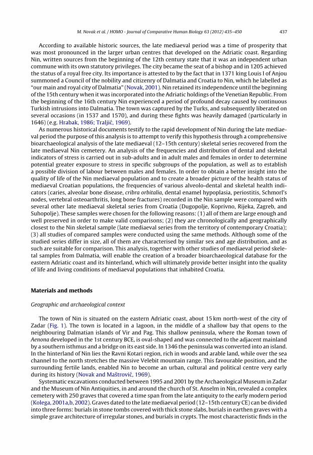
The town of Nin is situated on the eastern Adriatic coast, about 15 km north-west of the city ofZadar (Fig. 1). The town is located in a lagoon, in the middle of a shallow bay that opens to theneighbouring Dalmatian islands of Vir and Pag. This shallow peninsula, where the Roman town ofAenona developed in the 1st century BCE, is oval-shaped and was connected to the adjacent mainlandby a southern isthmus and a bridge on its east side. In 1346 the peninsula was converted into an island.In the hinterland of Nin lies the Ravni Kotari region, rich in woods and arable land, while over the seachannel to the north stretches the massive Velebit mountain range. This favourable position, and thesurrounding fertile lands, enabled Nin to become an urban, cultural and political centre very earlyduring its history (Novak and Mastrovic, 1969).
Systematic excavations conducted between 1995 and 2001 by the Archaeological Museum in Zadarand the Museum of Nin Antiquities, in and around the church of St. Anselm in Nin, revealed a complexcemetery with 250 graves that covered a time span from the late antiquity to the early modern period(Kolega, 2001a,b, 2002). Graves dated to the late mediaeval period (12–15th century CE) can be dividedinto three forms: burials in stone tombs covered with thick stone slabs, burials in earthen graves with asimple grave architecture of irregular stones, and burials in crypts. The most characteristic finds in the
438 M. Novak et al. / HOMO - Journal of Comparative Human Biology 63 (2012) 435– 450
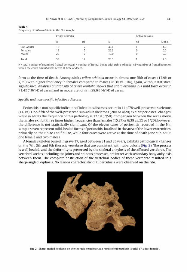
Fig. 1. Map of Croatia with the location of Nin.
late mediaeval graves were earrings (plain circlets, three-jointed circlets, and three-beaded earrings),rings, buttons, and coins (Hungarian and Venetian) (Kolega, 2001a,b, 2002).
The skeletal sample from Nin analysed in this study consists of 141 skeletons excavated fromgraves located in the southern part of the St. Anselm church complex, all of which have been reliablydated (through a combination of radiocarbon dating, typological analyses of recovered artefacts, andvertical and horizontal stratigraphy) to the late mediaeval period (12–15th century CE). The degree ofpreservation of the osteological material varies from very poor to very good, with only a few excellentlypreserved skeletons.
Methods used in the analysis
The anthropological analysis was carried out in the laboratory of the Department of Archaeologyof the Croatian Academy of Sciences and Arts in Zagreb. Sex and age-at-death of recovered individualswere determined using methods described in Buikstra and Ubelaker (1994). No attempt was madeto determine the sex of sub-adults (individuals under 15 years of age). The age of the adults wasdetermined within a 5-year range (e.g. 21–25), while the age of sub-adults was determined within arange of 1 year.
All skeletons were analysed for the possible presence of the following pathologic changes: alveolarbone disease, dental caries, dental enamel hypoplasia, cribra orbitalia, non-specific periostitis, tuber-culosis, Schmorl’s nodes, vertebral osteoarthritis, and bone fractures. All pathologies were recordedaccording to criteria described by Ortner (2003). Most of the pathological changes were calculatedby the element (tooth/alveolus/vertebra/long bone), and not by the individual/skeleton due to poorand/or partial preservation of some skeletons.
For the purposes of this analysis, alveolar bone disease was defined as the presence of periodontal orperiapical abscesses, or antemortem tooth loss. The presence of caries was diagnosed macroscopically,under strong illumination, with the help of a dental probe. Caries size was classified into four categoriesaccording to Metress and Conway (1975): (1) pit or slight fissural start of lesion, (2) lesion coveringless than 50% of the tooth’s surface, (3) lesion covering over 50% of the tooth’s surface, and (4) lesion
M. Novak et al. / HOMO - Journal of Comparative Human Biology 63 (2012) 435– 450 439
that has completely destroyed the crown with only the root remaining. The presence of dental enamelhypoplasia was analysed on permanent maxillary central incisors and on permanent maxillary andmandibular canines for the following reasons: (1) central incisors and canines are more susceptibleto hypoplastic defects than other teeth (Goodman and Rose, 1990); (2) canines develop and growfor a relatively long time – from the fourth month to the sixth year of life; (3) incisors and canineshave the lowest amount of mineralised dental deposits which sometimes may cover the crown ofthe teeth and prevent determination of the presence of hypoplasia. All skulls with preserved bothorbital roofs were macroscopically examined under powerful illumination for the possible presenceof cribra orbitalia. All observed lesions were classified based on intensity (mild, moderate or severe)and condition (active or healed) at the time of death according to criteria proposed by Mensforth et al.(1978), and Mittler and Van Gerven (1994). Non-specific periostitis was diagnosed when two or moreskeletal elements exhibited active or healed periostitis – criteria for inclusion in the sample were thepresence of at least 50% of all cranial bones and long bones. Spinal tuberculosis was diagnosed based onthe presence of: lower thoracic vertebral collapse with kyphosis and fusion coupled with an absenceof new bone formation, and normality of the laminae and posterior spinous processes (Aufderheideand Rodríguez-Martín, 1998; Resnick and Niwayama, 1981). The presence of trauma was establishedby macroscopic analysis that included certification of bilateral asymmetry, angular deformities, thepresence of bone calluses and healed depressions, blunt force trauma, cutting and projectile injuriesto the skull; only long bones preserved to the extent of at least two thirds of their diaphyses andwith all major articular surfaces preserved were analysed (humerii, radii, ulnae, femora, tibiae andfibulae). Inclusion of cranial elements was dependent on the presence of at least 3/4 of the affectedbone.
Some of the described diseases (e.g. dento-alveolar lesions) are age-dependent, i.e. their frequencyincreases with advanced age. Therefore, when tabulating the data, age was controlled by dividingthe sample into two broad categories: young adults (individuals aged between 16 and 35 years) andold adults (individuals older than 36 years). Differences in the frequencies of the studied patholog-ical changes between sub-adults and adults, between two adult age subgroups, and between malesand females were evaluated with the Chi-squared test using Yates correction where appropriate.The statistical computer package SPSS 14.0 for Windows was used for all statistical calculations andtests.
Results
Demography
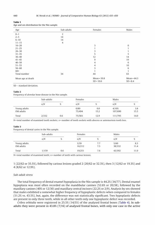
The age and sex distribution of the Nin skeletal sample is presented in Table 1. The sample consists of141 individuals: 34 sub-adults (24.1%), 44 females (31.2%), and 63 males (44.7%). The highest mortalityin the analysed series occurs between the ages of 41 and 45 when almost one-fifth (19.9%) of allindividuals died.
Dento-alveolar lesions
The overall frequency of alveolar bone disease in Nin is 11.0% (188/1710) (Table 2). In sub-adults itwas registered in only two cases (2/332 or 0.6%), while in adults it occurs in 13.5% (186/1378) of theobserved alveoli. Both sexes exhibit similar prevalence of this pathology – males 14.0%, and females12.9%. A considerable increase in alveolar bone disease among older individuals in both sexes is clearlyvisible – these differences are statistically significant (females �2 = 14.182, P < 0.001; males �2 = 9.43,P = 0.002).
Caries is present in 7.9% (63/802) of the teeth (Table 3). Carious lesions were recorded in only onesub-adult tooth (1/159 or 0.6%), while in adults the lesions occur in 9.6% (62/643) of the analysedteeth. When the sexes are compared, males exhibit higher frequencies (11.0% vs. 7.6%), however, thedifference is not statistically significant. Caries frequency in males is higher in the older age group, butthe difference is not statistically significant; in females frequencies of caries in both age groups arealmost identical. Regarding the caries size, among adults the most frequent are carious lesions graded
440 M. Novak et al. / HOMO - Journal of Comparative Human Biology 63 (2012) 435– 450
Table 1Age and sex distribution for the Nin sample.
Age Sub-adults Females Males
0–1 22–5 146–10 1411–15 416–20 3 021–25 1 126–30 3 131–35 5 536–40 12 1441–45 9 1946–50 4 651–55 4 1056–60 3 660+ 1 1Total number 34 44 63
Mean age at death Mean = 39.8 Mean = 44.3SD = 10.6 SD = 8.4
SD – standard deviation.
Table 2Frequency of alveolar bone disease in the Nin sample.
Sub-adults Females Males
n/N % n/N % n/N %
Young adults 0/89 0.0 4/105 3.8Old adults 75/494 15.2 107/690 15.5
Total 2/332 0.6 75/583 12.9 111/795 14.0
N = total number of examined tooth sockets; n = number of tooth sockets with abscess or antemortem tooth loss.
Table 3Frequency of dental caries in the Nin sample.
Sub-adults Females Males
n/N % n/N % n/N %
Young adults 3/39 7.7 5/60 8.3Old adults 16/212 7.5 38/332 11.4
Total 1/159 0.6 19/251 7.6 43/392 11.0
N = total number of examined teeth; n = number of teeth with carious lesions.
1 (22/62 or 35.5%), followed by carious lesions graded 2 (20/62 or 32.3%), then 3 (12/62 or 19.3%) and4 (8/62 or 12.9%).
Sub-adult stress
The total frequency of dental enamel hypoplasia in the Nin sample is 44.2% (34/77). Dental enamelhypoplasia was most often recorded on the mandibular canines (52.6% or 20/38), followed by themaxillary canines (40% or 12/30) and maxillary central incisors (22.2% or 2/9). Analysis by sex showedthat males exhibited a somewhat higher frequency of hypoplastic defects when compared to females(51.2% vs. 43.5%), but, again, the difference was not statistically significant. Two hypoplastic defectsare present in only three teeth, while in all other teeth only one hypoplastic defect was recorded.
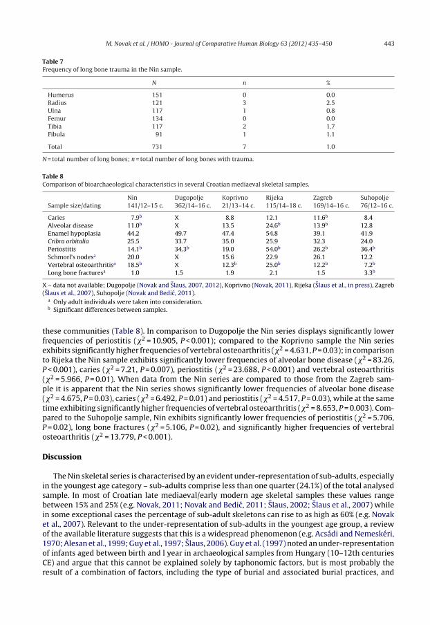
Cribra orbitalia were registered in 25.5% (14/55) of the analysed frontal bones (Table 4). In sub-adults they were present in 43.8% (7/16) of analysed frontal bones, with only one case in the active
M. Novak et al. / HOMO - Journal of Comparative Human Biology 63 (2012) 435– 450 441
Table 4Frequency of cribra orbitalia in the Nin sample.
Cribra orbitalia Active lesions
N n1 % n2 % of n1
Sub-adults 16 7 43.8 1 14.3Females 19 5 26.3 0 0.0Males 20 2 10.0 0 0.0
Total 55 14 25.5 1 4.0
N = total number of examined frontal bones; n1 = number of frontal bones with cribra orbitalia; n2 = number of frontal bones onwhich the cribra orbitalia was active at time of death.
form at the time of death. Among adults cribra orbitalia occur in almost one fifth of cases (17.9% or7/39) with higher frequency in females compared to males (26.3% vs. 10%), again, without statisticalsignificance. Analysis of intensity of cribra orbitalia shows that cribra orbitalia in a mild form occur in71.4% (10/14) of cases, and in moderate form in 28.6% (4/14) of cases.
Specific and non-specific infectious diseases
Periostitis, a non-specific indicator of infectious diseases occurs in 11 of 78 well-preserved skeletons(14.1%). One-fifth of the well-preserved sub-adult skeletons (20% or 4/20) exhibit periosteal changes,while in adults the frequency of this pathology is 12.1% (7/58). Comparison between the sexes showsthat males exhibit three times higher frequencies than females (15.8% or 6/38 vs. 5% or 1/20), however,the difference is not statistically significant. Of the eleven cases of periostitis recorded in the Ninsample seven represent mild, healed forms of periostitis, localised in the area of the lower extremities,primarily on the tibiae and fibulae, while four cases were active at the time of death (one sub-adult,one female and two males).
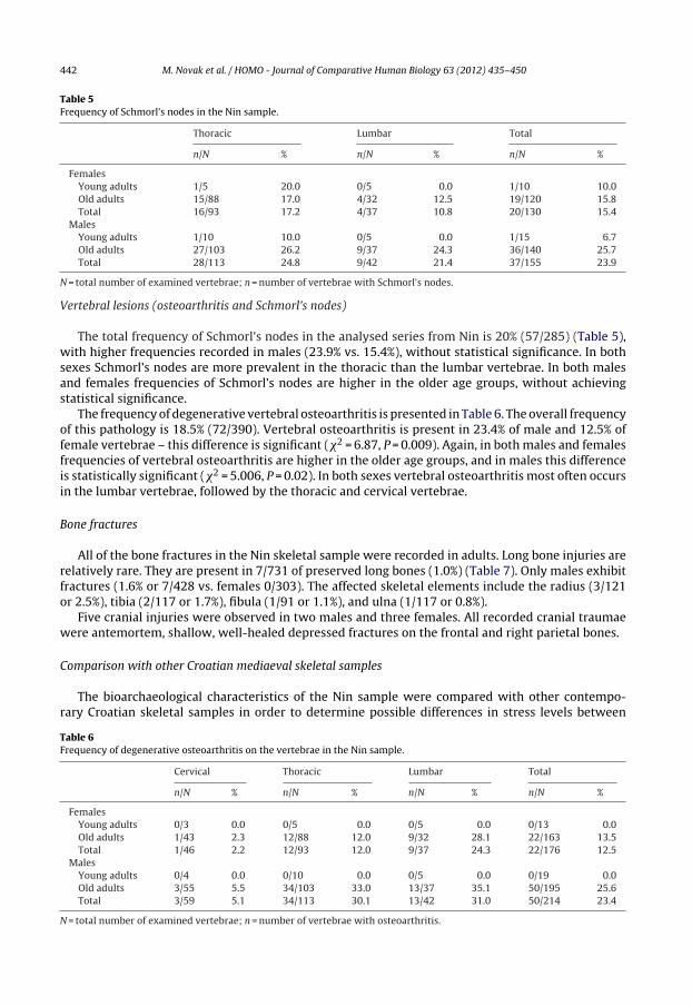
A female skeleton buried in grave 17, aged between 31 and 35 years, exhibits pathological changeson the 7th, 8th and 9th thoracic vertebrae that are consistent with tuberculosis (Fig. 2). The processis well healed, and the deformity is preserved by the skeletal ankylosis of the affected vertebrae. Thevertebral arches, including the joints and spinous processes, are intact with secondary bony ankylosisbetween them. The complete destruction of the vertebral bodies of these vertebrae resulted in asharp-angled kyphosis. No lesions characteristic of tuberculosis were observed on the ribs.
Fig. 2. Sharp-angled kyphosis on the thoracic vertebrae as a result of tuberculosis (burial 17, adult female).
442 M. Novak et al. / HOMO - Journal of Comparative Human Biology 63 (2012) 435– 450
Table 5Frequency of Schmorl’s nodes in the Nin sample.
Thoracic Lumbar Total
n/N % n/N % n/N %
FemalesYoung adults 1/5 20.0 0/5 0.0 1/10 10.0Old adults 15/88 17.0 4/32 12.5 19/120 15.8Total 16/93 17.2 4/37 10.8 20/130 15.4
MalesYoung adults 1/10 10.0 0/5 0.0 1/15 6.7Old adults 27/103 26.2 9/37 24.3 36/140 25.7Total 28/113 24.8 9/42 21.4 37/155 23.9
N = total number of examined vertebrae; n = number of vertebrae with Schmorl’s nodes.
Vertebral lesions (osteoarthritis and Schmorl’s nodes)
The total frequency of Schmorl’s nodes in the analysed series from Nin is 20% (57/285) (Table 5),with higher frequencies recorded in males (23.9% vs. 15.4%), without statistical significance. In bothsexes Schmorl’s nodes are more prevalent in the thoracic than the lumbar vertebrae. In both malesand females frequencies of Schmorl’s nodes are higher in the older age groups, without achievingstatistical significance.
The frequency of degenerative vertebral osteoarthritis is presented in Table 6. The overall frequencyof this pathology is 18.5% (72/390). Vertebral osteoarthritis is present in 23.4% of male and 12.5% offemale vertebrae – this difference is significant (�2 = 6.87, P = 0.009). Again, in both males and femalesfrequencies of vertebral osteoarthritis are higher in the older age groups, and in males this differenceis statistically significant (�2 = 5.006, P = 0.02). In both sexes vertebral osteoarthritis most often occursin the lumbar vertebrae, followed by the thoracic and cervical vertebrae.
Bone fractures
All of the bone fractures in the Nin skeletal sample were recorded in adults. Long bone injuries arerelatively rare. They are present in 7/731 of preserved long bones (1.0%) (Table 7). Only males exhibitfractures (1.6% or 7/428 vs. females 0/303). The affected skeletal elements include the radius (3/121or 2.5%), tibia (2/117 or 1.7%), fibula (1/91 or 1.1%), and ulna (1/117 or 0.8%).
Five cranial injuries were observed in two males and three females. All recorded cranial traumaewere antemortem, shallow, well-healed depressed fractures on the frontal and right parietal bones.
Comparison with other Croatian mediaeval skeletal samples
The bioarchaeological characteristics of the Nin sample were compared with other contempo-rary Croatian skeletal samples in order to determine possible differences in stress levels between
Table 6Frequency of degenerative osteoarthritis on the vertebrae in the Nin sample.
Cervical Thoracic Lumbar Total
n/N % n/N % n/N % n/N %
FemalesYoung adults 0/3 0.0 0/5 0.0 0/5 0.0 0/13 0.0Old adults 1/43 2.3 12/88 12.0 9/32 28.1 22/163 13.5Total 1/46 2.2 12/93 12.0 9/37 24.3 22/176 12.5
MalesYoung adults 0/4 0.0 0/10 0.0 0/5 0.0 0/19 0.0Old adults 3/55 5.5 34/103 33.0 13/37 35.1 50/195 25.6Total 3/59 5.1 34/113 30.1 13/42 31.0 50/214 23.4
N = total number of examined vertebrae; n = number of vertebrae with osteoarthritis.
M. Novak et al. / HOMO - Journal of Comparative Human Biology 63 (2012) 435– 450 443
Table 7Frequency of long bone trauma in the Nin sample.
N n %
Humerus 151 0 0.0Radius 121 3 2.5Ulna 117 1 0.8Femur 134 0 0.0Tibia 117 2 1.7Fibula 91 1 1.1
Total 731 7 1.0
N = total number of long bones; n = total number of long bones with trauma.
Table 8Comparison of bioarchaeological characteristics in several Croatian mediaeval skeletal samples.
Nin Dugopolje Koprivno Rijeka Zagreb SuhopoljeSample size/dating 141/12–15 c. 362/14–16 c. 21/13–14 c. 115/14–18 c. 169/14–16 c. 76/12–16 c.
Caries 7.9b X 8.8 12.1 11.6b 8.4Alveolar disease 11.0b X 13.5 24.6b 13.9b 12.8Enamel hypoplasia 44.2 49.7 47.4 54.8 39.1 41.9Cribra orbitalia 25.5 33.7 35.0 25.9 32.3 24.0Periostitis 14.1b 34.3b 19.0 54.0b 26.2b 36.4b
Schmorl’s nodesa 20.0 X 15.6 22.9 26.1 12.2Vertebral osteoarthritisa 18.5b X 12.3b 25.0b 12.2b 7.2b
Long bone fracturesa 1.0 1.5 1.9 2.1 1.5 3.3b
X – data not available; Dugopolje (Novak and Slaus, 2007, 2012), Koprivno (Novak, 2011), Rijeka (Slaus et al., in press), Zagreb(Slaus et al., 2007), Suhopolje (Novak and Bedic, 2011).
a Only adult individuals were taken into consideration.b Significant differences between samples.
these communities (Table 8). In comparison to Dugopolje the Nin series displays significantly lowerfrequencies of periostitis (�2 = 10.905, P < 0.001); compared to the Koprivno sample the Nin seriesexhibits significantly higher frequencies of vertebral osteoarthritis (�2 = 4.631, P = 0.03); in comparisonto Rijeka the Nin sample exhibits significantly lower frequencies of alveolar bone disease (�2 = 83.26,P < 0.001), caries (�2 = 7.21, P = 0.007), periostitis (�2 = 23.688, P < 0.001) and vertebral osteoarthritis(�2 = 5.966, P = 0.01). When data from the Nin series are compared to those from the Zagreb sam-ple it is apparent that the Nin series shows significantly lower frequencies of alveolar bone disease(�2 = 4.675, P = 0.03), caries (�2 = 6.492, P = 0.01) and periostitis (�2 = 4.517, P = 0.03), while at the sametime exhibiting significantly higher frequencies of vertebral osteoarthritis (�2 = 8.653, P = 0.003). Com-pared to the Suhopolje sample, Nin exhibits significantly lower frequencies of periostitis (�2 = 5.706,P = 0.02), long bone fractures (�2 = 5.106, P = 0.02), and significantly higher frequencies of vertebralosteoarthritis (�2 = 13.779, P < 0.001).
Discussion
The Nin skeletal series is characterised by an evident under-representation of sub-adults, especiallyin the youngest age category – sub-adults comprise less than one quarter (24.1%) of the total analysedsample. In most of Croatian late mediaeval/early modern age skeletal samples these values rangebetween 15% and 25% (e.g. Novak, 2011; Novak and Bedic, 2011; Slaus, 2002; Slaus et al., 2007) whilein some exceptional cases the percentage of sub-adult skeletons can rise to as high as 60% (e.g. Novaket al., 2007). Relevant to the under-representation of sub-adults in the youngest age group, a reviewof the available literature suggests that this is a widespread phenomenon (e.g. Acsádi and Nemeskéri,1970; Alesan et al., 1999; Guy et al., 1997; Slaus, 2006). Guy et al. (1997) noted an under-representationof infants aged between birth and l year in archaeological samples from Hungary (10–12th centuriesCE) and argue that this cannot be explained solely by taphonomic factors, but is most probably theresult of a combination of factors, including the type of burial and associated burial practices, and
444 M. Novak et al. / HOMO - Journal of Comparative Human Biology 63 (2012) 435– 450
archaeological recovery strategies. Acsádi and Nemeskéri (1970) recorded the under-representationof infants in Hungarian mediaeval skeletal samples and argued that as these series were manuallyrecovered, the under-representation of individuals from the youngest age group could have beenthe result of poor recovery techniques applied to fragile perinatal infant bones rather than due tovarious taphonomic factors. According to the archaeologists that excavated the St. Anselm site, thetwo most important factors contributing to the under-representation of sub-adults in Nin are thefrequent reuse of the burial site throughout history, different funeral customs for neonatal deaths andshallower graves of infants aged 0–l years.
The frequencies of oral pathologies, especially caries, in mediaeval Nin are relatively low and aremore similar to frequencies recorded in Croatian samples from antiquity than in those from the medi-aeval period. The results of several studies (Novak et al., 2010; Slaus, 2006, 2008; Slaus et al., 2011)strongly indicate a deterioration of oral health, i.e. a significant increase of the frequencies of alveolo-dental pathologies in the Middle Ages when compared to the antiquity in Croatia, which is explainedby a significantly higher dependence on carbohydrates in the mediaeval diet and higher protein con-tent in the late antiquity diet (Slaus et al., 2011). The recorded low prevalence of caries in Nin may,tentatively, be associated with a diet based on fish and other seafood, accessible to the local popu-lation. This type of diet, which is relatively rich in proteins, is less favourable to the development ofcaries, particularly in comparison to diets dependent on cereals rich in carbohydrates that stimulatebacterial growth and the formation of dental plaque. In addition, it is recognised that some speciesof fish (e.g. anchovies and sardines) are extremely rich in fluoride, which protects teeth from cariesand bleeding (Konig, 2000; Van Loveren, 2000). About the importance of fishery in the mediaevalNin testifies the archaeological evidence such as the presence of fish cleaning knives in graves in theearly mediaeval period graveyard at the Nin-Zdrijac site (Belosevic, 2007). Besides, “The Statute of theLeague of the Nin District”, dated to the 15th century (Brajkovic, 1969), provides numerous detailedregulations and penalties related to fishing, thus witnessing the importance of fish in the everyday lifeof the late mediaeval Nin. Males from Nin exhibit a slightly higher prevalence of caries than females,which is most probably the result of a longer average life span of males (4.5 years). Simply put, maleshad more time to develop carious lesions and more time for it to progress from a small lesion to asevere problem (Slaus et al., 1997). Besides, somewhat higher prevalence of caries in males could be aresult of differences in behavioural activities such as diet, cultural practices and oral hygiene. Namely,Slaus et al. (1997) suggested that the higher frequency of caries in younger males in Nova Raca (con-tinental Croatia) is probably caused by their importance in obtaining food through skill-intensiveagricultural labour, i.e. young males were selectively buffered from the effects of malnutrition andhad more access to limited food resources than young females. The question is, whether and to whatextent, this assumption may be applied to the Nin skeletal series.
The low frequencies of alveolar bone diseases recorded in the Nin sample are most probably a resultof the low frequencies of caries, as many authors suggest that alveolar bone disease in archaeologicalseries is strongly correlated to carious lesions, along with other pathologies such as periapical osteitisand the accumulation of calculus (e.g. Bonfiglioli et al., 2003; Lukacs, 1989). Additionally, Hillson (1996)reported that gingival inflammation, which results from bacterial plaque, leads to bone resorption and,eventually, tooth loss, i.e. he suggested that insufficient oral hygiene, which should result in removingdental plaque, may be the basis of future dental loss. However, at the moment it is not possible toassess the level of oral hygiene of the late mediaeval inhabitants of Nin, so this assumption in the caseof Nin sample cannot be verified.
Hypoplastic defects can be caused by factors that affect child’s growth: congenital and infectiousdisease, changes in dietary habits, malnutrition, intoxication and others (Pindborg, 1970). The preva-lence of dental enamel hypoplasia recorded in the Nin sample closely corresponds to frequenciesobserved in other late mediaeval Croatian populations (Table 8), suggesting that a large part of thestudied sample had, at one time, experienced significant metabolic stress during childhood. Such val-ues are characteristic of communities with sedentary lifestyle and diet primarily based on agriculture(Lanphear, 1990). Specifically, several studies have demonstrated that a sudden increase in the fre-quency of dental enamel hypoplasia occurred during the transition from a hunter–gatherer economyto an economy based on agriculture (e.g. Goodman et al., 1980; Wood, 1996). It has been believedthat a sedentary lifestyle, dietary changes, and rapid population growth led to a significant increase
M. Novak et al. / HOMO - Journal of Comparative Human Biology 63 (2012) 435– 450 445
in the amount of stress that was reflected in an increasing incidence of hypoplastic defects (Cohenand Armelagos, 1984). It has been also possible that a considerable portion of metabolic stress in thelate mediaeval Nin occurred during the weaning period. Some authors have noted that most of thehypoplastic defects in sedentary populations were formed between the first and third year of life, i.e.during the transition from the diet based on the sterile breast milk to the diet rich with microorgan-isms (Goodman, 1988; Goodman et al., 1984; Lanphear, 1990). However, it is important to stress thatserious doubts about this interpretation have been raised (Blakey et al., 1994), and that serious method-ological problems related to the age assessment of the defects have been also identified (Hodges andWilkinson, 1990). Additionally, Ritzman et al. (2008) have reported that histological studies providesignificantly higher age estimates than the commonly used macroscopic methods and this differenceis particularly marked in early forming hypoplastic defects. They also suggest that re-evaluation of themethods used to estimate ages of dental enamel hypoplasia formation may be justified.
As previously noted, it was proposed that one of the major causes for the occurrence of dentalenamel hypoplasia were infectious diseases. Nevertheless, the low prevalence of non-specific indica-tors of infectious diseases (periostitis) in the Nin skeletal sample strongly suggested that, at least inthe case of late mediaeval Nin, infectious diseases did not contribute significantly to the occurrenceof dental enamel hypoplasia.
Analysis of cribra orbitalia frequencies in Nin showed that one quarter of the frontal bones exhib-ited this condition. Some authors have suggested that porotic hyperostosis and cribra orbitalia occur incorrelation with hereditary hemolytic anaemia (thalassemia and sickle cell anaemia), usually preva-lent in populations where malaria was endemic (e.g. Angel, 1966; Soren et al., 1995). In contrastto this, Walker et al. (2009) have hypothesised that porotic hyperostosis and many cases of cribraorbitalia were the result of megaloblastic anaemia acquired by nursing infants through the syner-gistic effects of depleted maternal vitamin B12 reserves, and unsanitary living conditions that wereconducive to additional nutrient losses from gastrointestinal infections around the time of wean-ing. While cribra orbitalia could be attributed to a greater range of causes than porotic hyperostosis(e.g. subperiosteal bleeding associated with a co-deficiency of vitamin C and B12). However, todaymost authors recognise that cribra orbitalia are the result of iron deficiency anaemia which is usu-ally defined as a reduction of haemoglobin and haematocrit in the blood below normal levels (e.g.Huss-Ashmore et al., 1982; Mensforth et al., 1978; Mittler and Van Gerven, 1994; Stuart-Macadam,1985, 1991). Hengen (1971) have concluded that iron deficiency anaemia is primarily the result ofparasitism, while Stuart-Macadam (1992) have suggested that this type of anaemia is an adjustmentof the organism to the disease, and its attempt to exhaust and starve pathogens such as bacteriaand viruses that need iron in order to be able to reproduce in the body of the host. Factors relatedto the occurrence of iron deficiency anaemia in past populations include poor and inadequate diet,gastrointestinal and parasitic infections (Larsen and Sering, 2000; Reinhard, 1992; Walker, 1986),lead poisoning (Stuart-Macadam, 1991), thalassemia (Ascenzi et al., 1991), changes in dietary habits(Roberts and Manchester, 1995), and diet rich in cereal phytates which lower the absorption of iron inthe intestine (Stuart-Macadam, 1992). Facchini et al. (2004) and Salvadei et al. (2001) have associatedthe occurrence of active forms of cribra orbitalia among sub-adults with unhealthy ecological systems,i.e. marshy and wooded environment that could be a host for various parasitic infections.
Although there are numerous potential causes for the occurrence of cribra orbitalia in the latemediaeval Nin series, two most common causes appear to be most probable. One concerns unsanitaryliving conditions, at least for that part of Nin’s population that inhabited the overcrowded fortifiedtown, and which were a direct result of rapid urbanisation and population growth in Nin during thelate mediaeval period. This relationship has already been hypothesised by Novak and Slaus (2010)who registered high frequencies of cribra orbitalia and periostitis in the nearby Roman period city ofZadar that was similarly overcrowded. The second possible cause for the occurrence of cribra orbitaliain mediaeval Nin might be the presence of malaria in the region during this period. Historic sourcestestify that the man-made disruption of the course of the Ricina river located on the north-easternside of town caused silting, a rise in water levels, and consequently the formation of large marsheswhich in time surrounded the town (Ilakovac, 1995/1996). These marshes were ideal habitats formosquitoes that transmitted malaria, the disease caused by “bad air” (male aria). The deteriorationof health caused by the “bad air” disease was one of the reasons for the periodic migrations from
446 M. Novak et al. / HOMO - Journal of Comparative Human Biology 63 (2012) 435– 450
Nin, particularly during the early modern period (Ilakovac, 1995/1996). Malaria was not, however, amajor concern only in the Nin area because Dalmatia as a Mediterranean region has traditionally beenafflicted by malaria. Some authors reported that during the early 20th century some 180,000 people(about one third of the entire population of Dalmatia) suffered from the disease, with the death ratebetween 43% and 49% (Dugacki, 2005). In 1902 the prevalence of the disease in the Nin area was about80% (Dugacki, 2005). Malaria in the Nin area was finally put under control between 1906 and 1909when the surrounding marshes were drained and a prophylactic medical treatment was applied tothe general population (Dugacki, 2005).
Studies of populations from different time periods have confirmed the synergistic relationshipbetween the occurrence of infectious diseases and poor nutrition, i.e. acutely or chronically under-nourished individuals were less resistant to infectious diseases compared to individuals with normaldiets (e.g. Keusch and Farthing, 1986; Mensforth et al., 1978; Scrimshaw et al., 1968). The overallprevalence of periostitis in Nin was low when compared to frequencies reported in other Croatianarchaeological series that usually ranged between 20% and 50%. Not surprisingly therefore, the preva-lence of periostitis in children from Nin (20%) was considerably lower than in other skeletal samplesfrom Croatia. In the early modern age samples such as Koprivno and Torcec these values raised up to92% (Novak et al., 2007; Novak and Krznar, 2010). The occurrence of periostitis in the Nin sample sug-gests that the mediaeval inhabitants of Nin were characterised by relatively strong immune systemsenabling them to survive the occurrence of various infectious diseases. This is most probably relatedto the favourable geographic location of Nin with easy access to abundant food resources.
The unequivocal presence of tuberculosis in the late mediaeval Nin is interesting. Tuberculosis isan infectious disease caused by Mycobacterium tuberculosis that is most often transmitted through therespiratory system. The primary infection usually starts in the lungs, after which it disseminates by theblood stream to other parts of the body – kidneys, brain and bones. The spine is the most commonlyaffected bone element (in approximately 40% of cases; Aufderheide and Rodríguez-Martín, 1998). Theskeleton of an adult female from burial 17 from Nin exhibits lesions characteristic of vertebral tuber-culosis: T7, T8 and T9 collapsed and fused together resulting in a sharply angular kyphosis (gibbus);this block is furthermore fused with one superior (T6) and three inferior vertebrae (T10–T12) givinga total of seven fused vertebrae. In pre-antibiotic times sharp angular kyphosis was present in nearly60% of cases involving spinal tuberculosis (Reinhart, 1932). In the case from Nin differential diagnosishas excluded osteomyelitis, vertebral compression fracture, and septic arthritis as possible factors forthe noted changes. Osteomyelitis of the spine usually manifests as a destructive lesion of just onevertebra and does not end with ankylosis and kyphosis (Aufderheide and Rodríguez-Martín, 1998).Usually only one vertebra is involved in vertebral compression fractures with angular deformity, andthe vertebral body is generally destroyed to a lesser degree (Ortner, 2003). Although septic arthritisoften terminates in bony ankylosis and is very similar to tuberculous arthritis, bone destruction isnot as severe as in tuberculosis (Aufderheide and Rodríguez-Martín, 1998). In addition to Nin, onlyone case of skeletal tuberculosis dated to the late Middle Ages from the territory of the present-dayCroatia was registered – an adult female from the town of Rijeka (Slaus et al., in press). Both cases sofar discovered in Croatian skeletal series have, thus, been registered in urban settings characterisedby high population density. According to some authors the most important factors for the emer-gence of numerous outbreaks of tuberculosis during the late mediaeval period in Europe were rapidurbanisation and population growth during the 15th and 16th centuries, as well as the developmentof trade that helped to accelerate the spread of the disease (e.g. Aufderheide and Rodríguez-Martín,1998; Lindemann, 1999). High population density and limited available housing provided the idealconditions for aerial transmission of the tubercle bacillus causing lung infection (Aufderheide andRodríguez-Martín, 1998).
Schmorl’s nodes in the Nin mediaeval sample were much more prevalent in males than in females.A recent comprehensive study of vertebral pathologies conducted on two early modern age skeletalsamples from Croatia (Novak and Slaus, 2011) have demonstrated a significantly higher prevalence ofSchmorl’s nodes in males, leading the authors to suggest that the observed sexual dimorphism mayhave been a result of sexual division of labour where males were engaged in more demanding physicalactivities, an assumption that was also supported by historical and ethnographic data. An additionalconclusion from this study was that Schmorl’s nodes were not correlated with increased age (Novak
M. Novak et al. / HOMO - Journal of Comparative Human Biology 63 (2012) 435– 450 447
and Slaus, 2011). Values observed in this analysis are similar to the results recorded by other authors(Stirland and Waldron, 1997; Slaus, 2000; Üstündag, 2008) suggesting that Schmorl’s nodes can beused as a reliable indicator of activity related stress and different lifestyles in archaeological samples.If the hypothesis suggested by Novak and Slaus (2011) is applied to the Nin skeletal sample a higherprevalence of Schmorl’s nodes in the Nin males was probably a consequence of the different lifestylesbetween the sexes, i.e. a sexual division of labour where males performed more demanding physicaltasks.
The frequency of vertebral osteoarthritis in Nin is higher in comparison to other late mediaevalCroatian skeletal samples. The main factors influencing the presence and severity of degenerativeosteoarthritis in past and modern populations are age, followed by repetitive mechanical loading andmovement, and also genetic factors (Weiss and Jurmain, 2007). The study conducted by Novak andSlaus (2011) showed that, unlike Schmorl’s nodes, vertebral osteoarthritis is strongly correlated withincreasing age. Therefore, the differences between Nin and other mediaeval Croatian communities, aswell as the significantly higher frequency of vertebral osteoarthritis in Nin males, could be explained bythe relatively long life span of Nin’s inhabitants, and the somewhat longer average life span of Nin malesin comparison to females (4.5 years). Of course, this hypothesis cannot be confirmed with certainty,as vertebral osteoarthritis can have a wide variety of etiologies. Further research of archaeologicallyderived skeletal samples is necessary in order to clarify this issue.
The frequency, morphology and location of the long bone fractures in the Nin series strongly suggesta low level of interpersonal violence in this community during the late mediaeval period. Additionalsupport for this assumption lies in the low frequency of defensive fractures. Ulnar ‘parry’ fractureshave, for instance, frequently been used as an indicator of deliberate violence in a community (Juddand Roberts, 1999; Smith, 1996). In the Nin series only one ulnar fracture has been recorded. Longbone fractures recorded in the series are typical of those usually related to accidents (e.g. Djuric et al.,2006; Judd, 2004; Russell et al., 1991) such as fractures of radius, tibia, and fibula. The hypothesisof the relatively low risk of deliberate violence in the late mediaeval Nin is further supported by thecomplete absence of perimortem trauma and trauma inflicted by sharp-edged weapons. Five cranialfractures might suggest sporadic occurrence of deliberate violence of a lesser intensity such as streetfights and tavern brawls without fatal consequences, and with the occasional use of blunt weapons.
A comparison of the bioarchaeological characteristics of the Nin series with other Croatianmediaeval skeletal series shows that the Nin population was characterised by lower prevalence ofalveolo-dental pathologies, periostitis and long bone fractures than its contemporaries from othermediaeval communities inhabiting the territory of present-day Croatia. Our observations suggest rel-atively good living conditions that are possibly a result of the favourable geographical location of Ninthat allowed access to adequate food sources (an abundance of seafood and proximity of the fertilefields in the Ravni Kotari region). In addition, its location on an island surrounded by strong fortifica-tions allowed for a relatively peaceful life, without major episodes of interpersonal violence. On thenegative side, the rapid urbanisation of Nin during the late Middle Ages also led to deterioration ofsanitary conditions, at least for part of its population, which may have been reflected in the relativelyhigh frequencies of sub-adult stress indicators (dental enamel hypoplasia and cribra orbitalia) andthe presence of infectious diseases such as tuberculosis. Additionally, one of the possible causes forthe relatively high frequency of cribra orbitalia in the Nin series could be the historically documentedpresence of malaria in the region.
Conclusion
A comprehensive bioarchaeological analysis of human skeletal remains from Nin revealed newdetails about the living conditions of the mediaeval inhabitants of this ancient city. Historical datatestify to the increased urban development of Nin between the 12th and 15th centuries. The lowprevalence of dental pathologies in the skeletal series related to the late mediaeval component ofthe Nin cemetery suggests an adequate diet, relatively high in proteins, while the low frequency ofbone trauma testifies to the relatively peaceful life of Nin’s inhabitants. Increased urban developmentduring this period also had a negative effect on the health of its inhabitants as seen in the relatively
448 M. Novak et al. / HOMO - Journal of Comparative Human Biology 63 (2012) 435– 450
high frequencies of dental enamel hypoplasia and cribra orbitalia that may have been related to aworsening of sanitary conditions caused by overcrowding. That additionally may have facilitated theemergence of infectious diseases such as tuberculosis.
Acknowledgments
The authors would like to thank Dr. Marija Kolega from the Department of Art History, Universityin Zadar for enabling the access to the skeletal material from the Nin-St. Anselm site. They wouldalso like to thank the Editor in-Chief of HOMO, Prof. Maciej Henneberg, a member of HOMO editorialteam, Prof. Andrea Cucina, and the anonymous reviewers for constructive suggestions and commentsthat substantially improved the manuscript. This study was financially supported by a grant from theMinistry of Science, Education and Sports of the Republic of Croatia (Grant Number 101-197-0677-0670).
References
Acsádi, G., Nemeskéri, J., 1970. History of Human Life Span and Mortality. Akadémiai Kiadó, Budapest.Alesan, A., Malgosa, A., Simó, C., 1999. Looking into the demography of an Iron Age population in the Western Mediterranean.
I. Mortality. Am. J. Archaeol. 110, 285–301.Angel, J.L., 1966. Porotic hyperostosis, anemias, malarias and marshes in the prehistoric Eastern Mediterranean. Science 153,
760–763.Ascenzi, A., Bellelli, A., Brunori, M., Citro, G., Ippoliti, R., Lendaro, E., Zito, R., 1991. Diagnosis of thalassemia in ancient bones:
problems and prospects in pathology. In: Ortner, D., Aufderheide, A. (Eds.), Human Paleopathology. Current Synthesis andFuture Options. Smithsonian Institution Press, Washington, DC, pp. 73–75.
Aufderheide, A.C., Rodríguez-Martín, C., 1998. The Cambridge Encyclopedia of Human Paleopathology. Cambridge UniversityPress, Cambridge.
Belosevic, J., 2007. Starohrvatsko groblje na Zdrijacu u Ninu. Arheoloski muzej Zadar, Zadar.Blakey, M.L., Leslie, T.E., Reidy, J.P., 1994. Frequency and chronological distribution of dental enamel hypoplasia in enslaved
African Americans: a test of the weaning hypothesis. Am. J. Phys. Anthropol. 95, 371–383.Bonfiglioli, B., Brasili, P., Belcastro, M.G., 2003. Dento-alveolar lesions and nutritional habits of a Roman Imperial age population
(1st–4th c. AD): Quadrella (Molise, Italy). HOMO – J. Comp. Hum. Biol. 54, 36–56.Brajkovic, V., 1969. Odredbe prava mora i ribolova u Statutu Lige kotara ninskog. Rad. Inst. Jugosl. Akad. Zadar 16/17, 479–483.Buikstra, J.E., Ubelaker, D.H., 1994. Standards for Data Collection from Human Skeletal Remains. Arkansas Archaeological Survey,
Fayetteville.Cohen, M.N., Armelagos, G.J., 1984. Editor’s summation. In: Cohen, M.N., Armelagos, G.J. (Eds.), Paleopathology at the Origins of
Agriculture. Academic Press, Orlando, pp. 585–601.Djuric, M.P., Roberts, C.A., Rakocevic, Z.B., Djonic, D.D., Lesic, A.R., 2006. Fractures in late medieval skeletal populations from
Serbia. Am. J. Phys. Anthropol. 130, 167–178.Dugacki, V., 2005. Akcija dr Rudolfa Battare u Ninu god. 1902, prvi sustavni pokusaj borbe protiv malarije u Hrvatskoj. Med.
Jadert. 35 (Suppl. 1–2), 33–40.Facchini, F., Rastelli, E., Brasili, P., 2004. Cribra orbitalia and cribra cranii in Roman skeletal remains from the Ravenna area and
Rimini (I–IV century AD). Int. J. Osteoarcheol. 14, 126–136.Goodman, A.H., 1988. The chronology of enamel hypoplasias in industrial population: a reppraisal of Sarnat and Schour (1941,
1942). Hum. Biol. 60, 781–791.Goodman, A.H., Armelagos, G.J., Rose, J.C., 1980. Enamel hypoplasias as indicators of stress in three prehistoric populations from
Illinois. Hum. Biol. 52, 515–528.Goodman, A.H., Armelagos, G.J., Rose, J.C., 1984. The chronological distribution of enamel hypoplasias from prehistoric Dickson
Mounds populations. Am. J. Phys. Anthropol. 65, 259–266.Goodman, A.H., Rose, J.C., 1990. Assessment of systemic physiological perturbations from dental enamel hypoplasias and
associated histological structures. Yrbk Phys. Anthropol. 33, 59–110.Guy, H., Masset, C., Baud, C.A., 1997. Infant taphonomy. Int. J. Osteoarchaeol. 7, 221–229.Hengen, O.P., 1971. Cribra orbitalia: pathogenesis and probable etiology. HOMO – J. Comp. Hum. Biol. 22, 57–75.Hillson, S., 1996. Dental Anthropology. Cambridge University Press, Cambridge.Hodges, D.C., Wilkinson, R.G., 1990. Effect of tooth size on the ageing and chronology distribution of enamel hypoplastic defects.
Am. J. Hum. Biol. 2, 553–560.Hrabak, B., 1986. Turske provale i osvajanja na podrucju danasnje severne Dalmacije do sredine XVI stoleca. Rad. Inst. hrvat.
povij. 19, 69–99.Huss-Ashmore, R., Goodman, A.H., Armelagos, G.J., 1982. Nutritional interference from paleopathology. In: Schiffer, M.B. (Ed.),
Advances in Archaeological Method and Theory, vol. 5. Academic Press, New York, pp. 395–474.Ilakovac, B., 1995/1996. Postanak i razvoj ninskih (Aenona) mostova. Vjesn. Arheol. muz. Zagreb 28/29, 73–95.Judd, M.A., 2004. Trauma in the city of Kerma: ancient versus modern injury patterns. Int. J. Osteoarchaeol. 14, 34–51.Judd, M.A., Roberts, C.A., 1999. Fracture trauma in a medieval British farming village. Am. J. Phys. Anthropol. 109, 229–243.Keusch, G.T., Farthing, M.J.G., 1986. Nutrition and infection. Annu. Rev. Nutr. 6, 131–154.Kolega, M., 2001a. Nin – arheoloska istrazivanja u sklopu zupne crkve sv. Asela (Anselma). Obavijest. Hrvat. arheol. drus. 2,
83–90.
M. Novak et al. / HOMO - Journal of Comparative Human Biology 63 (2012) 435– 450 449
Kolega, M., 2001b. Nin – arheoloska istrazivanja ispred zupne crkve sv. Asela (Anselma). Obavijest. Hrvat. arheol. drus. 2, 91–95.Kolega, M., 2002. Nin – nadzupni kompleks sv. Anselma (Asela). Istrazivanja godine 2001. Obavijest. Hrvat. arheol. drus. 2,
73–78.Konig, K.G., 2000. Diet and oral health. Int. Dent. J. 50, 162–174.Lanphear, K.M., 1990. Frequency and distribution of enamel hypoplasias in a historic skeletal sample. Am. J. Phys. Anthropol.
81, 35–43.Larsen, C.S., Sering, L., 2000. Inferring iron deficiency anemia from human skeletal remains: the case of the Georgia Bight. In:
Lambert, P. (Ed.), Bioarchaeological Studies in Life in the Age of Agriculture. University of Alabama Press, Tuscaloosa, pp.116–133.
Lindemann, M., 1999. Medicine and Society in Early Modern Europe. Cambridge University Press, Cambridge.Lukacs, J.R., 1989. Dental paleopathology: methods for reconstructing dietary patterns. In: Is can, M.Y., Kennnedy, K.A.R. (Eds.),
Reconstruction of Life from the Skeleton. Alan R. Liss, New York, pp. 261–286.Mensforth, R.P., Lovejoy, C.O., Lallo, J.W., Armelagos, G.J., 1978. The role of constitutional factors, diet and infectious disease
in the etiology of porotic hyperostosis and periosteal reactions in prehistoric infants and children. Med. Anthropol. 2,1–59.
Metress, J.F., Conway, T., 1975. Standardized system for recording dental caries in prehistoric skeletons. J. Dent. Res. 54, 908.Mikic, Z., 1983. Antropoloski prikaz srednjovekovnih stanovnika Ricica. In: Jelicic, J. (Ed.), Ricice – nekropole stecaka. Regionalni
zavod za zastitu spomenika kulture, Split, pp. 45–59.Mittler, D.M., Van Gerven, D.P., 1994. Developmental, diachronic, and demographic analysis of cribra orbitalia in the medieval
Christian populations of Kulubnarti. Am. J. Phys. Anthropol. 93, 287–297.Novak, G., 2001. Proslost Dalmacije. Golden Marketing, Zagreb.Novak, M., 2011. Bioarchaeological analysis of the human skeletal remains from the late mediaeval cemetery of Koprivno,
southern Croatia. Bull. Int. Assoc. Paleodont. 5, 13–23.Novak, M., Bedic, Z., 2011. Bioarheoloske karakteristike srednjovjekovne populacije s nalazista Suhopolje-Kliskovac. In: Tomicic,
Z., Jelincic, K. (Eds.), Suhopolje-Kliskovac. Od mjestopisa do arheoloske spoznaje. Institut za arheologiju u Zagrebu i OpcinaSuhopolje, Zagreb – Suhopolje, pp. 165–174.
Novak, M., Krznar, S., 2010. Prilozi poznavanju uvjeta i kvalitete zivota u ranonovovjekovnom podravskom selu na primjeruTorceca kraj Koprivnice. Podravina 18, 59–88.
Novak, G., Mastrovic, V., 1969. Povijest grada Nina. Institut Jugoslavenske akademije znanosti i umjetnosti, Zadar.Novak, M., Slaus, M., 2007. Ucestalost i distribucija cribrae orbitaliae u kasnosrednjovjekovnoj populaciji iz Dugopolja. Starohrv.
prosvj. 34, 451–475.Novak, M., Slaus, M., 2010. Health and disease in a Roman walled city: an example of Colonia Iulia Iader. J. Antropol. Sci. 88,
189–206.Novak, M., Slaus, M., 2011. Vertebral pathologies in two early modern period (16th–19th century) populations from Croatia.
Am. J. Phys. Anthropol. 145, 270–281.Novak, M., Slaus, M., 2012. Frequency and patterning of bone trauma in the late medieval population (13–16th century) from
Dugopolje, southern Croatia. Anthropol. Anz. 69, 335–350.Novak, M., Slaus, M., Pasaric, M., 2007. Bioarchaeological characteristics of the Early Modern population from the Koprivno –
Kod kriza site near Klis. Opusc. archaeol. 31, 303–346.Novak, M., Slaus, M., Bedic, Z., Vyroubal, V., 2010. Dental pathologies in rural mediaeval populations from continental Croatia.
Anthropol. Közlem. 51, 11–21.Ortner, D.J., 2003. Identification of Pathological Conditions in Human Skeletal Remains. Academic Press, San Diego.Pindborg, J.J., 1970. Pathology of the Dental Hard Tissues. W.B. Saunders, Philadelphia.Rajic Sikanjic, P., Vlak, D., 2010a. Autoimmune joint diseases in late medieval skeletal sample from Croatia. Rheumatol. Int. 30,
349–356.Rajic Sikanjic, P., Vlak, D., 2010b. Elongated styloid process in late medieval skeletons from Uzdolje-Grablje, Croatia. Int. J.
Osteoarchaeol. 20, 248–252.Reinhard, K., 1992. Patterns of diet, parasitism and anemia in prehistoric west North America. In: Stuart-Macadam, P., Kent, S.
(Eds.), Diet, Demography, and Disease: Changing Perspectives on Anemia. Aldine de Gruyter, New York, pp. 219–258.Reinhart, M., 1932. Die Fälle von Spondylitis Tuberculosa in der Aargauischen Heilstätte Barmelweid aus den Jahren 1912–1930.
Beitrage zur Klinik der Tuberkulose 79, 745–749.Resnick, D., Niwayama, G., 1981. Diagnosis of Bone and Joint Disorders. W.B. Saunders, Philadelphia.Ritzman, T.B., Baker, B.J., Schwartz, G.T., 2008. A fine line: a comparison of methods for estimating ages of linear enamel
hypoplasia formation. Am. J. Phys. Anthropol. 135, 348–361.Roberts, C.A., Manchester, K., 1995. The Archaeology of Disease. Cornell University Press, New York.Russell, T., Taylor, J., LaVelle, D., 1991. Fractures of the tibia and fibula. In: Rockwood, C., Green, D., Bucholz, R. (Eds.), Rockwood
and Green’s fractures in Adults, vol. 2. Lippincott, Philadelphia, pp. 1915–1982.Salvadei, L., Ricci, F., Manzi, G., 2001. Porotic hyperostosis as a marker of health and nutritional conditions during childhood:
studies at the transition between Imperial Rome and the Early Middle Ages. Am. J. Hum. Biol. 13, 709–717.Scrimshaw, N.S., Taylor, C.E., Gordon, J.E., 1968. Interactions of Nutrition and Infection. World Health Organization, Geneva.Smith, M.O., 1996. Parry fractures and female directed interpersonal violence: implications from the Late Archaic Period of west
Tennessee. Int. J. Osteoarchaeol. 6, 84–91.Soren, D., Fenton, T., Birkey, W., 1995. The late Roman infant cemetery near Lugnano in Teverina, Italy: some implications. J.
Paleopathol. 7, 13–47.Stirland, A.J., Waldron, T., 1997. Evidence for activity related markers in the vertebrae of the crew of the Mary Rose. J. Archaeol.
Sci. 24, 329–335.Stuart-Macadam, P., 1985. Porotic hyperostosis: representative of a childhood condition. Am. J. Phys. Anthropol. 66, 391–398.Stuart-Macadam, P., 1991. Anaemia in Roman Brita. In: Poundbury, Camp., In: Bush, H., Zvelebil, M. (Eds.), Health in Past Societies.
Biocultural Interpretations of Human Skeletal Remains in Archaeological Contexts. Archaeopress, Oxford, pp. 101–113.Stuart-Macadam, P., 1992. Porotic hyperostosis: a new perspective. Am. J. Phys. Anthropol. 87, 39–47.
450 M. Novak et al. / HOMO - Journal of Comparative Human Biology 63 (2012) 435– 450
Slaus, M., 1996. Antropoloska analiza kasnosrednjovjekovne populacije iz Danila Gornjeg kraj Sibenika. Arh. rad. raspr. 12,343–364.
Slaus, M., 2000. Biocultural analysis of sex differences in mortality profiles and stress levels in the late medieval populationfrom Nova Raca, Croatia. Am. J. Phys. Anthropol. 111, 193–209.
Slaus, M., 2002. The Bioarchaeology of Continental Croatia. An Analysis of Human Skeletal Remains from the Prehistoric toPost-medieval Periods. Archaeopress, Oxford.
Slaus, M., 2006. Bioarheologija – demografija, zdravlje, traume i prehrana starohrvatskih populacija. Skolska knjiga, Zagreb.Slaus, M., 2008. Osteological and dental markers of health in the transition from the late antique to the early medieval period
in Croatia. Am. J. Phys. Anthropol. 136, 455–469.Slaus, M., Pecina-Hrncevic, A., Jakovljevic, G., 1997. Dental disease in the late medieval population from Nova Raca, Croatia. Coll.
Antropol. 21, 561–572.Slaus, M., Novak, M., Bedic, Z., Vyroubal, V., 2007. Antropoloska analiza kasnosrednjovjekovnog groblja kraj crkve svetog Franje
na Opatovini u Zagrebu. Arh. rad. raspr. 15, 211–247.Slaus, M., Bedic, Z., Rajic Sikanjic, P., Vodanovic, M., Domic Kunic, A., 2011. Dental health at the transition from the Late Antique
to the early Medieval period on Croatia’s eastern Adriatic coast. Int. J. Osteoarchaeol. 21, 577–590.Slaus, M., Novak, M., Cavka, M., 2012. Four cases of ankylosing spondylitis in medieval skeletal series from Croatia. Rheumatol.
Int., http://dx.doi.org/10.1007/s00296-011-2343-7.Slaus, M., Novak, M., Bedic, Z., Vyroubal, V. Antropoloska analiza ljudskih kostanih ostataka s nalazista Rijeka – Trg pul Vele
Crikve, in press.Traljic, M.S., 1969. Nin pod udarom mletackih ratova. Rad. Zav. povij. zna. 16/17, 529–548.Van Loveren, C., 2000. Diet and dental caries: cariogenicity may depend more on oral hygiene using fluorides than on diet or
type of carbohydrates. Eur. J. Paediatr. Dent. 1, 55–62.Walker, P.L., 1986. Porotic hyperostosis in a marine-dependent California Indian population. Am. J. Phys. Anthropol. 69, 345–354.Walker, P.L., Bathurst, R.R., Richman, R., Gjerdrum, T., Andrushko, V.A., 2009. The causes of porotic hyperostosis and cribra
orbitalia: a reappraisal of the iron-deficiency-anemia hypothesis. Am. J. Phys. Anthropol. 139, 109–125.Weiss, E., Jurmain, R., 2007. Osteoarthritis revisited: a contemporary review of aetiology. Int. J. Osteoarchaeol. 17, 437–450.Wood, L., 1996. Frequency and chronological distribution of linear enamel hypoplasia in a North American colonial skeletal
sample. Am. J. Phys. Anthropol. 100, 233–247.Üstündag, H., 2008. Schmorl’s nodes in a post-medieval skeletal sample from Klostermarienberg, Austria. Int. J. Osteoarchaeol.
19, 695–710.
Related Documents