Reference: Biol. Bull. 196: 229-244. (June 1999) Intracapsular Feeding by Embryos of the Gastropod Genus Littorina A. L. MORAN’ Oregon Institute of Marine Biology, University of Oregon, Charleston, Oregon 97420 Abstract. Many gastropod species develop within egg capsules within which larvae are provided with extraembry- onic nutrients. Species with encapsulated development fre- quently have transitory embryonic organs, such as “larval kidneys,” that may represent specializations for consump- tion of intracapsular nutrition. Larvae of Littorina species with nonplanktonic, encapsulated development consume in- tracapsular albumen, but they lack obvious morphological modifications for albumen consumption. To determine the mechanism and location of protein uptake, larvae of seven species of Littorina (L. keenae, L. littorea, L. plena, L. saxatilis, L. scutulata, L. sitkana, L. subrotundata) were exposed to solutions of either fluorescently labeled protein (FITC-bovine serum albumen) or ferritin. Under fluores- cence microscopy, larvae of all species with encapsulated, nonplanktonic development displayed strong regional affin- ity for FITC in the ciliated cells of the velum, whereas hatched larvae of planktotrophic Littorina species did not. Transmission electron microscopy of epithelial cells of non- planktotrophic veligers exposed to ferritin supported the interpretation that localized affinity for labeled protein in- dicated endocytotic protein uptake. Planktotrophic Littorina and Littorina with encapsulated, nonplanktonic develop- ment were shown to share equivalent velar width/larval length ratios during early embryonic development, whereas a literature search suggested that in other nonplanktotrophic prosobranchs the velum is relatively smaller than in plank- totrophs. Retention of a large velum in Littorina that de- velop entirely within egg capsules may facilitate feeding on intracapsular protein, in the absence of specialized assimi- lative organs found in other species with encapsulated de- velopment. Received 9 September 1997; accepted 12 February 1999. ’ Present address: Friday Harbor Laboratories, University of Washing- ton, 620 University Road, Friday Harbor, WA 98250. Email: moran@ fhl.washington.edu Introduction Marine invertebrates exhibit a remarkable variety of re- productive and developmental modes both within and among taxa, and this variation provides a powerful compar- ative means of studying the integration of development, life history, and evolution. One of the best-known dichotomies in invertebrate development is between species with larvae that must feed in the plankton to grow and attain metamor- phic competence (planktotrophic), and species that reach competence without feeding in the plankton (nonplanktotro- phic) (Thorson, 1946; Jablonski and Lutz, 1983; Strath- mann, 1985). Another important distinction can be made between species with entirely planktonic development, and species that spend all or part of development in egg capsules (planktonic, encapsulated nonplanktonic, and mixed devel- opment, respectively) (Thorson, 1946; Pechenik, 1979; Per- ron, 198 1). Larvae of many species with encapsulated, nonplanktonic development are morphologically similar to larvae of re- lated planktotrophic species, and retain structures that pre- sumably had an ancestral role in larval swimming and feeding. For example, larvae of most gastropod molluscs with nonplanktonic, encapsulated development possess a velum (e.g., Fretter and Graham, 1962; Buckland-Nicks et al., 1973; Strathmann, 1978; Hadfield and Iaea, 1989), which is the primary larval structure that planktotrophic molluscan larvae use in swimming and food collection (Strathmann and Leise, 1979). The velum may be smaller in species that lack free-living larvae than in planktotrophic species (Jagersten, 1972; Webber, 1977; Rives& 1983; but see Hadfield and Iaea, 1989), and it may have different patterns of ciliation (e.g., Lyons and Spight, 1973; Hadfield and Iaea, 1989). These changes in gastropod larval mor- phology have been interpreted as the loss of complex, ancestral planktotrophic features due to relaxation of stabi- lizing selection, and as functional modifications that en- 229

Welcome message from author
This document is posted to help you gain knowledge. Please leave a comment to let me know what you think about it! Share it to your friends and learn new things together.
Transcript
Reference: Biol. Bull. 196: 229-244. (June 1999)
Intracapsular Feeding by Embryos of the Gastropod Genus Littorina
A. L. MORAN’
Oregon Institute of Marine Biology, University of Oregon, Charleston, Oregon 97420
Abstract. Many gastropod species develop within egg capsules within which larvae are provided with extraembry- onic nutrients. Species with encapsulated development fre- quently have transitory embryonic organs, such as “larval kidneys,” that may represent specializations for consump- tion of intracapsular nutrition. Larvae of Littorina species with nonplanktonic, encapsulated development consume in- tracapsular albumen, but they lack obvious morphological modifications for albumen consumption. To determine the mechanism and location of protein uptake, larvae of seven species of Littorina (L. keenae, L. littorea, L. plena, L. saxatilis, L. scutulata, L. sitkana, L. subrotundata) were exposed to solutions of either fluorescently labeled protein (FITC-bovine serum albumen) or ferritin. Under fluores- cence microscopy, larvae of all species with encapsulated, nonplanktonic development displayed strong regional affin- ity for FITC in the ciliated cells of the velum, whereas hatched larvae of planktotrophic Littorina species did not. Transmission electron microscopy of epithelial cells of non- planktotrophic veligers exposed to ferritin supported the interpretation that localized affinity for labeled protein in- dicated endocytotic protein uptake. Planktotrophic Littorina and Littorina with encapsulated, nonplanktonic develop- ment were shown to share equivalent velar width/larval length ratios during early embryonic development, whereas a literature search suggested that in other nonplanktotrophic prosobranchs the velum is relatively smaller than in plank- totrophs. Retention of a large velum in Littorina that de- velop entirely within egg capsules may facilitate feeding on intracapsular protein, in the absence of specialized assimi- lative organs found in other species with encapsulated de- velopment.
Received 9 September 1997; accepted 12 February 1999. ’ Present address: Friday Harbor Laboratories, University of Washing-
ton, 620 University Road, Friday Harbor, WA 98250. Email: moran@ fhl.washington.edu
Introduction
Marine invertebrates exhibit a remarkable variety of re- productive and developmental modes both within and among taxa, and this variation provides a powerful compar- ative means of studying the integration of development, life history, and evolution. One of the best-known dichotomies in invertebrate development is between species with larvae that must feed in the plankton to grow and attain metamor- phic competence (planktotrophic), and species that reach competence without feeding in the plankton (nonplanktotro- phic) (Thorson, 1946; Jablonski and Lutz, 1983; Strath- mann, 1985). Another important distinction can be made between species with entirely planktonic development, and species that spend all or part of development in egg capsules (planktonic, encapsulated nonplanktonic, and mixed devel- opment, respectively) (Thorson, 1946; Pechenik, 1979; Per- ron, 198 1).
Larvae of many species with encapsulated, nonplanktonic development are morphologically similar to larvae of re- lated planktotrophic species, and retain structures that pre- sumably had an ancestral role in larval swimming and feeding. For example, larvae of most gastropod molluscs with nonplanktonic, encapsulated development possess a velum (e.g., Fretter and Graham, 1962; Buckland-Nicks et al., 1973; Strathmann, 1978; Hadfield and Iaea, 1989), which is the primary larval structure that planktotrophic molluscan larvae use in swimming and food collection (Strathmann and Leise, 1979). The velum may be smaller in species that lack free-living larvae than in planktotrophic species (Jagersten, 1972; Webber, 1977; Rives& 1983; but see Hadfield and Iaea, 1989), and it may have different patterns of ciliation (e.g., Lyons and Spight, 1973; Hadfield and Iaea, 1989). These changes in gastropod larval mor- phology have been interpreted as the loss of complex, ancestral planktotrophic features due to relaxation of stabi- lizing selection, and as functional modifications that en-
229
230 A. L. MORAN
hance performance during development in the egg capsule (Fretter and Graham, 1962; Lyons and Spight, 1973; Had- field and Iaea, 1989).
Egg capsules protect offspring (Shuto, 1974; Spight, 1977; Pechenik, 1984; Hawkins and Hutchinson, 1988; Rawlings, 1990, 1996) and retain progeny within suitable adult habitat (Wells and Wells, 1962; Chapman, 1965; Pechenik, 1979); in many species, capsules also provide a nutrient-rich environment for developing embryos (Fretter and Graham, 1962; Fioroni, 1977, 1988). Larvae of many gastropods with mixed or entirely encapsulated develop- ment have transitory structures that may represent special- izations for consumption of nutritive materials such as al- bumen or nurse eggs (Portmann, 1955; Portmann and Sandmeier, 1965; Cather and Tompa, 1972; Lyons and Spight, 1973; Rivest, 1983, 1992; Rivest and Strathmann, 1995). However, in many species the mechanisms of con- sumption of intracapsular nutrition are poorly known. Like- wise, the extent to which planktotrophic larval characters have been modified in species with nonplanktonic, encap- sulated larvae is not fully understood, and has not previ- ously been addressed by comparing closely related taxa with contrasting developmental modes.
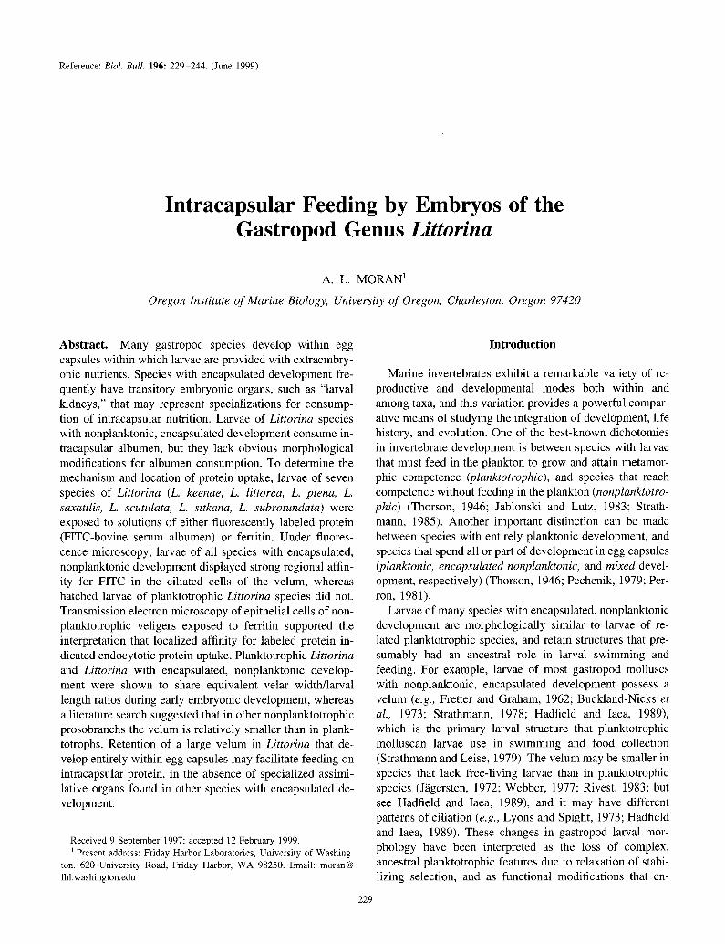
The gastropod genus Littorina, the periwinkle snails, contains - 19 species found in the high-shore zone through- out the northern Atlantic and Pacific oceans (Reid, 1989; Reid et al., 1996). The genus contains both planktotrophic and nonplanktotrophic species (see Reid, 1989, for review); molecular phylogenetic evidence supports planktotrophy as the ancestral state within the genus (see Rumbak et al., 1994). All species of Littorina undergo early development in egg capsules. Planktotrophic species have “mixed” de- velopment: early developmental stages are contained in complex pelagic capsules (Fig. lA), within which each larva is encased in an individual egg envelope (Fig. 1A) from which it emerges just prior to hatching from the capsule as a swimming, planktotrophic veliger. Other spe- cies lack a planktonic stage altogether, metamorphosing from veligers to juveniles within benthic or brooded egg masses. Egg capsules of these nonplanktotrophic species are filled with granular albumen (Fig. 1B) that larvae consume during development (Buckland-Nicks et cd., 1973). Litto- rina veligers have been reported to lack albumen-absorbing larval kidneys (Rivest, 1981; 1992), and the site and mech- anism of albumen consumption in encapsulated embryos and larvae of nonplanktotrophic Littorinu species have not been established.
The objectives of this study were to (1) determine the location of nutrient assimilation by larvae of encapsulated, nonplanktonic species in the gastropod genus Littorina; (2) investigate the extent to which congeneric planktotrophs share similar patterns of assimilation; and (3) compare the larval functional feeding morphologies of planktotrophic
Figure 1. (A) Planktonic egg capsule of Littorina scutulata, a species with planktotrophic development, containing three prehatching veliger-
stage larvae surrounded by individual egg envelopes. (B) Albumen-filled capsule of Littorina saxutilis, a species with encapsulated, nonplanktonic development. Capsule was removed from the oviduct of a gravid female.
al, albumen; c, capsule; 1, larva; en, egg envelope. Scale bar = 200 pm.
Littorina and their congeners that develop entirely within egg capsules.
Materials and Methods
Terminology
Because the words embryo and larva can have multiple interpretations in taxa with complex life histories, I use the terminology for early developmental stages as defined by McEdward and Janies (1993). Embryo refers to develop- mental stages from fertilized egg to gastrula; larva describes
INTRACAPSULAR FEEDING BY LZTTORZNA 231
any premetamorphic stage with recognizable larval features (e.g., the prototroch), regardless of whether stages are free- living or not.
Spawning and larval rearing
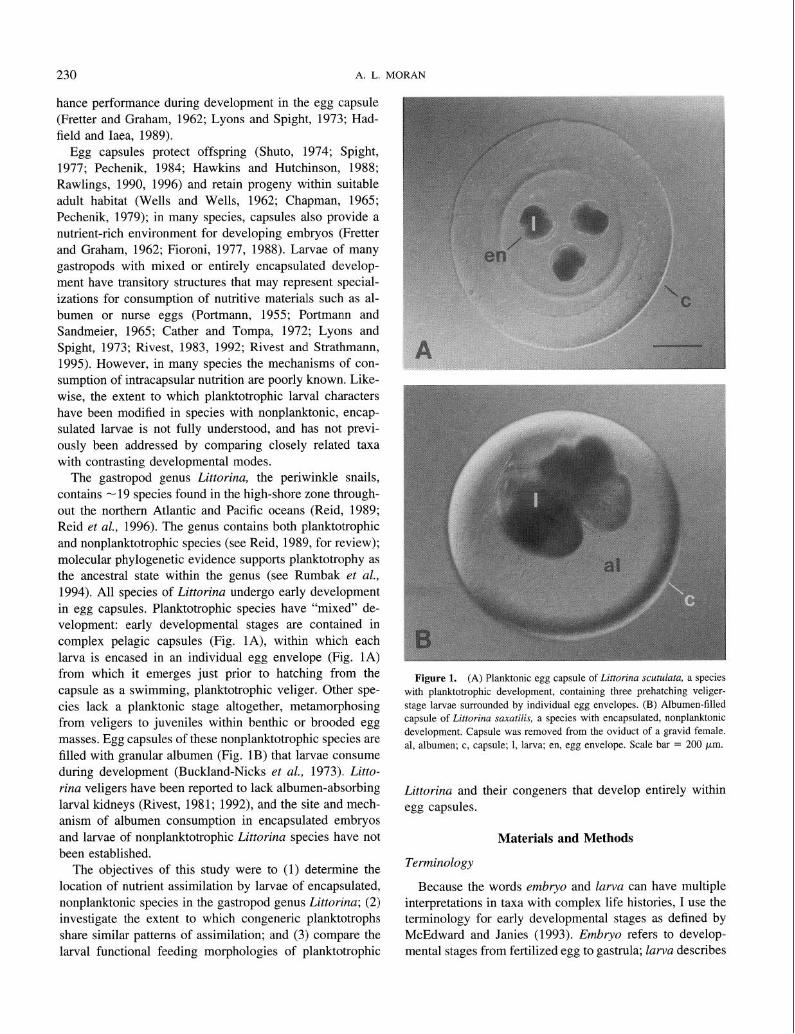
Seven Littorina species were used in these experiments: four planktotrophs (L. littorea (Linnaeus, 1758), L. keenae Rosewater, 1978, L. plena Gould 1849, L. scutulata Gould, 1849) and three species with encapsulated, nonplanktonic development (L. saxatilis (Olivi, 1792), L. sitkana Philippi, 1846, L. subrotundata (Carpenter, 1864)) (Table I). Egg capsules of planktotrophic species and species with benthic egg masses (L. sitkana, L. subrotundata) were obtained by placing live, freshly collected adult animals into mesh- walled containers (< 1 mm diameter mesh size), and im- mersing the containers in vigorously aerated seawater for 1 to 7 days. Egg capsules of the nonplanktotrophic species L. saxatilis, which broods larvae to metamorphosis in the oviduct, were obtained by cracking adult animals with nee- dle-nose pliers and removing capsules from the brood chamber.
Larvae of planktotrophic species were reared at concen- trations of about one larva per milliliter in 0.45 pm filtered seawater changed every 4 days. Planktotrophic larvae were fed ad libitum on a mixture of the single-celled algae Zsochrysis galbani (CCMP #1324 [T-ISO]) and Dunaliella tertiolecta (CCMP #1320 [DUN]). Egg masses of species with encapsulated, nonplanktonic development were main- tained in glass dishes of filtered (0.45 pm) seawater at 12°C.
Fluorescence microscopy
To test for regions of affinity for labeled albumen protein, embryos and larvae of three species with encapsulated, nonplanktonic development (L. saxatilis, L. sitkana, L. sub- rotundata) were removed from their capsules at develop-
mental stages from early cleavage to metamorphosis. Fine forceps were used to free embryos and larvae from capsules. Veliger larvae of four planktotrophic species (L. Zittorea, L. planaxis, L. plena, L. scutulata) were examined both before and after they hatched from the egg capsule. Because the egg envelope that surrounded earlier developmental stages of planktotrophs could not be removed without damage to developing larvae, early larval stages of one planktotrophic species (L. plena) were exposed to test solutions while they were still in the egg envelope.
Embryos and larvae were placed in solutions of bovine serum albumen labeled with fluoroscein isothiocyanate (FITC-BSA, Sigma #A-9771), a solution useful for demon- strating receptor-mediated endocytosis of proteins (Rivest, 1992; Rivest and Strathmann, 1995). FITC-BSA was also made in the laboratory from commercially available BSA and FITC (Sigma #F-7250) using the methods of Rivest (1981). To remove unconjugated FITC, FITC-BSA was dialyzed for 24 h against several changes of filtered (0.45 pm) seawater or treated with excess charcoal for 30 min (Rivest, 1981).
Embryos and larvae were placed in test solutions of 10-1000 pg/ml FITC-BSA in filtered seawater at 12°C for 15 min to 24 h, then rinsed in filtered seawater for periods ranging from 1 to 48 h. Controls were exposed to test solutions containing filtered seawater only, unlabeled BSA, or unconjugated FITC. Experimental and control embryos and larvae were examined with an Olympus epifluorescence microscope fitted with an FITC filter set (Omega Optics stock number XF23, excitation maximum 485 nm, emission 535 nm).
Transmission electron microscopy
To identify regions active in protein uptake, veligers of one species with encapsulated, nonplanktonic development
Table I
Species, development and collection information for Littorina utilized in this study
Species, Authority Mode’ Collection locality Collection habitat
Littorina Zittorea (Linnaeus, 1758)
Littorina keenae Rosewater, 1978 Littorina plena Gould, 1849
Littorina scutulata Gould, 1849
Littorina sax&is (Olivi, 1792)
Littorina sitkuna Philippi, 1846
Littorina subrotundata (Carpenter, 1864)
P
P
P P
NP
NP
NP
Woods Hole, MA Mystic, CT Monterey, CA
Charleston, OR Charleston, OR Monterey, CA
Woods Hole, MA Mystic, CT
Charleston, OR Friday Harbor, WA
Charleston, OR
Rocky shoreline
Rocky shoreline
Rocky shoreline Protected estuary Rocky shoreline
Rocky shoreline
Estuarine marsh
Rocky shoreline Estuarine marsh
r P = planktotrophic, NP = nonplanktotrophic.
232 A. L. MORAN
(L. sitkuna) were removed from their capsules and prepared for transmission electron microscopy, using the methods of Rivest and Strathmann (1995) with minor modifications. Larvae were placed for 10 min in a solution of 0.05% osmium tetroxide and 3% glutaraldehyde in 0.1 M (pH 7.35) phosphate buffer, with the osmolarity raised to 990 mOsM with sucrose. Next, larvae were placed in a solution of 3% glutaraldehyde in 0.1 M phosphate buffer with the osmo- larity raised to 990 mOsM with sucrose for 1 h, after which an equal volume of 10% EDTA was added (to dissolve the shell) and larvae were fixed for another hour. Larvae were postfixed for 1 h at room temperature in 2% osmium te- troxide in 1.25% sodium bicarbonate, then dehydrated in an ethanol series. Finally, specimens were exchanged in pro- pylene oxide, embedded in epoxy resin, and thin sections were cut on a Reichert Ultracut E ultramicrotome. Sections were picked up on Butvar films on 200-pm hex grids, stained with uranyl acetate and lead citrate (after Reynolds, 1963), and examined with a Philips CM 12 electron micro- scope.
To determine whether protein was assimilated into velar cells, some larvae were exposed to a solution of 1 mg/ml ferritin (Sigma catalog #F4503) in filtered seawater for 12 h and rinsed in seawater for 2 h prior to fixation. Ferritin is an electron-dense protein useful as a marker for endocytosis (Rivest, 1981). Five ferritin-exposed larvae and four con- trols were examined with transmission electron microscopy.
Measurement of velar aspect ratios
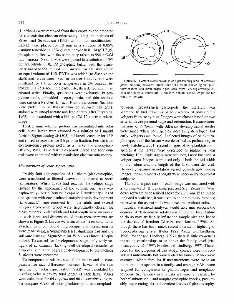
Freshly laid egg capsules of L. plena (planktotrophic) were transferred to filtered seawater and reared at room temperature. When larvae had reached the veliger stage (judged by the appearance of the velum), one larva was haphazardly chosen from each capsule. Brooded capsules of one species with encapsulated, nonplanktonic development (L. suxatilis) were removed from the adult, and several veligers from each brood were haphazardly chosen for measurements. Velar width and total length were measured on each larva, and dimensions of these measurements are shown in Figure 2. Larvae were traced with a camera lucida attached to a compound microscope, and measurements were made using a SummaSketch II digitizing pad and the software package SigmaScan for Windows (Jandel Corpo- ration). To control for developmental stage, only early ve- ligers of L. suxatilis (lacking well-developed tentacles or propodia, similar in degree of development to prehatching L. plena) were measured.
To compare the relative size of the velum and to com- pensate for size differences between larvae of the two species, the “velar aspect ratio” (VAR) was calculated by dividing velar width by total length of each larva. VARs were calculated for 64 L. plena and 60 L. saxatilis veligers. To compare VARs of other planktotrophic and nonplank-
,en .
I I I I
Figure 2. Camera lucida drawings of a prehatching larva of Littorina plena indicating measured dimensions: velar width (left on figure; apical
view of larva) and larval length (right; lateral view). en, egg envelope; cil, cilia of velum; o, operculum; s, shell; v, velum). Larval length bar (on right) = 110 pm.
totrophic prosobranch gastropods, the literature was searched to find drawings or photographs of prosobranch veligers from many taxa. Images were chosen based on two criteria, developmental stage and orientation. Because com- parisons of Littorina with different developmental modes were made when both species were fully developed, but early, veligers (see above), I selected images of planktotro- phic species if the larvae were described as prehatching or newly hatched, and I rejected images of nonplanktotrophic species if the larvae were described as mature or near hatching. If multiple stages were pictured, I used the earliest veliger stage. Images were used only if both the full width of the velum and the length of the larva were depicted. However, because orientation varied considerably among images, measurements of length were necessarily somewhat subjective.
The velar aspect ratio of each image was measured with a SummaSketch II digitizing pad and SigmaScan for Win- dows software as described above for Littorina. If an image included a scale bar, it was used to calibrate measurements; otherwise, the aspect ratio was measured without units.
Ideally, statistical analyses would take into account the degrees of phylogenetic relatedness among all taxa; failure to do so may artificially inflate the sample size and hence the degrees of freedom (Martins and Hansen, 1996). Al- though there has been much recent interest in higher gas- tropod phylogeny (e.g., Bieler, 1992; Ponder and Lindberg, 1996; Ponder and Lindberg, 1997), there is little consensus regarding relationships at or above the family level (Ha- rasewych et al., 1997; Ponder and Lindberg, 1997). There- fore, for the purposes of this study, species were not con- sidered individually but were sorted by family. VARs were averaged within families if measurements were made on more than one species in a family, and average VARs were graphed for comparison of planktotrophs and nonplank- totrophs. Six families in this data set were represented by both planktotrophic and nonplanktotrophic species, presum- ably representing six independent losses of planktotrophy.
INTRACAPSULAR FEEDING BY LI7-TORINA 233
To compare velar sizes among developmental modes, mean VARs of planktotrophs and nonplanktotrophs of these six families were compared by using a paired Student’s t test.
Measurement of cilia length
To determine whether ciliary length and growth differed between species with contrasting developmental modes, freshly spawned egg capsules of three planktotrophs (L. keenae, L. plena, L. scutulata) and egg masses of one species with encapsulated, nonplanktonic development (L. sitkana) were collected, transferred to filtered seawater, and reared at 12°C. Planktotrophs were observed from first cleavage through hatching; they were initially examined every 2-3 h and at intervals of about 1 d or longer in later development. The nonplanktotrophs, which developed more slowly, were observed from first cleavage through reaching a fully formed veliger; they were initially examined daily and during later development at intervals of several days. Larvae were immobilized under a coverslip and examined on a compound microscope equipped with a camera lucida. The cilia were drawn at their greatest extensions and the images were then digitized (as described for velar aspect ratios) to measure greatest length. In addition, veligers of L. saxatilis were removed from their capsules and placed in seawater to determine whether larvae could swim.
Results
Fluorescence microscopy
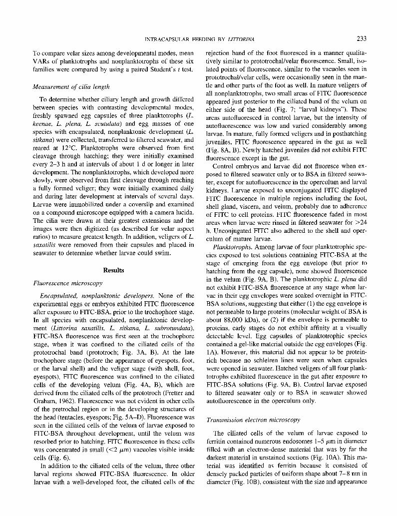
Encapsulated, nonplanktonic developers. None of the experimental eggs or embryos exhibited FITC fluorescence after exposure to FITC-BSA, prior to the trochophore stage. In all species with encapsulated, nonplanktonic develop- ment (Littorina saxatilis, L. sitkana, L. subrotundata), FITC-BSA fluorescence was first seen at the trochophore stage, when it was confined to the ciliated cells of the prototrochal band (prototroch; Fig. 3A, B). At the late trochophore stage (before the appearance of eyespots, foot, or the larval shell) and the veliger stage (with shell, foot, eyespots), FITC fluorescence was confined to the ciliated cells of the developing velum (Fig. 4A, B), which are derived from the ciliated cells of the prototroch (Fretter and Graham, 1962). Fluorescence was not evident in other cells of the pretrochal region or in the developing structures of the head (tentacles, eyespots; Fig. 5A-D). Fluorescence was seen in the ciliated cells of the velum of larvae exposed to FITC-BSA throughout development, until the velum was resorbed prior to hatching. FITC fluorescence in these cells was concentrated in small (<2 pm) vacuoles visible inside cells (Fig. 6).
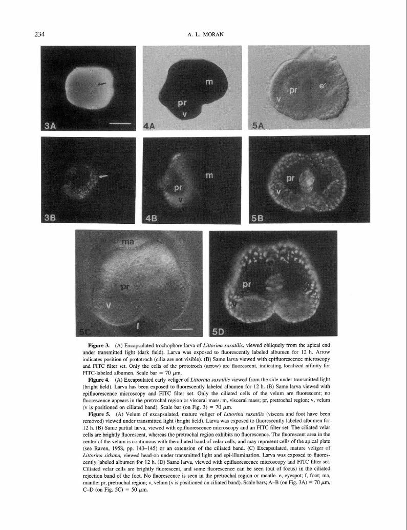
In addition to the ciliated cells of the velum, three other larval regions showed FITC-BSA fluorescence. In older larvae with a well-developed foot, the ciliated cells of the
rejection band of the foot fluoresced in a manner qualita- tively similar to prototrochal/velar fluorescence. Small, iso- lated points of fluorescence, similar to the vacuoles seen in prototrochal/velar cells, were occasionally seen in the man- tle and other parts of the foot as well. In mature veligers of all nonplanktotrophs, two small areas of FITC fluorescence appeared just posterior to the ciliated band of the velum on either side of the head (Fig. 7; “larval kidneys”). These areas autofluoresced in control larvae, but the intensity of autofluorescence was low and varied considerably among larvae. In mature, fully formed veligers and in posthatching juveniles, FITC fluorescence appeared in the gut as well (Fig. 8A, B). Newly hatched juveniles did not exhibit FITC fluorescence except in the gut.
Control embryos and larvae did not fluoresce when ex- posed to filtered seawater only or to BSA in filtered seawa- ter, except for autofluorescence in the operculum and larval kidneys. Larvae exposed to unconjugated FITC displayed FITC fluorescence in multiple regions including the foot, shell gland, viscera, and velum, probably due to adherence of FITC to cell proteins. FITC fluorescence faded in most areas when larvae were rinsed in filtered seawater for >24 h. Unconjugated FITC also adhered to the shell and oper- culum of mature larvae.
Planktotrophs. Among larvae of four planktotrophic spe- cies exposed to test solutions containing FITC-BSA at the stage of emerging from the egg envelope (but prior to hatching from the egg capsule), none showed fluorescence in the velum (Fig. 9A, B). The planktotrophic L. plena did not exhibit FITC-BSA fluorescence at any stage when lar- vae in their egg envelopes were soaked overnight in FITC- BSA solutions, suggesting that either (1) the egg envelope is not permeable to large proteins (molecular weight of BSA is about 88,000 kDa), or (2) if the envelope is permeable to proteins, early stages do not exhibit affinity at a visually detectable level. Egg capsules of planktotrophic species contained a gel-like material outside the egg envelopes (Fig. 1A). However, this material did not appear to be protein- rich because no schleiren lines were seen when capsules were opened in seawater. Hatched veligers of all four plank- totrophs exhibited fluorescence in the gut after exposure to FITC-BSA solutions (Fig. 9A, B). Control larvae exposed to filtered seawater only or to BSA in seawater showed autofluorescence in the operculum only.
Transmission electron microscopy
The ciliated cells of the velum of larvae exposed to ferritin contained numerous endosomes l-5 pm in diameter filled with an electron-dense material that was by far the darkest material in unstained sections (Fig. 10A). This ma- terial was identified as ferritin because it consisted of densely packed particles of uniform shape about 7-8 nm in diameter (Fig. lOB), consistent with the size and appearance
234 A. L. MORAN
Figure 3. (A) Excapsulated trochophore larva of Littorina saxutilis, viewed obliquely from the apical end under transmitted light (dark field). Larva was exposed to fluorescently labeled albumen for 12 h. Arrow indicates position of prototroch (cilia are not visible). (B) Same larva viewed with epifluorescence microscopy
and FITC filter set. Only the cells of the prototroch (arrow) are fluorescent, indicating localized affinity for FITC-labeled albumen. Scale bar = 70 pm.
Figure 4. (A) Excapsulated early veliger of Littorina suxatilis viewed from the side under transmitted light
(bright field). Larva has been exposed to fluorescently labeled albumen for 12 h. (B) Same larva viewed with
epifluorescence microscopy and FITC filter set. Only the ciliated cells of the velum are fluorescent; no fluorescence appears in the pretrochal region or visceral mass. m, visceral mass; pr, pretrochal region; v, velum (v is positioned on ciliated band). Scale bar (on Fig. 3) = 70 ym.
Figure 5. (A) Velum of excapsulated, mature veliger of Littorina saxutilis (viscera and foot have been
removed) viewed under transmitted light (bright field). Larva was exposed to fluorescently labeled albumen for 12 h. (B) Same partial larva, viewed with epifluorescence microscopy and an FITC filter set. The ciliated velar
cells are brightly fluorescent, whereas the pretrochal region exhibits no fluorescence. The fluorescent area in the center of the velum is continuous with the ciliated band of velar cells, and may represent cells of the apical plate
(see Raven, 1958, pp. 143-145) or an extension of the ciliated band. (C) Excapsulated, mature veliger of Littorina sit/cam, viewed head-on under transmitted light and epi-illumination. Larva was exposed to fluores- cently labeled albumen for 12 h. (D) Same larva, viewed with epifluorescence microscopy and FITC filter set.
Ciliated velar cells are brightly fluorescent, and some fluorescence can be seen (out of focus) in the ciliated rejection band of the foot. No fluorescence is seen in the pretrochal region or mantle. e, eyespot; f, foot; ma, mantle; pr, pretrochal region; v, velum (v is positioned on ciliated band). Scale bars; A-B (on Fig. 3A) = 70 Km,
C-D (on Fig. 5C) = 50 pm.
INTRACAPSULAR FEEDING BY LITTORINA
Figure 6. High-magnification view of velum of mature Littorina suxutilis larva viewed with epifluorescence microscopy (FITC filter set). Larva was exposed to fluorescently labeled albumen for 12 h. Fluorescence appears
in the ciliated band of velar cells but not the pretrochal region. Fluorescence is concentrated in small (<2 pm) vacuoles within the prototrochal cells. pr, pretrochal region; v, velum (positioned on ciliated band). Scale bar = 20 km.
Figure 7. Veliger of Littorina saxatilis, exposed to FITC-BSA and viewed laterally under fluorescent light with FITC filter set. The orientation of this larva is similar to Fig. 3. Immediately posterior to the ciliated band
is one of two symmetrically positioned fluorescent regions (larval kidneys) that were found in fully formed veligers. k, larval kidney; v, velum. Scale bar = 75 pm.
Figure 8. (A) Mature veliger of Littorina saxatilis, close to hatching, viewed under transmitted light. Larva
was exposed to fluorescently labeled albumen for 12 h. (B) Same larva viewed under fluorescent light with FITC filter set. Fluorescence can be seen in the ciliated cells of the velum, the autofluorescent operculum, and in a short
region of the gut. The dim illumination of the foot is due to light reflected from the operculum. f, foot; g, gut; o, operculum; s, shell; u, umbilicus; v, velum. Scale bar = 150 pm.
Figure 9. (A) Newly hatched veliger of Littorina plena (planktotroph), viewed under transmitted light.
Larva was exposed to fluorescently labeled albumen for 12 h. (B) Same larva viewed under fluorescent light with FITC filter set, showing strong FITC fluorescence in the gut. The operculum is autofluorescent, and the foot is
dimly illuminated by light reflected from the operculum. Note absence of fluorescence in the velum. f, foot; g, gut; o, operculum; pc, prototrochal cilia; s, shell; v, velum. Scale bar = 25 pm.
235
236 A. L. MORAN
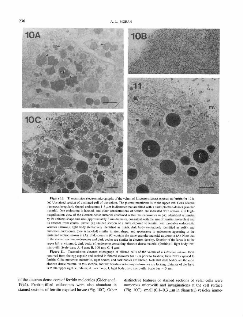
Figure 10. Transmission electron micrographs of the velum of Littorina sitkana exposed to ferritin for 12 h.
(A) Unstained section of a ciliated cell of the velum. The plasma membrane is to the upper left. Cells contain numerous irregularly shaped endosomes l-5 pm in diameter that are filled with a dark (electron-dense) granular material. One endosome is labeled, and other concentrations of ferritin are indicated with arrows. (B) High-
magnification view of the electron-dense material contained within the endosomes in (A), identified as ferritin by its uniform shape and size (approximately 8 nm diameter, consistent with the size of ferritin molecules) and its absence from control larvae. (C) Stained section of a larva exposed to ferritin, with probable endocytotic
vesicles (arrows), light body (tentatively identified as lipid), dark body (tentatively identified as yolk), and numerous endosomes (one is labeled) similar in size, shape, and appearance to endosomes appearing in the
unstained section shown in (A). Endosomes in (C) contain the same granular material as those in (A). Note that in the stained section, endosomes and dark bodies are similar in electron density. Exterior of the larva is to the upper left. c, cilium; d, dark body; ef, endosome containing electron-dense material (ferritin); 1, light body; mv,
microvilli. Scale bars; A, 4 ym; B, 100 nm; C, 4 pm. Figure 11. Transmission electron micrograph of ciliated cells of the velum of a Littorina sitkana larva
removed from the egg capsule and soaked in filtered seawater for 12 h prior to fixation; larva NOT exposed to ferritin. Cilia, numerous microvilli, light bodies, and dark bodies are labeled. Note that dark bodies are the most
electron-dense material in this section, and that ferritin-containing endosomes are lacking. Exterior of the larva is to the upper right. c, cilium; d, dark body; 1, light body; mv, microvilli. Scale bar = 3 pm.
of the electron-dense core of ferritin molecules (Gider et al., distinctive features of stained sections of velar cells were 1995). Ferritin-filled endosomes were also abundant in numerous microvilli and invaginations at the cell surface stained sections of ferritin-exposed larvae (Fig. 1OC). Other (Fig. lOC), small (0.1-0.3 pm in diameter) vesicles imme-
INTRACAPSULAR FEEDING BY LIZTORINA 237
diately inside the cell surface (Fig. lOC), “light bodies” tentatively identified as lipid, and “dark bodies” tentatively identified as yolk on the basis of the lack of a surrounding membrane (tiig. 1OC).
Ciliated velar cells of larvae not exposed to ferritin con- tained many similar features, including surface cilia and microvilli and light and dark bodies (Fig. 11). Sections of control larvae were distinguished by the absence of ferritin- filled endosomes; ferritin or ferritin-like electron-dense ma- terial was not seen in velar sections of any of four control larvae examined.
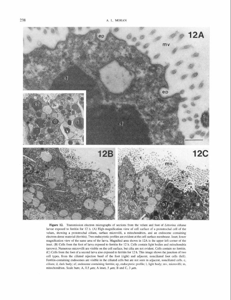
At higher magnification, the cell membrane of ciliated velar cells of larvae exposed to ferritin appeared to be endocytotically active. Probable endocytotic profiles were evident in the plasma membrane (Fig. 12A), and ferritin- filled endosomes appeared immediately inside the cells (Fig. 12A, 12A inset). Nonciliated epithelial cells of ferritin- exposed larvae shared some features with ciliated cells of the velum and foot, including the presence of microvilli and light bodies. However, ferritin was not present in these cells (Fig. 12B) even when ciliated and nonciliated cells were immediately adjacent to each other, as in ciliated and non- ciliated areas of the foot (Fig. 12C).
Velar aspect ratios
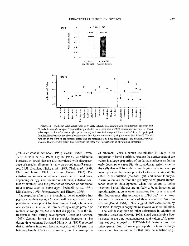
The velar aspect ratios (VARs) of early Littorina saxatilis (nonplanktotroph) veligers were not significantly different from the velar aspect ratio of prehatching L. plena (plank- totroph) veligers (Student’s two-sample t test, P = 0.77; Fig. 13A). The VARs of planktotrophs from the literature were generally greater than VARs of confamilial nonplank- totrophs, although there was considerable overlap (Table II; Fig. 13B). VARs of planktotrophs were significantly greater than nonplanktotrophs when average VARs were compared among planktotrophs and nonplanktotrophs from within six families (Fig. 13B; paired Student’s t test, P = 0.02).
Cilia length
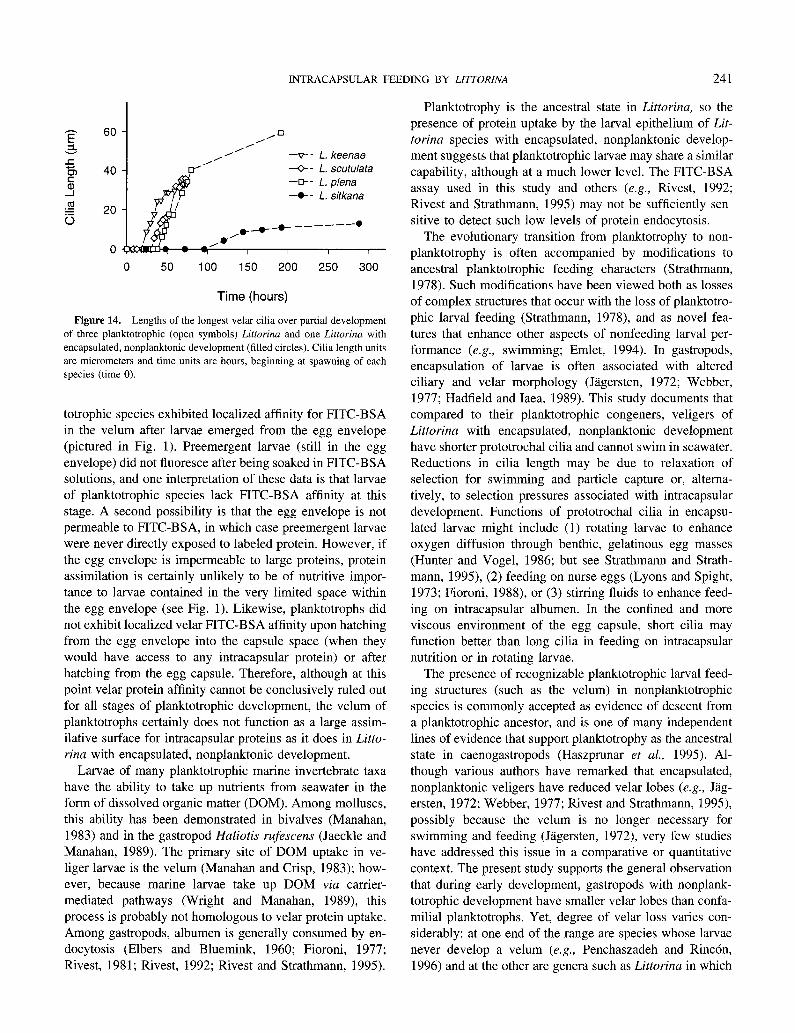
Velar cilia of planktotrophic Littorina larvae grew faster than cilia of L. sitkana, and reached a much greater total length (Fig. 14). Observations of cilia length in additional species with encapsulated, nonplanktonic development (L. saxatilis, L. subrotundata) were consistent with this pattern. The velum of these species bore cilia that appeared to be simple rather than compound, and when removed from their egg capsules, veligers of these species rotated on the bottom of the dish but did not swim.
Discussion
Two lines of evidence indicate that the ciliated cells of the velum and foot of Littorina with encapsulated, non- planktonic development are active in localized uptake of
capsular proteins. First, these cells fluoresced brightly after exposure to fluorescently labeled albumen, clearly indicat- ing regional affinity for labeled protein. Affinity strongly suggested uptake, because fluorescently labeled vacuoles were interspersed with cell structures such as lipid droplets in optical sections. Second, ciliated cells of the velum and rejection band of the foot of larvae exposed to ferritin solutions contained large amounts of electron-dense mate- rial identifiable as ferritin by the size of molecules, whereas adjacent areas of the foot did not contain ferritin; likewise, no ferritin was seen in control larvae. The absence of ferritin from nonciliated cells of the foot (which did not fluoresce after exposure to FITC-BSA) further supports the hypothe- sis that regional FITC-BSA fluorescence indicates localized uptake of intracapsular albumen.
Other larval structures have been associated with the endocytotic uptake of capsular proteins by gastropods. These include the larval kidneys of many prosobranch spe- cies (Rivest, l992), the “pedal cell complex” of neritoideans (Rivest and Strathmann, 199.5), and the podocyst of some pulmonates (Cather and Tompa, 1972), all of which assim- ilate protein via receptor-mediated endocytosis (Cather and Tompa, 1972; Rivest, 1981, 1992; Rivest and Strathmann, 1995). Structures such as the pedal cell complex and larval kidneys are unique to encapsulated larval forms, and in species with mixed development these structures are resorbed before or soon after hatching (Rives& 1992; Rivest and Strathmann, 1995). Thus, it has been proposed that such structures may have evolved to facilitate intracapsular de- velopment by enhancing capsular nutrient uptake (Rivest, 1992; Rivest and Strathmann, 1995) or respiration (Cather and Tompa, 1972; Rivest and Strathmann, 1995). The ve- lum, in contrast, is an ancestral structure that is present in all Littorina and functions in swimming and particle capture in planktotrophic species; therefore, intracapsular protein up- take by the velum of nonplanktotrophic Littorina likely represents an exaptation of an ancestral character to the evolutionarily novel (within the genus) condition of devel- oping to metamorphosis in the egg capsule.
In many taxa, egg capsules provide a nutrient-rich envi- ronment for developing embryos (Rivest and Strathmann, 1995), in addition to other functions such as protecting offspring from environmental stress and predation (Shuto, 1974; Spight, 1977; Pechenik, 1984; Hawkins and Hutchin- son, 1988; Rawlings, 1990, 1996) and retaining progeny within suitable adult habitat (Wells and Wells, 1962; Chap- man, 1965; Pechenik, 1979). Albumen is a common nutrient source among gastropods, and in many taxa albumen is rich in proteins, carbohydrates, or free amino acids (Hiirstmann, 1956; De Mahieu et al., 1974; Rivest, 1992; Stbckmann- Bosbach and Althoff, 1989; Penchaszadeh and Rincbn, 1996). Albumen has been implicated in larval nutrition through a decline in nutrient content of the fluid during development, and by a correlated increase in larval lipid and
238 A. L. MORAN
Figure 12. Transmission electron micrographs of sections from the velum and foot of Littorina sitkuna larvae exposed to ferritin for 12 h. (A) High-magnification view of cell surface of a prototrochal cell of the
velum, showing a prototrochal cilium, surface microvilli, a mitochondrion, and an endosomc containing electron-dense material (ferritin). Two endocytotic profiles are evident at the cell surface membrane. Inset; lower
magnification view of the same area of the larva. Magnified area shown in 12A is the upper left corner of the inset. (B) Cells from the foot of larva exposed to ferritin for 12 h. Cells contain light bodies and mitochondria
(arrows). Numerous microvilli are visible on the cell surface, but cilia are not evident. Cells contain no ferritin. (C) Cells from the foot of a second larva also exposed to ferritin for 12 h. This image shows the junction of two
cell types, from the ciliated rejection band of the foot (right) and adjacent, nonciliated foot cells (left). Ferritin-containing endosomes are visible in the ciliated cells but are not seen in adjacent, nonciliated cells. c,
cilium; d, dark body; ef, endosome containing ferritin; ep, endocytotic profile; 1, light body; mv, microvilli; m, mitochondrion. Scale bars; A, 0.5 pm; A inset, 5 pm; B and C, 3 pm.
INTRACAPSULAR FEEDING BY LZlTORZNA 239
13A 1.2 p = 0.77. n = 124
.s 1.0 x=0.83 x = 0.82
2 s 0.6
$ 0 0.6
3 9 0.4
0.2
L. p/em L. saxatilis (planktotroph) (nonplanktotroph)
1.4 - 13 B I
Figure 13. (A) Mean velar aspect ratios of 64 early veligers of Littorina plena (planktotroph, open bar) and 60 early L. samtilis veligers (nonplanktotroph; shaded bar). Error bars are 95% confidence intervals. (B) Mean
velar aspect ratios of planktotrophs (open circles) and nonplanktotrophs (closed circles) from 21 gastropod families. Error bars are not shown because most families are represented by single species (see Table I). The six
families to the right of the vertical dotted line are represented by both planktotrophic and nonplanktotrophic species. The horizontal dotted line represents the mean velar aspect ratio of all families combined.
protein content (Horstmann, 1956; Morrill, 1964; Raven, 1972; Morrill et al., 1976; Taylor, 1983). Considerable increases in larval size are also correlated with disappear- ance of capsular albumen in many gastropod taxa (Rasmus- sen, 1951; Buckland-Nicks et al., 1973; Clark et al., 1979; Clark and Jensen, 1981; Losse and Greven, 1993). The nutritive importance of albumen varies in different taxa, depending on egg size, volume of albumen, nutritive con- tent of albumen, and the presence or absence of additional food sources such as nurse eggs (Pechenik et al., 1984; Miloslavich, 1996; Penchaszadeh and Rincbn, 1996).
Intracapsular albumen is thought to be of nutritive im- portance to developing Littorina with encapsulated, non- planktonic development for two reasons. First, albumen of one species, L. saxatilis, is dominated by a single protein of molecular weight 80,000 kDa that disappears from the in- tracapsular fluid during development (Losse and Greven, 1993). Second, larvae of these species increase in size during development; Buckland-Nicks et al. (1973) reported that L. sitkana increases from an egg size of 175 pm to a hatching length of 575 pm, presumably due to consumption
of albumen. Velar albumen assimilation is likely to be important to larval nutrition, because the surface area of the velum is a large proportion of the larval surface area during early development (see Fig. 4). In addition, assimilation by the cells that will form the velum begins early in develop- ment, prior to the development of other structures impli- cated in assimilation (the foot, gut, and larval kidneys). Assimilation via the foot and gut may be of greater impor- tance later in development, when the velum is being resorbed. Larval kidneys are unlikely to be as important in protein assimilation as other structures; their small size and dim fluorescence after exposure to FITC-BSA, which may account for previous reports of their absence in Littorina sitkuna (Rivest, 1981, 1992), suggests that assimilation by the larval kidneys is negligible relative to velar assimilation.
The velum may take up other substances in addition to proteins. Losse and Greven (1993) noted considerable fluo- rescence in the gut, hepatopancreas, and velum of L. su.xu- tilis veligers exposed to FITC-labeled dextrans, and the intracapsular fluid of some gastropods contains carbohy- drates and free amino acids that may be nutritive (e.g.,
240 A. L. MORAN
Table II
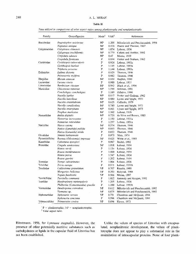
Taxa utilized in comparisons of velar aspect ratios among planktotrophs and nonplanktotrophs
Family Genus/Species Mode’ VAR’ References
Buccinidae
Calyptraeidae
Cerithiidae
Eulimidae
Harpidae Lacunidae Littorinidae
Muricidae
Nassariidae
Naticidae
Olvulidae Pyramidellidae
Ranellidae Rissoidae
Tomidae Triviidae
Trochidae
Turritellidae Turridae
Vermetidae
Siphonariidae
Trimusculidae
Engoniophos unicinctus Neptunea antiqua
Calyptraea chinensis Calyptraea trochijormis
Crepidula adunca Crepidula fornicata Cerithiopsis tubercularis
Cerithiopsis barleei Triphora perversa Eulima distorta
Pelseneeria sty&era Morum onuscus Lacuna vincta
Bembicium vittatum Chicoreous ramosus Concholepas concholepas
Nucella lapilus Nucella lamellosa Nucella crassilabrum
Nucella canaliculata Nucella emarginata
Trophon muricatus Bullia digitalis Nassarius incrassatus
Nassarius reticulatus Natica catena Natica (Lunatida) pallida
Natica (Lunatida) nitida Simnia barbarensis
Boonea (Odostomia) impressa Cabestana spengleri Cingula semicostata
Rissoa sarsii Rissoa membranacea Rissoa parva
Rissoa guerini Tornus subcarinatus Trivia europa
Calliostoma granulatum Margarites helicinus
Tegula funebratis Turritella communis Haedropleura septangularis
Philbertia (Comarmondia) gracilis Dendropoma corrodens
Vermetus sp. Siphonaria serrata Siphonaria concinna
Trimusculus conica
NP
NP NP NP
NP P P
P P P
P NP
P NP NP P
NP NP
NP NP
NP NP NP
P P
NP
NP P
P NP P
NP P P
P P P
P NP NP
NP P
P P
NP NP NP
P NP
1.205 Miloslavich and Penchaszadeh, 1994
0.418 Pearce and Thorson, 1967
1.074 Lebour, 1936 0.779 Cahete and Ambler, 1992
0.47 Moritz, 1939 0.834 Fretter and Graham, 1962 0.924 Lebour, 1993a
1.135 Lebour, 1993a 1.144 Lebour, 1993a
0.829 Thorson, 1946
0.982 Thorson, 1946 0.839 Hughes, 1990
0.900 Lebour, 1937
0.943 Black et al., 1994 1.599 Soliman, 1991
1.169 DiSalvo, 1988
0.817 Fretter and Graham, 1962 0.880 Lyons and Spight, 1973
0.625 Gallardo, 1979
0.780 Lyons and Spight, 1973
0.861 Lyons and Spight, 1973
1.102 Lebour, 1936 0.720 da Silva and Brown, 1985
1.150 Lebour, 1931a
1.257 Lebour, 1931a
0.574 Thorson, 1946
0.843 Thorson, 1946
0.822 Thorson, 1946
0.973 Main, 1974
0.825 White et al., 1985 0.987 Reidel, 1992
1.018 Lebour, 1934 1.134 Lebour, 1934
1.000 Lebour, 1934
1.247 Lebour, 1934 1.202 Lebour, 1934
1.064 Lebour, 1936
0.971 Lebour, 1931b 0.707 Ramon, 1990
0.393 Holyoak, 1988
0.566 Moran, 1997
1.022 Kennedy and Keegan, 1992
1.205 Lebour, 1936
1.380 Lebour, 1993b
0.612 Miloslavich and Penchaszadeh, 1992
0.875 Miloslavich and Penchaszadeh, 1992 0.731 Chambers and McQuaid, 1994
0.996 Chambers and McQuaid, 1994 0.696 Haven, 1973 -
r P = planktotrophic; NP = nonplanktotrophic. ’ Velar aspect ratio.
Horstmann, 1956, for Lyrnnaea stugnalis). However, the presence of other potentially nutritive substances such as carbohydrates or lipids in the capsular fluid of Littorina has not been established.
Unlike the velum of species of Littorina with encapsu- lated, nonplanktonic development, the velum of plank- totrophs does not appear to play a substantial role in the assimilation of intracapsular proteins. None of four plank-
INTRACAPSULAR FEEDING BY LZ7TORZNA 241
-is 60 ,a
0
A’ 1’
1’ -v-- L. keenae
d
+- L. scutulata -D- L. plena
7% -*- L. sitkana
&. _ /* /---------”
I I I I
0 50 100 150 200 250 300
Time (hours)
Figure 14. Lengths of the longest velar cilia over partial development of three planktotrophic (open symbols) Littorina and one Littorina with encapsulated, nonplanktonic development (filled circles). Cilia length units
are micrometers and time units are hours, beginning at spawning of each species (time 0).
totrophic species exhibited localized affinity for FITC-BSA in the velum after larvae emerged from the egg envelope (pictured in Fig. 1). Preemergent larvae (still in the egg envelope) did not fluoresce after being soaked in FITC-BSA solutions, and one interpretation of these data is that larvae of planktotrophic species lack FITC-BSA affinity at this stage. A second possibility is that the egg envelope is not permeable to FITC-BSA, in which case preemergent larvae were never directly exposed to labeled protein. However, if the egg envelope is impermeable to large proteins, protein assimilation is certainly unlikely to be of nutritive impor- tance to larvae contained in the very limited space within the egg envelope (see Fig. 1). Likewise, planktotrophs did not exhibit localized velar FITC-BSA affinity upon hatching from the egg envelope into the capsule space (when they would have access to any intracapsular protein) or after hatching from the egg capsule. Therefore, although at this point velar protein affinity cannot be conclusively ruled out for all stages of planktotrophic development, the velum of planktotrophs certainly does not function as a large assim- ilative surface for intracapsular proteins as it does in Litto- rina with encapsulated, nonplanktonic development.
Larvae of many planktotrophic marine invertebrate taxa have the ability to take up nutrients from seawater in the form of dissolved organic matter (DOM). Among molluscs, this ability has been demonstrated in bivalves (Ma&ran, 1983) and in the gastropod Haliotis rufescens (Jaeckle and Manahan, 1989). The primary site of DOM uptake in ve- liger larvae is the velum (Manahan and Crisp, 1983); how- ever, because marine larvae take up DOM via carrier- mediated pathways (Wright and Manahan, 1989), this process is probably not homologous to velar protein uptake. Among gastropods, albumen is generally consumed by en- docytosis (Elbers and Bluemink, 1960; Fioroni, 1977; Rivest, 1981; Rivest, 1992; Rivest and Strathmann, 1995).
Planktotrophy is the ancestral state in Littorina, so the presence of protein uptake by the larval epithelium of Lit- torina species with encapsulated, nonplanktonic develop- ment suggests that planktotrophic larvae may share a similar capability, although at a much lower level. The FITC-BSA assay used in this study and others (e.g., Rivest, 1992; Rivest and Strathmann, 1995) may not be sufficiently sen- sitive to detect such low levels of protein endocytosis.
The evolutionary transition from planktotrophy to non- planktotrophy is often accompanied by modifications to ancestral planktotrophic feeding characters (Strathmann, 1978). Such modifications have been viewed both as losses of complex structures that occur with the loss of planktotro- phic larval feeding (Strathmann, 1978), and as novel fea- tures that enhance other aspects of nonfeeding larval per- formance (e.g., swimming; Emlet, 1994). In gastropods, encapsulation of larvae is often associated with altered ciliary and velar morphology (Jagersten, 1972; Webber, 1977; Hadfield and Iaea, 1989). This study documents that compared to their planktotrophic congeners, veligers of Littorina with encapsulated, nonplanktonic development have shorter prototrochal cilia and cannot swim in seawater. Reductions in cilia length may be due to relaxation of selection for swimming and particle capture or, altema- tively, to selection pressures associated with intracapsular development. Functions of prototrochal cilia in encapsu- lated larvae might include (1) rotating larvae to enhance oxygen diffusion through benthic, gelatinous egg masses (Hunter and Vogel, 1986; but see Strathmann and Strath- mann, 1995), (2) feeding on nurse eggs (Lyons and Spight, 1973; Fioroni, 1988), or (3) stirring fluids to enhance feed- ing on intracapsular albumen. In the confined and more viscous environment of the egg capsule, short cilia may function better than long cilia in feeding on intracapsular nutrition or in rotating larvae.
The presence of recognizable planktotrophic larval feed- ing structures (such as the velum) in nonplanktotrophic species is commonly accepted as evidence of descent from a planktotrophic ancestor, and is one of many independent lines of evidence that support planktotrophy as the ancestral state in caenogastropods (Haszprunar et al., 1995). Al- though various authors have remarked that encapsulated, nonplanktonic veligers have reduced velar lobes (e.g., Jag- ersten, 1972; Webber, 1977; Rivest and Strathmann, 1995), possibly because the velum is no longer necessary for swimming and feeding (Jlgersten, 1972), very few studies have addressed this issue in a comparative or quantitative context. The present study supports the general observation that during early development, gastropods with nonplank- totrophic development have smaller velar lobes than confa- milial planktotrophs. Yet, degree of velar loss varies con- siderably: at one end of the range are species whose larvae never develop a velum (e.g., Penchaszadeh and Rincbn, 1996) and at the other are genera such as Littorina in which
242 A. L. MORAN
planktotrophs and nonplanktotrophs have velums that are proportionally equivalent in early development. Retention of the velum may be attributed to several causes, including morphogenetic or other developmental constraints and an evolutionarily recent loss of planktotrophy. However, the lack of a velum in some species (e.g., Penchaszadeh and Rincon, 1996) argues that the presence of velar lobes may not be necessary for prosobranch morphogenesis, and the second hypothesis has yet to be tested in a comparative, historical context. A third possibility is that nonplank- totrophs that retain a large velum do so in part because the velum has been coopted to perform specific functions in the egg capsule, such as feeding on nurse eggs (e.g., Fioroni and Sandmeier, 1964) or providing a respiratory surface (Fretter and Graham, 1962). In Littorina with encapsulated, non- planktonic development the velum performs a third and previously undescribed function, assimilation of intracapsu- lar albumen. A large velum may be an advantage because it provides a large surface for assimilation of capsule proteins.
In summary, the velum of Littorina species with encap- sulated, nonplanktonic development is active in endocytotic albumen assimilation. In contrast, affinity for labeled albu- men was not seen in the velum of planktotrophic Littorina species. This suggests that, since their loss of planktotrophy, the former species have undergone evolutionary changes that enhance assimilation of intracapsular nutrients. The feeding morphologies of Littorina veligers differ in some ways, depending on developmental mode: species with en- capsulated, nonplanktonic development have prototrochal cilia that are much shorter and grow more slowly, and the larvae cannot swim when removed from the egg capsule. However, the velar apparatuses of two Littorina species with contrasting development are comparable in size during early development. A survey of prosobranch gastropods suggests a general trend towards reduction in the relative size of the velum in nonplanktotrophic species. The size of the velum of Littorina with encapsulated, nonplanktonic development may be retained in part because of the velum’s role in assimilation of intracapsular proteins.
Acknowledgments
I thank E. Schabtach and T. Schroeder for invaluable and generous assistance with TEM; S. Balser, B. Bradshaw, R. Emlet, W. Jaeckle, P. Marko, E. Martins, A. Shanks, and four anonymous reviewers for valuable comments on meth- ods and the manuscript; J. Carlton, M. Ryan, and N. Ter- williger for technical advice; B. Butler for help with refer- ence material; and L. Crumrine and A. Kuzirian for providing biological specimens. I also thank the faculty and staff of the Oregon Institute of Marine Biology where this study was largely performed. This research was supported by grants from Sigma Xi and the Lerner-Gray Fund for
Marine Research to A. Moran, by NSF Grant OCE-9416590 to R. Emlet, and by the Friday Harbor Laboratories.
Literature Cited
Bieler, R. 1992. Gastropod phylogeny and systematics. Annu. Rev. Ecol.
Syst. 23: 311-338. Black, R., S. J. Turner, and M. S. Johnson. 1994. The early life history
of Bembicium vittatum Philippi, 1846 (Gastropoda: Littorinidae). Ve-
liger 37: 393-399. Buckland-Nicks, J., F.-S. Chia, and S. Behrens. 1973. Oviposition and
development of two intertidal snails, Littorina sitkana and Littorina
scutulata. Can. J. Zool. 51: 359-365. Caiiete, J. I., and R. P. Ambler. 1992. Desarollo intracapsular de1
gastropod0 comestible Calyptraea (Trochita) trochiformis (Born, 1778), en Chile. Rev. Chil. Hist. Nat. 65: 255-266.
Cather, J., and A. S. Tampa. 1972. The podocyst in pulmonate evolu-
tion. Malacol. Rev. 5: l-3. Chambers, R. J., and C. D. McQuaid. 1994. Notes on the taxonomy,
spawn and larval development of South African species of the intertidal
limpet Siphonaria (Gastropoda, Pulmonata). J. Molluscan Stud. 60: 263-275.
Chapman, G. 1965. The egg cocoons of Scoloplos armiger 0. F. Mtiller.
Biol. Bull. 128: 189-197. Clark, K. B., and K. R. Jensen. 1981. A comparison of egg size,
capsule size and development patterns in the order Ascoglossa (Saco- glossa) (Mollusca: Opisthobranchia). ht. J. Invertebr. Reprod. 3: 57- 64.
Clark, K. B., M. Busacca, and H. Stirts. 1979. Nutritional aspects of development of the ascoglossan Elysia cauze. Pp. 1 l-24 in Reproduc- tive Ecology of Marine Invertebrates, S. E. Stancyk, ed. University of
Carolina Press, Columbia, SC. da Silva, F. M., and A. C. Brown. 1985. Egg capsules and veligers of
the whelk Bullia digitalis (Gastropoda: Nassariidae). Veliger 8: 200-
203. De Mahieu, G., P. E. Penchaszadeh, and A. B. Casal. 1974. Algunos
aspectos de las variaciones de proteinas y aminoacidos libres totales de1 liquid0 intracapsular en relacidn al desarollo embrionario en Adelo-
melon brasiliana (Lamarck, 1811) (Gastropoda, Prosobranchia, Voluti- dae). Cah. Biol. Mar. 15: 215-227.
DiSalvo, L. H. 1988. Observations on the larval and post-metamorphic
life of Concholepas concholepas (Brugiere, 1789) in laboratory culture. Veliger 30: 358-368.
Elbers, P. F., and J. G. Bluemink. 1960. Pinocytosis in the developing egg of Limnaea stagnalis. Expt. Cell. Res. 21: 619-622.
Emlet, R. B. 1994. Body form and patterns of ciliation in nonfeeding
larvae of echinoderms-functional solutions to swimming in the plank- ton. Am. Zool. 34: 570-585.
Fioroni, P. 1977. On the peripheric resorption of albumen in gastropods
and its significance for general embryology. Zool. Jahrb., Abt. Anat.
Ontog. Tiere 98: 181-202. Fioroni, V. P. 1988. Die Prosbranchier-Entwicklung mit Nahreiem.
Zool. Anz. 221: 201-247.
Fioroni, V. P., and E. Sandmeier. 1964. Uber eine neue Art der Nahr- eibewaltigung bei Prosobranchierveligern. Vie Milieu (suppl.) 17.
Fretter, V., and A. Graham. 1962. British Prosobranch Molluscs. Ray
Society, London. Gallardo, C. S. 1979. Developmental patterns and adaptations for re-
production in Nucella crassilabrum and other muricacean gastropods. Biol. Bull. 157: 453-463.
Gider, S., D. D. Awschalom, T. Douglas, S. Mann, and M. Chaparala.
1995. Classical and quantum magnetic phenomena in natural and artificial ferritin proteins. Science 268: 77-79.
Hadfield, M. G., and D. K. Iaea. 1989. Velum of encapsulated veligers
INTRACAPSULAR FEEDING BY LITTORINA 243
of Petaloconchus (Gastropoda), and the problem of re-evolution of planktotrophic larvae. Bull. Mar. Sci. 45: 377-386.
Harasewych, M. G., S. L. Adamkewicz, J. A. Blake, D. Saudek, T. Spriggs, and C. J. Bult. 1997. Neogastropod phylogeny: a molecular
perspective. J. Molluscan Stud. 63: 327-35 1, Haszprunar, G., L. v. Salvini-Plawen, and R. M. Rieger. 1995. Larval
planktotrophy-a primitive trait in the Bilateria? Actu 2001. 76: 141- 154.
Haven, N. 1973. Reproduction, development and feeding of the Austra-
lian marine pulmonate, Trimusculus (Gad&) conica. Veliger 16: 61-65.
Hawkins, L. E., and S. Hutchinson. 1988. Egg capsule structure and
hatching mechanism of Ocenebra erinacea (L.) (Prosobranchia: Muri-
cidae). J. Exp. Mar. Biol. Ecol. 119: 269-283. Holyoak, A. R. 1988. Spawning, egg mass formation and larval devel-
opment of the trochid gastropod Margarites helicinus (Fabricius). Veliger 31: 11 l-l 12.
Hiirstmann, H. J. 1956. Der Galktogengehalt der Eier von Lymnaea stagnalis L. wahrend der Embryonalentwicklung. Biochem. Z. 328: 342-341.
Hughes, R. N. 1990. Larval development of Morum onuscus (L.) (Gas-
tropoda: Harpidae). J. Molluscan Stud. 56: l-8. Hunter, T., and S. Vogel. 1986. Spinning embryos enhance diffusion
through gelatinous egg masses. J. Exp. Mar. Biol. Ecol. 96: 303-308. Jablonski, D., and R. A. Lutz. 1983. Larval ecology of marine inver-
tebrates: paleobiological implications. Biol. Rev. Camb. Philos. Sot. 58: 21-89.
Jaeckle, W. B., and D. T. Manahan. 1989. Feeding by a “nonfeeding” larva: uptake of dissolved amino acids from seawater by lecithotrophic
larvae of the gastropod Haliotis rufescens. Mar. Biol. 103: 87-94. Jlgersten, G. 1972. Evolution of the Metazoun Life Cycle. Academic
Press, New York. 282 pp. Kennedy, J. J., and B. F. Keegan. 1992. The encapsular developmental
sequence of the mesogastropod Turritella communis (Gastropoda: Tur-
ritellidae). J. Mar. Biol. Assoc. UK 72: 783-805. Lehour, M. V. 1931a. The larval stages of Nassarius reticulatus and
Nassarius incrassatus. J. Mar. Biol. Assoc. UK 17: 797-818. Lebour, M. V. 1931b. The larval stages of Trivia europa. J. Mar. Biol.
Assoc. UK 17: 819-831. Lehour, M. V. 1933a. The eggs and larvae of Philbertia gracilis (Mon-
tagu). J. Mar. Biol. Assoc. UK 18: 507-510. Lehour, M. V. 1993h. The life histories of Cerithiopsis tubercularis
(Montagu), C. barleei Jeffreys and Triphora perversa (L.). J. Mar. Biol. Assoc. UK 18: 491-506.
Lebour, M. V. 1934. Rissoid larvae as food of the young herring. The eggs and larvae of the Plymouth Rissoidae. J. Mar. Biol. Assoc. UK 19:
523-539. Lebour, M. V. 1936. Notes on the eggs and larvae of some Plymouth
prosobranchs. J. Mar. Biol. Assoc. UK 20: 547-565. Lehour, M. V. 1937. The eggs and larvae of British prosobranchs with
special reference to those living in the plankton. J. Mar. Biol. Assoc.
UK 22: 105-166. Losse, G., and H. Greven. 1993. Structure, composition and permeabil-
ity of the egg covering in the viviparous prosobranch gastropod Litto-
rina saxatilis. Invertebr. Reprod. Dev. 24: 2255235.
Lyons, A., and T. M. Spight. 1973. Diversity of feeding mechanisms among embryos of Pacific Northwest Thais. Veliger 16: 189-194.
Main, K. L. 1974. The early development of two ovulid snails, Simnia uequalis and Simnia burbarensis. Veliger 24: 252-258.
Manahan, D. T. 1983. The uptake and metabolism of dissolved organic
amino acids by bivalve larvae. Biol. Bull. 164: 236-250. Manahan, D. T., and D. J. Crisp. 1983. Autoradiographic studies on the
uptake of dissolved amino acids by bivalve larvae. J. Mar. Biol. Assoc. UK 63: 673-682.
Martins, E. P., and T. F. Hansen. 1996. The statistical analysis of
interspecific data: a review and evaluation of phylogenetic comparative
methods. Pp. 22-75 in Phylogenies and the Comparative Method in Animal Behavior, E. P. Martins, ed. Oxford University Press, New
York.
McEdward, L. R., and D. A. Janies. 1993. Life cycle evolution in
asteroids: What is a larva? Biol. Bull. 184: 255-268. Miloslavich, P. 1996. Nurse-egg feeding prosobranchs: a comparative
biochemical and electrophoretic analysis of eggs and hatchlings. Am.
Malacol. Bull. 13: 37-46.
Miloslavich, P., and P. E. Penchaszadeh. 1992. Reproductive biology
of Vermetus sp. and Dendropoma corrodens (Orbigny, 1942): two
vermetid gastropods from the southern Caribbean. Veliger 35: 78-88.
Miloslavich, P., and P. E. Penchaszadeh. 1994. Spawn and larval
development of Engoniophos unicinctus (Say, 1825) (Gastropoda: Prosobranchia) from the southern Caribbean Sea. Veliger 37: 425-429.
Moran, A. L. 1997. Spawning and larval development of the black
turban snail Teguln funebrulis (Prosobranchia, Trochidae). Mar. Biol.
128: 107-l 14. Moritz, C. E. 1939. Organogenesis in Crepdulu aduncu Sowerby. Univ.
Calif: Publ. Zool. 43: 217-248.
Merrill, J. B. 1964. Protein content and dipeptidase activity of normal
and cobalt-treated embryos of Limnaeu palustris. Acta Embtyol. Mor-
phol. Exp. 7: 131-142.
Merrill, J. B., R. W. Rubin, and M. Grandi. 1976. Protein synthesis
and differentiation during pulmonate development. Am. Zool. 16: 547- 562.
Pearce, J. B., and G. T. Thorson. 1967. The feeding and reproductive
biology of the red whelk, Neptunea antiqua (L.) (Gastropoda, Proso-
branchia). Ophelia 4: 277-314. Pechenik, J. A. 1979. Role of encapsulation in invertebrate life histories.
Am. Nut. 114: 859-870.
Pechenik, J. A. 1984. Egg capsules of Nucella lapillus protect against
low-salinity stress. J. Exp. Mar. Biol. Ecol. 63: 1955208.
Pechenik, J. A., S. C. Chang, and A. Lord. 1984. Encapsulated devel-
opment of the marine prosobranch gastropod Nucella lapillus. Mar.
Biol. 78: 223-229.
Penchaszadeh, P. E., and A. Rinc6n. 1996. Egg capsules and develop-
ment of Prunum prunum (Gmelin, 1791) (Prosobranchia: Marginelli-
dae) from the Venezuelan Caribbean. Veliger 39: 83386.
Perron, F. E. 1981. The partitioning of reproductive energy between ova
and protective capsules in marine gastropods of the genus Conus. Am.
Nut. 118: 110-I 18. Ponder, W. F., and D. R. Lindherg. 1996. Gastropod phylogeny-
challenges for the 90s. Pp. 135-154 in Origin and Evolutionury Rudi-
ation of the Mollusca, J. Taylor, ed. Oxford University Press, Oxford.
Ponder, W. F., and D. R. Lindberg. 1997. Towards a phylogeny of
gastropod molluscs-an analysis using morphological characters. Zool.
J. Linn. Sot. 119: 83-265.
Portmann, A. 1955. La metamorphosis “abritee” de Fusus (Gast., Proso-
branchs). Rev. Suisse Zool. 62: 236-252. Portmann, A., and E. Sandmeier. 1965. Die Entwicklung von Vorder-
darm, Macromeren und Enddarm unter dem Einfuss von Nahreiern bei
Buccinum, Murex und Nucella (Gastropoda, Prosobranchia). Rev. Su-
isse Zool. 72: 187-204. Ram& M. 1990. Spawning and development of Culliostoma grunulu-
turn in the Mediterranean Sea. J. Mar. Biol. Assoc. UK 70: 321-328.
Rasmussen, E. 1951. Faunistic and biological notes on marine inverte-
brates II. The eggs and larvae of some Danish marine gastropods.
Vidensk. Medd. Naturhist. Foren. 113: 201-249. Raven, C. P. 1958. Morphogenesis: The Analysis of Molluscan Devel-
opment. Pergamon Press, London.
Raven, C. P. 1972. Chemical embryology of Mollusca. Pp. 155-185 in
244 A. L. MORAN
Chemical Zoology, Vol VII: Mollusca. M. Florkin and B. T. Scheer, eds. Academic Press, New York.
Rawlings, T. A. 1990. Associations between egg capsule morphology and predation among populations of the marine gastropod, Nucella emarginata. Biol. Bull. 179: 312-325.
Rawlings, T. A. 1996. Shields against ultraviolet radiation-an addi- tional protective role for the egg capsules of benthic marine gastropods. Mar. Ecol. Prog. Ser. 136: 81-95.
Reid, D. G. 1989. The comparative morphology, phylogeny and evolu- tion of the gastropod family Littorinidae. Philos. Trans. R. Sot. Lond. B 324: l-l 10.
Reid, D. G., E. Rumbak, and R. H. Thomas. 1996. DNA, morphology and fossils: phylogeny and evolutionary rates of the gastropod genus Littorina. Philos. Trans. R. Sot. Lond. B 351: 877-895.
Reynolds, E. S. 1963. The use of lead citrate at high pH as an electron- opaque stain in electron microscopy. J. Cell Biol. 17: 208-211.
Riedel, F. 1992. A re-evaluation of the ontogeny of Cabestana spengleri (Perry, 1811) (Gastropoda: Tonnoidea: Ranellidae). Veliger 35: 11 l-
121. Rives& B. R. 1981. Nurse egg consumption and the uptake of albumen in
the embryonic nutrition of marine snails. Ph.D. dissertation, University
of Washington, Seattle. Rivest, B. R. 1983. Development and the influence of nurse egg allot-
ment on hatching size in Searlesia dira (Reeve, 1846) (Prosobranchia, Buccinidae). .I. Exp. Mar. Biol. Ecol. 69: 217-241.
Rivest, B. R. 1992. Studies on the structure and function of the larval kidney complex of prosobranch gastropods. Biol. Bull. 182: 305-325.
Rives& B. R., and R. R. Stratbmann. 1995. Uptake of protein by an independently evolved transitory cell complex in encapsulated embryos of neritoidean gastropods. Pp. 166-176 in Reproduction and Develop-
ment of Marine Invertebrates, W. H. Wilson, S. A. Strickler, and G. L. Shinn, eds. Johns Hopkins University Press, Baltimore, MD.
Rumbak, E., D. G. Reid, and R. H. Thomas. 1994. Reconstruction of phylogeny of 11 species of Littorina (Gastropoda, Littorinidae) using mitochondrial DNA sequence data. Nautilus 108 (Suppl. 2): 91-97.
Shuto, T. 1974. Larval ecology of prosobranch gastropods and its bear- ing on biogeography and paleontology. Lethaia 7: 39-256.
Soliman, G. N. 1991. A comparative review of the spawning, develop- ment and metamorphosis of prosobranch and opisthobranch gastropods
with special reference to those from the northwestern Red Sea. Mala- cologia 32: 257-272.
Spight, T. M. 1977. Do intertidal snails spawn in the right places?
Evolution 31: 682-691.
Stockman-Bosbach, R., and J. Althoff. 1989. A correlated morpholog- ical and biochemical study of capsular fluid of Nucella lapillus (Gas-
tropoda: Prosobranchia: Muricidae). Mar. Biol. 102: 283-289.
Strathmann, R. R. 1978. The evolution and loss of feeding larval stages
in marine invertebrates. Evolution 32: 894-906. Strathmann, R. R. 1985. Feeding and non-feeding larval development
and life-history evolution in marine invertebrates. Annu. Rev. Ecol.
Syst. 16: 339-361.
Strathmann, R. R., and E. Leise. 1979. On feeding mechanisms and clearance rates of molluscan veligers. Biol. Bull. 157: 524-535.
Strathmann, R. R., and M. F. Strathmann. 1995. Oxygen supply and limits on aggregation of embryos. J. Mar. Biol. Assoc. UK 75: 413-
428. Taylor, H. H. 1983. The ionic properties of the capsular fluid bathing
embryos of Lymnaea stagnalis and Biomphalaria sudanica (Mollusca:
Pulmonata). J. Exp. Biol. 59: 543-564. Thorson, G. 1946. Reproduction and larval development of Danish
marine bottom invertebrates. Medd. Komm. Dan. Fisk-og. Havunders,
Ser. Plankton 4: l-52.
Webber, H. H. 1977. Gastropoda: Prosobranchia. Pp. l-97 (Chap. 1) in Reproduction of Marine Invertebrates, Vol. IV, A. C. Giese and J. S.
Pearse, eds. Academic Press, New York. 369 pp.
Wells, H. W., and M. J. Wells. 1962. The distinction between Acteucina
candei and Retusa canaliculata. Nautilus 75: 87-93.
White, M. E., C. L. Ritting, and E. N. Powell. 1985. Aspects of reproduction, larval development, and morphometrics in the pyramidel-
lid Boonea impressa (= Odostomia impressa) (Gastropoda: Opistho-
branchia). Veliger 28: 37-51.
Wright, S. H., and D. T. Manahan. 1989. Integumental nutrient uptake by aquatic organisms. Annu. Rev. Physiol. 51: 585-600.
Related Documents