SIRT1 deacetylase protects against neurodegeneration in models for Alzheimer’s disease and amyotrophic lateral sclerosis Dohoon Kim 1,2,7 , Minh Dang Nguyen 3,7,8 , Matthew M Dobbin 1 , Andre Fischer 1,9 , Farahnaz Sananbenesi 1,9 , Joseph T Rodgers 4,5 , Ivana Delalle 1 , Joseph A Baur 6 , Guangchao Sui 3 , Sean M Armour 6 , Pere Puigserver 4,5 , David A Sinclair 6, * and Li-Huei Tsai 1, * 1 Howard Hughes Medical Institute, Picower Insitute for Learning and Memory, Riken-MIT Neuroscience Research Center, Department of Brain and Cognitive Sciences, Massachusetts Institute of Technology, Boston, MA, USA, 2 Division of Medical Sciences, Harvard Medical School, Boston, MA, USA, 3 Department of Pathology, Harvard Medical School, Boston, MA, USA, 4 Dana Farber Cancer Institute and Department of Cell Biology, Harvard Medical School, Boston, MA, USA, 5 Department of Cell Biology, Johns Hopkins University School of Medicine, Boston, MA, USA and 6 Department of Pathology and Paul F Glenn Laboratories for the Biological Mechanisms of Aging, Harvard Medical School, Boston, MA, USA A progressive loss of neurons with age underlies a variety of debilitating neurological disorders, including Alzheimer’s disease (AD) and amyotrophic lateral sclerosis (ALS), yet few effective treatments are currently available. The SIR2 gene promotes longevity in a variety of organ- isms and may underlie the health benefits of caloric restriction, a diet that delays aging and neurodegeneration in mammals. Here, we report that a human homologue of SIR2, SIRT1, is upregulated in mouse models for AD, ALS and in primary neurons challenged with neurotoxic insults. In cell-based models for AD/tauopathies and ALS, SIRT1 and resveratrol, a SIRT1-activating molecule, both promote neuronal survival. In the inducible p25 transgenic mouse, a model of AD and tauopathies, resver- atrol reduced neurodegeneration in the hippocampus, prevented learning impairment, and decreased the acety- lation of the known SIRT1 substrates PGC-1alpha and p53. Furthermore, injection of SIRT1 lentivirus in the hippo- campus of p25 transgenic mice conferred significant protection against neurodegeneration. Thus, SIRT1 consti- tutes a unique molecular link between aging and human neurodegenerative disorders and provides a promising avenue for therapeutic intervention. The EMBO Journal (2007) 26, 3169–3179. doi:10.1038/ sj.emboj.7601758; Published online 21 June 2007 Subject Categories: neuroscience; molecular biology of disease Keywords: AD; ALS; neurodegeneration; p25; SIRT1 Introduction Although neurodegenerative disorders are relatively cell type specific, many of the underlying pathogenic processes are similar, including protein misfolding, oxidative stress, cytos- keletal abnormalities, disruption of calcium homeostasis, and inflammation, all of which increase during aging (Bossy- Wetzel et al, 2004; Forman et al, 2004; Selkoe, 2004). The existence of related mechanisms underlying neurodegenera- tion raises the possibility of developing a class of therapeutic interventions that treat a variety of neurological disorders by activating the body’s own defenses against age-related dete- rioration and cell death (Bossy-Wetzel et al, 2004; Forman et al, 2004; Selkoe, 2004). Studies from yeast identified the evolutionarily conserved NAD þ -dependent deacetylase Sir2 as a critical regulator of the aging process (Kaeberlein et al, 1999; Imai et al, 2000; Anderson et al, 2003a, b; Howitz et al, 2003; Cohen et al, 2004b). An additional copy of the SIR2 gene extends lifespan in yeast and metazoans by a process seemingly analogous to caloric restriction (Lin et al, 2000; Anderson et al, 2003a, b), a diet that delays diseases of aging in mammals including neurodegeneration (Luo et al, 2001; Vaziri et al, 2001; Langley et al, 2002; Howitz et al, 2003; Brunet et al, 2004; Cohen et al, 2004a, b; Motta et al, 2004; Qin et al, 2006). Mammals possess seven Sir2 homologues (SIRT1-7) whose biological functions remain poorly defined. The SIRT1 gene is believed to provide cell protection during times of cell stress (Brunet et al, 2004; Cohen et al, 2004a, b; Chen et al, 2005; Tang, 2006). Consistent with this, knock- down of the SIRT1 gene in cultured mouse dorsal roots ganglion sensory neurons abrogates the protective effects of increased NAD þ synthesis on axonal degeneration following acute axotomy (Araki et al, 2004). On the other hand, a recent study suggests that SIRT1 is not required for NAD- dependent protection, rendering the role of SIRT1 in periph- eral axotomy unclear (Wang et al, 2005). Furthermore, Sir2 seems to block extreme lifespan in post-mitotic cells in yeast, raising the possibility that SIRT1 may play a dual role in the CNS (Fabrizio et al, 2005). Most importantly, the role of SIRT1 in vivo in age-dependent chronic neurodegenerative disorders remains undefined. Received: 11 July 2006; accepted: 22 May 2007; published online: 21 June 2007 *Corresponding authors. L-H Tsai, Tsai Brain and Cognitive Sciences, Massachusetts Institute of Technology, 32 Vassar Street,Boston, MA 02139, USA. Tel.: þ 1 617 324 1660; Fax: þ 1 617 324 1657; E-mail: [email protected] or DA Sinclair, Department of Pathology and Paul F Glenn Laboratories for the Biological Mechanisms of Aging, HarvardMedical School, 77 Avenue Louis Pasteur, Boston MA 02115, USA. Tel.: þ 1 617 432 3931; Fax: þ 1 617 432 6225; E-mail: [email protected] 7 These authors contributed equally to this work 8 Present address: Hotchkiss Brain Institute, University of Calgary, 3330 Hospital Drive NW, Heritage Medical Building, Room 150, Alberta, Canada T2N 4N1 9 Present address: European Neuroscience Institute (ENI), Medical School Georgia Augusta University Goettingen, Max Planck Society, Germany The EMBO Journal (2007) 26, 3169–3179 | & 2007 European Molecular Biology Organization | All Rights Reserved 0261-4189/07 www.embojournal.org & 2007 European Molecular Biology Organization The EMBO Journal VOL 26 | NO 13 | 2007 EMBO THE EMBO JOURNAL THE EMBO JOURNAL 3169

Welcome message from author
This document is posted to help you gain knowledge. Please leave a comment to let me know what you think about it! Share it to your friends and learn new things together.
Transcript
SIRT1 deacetylase protects againstneurodegeneration in models for Alzheimer’sdisease and amyotrophic lateral sclerosis
Dohoon Kim1,2,7, Minh Dang Nguyen3,7,8,Matthew M Dobbin1, Andre Fischer1,9,Farahnaz Sananbenesi1,9, Joseph TRodgers4,5, Ivana Delalle1, Joseph A Baur6,Guangchao Sui3, Sean M Armour6,Pere Puigserver4,5, David A Sinclair6,*and Li-Huei Tsai1,*1Howard Hughes Medical Institute, Picower Insitute for Learning andMemory, Riken-MIT Neuroscience Research Center, Department ofBrain and Cognitive Sciences, Massachusetts Institute of Technology,Boston, MA, USA, 2Division of Medical Sciences, Harvard MedicalSchool, Boston, MA, USA, 3Department of Pathology, Harvard MedicalSchool, Boston, MA, USA, 4Dana Farber Cancer Institute andDepartment of Cell Biology, Harvard Medical School, Boston, MA, USA,5Department of Cell Biology, Johns Hopkins University School ofMedicine, Boston, MA, USA and 6Department of Pathology and Paul FGlenn Laboratories for the Biological Mechanisms of Aging, HarvardMedical School, Boston, MA, USA
A progressive loss of neurons with age underlies a variety
of debilitating neurological disorders, including
Alzheimer’s disease (AD) and amyotrophic lateral sclerosis
(ALS), yet few effective treatments are currently available.
The SIR2 gene promotes longevity in a variety of organ-
isms and may underlie the health benefits of caloric
restriction, a diet that delays aging and neurodegeneration
in mammals. Here, we report that a human homologue of
SIR2, SIRT1, is upregulated in mouse models for AD, ALS
and in primary neurons challenged with neurotoxic
insults. In cell-based models for AD/tauopathies and
ALS, SIRT1 and resveratrol, a SIRT1-activating molecule,
both promote neuronal survival. In the inducible p25
transgenic mouse, a model of AD and tauopathies, resver-
atrol reduced neurodegeneration in the hippocampus,
prevented learning impairment, and decreased the acety-
lation of the known SIRT1 substrates PGC-1alpha and p53.
Furthermore, injection of SIRT1 lentivirus in the hippo-
campus of p25 transgenic mice conferred significant
protection against neurodegeneration. Thus, SIRT1 consti-
tutes a unique molecular link between aging and human
neurodegenerative disorders and provides a promising
avenue for therapeutic intervention.
The EMBO Journal (2007) 26, 3169–3179. doi:10.1038/
sj.emboj.7601758; Published online 21 June 2007
Subject Categories: neuroscience; molecular biology
of disease
Keywords: AD; ALS; neurodegeneration; p25; SIRT1
Introduction
Although neurodegenerative disorders are relatively cell type
specific, many of the underlying pathogenic processes are
similar, including protein misfolding, oxidative stress, cytos-
keletal abnormalities, disruption of calcium homeostasis, and
inflammation, all of which increase during aging (Bossy-
Wetzel et al, 2004; Forman et al, 2004; Selkoe, 2004). The
existence of related mechanisms underlying neurodegenera-
tion raises the possibility of developing a class of therapeutic
interventions that treat a variety of neurological disorders by
activating the body’s own defenses against age-related dete-
rioration and cell death (Bossy-Wetzel et al, 2004; Forman
et al, 2004; Selkoe, 2004). Studies from yeast identified the
evolutionarily conserved NADþ -dependent deacetylase Sir2
as a critical regulator of the aging process (Kaeberlein et al,
1999; Imai et al, 2000; Anderson et al, 2003a, b; Howitz et al,
2003; Cohen et al, 2004b). An additional copy of the SIR2
gene extends lifespan in yeast and metazoans by a process
seemingly analogous to caloric restriction (Lin et al, 2000;
Anderson et al, 2003a, b), a diet that delays diseases of aging
in mammals including neurodegeneration (Luo et al, 2001;
Vaziri et al, 2001; Langley et al, 2002; Howitz et al, 2003;
Brunet et al, 2004; Cohen et al, 2004a, b; Motta et al, 2004;
Qin et al, 2006). Mammals possess seven Sir2 homologues
(SIRT1-7) whose biological functions remain poorly defined.
The SIRT1 gene is believed to provide cell protection during
times of cell stress (Brunet et al, 2004; Cohen et al, 2004a, b;
Chen et al, 2005; Tang, 2006). Consistent with this, knock-
down of the SIRT1 gene in cultured mouse dorsal roots
ganglion sensory neurons abrogates the protective effects of
increased NADþ synthesis on axonal degeneration following
acute axotomy (Araki et al, 2004). On the other hand, a
recent study suggests that SIRT1 is not required for NAD-
dependent protection, rendering the role of SIRT1 in periph-
eral axotomy unclear (Wang et al, 2005). Furthermore, Sir2
seems to block extreme lifespan in post-mitotic cells in yeast,
raising the possibility that SIRT1 may play a dual role in the
CNS (Fabrizio et al, 2005). Most importantly, the role of
SIRT1 in vivo in age-dependent chronic neurodegenerative
disorders remains undefined.Received: 11 July 2006; accepted: 22 May 2007; published online: 21June 2007
*Corresponding authors. L-H Tsai, Tsai Brain and Cognitive Sciences,Massachusetts Institute of Technology, 32 Vassar Street, Boston, MA02139, USA. Tel.: þ 1 617 324 1660; Fax: þ 1 617 324 1657;E-mail: [email protected] or DA Sinclair, Department of Pathology andPaul F Glenn Laboratories for the Biological Mechanisms of Aging,Harvard Medical School, 77 Avenue Louis Pasteur, Boston MA 02115,USA. Tel.: þ 1 617 432 3931; Fax: þ 1 617 432 6225;E-mail: [email protected] authors contributed equally to this work8Present address: Hotchkiss Brain Institute, University of Calgary, 3330Hospital Drive NW, Heritage Medical Building, Room 150, Alberta,Canada T2N 4N19Present address: European Neuroscience Institute (ENI), MedicalSchool Georgia Augusta University Goettingen, Max Planck Society,Germany
The EMBO Journal (2007) 26, 3169–3179 | & 2007 European Molecular Biology Organization | All Rights Reserved 0261-4189/07
www.embojournal.org
&2007 European Molecular Biology Organization The EMBO Journal VOL 26 | NO 13 | 2007
EMBO
THE
EMBOJOURNAL
THE
EMBOJOURNAL
3169
Results
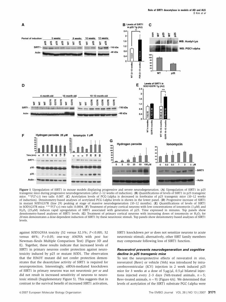
Levels of SIRT1 in models of neurodegeneration
We hypothesized that SIRT1 levels may increase as a protec-
tive response to neurodegenerative conditions and examined
levels of SIRT1 in various mouse models for human age-
dependent neurodegeneration. Mice inducibly overexpres-
sing a toxic coactivator of cyclin-dependent kinase 5
(CDK5), p25, display massive degeneration of forebrain
with features of AD (Cruz et al, 2003, 2006), whereas
transgenic mice expressing a mutant form of superoxide
dismutase 1 (SOD1G37R), which has been linked to human
amyotrophic lateral sclerosis (ALS), exhibit severe motor
neuron and axon degeneration in spinal cord (Gurney et al,
1994; Wong et al, 1995). Interestingly, in the forebrains of p25
transgenic mice (n¼ 9), SIRT1 protein levels increased as
early as 2 weeks after p25 induction and persisted throughout
the progression of the pathology to 12 weeks (Figure 1A and B).
In accordance with increased protein levels of SIRT1, there
was a decrease in the acetylation state of PGC-1alpha, a target
for SIRT1 deacetylase activity (Nemoto et al, 2005; Rodgers
et al, 2005; St-Pierre et al, 2006) (Figure 1C). In the spinal
cords of mutant SOD1G37R mice, SIRT1 was only slightly
upregulated at 4 months (n¼ 4), a stage with limited degen-
eration; however, levels of SIRT1 were significantly upregu-
lated when severe neurodegeneration was evident at 10–12
months (n¼ 8) (Nguyen et al, 2001) (Figure 1D and E). Mice
expressing a mutant form of amyloid precursor protein (APP)
linked to Familial AD (PDAPP-V717F, n¼ 7; 2–12 months)
(Games et al, 1995) do not exhibit significant neuronal loss,
although they display, in an age-dependent manner, substan-
tial b-amyloid plaques, a hallmark of AD (Games et al, 1995).
These mice showed no significant increase in SIRT1 in the
forebrain (Supplementary Figure 1). Together, these results
indicate that SIRT1 levels correlate with neurodegeneration
accompanied by progressive and severe loss of neurons, but
not with b-amyloid plaque pathology in the absence of
neuronal loss. In all the mouse models analyzed, as well as
in human brains, SIRT1 is not only enriched in the nucleus
but also localized in the cytoplasm (Supplementary Figure 2;
unpublished data).
Since p25 and mutant SOD1 trigger disruption of calcium
homeostasis and generate oxidative stress (Bruijn et al, 2004;
Cruz and Tsai, 2004), we tested whether SIRT1 is induced in
neurons in response to ionomycin (1 mM), a calcium iono-
phore, or hydrogen peroxide (H2O2) (25 mM), a free radical
generator. These specific stresses have previously been
shown to trigger the deterioration of neuronal morphology
and the formation of p25 in cultured neurons (Kusakawa
et al, 2000; Lee et al, 2000; Nath et al, 2000). Treatment of
primary cortical neurons with either ionomycin or H2O2
rapidly induced SIRT1 protein expression, and did so in a
dose-dependent manner (Figure 1F and G). Thus, SIRT1 is not
only induced in mouse models of neurodegeneration but also
in primary cultured neurons under neurotoxic stresses.
Resveratrol-mediated SIRT1 activation protects against
p25 and mutant SOD1
To understand the physiological significance of SIRT1 activa-
tion in context of p25 and mutant SOD1 toxicity, we first
tested the effects of resveratrol, a polyphenolic SIRT1-activat-
ing compound (STAC) (Howitz et al, 2003), on the viability of
primary mouse neurons overexpressing p25 or a mutant form
of SOD1 linked to ALS (SOD1G93A). The SOD1G93A muta-
tion, which has been linked to ALS, has been widely used in
primary neurons to examine neurotoxicity related to ALS.
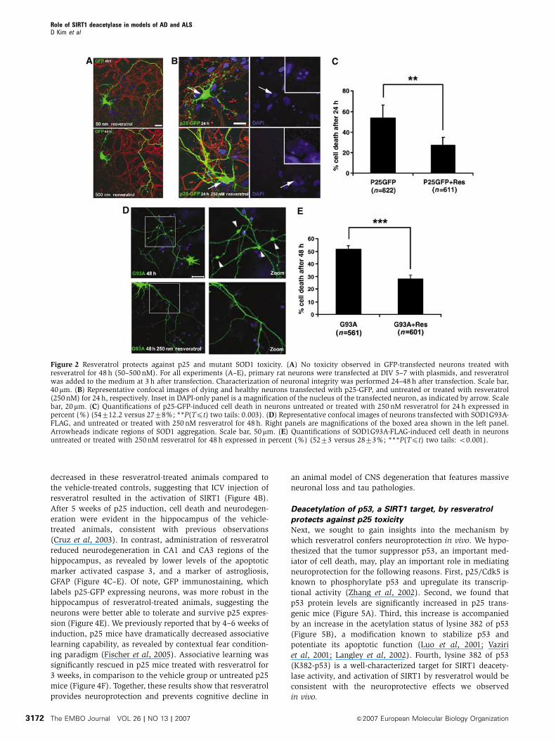
Doses of up to 500 nM resveratrol showed no evidence of
toxicity to primary neurons transfected with GFP (Figure 2A).
As previously reported (Patrick et al, 1999; Lee et al, 2000;
Zhang et al, 2002; Hamdane et al, 2003), transfection with
p25-GFP resulted in a high degrees of cell death (54% after
24 h), which were scored on the basis of neuritic integrity and
nuclear morphology, as described in Materials and methods
(Figure 2B and C). Remarkably, resveratrol treatment signifi-
cantly reduced the extent of cell death caused by p25 (Figure
2B and C) (54 versus 27%; P(Tpt) two tails: 0.003). The
protective effect of resveratrol against p25-mediated toxicity
was further confirmed by examination of propidium iodide
uptake as a marker for loss of membrane integrity and
viability (Supplementary Figure 3A and B). In this experi-
ment, resveratrol treatment reduced p25-induced propidium
uptake (37 versus 18.2%; P(Tpt) two tails: 0.007). We also
examined whether resveratrol protected against neurotoxicity
elicited by expression of SOD1G93A. Approximately 52% of
primary neurons transfected with mutant SOD1G93A for 48 h
exhibited cytoskeletal disruption and SOD1 aggregates, two
hallmarks of ALS-associated SOD1 toxicity (Figure 2D and E).
Resveratrol treatment significantly attenuated SOD1G93A-
mediated neurotoxicity (52 versus 28%; P(Tpt) two tails
o0.001) (Figure 2E). These results are in line with a recent
report showing that in cultured neurons derived from trans-
genic mice overexpressing a mutant (109Q) huntingtin, re-
sveratrol suppressed the neurotoxic effects of the mutant
protein (Parker et al, 2005). In addition, we examined
whether the acetylation of PGC-1alpha was decreased follow-
ing resveratrol treatment. Indeed, we observed a decrease in
PGC-1alpha (Supplementary Figure 4), suggesting that SIRT1
activity is increased following resveratrol treatment of
primary neurons.
Deacetylase activity of SIRT1 confers neuroprotection
against p25 and mutant SOD1
To directly verify the protective role of SIRT1 in neurodegen-
eration, we transfected primary neurons with p25-GFP or
SOD1G93A, together with either SIRT1 or SIRT1 lacking
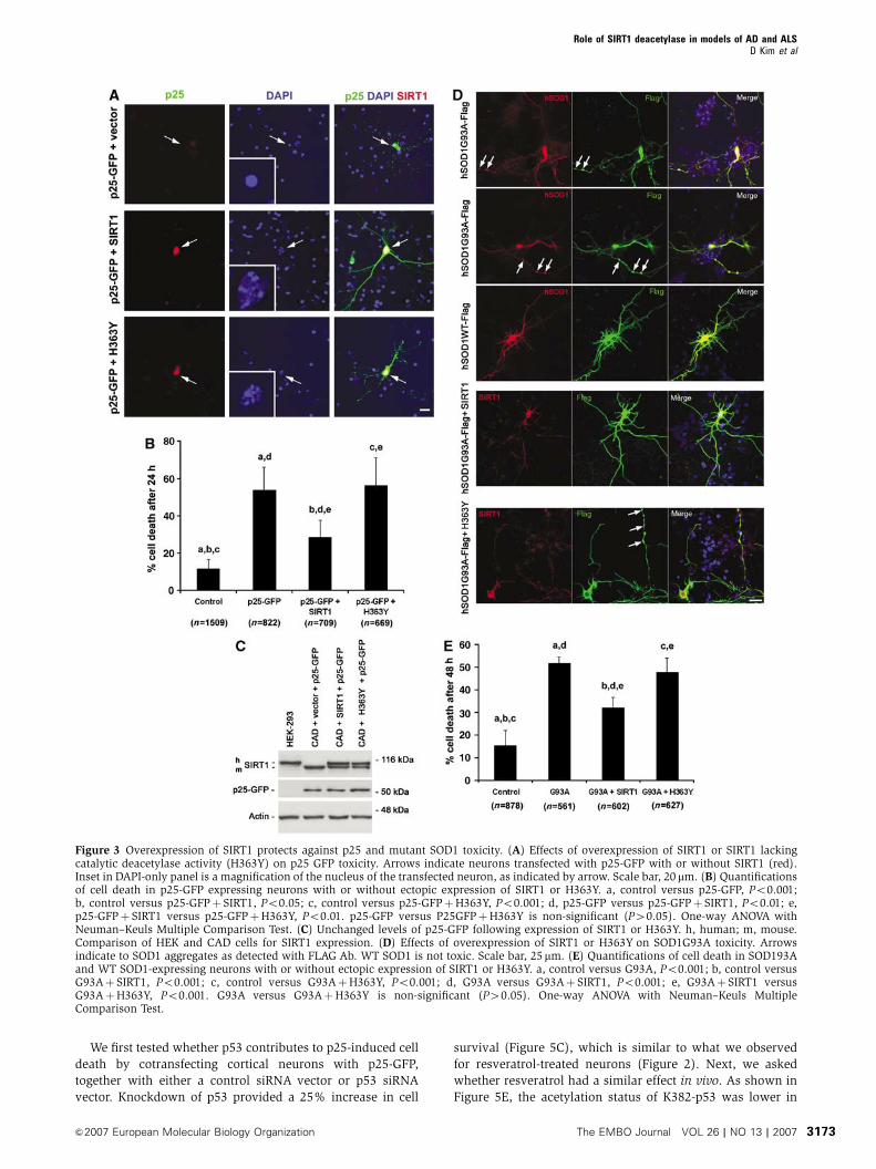
catalytic activity (H363Y). Overexpression of SIRT1, but not
H363Y, significantly rescued the rate of p25-GFP-mediated
cell death (54 versus 28.7%; Po0.01; 54 versus 56.5%;
P40.05; one-way ANOVA with post hoc Newman–Keuls
Multiple Comparison Test) (Figure 3A and B). The morphology
of the p25-GFP/SIRT1-overexpressing neurons appeared nor-
mal and indistinguishable from control GFP-transfected neu-
rons. This protective effect was unlikely to be an effect of
SIRT1 on the stability of p25-GFP, because similar levels of
p25-GFP were detected in the presence or absence of SIRT1
overexpression in CAD cells (Figure 3C). Similarly, a neuro-
protective effect of SIRT1, but not the H363Y mutant, was
observed when examining p25-induced propidium iodide
uptake (37 versus 17.7%; Po0.05; 37 versus 41.6%;
P40.05; one-way ANOVA with post hoc Newman–Keuls
Multiple Comparison Test) (Supplementary Figure 3A and C).
We also sought to determine whether SIRT1 overexpres-
sion protected against mutant SOD1-induced neurotoxicty.
The overexpression of SIRT1, but not H363Y, protected
Role of SIRT1 deacetylase in models of AD and ALSD Kim et al
The EMBO Journal VOL 26 | NO 13 | 2007 &2007 European Molecular Biology Organization3170
against SOD1G93A toxicity (52 versus 32.3%; Po0.001; 52
versus 48%; P40.05; one-way ANOVA with post hoc
Newman–Keuls Multiple Comparison Test) (Figure 3D and
E). Together, these results indicate that increased levels of
SIRT1 in primary neurons confer protection against neuro-
toxicity induced by p25 or mutant SOD1. The observation
that the H363Y mutant did not confer protection demon-
strates that the deacetylase activity of SIRT1 is required for
neuroprotection. Interestingly, siRNA-mediated knockdown
of SIRT1 in primary neurons was not neurotoxic per se and
did not result in increased sensitivity of neurons to neuro-
toxic stimuli (Supplementary Figure 5). This suggests that in
contrast to the survival benefit of increased SIRT1 activation,
SIRT1 knockdown per se does not sensitize neurons to acute
neurotoxic stimuli; alternatively, other SIRT family members
may compensate following loss of SIRT1 function.
Resveratrol prevents neurodegeneration and cognitive
decline in p25 transgenic mice
To test the neuroprotective effects of resveratrol in vivo,
resveratrol (Resv) or vehicle (Veh) was introduced by intra-
cerebroventricular (ICV) injection in 2 week induced p25
mice for 3 weeks at a dose of 5mg/ml, 0.5 ml bilateral injec-
tions injected every 2–3 days (Veh-treated animals, n¼ 5;
Resv-treated animals, n¼ 9) (Figure 4A). We determined that
levels of acetylation of the SIRT1 substrate PGC-1alpha were
Figure 1 Upregulation of SIRT1 in mouse models displaying progressive and severe neurodegeneration. (A) Upregulation of SIRT1 in p25transgenic mice during progressive neurodegeneration (after 2–12 weeks of induction). (B) Quantifications of levels of SIRT1 in p25 transgenicmice. **P(Tpt) two tails: 0.007. (C) Acetylation levels of PCG-1alpha is decreased in forebrains of p25 transgenic mice (10–12 weeksof induction). Densitometry-based analyses of acetylated PCG-1alpha levels is shown in the lower panel. (D) Progressive increase of SIRT1in mutant SOD1G37R (line 29) peaking at stage of massive neurodegeneration (10–12 months). (E) Quantifications of levels of SIRT1in SOD1G37R mice. ***P(Tpt) two tails: 0.0004. (F) Treatment of primary cortical neurons with low concentrations of ionomycin (1mM) andH2O2 (25mM) induces rapid upregulation of SIRT1 associated with generation of p25. Time expressed in minutes. Top panels showdensitometry-based analyses of SIRT1 levels. (G) Treatment of primary cortical neurons with increasing doses of ionomycin or H2O2 for20 min demonstrates a dose-dependent induction of SIRT1 by these neurotoxic stimuli. Top panels show densitometry-based analyses of SIRT1levels.
Role of SIRT1 deacetylase in models of AD and ALSD Kim et al
&2007 European Molecular Biology Organization The EMBO Journal VOL 26 | NO 13 | 2007 3171
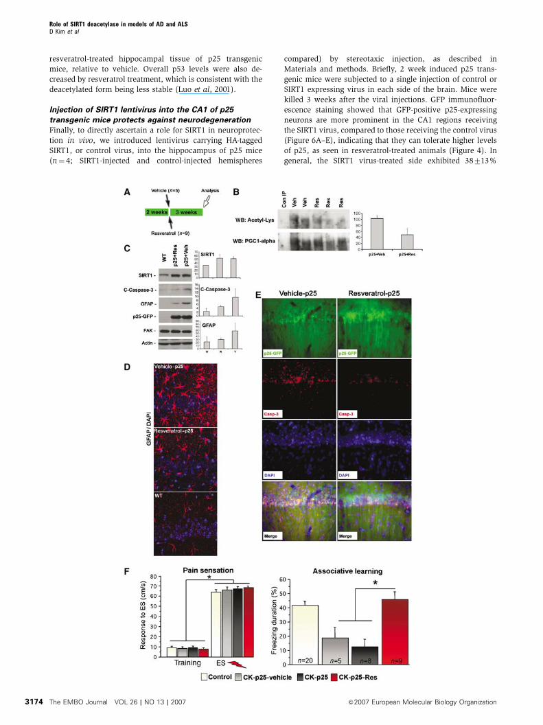
decreased in these resveratrol-treated animals compared to
the vehicle-treated controls, suggesting that ICV injection of
resveratrol resulted in the activation of SIRT1 (Figure 4B).
After 5 weeks of p25 induction, cell death and neurodegen-
eration were evident in the hippocampus of the vehicle-
treated animals, consistent with previous observations
(Cruz et al, 2003). In contrast, administration of resveratrol
reduced neurodegeneration in CA1 and CA3 regions of the
hippocampus, as revealed by lower levels of the apoptotic
marker activated caspase 3, and a marker of astrogliosis,
GFAP (Figure 4C–E). Of note, GFP immunostaining, which
labels p25-GFP expressing neurons, was more robust in the
hippocampus of resveratrol-treated animals, suggesting the
neurons were better able to tolerate and survive p25 expres-
sion (Figure 4E). We previously reported that by 4–6 weeks of
induction, p25 mice have dramatically decreased associative
learning capability, as revealed by contextual fear condition-
ing paradigm (Fischer et al, 2005). Associative learning was
significantly rescued in p25 mice treated with resveratrol for
3 weeks, in comparison to the vehicle group or untreated p25
mice (Figure 4F). Together, these results show that resveratrol
provides neuroprotection and prevents cognitive decline in
an animal model of CNS degeneration that features massive
neuronal loss and tau pathologies.
Deacetylation of p53, a SIRT1 target, by resveratrol
protects against p25 toxicity
Next, we sought to gain insights into the mechanism by
which resveratrol confers neuroprotection in vivo. We hypo-
thesized that the tumor suppressor p53, an important med-
iator of cell death, may, play an important role in mediating
neuroprotection for the following reasons. First, p25/Cdk5 is
known to phosphorylate p53 and upregulate its transcrip-
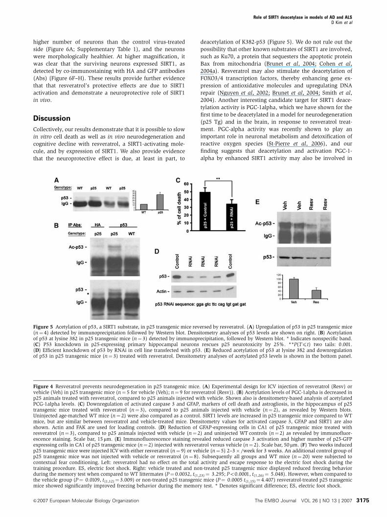
tional activity (Zhang et al, 2002). Second, we found that
p53 protein levels are significantly increased in p25 trans-
genic mice (Figure 5A). Third, this increase is accompanied
by an increase in the acetylation status of lysine 382 of p53
(Figure 5B), a modification known to stabilize p53 and
potentiate its apoptotic function (Luo et al, 2001; Vaziri
et al, 2001; Langley et al, 2002). Fourth, lysine 382 of p53
(K382-p53) is a well-characterized target for SIRT1 deacety-
lase activity, and activation of SIRT1 by resveratrol would be
consistent with the neuroprotective effects we observed
in vivo.
Figure 2 Resveratrol protects against p25 and mutant SOD1 toxicity. (A) No toxicity observed in GFP-transfected neurons treated withresveratrol for 48 h (50–500 nM). For all experiments (A–E), primary rat neurons were transfected at DIV 5–7 with plasmids, and resveratrolwas added to the medium at 3 h after transfection. Characterization of neuronal integrity was performed 24–48 h after transfection. Scale bar,40mm. (B) Representative confocal images of dying and healthy neurons transfected with p25-GFP, and untreated or treated with resveratrol(250 nM) for 24 h, respectively. Inset in DAPI-only panel is a magnification of the nucleus of the transfected neuron, as indicated by arrow. Scalebar, 20mm. (C) Quantifications of p25-GFP-induced cell death in neurons untreated or treated with 250 nM resveratrol for 24 h expressed inpercent (%) (54712.2 versus 2778%; **P(Tpt) two tails: 0.003). (D) Representative confocal images of neurons transfected with SOD1G93A-FLAG, and untreated or treated with 250 nM resveratrol for 48 h. Right panels are magnifications of the boxed area shown in the left panel.Arrowheads indicate regions of SOD1 aggregation. Scale bar, 50mm. (E) Quantifications of SOD1G93A-FLAG-induced cell death in neuronsuntreated or treated with 250 nM resveratrol for 48 h expressed in percent (%) (5273 versus 2873%; ***P(Tpt) two tails: o0.001).
Role of SIRT1 deacetylase in models of AD and ALSD Kim et al
The EMBO Journal VOL 26 | NO 13 | 2007 &2007 European Molecular Biology Organization3172
We first tested whether p53 contributes to p25-induced cell
death by cotransfecting cortical neurons with p25-GFP,
together with either a control siRNA vector or p53 siRNA
vector. Knockdown of p53 provided a 25% increase in cell
survival (Figure 5C), which is similar to what we observed
for resveratrol-treated neurons (Figure 2). Next, we asked
whether resveratrol had a similar effect in vivo. As shown in
Figure 5E, the acetylation status of K382-p53 was lower in
Figure 3 Overexpression of SIRT1 protects against p25 and mutant SOD1 toxicity. (A) Effects of overexpression of SIRT1 or SIRT1 lackingcatalytic deacetylase activity (H363Y) on p25 GFP toxicity. Arrows indicate neurons transfected with p25-GFP with or without SIRT1 (red).Inset in DAPI-only panel is a magnification of the nucleus of the transfected neuron, as indicated by arrow. Scale bar, 20 mm. (B) Quantificationsof cell death in p25-GFP expressing neurons with or without ectopic expression of SIRT1 or H363Y. a, control versus p25-GFP, Po0.001;b, control versus p25-GFPþ SIRT1, Po0.05; c, control versus p25-GFPþH363Y, Po0.001; d, p25-GFP versus p25-GFPþ SIRT1, Po0.01; e,p25-GFPþ SIRT1 versus p25-GFPþH363Y, Po0.01. p25-GFP versus P25GFPþH363Y is non-significant (P40.05). One-way ANOVA withNeuman–Keuls Multiple Comparison Test. (C) Unchanged levels of p25-GFP following expression of SIRT1 or H363Y. h, human; m, mouse.Comparison of HEK and CAD cells for SIRT1 expression. (D) Effects of overexpression of SIRT1 or H363Y on SOD1G93A toxicity. Arrowsindicate to SOD1 aggregates as detected with FLAG Ab. WT SOD1 is not toxic. Scale bar, 25mm. (E) Quantifications of cell death in SOD193Aand WT SOD1-expressing neurons with or without ectopic expression of SIRT1 or H363Y. a, control versus G93A, Po0.001; b, control versusG93Aþ SIRT1, Po0.001; c, control versus G93AþH363Y, Po0.001; d, G93A versus G93Aþ SIRT1, Po0.001; e, G93Aþ SIRT1 versusG93AþH363Y, Po0.001. G93A versus G93AþH363Y is non-significant (P40.05). One-way ANOVA with Neuman–Keuls MultipleComparison Test.
Role of SIRT1 deacetylase in models of AD and ALSD Kim et al
&2007 European Molecular Biology Organization The EMBO Journal VOL 26 | NO 13 | 2007 3173
resveratrol-treated hippocampal tissue of p25 transgenic
mice, relative to vehicle. Overall p53 levels were also de-
creased by resveratrol treatment, which is consistent with the
deacetylated form being less stable (Luo et al, 2001).
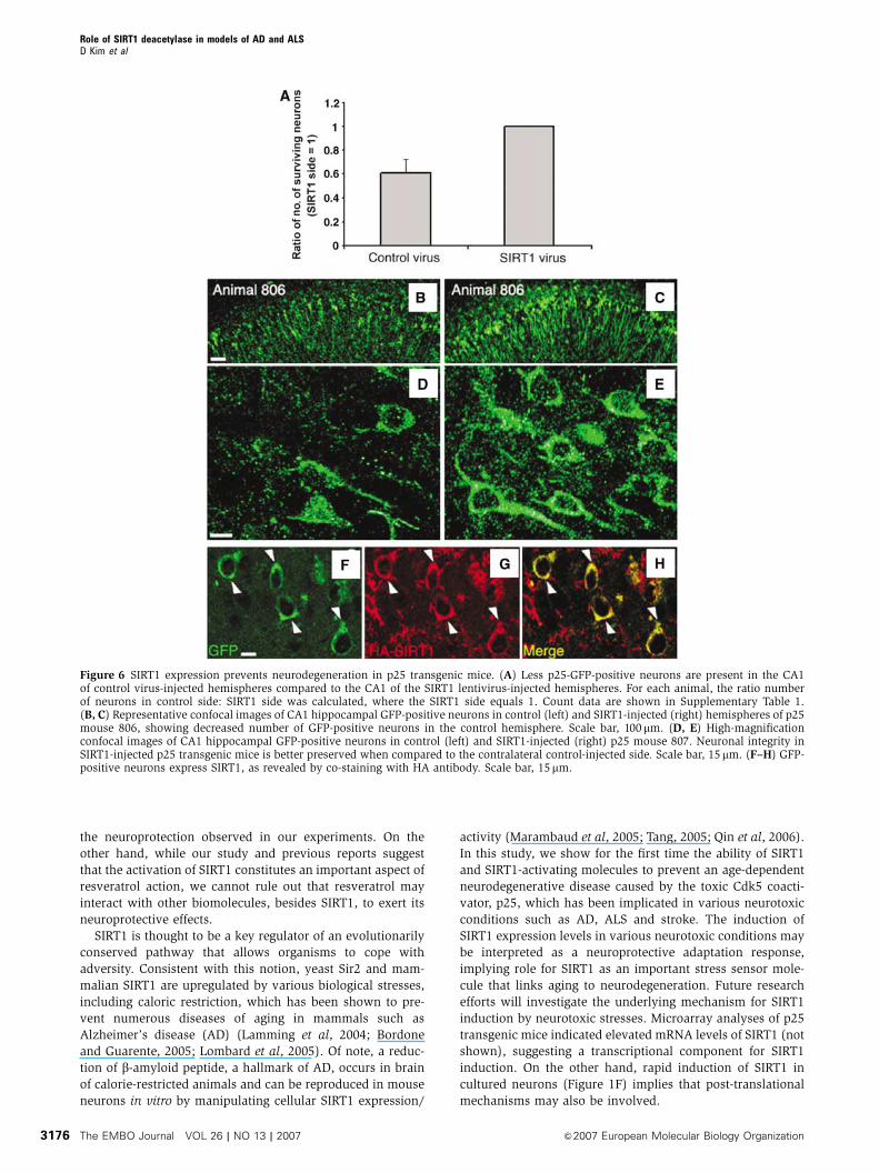
Injection of SIRT1 lentivirus into the CA1 of p25
transgenic mice protects against neurodegeneration
Finally, to directly ascertain a role for SIRT1 in neuroprotec-
tion in vivo, we introduced lentivirus carrying HA-tagged
SIRT1, or control virus, into the hippocampus of p25 mice
(n¼ 4; SIRT1-injected and control-injected hemispheres
compared) by stereotaxic injection, as described in
Materials and methods. Briefly, 2 week induced p25 trans-
genic mice were subjected to a single injection of control or
SIRT1 expressing virus in each side of the brain. Mice were
killed 3 weeks after the viral injections. GFP immunofluor-
escence staining showed that GFP-positive p25-expressing
neurons are more prominent in the CA1 regions receiving
the SIRT1 virus, compared to those receiving the control virus
(Figure 6A–E), indicating that they can tolerate higher levels
of p25, as seen in resveratrol-treated animals (Figure 4). In
general, the SIRT1 virus-treated side exhibited 38713%
Role of SIRT1 deacetylase in models of AD and ALSD Kim et al
The EMBO Journal VOL 26 | NO 13 | 2007 &2007 European Molecular Biology Organization3174
higher number of neurons than the control virus-treated
side (Figure 6A; Supplementary Table 1), and the neurons
were morphologically healthier. At higher magnification, it
was clear that the surviving neurons expressed SIRT1, as
detected by co-immunostaining with HA and GFP antibodies
(Abs) (Figure 6F–H). These results provide further evidence
that that resveratrol’s protective effects are due to SIRT1
activation and demonstrate a neuroprotective role of SIRT1
in vivo.
Discussion
Collectively, our results demonstrate that it is possible to slow
in vitro cell death as well as in vivo neurodegeneration and
cognitive decline with resveratrol, a SIRT1-activating mole-
cule, and by expression of SIRT1. We also provide evidence
that the neuroprotective effect is due, at least in part, to
deacetylation of K382-p53 (Figure 5). We do not rule out the
possibility that other known substrates of SIRT1 are involved,
such as Ku70, a protein that sequesters the apoptotic protein
Bax from mitochondria (Brunet et al, 2004; Cohen et al,
2004a). Resveratrol may also stimulate the deacetylation of
FOXO3/4 transcription factors, thereby enhancing gene ex-
pression of antioxidative molecules and upregulating DNA
repair (Nguyen et al, 2002; Brunet et al, 2004; Smith et al,
2004). Another interesting candidate target for SIRT1 deace-
tylation activity is PGC-1alpha, which we have shown for the
first time to be deacetylated in a model for neurodegeneration
(p25 Tg) and in the brain, in response to resveratrol treat-
ment. PGC-alpha activity was recently shown to play an
important role in neuronal metabolism and detoxification of
reactive oxygen species (St-Pierre et al, 2006), and our
finding suggests that deacetylation and activation PGC-1-
alpha by enhanced SIRT1 activity may also be involved in
Figure 4 Resveratrol prevents neurodegeneration in p25 transgenic mice. (A) Experimental design for ICV injection of resveratrol (Resv) orvehicle (Veh) in p25 transgenic mice (n¼ 5 for vehicle (Veh); n¼ 9 for resveratrol (Resv)). (B) Acetylation levels of PGC-1alpha is decreased inp25 animals treated with resveratrol, compared to p25 animals injected with vehicle. Shown also is densitometry-based analysis of acetylatedPCG-1alpha levels. (C) Downregulation of activated caspase 3 and GFAP, markers of cell death and astrogliosis, in the hippocampus of p25transgenic mice treated with resveratrol (n¼ 3), compared to p25 animals injected with vehicle (n¼ 2), as revealed by Western blots.Uninjected age-matched WT mice (n¼ 2) were also compared as a control. SIRT1 levels are increased in p25 transgenic mice compared to WTmice, but are similar between resveratrol and vehicle-treated mice. Densitometry values for activated caspase 3, GFAP and SIRT1 are alsoshown. Actin and FAK are used for loading controls. (D) Reduction of GFAP-expressing cells in CA1 of p25 transgenic mice treated withresveratrol (n¼ 3), compared to p25 animals injected with vehicle (n¼ 2) and uninjected WT controls (n¼ 2) as revealed by immunofluor-escence staining. Scale bar, 15 mm. (E) Immunofluorescence staining revealed reduced caspase 3 activation and higher number of p25-GFPexpressing cells in CA1 of p25 transgenic mice (n¼ 2) injected with resveratrol versus vehicle (n¼ 2). Scale bar, 50mm. (F) Two weeks inducedp25 transgenic mice were injected ICV with either resveratrol (n¼ 9) or vehicle (n¼ 5) 2–3� /week for 3 weeks. An additional control group ofp25 transgenic mice was not injected with vehicle or resveratrol (n¼ 8). Subsequently all groups and WT mice (n¼ 20) were subjected tocontextual fear conditioning. Left: resveratrol had no effect on the total activity and escape response to the electric foot shock during thetraining procedure. ES, electric foot shock. Right: vehicle treated and non-treated p25 transgenic mice displayed reduced freezing behaviorduring the memory test when compared to WT littermates (P¼ 0.0032, t(1,23)¼ 3.295; Po0.0001, t(1,26)¼ 5.048). However, when compared tothe vehicle group (P¼ 0.0109, t(1,12)¼ 3.009) or non-treated p25 transgenic mice (P¼ 0.0005 t(1,15)¼ 4.407) resveratrol-treated p25 transgenicmice showed significantly improved freezing behavior during the memory test. * Denotes significant difference; ES, electric foot shock.
Figure 5 Acetylation of p53, a SIRT1 substrate, in p25 transgenic mice reversed by resveratrol. (A) Upregulation of p53 in p25 transgenic mice(n¼ 4) detected by immunoprecipitation followed by Western blot. Densitometry analyses of p53 levels are shown on right. (B) Acetylationof p53 at lysine 382 in p25 transgenic mice (n¼ 3) detected by immunoprecipitation, followed by Western blot. * Indicates nonspecific band.(C) P53 knockdown in p25-expressing primary hippocampal neurons rescues p25 neurotoxicity by 25%. **P(Tpt) two tails: 0.001.(D) Efficient knockdown of p53 by RNAi in cell line transfected with p53. (E) Reduced acetylation of p53 at lysine 382 and downregulationof p53 in p25 transgenic mice (n¼ 3) treated with resveratrol. Densitometry analyses of acetylated p53 levels is shown in the bottom panel.
Role of SIRT1 deacetylase in models of AD and ALSD Kim et al
&2007 European Molecular Biology Organization The EMBO Journal VOL 26 | NO 13 | 2007 3175
the neuroprotection observed in our experiments. On the
other hand, while our study and previous reports suggest
that the activation of SIRT1 constitutes an important aspect of
resveratrol action, we cannot rule out that resveratrol may
interact with other biomolecules, besides SIRT1, to exert its
neuroprotective effects.
SIRT1 is thought to be a key regulator of an evolutionarily
conserved pathway that allows organisms to cope with
adversity. Consistent with this notion, yeast Sir2 and mam-
malian SIRT1 are upregulated by various biological stresses,
including caloric restriction, which has been shown to pre-
vent numerous diseases of aging in mammals such as
Alzheimer’s disease (AD) (Lamming et al, 2004; Bordone
and Guarente, 2005; Lombard et al, 2005). Of note, a reduc-
tion of b-amyloid peptide, a hallmark of AD, occurs in brain
of calorie-restricted animals and can be reproduced in mouse
neurons in vitro by manipulating cellular SIRT1 expression/
activity (Marambaud et al, 2005; Tang, 2005; Qin et al, 2006).
In this study, we show for the first time the ability of SIRT1
and SIRT1-activating molecules to prevent an age-dependent
neurodegenerative disease caused by the toxic Cdk5 coacti-
vator, p25, which has been implicated in various neurotoxic
conditions such as AD, ALS and stroke. The induction of
SIRT1 expression levels in various neurotoxic conditions may
be interpreted as a neuroprotective adaptation response,
implying role for SIRT1 as an important stress sensor mole-
cule that links aging to neurodegeneration. Future research
efforts will investigate the underlying mechanism for SIRT1
induction by neurotoxic stresses. Microarray analyses of p25
transgenic mice indicated elevated mRNA levels of SIRT1 (not
shown), suggesting a transcriptional component for SIRT1
induction. On the other hand, rapid induction of SIRT1 in
cultured neurons (Figure 1F) implies that post-translational
mechanisms may also be involved.
Figure 6 SIRT1 expression prevents neurodegeneration in p25 transgenic mice. (A) Less p25-GFP-positive neurons are present in the CA1of control virus-injected hemispheres compared to the CA1 of the SIRT1 lentivirus-injected hemispheres. For each animal, the ratio numberof neurons in control side: SIRT1 side was calculated, where the SIRT1 side equals 1. Count data are shown in Supplementary Table 1.(B, C) Representative confocal images of CA1 hippocampal GFP-positive neurons in control (left) and SIRT1-injected (right) hemispheres of p25mouse 806, showing decreased number of GFP-positive neurons in the control hemisphere. Scale bar, 100 mm. (D, E) High-magnificationconfocal images of CA1 hippocampal GFP-positive neurons in control (left) and SIRT1-injected (right) p25 mouse 807. Neuronal integrity inSIRT1-injected p25 transgenic mice is better preserved when compared to the contralateral control-injected side. Scale bar, 15mm. (F–H) GFP-positive neurons express SIRT1, as revealed by co-staining with HA antibody. Scale bar, 15mm.
Role of SIRT1 deacetylase in models of AD and ALSD Kim et al
The EMBO Journal VOL 26 | NO 13 | 2007 &2007 European Molecular Biology Organization3176
Our results predict that positive intervention into SIRT1
activity, such as through intake of SIRT1-activating mole-
cules, may have profound therapeutic benefits against var-
ious age-dependent neurodegenerative diseases. Conversely,
it may be worthwhile to explore whether mechanisms that
decrease SIRT1 activity or levels result in enhanced suscept-
ibility to age-dependent neurodegeneration. While knock-
down of SIRT1 did not appear to result in increased
susceptibility to acute neurotoxic stimuli in cultured neurons
(Supplementary Figure 5), the long-term effects of decreased
SIRT1 levels per se or in chronic neurodegenerative
conditions is an important question for future studies.
Interestingly, the SIRT1 gene resides in a locus on chromo-
some 10 that is associated with familial AD (WIPO, interna-
tional publication WO 2005/004815 A2) and future studies
are planned to determine whether mutations or polymorph-
isms in SIRT1 affect the susceptibility of individuals to AD
pathology.
Materials and methods
Protein preparation and Western blotsTotal protein extracts of mouse spinal cord, mouse forebrain, mousehippocampus or human prefrontal cortex were obtained byhomogenization in SDS–urea b-mercaptoethanol (0.5% SDS, 8 Murea in 7.4 phosphate buffer) or Triton X-100 (10 mM Tris–HCl(pH 7.5), 150 mM NaCl, 1 mM EDTA (pH 8.0) and 1% Triton).The protein concentration was estimated by the Bradford procedure(Bio-Rad Laboratories, Hercules, CA). Proteins were fractionated on7.5% SDS–PAGE and blotted on a nitrocellulose or PVDF membranefor Western blot analysis. Membranes were incubated with Absagainst SIRT1 (07–131, Upstate), a-tubulin (B512, Sigma), actin(MAB 1501, Chemicon), FAK (C-20, Santa Cruz Biotechnology), Bax(N-20, Santa Cruz) and GFP (B-2, Santa Cruz). The Western blotswere examined using RENAISSANCE, a Western blot chemilumi-nescence kit from NEN Life Science (Boston, MA). Quantitationswere corrected with levels of actin, a-tubulin and FAK andperformed with the Labscan program (Image Master, 2D softwarev 3.10, Amersham Pharmacia Biotech).
Culture, transfection and treatment of primary neuronsRat cortical primary neurons were isolated, cultured and transfectedat DIV 5–7 with Lipofectamine 2000, according to Nguyen et al(2004), in a ratio of 3:1 ((SIRT1 or SIRT1 H363Y or p53 RNAi):(p25-GFP, WT SOD1 or SOD1G93A or GFP)). Treatment ofprimary cortical neurons with ionomycin (1 mM), H2O2 (25mM)or resveratrol (50–500 nM) were performed according to Leeet al (2000).
Determination of cell deathPrimary neurons transfected with various constructs were scored ashealthy or dying, on the basis of neuritic integrity and neuronalmorphology, and in the case of mutant SOD1 overexpression,neuritic aggregation of SOD1. Specifically, fragmented neurites,nuclei with pyknosis or karyorrhexis, and SOD1 beading alongneurites were considered as signs of degeneration. A neuron withone or more of these features was scored as dying, and onlyneurons with none of these features were scored as healthy. Over100 transfected neurons were scored per condition per experiment,and experiments were carried out at least five times. Results areshown as percentage of transfected dying neurons out of the totaltransfected neurons. Counts were performed in a blind manner bymultiple researchers.
In addition, p25-GFP-mediated neurotoxicity was also scored bypropidium iodide uptake. Briefly, neurons transfected with thevarious constructs were stained in a 1:500 dilution of propidiumiodide staining stock solution (500mg/ml of propidium iodide in0.038 M sodium citrate, pH 7.0) for 1 h before fixation andimmunocytochemistry. Transfected neurons were scored for pre-sence of propidium iodide signal in the nuclei, which indicatespermeabilization of the membrane and subsequent binding of
propidium iodide with DNA. Thus, neurons that are positive forpropidium iodide are considered to be in an advanced state ofdegeneration in which membranes have been permeabilized. Over100 transfected neurons were scored per condition per experimentin a blind manner, and experiments were carried out at least threetimes. Results are displayed as a percentage of transfectedpropidium iodide-positive neurons out of the total transfectedneurons.
Immunofluorescence of primary neurons and humanprefrontal cortex tissuesCells were stained according to Nguyen et al (2004) with Absagainst tubulin (a-tubulin, Sigma Aldrich), GFP (Molecular Probes),SOD1 (Biodesign), FLAG (M2, Sigma), SIRT1 (Upstate). Staining ofspinal cord tissues was performed according to Cruz et al (2003)with Abs against SIRT1 (Upstate).
Generation of SOD1G37R transgenic mice and p25 inducibletransgenic miceTransgenic mice overexpressing SOD1G37R (line 29) (G37R) andp25-CK transgenic have been generated as described previously,and have been maintained on a pure C57BL6 background (Nguyenet al, 2001; Cruz et al, 2003).
Cannulation and injectionsDouble cannulae (Plastic1) were implanted 7 days before theexperiments, under 1.2% avertin anesthesia (0.4 ml/mouse), asdescribed previously (Fischer et al, 2004). For resveratrol injection,the cannulae were placed in both lateral brain ventricles, AP—0.5 mm, lateral 1 mm, depth 2 mm. Resveratrol (5mg/ml) or vehiclewas injected bilaterally 2–3� /week using a microinjector (CMA/microdialysis) over a 60 s period, so that a volume of 0.5ml wasinjected into each side. Resveratrol (25% DMSO/artificial cere-brospinal fluid) was prepared fresh immediately before eachinjection. For SIRT1 lentivirus injection, cannulae were placedin the dorsal hippocampus, AP—1.5 mm, lateral 1 mm, depth2 mm. SIRT1-HA lentivirus (1.5 ml) was injected as described aboveinto the left hippocampus, whereas SIRT1-HA lentivirus (1.5ml) wasinto the right hippocampus of 1 week induced CK-p25 mice.Number of GFP neurons was counted 1–2 mm caudal to theinjection site. A ratio neurons control side/neurons SIRT1 side wascalculated to quantify variations in percentage of neurons betweenboth sides.
Fear conditioningThe fear conditioning apparatus (TSE Systems) consisted of two testboxes with defined light and background noise that were connectedto a control unit and a PC computer. The experimental protocolswere designed and performed using TSE fear conditioning software.Box 1 contained a grid to apply the electric foot-shock and wascleaned with 70% ethanol before each training or test session. Thesecond test box had no grid and was cleaned with 1% acetic acidbefore each test. This box was used to analyze tone-dependent fearmemories. Fear conditioning consisted of a single exposure tocontext (box 1; 3 min) followed by a foot shock (2 s, 0.7 mA,constant current). Context-dependent freezing was measured24 h later The experimental boxes were equipped with light beamsthat allowed movement detection along the x-, y and z-axes. Thesystem was configured so that freezing was automatically counted ifno movement was observed for more than 3 s. The freezingduration is expressed as the percentage of time spent freezing,during the 180 s memory test. In addition, to verify these data, twoobservers scored freezing behavior every tenth second over 180 s ina blind manner.
Generation of RNAiP53 RNAi sequence were selected based on the criteria proposed bySui et al (2002). Complementary hairpin sequences were commer-cially synthesized and cloned into pSilencer 2.0 under promoter U6(Ambion). Sequence for p53 are basepairs: gga gtc ttc cag tgt gatgat. A random sequence without homology to any known mRNAwas used for control RNAi. All RNAi constructs were tested in celllines and primary neuronal cultures.
ImmunoprecipitationImmunoprecipitations were performed according to Nguyen et al(2004) on eight forebrains from p25 transgenic mice and wild-type
Role of SIRT1 deacetylase in models of AD and ALSD Kim et al
&2007 European Molecular Biology Organization The EMBO Journal VOL 26 | NO 13 | 2007 3177
(WT) mice, with a monoclonal Ab against p53 (Ab-3, Calbiochem/Oncogene). Membranes were probed with a homemade Ac-p53 Aband a mouse monoclonal p53 Ab (pAb-240, Abcam).
For examination of PGC-1alpha acetylation, brain samples orcultured primary neurons were lysed in RIPA buffer then dilutedthree-fold with PBS and protease inhibitors. Samples wereimmunoprecipitated using an anti-PGC-1alpha Ab (H-300, SantaCruz), washed extensively with a 1:2 solution (RIPA:PBSþproteaseinhibitors, nicotinamide, and TSA) and membranes were probedusing acetylated lysine Abs (Cell Signaling) and PGC-1alpha Abs(H-300, Santa Cruz).
Supplementary dataSupplementary data are available at The EMBO Journal Online(http://www.embojournal.org).
Acknowledgements
We thank Dr B Samuels for critical reading of the manuscript, Dr L Moyfor helpful discussions, and Drs M Urushitani and J-P Julien forSOD1 constructs and mice. This work was supported by theNational Institutes of Health (NIH) (DAS and L-HT), POI Grant(Poi AG027916) the National Institute of Aging (DAS), the CanadianInstitutes of Health Research (MDN) and the Paul F GlennFoundation for Medical Research (DAS). L-HT is an investigator atthe Howard Hughes Medical Institute. DAS is an Ellison MedicalResearch Foundation fellow. MDN is the Investigator at the BrendaStrafford Foundation Chair in Alzheimer research and a recipient ofa Career Development Award from the Human Frontier ScienceProgram Organization. AF held a Humbolt post-doctoral fellowship.FS was a fellow of the DFG (German Research Organization).JB holds an American Heart Association postdoctoral fellowship.
References
Anderson RM, Bitterman KJ, Wood JG, Medvedik O, Sinclair DA(2003a) Nicotinamide and PNC1 govern lifespan extensionby calorie restriction in Saccharomyces cerevisiae. Nature 423:181–185
Anderson RM, Latorre-Esteves M, Neves AR, Lavu S, Medvedik O,Taylor C, Howitz KT, Santos H, Sinclair DA (2003b) Yeast life-spanextension by calorie restriction is independent of NAD fluctua-tion. Science 302: 2124–2126
Araki T, Sasaki Y, Milbrandt J (2004) Increased nuclear NADbiosynthesis and SIRT1 activation prevent axonal degeneration.Science 305: 1010–1013
Bordone L, Guarente L (2005) Calorie restriction, SIRT1 and meta-bolism: understanding longevity. Nat Rev Mol Cell Biol 6: 298–305
Bossy-Wetzel E, Schwarzenbacher R, Lipton SA (2004) Molecularpathways to neurodegeneration. Nat Med 10 (Suppl): S2–S9
Bruijn LI, Miller TM, Cleveland DW (2004) Unraveling the mechan-isms involved in motor neuron degeneration in ALS. Annu RevNeurosci 27: 723–749
Brunet A, Sweeney LB, Sturgill JF, Chua KF, Greer PL, Lin Y, Tran H,Ross SE, Mostoslavsky R, Cohen HY, Hu LS, Cheng HL,Jedrychowski MP, Gygi SP, Sinclair DA, Alt FW, Greenberg ME(2004) Stress-dependent regulation of FOXO transcription factorsby the SIRT1 deacetylase. Science 303: 2011–2015
Chen J, Zhou Y, Mueller-Steiner S, Chen LF, Kwon H, Yi S, Mucke L,Gan L (2005) SIRT1 protects against microglia-dependent amy-loid-beta toxicity through inhibiting NF-kappaB signaling. J BiolChem 280: 40364–40374
Cohen HY, Lavu S, Bitterman KJ, Hekking B, Imahiyerobo TA,Miller C, Frye R, Ploegh H, Kessler BM, Sinclair DA (2004a)Acetylation of the C terminus of Ku70 by CBP and PCAF controlsBax-mediated apoptosis. Mol Cell 13: 627–638
Cohen HY, Miller C, Bitterman KJ, Wall NR, Hekking B, Kessler B,Howitz KT, Gorospe M, de Cabo R, Sinclair DA (2004b) Calorierestriction promotes mammalian cell survival by inducing theSIRT1 deacetylase. Science 305: 390–392
Cruz JC, Tsai LH (2004) A Jekyll and Hyde kinase: roles for Cdk5 inbrain development and disease. Curr Opin Neurobiol 14: 390–394
Cruz JC, Tseng HC, Goldman JA, Shih H, Tsai LH (2003) AberrantCdk5 activation by p25 triggers pathological events leadingto neurodegeneration and neurofibrillary tangles. Neuron 40:471–483
Cruz JC, Kim D, Moy LY, Dobbin MM, Sun X, Bronson RT, Tsai LH(2006) p25/cyclin-dependent kinase 5 induces production andintraneuronal accumulation of amyloid beta in vivo. J Neurosci26: 10536–10541
Fabrizio P, Gattazzo C, Battistella L, Wei M, Cheng C, McGrew K,Longo VD (2005) Sir2 blocks extreme life-span extension. Cell123: 655–667
Fischer A, Sananbenesi F, Pang PT, Lu B, Tsai LH (2005) Opposingroles of transient and prolonged expression of p25 in synapticplasticity and hippocampus-dependent memory. Neuron 48:825–838
Fischer A, Sananbenesi F, Schrick C, Spiess J, Radulovic J (2004)Distinct roles of hippocampal de novo protein synthesis and actinrearrangement in extinction of contextual fear. J Neurosci 24:1962–1966
Forman MS, Trojanowski JQ, Lee VM (2004) Neurodegenerativediseases: a decade of discoveries paves the way for therapeuticbreakthroughs. Nat Med 10: 1055–1063
Games D, Adams D, Alessandrini R, Barbour R, Berthelette P,Blackwell C, Carr T, Clemens J, Donaldson T, Gillespie F, GuidoT, Hagopian S, Johnson-Wood K, Khan K, Lee M, Leibowitz P,Lieberburg I, Little S, Masliah E, McConlogue L, Montoya-ZavalaM, Mucke L, Paganini L, Penniman E, Power M, Shenk D, SeubertP, Snyder B, Soriano F, Tan H, Vitale J, Wadsworth S, Wolozin B,Zhao J (1995) Alzheimer-type neuropathology in transgenic miceoverexpressing V717F beta-amyloid precursor protein. Nature373: 523–527
Gurney ME, Pu H, Chiu AY, Dal Canto MC, Polchow CY, AlexanderDD, Caliendo J, Hentati A, Kwon YW, Deng HX, Chen W,Sufit RL, Siddique T (1994) Motor neuron degeneration in micethat express a human Cu, Zn superoxide dismutase mutation.Science 264: 1772–1775
Hamdane M, Sambo AV, Delobel P, Begard S, Violleau A, DelacourteA, Bertrand P, Benavides J, Buee L (2003) Mitotic-like tauphosphorylation by p25–Cdk5 kinase complex. J Biol Chem278: 34026–34034
Howitz KT, Bitterman KJ, Cohen HY, Lamming DW, Lavu S, WoodJG, Zipkin RE, Chung P, Kisielewski A, Zhang LL, Scherer B,Sinclair DA (2003) Small molecule activators of sirtuins extendSaccharomyces cerevisiae lifespan. Nature 425: 191–196
Imai S, Armstrong CM, Kaeberlein M, Guarente L (2000)Transcriptional silencing and longevity protein Sir2 is an NAD-dependent histone deacetylase. Nature 403: 795–800
Kaeberlein M, McVey M, Guarente L (1999) The SIR2/3/4 complexand SIR2 alone promote longevity in Saccharomyces cerevisiae bytwo different mechanisms. Genes Dev 13: 2570–2580
Kusakawa G, Saito T, Onuki R, Ishiguro K, Kishimoto T, Hisanaga S(2000) Calpain-dependent proteolytic cleavage of the p35cyclin-dependent kinase 5 activator to p25. J Biol Chem 275:17166–17172
Lamming DW, Wood JG, Sinclair DA (2004) Small molecules thatregulate lifespan: evidence for xenohormesis. Mol Microbiol 53:1003–1009
Langley E, Pearson M, Faretta M, Bauer UM, Frye RA, Minucci S,Pelicci PG, Kouzarides T (2002) Human SIR2 deacetylates p53and antagonizes PML/p53-induced cellular senescence. EMBO J21: 2383–2396
Lee MS, Kwon YT, Li M, Peng J, Friedlander RM, Tsai LH (2000)Neurotoxicity induces cleavage of p35 to p25 by calpain. Nature405: 360–364
Lin SJ, Defossez PA, Guarente L (2000) Requirement of NAD andSIR2 for life-span extension by calorie restriction inSaccharomyces cerevisiae. Science 289: 2126–2128
Lombard DB, Chua KF, Mostoslavsky R, Franco S, Gostissa M, Alt FW(2005) DNA repair, genome stability, and aging. Cell 120: 497–512
Luo J, Nikolaev AY, Imai S, Chen D, Su F, Shiloh A, Guarente L, GuW (2001) Negative control of p53 by Sir2alpha promotes cellsurvival under stress. Cell 107: 137–148
Marambaud P, Zhao H, Davies P (2005) Resveratrol promotesclearance of Alzheimer’s disease amyloid-beta peptides. J BiolChem 280: 37377–37382
Role of SIRT1 deacetylase in models of AD and ALSD Kim et al
The EMBO Journal VOL 26 | NO 13 | 2007 &2007 European Molecular Biology Organization3178
Motta MC, Divecha N, Lemieux M, Kamel C, Chen D,Gu W, Bultsma Y, McBurney M, Guarente L (2004) MammalianSIRT1 represses forkhead transcription factors. Cell 116:551–563
Nath R, Davis M, Probert AW, Kupina NC, Ren X, Schielke GP, WangKK (2000) Processing of cdk5 activator p35 to its truncated form(p25) by calpain in acutely injured neuronal cells. BiochemBiophys Res Commun 274: 16–21
Nemoto S, Fergusson MM, Finkel T (2005) SIRT1 functionallyinteracts with the metabolic regulator and transcriptional coacti-vator PGC-1{alpha}. J Biol Chem 280: 16456–16460
Nguyen MD, Lariviere RC, Julien JP (2001) Deregulation of Cdk5 ina mouse model of ALS: toxicity alleviated by perikaryal neurofila-ment inclusions. Neuron 30: 135–147
Nguyen MD, Mushynski WE, Julien JP (2002) Cycling at the inter-face between neurodevelopment and neurodegeneration. CellDeath Differ 9: 1294–1306
Nguyen MD, Shu T, Sanada K, Lariviere RC, Tseng HC, Park SK,Julien JP, Tsai LH (2004) A NUDEL-dependent mechanism ofneurofilament assembly regulates the integrity of CNS neurons.Nat Cell Biol 6: 595–608
Parker JA, Arango M, Abderrahmane S, Lambert E, Tourette C,Catoire H, Neri C (2005) Resveratrol rescues mutant polygluta-mine cytotoxicity in nematode and mammalian neurons. NatGenet 37: 349–350
Patrick GN, Zukerberg L, Nikolic M, de la Monte S, Dikkes P, TsaiLH (1999) Conversion of p35 to p25 deregulates Cdk5 activity andpromotes neurodegeneration. Nature 402: 615–622
Qin W, Yang T, Ho L, Zhao Z, Wang J, Chen L, Thiyagarajan M,Macgrogan D, Rodgers JT, Puigserver P, Sadoshima J, Deng HH,Pedrini S, Gandy S, Sauve A, Pasinetti GM (2006) Neuronal SIRT1activation as a novel mechanism underlying the prevention ofAlzheimer’s disease amyloid neuropathology by calorie restric-tion. J Biol Chem 281: 21745–21754
Rodgers JT, Lerin C, Haas W, Gygi SP, Spiegelman BM, Puigserver P(2005) Nutrient control of glucose homeostasis through a com-plex of PGC-1alpha and SIRT1. Nature 434: 113–118
Selkoe DJ (2004) Cell biology of protein misfolding: the examples ofAlzheimer’s and Parkinson’s diseases. Nat Cell Biol 6: 1054–1061
Smith PD, O’Hare MJ, Park DS (2004) Emerging pathogenic rolefor cyclin dependent kinases in neurodegeneration. Cell Cycle 3:289–291
St-Pierre J, Drori S, Uldry M, Silvaggi JM, Rhee J, Jager S,Handschin C, Zheng K, Lin J, Yang W, Simon DK, Bachoo R,Spiegelman BM (2006) Suppression of reactive oxygen speciesand neurodegeneration by the PGC-1 transcriptional coactivators.Cell 127: 397–408
Sui G, Soohoo C, Affar el B, Gay F, Shi Y, Forrester WC (2002) ADNA vector-based RNAi technology to suppress gene expressionin mammalian cells. Proc Natl Acad Sci USA 99: 5515–5520
Tang BL (2005) Alzheimer’s disease: channeling APP to non-amyloidogenic processing. Biochem Biophys Res Commun 331:375–378
Tang BL (2006) SIRT1, neuronal cell survival and the insulin/IGF-1aging paradox. Neurobiol Aging 27: 501–505
Vaziri H, Dessain SK, Ng Eaton E, Imai SI, Frye RA, Pandita TK,Guarente L, Weinberg RA (2001) hSIR2(SIRT1) functions as anNAD-dependent p53 deacetylase. Cell 107: 149–159
Wang J, Zhai Q, Chen Y, Lin E, Gu W, McBurney MW, He Z (2005) Alocal mechanism mediates NAD-dependent protection of axondegeneration. J Cell Biol 170: 349–355
Wong PC, Pardo CA, Borchelt DR, Lee MK, Copeland NG, JenkinsNA, Sisodia SS, Cleveland DW, Price DL (1995) An adverseproperty of a familial ALS-linked SOD1 mutation causes motorneuron disease characterized by vacuolar degeneration of mito-chondria. Neuron 14: 1105–1116
Zhang J, Krishnamurthy PK, Johnson GV (2002) Cdk5 phosphor-ylates p53 and regulates its activity. J Neurochem 81: 307–313
Role of SIRT1 deacetylase in models of AD and ALSD Kim et al
&2007 European Molecular Biology Organization The EMBO Journal VOL 26 | NO 13 | 2007 3179
Related Documents