Applied Surface Science 257 (2010) 340–353 Contents lists available at ScienceDirect Applied Surface Science journal homepage: www.elsevier.com/locate/apsusc Single-wall carbon nanotube chemical attachment at platinum electrodes Belinda I. Rosario-Castro a , Enid J. Contés-de-Jesús a , Marisabel Lebrón-Colón b , Michael A. Meador b , M. Aulice Scibioh a , Carlos R. Cabrera a,∗ a Department of Chemistry and Center for Advanced Nanoscale Materials, University of Puerto Rico, Río Piedras Campus, PO Box 23346, San Juan 00931-3346, Puerto Rico b NASA John H. Glenn Research Center, 21000 Brookpark Road, Cleveland, OH 44135, United States article info Article history: Received 26 January 2010 Received in revised form 27 May 2010 Accepted 26 June 2010 Available online 1 August 2010 Keywords: Single-wall carbon nanotubes Self-assembled monolayers Platinum electrodes 4-Aminothiophenol Transmission electron microscopy Atomic force microscopy Raman spectroscopy abstract Self-assembled monolayer (SAM) techniques were used to adsorb 4-aminothiophenol (4-ATP) on plat- inum electrodes in order to obtain an amino-terminated SAM as the base for the chemical attachment of single-wall carbon nanotubes (SWCNTs). A physico-chemical, morphological and electrochemical charac- terizations of SWCNTs attached onto the modified Pt electrodes was done by using reflection-absorption infrared spectroscopy (RAIR), X-ray photoelectron spectroscopy (XPS), Raman spectroscopy, scanning electron microscopy (SEM), atomic force microscopy (AFM), and cyclic voltammetry (CV) techniques. The SWNTs/4-ATP/Pt surface had regions of small, medium, and large thickness of carbon nanotubes with heights of 100–200 nm, 700 nm to 1.5 m, and 1.0–3.0 m, respectively. Cyclic voltammetries (CVs) in sulfuric acid demonstrated that attachment of SWNTs on 4-ATP/Pt is markedly stable, even after 30 potential cycles. CV in ruthenium hexamine was similar to bare Pt electrodes, suggesting that SWNTs assembly is similar to a closely packed microelectrode array. © 2010 Published by Elsevier B.V. 1. Introduction Since the discovery of carbon nanotubes by Iijima [1], a spurt of research activities have been focused on single-wall carbon nanotubes (SWCNTs). SWCNTs exhibit an interesting combination of remarkable physico-chemical properties such as their dimen- sions (high length-to-diameter ratio), strength, chemical stability, as well as mechanical and electronic properties. These properties make them attractive and suitable for several areas of potential applications such as biomolecular recognition [2], reinforcement in polymer composites [3,4], scanning probe microscopy tips [5,6], chemically sensitive imaging of materials [7], transistors and logic circuit construction [8,9], hydrogen storage [10,11], support for metal catalysts [12] and lithium intercalation [13]. Most of these applications require molecular devices with a 3D nanostructure of monodispersed and aligned nanotubes [10]. The fabrication of many nanoelectronics devices frequently includes carbon nan- otubes as a fundamental constituent [14–17]. Understanding the chemistry of carbon nanotubes is essential in order to explore their ultimate potentialities for practical uses. The exposed surface of carbon nanotubes is made up of unreac- tive basal graphitic plane, and hence at the first sight, they may ∗ Corresponding author. Tel.: +787 764 0000x4807; fax: +787 756 8242. E-mail addresses: [email protected], [email protected] (C.R. Cabrera). appear to be chemically inert; however, when treated with acids their surfaces can be highly functionalized. SWCNTs are generally produced by the conversion of the sp 2 -hybridized carbon into its sp 3 form. Oxidation studies show that carbon nanotube ends are etched away via chemical oxidation, resulting in open-ended tubes [18]. Oxidized carbon nanotubes have several oxygen-containing functional groups such as phenol, carbonyl, carboxyl, at their open ends and side-wall defect sites, which make them more reactive than pristine SWCNTs. Consequently, smaller fragments of SWC- NTs obtained by acid treatment are important intermediates for the fabrication of SWCNTs devices. Individual SWCNTs deposition on chemically functionalized surfaces is a method that facilitates the formation of nanostructure devices from carbon nanotubes. The carboxyl functionalized ends of SWCNTs enable the attach- ment of carbon nanotubes to an amino-terminated self-assembled monolayer (SAM) deposited over a metal surface. The ideal acylating reagent would be a carboxylic acid, but the acids themselves are relatively unreactive with nucleophiles. A pos- sible route to make them active is to convert the carboxylic acid to a more reactive derivative such as an acyl chloride or anhydride. Yet another route would be to employ reagents that selectively acti- vate a carboxyl group toward nucleophilic substitution. Two such reagents are dicyclohexylcarbodiimide (DCC) and carbonyldiimi- dazole (Staab’s reagent) [19,20]. One of the most common methods for formation of an amide bond is the activation of the acid with car- bodiimides, particularly, DCC, and its subsequent reaction with an amine (see Scheme 1). 0169-4332/$ – see front matter © 2010 Published by Elsevier B.V. doi:10.1016/j.apsusc.2010.06.072

Welcome message from author
This document is posted to help you gain knowledge. Please leave a comment to let me know what you think about it! Share it to your friends and learn new things together.
Transcript
S
BMa
b
a
ARRAA
KSSP4TAR
1
onosamaiccmaomo
iTt
(
0d
Applied Surface Science 257 (2010) 340–353
Contents lists available at ScienceDirect
Applied Surface Science
journa l homepage: www.e lsev ier .com/ locate /apsusc
ingle-wall carbon nanotube chemical attachment at platinum electrodes
elinda I. Rosario-Castroa, Enid J. Contés-de-Jesúsa, Marisabel Lebrón-Colónb,ichael A. Meadorb, M. Aulice Scibioha, Carlos R. Cabreraa,∗
Department of Chemistry and Center for Advanced Nanoscale Materials, University of Puerto Rico, Río Piedras Campus, PO Box 23346, San Juan 00931-3346, Puerto RicoNASA John H. Glenn Research Center, 21000 Brookpark Road, Cleveland, OH 44135, United States
r t i c l e i n f o
rticle history:eceived 26 January 2010eceived in revised form 27 May 2010ccepted 26 June 2010vailable online 1 August 2010
a b s t r a c t
Self-assembled monolayer (SAM) techniques were used to adsorb 4-aminothiophenol (4-ATP) on plat-inum electrodes in order to obtain an amino-terminated SAM as the base for the chemical attachment ofsingle-wall carbon nanotubes (SWCNTs). A physico-chemical, morphological and electrochemical charac-terizations of SWCNTs attached onto the modified Pt electrodes was done by using reflection-absorptioninfrared spectroscopy (RAIR), X-ray photoelectron spectroscopy (XPS), Raman spectroscopy, scanningelectron microscopy (SEM), atomic force microscopy (AFM), and cyclic voltammetry (CV) techniques.
eywords:ingle-wall carbon nanotubeself-assembled monolayerslatinum electrodes-Aminothiophenolransmission electron microscopy
The SWNTs/4-ATP/Pt surface had regions of small, medium, and large thickness of carbon nanotubeswith heights of 100–200 nm, 700 nm to 1.5 �m, and 1.0–3.0 �m, respectively. Cyclic voltammetries (CVs)in sulfuric acid demonstrated that attachment of SWNTs on 4-ATP/Pt is markedly stable, even after 30potential cycles. CV in ruthenium hexamine was similar to bare Pt electrodes, suggesting that SWNTsassembly is similar to a closely packed microelectrode array.
tomic force microscopyaman spectroscopy
. Introduction
Since the discovery of carbon nanotubes by Iijima [1], a spurtf research activities have been focused on single-wall carbonanotubes (SWCNTs). SWCNTs exhibit an interesting combinationf remarkable physico-chemical properties such as their dimen-ions (high length-to-diameter ratio), strength, chemical stability,s well as mechanical and electronic properties. These propertiesake them attractive and suitable for several areas of potential
pplications such as biomolecular recognition [2], reinforcementn polymer composites [3,4], scanning probe microscopy tips [5,6],hemically sensitive imaging of materials [7], transistors and logicircuit construction [8,9], hydrogen storage [10,11], support foretal catalysts [12] and lithium intercalation [13]. Most of these
pplications require molecular devices with a 3D nanostructuref monodispersed and aligned nanotubes [10]. The fabrication ofany nanoelectronics devices frequently includes carbon nan-
tubes as a fundamental constituent [14–17].
Understanding the chemistry of carbon nanotubes is essentialn order to explore their ultimate potentialities for practical uses.he exposed surface of carbon nanotubes is made up of unreac-ive basal graphitic plane, and hence at the first sight, they may
∗ Corresponding author. Tel.: +787 764 0000x4807; fax: +787 756 8242.E-mail addresses: [email protected], [email protected]
C.R. Cabrera).
169-4332/$ – see front matter © 2010 Published by Elsevier B.V.oi:10.1016/j.apsusc.2010.06.072
© 2010 Published by Elsevier B.V.
appear to be chemically inert; however, when treated with acidstheir surfaces can be highly functionalized. SWCNTs are generallyproduced by the conversion of the sp2-hybridized carbon into itssp3 form. Oxidation studies show that carbon nanotube ends areetched away via chemical oxidation, resulting in open-ended tubes[18]. Oxidized carbon nanotubes have several oxygen-containingfunctional groups such as phenol, carbonyl, carboxyl, at their openends and side-wall defect sites, which make them more reactivethan pristine SWCNTs. Consequently, smaller fragments of SWC-NTs obtained by acid treatment are important intermediates forthe fabrication of SWCNTs devices. Individual SWCNTs depositionon chemically functionalized surfaces is a method that facilitatesthe formation of nanostructure devices from carbon nanotubes.The carboxyl functionalized ends of SWCNTs enable the attach-ment of carbon nanotubes to an amino-terminated self-assembledmonolayer (SAM) deposited over a metal surface.
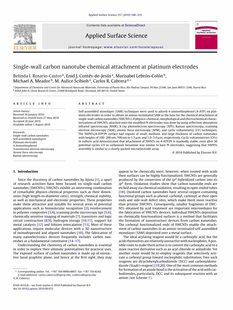
The ideal acylating reagent would be a carboxylic acid, but theacids themselves are relatively unreactive with nucleophiles. A pos-sible route to make them active is to convert the carboxylic acid to amore reactive derivative such as an acyl chloride or anhydride. Yetanother route would be to employ reagents that selectively acti-vate a carboxyl group toward nucleophilic substitution. Two such
reagents are dicyclohexylcarbodiimide (DCC) and carbonyldiimi-dazole (Staab’s reagent) [19,20]. One of the most common methodsfor formation of an amide bond is the activation of the acid with car-bodiimides, particularly, DCC, and its subsequent reaction with anamine (see Scheme 1).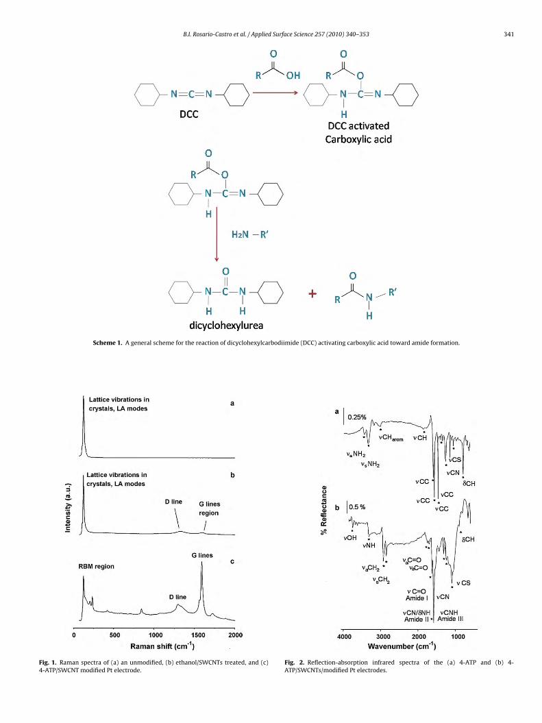
B.I. Rosario-Castro et al. / Applied Surface Science 257 (2010) 340–353 341
Scheme 1. A general scheme for the reaction of dicyclohexylcarbodiimide (DCC) activating carboxylic acid toward amide formation.
Fig. 1. Raman spectra of (a) an unmodified, (b) ethanol/SWCNTs treated, and (c)4-ATP/SWCNT modified Pt electrode.
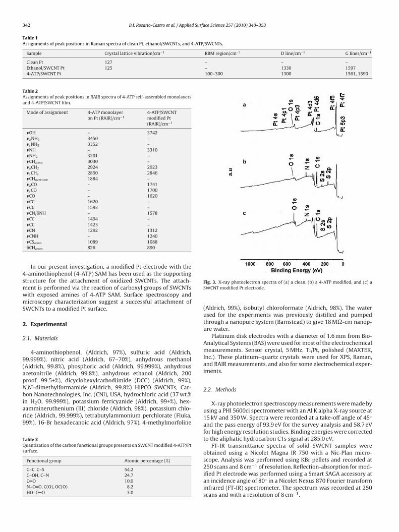
Fig. 2. Reflection-absorption infrared spectra of the (a) 4-ATP and (b) 4-ATP/SWCNTs/modified Pt electrodes.
342 B.I. Rosario-Castro et al. / Applied Surface Science 257 (2010) 340–353
Table 1Assignments of peak positions in Raman spectra of clean Pt, ethanol/SWCNTs, and 4-ATP/SWCNTs.
Sample Crystal lattice vibration/cm−1 RBM region/cm−1 D line/cm−1 G lines/cm−1
Clean Pt 127 – – –Ethanol/SWCNT Pt 125 – 1330 15974-ATP/SWCNT Pt 100–300 1300 1561, 1590
Table 2Assignments of peak positions in RAIR spectra of 4-ATP self-assembled monolayersand 4-ATP/SWCNT film.
Mode of assignment 4-ATP monolayeron Pt (RAIR)/cm−1
4-ATP/SWCNTmodified Pt(RAIR)/cm−1
�OH – 3742�aNH2 3450 –�sNH2 3352 –�NH – 3310�NH2 3201 –�CHarom 3030 –�aCH2 2924 2923�sCH2 2850 2846�CHovertone 1884 –�aCO – 1741�sCO – 1700�CO – 1620�CC 1620 –�CC 1593 –�CN/�NH – 1578�CC 1494 –�CC 1423 –�CN 1292 1312
4smwmS
2
2
9(apNbiar9
TQs
�CNH – 1240�CSarom 1089 1088�CHarom 826 890
In our present investigation, a modified Pt electrode with the-aminothiophenol (4-ATP) SAM has been used as the supportingtructure for the attachment of oxidized SWCNTs. The attach-ent is performed via the reaction of carboxyl groups of SWCNTsith exposed amines of 4-ATP SAM. Surface spectroscopy andicroscopy characterization suggest a successful attachment of
WCNTs to a modified Pt surface.
. Experimental
.1. Materials
4-aminothiophenol, (Aldrich, 97%), sulfuric acid (Aldrich,9.999%), nitric acid (Aldrich, 67–70%), anhydrous methanolAldrich, 99.8%), phosphoric acid (Aldrich, 99.999%), anhydrouscetonitrile (Aldrich, 99.8%), anhydrous ethanol (Aldrich, 200roof, 99.5+%), dicyclohexylcarbodiimide (DCC) (Aldrich, 99%),,N′-dimethylformamide (Aldrich, 99.8%) HiPCO SWCNTs, Car-on Nanotechnologies, Inc. (CNI), USA, hydrochloric acid (37 wt.%
n H2O, 99.999%), potassium ferricyanide (Aldrich, 99+%), hex-ammineruthenium (III) chloride (Aldrich, 98%), potassium chlo-ide (Aldrich, 99.999%), tetrabutylammonium perchlorate (Fluka,9%), 16-Br hexadecanoic acid (Aldrich, 97%), 4-methylmorfoline
able 3uantization of the carbon functional groups presents on SWCNT modified 4-ATP/Pt
urface.
Functional group Atomic percentage (%)
C–C, C–S 54.2C–OH, C–N 24.7C O 10.0N–C O, C(O), OC(O) 8.2HO–C O 3.0
Fig. 3. X-ray photoelectron spectra of (a) a clean, (b) a 4-ATP modified, and (c) aSWCNT modified Pt electrode.
(Aldrich, 99%), isobutyl chloroformate (Aldrich, 98%). The waterused for the experiments was previously distilled and pumpedthrough a nanopure system (Barnstead) to give 18 M�-cm nanop-ure water.
Platinum disk electrodes with a diameter of 1.6 mm from Bio-Analytical Systems (BAS) were used for most of the electrochemicalmeasurements. Sensor crystal, 5 MHz, Ti/Pt, polished (MAXTEK,Inc.). These platinum–quartz crystals were used for XPS, Raman,and RAIR measurements, and also for some electrochemical exper-iments.
2.2. Methods
X-ray photoelectron spectroscopy measurements were made byusing a PHI 5600ci spectrometer with an Al K alpha X-ray source at15 kV and 350 W. Spectra were recorded at a take-off angle of 45◦
and the pass energy of 93.9 eV for the survey analysis and 58.7 eVfor high energy resolution studies. Binding energies were correctedto the aliphatic hydrocarbon C1s signal at 285.0 eV.
FT-IR transmittance spectra of solid SWCNT samples wereobtained using a Nicolet Magna IR 750 with a Nic-Plan micro-scope. Analysis was performed using KBr pellets and recorded at250 scans and 8 cm−1 of resolution. Reflection-absorption for mod-
ified Pt electrode was performed using a Smart SAGA accessory atan incidence angle of 80◦ in a Nicolet Nexus 870 Fourier transforminfrared (FT-IR) spectrometer. The spectrum was recorded at 250scans and with a resolution of 8 cm−1.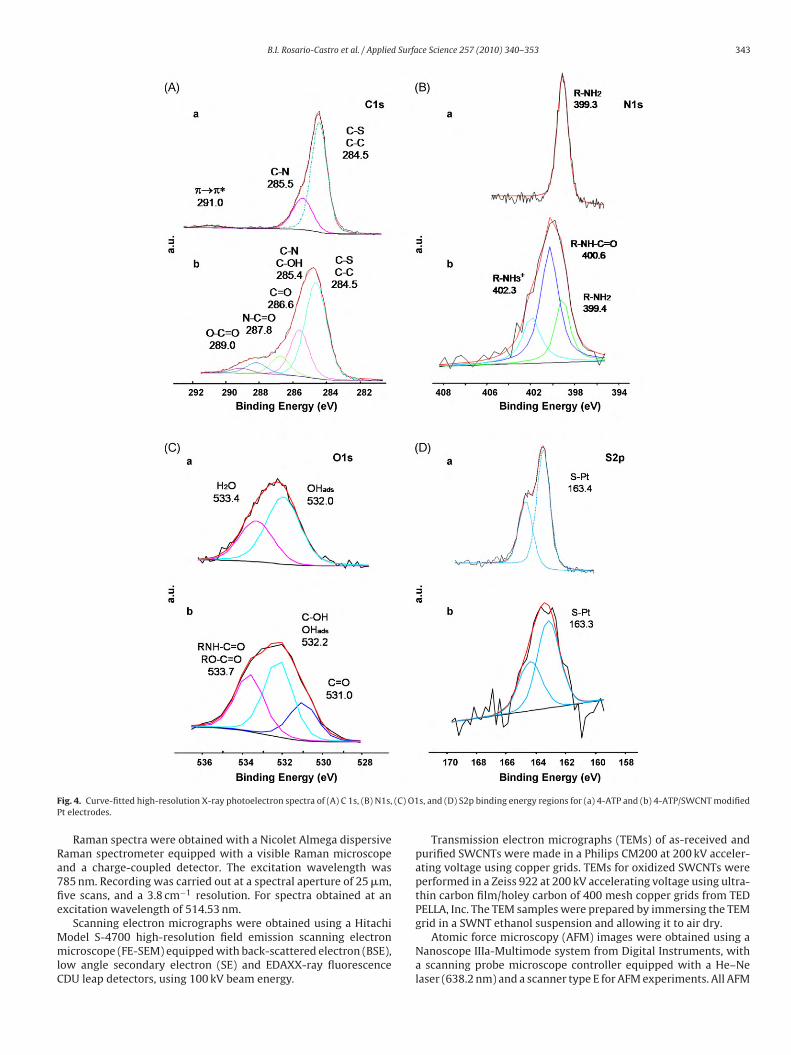
B.I. Rosario-Castro et al. / Applied Surface Science 257 (2010) 340–353 343
F (C) O1P
Ra7fie
MmlC
ig. 4. Curve-fitted high-resolution X-ray photoelectron spectra of (A) C 1s, (B) N1s,t electrodes.
Raman spectra were obtained with a Nicolet Almega dispersiveaman spectrometer equipped with a visible Raman microscopend a charge-coupled detector. The excitation wavelength was85 nm. Recording was carried out at a spectral aperture of 25 �m,ve scans, and a 3.8 cm−1 resolution. For spectra obtained at anxcitation wavelength of 514.53 nm.
Scanning electron micrographs were obtained using a Hitachiodel S-4700 high-resolution field emission scanning electronicroscope (FE-SEM) equipped with back-scattered electron (BSE),
ow angle secondary electron (SE) and EDAXX-ray fluorescenceDU leap detectors, using 100 kV beam energy.
s, and (D) S2p binding energy regions for (a) 4-ATP and (b) 4-ATP/SWCNT modified
Transmission electron micrographs (TEMs) of as-received andpurified SWCNTs were made in a Philips CM200 at 200 kV acceler-ating voltage using copper grids. TEMs for oxidized SWCNTs wereperformed in a Zeiss 922 at 200 kV accelerating voltage using ultra-thin carbon film/holey carbon of 400 mesh copper grids from TEDPELLA, Inc. The TEM samples were prepared by immersing the TEM
grid in a SWNT ethanol suspension and allowing it to air dry.Atomic force microscopy (AFM) images were obtained using aNanoscope IIIa-Multimode system from Digital Instruments, witha scanning probe microscope controller equipped with a He–Nelaser (638.2 nm) and a scanner type E for AFM experiments. All AFM
344 B.I. Rosario-Castro et al. / Applied Surface Science 257 (2010) 340–353
F of mag
is
teetwt
2
vtpacwattcdtiptt
2
aoeAuwb
2
tn
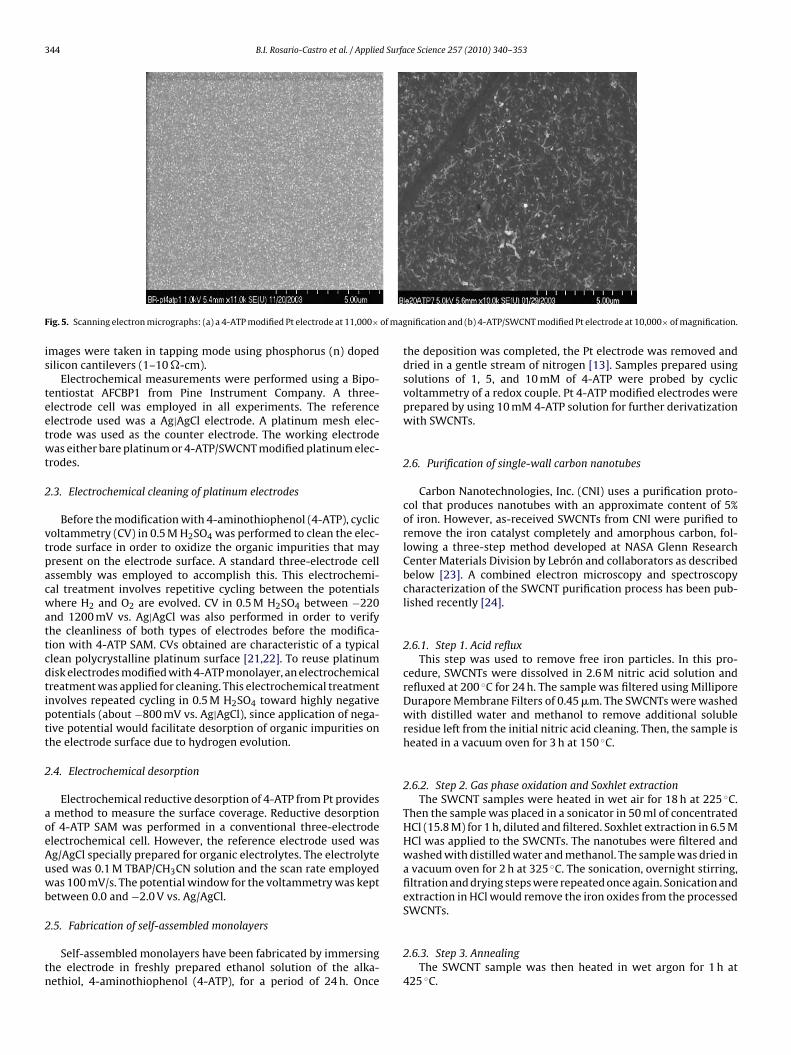
ig. 5. Scanning electron micrographs: (a) a 4-ATP modified Pt electrode at 11,000×
mages were taken in tapping mode using phosphorus (n) dopedilicon cantilevers (1–10 �-cm).
Electrochemical measurements were performed using a Bipo-entiostat AFCBP1 from Pine Instrument Company. A three-lectrode cell was employed in all experiments. The referencelectrode used was a Ag|AgCl electrode. A platinum mesh elec-rode was used as the counter electrode. The working electrodeas either bare platinum or 4-ATP/SWCNT modified platinum elec-
rodes.
.3. Electrochemical cleaning of platinum electrodes
Before the modification with 4-aminothiophenol (4-ATP), cyclicoltammetry (CV) in 0.5 M H2SO4 was performed to clean the elec-rode surface in order to oxidize the organic impurities that mayresent on the electrode surface. A standard three-electrode cellssembly was employed to accomplish this. This electrochemi-al treatment involves repetitive cycling between the potentialshere H2 and O2 are evolved. CV in 0.5 M H2SO4 between −220
nd 1200 mV vs. Ag|AgCl was also performed in order to verifyhe cleanliness of both types of electrodes before the modifica-ion with 4-ATP SAM. CVs obtained are characteristic of a typicallean polycrystalline platinum surface [21,22]. To reuse platinumisk electrodes modified with 4-ATP monolayer, an electrochemicalreatment was applied for cleaning. This electrochemical treatmentnvolves repeated cycling in 0.5 M H2SO4 toward highly negativeotentials (about −800 mV vs. Ag|AgCl), since application of nega-ive potential would facilitate desorption of organic impurities onhe electrode surface due to hydrogen evolution.
.4. Electrochemical desorption
Electrochemical reductive desorption of 4-ATP from Pt providesmethod to measure the surface coverage. Reductive desorption
f 4-ATP SAM was performed in a conventional three-electrodelectrochemical cell. However, the reference electrode used wasg/AgCl specially prepared for organic electrolytes. The electrolytesed was 0.1 M TBAP/CH3CN solution and the scan rate employedas 100 mV/s. The potential window for the voltammetry was kept
etween 0.0 and −2.0 V vs. Ag/AgCl.
.5. Fabrication of self-assembled monolayers
Self-assembled monolayers have been fabricated by immersinghe electrode in freshly prepared ethanol solution of the alka-ethiol, 4-aminothiophenol (4-ATP), for a period of 24 h. Once
nification and (b) 4-ATP/SWCNT modified Pt electrode at 10,000× of magnification.
the deposition was completed, the Pt electrode was removed anddried in a gentle stream of nitrogen [13]. Samples prepared usingsolutions of 1, 5, and 10 mM of 4-ATP were probed by cyclicvoltammetry of a redox couple. Pt 4-ATP modified electrodes wereprepared by using 10 mM 4-ATP solution for further derivatizationwith SWCNTs.
2.6. Purification of single-wall carbon nanotubes
Carbon Nanotechnologies, Inc. (CNI) uses a purification proto-col that produces nanotubes with an approximate content of 5%of iron. However, as-received SWCNTs from CNI were purified toremove the iron catalyst completely and amorphous carbon, fol-lowing a three-step method developed at NASA Glenn ResearchCenter Materials Division by Lebrón and collaborators as describedbelow [23]. A combined electron microscopy and spectroscopycharacterization of the SWCNT purification process has been pub-lished recently [24].
2.6.1. Step 1. Acid refluxThis step was used to remove free iron particles. In this pro-
cedure, SWCNTs were dissolved in 2.6 M nitric acid solution andrefluxed at 200 ◦C for 24 h. The sample was filtered using MilliporeDurapore Membrane Filters of 0.45 �m. The SWCNTs were washedwith distilled water and methanol to remove additional solubleresidue left from the initial nitric acid cleaning. Then, the sample isheated in a vacuum oven for 3 h at 150 ◦C.
2.6.2. Step 2. Gas phase oxidation and Soxhlet extractionThe SWCNT samples were heated in wet air for 18 h at 225 ◦C.
Then the sample was placed in a sonicator in 50 ml of concentratedHCl (15.8 M) for 1 h, diluted and filtered. Soxhlet extraction in 6.5 MHCl was applied to the SWCNTs. The nanotubes were filtered andwashed with distilled water and methanol. The sample was dried ina vacuum oven for 2 h at 325 ◦C. The sonication, overnight stirring,filtration and drying steps were repeated once again. Sonication andextraction in HCl would remove the iron oxides from the processedSWCNTs.
2.6.3. Step 3. AnnealingThe SWCNT sample was then heated in wet argon for 1 h at
425 ◦C.
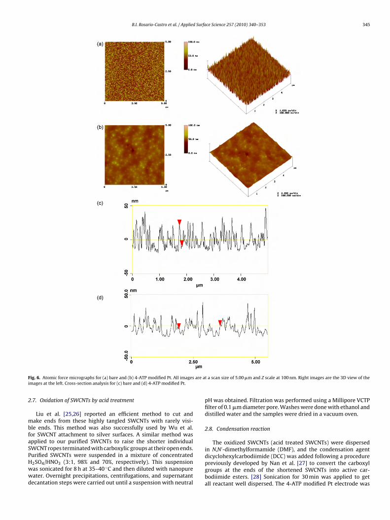
B.I. Rosario-Castro et al. / Applied Surface Science 257 (2010) 340–353 345
F are ai
2
mbfaSPHwwd
ig. 6. Atomic force micrographs for (a) bare and (b) 4-ATP modified Pt. All imagesmages at the left. Cross-section analysis for (c) bare and (d) 4-ATP modified Pt.
.7. Oxidation of SWCNTs by acid treatment
Liu et al. [25,26] reported an efficient method to cut andake ends from these highly tangled SWCNTs with rarely visi-
le ends. This method was also successfully used by Wu et al.or SWCNT attachment to silver surfaces. A similar method waspplied to our purified SWCNTs to raise the shorter individualWCNT ropes terminated with carboxylic groups at their open ends.
urified SWCNTs were suspended in a mixture of concentrated2SO4/HNO3 (3:1, 98% and 70%, respectively). This suspensionas sonicated for 8 h at 35–40 ◦C and then diluted with nanopureater. Overnight precipitations, centrifugations, and supernatantecantation steps were carried out until a suspension with neutralt a scan size of 5.00 �m and Z scale at 100 nm. Right images are the 3D view of the
pH was obtained. Filtration was performed using a Millipore VCTPfilter of 0.1 �m diameter pore. Washes were done with ethanol anddistilled water and the samples were dried in a vacuum oven.
2.8. Condensation reaction
The oxidized SWCNTs (acid treated SWCNTs) were dispersedin N,N′-dimethylformamide (DMF), and the condensation agent
dicyclohexylcarbodiimide (DCC) was added following a procedurepreviously developed by Nan et al. [27] to convert the carboxylgroups at the ends of the shortened SWCNTs into active car-bodiimide esters. [28] Sonication for 30 min was applied to getall reactant well dispersed. The 4-ATP modified Pt electrode was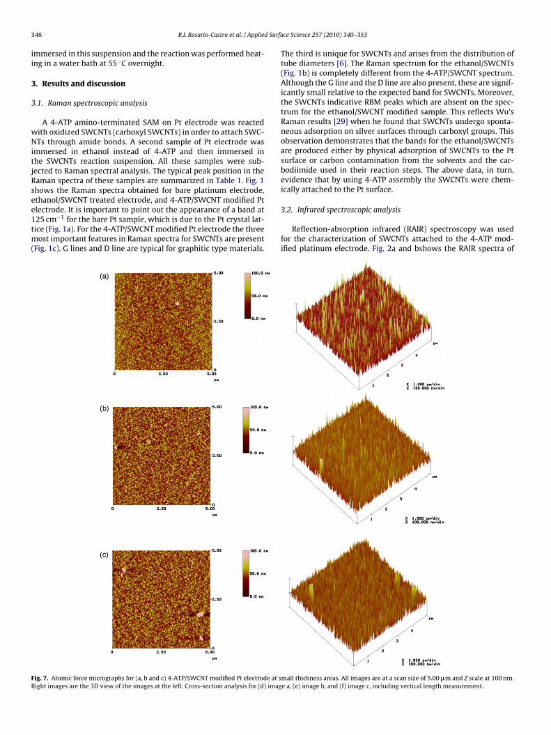
3 d Surfa
ii
3
3
wNitjRsee1tm(
FR
46 B.I. Rosario-Castro et al. / Applie
mmersed in this suspension and the reaction was performed heat-ng in a water bath at 55 ◦C overnight.
. Results and discussion
.1. Raman spectroscopic analysis
A 4-ATP amino-terminated SAM on Pt electrode was reactedith oxidized SWCNTs (carboxyl SWCNTs) in order to attach SWC-Ts through amide bonds. A second sample of Pt electrode was
mmersed in ethanol instead of 4-ATP and then immersed inhe SWCNTs reaction suspension. All these samples were sub-ected to Raman spectral analysis. The typical peak position in theaman spectra of these samples are summarized in Table 1. Fig. 1hows the Raman spectra obtained for bare platinum electrode,thanol/SWCNT treated electrode, and 4-ATP/SWCNT modified Pt
lectrode. It is important to point out the appearance of a band at25 cm−1 for the bare Pt sample, which is due to the Pt crystal lat-ice (Fig. 1a). For the 4-ATP/SWCNT modified Pt electrode the threeost important features in Raman spectra for SWCNTs are presentFig. 1c). G lines and D line are typical for graphitic type materials.
ig. 7. Atomic force micrographs for (a, b and c) 4-ATP/SWCNT modified Pt electrode at sight images are the 3D view of the images at the left. Cross-section analysis for (d) imag
ce Science 257 (2010) 340–353
The third is unique for SWCNTs and arises from the distribution oftube diameters [6]. The Raman spectrum for the ethanol/SWCNTs(Fig. 1b) is completely different from the 4-ATP/SWCNT spectrum.Although the G line and the D line are also present, these are signif-icantly small relative to the expected band for SWCNTs. Moreover,the SWCNTs indicative RBM peaks which are absent on the spec-trum for the ethanol/SWCNT modified sample. This reflects Wu’sRaman results [29] when he found that SWCNTs undergo sponta-neous adsorption on silver surfaces through carboxyl groups. Thisobservation demonstrates that the bands for the ethanol/SWCNTsare produced either by physical adsorption of SWCNTs to the Ptsurface or carbon contamination from the solvents and the car-bodiimide used in their reaction steps. The above data, in turn,evidence that by using 4-ATP assembly the SWCNTs were chem-ically attached to the Pt surface.
3.2. Infrared spectroscopic analysis
Reflection-absorption infrared (RAIR) spectroscopy was usedfor the characterization of SWCNTs attached to the 4-ATP mod-ified platinum electrode. Fig. 2a and bshows the RAIR spectra of
mall thickness areas. All images are at a scan size of 5.00 �m and Z scale at 100 nm.e a, (e) image b, and (f) image c, including vertical length measurement.
B.I. Rosario-Castro et al. / Applied Surfa
twsScciabtTTaaTNaacTstt
there must be contribution to the fitting peaks at 284.5 and
Fig. 7. (Continued ).
he 4-ATP modified Pt electrode before and after being derivatizedith carboxyl SWCNTs, respectively. Table 2 summarizes the peak
hifts for 4-ATP SAM and 4-ATP/SWCNT film. After treatment withWCNTS/DCC the �aNH2 amine bands at 3450 and 3352 cm−1 areonverted to the band at 3310 cm−1. Consequently, this new bandould be attributed to the �NH vibration of the amide functional-ty [30]. An additional new band from �C O appears at 1620 cm−1
nd is derived from the carboxyl groups of the nanotubes. Thisand, also named amide I, suggests amide bond formation betweenhe carboxylic groups of the SWCNTs and the 4-ATP amino groups.he �NH and �CN frequencies overlap, confirming the interaction.hat interaction results in the CNH vibrations, namely amide II andmide III [30]. For the SWCNT modified Pt surface the amide IInd amide III bands appear at 1578 and 1240 cm−1, respectively.he appearance of these bands suggests the attachment of SWC-Ts to the 4-ATP molecules through amide bonds. Furthermore, thebsence of the amine group peaks at the 4-ATP/SWCNT spectrumnd its chemical transformation to amide groups suggest that theonversions of the terminal groups proceeded in good yields [31].
he appearance of the �CN and �CSarom bands at the 4-ATP/SWCNTpectra is also significant. Thus, 4-ATP molecules are still bondedo the surface. This demonstrates that 4-ATP molecules toleratehe reaction conditions to serve as the bridge between the SWC-ce Science 257 (2010) 340–353 347
NTs and the Pt surface. There are two other peaks, �aC O at 1741and �sC O at 1700, which suggest additional bond formation. Itis well known that carbodiimide activated carboxyl groups canundergo condensation reaction in the presence of another activatedcarboxyl group [32]. Consequently, it is possible that two DCC acti-vated carboxyl groups on different carbon nanotubes react to formanhydride bond.
3.3. X-ray photoelectron spectroscopic analysis
X-ray photoelectron spectroscopy (XPS) measurements wereperformed to study the surface composition transformations thatthe 4-ATP SAM undergoes when it reacts with carboxyl SWCNTs.The XPS binding energy spectra for bare platinum, 4-ATP and 4-ATP/SWCNT treated platinum are shown in Fig. 3. The spectrumfor clean Pt (Fig. 3a) exhibited Pt peaks as well as smaller peaksfor C1s and O1s binding energies. The photoemission peak for C1smight arise from environment contamination, and the O1s peakmight arise from any Pt oxide formed at the surface of bare Pt.The XPS spectrum for 4-ATP modified Pt electrode (Fig. 3b) showsthe same peaks present at the bare Pt spectrum in addition tothe S2p, S2s, and N1s peaks. The appearance of photoemissionpeaks for nitrogen and sulfur is derived from 4-ATP moleculesadsorbed on Pt electrodes. As shown in Fig. 3c, when 4-ATP/Ptwas reacted with the carboxyl modified SWCNTs, a O1s bindingenergy peak with remarkably higher intensity was observed. Thisincrease in the O1s peak intensity could be due to the contribu-tion of oxygen-containing groups present at the SWCNTs, such ascarbonyl, hydroxyl, and carboxyl groups. The O/C concentrationwas calculated from the relative elemental composition obtainedfrom XPS analysis and the results are given in Table 3. The relativeO/C signal ratios were 0.00, 0.02, and 0.24 for bare, 4-ATP, and 4-ATP/SWCNT Pt electrodes. The relative amount of oxygen on the4-ATP/SWCNT sample is similar to that found for oxidized SWCNTsand more than 10 times higher than for the 4-ATP SAM sample.All these results are indicative of the SWCNT attachment to 4-ATPmonolayer on Pt surface. This in turn is indicative of the adsorptionof SWCNTs to the 4-ATP Pt electrode.
Another difference between 4-ATP and 4-ATP/SWCNT spectrais that the signals corresponding to sulfur and nitrogen have lowerrelative intensity at the 4-ATP/SWCNT spectrum than at the 4-ATPspectrum. Once the SWCNTs are chemically attached over the 4-ATP molecules they serve as a blocking barrier that prevents thesulfur and nitrogen photoelectrons access to the film/vacuum inter-face. Only those electrons emitted from the surface zone that havesuffered no energy loss will contribute to the photoemission peaks.As a final point, the background of the spectra increases from barePt to 4-ATP/SWCNTs. Again, both 4-ATP/SWCNTs and 4-ATP mono-layer serve as blocking barriers. Although photoelectrons from the4-ATP modified sample have better access to the film/vacuum inter-face, some electrons emitted have lost some energy due to inelasticinteractions and contribute to the scattering background. SinceSWCNT film has a higher blocking potential, and as a consequence,more electron energy losses occur and the background becomeshigher.
XPS high-resolution analysis was performed for the charac-terization of SWCNT attachment to the amino-terminated SAM.A curve-fitted spectrum for the C1s region is shown in Fig. 4.The curve-fitting peaks at binding energies of 284.5, 285.5, 286.6and 288.9 eV, are attributed to C—C, C—OH, C O, O—C O func-tional groups, as reported for oxidized SWCNTs [33,34]. However,
285.5 eV from the C–S and C–N bonded carbons in the 4-ATPmolecules. This makes it more difficult to obtain an absolute quan-tization from the atomic percent of these fitting peaks. The peakat 288.0 eV could be assigned to N—C O from the amide bond
348 B.I. Rosario-Castro et al. / Applied Surface Science 257 (2010) 340–353
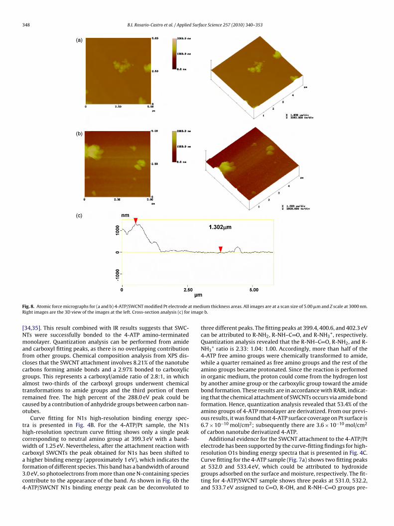
F t medR imag
[Nmafccgatrco
thcwcaf3c4
ig. 8. Atomic force micrographs for (a and b) 4-ATP/SWCNT modified Pt electrode aight images are the 3D view of the images at the left. Cross-section analysis (c) for
34,35]. This result combined with IR results suggests that SWC-Ts were successfully bonded to the 4-ATP amino-terminatedonolayer. Quantization analysis can be performed from amide
nd carboxyl fitting peaks, as there is no overlapping contributionrom other groups. Chemical composition analysis from XPS dis-loses that the SWCNT attachment involves 8.21% of the nanotubearbons forming amide bonds and a 2.97% bonded to carboxylicroups. This represents a carboxyl/amide ratio of 2.8:1, in whichlmost two-thirds of the carboxyl groups underwent chemicalransformations to amide groups and the third portion of thememained free. The high percent of the 288.0 eV peak could beaused by a contribution of anhydride groups between carbon nan-tubes.
Curve fitting for N1s high-resolution binding energy spec-ra is presented in Fig. 4B. For the 4-ATP/Pt sample, the N1sigh-resolution spectrum curve fitting shows only a single peakorresponding to neutral amino group at 399.3 eV with a band-idth of 1.25 eV. Nevertheless, after the attachment reaction with
arboxyl SWCNTs the peak obtained for N1s has been shifted to
higher binding energy (approximately 1 eV), which indicates theormation of different species. This band has a bandwidth of around.0 eV, so photoelectrons from more than one N-containing speciesontribute to the appearance of the band. As shown in Fig. 6b the-ATP/SWCNT N1s binding energy peak can be deconvoluted to
ium thickness areas. All images are at a scan size of 5.00 �m and Z scale at 3000 nm.e b.
three different peaks. The fitting peaks at 399.4, 400.6, and 402.3 eVcan be attributed to R-NH2, R-NH–C O, and R-NH3
+, respectively.Quantization analysis revealed that the R-NH–C O, R-NH2, and R-NH3
+ ratio is 2.33: 1.04: 1.00. Accordingly, more than half of the4-ATP free amino groups were chemically transformed to amide,while a quarter remained as free amino groups and the rest of theamino groups became protonated. Since the reaction is performedin organic medium, the proton could come from the hydrogen lostby another amine group or the carboxylic group toward the amidebond formation. These results are in accordance with RAIR, indicat-ing that the chemical attachment of SWCNTs occurs via amide bondformation. Hence, quantization analysis revealed that 53.4% of theamino groups of 4-ATP monolayer are derivatized. From our previ-ous results, it was found that 4-ATP surface coverage on Pt surface is6.7 × 10−10 mol/cm2; subsequently there are 3.6 × 10⁻¹⁰ mol/cm2
of carbon nanotube derivatized 4-ATP.Additional evidence for the SWCNT attachment to the 4-ATP/Pt
electrode has been supported by the curve-fitting findings for high-resolution O1s binding energy spectra that is presented in Fig. 4C.
Curve fitting for the 4-ATP sample (Fig. 7a) shows two fitting peaksat 532.0 and 533.4 eV, which could be attributed to hydroxidegroups adsorbed on the surface and moisture, respectively. The fit-ting for 4-ATP/SWCNT sample shows three peaks at 531.0, 532.2,and 533.7 eV assigned to C O, R-OH, and R-NH–C O groups pre-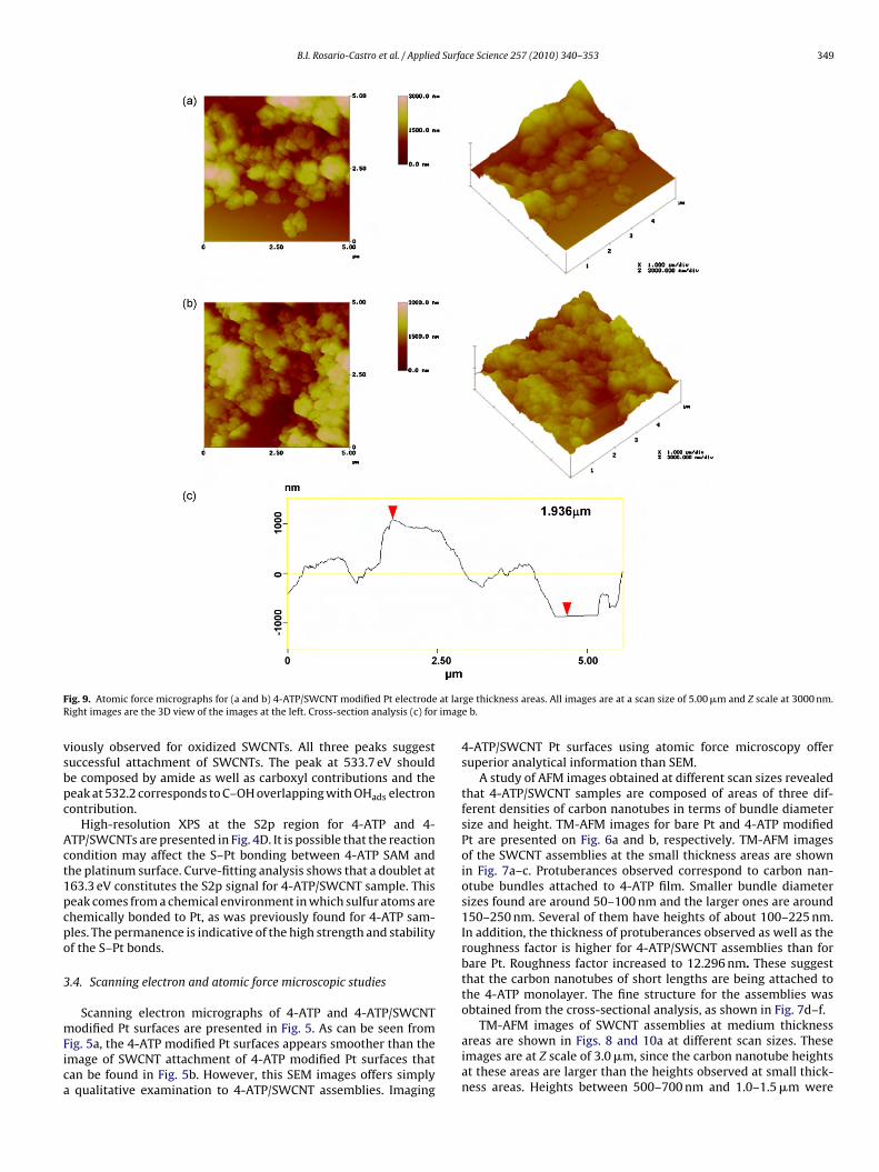
B.I. Rosario-Castro et al. / Applied Surface Science 257 (2010) 340–353 349
F at larR imag
vsbpc
Act1pcpo
3
mFica
ig. 9. Atomic force micrographs for (a and b) 4-ATP/SWCNT modified Pt electrodeight images are the 3D view of the images at the left. Cross-section analysis (c) for
iously observed for oxidized SWCNTs. All three peaks suggestuccessful attachment of SWCNTs. The peak at 533.7 eV shoulde composed by amide as well as carboxyl contributions and theeak at 532.2 corresponds to C–OH overlapping with OHads electronontribution.
High-resolution XPS at the S2p region for 4-ATP and 4-TP/SWCNTs are presented in Fig. 4D. It is possible that the reactionondition may affect the S–Pt bonding between 4-ATP SAM andhe platinum surface. Curve-fitting analysis shows that a doublet at63.3 eV constitutes the S2p signal for 4-ATP/SWCNT sample. Thiseak comes from a chemical environment in which sulfur atoms arehemically bonded to Pt, as was previously found for 4-ATP sam-les. The permanence is indicative of the high strength and stabilityf the S–Pt bonds.
.4. Scanning electron and atomic force microscopic studies
Scanning electron micrographs of 4-ATP and 4-ATP/SWCNT
odified Pt surfaces are presented in Fig. 5. As can be seen fromig. 5a, the 4-ATP modified Pt surfaces appears smoother than themage of SWCNT attachment of 4-ATP modified Pt surfaces thatan be found in Fig. 5b. However, this SEM images offers simplyqualitative examination to 4-ATP/SWCNT assemblies. Imaging
ge thickness areas. All images are at a scan size of 5.00 �m and Z scale at 3000 nm.e b.
4-ATP/SWCNT Pt surfaces using atomic force microscopy offersuperior analytical information than SEM.
A study of AFM images obtained at different scan sizes revealedthat 4-ATP/SWCNT samples are composed of areas of three dif-ferent densities of carbon nanotubes in terms of bundle diametersize and height. TM-AFM images for bare Pt and 4-ATP modifiedPt are presented on Fig. 6a and b, respectively. TM-AFM imagesof the SWCNT assemblies at the small thickness areas are shownin Fig. 7a–c. Protuberances observed correspond to carbon nan-otube bundles attached to 4-ATP film. Smaller bundle diametersizes found are around 50–100 nm and the larger ones are around150–250 nm. Several of them have heights of about 100–225 nm.In addition, the thickness of protuberances observed as well as theroughness factor is higher for 4-ATP/SWCNT assemblies than forbare Pt. Roughness factor increased to 12.296 nm. These suggestthat the carbon nanotubes of short lengths are being attached tothe 4-ATP monolayer. The fine structure for the assemblies wasobtained from the cross-sectional analysis, as shown in Fig. 7d–f.
TM-AFM images of SWCNT assemblies at medium thicknessareas are shown in Figs. 8 and 10a at different scan sizes. Theseimages are at Z scale of 3.0 �m, since the carbon nanotube heightsat these areas are larger than the heights observed at small thick-ness areas. Heights between 500–700 nm and 1.0–1.5 �m were
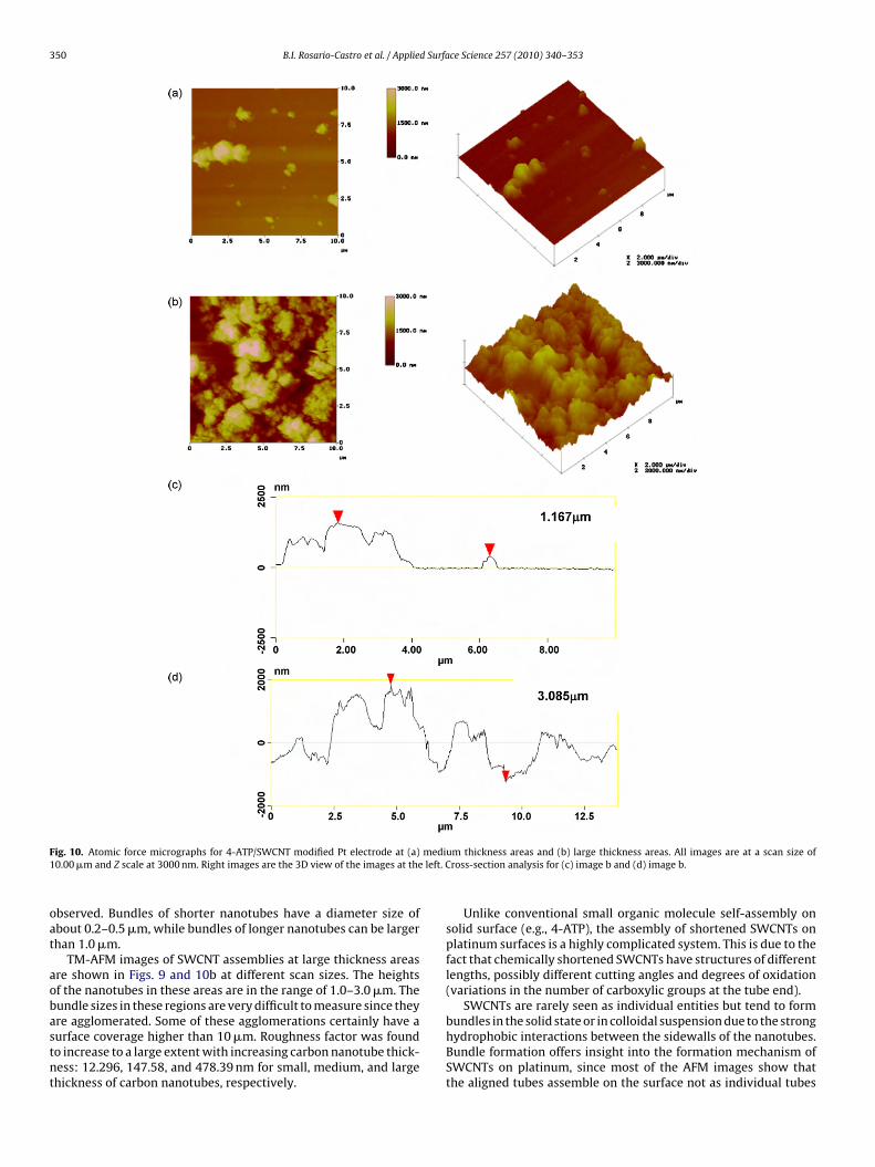
350 B.I. Rosario-Castro et al. / Applied Surface Science 257 (2010) 340–353
F medi1 left. C
oat
aobastnt
ig. 10. Atomic force micrographs for 4-ATP/SWCNT modified Pt electrode at (a)0.00 �m and Z scale at 3000 nm. Right images are the 3D view of the images at the
bserved. Bundles of shorter nanotubes have a diameter size ofbout 0.2–0.5 �m, while bundles of longer nanotubes can be largerhan 1.0 �m.
TM-AFM images of SWCNT assemblies at large thickness areasre shown in Figs. 9 and 10b at different scan sizes. The heightsf the nanotubes in these areas are in the range of 1.0–3.0 �m. Theundle sizes in these regions are very difficult to measure since they
re agglomerated. Some of these agglomerations certainly have aurface coverage higher than 10 �m. Roughness factor was foundo increase to a large extent with increasing carbon nanotube thick-ess: 12.296, 147.58, and 478.39 nm for small, medium, and largehickness of carbon nanotubes, respectively.um thickness areas and (b) large thickness areas. All images are at a scan size ofross-section analysis for (c) image b and (d) image b.
Unlike conventional small organic molecule self-assembly onsolid surface (e.g., 4-ATP), the assembly of shortened SWCNTs onplatinum surfaces is a highly complicated system. This is due to thefact that chemically shortened SWCNTs have structures of differentlengths, possibly different cutting angles and degrees of oxidation(variations in the number of carboxylic groups at the tube end).
SWCNTs are rarely seen as individual entities but tend to form
bundles in the solid state or in colloidal suspension due to the stronghydrophobic interactions between the sidewalls of the nanotubes.Bundle formation offers insight into the formation mechanism ofSWCNTs on platinum, since most of the AFM images show thatthe aligned tubes assemble on the surface not as individual tubes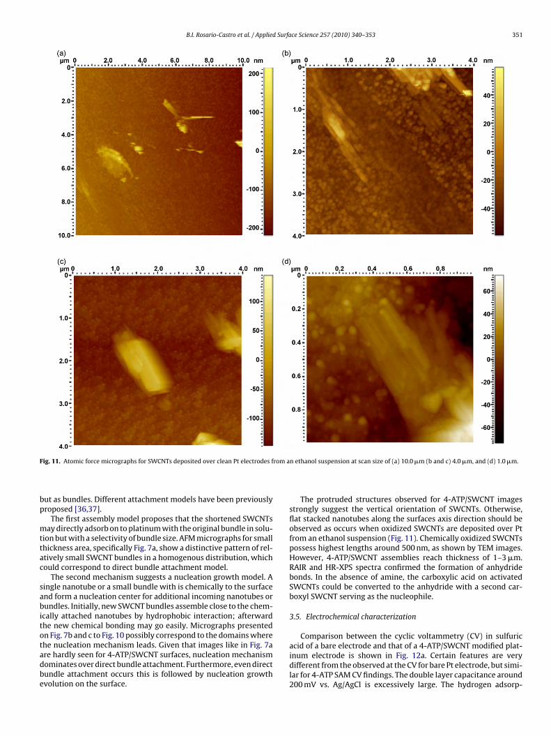
B.I. Rosario-Castro et al. / Applied Surface Science 257 (2010) 340–353 351
F rom a
bp
mttac
sabitotadbe
ig. 11. Atomic force micrographs for SWCNTs deposited over clean Pt electrodes f
ut as bundles. Different attachment models have been previouslyroposed [36,37].
The first assembly model proposes that the shortened SWCNTsay directly adsorb on to platinum with the original bundle in solu-
ion but with a selectivity of bundle size. AFM micrographs for smallhickness area, specifically Fig. 7a, show a distinctive pattern of rel-tively small SWCNT bundles in a homogenous distribution, whichould correspond to direct bundle attachment model.
The second mechanism suggests a nucleation growth model. Aingle nanotube or a small bundle with is chemically to the surfacend form a nucleation center for additional incoming nanotubes orundles. Initially, new SWCNT bundles assemble close to the chem-
cally attached nanotubes by hydrophobic interaction; afterwardhe new chemical bonding may go easily. Micrographs presentedn Fig. 7b and c to Fig. 10 possibly correspond to the domains where
he nucleation mechanism leads. Given that images like in Fig. 7are hardly seen for 4-ATP/SWCNT surfaces, nucleation mechanismominates over direct bundle attachment. Furthermore, even directundle attachment occurs this is followed by nucleation growthvolution on the surface.n ethanol suspension at scan size of (a) 10.0 �m (b and c) 4.0 �m, and (d) 1.0 �m.
The protruded structures observed for 4-ATP/SWCNT imagesstrongly suggest the vertical orientation of SWCNTs. Otherwise,flat stacked nanotubes along the surfaces axis direction should beobserved as occurs when oxidized SWCNTs are deposited over Ptfrom an ethanol suspension (Fig. 11). Chemically oxidized SWCNTspossess highest lengths around 500 nm, as shown by TEM images.However, 4-ATP/SWCNT assemblies reach thickness of 1–3 �m.RAIR and HR-XPS spectra confirmed the formation of anhydridebonds. In the absence of amine, the carboxylic acid on activatedSWCNTs could be converted to the anhydride with a second car-boxyl SWCNT serving as the nucleophile.
3.5. Electrochemical characterization
Comparison between the cyclic voltammetry (CV) in sulfuric
acid of a bare electrode and that of a 4-ATP/SWCNT modified plat-inum electrode is shown in Fig. 12a. Certain features are verydifferent from the observed at the CV for bare Pt electrode, but simi-lar for 4-ATP SAM CV findings. The double layer capacitance around200 mV vs. Ag/AgCl is excessively large. The hydrogen adsorp-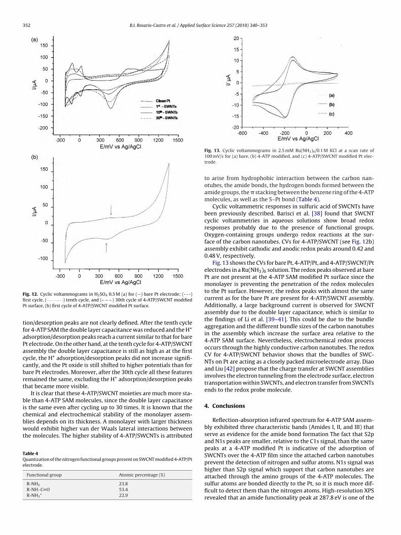
352 B.I. Rosario-Castro et al. / Applied Surface Science 257 (2010) 340–353
FfiP
tfaPaccbrt
bicbwt
TQe
ig. 12. Cyclic voltammograms in H2SO4 0.5 M (a) for (—) bare Pt electrode; (- - -)rst cycle, (· · · · · · · · ·) tenth cycle, and (-· -· -·) 30th cycle of 4-ATP/SWCNT modifiedt surface, (b) first cycle of 4-ATP/SWCNT modified Pt surface.
ion/desorption peaks are not clearly defined. After the tenth cycleor 4-ATP SAM the double layer capacitance was reduced and the H+
dsorption/desorption peaks reach a current similar to that for baret electrode. On the other hand, at the tenth cycle for 4-ATP/SWCNTssembly the double layer capacitance is still as high as at the firstycle, the H+ adsorption/desorption peaks did not increase signifi-antly, and the Pt oxide is still shifted to higher potentials than forare Pt electrodes. Moreover, after the 30th cycle all these featuresemained the same, excluding the H+ adsorption/desorption peakshat became more visible.
It is clear that these 4-ATP/SWCNT moieties are much more sta-le than 4-ATP SAM molecules, since the double layer capacitance
s the same even after cycling up to 30 times. It is known that the
hemical and electrochemical stability of the monolayer assem-lies depends on its thickness. A monolayer with larger thicknessould exhibit higher van der Waals lateral interactions betweenhe molecules. The higher stability of 4-ATP/SWCNTs is attributed
able 4uantization of the nitrogen functional groups present on SWCNT modified 4-ATP/Ptlectrode.
Functional group Atomic percentage (%)
R-NH2 23.8R-NH–C O 53.4R-NH3
+ 22.9
Fig. 13. Cyclic voltammograms in 2.5 mM Ru(NH3)6/0.1 M KCl at a scan rate of100 mV/s for (a) bare, (b) 4-ATP modified, and (c) 4-ATP/SWCNT modified Pt elec-trode.
to arise from hydrophobic interaction between the carbon nan-otubes, the amide bonds, the hydrogen bonds formed between theamide groups, the � stacking between the benzene ring of the 4-ATPmolecules, as well as the S–Pt bond (Table 4).
Cyclic voltammetric responses in sulfuric acid of SWCNTs havebeen previously described. Barisci et al. [38] found that SWCNTcyclic voltammetries in aqueous solutions show broad redoxresponses probably due to the presence of functional groups.Oxygen-containing groups undergo redox reactions at the sur-face of the carbon nanotubes. CVs for 4-ATP/SWCNT (see Fig. 12b)assembly exhibit cathodic and anodic redox peaks around 0.42 and0.48 V, respectively.
Fig. 13 shows the CVs for bare Pt, 4-ATP/Pt, and 4-ATP/SWCNT/Ptelectrodes in a Ru(NH3)6 solution. The redox peaks observed at barePt are not present at the 4-ATP SAM modified Pt surface since themonolayer is preventing the penetration of the redox moleculesto the Pt surface. However, the redox peaks with almost the samecurrent as for the bare Pt are present for 4-ATP/SWCNT assembly.Additionally, a large background current is observed for SWCNTassembly due to the double layer capacitance, which is similar tothe findings of Li et al. [39–41]. This could be due to the bundleaggregation and the different bundle sizes of the carbon nanotubesin the assembly which increase the surface area relative to the4-ATP SAM surface. Nevertheless, electrochemical redox processoccurs through the highly conductive carbon nanotubes. The redoxCV for 4-ATP/SWCNT behavior shows that the bundles of SWC-NTs on Pt are acting as a closely packed microelectrode array. Diaoand Liu [42] propose that the charge transfer at SWCNT assembliesinvolves the electron tunneling from the electrode surface, electrontransportation within SWCNTs, and electron transfer from SWCNTsends to the redox probe molecule.
4. Conclusions
Reflection-absorption infrared spectrum for 4-ATP SAM assem-bly exhibited three characteristic bands (Amides I, II, and III) thatserve as evidence for the amide bond formation The fact that S2pand N1s peaks are smaller, relative to the C1s signal, than the samepeaks at a 4-ATP modified Pt is indicative of the adsorption ofSWCNTs over the 4-ATP film since the attached carbon nanotubesprevent the detection of nitrogen and sulfur atoms. N1s signal was
higher than S2p signal which support that carbon nanotubes areattached through the amino groups of the 4-ATP molecules. Thesulfur atoms are bonded directly to the Pt, so it is much more dif-ficult to detect them than the nitrogen atoms. High-resolution XPSrevealed that an amide functionality peak at 287.8 eV is one of the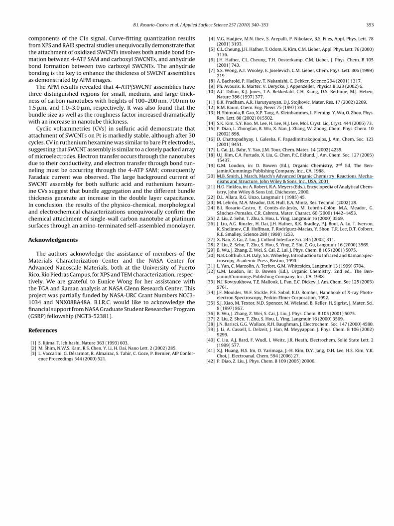
d Surfa
cftmbba
tn1bw
acsodnFSitIacs
A
MARttp1fi(
R
[
[[[
[[
[
[[
[
[
[
[[[
[[
[[[[
[[
[
[
[
[[[
B.I. Rosario-Castro et al. / Applie
omponents of the C1s signal. Curve-fitting quantization resultsrom XPS and RAIR spectral studies unequivocally demonstrate thathe attachment of oxidized SWCNTs involves both amide bond for-
ation between 4-ATP SAM and carboxyl SWCNTs, and anhydrideond formation between two carboxyl SWCNTs. The anhydrideonding is the key to enhance the thickness of SWCNT assembliess demonstrated by AFM images.
The AFM results revealed that 4-ATP/SWCNT assemblies havehree distinguished regions for small, medium, and large thick-ess of carbon nanotubes with heights of 100–200 nm, 700 nm to.5 �m, and 1.0–3.0 �m, respectively. It was also found that theundle size as well as the roughness factor increased dramaticallyith an increase in nanotube thickness.
Cyclic voltammetries (CVs) in sulfuric acid demonstrate thatttachment of SWCNTs on Pt is markedly stable, although after 30ycles. CV in ruthenium hexamine was similar to bare Pt electrodes,uggesting that SWCNT assembly is similar to a closely packed arrayf microelectrodes. Electron transfer occurs through the nanotubesue to their conductivity, and electron transfer through bond tun-eling must be occurring through the 4-ATP SAM; consequentlyaradaic current was observed. The large background current ofWCNT assembly for both sulfuric acid and ruthenium hexam-ne CVs suggest that bundle aggregation and the different bundlehickness generate an increase in the double layer capacitance.n conclusion, the results of the physico-chemical, morphologicalnd electrochemical characterizations unequivocally confirm thehemical attachment of single-wall carbon nanotube at platinumurfaces through an amino-terminated self-assembled monolayer.
cknowledgments
The authors acknowledge the assistance of members of theaterials Characterization Center and the NASA Center for
dvanced Nanoscale Materials, both at the University of Puertoico, Rio Piedras Campus, for XPS and TEM characterization, respec-ively. We are grateful to Eunice Wong for her assistance withhe TGA and Raman analysis at NASA Glenn Research Center. Thisroject was partially funded by NASA-URC Grant Numbers NCC3-034 and NNX08BA48A. B.I.R.C. would like to acknowledge thenancial support from NASA Graduate Student Researcher ProgramGSRP) fellowship (NGT3-52381).
eferences
[1] S. Iijima, T. Ichihashi, Nature 363 (1993) 603.[2] M. Shim, N.W.S. Kam, R.S. Chen, Y. Li, H. Dai, Nano Lett. 2 (2002) 285.[3] L. Vaccarini, G. Désarmot, R. Almairac, S. Tahir, C. Goze, P. Bernier, AIP Confer-
ence Proceedings 544 (2000) 521.
[
[
[
[
ce Science 257 (2010) 340–353 353
[4] V.G. Hadjiev, M.N. Iliev, S. Arepalli, P. Nikolaev, B.S. Files, Appl. Phys. Lett. 78(2001) 3193.
[5] C.L. Cheung, J.H. Hafner, T. Odom, K. Kim, C.M. Lieber, Appl. Phys. Lett. 76 (2000)3136.
[6] J.H. Hafner, C.L. Cheung, T.H. Oosterkamp, C.M. Lieber, J. Phys. Chem. B 105(2001) 743.
[7] S.S. Wong, A.T. Wooley, E. Joselevich, C.M. Lieber, Chem. Phys. Lett. 306 (1999)219.
[8] A. Bachtold, P. Hadley, T. Nakanishi, C. Dekker, Science 294 (2001) 1317.[9] Ph. Avouris, R. Marter, V. Derycke, J. Appenzeller, Physica B 323 (2002) 6.10] A.C. Dillon, K.J. Jones, T.A. Bekkedahl, C.H. Kiang, D.S. Bethune, M.J. Heben,
Nature 386 (1997) 377.11] B.K. Pradham, A.R. Harutyunyan, D.J. Stojkovic, Mater. Res. 17 (2002) 2209.12] R.M. Baum, Chem. Eng. News 75 (1997) 39.13] H. Shimoda, B. Gao, X.P. Tang, A. Kleinhammes, L. Fleming, Y. Wu, O. Zhou, Phys.
Rev. Lett. 88 (2002) 015502.14] S.K. Kim, S.Y. Koo, M. Lee, H. Lee, H.J. Lee, Mol. Cryst. Liq. Cryst. 444 (2006) 73.15] P. Diao, L. Zhongfan, B. Wu, X. Nan, J. Zhang, W. Zhong, Chem. Phys. Chem. 10
(2002) 898.16] D. Chattopadhyay, I. Galeska, F. Papadimitrakopoulos, J. Am. Chem. Soc. 123
(2001) 9451.17] L. Cai, J.L. Bahr, Y. Yao, J.M. Tour, Chem. Mater. 14 (2002) 4235.18] U.J. Kim, C.A. Furtado, X. Liu, G. Chen, P.C. Eklund, J. Am. Chem. Soc. 127 (2005)
15437.19] G.M. Loudon, in: D. Bowen (Ed.), Organic Chemistry, 2nd Ed, The Ben-
jamin/Cummings Publishing Company, Inc., CA, 1988.20] M.B. Smith, J. March, March’s Advanced Organic Chemistry: Reactions, Mecha-
nisms and Structure, John Wiley & Sons, Inc., USA, 2001.21] H.O. Finklea, in: A. Robert, R.A. Meyers (Eds.), Encyclopedia of Analytical Chem-
istry, John Wiley & Sons Ltd, Chichester, 2000.22] D.L. Allara, R.G. Unzo, Langmuir 1 (1985) 45.23] M. Lebrón, M.A. Meador, D.R. Hull, E.A. Mintz, Res. Technol. (2002) 29.24] B.I. Rosario-Castro, E. Contés-de-Jesús, M. Lebrón-Colón, M.A. Meador, G.
Sánchez-Pomales, C.R. Cabrera, Mater. Charact. 60 (2009) 1442–1453.25] Z. Liu, Z. Sehn, T. Zhu, S. Hou, L. Ying, Langmuir 16 (2000) 3569.26] J. Liu, A.G. Rinzler, H. Dai, J.H. Hafner, R.K. Bradley, P.J. Boul, A. Lu, T. Iverson,
K. Shelimov, C.B. Huffman, F. Rodríguez-Macias, Y. Shon, T.R. Lee, D.T. Colbert,R.E. Smalley, Science 280 (1998) 1253.
27] X. Nan, Z. Gu, Z. Liu, J. Colloid Interface Sci. 245 (2002) 311.28] Z. Liu, Z. Sehn, T. Zhu, S. Hou, S. Ying, Z. Shi, Z. Gu, Langmuir 16 (2000) 3569.29] B. Wu, J. Zhang, Z. Wei, S. Cai, Z. Lui, J. Phys. Chem. B 105 (2001) 5075.30] N.B. Colthub, L.H. Daly, S.E. Wiberley, Introduction to Infrared and Raman Spec-
troscopy, Academic Press, Boston, 1990.31] L. Yan, C. Marzolin, A. Terfort, G.M. Whitesides, Langmuir 13 (1999) 6704.32] G.M. Loudon, in: D. Bowen (Ed.), Organic Chemistry, 2nd ed., The Ben-
jamin/Cummings Publishing Company, Inc., CA, 1988.33] N.I. Kovtyukhova, T.E. Mallouk, L. Pan, E.C. Dickey, J. Am. Chem. Soc 125 (2003)
9761.34] J.F. Moulder, W.F. Stickle, P.E. Sobol, K.D. Bomber, Handbook of X-ray Photo-
electron Spectroscopy, Perkin-Elmer Corporation, 1992.35] S.J. Xiao, M. Textor, N.D. Spencer, M. Wieland, B. Keller, H. Sigrist, J. Mater. Sci.
8 (1997) 867.36] B. Wu, J. Zhang, Z. Wei, S. Cai, J. Liu, J. Phys. Chem. B 105 (2001) 5075.37] Z. Liu, Z. Shen, T. Zhu, S. Hou, L. Ying, Langmuir 16 (2000) 3569.38] J.N. Barisci, G.G. Wallace, R.H. Baughman, J. Electrochem. Soc. 147 (2000) 4580.39] J. Li, A. Cassell, L. Delzeit, J. Han, M. Meyyappan, J. Phys. Chem. B 106 (2002)
9299.40] C. Liu, A.J. Bard, F. Wudl, I. Weitz, J.R. Heath, Electrochem. Solid State Lett. 2
(1999) 577.41] X.J. Huang, H.S. Im, O. Yarimaga, J.-H. Kim, D.Y. Jang, D.H. Lee, H.S. Kim, Y.K.
Choi, J. Electroanal. Chem. 594 (2006) 27.42] P. Diao, Z. Liu, J. Phys. Chem. B 109 (2005) 20906.
Related Documents