Single-molecule Study on the Temperature-sensitive Reaction of F 1 -ATPase with a Hybrid F 1 Carrying a Single (E190D) * □ S Received for publication, June 1, 2009, and in revised form, June 25, 2009 Published, JBC Papers in Press, June 26, 2009, DOI 10.1074/jbc.M109.026401 Sawako Enoki, Rikiya Watanabe, Ryota Iino, and Hiroyuki Noji 1 From the Institute of Scientific and Industrial Research, Osaka University, 567-0047 Osaka, Japan F 1 -ATPase is a rotary molecular motor in which the -subunit rotates against the 3 3 cylinder. The unitary -rotation is a 120° step comprising 80 and 40° substeps, each of these initiated by ATP binding and ADP release and by ATP hydrolysis and inorganic phosphate release, respectively. In our previous study on -rotation at low temperatures, a highly temperature-sensi- tive (TS) reaction step of F 1 -ATPase from thermophilic Bacillus PS3 was found below 9 °C as an intervening pause before the 80° substep at the same angle for ATP binding and ADP release. However, it remains unclear as to which reaction step the TS reaction corresponds. In this study, we found that the mutant F 1 (E190D) from thermophilic Bacillus PS3 showed a clear pause of the TS reaction below 18 °C. In an attempt to identify the catalytic state of the TS reaction, the rotation of the hybrid F 1 , carrying a single copy of E190D, was observed at 18 °C. The hybrid F 1 showed a pause of the TS reaction at the same angle as for the ATP binding of the incorporated E190D, although kinetic analysis revealed that the TS reaction is not the ATP binding step. These findings suggest that the TS reaction is a structural rearrangement of before or after ATP binding. F 1 -ATPase (F 1 ) 2 is an ATP-driven rotary motor protein. The subunit composition of the bacterial F 1 -ATPase is 3 3 , and the minimum complex of F 1 -ATPase as a rotary motor is 3 3 subcomplex. This motor protein forms the F o F 1 -ATP synthase complex by binding to another rotary motor, namely, F o , which is driven by the proton flux resulting from the proton motive force across the membranes (1– 4). Under physiological condi- tions, where the proton motive force is sufficiently large, F o forcibly rotates F 1 -ATPase in the reverse direction of F 1 -ATPase, leading the reverse reaction of ATP hydrolysis, i.e. ATP synthesis from ADP and inorganic phosphate (P i ). When the proton motive force diminishes or F 1 is isolated from F o , F 1 -ATPase hydrolyzes ATP to rotate the -subunit against the 3 3 stator ring in the counterclockwise direction as viewed from the F o side (5). The catalytic sites are located at the inter- face of the - and -subunits, predominantly on the -subunit (6). Each -subunit carries out a single turnover of ATP hydrol- ysis during the -rotation of 360° following the common cata- lytic reaction pathway, whereas they are 120° different in the catalytic phase. In this manner, the three -subunits undergo different reaction steps of ATP hydrolysis upon each rotational step. The rotary motion of the -subunit has been demon- strated by biochemical (7) and spectroscopic methods (8) and directly proved in single-molecule observation studies (5). Since the establishment of the single-molecule rotation assay, the chemomechanical coupling scheme of F 1 has been studied extensively by resolving the rotation into discrete steps. The stepping rotation was first observed under an ATP-limit- ing condition where F 1 makes discrete 120° steps upon ATP binding (9). Then, high speed imaging of the rotation with a small probe of low friction was performed, which revealed that the 120° step comprises 80 and 40° substeps, each initiated by ATP binding, and two unknown consecutive reactions, respec- tively (10). This finding necessitated the identification of the two reactions that trigger the 40° substep. Hence, the rotation assay was performed using a mutant, namely F 1 (E190D), and a slowly hydrolyzed ATP analog, namely ATPS (11). Glutamate 190 of the -subunit of F 1 , derived from thermophilic Bacillus PS3 and the corresponding glutamates from other F 1 -ATPases (Glu-181 of F 1 from Escherichia coli and Glu-188 of F 1 from bovine mitochondria), has been identified as one of the most critical catalytic residues for ATP hydrolysis (6, 12–15). When this glutamate was substituted with aspartic acid, which has a shorter side chain than that of glutamate, the ATP cleavage step of F 1 was drastically slowed. In the rotation assay, this mutant showed a distinct long pause before the 40° substep. ATPS also caused a long pause before the 40° substep. These observations established that the 40° substep is initiated by hydrolysis. Accordingly, the pause angles before the 80 and 40° substeps are referred to as to the binding angle and the catalytic angle, respectively. Then, the rotation assay was performed in the presence of a high amount of P i in the solution. It was shown that P i rebinding caused the long pause at the catalytic angle, suggesting that P i is released before the 40° substep (16). However, the reaction scheme of F 1 cannot be established by simply assigning each reaction step to either the binding angle or the catalytic angle, because each reaction step must be assigned to one of the three binding or catalytic angles when considering the 360° cyclic reaction scheme of each -subunit. Direct information about the timing of ADP release was obtained by simultaneous imaging of fluorescently labeled nucleotides and rotation, which showed that each retains ADP until the rotates 240° after binding of the nucleotide as * This work was supported by Grants-in-aid 18074005 and 18201025 for Sci- entific Research (to H. N.) from the Ministry of Education, Culture, Sports, Science, and Technology of Japan and by a grant from the Post-Silicon Materials and Devices Research Alliance, Institute of Scientific and Indus- trial Research at Osaka University (to H. N.). Author’s Choice—Final version full access. □ S The on-line version of this article (available at http://www.jbc.org) contains supplemental Figs. 1–3. 1 To whom correspondence should be addressed. Tel.: 81-6-6879-8481; Fax: 81-6-6875-5724; E-mail: [email protected]. 2 The abbreviations used are: F 1 ,F 1 -ATPase; ATPS, adenosine 5-O-(thiotri- phosphate); TS, temperature-sensitive. THE JOURNAL OF BIOLOGICAL CHEMISTRY VOL. 284, NO. 34, pp. 23169 –23176, August 21, 2009 Author’s Choice © 2009 by The American Society for Biochemistry and Molecular Biology, Inc. Printed in the U.S.A. AUGUST 21, 2009 • VOLUME 284 • NUMBER 34 JOURNAL OF BIOLOGICAL CHEMISTRY 23169 by guest on May 20, 2016 http://www.jbc.org/ Downloaded from by guest on May 20, 2016 http://www.jbc.org/ Downloaded from by guest on May 20, 2016 http://www.jbc.org/ Downloaded from by guest on May 20, 2016 http://www.jbc.org/ Downloaded from by guest on May 20, 2016 http://www.jbc.org/ Downloaded from by guest on May 20, 2016 http://www.jbc.org/ Downloaded from by guest on May 20, 2016 http://www.jbc.org/ Downloaded from

Welcome message from author
This document is posted to help you gain knowledge. Please leave a comment to let me know what you think about it! Share it to your friends and learn new things together.
Transcript
Single-molecule Study on the Temperature-sensitive Reactionof F1-ATPase with a Hybrid F1 Carrying a Single �(E190D)*□S
Received for publication, June 1, 2009, and in revised form, June 25, 2009 Published, JBC Papers in Press, June 26, 2009, DOI 10.1074/jbc.M109.026401
Sawako Enoki, Rikiya Watanabe, Ryota Iino, and Hiroyuki Noji1
From the Institute of Scientific and Industrial Research, Osaka University, 567-0047 Osaka, Japan
F1-ATPase is a rotarymolecularmotor inwhich the�-subunitrotates against the �3�3 cylinder. The unitary �-rotation is a120° step comprising 80 and 40° substeps, each of these initiatedby ATP binding and ADP release and by ATP hydrolysis andinorganic phosphate release, respectively. In our previous studyon �-rotation at low temperatures, a highly temperature-sensi-tive (TS) reaction step of F1-ATPase from thermophilicBacillusPS3 was found below 9 °C as an intervening pause before the 80°substep at the same angle for ATP binding and ADP release.However, it remains unclear as to which reaction step the TSreaction corresponds. In this study, we found that the mutantF1(�E190D) from thermophilic Bacillus PS3 showed a clearpause of the TS reaction below 18 °C. In an attempt to identifythe catalytic state of the TS reaction, the rotation of the hybridF1, carrying a single copy of�E190D, was observed at 18 °C. Thehybrid F1 showed a pause of the TS reaction at the same angle asfor the ATP binding of the incorporated �E190D, althoughkinetic analysis revealed that the TS reaction is not the ATPbinding step. These findings suggest that the TS reaction is astructural rearrangement of � before or after ATP binding.
F1-ATPase (F1)2 is an ATP-driven rotary motor protein. Thesubunit composition of the bacterial F1-ATPase is�3�3���, andtheminimum complex of F1-ATPase as a rotarymotor is�3�3�subcomplex. This motor protein forms the FoF1-ATP synthasecomplex by binding to another rotarymotor, namely, Fo, whichis driven by the proton flux resulting from the proton motiveforce across the membranes (1–4). Under physiological condi-tions, where the proton motive force is sufficiently large, Foforcibly rotates F1-ATPase in the reverse direction ofF1-ATPase, leading the reverse reaction of ATP hydrolysis, i.e.ATP synthesis from ADP and inorganic phosphate (Pi). Whenthe proton motive force diminishes or F1 is isolated from Fo,F1-ATPase hydrolyzes ATP to rotate the �-subunit against the�3�3 stator ring in the counterclockwise direction as viewedfrom the Fo side (5). The catalytic sites are located at the inter-
face of the �- and �-subunits, predominantly on the �-subunit(6). Each�-subunit carries out a single turnover of ATP hydrol-ysis during the �-rotation of 360° following the common cata-lytic reaction pathway, whereas they are 120° different in thecatalytic phase. In this manner, the three �-subunits undergodifferent reaction steps of ATP hydrolysis upon each rotationalstep. The rotary motion of the �-subunit has been demon-strated by biochemical (7) and spectroscopic methods (8) anddirectly proved in single-molecule observation studies (5).Since the establishment of the single-molecule rotation
assay, the chemomechanical coupling scheme of F1 has beenstudied extensively by resolving the rotation into discrete steps.The stepping rotation was first observed under an ATP-limit-ing condition where F1 makes discrete 120° steps upon ATPbinding (9). Then, high speed imaging of the rotation with asmall probe of low friction was performed, which revealed thatthe 120° step comprises 80 and 40° substeps, each initiated byATP binding, and two unknown consecutive reactions, respec-tively (10). This finding necessitated the identification of thetwo reactions that trigger the 40° substep. Hence, the rotationassaywas performed using amutant, namely F1(�E190D), and aslowly hydrolyzed ATP analog, namely ATP�S (11). Glutamate190 of the �-subunit of F1, derived from thermophilic BacillusPS3 and the corresponding glutamates from other F1-ATPases(Glu-181 of F1 from Escherichia coli and Glu-188 of F1 frombovine mitochondria), has been identified as one of the mostcritical catalytic residues for ATP hydrolysis (6, 12–15). Whenthis glutamate was substituted with aspartic acid, which has ashorter side chain than that of glutamate, theATP cleavage stepof F1 was drastically slowed. In the rotation assay, this mutantshowed a distinct long pause before the 40° substep.ATP�S alsocaused a long pause before the 40° substep. These observationsestablished that the 40° substep is initiated by hydrolysis.Accordingly, the pause angles before the 80 and 40° substeps arereferred to as to the binding angle and the catalytic angle,respectively. Then, the rotation assay was performed in thepresence of a high amount of Pi in the solution. It was shownthat Pi rebinding caused the long pause at the catalytic angle,suggesting that Pi is released before the 40° substep (16).
However, the reaction scheme of F1 cannot be established bysimply assigning each reaction step to either the binding angleor the catalytic angle, because each reaction step must beassigned to one of the three binding or catalytic angles whenconsidering the 360° cyclic reaction scheme of each �-subunit.Direct information about the timing of ADP release wasobtained by simultaneous imaging of fluorescently labelednucleotides and � rotation, which showed that each � retainsADP until the � rotates 240° after binding of the nucleotide as
* This work was supported by Grants-in-aid 18074005 and 18201025 for Sci-entific Research (to H. N.) from the Ministry of Education, Culture, Sports,Science, and Technology of Japan and by a grant from the Post-SiliconMaterials and Devices Research Alliance, Institute of Scientific and Indus-trial Research at Osaka University (to H. N.).Author’s Choice—Final version full access.
□S The on-line version of this article (available at http://www.jbc.org) containssupplemental Figs. 1–3.
1 To whom correspondence should be addressed. Tel.: 81-6-6879-8481; Fax:81-6-6875-5724; E-mail: [email protected].
2 The abbreviations used are: F1, F1-ATPase; ATP�S, adenosine 5�-O-(thiotri-phosphate); TS, temperature-sensitive.
THE JOURNAL OF BIOLOGICAL CHEMISTRY VOL. 284, NO. 34, pp. 23169 –23176, August 21, 2009Author’s Choice © 2009 by The American Society for Biochemistry and Molecular Biology, Inc. Printed in the U.S.A.
AUGUST 21, 2009 • VOLUME 284 • NUMBER 34 JOURNAL OF BIOLOGICAL CHEMISTRY 23169
by guest on May 20, 2016
http://ww
w.jbc.org/
Dow
nloaded from
by guest on May 20, 2016
http://ww
w.jbc.org/
Dow
nloaded from
by guest on May 20, 2016
http://ww
w.jbc.org/
Dow
nloaded from
by guest on May 20, 2016
http://ww
w.jbc.org/
Dow
nloaded from
by guest on May 20, 2016
http://ww
w.jbc.org/
Dow
nloaded from
by guest on May 20, 2016
http://ww
w.jbc.org/
Dow
nloaded from
by guest on May 20, 2016
http://ww
w.jbc.org/
Dow
nloaded from
ATP and releases ADP between 240 and 320° (16, 17). Anotherpowerful approach is the use of a hybrid F1 carrying a mutant �that causes a characteristic pause during the rotation. In a pre-vious study, the hybrid F1 carrying a single copy of �(E190D),�3�2�(E190D)�, showed a distinct pause caused by the slowhydrolysis of�(E190D) at�200° from theATP binding angle ofthemutant � (18). From this observation, it was confirmed thateach � executes the chemical cleavage of the bound ATPat �200° from the angle where the ATP binds to �. The asym-metric feature of the pause of the hybrid F1 was also utilized inother experiments as a marker in the rotational trajectory tocorrelate the rotational angle and the conformational state of �(19) or to determine the state of F1 in the crystal structures asthe pausing state at catalytic angle (20).Recently, we have found a new reaction intermediate of F1
rotation as a clear intervening pause before the 80° substep inthe rotation assay below 9 °C (21). Furuike et al. (22) alsoobserved the TS reaction in a high speed imaging experiment.The rate constant of this reaction was remarkably sensitive totemperature, giving a Q10 factor around 19. When ADP wasadded to solution, the pause before the 80° substep was pro-longed, whereas the solution Pi caused a longer pause beforethe 40° substep (21). Although this result can be explained byassuming that the temperature-sensitive (TS) reaction isADP release, it was not decisive for the identification of theTS reaction.In this study, we found that the mutant F1(�E190D) also
exhibits the distinct pause of the TS reaction but at a highertemperature than for the wild-type F1, i.e. at 18 °C. This featurewas advantageous in identifying the angle position of the TSreaction in the catalytic cycle for each �-subunit coupled withthe 360° rotation. Taking advantage of the feature of the hybridF1, we analyzed the rotational behavior of the hybrid F1 at 18 °Cin order to assign the angle position of the TS reaction in thecatalytic cycle of the 360° rotation, and we have shown that theTS reaction is not directly involved in the ADP release but insome conformational rearrangement before or after ATP bind-ing step.
EXPERIMENTAL PROCEDURES
Preparation of F1-ATPase—The �(His6 at N terminus/C193S)3�(His10 at N terminus)3�(S108C/I211C) subcomplexof F1-ATPase from thermophilic Bacillus PS3, which wasmod-ified for the rotation assay, and the �(His6 at N terminus/C193S)3�(His10 at N terminus/E190D)3�(S108C/I211C) sub-complex were expressed in E. coli, purified, and biotinylatedas described previously (20). For simplicity, the former wasreferred to as wild-type F1 because the effect of the mutationson the ATP hydrolysis activity was minor, and the latter wasreferred to as F1(�E190D). The ATP hydrolysis activity ofF1(�E190D) was measured in bulk solution as described previ-ously (21). As with the wild-type F1 (21), the mutant showed alag phase followed by slow activation in the ATP hydrolysisassay. Therefore, the steady-state activity was determined asthe rate of ATP hydrolysis. The hybrid F1, �3�2�(E190D)�, wasprepared by mixing solutions of F1(�E190D) and the wild-typeF1 in a molar ratio of 1:2 and incubating the solution at 4 °C forlonger than 1 week in the presence of 500 mM NaCl.
Rotation Assay—The rotary motion of F1 was visualized byattaching a magnetic bead (�200 nm, Seradyn Inc., Indianapo-lis, IN) onto the �-subunit of F1 and immobilizing the �3�3 ringon a nickel-nitrilotriacetic acid glass surface. Bright-field orphase-contrast images of the rotating beads were obtainedusing an inverted optical microscope (IX-70; Olympus, Tokyo)equipped withmagnetic tweezers, which was used to reactivatepausing F1 molecules by forcibly rotating their � subunits over90°when the observedmolecules lapsed into theADP-inhibitedform (23). For the data shown in Fig. 1, A and B, and Fig. 3, theimages were captured at the video rate of 30 frames/s using aFC300M CCD camera (Takenaka System Co., Kyoto, Japan).The other image data were acquired at 500 or 1000 frames/susing a high speed HAS220 CCD camera (DETECT., Tokyo,Japan) or video rate at 30 frames/s with 1 ms exposure time/frame. All data for the dwell time analysis at 28 °C wereacquired at 500 or 1000 frames/s. The recorded images wereanalyzed with image analysis software (Digimo, Tokyo) or acustom-made program (K. Adachi). The experimental proce-dures of the rotational assay were the same as those describedpreviously (24) except for the buffer content. The assay solu-tions contained 20 mM HEPES, 50 mM KCl, 2 mM MgCl2, 5mg/ml bovine serum albumin, and an indicated amount of ATPwith an ATP-regenerating system (100 �g/ml pyruvate kinase,1.0 mM phosphoenolpyruvate) at pH 8.0. For the rotation assayin the presence of ADP, the ATP-regenerating system wasomitted, and the solution contained the indicated amount ofMgCl2. For the assay above 18 °C, a microscope was set in aroom at 18 °C, and a temperature control system (MATS-LH;Tokai Hit, Shizuoka, Japan) was attached to the objective lens.For the assay below 17 °C, a stable microscope stage with atemperature-controlled specimen holder (22) (Ikedarika,Japan) was used. The actual temperature of the specimen wasmonitored with a thermocouple probe attached to the samplechamber. The precision of temperature control was �1 °C.
RESULTS
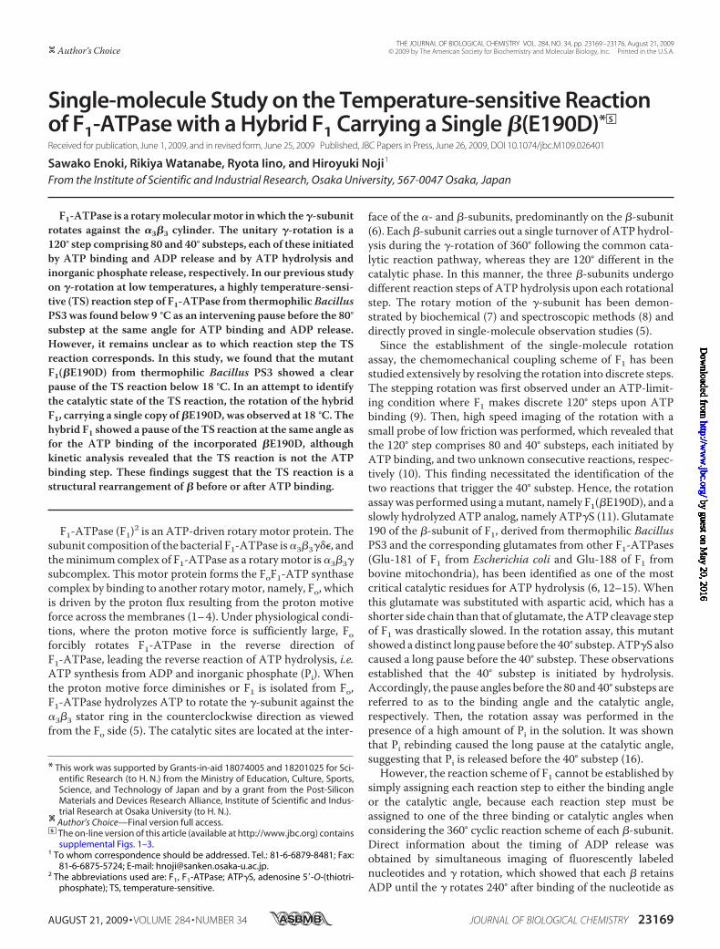
Temperature Dependence of the Rotational Rate ofF1(�E190D)—The rotational rate of F1(�E190D) wasmeasuredat 2 mM ATP, which was later confirmed to be a saturationcondition, at various temperatures ranging from 10 to 32 °C, byusing a �200-nm magnetic bead as a rotation marker at thevideo rate (Fig. 1, A and C). Although the previously reportedrotation rate of the wild-type F1 was limited by the viscousfriction on the probe at room temperatures (�10 °C) (21), thatof themutant was not determined by the viscous friction but bythe sluggish hydrolysis step of which the time constant is �300ms at room temperature (11). Therefore, the rotational rate ofthe mutant was in good agreement with the estimated rota-tional rate, which was one-third of the ATP hydrolysis ratedetermined in the bulk solution based on the coupling ratio of 3ATPs/turn; the wild-type F1 showed a large difference betweentwo values around room temperature (Fig. 1A) (21). The rota-tional velocity of themutant at 22–24 °C was 0.9 Hz, which wasconsistent with a previous report (11). At below 17 °C,F1(�E190D) decreased the rotational rate significantly, suggest-ing that a different reaction step dominates the pace of thecatalytic turnover cycle. This point was clearly shown in the
Temperature-sensitive Reaction of Hybrid F1-ATPase
23170 JOURNAL OF BIOLOGICAL CHEMISTRY VOLUME 284 • NUMBER 34 • AUGUST 21, 2009
by guest on May 20, 2016
http://ww
w.jbc.org/
Dow
nloaded from
Arrhenius plots of the rotational rate and the bulk ATP hydrol-ysis rate, both of which showed a breakpoint around 18 °C (Fig.1B). Thus, F1(�E190D) has a TS reaction step similar to that ofthe wild type; however, the temperature range at which the TSreaction of the mutant becomes prominent is different fromthat of the wild-type F1 (�9 °C). For a comparison of the tem-perature dependence between themutant and the wild-type F1,the apparent activation energy of the TS reaction was deter-mined instead of theQ10 factor from the slope of the Arrhenius
plot, because theQ10 factor changesdepending on the temperaturerange from which it is determined.The activation energy of the TSreaction of F1(�E190D) was 82–86kJ/mol, which was remarkably highand essentially the same as the acti-vation energy of the TS reaction ofthe wild-type F1, which was 97–98kJ/mol (Table 1). However, the acti-vation energy for the hydrolysis stepof F1(�E190D) was 11–18 kJ/mol,which was much smaller than thatof the TS reaction. The wild-type F1also showed smaller activationenergy for hydrolysis than that forthe TS reaction. Thus, the TS reac-tion of F1(�E190D) showed thesame temperature dependence asthe wild-type F1.Substeps of F1(�E190D) at Satu-
rating ATP Concentration—It hasbeen shown that the wild-type F1has a pause for theTS reaction at theATP binding angle (21). This pointwas also confirmed for the TS reac-tion of F1(�E190D); the angle histo-gram of the rotation of F1(�E190D)at 28 and 22 °C showed three mainpeaks corresponding to the hydrol-ysis dwell time at the catalytic angles(Fig. 1D) (11). In addition, a smallpeak at the right side of each cata-lytic angle was observed, althoughsome peaks were not clear. Below18 °C, where the TS reaction limitsthe pace of the overall reactioncycle, these peaks appeared as clearmain peaks at around �40° fromthe catalytic angle, suggesting thatF1(�E190D) also creates a pause atthe binding angle to wait for the TSreaction.This point was confirmed by
changing the specimen temperaturefrom28 to 18 °C during the observa-tion of a rotating particle (Fig. 2).Even at 28 °C, F1(�E190D) showedshort pauses concomitant with long
hydrolysis (Fig. 2A, inset) when observed with a high speedcamera at 500 or 1000 frame/s. In the angle histogram, theseshort pauses are shown as protuberant tails on the right side ofthe large peaks of the hydrolysis pauses (Fig. 2D). When thespecimen was cooled to 18 °C, the short pauses were elicited asthe major pauses, meaning that the short pause found at 28 °Ccorresponds to the waiting state for the TS reaction. The histo-grams of the dwell time of the short pause verified this point(Fig. 2B). By fitting the dwell time histogram of the short pause
FIGURE 1. A, temperature dependence of the rotational rate (circles) and ATP hydrolysis rate in a bulk solution(crosses) of F1(�E190D) (red) and wild-type F1 (blue) (21) at saturating ATP concentration (2 mM for F1(�E190D)and 1 mM for wild-type F1). The ATP hydrolysis rate was divided by 3 for comparison with the rotational rate.B, Arrhenius plots of traces in A. The rate-limiting reactions of the rotational rates at low temperature (below17 °C for the mutant F1 and below 4 °C for wild-type F1) were analyzed to determine the activation energy(Table 1) using linear regression (solid lines). The rotational rates of F1(�E190D) above 20 °C were also fitted bylinear regression (dotted red line). C, time courses of rotation of F1(�E190D) at various temperatures. D, histo-grams of the angle from traces in C.
Temperature-sensitive Reaction of Hybrid F1-ATPase
AUGUST 21, 2009 • VOLUME 284 • NUMBER 34 JOURNAL OF BIOLOGICAL CHEMISTRY 23171
by guest on May 20, 2016
http://ww
w.jbc.org/
Dow
nloaded from

with an exponential function, the time constant of the shortpause at the binding anglewas determined to be 219ms at 18 °Cand 32ms at 28 °C (Fig. 2B), giving the large activation energy of78 kJ/mol, which coincides with the activation energy deter-mined from the average rotational velocity and the bulkATPase assay (Table 1).On the other hand, the time constant ofthe long hydrolysis pause was similar between 224 ms at 18 °Cand 183ms at 28 °C (Fig. 2C), giving the small activation energy
of 8 kJ/mol (Table 1). Then, the angle distance between thecatalytic pause and the TS pause was analyzed. As seen in Fig.2D, theTS pause always appeared at the right side of the hydrol-ysis peaks in the angle histogram. The average angle distance ofthe TS pause from the catalytic pause (��) was �40.0 � 5.6° at28 °C and �42.4 � 10.3° at 18 °C (Fig. 2, E and F). Thus, it wasverified that the TS reaction of F1(�E190D) occurs at the ATPbinding angle, as does the TS reaction of the wild-type F1.Rotation of F1(�E190D) atVariousATPConcentrations—For
further characterization of the TS reaction of F1(�E190D), therotational rate was measured at various ATP concentrations at18 °C, and Michaelis-Menten curves were obtained (Fig. 3A).The Vmax and Km values at 18 °C were 0.55 s�1 and 1.3 �M,respectively. The binding rate of ATP determined from3 Vmax/Km was 1.3 106 M�1s�1. Therefore, the dwell timefor ATP binding at 2 mM ATP should be 0.39 ms, which is tooshort to explain the dwell time of the TS reaction (219 ms).Thus, the TS reaction cannot be the ATP binding step fromsolution. To compare the angle positions for the TS reactionand the ATP binding, buffer exchange experiments were con-ducted in which the ATP level was increased from 0.5 �M to 2mM during the observation of rotating molecules (Fig. 3, B andC). When ATP was increased, three major peaks of the ATPbinding dwell diminished, but the pause at the binding anglewas still clearly observed at 2 mM ATP due to the pause caused
FIGURE 2. A, time courses of rotations of a molecule of F1(�E190D) at different temperatures (18 and 28 °C). During observation, the specimen temperature waschanged from 28 to 18 °C. The inset shows a magnified view of the trace at 28 °C to show the short pauses by the TS reaction (arrows). B and C, dwell timehistograms of the pause for TS reaction (B) and hydrolysis (C) at 18 °C (blue) and 28 °C (red). The histogram was fitted with single exponential decay. D, histo-grams of the angle from traces in A at 28 °C (top) and 18 °C (bottom). �� represents angle distances from the pause angle for hydrolysis to that for the TS reaction.E and F, histograms of ��at 28 °C (E) and 18 °C (F).
TABLE 1Activation energy of the TS reaction and hydrolysis of F1(�E190D)and wild-type F1
Activation energy was calculated by the linear fitting of the Arrhenius plots ofATPase and the rotational rate as shown in Fig. 1B or by the comparison of rateconstants at 18 and 28 °C determined from the dwell time histograms shown inFig. 2, B and C, for �E190D and Fig. 4, C and D, for �E190D(hybrid).
Reaction F1 Activation energy
kJ/molTS �E190D 86 (ATPase), 82 (rotation rate), 78 (dwell time)
�E190D(hybrid) 62 (dwell time)
Hydrolysis �E190D 11 (ATPase), 18 (rotation rate), 8.3 (dwell time)�E190D(hybrid) 16 (dwell time)
TS Wild type 97a (ATPase), 98a (rotation rate)
Hydrolysis(ATP�S)
Wild type 2.4b (dwell time)
a Determined from the temperature dependence of the maximum ATPase pub-lished in Watanabe et al. (21).
b Determined by a comparison of the hydrolysis rates at 4 °C (21) and at roomtemperature (11).
Temperature-sensitive Reaction of Hybrid F1-ATPase
23172 JOURNAL OF BIOLOGICAL CHEMISTRY VOLUME 284 • NUMBER 34 • AUGUST 21, 2009
by guest on May 20, 2016
http://ww
w.jbc.org/
Dow
nloaded from

by the TS reaction (Fig. 3C). The angle distance (��) from theATP binding pause at 0.5 �M ATP to the TS pause determinedat 2mMATPwas�0.2� 14.1° (15 events for 5molecules). Thisresult also supports the above conclusion that the TS reactionoccurs at the binding angle.Temperature Dependence of the Rotation of Hybrid F1—The
above experiments established that the TS reaction ofF1(�E190D) is essentially the same as that of the wild-type F1,and the dwell time of the TS reaction of the mutant is longenough to be detected in the conventional rotation assay, evenat 18 °C where the wild type does not show the distinct pause oftheTSreaction.Takingadvantageof this featureof themutant, thehybridF1 carrying a single copyof�(E190D),�3�2�(E190D)�,wasanalyzed in order to identify the angle position of the TS pause
in the 360° cyclic reaction scheme.The hybrid F1 was prepared by mix-ing the F1(�E190D) and the wild-type F1 in a 1:2 ratio. The rotation ofthe hybrid F1 was found at 28 °C and2 mM ATP under optical micro-scope. Although other complexes,such as the wild-type F1,�3��(E190D)2�, or F1(�E190D),were also contained in the sample,�3�2�(E190D)� was able to be dis-tinguished from the others as amol-ecule rotating with long hydrolysispauses at a single angle, as reportedin previous works (18–20). Wedefined the hydrolysis pause as thepause of which dwell time was lon-ger than 150 ms. Although most ofmolecules showed the concomitantTS pause as mentioned below, thedwell time of TS pause (36 ms) wasmuch shorter andwas able to be dis-tinguished from the hydrolysispause. More than half (59%) of therotating molecules showed thehydrolysis pause at a single angle(�3�2�(E190D)�), whereas 27%showed no pause (wild), 12% hadtwo pause positions (�3��(E190D)2�),and 2% had three pause positions(F1(�E190D)). When observed witha high speed camera, a single shortpause was also found in addition tothe single hydrolysis pause in therotation of �3�2�(E190D)�. Theangle distance of the short pausefrom the hydrolysis pause (��) wasdetermined to be �200.5 � 11.0° at28 °C (supplemental Fig. S1). Forhybrid F1,�3�2�(E190D)�, the tem-perature change experiment wasconducted to determine the angleposition of the TS reaction of�E190D in relation to the long
hydrolysis pause of �(E190D). Selected molecules respondedwell to the temperature change by extending the dwell timeof the short pause unless the experiment was obstructed bynonspecific surface immobilization or the detachment ofmolecules. The time constant of the short pause of thehybrid F1 was determined to be 36 ms at 28 °C and 174 ms at18 °C from the dwell time analysis, both of which were con-sistent with the time constant of the TS reaction determinedfor F1(�E190D), suggesting that the short pause corre-sponded to the TS pause of the incorporated �E190D in thehybrid F1 (Fig. 4C). The time constant of the hydrolysis dwellwas less sensitive (220 ms at 28 °C and 320 ms at 18 °C),which was also consistent with the values determined forF1(�E190D) (Fig. 4D).
FIGURE 3. A, ATP concentration dependence of the rotational rate of F1(�E190D) at 18 °C. Curve shows thefitting line with the Michaelis-Menten equation, V Vmax[ATP]/(Km�[ATP]); Vmax 0.55 s�1, Km 1.3 �M.B, time courses of the rotation at 18 °C of a molecule of F1(�E190D) at 0.5 �M ATP (blue) and 2 mM ATP (red).During observation, the buffer was exchanged to increase ATP from 0.5 �M to 2 mM. C, histograms of theangle from traces in B. �� represents angle distances of the TS angle at 2 mM ATP from the ATP bindingangle at 0.5 �M ATP.
Temperature-sensitive Reaction of Hybrid F1-ATPase
AUGUST 21, 2009 • VOLUME 284 • NUMBER 34 JOURNAL OF BIOLOGICAL CHEMISTRY 23173
by guest on May 20, 2016
http://ww
w.jbc.org/
Dow
nloaded from
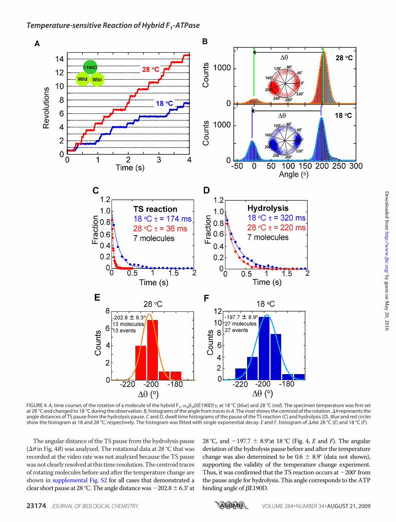
The angular distance of the TS pause from the hydrolysis pause(�� in Fig. 4B) was analyzed. The rotational data at 28 °C that wasrecorded at the video rate was not analyzed because the TS pausewasnotclearly resolvedat this timeresolution.Thecentroid tracesof rotatingmolecules before and after the temperature change areshown in supplemental Fig. S2 for all cases that demonstrated aclear short pause at 28 °C.The angle distancewas�202.8� 6.3° at
28 °C, and �197.7 � 8.9°at 18 °C (Fig. 4, E and F). The angulardeviation of the hydrolysis pause before and after the temperaturechange was also determined to be 0.6 � 8.9° (data not shown),supporting the validity of the temperature change experiment.Thus, it was confirmed that the TS reaction occurs at�200° fromthe pause angle for hydrolysis. This angle corresponds to the ATPbinding angle of �E190D.
FIGURE 4. A, time courses of the rotation of a molecule of the hybrid F1, �3�2�(E190D)�, at 18 °C (blue) and 28 °C (red). The specimen temperature was first setat 28 °C and changed to 18 °C during the observation. B, histograms of the angle from traces in A. The inset shows the centroid of the rotation. �� represents theangle distances of TS pause from the hydrolysis pause. C and D, dwell time histograms of the pause of the TS reaction (C) and hydrolysis (D). Blue and red circlesshow the histogram at 18 and 28 °C, respectively. The histogram was fitted with single exponential decay. E and F, histogram of ��at 28 °C (E) and 18 °C (F).
Temperature-sensitive Reaction of Hybrid F1-ATPase
23174 JOURNAL OF BIOLOGICAL CHEMISTRY VOLUME 284 • NUMBER 34 • AUGUST 21, 2009
by guest on May 20, 2016
http://ww
w.jbc.org/
Dow
nloaded from
DISCUSSION
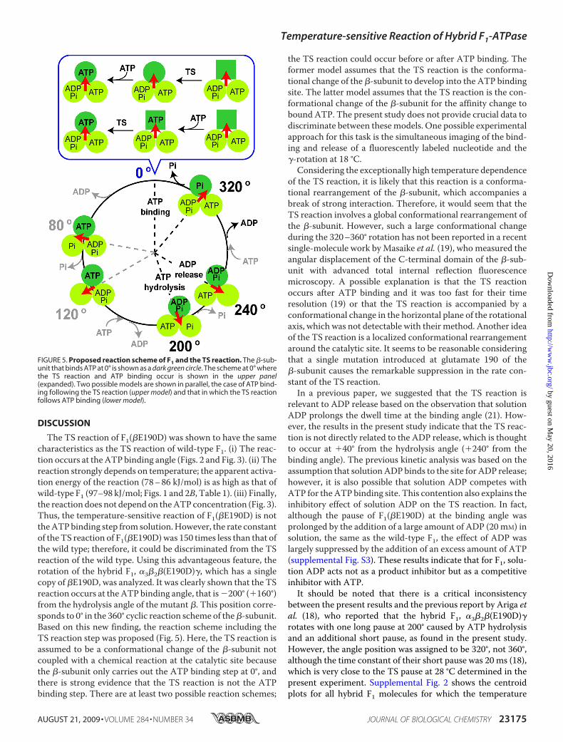
The TS reaction of F1(�E190D) was shown to have the samecharacteristics as the TS reaction of wild-type F1. (i) The reac-tion occurs at theATP binding angle (Figs. 2 and Fig. 3). (ii) Thereaction strongly depends on temperature; the apparent activa-tion energy of the reaction (78–86 kJ/mol) is as high as that ofwild-type F1 (97–98 kJ/mol; Figs. 1 and 2B, Table 1). (iii) Finally,the reaction does not dependon theATP concentration (Fig. 3).Thus, the temperature-sensitive reaction of F1(�E190D) is nottheATPbinding step from solution.However, the rate constantof the TS reaction of F1(�E190D)was 150 times less than that ofthe wild type; therefore, it could be discriminated from the TSreaction of the wild type. Using this advantageous feature, therotation of the hybrid F1, �3�2�(E190D)�, which has a singlecopy of �E190D, was analyzed. It was clearly shown that the TSreaction occurs at the ATP binding angle, that is�200° (�160°)from the hydrolysis angle of the mutant �. This position corre-sponds to 0° in the 360° cyclic reaction scheme of the�-subunit.Based on this new finding, the reaction scheme including theTS reaction step was proposed (Fig. 5). Here, the TS reaction isassumed to be a conformational change of the �-subunit notcoupled with a chemical reaction at the catalytic site becausethe �-subunit only carries out the ATP binding step at 0°, andthere is strong evidence that the TS reaction is not the ATPbinding step. There are at least two possible reaction schemes;
the TS reaction could occur before or after ATP binding. Theformer model assumes that the TS reaction is the conforma-tional change of the �-subunit to develop into the ATP bindingsite. The latter model assumes that the TS reaction is the con-formational change of the �-subunit for the affinity change tobound ATP. The present study does not provide crucial data todiscriminate between thesemodels. One possible experimentalapproach for this task is the simultaneous imaging of the bind-ing and release of a fluorescently labeled nucleotide and the�-rotation at 18 °C.
Considering the exceptionally high temperature dependenceof the TS reaction, it is likely that this reaction is a conforma-tional rearrangement of the �-subunit, which accompanies abreak of strong interaction. Therefore, it would seem that theTS reaction involves a global conformational rearrangement ofthe �-subunit. However, such a large conformational changeduring the 320–360° rotation has not been reported in a recentsingle-molecule work byMasaike et al. (19), who measured theangular displacement of the C-terminal domain of the �-sub-unit with advanced total internal reflection fluorescencemicroscopy. A possible explanation is that the TS reactionoccurs after ATP binding and it was too fast for their timeresolution (19) or that the TS reaction is accompanied by aconformational change in the horizontal plane of the rotationalaxis, which was not detectable with their method. Another ideaof the TS reaction is a localized conformational rearrangementaround the catalytic site. It seems to be reasonable consideringthat a single mutation introduced at glutamate 190 of the�-subunit causes the remarkable suppression in the rate con-stant of the TS reaction.In a previous paper, we suggested that the TS reaction is
relevant to ADP release based on the observation that solutionADP prolongs the dwell time at the binding angle (21). How-ever, the results in the present study indicate that the TS reac-tion is not directly related to the ADP release, which is thoughtto occur at �40° from the hydrolysis angle (�240° from thebinding angle). The previous kinetic analysis was based on theassumption that solutionADP binds to the site for ADP release;however, it is also possible that solution ADP competes withATP for theATP binding site. This contention also explains theinhibitory effect of solution ADP on the TS reaction. In fact,although the pause of F1(�E190D) at the binding angle wasprolonged by the addition of a large amount of ADP (20mM) insolution, the same as the wild-type F1, the effect of ADP waslargely suppressed by the addition of an excess amount of ATP(supplemental Fig. S3). These results indicate that for F1, solu-tion ADP acts not as a product inhibitor but as a competitiveinhibitor with ATP.It should be noted that there is a critical inconsistency
between the present results and the previous report by Ariga etal. (18), who reported that the hybrid F1, �3�2�(E190D)�rotates with one long pause at 200° caused by ATP hydrolysisand an additional short pause, as found in the present study.However, the angle position was assigned to be 320°, not 360°,although the time constant of their short pause was 20 ms (18),which is very close to the TS pause at 28 °C determined in thepresent experiment. Supplemental Fig. 2 shows the centroidplots for all hybrid F1 molecules for which the temperature
FIGURE 5. Proposed reaction scheme of F1 and the TS reaction. The �-sub-unit that binds ATP at 0° is shown as a dark green circle. The scheme at 0° wherethe TS reaction and ATP binding occur is shown in the upper panel(expanded). Two possible models are shown in parallel, the case of ATP bind-ing following the TS reaction (upper model) and that in which the TS reactionfollows ATP binding (lower model).
Temperature-sensitive Reaction of Hybrid F1-ATPase
AUGUST 21, 2009 • VOLUME 284 • NUMBER 34 JOURNAL OF BIOLOGICAL CHEMISTRY 23175
by guest on May 20, 2016
http://ww
w.jbc.org/
Dow
nloaded from

change experiment was successfully carried out. In the data set,all molecules showed the short pause not at 320 but at 360°.Statistical analysis confirms this conclusion. The reason for thisinconsistency is not clear. Although there are some small dif-ferences in the experimental procedures andmaterials, these donot seem to explain this critical inconsistency. Even if there isanother short pause in addition to that of the TS reaction, thepause of theTS reaction should have been included in their dataset.
Acknowledgments—We thank Dr. S. Nishikawa, Mr. K. Ikezaki, Dr.M. Sugawa, and Dr. T. Yanagida for technical help, Dr. K. Adachi forthe custom image analysis program, Dr. T. Okamoto for critical dis-cussion, and members of the Noji laboratory for help and advice.
REFERENCES1. Boyer, P. D. (1997) Annu. Rev. Biochem. 66, 717–7492. Cross, R. L. (2000) Biochim. Biophys. Acta 1458, 270–2753. Senior, A. E., Nadanaciva, S., andWeber, J. (2002) Biochim. Biophys. Acta
1553, 188–2114. Yoshida,M.,Muneyuki, E., andHisabori, T. (2001)Nat. Rev.Mol. Cell Biol.
2, 669–6775. Noji, H., Yasuda, R., Yoshida, M., and Kinosita, K., Jr. (1997) Nature 386,
299–3026. Abrahams, J. P., Leslie, A. G., Lutter, R., and Walker, J. E. (1994) Nature
370, 621–6287. Duncan, T. M., Bulygin, V. V., Zhou, Y., Hutcheon, M. L., and Cross, R. L.
(1995) Proc. Natl. Acad. Sci. U.S.A. 92, 10964–109688. Sabbert, D., Engelbrecht, S., and Junge, W. (1996) Nature 381, 623–625
9. Yasuda, R., Noji, H., Kinosita, K., Jr., and Yoshida, M. (1998) Cell 93,1117–1124
10. Yasuda, R., Noji, H., Yoshida, M., Kinosita, K., Jr., and Itoh, H. (2001)Nature 410, 898–904
11. Shimabukuro, K., Yasuda, R., Muneyuki, E., Hara, K. Y., Kinosita, K., Jr.,and Yoshida, M. (2003) Proc. Natl. Acad. Sci. U.S.A. 100, 14731–14736
12. Amano, T., Tozawa, K., Yoshida, M., andMurakami, H. (1994) FEBS Lett.348, 93–98
13. Ohtsubo, M., Yoshida, M., Ohta, S., Kagawa, Y., Yohda, M., and Date, T.(1987) Biochem. Biophys. Res. Commun. 146, 705–710
14. Park, M. Y., Omote, H., Maeda, M., and Futai, M. (1994) J. Biochem. 116,1139–1145
15. Senior, A. E., and al-Shawi, M. K. (1992) J. Biol. Chem. 267, 21471–2147816. Adachi, K., Oiwa, K., Nishizaka, T., Furuike, S., Noji, H., Itoh, H., Yoshida,
M., and Kinosita, K., Jr. (2007) Cell 130, 309–32117. Nishizaka, T., Oiwa, K., Noji, H., Kimura, S., Muneyuki, E., Yoshida, M.,
and Kinosita, K., Jr. (2004) Nat. Struct. Mol. Biol. 11, 142–14818. Ariga, T., Muneyuki, E., and Yoshida, M. (2007)Nat. Struct. Mol. Biol. 14,
841–84619. Masaike, T., Koyama-Horibe, F., Oiwa, K., Yoshida, M., and Nishizaka, T.
(2008) Nat. Struct. Mol. Biol. 15, 1326–133320. Okuno, D., Fujisawa, R., Iino, R., Hirono-Hara, Y., Imamura, H., and Noji,
H. (2008) Proc. Natl. Acad. Sci. U.S.A. 105, 20722–2072721. Watanabe, R., Iino, R., Shimabukuro, K., Yoshida, M., and Noji, H. (2008)
EMBO Rep. 9, 84–9022. Furuike, S., Adachi, K., Sakaki, N., Shimo-Kon, R., Itoh, H., Muneyuki, E.,
Yoshida, M., and Kinosita, K., Jr. (2008) Biophys. J. 95, 761–77023. Hirono-Hara, Y., Ishizuka, K., Kinosita, K., Jr., Yoshida, M., and Noji, H.
(2005) Proc. Natl. Acad. Sci. U.S.A. 102, 4288–429324. Rondelez, Y., Tresset, G., Nakashima, T., Kato-Yamada, Y., Fujita, H.,
Takeuchi, S., and Noji, H. (2005) Nature 433, 773–777
Temperature-sensitive Reaction of Hybrid F1-ATPase
23176 JOURNAL OF BIOLOGICAL CHEMISTRY VOLUME 284 • NUMBER 34 • AUGUST 21, 2009
by guest on May 20, 2016
http://ww
w.jbc.org/
Dow
nloaded from

Supplemental Figure 1
Cou
nts
-200.5 ± 11.0o
20 molecules
∆θ (o)012345678
-240 -220 -200 -180 -160
Supplemental Figure 1Histogram of the angular distances from the long pause angle (hydrolysis) to the short pause angle (TS reaction) of the hybrid F1, α3β2β(E190D)γ, which rotated with 1 long and 1 short pause at 28oC.
1.
40°
0°
320°
80°120°
160°
200°
240° 280°
40°
0°
320°
80°120°
160°
200°
240° 280°
40°
0°
320°
80°120°
160°
200°
240° 280°
40°
0°
320°
80°120°
160°
200°
240° 280°
3.
5.
40°
0°
320°
80°120°
160°
200°
240° 280°
2.
4.
40°
0°
320°
80°120°
160°
200°
240° 280°
40°
0°
320°
80°120°
160°
200°
240° 280°
6.
40°
0°
320°
80°120°
160°
200°
240° 280°
40°
0°
320°
80°120°
160°
200°
240° 280°
7. 8.
10.
40°
0°
320°
80°120°
160°
200°
240° 280°
40°
0°
320°
80°120°
160°
200°
240° 280°
9.
40°
0°
320°
80°120°
160°
200°
240° 280°
40°
0°
320°
80°120°
160°
200°
240° 280°
Supplemental Figure 2-1
40°
0°
320°
80°120°
160°
200°
240° 280°
40°
0°
320°
80°120°
160°
200°
240° 280°
40°
0°
320°
80°120°
160°
200°
240° 280°
40°
0°
320°
80°120°
160°
200°
240° 280°
40°
0°
320°
80°120°
160°
200°
240° 280°
40°
0°
320°
80°120°
160°
200°
240° 280°
40°
0°
320°
80°120°
160°
200°
240° 280°

Supplemental Figure 2-2
11. 12.
40°
0°
320°
80°120°
160°
200°
240° 280°
40°
0°
320°
80°120°
160°
200°
240° 280°
40°
0°
320°
80°120°
160°
200°
240° 280°
40°
0°
320°
80°120°
160°
200°
240° 280°
13.
40°
0°
320°
80°120°
160°
200°
240° 280°
40°
0°
320°
80°120°
160°
200°
240° 280°
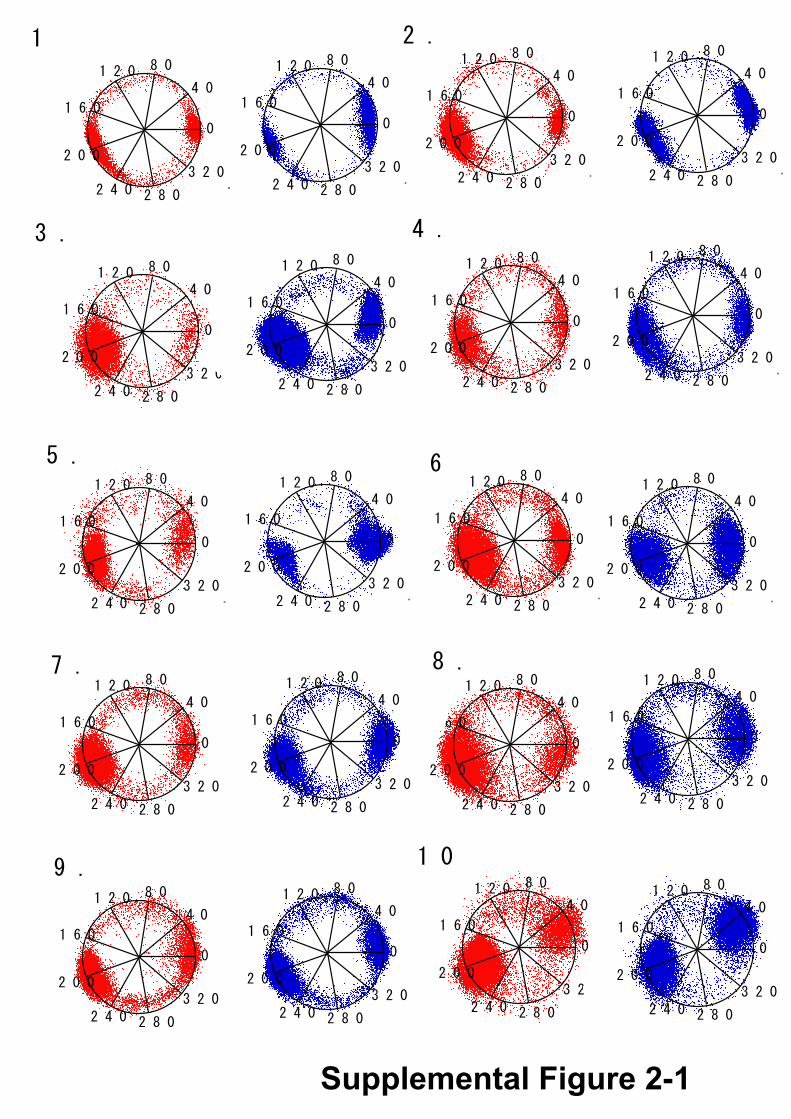
Supplemental Figure 2Rotational trajectories of the hybrid F1(α3β2β(E190D)γ) , in the temperature changing experiment from 28οC (red) to 18οC (blue) (Fig. 4). Only the data recorded at 500 or 1000 frame/s were shown. The hydrolysis angle determined by Gaussian fitting was fixed at 200ο.
Cou
nts
-5.0 ± 9.4o
4 molecules12 events
ATP 1mM
0
200C
ount
s
0
300
Cou
nts
0
200
0 120 240 360
Cou
nts
Angle (o)
ATP 1mMADP 20 mM
ATP 40mMADP 20 mM
Supplemental Figure 3
A
B C
0
1.0
0 2 4 6Time (s)
Frac
tion
ATP 1mMATP 1mM, ADP 20mMATP 40mM, ADP 20mM
∆θ (o)
∆θ
0
2
4
6
-30-20 -10 0 10 20 30

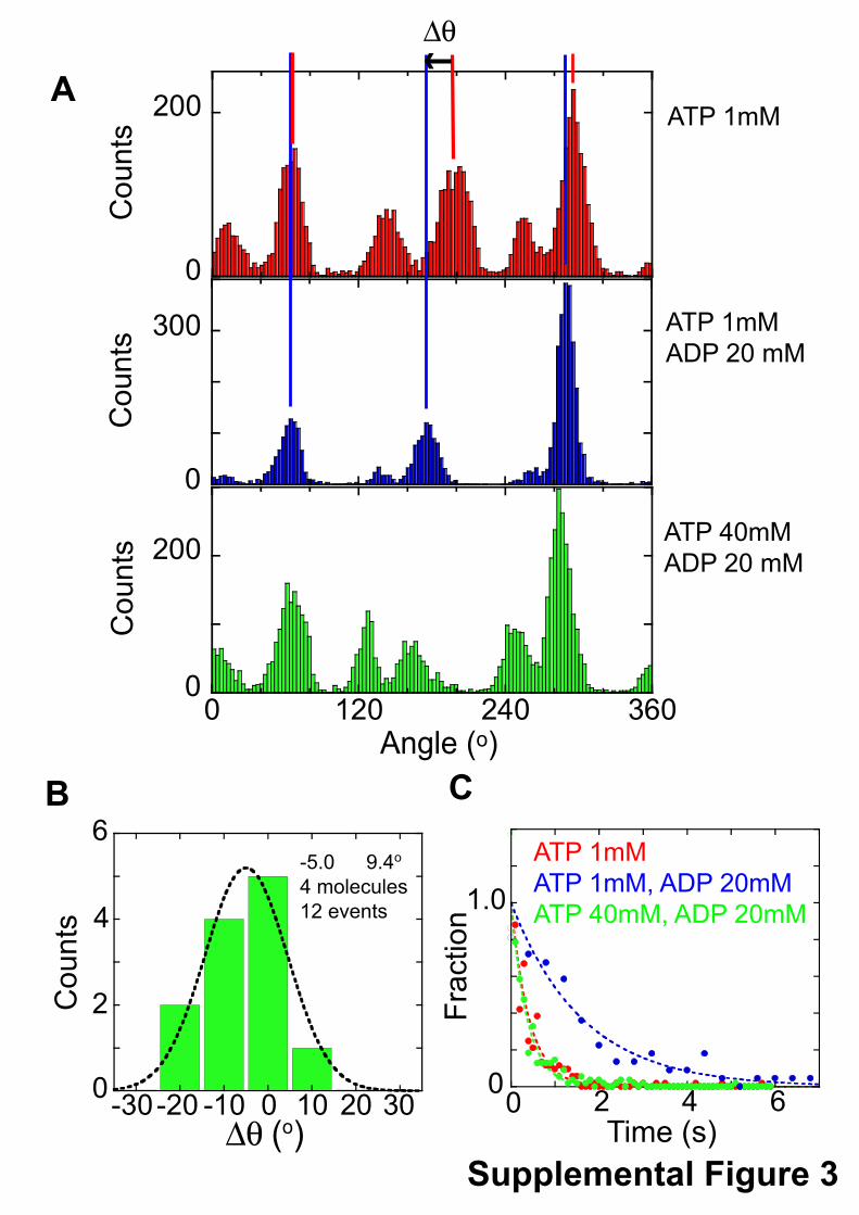
Supplemental Figure 3Rotation of F1(βE190D) in the presence of an excess amount of ADP at 18oC. After the observation of the rotation of F1(βE190D) at 1 mM ATP and 50 mM MgCl2, the buffer solution was replaced with the solution containing 1 mM ATP, 20 mM ADP and 50 mM MgCl2. Then the buffer was replaced with the solution containing 40 mM ATP, 20 mM ADP, and 100 mM MgCl2. A. Histograms of the angle of the rotation at 1 mM ATP and 50 mM MgCl2 (Top), 1 mM ATP, 20 mM ADP, and 50 mM MgCl2 (middle), and 40 mM ATP, 20 mM ADP, and 100 mM MgCl2 (bottom). B. Angular distance (∆θ) from the pause angle for the TS reaction at 1 mM ATP to the prolonged pause angle by addition of 20 mM ADP. C. Dwell-time histograms of the pause before the 80o
substep.
Sawako Enoki, Rikiya Watanabe, Ryota Iino and Hiroyuki Noji(E190D)β Carrying a Single 1Hybrid F
-ATPase with a1Single-molecule Study on the Temperature-sensitive Reaction of F
doi: 10.1074/jbc.M109.026401 originally published online June 26, 20092009, 284:23169-23176.J. Biol. Chem.
10.1074/jbc.M109.026401Access the most updated version of this article at doi:
Alerts:
When a correction for this article is posted•
When this article is cited•
to choose from all of JBC's e-mail alertsClick here
Supplemental material:
http://www.jbc.org/content/suppl/2009/06/26/M109.026401.DC1.html
http://www.jbc.org/content/284/34/23169.full.html#ref-list-1
This article cites 24 references, 6 of which can be accessed free at
by guest on May 20, 2016
http://ww
w.jbc.org/
Dow
nloaded from
Related Documents


![V-ATPase · From Wiki: Vacuolar-type H+ -ATPase (V-ATPase) is a highly conserved evolutionarily ancient enzyme with remarkably diverse functions in eukaryotic organisms.[1] membranes](https://static.cupdf.com/doc/110x72/5fa3fb056ad5ca477269e2ce/v-atpase-from-wiki-vacuolar-type-h-atpase-v-atpase-is-a-highly-conserved-evolutionarily.jpg)