RESEARCH ARTICLE Single histidine residue in head-group region is sufficient to impart remarkable gene transfection properties to cationic lipids: evidence for histidine- mediated membrane fusion at acidic pH VV Kumar 1 , C Pichon 2 , M Refregiers 2 , B Guerin 2 , P Midoux 2 and A Chaudhuri 1 1 Division of Lipid Science and Technology, Indian Institute of Chemical Technology, India; and 2 Centre de Biophysique Mole ´culaire, CNRS UPR 4301, Orleans Cedex, France Presence of endosome-disrupting multiple histidine function- alities in the molecular architecture of cationic polymers, such as polylysine, has previously been demonstrated to significantly enhance their in vitro gene delivery efficiencies. Towards harnessing improved transfection property through covalent grafting of endosome-disrupting single histidine functionality in the molecular structure of cationic lipids, herein, we report on the design, the synthesis and the transfection efficiency of two novel nonglycerol-based histidylated cationic amphiphiles. We found that L-histidine- (N,N-di-n-hexadecylamine)ethylamide (lipid 1) and L-histi- dine-(N,N-di-n-hexadecylamine,-N-methyl)ethylamide (lipid 2) in combination with cholesterol gave efficient transfections into various cell lines. The transfection efficiency of Chol/lipid 1 lipoplexes into HepG2 cells was two order of magnitude higher than that of FuGENE TM 6 and DC-Chol lipoplexes, whereas it was similar into A549, 293T7 and HeLa cells. A better efficiency was obtained with Chol/lipid 2 lipoplexes when using the cytosolic luciferase expression vector (pT7Luc) under the control of the bacterial T7 promoter. Membrane fusion activity measurements using fluorescence resonance energy transfer (FRET) technique showed that the histidine head-groups of Chol/lipid 1 liposomes mediated membrane fusion in the pH range 5–7. In addition, the transgene expression results using the T7Luc expression vector convincingly support the endosome-disrupting role of the presently described mono-histidylated cationic transfec- tion lipids and the release of DNA into the cytosol. We conclude that covalent grafting of a single histidine amino acid residue to suitable twin-chain hydrophobic compounds is able to impart remarkable transfection properties on the resulting mono-histidylated cationic amphiphile, presumably via the endosome-disrupting characteristics of the histidine functionalities. Gene Therapy (2003) 10, 1206–1215. doi:10.1038/ sj.gt.3301979 Keywords: gene transfer; cationic lipids; liposomes Introduction Gene therapy, because of its aim to eradicate causes rather than symptoms of diseases, is believed by many to be the therapy of the future. Recent completion of the working draft of human genome has strengthened such belief. However, spontaneous delivery and expression of the supplemented correct copy of the malfunctioning genes into cells is unlikely to be an efficient process due to the unfavorable electrostatic interactions between macromolecular DNA and biological cell surface (both being negatively charged) and due to the nuclease sensitiveness of nonprotected DNA. To develop an efficient gene therapeutic approach, one must design safe and efficient gene delivery reagents. These goals are inseparable in a sense that any shortcomings in one will adversely affect the success of the other. In other words, realization of the full potential of gene therapy will depend, in a major way, on the future development of safe and efficient gene delivery reagents. Transfection vectors commonly used in gene therapy are mainly of two types: viral and nonviral. The efficiencies of viral transfection vectors 1 are, in general, superior to their nonviral counterparts. However, serious immunogenicity concerns associated with the use of viral vectors 2 combined with their limited insert-size and high product cost are increasingly making the nonviral gene delivery reagents the vectors of choice. Among the existing arsenal of nonviral gene delivery reagents, the distinct advantages associated with the use of cationic liposomes include their low immunogenic response, robust manufacture, ease in handling and preparation techniques, and the ability to inject large lipid:DNA complexes. Ever since Felgner et al 3 reported their pioneering development of cationic lipid-mediated gene delivery protocol in 1987, many reports aimed at developing more efficient cationic transfection lipids have appeared in the literature. 4–10 Recently, we have Received 5 September 2002; accepted 4 December 2002 Correspondence: Dr A Chaudhuri, Division of Lipid Science and Technology, Indian Institute of Chemical Technology, Hyderabad 500 007, India or Dr P Midoux, Centre de Biophysique Mole ´culaire, CNRS UPR 4301, Orleans Cedex, France Gene Therapy (2003) 10, 1206–1215 & 2003 Nature Publishing Group All rights reserved 0969-7128/03 $25.00 www.nature.com/gt

Welcome message from author
This document is posted to help you gain knowledge. Please leave a comment to let me know what you think about it! Share it to your friends and learn new things together.
Transcript
RESEARCH ARTICLE
Single histidine residue in head-group region issufficient to impart remarkable gene transfectionproperties to cationic lipids: evidence for histidine-mediated membrane fusion at acidic pH
VV Kumar1, C Pichon2, M Refregiers2, B Guerin2, P Midoux2 and A Chaudhuri1
1Division of Lipid Science and Technology, Indian Institute of Chemical Technology, India; and 2Centre de Biophysique Moleculaire,CNRS UPR 4301, Orleans Cedex, France
Presence of endosome-disrupting multiple histidine function-alities in the molecular architecture of cationic polymers,such as polylysine, has previously been demonstrated tosignificantly enhance their in vitro gene delivery efficiencies.Towards harnessing improved transfection property throughcovalent grafting of endosome-disrupting single histidinefunctionality in the molecular structure of cationic lipids,herein, we report on the design, the synthesis and thetransfection efficiency of two novel nonglycerol-basedhistidylated cationic amphiphiles. We found that L-histidine-(N,N-di-n-hexadecylamine)ethylamide (lipid 1) and L-histi-dine-(N,N-di-n-hexadecylamine,-N-methyl)ethylamide (lipid2) in combination with cholesterol gave efficient transfectionsinto various cell lines. The transfection efficiency of Chol/lipid1 lipoplexes into HepG2 cells was two order of magnitudehigher than that of FuGENETM6 and DC-Chol lipoplexes,whereas it was similar into A549, 293T7 and HeLa cells. Abetter efficiency was obtained with Chol/lipid 2 lipoplexes
when using the cytosolic luciferase expression vector(pT7Luc) under the control of the bacterial T7 promoter.Membrane fusion activity measurements using fluorescenceresonance energy transfer (FRET) technique showed thatthe histidine head-groups of Chol/lipid 1 liposomes mediatedmembrane fusion in the pH range 5–7. In addition, thetransgene expression results using the T7Luc expressionvector convincingly support the endosome-disrupting role ofthe presently described mono-histidylated cationic transfec-tion lipids and the release of DNA into the cytosol. Weconclude that covalent grafting of a single histidine aminoacid residue to suitable twin-chain hydrophobic compoundsis able to impart remarkable transfection properties on theresulting mono-histidylated cationic amphiphile, presumablyvia the endosome-disrupting characteristics of the histidinefunctionalities.Gene Therapy (2003) 10, 1206–1215. doi:10.1038/sj.gt.3301979
Keywords: gene transfer; cationic lipids; liposomes
Introduction
Gene therapy, because of its aim to eradicate causesrather than symptoms of diseases, is believed by many tobe the therapy of the future. Recent completion of theworking draft of human genome has strengthened suchbelief. However, spontaneous delivery and expression ofthe supplemented correct copy of the malfunctioninggenes into cells is unlikely to be an efficient process dueto the unfavorable electrostatic interactions betweenmacromolecular DNA and biological cell surface (bothbeing negatively charged) and due to the nucleasesensitiveness of nonprotected DNA. To develop anefficient gene therapeutic approach, one must designsafe and efficient gene delivery reagents. These goals areinseparable in a sense that any shortcomings in one will
adversely affect the success of the other. In other words,realization of the full potential of gene therapy willdepend, in a major way, on the future development ofsafe and efficient gene delivery reagents.
Transfection vectors commonly used in gene therapyare mainly of two types: viral and nonviral. Theefficiencies of viral transfection vectors1 are, in general,superior to their nonviral counterparts. However, seriousimmunogenicity concerns associated with the use of viralvectors2 combined with their limited insert-size and highproduct cost are increasingly making the nonviral genedelivery reagents the vectors of choice. Among theexisting arsenal of nonviral gene delivery reagents, thedistinct advantages associated with the use of cationicliposomes include their low immunogenic response,robust manufacture, ease in handling and preparationtechniques, and the ability to inject large lipid:DNAcomplexes. Ever since Felgner et al3 reported theirpioneering development of cationic lipid-mediated genedelivery protocol in 1987, many reports aimed atdeveloping more efficient cationic transfection lipidshave appeared in the literature.4–10 Recently, we haveReceived 5 September 2002; accepted 4 December 2002
Correspondence: Dr A Chaudhuri, Division of Lipid Science andTechnology, Indian Institute of Chemical Technology, Hyderabad 500007, India or Dr P Midoux, Centre de Biophysique Moleculaire, CNRSUPR 4301, Orleans Cedex, France
Gene Therapy (2003) 10, 1206–1215& 2003 Nature Publishing Group All rights reserved 0969-7128/03 $25.00
www.nature.com/gt
also developed efficient nonglycerol-based cationictransfection lipids.11–15 In sharp contrast, details ofcationic lipid-mediated transfection pathways still re-main elusive. According to our current understanding,the intracellular pathways involved in lipoplex (lipid–DNA complex)-mediated transfection include: (a) en-docytotic cellular uptake of lipoplex; (b) the endosomalrelease of DNA into the cytosol and (c) the transport ofDNA into the cell nucleus followed by its transcriptionand expression.16–19
The weak efficiency of plasmid transfer into thecytosol remains one of the major limiting factors toachieve efficient transfection. For lipofection, the neutralfusogenic colipid 1,2-dioleoyl-sn-glycero-3-phosphoetha-nolamine (DOPE), in combination with a cationic lipid, isusually used to favor the cytosolic delivery of plasmid.In the area of cationic polymer-mediated gene delivery,remarkable transfection efficiency was previouslyachieved by one of our groups through covalent graftingof endosome-disrupting multiple histidine functional-ities in the molecular architecture of cationic polymer.20
Optimal conditions for transfection have been obtainedwhen polylysine was substituted with 7279 histidylresidues corresponding to a polylysine substitution levelof 3875%.21 Based on these data, N-Ac-poly(L-histidine)-graft-poly(L-lysine) comb-shaped polymer has also beendesigned.22 More recently, Chen et al23,24 have succeededin demonstrating the enhanced transfection efficienciesof cationic liposomes used in combination with copoly-mers of histidine and lysine.
Herein, we report on the design, synthesis andtransfection efficiency of two novel histidylated cationicamphiphiles (1 and 2, Schemes I and II) containinga single endosome-disrupting histidine head-group. Asdelineated below, membrane fusion activity measure-ments using resonance energy transfer technique in thepH range 5–7 support the view that the release of DNAfrom the endosomally trapped lipoplexes to the cytosol islikely to be mediated by the histidine head-groups of thecationic amphiphiles. Cytosolic transgene expression in293T7 cells using the pT7Luc plasmid encoding lucifer-ase gene under the control of the bacterial T7 promotorfurther supports this cytosolic release of DNA fromlipoplexes made of the present mono-histidylated catio-nic lipids.
Results and discussion
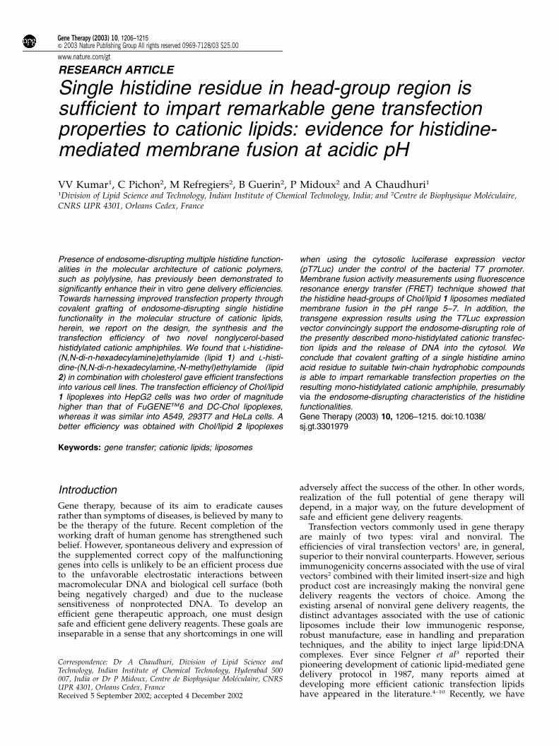
ChemistryWe designed and synthesized two simple mono-histidy-lated cationic lipids 1 and 2 using the precursor mixedprimary–tertiary amine II (Scheme 1) readily available inone of our laboratories. The endosome-disrupting histi-dine functionality in the head-group region of lipid 1 wascovalently grafted by conventional dicyclohexyl-carbo-diimide (DCC) coupling of di-tetr-butyl-pyrocarbonate(di-Boc)-protected histidine (IV) and the hydrophobicmixed tertiary–primary amine (III) followed by aciddeprotection and chloride ion-exchange chromatographyof the resulting intermediate V as outlined in Scheme 1.The mixed tertiary–primary amine III was prepared byreacting N,N-di-hexadecylamine (I) with N-tert-butylox-ycarbonyl protected 2-bromoethylamine in ethyl acetate(EtoAc) in the presence of anhydrous potassium carbo-
nate followed by deprotection and neutralization of theresulting intermediate II (Scheme 1).
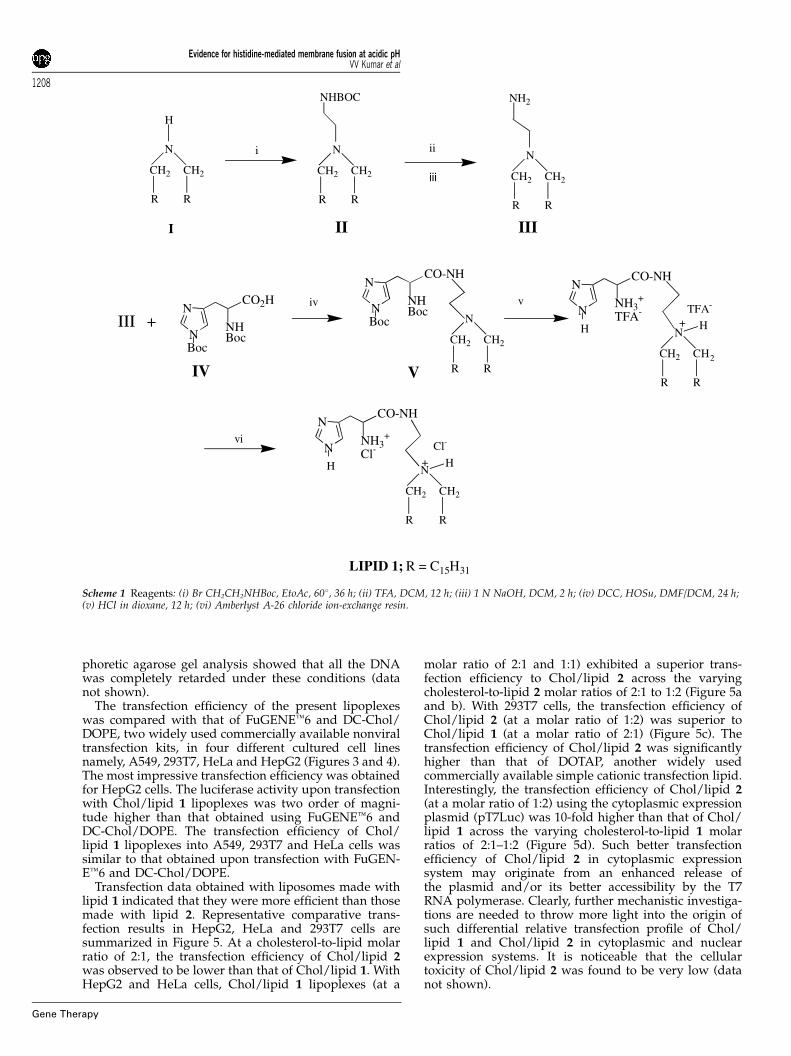
Lipid 2 was synthesized by quaternizing the commonintermediate V with excess methyl iodide followed byacid deprotection and chloride ion-exchange chromato-graphy of the resulting quaternized intermediate VII(Scheme 2).
Transfection efficiencyToward addressing the comparative transfection proper-ties of lipids 1 and 2 in nuclear and cytoplasmicexpression systems, both a conventionally used nuclearexpression plasmid pCMVLuc and a cytosolic expressionplasmid pT7Luc have been used in human embryonickidney 293-T7 cells. These cells stably express low levelsof T7 RNA polymerase, an enzyme necessary for thecytoplasmic transcription of genes controlled by thebacteriophage T7 RNA polymerase promoter. The use ofcytoplasmic expression systems with T7 RNA polymer-ase capable of ensuing cytoplasmic expression of thetransfected genes has been demonstrated to be an elegantway to avoid nuclear barrier of transfection pathways.25–29
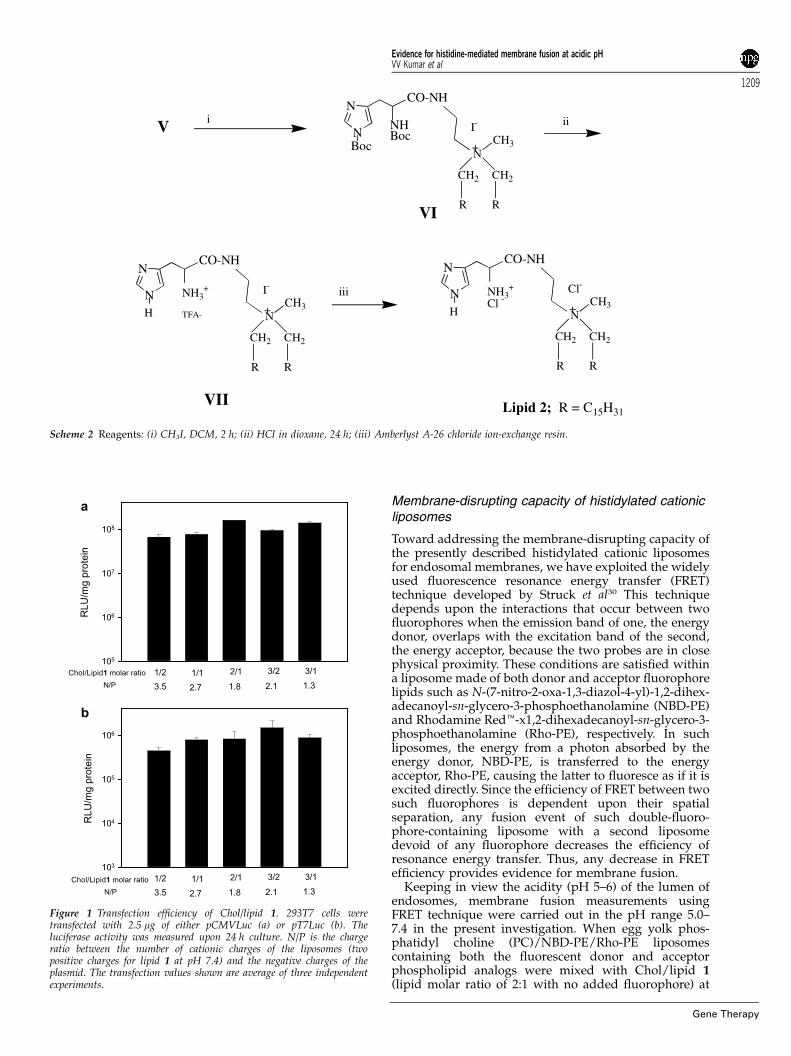
Experiments conducted with lipid 1 using bothpCMVLuc and pT7Luc plasmids in 293T7 cell linesrevealed that the transfection efficiency was not sensitiveto either cholesterol-to-lipid 1 molar ratios (in the range1:2–3:1) or to the lipid 1-to-DNA charge ratios (in therange 3.5–1.3) (Figure 1). The observed lower transfectionefficiencies for pT7Luc compared to that for pCMVLuccould result from the low level of the T7 RNApolymerase present in 293T7 cells and/or the weakstrength of the promoter. Increasing the lipid-to-DNAcharge ratios significantly enhanced the transfectionefficiency with pCMVLuc at a constant cholesterol-to-lipid 1 molar ratio of 2:1 (Figure 2a). The luciferaseexpression was 10-fold higher at N/P¼1.75 than at N/P¼1. However, the expression of pT7Luc was optimalwith the lipid-to-DNA charge ratio around N/P¼1(Figure 2b). Above this ratio, no benefit was obtained:the luciferase activity was lower at N/P¼1.75. An excessof cationic charges is likely to increase interactionsbetween liposomes and DNA. In the case of the nucleargene expression system (pCMVLuc), it would prevent arapid dissociation of the lipoplexes inside the cell,protecting DNA from degradation and thereby favoringits nuclear delivery. Conversely, in the case of thecytosolic gene expression system (pT7Luc), a highlipoplexes stability could be a limiting factor. Therefore,a decrease of the amount of cationic liposomes wouldreduce the stability of lipoplexes, facilitating theirdissociation and the accessibility of the DNA to thecytoplasmic expression machinery.
Cholesterol in combination with lipid 1 with a molarratio of 2:1 formed liposomes (Chol/lipid 1) with a sizeof 78724 nm. Under the same conditions, the sizes ofcommercially available liposomes such as Lipofectin,LipofectAMINE, DOTAP and DC-Chol were found to be68720, 77725, 51 and 170750 nm, respectively. The xpotential (the global charge) of Chol/lipid 1 was3271.5 mV. This value was similar to that of Lipofectinand LipofectAMINE but, higher than that of DC-Chol(1671.5 mV) and lower than that of DOTAP (5572 mV).When mixed with 5 mg of plasmid DNA, Chol/lipid 1(2:1) formed lipoplexes with a size of 490720 nm andexhibited a negative x potential of �2.5 mV. Electro-
Evidence for histidine-mediated membrane fusion at acidic pHVV Kumar et al
1207
Gene Therapy
phoretic agarose gel analysis showed that all the DNAwas completely retarded under these conditions (datanot shown).
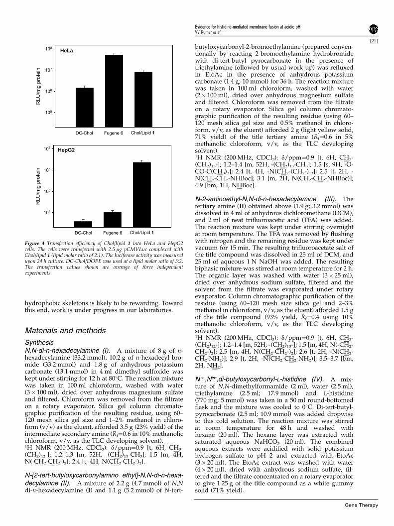
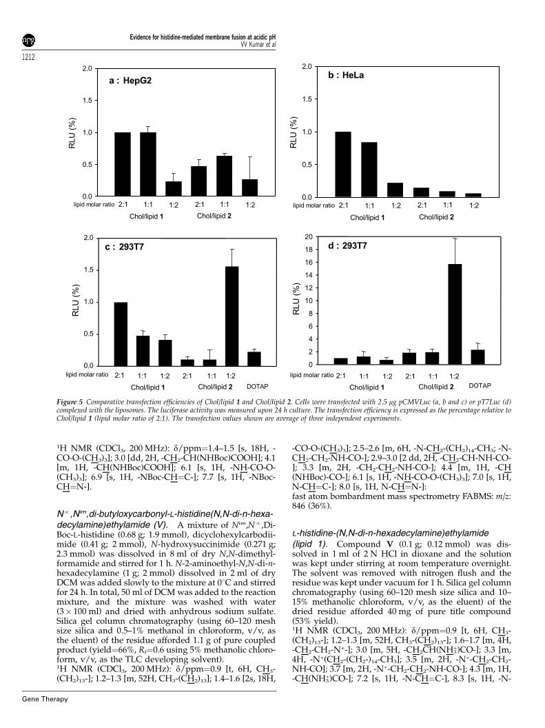
The transfection efficiency of the present lipoplexeswas compared with that of FuGENEt6 and DC-Chol/DOPE, two widely used commercially available nonviraltransfection kits, in four different cultured cell linesnamely, A549, 293T7, HeLa and HepG2 (Figures 3 and 4).The most impressive transfection efficiency was obtainedfor HepG2 cells. The luciferase activity upon transfectionwith Chol/lipid 1 lipoplexes was two order of magni-tude higher than that obtained using FuGENEt6 andDC-Chol/DOPE. The transfection efficiency of Chol/lipid 1 lipoplexes into A549, 293T7 and HeLa cells wassimilar to that obtained upon transfection with FuGEN-Et6 and DC-Chol/DOPE.
Transfection data obtained with liposomes made withlipid 1 indicated that they were more efficient than thosemade with lipid 2. Representative comparative trans-fection results in HepG2, HeLa and 293T7 cells aresummarized in Figure 5. At a cholesterol-to-lipid molarratio of 2:1, the transfection efficiency of Chol/lipid 2was observed to be lower than that of Chol/lipid 1. WithHepG2 and HeLa cells, Chol/lipid 1 lipoplexes (at a
molar ratio of 2:1 and 1:1) exhibited a superior trans-fection efficiency to Chol/lipid 2 across the varyingcholesterol-to-lipid 2 molar ratios of 2:1 to 1:2 (Figure 5aand b). With 293T7 cells, the transfection efficiency ofChol/lipid 2 (at a molar ratio of 1:2) was superior toChol/lipid 1 (at a molar ratio of 2:1) (Figure 5c). Thetransfection efficiency of Chol/lipid 2 was significantlyhigher than that of DOTAP, another widely usedcommercially available simple cationic transfection lipid.Interestingly, the transfection efficiency of Chol/lipid 2(at a molar ratio of 1:2) using the cytoplasmic expressionplasmid (pT7Luc) was 10-fold higher than that of Chol/lipid 1 across the varying cholesterol-to-lipid 1 molarratios of 2:1–1:2 (Figure 5d). Such better transfectionefficiency of Chol/lipid 2 in cytoplasmic expressionsystem may originate from an enhanced release ofthe plasmid and/or its better accessibility by the T7RNA polymerase. Clearly, further mechanistic investiga-tions are needed to throw more light into the origin ofsuch differential relative transfection profile of Chol/lipid 1 and Chol/lipid 2 in cytoplasmic and nuclearexpression systems. It is noticeable that the cellulartoxicity of Chol/lipid 2 was found to be very low (datanot shown).
N
H
CH2 CH2
R R
N
CH2 CH2
R R
NHBOC
N
CH2 CH2
R R
NH2
III
R = C15H31
II
ΙΙΙ + N
CH2 CH2
R R
N
NNHBoc
CO-NH
BocN
CH2 2CH
R R
N
NNH3
+
TFA--
CO-NH
H H
TFAiv v
+
V
N
NNHBoc
CO2H
Boc
IV
N
CH2 CH2
R R
N
NNH3
+
Cl-
CO-NH
H H
Cl-
+
LIPID 1;
I
i ii
iii
vi
Scheme 1 Reagents: (i) Br CH2CH2NHBoc, EtoAc, 601, 36 h; (ii) TFA, DCM, 12 h; (iii) 1 N NaOH, DCM, 2 h; (iv) DCC, HOSu, DMF/DCM, 24 h;(v) HCl in dioxane, 12 h; (vi) Amberlyst A-26 chloride ion-exchange resin.
Evidence for histidine-mediated membrane fusion at acidic pHVV Kumar et al
1208
Gene Therapy
Membrane-disrupting capacity of histidylated cationicliposomes
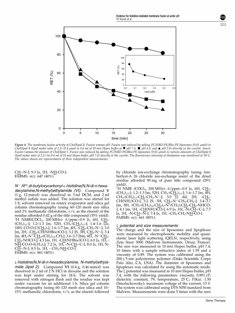
Toward addressing the membrane-disrupting capacity ofthe presently described histidylated cationic liposomesfor endosomal membranes, we have exploited the widelyused fluorescence resonance energy transfer (FRET)technique developed by Struck et al30 This techniquedepends upon the interactions that occur between twofluorophores when the emission band of one, the energydonor, overlaps with the excitation band of the second,the energy acceptor, because the two probes are in closephysical proximity. These conditions are satisfied withina liposome made of both donor and acceptor fluorophorelipids such as N-(7-nitro-2-oxa-1,3-diazol-4-yl)-1,2-dihex-adecanoyl-sn-glycero-3-phosphoethanolamine (NBD-PE)and Rhodamine Redt-x1,2-dihexadecanoyl-sn-glycero-3-phosphoethanolamine (Rho-PE), respectively. In suchliposomes, the energy from a photon absorbed by theenergy donor, NBD-PE, is transferred to the energyacceptor, Rho-PE, causing the latter to fluoresce as if it isexcited directly. Since the efficiency of FRET between twosuch fluorophores is dependent upon their spatialseparation, any fusion event of such double-fluoro-phore-containing liposome with a second liposomedevoid of any fluorophore decreases the efficiency ofresonance energy transfer. Thus, any decrease in FRETefficiency provides evidence for membrane fusion.
Keeping in view the acidity (pH 5–6) of the lumen ofendosomes, membrane fusion measurements usingFRET technique were carried out in the pH range 5.0–7.4 in the present investigation. When egg yolk phos-phatidyl choline (PC)/NBD-PE/Rho-PE liposomescontaining both the fluorescent donor and acceptorphospholipid analogs were mixed with Chol/lipid 1(lipid molar ratio of 2:1 with no added fluorophore) at
N
CH2 CH2
R R
N
NNHBoc
CO-NH
Boc CH3+
I-
N
CH2 CH2
R R
N
N
CO-NH
CH3+
I-NH3
+
TFA-H N
CH2 CH2
R R
N
N
CO-NH
CH3+
Cl-NH3+
Cl -
H
i ii
iii
VI
R = C15H31
V
Lipid 2;VII
Scheme 2 Reagents: (i) CH3I, DCM, 2 h; (ii) HCl in dioxane, 24 h; (iii) Amberlyst A-26 chloride ion-exchange resin.
RLU
/mg p
rote
in
105
106
107
108
1/2 1/1 2/1 3/2 3/1Chol/Lipid1 molar ratio
3.5 2.7 1.8 2.1 1.3N/P
a
RLU
/mg p
rote
in
103
104
105
106
1/2 1/1 2/1 3/2 3/1Chol/Lipid1 molar ratio
3.5 2.7 1.8 2.1 1.3N/P
b
Figure 1 Transfection efficiency of Chol/lipid 1. 293T7 cells weretransfected with 2.5 mg of either pCMVLuc (a) or pT7Luc (b). Theluciferase activity was measured upon 24 h culture. N/P is the chargeratio between the number of cationic charges of the liposomes (twopositive charges for lipid 1 at pH 7.4) and the negative charges of theplasmid. The transfection values shown are average of three independentexperiments.
Evidence for histidine-mediated membrane fusion at acidic pHVV Kumar et al
1209
Gene Therapy
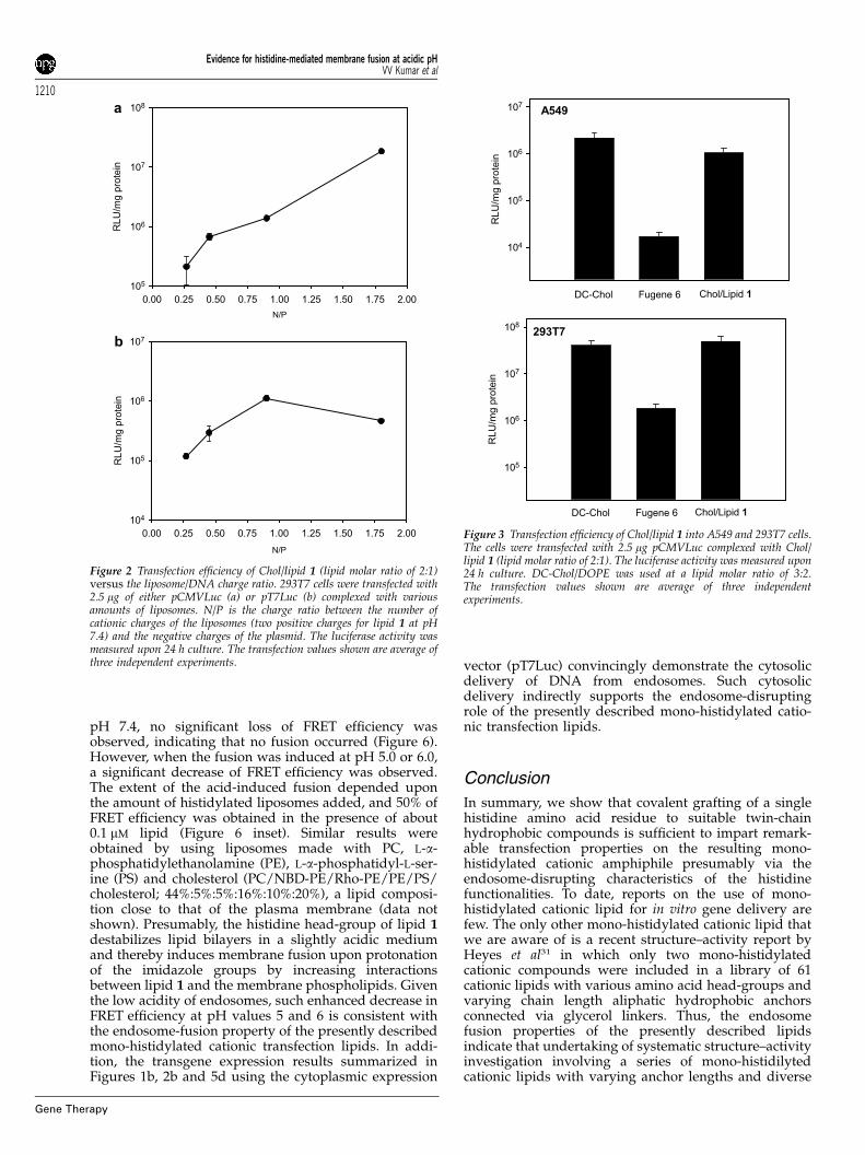
pH 7.4, no significant loss of FRET efficiency wasobserved, indicating that no fusion occurred (Figure 6).However, when the fusion was induced at pH 5.0 or 6.0,a significant decrease of FRET efficiency was observed.The extent of the acid-induced fusion depended uponthe amount of histidylated liposomes added, and 50% ofFRET efficiency was obtained in the presence of about0.1 mM lipid (Figure 6 inset). Similar results wereobtained by using liposomes made with PC, L-a-phosphatidylethanolamine (PE), L-a-phosphatidyl-L-ser-ine (PS) and cholesterol (PC/NBD-PE/Rho-PE/PE/PS/cholesterol; 44%:5%:5%:16%:10%:20%), a lipid composi-tion close to that of the plasma membrane (data notshown). Presumably, the histidine head-group of lipid 1destabilizes lipid bilayers in a slightly acidic mediumand thereby induces membrane fusion upon protonationof the imidazole groups by increasing interactionsbetween lipid 1 and the membrane phospholipids. Giventhe low acidity of endosomes, such enhanced decrease inFRET efficiency at pH values 5 and 6 is consistent withthe endosome-fusion property of the presently describedmono-histidylated cationic transfection lipids. In addi-tion, the transgene expression results summarized inFigures 1b, 2b and 5d using the cytoplasmic expression
vector (pT7Luc) convincingly demonstrate the cytosolicdelivery of DNA from endosomes. Such cytosolicdelivery indirectly supports the endosome-disruptingrole of the presently described mono-histidylated catio-nic transfection lipids.
Conclusion
In summary, we show that covalent grafting of a singlehistidine amino acid residue to suitable twin-chainhydrophobic compounds is sufficient to impart remark-able transfection properties on the resulting mono-histidylated cationic amphiphile presumably via theendosome-disrupting characteristics of the histidinefunctionalities. To date, reports on the use of mono-histidylated cationic lipid for in vitro gene delivery arefew. The only other mono-histidylated cationic lipid thatwe are aware of is a recent structure–activity report byHeyes et al31 in which only two mono-histidylatedcationic compounds were included in a library of 61cationic lipids with various amino acid head-groups andvarying chain length aliphatic hydrophobic anchorsconnected via glycerol linkers. Thus, the endosomefusion properties of the presently described lipidsindicate that undertaking of systematic structure–activityinvestigation involving a series of mono-histidilytedcationic lipids with varying anchor lengths and diverse
N/P
0.00 0.25 0.50 0.75 1.00 1.25 1.50 1.75 2.00
RL
U/m
g p
rote
in
105
106
107
108a
N/P
0.00 0.25 0.50 0.75 1.00 1.25 1.50 1.75 2.00
RL
U/m
g p
rote
in
104
105
106
107b
Figure 2 Transfection efficiency of Chol/lipid 1 (lipid molar ratio of 2:1)versus the liposome/DNA charge ratio. 293T7 cells were transfected with2.5 mg of either pCMVLuc (a) or pT7Luc (b) complexed with variousamounts of liposomes. N/P is the charge ratio between the number ofcationic charges of the liposomes (two positive charges for lipid 1 at pH7.4) and the negative charges of the plasmid. The luciferase activity wasmeasured upon 24 h culture. The transfection values shown are average ofthree independent experiments.
293T7
RLU
/mg p
rote
in
105
106
107
108
A549
RL
U/m
g p
rote
in
104
105
106
107
DC-Chol Fugene 6 Chol/Lipid 1
DC-Chol Fugene 6 Chol/Lipid 1
Figure 3 Transfection efficiency of Chol/lipid 1 into A549 and 293T7 cells.The cells were transfected with 2.5 mg pCMVLuc complexed with Chol/lipid 1 (lipid molar ratio of 2:1). The luciferase activity was measured upon24 h culture. DC-Chol/DOPE was used at a lipid molar ratio of 3:2.The transfection values shown are average of three independentexperiments.
Evidence for histidine-mediated membrane fusion at acidic pHVV Kumar et al
1210
Gene Therapy
hydrophobic skeletons is likely to be rewarding. Towardthis end, work is under progress in our laboratories.
Materials and methods
SynthesisN,N-di-n-hexadecylamine (I). A mixture of 8 g of n-hexadecylamine (33.2 mmol), 10.2 g of n-hexadecyl bro-mide (33.2 mmol) and 1.8 g of anhydrous potassiumcarbonate (13.1 mmol) in 4 ml dimethyl sulfoxide waskept under stirring for 12 h at 801C. The reaction mixturewas taken in 100 ml chloroform, washed with water(3� 100 ml), dried over anhydrous magnesium sulfateand filtered. Chloroform was removed from the filtrateon a rotary evaporator. Silica gel column chromato-graphic purification of the resulting residue, using 60–120 mesh silica gel size and 1–2% methanol in chloro-form (v/v) as the eluent, afforded 3.5 g (23% yield) of theintermediate secondary amine (Rf¼0.6 in 10% methanolicchloroform, v/v, as the TLC developing solvent).1H NMR (200 MHz, CDCl3): d/ppm¼0.9 [t, 6H, CH3-(CH2)12-]; 1.2–1.3 [m, 52H, -(CH2)13-CH3]; 1.5 [m, 4H,N(-CH2-CH2-)2]; 2.4 [t, 4H, N(CH2-CH2-)2].
N-[2-tert-butyloxycarbonylamino ethyl]-N,N-di-n-hexa-decylamine (II). A mixture of 2.2 g (4.7 mmol) of N,Ndi-n-hexadecylamine (I) and 1.1 g (5.2 mmol) of N-tert-
butyloxycarbonyl-2-bromoethylamine (prepared conven-tionally by reacting 2-bromoethylamine hydrobromidewith di-tert-butyl pyrocarbonate in the presence oftriethylamine followed by usual work up) was refluxedin EtoAc in the presence of anhydrous potassiumcarbonate (1.4 g; 10 mmol) for 36 h. The reaction mixturewas taken in 100 ml chloroform, washed with water(2� 100 ml), dried over anhydrous magnesium sulfateand filtered. Chloroform was removed from the filtrateon a rotary evaporator. Silica gel column chromato-graphic purification of the resulting residue (using 60–120 mesh silica gel size and 0.5% methanol in chloro-form, v/v, as the eluent) afforded 2 g (light yellow solid,71% yield) of the title tertiary amine (Rf¼0.6 in 5%methanolic chloroform, v/v, as the TLC developingsolvent).1H NMR (200 MHz, CDCI3): d/ppm¼0.9 [t, 6H, CH3-(CH2)13-]; 1.2–1.4 [m, 52H, -(CH2)13-CH3]; 1.5 [s, 9H, -O-CO-C(CH3)3]; 2.4 [t, 4H, -N(CH2-(CH2-)13]; 2.5 [t, 2H, -N(CH2-CH2-NHBoc]; 3.1 [m, 2H, N(CH2-CH2-NHBoc)];4.9 [bm, 1H, NHBoc].
N-2-aminoethyl-N,N-di-n-hexadecylamine (III). Thetertiary amine (II) obtained above (1.9 g; 3.2 mmol) wasdissolved in 4 ml of anhydrous dichloromethane (DCM),and 2 ml of neat trifluoroacetic acid (TFA) was added.The reaction mixture was kept under stirring overnightat room temperature. The TFA was removed by flushingwith nitrogen and the remaining residue was kept undervacuum for 15 min. The resulting trifluoroacetate salt ofthe title compound was dissolved in 25 ml of DCM, and25 ml of aqueous 1 N NaOH was added. The resultingbiphasic mixture was stirred at room temperature for 2 h.The organic layer was washed with water (3� 25 ml),dried over anhydrous sodium sulfate, filtered and thesolvent from the filtrate was evaporated under rotaryevaporator. Column chromatographic purification of theresidue (using 60–120 mesh size silica gel and 2–3%methanol in chloroform, v/v, as the eluent) afforded 1.5 gof the title compound (93% yield, Rf¼0.4 using 10%methanolic chloroform, v/v, as the TLC developingsolvent).1H NMR (200 MHz, CDCl3): d/ppm¼0.9 [t, 6H, CH3-(CH2)12-]; 1.2–1.4 [m, 52H, -(CH2)13-]; 1.5 [m, 4H, N(-CH2-CH2-)2]; 2.5 [m, 4H, N(CH2-CH2-)2]; 2.6 [t, 2H, -N(CH2-CH2-NH2)]; 2.9 [t, 2H, -N(CH2-CH2-NH2)]; 3.5–3.7 [bm,2H, NH2].
Np,Nim,di-butyloxycarbonyl-L-histidine (IV). A mix-ture of N,N-dimethylformamide (2 ml), water (2.5 ml),triethylamine (2.5 ml; 17.9 mmol) and L-histidine(770 mg; 5 mmol) was taken in a 50 ml round-bottomedflask and the mixture was cooled to 01C. Di-tert-butyl-pyrocarbonate (2.5 ml; 10.9 mmol) was added dropwiseto this cold solution. The reaction mixture was stirredat room temperature for 48 h and washed withhexane (20 ml). The hexane layer was extracted withsaturated aqueous NaHCO3 (20 ml). The combinedaqueous extracts were acidified with solid potassiumhydrogen sulfate to pH 2 and extracted with EtoAc(3� 20 ml). The EtoAc extract was washed with water(4� 20 ml), dried with anhydrous sodium sulfate, fil-tered and the filtrate concentrated on a rotary evaporatorto give 1.25 g of the title compound as a white gummysolid (71% yield).
RLU
/mg p
rote
in
105
106
107
108
DC-Chol Fugene 6 Chol/Lipid 1
DC-Chol Fugene 6 Chol/Lipid 1
RLU
/mg
pro
tein
104
105
106
107
HeLa
HepG2
Figure 4 Transfection efficiency of Chol/lipid 1 into HeLa and HepG2cells. The cells were transfected with 2.5 mg pCMVLuc complexed withChol/lipid 1 (lipid molar ratio of 2:1). The luciferase activity was measuredupon 24 h culture. DC-Chol/DOPE was used at a lipid molar ratio of 3:2.The transfection values shown are average of three independentexperiments.
Evidence for histidine-mediated membrane fusion at acidic pHVV Kumar et al
1211
Gene Therapy
1H NMR (CDCl3, 200 MHz): d/ppm¼1.4–1.5 [s, 18H, -CO-O-(CH3)3]; 3.0 [dd, 2H, -CH2-CH(NHBoc)COOH]; 4.1[m, 1H, -CH(NHBoc)COOH]; 6.1 [s, 1H, -NH-CO-O-(CH3)3]; 6.9 [s, 1H, -NBoc-CH¼C-]; 7.7 [s, 1H, -NBoc-CH¼N-].
Np,Nim,di-butyloxycarbonyl-L-histidine(N,N-di-n-hexa-decylamine)ethylamide (V). A mixture of Nim,Np,Di-Boc-L-histidine (0.68 g; 1.9 mmol), dicyclohexylcarbodii-mide (0.41 g; 2 mmol), N-hydroxysuccinimide (0.271 g;2.3 mmol) was dissolved in 8 ml of dry N,N-dimethyl-formamide and stirred for 1 h. N-2-aminoethyl-N,N-di-n-hexadecylamine (1 g; 2 mmol) dissolved in 2 ml of dryDCM was added slowly to the mixture at 01C and stirredfor 24 h. In total, 50 ml of DCM was added to the reactionmixture, and the mixture was washed with water(3� 100 ml) and dried with anhydrous sodium sulfate.Silica gel column chromatography (using 60–120 meshsize silica and 0.5–1% methanol in chloroform, v/v, asthe eluent) of the residue afforded 1.1 g of pure coupledproduct (yield¼66%, Rf¼0.6 using 5% methanolic chloro-form, v/v, as the TLC developing solvent).1H NMR (CDCl3, 200 MHz): d/ppm¼0.9 [t, 6H, CH3-(CH2)13-]; 1.2–1.3 [m, 52H, CH3-(CH2)13]; 1.4–1.6 [2s, 18H,
-CO-O-(CH3)3]; 2.5–2.6 [m, 6H, -N-CH2-(CH2)14-CH3; -N-CH2-CH2-NH-CO-]; 2.9–3.0 [2 dd, 2H, -CH2-CH-NH-CO-]; 3.3 [m, 2H, -CH2-CH2-NH-CO-]; 4.4 [m, 1H, -CH(NHBoc)-CO-]; 6.1 [s, 1H, -NH-CO-O-(CH3)3]; 7.0 [s, 1H,N-CH¼C-]; 8.0 [s, 1H, N-CH¼N-]:fast atom bombardment mass spectrometry FABMS: m/z:846 (36%).
L-histidine-(N,N-di-n-hexadecylamine)ethylamide(lipid 1). Compound V (0.1 g; 0.12 mmol) was dis-solved in 1 ml of 2 N HCl in dioxane and the solutionwas kept under stirring at room temperature overnight.The solvent was removed with nitrogen flush and theresidue was kept under vacuum for 1 h. Silica gel columnchromatography (using 60–120 mesh size silica and 10–15% methanolic chloroform, v/v, as the eluent) of thedried residue afforded 40 mg of pure title compound(53% yield).1H NMR (CDCl3, 200 MHz): d/ppm¼0.9 [t, 6H, CH3-(CH2)13-]; 1.2–1.3 [m, 52H, CH3-(CH2)13-]; 1.6–1.7 [m, 4H,-CH2-CH2-N+-]; 3.0 [m, 5H, -CH2CH(NH3
+)CO-]; 3.3 [m,4H, -N+(CH2-(CH2-)14-CH3]; 3.5 [m, 2H, -N+-CH2-CH2-NH-CO]; 3.7 [m, 2H, -N+-CH2-CH2-NH-CO-]; 4.3 [m, 1H,-CH(NH3
+)CO-]; 7.2 [s, 1H, -N-CH¼C-], 8.3 [s, 1H, -N-
RLU
(%
)
0.0
0.5
1.0
1.5
2.0
lipid molar ratio 2:11:1 1:22:1 1:11:2
Chol/lipid 1 Chol/lipid 2
RLU
(%
)
0.0
0.5
1.0
1.5
2.0
lipid molar ratio 2:11:1 1:22:1 1:11:2
Chol/lipid 1 Chol/lipid 2
a : HepG2b : HeLa
RL
U (
%)
0.0
0.5
1.0
1.5
2.0
DOTAP
lipid molar ratio 2:11:1 1:22:1 1:11:2
Chol/lipid 1 Chol/lipid 2
c : 293T7
RL
U (
%)
0
2
4
6
8
10
12
14
16
18
20
lipid molar ratio 2:11:1 1:22:1 1:11:2
Chol/lipid 1 Chol/lipid 2
d : 293T7
DOTAP
Figure 5 Comparative transfection efficiencies of Chol/lipid 1 and Chol/lipid 2. Cells were transfected with 2.5 mg pCMVLuc (a, b and c) or pT7Luc (d)complexed with the liposomes. The luciferase activity was measured upon 24 h culture. The transfection efficiency is expressed as the percentage relative toChol/lipid 1 (lipid molar ratio of 2:1). The transfection values shown are average of three independent experiments.
Evidence for histidine-mediated membrane fusion at acidic pHVV Kumar et al
1212
Gene Therapy
CH¼N-]; 9.3 [s, 1H, -NH-CO-].FABMS: m/z: 647 (40%).
Np,Nim,di-butyloxycarbonyl-L-histidine(N,N-di-n-hexa-decylamine,N-methyl)ethylamide (VI). Compound V(1 g, 12 mmol) was dissolved in 3 ml DCM, and 2 mlmethyl iodide was added. The solution was stirred for1 h, solvent removed on rotary evaporator and silica gelcolumn chromatography (using 60–120 mesh size silicaand 2% methanolic chloroform, v/v, as the eluent) of theresidue afforded 0.42 g of the title compound (35% yield).1H NMR(CDCl3, 200 MHz): d/ppm¼0.9 [t, 6H, CH3-(CH2)13-]; 1.2–1.3 [m, 52H, CH3-(CH2)13-]; 1.4–1.6 [2s,18H, CO-O-C(CH3)3]; 1.6–1.7 [m, 4H, -CH2-CH2-N+-]; 3.0[m, 2H, -CH2-CH(NHBoc)CO-]; 3.2 [S, 3H, CH3-N+-]; 3.4[m, 4H,-N+-CH2-(CH2)14-CH3]; 3.6–3.7 [bm, 4H, -N+-CH2-CH2-NHCO-]; 4.3 [m, 1H, -CH(NHBoc)CO-]; 6.0 [s, 1H, -NH-CO-O-(CH3)3]; 7.2 [s, 1H, -N-CH¼C-]; 8.0 [s, 1H, N-CH¼N-]; 8.5 [s, 1H, - CH2-NH-CO-].FABMS: m/z: 861 (40%).
L-histidine(N,N-di-n-hexadecylamine, N-methyl)ethyla-mide (lipid 2). Compound VI (0.4 g, 0.46 mmol) wasdissolved in 2 ml of 2 N HCl in dioxane and the solutionwas kept under stirring for 24 h. The solvent wasremoved with nitrogen flush and the residue was keptunder vacuum for an additional 1 h. Silica gel columnchromatography (using 60–120 mesh size silica and 10–15% methanolic chloroform, v/v, as the eluent) followed
by chloride ion-exchange chromatography (using Am-berlyst-A 26 chloride ion-exchange resin) of the driedresidue afforded 90 mg of pure title compound (29%yield).1H NMR (CDCl3, 200 MHz): d/ppm¼0.9 [s, 6H, CH3-(CH2)13-]; 1.2–1.3 [m, 52H, CH3-(CH2)13-]; 1.6–1.7 [m, 4H,CH3-(CH2)13-CH2-CH2-N+-]; 3.0 [2 dd, 2H, -CH2-CH(NH3
+)CO-]; 3.2 [S, 3H, CH3-N+-CH2-CH2-]; 3.4–3.7[m, 8H, (CH3-(CH2)14-CH2)2-N+(CH3)-CH2-CH2-NHCO-]; 4.1 [m, 1H, -CH(NH3
+)CO-]; 6.9 [s, 1H, -N-CH¼C-]; 7.7[s, 1H, -N-CH¼N-]; 7.4 [s, 1H, -CH2-CH2-NH-CO-].FABMS: m/z: 661 (80%).
x potential and size measurementsThe charge and the size of liposomes and lipoplexeswere measured by electrophoretic mobility and quasi-elastic laser light scattering (QELS), respectively, usingZeta Sizer 3000 (Malvern Instruments, Orsay, France).The size was measured in 10 mM Hepes buffer, pH 7.4,10 times with a sample refractive index of 1.59 and aviscosity of 0.89. The system was calibrated using the20075 nm polystyrene polymer (Duke Scientific CorpsPalo Alto, CA, USA). The diameter of liposomes andlipoplexes was calculated by using the automatic mode.The x potential was measured in 10 mM Hepes buffer, pH7.4, with the following parameters: viscosity, 0.891 cP;dielectric constant, 79; temperature, 251C; F(Ka), 1.50(Smoluchowsky); maximum voltage of the current, 15 V.The system was calibrated using DTS 5050 standard fromMalvern. Measurements were done 5 times with the zero
0 5 10 15 20 25 30
0
20
40
60
80
100
0.0
0
20
40
60
80
100
time (min)
RE
T (
%)
0.50.40.30.20.1
Lipid (µM)
RE
T (
%)
Figure 6 The membrane fusion activity of Chol/lipid 1. Fusion versus pH. Fusion was induced by adding PC/NBD-PE/Rho-PE liposomes (0.01 mmol) toChol/lipid 1 (lipid molar ratio of 2:1) (0.4 mmol in 0.6 ml of 10 mM Hepes buffer at � pH 7.4, ’ pH 6.0, and m pH 5.0) directly in the cuvette. Insert:Fusion versus the amount of Chol/lipid 1. Fusion was induced by adding PC/NBD-PE/Rho-PE liposomes (0.01 mmol) to various amounts of Chol/lipid 1(lipid molar ratio of 2:1) (in 0.6 ml of 10 mM Hepes buffer, pH 7.4) directly in the cuvette. The fluorescence intensity of rhodamine was monitored at 301C.The values shown are representative of three independent measurements.
Evidence for histidine-mediated membrane fusion at acidic pHVV Kumar et al
1213
Gene Therapy
field correction. The x potential was calculated using theSmoluchowsky approximation.
Cells and Cell CultureHuman hepatocarcinoma HepG2 cells (8055 HB, ATCC,Rockville, MD, USA), human epithelial ovary carcinomaHeLa cells (CCL21, ATCC), human carcinoma lung A549cells (CCL185, ATCC) and human embryo kidney 293T7cells25 were cultured in MEM containing 10% heat-inactivated fetal bovine serium (Life Technologies, CergyPontoise, France), 2 mM L-glutamine (Life Technologies),100 U/ml penicillin (Life Technologies) and 100 U/mlstreptomycin (Life Technologies). 293T7 cells werecultured in the presence of geneticin (400 mg/ml). Cellswere mycoplasma-free as evidenced by bisbenzimida-zole (Hoechst 33258, Molecular Probes).32
PlasmidspCMVLuc (pUT650, 5.15 kb, Cayla, Toulouse, France)and pT7Luc (kindly given by Dr M Brisson) wereplasmid DNA encoding the firefly luciferase under thecontrol of the human cytomegalovirus and the bacter-iophage T7 RNA polymerase promoter, respectively.Supercoiled plasmid DNA was isolated by a standardalkaline lysis method and purification was carried outwith the QIAGENs Plasmid Mega kit (QIAGEN,Courtaboeuf, France).
TransfectionsTwo days prior to transfection, cells were seeded at2� 105 cells in 2 ml culture medium in a 12-wells plate.At the time of the experiment, cell cultures were 80%confluent and lipofections were performed as describedbelow. Liposomes were prepared by ethanol injectionmethod: 15 ml of an ethanol solution of the lipid mixture– Chol/lipid1, Chol/lipid2 or DC-Chol/DOPE (DC-Choland DOPE from Sigma) – at 2.7 mM or 5.4 mM wasinjected rapidly into 200 ml of 10 mM Hepes buffer, pH7.4. After 15 min at room temperature, the liposomeswere mixed with the plasmid (5 mg in 20 ml of 10 mM
Hepes buffer, pH 7.4). After 15 min at room temperature,the lipoplexes solution was completed at 1 ml withserum-free medium and the NaCl concentration wasadjusted at 0.15 M with a 5 M NaCl solution. Beforeincubation with 0.5 ml lipoplexes, cells were washed 2times with serum-free culture medium. Cells wereincubated for 4 h at 371C, the medium was removedand cells were cultured for 48 h at 371C in completeculture medium without any additives.
LipofectAMINEt (Life Technologies) or Lipofectint(Roche Diagnostic, Meylan, France), DOTAP (RocheDiagnostic), FuGENEt6 (Roche Diagnostic) were usedaccording to the manufacturer’s procedure. Briefly, 25 mlof Lipofectin or Lipofectamine was added to 100 ml ofOPTIMEM, left for 15 min at room temperature and thenmixed with DNA (2.5 mg in 100 ml OPTIMEM). After15 min, the lipoplexes solution was completed at 1 mlwith OPTIMEM. DOTAP liposomes were made byadding 30 ml of DOTAP in 100 ml of 20 mM Hepes buffer,pH 7.4. After 15 min at room temperature, liposomeswere mixed with DNA (5 mg in 100 ml of 20 mM Hepesbuffer, pH 7.4). Upon 15 min complexation at roomtemperature, the lipoplexes solution was completed at2 ml with serum-free MEM. FuGENEt6 liposomes wereprepared by adding 6 ml of FuGENEt6 in 100 ml of
DMEM. After 15 min at room temperature, the liposomeswere mixed with DNA (2.5 mg in 100 ml DMEM). Then,the lipoplexes solution was completed at 1 ml withserum-free DMEM. The cell transfection procedure usingthese reagents was similar as described above.
Luciferase assayLuciferase gene expression was measured by monitoringthe luminescence activity according to De Wet et al.33 Themedium was discarded and cells were washed 3 timeswith PBS. The homogenization buffer (200 ml of 8 mM
MgCl2, 1 mM dithiothreitol, 1 mM ethylenetetra aceticacid, 1% Triton X-100, 15% glycerol, 25 mM Tris-phos-phate buffer, pH 7.8) was added into each well and tissueculture plates were kept for 15 min at 201C. The solutionwas recovered and spun down (5 min at 800 g). A total,95 ml of a 2 mM ATP solution in the homogenizationbuffer without Triton X-100 was added to 60 ml super-natant and the solution was shaken with a vortex. Theluminescence was recorded for 4 s in a Lumat LB 9501luminometer (Berthold, Wildbach, Germany) upon addi-tion of 150 ml of a 167 mM luciferin solution in water.Measurements were done in duplicate. The number ofrelative light units (RLU) of 1 pg/ml of luciferase was2000 under these assay conditions. The data showncorrespond to the number of RLU per mg proteins.Proteins were determined on each sample using themodified bicinchoninic acid (BCA) colorimetric assay.34,35
Membrane fusionThe membrane fusion activity of Chol/lipid 1 liposomeswas measured with the FRET assay as previouslydescribed.30 NBD-PE (Molecular Probes, Eugene, OR,USA) and Rho-PE (Molecular Probes) were used asdonor and acceptor fluorescent lipids, respectively. PC(1.14 mg; 1.8 mmol in chloroform) (Sigma), NBD-PE(0.05 mg; 0.052 mmol in ethanol) and Rho-PE (0.14 mg;0.096 mmol in ethanol) were mixed and dried underreduced pressure. The dried lipid films were hydratedovernight at 41C in 4 ml of 10 mM Hepes buffer, pH 7.4.The suspension was vigorously vortexed for 2–5 min atroom temperature and sonicated for 15 min in cold bathsonicator at 35 kHz (Bioblock Scientific, Strasbourg,France). Fusion was induced by adding PC/NBD-PE/Rho-PE liposomes (0.01 mmol) to Chol/lipid 1 (lipidmolar ratio 2:1) liposomes (0.4 mmol in 0.6 ml of 10 mM
Hepes buffer, pH 7.4) directly in the cuvette. Thefluorescence intensity of rhodamine was monitored witha spectrofluorometer (Jobin-Yvon Fluoromax-2). Theexcitation was at 465 nm and emissions were at 530and 580 nm for NDB and Rho, respectively. All experi-ments were done at 301C. Fusion (100%) was determinedfrom the rhodamine fluorescence intensity of PC/NBD-PE/Rho-PE liposomes in the presence of 0.1% TritonX100.
Acknowledgements
We express our gratitude to Dr JP Behr (Strasbourg,France) for initiating this Indo-French collaboration. Wethank warmly Drs L Huang and M Brisson (University ofPittsburgh, Pittsburgh, PA, USA) for 293T7 cells andpT7Luc, and Miss Laetitia Million for her help in FRETmeasurements. This work was supported by grants from
Evidence for histidine-mediated membrane fusion at acidic pHVV Kumar et al
1214
Gene Therapy
Agence Nationale de Recherche sur le Sida (ANRS) andAssociation pour la Recherche sur le Cancer (ARC). ACand VVK gratefully acknowledge the financial supportreceived from the Department of Biotechnology, Govern-ment of India, New Delhi. PM is Research Director atINSERM. CP is Professor Assistant at the University ofOrleans.
References
1 Mountain A. Gene therapy: the first decade. Trends Biotechnol2000; 18: 119–128.
2 Verma IM, Somina M. Gene therapy – promises, problems andprospects. Nature 1997; 389: 239–242.
3 Felgner PL et al. Lipofection: a highly efficient, lipid-mediatedDNA transfection procedure. Proc Natl Acad Sci USA 1987; 84:7413–7417.
4 Gao X, Huang L. Cationic liposome-mediated gene transfer. GeneTher 1995; 2: 710–722.
5 Guenin E et al. Cationic phosphonolipids containing quaternaryphosphonium and arsonium groups for DNA transfection withgood efficiency and low cellular toxicity. Angew Chem Int Ed Engl2000; 39: 629–631.
6 Floch V et al. Cation substitution in cationic phosphonolipids: anew concept to improve transfection activity and decreasecellular toxicity. J Med Chem 2000; 43: 4617–4628.
7 Floch V et al. Systemic administration of cationic phosphonoli-pids/DNA complexes and the relationship between formulationand lung transfection efficiency. Biochim Biophys Acta 2000; 1464:95–103.
8 Pitard B et al. Sterically stabilized BGTC-based lipoplexes:structural features and gene transfection into the mouse airwaysin vivo. J Gene Med 2001; 3: 478–487.
9 Oudrhiri N et al. Gene transfer by guanidinium-cholesterolcationic lipids into airway epithelial cells in vitro and in vivo. ProcNatl Acad Sci USA 1997; 94: 1651–1656.
10 Tan F, Hughes JA. Introduction of a disulfide bond into a cationiclipid enhances transgene expression of plasmid DNA. BiochemBiophys Res Commun 1998; 242: 141–145.
11 Banerjee R et al. Design, synthesis and transfection biology ofnovel cationic glycolipids for use in liposomal gene delivery.J Med Chem 2001; 44: 4176–4185.
12 Ayyagari AA et al. Novel non-glycerol based cytofectins withlactic acid derived head groups. Biochem Biophys Res Commun2001; 289: 1057–1062.
13 Sunil Singh R et al. Anchor-dependency for non-glycerol basedcationic lipofectins: mixed bag of regular and anomaloustransfection profiles. Chem Eur J 2002; 8: 900–909.
14 Sri Lakshmi GV et al. Anchor-dependent Lipofection with non-glycerol based cytofectins containing single 2-hydroxyethyl headgroups. Biochim Biophys Acta 2002; 1559: 87–95.
15 Banerjee R et al. A novel series of non-glycerol based cationictransfection lipids for use in liposomal gene delivery. J Med Chem1999; 42: 4292–4299.
16 Bally MB et al. Biological barriers to cellular delivery of lipid-based DNA carriers. Adv Drug Deliv Rev 1999; 38: 291–315.
17 Zabner J et al. Cellular and molecular barriers to gene transfer bya cationic lipid. J Biol Chem 1995; 270: 18997–19007.
18 Friend DS, Papahadjopoulos D, Debs RJ. Endocytosis andIntracellular processing accompanying transfection mediatedby cationic liposomes. Biochim Biophys Acta 1996; 1278: 41–50.
19 Xu Y, Szoka Jr FC. Mechanism of DNA release from cationicliposome/DNA complexes used in cell transfection. Biochemistry1996; 35: 5616–5623.
20 Pichon C, Goncalves C, Midoux P. Histidine-rich peptides andpolymers for nucleic acids delivery. Adv Drug Deliv Rev 2001; 53:75–94.
21 Midoux P, Monsigny M. Efficient gene transfer by histidylatedpolylysine/pDNA complexes. Bioconjugate Chem 1999; 10: 406–411.
22 Benns JM et al. pH-sensitive cationic polymer gene deliveryvehicle:N-Ac-poly(L-histidine)-graft-poly(L-lysine) comb shapedpolymer. Bioconjugate Chem 2000; 11: 637–645.
23 Chen Q-R, Zhang L, Stass SA, Mixon AJ. Co-polymer of histidineand lysine markedly enhances transfection efficiency of lipo-somes. Gene Ther 2000; 7: 1698–1705.
24 Chen Q-R, Zhang L, Luther PW, Mixson AJ. Optimal transfec-tion with the HK polymer depends on its degree of branchingand the pH of endocytic vesicles. Nucleic Acids Res 2002; 30:1338–1345.
25 Brisson M et al. Subcellular trafficking of the cytoplasmicexpression system. Hum Gene Ther 1999; 10: 2601–2613.
26 Gao X, Huang L. Cytoplasmic expression of a reporter geneby co-delivery of T7 RNA polymerase and T7 promotersequence with cationic liposomes. Nucleic Acids Res 1993; 21:2867–2872.
27 Gao X, Jaffurs D, Robbins PD, Huang L. A sustained cytoplasmictransgene expression system delivered by cationic liposomes.Biochem Biophys Res Commun 1994; 200: 1201–1206.
28 Deng H, Wolff JA. Self-amplifying Expression from the T7Promoter in 3T3 mouse fibroblasts. Gene 1994; 143: 245–249.
29 Li S, Brisson M, He Y, Huang L. Delivery of a PCR amplifiedDNA fragment into cells: a model for using synthetic genes forgene therapy. Gene Ther 1997; 4: 449–454.
30 Struck DK, Hoekstra D, Pagano RE. Use of resonance energytransfer to monitor membrane fusion. Biochemistry 1981; 20:4093–4099.
31 Heyes JA, Duvaz DN, Cooper RG, Springer CJ. Synthesis ofnovel cationic lipids: effect of structural modification on theefficiency of gene transfer. J Med Chem 2001; 45: 99–114.
32 Chen TR. In situ detection of mycoplasma contamination in cellcultures by fluorescent Hoeschst 33258 stain. Exp Cell Res 1977;104: 255–262.
33 De Wet JR et al. Firefly luciferase gene structure and expressionin mammalian cells. Mol Cell Biol 1987; 7: 725–737.
34 Smith PK et al. Measurement of protein using bicinchononicacid. Anal Biochem 1985; 150: 76–85.
35 Hill HD, Straka JG. Protein determination using bicinchoninicacid in the presence of sulfhydryl reagents. Anal Biochem 1988;170: 203–208.
Evidence for histidine-mediated membrane fusion at acidic pHVV Kumar et al
1215
Gene Therapy
Related Documents


