Scanning Electrochemical Microscopy DOI: 10.1002/anie.200604851 Single-Cell Microelectrochemistry Albert Schulte and Wolfgang Schuhmann* Angewandte Chemie Keywords: exocytosis · living cells · scanning electrochemical microscopy (SECM) · ultramicroelectrodes · voltammetry W. Schuhmann and A. Schulte Reviews 8760 www.angewandte.org # 2007 Wiley-VCH Verlag GmbH & Co. KGaA, Weinheim Angew. Chem. Int. Ed. 2007, 46, 8760 – 8777 &&&&&&&&&&&&&&&&&&&&&&&&&&&&&&&&&&&&&&&&&&&&&&&&&&&&&&&&&&&&&&&&&&&&&&&&&&&&&&&&&&&&&&&&&&&&&&&&&&&&&&&&&&&&&&&&&&&&&&&&&&&&&&&&&&&&&&&&&&&&&&&&&&&&&&&&&&&& &&&&&&&&&&&&&&&&&&&&&&&&&&&&&&&&&&&& Take advantage of blue reference links &&&&&&&&&&&&&&&&&&&&&&&&&&&&&&&&&&&& &&&&&&&&&&&&&&&&&&&&&&&&&&&&&&&&&&&&&&&&&&&&&&&&&&&&&&&&&&&&&&&&&&&&&&&&&&&&&&&&&&&&&&&&&&&&&&&&&&&&&&&&&&&&&&&&&&&&&&&&&&&&&&&&&&&&&&&&&&&&&&&&&&&&&&&&&&&&

Welcome message from author
This document is posted to help you gain knowledge. Please leave a comment to let me know what you think about it! Share it to your friends and learn new things together.
Transcript
Scanning Electrochemical MicroscopyDOI: 10.1002/anie.200604851
Single-Cell MicroelectrochemistryAlbert Schulte and Wolfgang Schuhmann*
AngewandteChemie
Keywords:exocytosis · living cells ·scanning electrochemicalmicroscopy (SECM) ·ultramicroelectrodes ·voltammetry
W. Schuhmann and A. SchulteReviews
8760 www.angewandte.org � 2007 Wiley-VCH Verlag GmbH & Co. KGaA, Weinheim Angew. Chem. Int. Ed. 2007, 46, 8760 – 8777
&&&&&&&&&&&&&&&&&&&&&&&&&&&&&&&&&&&&&&&&&&&&&&&&&&&&&&&&&&&&&&&&&&&&&&&&&&&&&&&&&&&&&&&&&&&&&&&&&&&&&&&&&&&&&&&&&&&&&&&&&&&&&&&&&&&&&&&&&&&&&&&&&&&&&&&&&&&&
&&&&&&&&&&&&&&&&&&&&&&&&&&&&&&&&&&&& Take advantage of blue reference links &&&&&&&&&&&&&&&&&&&&&&&&&&&&&&&&&&&&
&&&&&&&&&&&&&&&&&&&&&&&&&&&&&&&&&&&&&&&&&&&&&&&&&&&&&&&&&&&&&&&&&&&&&&&&&&&&&&&&&&&&&&&&&&&&&&&&&&&&&&&&&&&&&&&&&&&&&&&&&&&&&&&&&&&&&&&&&&&&&&&&&&&&&&&&&&&&
1. Introduction
Vast technological improvements in the areas of inte-grated electronic circuits, microprocessors, and software inthe past few decades have brought about major advances inelectrochemical instrumentation. This progress allowed elec-trochemistry, already a well-established scientific field, tomake remarkable steps forward in almost all its fields ofapplication. Easy-to-use, high-precision computerized poten-tiostats appeared on the market and allowed the measure-ment of very small currents with superb noise levels andaccuracy. In parallel, the routine fabrication and positioningof ultrasmall voltammetric electrodes in many laboratoriesenabled their use as miniaturized electroanalytical tools forthe determination of redox-active compounds with highspatiotemporal resolution, excellent sensitivity, and lowdetection limits.
In voltammetry, a constant or systematically changingpotential is applied to the working electrode and its currentresponse is monitored as a function of time and/or potential.Unlike conventional electrodes, voltammetric ultramicro-electrodes (UMEs) have their characteristic geometricdimension (e.g. the diameter/radius for disk or the width forband electrodes) significantly decreased from the macro-scopic to the microscopic scale.[1] The resulting extremelysmall active electrode surfaces, however, are intrinsicallyassociated with very tiny measurable currents, and it was infact the access to highly sensitive electrochemical amplifiersand the ability to precisely monitor current levels down toseveral 100 fA that paved the way for more broadly exploringthe range of applications of voltammetric UMEs in traceanalysis, kinetics measurements, as well as surface andbiomedical sciences.
Voltammetric UMEs are now available with variousgeometries (cylinder, hemisphere, disk, band, ring, andcombinations thereof), dimensions (mm to nm), and materials(C, Pt, Au, Ag). Their theory, fabrication, and performance
have been presented in a monograph[2] and in several reviewarticles.[3] Briefly, the advantages of UMEs for voltammetricand chronoamperometric experiments include:1) improved signal-to-noise ratio primarily because the
analytically relevant Faraday currents are greatlyenhanced; this enhancement results from the hemispher-ical or spherical (rather than planar) diffusion towardsUMEs, which produces higher mass transfer rates of theelectroactive species;
2) less distortion of measurement by the iR drop, because thetotal currents (i) typically measured at UME workingelectrodes are smaller than those at conventional elec-trodes. Thus, measurements can be performed in highlyresistive solutions (e.g., solutions without added support-ing electrolyte);
3) a fast response time owing to the small double-layercapacitances (C) and thus low RC time constants (t). Thisproperty allows rapid changes in the concentration ofredox-active species to be monitored at nanosecond timescales;
4) the small overall size enables electrochemical experimentsin limited volumes and at microscopically small objects.
With their outstanding electroanalytical and geometricalproperties, needle-type UMEs with micrometer-sized total tipdimensions are superior for spatially and temporally resolved
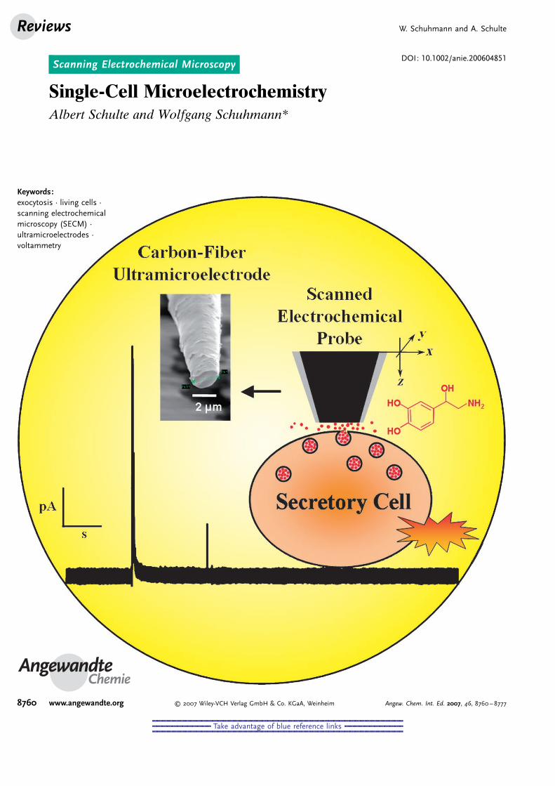
Needle-type voltammetric ultramicroelectrodes show exceptionalsensitivity for the detection of redox-active substances, rapid responsetimes, and total tip diameters in the lower micrometer range. Thesecharacteristics make them ideal for analyzing the chemical environ-ment and the activity of isolated living cells, which in their variousforms are the microscopic building blocks of human, animal, andother life forms. Prerequisites for successful local electrochemicalmeasurements in the vicinity of the tiny biological objects are gentle,stress-free, and accurate placement of the tip at the cell, exact knowl-edge of the tip-to-cell distance, and appropriate selectivity of theultramicroelectrode tip for species that may change in concentration asa result of cellular actions such as growth, respiration, or transmitterand metabolite uptake or release. The concepts of single-cell micro-electrochemistry are considered and an overview is given of recentresults on the fundamental mechanisms of cell functions.
From the Contents
1. Introduction 8761
2. Voltammetric UMEs forDetection at and in SingleLiving Cells 8763
3. Examples of IntracellularVoltammetry 8765
4. Examples of ConventionalExtracellular Voltammetry 8766
5. Single-Cell ScanningElectrochemical Microscopy(SECM) 8768
6. Conclusion and Future Aspects 8773
[*] Prof. Dr. W. SchuhmannAnalytische Chemie—Elektroanalytik & SensorikRuhr-Universit4t BochumUniversit4tsstrasse 150, 44780 Bochum (Germany)Fax: (+49)234-3214683E-mail: [email protected]
Assoc. Prof. Dr. A. SchulteSchool of Chemistry, Institute of ScienceSuranaree University of Technology111 University Avenue, Muang District, Nakhon Ratchasima 30000(Thailand)
MicroelectrochemistryAngewandte
Chemie
8761Angew. Chem. Int. Ed. 2007, 46, 8760 – 8777 � 2007 Wiley-VCH Verlag GmbH & Co. KGaA, Weinheim
&&&&&&&&&&&&&&&&&&&&&&&&&&&&&&&&&&&&&&&&&&&&&&&&&&&&&&&&&&&&&&&&&&&&&&&&&&&&&&&&&&&&&&&&&&&&&&&&&&&&&&&&&&&&&&&&&&&&&&&&&&&&&&&&&&&&&&&&&&&&&&&&&&&&&&&&&&&&
&&&&&&&&&&&&&&&&&&&&&&&&&&&&&&&&&&&& Take advantage of blue reference links &&&&&&&&&&&&&&&&&&&&&&&&&&&&&&&&&&&&
&&&&&&&&&&&&&&&&&&&&&&&&&&&&&&&&&&&&&&&&&&&&&&&&&&&&&&&&&&&&&&&&&&&&&&&&&&&&&&&&&&&&&&&&&&&&&&&&&&&&&&&&&&&&&&&&&&&&&&&&&&&&&&&&&&&&&&&&&&&&&&&&&&&&&&&&&&&&
voltammetric measurements in biological systems and henceintensely utilized in the life and medical sciences. The mostactive disciplines are neurochemistry and cell physiology, inwhich miniaturized voltammetric sensors are frequentlyemployed for observing the dynamics of a variety of cellularprocesses and tracking changes in the chemical compositionof the intra- and extracellular fluid. These changes are eitherattributable to cell activities such as growth, reproduction,respiration, and cell-to-cell communication, or they revealmetabolic reactions that result from strains and stresses suchas food shortage, physical exercise, or drug uptake. In general,in vivo applications in which the active tips of UMEs areimplanted with as little impact as possible into selected tissueof laboratory rodents must be distinguished from in vitroapplications on isolated biological cells or acute tissue slices.
In vivo voltammetry in the central nervous system ofliving animals with the aim of elucidating the neurochemistryof cells involved in complex behavior is certainly one of theexciting applications of this type of electroanalysis. The fieldwas actually established by Adams and co-workers at thebeginning of the 1970s with their efforts to observe fluctua-tions in the levels of catecholamine neurotransmitters invarious regions of the rat brain subsequent to stimulatedsynaptic release.[4] Since this pioneering work, the progress ofin vivo voltammetry has been remarkable, which is reflectedin the number of published review articles.[5] Two recentachievements are the wireless voltammetry in the brains offreely moving rats by implementation of telemetric systems[6]
and the real-time monitoring of naturally occurring tonic orphased dopamine signals in the extracellular fluid of differentregions of the brain of alert (not anesthetized) rats, whichwere correlated to behavior as diverse as sexual arousal,reward, food and novelty seeking, or drug-taking.[7,8]
This review article focuses on single-cell microelectro-chemistry, or to be more exact electrochemical measurementsthat are performed with voltammetric UMEs on the smallestsustainable units of life—isolated biological cells in cellcultures. Although estranged from their indigenous surround-ings, individual isolated or cultured cells can sustain theircharacteristic metabolic and (neuro)physiological processes,thus resembling to a certain extent cells in the body.Accordingly, single cells are good experimental modelsystems for examining complex biological processes andfunctions in a controlled and straightforward manner. In
general, these experiments are not influenced by interfer-ences that often cause problems in the highly heterogeneousintact tissue. However, living cells have microscopic dimen-sions, and responses attributable to their distinct activity aretherefore small and often occur on a very fast timescale.Screening and visualizing of single cells hence requiresophisticated analytical techniques with adequate sensitivityand high spatiotemporal resolution. Examples includeadvanced optical[9] and electrophysiology techniques,[10] andthe use of voltammetric UMEs.
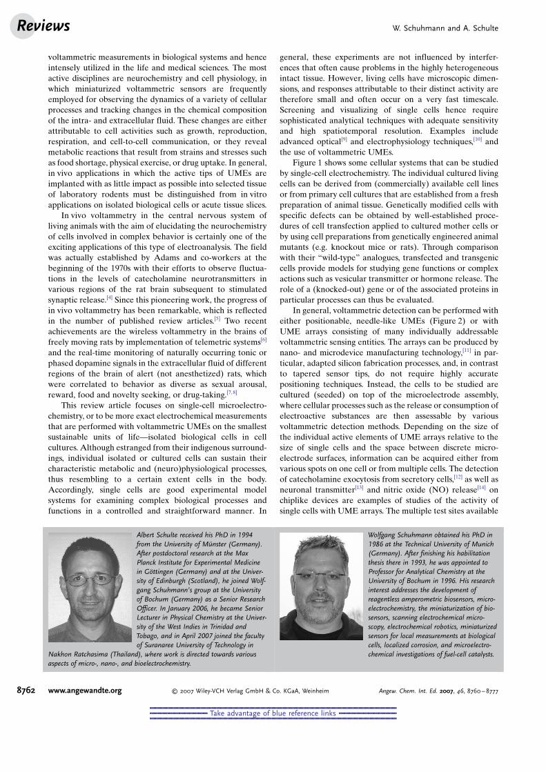
Figure 1 shows some cellular systems that can be studiedby single-cell electrochemistry. The individual cultured livingcells can be derived from (commercially) available cell linesor from primary cell cultures that are established from a freshpreparation of animal tissue. Genetically modified cells withspecific defects can be obtained by well-established proce-dures of cell transfection applied to cultured mother cells orby using cell preparations from genetically engineered animalmutants (e.g. knockout mice or rats). Through comparisonwith their “wild-type” analogues, transfected and transgeniccells provide models for studying gene functions or complexactions such as vesicular transmitter or hormone release. Therole of a (knocked-out) gene or of the associated proteins inparticular processes can thus be evaluated.
In general, voltammetric detection can be performed witheither positionable, needle-like UMEs (Figure 2) or withUME arrays consisting of many individually addressablevoltammetric sensing entities. The arrays can be produced bynano- and microdevice manufacturing technology,[11] in par-ticular, adapted silicon fabrication processes, and, in contrastto tapered sensor tips, do not require highly accuratepositioning techniques. Instead, the cells to be studied arecultured (seeded) on top of the microelectrode assembly,where cellular processes such as the release or consumption ofelectroactive substances are then assessable by variousvoltammetric detection methods. Depending on the size ofthe individual active elements of UME arrays relative to thesize of single cells and the space between discrete micro-electrode surfaces, information can be acquired either fromvarious spots on one cell or from multiple cells. The detectionof catecholamine exocytosis from secretory cells,[12] as well asneuronal transmitter[13] and nitric oxide (NO) release[14] onchiplike devices are examples of studies of the activity ofsingle cells with UME arrays. The multiple test sites available
Albert Schulte received his PhD in 1994from the University of M nster (Germany).After postdoctoral research at the MaxPlanck Institute for Experimental Medicinein G+ttingen (Germany) and at the Univer-sity of Edinburgh (Scotland), he joined Wolf-gang Schuhmann’s group at the Universityof Bochum (Germany) as a Senior ResearchOfficer. In January 2006, he became SeniorLecturer in Physical Chemistry at the Univer-sity of the West Indies in Trinidad andTobago, and in April 2007 joined the facultyof Suranaree University of Technology in
Nakhon Ratchasima (Thailand), where work is directed towards variousaspects of micro-, nano-, and bioelectrochemistry.
Wolfgang Schuhmann obtained his PhD in1986 at the Technical University of Munich(Germany). After finishing his habilitationthesis there in 1993, he was appointed toProfessor for Analytical Chemistry at theUniversity of Bochum in 1996. His researchinterest addresses the development ofreagentless amperometric biosensors, micro-electrochemistry, the miniaturization of bio-sensors, scanning electrochemical micro-scopy, electrochemical robotics, miniaturizedsensors for local measurements at biologicalcells, localized corrosion, and microelectro-chemical investigations of fuel-cell catalysts.
W. Schuhmann and A. SchulteReviews
8762 www.angewandte.org � 2007 Wiley-VCH Verlag GmbH & Co. KGaA, Weinheim Angew. Chem. Int. Ed. 2007, 46, 8760 – 8777
&&&&&&&&&&&&&&&&&&&&&&&&&&&&&&&&&&&&&&&&&&&&&&&&&&&&&&&&&&&&&&&&&&&&&&&&&&&&&&&&&&&&&&&&&&&&&&&&&&&&&&&&&&&&&&&&&&&&&&&&&&&&&&&&&&&&&&&&&&&&&&&&&&&&&&&&&&&&
&&&&&&&&&&&&&&&&&&&&&&&&&&&&&&&&&&&& Take advantage of blue reference links &&&&&&&&&&&&&&&&&&&&&&&&&&&&&&&&&&&&
&&&&&&&&&&&&&&&&&&&&&&&&&&&&&&&&&&&&&&&&&&&&&&&&&&&&&&&&&&&&&&&&&&&&&&&&&&&&&&&&&&&&&&&&&&&&&&&&&&&&&&&&&&&&&&&&&&&&&&&&&&&&&&&&&&&&&&&&&&&&&&&&&&&&&&&&&&&&
on striplike sensor devices and the suitability for automatedloading and analysis make UME arrays suitable for high-throughput screening of cellular activity. Several analytes canbe detected simultaneously and interconnected networks ofneuronal or other cells can be chemically probed. However,the possible applications of UME arrays in voltammetricdetection at the single-cell level have not yet been fullyexplored.
The first section of this review gives a general overview ofmethods for the fabrication of voltammetric UMEs with tipsthat are appropriate for in vitro voltammetric measurementsinside the cytoplasm (intracellular voltammetry) or in closeproximity of single biological cells (extracellular voltamme-try). Selected examples of reports on intracellular voltamme-try and on conventional “extracellular” voltammetric detec-tion of chemical messengers such as catecholamines, nitricoxide (NO), and glutamate, monitored with stationary UMEtips placed next to the outer membrane of a secretory cell arebriefly discussed. Scanning electrochemical microscopy(SECM), a scanning probe microscopy technique that usesprecisely movable voltammetric or potentiometric UMEs ashigh-resolution imaging tools, has been established[15] andapplied for single-cell studies in which cell morphology andcellular (redox) activity are imaged simultaneously.[16] Theo-retical and practical aspects that are relevant for SECMmeasurements on living cells are provided. This sectionincludes a discussion of the drawbacks of the constant-heightmode and the advantages of the constant-distance mode ofSECM for imaging soft three-dimensional biological objects.Important developments in SECM instrumentation arereviewed and illustrations of single-cell SECM studies areused to highlight the impact that single-cell SECM may haveas a modern electrochemical tool in different disciplines oflife science and medicine. Finally, potential future aspects andchallenges of single-cell electrochemistry are discussed.
2. Voltammetric UMEs for Detection at and inSingle Living Cells
Quite a few papers over the past years have describedprocedures for the fabrication of tiny voltammetric probesthat are, in addition to other microelectrochemical applica-tions, suitable for spatially confined measurements very closeto or even inside living cells. The optimum type of electrode isvery much dependent on the particular cell and the cellularprocess under investigation. In any case, voltammetric UMEsfor single-cell electrochemistry should offer not only anappropriate tip size but also excellent sensitivity towards theanalyte, low background current, and good stability inphysiological buffer solutions. Furthermore, they should beeasy to fabricate and handle. The materials used predom-inantly for the fabrication of UMEs are carbon and platinum.Carbon is ideal for detecting the release of catecholamineneurotransmitters in physiological saline solutions, as carbonelectrode surfaces are less susceptible to electrode fouling.Platinum is favored for measuring, for example, extra- orintracellular concentrations of oxygen thanks to its electro-catalytical properties.
Figure 1. Representative systems that have been studied by single-cellmicroelectrochemistry. Top: Individual living cells can be obtainedeither through enzymatic dissociation of explanted tissue of laboratoryrodents or farm animals or from the many commercially available celllines and may be genetically modified for specific cell activity measure-ments. Bottom: Single cultured cells may be studied with the tips ofpointed micro- or nanoelectrodes positioned close to their plasmamembranes or while resting on the active microscopic surfaces ofmicroelectrode arrays. For the detection of electroactive species in thevicinity of active cells, the chosen microsensors may be operated in avoltammetric mode by continuously changing the working electrodepotential E as a linear function of time t between two user-definedvalues with DE/Dt referred to as the scan rate or in an amperometricmode by keeping E at a constant value that is appropriate to oxidize orreduce the redox species of interest.
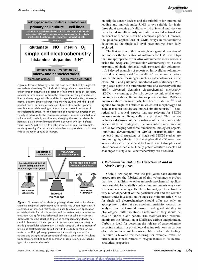
Figure 2. Schematic of an electrophysiological workstation for electro-chemical single-cell experiments with needle-type voltammetric micro-electrodes. An inverted microscope is used to operate an applicationor patch pipette for cell stimulation and the voltammetric ultramicro-electrode (UME) for electrochemical detection of cellular responses.Both tools must be attached to precise micropositioning devices forcareful placement of their tips next to (extracellular voltammetry) orinside (intracellular voltammetry) a single living cell. The utilization oflow-noise electrochemical amplifiers with the ability to monitor cur-rents in the fA to pA range guarantees the sensitivity needed fortracing tiny changes in concentration of redox-active species resultingfrom cellular activities such as secretion or respiration. m-CE: needle-type micro-counter electrode.
MicroelectrochemistryAngewandte
Chemie
8763Angew. Chem. Int. Ed. 2007, 46, 8760 – 8777 � 2007 Wiley-VCH Verlag GmbH & Co. KGaA, Weinheim www.angewandte.org
&&&&&&&&&&&&&&&&&&&&&&&&&&&&&&&&&&&&&&&&&&&&&&&&&&&&&&&&&&&&&&&&&&&&&&&&&&&&&&&&&&&&&&&&&&&&&&&&&&&&&&&&&&&&&&&&&&&&&&&&&&&&&&&&&&&&&&&&&&&&&&&&&&&&&&&&&&&&
&&&&&&&&&&&&&&&&&&&&&&&&&&&&&&&&&&&& Take advantage of blue reference links &&&&&&&&&&&&&&&&&&&&&&&&&&&&&&&&&&&&
&&&&&&&&&&&&&&&&&&&&&&&&&&&&&&&&&&&&&&&&&&&&&&&&&&&&&&&&&&&&&&&&&&&&&&&&&&&&&&&&&&&&&&&&&&&&&&&&&&&&&&&&&&&&&&&&&&&&&&&&&&&&&&&&&&&&&&&&&&&&&&&&&&&&&&&&&&&&
For single-cell measurements in the extracellular spacevery close to single living cells, the preferable sensor geometryis a disk or disklike active electrode surface, a nonbulkyinsulation in the apex region, and dimensions not bigger thanthat of the investigated cells themselves. The electrodes canbe fabricated by sealing carbon fibers with graphite-likeconductivity or Pt microwires with diameters down to about5 mm in glass or polymer coatings, thus exposing electroactivemicrodisks surrounded by an insulating sheath after carefulpolishing (glass) or scalpel transection (polymer). Three typesof low-noise carbon-fiber ultramicroelectrodes (CF-UMEs)with typical diameters of the carbon disks of about 5–10 mmare routinely used for neurotransmitter release measurementson secretory cells and individual cultured neurons:1) “Wightman-type” glass-insulated CF-UMEs with ellipti-
cal geometry,[17]
(2)“Chow-type” polyethylene (PE) or -propylene (PP)insulated disk-shaped CF-UMEs,[18] and
3) “Schulte-type” electropainted disk-shaped CF-UMEs.[19]
Glass-insulated CF-UMEs are fabricated by sealing singlecarbon fibers with epoxy glue tightly into fine tips of pulledglass capillaries and then cautiously polishing the tip at a 458angle on a micropipette beveller to produce an oval surface.The electroactive area of the polished carbon microellipse ismore than twice that of a disk electrode from the samematerial. At the same time, the background current is notsignificantly increased,[17] so the sensitivity is improved. PE-or PP-insulated CF-UMEs are obtained by loading shortpieces of thin PE or PP tubes with single carbon fibers andthen locally melting and pulling them. In this way, the carbonfiber is trapped within the tip of a tapered plastic pipette andcovered with an almost invisible thin polymer film. Cuttingthe plastic tip under a microscope with sharp razor bladesfinally reveals the required carbon microdisk. The productionof PE- or PP-insulated CF-UMEs is, however, fiddly.
Comparatively simple is the fabrication of disk-shapedCF-UMEs that are insulated by means of electrodeposition ofpaint. This procedure is used in the canning and automobileindustry for effectively providing chemically stable anticor-rosion coatings. Water-based electrodeposition paints (EDPs)are commercially available as anodic and cathodic systems.Their electrochemically induced deposition onto conductingsurfaces is based on the pH-dependent solubility of theapplied polymers. For example, an anodic EDP is made up ofan aqueous dispersion of “water-soluble”, negatively chargedmicelles of a poly(acryliccarboxylic acid) resin. The target tobe electropainted forms the anode in an electrochemical cell,which contains the EDP suspension as electrolyte and is keptat a potential that is suitable for vigorously splitting water byanodic oxidation to generate protons at the anode/electrolyteinterface. Negatively charged EDP micelles migrate towardsthe oppositely charged anode, where the acidification of theelectrolyte near the electrode surface protonates the carbox-ylate side groups of the polymer and lowers their solubility.This well-controlled process results in the deposition of a filmof EDP paint on the entire immersed electroactive surface.The precipitation of an EDP leads to even, thin, and defect-free layers on carbon fibers that bind tightly to the carbon
surface. To transform the water-containing polymer film intoa well-insulating dielectric that resists decomposition even athigh voltages and insulates the cylindrical face of the fiber, thefreshly electropainted carbon fiber must be cured at elevatedtemperature. Immediately before use, the tip is cut with ascalpel to give a thinly insulated carbon microdisk (Figure 3).
Carbon disk UMEs with even smaller diameters arerequired for improved spatial resolution in the local detectionof cellular catecholamine transmitter release. However, theprecursors for disk-shaped CF-UMEs, high-performancecarbon fibers, are normally used as structural components infiber-reinforced plastics and are commercially available onlywith diameters down to about 5 mm. Thinner carbon fibers forultrasmall (disk-shaped) CF-UMEs could be obtained byelectrochemical,[20] electrical,[21] flame,[20c,22] or ion beam[23]
etching procedures for tapering carbon fibers. CF-UMEswith effective radii of less than 1 mm could be prepared byfurther subjecting conically or cylindrically etched carbonfiber tips to an applicable insulating strategy, such as electro-chemically induced polymer deposition.[20a,22c,24] In anotherapproach, very small carbon disk UMEs were fabricated bypyrolysis of short-chain alkanes on the inside walls of the tipsof heated quartz micropipettes. The pipette openings werecompletely filled with carboneous material. The obtaineddeposit showed sufficient conductivity to be used as mini-aturized electrode surface.[25]
The detection of chemical messengers other than cat-echolamines at the single-cell level relies on the use ofchemically modified UMEs with catalytic activity towardsredox reactions of the analyte of interest. Information onmodification strategies used to optimize the detection ofphysiologically important molecules such as NO, glucose, orinsulin in biological samples can be found in recent reviewarticles.[26] Surface modification with porphyrin complexeswas shown to be optimal to produce UMEs for the detectionof NO release from single cells. Miniaturized enzyme-basedmicrobiosensors incorporating glucose (GOx) or glutamate(GluOx) oxidase have become tools for single-cell glucoseand glutamate monitoring. Single-cell insulin secretion could
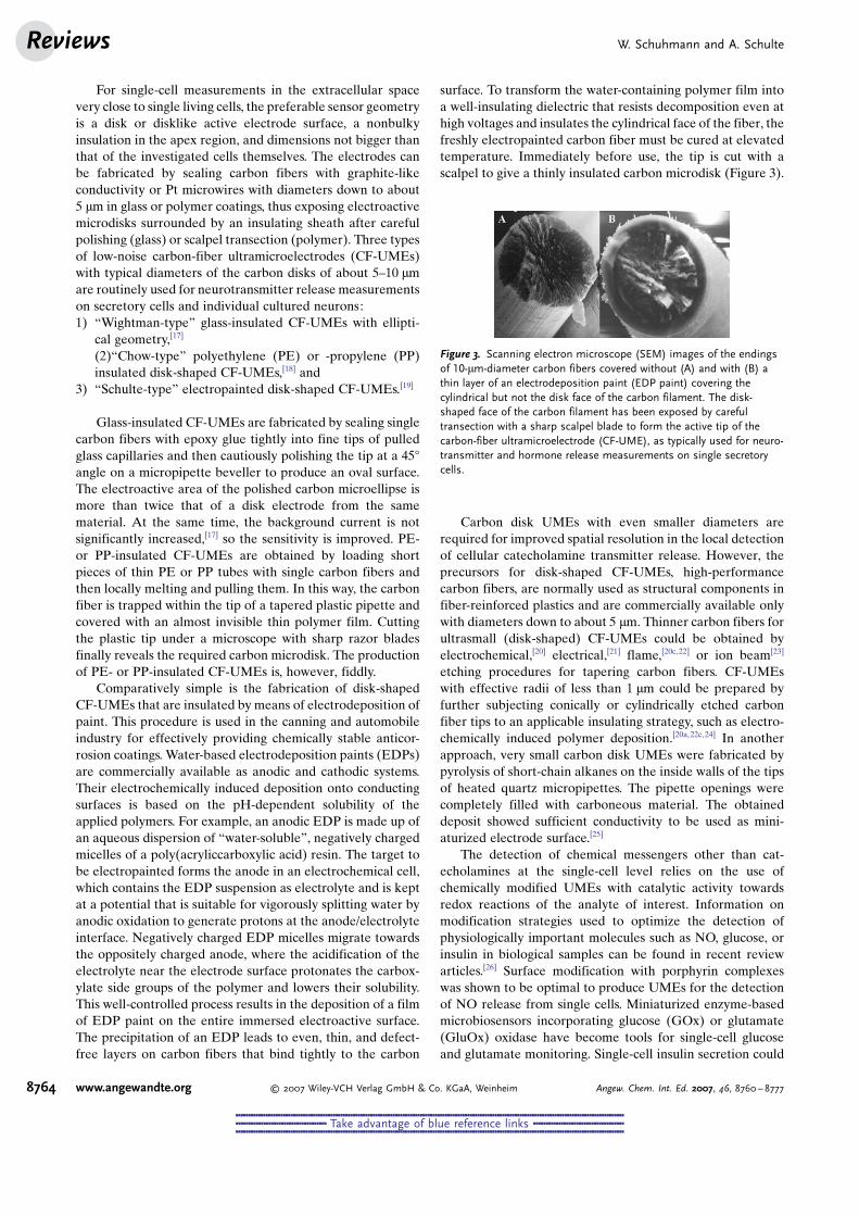
Figure 3. Scanning electron microscope (SEM) images of the endingsof 10-mm-diameter carbon fibers covered without (A) and with (B) athin layer of an electrodeposition paint (EDP paint) covering thecylindrical but not the disk face of the carbon filament. The disk-shaped face of the carbon filament has been exposed by carefultransection with a sharp scalpel blade to form the active tip of thecarbon-fiber ultramicroelectrode (CF-UME), as typically used for neuro-transmitter and hormone release measurements on single secretorycells.
W. Schuhmann and A. SchulteReviews
8764 www.angewandte.org � 2007 Wiley-VCH Verlag GmbH & Co. KGaA, Weinheim Angew. Chem. Int. Ed. 2007, 46, 8760 – 8777
&&&&&&&&&&&&&&&&&&&&&&&&&&&&&&&&&&&&&&&&&&&&&&&&&&&&&&&&&&&&&&&&&&&&&&&&&&&&&&&&&&&&&&&&&&&&&&&&&&&&&&&&&&&&&&&&&&&&&&&&&&&&&&&&&&&&&&&&&&&&&&&&&&&&&&&&&&&&
&&&&&&&&&&&&&&&&&&&&&&&&&&&&&&&&&&&& Take advantage of blue reference links &&&&&&&&&&&&&&&&&&&&&&&&&&&&&&&&&&&&
&&&&&&&&&&&&&&&&&&&&&&&&&&&&&&&&&&&&&&&&&&&&&&&&&&&&&&&&&&&&&&&&&&&&&&&&&&&&&&&&&&&&&&&&&&&&&&&&&&&&&&&&&&&&&&&&&&&&&&&&&&&&&&&&&&&&&&&&&&&&&&&&&&&&&&&&&&&&
be detected at disk-shaped UMEs covered with mixed-valentruthenium oxide/cyanoruthenate films.
Bare, disk-shaped Pt UMEs are predominantly used forlocal measurements of O2 in the extracellular milieu adjacentto “breathing” living cells. Typically, they are made by sealingPt microwires in thin tapers of pulled glass capillaries andthen polishing to produce glass-sealed Pt tips. Variousminiaturized, membrane-covered Clark-type O2 electrodesfor single-cell studies are commercially available. Nanometer-sized Pt UMEs[27] have recently been described by severalgroups. These UMEs could be suitable for improving thespatial resolution of the detection of cellular O2 consumption.However, the potential for this application has not yet beenexplored because of the difficulties in their accurate position-ing at the cell membrane. A self-referencing, polarographic,O2-selective microelectrode was developed for measuring O2
fluxes from single cells with a good sensitivity and spatialresolution in real time.[28]
Disk-shaped electrodes are well-suited for extracellularmeasurements, but intracellular voltammetric detectionrequires much sharper UMEs. The tips must be able tosmoothly penetrate cell membranes to reach the cytoplasmwithout serious cell damage. Already in 1967, the design of avoltammetric microelectrode for measuring intracellularpartial pressure of O2 was reported.[29] Much later, EwingGsgroup constructed ultrasmall carbon ring UMEs,[30] whichcould be used for intracellular voltammetry of dopamine[31]
and, upon surface modification with layers of Pt/nafion or Pt/GOx/nafion, of O2
[32] or glucose,[33] respectively. Meulemanset al. developed methods for the construction of needle-type,glass-insulated Pt[34] and C[35] microelectrodes for intracellularvoltammetric measurements of redox-active species. Tahaand co-workers reported already in the early 1990s thatchemically modified tips of flame-etched and polymer-insulated carbon fibers can be effectively pushed throughcell membranes and used in the cytoplasm for quantifyingtrace levels of NO[36] (CF-UME coated with thin polymericfilms of TMHPP-Ni/nafion; TMHPP-Ni= nickel(II) tetra-kis(3-methoxy-4-hydroxyphenyl)porphyrin) and metal ions[37]
(CF-UME coated with TMHPP-Ni/nafion or mercury).
3. Examples of Intracellular Voltammetry
The meta- and catabolic pathways that are responsible foran ongoing biochemical processing and signaling within thecell cytosol naturally engage a vast number of (bio)chemicalcompounds, some of which carry redox-active functionalitiesand hence are voltammetrically detectable. The developmentof intracellular voltammetry was thus motivated by the desireto track in real time the dynamics of cytosolic metabolites andchemical transmitter molecules, including time-dependentchanges in their concentration as result of membranetrafficking. Such knowledge is important for a better under-standing of the behavior of individual cells in terms of theirmetabolism, function, and regulation. Additionally, the inves-tigations may help to reveal the signaling mechanisms that areemployed by large networks of cells for intercellular commu-nication. Furthermore, quantitative detection of the transport
of electroactive drugs and toxins across cell membranes mayprovide helpful information for developments in pharmacol-ogy and toxicology.
In the 1980s, a number of laboratories developed voltam-metric probe tips that were sharp enough to pierce cellmembranes with minimal damage and negligible loss ofsensitivity. The first reports explored the potential of micro-electrode voltammetry in the interior of cells for monitoringintracellular concentrations of O2, glucose, NO, and a numberof biologically relevant trace metals and drugs. Meulemanset al. , for example, exposed individual cultured neurons fromthe dissected buccal ganglions of the marine mollusk AplysiaCalifornica to physiological buffer solutions with or withoutantipyrine and metronidazole. They then followed the timecourse of the cellular uptake and clearance of these redox-active drugs by means of intracellular differential pulsevoltammetry (DPV).[35] They could also determine the intra-neuronal concentration of serotonin (5-HT) in live seroto-nergic metacerebral cells of the same animal. Changes inintracellular 5-HT concentrations were measured in real timewith DPVat needle-type, glass-insulated Pt UMEs for severalhours after neuronal stimulation, intracellular injection of 5-HT, or extracellular application of l-tryptophan, reserpine, orp-chlorophenylalanine.[34] Tips of carbon-ring UMEs wereplaced inside the giant dopamine neuron of the snail P.Corneus and operated in amperometric mode at constantanodic potential to monitor cytosolic concentrations ofdopamine and to quantify its membrane transport andmetabolic clearance.[31,38] These investigations proved thatthe refilling of freshly formed but still empty intracellularstorage vesicles with locally available messenger molecules isan important process at the early stage of exocytosis but has tocompete efficiently with the escape of the transmitter throughthe cell membrane.
Respiration is an additional activity that is of criticalimportance for the proper physiological function of singlecells. Efficient O2 uptake is strongly related to sufficientprovision of energy for the various biochemical processes inthe cytoplasm. Ewing et al. used intracellular quantitative O2
monitoring at nafion-coated platinized carbon-ring UMEs foraccessing the relative cytosolic O2 concentrations of giantdopamine neurons of P. Corneus (another preparationroutinely used in single-cell neuroscience experiments).Comparison of the values obtained from resting neuronsand neurons stimulated by high potassium concentrationsuggested that increased intracellular O2 consumption tookplace upon evoked vesicular dopamine release. This effectwas related to an increase in internal energy consumption ofthe cells for vesicle transport and exocytosis.[32]
Gaseous NO may act in biological systems as a cytotoxicagent or as a fast-diffusing molecular messenger. NO is ableto quickly mediate a variety of signaling pathways in thetarget cells and as such is known to be involved in processessuch as neuronal signaling, immune response, modulation ofion channels, cellular defense mechanisms, and vasodilata-tion. NO is synthesized within particular cells through theCa2+/calmodulin-dependent enzymatic reaction of NO syn-thase (NOS) with conversion of l-arginine to citrulline. Thus,a detailed investigation of the complex mechanisms behind an
MicroelectrochemistryAngewandte
Chemie
8765Angew. Chem. Int. Ed. 2007, 46, 8760 – 8777 � 2007 Wiley-VCH Verlag GmbH & Co. KGaA, Weinheim www.angewandte.org
&&&&&&&&&&&&&&&&&&&&&&&&&&&&&&&&&&&&&&&&&&&&&&&&&&&&&&&&&&&&&&&&&&&&&&&&&&&&&&&&&&&&&&&&&&&&&&&&&&&&&&&&&&&&&&&&&&&&&&&&&&&&&&&&&&&&&&&&&&&&&&&&&&&&&&&&&&&&
&&&&&&&&&&&&&&&&&&&&&&&&&&&&&&&&&&&& Take advantage of blue reference links &&&&&&&&&&&&&&&&&&&&&&&&&&&&&&&&&&&&
&&&&&&&&&&&&&&&&&&&&&&&&&&&&&&&&&&&&&&&&&&&&&&&&&&&&&&&&&&&&&&&&&&&&&&&&&&&&&&&&&&&&&&&&&&&&&&&&&&&&&&&&&&&&&&&&&&&&&&&&&&&&&&&&&&&&&&&&&&&&&&&&&&&&&&&&&&&&
NO-regulated cellular function involves the elucidation of thedynamics of intracellular NO production. Malinski and Tahawere the first to attempt this task electrochemically by placingthe tips of NO-sensitive CF-UMEs inside individual culturedendothelial cells. The sensors were applied in real time forvoltammetric monitoring of the increase in cytosolic NOlevels that occurs following bradykinin-stimulated onset ofNOS activity.[36] If poly-TMHPP-Ni films on CF-UMEs weresubjected to demetalation in acidic solutions, the sensor wasable to reincorporate Ni2+ ions very selectively by means of anion-exchange process in which protons from the porphyrinrings are exchanged for Ni2+ ions. The current from the Ni2+/Ni3+ oxidation in the polymeric film generated the analyticalsignal for Ni2+ ion quantification. This strategy, employed inthe cytosol of individual myocytes and hepatoma cells, wassensitive enough to detect low concentrations of Ni2+ ions incells and follow the time course of Ni2+ ion uptake in thecytoplasm when the cells were incubated in buffer solutionscontaining higher concentrations of this ion.[37]
The above-mentioned examples show that intracellularvoltammetry can provide insights into physiologically rele-vant cellular activities. However, in contrast to investigationsby extracellular voltammetry at disk-shaped UMEs near theouter of cells, the initial phase of enthusiasm was followed bya significant drop in research activity, which may reflect atleast to a certain extent the difficulty in fabricating good-functioning probe tips for intracellular electrochemical meas-urements and placing and operating them at the right placewithout too much harm to the living objects of study.
4. Examples of “Conventional” Extracellular Voltam-metry
“Conventional” extracellular voltammetry is definedherein as voltammetric detection carried out with disk-shaped UMEs that are brought into extreme proximity tothe outer cell membrane with the aid of an inverted micro-scope and user-controlled motorized, piezoelectric orhydraulic micropositioning devices but not supported byany topographic information about the investigated cell.
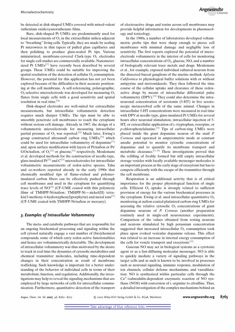
The most prominent example of this type of biologicalvoltammetry is the highly sensitive electrochemical detectionof Ca2+-dependent regulated exocytosis, a well-synchronizedcascade of intracellular events (Figure 4A) executed bysecretory and neuronal cells for the release of hormonesand neurotransmitters from membrane-bound cytosolic stor-age vesicles into the extracellular space or synaptic cleft. Insteps that precede exocytosis, secretory vesicles are formedand loaded with a particular chemical transmitter. The packedvesicles are initially kept in a reserve pool and, when neededfor cell signaling, transported towards the plasma membrane,with which they tether and dock upon arrival by complexinteractions between their membrane proteins. A newlydocked vesicle has to go through a series of molecularrearrangements of the established protein bonds before it isready for instantaneous release. A suitable external stimulusable to trigger the opening of Ca2+ ion channels will at thisstage almost instantaneously induce exocytosis through the
sudden increase in intracellular Ca2+ ion concentration.Fusion of the vesicular and plasma membranes will occurand be followed by the opening of a tiny fusion pore andcomplete discharge of transmitter molecules from the lumenof the collapsing vesicle.
A well-ordered dynamic interaction of the many vesicleand plasma membrane proteins was found to be of keyimportance not only for vesicle docking and maturation butalso for the later timing of membrane fusion and release of
Figure 4. A) The steps involved in Ca2+-triggered secretory vesicleexocytosis leading to quantal release of secretory products. Ampero-metric current spikes resulting from oxidation of discharged trans-mitter molecules at a close CF-UME reveal the late steps of the fusionpore opening as a smaller preceding “foot signal” and full release as amore pronounced boost. B) The three main analytical approaches forfollowing intracellular movements of secretory granules and monitor-ing exocytotic release of neurotransmitters and hormones in real timeand on the level of single fusing and releasing secretory vesicles:Fluorescence microscopy in its various configurations can visualize theentire life cycle of a secretory vesicle when its content and/ormembrane are labeled with powerful fluorescent dyes. Patch-clampcapacitance measurements are able to monitor changes in total capaci-tance of the cell membrane and thus detect the processes of fusion ofvesicles and opening of fusion pores. Amperometry at suitably placed,polarized carbon-fiber ultramicroelectrodes can detect with excellentsensitivity the oxidation of chemical messenger molecules at thesensor tip and provides information about the time course of therelease event.
W. Schuhmann and A. SchulteReviews
8766 www.angewandte.org � 2007 Wiley-VCH Verlag GmbH & Co. KGaA, Weinheim Angew. Chem. Int. Ed. 2007, 46, 8760 – 8777
&&&&&&&&&&&&&&&&&&&&&&&&&&&&&&&&&&&&&&&&&&&&&&&&&&&&&&&&&&&&&&&&&&&&&&&&&&&&&&&&&&&&&&&&&&&&&&&&&&&&&&&&&&&&&&&&&&&&&&&&&&&&&&&&&&&&&&&&&&&&&&&&&&&&&&&&&&&&
&&&&&&&&&&&&&&&&&&&&&&&&&&&&&&&&&&&& Take advantage of blue reference links &&&&&&&&&&&&&&&&&&&&&&&&&&&&&&&&&&&&
&&&&&&&&&&&&&&&&&&&&&&&&&&&&&&&&&&&&&&&&&&&&&&&&&&&&&&&&&&&&&&&&&&&&&&&&&&&&&&&&&&&&&&&&&&&&&&&&&&&&&&&&&&&&&&&&&&&&&&&&&&&&&&&&&&&&&&&&&&&&&&&&&&&&&&&&&&&&
secretory products.[39] The exact function of the identifiedproteins and their impact on the dynamics of vesicleexocytosis thus became a very active area of research withthe goal of unraveling the molecular mechanisms behind thiscommon mode of chemical cell-to-cell signaling in the humanbrain and the mammalian nervous system.
Figure 4B depicts three modern bioanalytical detectionschemes that are frequently used for studying the distinctphases of vesicular release of chemical messengers at the levelof single cells. Sophisticated variants of high-resolutionfluorescence microscopy have been developed into routineoptical tools for the detailed investigation of practically everystep throughout the life cycle of secretory vesicles. Thesuccess of the method relies strongly on the choice of asuitable fluorescent label for the reactants and the quality ofsensitive optical imaging techniques, such as confocal andtotal internal reflection microscopy.[40] The progress ofmembrane fusion and fusion pore opening at the beginningof single-vesicle exocytosis has also been revealed by meansof high-resolution patch-clamp cell-membrane capacitancemeasurements.[41] The technique is actually sensitive enoughfor visualizing in real time the tiny increases in cell surfacearea resulting from the incorporation of the membrane offusing granules as noticeable differences in the total mem-brane capacitance. Finally, spatially confined voltammetry atCF-UMEs positioned close to the cell membrane is acomplementary indicator of fusion events. It provides ahighly sensitive, direct, and quantitative measure of both theearly leakage of transmitter molecules out of prematurefusion pores and the complete release from the finally fullycollapsed secretory vesicles. The advantages and limitationsof constant-potential carbon-fiber amperometry and carbon-fiber fast-scan cyclic voltammetry as well as applications ofthese methods for the detection of exocytosis from single cellsand a description of the analysis of data from such measure-ments are covered in several review articles.[42]
The adrenal glands of cows and rats provided the firstmodel secretory cells at which direct electrochemical detec-tion of distinct exocytotic events was achieved.[43] In thepioneering trials, the tip of an anodically polarized disk-shaped CF-UME was placed as close as possible to the cellmembrane of an individual adrenaline-releasing chromaffincell and the low-noise amperometric current response of themicroelectrode was monitored over time. Under these con-ditions, chemical cell stimulation led reproducibly to theappearance of bursts of current spikes, which could be relatedto the release of discrete packets (“quantums”) of neuro-transmitter molecules from single fusing and collapsingvesicles. The released neurotransmitter molecules weredetected by electrochemically induced oxidation of thesecreted species at the ultramicroelectrode surface. Afterthe initial experimental breakthrough, amperometric detec-tion of secretion was further improved and soon demon-strated to be capable of detecting even the small pedestal or“foot” signals that occasionally occur at the onset of unitaryexocytotic current spikes. These pedestal signals correspondto slow leakage of catecholamine molecules out of just-opened narrow fusion pores prior to complete vesicle collapseand exocytosis.[44]
It was recognized quickly that quantitative analysis of thecharacteristic features of the current transients in ampero-metric recordings of exocytosis events could offer valuableinformation about the mechanism and kinetics of exocytoticneurotransmission. The obligatory secretory spike analysisand interpretation has to be a thorough statistical treatmentof key parameters such as the spike frequency, rise time,amplitude, charge, and half-width as well as the number andcontours of the foot signals. As amperometric recordings ofsingle-vesicle exocytosis usually involve huge data files, user-written acquisition/analysis software in different program-ming languages has been developed for manual or automaticspike analysis.[44,45] On average, a certain number of trans-mitter molecules are released from each vesicle. Thus, thepresence of a population of vesicles varying in size orconcentration of their content can be derived from histogramsof the charge transferred during individual spikes. The timecourse of the fusion pore opening and the late period of therelease may be assessed by interpreting the distribution of therise times and half widths of the spikes. If the chemical natureof the released transmitter is unknown, it can be determinedby fast-scan cyclic voltammetry at the positioned CF-UME;[46]
the background-subtracted voltammograms act as finger-prints for identification of the secretory products. Voltam-metric detection of exocytosis can normally be performedonly on cells that secrete readily oxidizable neurotransmittersor hormones. To circumvent this limitation, cell cultureprotocols have been developed for loading secretory cellswith exogenous neurotransmitters that are readily oxidizedon a suitably polarized CF-UME.[47]
On its own or in conjunction with patch-clamp andfluorescence measurements, single-cell carbon-fiber amper-ometry is today a standard microelectrochemical assay forinvestigating the molecular events that mediate the exocytoticrelease of chemical messengers. Isolated bovine, rat, or mousechromaffin cells have been systematically studied and are stillunder thorough investigation as native or genetically modi-fied preparations.[48] Other cells used in exocytosis researchinclude pheochromocytoma (PC12) cells,[49] mast cells,[49d,50]
pancreatic beta cells,[49d,51] human carcinoid BON cells,[52] ratbasophilic leukemia (RBl-2H3) cells,[53] rat hippocampalastrocytes,[54] various cultured neurons,[55] chromaffin cells(for example, in slices of mouse adrenal glands),[56] glomuscells in slices of the rat carotid body,[57] and neurons in acutebrain slices.[58]
The results of the many published studies—from whichonly a representative sample is cited in references [48–58]—have led to an improved understanding of how secretory cellsfrom the endocrine systems and neurons from the brainrelease their chemical messengers. As reviewed recently in anumber of publications, at least three different types ofvesicular exocytosis have been identified.[39] According to an“all-or-nothing” mechanism, secretory vesicles fuse with thecell membrane, form a fusion pore, collapse, and distributetheir entire contents straight into the extracellular milieu.However, after formation the narrow fusion pore sometimesfluctuates around a small mean pore diameter (“fusion poreflickering”) or even closes again transiently, before finallyexpanding irreversibly and releasing its load. This flickering
MicroelectrochemistryAngewandte
Chemie
8767Angew. Chem. Int. Ed. 2007, 46, 8760 – 8777 � 2007 Wiley-VCH Verlag GmbH & Co. KGaA, Weinheim www.angewandte.org
&&&&&&&&&&&&&&&&&&&&&&&&&&&&&&&&&&&&&&&&&&&&&&&&&&&&&&&&&&&&&&&&&&&&&&&&&&&&&&&&&&&&&&&&&&&&&&&&&&&&&&&&&&&&&&&&&&&&&&&&&&&&&&&&&&&&&&&&&&&&&&&&&&&&&&&&&&&&
&&&&&&&&&&&&&&&&&&&&&&&&&&&&&&&&&&&& Take advantage of blue reference links &&&&&&&&&&&&&&&&&&&&&&&&&&&&&&&&&&&&
&&&&&&&&&&&&&&&&&&&&&&&&&&&&&&&&&&&&&&&&&&&&&&&&&&&&&&&&&&&&&&&&&&&&&&&&&&&&&&&&&&&&&&&&&&&&&&&&&&&&&&&&&&&&&&&&&&&&&&&&&&&&&&&&&&&&&&&&&&&&&&&&&&&&&&&&&&&&
mechanism is typical for the small synaptic vesicles ofneuronal cells and may provide them with an option forcontrolling the level of release. A “kiss-and-run” mechanismwas observed as a third type of exocytosis, whereby the vesiclebriefly fuses with the plasma membrane, ejects a little ofstored transmitter molecules, and then moves back into thecell, where it either may be used again for another run or firstbe refilled.
The fine details of fusion pore formation and dynamics arestill poorly understood at the molecular level. Also, it is notyet known why neuronal cells and vesicles trigger a particulartype of exocytosis under certain conditions. Achieving thesegoals requires continuation of systematic studies on genet-ically modified secretory cells that lack one of the manyhighly specialized secretory plasma and vesicle membraneproteins. Ideal experiments are multidimensional in terms ofassessing the exocytotic response of targeted cells simulta-neously with carbon-fiber amperometry and the complemen-tary optical and electrophysiological detection schemesmentioned above. This level of detailed analysis of single-vesicle exocytosis should eventually allow the influence ofindividual proteins on pore formation and dilation as well asthe time course of release to be resolved and help unravel howvesicular chemical release is orchestrated.
Some promising new electrochemistry strategies at thelevel of single secretory cells are worth being mentioned eventhough they are beyond our earlier definition of conventionalextracellular voltammetry. For example, glass slides werepatterned with optically transparent and electrically conduc-tive indium tin oxide (ITO) to fabricate well-defined ITOmicroelectrodes onto which secretory cells can be seeded at adesired density for secretion experiments.[59] The suitability ofthe miniaturized ITO structures for combined optical andelectrochemical studies on the single cell level has beendemonstrated by Amatore et al. , who were in fact able toapply the methodology for both imaging stained chromaffincells with fluorescence microscopy and detecting theirchemical release with the ITO microelectrodes operated asamperometric sensors.[59a] After further miniaturization andoptimization, ITO-based microdevices could soon becometools for high-throughput optoelectrochemical screening ofexocytosis. A microfluidic device for on-chip cell transport,cell location, and amperometric detection of single-cellsecretion[60] and disk-shaped Pt-UMEs microfabricated atthe bottom of an array of picoliter wells into which singlechromaffin cells can be captured[61] have also been proposedfor rapid and automated analysis of single-cell exocytosis.Hafez et al. microfabricated an arrangement of four closelyspaced Pt microelectrodes on glass coverslips and used thedevice for the spatially defined detection of fusion poreopenings at different release sites of a single secretory cellthat was resting on top of the detector array and stimulated torelease its transmitter.[12] Finally, cell-attached patch amper-ometry combining patch-clamp measurements of cell mem-brane capacitance changes (indicating vesicle size) withamperometry at a CF-UME that is placed inside of thepatch pipette (quantifying transmitter release) was intro-duced in particular for studying the exocytosis of the smallestsecretory organelle, the small synaptic vesicles.[62] These
vesicles have diameters of only a few tens of nanometers,which is a lot smaller than the dense-core vesicles ofchromaffin or mast cells; however, patch amperometry wassensitive enough to resolve in real time the modest capaci-tance changes induced by the fusion of synaptic vesicles anddisplay them together with the corresponding amperometricspikes resulting from the actual release of the tiny amounts ofvesicular transmitter.
The majority of current work on microelectrode voltam-metry with UMEs positioned at the outer surface of singlecells involves detection of the release of neurotransmittersand hormones (exocytosis) from secretory cells. This type ofmeasurement was thus given the main emphasis herein;however, other important physiological processes can also beapproached in a similar fashion. Good examples are: 1) thedetection of transmembrane fluxes of oxygen with self-referencing oxygen-sensitive Pt microelectrodes,[28] 2) theelectrochemical visualization of oxygen production in singlealgal protoplasts upon exposure to light,[63] 3) monitoring thecellular release of reactive oxygen species (e.g. the superoxideion O2C
� , H2O2, or NO) in real time at microelectrodes placednext to the specific cells,[64] 4) the electrochemical detection ofdrug efflux from preloaded single cells with disk-shaped CF-UMEs,[65] and 5) the detection of cholesterol contained in theplasmamembrane of single cells with Pt-UMEsmodified withlipid membranes containing cholesterol oxidase.[66]
5. Single-Cell Scanning Electrochemical Microscopy(SECM)
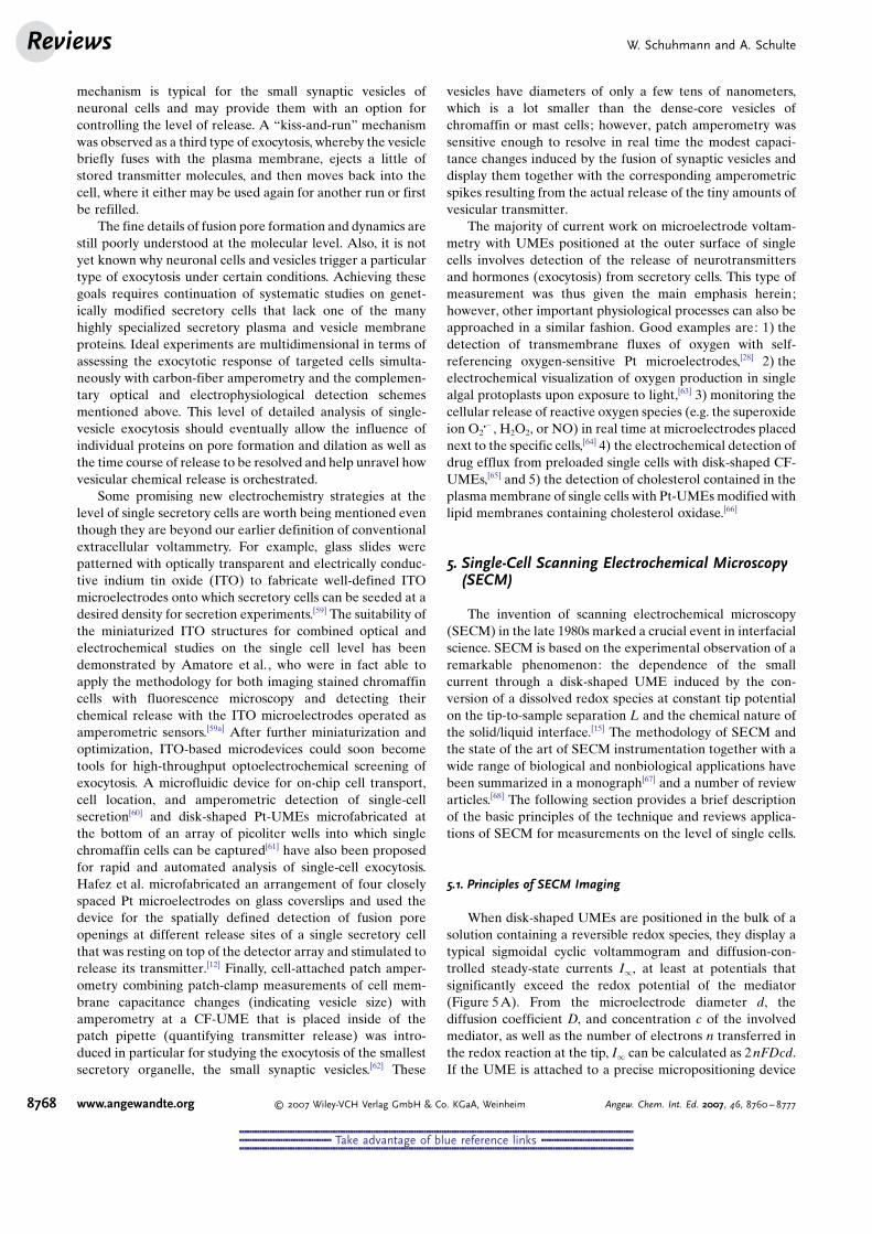
The invention of scanning electrochemical microscopy(SECM) in the late 1980s marked a crucial event in interfacialscience. SECM is based on the experimental observation of aremarkable phenomenon: the dependence of the smallcurrent through a disk-shaped UME induced by the con-version of a dissolved redox species at constant tip potentialon the tip-to-sample separation L and the chemical nature ofthe solid/liquid interface.[15] The methodology of SECM andthe state of the art of SECM instrumentation together with awide range of biological and nonbiological applications havebeen summarized in a monograph[67] and a number of reviewarticles.[68] The following section provides a brief descriptionof the basic principles of the technique and reviews applica-tions of SECM for measurements on the level of single cells.
5.1. Principles of SECM Imaging
When disk-shaped UMEs are positioned in the bulk of asolution containing a reversible redox species, they display atypical sigmoidal cyclic voltammogram and diffusion-con-trolled steady-state currents I1, at least at potentials thatsignificantly exceed the redox potential of the mediator(Figure 5A). From the microelectrode diameter d, thediffusion coefficient D, and concentration c of the involvedmediator, as well as the number of electrons n transferred inthe redox reaction at the tip, I1 can be calculated as 2nFDcd.If the UME is attached to a precise micropositioning device
W. Schuhmann and A. SchulteReviews
8768 www.angewandte.org � 2007 Wiley-VCH Verlag GmbH & Co. KGaA, Weinheim Angew. Chem. Int. Ed. 2007, 46, 8760 – 8777
&&&&&&&&&&&&&&&&&&&&&&&&&&&&&&&&&&&&&&&&&&&&&&&&&&&&&&&&&&&&&&&&&&&&&&&&&&&&&&&&&&&&&&&&&&&&&&&&&&&&&&&&&&&&&&&&&&&&&&&&&&&&&&&&&&&&&&&&&&&&&&&&&&&&&&&&&&&&
&&&&&&&&&&&&&&&&&&&&&&&&&&&&&&&&&&&& Take advantage of blue reference links &&&&&&&&&&&&&&&&&&&&&&&&&&&&&&&&&&&&
&&&&&&&&&&&&&&&&&&&&&&&&&&&&&&&&&&&&&&&&&&&&&&&&&&&&&&&&&&&&&&&&&&&&&&&&&&&&&&&&&&&&&&&&&&&&&&&&&&&&&&&&&&&&&&&&&&&&&&&&&&&&&&&&&&&&&&&&&&&&&&&&&&&&&&&&&&&&
and gently brought as an electrochemical scanning probe(“SECM tip”) into close vicinity of an insulating specimen,the hemispherical diffusion of mediator molecules towardsthe active microelectrode disk is physically hindered and adrop in the tip current is observed (negative feedback,Figure 5B). In contrast, mediator molecules that wereinitially reduced (or oxidized) at the tip can be reconvertedinto their original state when the SECM tip is positionedabove an electrochemically active surface. This redox recy-cling in turn leads to increased tip currents relative to thoseseen in the bulk solution (positive feedback, Figure 5C).
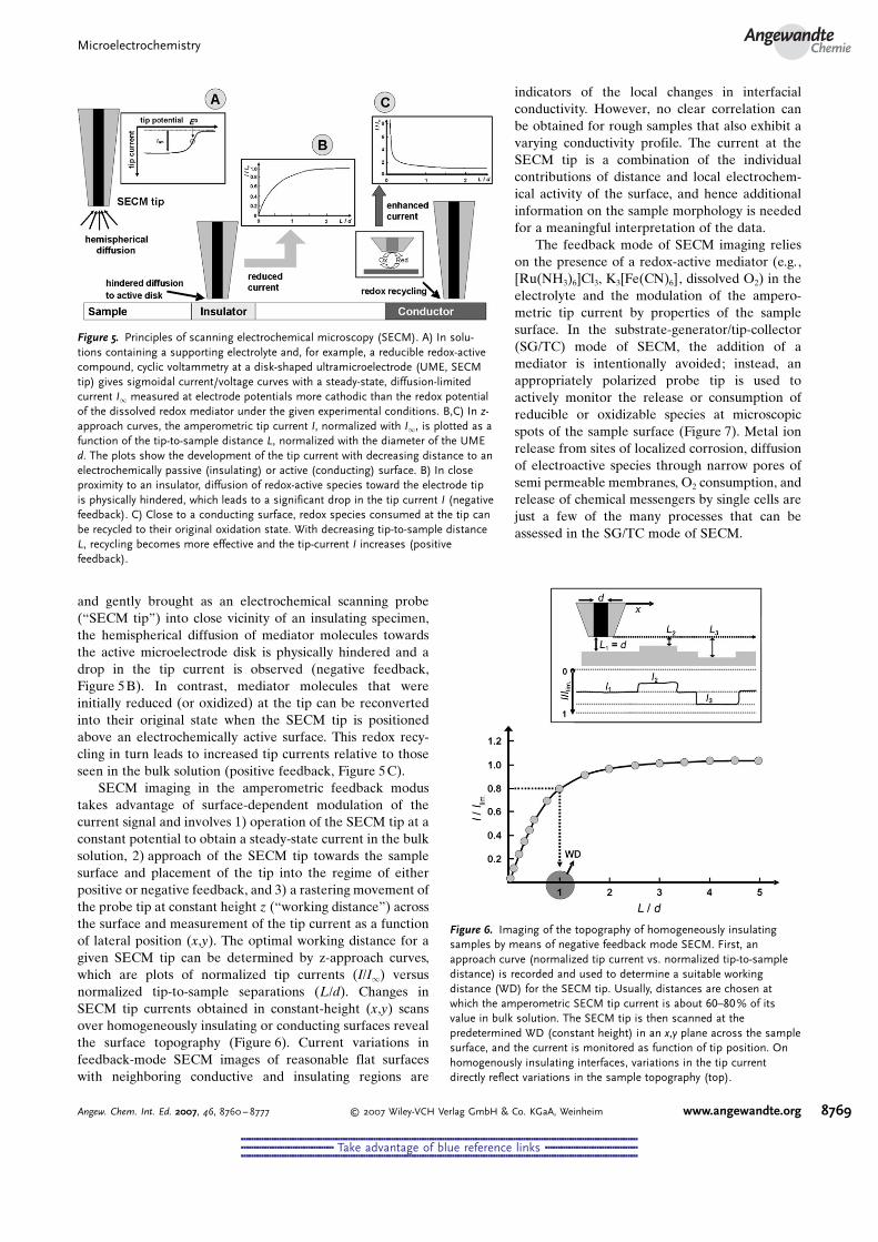
SECM imaging in the amperometric feedback modustakes advantage of surface-dependent modulation of thecurrent signal and involves 1) operation of the SECM tip at aconstant potential to obtain a steady-state current in the bulksolution, 2) approach of the SECM tip towards the samplesurface and placement of the tip into the regime of eitherpositive or negative feedback, and 3) a rastering movement ofthe probe tip at constant height z (“working distance”) acrossthe surface and measurement of the tip current as a functionof lateral position (x,y). The optimal working distance for agiven SECM tip can be determined by z-approach curves,which are plots of normalized tip currents (I/I1) versusnormalized tip-to-sample separations (L/d). Changes inSECM tip currents obtained in constant-height (x,y) scansover homogeneously insulating or conducting surfaces revealthe surface topography (Figure 6). Current variations infeedback-mode SECM images of reasonable flat surfaceswith neighboring conductive and insulating regions are
indicators of the local changes in interfacialconductivity. However, no clear correlation canbe obtained for rough samples that also exhibit avarying conductivity profile. The current at theSECM tip is a combination of the individualcontributions of distance and local electrochem-ical activity of the surface, and hence additionalinformation on the sample morphology is neededfor a meaningful interpretation of the data.
The feedback mode of SECM imaging relieson the presence of a redox-active mediator (e.g.,[Ru(NH3)6]Cl3, K3[Fe(CN)6], dissolved O2) in theelectrolyte and the modulation of the ampero-metric tip current by properties of the samplesurface. In the substrate-generator/tip-collector(SG/TC) mode of SECM, the addition of amediator is intentionally avoided; instead, anappropriately polarized probe tip is used toactively monitor the release or consumption ofreducible or oxidizable species at microscopicspots of the sample surface (Figure 7). Metal ionrelease from sites of localized corrosion, diffusionof electroactive species through narrow pores ofsemi permeable membranes, O2 consumption, andrelease of chemical messengers by single cells arejust a few of the many processes that can beassessed in the SG/TC mode of SECM.
Figure 5. Principles of scanning electrochemical microscopy (SECM). A) In solu-tions containing a supporting electrolyte and, for example, a reducible redox-activecompound, cyclic voltammetry at a disk-shaped ultramicroelectrode (UME, SECMtip) gives sigmoidal current/voltage curves with a steady-state, diffusion-limitedcurrent I1 measured at electrode potentials more cathodic than the redox potentialof the dissolved redox mediator under the given experimental conditions. B,C) In z-approach curves, the amperometric tip current I, normalized with I1, is plotted as afunction of the tip-to-sample distance L, normalized with the diameter of the UMEd. The plots show the development of the tip current with decreasing distance to anelectrochemically passive (insulating) or active (conducting) surface. B) In closeproximity to an insulator, diffusion of redox-active species toward the electrode tipis physically hindered, which leads to a significant drop in the tip current I (negativefeedback). C) Close to a conducting surface, redox species consumed at the tip canbe recycled to their original oxidation state. With decreasing tip-to-sample distanceL, recycling becomes more effective and the tip-current I increases (positivefeedback).
Figure 6. Imaging of the topography of homogeneously insulatingsamples by means of negative feedback mode SECM. First, anapproach curve (normalized tip current vs. normalized tip-to-sampledistance) is recorded and used to determine a suitable workingdistance (WD) for the SECM tip. Usually, distances are chosen atwhich the amperometric SECM tip current is about 60–80% of itsvalue in bulk solution. The SECM tip is then scanned at thepredetermined WD (constant height) in an x,y plane across the samplesurface, and the current is monitored as function of tip position. Onhomogenously insulating interfaces, variations in the tip currentdirectly reflect variations in the sample topography (top).
MicroelectrochemistryAngewandte
Chemie
8769Angew. Chem. Int. Ed. 2007, 46, 8760 – 8777 � 2007 Wiley-VCH Verlag GmbH & Co. KGaA, Weinheim www.angewandte.org
&&&&&&&&&&&&&&&&&&&&&&&&&&&&&&&&&&&&&&&&&&&&&&&&&&&&&&&&&&&&&&&&&&&&&&&&&&&&&&&&&&&&&&&&&&&&&&&&&&&&&&&&&&&&&&&&&&&&&&&&&&&&&&&&&&&&&&&&&&&&&&&&&&&&&&&&&&&&
&&&&&&&&&&&&&&&&&&&&&&&&&&&&&&&&&&&& Take advantage of blue reference links &&&&&&&&&&&&&&&&&&&&&&&&&&&&&&&&&&&&
&&&&&&&&&&&&&&&&&&&&&&&&&&&&&&&&&&&&&&&&&&&&&&&&&&&&&&&&&&&&&&&&&&&&&&&&&&&&&&&&&&&&&&&&&&&&&&&&&&&&&&&&&&&&&&&&&&&&&&&&&&&&&&&&&&&&&&&&&&&&&&&&&&&&&&&&&&&&
5.2. Constant-Height SECM Studies on Single Living Cells
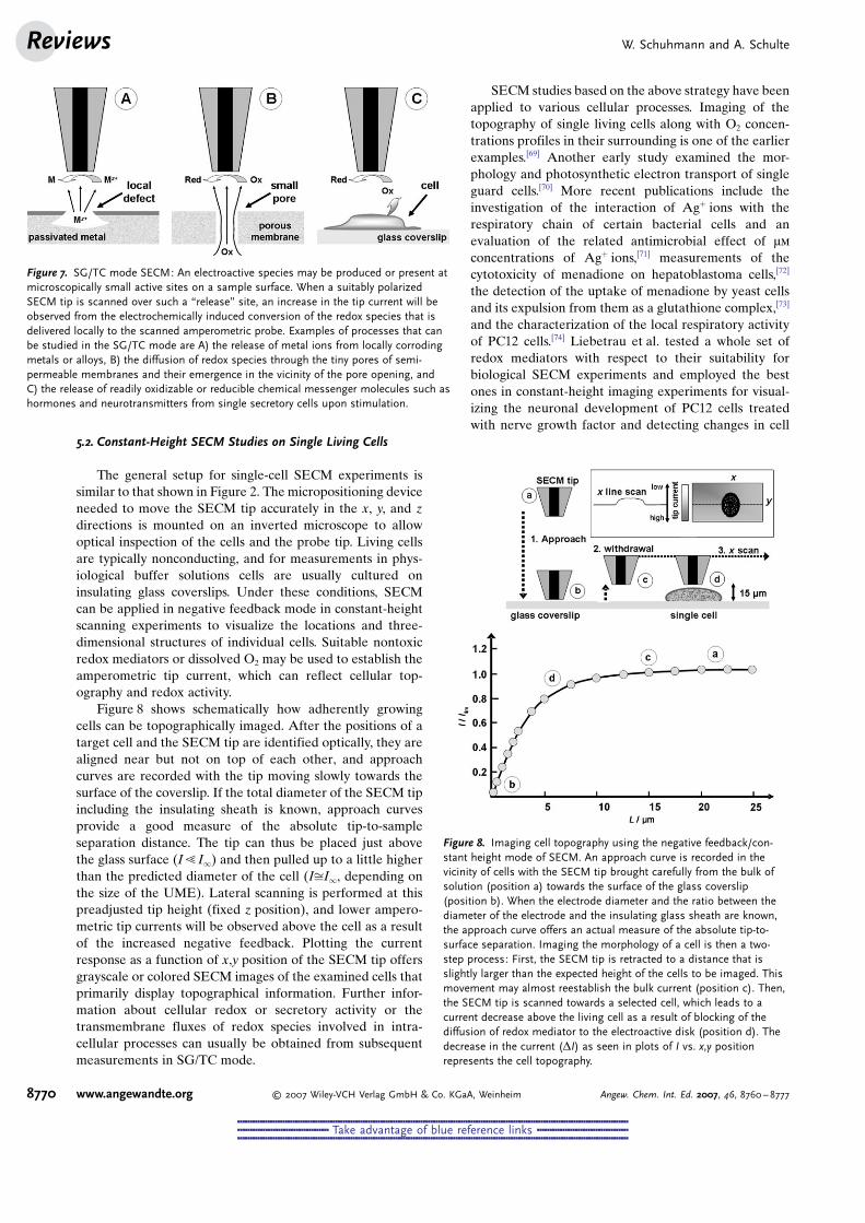
The general setup for single-cell SECM experiments issimilar to that shown in Figure 2. The micropositioning deviceneeded to move the SECM tip accurately in the x, y, and zdirections is mounted on an inverted microscope to allowoptical inspection of the cells and the probe tip. Living cellsare typically nonconducting, and for measurements in phys-iological buffer solutions cells are usually cultured oninsulating glass coverslips. Under these conditions, SECMcan be applied in negative feedback mode in constant-heightscanning experiments to visualize the locations and three-dimensional structures of individual cells. Suitable nontoxicredox mediators or dissolved O2 may be used to establish theamperometric tip current, which can reflect cellular top-ography and redox activity.
Figure 8 shows schematically how adherently growingcells can be topographically imaged. After the positions of atarget cell and the SECM tip are identified optically, they arealigned near but not on top of each other, and approachcurves are recorded with the tip moving slowly towards thesurface of the coverslip. If the total diameter of the SECM tipincluding the insulating sheath is known, approach curvesprovide a good measure of the absolute tip-to-sampleseparation distance. The tip can thus be placed just abovethe glass surface (I! I1) and then pulled up to a little higherthan the predicted diameter of the cell (IffiI1, depending onthe size of the UME). Lateral scanning is performed at thispreadjusted tip height (fixed z position), and lower ampero-metric tip currents will be observed above the cell as a resultof the increased negative feedback. Plotting the currentresponse as a function of x,y position of the SECM tip offersgrayscale or colored SECM images of the examined cells thatprimarily display topographical information. Further infor-mation about cellular redox or secretory activity or thetransmembrane fluxes of redox species involved in intra-cellular processes can usually be obtained from subsequentmeasurements in SG/TC mode.
SECM studies based on the above strategy have beenapplied to various cellular processes. Imaging of thetopography of single living cells along with O2 concen-trations profiles in their surrounding is one of the earlierexamples.[69] Another early study examined the mor-phology and photosynthetic electron transport of singleguard cells.[70] More recent publications include theinvestigation of the interaction of Ag+ ions with therespiratory chain of certain bacterial cells and anevaluation of the related antimicrobial effect of mm
concentrations of Ag+ ions,[71] measurements of thecytotoxicity of menadione on hepatoblastoma cells,[72]
the detection of the uptake of menadione by yeast cellsand its expulsion from them as a glutathione complex,[73]
and the characterization of the local respiratory activityof PC12 cells.[74] Liebetrau et al. tested a whole set ofredox mediators with respect to their suitability forbiological SECM experiments and employed the bestones in constant-height imaging experiments for visual-izing the neuronal development of PC12 cells treatedwith nerve growth factor and detecting changes in cell
Figure 7. SG/TC mode SECM: An electroactive species may be produced or present atmicroscopically small active sites on a sample surface. When a suitably polarizedSECM tip is scanned over such a “release” site, an increase in the tip current will beobserved from the electrochemically induced conversion of the redox species that isdelivered locally to the scanned amperometric probe. Examples of processes that canbe studied in the SG/TC mode are A) the release of metal ions from locally corrodingmetals or alloys, B) the diffusion of redox species through the tiny pores of semi-permeable membranes and their emergence in the vicinity of the pore opening, andC) the release of readily oxidizable or reducible chemical messenger molecules such ashormones and neurotransmitters from single secretory cells upon stimulation.
Figure 8. Imaging cell topography using the negative feedback/con-stant height mode of SECM. An approach curve is recorded in thevicinity of cells with the SECM tip brought carefully from the bulk ofsolution (position a) towards the surface of the glass coverslip(position b). When the electrode diameter and the ratio between thediameter of the electrode and the insulating glass sheath are known,the approach curve offers an actual measure of the absolute tip-to-surface separation. Imaging the morphology of a cell is then a two-step process: First, the SECM tip is retracted to a distance that isslightly larger than the expected height of the cells to be imaged. Thismovement may almost reestablish the bulk current (position c). Then,the SECM tip is scanned towards a selected cell, which leads to acurrent decrease above the living cell as a result of blocking of thediffusion of redox mediator to the electroactive disk (position d). Thedecrease in the current (DI) as seen in plots of I vs. x,y positionrepresents the cell topography.
W. Schuhmann and A. SchulteReviews
8770 www.angewandte.org � 2007 Wiley-VCH Verlag GmbH & Co. KGaA, Weinheim Angew. Chem. Int. Ed. 2007, 46, 8760 – 8777
&&&&&&&&&&&&&&&&&&&&&&&&&&&&&&&&&&&&&&&&&&&&&&&&&&&&&&&&&&&&&&&&&&&&&&&&&&&&&&&&&&&&&&&&&&&&&&&&&&&&&&&&&&&&&&&&&&&&&&&&&&&&&&&&&&&&&&&&&&&&&&&&&&&&&&&&&&&&
&&&&&&&&&&&&&&&&&&&&&&&&&&&&&&&&&&&& Take advantage of blue reference links &&&&&&&&&&&&&&&&&&&&&&&&&&&&&&&&&&&&
&&&&&&&&&&&&&&&&&&&&&&&&&&&&&&&&&&&&&&&&&&&&&&&&&&&&&&&&&&&&&&&&&&&&&&&&&&&&&&&&&&&&&&&&&&&&&&&&&&&&&&&&&&&&&&&&&&&&&&&&&&&&&&&&&&&&&&&&&&&&&&&&&&&&&&&&&&&&
height induced by exposure to hypotonic or hypertonic buffersolutions in real time.[75]
The work of MirkinGs group on imaging normal andmetastatic human breast cells is another example of thepotential of single-cell SECM as a modern tool for biomedicalanalysis and the monitoring of physiologically and patholog-ically relevant cellular function.[76] Individual breast cells wereexposed in physiological saline solution to a chemicalmediator (e.g., quinone) that was able to rapidly cross thecell membrane and take part in intracellular redox reactionsin the cytoplasm. Changes in extracellular concentrations ofthe mediator resulting from cytosolic turnovers were moni-tored on a microsecond timescale by the SECM tip, which wasplaced close to the cell membrane. Variations in rateconstants were observed for cells with different healthstatus and it was shown that they can be used as an indicatorfor the onset of cell metastasis and identification of singlecancerous cells in a field of nontransformed neighbors.
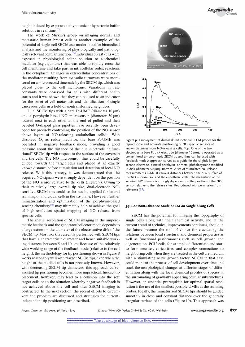
Dual SECM tips with a bare Pt-UME (diameter 10 mm)and a porphyrin-based NO microsensor (diameter 50 mm)located next to each other at the end of pulled and thenbeveled V-shaped glass pipettes have recently been devel-oped for precisely controlling the position of the NO sensorabove layers of NO-releasing endothelian cells.[77] Withdissolved O2 as redox mediator, the bare Pt-UME wasoperated in negative feedback mode, providing a goodmeasure about the distance of the dual-electrode “bifunc-tional” SECM tip with respect to the surface of the coverslipand the cells. The NO microsensor thus could be carefullyguided towards the target cells and placed at an exactlyknown distance before stimulation and detection of local NOrelease. With this strategy, it was demonstrated that theacquired NO signals were strongly dependent on the positionof the NO sensor relative to the cells (Figure 9). Owing totheir relatively large overall tip size, dual-electrode NO-sensitive SECM tips could so far not be applied for lateralscanning on individual cells in the x,y plane. However, furtherminiaturization and optimization of the porphyrin-basedsensing chemistry[78] may ultimately help to achieve the goalof high-resolution spatial mapping of NO release fromspecific cells.
The spatial resolution of SECM imaging in the ampero-metric feedback and the generator/collector mode depends toa large extent on the diameter of the electroactive disk of theSECM tip. Most work is currently performed with SECM tipsthat have a characteristic diameter and hence suitable work-ing distances between 5 and 10 mm. Because of the relativelywide working range of the feedback mode (relative to the cellheight), the methodology for tip positioning shown in Figure 8works reasonably well with “large” SECM tips, even when theheight of the studied cells is not precisely known. However,with decreasing SECM tip diameters, this approach-curve-assisted tip positioning becomes more impractical. Inexact tipplacement, however, may lead to a collision into the softtarget cells or to the situation whereby negative feedback isnot achieved above the cell and thus SECM imaging isobstructed. In the next section, the recent efforts to circum-vent the problem are discussed and strategies for current-independent tip positioning are described.
5.3. Constant-Distance Mode SECM on Single Living Cells
SECM has the potential for imaging the topography ofsingle cells along with their chemical activity, and, if thecurrent trend of technical improvements continues, should inthe future become the tool of choice for elucidating therelations between local structural and chemical properties aswell as functional performances such as cell growth anddegeneration. PC12 cells, for example, differentiate and startto form neurites, varicosities, and complex connections toneighboring cells when they are treated in the culture mediumwith a stimulating nerve growth factor. SECM in that casecould monitor the process of cell development over time andtrack the morphological changes at different stages of differ-entiation along with the local chemical profiles of species inthe surrounding of gradually appearing cellular substructures.However, an essential prerequisite for optimal spatial reso-lution is the use of the smallest possible UMEs as the scanningprobes. Ideally, the miniaturized SECM tips should be guidedsmoothly in close and constant distance over the generallyirregular surface of the cells (Figure 10). This approach was
Figure 9. Employment of dual-disk, bifunctional SECM probes for thereproducible and accurate positioning of NO-specific sensors atknown distances from NO-releasing cells. Top: One of the twoelectrodes, a bare Pt disk electrode (diameter 10 mm), is operated as aconventional amperometric SECM tip and thus can be used withfeedback-mode z-approach curves as a guide for the slightly largersecond electrode, a metal-porphyrin- or metal-phthalocyanine-modifiedPt disk (diameter 50 mm). Bottom: A set of stimulated NO-releasemeasurements made at various distances between the disk surface ofthe NO microsensor and the endothelial cells. The magnitude of theacquired NO signals is strongly dependent on the position of the NOsensor relative to the release sites. Reproduced with permission fromreference [77a].
MicroelectrochemistryAngewandte
Chemie
8771Angew. Chem. Int. Ed. 2007, 46, 8760 – 8777 � 2007 Wiley-VCH Verlag GmbH & Co. KGaA, Weinheim www.angewandte.org
&&&&&&&&&&&&&&&&&&&&&&&&&&&&&&&&&&&&&&&&&&&&&&&&&&&&&&&&&&&&&&&&&&&&&&&&&&&&&&&&&&&&&&&&&&&&&&&&&&&&&&&&&&&&&&&&&&&&&&&&&&&&&&&&&&&&&&&&&&&&&&&&&&&&&&&&&&&&
&&&&&&&&&&&&&&&&&&&&&&&&&&&&&&&&&&&& Take advantage of blue reference links &&&&&&&&&&&&&&&&&&&&&&&&&&&&&&&&&&&&
&&&&&&&&&&&&&&&&&&&&&&&&&&&&&&&&&&&&&&&&&&&&&&&&&&&&&&&&&&&&&&&&&&&&&&&&&&&&&&&&&&&&&&&&&&&&&&&&&&&&&&&&&&&&&&&&&&&&&&&&&&&&&&&&&&&&&&&&&&&&&&&&&&&&&&&&&&&&
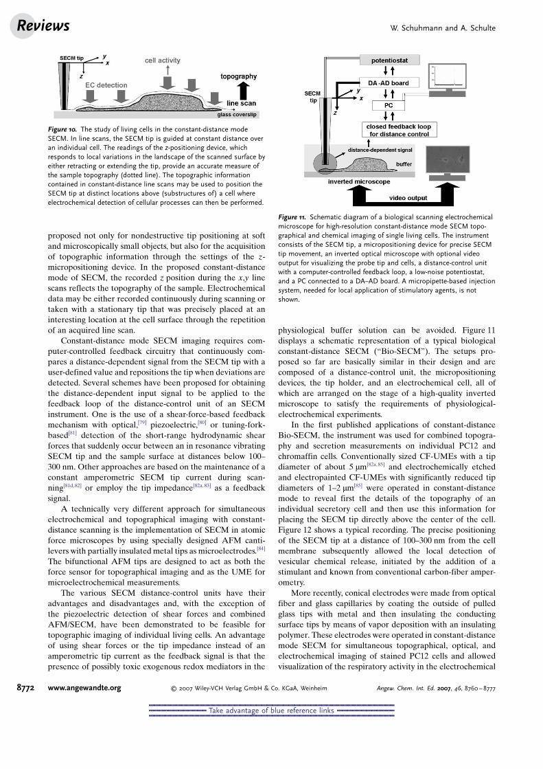
proposed not only for nondestructive tip positioning at softand microscopically small objects, but also for the acquisitionof topographic information through the settings of the z-micropositioning device. In the proposed constant-distancemode of SECM, the recorded z position during the x,y linescans reflects the topography of the sample. Electrochemicaldata may be either recorded continuously during scanning ortaken with a stationary tip that was precisely placed at aninteresting location at the cell surface through the repetitionof an acquired line scan.
Constant-distance mode SECM imaging requires com-puter-controlled feedback circuitry that continuously com-pares a distance-dependent signal from the SECM tip with auser-defined value and repositions the tip when deviations aredetected. Several schemes have been proposed for obtainingthe distance-dependent input signal to be applied to thefeedback loop of the distance-control unit of an SECMinstrument. One is the use of a shear-force-based feedbackmechanism with optical,[79] piezoelectric,[80] or tuning-fork-based[81] detection of the short-range hydrodynamic shearforces that suddenly occur between an in resonance vibratingSECM tip and the sample surface at distances below 100–300 nm. Other approaches are based on the maintenance of aconstant amperometric SECM tip current during scan-ning[81d,82] or employ the tip impedance[82a,83] as a feedbacksignal.
A technically very different approach for simultaneouselectrochemical and topographical imaging with constant-distance scanning is the implementation of SECM in atomicforce microscopes by using specially designed AFM canti-levers with partially insulated metal tips as microelectrodes.[84]
The bifunctional AFM tips are designed to act as both theforce sensor for topographical imaging and as the UME formicroelectrochemical measurements.
The various SECM distance-control units have theiradvantages and disadvantages and, with the exception ofthe piezoelectric detection of shear forces and combinedAFM/SECM, have been demonstrated to be feasible fortopographic imaging of individual living cells. An advantageof using shear forces or the tip impedance instead of anamperometric tip current as the feedback signal is that thepresence of possibly toxic exogenous redox mediators in the
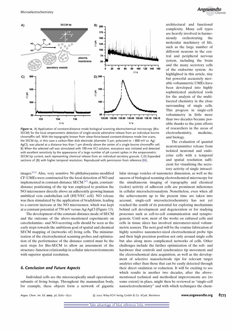
physiological buffer solution can be avoided. Figure 11displays a schematic representation of a typical biologicalconstant-distance SECM (“Bio-SECM”). The setups pro-posed so far are basically similar in their design and arecomposed of a distance-control unit, the micropositioningdevices, the tip holder, and an electrochemical cell, all ofwhich are arranged on the stage of a high-quality invertedmicroscope to satisfy the requirements of physiological-electrochemical experiments.
In the first published applications of constant-distanceBio-SECM, the instrument was used for combined topogra-phy and secretion measurements on individual PC12 andchromaffin cells. Conventionally sized CF-UMEs with a tipdiameter of about 5 mm[82a,85] and electrochemically etchedand electropainted CF-UMEs with significantly reduced tipdiameters of 1–2 mm[85] were operated in constant-distancemode to reveal first the details of the topography of anindividual secretory cell and then use this information forplacing the SECM tip directly above the center of the cell.Figure 12 shows a typical recording. The precise positioningof the SECM tip at a distance of 100–300 nm from the cellmembrane subsequently allowed the local detection ofvesicular chemical release, initiated by the addition of astimulant and known from conventional carbon-fiber amper-ometry.
More recently, conical electrodes were made from opticalfiber and glass capillaries by coating the outside of pulledglass tips with metal and then insulating the conductingsurface tips by means of vapor deposition with an insulatingpolymer. These electrodes were operated in constant-distancemode SECM for simultaneous topographical, optical, andelectrochemical imaging of stained PC12 cells and allowedvisualization of the respiratory activity in the electrochemical
Figure 10. The study of living cells in the constant-distance modeSECM. In line scans, the SECM tip is guided at constant distance overan individual cell. The readings of the z-positioning device, whichresponds to local variations in the landscape of the scanned surface byeither retracting or extending the tip, provide an accurate measure ofthe sample topography (dotted line). The topographic informationcontained in constant-distance line scans may be used to position theSECM tip at distinct locations above (substructures of) a cell whereelectrochemical detection of cellular processes can then be performed.
Figure 11. Schematic diagram of a biological scanning electrochemicalmicroscope for high-resolution constant-distance mode SECM topo-graphical and chemical imaging of single living cells. The instrumentconsists of the SECM tip, a micropositioning device for precise SECMtip movement, an inverted optical microscope with optional videooutput for visualizing the probe tip and cells, a distance-control unitwith a computer-controlled feedback loop, a low-noise potentiostat,and a PC connected to a DA–AD board. A micropipette-based injectionsystem, needed for local application of stimulatory agents, is notshown.
W. Schuhmann and A. SchulteReviews
8772 www.angewandte.org � 2007 Wiley-VCH Verlag GmbH & Co. KGaA, Weinheim Angew. Chem. Int. Ed. 2007, 46, 8760 – 8777
&&&&&&&&&&&&&&&&&&&&&&&&&&&&&&&&&&&&&&&&&&&&&&&&&&&&&&&&&&&&&&&&&&&&&&&&&&&&&&&&&&&&&&&&&&&&&&&&&&&&&&&&&&&&&&&&&&&&&&&&&&&&&&&&&&&&&&&&&&&&&&&&&&&&&&&&&&&&
&&&&&&&&&&&&&&&&&&&&&&&&&&&&&&&&&&&& Take advantage of blue reference links &&&&&&&&&&&&&&&&&&&&&&&&&&&&&&&&&&&&
&&&&&&&&&&&&&&&&&&&&&&&&&&&&&&&&&&&&&&&&&&&&&&&&&&&&&&&&&&&&&&&&&&&&&&&&&&&&&&&&&&&&&&&&&&&&&&&&&&&&&&&&&&&&&&&&&&&&&&&&&&&&&&&&&&&&&&&&&&&&&&&&&&&&&&&&&&&&
images.[81a] Also, very sensitive Ni–phthalocyanine-modifiedCF-UMEs were constructed for the local detection of NO andimplemented in constant-distance SECM.[87] Again, constant-distance positioning of the tip was employed to position theNOmicrosensor directly above an adherently growing humanumbilical vein endothelian cell (HUVEC cell). NO releasewas then stimulated by the application of bradykinin, leadingto a current increase at the NO microsensor, which was keptat a constant potential of 750 mV versus Ag/AgCl (Figure 13).
The development of the constant-distance mode of SECMand the outcome of the above-mentioned experiments oncatecholamine- and NO-secreting cells should be seen as theearly steps towards the ambitious goal of spatial and chemicalSECM mapping of (networks of) living cells. The miniatur-ization of the electrochemical scanning probes and optimiza-tion of the performance of the distance control must be thenext steps for Bio-SECM to allow an assessment of thestructure–function relationship in cellular microenvironmentswith superior spatial resolution.
6. Conclusion and Future Aspects
Individual cells are the microscopically small operationalsubunits of living beings. Throughout the mammalian body,for example, these objects form a network of gigantic
architectural and functionalcomplexity. Many cell typesare heavily involved in harmo-niously orchestrating themolecular machinery of life,such as the large number ofdifferent neurons in the cen-tral and peripheral nervoussystem, including the brainand the many secretory cellsof the endocrine system. Ashighlighted in this article, tinybut powerful accurately mov-able voltammetric UMEs havebeen developed into highlysophisticated analytical toolsfor the analysis of the multi-faceted chemistry in the closesurrounding of single cells.This progress in single-cellvoltammetry in little morethan two decades became pos-sible thanks to the joint effortsof researchers in the areas ofelectrochemistry, medicine,and biology.
The evaluation of quantalneurotransmitter release fromisolated neuronal and endo-crine cells with a temporaland spatial resolution suffi-cient for visualizing the secre-tory activity of single intracel-
lular storage vesicles of nanometer dimension, as well as thesuccess of biological scanning electrochemical microscopy forthe simultaneous imaging of topography and chemical(redox) activity of adherent cells are prominent milestonesin cellular microelectroanalysis. Nonetheless, even when allthe achievements up to the present time are taken intoaccount, single-cell microelectrochemistry has not yetreached the zenith of its potential for exploring mechanismsbehind cell development and degeneration or for studyingprocesses such as cell-to-cell communication and synapto-genesis. Until now, most of the works on cultured cells andcells in tissue slices has involved micrometer-sized voltam-metric sensors. The next goal will be the routine fabrication ofhighly sensitive nanometer-sized electrochemical probe tipsand their high precision position not only around single cellsbut also along more complicated networks of cells. Otherchallenges include the further optimization of the soft- andhardware that controls and synchronizes tip movement andthe electrochemical data acquisition, as well as the develop-ment of selective nanoelectrode tips for relevant targetanalytes other than those that can be easily detected throughtheir direct oxidation or reduction. It will be exciting to seewhich results in another two decades, after the above-mentioned technical and methodical improvements are (tosome extent) in place, might then be reviewed as “single-cellnanoelectrochemistry” and with which techniques the chem-
Figure 12. A) Application of constant-distance mode biological scanning electrochemical microscopy (Bio-SECM) for the local amperometric detection of single-vesicle adrenaline release from an individual bovinechromaffin cell. With the topography known from shear-force-based constant-distance mode line scans,the SECM tip, in this case a carbon-fiber disk electrode (diameter 5 mm, polarized to +800 mV vs. Ag/AgCl), was placed at a distance less than 1 mm directly above the center of a single bovine chromaffin cell.B) When the selected cell was stimulated with 100 mm KCl solution, exocytosis was initiated and detectedwith excellent sensitivity by the appearance of a large number of pA current spikes in the amperometricSECM tip current, each representing chemical release from an individual secretory granule. C,D) Expandedsections of (B) with higher temporal resolution. Reproduced with permission from reference [86].
MicroelectrochemistryAngewandte
Chemie
8773Angew. Chem. Int. Ed. 2007, 46, 8760 – 8777 � 2007 Wiley-VCH Verlag GmbH & Co. KGaA, Weinheim www.angewandte.org
&&&&&&&&&&&&&&&&&&&&&&&&&&&&&&&&&&&&&&&&&&&&&&&&&&&&&&&&&&&&&&&&&&&&&&&&&&&&&&&&&&&&&&&&&&&&&&&&&&&&&&&&&&&&&&&&&&&&&&&&&&&&&&&&&&&&&&&&&&&&&&&&&&&&&&&&&&&&
&&&&&&&&&&&&&&&&&&&&&&&&&&&&&&&&&&&& Take advantage of blue reference links &&&&&&&&&&&&&&&&&&&&&&&&&&&&&&&&&&&&
&&&&&&&&&&&&&&&&&&&&&&&&&&&&&&&&&&&&&&&&&&&&&&&&&&&&&&&&&&&&&&&&&&&&&&&&&&&&&&&&&&&&&&&&&&&&&&&&&&&&&&&&&&&&&&&&&&&&&&&&&&&&&&&&&&&&&&&&&&&&&&&&&&&&&&&&&&&&

ical communication of and between individual cells inresponse to physiological, pathological, or pharmacologicalstimulation may be routinely monitored in life sciencelaboratories.
Received: November 30, 2006Published online: October 19, 2007
[1] K. Stulik, C. Amatore, K. Holub, V. Marecek, W. Kutner, PureAppl. Chem. 2000, 72, 1483 – 1492.
[2] Microelectrodes: Theory and Application, NATO ASI SeriesE197 (Eds.: M. I. Montenegro, M. A. Queiros, J. L. Daschbach),Kluver, Dordrecht, 1991.
[3] a) C. Amatore, E. Maisonhaute, Anal. Chem. 2005, 77, 303A–311A; b) C. G. Zoski,Electroanalysis 2002, 14, 1041 – 1051; c) M.Ciszkowska, Z. Stojek, Anal. Chem. 2000, 72, 754A– 760A;d) R. J. Foster, Chem. Soc. Rev. 1994, 23, 289 – 297; e) J. Heinze,Angew. Chem. 1993, 105, 1327 – 1349; Angew. Chem. Int. Ed.Engl. 1993, 32, 1268 – 1288; f) K. Aoki, Electroanalysis 1993, 5,627 – 639; g) H.-P. Nirmaier, G. Henze, Electroanalysis 1997, 9,619 – 624; h) R. M. Wightman, Anal. Chem. 1981, 53, A1125 –A1134.
[4] a) R. N. Adams, Prog. Neurobiol. 1990, 35, 297 – 311; b) J. O.Schenk, E.Miller, M. E. Rice, R. N. Adams,Brain Res. 1983, 277,1 – 8; c) H.-Y. Cheng, J. Schenk, R. Huff, R. N. Adams, J.Electroanal. Chem. Interfacial Electrochem. 1979, 100, 23 – 31;d) R. L. McCreery, R. Dreiling, R. N. Adams, Brain Res. 1974,
73, 15 – 21; e) P. T. Kissinger, J. B. Hart, R. N. Adams, Brain Res.1973, 55, 209 – 213.
[5] a) M. Fillenz, Neurosci. Biobehav. Rev. 2005, 29, 949 – 962;b) D. L. Robinson, B. J. Venton, M. L. A. V. Heien, R. M.Wightman,Clin. Chem. 2003, 49, 1763 – 1773; c) P. E. M. Phillips,R. M. Wightman, Trends Anal. Chem. 2003, 22, 509 – 514;d) K. P. Troyer, M. L. A. V. Heien, B. J. Venton, R. M. Wight-man, Curr. Opin. Chem. Biol. 2002, 6, 696 – 703; e) J. A.Stamford, J. B. Justice, Anal. Chem. 1996, 68, 359A– 363A;f) R. D. OGNeill, Analyst 1994, 119, 767 – 779; g) J. A. Stamford,J. Neurosci. Methods 1986, 17, 1 – 29; h) J. A. Stamford, BrainRes. 1985, 357, 119 – 135.
[6] a) F. Crespi, D. Dalessandro, V. Annovazzi-Lodi, C. Heidbreder,M. Norgia, J. Neurosci. Methods 2004, 140, 153 – 161; b) P. A.Garris, R. Ensmann, J. Poehlman, A. Alexander, P. E. Langley,S. G. Sandberg, P. G. Greco, R. M. Wightman, G. V. Rebec, J.Neurosci. Methods 2004, 140, 103 – 115; c) M. G. De Simoni, A.De Luigi, L. Imeri, S. Algeri, J. Neurosci. Methods 1990, 33, 233 –240; d) V. Annovazzi-Lodi, S. Donati, IEEETrans. Biomed. Eng.1988, 35, 595 – 606.
[7] B. J. Venton, R. M. Wightman, Anal. Chem. 2003, 414A– 421A.[8] a) G. D. Stuber, R. M. Wightman, R. M. Carelli, Neuron 2005,
46, 661 – 669; b) M. F. Roitman, G. D. Stuber, P. E. M. Phillips,R. M. Wightman, R. M. Carelli, J. Neurosci. 2004, 24, 1265 –1271; c) P. G. Greco, P. A. Garris, Eur. J. Pharmacol. 2003, 479,117 – 125; d) P. E. M. Phillips, G. D. Stuber, M. L. Helen, R. M.Wightman, R. M. Carelli, Nature 2003, 422, 614 – 618; e) R. M.Wightman, D. L. Robinson, J. Neurochem. 2002, 82, 721 – 735;f) D. L. Robinson, P. E. M. Phillips, E. A. Budygin, B. J. Trafton,P. A. Garris, R. M.Wightman,Neuroreport 2001, 12, 2549 – 2552;g) M. R. Kilpatrick, M. B. Rooney, D. J. Michael, R. M. Wight-man, Neuroscience 2000, 96, 697 – 706; h) G. V. Rebec, J. R.Christensen, C. Guerra, M. T. Bardo, Brain Res. 1997, 776, 61 –67.
[9] a) D. J. Stephens, V. J. Allan, Science 2003, 300, 82 – 86; b) W. R.Zipfel, R. M. Williams, W. W. Webb, Nat. Biotechnol. 2003, 21,1369 – 1377; c) E. S. Yeung, Anal. Chem. 1999, 71, A522 –A529;d) M. Dailey, G. Marrs, J. Satz, M. Waite, Biol. Bull. 1999, 197,115 – 122.
[10] a) Single-Channel Recording, 2nd ed. (Eds.: B. Sakmann, E.Neher), Kluwer, Dordrecht, 1995 ; b)Microelectrode techniques,The Plymouth workshop handbook, 2nd ed. (Ed.: D. C. Ogden),Company of Biologists, Cambridge, 1994.
[11] R. Feeney, S. P. Kounaves, Electroanalysis 2000, 12, 677 – 684.[12] I. Hafez, K. Kisler, K. Berberian, G. Demick, V. Valero, M. G.
Yong, H. G. Craighead, M. Lindau, Proc. Natl. Acad. Sci. USA2005, 102, 13879 – 13884.
[13] a) H.-F. Cui, J.-S. Shan, Y. Chen, S.-C. Chong, X. Liu, T.-M. Lim,F.-S. Sheu, Sens. Actuators B 2006, 115, 634 – 641; b) T. S. Strong,H. C. Cantor, R. B. Brown, Sens. Actuators A 2001, 91, 357 – 362.
[14] S. Isik, L. Berdondini, J. Oni, A. Bloechl, M. Koudelka-Hep, W.Schuhmann, Biosens. Bioelectron. 2005, 20, 1566 – 1572.
[15] a) A. J. Bard, F. R. F. Fan, J. Kwak, O. Lev,Anal. Chem. 1989, 61,132 – 138; b) R. C. Engstroem, M. Weber, D. J. Wunder, R.Burgess, S. Winquist, Anal. Chem. 1986, 58, 844 – 848.
[16] a) M. A. Edwards, S. Martin, A. L.Whitworth, J. V. Macpherson,P. R. Unwin, Physiol. Meas. 2006, 27, R63 –R108; b) A. J. Bard,X. Li, W. Zhan, Biosens. Bioelectron. 2006, 22, 461 – 472; c) S.Amemiya, J. D. Guo, H. Xiong, D. A. Gross, Anal. Bioanal.Chem. 2006, 386, 458 – 471; d) M. Navratil, G. A. Mabbott, E. A.Arriaga, Anal. Chem. 2006, 78, 4005 – 4019; e) B. F. Brehm-Stecher, E. A. Johnson,Microbiol. Mol. Biol. Rev. 2004, 68, 538 –559; f) T. Yasukawa, T. Kaya, T. Matsue, Electroanalysis 2000,12, 653 – 659.
[17] R. S. Kelly, R. M. Wightman, Anal. Chim. Acta 1986, 187, 79 –87.
[18] R. H. Chow, L. Von Rueden, E. Neher,Nature 1992, 356, 60 – 63.
Figure 13. A) Shear-force-based constant-distance line scan used forpositioning the NO microsensor exactly above a single humanumbilical vein endothelian (HUVEC) cell. The inset shows the shadowof the microsensor above the selected single cell in a simultaneouslyacquired video microscope image. B) Amperometric detection of NOreleased from a single HUVEC cell upon stimulation with bradykinin.Reproduced with permission from reference [87].
W. Schuhmann and A. SchulteReviews
8774 www.angewandte.org � 2007 Wiley-VCH Verlag GmbH & Co. KGaA, Weinheim Angew. Chem. Int. Ed. 2007, 46, 8760 – 8777
&&&&&&&&&&&&&&&&&&&&&&&&&&&&&&&&&&&&&&&&&&&&&&&&&&&&&&&&&&&&&&&&&&&&&&&&&&&&&&&&&&&&&&&&&&&&&&&&&&&&&&&&&&&&&&&&&&&&&&&&&&&&&&&&&&&&&&&&&&&&&&&&&&&&&&&&&&&&
&&&&&&&&&&&&&&&&&&&&&&&&&&&&&&&&&&&& Take advantage of blue reference links &&&&&&&&&&&&&&&&&&&&&&&&&&&&&&&&&&&&
&&&&&&&&&&&&&&&&&&&&&&&&&&&&&&&&&&&&&&&&&&&&&&&&&&&&&&&&&&&&&&&&&&&&&&&&&&&&&&&&&&&&&&&&&&&&&&&&&&&&&&&&&&&&&&&&&&&&&&&&&&&&&&&&&&&&&&&&&&&&&&&&&&&&&&&&&&&&
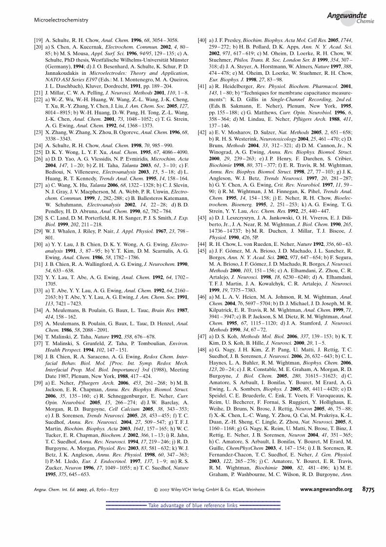
[19] A. Schulte, R. H. Chow, Anal. Chem. 1996, 68, 3054 – 3058.[20] a) S. Chen, A. Kucernak, Electrochem. Commun. 2002, 4, 80 –
85; b) M. S. Mousa, Appl. Surf. Sci. 1996, 94/95, 129 – 135; c) A.Schulte, PhD thesis, WestfOlische Wilhelms-UniversitOt MPnster(Germany), 1994 ; d) J. O. Besenhard, A. Schulte, K. Schur, P. D.Jannakoudakis in Microelectrodes: Theory and Application,NATOASI Series E197 (Eds.: M. I. Montenegro, M. A. Queiros,J. L. Daschbach), Kluver, Dordrecht, 1991, pp. 189 – 204.
[21] J. Millar, C. W. A. Pelling, J. Neurosci. Methods 2001, 110, 1 – 8.[22] a) W.-Z. Wu, W.-H. Huang, W. Wang, Z.-L. Wang, J.-K. Cheng,
T. Xu, R.-Y. Zhang, Y. Chen, J. Liu, J. Am. Chem. Soc. 2005, 127,8014 – 8915; b) W.-H. Huang, D.-W. Pang, H. Tong, Z.-L. Wang,J.-K. Chen, Anal. Chem. 2001, 73, 1048 – 1052; c) T. G. Strein,A. G. Ewing, Anal. Chem. 1992, 64, 1368 – 1373.
[23] X. Zhang,W. Zhang, X. Zhou, B. Ogorevc,Anal. Chem. 1996, 68,3338 – 3343.
[24] A. Schulte, R. H. Chow, Anal. Chem. 1998, 70, 985 – 990.[25] D. K. Y. Wong, L. Y. F. Xu, Anal. Chem. 1995, 67, 4086 – 4090.[26] a) D. D. Yao, A. G. Vlessidis, N. P. Evmiridis, Microchim. Acta
2004, 147, 1 – 20; b) Z. H. Taha, Talanta 2003, 61, 3 – 10; c) F.Bedioui, N. Villeneuve, Electroanalysis 2003, 15, 5 – 18; d) L.Huang, R. T. Kennedy, Trends Anal. Chem. 1995, 14, 158 – 164.
[27] a) C. Wang, X. Hu, Talanta 2006, 68, 1322 – 1328; b) C. J. Slevin,N. J. Gray, J. V. Macpherson, M. A. Webb, P. R. Unwin, Electro-chem. Commun. 1999, 1, 282,-288; c) B. Ballesteros Katemann,W. Schuhmann, Electroanalysis 2002, 14, 22 – 28; d) B. D.Pendley, H. D. Abruna, Anal. Chem. 1990, 62, 782 – 784.
[28] S. C. Land, D. M. Porterfield, R. H. Sanger, P. J. S. Smith, J. Exp.Biol. 1999, 202, 211 – 218.
[29] W. J. Whalen, J. Riley, P. Nair, J. Appl. Physiol. 1967, 23, 798 –801.
[30] a) Y. Y. Lau, J. B. Chien, D. K. Y. Wong, A. G. Ewing, Electro-analysis 1991, 3, 87 – 95; b) Y. T. Kim, D. M. Scarnulis, A. G.Ewing, Anal. Chem. 1986, 58, 1782 – 1786.
[31] J. B. Chien, R. A. Wallingford, A. G. Ewing, J. Neurochem. 1990,54, 633 – 638.
[32] Y. Y. Lau, T. Abe, A. G. Ewing, Anal. Chem. 1992, 64, 1702 –1705.
[33] a) T. Abe, Y. Y. Lau, A. G. Ewing, Anal. Chem. 1992, 64, 2160 –2163; b) T. Abe, Y. Y. Lau, A. G. Ewing, J. Am. Chem. Soc. 1991,113, 7421 – 7423.
[34] A. Meulemans, B. Poulain, G. Baux, L. Tauc, Brain Res. 1987,414, 158 – 162.
[35] A. Meulemans, B. Poulain, G. Baux, L. Tauc, D. Henzel, Anal.Chem. 1986, 58, 2088 – 2091.
[36] T. Malinski, Z. Taha, Nature 1992, 358, 676 – 678.[37] T. Malinski, S. Grunfeld, Z. Taha, P. Tomboulian, Environ.
Health Perspect. 1994, 102, 147 – 151.[38] J. B. Chien, R. A. Saraceno, A. G. Ewing, Redox Chem. Inter-
facial Behav. Biol. Mol. [Proc. Int. Symp. Redox Mech.Interfacial Prop. Mol. Biol. Importance] 3rd (1988), MeetingDate 1987, Plenum, New York, 1988, 417 – 424.
[39] a) E. Neher, Pfluegers Arch. 2006, 453, 261 – 268; b) M. B.Jackson, E. R. Chapman, Annu. Rev. Biophys. Biomol. Struct.2006, 35, 135 – 160; c) R. Schneggenburger, E. Neher, Curr.Opin. Neurobiol. 2005, 15, 266 – 274; d) J. W. Barclay, A.Morgan, R. D. Burgoyne, Cell Calcium 2005, 38, 343 – 353;e) J. B. Sorensen, Trends Neurosci. 2005, 28, 453 – 455; f) T. C.Suedhof, Annu. Rev. Neurosci. 2004, 27, 509 – 547; g) T. F. J.Martin, Biochim. Biophys. Acta 2003, 1641, 157 – 165; h) W. C.Tucker, E. R. Chapman, Biochem. J. 2002, 366, 1 – 13; i) R. Jahn,T. C. Suedhof, Annu. Rev. Neurosci. 1994, 17, 219 – 246; j) R. D.Burgoyne, A. Morgan, Physiol. Rev. 2003, 83, 581 – 632; k) W. J.Betz, J. K. Angleson, Annu. Rev. Physiol. 1998, 60, 347 – 363;l) P.-M. Lledo, Eur. J. Endocrinol. 1997, 137, 1 – 9; m) R. S.Zucker, Neuron 1996, 17, 1049 – 1055; n) T. C. Suedhof, Nature1995, 375, 645 – 653.
[40] a) J. F. Presley,Biochim. Biophys. Acta Mol. Cell Res. 2005, 1744,259 – 272; b) H. B. Pollard, D. K. Apps, Ann. N. Y. Acad. Sci.2002, 971, 617 – 619; c) M. Oheim, D. Loerke, R. H. Chow, W.Stuehmer, Philos. Trans. R. Soc. London Ser. B 1999, 354, 307 –318; d) J. A. Steyer, A. Horstmann, W. Almers,Nature 1997, 388,474 – 478; e) M. Oheim, D. Loerke, W. Stuehmer, R. H. Chow,Eur. Biophys. J. 1998, 27, 83 – 98.
[41] a) R. Heidelberger, Rev. Physiol. Biochem. Pharmacol. 2001,143, 1 – 80; b) “Techniques for membrane capacitance measure-ments”: K. D. Gillis in Single-Channel Recording, 2nd ed.(Eds. B. Sakmann, E. Neher), Plenum, New York, 1995,pp. 155 – 188; c) G. Matthews, Curr. Opin. Neurobiol. 1996, 6,358 – 364; d) M. Lindau, E. Neher, PflAgers Arch. 1988, 411,137 – 146.
[42] a) E. V. Mosharov, D. Sulzer, Nat. Methods 2005, 2, 651 – 658;b) R. H. S. Westerink, Neurotoxicology 2004, 25, 461 – 470; c) D.Bruns, Methods 2004, 33, 312 – 321; d) D. M. Cannon, Jr., N.Winograd, A. G. Ewing, Annu. Rev. Biophys. Biomol. Struct.2000, 29, 239 – 263; e) J. P. Henry, F. Darchen, S. Cribier,Biochimie 1998, 80, 371 – 377; f) E. R. Travis, R. M. Wightman,Annu. Rev. Biophys. Biomol. Struct. 1998, 27, 77 – 103; g) J. K.Angleson, W. J. Betz, Trends Neurosci. 1997, 20, 281 – 287;h) G. Y. Chen, A. G. Ewing, Crit. Rev. Neurobiol. 1997, 11, 59 –90; i) R. M. Wightman, J. M. Finnegan, K. Pihel, Trends Anal.Chem. 1995, 14, 154 – 158; j) E. Neher, R. H. Chow, Bioelec-trochem. Bioenerg. 1995, 2, 251 – 253; k) A. G. Ewing, T. G.Strein, Y. Y. Lau, Acc. Chem. Res. 1992, 25, 440 – 447.
[43] a) D. J. Leszczyszyn, J. A. Jankowski, O. H. Viveros, E. J. Dili-berto, Jr., J. A. Near, R. M. Wightman, J. Biol. Chem. 1990, 265,14736 – 14737; b) M. R. Duchen, J. Millar, T. J. Biscoe, J.Physiol. 1990, 426, 5P.
[44] R. H. Chow, L. von Rueden, E. Neher, Nature 1992, 356, 60 – 63.[45] a) J. F. GQmez, M. A. Brioso, J. D. Machado, J. L. Sanchez, R.
Borges, Ann. N. Y. Acad. Sci. 2002, 971, 647 – 654; b) F. Segura,M. A. Brioso, J. F. GQmez, J. D. Machado, R. Borges, J. Neurosci.Methods 2000, 103, 151 – 156; c) A. Elhamdani, Z. Zhou, C. R.Artalejo, J. Neurosci. 1998, 18, 6230 – 6240; d) A. Elhamdani,T. F. J. Martin, J. A. Kowalchyk, C. R. Artalejo, J. Neurosci.1999, 19, 7375 – 7383.
[46] a) M. L. A. V. Heien, M. A. Johnson, R. M. Wightman, Anal.Chem. 2004, 76, 5697 – 5704; b) D. J. Michael, J. D. Joseph, M. R.Kilpatrick, E. R. Travis, R. M. Wightman, Anal. Chem. 1999, 71,3941 – 3947; c) B. P. Jackson, S. M. Dietz, R. M. Wightman,Anal.Chem. 1995, 67, 1115 – 1120; d) J. A. Stamford, J. Neurosci.Methods 1990, 34, 67 – 72.
[47] a) D. S. Koh, Methods Mol. Biol. 2006, 337, 139 – 153; b) K. T.Kim, D. S. Koh, B. Hille, J. Neurosci. 2000, 20, 1 – 5.
[48] a) G. Nagy, J. H. Kim, Z. P. Pang, U. Matti, J. Rettig, T. C.Suedhof, J. B. Sorensen, J. Neurosci. 2006, 26, 632 – 643; b) C. L.Haynes, L. A. Buhler, R. M. Wightman, Biophys. Chem. 2006,123, 20 – 24; c) J. R. Constable, M. E. Graham, A. Morgan, R. D.Burgoyne, J. Biol. Chem. 2005, 280, 31615 – 31623; d) C.Amatore, S. Arbault, I. Bonifas, Y. Bouret, M Erard, A. G.Ewing, L. A. Sombers, Biophys. J. 2005, 88, 4411 – 4420; e) D.Speidel, C. E. Bruederle, C. Enk, T. Voets, F. Varoqueaux, K.Reim, U. Becherer, F. Fornai, S. Ruggieri, Y. Hollighaus, E.Weihe, D. Bruns, N. Brose, J. Rettig, Neuron 2005, 46, 75 – 88;f) X.-K. Chen, L.-C. Wang, Y. Zhou, Q. Cai, M. Prakriya, K.-L.Duan, Z.-H. Sheng, C. Lingle, Z. Zhou, Nat. Neurosci. 2005, 8,1160 – 1168; g) G. Nagy, K. Reim, U. Matti, N. Brose, T. Binz, J.Rettig, E. Neher, J. B. Sorensen, Neuron 2004, 41, 351 – 365;h) C. Amatore, S. Arbault, I. Bonifas, Y. Bouret, M Erard, M.Guille, ChemPhysChem 2003, 4, 147 – 154; i) J. B. Sorensen, R.Fernandez-Chacon, T. C. Suedhof, E. Neher, J. Gen. Physiol.2003, 122, 265 – 276; j) C. Amatore, Y. Bouret, E. R. Travis,R. M. Wightman, Biochimie 2000, 82, 481 – 496; k) M. E.Graham, P. Washbourne, M. C. Wilson, R. D. Burgoyne, Ann.
MicroelectrochemistryAngewandte
Chemie
8775Angew. Chem. Int. Ed. 2007, 46, 8760 – 8777 � 2007 Wiley-VCH Verlag GmbH & Co. KGaA, Weinheim www.angewandte.org
&&&&&&&&&&&&&&&&&&&&&&&&&&&&&&&&&&&&&&&&&&&&&&&&&&&&&&&&&&&&&&&&&&&&&&&&&&&&&&&&&&&&&&&&&&&&&&&&&&&&&&&&&&&&&&&&&&&&&&&&&&&&&&&&&&&&&&&&&&&&&&&&&&&&&&&&&&&&
&&&&&&&&&&&&&&&&&&&&&&&&&&&&&&&&&&&& Take advantage of blue reference links &&&&&&&&&&&&&&&&&&&&&&&&&&&&&&&&&&&&
&&&&&&&&&&&&&&&&&&&&&&&&&&&&&&&&&&&&&&&&&&&&&&&&&&&&&&&&&&&&&&&&&&&&&&&&&&&&&&&&&&&&&&&&&&&&&&&&&&&&&&&&&&&&&&&&&&&&&&&&&&&&&&&&&&&&&&&&&&&&&&&&&&&&&&&&&&&&
N. Y. Acad. Sci. 2002, 971, 210 – 221; l) T. L. Colliver, E. J. Hess,A. G. Ewing, J. Neurosci. Methods 2001, 105, 95 – 103; m) T.Voets, T. Moser, P.-E. Lund, R. H. Chow, M. Geppert, T. C.Suedhof, E. Neher, Proc. Natl. Acad. Sci. USA 2001, 98, 11680 –11685; n) M. Haller, C. Heinemann, R. H. Chow, R. Heidel-berger, E. Neher, Biophys. J. 1998, 74, 2100 – 2113; o) A. F.Oberhauser, I. M. Robinson, J. M. Fernandez, Biophys. J. 1996,71, 1131 – 1139; p) R. H. Chow, J. Klingauf, E. Neher, Proc. Natl.Acad. Sci. USA 1994, 91, 12765 – 12769.
[49] a) J. M. Moore, J. B. Papke, A. L. Cahill, A. B. Harkins, Am. J.Physiol. 2006, 291, C270 –C281; b) L. A. Sombers, H. J. Han-char, T. L. Colliver, N. Wittenberg, A. Cans, S. Arbault, C.Amatore, A. G. Ewing, J. Neurosci. 2004, 24, 303 – 309; c) J. Bai,C.-T. Wang, D. A. Richards, M. B. Jackson, E. R. Chapman,Neuron 2004, 41, 929 – 941; d) J. M. Finnegan, K. Pihel, P. S.Cahill, L. Huang, S. E. Zerby, A. G. Ewing, R. T. Kennedy, R. M.Wightman, J. Neurochem. 1996, 66, 1914 – 1923; e) T. K. Chen,G. Luo, A. G. Ewing, Anal. Chem. 1994, 66, 3031 – 3035.
[50] a) E. H. Jaffe, P. Bulanos, C. Caputo,Cell Calcium 2001, 29, 199 –209; b) K. Pihel, S. Hsieh, J. W. Jorgensen, R. M. Wightman,Biochemistry 1998, 37, 1046 – 1052; c) A. F. Oberhauser, I. M.Robinson, J. M. Fernandez, Biophys. J. 1996, 71, 1131 – 1139;d) P. E. Tatham, M. R. Duchen, J. Millar, Pflugers Arch. 1991,419, 409 – 414.
[51] a) D. J. Michael, R. A. Ritzel, L. Haataja, R. H. Chow, Diabetes2006, 55, 600 – 607; b) K. Bokvist, M. Holmqvist, J. Gromada, P.Rorsman, Pflugers Arch. 2000, 439, 634 – 645; c) C. A. Aspin-wall, L. Huang, J. R. Lakey, R. T. Kennedy, Anal. Chem. 1999,71, 5551 – 5556; d) L. Huang, H. Shen, M. A. Atkinson, R. T.Kennedy, Proc. Natl. Acad. Sci. USA 1995, 92, 9608 – 9612;e) R. T. Kennedy, L. Huang, M. A. Atkinson, P. Dush, Anal.Chem. 1993, 65, 1882 – 1887.
[52] V. S. Tran, A. M. Marion-Audibert, E. Karatekin, S. Huet, S.Cribier, K. Guillaumie, C. Chapuis, C. Desnos, F. Darchen, J. P.Henry, Ann. N. Y. Acad. Sci. 2004, 1014, 179 – 188.
[53] S. Iwaki, M. Ogasawara, R. Kurita, O. Niwa, K. Tanizawa, Y.Ohashi, K. Maeyama, Anal. Biochem. 2002, 304, 236 – 243.
[54] X. Chen, L. Wang, Y. Zhou, L.-H. Zheng, Z. Zhou, J. Neurosci.2005, 25, 9236 – 9243.
[55] a) R. G. Staal, E. V. Mosharov, D. Sulzer, Nat. Neurosci. 2004, 7,341 – 346; b) E. N. Pothos, V. Davila, D. Sulzer, J. Neurosci. 1998,18, 4106 – 4118; c) B. B. Anderson, A. G. Ewing, J. Pharmacol.Biomed. Anal. 1999, 19, 15 – 32; d) D.-S. Koh, B. Hille, Proc.Natl. Acad. Sci. USA 1997, 94, 1506 – 1511; e) Z. Zhou, S. Misler,Proc. Natl. Acad. Sci. USA 1995, 92, 6938 – 6942; f) D. Bruns, R.Jahn, Nature 1995, 377, 62 – 65.
[56] G. Arroyo, J. Fuentealba, N. Sevane-Fernandez, M. Aldea, A. G.Garcia, A. Albillos, J. Neurophysiol. 2006, 96, 1196 – 1202.
[57] R. Pardal, U. Ludewig, J. Garcia-Hirschfeld, J. Lopez-Barneo,Proc. Natl. Acad. Sci. USA 2000, 97, 2361 – 2366.
[58] E. H. Jaffe, A. Marty, A. Schulte, R. H. Chow, J. Neurosci. 1998,18, 3548 – 3553.
[59] a) C. Amatore, S. Arbault, Y. Chen, C. Crozatier, F. Lemaitre, Y.Verchier, Angew. Chem. 2006, 118, 4104 – 4107; Angew. Chem.Int. Ed. 2006, 45, 4000 – 4003; b) X. Sun, K. D. Gillis, Anal.Chem. 2006, 78, 2521 – 2525.
[60] W.-H. Huang, W. Cheng, Z. Zhang, D.-W. Pang, Z.-L. Wang,J.-K. Cheng, D.-F. Cui, Anal. Chem. 2004, 76, 483 – 488.
[61] P. Chen, B. Xu, N. Tokranova, X. Feng, J. Castracane, K. D.Gillis, Anal. Chem. 2003, 75, 518 – 524.
[62] a) G. Dernick, L.-W. Gong, L. Tabares, G. Alvarez de Toledo, M.Lindau, Nat. Methods 2005, 2, 699 – 6708; b) L. Tabares, M.Lindau, G. Alvarez de Toledo, Biochem. Soc. Trans. 2003, 31,837 – 841; c) G. Dernick, G. Alvarez de Toledo, M. Lindau, Nat.Cell Biol. 2003, 5, 358 – 362; d) E. V. Mosharov, L. W. Gong, B.Khanna, D. Sulzer, M. Lindau, J. Neurosci. 2003, 23, 5835 – 5845;
e) A. Albillos, G. Dernick, H. Horstmann, W. Almers, G.Alvarez de Toledo, M. Lindau, Nature 1997, 389, 509 – 512.
[63] T. Yasukawa, I. Uchida, T. Matsue, Biophys. J. 1999, 76, 1129 –1135.
[64] a) C. Amatore, S. Arbault, C. Bouton, K. Coffi, J. C. Drapier, H.Ghandour, Y. Tong, ChemBioChem 2006, 7, 653 – 661; b) B. A.Patel, M. Arundell, K. H. Parker, M. S. Yeoman, D. OGHare,Anal. Chem. 2006, 78, 7643 – 7648; c) S. Kasai, H. Shiku, Y.-S.Torisawa, H. Noda, J. Yoshitake, T. Shiraishi, T. Yasukawa, T.Watanabe, T. Matsue, T. Yoshimura,Anal. Chim. Acta 2005, 549,14 – 19; d) L. A. Blatter, Z. Taha, S. Mesaros, P. S. Shaklock,W. G. Gier, T. Malinski, Cir. Res. 1995, 76, 922 – 924.
[65] H. Lu, M. Gratzl, Anal. Chem. 1999, 71, 2821 – 2830.[66] A. Devadoss, J. D. Burgess, J. Am. Chem. Soc. 2004, 126, 10214 –
10215.[67] Scanning Electrochemical Microscopy (Eds.: A. J. Bard, M. V.
Mirkin), Marcel Dekker, New York, 2001.[68] a) G. Wittstock, M. Burchardt, S. E. Pust, Y. Shen, C. Zhao,
Angew. Chem. 2007, 119, 1604 – 1640; Angew. Chem. Int. Ed.2007, 46, 1584 – 1617; b) G. Wittstock, Fresenius J. Anal. Chem.2001, 370, 303 – 315; c) M. V. Mirkin, B. R. Horrocks, Anal.Chim. Acta 2000, 406, 119 – 146; d) G. Nagy, L. Nagy, Fresenius J.Anal. Chem. 2000, 36, 735 – 744; e) A. L. Barker, M. Gonsalves,J. V. MacPherson, C. J. Slevin, P. R. Unwin, Anal. Chim. Acta1999, 385, 223 – 240; f) M. V. Mirkin,Mikrochim. Acta 1999, 130,127 – 153.
[69] T. Yasukawa, T. Kaya, T. Matsue, Anal. Chem. 1999, 71, 4637 –4641.
[70] M. Tsionky, Z. G. Cardon, A. J. Bard, R. B. Jackson, PlantPhysiol. 1997, 113, 895 – 901.
[71] K. B. Holt, A. J. Bard, Biochemistry 2005, 44, 13214 – 13223.[72] J. Mauzeroll, A. J. Bard, O. Owhadian, T. J. Monks, Proc. Natl.
Acad. Sci. USA 2004, 101, 17582 – 17587.[73] J. Mauzeroll, A. J. Bard, Proc. Natl. Acad. Sci. USA 2004, 101,
7862 – 7867.[74] Y. Takii, K. Takoh, M. Nishizawa, T. Matsue, Electrochim. Acta
2003, 48, 3381 – 3385.[75] J. M. Liebetrau, H. M. Miller, J. E. Baur, S. A. Takacs, V.
Anupunpisit, P. A. Garris, D. O. Wipf, Anal. Chem. 2003, 75,563 – 571.
[76] a) S. A. Rotenberg, M. V. Mirkin, J. Mammary Gland Biol.Neoplasia 2004, 9, 375 – 382; b) W. J. Feng, S. A. Rotenberg,M. V. Mirkin, Anal. Chem. 2003, 75, 4148 – 4154; c) B. Liu, S. A.Rotenberg, M. V. Mirkin, Proc. Natl. Acad. Sci. USA 2000, 97,9855 – 9860.
[77] a) S. Isik, M. Etienne, J. Oni, A. Bloechl, S. Reiter, W.Schuhmann, Anal. Chem. 2004, 76, 6389 – 6394; b) A. Pailleret,J. Oni, S. Reiter, S. Isik, M. Etienne, F. Bedioui, W. Schuhmann,Electrochem. Commun. 2003, 5, 847 – 852.
[78] a) V. Ryabova, A. Schulte, T. Erichsen, W. Schuhmann, Analyst2005, 130, 1245 – 1252; b) N. Diab, J. Oni, W. Schuhmann,Bioelectrochemistry 2005, 66, 105 – 110; c) B. Ngounou, S.Neugebauer, A. Frodl, S. Reiter, W. Schuhmann, Electrochim.Acta 2004, 49, 3855 – 3863; d) S. Isik, J. Oni, V. Rjabova, S.Neugebauer,W. Schuhmann,Microchim. Acta 2004, 148, 59 – 64;e) S. Neugebauer, S. Isik, A. Schulte, W. Schuhmann, Anal. Lett.2003, 36, 2005 – 2020.
[79] a) A. Hengstenberg, C. Kranz, W. Schuhmann, Chem. Eur. J.2000, 6, 1547 – 1554; b) M. Ludwig, C. Kranz, W. Schuhmann,H. E. Gaub, Rev. Sci. Instrum. 1995, 66, 2857 – 2860.
[80] B. Ballesteros Katemann, A. Schulte, W. Schuhmann, Chem.Eur. J. 2003, 9, 2025 – 2033.
[81] a) Y. Takahashi, Y. Hirano, T. Yasukawa, H. Shiku. H. Yamada,T. Matsue, Langmuir 2006, 22, 10299 – 10306; b) H. Yamada, H.Fukumoto, T. Yokoyama, T. Koike,Anal. Chem. 2005, 77, 1785 –1790; c) D. Oyamatsu, Y. Hirano, N. Kanaya, Y. Mase, M.Nishizawa, T. Matsue, Bioelectrochemistry 2003, 60, 115 – 121;
W. Schuhmann and A. SchulteReviews
8776 www.angewandte.org � 2007 Wiley-VCH Verlag GmbH & Co. KGaA, Weinheim Angew. Chem. Int. Ed. 2007, 46, 8760 – 8777
&&&&&&&&&&&&&&&&&&&&&&&&&&&&&&&&&&&&&&&&&&&&&&&&&&&&&&&&&&&&&&&&&&&&&&&&&&&&&&&&&&&&&&&&&&&&&&&&&&&&&&&&&&&&&&&&&&&&&&&&&&&&&&&&&&&&&&&&&&&&&&&&&&&&&&&&&&&&
&&&&&&&&&&&&&&&&&&&&&&&&&&&&&&&&&&&& Take advantage of blue reference links &&&&&&&&&&&&&&&&&&&&&&&&&&&&&&&&&&&&
&&&&&&&&&&&&&&&&&&&&&&&&&&&&&&&&&&&&&&&&&&&&&&&&&&&&&&&&&&&&&&&&&&&&&&&&&&&&&&&&&&&&&&&&&&&&&&&&&&&&&&&&&&&&&&&&&&&&&&&&&&&&&&&&&&&&&&&&&&&&&&&&&&&&&&&&&&&&
d) Y. Lee, Z. E. Ding, A. J. Bard, Anal. Chem. 2002, 74, 3634 –3643; e) P. J. James, L. F. Garfias-Mesias, P. J. Moyer, W. H.Smyrl, J. Electrochem. Soc. 1998, 145, L64 –L66.
[82] a) R. T. Kurulugama, D. O. Wipf, S. A. Takacs, S. Pongmaytee-gul, P. A. Garris, J. E. Baur, Anal. Chem. 2005, 77, 1111 – 1117;b) D. O. Wipf, A. J. Bard, D. E. Tallman, Anal. Chem. 1993, 65,1373 – 1377.
[83] a) E. N. Ervin, H. S. White, L. A. Baker, C. R. Martin, Anal.Chem. 2006, 78, 6535 – 6541; b) M. A. Aviles, D. O. Wipf, Anal.Chem. 2001, 73, 4873 – 4881.
[84] a) R. J. Fasching, S. J. Bai, T. Fabian, F. B. Prinz, Microelectron.Eng. 2006, 83, 1638 – 1641; b) C. Kranz, G. Friedbacher, B.
Mizaikoff, A. Lugstein, J. Smoliner, E. Bertagnolli, Anal. Chem.2001, 73, 2491 – 2500; c) J. V. Macpherson, P. R. Unwin, Anal.Chem. 2001, 73, 550 – 557.
[85] L. Pitta Bauermann, W. Schuhmann, A. Schulte, Phys. Chem.Chem. Phys. 2004, 6, 4003 – 4008.
[86] A. Schulte, W. Schuhmann in Electrochemical Methods forNeuroscience, Frontiers in Neuroengineering Series, Vol. 1 (Eds.:A. Michael, L. M. Borland), Taylor & Francis, Boca Raton, 2006,chap. 17.
[87] S. Isik, W. Schuhmann, Angew. Chem. 2006, 118, 7611 – 7614;Angew. Chem. Int. Ed. 2006, 45, 7451 – 7454.
MicroelectrochemistryAngewandte
Chemie
8777Angew. Chem. Int. Ed. 2007, 46, 8760 – 8777 � 2007 Wiley-VCH Verlag GmbH & Co. KGaA, Weinheim www.angewandte.org
&&&&&&&&&&&&&&&&&&&&&&&&&&&&&&&&&&&&&&&&&&&&&&&&&&&&&&&&&&&&&&&&&&&&&&&&&&&&&&&&&&&&&&&&&&&&&&&&&&&&&&&&&&&&&&&&&&&&&&&&&&&&&&&&&&&&&&&&&&&&&&&&&&&&&&&&&&&&
&&&&&&&&&&&&&&&&&&&&&&&&&&&&&&&&&&&& Take advantage of blue reference links &&&&&&&&&&&&&&&&&&&&&&&&&&&&&&&&&&&&
&&&&&&&&&&&&&&&&&&&&&&&&&&&&&&&&&&&&&&&&&&&&&&&&&&&&&&&&&&&&&&&&&&&&&&&&&&&&&&&&&&&&&&&&&&&&&&&&&&&&&&&&&&&&&&&&&&&&&&&&&&&&&&&&&&&&&&&&&&&&&&&&&&&&&&&&&&&&
Related Documents