Journal of Chromatography B, 722 (1999) 225–254 Review Single cell gel electrophoresis assay: methodology and applications * E. Rojas , M.C. Lopez, M. Valverde ´ ´ ´ ´ Laboratorio de Genetica y Toxicologıa Molecular, Departamento de Genetica y Toxicologıa Ambiental, Instituto de Investigaciones Biomedicas, U.N. A.M., P .O. Box 70228, Ciudad Universitaria, CP 04510 Mexico D.F., Mexico Abstract The single cell gel electrophoresis or Comet assay is a sensitive, reliable, and rapid method for DNA double- and single-strand breaks, alkali-labile sites and delayed repair site detection, in eukariotic individual cells. Given its overall characteristics, this method has been widely used over the past few years in several different areas. In this paper we review the studies published to date about the principles, the basic methodology with currently used variations. We also explore the applications of this assay in: genotoxicology, clinical area, DNA repair studies, environmental biomonitoring and human monitoring. 1999 Elsevier Science B.V. All rights reserved. Keywords: Reviews; Single cell gel electrophoresis; Comet assay; DNA Contents 1. Introduction ............................................................................................................................................................................ 226 2. The SCGE / Comet methodology............................................................................................................................................... 227 2.1. Lysis solution ................................................................................................................................................................. 277 2.2. Unwinding and electrophoresis buffer .............................................................................................................................. 277 2.3. Other experimental variables ........................................................................................................................................... 228 2.3.1. Cell suspension preparation.................................................................................................................................. 229 2.3.2. Slide preparation ................................................................................................................................................. 230 2.3.3. Neutralization time .............................................................................................................................................. 230 2.3.4. Permanent slides ................................................................................................................................................. 230 2.3.5. DNA-specific dye and magnification for data collection ......................................................................................... 230 2.3.6. Data analysis ...................................................................................................................................................... 232 2.3.7. Modifications for DNA repair studies and cross-linking agents ............................................................................... 232 3. Applications of the assay ......................................................................................................................................................... 233 3.1. Studies on genotoxicity ................................................................................................................................................... 233 3.2. Clinical applications ........................................................................................................................................................ 239 3.3. DNA repair studies ......................................................................................................................................................... 240 3.4. Environmental biomonitoring .......................................................................................................................................... 240 3.5. Human monitoring .......................................................................................................................................................... 245 4. Future directions and conclusions ............................................................................................................................................. 247 5. List of abbreviations ................................................................................................................................................................ 248 Acknowledgements ...................................................................................................................................................................... 249 References .................................................................................................................................................................................. 249 * Corresponding author. 0378-4347 / 99 / $ – see front matter 1999 Elsevier Science B.V. All rights reserved. PII: S0378-4347(98)00313-2

Welcome message from author
This document is posted to help you gain knowledge. Please leave a comment to let me know what you think about it! Share it to your friends and learn new things together.
Transcript
Journal of Chromatography B, 722 (1999) 225–254
Review
Single cell gel electrophoresis assay: methodology and applications
*E. Rojas , M.C. Lopez, M. Valverde´ ´ ´ ´Laboratorio de Genetica y Toxicologıa Molecular, Departamento de Genetica y Toxicologıa Ambiental, Instituto de Investigaciones
Biomedicas, U.N.A.M., P.O. Box 70228, Ciudad Universitaria, CP 04510 Mexico D.F., Mexico
Abstract
The single cell gel electrophoresis or Comet assay is a sensitive, reliable, and rapid method for DNA double- andsingle-strand breaks, alkali-labile sites and delayed repair site detection, in eukariotic individual cells. Given its overallcharacteristics, this method has been widely used over the past few years in several different areas. In this paper we reviewthe studies published to date about the principles, the basic methodology with currently used variations. We also explore theapplications of this assay in: genotoxicology, clinical area, DNA repair studies, environmental biomonitoring and humanmonitoring. 1999 Elsevier Science B.V. All rights reserved.
Keywords: Reviews; Single cell gel electrophoresis; Comet assay; DNA
Contents1. Introduction ............................................................................................................................................................................ 2262. The SCGE/Comet methodology............................................................................................................................................... 227
2.1. Lysis solution ................................................................................................................................................................. 2772.2. Unwinding and electrophoresis buffer .............................................................................................................................. 2772.3. Other experimental variables ........................................................................................................................................... 228
2.3.1. Cell suspension preparation.................................................................................................................................. 2292.3.2. Slide preparation ................................................................................................................................................. 2302.3.3. Neutralization time.............................................................................................................................................. 2302.3.4. Permanent slides ................................................................................................................................................. 2302.3.5. DNA-specific dye and magnification for data collection ......................................................................................... 2302.3.6. Data analysis ...................................................................................................................................................... 2322.3.7. Modifications for DNA repair studies and cross-linking agents ............................................................................... 232
3. Applications of the assay ......................................................................................................................................................... 2333.1. Studies on genotoxicity ................................................................................................................................................... 2333.2. Clinical applications........................................................................................................................................................ 2393.3. DNA repair studies ......................................................................................................................................................... 2403.4. Environmental biomonitoring .......................................................................................................................................... 2403.5. Human monitoring .......................................................................................................................................................... 245
4. Future directions and conclusions ............................................................................................................................................. 2475. List of abbreviations ................................................................................................................................................................ 248Acknowledgements ...................................................................................................................................................................... 249References .................................................................................................................................................................................. 249
*Corresponding author.
0378-4347/99/$ – see front matter 1999 Elsevier Science B.V. All rights reserved.PI I : S0378-4347( 98 )00313-2
226 E. Rojas et al. / J. Chromatogr. B 722 (1999) 225 –254
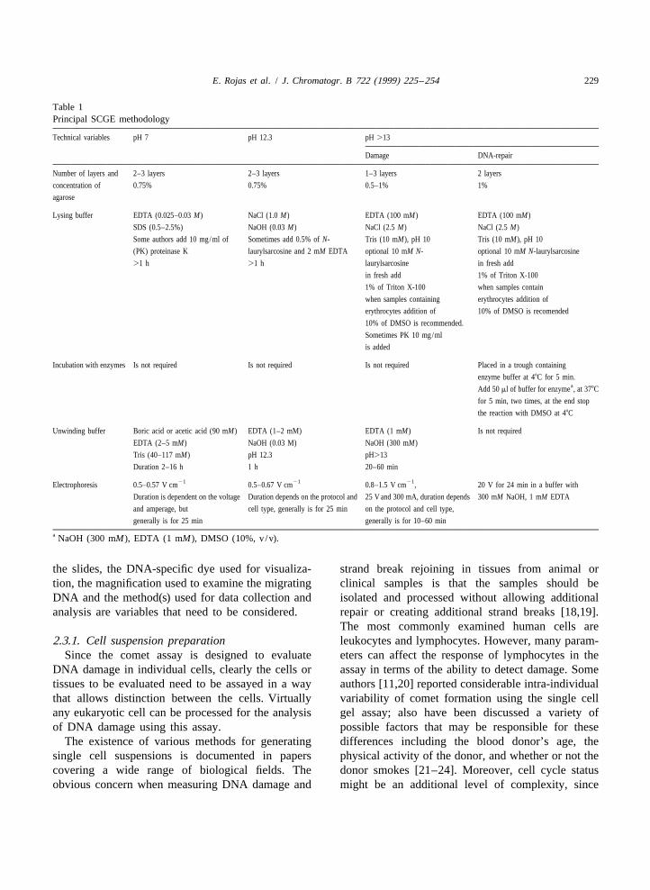
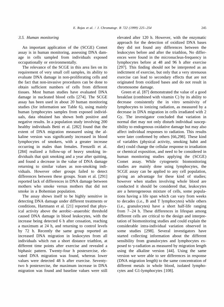
1. Introduction In the Singh version of the assay, a single cellsuspension of the mammalian cell culture or tissue
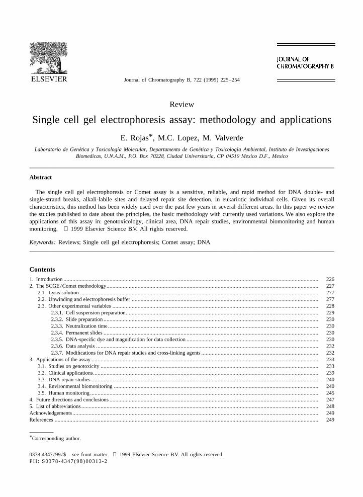
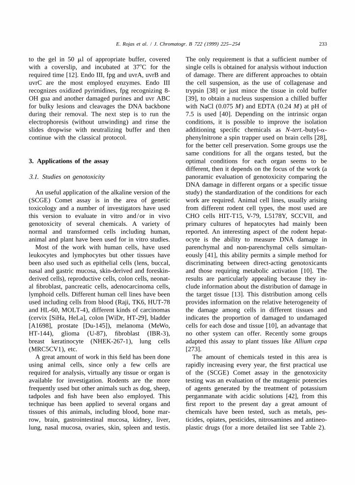
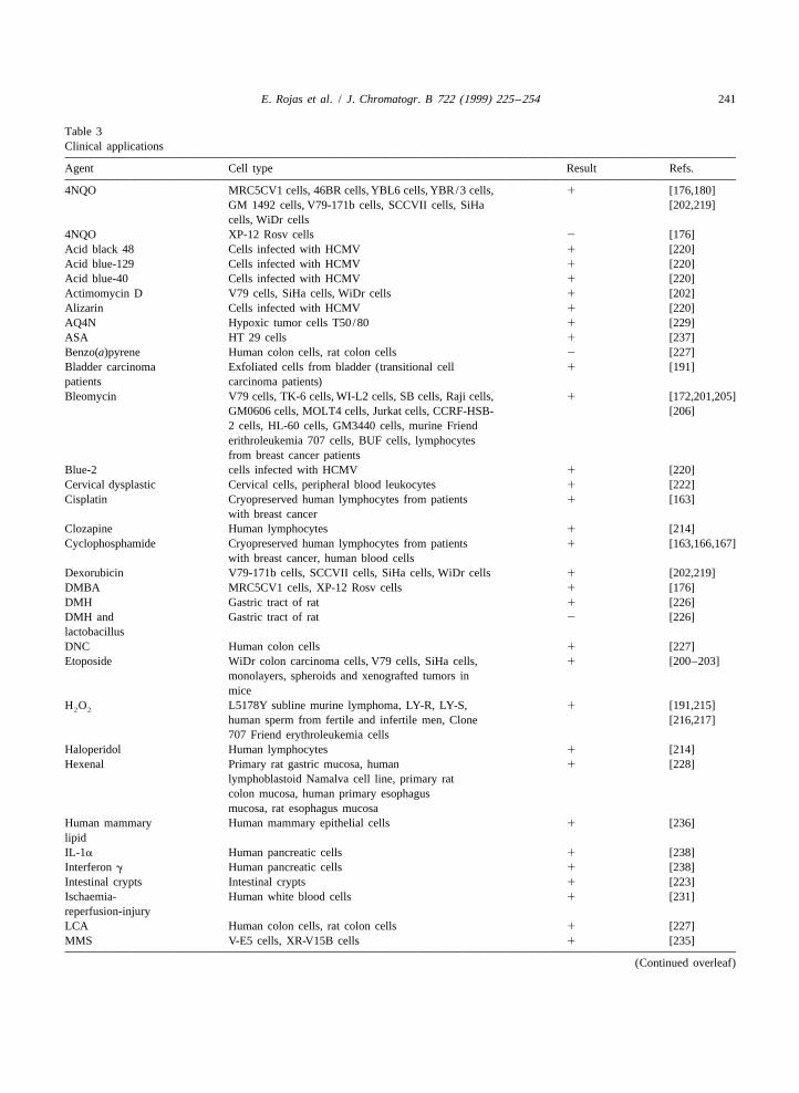
In the last two decades, the search for new under study is embedded in low-melting-point aga-methodologies which are able to assess DNA dam- rose in an agar gel sandwich on a microscope slide,age have been developed. Rydberg and Johanson [1] lysed by detergents and high salt concentration at pHwere the first to directly quantitate DNA damage in 10 and then electrophoresed for a short time underindividual cells by lysing and embedding them in alkaline conditions. Lysis removes the cell contentsagarose on slides under mild alkali conditions to except for the nuclear material. DNA remains highlyallow the partial unwinding of DNA. After neutrali- supercoiled in the presence of a small amount ofzation, cells were stained with acridine orange and non-histone protein but when placed in alkali, itthe extent of DNA damage quantitated by measuring starts to unwind from sites of strand breakage. Cellsthe ratio of green (indicating double-stranded DNA) with increased DNA damage display increased mi-to red (indicating single-stranded DNA) fluorescence gration of the DNA from the nucleus towards theusing a photometer. To improve the sensitivity for anode under an electrical current, giving the appear-detecting DNA damage in isolated cells, Ostling and ance of a ‘‘comet tail’’ (Fig. 1).Johanson [2] developed a microgel electrophoresis Depending on pH conditions for lysis and electro-technique, commonly known as the Comet assay. In phoresis, the sensitivity of the technique can change.this technique cells embedded in agarose gel were Employing neutral conditions for both variables,placed on a microscope slide, the cells lysed by allows to detect DNA double strand breaks; but thedetergents and high salt treatment and the liberated pH 12.3 detects single strand breaks and delay DNADNA electrophoresed under neutral conditions (pH repair sites, while at pH 13 the sensitivity allows toof 9.5) which means that no separation of DNA evaluate alkali labile sites, single strand breaks andstrands occurred after electrophoresis of gamma- delay repair sites of DNA, hereby is important toirradiated cells; the DNA then stained with a fluores- know the purpose of the study.cent dye (ethidium bromide), resembled a comet with About the sensitivity of the (SCGE) Comet assay,head and tail. However this technique permits the McKelvey-Martin et al. [11] and Collins et al. [12]detection of double-stranded DNA breaks only and reported that the assay resolves break frequencies upthe presence of RNA can lead to potential artifacts, to a few hundred per cell, definitely well beyond thedue to this utility it been limited to studies involving range of fragment size for which conventionalradiation and radiomimetic chemicals [3–5]. electrophoresis is suitable.
Two versions of the Comet assay are currently in The present paper reviews and discuses meth-use, one introduced by Singh et al. [6], who used odology modifications and applications of thealkaline electrophoresis (pH.13) to analyze DNA SCGE/Comet assay. For earlier reviews, see Mc-damage after treatment with X-rays or H O , which Kelvy-Martin et al. [11], Fairbairn et al. [13] and2 2
is capable of detecting DNA single-strand breaks and Tice [10,14].alkali labile sites in individuals cells. This version isknown as the ‘‘single cell gel electrophoresis(SCGE) technique’’, although for historical reasonsmany investigators refer to this method as the‘‘Comet assay’’. Subsequently, Olive and co-workersdeveloped versions of the neutral technique ofOstling and Johanson, which involved lysis in alkalitreatment followed by electrophoresis at either neu-tral [7] or mild alkaline (pH 12.3) conditions [8] todetect single strand breaks.
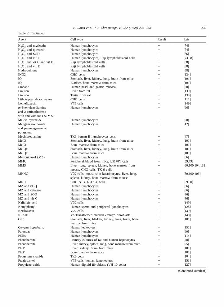
The Singh and Olive methods are identical inprinciple and similar in practice, but the Singhmethod appears to be at least one- or two-orders of Fig. 1. Photograph showing typical appareance of a ‘‘Comet’’magnitude more sensitive [9,10]. image. Human lymphocytes (amplification 603).
E. Rojas et al. / J. Chromatogr. B 722 (1999) 225 –254 227
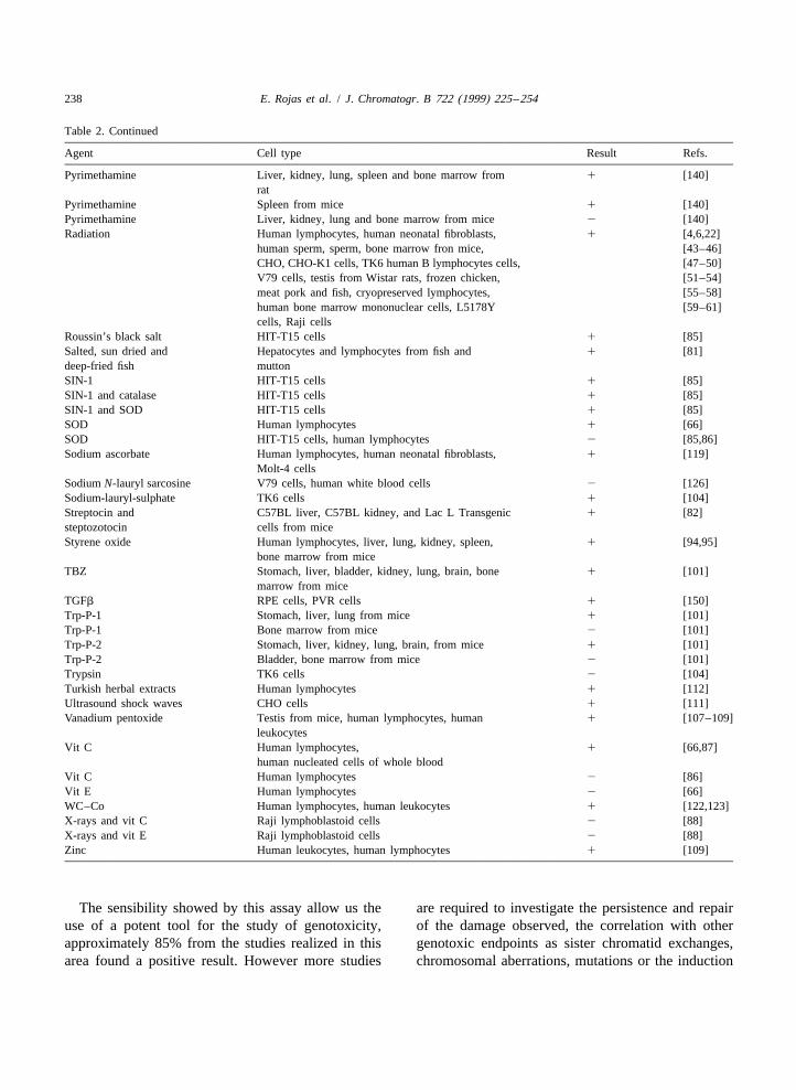
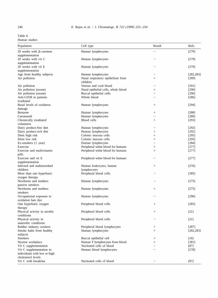
2. The SCGE/Comet methodology teristics of DNA, for this reason the use of PK isrecommended to remove any residual protein.
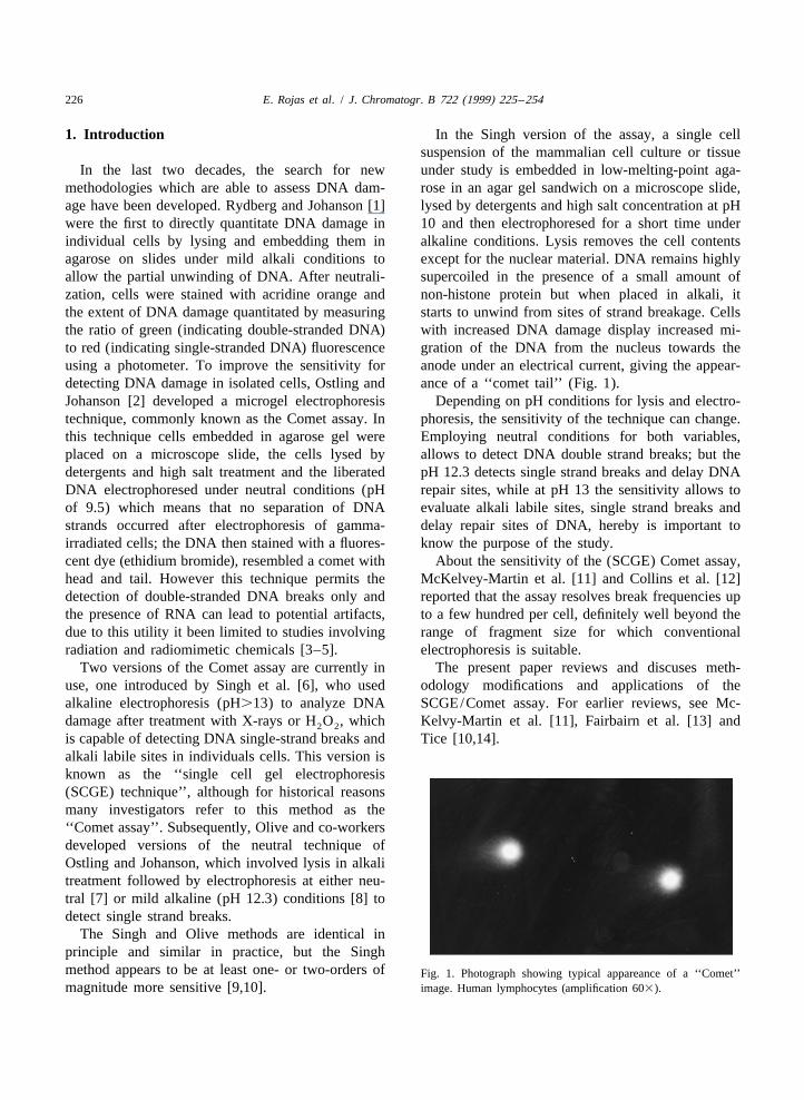
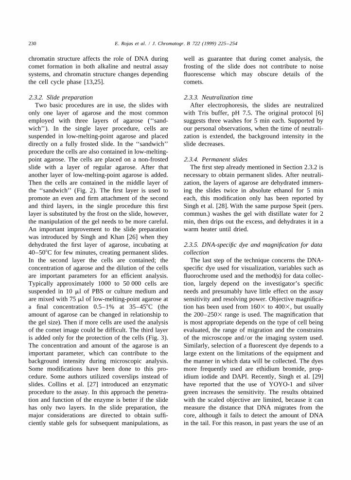
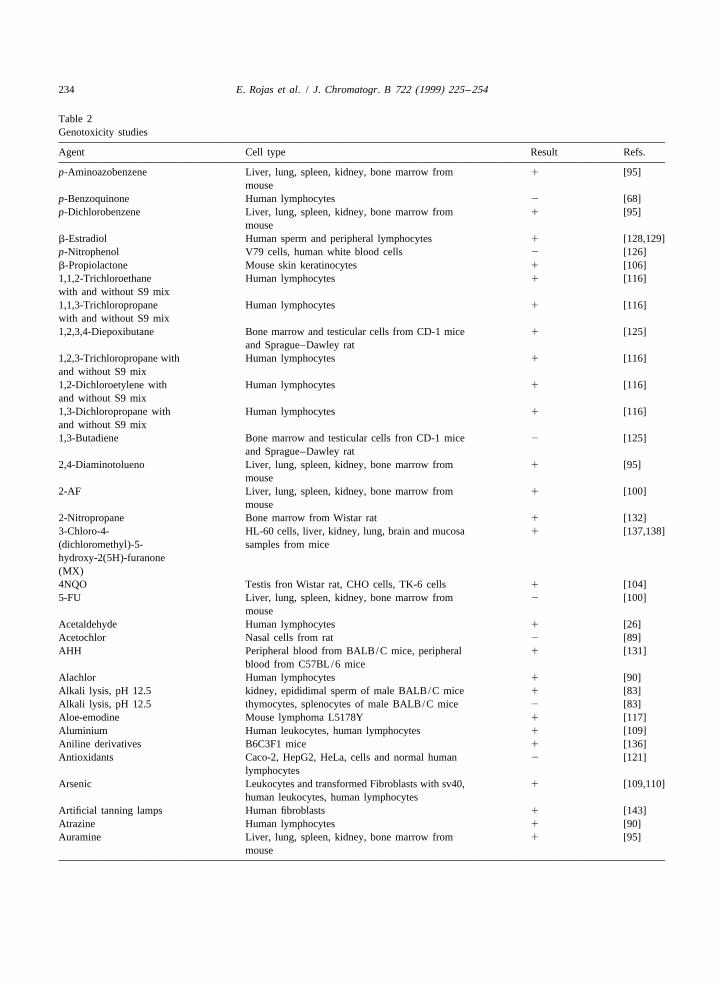
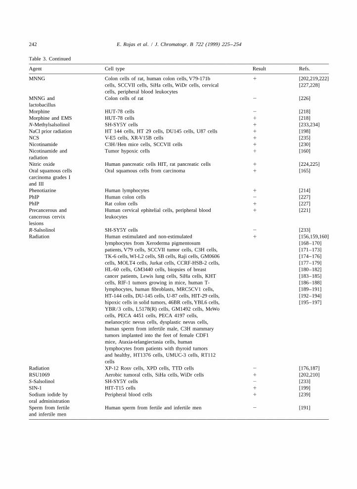
The basic procedure of the technique is describedin detail in various papers [6,11,15]. Briefly, cells aremixed with 0.5% low-melting agarose at 378C and 2.2. Unwinding and electrophoresis bufferthen placed on a microscope slide coated with 0.5%normal agarose. When the agarose has solidified, an Prior to electrophoresis, the slides are equilibratedadditional layer of agarose is added. After the in alkaline electrophoretic solution, which containspreparation of the three layers of this material, the low salt, no detergents and higher pH (.12.3)cells are lysed in a detergent solution for at least 1 h generally. The reported time difference during bothand then the slides are put into an alkaline or neutral the pre-electrophoresis wash or unwinding and elec-buffer in a electrophoresis chamber, allowing the trophoresis steps can be attributed largely to theDNA unwinding, the electrophoresis is carried out, extent of damage and desired detectability. Neutralresulting in the migration of small pieces from the protocol requires more than 1 h of unwinding time tocore of DNA, toward the electric field. After electro- get free of associated proteins with DNA. Much ofphoresis the slides are rinsed with neutralization the variation in the reported protocols is foundbuffer or PBS and cells are stained with a fluoro- during electrophoresis. The desired voltage and timechrome dye (Fig. 2). of electrophoresis will obviously be related to the
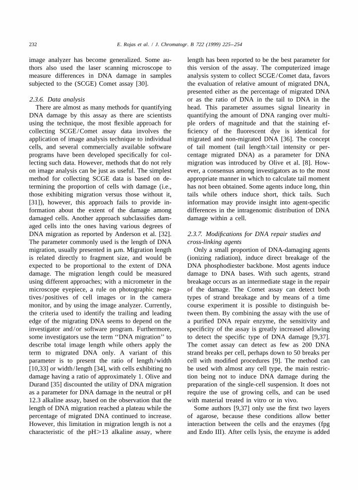
In the past years, the (SCGE) Comet assay has levels of DNA damage expressed in the cells and thehad several modifications but the underlying princi- salt concentration of the running buffer. Since DNAples are based on the neutral or alkaline version. This is required to migrate only a fraction of a millimeterassay has technical variables which affect the sen- for microscopic observation, significant DNA migra-sitivity, the main ones are: the composition and pH tion, which leads to comet formation, is possibleof the lysing solution; the composition and pH of the with very short electrophoresis runs (5–30 min) andelectrophoretic buffer; and the electrophoretic con- low voltages (0.5–5 V/cm) as compared to the mostditions basically voltage, amperage and unwinding conventional DNA electrophoretic techniques. Thelength and running time (Table 1). length of unwinding (alkaline) and the duration of
the electrophoresis are variables which depend on the2.1. Lysis solution cell type being investigated and the type of damage
being assessed. It is important to consider someNeutral and alkaline lysis solutions are used for changes in these steps, larger comets can be obtained
double and single strand breaks detection, respective- by using a higher voltage or time of electrophoresis.ly (Table 1). Selection of which method to use Table 1 shows the most frequently used conditions inshould depend on the purpose of the study. Alkaline neutral, mild alkaline and alkaline assays. Greaterlysis, which is more frequently cited in the literature, sensitivity may be achieved in the assay by increas-consists of immersing the cells in a high salt solution ing the length of time between placing the slide inwith detergents at a pH of 10 to .12 for at least 1 h electrophoresis buffer and applying the current.(for a detailed description see Table 1). Some Green et al. [9] reported that increasing this timemodifications in the composition of this lysis had beyond 40 min causes an increase in comet forma-been reported by McKelvey-Martin et al. [11]. They tion on control slides. This period of incubation isobtained similar lysis results using or not using nominally to allow unwinding of DNA to be initiatedN-laurylsarcosine in the detergents mix, another is from strand breaks but its main function might be tothe addition of PK to remove any residual protein allow the high salt lysis solution to diffuse out of thesuch as Rojas et al. [16] reported for buccal epitheli- agar on the slide, where it competes with DNA as anal cells. Olive [17] introduced other composition electrolyte. Increasing the temperature of incubationconditions and pH modification (12.3), in this case for unwinding and electrophoresis increases thethe solution only had high salt concentration; in the assay sensitivity but also comet formation in con-neutral version the concentration is higher than in trols. A temperature of 158C appears to give maxi-other protocols, due to the physicochemical charac- mum discrimination. This temperature, and the 40
228 E. Rojas et al. / J. Chromatogr. B 722 (1999) 225 –254
Fig. 2. SCGE basic methodology.
min unwinding time, give maximum sensitivity but 2.3. Other experimental variablesare on the borderline of producing acceptable con-trols. Some researchers might prefer to trade a slight Cell suspension preparation, slides and gel sizeloss of sensitivity in order to obtain a robust assay. preparation, time of neutralization, dehydration of
E. Rojas et al. / J. Chromatogr. B 722 (1999) 225 –254 229
Table 1Principal SCGE methodology
Technical variables pH 7 pH 12.3 pH .13
Damage DNA-repair
Number of layers and 2–3 layers 2–3 layers 1–3 layers 2 layers
concentration of 0.75% 0.75% 0.5–1% 1%
agarose
Lysing buffer EDTA (0.025–0.03 M) NaCl (1.0 M) EDTA (100 mM) EDTA (100 mM)
SDS (0.5–2.5%) NaOH (0.03 M) NaCl (2.5 M) NaCl (2.5 M)
Some authors add 10 mg/ml of Sometimes add 0.5% of N- Tris (10 mM), pH 10 Tris (10 mM), pH 10
(PK) proteinase K laurylsarcosine and 2 mM EDTA optional 10 mM N- optional 10 mM N-laurylsarcosine
.1 h .1 h laurylsarcosine in fresh add
in fresh add 1% of Triton X-100
1% of Triton X-100 when samples contain
when samples containing erythrocytes addition of
erythrocytes addition of 10% of DMSO is recomended
10% of DMSO is recommended.
Sometimes PK 10 mg/ml
is added
Incubation with enzymes Is not required Is not required Is not required Placed in a trough containing
enzyme buffer at 48C for 5 min.aAdd 50 ml of buffer for enzyme , at 378C
for 5 min, two times, at the end stop
the reaction with DMSO at 48C
Unwinding buffer Boric acid or acetic acid (90 mM) EDTA (1–2 mM) EDTA (1 mM) Is not required
EDTA (2–5 mM) NaOH (0.03 M) NaOH (300 mM)
Tris (40–117 mM) pH 12.3 pH.13
Duration 2–16 h 1 h 20–60 min
21 21 21Electrophoresis 0.5–0.57 V cm 0.5–0.67 V cm 0.8–1.5 V cm , 20 V for 24 min in a buffer with
Duration is dependent on the voltage Duration depends on the protocol and 25 V and 300 mA, duration depends 300 mM NaOH, 1 mM EDTA
and amperage, but cell type, generally is for 25 min on the protocol and cell type,
generally is for 25 min generally is for 10–60 min
a NaOH (300 mM), EDTA (1 mM), DMSO (10%, v/v).
the slides, the DNA-specific dye used for visualiza- strand break rejoining in tissues from animal ortion, the magnification used to examine the migrating clinical samples is that the samples should beDNA and the method(s) used for data collection and isolated and processed without allowing additionalanalysis are variables that need to be considered. repair or creating additional strand breaks [18,19].
The most commonly examined human cells are2.3.1. Cell suspension preparation leukocytes and lymphocytes. However, many param-
Since the comet assay is designed to evaluate eters can affect the response of lymphocytes in theDNA damage in individual cells, clearly the cells or assay in terms of the ability to detect damage. Sometissues to be evaluated need to be assayed in a way authors [11,20] reported considerable intra-individualthat allows distinction between the cells. Virtually variability of comet formation using the single cellany eukaryotic cell can be processed for the analysis gel assay; also have been discussed a variety ofof DNA damage using this assay. possible factors that may be responsible for these
The existence of various methods for generating differences including the blood donor’s age, thesingle cell suspensions is documented in papers physical activity of the donor, and whether or not thecovering a wide range of biological fields. The donor smokes [21–24]. Moreover, cell cycle statusobvious concern when measuring DNA damage and might be an additional level of complexity, since
230 E. Rojas et al. / J. Chromatogr. B 722 (1999) 225 –254
chromatin structure affects the role of DNA during well as guarantee that during comet analysis, thecomet formation in both alkaline and neutral assay frosting of the slide does not contribute to noisesystems, and chromatin structure changes depending fluorescense which may obscure details of thethe cell cycle phase [13,25]. comets.
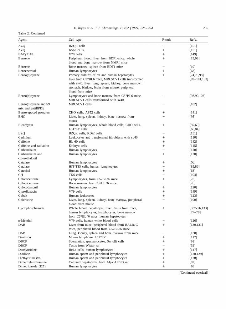
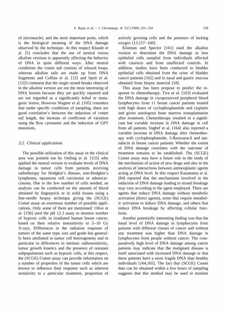
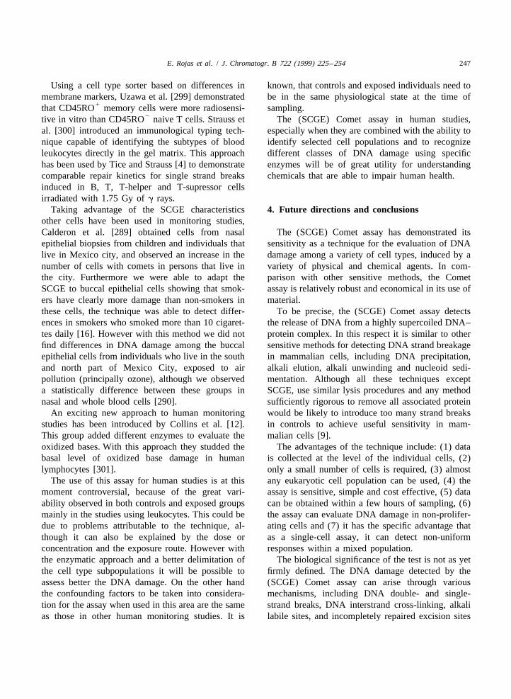
2.3.2. Slide preparation 2.3.3. Neutralization timeTwo basic procedures are in use, the slides with After electrophoresis, the slides are neutralized
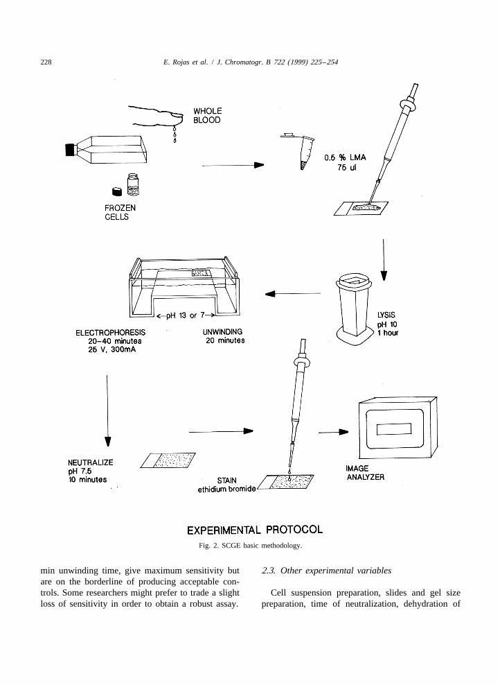
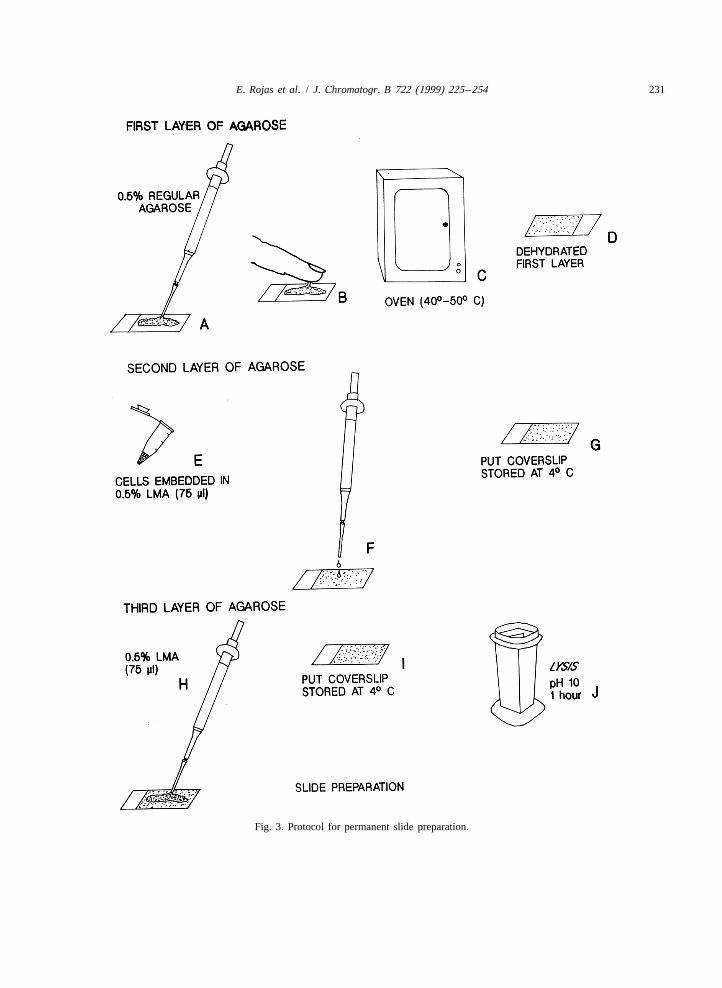
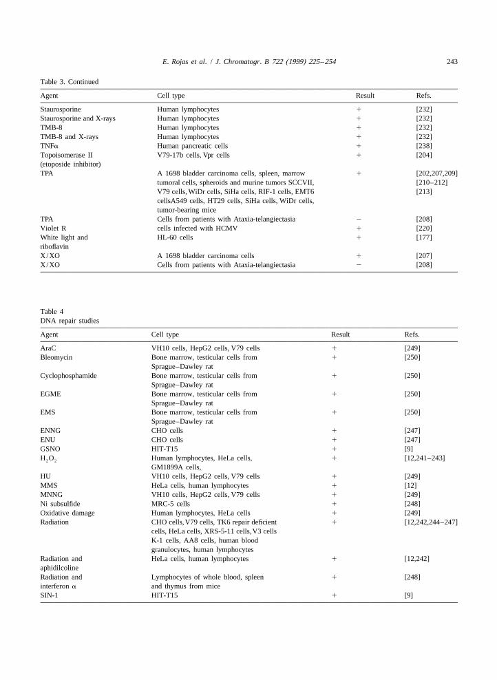
only one layer of agarose and the most common with Tris buffer, pH 7.5. The original protocol [6]employed with three layers of agarose (‘‘sand- suggests three washes for 5 min each. Supported bywich’’). In the single layer procedure, cells are our personal observations, when the time of neutrali-suspended in low-melting-point agarose and placed zation is extended, the background intensity in thedirectly on a fully frosted slide. In the ‘‘sandwich’’ slide decreases.procedure the cells are also contained in low-melting-point agarose. The cells are placed on a non-frosted 2.3.4. Permanent slidesslide with a layer of regular agarose. After that The first step already mentioned in Section 2.3.2 isanother layer of low-melting-point agarose is added. necessary to obtain permanent slides. After neutrali-Then the cells are contained in the middle layer of zation, the layers of agarose are dehydrated immers-the ‘‘sandwich’’ (Fig. 2). The first layer is used to ing the slides twice in absolute ethanol for 5 minpromote an even and firm attachment of the second each, this modification only has been reported byand third layers, in the single procedure this first Singh et al. [28]. With the same purpose Speit (pers.layer is substituted by the frost on the slide, however, commun.) washes the gel with distillate water for 2the manipulation of the gel needs to be more careful. min, then drips out the excess, and dehydrates it in aAn important improvement to the slide preparation warm heater until dried.was introduced by Singh and Khan [26] when theydehydrated the first layer of agarose, incubating at 2.3.5. DNA-specific dye and magnification for data40–508C for few minutes, creating permanent slides. collectionIn the second layer the cells are contained; the The last step of the technique concerns the DNA-concentration of agarose and the dilution of the cells specific dye used for visualization, variables such asare important parameters for an efficient analysis. fluorochrome used and the method(s) for data collec-Typically approximately 1000 to 50 000 cells are tion, largely depend on the investigator’s specificsuspended in 10 ml of PBS or culture medium and needs and presumably have little effect on the assayare mixed with 75 ml of low-melting-point agarose at sensitivity and resolving power. Objective magnifica-a final concentration 0.5–1% at 35–458C (the tion has been used from 1603 to 4003, but usuallyamount of agarose can be changed in relationship to the 200–2503 range is used. The magnification thatthe gel size). Then if more cells are used the analysis is most appropriate depends on the type of cell beingof the comet image could be difficult. The third layer evaluated, the range of migration and the constrainsis added only for the protection of the cells (Fig. 3). of the microscope and/or the imaging system used.The concentration and amount of the agarose is an Similarly, selection of a fluorescent dye depends to aimportant parameter, which can contribute to the large extent on the limitations of the equipment andbackground intensity during microscopic analysis. the manner in which data will be collected. The dyesSome modifications have been done to this pro- more frequently used are ethidium bromide, prop-cedure. Some authors utilized coverslips instead of idium iodide and DAPI. Recently, Singh et al. [29]slides. Collins et al. [27] introduced an enzymatic have reported that the use of YOYO-1 and silverprocedure to the assay. In this approach the penetra- green increases the sensitivity. The results obtainedtion and function of the enzyme is better if the slide with the scaled objective are limited, because it canhas only two layers. In the slide preparation, the measure the distance that DNA migrates from themajor considerations are directed to obtain suffi- core, although it fails to detect the amount of DNAciently stable gels for subsequent manipulations, as in the tail. For this reason, in past years the use of an
E. Rojas et al. / J. Chromatogr. B 722 (1999) 225 –254 231
Fig. 3. Protocol for permanent slide preparation.
232 E. Rojas et al. / J. Chromatogr. B 722 (1999) 225 –254
image analyzer has become generalized. Some au- length has been reported to be the best parameter forthors also used the laser scanning microscope to this version of the assay. The computerized imagemeasure differences in DNA damage in samples analysis system to collect SCGE/Comet data, favorssubjected to the (SCGE) Comet assay [30]. the evaluation of relative amount of migrated DNA,
presented either as the percentage of migrated DNA2.3.6. Data analysis or as the ratio of DNA in the tail to DNA in the
There are almost as many methods for quantifying head. This parameter assumes signal linearity inDNA damage by this assay as there are scientists quantifying the amount of DNA ranging over multi-using the technique, the most flexible approach for ple orders of magnitude and that the staining ef-collecting SCGE/Comet assay data involves the ficiency of the fluorescent dye is identical forapplication of image analysis technique to individual migrated and non-migrated DNA [36]. The conceptcells, and several commercially available software of tail moment (tail length3tail intensity or per-programs have been developed specifically for col- centage migrated DNA) as a parameter for DNAlecting such data. However, methods that do not rely migration was introduced by Olive et al. [8]. How-on image analysis can be just as useful. The simplest ever, a consensus among investigators as to the mostmethod for collecting SCGE data is based on de- appropriate manner in which to calculate tail momenttermining the proportion of cells with damage (i.e., has not been obtained. Some agents induce long, thinthose exhibiting migration versus those without it, tails while others induce short, thick tails. Such[31]), however, this approach fails to provide in- information may provide insight into agent-specificformation about the extent of the damage among differences in the intragenomic distribution of DNAdamaged cells. Another approach subclassifies dam- damage within a cell.aged cells into the ones having various degrees ofDNA migration as reported by Anderson et al. [32]. 2.3.7. Modifications for DNA repair studies andThe parameter commonly used is the length of DNA cross-linking agentsmigration, usually presented in mm. Migration length Only a small proportion of DNA-damaging agentsis related directly to fragment size, and would be (ionizing radiation), induce direct breakage of theexpected to be proportional to the extent of DNA DNA phosphodiester backbone. Most agents inducedamage. The migration length could be measured damage to DNA bases. With such agents, strandusing different approaches; with a micrometer in the breakage occurs as an intermediate stage in the repairmicroscope eyepiece, a rule on photographic nega- of the damage. The Comet assay can detect bothtives /positives of cell images or in the camera types of strand breakage and by means of a timemonitor, and by using the image analyzer. Currently, course experiment it is possible to distinguish be-the criteria used to identify the trailing and leading tween them. By combining the assay with the use ofedge of the migrating DNA seems to depend on the a purified DNA repair enzyme, the sensitivity andinvestigator and/or software program. Furthermore, specificity of the assay is greatly increased allowingsome investigators use the term ‘‘DNA migration’’ to to detect the specific type of DNA damage [9,37].describe total image length while others apply the The comet assay can detect as few as 200 DNAterm to migrated DNA only. A variant of this strand breaks per cell, perhaps down to 50 breaks perparameter is to present the ratio of length /width cell with modified procedures [9]. The method can[10,33] or width / length [34], with cells exhibiting no be used with almost any cell type, the main restric-damage having a ratio of approximately 1. Olive and tion being not to induce DNA damage during theDurand [35] discounted the utility of DNA migration preparation of the single-cell suspension. It does notas a parameter for DNA damage in the neutral or pH require the use of growing cells, and can be used12.3 alkaline assay, based on the observation that the with material treated in vitro or in vivo.length of DNA migration reached a plateau while the Some authors [9,37] only use the first two layerspercentage of migrated DNA continued to increase. of agarose, because these conditions allow betterHowever, this limitation in migration length is not a interaction between the cells and the enzymes (fpgcharacteristic of the pH.13 alkaline assay, where and Endo III). After cells lysis, the enzyme is added
E. Rojas et al. / J. Chromatogr. B 722 (1999) 225 –254 233
to the gel in 50 ml of appropriate buffer, covered The only requirement is that a sufficient number ofwith a coverslip, and incubated at 378C for the single cells is obtained for analysis without inductionrequired time [12]. Endo III, fpg and uvrA, uvrB and of damage. There are different approaches to obtainuvrC are the most employed enzymes. Endo III the cell suspension, as the use of collagenase andrecognizes oxidized pyrimidines, fpg recognizing 8- trypsin [38] or just mince the tissue in cold bufferOH gua and another damaged purines and uvr ABC [39], to obtain a nucleus suspension a chilled bufferfor bulky lesions and cleavages the DNA backbone with NaCl (0.075 M) and EDTA (0.24 M) at pH ofduring their removal. The next step is to run the 7.5 is used [40]. Depending on the intrinsic organelectrophoresis (without unwinding) and rinse the conditions, it is possible to improve the isolationslides dropwise with neutralizing buffer and then additioning specific chemicals as N-tert.-butyl-a-continue with the classical protocol. phenylnitrone a spin trapper used on brain cells [28],
for the better cell preservation. Some groups use thesame conditions for all the organs tested, but the
3. Applications of the assay optimal conditions for each organ seems to bedifferent, then it depends on the focus of the work (a
3.1. Studies on genotoxicity panoramic evaluation of genotoxicity comparing theDNA damage in different organs or a specific tissue
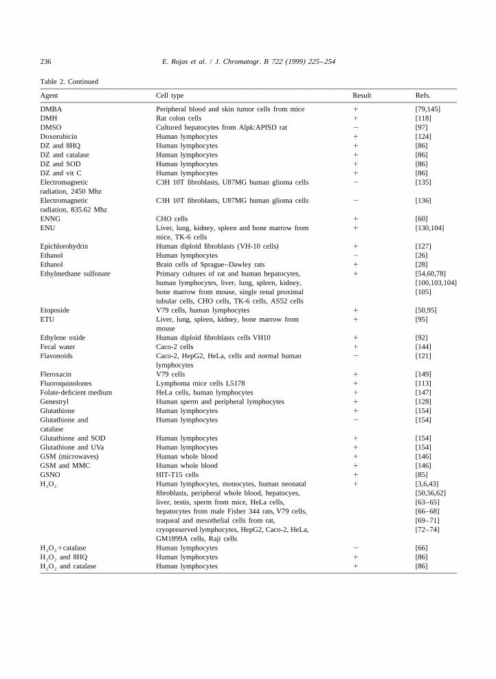
An useful application of the alkaline version of the study) the standardization of the conditions for each(SCGE) Comet assay is in the area of genetic work are required. Animal cell lines, usually arisingtoxicology and a number of investigators have used from different rodent cell types, the most used arethis version to evaluate in vitro and/or in vivo CHO cells HIT-T15, V-79, L5178Y, SCCVII, andgenotoxicity of several chemicals. A variety of primary cultures of hepatocytes had mainly beennormal and transformed cells including human, reported. An interesting aspect of the rodent hepat-animal and plant have been used for in vitro studies. ocyte is the ability to measure DNA damage in
Most of the work with human cells, have used parenchymal and non-parenchymal cells simultan-leukocytes and lymphocytes but other tissues have eously [41], this ability permits a simple method forbeen also used such as epithelial cells (lens, buccal, discriminating between direct-acting genotoxicantsnasal and gastric mucosa, skin-derived and foreskin- and those requiring metabolic activation [10]. Thederived cells), reproductive cells, colon cells, neonat- results are particularly appealing because they in-al fibroblast, pancreatic cells, adenocarcinoma cells, clude information about the distribution of damage inlymphoid cells. Different human cell lines have been the target tissue [13]. This distribution among cellsused including cells from blood (Raji, TK6, HUT-78 provides information on the relative heterogeneity ofand HL-60, MOLT-4), different kinds of carcinomas the damage among cells in different tissues and(cervix [SiHa, HeLa], colon [WiDr, HT-29], bladder indicates the proportion of damaged to undamaged[A1698], prostate [Du-145]), melanoma (MeWo, cells for each dose and tissue [10], an advantage thatHT-144), glioma (U-87), fibroblast (IBR-3), no other system can offer. Recently some groupsbreast keratinocyte (NHEK-267-1), lung cells adapted this assay to plant tissues like Allium cepa(MRC5CV1), etc. [273].
A great amount of work in this field has been done The amount of chemicals tested in this area isusing animal cells, since only a few cells are rapidly increasing every year, the first practical userequired for analysis, virtually any tissue or organ is of the (SCGE) Comet assay in the genotoxicityavailable for investigation. Rodents are the more testing was an evaluation of the mutagenic potenciesfrequently used but other animals such as dog, sheep, of agents generated by the treatment of potassiumtadpoles and fish have been also employed. This perganmanate with acidic solutions [42], from thistechnique has been applied to several organs and first report to the present day a great amount oftissues of this animals, including blood, bone mar- chemicals have been tested, such as metals, pes-row, brain, gastrointestinal mucosa, kidney, liver, ticides, opiates, pesticides, nitrosamines and antineo-lung, nasal mucosa, ovaries, skin, spleen and testis. plastic drugs (for a more detailed list see Table 2).
234 E. Rojas et al. / J. Chromatogr. B 722 (1999) 225 –254
Table 2Genotoxicity studies
Agent Cell type Result Refs.
p-Aminoazobenzene Liver, lung, spleen, kidney, bone marrow from 1 [95]mouse
p-Benzoquinone Human lymphocytes 2 [68]p-Dichlorobenzene Liver, lung, spleen, kidney, bone marrow from 1 [95]
mouseb-Estradiol Human sperm and peripheral lymphocytes 1 [128,129]p-Nitrophenol V79 cells, human white blood cells 2 [126]b-Propiolactone Mouse skin keratinocytes 1 [106]1,1,2-Trichloroethane Human lymphocytes 1 [116]with and without S9 mix1,1,3-Trichloropropane Human lymphocytes 1 [116]with and without S9 mix1,2,3,4-Diepoxibutane Bone marrow and testicular cells from CD-1 mice 1 [125]
and Sprague–Dawley rat1,2,3-Trichloropropane with Human lymphocytes 1 [116]and without S9 mix1,2-Dichloroetylene with Human lymphocytes 1 [116]and without S9 mix1,3-Dichloropropane with Human lymphocytes 1 [116]and without S9 mix1,3-Butadiene Bone marrow and testicular cells fron CD-1 mice 2 [125]
and Sprague–Dawley rat2,4-Diaminotolueno Liver, lung, spleen, kidney, bone marrow from 1 [95]
mouse2-AF Liver, lung, spleen, kidney, bone marrow from 1 [100]
mouse2-Nitropropane Bone marrow from Wistar rat 1 [132]3-Chloro-4- HL-60 cells, liver, kidney, lung, brain and mucosa 1 [137,138](dichloromethyl)-5- samples from micehydroxy-2(5H)-furanone(MX)4NQO Testis fron Wistar rat, CHO cells, TK-6 cells 1 [104]5-FU Liver, lung, spleen, kidney, bone marrow from 2 [100]
mouseAcetaldehyde Human lymphocytes 1 [26]Acetochlor Nasal cells from rat 2 [89]AHH Peripheral blood from BALB/C mice, peripheral 1 [131]
blood from C57BL/6 miceAlachlor Human lymphocytes 1 [90]Alkali lysis, pH 12.5 kidney, epididimal sperm of male BALB/C mice 1 [83]Alkali lysis, pH 12.5 thymocytes, splenocytes of male BALB/C mice 2 [83]Aloe-emodine Mouse lymphoma L5178Y 1 [117]Aluminium Human leukocytes, human lymphocytes 1 [109]Aniline derivatives B6C3F1 mice 1 [136]Antioxidants Caco-2, HepG2, HeLa, cells and normal human 2 [121]
lymphocytesArsenic Leukocytes and transformed Fibroblasts with sv40, 1 [109,110]
human leukocytes, human lymphocytesArtificial tanning lamps Human fibroblasts 1 [143]Atrazine Human lymphocytes 1 [90]Auramine Liver, lung, spleen, kidney, bone marrow from 1 [95]
mouse
E. Rojas et al. / J. Chromatogr. B 722 (1999) 225 –254 235
Table 2. Continued
Agent Cell type Result Refs.
AZQ BZQR cells 2 [151]AZQ K562 cells 1 [151]BAYy3118 V79 cells 1 [149]Benzene Peripheral blood, liver from BDF1-mice, whole 1 [19,93]
blood and bone marrow from NMRI miceBenzene Bone marrow, spleen from BDF1-mice 2 [19]Benzenethiol Human lymphocytes 1 [68]Benzo(a)pyrene Primary cultures of rat and human hepatocytes, 1 [74,78,98]
liver from C57BL6 mice, MRC5CV1 cells transformed [99–101,133]with sv40, liver, lung, spleen, kidney, bone marrow,stomach, bladder, brain from mouse, peripheralblood from mice
Benzo(a)pyrene Lymphocytes and bone marrow from C57BL6 mice, 2 [98,99,102]MRC5CV1 cells transformed with sv40,
Benzo(a)pyrene and S9 MRC5CV1 cells 2 [102]mix and antiBPDEBenzo-spaced psoralen CHO cells, AS52 cells 1 [141]BHC Liver, lung, spleen, kidney, bone marrow from 2 [95]
mouseBleomycin Human lymphocytes, whole blood cells, CHO cells, 1 [59,60]
L5178Y cells [66,84]BZQ BZQR cells, K562 cells 2 [151]Cadmium Leukocytes and transformed fibroblasts with sv40 1 [110]Caffeine HL-60 cells 1 [142]Caffeine and radiation Embryo cells 1 [115]Carbendazim Human lymphocytes 2 [120]Carbendazim and Human lymphocytes 1 [120]chlorothalonilCatalase Human lymphocytes 1 [66]Catalase HIT-T15 cells, human lymphocytes 2 [85,86]Catechol Human lymphocytes 1 [68]CH TK6 cells 2 [104]Chlorobenzene Lymphocytes, from C57BL/6 mice 1 [76]Chlorobenzene Bone marrow fron C57BL/6 mice 2 [76]Chlorothalonil Human lymphocytes 1 [120]Ciprofloxacin V79 cells 1 [149]Cobalt Human leukocytes 1 [123]Colchicine Liver, lung, spleen, kidney, bone marrow, peripheral 2 [100]
blood from mouseCyclophosphamide Whole blood, hepatocyes, liver, testis from mice, 1 [3,75,76,133]
human lymphocytes, lymphocytes, bone marrow [77–79]from C57BL/6 mice, human hepatocytes
D-Menthol V79 cells, human white blood cells 2 [126]DAB Liver from mice, peripheral blood from BALB/C 1 [130,131]
mice, peripheral blood from C57BL/6 miceDAB Lung, kidney, spleen and bone marrow from mice 2 [130]Danthron Mouse lymphoma L5178Y 1 [117]DBCP Spermatids, spermatocytes, Sertolli cells 1 [91]DBCP Testis from Wistar rat 2 [52]Deoxyuridine HeLa cells, human lymphocytes 1 [147]Diadzein Human sperm and peripheral lymphocytes 1 [128,129]Diethylstilbestrol Human sperm and peripheral lymphocytes 1 [128]Dimethylnitrosamine Cultured hepatocytes from Alpk:APfSD rat 1 [97]Dimetridazole (DZ) Human lymphocytes 1 [86]
(Continued overleaf)
236 E. Rojas et al. / J. Chromatogr. B 722 (1999) 225 –254
Table 2. Continued
Agent Cell type Result Refs.
DMBA Peripheral blood and skin tumor cells from mice 1 [79,145]DMH Rat colon cells 1 [118]DMSO Cultured hepatocytes from Alpk:APfSD rat 2 [97]Doxorubicin Human lymphocytes 1 [124]DZ and 8HQ Human lymphocytes 1 [86]DZ and catalase Human lymphocytes 1 [86]DZ and SOD Human lymphocytes 1 [86]DZ and vit C Human lymphocytes 1 [86]Electromagnetic C3H 10T fibroblasts, U87MG human glioma cells 2 [135]radiation, 2450 MhzElectromagnetic C3H 10T fibroblasts, U87MG human glioma cells 2 [136]radiation, 835.62 MhzENNG CHO cells 1 [60]ENU Liver, lung, kidney, spleen and bone marrow from 1 [130,104]
mice, TK-6 cellsEpichlorohydrin Human diploid fibroblasts (VH-10 cells) 1 [127]Ethanol Human lymphocytes 2 [26]Ethanol Brain cells of Sprague–Dawley rats 1 [28]Ethylmethane sulfonate Primary cultures of rat and human hepatocytes, 1 [54,60,78]
human lymphocytes, liver, lung, spleen, kidney, [100,103,104]bone marrow from mouse, single renal proximal [105]tubular cells, CHO cells, TK-6 cells, AS52 cells
Etoposide V79 cells, human lymphocytes 1 [50,95]ETU Liver, lung, spleen, kidney, bone marrow from 1 [95]
mouseEthylene oxide Human diploid fibroblasts cells VH10 1 [92]Fecal water Caco-2 cells 1 [144]Flavonoids Caco-2, HepG2, HeLa, cells and normal human 2 [121]
lymphocytesFleroxacin V79 cells 1 [149]Fluoroquinolones Lymphoma mice cells L5178 1 [113]Folate-deficient medium HeLa cells, human lymphocytes 1 [147]Genestryl Human sperm and peripheral lymphocytes 1 [128]Glutathione Human lymphocytes 1 [154]Glutathione and Human lymphocytes 2 [154]catalaseGlutathione and SOD Human lymphocytes 1 [154]Glutathione and UVa Human lymphocytes 1 [154]GSM (microwaves) Human whole blood 1 [146]GSM and MMC Human whole blood 1 [146]GSNO HIT-T15 cells 1 [85]H O Human lymphocytes, monocytes, human neonatal 1 [3,6,43]2 2
fibroblasts, peripheral whole blood, hepatocyes, [50,56,62]liver, testis, sperm from mice, HeLa cells, [63–65]hepatocytes from male Fisher 344 rats, V79 cells, [66–68]traqueal and mesothelial cells from rat, [69–71]cryopreserved lymphocytes, HepG2, Caco-2, HeLa, [72–74]GM1899A cells, Raji cells
H O 1catalase Human lymphocytes 2 [66]2 2
H O and 8HQ Human lymphocytes 1 [86]2 2
H O and catalase Human lymphocytes 1 [86]2 2
E. Rojas et al. / J. Chromatogr. B 722 (1999) 225 –254 237
Table 2. Continued
Agent Cell type Result Refs.
H O and myricetin Human lymphocytes 2 [74]2 2
H O and quercetin Human lymphocytes 2 [74]2 2
H O and SOD Human lymphocytes 1 [86]2 2
H O and vit C Human lymphocytes, Raji lymphoblastoid cells 2 [73,88]2 2
H O and vit C and vit E Raji lymphoblastoid cells 2 [88]2 2
H O and vit E Raji lymphoblastoid cells 2 [88]2 2
Hydroquinone Human lymphocytes 2 [68]INO2 CHO cells 1 [134]IQ Stomach, liver, kidney, lung, brain from mice 1 [101]IQ Bladder, bone marrow from mice 2 [101]Lindane Human nasal and gastric mucosa 1 [80]Linuron Liver from rat 1 [139]Linuron Testis from rat 2 [139]Lithotripter shock waves CHO cells 2 [111]Lomefloxacin V79 cells 1 [149]m-Phenylenediamine Human lymphocytes 1 [96]and 2-aminofluorenewith and without TX1MXMaleic hydrazide Human lymphocytes 1 [90]Manganese-chloride Human lymphocytes 1 [42]and permanganate ofpotassiumMechlorethamine TK6 human B lymphocytes cells 2 [47]MeIQ Stomach, liver, kidney, lung, brain from mice 1 [101]MeIQ Bone marrow from mice 2 [101]MeIQx Stomach, liver, kidney, lung, brain from mice 1 [101]MeIQx Bone marrow from mice 2 [101]Metronidazol (MZ) Human lymphocytes 1 [86]MMC Peripheral blood from mice, L5178Y cells 1 [59,79]MMS Liver, lung, spleen, kidney, bone marrow from 1 [60,100,104,133]
mouse, CHO cells, TK-6 cellsMNNG V79 cells, mouse skin keratinocytes, liver, lung, 1 [50,100,106]
spleen, kidney, bone marrow from mouseMNU CHO cells, L5178Y cells 1 [59,60]MZ and 8HQ Human lymphocytes 1 [86]MZ and catalase Human lymphocytes 1 [86]MZ and SOD Human lymphocytes 1 [86]MZ and vit C Human lymphocytes 1 [86]Nalidixic acid V79 cells 1 [149]Nonylphenyl Human sperm and peripheral lymphocytes 1 [128]Norfloxacin V79 cells 1 [149]NSAID src-Transformed chicken embryo fibroblasts 1 [148]OPP Stomach, liver, bladder, kidney, lung, brain, bone 1 [101]
marrow from miceOxygen hyperbaric Human leukocytes 1 [152]Paraquat Human lymphocytes 1 [90]PCBs Human lymphocytes 2 [114]Phenobarbital Primary cultures of rat and human hepatocytes 2 [78]Phenobarbital Liver, kidney, spleen, lung, bone marrow from mice 1 [95]PhIP Liver, kidney, brain from mice 1 [101]PhIP Bone marrow from mice 2 [101]Potassium cyanide TK6 cells 1 [104]Praziquantel V79 cells, human lymphocytes 1 [153]Propylene oxide Human diploid fibroblasts (VH-10 cells) 1 [127]
(Continued overleaf)
238 E. Rojas et al. / J. Chromatogr. B 722 (1999) 225 –254
Table 2. Continued
Agent Cell type Result Refs.
Pyrimethamine Liver, kidney, lung, spleen and bone marrow from 1 [140]rat
Pyrimethamine Spleen from mice 1 [140]Pyrimethamine Liver, kidney, lung and bone marrow from mice 2 [140]Radiation Human lymphocytes, human neonatal fibroblasts, 1 [4,6,22]
human sperm, sperm, bone marrow fron mice, [43–46]CHO, CHO-K1 cells, TK6 human B lymphocytes cells, [47–50]V79 cells, testis from Wistar rats, frozen chicken, [51–54]meat pork and fish, cryopreserved lymphocytes, [55–58]human bone marrow mononuclear cells, L5178Y [59–61]cells, Raji cells
Roussin’s black salt HIT-T15 cells 1 [85]Salted, sun dried and Hepatocytes and lymphocytes from fish and 1 [81]deep-fried fish muttonSIN-1 HIT-T15 cells 1 [85]SIN-1 and catalase HIT-T15 cells 1 [85]SIN-1 and SOD HIT-T15 cells 1 [85]SOD Human lymphocytes 1 [66]SOD HIT-T15 cells, human lymphocytes 2 [85,86]Sodium ascorbate Human lymphocytes, human neonatal fibroblasts, 1 [119]
Molt-4 cellsSodium N-lauryl sarcosine V79 cells, human white blood cells 2 [126]Sodium-lauryl-sulphate TK6 cells 1 [104]Streptocin and C57BL liver, C57BL kidney, and Lac L Transgenic 1 [82]steptozotocin cells from miceStyrene oxide Human lymphocytes, liver, lung, kidney, spleen, 1 [94,95]
bone marrow from miceTBZ Stomach, liver, bladder, kidney, lung, brain, bone 1 [101]
marrow from miceTGFb RPE cells, PVR cells 1 [150]Trp-P-1 Stomach, liver, lung from mice 1 [101]Trp-P-1 Bone marrow from mice 2 [101]Trp-P-2 Stomach, liver, kidney, lung, brain, from mice 1 [101]Trp-P-2 Bladder, bone marrow from mice 2 [101]Trypsin TK6 cells 2 [104]Turkish herbal extracts Human lymphocytes 1 [112]Ultrasound shock waves CHO cells 1 [111]Vanadium pentoxide Testis from mice, human lymphocytes, human 1 [107–109]
leukocytesVit C Human lymphocytes, 1 [66,87]
human nucleated cells of whole bloodVit C Human lymphocytes 2 [86]Vit E Human lymphocytes 2 [66]WC–Co Human lymphocytes, human leukocytes 1 [122,123]X-rays and vit C Raji lymphoblastoid cells 2 [88]X-rays and vit E Raji lymphoblastoid cells 2 [88]Zinc Human leukocytes, human lymphocytes 1 [109]
The sensibility showed by this assay allow us the are required to investigate the persistence and repairuse of a potent tool for the study of genotoxicity, of the damage observed, the correlation with otherapproximately 85% from the studies realized in this genotoxic endpoints as sister chromatid exchanges,area found a positive result. However more studies chromosomal aberrations, mutations or the induction
E. Rojas et al. / J. Chromatogr. B 722 (1999) 225 –254 239
of micronuclei, and the most important point, which actively growing cells and the presence of lackingis the biological meaning of the DNA damage oxygen [13,157–160].observed by the technique. At this respect Klaude et Klieman and Spector [161] used the alkalineal. [5] concludes that the use of neutral versus version to determine the DNA damage in lensalkaline versions is apparently affecting the behavior epithelial cells sampled from individuals affectedof DNA in quite different ways. After neutral with cataracts and from unaffected controls. Inconditions the comet tail consists of relaxed loops, addition, studies have been conducted in bladderwhereas alkaline tails are made up from DNA epithelial cells obtained from the urine of bladderfragments and Collins et al. [12] and Speit et al. cancer patients [162] and in nasal and gastric mucosa[152] comment that the single strand breaks observed obtained from biopsy material [18].in the alkaline version are not the most interesting of This assay has been propose to predict the re-DNA lesions because they are quickly repaired and sponse to chemotherapy, Tice et al. [163] evaluatedare not regarded as a significantly lethal or muta- the DNA damage in cryopreserved peripheral bloodgenic lesion. However Wagner et al. [105] considers lymphocytes from 11 breast cancer patients treatedthat under specific conditions of sampling, there are with high doses of cyclophosphamide and cisplatingood correlation’s between the induction of comet and given autologous bone marrow transplantationtail length, the increase of coefficient of variation after treatment. Chemotherapy resulted in a signifi-using the flow cytometer and the induction of GPT cant but variable increase in DNA damage in cellmutations. from all patients. Vaghef et al. [164] also reported a
variable increase in DNA damage after chemother-apy with cyclophosphamide, 5-fluorouracil and epi-
3.2. Clinical applications rubicin in breast cancer patients. Whether the extentof DNA damage correlates with the outcome of
The possible utilization of this assay in the clinical treatment remains to be established. The (SCGE)area was pointed out by Ostling et al. [155] who Comet assay may have a future role in the study ofapplied the neutral version to evaluate levels of DNA the mechanism of action of new drugs and also in thedamage in tumor cells from patients receiving analysis of interactions between antineoplastic agentsradiotherapy for Hodgkin’s disease, non-Hodgkin’s acting at DNA level. In this respect Kasamatsu et al.lymphoma, squamous cell carcinoma or adenocar- [84] reported that the mechanisms involved in thecinoma. Due to the few number of cells needed, an induction of DNA damage leading to strand breakageanalysis can be conducted on the amount of blood may vary according to the agent employed. There areobtained by fingerprick or in solid tissues using a agents that induce DNA damage without metabolicfine-needle biopsy technique giving the (SCGE) activation (direct agents), some that require metabol-Comet assay an enormous number of possible appli- ic activation to induce DNA damage, and others thatcations. Only some of them are mentioned: Olive et induce DNA breakage by affecting cellular func-al. [156] used the pH 12.3 assay to monitor number tions.of hypoxic cells in irradiated human breast cancer, Another potentially interesting finding was that thebased on their relative insensitivity to 5–10 Gy basal level of DNA damage in lymphocytes fromX-rays. Differences in the radiation response of patients with different classes of cancer and withouttumors of the same type, size and grade has general- any treatment was higher than DNA damage inly been attributed to tumor cell heterogeneity and in lymphocytes from people without cancer. The com-particular to differences in intrinsic radiosensitivity, paratively high level of DNA damage among cancertumor growth kinetics and the presence of resistant patients may indicate that the malignant disease issubpopulations such as hypoxic cells, at this respect, itself associated with increased DNA damage or thatthe (SCGE) Comet assay can provide information on these patients have a more fragile DNA than healthya number of properties of the tumor cells which are individuals [164,165]. The fact that (SCGE) Cometknown to influence their response such as inherent data can be obtained within a few hours of samplingsensitivity to a particular treatment, proportion of suggests that this method may be used to monitor
240 E. Rojas et al. / J. Chromatogr. B 722 (1999) 225 –254
levels of damage in individual patients associated viduals affected with Xeroderma pigmentosum,with a regimen treatment and that the regimen could which are incapable to repair the UV damage,be changed accordingly [10]. For a more detailed list observed a non-induction of DNA strand breaks inof the works done in this area see Table 3. this cells after irradiation, meanwhile in normal cells
an increased DNA migration was observed after 1 h3.3. DNA repair studies of irradiation. The assay has been suggested as a
diagnostic tool for Xeroderma pigmentosum andBecause of its characteristics, the (SCGE) Comet other syndromes characterized by defects in excision
assay has been used to evaluate the ability of repair [250].virtually any type of eukaryotic cell to repair differ- The ability to detect excision repair sites using theent kinds of DNA damage, including double- and alkaline version can be enhanced by the inclusion ofsingle-strand breaks and base damage. The neutral repair inhibitors, DNA synthesis inhibitors or chainand alkaline version of the assay have been used to terminators such as N-hidroxyurea, aphidilcolin andassess the repair of double-strand and single-strand cytosine arabinoside, respectively [250,252]. Thebreaks, respectively. Single- and double-strand enzymatic approach of Collins and co-workers canbreaks induced by ionizing radiation are efficiently be used in the alkaline version to evaluate the repairrepaired in normal cells [4,6,8,22,29,240] (for more kinetics of various classes of DNA lesions in treateddetailed data see Table 4). Fifty percent of the cells. The UV-damage-specific T4 endonuclease candamage is repaired within 15 min and complete be used to study the removal of UV-induced py-repair occurs within 1–2 h. This rate of repair has rimidine dimers [250] while Endo III can be used toalso been demonstrated for subsets of irradiated monitor the removal of oxidized pyrimidines [241].human lymphocytes [4]. Hydrogen peroxide induced damage levels which
To evaluate the effect of subjects age on strand- showed to be higher with endonuclease III treatment,break repair, Singh et al. [22] employed the alkaline demonstrating the persistence of oxidized base dam-version to measure the DNA damage and repair in age and therefore the differential expression oflymphocytes isolated from the peripheral blood of lesions following repair [253]. The oxidative andhealthy subjects and exposed them in vitro to 2 Gy other DNA damage induced endogenously can beof X-radiation. For all subjects the mean level of readily studied using experimental variations of theDNA damage was restored to pre-irradiation control (SCGE) Comet assay because of its sensitivity.levels within 2 h of incubation at 378C. However, Apoptotic DNA fragmentation could be also studieddue to the advantage of being able to analyze single [134]. Here again, the Comet assay provides a simplecells, a distribution analysis of DNA damage among assay system with the advantage of allowing exami-cells within each sample indicated the presence of a nation of the sequential steps of incision /excisionfew highly damaged cells in the 2-h sample, the and resynthesis / ligation.occurrence of which was significantly more commonamong aged individuals. Another important feature 3.4. Environmental biomonitoringof this kind of strand break repair studies is thecapacity for the recognition of individuals with an An optimal method for detecting genotoxicityaltered repair capability, Rojas et al. [108] found an damage in sentinel organisms should be capable ofimportant repair delay in lymphocytes from a healthy detecting many classes of damage in a variety of celldonor exposed to vanadium pentoxide. types from a range of organisms, provide data at the
Agents such as UV radiation that produce lesions level of the individual cell and be sensitive, rapidwhich do not form strand breaks directly can be and cost effective [14]. The (SCGE) Comet assay,examined using the assay. Rather than detecting again because of its simplicity, sensitivity, and needstrand breaks produced by the irradiation, it is for only small number of cells has been suggested aspossible to detect strand breaks produced by the cell an ideal technique for such studies.in its attempt to repair the lesion. With this approach To monitor for genotoxic pollutants at a hazardousGedik et al. [46,250,251] using cells from indi- site Nascimbeni et al. [254] conducted a pilot study
E. Rojas et al. / J. Chromatogr. B 722 (1999) 225 –254 241
Table 3Clinical applications
Agent Cell type Result Refs.
4NQO MRC5CV1 cells, 46BR cells, YBL6 cells, YBR/3 cells, 1 [176,180]GM 1492 cells, V79-171b cells, SCCVII cells, SiHa [202,219]cells, WiDr cells
4NQO XP-12 Rosv cells 2 [176]Acid black 48 Cells infected with HCMV 1 [220]Acid blue-129 Cells infected with HCMV 1 [220]Acid blue-40 Cells infected with HCMV 1 [220]Actimomycin D V79 cells, SiHa cells, WiDr cells 1 [202]Alizarin Cells infected with HCMV 1 [220]AQ4N Hypoxic tumor cells T50/80 1 [229]ASA HT 29 cells 1 [237]Benzo(a)pyrene Human colon cells, rat colon cells 2 [227]Bladder carcinoma Exfoliated cells from bladder (transitional cell 1 [191]patients carcinoma patients)Bleomycin V79 cells, TK-6 cells, WI-L2 cells, SB cells, Raji cells, 1 [172,201,205]
GM0606 cells, MOLT4 cells, Jurkat cells, CCRF-HSB- [206]2 cells, HL-60 cells, GM3440 cells, murine Frienderithroleukemia 707 cells, BUF cells, lymphocytesfrom breast cancer patients
Blue-2 cells infected with HCMV 1 [220]Cervical dysplastic Cervical cells, peripheral blood leukocytes 1 [222]Cisplatin Cryopreserved human lymphocytes from patients 1 [163]
with breast cancerClozapine Human lymphocytes 1 [214]Cyclophosphamide Cryopreserved human lymphocytes from patients 1 [163,166,167]
with breast cancer, human blood cellsDexorubicin V79-171b cells, SCCVII cells, SiHa cells, WiDr cells 1 [202,219]DMBA MRC5CV1 cells, XP-12 Rosv cells 1 [176]DMH Gastric tract of rat 1 [226]DMH and Gastric tract of rat 2 [226]lactobacillusDNC Human colon cells 1 [227]Etoposide WiDr colon carcinoma cells, V79 cells, SiHa cells, 1 [200–203]
monolayers, spheroids and xenografted tumors inmice
H O L5178Y subline murine lymphoma, LY-R, LY-S, 1 [191,215]2 2
human sperm from fertile and infertile men, Clone [216,217]707 Friend erythroleukemia cells
Haloperidol Human lymphocytes 1 [214]Hexenal Primary rat gastric mucosa, human 1 [228]
lymphoblastoid Namalva cell line, primary ratcolon mucosa, human primary esophagusmucosa, rat esophagus mucosa
Human mammary Human mammary epithelial cells 1 [236]lipidIL-1a Human pancreatic cells 1 [238]Interferon g Human pancreatic cells 1 [238]Intestinal crypts Intestinal crypts 1 [223]Ischaemia- Human white blood cells 1 [231]reperfusion-injuryLCA Human colon cells, rat colon cells 1 [227]MMS V-E5 cells, XR-V15B cells 1 [235]
(Continued overleaf)
242 E. Rojas et al. / J. Chromatogr. B 722 (1999) 225 –254
Table 3. Continued
Agent Cell type Result Refs.
MNNG Colon cells of rat, human colon cells, V79-171b 1 [202,219,222]cells, SCCVII cells, SiHa cells, WiDr cells, cervical [227,228]cells, peripheral blood leukocytes
MNNG and Colon cells of rat 2 [226]lactobacillusMorphine HUT-78 cells 2 [218]Morphine and EMS HUT-78 cells 1 [218]N-Methylsalsolinol SH-SY5Y cells 1 [233,234]NaCl prior radiation HT 144 cells, HT 29 cells, DU145 cells, U87 cells 1 [198]NCS V-E5 cells, XR-V15B cells 1 [235]Nicotinamide C3H/Hen mice cells, SCCVII cells 1 [230]Nicotinamide and Tumor hypoxic cells 1 [160]radiationNitric oxide Human pancreatic cells HIT, rat pancreatic cells 1 [224,225]Oral squamous cells Oral squamous cells from carcinoma 1 [165]carcinoma grades Iand IIIPhenotiazine Human lymphocytes 1 [214]PhIP Human colon cells 2 [227]PhIP Rat colon cells 1 [227]Precancerous and Human cervical ephitelial cells, peripheral blood 1 [221]cancerous cervix leukocyteslesionsR-Salsolinol SH-SY5Y cells 2 [233]Radiation Human estimulated and non-estimulated 1 [156,159,160]
lymphocytes from Xeroderma pigmentosum [168–170]patients, V79 cells, SCCVII tumor cells, C3H cells, [171–173]TK-6 cells, WI-L2 cells, SB cells, Raji cells, GM0606 [174–176]cells, MOLT4 cells, Jurkat cells, CCRF-HSB-2 cells, [177–179]HL-60 cells, GM3440 cells, biopsies of breast [180–182]cancer patients, Lewis lung cells, SiHa cells, KHT [183–185]cells, RIF-1 tumors growing in mice, human T- [186–188]lymphocytes, human fibroblasts, MRC5CV1 cells, [189–191]HT-144 cells, DU-145 cells, U-87 cells, HIT-29 cells, [192–194]hipoxic cells in solid tumors, 46BR cells, YBL6 cells, [195–197]YBR/3 cells, L5178(R) cells, GM1492 cells, MeWocells, PECA 4451 cells, PECA 4197 cells,melanocytic nevus cells, dysplastic nevus cells,human sperm from infertile male, C3H mammarytumors implanted into the feet of female CDF1mice, Ataxia-telangiectasia cells, humanlymphocytes from patients with thyroid tumorsand healthy, HT1376 cells, UMUC-3 cells, RT112cells
Radiation XP-12 Rosv cells, XPD cells, TTD cells 2 [176,187]RSU1069 Aerobic tumoral cells, SiHa cells, WiDr cells 1 [202,210]S-Salsolinol SH-SY5Y cells 2 [233]SIN-1 HIT-T15 cells 1 [199]Sodium iodide by Peripheral blood cells 1 [239]oral administrationSperm from fertile Human sperm from fertile and infertile men 2 [191]and infertile men
E. Rojas et al. / J. Chromatogr. B 722 (1999) 225 –254 243
Table 3. Continued
Agent Cell type Result Refs.
Staurosporine Human lymphocytes 1 [232]Staurosporine and X-rays Human lymphocytes 1 [232]TMB-8 Human lymphocytes 1 [232]TMB-8 and X-rays Human lymphocytes 1 [232]TNFa Human pancreatic cells 1 [238]Topoisomerase II V79-17b cells, Vpr cells 1 [204](etoposide inhibitor)TPA A 1698 bladder carcinoma cells, spleen, marrow 1 [202,207,209]
tumoral cells, spheroids and murine tumors SCCVII, [210–212]V79 cells, WiDr cells, SiHa cells, RIF-1 cells, EMT6 [213]cellsA549 cells, HT29 cells, SiHa cells, WiDr cells,tumor-bearing mice
TPA Cells from patients with Ataxia-telangiectasia 2 [208]Violet R cells infected with HCMV 1 [220]White light and HL-60 cells 1 [177]riboflavinX/XO A 1698 bladder carcinoma cells 1 [207]X/XO Cells from patients with Ataxia-telangiectasia 2 [208]
Table 4DNA repair studies
Agent Cell type Result Refs.
AraC VH10 cells, HepG2 cells, V79 cells 1 [249]Bleomycin Bone marrow, testicular cells from 1 [250]
Sprague–Dawley ratCyclophosphamide Bone marrow, testicular cells from 1 [250]
Sprague–Dawley ratEGME Bone marrow, testicular cells from 1 [250]
Sprague–Dawley ratEMS Bone marrow, testicular cells from 1 [250]
Sprague–Dawley ratENNG CHO cells 1 [247]ENU CHO cells 1 [247]GSNO HIT-T15 1 [9]H O Human lymphocytes, HeLa cells, 1 [12,241–243]2 2
GM1899A cells,HU VH10 cells, HepG2 cells, V79 cells 1 [249]MMS HeLa cells, human lymphocytes 1 [12]MNNG VH10 cells, HepG2 cells, V79 cells 1 [249]Ni subsulfide MRC-5 cells 1 [248]Oxidative damage Human lymphocytes, HeLa cells 1 [249]Radiation CHO cells,V79 cells, TK6 repair deficient 1 [12,242,244–247]
cells, HeLa cells, XRS-5-11 cells,V3 cellsK-1 cells, AA8 cells, human bloodgranulocytes, human lymphocytes
Radiation and HeLa cells, human lymphocytes 1 [12,242]aphidilcolineRadiation and Lymphocytes of whole blood, spleen 1 [248]interferon a and thymus from miceSIN-1 HIT-T15 1 [9]
244 E. Rojas et al. / J. Chromatogr. B 722 (1999) 225 –254
to assess the extent of DNA damage in different sampled from bullheads and carp collected at pol-tissues of the golden mouse Ochrotomys nuttalli, luted sites around the great lakes [258] other fishlive-trapped at a Superfund site in North Carolina, species and invertebrate animals have also been usedUSA. The level of DNA damage, as measured by [14,259]. De Boeck and Kirsh-Volders [260] havemean migration length, was increased in all tissues used an annelid (Nereis virens) for the genotoxicof animals from the hazardous waste site, but this evaluation of PAH exposure.increase was significant only in brain cells. Fairbain Another interesting application to the environmen-et al. [255] exposed Raji cells prepared using en- tal biomonitoring is the approach used by Betti andvironmental water samples collected from various Nigro [261] for the evaluation of genetic hazard ofindustrial sources in the Utah valley. pollutants in cetaceans. Recently the adaptation of
The alkaline version has also been used success- this technique to plant cells [41] opens new possi-fully to examine the extent of DNA damage in bilities for studies in this area. The potential applica-coelomocytes collected from earthworms maintained tion of the (SCGE) Comet assay in environmentalin different soil samples as an indicator of soil biomonitoring is almost unlimited, with any organ-pollution [256,257]. Ralph and Petras have demon- ism being suitable for investigation. For a morestrated the utility of the method for detecting in- detailed reference of the data in this field, see Tablecreased levels of DNA damage in erythrocytes 5.
Table 5Environmental biomonitoring studies
Agent Cell type Result Refs.
4NQO Unicellular green alga 1 [272]Atrex Nune-O Tadpole erythrocytes 1 [267]Basal damage in tadpoles Whole blood from tadpole 1 [265]naturally exposedCd Cells of root from Vicia faba 1 [266]CH Cells of root from Vicia faba 2 [266]Cr Cells of root from Vicia faba 1 [266]Dual-960E Tadpole erythrocytes 1 [267]EMS Cells of root from Vicia faba 1 [267]Hazard waste site Ochrotomys nutally 1 [254]Hg Dolphin lymphocytes 1 [270]MMC Cells of root from Vicia faba 1 [266]MMS Cells of root from Vicia faba 1 [266]
erythrocytes from tadpoleN-Nitrosodimethylamine Unicellular green alga 1 [272]NT-spiked Cells of marine fish and 1 [269]sediment invertebratesOrganic extracts of river Primary cultures of rainbow 1 [271]sediments trout hepatocytesPAH Puncturing the coelomic cavity 1 [272]
from PolychaetaPollutants in coke oven area Coelomic leukocytes 1 [268]Radiation Bone marrow and stromal cells 1 [263,264,273]
from canine, Allium cepa L.roots
Roundup Tadpole erythrocytes 1 [267]Sencor-500F Tadpole erythrocytes 2 [267]Water of differents lakes Erythrocytes from fish 2 [262]polluted with PAHsWater of differents lakes Erythrocytes from fish 1 [262]polluted with PCBs
E. Rojas et al. / J. Chromatogr. B 722 (1999) 225 –254 245
3.5. Human monitoring elevated after 120 h. However, with the enzymaticapproach for the detection of oxidized DNA bases
An important application of the (SCGE) Comet they did not found any differences between theassay is in human monitoring, assessing DNA dam- leukocytes before and after the triathlon, No differ-age in cells sampled from individuals exposed ences were found in the micronucleus-frequency inoccupationally or environmentally. lymphocytes before at 48 and 96 h after exercise
The relevance of the SCGE in this area lies on its [297]. This finding should not be interpreted as anrequirement of very small cell samples, its ability to indictment of exercise, but only that a very strenuousevaluate DNA damage in non-proliferating cells and exercise can lead to secondary effects that are notthe fact that non-invasive procedures can be done to originated from oxidized bases and do not result inobtain sufficient numbers of cells from different chromosome damage.tissues. Most human studies have evaluated DNA Green et al. [87] demonstrated the value of a gooddamage in nucleated blood cells [274]. The SCGE breakfast (combined with vitamin C) by its ability toassay has been used in about 20 human monitoring decrease consistently the in vitro sensitivity ofstudies (for information see Table 6), using mainly lymphocytes to ionizing radiation, as measured by ahuman lymphocytes samples from exposed individ- decrease in DNA migration in cells irradiated with 2uals, data obtained has shown both positive and Gy. The investigator concluded that variation innegative results. In a population study involving 200 normal diet may not only disturb individual suscep-healthy individuals Betti et al. [282] found that the tibility to endogenous oxidative damage but may alsoextent of DNA migration measured using the al- affect individual responses to radiation. This resultskaline version was significantly increased in blood were later confirmed by others [66,298]. These kindlymphocytes of smokers, with a greater increase of variables (physical activity, smoking habit andoccurring in males than females. Frenzelli et al. diet) could change the cellular response to irradiation[284] realized a follow-up of heavy smoking in- or chemical exposition, and need to be considered individuals that quit smoking and a year after quitting, human monitoring studies applying the (SCGE)and found a decrease in the value of DNA damage Comet assay. While cytogenetic biomonitoringreturning to similar values as non-smoking indi- studies are mainly done using lymphocytes, theviduals. However other groups failed to detect SCGE assay can be applied to any cell population,differences between these groups. Sram et al. [291] giving an advantage for these kind of studies;reported lack of differences in DNA damage between however when studies involving whole blood aremothers who smoke versus mothers that did not conducted it should be considered that, leukocytessmoke in a Bohemian population. are a heterogeneous mixture of cells, some popula-
The assay shows itself to be highly sensitive in tions having a life span which can vary from weeksdetecting DNA damage under different treatments or to decades (i.e., B and T lymphocytes) while othersconditions, Hartmann et al. [21] reported that phys- (i.e., granulocytes) have a short half-life rangingical activity above the aerobic–anaerobic threshold from 7–24 h. These differences in lifespan amongcaused DNA damage in blood leukocytes, with the different cells are critical to the design and interpre-increase being detected 6 h after cessation, reaching tation of biomonitoring studies and could explain thea maximum at 24 h, and returning to control levels considerable intra-individual variation observed inby 72 h. Recently the same group reported an some studies [298]. Several investigators haveincreased DNA migration in leukocytes from all started collecting information about the differentindividuals which run a short distance triathlon, at sensibility from granulocytes and lymphocytes ex-different time points after exercise and revealed a posed to g-radiation as measured by migration lengthbiphasic pattern. Twenty-four h postexercise, ele- using the alkaline version [44]. Using the samevated DNA migration was found, whereas lower version we were able to see differences in responsevalues were detected 48 h after exercise. Seventy- (DNA migration length) to the same concentration oftwo h postexercise, the maximum increase in DNA different metals in whole blood, isolated lympho-migration was found and baseline values were still cytes and G1-lymphocytes [108].
246 E. Rojas et al. / J. Chromatogr. B 722 (1999) 225 –254
Table 6Human studies
Population Cell type Result Refs.
20 weeks with b-carotens Human lymphocytes 2 [279]supplementation20 weeks with vit C Human lymphocytes 2 [279]supplementation20 weeks with vit E Human lymphocytes 2 [279]supplementationAge from healthy subjects Human lymphocytes 2 [282,283]Air pollution Nasal respiratory epithelium from 1 [289]
childrenAir pollution Venous and cord blood 2 [291]Air pollution (ozone) Nasal epithelial cells, whole blood 1 [290]Air pollution (ozone) Buccal epithelial cells 2 [290]Anti-CD38 in patients Whole blood 2 [286]irradiatedBasal levels of oxidative Human lymphocytes 1 [294]damageBenzene Human lymphocytes 1 [288]Carotenoid Human lymphocytes 2 [280]Chronically irradiated Blood cells 1 [293]volunteersDairy product-free diet Human lymphocytes 2 [292]Dairy product-rich diet Human lymphocytes 1 [292]Diets high risk Colonic mucosa cells 1 [295]Diets low risk Colonic mucosa cells 1 [295]Ex-smokers (1 year) Human lymphocytes 2 [284]Exercise Peripheral white blood for humans 1 [277]Exercise and multivitamin Peripheral white blood for humans 1 [277]pillsExercise and vit E Peripheral white blood for humans 2 [277]supplementationInfected and malnourished Human leukocytes, human 1 [276]children lymphocytesMore than one hyperbaric Peripheral blood cells 2 [285]oxygen therapyNewborns and mothers Human lymphocytes 2 [275]passive smokersNewborns and mothers Human lymphocytes 1 [275]smokersOccupational exposure to Human lymphocytes 2 [296]oxidation hair dyeOne hyperbaric oxygen Peripheral blood cells 1 [285]therapyPhysical activity in aerobic Peripheral blood cells 1 [21]conditionsPhysical activity in Peripheral blood cells 2 [21]anaerobic conditionsRubber industry workers Peripheral blood lymphocytes 1 [287]Smoke habit from healthy Human lymphocytes 1 [282,283]subjectsSmokers Buccal epithelial cell 1 [16]Styrene workplace Human T-lymphocytes from blood 1 [281]Vit C supplementation Nucleated cells of blood 2 [87]Vit C supplementation in Human blood lymphocytes 2 [278]individuals with low or highcholesterol levelsVit C with breakfast Nucleated cells of blood 2 [87]
E. Rojas et al. / J. Chromatogr. B 722 (1999) 225 –254 247
Using a cell type sorter based on differences in known, that controls and exposed individuals need tomembrane markers, Uzawa et al. [299] demonstrated be in the same physiological state at the time of
1that CD45RO memory cells were more radiosensi- sampling.2tive in vitro than CD45RO naive T cells. Strauss et The (SCGE) Comet assay in human studies,
al. [300] introduced an immunological typing tech- especially when they are combined with the ability tonique capable of identifying the subtypes of blood identify selected cell populations and to recognizeleukocytes directly in the gel matrix. This approach different classes of DNA damage using specifichas been used by Tice and Strauss [4] to demonstrate enzymes will be of great utility for understandingcomparable repair kinetics for single strand breaks chemicals that are able to impair human health.induced in B, T, T-helper and T-supressor cellsirradiated with 1.75 Gy of g rays.
Taking advantage of the SCGE characteristics 4. Future directions and conclusionsother cells have been used in monitoring studies,Calderon et al. [289] obtained cells from nasal The (SCGE) Comet assay has demonstrated itsepithelial biopsies from children and individuals that sensitivity as a technique for the evaluation of DNAlive in Mexico city, and observed an increase in the damage among a variety of cell types, induced by anumber of cells with comets in persons that live in variety of physical and chemical agents. In com-the city. Furthermore we were able to adapt the parison with other sensitive methods, the CometSCGE to buccal epithelial cells showing that smok- assay is relatively robust and economical in its use ofers have clearly more damage than non-smokers in material.these cells, the technique was able to detect differ- To be precise, the (SCGE) Comet assay detectsences in smokers who smoked more than 10 cigaret- the release of DNA from a highly supercoiled DNA–tes daily [16]. However with this method we did not protein complex. In this respect it is similar to otherfind differences in DNA damage among the buccal sensitive methods for detecting DNA strand breakageepithelial cells from individuals who live in the south in mammalian cells, including DNA precipitation,and north part of Mexico City, exposed to air alkali elution, alkali unwinding and nucleoid sedi-pollution (principally ozone), although we observed mentation. Although all these techniques excepta statistically difference between these groups in SCGE, use similar lysis procedures and any methodnasal and whole blood cells [290]. sufficiently rigorous to remove all associated protein
An exciting new approach to human monitoring would be likely to introduce too many strand breaksstudies has been introduced by Collins et al. [12]. in controls to achieve useful sensitivity in mam-This group added different enzymes to evaluate the malian cells [9].oxidized bases. With this approach they studded the The advantages of the technique include: (1) databasal level of oxidized base damage in human is collected at the level of the individual cells, (2)lymphocytes [301]. only a small number of cells is required, (3) almost
The use of this assay for human studies is at this any eukaryotic cell population can be used, (4) themoment controversial, because of the great vari- assay is sensitive, simple and cost effective, (5) dataability observed in both controls and exposed groups can be obtained within a few hours of sampling, (6)mainly in the studies using leukocytes. This could be the assay can evaluate DNA damage in non-prolifer-due to problems attributable to the technique, al- ating cells and (7) it has the specific advantage thatthough it can also be explained by the dose or as a single-cell assay, it can detect non-uniformconcentration and the exposure route. However with responses within a mixed population.the enzymatic approach and a better delimitation of The biological significance of the test is not as yetthe cell type subpopulations it will be possible to firmly defined. The DNA damage detected by theassess better the DNA damage. On the other hand (SCGE) Comet assay can arise through variousthe confounding factors to be taken into considera- mechanisms, including DNA double- and single-tion for the assay when used in this area are the same strand breaks, DNA interstrand cross-linking, alkalias those in other human monitoring studies. It is labile sites, and incompletely repaired excision sites
248 E. Rojas et al. / J. Chromatogr. B 722 (1999) 225 –254
present at the time of lysis. The alkaline version can CH Cyclohexamidebe modified in such a way that specific classes of DAB p-Dimethylaminoazobenzenedamage in selected cell types can be easily investi- DAPI 4,6-Diamidino-2-phenylindolegated. A positive response in the assay means that DMBA 7,12-Dimetylbenzo[a]anthracene
˜the above events have been detected. DMH 1,2-DimethynhydracineThe potential applications of the (SCGE) Comet DMSO Dimethyl sulfoxide
assay in such areas as genotoxicity, clinical, DNA DNC Dinitrosocaffeidinerepair, environmental biomonitoring and human EDTA Ethylenediaminetetraacetic acidmonitoring is almost unlimited. However we are in Endo III Endonuclease IIIagree with Tice [10] that due to its easy application it ENNG Ethyl-N-nitroso guanidineensures that the assay will be misused and the ENU N-Nitoso-N-ethylurearesulting data misinterpreted. To minimize such ETU Ethylene thioureaoccurrences, several important issues need to be Fpg Formamido pyrimidine glycosylaseaddressed. GSNO S-Nitrosoglutathione
A new exciting approach has been introduced by Gy GrayMcKelvey-Martin et al. [197] adapting the fluores- H Hourcent in situ hybridization to the (SCGE) Comet assay H O Hydrogen peroxide2 2
methodology, opening a new opportunity to identify Hg Mercurygene breakage or gene amplification and DNA HU Hydroxyureadamage in the same slide. INO2 1-Methyl-2-nitroimidazole
The future applications of the (SCGE) Comet IQ 2-Amino-3-methyl-3H-imidazo[4,5f ]-assay could impact some other important areas, quinolinecertainly, one of the limiting factors to its utility is LCA Lithocholic acidthe imagination of the investigator. MeIQ Heterocyclic amine
MeIQx 2-Amino-3,8-dimethylimidazol[4,5f ]-quinoline
MMC Mitomycin C5. List of abbreviations MMS Methylmethane sulphonate
MNNG N-Methyl-N-nitro-N-nitrosoguanidine2AF 2-Amino fluorene NaCl Sodium chloride4NQO 4-Nitroquinolone-N-oxide NCS Neocarzinostatin4Q4N h1,4-bis-([2-(dimethylamino-N-oxide)- NMU N-Nitroso-N-methyl urea
ethyl] amino)5,8-dihydroxyanthracene- NSAID Non-steroidal anti-inflamatory drug9,10-dionej OPP ortho-Phenyl phenol
5-FU 5-Fluorouracil PAH Polycyclic aromatic hydrocarbon8-OH gua 8-Hydroxyguanine PBS Phosphate-buffered saline8HQ 8-Hydroxyquinoline PCB Polychlorinated biphenyl8HQ 8-Hydroxyquinoline PhIP 2-Amino-1-methyl-6-phenylimidazo-AHH Arylhydrocarbon hydroxylase [4,5b]pyridineAra C Cytosine arabinoside PK Proteinase KASA Aspirin SCGE Single cell gel electrophoresisAZQ 2,5-Diaziridinyl-3,6-bis(carboethoxy- SIN-1 3-Morpholinosydomine
amino)-1,4-benzoquinone SOD Superoxide dismutaseBHC Benzene-1,2,3,4,5,6-hexachloride TBZ 2-(4-Thiazolyl)benzimidazoleBPDE Benzo[a]pyrene diol epoxide TGFb Factor growing tumor betaBZQ 2,5-Diaziridinyl-3,6-bis(ethanolamino)- TNFa Factor necrosis tumor alpha
1,4-benzoquinone TPA 3-Amino-1,2,4-benzotriazine-1,4-dioxideCd Cadmium Trp-P-1 3-Amino-1,4-dimethyl-5H-pyrido-Cr Chromium [4,3-b]indole
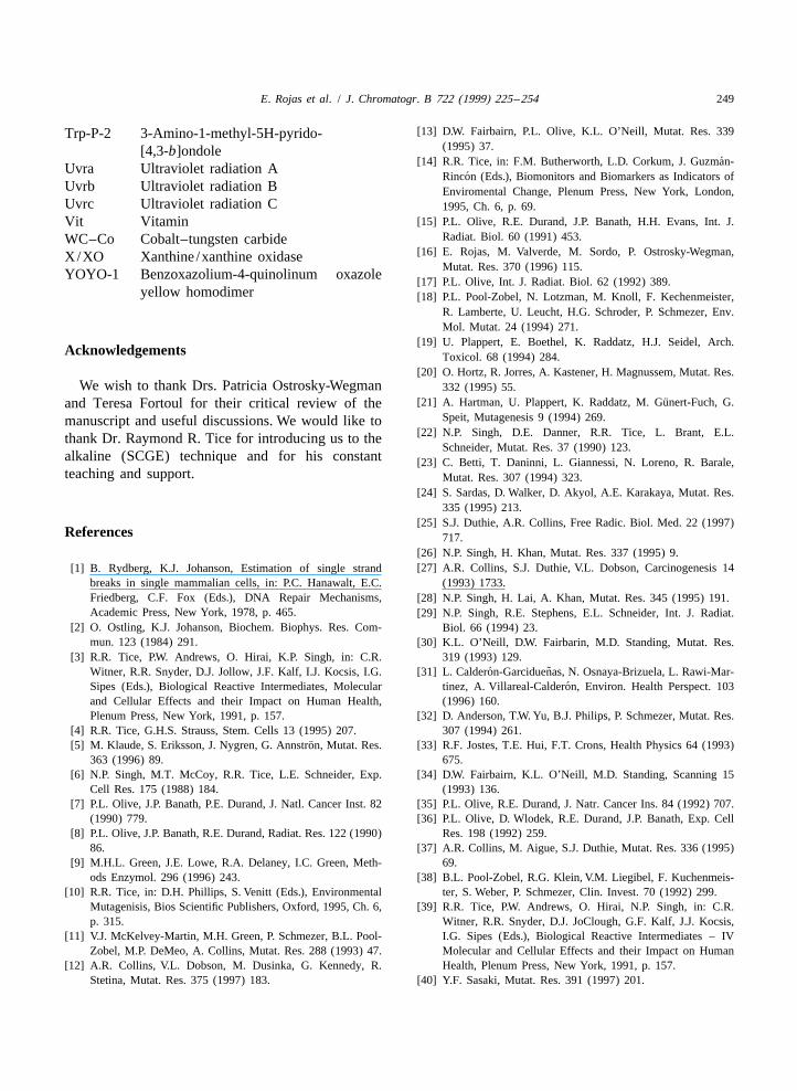
E. Rojas et al. / J. Chromatogr. B 722 (1999) 225 –254 249
[13] D.W. Fairbairn, P.L. Olive, K.L. O’Neill, Mutat. Res. 339Trp-P-2 3-Amino-1-methyl-5H-pyrido-(1995) 37.[4,3-b]ondole
´[14] R.R. Tice, in: F.M. Butherworth, L.D. Corkum, J. Guzman-Uvra Ultraviolet radiation A´Rincon (Eds.), Biomonitors and Biomarkers as Indicators of
Uvrb Ultraviolet radiation B Enviromental Change, Plenum Press, New York, London,Uvrc Ultraviolet radiation C 1995, Ch. 6, p. 69.
[15] P.L. Olive, R.E. Durand, J.P. Banath, H.H. Evans, Int. J.Vit VitaminRadiat. Biol. 60 (1991) 453.WC–Co Cobalt–tungsten carbide
[16] E. Rojas, M. Valverde, M. Sordo, P. Ostrosky-Wegman,X/XO Xanthine /xanthine oxidaseMutat. Res. 370 (1996) 115.
YOYO-1 Benzoxazolium-4-quinolinum oxazole[17] P.L. Olive, Int. J. Radiat. Biol. 62 (1992) 389.
yellow homodimer [18] P.L. Pool-Zobel, N. Lotzman, M. Knoll, F. Kechenmeister,R. Lamberte, U. Leucht, H.G. Schroder, P. Schmezer, Env.Mol. Mutat. 24 (1994) 271.
[19] U. Plappert, E. Boethel, K. Raddatz, H.J. Seidel, Arch.Acknowledgements Toxicol. 68 (1994) 284.
[20] O. Hortz, R. Jorres, A. Kastener, H. Magnussem, Mutat. Res.We wish to thank Drs. Patricia Ostrosky-Wegman 332 (1995) 55.
¨[21] A. Hartman, U. Plappert, K. Raddatz, M. Gunert-Fuch, G.and Teresa Fortoul for their critical review of theSpeit, Mutagenesis 9 (1994) 269.manuscript and useful discussions. We would like to
[22] N.P. Singh, D.E. Danner, R.R. Tice, L. Brant, E.L.thank Dr. Raymond R. Tice for introducing us to theSchneider, Mutat. Res. 37 (1990) 123.
alkaline (SCGE) technique and for his constant [23] C. Betti, T. Daninni, L. Giannessi, N. Loreno, R. Barale,teaching and support. Mutat. Res. 307 (1994) 323.
[24] S. Sardas, D. Walker, D. Akyol, A.E. Karakaya, Mutat. Res.335 (1995) 213.
[25] S.J. Duthie, A.R. Collins, Free Radic. Biol. Med. 22 (1997)References 717.
[26] N.P. Singh, H. Khan, Mutat. Res. 337 (1995) 9.[27] A.R. Collins, S.J. Duthie, V.L. Dobson, Carcinogenesis 14[1] B. Rydberg, K.J. Johanson, Estimation of single strand
breaks in single mammalian cells, in: P.C. Hanawalt, E.C. (1993) 1733.Friedberg, C.F. Fox (Eds.), DNA Repair Mechanisms, [28] N.P. Singh, H. Lai, A. Khan, Mutat. Res. 345 (1995) 191.Academic Press, New York, 1978, p. 465. [29] N.P. Singh, R.E. Stephens, E.L. Schneider, Int. J. Radiat.
[2] O. Ostling, K.J. Johanson, Biochem. Biophys. Res. Com- Biol. 66 (1994) 23.mun. 123 (1984) 291. [30] K.L. O’Neill, D.W. Fairbarin, M.D. Standing, Mutat. Res.
[3] R.R. Tice, P.W. Andrews, O. Hirai, K.P. Singh, in: C.R. 319 (1993) 129.˜´Witner, R.R. Snyder, D.J. Jollow, J.F. Kalf, I.J. Kocsis, I.G. [31] L. Calderon-Garciduenas, N. Osnaya-Brizuela, L. Rawi-Mar-
´Sipes (Eds.), Biological Reactive Intermediates, Molecular tinez, A. Villareal-Calderon, Environ. Health Perspect. 103and Cellular Effects and their Impact on Human Health, (1996) 160.Plenum Press, New York, 1991, p. 157. [32] D. Anderson, T.W. Yu, B.J. Philips, P. Schmezer, Mutat. Res.
[4] R.R. Tice, G.H.S. Strauss, Stem. Cells 13 (1995) 207. 307 (1994) 261.¨[5] M. Klaude, S. Eriksson, J. Nygren, G. Annstron, Mutat. Res. [33] R.F. Jostes, T.E. Hui, F.T. Crons, Health Physics 64 (1993)
363 (1996) 89. 675.[6] N.P. Singh, M.T. McCoy, R.R. Tice, L.E. Schneider, Exp. [34] D.W. Fairbairn, K.L. O’Neill, M.D. Standing, Scanning 15
Cell Res. 175 (1988) 184. (1993) 136.[7] P.L. Olive, J.P. Banath, P.E. Durand, J. Natl. Cancer Inst. 82 [35] P.L. Olive, R.E. Durand, J. Natr. Cancer Ins. 84 (1992) 707.
(1990) 779. [36] P.L. Olive, D. Wlodek, R.E. Durand, J.P. Banath, Exp. Cell[8] P.L. Olive, J.P. Banath, R.E. Durand, Radiat. Res. 122 (1990) Res. 198 (1992) 259.
86. [37] A.R. Collins, M. Aigue, S.J. Duthie, Mutat. Res. 336 (1995)[9] M.H.L. Green, J.E. Lowe, R.A. Delaney, I.C. Green, Meth- 69.
ods Enzymol. 296 (1996) 243. [38] B.L. Pool-Zobel, R.G. Klein, V.M. Liegibel, F. Kuchenmeis-[10] R.R. Tice, in: D.H. Phillips, S. Venitt (Eds.), Environmental ter, S. Weber, P. Schmezer, Clin. Invest. 70 (1992) 299.
Mutagenisis, Bios Scientific Publishers, Oxford, 1995, Ch. 6, [39] R.R. Tice, P.W. Andrews, O. Hirai, N.P. Singh, in: C.R.p. 315. Witner, R.R. Snyder, D.J. JoClough, G.F. Kalf, J.J. Kocsis,
[11] V.J. McKelvey-Martin, M.H. Green, P. Schmezer, B.L. Pool- I.G. Sipes (Eds.), Biological Reactive Intermediates – IVZobel, M.P. DeMeo, A. Collins, Mutat. Res. 288 (1993) 47. Molecular and Cellular Effects and their Impact on Human
[12] A.R. Collins, V.L. Dobson, M. Dusinka, G. Kennedy, R. Health, Plenum Press, New York, 1991, p. 157.Stetina, Mutat. Res. 375 (1997) 183. [40] Y.F. Sasaki, Mutat. Res. 391 (1997) 201.
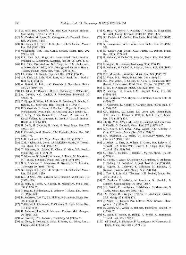
250 E. Rojas et al. / J. Chromatogr. B 722 (1999) 225 –254
[41] O. Hirai, P.W. Andrews, R.R. Tice, C.H. Nauman, Environ. [71] O. Holz, R. Jorres, A. Kastner, T. Krause, H. Magnussen,Mol. Mutag. 17(S19) (1991) 31. Int. Arch. Occup. Environ. Health 67 (1995) 305.
[42] M. DeMeo, M. Laget, M. Castegnero, G. Dumenil, Mutat. [72] S.J. Duthie, A.R. Collins, Free Radic. Biol. Med. 22 (1997)Res. 260 (1991) 295. 717.
[43] N.P. Singh, R.R. Tice, R.E. Stephens, E.L. Schneider, Mutat. [73] M. Panayiotidis, A.R. Collins, Free Radic. Res. 27 (1997)Res. 252 (1991) 289. 533.
[44] Vijayalaxmi, R.R. Tice, G.H.S. Strauss, Mutat. Res., 292 [74] S.J. Duthie, A.R. Collins, G.G. Duthie, V.L. Dobson, Mutat.(1993) 123. Res. 393 (1997) 223.
¨[45] R.R. Tice, N.P. Singh, 6th International Conf. Environ. [75] B. Hellman, H. Vaghef, B. Bostrom, Mutat. Res. 336 (1995)Mutagen. S., Melbourne, Australia, Feb. 21–26 1993, p. 41. 123.
[46] R.R. Tice, P.W. Andrew, N.P. Singh, in: B.M. Sutherland, [76] H. Vaghef, B. Hellman, Toxicology 96 (1995) 19.A.D. Woodhead (Eds.), DNA Damage and Repair in Human [77] B. Hellman, H. Vaghef, B. Bostrom, Mutat. Res. 336 (1995)Tissues, Plenum Press, New York, 1990, p. 291. 13.
´[47] P.L. Olive, J.P. Banath, Exp. Cell Res. 221 (1995) 19. [78] D.K. Monteith, J. Vanstone, Mutat. Res. 345 (1995) 79.[48] C.R. Kent, J.J. Eady, G.M. Ross, G.G. Steel, Int. J. Radiat. [79] M. Vrzoc, M.L. Petras, Mutat. Res. 381 (1997) 31.
Biol. 67 (1995) 55. [80] B.L. Pool-Zobel, C. Guigas, R. Klein, C. Neudecker, H.W.[49] A. DeWith, G. Leitz, K.O. Greulich, J. Photochem. Photo- Renner, P. Schmetzer, Food Chem. Toxicol. 31 (1993) 271.
biol. 24 (1994) 47. [81] S. Taj, B. Nagarajan, Mutat. Res. 322 (1994) 45.[50] P.L. Olive, J.P. Banath, C.D. Fjell, Cytometry 16 (1994) 305. [82] P. Schmezer, C. Eckert, U.M. Liegibel, Mutat. Res. 307[51] A. DeWith, K.O. Geulich, J. Photochem. Photobiol. 30 (1994) 495.
(1995) 71. [83] D.W. Fairbairn, W.A. Reyes, K.L. O’Neill, Cancer Lett. 81[52] C. Bjorge, R. Wiger, J.A. Holme, G. Brunborg, T. Scholz, E. (1994) 67.
Dybing, E.J. Soderlund, Rep. Toxicol. 10 (1996) 51. [84] T. Kasamatsu, K. Konda, Y. Kawazoe, Biol. Pharm. Bull. 19[53] K.O. Greulich, E. Bauer, U. Fiedler, C. Hoyer, K. Koemi, S. (1996) 632.
Monajenbashi, Proc. Spie. Int. Opt. Eng. 2629 (1996) 62. [85] C.A. Delaney, I.C. Green, J.E. Lowe, J.M. Cunningham,[54] T. Leroy, P. Van Hummelen, D. Arnard, P. Castelain, M. A.R. Butler, L. Renton, Y. D’Costa, M.H.L. Green, Mutat.
Kirsch-Volders, R. Lauwerys, D. Lison, J. Toxicol. Environ. Res. 375 (1997) 137.´Health 47 (1996) 409. [86] J.L. Re, M.P. DeMeo, M. Laget, H. Guiraud, M. Castegnaro,
[55] H. Cerda, H. Delincee, H. Haine, H. Rupp, Mutat. Res. 375 P. Vanelle, G. Dumenil, Mutat. Res. 375 (1997) 147.(1997) 167. [87] M.H. Green, L.E. Lowe, A.P.W. Waugh, K.E. Aldridge, J.
[56] E. Visvardis, A.M. Tassiou, S.M. Piperakis, Mutat. Res. 383 Cole, C.F. Arlett, Mutat. Res. 316 (1994) 91.(1997) 71. [88] S.F. Sweetman, J.J. Strain, V.J. McKelvey-Martin, Nutr.
[57] M.H. Lankinen, J.A. Vilpo, Mutat. Res. 373 (1997) 31. Cancer 27 (1997) 122.[58] C.M. Hughes, S.E. Lewis, V.J. McKelvey-Martin, W. Thomp- [89] J. Ashby, L. Krer, A. Wilson, T. Green, P.A. Lefevre, H.
son, Mutat. Res. 374 (1997) 261. Tinwell, G.A. Willis, W.F. Heydens, M. Clapp, Hum. Exp.[59] Y. Miyamae, K. Zaizen, K. Ohara, Y. Mine, Y.F. Sasaki, Toxicol. 15 (1996) 702.
Mutat. Res. 393 (1997) 99. [90] G. Ribas, G. Frenzilli, R. Barale, R. Marcos, Mutat. Res. 344[60] Y. Miyamae, K. Iwasaki, W. Kinae, S. Tsuda, M. Murakami, (1995) 41.
M. Tanaka, Y. Sasaki, Mutat. Res. 393 (1997) 107. [91] C. Bjorge, R. Wiger, J.A. Holme, G. Brunborg, R. Andersen,[61] G.G. Afanesev, T. Iwanenko, M. Kruszeuski, Y. Pelevina, E. Dybing, E.J. Soderlund, Reprod. Toxicol. 9 (1995) 461.
Tsitologiia 39 (1998) 74073. [92] J. Nygren, B. Cedervall, S. Eriksoson, M. Dusinka, A.[62] N.P. Singh, R.R. Tice, R.E. Stephens, E.L. Schneider, Mutat. Kolman, Environ. Mol. Mutag. 24 (1994) 161.
Res. 252 (1991) 289. [93] J. Tuo, S. Loft, M.S. Thomsen, H.E. Pouken, Mutat. Res.[63] K.L. O’Neill, D.W. Fairbairn, M.D. Staiding, Mutat. Res. 319 368 (1996) 213.
(1993) 129. [94] T. Bartlova, P. Vodkcka, K. Peterkova, K. Heminki, B.[64] O. Holz, R. Jorres, A. Kastner, H. Magnussen, Mutat. Res. Lambert, Carcinogenesis 16 (1995) 2357.
332 (1995) 55. [95] Y.F. Sasaki, F. Izumiyama, E. Nishidate, N. Matsusaka, S.[65] Y. Higami, I. Shimokawa, T. Okimoto, T. Ikeda, Lab. Invest. Tsuda, Mutat. Res. 391 (1997) 201.
71 (1994) 650. [96] J.M. Plewa, D.E. Wagner, T.W. Yu, D. Anderson, Environ.[66] D. Anderson, T.W. Yu, B.J. Phillips, P. Schmezer, Mutat. Res. Mol. Mutag. 26 (1995) 171.
307 (1994) 261. [97] J. Ashby, H. Tinwell, P.A. Lefevre, M.A. Browne, Muta-[67] Y. Higami, I. Shimokawa, T. Okimoto, T. Ikeda, Mutat. Res. genesis 10 (1995) 85.
316 (1994) 59. [98] H. Vaghef, A.C. Wisen, B. Hellman, Pharmacol. Toxicol. 78[68] D. Anderson, T.W. Yu, P. Schmezer, Environ. Mol. Mutagen. (1996) 37.
26 (1995) 305. [99] G. Speit, S. Hanelt, R. Helbig, A. Seidel, A. Hartmman,[69] A. Sweeney, P.T. Tomkins, Teratology 51 (1995) 30. Toxicol. Lett. 88 (1996) 91.[70] A. Chreg, B. Keeling, B. Gilks, S. Porter, P.L. Olive, Am. J. [100] Y.F. Sasaki, E. Nishidate, F. Uzumiyama, N. Matsusaka, S.
Physiol. 268 (1995) 832. Tsuda, Mutat. Res. 391 (1997) 215.
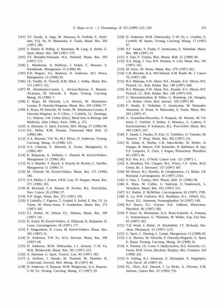
E. Rojas et al. / J. Chromatogr. B 722 (1999) 225 –254 251
[101] Y.F. Sasaki, A. Saga, M. Akasawa, K. Yoshida, E. Nishi- [129] D. Anderson, M.M. Dobrzynska, T.-W. Yu, L. Gaudini, E.date, Y.Q. Su, N. Matsusaka, S. Tsuda, Mutat. Res. 395 Cordelli, M. Spano, Teratog. Carcinog. Mutag. 17 (1997)(1997) 189. 97.
[102] S. Hanelt, R. Helbig, A. Hartmann, M. Lang, A. Seidel, G. [130] Y.F. Sasaki, S. Tsuda, F. Izumiyama, E. Nishidate, Mutat.Speit, Mutat. Res. 390 (1997) 179. Res. 388 (1997) 33.
[103] S.Y. Brendler-Schwaab, B.A. Herbold, Mutat. Res. 393 [131] S. Sato, Y. Tomita, Biol. Pharm. Bull. 21 (1998) 90.(1997) 175. [132] X.S. Deng, J. Tuo, H.E. Poulsen, S. Loft, Mutat. Res. 391
[104] L. Henderson, A. Wolfreys, J. Fedyk, C. Bourner, S. (1997) 165.Windebank, Mutagenesis 13 (1998) 89. [133] M. Vrzoc, M. Petras, Mutat. Res. 379 (1997) 263.
[105] E.D. Wagner, S.L. Rayburn, D. Anderson, M.J. Plewa, [134] C.B. Brezden, R.A. McClelland, A.M. Rauth, Br. J. CancerMutagenesis 13 (1998) 81. 76 (1997) 180.
[106] J.E. Yendle, H. Tinwell, B.M. Elliot, J. Ashby, Mutat. Res. [135] R.S. Malyapa, E.W. Ahern, W.L. Straube, E.G. Moros, W.F.375 (1997) 125. Pickard, J.L. Roti, Radiat. Res. 148 (1997) 608.
[107] M. Altamirano-Lozano, L. Alvarez-Barrera, F. Basurto- [136] R.S. Malyapa, E.W. Ahem, W.L. Straube, E.G. Moros, W.F.Alcantara, M. Valverde, E. Rojas, Teratog. Carcinog. Pickard, J.L. Roti, Radiat. Res. 148 (1997) 618.Mutag. 16 (1996) 7. [137] U. Marsteinstredent, R. Wiber, G. Brunborg, J.K. Hongslo,
[108] E. Rojas, M. Valverde, L.A. Herrera, M. Altamirano- J.A. Holme, Chem. Biol. Interact. 106 (1997) 89.Lozano, P. Ostrosky-Wegman, Mutat. Res. 359 (1996) 77. [138] F. Sasaki, E. Nishidate, F. Izumiyama, M. Watanabe-
[109] E. Rojas, M. Valverde, M. Sordo, M. Altamirano-Lozano, P. Akanuma, N. Kinae, N. Matsusaka, S. Tsuda, Mutat. Res.Ostrosky-Wegman, in: P. Cllery, J. Corbella, J.L. Domingo, 393 (1997) 47.J.C. Ethiene, J.M. Llobet (Eds.), Metal Ions in Biology and [139] G. Scassellati-Sforzolini, P. Pasquini, M. Moretti, M. Vil-Medicine, John Libbey, Paris, 1996, p. 375. larini, C. Fatifoni, P. Dolara, S. Monarca, G. Caderni, F.
[110] A. Hartman, G. Speit, Environ. Mol. Mutag. 27 (1996) 98. Kuchenmeister, P. Schmezer, B.L. Pool-Zobel, Mutat. Res.[111] D.L. Miller, R.M. Thomas, Ultrasound Med. Biol. 22 390 (1997) 207.
(1996) 681. [140] F. Sasaki, I. Fusako, N. Emi, O. Toshihiro, O. Tomoko, M.[112] A.A. Basaran, T.W. Yu, M.J. Plewa, D. Anderson, Teratog. Naonori, T. Shuji, Mutat. Res. 392 (1997) 251.
Carcinog. Mutag. 16 (1996) 125. [141] W. Adam, K. Mielke, C.R. Saha-Moller, M. Moller, H.[113] A.A. Chetelat, S. Albertini, E. Gocke, Mutagenesis 11 Stopper, R. Huterer, F.W. Schneider, D. Ballmaier, B. Epe,
(1996) 497. F.F. Gasparro, X. Chen, J. Kagan, Photochem. Photobiol.[114] K. Belpaeme, K. Delbeke, L. Zhuand, M. Kirsch-Volders, 66 (1997) 46.
Mutagenesis 11 (1996) 383. [142] B.S. Poe, K.L. O’Neill, Cancer Lett. 121 (1997) 1.[115] W. U Mueller, T. Bauch, A. Wojcik, W. Bocker, C. Streffer, [143] A. Woodons, P.H. Clingen, M.L. Priece, C.F. Arlett, M.H.
Mutagenesis 11 (1996) 57. Green, Br. J. Dermatol. 137 (1997) 687.[116] M. Tafazoli, M. Kirsch-Volders, Mutat. Res. 371 (1996) [144] M. Venturi, R.J. Hambly, B. Glinghammar, J.J. Rafter, I.R.
185. Rowland, Carcinogenesis 18 (1997) 2353.[117] S.O. Muller, I. Eckert, I.W.K. Lutz, H. Stopper, Mutat. Res. [145] S. Sato, I. Tomita, Biol. Pharm. Bull. 21 (1996) 90.
371 (1996) 165. [146] A. Maes, M. Collier, U. VanGorp, S. Vandoninck, L.[118] R. Rowland, C.A. Bearne, R. Fischer, B.L. Pool-Zobel, Verscheve, Mutat. Res. 193 (1997) 151.
Nutr. Cancer 26 (1996) 37. [147] S.J. Duthie, P. McMillan, Carcinogenesis 18 (1997) 1709.´[119] N.P. Singh, Mutat. Res. 375 (1997) 195. [148] X. Lu, D.W. Fairbairn, W.S. Bradshaw, K.L. ONeill, D.L.
[120] P. Lebailly, C.Vigreux, T. Godard, F. Sichel, E. Bar, J.Y. Le Ewert, D.L. Simmons, Prostanglandins 54 (1997) 549.Talaer, M. Henry-Amar, P. Gauduchon, Mutat. Res. 375 [149] H.J. Reavy, N.J. Trayner, N.K. Gibbons, Photochem.(1997) 205. Photobiol. 66 (1997) 368.
[121] S.J. Duthie, W. Johson, V.L. Dobson, Mutat. Res. 390 [150] P. Esser, K. Heimmann, K.O. Bratz-Schmidt, A. Fontana,(1997) 141. U. Schraermayer, G. Thumann, M. Weller, Exp. Eye Res.
[122] D. Anard, M. Kirsch-Volders, A. Elhajouji, K. Belpaeme, D. 65 (1997) 365.Lison, Carcinogenesis 18 (1997) 177. [151] T.H. Ward, J. Butler, H. Shanbakhti, J.T. Richards, Bio-
[123] F. Vangoethem, D. Lison, M. Kirsch-Volders, Mutat. Res. chem. Pharmacol. 53 (1997) 1115.392 (1997) 31. [152] G. Speit, C. Dennog, L. Lampi, Mutagenesis 13 (1998) 85.
[124] D. Anderson, T.W. Yu, M.A. Browne, Mutat. Res. 390 [153] L.A. Herrera, M. Valverde, P. Ostrosky-Wegman, G. Speit,(1997) 69. E. Rojas, Teratog. Carcinog. Mutag. 18 (1998) 41.
[125] D. Anderson, M.M. Dobrzynka, L.I. Jackson, T.-W. Yu, [154] S. Thomas, J.E. Lowe, V. Hadjivassilou, R.G. Knowles, I.C.M.H. Brinkworth, Mutat. Res. 391 (1997) 233. Green, M.H. Green, Biochem. Biophys. Res. Commun. 243
[126] A. Hartman, G. Speit, Toxicol. Lett. 90 (1997) 183. (1998) 241.[127] A. Kolman, I. Spinak, M. Naslund, M. Dusinka, B. [155] O. Ostling, K.J. Johanson, E. Biomquist, E. Hagelquist,
Cedervaall, Environ. Mol. Mutag. 30 (1997) 40. Acta Oncol. 26 (1987) 45.[128] D. Anderson, N. Basaran, M.M. Malgorzata, A.A. Basaran, [156] P.L. Olive, R.E. Durand, J. Le Riche, A. Olivotto, S.M.
T.-W. Yu, Teratog. Carcinog. Mutag. 17 (1997) 29. Jackson, Cancer Res. 53 (1993) 733.
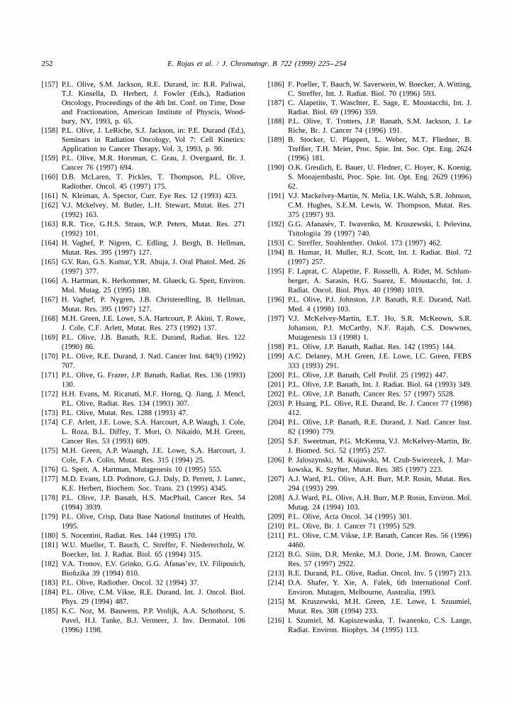
252 E. Rojas et al. / J. Chromatogr. B 722 (1999) 225 –254
[157] P.L. Olive, S.M. Jackson, R.E. Durand, in: B.R. Paliwai, [186] F. Poeller, T. Bauch, W. Saverwein, W. Boecker, A. Witting,T.J. Kinsella, D. Herbert, J. Fowler (Eds.), Radiation C. Streffer, Int. J. Radiat. Biol. 70 (1996) 593.Oncology, Proceedings of the 4th Int. Conf. on Time, Dose [187] C. Alapetite, T. Waschter, E. Sage, E. Moustacchi, Int. J.and Fractionation, American Institute of Physcis, Wood- Radiat. Biol. 69 (1996) 359.bury, NY, 1993, p. 65. [188] P.L. Olive, T. Trotters, J.P. Banath, S.M. Jackson, J. Le
[158] P.L. Olive, J. LeRiche, S.J. Jackson, in: P.E. Durand (Ed.), Riche, Br. J. Cancer 74 (1996) 191.Seminars in Radiation Oncology, Vol 7: Cell Kinetics: [189] B. Stocker, U. Plappert, L. Weber, M.T. Fliedner, B.Application to Cancer Therapy, Vol. 3, 1993, p. 90. Treffier, T.H. Meier, Proc. Spie. Int. Soc. Opt. Eng. 2624
[159] P.L. Olive, M.R. Horsman, C. Grau, J. Overgaard, Br. J. (1996) 181.Cancer 76 (1997) 694. [190] O.K. Greulich, E. Bauer, U. Fledner, C. Hoyer, K. Koenig,
[160] D.B. McLaren, T. Pickles, T. Thompson, P.L. Olive, S. Monajembashi, Proc. Spie. Int. Opt. Eng. 2629 (1996)Radiother. Oncol. 45 (1997) 175. 62.
[161] N. Kleiman, A. Spector, Curr. Eye Res. 12 (1993) 423. [191] V.J. Mackelvey-Martin, N. Melia, I.K. Walsh, S.R. Johnson,[162] V.J. Mckelvey, M. Butler, L.H. Stewart, Mutat. Res. 271 C.M. Hughes, S.E.M. Lewis, W. Thompson, Mutat. Res.
(1992) 163. 375 (1997) 93.´[163] R.R. Tice, G.H.S. Straus, W.P. Peters, Mutat. Res. 271 [192] G.G. Afanasev, T. Iwavenko, M. Kruszewski, I. Pelevina,
(1992) 101. Tsitologiia 39 (1997) 740.[164] H. Vaghef, P. Nigren, C. Edling, J. Bergh, B. Hellman, [193] C. Streffer, Strahlenther. Onkol. 173 (1997) 462.
Mutat. Res. 395 (1997) 127. [194] B. Humar, H. Muller, R.J. Scott, Int. J. Radiat. Biol. 72[165] G.V. Rao, G.S. Kumar, Y.R. Ahuja, J. Oral Phatol. Med. 26 (1997) 257.
(1997) 377. [195] F. Laprat, C. Alapetite, F. Rosselli, A. Ridet, M. Schlum-[166] A. Hartman, K. Herkommer, M. Glueck, G. Speit, Environ. berger, A. Sarasin, H.G. Suarez, E. Moustacchi, Int. J.
Mol. Mutag. 25 (1995) 180. Radiat. Oncol. Biol. Phys. 40 (1998) 1019.[167] H. Vaghef, P. Nygren, J.B. Christeredling, B. Hellman, [196] P.L. Olive, P.J. Johnston, J.P. Banath, R.E. Durand, Natl.
Mutat. Res. 395 (1997) 127. Med. 4 (1998) 103.[168] M.H. Green, J.E. Lowe, S.A. Hartcourt, P. Akini, T. Rowe, [197] V.J. McKelvey-Martin, E.T. Ho, S.R. McKeown, S.R.
J. Cole, C.F. Arlett, Mutat. Res. 273 (1992) 137. Johanson, P.J. McCarthy, N.F. Rajab, C.S. Dowwnes,[169] P.L. Olive, J.B. Banath, R.E. Durand, Radiat. Res. 122 Mutagenesis 13 (1998) 1.
(1990) 86. [198] P.L. Olive, J.P. Banath, Radiat. Res. 142 (1995) 144.[170] P.L. Olive, R.E. Durand, J. Natl. Cancer Inst. 84(9) (1992) [199] A.C. Delaney, M.H. Green, J.E. Lowe, I.C. Green, FEBS
707. 333 (1993) 291.[171] P.L. Olive, G. Frazer, J.P. Banath, Radiat. Res. 136 (1993) [200] P.L. Olive, J.P. Banath, Cell Prolif. 25 (1992) 447.
130. [201] P.L. Olive, J.P. Banath, Int. J. Radiat. Biol. 64 (1993) 349.[172] H.H. Evans, M. Ricanati, M.F. Horng, Q. Jiang, J. Mencl, [202] P.L. Olive, J.P. Banath, Cancer Res. 57 (1997) 5528.
P.L. Olive, Radiat. Res. 134 (1993) 307. [203] P. Huang, P.L. Olive, R.E. Durand, Br. J. Cancer 77 (1998)[173] P.L. Olive, Mutat. Res. 1288 (1993) 47. 412.[174] C.F. Arlett, J.E. Lowe, S.A. Harcourt, A.P. Waugh, J. Cole, [204] P.L. Olive, J.P. Banath, R.E. Durand, J. Natl. Cancer Inst.
L. Roza, B.L. Diffey, T. Mori, O. Nikaido, M.H. Green, 82 (1990) 779.Cancer Res. 53 (1993) 609. [205] S.F. Sweetman, P.G. McKenna, V.J. McKelvey-Martin, Br.
[175] M.H. Green, A.P. Waungh, J.E. Lowe, S.A. Harcourt, J. J. Biomed. Sci. 52 (1995) 257.Cole, F.A. Colin, Mutat. Res. 315 (1994) 25. [206] P. Jaloszynski, M. Kujawski, M. Czub-Swierezek, J. Mar-
[176] G. Speit, A. Hartman, Mutagenesis 10 (1995) 555. kowska, K. Szyfter, Mutat. Res. 385 (1997) 223.[177] M.D. Evans, I.D. Podmore, G.J. Daly, D. Perrett, J. Lunec, [207] A.J. Ward, P.L. Olive, A.H. Burr, M.P. Rosin, Mutat. Res.
K.E. Herbert, Biochem. Soc. Trans. 23 (1995) 4345. 294 (1993) 299.[178] P.L. Olive, J.P. Banath, H.S. MacPhail, Cancer Res. 54 [208] A.J. Ward, P.L. Olive, A.H. Burr, M.P. Rosin, Environ. Mol.
(1994) 3939. Mutag. 24 (1994) 103.[179] P.L. Olive, Crisp, Data Base National Institutes of Health, [209] P.L. Olive, Acta Oncol. 34 (1995) 301.
1995. [210] P.L. Olive, Br. J. Cancer 71 (1995) 529.[180] S. Nocentini, Radiat. Res. 144 (1995) 170. [211] P.L. Olive, C.M. Vikse, J.P. Banath, Cancer Res. 56 (1996)[181] W.U. Mueller, T. Bauch, C. Streffer, F. Niederercholz, W. 4460.
Boecker, Int. J. Radiat. Biol. 65 (1994) 315. [212] B.G. Siim, D.R. Menke, M.J. Dorie, J.M. Brown, Cancer[182] V.A. Tronov, E.V. Grinko, G.G. Afanas’ev, I.V. Filipouich, Res. 57 (1997) 2922.
Biofizika 39 (1994) 810. [213] R.E. Durand, P.L. Olive, Radiat. Oncol. Inv. 5 (1997) 213.[183] P.L. Olive, Radiother. Oncol. 32 (1994) 37. [214] D.A. Shafer, Y. Xie, A. Falek, 6th International Conf.[184] P.L. Olive, C.M. Vikse, R.E. Durand, Int. J. Oncol. Biol. Environ. Mutagen, Melbourne, Australia, 1993.
Phys. 29 (1994) 487. [215] M. Kruszewski, M.H. Green, J.E. Lowe, I. Szuumiel,[185] K.C. Noz, M. Bauwens, P.P. Vrolijk, A.A. Schothorst, S. Mutat. Res. 308 (1994) 233.
Pavel, H.J. Tanke, B.J. Vermeer, J. Inv. Dermatol. 106 [216] I. Szumiel, M. Kapiszewaska, T. Iwanenko, C.S. Lange,(1996) 1198. Radiat. Environ. Biophys. 34 (1995) 113.
E. Rojas et al. / J. Chromatogr. B 722 (1999) 225 –254 253
[217] P.J. McCarthy, S.F. Sweetman, P.G. Mckenna, V.J. Mck- [247] Z.X. Zhuang, Y. Shen, H.M. Shen, V. Ng, C.N. Ong, Hum.elvey-Martin, Mutagenesis 12 (1997) 209. Exp. Toxicol. 15 (1996) 891.
[218] D.A. Shafer, Y. Wie, A. Falek, Environ. Mol. Mutag. 23 [248] D. Slamenova, A. Gadelova, L. Ruzekova, I. Chalupa, E.(1994) 37. Horvathova, T. Farkasova, E. Bozsakyova, R. Stetina,
[219] P.L. Olive, J.P. Banath, R.E. Durand, Mutat. Res. 375 Mutat. Res. 383 (1997) 243.(1997) 157. [249] D. Anderson, A. Dhawan, T.-W. Yu, M.J. Plewa, Mutat.
[220] A.D. Barnard, D.W. Fairbairn, K.L. O’Neill, T.L. Gage, Res. 370 (1996) 159.R.W. Sidwell, Antiviral Res. 28 (1995) 317. [250] C.M. Gedik, S.W.B. Ewen, A.R. Coolins, Int. J. Radiat.
[221] M. Jaiswal, G. Anoradha, N. Rajeswar, K.N. Raju, N. Biol. 62 (1992) 313.Belarishna, K.V. Rao, I.S. Prasad, S.N. Jair, Y.R. Ahuja, [251] M.H. Green, J.E. Lowe, S.A. Harcourt, P. Akinluyi, T.Med. Sci. Res. 22 (1994) 879. Rowe, J. Cole, A.V. Antey, C.F. Arlett, Mutat. Res. 273
[222] A. Udumundi, M. Jaiswal, N. Rajeswari, N. Desai, S. Jain, (1992) 137.N. Balakrishna, K.V. Rao, Y.R. Ahuja, Mutat. Res. 412 [252] P.W. Andrews, R.R. Tice, S.H. Naumann, Environ. Mol.(1998) 195. Mutag. 15 (1990) 6.
[223] R.A. Brooks, J. Winton, J. Cell Sci. 109 (1996) 2061. [253] A.R. Collins, S.J. Duthie, V.L. Dobdon, Carcinogenesis 14[224] D.L. Eizirik, C.A. Delaney, M.H. Green, J.M. Cunningham, (1993) 1733.
¨J.R. Thorpe, D.G. Pipileers, C. Hellerstromm, I.C. Green, [254] B. Nascimbeni, M.D. Phillips, D.K. Croom, P.W. Andrews,Mol. Cell. Endocrinol. 118 (1996) 71. R.R. Tice, Environ. Mol. Mutag. 17 (1991) 55.
[225] C.A. Delaney, D.L. Eizirik, Braz. J. Med. Biol. Res. 29 [255] D. Fairbairn, D. Meyers, K. O’Neill, Bull. Environ. Con-(1996) 569. tam. Toxicol. 52 (1994) 687.
[226] B.L. Pool-Zobel, C. Neudecker, I. Domizlaff, S.J.V. Schill- [256] L. Verschaeve, J. Gilles, J. Schoctors, R. Van Cleuven-inger, C. Rumney, M. Moretti, I. Vilarini, R. Scassellat, S. bergen, J. DeFrie, The single cell gel electrophoresisForzolini, I. Roland, Nutr. Cancer 26 (1996) 365. technique or comet test for monitoring dioxinpollution and
[227] B.L. Pool-Zobel, U. Leucht, Mutat. Res. 375 (1997) 105. effects, in: H. Fiedler, H. Frank, O. Hutzinger, W. Parzefall,[228] P. Golzer, C. Janzowski, B.L. Pool-Zobel, G. Eisenbrand, A. Riss, S. Safe (Eds.), Organohalogen Compounds II,
Chem. Res. Toxicol. 9 (1996) 1207. Federal Enviromental Agency, Austria, 1993, p. 213.[229] M.V. Hejmadi, S.R. Mckeown, O.P. Friery, I.A. McIntyre, [257] J. Salgovic, J. Gilles, L. Verschaeve, I. Kalina, Folia Biol.
L.M. Patterson, D.G. Hirst, Br. J. Cancer 73 (1996) 499. 42 (1996) 17.[230] H. Zheng, P.L. Olive, Cancer Res. 56 (1996) 2801. [258] S. Ralph, M. Petras, Environ. Mol. Mutag. 29 (1997) 418.[231] S. Dahouk, U. Plappert, H. Gerngross, C. Willy, Langen- [259] D.E. Nacci, S. Cayula, E. Jackim, Aquat. Toxicol. 35
becks Arch. Chir. 114 (1997) 601. (1996) 197.[232] H. Zheng, P.L. Olive, Int. J. Radiat. Biol. 71 (1997) 275. [260] M. De Boeck, M. Kirsch-Volders, Environ. Mol. Mutag. 30[233] W. Maruyama, M. Naoi, T. Kasamatsu, Y. Hashizume, T. (1997) 82.
Takahasi, K. Konda, P. Dostert, J. Neurochem. 69 (1997) [261] C. Betti, M. Nigro, Mar. Pollut. Bull. 32 (1996) 545.322. [262] R. Pandrangi, M. Petras, S. Ralph, M. Vrozoc, Environ.
[234] W. Maruyama, M.S. Benedetti, T. Takahashi, M. Naoi, Mol. Mutag. 26 (1995) 345.Neurosci. Lett. 232 (1997) 147. [263] L. Kreja, C. Selig, U. Plappert, W. Northdurft, Environ.
[235] R. Helbig, G. Speit, Mutat. Res. 377 (1997) 279. Mol. Mutag. 27 (1996) 39.[236] F.L. Martin, S. Venitt, P.L. Carmichael, C. Crofton-Sleigh, [264] L. Kreja, C. Selig, W. Northdurft, Mutat. Res. 359 (1996)
E.M. Stone, K.J. Cole, B.A. Gusternov, P.L. Grover, D.H. 63.Phillips, Carcinogenesis 18 (1997) 2299. [265] S. Ralph, M. Petras, R. Pandrangi, M. Vrzoc, Environ. Mol.
[237] L. Qiao, R. Hanif, E. Sphicas, S.J. Shiff, B. Rigas, Mutag. 28 (1996) 112.Biochem. Pharmacol. 55 (1997) 53. [266] G. Koppen, L. Verschaeve, Mutat. Res. 360 (1996) 193.
[238] A. Hoorens, D.G. Pipeleers, C.A. Delaney, D. Pavlovic, [267] C. Clements, S. Ralph, M. Petras, Environ. Mol. Mutag. 29D.L. Eizirik, Endocrinology 138 (1997) 2610. (1997) 277.
[239] S. Gutierrez, E. Carbonell, P. Galofre, A. Creus, R. Marcos, [268] J. Salagovic, J. Gillest, L. Vershaeve, I. Kalina, Folia Biol.Mutagenesis 13 (1998) 95. (Praha) 43 (1997) 79.
[240] P.L. Olive, D. Wlodek, J.P. Banath, Cancer Res. 51 (1991) [269] D.E. Nacci, Aquat. Toxicol. 35 (1996) 197.4671. [270] C. Betti, M. Nigro, Mar. Pollut. Bull. 32 (1996) 545.
[241] A.R. Collins, J.S. Duthie, L.V. Dobson, Int. J. Radiat. Biol. [271] A. Devaux, M. Pesone, G. Monod, Toxicol. In Vitro 1162 (1993) 313. (1997) 71.
[242] A.R. Collins, M. Aiguo, S.J. Duthie, Mutat. Res. 336 [272] M. De Boeck, M. Kirsch-Volders, Environ. Mol. Mutag. 30(1995) 69. (1997) 82.
[243] P.L. Olive, J.P. Banath, Mutat. Res. 294 (1993) 275. [273] M.H. Navarrete, P. Carrera, M. De Miguel, C. De la Torre,[244] Q. Hu, R.P. Hill, Radiat. Res. 146 (1996) 636. Mutat. Res. 389 (1997) 271.[245] M.H. Lankinen, C.M. Vilpo, J.A. Vilpo, Mutat. Res. 352 [274] P. Ostrosky-Wegman, M.E. Gonsebatt, Environ. Health
(1996) 31. Perspect. 104 (1996) 599.[246] P. Fortini, G. Raspaglio, M. Falchi, E. Dogliotti, Muta- [275] S. Sardas, D. Walker, D. Akyol, A.E. Karakaya, Mutat. Res.
genesis 11 (1996) 169. 335 (1995) 213.
254 E. Rojas et al. / J. Chromatogr. B 722 (1999) 225 –254
´ ´ ´[276] M. Betancourt, R. Ortiz, C. Gonzalez, P. Perez, L. Cortes, [290] M.Valverde, M.C. Lopez, I. Lopez, I. Sanchez, T.I. Fortoul,˜´L. Rodrıguez, L. Villasenor, Mutat. Res. 331 (1995) 65. P. Ostrosky-Wegman, E. Rojas, Environ. Mol. Mutag. 30
[277] A. Hartmann, A.M. Niess, M. Gruenert-Fuchs, B. Poch, G. (1997) 147.Speit, Mutat. Res. 346 (1995) 195. [291] R.J. Sram, K. Pdrazilova, J. Dejmek, G. Mrackova, T.
[278] D. Anderson, B.J. Phillip, T.-W. Yu, A.J. Edwards, R. Pilicik, Mutagnesis 13 (1998) 99.Ayesh, K.R. Butterworth, Environ. Mol. Mutag. 39 (1997) [292] B. Glinghammar, Am. J. Clin. Nutr. 66 (1997) 1277.161. [293] U. Plappert, B. Stocker, H. Fender, T. Fliedner, Environ.
[279] S.J. Duthie, M.A. Ross, A.R. Collins, Cancer Res. 56 Mol. Mutag. 30 (1997) 153.(1996) 1291. [294] A.R. Collins, M. Dusinska, M. Franklin, M. Somorovska,
[280] B.L. Pool-Zobel, A. Bub, H. Muller, I. Wollowski, G. H. Petrovska, S. Duthie, L. Fillio, M. Panayioditis, K.Rechkemmer, Carcinogenesis 18 (1997) 1847. Raslova, N. Vaughan, Environ. Mol. Mutag. 39 (1997) 139.
[281] P. Vodicka, T. Bastlova, L. Vodickova, K. Peterkova, B. [295] R.J. Hambly, C.J. Rumney, M. Cunninghame, J.M. Flet-Lambert, K. Hemminki, Carcinogenesis 16 (1995) 1473. cher, P.J. Rijken, I.R. Rowland, Carcinogenesis 18 (1997)
[282] C. Betti, T. Daninni, L. Giannessi, N. Lorieno, R. Barale, 1535.Mutat. Res. 343 (1995) 201. [296] S. Sardas, N. Aygun, A.E. Kaeakaya, Mutat. Res. 394
[283] C. Betti, T. Daninni, L. Giannessi, N. Lorieno, R. Barale, (1997) 153.Mutat. Res. 307 (1994) 323. [297] A. Hartmann, S. Pfuhler, C. Dennog, D. Germadnik, A.
[284] G. Frenzilli, C. Betti, T. Davini, M. Desideri, E. Fornai, L. Pilger, G. Speit, Free Radic. Biol. Med. 24 (1998) 245.Giannessi, F. Maggiorelli, P. Paoletti, R. Barale, Mutat. [298] G.M. Ross, T.J. McMillan, P. Wilcox, A.R. Collins, Mutat.Res. 375 (1997) 117. Res. 337 (1995) 57.
[285] C. Dennog, A. Hartmann, G. Frey, G. Speit, Mutagenesis [299] A. Uzawa, G. Suzuki, Y. Nakata, M. Akashi, H. Ohyama,11 (1996) 605. A. Akanuma, Radiat. Res. 137 (1994) 25.
[286] M. Wojewodzka, M. Kruszewski, I. Szumiel, Mutagenesis [300] G.H.S. Strauss, W.P. Peters, R.B. Everson, Mutat. Res. 30411 (1996) 593. (1994) 211.
[287] M. Moretti, G. Scassellati-Sforzolini, S. Monarca, M. [301] A.R. Collins, M. Dosinska, M. Franklin, M. Somorovska,Libraro, F. Donato, C. Leonardis, L. Perego, Environ. H. Petrouska, S. Duthie, L. Fillion, M. Panayiotidis, K.Health Perspect. 104 (1996) 543. Raslowa, N. Vaughan, Environ. Mol. Mutag. 30 (1997) 139.
[288] C. Andreoli, P. Leopardi, R. Crebelli, Mutat. Res. 377(1997) 95.
˜ ´[289] L. Calderon-Garciduenas, N. Osnaya, A. Rodrıguez-Al-´caraz, A. Villarreal-Calderon, Environ. Mol. Mutag. 30
(1997) 11.
Related Documents