Singh Sandeep Kumar et al. Int. Res. J. Pharm. 2013, 4 (4) Page 185 INTERNATIONAL RESEARCH JOURNAL OF PHARMACY www.irjponline.com ISSN 2230 – 8407 Research Article IN VIVO LETHAL AND CYTOTOXICITY ASSESSMENT OF TRICHOSANTHES CUCUMERINA Singh Sandeep Kumar* 1 , Prakash Veeru 1 , Kumar Arun 2 , Kumar Ranjit 2 , Ali Mohammad 2 1 Department of Biochemistry & Biochemical Engineering, Sam Higginbottom Institute of Agriculture, Technology and Sciences, Allahabad, Uttar Pradesh, India 2 Research Department, Mahavir Cancer Sansthan & Research Centre, Phulwari Sharif, Patna, Bihar, India Email: [email protected] Article Received on: 13/02/13 Revised on: 11/03/13 Approved for publication: 21/04/13 DOI: 10.7897/2230-8407.04436 IRJP is an official publication of Moksha Publishing House. Website: www.mokshaph.com © All rights reserved. ABSTRACT Trichosanthes Cucumerina is being used as food as well as traditional medicine for long days. The aim of the present study was to evaluate the lethal dose and cytotoxicity of ethanolic extract of Trichosanthes cucumerina fruits (TCFEE) and leaves (TCLEE). For lethal dose estimation graded doses ranging from 200- 2000mg/ kg body weight (BW) of both extracts were administered orally and observed mortality up to 72 h. In lethal toxicity study the LD50 value of TCFEE and TCLEE was found as 1400 mg/kg BW and 1300 mg/kg BW. The cytotoxicity study of both extracts was done by evaluation of comparative difference of hepatorenal biochemical parameters (SGOT, SGPT, ALP, Urea, Uric acid and Creatinine) and histopathological slides of liver and kidney of both normal control and treated rats. Results showed no significant difference (p>0.05) between biochemical parameters and no changes in anatomy of liver and kidney. Hence the results suggest that ethanolic extracts of T. cucumerina fruit and leave are quite safe and can be used in the treatment of the chronic diseases like diabetes without any toxicity. Keywords: Trichosanthes Cucumerina, LD50, Ethanolic extract, Cytotoxicity, Histopathological study, Charles foster strain rats. INTRODUCTION Herbal plants and their processed products are big concern of today’s modern pharmacological approach for remedial of several disease due to their medicinal properties and less side effects. Generally people of developing countries go to use of traditional medicine in primary medical problems. 1 But without the proper information about efficacy and safety of these traditional medicines sometimes it would be harmful. Cucurbetaceae family having a large no of vegetables plants used in daily diet of people. Trichosanthes cucumerina (ver. Snake tomato) is one such plant that is frequently being used as food as well as traditional medicine for long days. The plant is richly constituted with a series of chemical constituents like flavonoids, carotenoids, phenolic acids which makes the plant pharmacologically and therapeutically active. It has a prominent place in alternative systems of medicines due to its various pharmacological activities like antidiabetic, hepatoprotective, cytotoxic, anti inflammatory, larvicidal effects. 2 Fruit is regarded as anthelmintic, purgative, vomitive. 3 Diabetic patients are advised to consume young fruits as it is having low sugar and excellent sources of fibres, vitamins, minerals and proteins. 4 Leaf is cardiotonic, antipyretic, antiperiodic, useful for intestinal worms and its juice rubbed over the liver in remittent fever 5 , skin disease, biliousness, emetic, externally applied over bald patches of alopecia 6 . Its ethanolic extract showed antiovulatory activity in female albino rats 7 while methanolic extracts showed antibacterial activity. 8 Although its traditional use and extensive phytopharmacological studies are known but the toxicity profile, especially of its ethanolic extracts, has not been yet explored. The present investigation was therefore carried out to study the lethal dose and cytotoxicity of ethanolic extract of Trichosantes cucumerina fruits (TCFEE) and leaves (TCLEE). MATERIALS AND METHODS Plant Material Fresh fruits and leave of Trichosanthes cucumerina were collected from Allahabad district and authenticated by an Agronomist, Department of Agronomy, SHIATS, Allahabad, India. Preparation of ethanolic extract The Fresh fruits and leave of T. cucumerina were collected, washed, dried under shade and powdered into fine particles. The 50g powder was macerated in 600 ml of 95% ethanol at room temperature for 48 hours with occasional shaking at 8 hours. It was then filtered by Whatmann filter paper (size no.1) and the filtrate evaporated on rotary evaporator to concentrate in crude extract form at 40ºC. Reddish orange and dark green crude residues (8.33% w/v) were obtained. The liquid suspensions were made by using 4% (v/v) ethanolic solvent and kept in air tight bottle in a refrigerator until used. Phytochemical Screening Preliminary phytochemicals screening of the extracts were carried out using standard methods. 9 Experimental Animals Normal Charles foster strain rats of either sex weighing 200 ± 20 gm were used for whole study and maintained on standard pellet diet and water ad libitum. The temperature of housing environment was maintained at 25 ± 2 ˙C with 12 hours repeated light/dark cycle. The study was approved by the Institutional Animal Ethics Committee by Ethic No. IAEC/MCS/2011/12/03. Experimental design and Selection of doses Experimental rats were divided into 6 groups (4 rats/ group). In order to select optimum dose of TCFEE and TCLEE, different doses were designed as 200, 400, 800, 1200, 1600 and 2000 mg/kg BW respectively. Lethal toxicity study 10 was

Welcome message from author
This document is posted to help you gain knowledge. Please leave a comment to let me know what you think about it! Share it to your friends and learn new things together.
Transcript

Singh Sandeep Kumar et al. Int. Res. J. Pharm. 2013, 4 (4)
Page 185
INTERNATIONAL RESEARCH JOURNAL OF PHARMACY www.irjponline.com ISSN 2230 – 8407
Research Article
IN VIVO LETHAL AND CYTOTOXICITY ASSESSMENT OF TRICHOSANTHES CUCUMERINA
Singh Sandeep Kumar*1, Prakash Veeru1, Kumar Arun2, Kumar Ranjit2, Ali Mohammad2 1Department of Biochemistry & Biochemical Engineering, Sam Higginbottom Institute of Agriculture,
Technology and Sciences, Allahabad, Uttar Pradesh, India 2Research Department, Mahavir Cancer Sansthan & Research Centre, Phulwari Sharif, Patna, Bihar, India
Email: [email protected]
Article Received on: 13/02/13 Revised on: 11/03/13 Approved for publication: 21/04/13
DOI: 10.7897/2230-8407.04436 IRJP is an official publication of Moksha Publishing House. Website: www.mokshaph.com © All rights reserved. ABSTRACT Trichosanthes Cucumerina is being used as food as well as traditional medicine for long days. The aim of the present study was to evaluate the lethal dose and cytotoxicity of ethanolic extract of Trichosanthes cucumerina fruits (TCFEE) and leaves (TCLEE). For lethal dose estimation graded doses ranging from 200-2000mg/ kg body weight (BW) of both extracts were administered orally and observed mortality up to 72 h. In lethal toxicity study the LD50 value of TCFEE and TCLEE was found as 1400 mg/kg BW and 1300 mg/kg BW. The cytotoxicity study of both extracts was done by evaluation of comparative difference of hepatorenal biochemical parameters (SGOT, SGPT, ALP, Urea, Uric acid and Creatinine) and histopathological slides of liver and kidney of both normal control and treated rats. Results showed no significant difference (p>0.05) between biochemical parameters and no changes in anatomy of liver and kidney. Hence the results suggest that ethanolic extracts of T. cucumerina fruit and leave are quite safe and can be used in the treatment of the chronic diseases like diabetes without any toxicity. Keywords: Trichosanthes Cucumerina, LD50, Ethanolic extract, Cytotoxicity, Histopathological study, Charles foster strain rats. INTRODUCTION Herbal plants and their processed products are big concern of today’s modern pharmacological approach for remedial of several disease due to their medicinal properties and less side effects. Generally people of developing countries go to use of traditional medicine in primary medical problems.1 But without the proper information about efficacy and safety of these traditional medicines sometimes it would be harmful. Cucurbetaceae family having a large no of vegetables plants used in daily diet of people. Trichosanthes cucumerina (ver. Snake tomato) is one such plant that is frequently being used as food as well as traditional medicine for long days. The plant is richly constituted with a series of chemical constituents like flavonoids, carotenoids, phenolic acids which makes the plant pharmacologically and therapeutically active. It has a prominent place in alternative systems of medicines due to its various pharmacological activities like antidiabetic, hepatoprotective, cytotoxic, anti inflammatory, larvicidal effects.2 Fruit is regarded as anthelmintic, purgative, vomitive.3 Diabetic patients are advised to consume young fruits as it is having low sugar and excellent sources of fibres, vitamins, minerals and proteins.4 Leaf is cardiotonic, antipyretic, antiperiodic, useful for intestinal worms and its juice rubbed over the liver in remittent fever5, skin disease, biliousness, emetic, externally applied over bald patches of alopecia6. Its ethanolic extract showed antiovulatory activity in female albino rats7 while methanolic extracts showed antibacterial activity.8 Although its traditional use and extensive phytopharmacological studies are known but the toxicity profile, especially of its ethanolic extracts, has not been yet explored. The present investigation was therefore carried out to study the lethal dose and cytotoxicity of ethanolic extract of Trichosantes cucumerina fruits (TCFEE) and leaves (TCLEE).
MATERIALS AND METHODS Plant Material Fresh fruits and leave of Trichosanthes cucumerina were collected from Allahabad district and authenticated by an Agronomist, Department of Agronomy, SHIATS, Allahabad, India. Preparation of ethanolic extract The Fresh fruits and leave of T. cucumerina were collected, washed, dried under shade and powdered into fine particles. The 50g powder was macerated in 600 ml of 95% ethanol at room temperature for 48 hours with occasional shaking at 8 hours. It was then filtered by Whatmann filter paper (size no.1) and the filtrate evaporated on rotary evaporator to concentrate in crude extract form at 40ºC. Reddish orange and dark green crude residues (8.33% w/v) were obtained. The liquid suspensions were made by using 4% (v/v) ethanolic solvent and kept in air tight bottle in a refrigerator until used. Phytochemical Screening Preliminary phytochemicals screening of the extracts were carried out using standard methods.9 Experimental Animals Normal Charles foster strain rats of either sex weighing 200 ± 20 gm were used for whole study and maintained on standard pellet diet and water ad libitum. The temperature of housing environment was maintained at 25 ± 2 ˙C with 12 hours repeated light/dark cycle. The study was approved by the Institutional Animal Ethics Committee by Ethic No. IAEC/MCS/2011/12/03. Experimental design and Selection of doses Experimental rats were divided into 6 groups (4 rats/ group). In order to select optimum dose of TCFEE and TCLEE, different doses were designed as 200, 400, 800, 1200, 1600 and 2000 mg/kg BW respectively. Lethal toxicity study10 was

Singh Sandeep Kumar et al. Int. Res. J. Pharm. 2013, 4 (4)
Page 186
carried out after 72 hours by oral feeding of these doses to respective group rats. In another experiment 8 rats are divided in 2 groups for each extract treatment. Group I having normal control healthy rats receiving normal saline for 7days while Group II having normal healthy rats receiving ethanolic extracts at a doses of 200 and 400 mg/kg BW for 7 days. The animals were observed for toxicity signs and analyzed for post treatment cytotoxicity by assay of liver and kidney function markers. Biochemical Analysis The effects of TCFEE and TCLEE on liver and kidney function markers were evaluated by estimation of SGOT11, SGPT11, ALP12, Urea13, Uric acid14 and Creatinine15 in plasma samples of normal control and extract treated rats. All analyses were performed by standard enzymatic methods using commercially available kit from CREST BIOSYSTEM LTD.
Histopathological Study All treated rats with selected dose of TCFEE and TCLEE were subjected to death after blood samples collection under guidelines of IAEC. The tissue samples of liver and kidney were obtained, washed and fixed in 10% neutral formalin and used for histopathological slide preparation as described by Lillie, R. D. (1965).16 Slides were observed using X200, X400 objectives and results recorded. Statistical Analysis All results of liver and kidney function are expressed as mean ± SD. Data were analysed by one way ANOVA and fallowed by Dunnett’s multiple comparison test using graph pad prism software (version 5.03) for windows (Graph Pad Software, San Diego, USA).
Table-1. Percentage mortalities in rats at different oral doses of Trichosanthes cucumerina fruit’s ethanolic extract for one week
Group No. of Animals Dose (mg/kg) No. of death % Mortality
1 4 200 0 0 2 4 400 0 0 3 4 800 1 25 4 4 1200 1 25 5 4 1600 2 50 6 4 2000 4 100
Table-2. Toxicity signs observed in rats at single oral doses of 200 and 400 mg/kg body weight of Trichosanthes cucumerina fruit’s ethanolic extract
for one week
Dose (mg/kg body weight)
Sign of toxicity Inappetence Depression Aggressiveness Respiratory distress Body weight loss Death
200 - - - - - - 400 - - - - - -
+ = present, - = absent
Table-3. Percentage mortalities in rats at different oral doses of Trichosanthes cucumerina leave’s ethanolic extract for one week
Group No. of Animals Dose (mg/kg) No. of death % Mortality 1 4 200 0 0 2 4 400 0 0 3 4 800 1 25 4 4 1200 2 50 5 4 1600 2 50 6 4 2000 4 100
Table-4. Toxicity signs observed in rats at single oral doses of 200 and 400 mg/kg body weight of Trichosanthes cucumerina leave’s ethanolic extract
for one week
Dose (mg/kg body weight)
Sign of toxicity Inappetence Depression Aggressiveness Respiratory distress Body weight loss Death
200 - - - - - - 400 - - - - - -
+ = present, - = absent
Table-5. Liver function markers in plasma of TCFEE and TCLEE treated rats at the dose of 400 mg/ kg bw. (values are mean ± sd)
Liver function markers Group I Group II TCFEE treated TCLEE treated
SGPT (U/ml) 72.87 ± 1.44 73.25 ± 0.87* 71.75 ± 0.87* SGOT (U/ml) 86.25 ± 0.72 83.50 ± 1.22* 85.00 ± 1.77*
ALP (KA units/ml) 9.25 ± 0.69 8.83 ± 0.58* 8.74 ± 0.69*
Table-6. Kidney function markers in plasma of TCFEE and TCLEE treated rats at the dose of 400 mg/ kg bw. (values are mean ± sd)
Kidney function markers Group I Group II TCFEE treated TCLEE treated
Urea (mg/dl) 33.86 ± 0.67 30.20 ± 1.73** 32.86 ± 1.56* Uric acid (mg/dl) 6.43 ± 0.38 6.02 ± 0.20* 6.23 ± 0.37* Creatinine (mg/dl) 1.19 ± 0.24 1.25 ± 0.29* 1.25 ± 0.24*
*means are significantly not different as p > 0.05 for treated group II vs. control group I; **means are significantly different as p < 0.05 for treated group II vs. control group I
Singh Sandeep Kumar et al. Int. Res. J. Pharm. 2013, 4 (4)
Page 187
=
TCFEE
TCLEE0
200400600800
100012001400
Plant extractsLD
50 (m
g/kg
BW
) ora
l
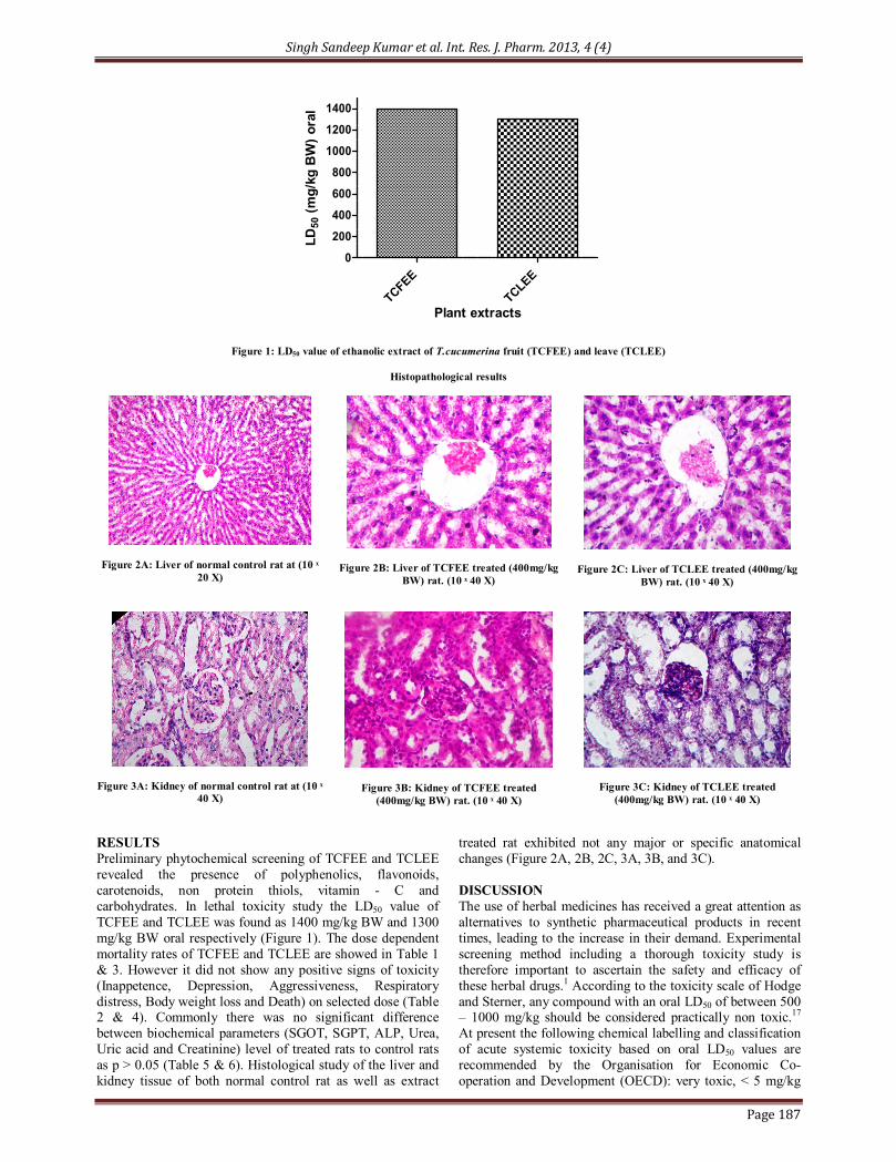
Figure 1: LD50 value of ethanolic extract of T.cucumerina fruit (TCFEE) and leave (TCLEE)
Histopathological results
Figure 2A: Liver of normal control rat at (10 ˣ 20 X)
Figure 2B: Liver of TCFEE treated (400mg/kg BW) rat. (10 ˣ 40 X)
Figure 2C: Liver of TCLEE treated (400mg/kg BW) rat. (10 ˣ 40 X)
Figure 3A: Kidney of normal control rat at (10 ˣ 40 X)
Figure 3B: Kidney of TCFEE treated (400mg/kg BW) rat. (10 ˣ 40 X)
Figure 3C: Kidney of TCLEE treated (400mg/kg BW) rat. (10 ˣ 40 X)
RESULTS Preliminary phytochemical screening of TCFEE and TCLEE revealed the presence of polyphenolics, flavonoids, carotenoids, non protein thiols, vitamin - C and carbohydrates. In lethal toxicity study the LD50 value of TCFEE and TCLEE was found as 1400 mg/kg BW and 1300 mg/kg BW oral respectively (Figure 1). The dose dependent mortality rates of TCFEE and TCLEE are showed in Table 1 & 3. However it did not show any positive signs of toxicity (Inappetence, Depression, Aggressiveness, Respiratory distress, Body weight loss and Death) on selected dose (Table 2 & 4). Commonly there was no significant difference between biochemical parameters (SGOT, SGPT, ALP, Urea, Uric acid and Creatinine) level of treated rats to control rats as p > 0.05 (Table 5 & 6). Histological study of the liver and kidney tissue of both normal control rat as well as extract
treated rat exhibited not any major or specific anatomical changes (Figure 2A, 2B, 2C, 3A, 3B, and 3C). DISCUSSION The use of herbal medicines has received a great attention as alternatives to synthetic pharmaceutical products in recent times, leading to the increase in their demand. Experimental screening method including a thorough toxicity study is therefore important to ascertain the safety and efficacy of these herbal drugs.1 According to the toxicity scale of Hodge and Sterner, any compound with an oral LD50 of between 500 – 1000 mg/kg should be considered practically non toxic.17 At present the following chemical labelling and classification of acute systemic toxicity based on oral LD50 values are recommended by the Organisation for Economic Co-operation and Development (OECD): very toxic, < 5 mg/kg

Singh Sandeep Kumar et al. Int. Res. J. Pharm. 2013, 4 (4)
Page 188
body weight; toxic, > 5 < 50 mg/kg; harmful, >50<500 mg/kg; and no label, > 500 < 2000 mg/kg. The concept of basal cytotoxicity states that the mechanisms of action of most toxic chemicals are related to biochemical processes expressed in all cells.18,19 In present study the LD50 value of ethanolic extract of T. cucumerina fruits (TCFEE) and leave (TCLEE) was found as 1400 mg/kg BW and 1300 mg/kg BW oral respectively which exhibits its safe oral dose for therapeutical uses. Increase in the level of SGPT, SGOT, ALP, Urea, Uric acid and Creatinine reflects the structural and functional dysfunction of liver cell and kidney cell membrane or cell rupture or inflammation.1 Biochemical analysis of present study revealed not any significant difference in level of liver and kidney function markers of normal control versus extracts treated rats as p > 0.05. It
shows safety profile of extracts on liver and kidney function. Histological observations correlate our results showing the normal cellular anatomy of liver and kidney in the treated group of animals. CONCLUSION It is therefore concluded that the dose below LD50 value of ethanolic extracts of fruits and leave of T. cucumerina may safely use for therapeutically benefits. ACKNOWLEDGEMENT The author is grateful to Dr. Veeru Prakash, Department of Biochemistry & Biochemical Engineering, SHIATS, Allahabad, Uttarpradesh, India and Dr. Arun Kumar, Research Department, Mahavir Cancer Sansthan, Patna, Bihar, India for their guidance to carry out present work.
REFERENCES 1. Saha P, Mazumder UK, Haldar PK. Acute and Subchronic Toxicity of
C. maxima Aerial Parts. International Journal of Research in Pharmaceutical and Biomedical Sciences 2011; Vol. 2 (2): 634-639.
2. Sandhya S, Vinod KR, Chandra Sekhar J, Aradhana R, Vamshi Sarath Nath. An updated review on Tricosanthes cucumerina. International Journal of Pharmaceutical Sciences Review and Research 2010; Volume 1, Issue 2, p 56-60.
3. Jeffrey C. Cucurbitaceae. In: Stoffers, A.L. and J.C. Lindeman, eds., Flora of Surinam. Vol. 5, Part 1. Leiden: E.J. Brill. 1984, p. 457-518.
4. Ujwal Deshmukh. Fruits for Diabetics. [cited 2013 may 20]. Available from: http://www.buzzle.com/articles/fruits-for-diabetics.html
5. Kirtikar, Basu. Indian Medicinal Plants, Sri Satguru Publications, 2000; New Delhi p. (5): 1545-1547.
6. Devendra NK, Seetharam YN. Trichosanthes cucumerina L.: an Ayurvedic Medicinal plant. Pharmakine 2011; Vol. III, Issue I.
7. Devendra NK, Malashetty VB, Seetharam YN, Suresh P, Patil SB. Effect of ethanol extract of whole plant of Trichosanthes cucumerina var. Cucumerina L. on gonadotropins, ovarian follicular kinetics and estrous cycle for screening of antifertility activity in albino rats. Int. J. Morphology 2009; 27(1): 173-182.
8. Reddy LJ, Jose B, Anjana JC, Ruveena TN. Evaluation of antibacterial activity of Trichosanthes cucumerina and Cassia didymobotrya fres. Leaves, Int J Pharm Pharm Sci 2010; Vol 2, Suppl 4, p 153-155.
9. Mahadevan A, Sridhar. Secondary metabolites. In: Methods in Physiological Plant Pathology. 3rd ed. Sivakami Publications, 1986, Chennai, p 9-11.
10. Aliu YO, Nwude N. LD50 determination. In: Veterinary Pharmacology and Toxicology Experiments. 1st ed. ABU Press, 1982, p 104–110.
11. Reitman S, Frankel S. Determination of aspartat and alanine
aminotransferase in blood serum and tissue homogenate, Amer. J. Clin. Path. 1957; vol. 28, p-56.
12. Kind PRN, King EJ. Estimation of Plasma Phosphatase by Determination of Hydrolysed Phenol with Amino-antipyrine, J. Clin. Path. 1954; 7(4): 322-326. http://dx.doi.org/10.1136/jcp.7.4.322
13. Fawcett JK, Scott JE. A rapid and precise method for the determination of urea. J Clin Pathol. 1960; 13(2): 156–159. http://dx.doi.org/10.1136 /jcp.13.2.156
14. Fossati P, Prencipe L, Berti G. Use of 3, 5-Dichloro- 2-hydroxybenzenesulfonic Acid/4-Ami nophenazone Chromogenic System in Direct Enzymic Assay of Uric Acid in Serum and Urine. J Clin. Chem. 1980; 26(2): 227-231.
15. Bonsnes RW, Taussky HH. On the colorimetric determination of Creatinine by the Jaffe reaction. J Biol Chem. 1945; 158: 581-91.
16. Lillie RD. Tissue sectioning and staining. In: Histopathological technique and practical histochemistry. 3rd ed. Published by Blakistar Division of Mc. Graw Hill, 1965, 10-New York, Toronto, London.
17. Agaie BM, Onyeyili PA, Muhammad BY, Ladan MJ. Acute toxicity effects of the aqueous leaf extract of Anogeissus leiocarpus in rats. African Journal of Biotechnology 2007; Vol. 6 (7), p. 886-889.
18. OECD. OECD Guideline for Testing of Chemicals, No 423: Acute Oral Toxicity-Acute Toxic Class Method. Paris:Organisation for Economic Co-operation and Development, 1996.
19. Walum E. Acute oral toxicity. Environmental Health Perspect 1998; 106: 497 – 503. http://dx.doi.org/10.1289/ehp.98106497
Cite this article as: Singh Sandeep Kumar, Prakash Veeru, Kumar Arun, Kumar Ranjit, Ali Mohammad. In vivo lethal and cytotoxicity assessment of Trichosanthes cucumerina. Int. Res. J. Pharm. 2013; 4(4):185-188
Source of support: Nil, Conflict of interest: None Declared
Related Documents
![Int J Ayu Pharm Chemijapc.com/volume5-third-issue/V5-I3-20-P-116-129.pdfInt J Ayu Pharm Chem 2016Vol. 5 Issue 3 116 [e ISSN 2350-0204] Int J Ayu Pharm Chem REVIEW ARTICLE ...](https://static.cupdf.com/doc/110x72/5ab8132f7f8b9a684c8c625a/int-j-ayu-pharm-j-ayu-pharm-chem-2016vol-5-issue-3-116-e-issn-2350-0204-int-j.jpg)


![Int J Ayu Pharm Chem - ijapc.comijapc.com/volume10-second-issue/MNAPC-V10-I1-(v10-i1-48)-p-1-10.pdf · Int J Ayu Pharm Chem 2019 Vol. 10 Issue 2 1 [e ISSN 2350-0204] Int J Ayu Pharm](https://static.cupdf.com/doc/110x72/5d5fd2bc88c993c53c8bbf54/int-j-ayu-pharm-chem-ijapc-v10-i1-48-p-1-10pdf-int-j-ayu-pharm-chem-2019.jpg)
![Int J Ayu Pharm Chem - International Journal of …ijapc.com/volume4-first-issue/v4-i1-13-95-106.pdfInt J Ayu Pharm Chem 2015 Vol. 4 Issue 1 95 [e ISSN 2350-0204] Int J Ayu Pharm Chem](https://static.cupdf.com/doc/110x72/5ae508277f8b9acc268b7af0/int-j-ayu-pharm-chem-international-journal-of-ijapccomvolume4-first-issuev4-i1-13-95-106pdfint.jpg)