Simultaneous single-molecule mapping of protein-DNA interactions and DNA methylation by MAPit Carolina E. Pardo, Russell P. Darst, Nancy H. Nabilsi, Amber L. Delmas, and Michael P. Kladde * Department of Biochemistry and Molecular Biology and UF Shands Cancer Center Program in Cancer Genetics, Epigenetics and Tumor Virology, University of Florida College of Medicine, 2033 Mowry Road, Box 103633, Gainesville, Florida 32610-3633, USA, Telephone: (352) 273-8541, Fax: (352) 273-8299 Abstract Sites of protein binding to DNA are inferred from footprints or spans of protection against a probing reagent. In most protocols, sites of accessibility to a probe are detected by mapping breaks in DNA strands. As discussed in this unit, such methods obscure molecular heterogeneity by averaging cuts at a given site over all DNA strands in sample population. DNA methyltransferase accessibility protocol for individual templates (MAPit), an alternative method described in this unit, localizes protein-DNA interactions by probing with cytosine-modifying DNA methyltransferases followed by bisulfite sequencing. Sequencing individual DNA products after amplification of bisulfite-converted sequences permits assignment of the methylation status of every enzyme target site along a single DNA strand. Use of the GC-methylating enzyme M.CviPI allows simultaneous mapping of chromatin accessibility and endogenous CpG methylation. MAPit is therefore the only footprinting method that can detect subpopulations of molecules with distinct patterns of protein binding or chromatin architecture, and correlate them directly with the occurrence of endogenous methylation. Additional advantages of MAPit methylation footprinting as well as considerations for experimental design and potential sources of error are discussed. Keywords Chromatin; Nucleosomes; DNA methylation; DNA methyltransferases; Footprinting; Single- molecule analysis * To whom correspondence should be addressed. [email protected]. Key References Kladde et al., 1996. See above. First demonstration of the utility of M.SssI for detection of nucleosome position and transcription factor binding. Fatemi et al., 2005. See above. Jessen et al., 2006. See above. First demonstrations of the use of C-5 DNMTs in single-molecule footprinting. Kilgore et al., 2007. See above. First documented use of MAPit with M.CviPI, yielding simultaneous detection of chromatin accessibility and endogenous m 5 CG at the single-molecule level. Pardo et al., 2010. See above. Development of MethylViewer program for rapid analysis of MAPit datasets. Internet Resources http://dna.leeds.ac.uk/methylviewer/ Site for download of MethylViewer program and detailed usage instructions. NIH Public Access Author Manuscript Curr Protoc Mol Biol. Author manuscript; available in PMC 2012 July 1. Published in final edited form as: Curr Protoc Mol Biol. 2011 July ; CHAPTER: Unit–21.22. doi:10.1002/0471142727.mb2122s95. NIH-PA Author Manuscript NIH-PA Author Manuscript NIH-PA Author Manuscript

Welcome message from author
This document is posted to help you gain knowledge. Please leave a comment to let me know what you think about it! Share it to your friends and learn new things together.
Transcript
Simultaneous single-molecule mapping of protein-DNAinteractions and DNA methylation by MAPit
Carolina E. Pardo, Russell P. Darst, Nancy H. Nabilsi, Amber L. Delmas, and Michael P.Kladde*
Department of Biochemistry and Molecular Biology and UF Shands Cancer Center Program inCancer Genetics, Epigenetics and Tumor Virology, University of Florida College of Medicine,2033 Mowry Road, Box 103633, Gainesville, Florida 32610-3633, USA, Telephone: (352)273-8541, Fax: (352) 273-8299
AbstractSites of protein binding to DNA are inferred from footprints or spans of protection against aprobing reagent. In most protocols, sites of accessibility to a probe are detected by mapping breaksin DNA strands. As discussed in this unit, such methods obscure molecular heterogeneity byaveraging cuts at a given site over all DNA strands in sample population. DNA methyltransferaseaccessibility protocol for individual templates (MAPit), an alternative method described in thisunit, localizes protein-DNA interactions by probing with cytosine-modifying DNAmethyltransferases followed by bisulfite sequencing. Sequencing individual DNA products afteramplification of bisulfite-converted sequences permits assignment of the methylation status ofevery enzyme target site along a single DNA strand. Use of the GC-methylating enzyme M.CviPIallows simultaneous mapping of chromatin accessibility and endogenous CpG methylation. MAPitis therefore the only footprinting method that can detect subpopulations of molecules with distinctpatterns of protein binding or chromatin architecture, and correlate them directly with theoccurrence of endogenous methylation. Additional advantages of MAPit methylation footprintingas well as considerations for experimental design and potential sources of error are discussed.
KeywordsChromatin; Nucleosomes; DNA methylation; DNA methyltransferases; Footprinting; Single-molecule analysis
*To whom correspondence should be addressed. [email protected] ReferencesKladde et al., 1996. See above.First demonstration of the utility of M.SssI for detection of nucleosome position and transcription factor binding.Fatemi et al., 2005. See above.Jessen et al., 2006. See above.First demonstrations of the use of C-5 DNMTs in single-molecule footprinting.Kilgore et al., 2007. See above.First documented use of MAPit with M.CviPI, yielding simultaneous detection of chromatin accessibility and endogenous m5CG atthe single-molecule level.Pardo et al., 2010. See above.Development of MethylViewer program for rapid analysis of MAPit datasets.Internet Resourceshttp://dna.leeds.ac.uk/methylviewer/Site for download of MethylViewer program and detailed usage instructions.
NIH Public AccessAuthor ManuscriptCurr Protoc Mol Biol. Author manuscript; available in PMC 2012 July 1.
Published in final edited form as:Curr Protoc Mol Biol. 2011 July ; CHAPTER: Unit–21.22. doi:10.1002/0471142727.mb2122s95.
NIH
-PA Author Manuscript
NIH
-PA Author Manuscript
NIH
-PA Author Manuscript
INTRODUCTIONCrucial to a complete understanding of any biological function of DNA is the footprinting ormapping protein-DNA interactions at high resolution. Formerly, footprinting methods haverelied on assaying accessibility of sites in DNA to probing reagents that result in DNAcleavage, such as nucleases (e.g., DNase I and micrococcal nuclease; UNITS 12.4 and 21.1,respectively) or chemicals (e.g. dimethylsulfate). Locations of protein binding to DNA areinferred by comparing sites of protection against damage in the absence and presence of theputative DNA-binding protein. Such conditions can be set up either in vitro or in vivo, e.g.wild-type cells versus the same cells with expression knock-down or bearing a null mutationin the gene of the factor of interest. While conventional footprinting methods have provenenormously informative, they are subject to several theoretical and practical limitations, asdiscussed in the Commentary. In particular, as only a single DNA break can be mapped perDNA molecule, conditions must approach limiting or single-hit kinetic levels of cleavage.By mapping cleavages at a given site over all molecules in a sample, the inherentcomplexity of protein-DNA interactions in biological systems is obscured by populationaveraging (Pondugula and Kladde, 2008).
These problems are overcome by probing protein-DNA interactions with DNAmethyltransferases (DNMTs) that modify cytosine followed by bisulfite sequencing(Frommer et al., 1992; Clark et al., 1994) (UNIT 7.9), termed MAPit (Jessen et al., 2006;Kilgore et al., 2007; Pardo et al., 2009; Pondugula and Kladde, 2008). A key advantage ofMAPit over other techniques is that it reports the methylation status (i.e. accessibility versusprotection) of every C residue along one strand of individually-cloned and sequenced DNAmolecules. This provides a single-molecule, non-averaged view of protein-DNA interactionsthat permits correlation between different footprints in a region within a sample population.
Methyl-5-C (m5C), occurring predominantly at CpG sites (CG hereafter), is a common post-replicative DNA modification in vertebrates and many other organisms. A DNMT withdifferent sequence specificity is needed to fully leverage MAPit. To this end, we cloned andcharacterized M.CviPI, an enzyme that methylates GC sites at C-5 (Xu et al., 1998a).Bisulfite sequencing of mammalian chromatin probed with M.CviPI therefore allows themethylation status of both CG and GC sites to be determined along a single DNA strand(Kilgore et al., 2007).
This unit describes a MAPit protocol using M.CviPI for simultaneous mapping of bothendogenous cytosine methylation and protein-DNA interactions in cultured mammaliancells.
BASIC PROTOCOL PROBING MAMMALIAN NUCLEAR CHROMATIN WITHDNMTs
The three basic steps of MAPit are: (1) delivery of a suitable C-modifying DNMT to probeaccessibility of DNA or chromatin; (2) bisulfite sequencing (UNIT 7.9), including bisulfiteconversion of isolated and denatured DNA, PCR amplification of deaminated DNA, andsequencing cloned individual molecules from the PCR amplicon; and (3) assignment of themethylation status to each potential DNMT target sequence (Figure 1).
MAPit can be performed on organisms that lack detectable DNA methylation, such as thebudding yeast Saccharomyces cerevisiae (Proffitt et al., 1984). Readers interested in MAPitprobing of budding yeast chromatin are urged to consult any of several previously publisheddetailed protocols (Jessen et al., 2004; Hoose and Kladde, 2006; Kilgore et al., 2007; Pardoet al., 2009). In principle, a DNMT probe can be used that modifies cytosine in any
Pardo et al. Page 2
Curr Protoc Mol Biol. Author manuscript; available in PMC 2012 July 1.
NIH
-PA Author Manuscript
NIH
-PA Author Manuscript
NIH
-PA Author Manuscript
sequence context on one of several positions. This is because bisulfite sequencing detectsm5C as well as methyl-N4-C (mN4C) and hydroxymethyl-5-C (hm5C) (Kriaucionis andHeintz, 2009; Tahiliani et al., 2009; Huang et al., 2010). Therefore, in organisms lackingdetectable DNA methylation, the choice of DNMT need only consider the frequency anddistribution of target sites on the analyzed strand of the locus of interest. However, C-modifying DNMTs with short recognition specificities, such as the C-5 DNMTs M.SssI(CG; Renbaum et al., 1990) and M.CviPI (GC; Xu et al., 1998a), are the most useful probesfor MAPit analysis because they provide the highest footprinting resolution. An additionalconsideration of probe choice is that most genomic regions in vertebrate cells contain somem5CG, limiting the usefulness of the CG-modification enzyme M.SssI (Fatemi et al., 2005;Gal-Yam et al., 2006). For this reason, we previously identified, cloned, overexpressed, andcharacterized the GC probe M.CviPI (Xu et al., 1998a), which is now commerciallyavailable.
MaterialsMammalian cell lines cultured under appropriate experimental conditions
Trypsin-EDTA solution (see recipe; store up to 6 months at −20°C)
Phosphate buffered saline (PBS; store indefinitely at 4°C)
Cell resuspension buffer (see recipe)
Cell lysis buffer (see recipe)
Methylation buffer (see recipe)
Methylation stop buffer (see recipe)
Enzyme dilution buffer (see recipe)
DNMT storage buffer (see recipe; store indefinitely at −20°C)
1 M dithiothreitol (DTT; store in single-use aliquots at −20°C)
100 mM phenylmethylsulfonyl fluoride (PMSF; dissolved in absolute ethanol; store upto six months at −20°C)
0.4% (w/v) trypan blue solution (store indefinitely at room temperature)
32 mM S-adenosyl-L-methionine (SAM) (store in single-use aliquots at −80°C)
80 U/μl M.CviPI fused to maltose binding protein (MBP, New England Biolabs) orfused to glutathione-S-transferase (GST, Zymo Research Corp.); aliquot and store at−20°C in non-frost-free freezer; see recipe for dilutions)
20 mg/ml proteinase K (store at −20°C in non-frost-free freezer)
5 U/μl HotStar Taq (Qiagen) (store at −20°C in non-frost-free freezer)
Phenol chloroform solution (see recipe; store indefinitely at 4°C)
10.0 M ammonium acetate, pH 8.0
Absolute ethanol
0.1× TE buffer (see recipe; store indefinitely at room temperature)
70% (v/v) ethanol (see recipe; store indefinitely at room temperature)
NOTE: Reagents should be prepared in sterile disposable labware. Use only distilled H2O inall steps and solutions. Nuclei isolation and methylation buffers should be freshly preparedon the day of the experiment. DTT, PMSF and SAM should be added to solutions
Pardo et al. Page 3
Curr Protoc Mol Biol. Author manuscript; available in PMC 2012 July 1.
NIH
-PA Author Manuscript
NIH
-PA Author Manuscript
NIH
-PA Author Manuscript
immediately before use to avoid oxidation or hydrolysis. M.CviPI activity is very dependenton fresh addition of DTT.
Cell lines and growing conditions will vary according to the question being addressed andresearcher discretion. Cells should be cultured using standard tissue culture techniques underdesired experimental conditions until at least 1.5 × 10⁶ cells per experimental sample (e.g.DNMT dose) are obtained. A refrigerated centrifuge and microcentrifuge or one in a coldroom is recommended for isolation of nuclei.
Harvest cells1 Add an appropriate volume of trypsin-EDTA solution pre-warmed to 37°C to
remove cells from tissue culture plates or flasks. Incubate cells at roomtemperature until they detach from the growth surface.
The time needed for cell detachment varies from one cell line to another (~2–12min), and can be determined by visualization with a light microscope. Cells canalternatively be harvested by adding ice-cold PBS directly to plates and scrapinginto 50 ml conical tubes on ice. Skip this step and step 2 if cells have beengrown in suspension.
2 Add cell growth medium pre-warmed to 37°C (3× volume of trypsinizationsolution used in step 1) to terminate trypsinization.
Trypsin activity is inhibited by the protease inhibitor alpha-1-antitrypsin foundin fetal bovine serum.
3 Centrifuge for 5 min at 1,000 × g at 4°C to pellet cells. Carefully aspiratesupernatant.
4 Add 5 ml ice-cold PBS. Vortex gently and briefly to resuspend cell pellet andwash cells.
5 Centrifuge for 5 min at 1,000 × g at 4°C to pellet cells. Decant supernatant.
6 Resuspend cells with ice cold PBS to an approximate concentration of 106/mland keep cells on ice.
7 Mix an aliquot of 20 μl cell suspension with 20 μl 0.4% (w/v) trypan bluesolution. Pipette up-and-down several times to disperse cells and make the cellsuspension homogeneous.
8 Count the number of live cells that exclude trypan blue either manually with ahemocytometer or using an automated cell counting device.
9 Aliquot 1.1 × 10⁶ cells per experimental sample into pre-labeled 1.7 mlmicrocentrifuge tubes on ice.
Each DNMT probing reaction requires 106 cells. Starting with 1.1× 10⁶ cells perreaction (one reaction is one DNMT dose) allows for some loss duringpreparation of nuclei. We recommend setting up an untreated sample (0 UDNMT) and two concentrations of M.CviPI, therefore requiring 3.3 × 10⁶ cellsper experimental condition. In our experience, 30 and 100 U M.CviPI are goodstarting doses for either the M.CviPI-MBP or M.CviPI-GST reagents. Using twodifferent concentrations of enzyme, while not essential, allows one to assessdifferent degrees of chromatin accessibility and the extent of saturation ofmethylation by exogenously added M.CviPI at each GC site. The untreatedsample (0 U DNMT) serves as a background control to monitor non-conversionof C in GC sites by bisulfite and/or sequencing errors. The untreated sample also
Pardo et al. Page 4
Curr Protoc Mol Biol. Author manuscript; available in PMC 2012 July 1.
NIH
-PA Author Manuscript
NIH
-PA Author Manuscript
NIH
-PA Author Manuscript
shows the level of endogenous CG methylation in the sample before probing,which should be taken into account when inferring whether GCG sites werelikely methylated by endogenous DNMTs or exogenously-added by DNMTprobe.
10 Centrifuge for 5 min at 1,000 × g at 4°C in a microcentrifuge to pellet cells.
11 Add 200 μl ice-cold cell resuspension buffer per 1.1 × 10⁶ cells (i.e. add 600 μl,if 0, 30 and 100 U M.CviPI are used). Resuspend pellet by tapping tube gently.
NOTE: Isolating all nuclei for each experimental condition together in a singletube and aliquoting to separate tubes in step 17 ensures that the only variablewill be the DNMT concentration.
12 Centrifuge for 5 min at 1,000 × g at 4°C to pellet cells. Decant supernatant.
Isolate mammalian nuclei13 Resuspend cell pellet in 38.5 μl of ice cold cell lysis buffer per 1.1 × 10⁶ cells
(i.e. add 115.5 μl, if 0, 30 and 100 U M.CviPI are used). Incubate for 10 min at4°C to lyse cells.
Inclusion of the non-ionic detergent Nonidet P-40 in cell lysis buffer allows forcell membrane lysis while maintaining nuclear integrity. Nonidet P-40concentration and lysis time may need to be optimized for different cell types inorder to obtain complete cell lysis without disrupting integrity of the nuclearenvelope. To preserve nuclear structural integrity and native protein-DNAinteractions, all steps for nuclei preparation should be done at 4°C. Nucleishould be handled carefully as they are prone to lysis. Avoid pipetting of nucleiuntil step 17; instead, resuspend by gentle tapping of the tube with a finger.
14 Prepare ice cold methylation buffer while cells are undergoing lysis by mixingon ice:
60.5 μl ice-cold cell resuspension buffer
0.55 μl freshly-thawed 32 mM SAM.
NOTE: These volumes are per each sample containing about 106 nuclei. Makeenough extra solution to account for pipetting error. The methylation buffercontains 290 μM SAM, which will be diluted to a final concentration of 160 μMin the methylation reactions in step 20.
15 Add 61 μl ice-cold methylation buffer per 106 nuclei (i.e. add 183 μl, if 0, 30and 100 U M.CviPI are used) to dilute Nonidet P-40 concentration. Mix bygently tapping the tube.
Dilution of Nonidet P-40 detergent to 0.08% (v/v) in this step helps maintainnuclear integrity.
16 Check the structural integrity of the nuclei. Stain an aliquot of 2 μl nucleisolution by adding 2 μl of 0.4% (w/v) trypan blue solution in a separate tube.Mix by gently tapping the tube, incubate for 3 min at room temperature, andexamine nuclei by light microscopy.
Nuclei should stain blue as well as appear round and granular with no attachedcytoplasmic debris. Nuclei may swell slightly during isolation and subsequentmanipulations.
Pardo et al. Page 5
Curr Protoc Mol Biol. Author manuscript; available in PMC 2012 July 1.
NIH
-PA Author Manuscript
NIH
-PA Author Manuscript
NIH
-PA Author Manuscript
17 Aliquot 90 μl of nuclei resuspension containing 106 nuclei into 1.7 mlmicrocentrifuge tubes pre-labeled with each unit concentration of M.CviPI beingused.
Probe nuclear chromatin structure by methylation with exogenous M.CviPI18 On ice, freshly prepare M.CviPI dilutions for the methylation reaction.
For the 30 and 100 U M.CviPI samples, appropriate volumes of 3 U/μl and 10U/μl M.CviPI solution are needed, respectively. Best results are achieved bysetting up a dilution series to ensure that all samples are subjected to identicalconditions in parallel (i.e., salt and 160 μM SAM), with the DNMTconcentration being the only variable.
19 Pre-warm the nuclei dispensed in each tube by incubation for 5 min at 37°C. Atthe same time, pre-warm to 50°C a sufficient volume of 2× methylation stopbuffer (100 μl per methylation reaction plus some extra to allow for pipettingerror).
20 Staggering M.CviPI addition to each tube by 30 sec, add 10 μl of thecorresponding M.CviPI dilution to each pre-warmed sample. Pipette up-and-down gently to mix and methylate for 15 min at 37°C.
Staggered addition of enzyme and respective staggered termination ofmethylation in step 21, ensure that the incubation time with the chromatinprobing enzyme is held constant. The Nonidet P-40 detergent is diluted in thisstep to 0.07% (v/v), which is necessary for full activity of M.CviPI (andM.SssI). Parameters used during the chromatin probing reaction can be changedaccording to the requirements of the experiment. We recommend performing apilot experiment under the conditions described here. Time and enzymeconcentration can be adjusted accordingly (see Commentary).
21 Terminate each methylation reaction by adding 100 μl of 2× methylation stopbuffer pre-warmed to 50°C, corresponding to the staggering scheme used in step20. Vortex each sample immediately.
22 Add 1μl 20 mg/ml proteinase K to a final concentration of 100 μg/ml. Mix byinverting tubes and incubate overnight at 50°C.
Complete removal of protein from the DNA is necessary to achieve completedenaturation and hence bisulfite conversion (Warnecke et al., 2002). In ourexperience, incubation with proteinase K for at least 16 hr is required.
Purify mammalian genomic DNA23 Extract proteins from the genomic DNA solution from step 22 by adding 200 μl
(an equal volume) of phenol chloroform solution (UNIT 2.3). Vortex vigorouslyfor 30 sec at room temperature to obtain a homogeneous suspension.
24 Separate the aqueous and organic phases by centrifugation for 5 min at 14,000 ×g at room temperature in a microcentrifuge. Transfer the aqueous (top) phase toa new 1.7 ml microcentrifuge tube carefully avoiding transfer of denaturedprotein and SDS (white material located at the phase interface).
25 Add 1/4 volume of 10.0 M Ammonium acetate (i.e. 2.5 M final), and vortexbriefly to mix.
26 Add 2.5 volumes of absolute ethanol, mix thoroughly by gentle inversion.
Pardo et al. Page 6
Curr Protoc Mol Biol. Author manuscript; available in PMC 2012 July 1.
NIH
-PA Author Manuscript
NIH
-PA Author Manuscript
NIH
-PA Author Manuscript
At this point, samples can be stored indefinitely at −20°C. Overnight incubationat −20°C increases recovery of low concentrations of nucleic acid.
27 Centrifuge for 5 min at 14,000 × g at room temperature in a microcentrifuge topellet the nucleic acid.
28 Draw off supernatant carefully so as not to dislodge the nucleic acid pellet.
29 Add 0.4 ml 70% (v/v) ethanol. Vortex briefly to wash nucleic acid pellet.
30 Centrifuge for 5 min at 14,000 × g at room temperature in a microcentrifuge topellet nucleic acid.
30 Carefully draw off supernatant without disturbing the pellet and air dry pellet forapproximately 10 min.
31 Resuspend genomic DNA in 50 μl 0.1× TE.
Genomic DNA usually requires overnight incubation at 4°C to solubilizecompletely. Removal of RNA prior to bisulfite sequencing is not necessary.Samples can be stored at 4°C for many months or indefinitely at −20°C.
Bisulfite sequencing of mammalian DNAApproximately 5–7 μg DNA are recovered from each reaction containing 106 nuclei.Bisulfite sequencing, including bisulfite conversion of purified DNA, PCR amplification ofsequences of interest, cloning individual molecules from the PCR product, and sequencingcloned molecules, is performed as described in UNIT 7.9. Once clones of individualmolecules have been sequenced the data are analyzed by MethylViewer(http://dna.leeds.ac.uk/methylviewer/) (Pardo et al., 2010). This computer program canconcurrently score the methylation status of up to four user-defined sites either directly from*.ab1 sequencing files or from a FASTA file of sequences aligned with another program.For MAPit analysis of mammalian chromatin with M.CviPI, MethylViewer is used toconcurrently score methylation at CG and GC sites along each sequenced molecule, andexport publication-quality images. Other features, such as verification of bisulfite-conversion efficiency at non-CG and non-GC sequences can also be obtained. Occasionalsequences with conversion efficiencies of <97% are typically omitted from further analyses,but this is up to the discretion of the investigator.
SUPPORT PROTOCOLS VERIFICATION OF METHYLATION OF DNA BYM.CviPI
When using a new enzyme preparation, one may wish to determine activity before investingtime in sequencing and analysis of MAPit data. It is of course possible to methylate purifiedplasmids and test with various restriction enzymes. However, higher enzymatic activity isneeded to methylate chromatin. We have found it most convenient to assay activity bymethylation of nuclear DNA, using the actual experimental samples. To confirm that theDNMT used was active, we use one of two methods to screen for GC methylation, eitherquantitative methylation-sensitive restriction enzyme digestion (qMSRE) or methylation-specific PCR (MSP).
SUPPORT PROTOCOL IFor qMSRE, 20–250 ng of purified genomic DNA (not bisulfite treated) is subject todigestion with the methylation sensitive enzyme R.HaeIII. This enzyme can digestunmethylated GGCC sites but not GG-m5CC sites. A parallel “mock” reaction containing allreaction components except R.HaeIII (replaced with glycerol) is included for each sample.
Pardo et al. Page 7
Curr Protoc Mol Biol. Author manuscript; available in PMC 2012 July 1.
NIH
-PA Author Manuscript
NIH
-PA Author Manuscript
NIH
-PA Author Manuscript
DNA from the R.HaeIII-digested or mock reaction is then amplified by real-time PCR withprimers to a known open region containing a HaeIII site, such as the human GAPDHpromoter (primers TACTAGCGGTTTTACGGGCG andTCGAACAGGAGGAGCAGAGAGCGA). Results are normalized to each sample’s mockdigestion control and quantified using the ΔΔCT method to determine the levels ofprotection from R.HaeIII digestion achieved by each dose of DNMT.
SUPPORT PROTOCOL IIFor MSP, 20 ng of bisulfite-treated DNA is amplified with two sets of primers that targethuman long interspersed nucleotide elements (LINE-1). One primer pair amplifies GCunmethylated or “U” LINE (primer sequences AGGTATTGTTTTATTTGGGAAGTGT andCCTTACAATTTAATCTCAAACTACTATA) and the second pair amplifies GC-methylated or “M” LINE (primers CATTGCTTTATTTGGGAAGCGC andCTTGCAATTTAATCTCAAACTGCTATG) DNA. The product of each PCR reaction isvisualized on an agarose gel: the “M” product will be more abundant than the “U” product ifthe DNMT was active.
REAGENTS AND SOLUTIONSUse deionized, distilled water in all recipes and protocol steps. For common stock solutions,see APPENDIX 2; for suppliers, see APPENDIX 4.
Trypsin-EDTA solution0.25% (w/v) trypsin
2.21 mM ethylenediamine tetraacetic acid (EDTA)
Dissolved in Hank’s-balanced salt solution (HBSS) without sodium bicarbonate,calcium and magnesium. Tested for porcine parvovirus as the same solution is used toharvest cells for passaging. Any good quality commercially-available solution willsuffice.
Cell resuspension buffer20 mM HEPES, pH 7.5
70 mM NaCl
0.25 mM EDTA, pH 8.0
0.5 mM EGTA, pH 8.0
0.5% (v/v) glycerol
10 mM DTT (always add fresh immediately before use, store at −20°C)
0.25 mM PMSF (always add fresh immediately before use, store at −20°C)
Cell lysis buffer—Cell resuspension buffer + 0.19% (v/v) Nonidet P-40
Nuclei methylation buffer—Cell resuspension buffer + 290 μM SAM (always add SAMimmediately before use)
NOTE: The final concentration of SAM will be 160 μM in the methylation reaction.
2X Methylation stop buffer100 mM NaCl
Pardo et al. Page 8
Curr Protoc Mol Biol. Author manuscript; available in PMC 2012 July 1.
NIH
-PA Author Manuscript
NIH
-PA Author Manuscript
NIH
-PA Author Manuscript
10 mM EDTA, pH 8.0
1% (w/v) SDS
DNMT storage buffer (M.CviPI)15 mM Tris-HCl
200 mM NaCl
1 mM DTT
0.1 mM EDTA
200 μg/ml acetylated BSA (i.e. nuclease free)
50% (v/v) glycerol
pH 7.4 at 25°C
Enzyme dilution buffer (M.CviPI)—Dilute M.CviPI storage buffer by 8-fold (i.e. 1:7)with methylation buffer
M.CviPI dilutions—Immediately before use, on ice, prepare M.CviPI dilutions as follows:
1. Dilute 80 U/μl commercial stock of M.CviPI by 8-fold with ice-cold methylationbuffer to make 10 U/1 μl dilution.
2. Make a 3.33-fold serial dilution of the 10 U/μl dilution with enzyme dilution bufferto make the 3 U/μl dilution.
About 8-fold less activity is needed if wild-type M.CviPI (i.e. non-fusion protein) is used.Adjust dilutions accordingly if a different stock concentration of commercial M.CviPI isused.
Phenol chloroform solution—Mix molecular biology grade phenol equilibrated to pH8.0, chloroform, and isoamyl alcohol in the ratio of 25:24:1, respectively.
0.1× TE buffer1 mM Tris-HCl, pH 8.0 (APPENDIX 2)
0.1 mM Na2EDTA, pH 8.0
Autoclave and store indefinitely at room temperature.
70% (v/v) ethanol—Mix absolute ethanol and 0.1× TE buffer in the ratio of 37 ml:13 ml,respectively.
CAUTION: Flammable.
COMMENTARYBackground Information
Protein-DNA interactions play crucial roles in mediating all biological functions of DNA inevery organism. Eukaryotes package their DNA into chromatin comprising a protein contentof roughly half non-histone regulatory factors and half core histones. The fundamentalrepeating unit of eukaryotic chromosomes is the nucleosome core particle, composed of ahistone octamer (central histone tetramer (H3–H4)2 and H2A–H2B dimers) wrapped by aleft-handed superhelix consisting of 1.65 turns or 147 bp of DNA (Luger et al., 1997).
Pardo et al. Page 9
Curr Protoc Mol Biol. Author manuscript; available in PMC 2012 July 1.
NIH
-PA Author Manuscript
NIH
-PA Author Manuscript
NIH
-PA Author Manuscript
Individual nucleosomes are repeated at a distance characteristic for each eukaryotic species,i.e. separated by a modal length of histone-free linker DNA in bulk chromatin. In contrast, atthe single-molecule level, there can be considerable variation in linker length within a givenregion of chromatin. Nucleosomes are among the most stable protein-DNA interactions ineukaryotic chromosomes and act in concert with DNA-binding factors and other chromatin-associated factors to exert tight control of gene expression and other DNA functions(Kouzarides, 2007; Li et al., 2007).
In many eukaryotes, endogenous DNMTs post-replicatively modify the DNA component ofchromatin at the 5 position of the cytosine base ring. C-5 methylation (m5C) in vertebratesappears to occur exclusively at CG sites in most cell types, and plays essential roles indiverse aspects of vertebrate genome function (Bestor, 2000; Bird, 2002). These includerepression or silencing of transcription, embryonic development, genomic imprinting ofeither the paternal of maternal alleles of some genes, inactivation of one of two Xchromosomes in normal females of Eutherian mammals, and suppression of the mobility ofparasitic genetic elements, e.g. retrotransposons (Robertson and Wolffe, 2000; Bestor andBourc’his, 2004; Robertson, 2005; Goll and Bestor, 2005; McCabe et al., 2009). AberrantDNA methylation is frequently associated with human aging and diseases, such as cancer(Robertson, 2001; Bird, 2002; Jaenisch and Bird, 2003; Robertson, 2005; Feinberg et al.,2006; Jones and Baylin, 2007). In other cases, m5CG has been shown to activatetranscription when it blocks binding of proteins to DNA that exert transcriptional repression(Nabilsi et al., 2009; Lai et al., 2010; Wu et al., 2010). At lower levels, m5C is also presentat non-CG sites, CHG and CHH (H is either A, C or T), in undifferentiated humanembryonic stem cells (Kouidou et al., 2005; Grandjean et al., 2007; Latham et al., 2008;Lister et al., 2009; Hawkins et al., 2010; Laurent et al., 2010).
Chromatin structure is highly dynamic; nucleosomes are constantly being mobilized todifferent positions and/or are disassembled via the action of ATP-dependent chromatinremodelers, histone chaperones, or both (Längst and Becker, 2004; Saha et al., 2006; Clapierand Cairns, 2009). Nucleosome depletion at transcription start sites, for example, is oftendiagnostic of transcription initiation (Boeger et al., 2004; Korber et al., 2004; Mito et al.,2005; Jiang and Pugh, 2009). Although a hallmark of epigenetic m5CG is its heritabilityfrom one cell to another, DNA methylation is also dynamic. First, methylation is notprecisely maintained and thus modification of specific CG sites can fluctuate considerably.Second, cellular differentiation has recently been shown to involve oxidation of m5C tohydroxymethyl C (hm5C), which is subsequently removed by an as yet unknown mechanism(Tahiliani et al., 2009; Ito et al., 2010). In sum, dynamic changes in DNA methylation aswell as occupancy by nucleosomes and non-histone regulatory factors lead to considerableepigenetic heterogeneity in chromatin.
Detection of the diverse epigenetic signatures present at a given region of interest byconventional footprinting methods poses several challenges (Pondugula and Kladde, 2008).Most of these stem from the nature of mapping DNA breaks introduced by nucleases or bygenomic footprinting with chemical agents (e.g. dimethylsulfate). Limited digestion orchemical treatment of the footprinted sample is employed to achieve so-called single-hitkinetic levels of DNA cleavage, which are supposed to approximate a random Poissondistribution of cut sites. In practice, however, adherence to random Poisson statistics ishampered by biological complexity and non-randomness, especially when footprintedsamples are of cellular origin. Second, even when single-hit digestion is achieved, only onecut site proximal to a radiolabeled DNA end, hybridizing primer, etc. can be mapped persingle DNA molecule. Therefore, the position of nucleosomes or DNA-bound factorsrelative to one another on the same molecule, which requires mapping >1 cleavage site,cannot be determined. Third, a population of cut DNA molecules must be analyzed to
Pardo et al. Page 10
Curr Protoc Mol Biol. Author manuscript; available in PMC 2012 July 1.
NIH
-PA Author Manuscript
NIH
-PA Author Manuscript
NIH
-PA Author Manuscript
identify a footprint. Such population-ensemble methods average away differences betweenmolecules and thus obscure molecular heterogeneity.
These problems are overcome by MAPit; single-molecule detection of protein-DNAinteractions by exogenously-supplied C-methylating DNMTs (Jessen et al., 2006; Kilgore etal., 2007; Pardo et al., 2009; Pardo et al., 2010). MAPit builds on a large body of earlierstudies by us and others demonstrating the usefulness of DNMTs as chromatin structuralprobes. To our knowledge, the earliest hints that chromatin structure might affectsusceptibility to a DNMT were the preferential depletion of endogenous methyl-N6-adenineupon incubation of Tetrahymena nuclei with micrococcal nuclease (Pratt and Hattman,1981; Pratt and Hattman, 1983). This suggested accessibility of linker DNA to the DNMTand its exclusion from nucleosome core DNA. Fehér et al. (1983) were the first to suggestthat chromatin impeded access of specific sites in a yeast minichromosome to a C-5 DNMTexpressed in vivo.. This observation was repeated almost a decade later, when E. coliM.Dam was used to differentiate between “open” and “closed” chromatin in budding yeast(Singh and Klar, 1992; Gottschling, 1992). We subsequently demonstrated that positionednucleosomes and factors bound site-specifically to DNA impeded accessibility of M.Dam(Kladde and Simpson, 1994).
With the advent of bisulfite sequencing for detection of m5C (Frommer et al., 1992; Clark etal., 1994), C-5 DNMTs became the logical choice for use as probes of protein-DNAinteractions. M.SssI was used to probe chromatin structure first, because of its CGdinucleotide resolution and commercial availability (Kladde et al., 1996). In bisulfitesequencing, denatured DNA is subject to hydrolytic deamination of C to U with bisulfiteion, whereas m5C is relatively non-reactive under optimal conditions (Hayatsu, 1976;Hayatsu et al., 2008; UNIT 7.9). Initially, we used C-5 DNMTs as in vivo probes ofchromatin structure in yeast and for in vitro footprinting of yeast and mammalian factors(Kladde et al., 1996; Xu et al., 1998b; Dong et al., 1999; Duan et al., 1999; Vyhlidal et al.,2000; Samudio et al., 2001; Jessen et al., 2004). In these studies, PCR products obtainedfrom DNMT-probed and bisulfite-converted samples were sequenced directly, generating apopulation-averaged view of chromatin accessibility. Subsequent work by us and others(Fatemi et al., 2005; Jessen et al., 2006) published within two months of each other, took thefurther steps to clone and sequence individual DNA molecules from PCR amplicons. Thisyielded the methylation status and hence accessibility state of the cytosine in each and everypotential DNMT target site along single DNA strands; a powerful single-molecule view ofchromatin accessibility. A later manuscript (Gal-Yam et al., 2006), introduced the namemethylase-based single promoter analysis (M-SPA).
An important consideration for the utility of DNMT-base footprinting in vertebrate systemsis to employ a DNMT with a sequence specificity that differs from the CG methylated byendogenous enzymes. To this end, we cloned the gene encoding the GC DNMT M.CviPI(Xu et al., 1998a). The first M.CviPI footprinting of single mammalian promoters proved itsutility for chromatin-structure analysis (Kilgore et al., 2007; Pardo et al., 2010). A secondadvantage is that, unlike M.SssI, M.CviPI footprinting resolution is not limited by density ofCG dinucleotides. Thus, MAPit need not be limited to studies of CpG islands, and may wellbe extended to studies of chromatin at regulatory elements besides promoters. Given theseminal advance afforded by M.CviPI – single-molecule, simultaneous mapping of m5CGand chromatin accessibility at any genomic region – we respectfully request that users of theenzyme adopt the MAPit nomenclature.
As shown by the example in Figure 2, MAPit footprinting with M.CviPI has manyadvantages over other footprinting techniques. First, it is not at all subject to the constraintsof single-hit kinetics, meaning that methylation of many CG and GC sites can be detected
Pardo et al. Page 11
Curr Protoc Mol Biol. Author manuscript; available in PMC 2012 July 1.
NIH
-PA Author Manuscript
NIH
-PA Author Manuscript
NIH
-PA Author Manuscript
per sequenced molecule. This makes MAPit the only method capable of correlatingfootprints, i.e. sequential or cooperative binding events, along individual DNA molecules(Jessen et al., 2006; Gal-Yam et al., 2006). In contrast, as only the first DNA cut can bemapped in nuclease-based footprinting, there is much potential for multiple cuts to “mask”signal at locations farther removed from the mapping primer, hybridization probe, etc.Second, MAPit data sets include molecules with no accessibility. Nuclease footprintingcannot score such molecules, as all signal generated from uncut molecules coalesces in the“parent” or “run-off” band. Lastly, the single-molecule view of footprints completelysidesteps population averaging and is thus able to detect distinct subclasses of molecules.
Critical Parameters and TroubleshootingExogenous DNMT, concentration, and treatment time—These are probably themost important variables to control when performing MAPit. Perhaps as expected, the wild-type M.CviPI polypeptide appears to be the most efficient probe for use in MAPit. This maybe because DNMT fusion proteins have decreased affinity for DNA, catalytic activity, orboth (Xu and Bestor, 1997). However, insolubility of wild-type M.CviPI led us to constructtwo commercially-available versions, M.CviPI fused to either maltose binding protein(MBP) or glutathione S-transferase (GST). While a high level of modification is desired forsingle-molecule footprinting, excessive DNMT activity (concentration and/or time ofmethylation) has the potential to physically compete for DNA-binding sites of proteinsbeing footprinted. For example, we have observed that very high DNMT concentrations caninvade the edges of nucleosomes in vitro. By no means is this a problem unique to probingwith DNMTs, as all enzymes that act on DNA bind their substrate with measurable affinity.One advantage of DNMTs over nucleases is that multiple sites can be methylated perenzyme binding event. This is likely because DNMTs, like many proteins that associate withDNA, can slide or scan along DNA (Matsuo et al., 1994; Renbaum and Razin, 1992;Vilkaitis et al., 2005; Holz-Schietinger and Reich, 2010). That stated, we have not observednor are we aware of any situations in which DNMTs have displaced either site-specificDNA-binding factors from DNA or histone octamers from nucleosomes.
It is recommended that pilot experiments be conducted to optimize footprinting results. Inprinciple, enzyme concentration, time, or both can be varied. We have opted, however, tovary enzyme concentration in pilot studies in keeping with most footprinting protocols.Longer times of incubation may also lead to potential loss of DNMT activity, hydrolysis ofSAM cofactor, and dissociation of factors of interest from DNA. It is important to realizethat methylation by exogenous DNA probes is irreversible during the methylation probingstep. Therefore, factors that subsequently bind to methylated sites cannot be footprinted. It isequally important to use a consistent number of nuclei (i.e. mass of chromatin) in eachexperiment. The conditions indicated in this basic protocol (number of nuclei, DNMTdosages, time, solutions, and temperature) have been standardized to provide an adequatelevel of modification in our hands.
Buffer composition—Buffers adopted in this protocol have been previously establishedas maintaining the structure of native chromatin (Richard-Foy and Hager, 1987). Buffers canbe changed to suit specific needs, but care needs to be exercised to avoid reagents that affectDNMT activity. High salt concentrations, for example, inhibit DNMT activity, which arealso undesirable as they disrupt protein-DNA interactions. It is critical when using M.CviPIto add DTT to a final concentration of 10 mM immediately prior to conducting thechromatin methylation reaction. SAM, the universal cofactor and methyl donor formethyltransferases (Hermann et al., 2004), hydrolyzes with repeated freeze-thaw cycles. It istherefore important to store SAM at −80°C as single-use aliquots and add freshlyimmediately prior to methylating chromatin.
Pardo et al. Page 12
Curr Protoc Mol Biol. Author manuscript; available in PMC 2012 July 1.
NIH
-PA Author Manuscript
NIH
-PA Author Manuscript
NIH
-PA Author Manuscript
DNA purification—Thorough degradation of DNA-bound proteins with proteinase K isrequired in order to obtain DNA of high purity. Incomplete proteinase K treatment caninterfere with the efficiency of bisulfite conversion (Warnecke et al., 2002). To avoiddenaturation, proteinase K should not be vortexed. Digest for at least 16 hr at 50°C.Removal of RNA has also been reported to be necessary for efficient bisulfite conversion;however, in our experience, using the bisulfite treatment protocol described in UNIT 7.9,this does not appear to pose a problem. Perhaps this is because the described “home brew”method uses a solution saturated with sodium metabisulfite and thus contains a higherconcentration of reactive bisulfite ion than most other protocols. In addition, RNA iscompletely hydrolyzed under the alkaline and high temperature conditions used to denatureDNA prior to deamination.
PCR amplification of sequences from bisulfite-converted DNA—Performing PCRwith deaminated DNA as template presents several challenges. The main hurdle is that,although the genome remains the same size, it is reduced in complexity by bisulfiteconversion, i.e. it has reduced GC content. Considerations for PCR with deaminatedtemplates are discussed extensively in UNIT 7.9. It is worth mentioning that DNA strandsare no longer complementary after bisulfite conversion, so strand-specific amplification isdetermined by primer design (see below). Ideally, one would design primer pairs foramplification of both strands of the locus of interest. Artifacts of DNA sequence can impairamplification or cloning of sequences corresponding to certain chromatin conformations atsome loci, causing amplicon bias. Because each strand will produce a different sequence,they would not likely share amplicon biases. Comparison of data from both strands willtherefore identify most biases, which must be known for quantitative interpretation ofMAPit data. Alternatively, bisulfite sequencing of a set of mixtures (0:100, 25:75, 50:50,75:25, 100:0) of placental DNA (primarily unmethylated):methylated DNA (genomic DNAmethylated in vitro with M.SssI and/or M.CviPI) can be used to provide a direct test foramplification and cloning biases.
As it is single-stranded, deaminated DNA is prone to forming secondary structures that leadto spurious amplification. Performing hot-start PCR will avoid this amplification problem.In our hands HotStar Taq (Qiagen) has given good results with mammalian DNA templates.DNA polymerases can vary in tolerance to uracil containing templates, such as deaminatedDNA. Long extension times of 2–4 min per kb can improve amplification yield, as canincreasing the number of PCR cycles. Finally, for loci that are difficult to amplify, weemploy PCR enhancers such as trimethylammonium chloride (TMAC; titrate concentrationaround 0.75 mM) or the Coral buffer supplied with HotStar Taq.
Primer design—Considerations for primer design are discussed in detail in UNIT 7.9. Amain concern when working with native mammalian DNA or that which has been probedwith the CG DNMT M.SssI is the presence of m5CG. In such samples, PCR primers foramplification of bisulfite converted samples are designed to avoid CG sites, which may bepotentially methylated. When using MAPit with the GC probe M.CviPI to footprint protein-DNA interactions, avoid CG and GC sites with primer binding sites as much as possible.When this is not feasible, degenerate bases should be incorporated into primers to avoidPCR bias towards molecules in which the primer binding sites are either methylated orunmethylated. Conventional guidelines for primer design, PCR conditions, and cyclingparameters for PCR with bisulfite-converted DNA template are described in the PCRamplification step for bisulfite sequencing (UNIT 7.9).
Pardo et al. Page 13
Curr Protoc Mol Biol. Author manuscript; available in PMC 2012 July 1.
NIH
-PA Author Manuscript
NIH
-PA Author Manuscript
NIH
-PA Author Manuscript
Anticipated ResultsMAPit analysis of a mammalian tumor suppressor gene promoter is shown in Figure 2 as anexample of obtained results. The SIM2 (single-minded 2) gene encodes a transcription factorthat is highly expressed in breast tissue, where it has recently been reported to have tumorsuppressor function (Metz et al., 2006; Kwak et al., 2007). We performed MAPit with wild-type M.CviPI on the immortalized human mammary epithelial cell line MCF10A. The zeroM.CviPI control shows the level and distribution of endogenous m5CG, as would be seen inany bisulfite sequencing experiment. For both m5CG and G-m5C, some background level isexpected to result from incomplete deamination, base misincorporation during PCR, andsequencing error. The background can be estimated as equal to the percent unconvertedcytosine outside methylation sites. Where endogenous m5CG is not above background, asseen at SIM2, GCG methylation, which is otherwise ambiguous, can be ascribed to M.CviPI.This increases the resolution of MAPit.
The sequences reveal a nucleosome-free region of about 147 bp, located upstream of theTSS, and flanked by two protected areas that may accommodate at least one nucleosome oneach side. Nucleosomes bound to DNA will generate protection footprints of ~150 bp. In apopulation of molecules, nucleosomal footprints can be shifted by several base pairs toeither side due to different translational positions. Footprints comprising smaller sizes can beinterpreted as DNA-bound factors, especially when located at known factor binding sites(Kladde et al., 1996; Xu et al., 1998b; Jessen et al., 2004; Hoose and Kladde, 2006). Higher-order chromatin structures and areas where nucleosomes are closely packed may generatelarger footprints (Dechassa et al., 2010). Hypotheses derived from MAPit data can be tested,if necessary, by use of other techniques such as chromatin immunoprecipitation orexpression knock-down.
Time ConsiderationsThis protocol typically requires 4–5 days to complete, plus the time required to obtain DNAsequencing data. Probing isolated nuclei with exogenous DNMT can be performed in oneday, including an overnight proteinase K digest, but may take more time depending onspecific experimental goals and design. DNA purification takes 4 hr, plus an overnightelution step. Together, bisulfite conversion of purified DNA samples, PCR amplification,ligation and transformation take as many as 20 hr, which may be broken into separate days.After growing colonies overnight, analysis of cloning efficiency and preparation of 96-wellsequencing plates takes under 6 hr. Plates are grown overnight, and preparation for transferto a sequencing facility takes 1 hr the next day. Sequencing time depends on the sequencingfacility.
AcknowledgmentsThe authors acknowledge support from the National Institutes of Health (CA102289 to KDB and CA95525 toMPK) as well as the Department of Defense, Breast Cancer Research Program (BC062914 and BC087311 toMPK).
Literature CitedBestor TH. The DNA methyltransferases of mammals. Hum Mol Genet. 2000; 9:2395–2402.
[PubMed: 11005794]Bestor TH, Bourc’his D. Transposon silencing and imprint establishment in mammalian germ cells.
Cold Spring Harb Symp Quant Biol. 2004; 69:381–387. [PubMed: 16117671]Bird A. DNA methylation patterns and epigenetic memory. Genes Dev. 2002; 16:6–21. [PubMed:
11782440]
Pardo et al. Page 14
Curr Protoc Mol Biol. Author manuscript; available in PMC 2012 July 1.
NIH
-PA Author Manuscript
NIH
-PA Author Manuscript
NIH
-PA Author Manuscript
Boeger H, Griesenbeck J, Strattan JS, Kornberg RD. Removal of promoter nucleosomes bydisassembly rather than sliding in vivo. Mol Cell. 2004; 14:667–673. [PubMed: 15175161]
Clapier CR, Cairns BR. The biology of chromatin remodeling complexes. Ann Rev Biochem. 2009;78:273–304. [PubMed: 19355820]
Clark SJ, Harrison J, Paul CL, Frommer M. High sensitivity mapping of methylated cytosines. NucleicAcids Res. 1994; 22:2990–2997. [PubMed: 8065911]
Dechassa ML, Sabri A, Pondugula S, Kassabov SR, Chatterjee N, Kladde MP, Bartholomew B. SWI/SNF has intrinsic nucleosome disassembly activity that Is dependent on adjacent nucleosomes. MolCell. 2010; 38:590–602. [PubMed: 20513433]
Dong L, Wang W, Wang F, Stoner M, Reed JC, Harigai M, Samudio I, Kladde MP, Vyhlidal C, SafeS. Mechanisms of transcriptional activation of bcl-2 gene expression by 17β-estradiol in breastcancer cells. J Biol Chem. 1999; 274:32099–32107. [PubMed: 10542244]
Duan R, Porter W, Samudio I, Vyhlidal C, Kladde M, Safe S. Transcriptional activation of c-fosprotooncogene by 17β-estradiol: mechanism of aryl hydrocarbon receptor-mediated inhibition. MolEndocrinol. 1999; 13:1511–1521. [PubMed: 10478842]
Fatemi M, Pao MM, Jeong S, Gal-Yam EN, Egger G, Weisenberger DJ, Jones PA. Footprinting ofmammalian promoters: use of a CpG DNA methyltransferase revealing nucleosome positions at asingle molecule level. Nucleic Acids Res. 2005; 33:e176. [PubMed: 16314307]
Fehér Z, Kiss A, Venetianer P. Expression of a bacterial modification methylase gene in yeast. Nature.1983; 302:266–268. [PubMed: 6339942]
Feinberg AP, Ohlsson R, Henikoff S. The epigenetic progenitor origin of human cancer. Nat RevGenet. 2006; 7:21–33. [PubMed: 16369569]
Frommer M, McDonald LE, Millar DS, Collis CM, Watt F, Grigg GW, Molloy PL, Paul CL. Agenomic sequencing protocol that yields a positive display of 5-methylcytosine residues inindividual DNA strands. Proc Natl Acad Sci USA. 1992; 89:1827–1831. [PubMed: 1542678]
Gal-Yam EN, Jeong S, Tanay A, Egger G, Lee AS, Jones PA. Constitutive nucleosome depletion andordered factor assembly at the GRP78 promoter revealed by single molecule footprinting. PLoSGenet. 2006; 2:e160. [PubMed: 17002502]
Goll MG, Bestor TH. Eukaryotic cytosine methyltransferases. Ann Rev Biochem. 2005; 74:481–514.[PubMed: 15952895]
Gottschling DE. Telomere-proximal DNA in Saccharomyces cerevisiae is refractory tomethyltransferase activity in vivo. Proc Natl Acad Sci USA. 1992; 89:4062–4065. [PubMed:1570334]
Grandjean V, Yaman R, Cuzin F, Rassoulzadegan M. Inheritance of an epigenetic mark: the CpGDNA methyltransferase 1 is required for de novo establishment of a complex pattern of non-CpGmethylation. PLoS One. 2007; 2:e1136. [PubMed: 17989773]
Hawkins RD, Hon GC, Lee LK, Ngo Q, Lister R, Pelizzola M, Edsall LE, Kuan S, Luu Y, Klugman S,et al. Distinct epigenomic landscapes of pluripotent and lineage-committed human cells. Cell StemCell. 2010; 6:479–491. [PubMed: 20452322]
Hayatsu H. Bisulfite modification of nucleic acids and their constituents. Prog Nucleic Acids Res.1976; 16:75–124.
Hayatsu H, Shiraishi M, Negishi K. Bisulfite modification for analysis of DNA methylation. CurrProtoc Nucleic Acid Chem. 2008; Chapter 6(Unit 6.10)
Hermann A, Gowher H, Jeltsch A. Biochemistry and biology of mammalian DNA methyltransferases.Cell Mol Life Sci. 2004; 61:2571–2587. [PubMed: 15526163]
Holz-Schietinger C, Reich NO. The inherent processivity of the human de novo methyltransferase 3A(DNMT3A) is enhanced by DNMT3L. J Biol Chem. 2010; 285:29091–29100. [PubMed:20630873]
Hoose SA, Kladde MP. DNA methyltransferase probing of DNA-protein interactions. Methods MolBiol. 2006; 338:225–244. [PubMed: 16888362]
Huang Y, Pastor WA, Shen Y, Tahiliani M, Liu DR, Rao A. The behaviour of 5-hydroxymethylcytosine in bisulfite sequencing. PloS One. 2010; 5:e8888. [PubMed: 20126651]
Pardo et al. Page 15
Curr Protoc Mol Biol. Author manuscript; available in PMC 2012 July 1.
NIH
-PA Author Manuscript
NIH
-PA Author Manuscript
NIH
-PA Author Manuscript
Ito S, D’Alessio AC, Taranova OV, Hong K, Sowers LC, Zhang Y. Role of Tet proteins in 5mC to5hmC conversion, ES-cell self-renewal and inner cell mass specification. Nature. 2010; 466:1129–1133. [PubMed: 20639862]
Jaenisch R, Bird A. Epigenetic regulation of gene expression: how the genome integrates intrinsic andenvironmental signals. Nat Genet. 2003; 33:245–254. [PubMed: 12610534]
Jessen WJ, Dhasarathy A, Hoose SA, Carvin CD, Risinger AL, Kladde MP. Mapping chromatinstructure in vivo using DNA methyltransferases. Methods. 2004; 33:68–80. [PubMed: 15039089]
Jessen WJ, Hoose SA, Kilgore JA, Kladde MP. Active PHO5 chromatin encompasses variablenumbers of nucleosomes at individual promoters. Nat Struct Mol Biol. 2006; 13:256–263.[PubMed: 16491089]
Jiang C, Pugh BF. Nucleosome positioning and gene regulation: advances through genomics. Nat RevGenet. 2009; 10:161–172. [PubMed: 19204718]
Jones PA, Baylin SB. The epigenomics of cancer. Cell. 2007; 128:683–692. [PubMed: 17320506]Kilgore JA, Hoose SA, Gustafson TL, Porter W, Kladde MP. Single-molecule and population probing
of chromatin structure using DNA methyltransferases. Methods. 2007; 41:320–332. [PubMed:17309843]
Kladde MP, Simpson RT. Positioned nucleosomes inhibit Dam methylation in vivo. Proc Natl AcadSci USA. 1994; 91:1361–1365. [PubMed: 8108416]
Kladde MP, Xu M, Simpson RT. Direct study of DNA-protein interactions in repressed and activechromatin in living cells. EMBO J. 1996; 15:6290–6300. [PubMed: 8947052]
Korber P, Luckenbach T, Blaschke D, Hörz W. Evidence for histone eviction in trans upon inductionof the yeast PHO5 promoter. Mol Cell Biol. 2004; 24:10965–10974. [PubMed: 15572697]
Kouidou S, Agidou T, Kyrkou A, Andreou A, Katopodi T, Georgiou E, Krikelis D, Dimitriadou A,Spanos P, Tsilikas C, et al. Non-CpG cytosine methylation of p53 exon 5 in non-small cell lungcarcinoma. Lung Cancer. 2005; 50:299–307. [PubMed: 16125822]
Kouzarides T. Chromatin modifications and their function. Cell. 2007; 128:693–705. [PubMed:17320507]
Kriaucionis S, Heintz N. The nuclear DNA base 5-hydroxymethylcytosine is present in Purkinjeneurons and the brain. Science. 2009; 324:929–930. [PubMed: 19372393]
Kwak HI, Gustafson T, Metz RP, Laffin B, Schedin P, Porter WW. Inhibition of breast cancer growthand invasion by single-minded 2s. Carcinogenesis. 2007; 28:259–266. [PubMed: 16840439]
Lai AY, Fatemi M, Dhasarathy A, Malone C, Sobol SE, Geigerman C, Jaye DL, Mav D, Shah R, Li L,Wade PA. DNA methylation prevents CTCF-mediated silencing of the oncogene BCL6 in B celllymphomas. J Exp Med. 2010; 207:1939–1950. [PubMed: 20733034]
Längst G, Becker PB. Nucleosome remodeling: one mechanism, many phenomena? Biochim BiophysActa. 2004; 1677:58–63. [PubMed: 15020046]
Latham T, Gilbert N, Ramsahoye B. DNA methylation in mouse embryonic stem cells anddevelopment. Cell Tissue Res. 2008; 331:31–55. [PubMed: 18060563]
Laurent L, Wong E, Li G, Huynh T, Tsirigos A, Ong CT, Low HM, Kin Sung KW, Rigoutsos I,Loring J, Wei CL. Dynamic changes in the human methylome during differentiation. Genome Res.2010; 20:320–331. [PubMed: 20133333]
Li B, Carey M, Workman JL. The role of chromatin during transcription. Cell. 2007; 128:707–719.[PubMed: 17320508]
Lister R, Pelizzola M, Dowen RH, Hawkins RD, Hon G, Tonti-Filippini J, Nery JR, Lee L, Ye Z, NgoQM, et al. Human DNA methylomes at base resolution show widespread epigenomic differences.Nature. 2009; 462:315–322. [PubMed: 19829295]
Luger K, Mader AW, Richmond RK, Sargent DF, Richmond TJ. Crystal structure of the nucleosomecore particle at 2.8 Å resolution. Nature. 1997; 389:251–260. [PubMed: 9305837]
Matsuo K, Silke J, Gramatikoff K, Schaffner W. The CpG-specific methylase SssI has topoisomeraseactivity in the presence of Mg2+ Nucleic Acids Res. 1994; 22:5354–5359. [PubMed: 7816625]
McCabe MT, Brandes JC, Vertino PM. Cancer DNA methylation: molecular mechanisms and clinicalimplications. Clin Cancer Res. 2009; 15:3927–3937. [PubMed: 19509173]
Pardo et al. Page 16
Curr Protoc Mol Biol. Author manuscript; available in PMC 2012 July 1.
NIH
-PA Author Manuscript
NIH
-PA Author Manuscript
NIH
-PA Author Manuscript
Metz RP, Kwak HI, Gustafson T, Laffin B, Porter WW. Differential transcriptional regulation bymouse single-minded 2s. J Biol Chem. 2006; 281:10839–10848. [PubMed: 16484282]
Mito Y, Henikoff JG, Henikoff S. Genome-scale profiling of histone H3.3 replacement patterns. NatGenet. 2005; 37:1090–1097. [PubMed: 16155569]
Nabilsi NH, Broaddus RR, Loose DS. DNA methylation inhibits p53-mediated survivin repression.Oncogene. 2009; 28:2046–2050. [PubMed: 19363521]
Pardo C, Hoose SA, Pondugula S, Kladde MP. DNA methyltransferase probing of chromatin structurewithin populations and on single molecules. Methods Mol Biol. 2009; 523:41–65. [PubMed:19381922]
Pardo CE, Carr IM, Hoffman CJ, Darst RP, Markham AF, Bonthron DT, Kladde MP. MethylViewer:computational analysis and editing for bisulfite sequencing and methyltransferase accessibilityprotocol for individual templates (MAPit) projects. Nucleic Acids Res. 2010; 39:e5. [PubMed:20959287]
Pondugula S, Kladde MP. Single-molecule analysis of chromatin: Changing the view of genomes onemolecule at a time. J Cell Biochem. 2008; 105:330–337. [PubMed: 18615586]
Pratt K, Hattman S. Nucleosome phasing in Tetrahymena macronuclei. J Protozool. 1983; 30:592–598.[PubMed: 6644630]
Pratt KI, Hattman S. Deoxyribonucleic acid methylation and chromatin organization in Tetrahymenathermophila. Mol Cell Biol. 1981; 1:600–608. [PubMed: 9279374]
Proffitt JH, Davie JR, Swinton D, Hattman S. 5-Methylcytosine is not detectable in Saccharomycescerevisiae DNA. Mol Cell Biol. 1984; 4:985–988. [PubMed: 6374428]
Renbaum P, Abrahamove D, Fainsod A, Wilson G, Rottem S, Razin A. Cloning, characterization, andexpression in Escherichia coli of the gene coding for the CpG DNA from Spiroplasma sp strainMQ-1 (M.SssI). Nucleic Acids Res. 1990; 18:1145–1152. [PubMed: 2181400]
Renbaum P, Razin A. Mode of action of the Spiroplasma CpG methylase M.SssI. FEBS Lett. 1992;313:243–247. [PubMed: 1446743]
Richard-Foy H, Hager GL. Sequence specific positioning of nucleosomes over the steroid inducibleMMTV promoter. EMBO J. 1987; 6:2321–2328. [PubMed: 2822386]
Robertson KD. DNA methylation, methyltransferases, and cancer. Oncogene. 2001; 20:3139–3155.[PubMed: 11420731]
Robertson KD. DNA methylation and human disease. Nat Rev Genet. 2005; 6:597–610. [PubMed:16136652]
Robertson KD, Wolffe AP. DNA methylation in health and disease. Nat Rev Genet. 2000; 1:11–19.[PubMed: 11262868]
Saha A, Wittmeyer J, Cairns BR. Chromatin remodelling: the industrial revolution of DNA aroundhistones. Nat Rev Mol Cell Biol. 2006; 7:437–447. [PubMed: 16723979]
Samudio I, Vyhlidal C, Wang F, Stoner M, Chen I, Kladde M, Barhoumi R, Burghardt R, Safe S.Transcriptional activation of deoxyribonucleic acid polymerase α gene expression in MCF-7 cellsby 17β-estradiol. Endocrinology. 2001; 142:1000–1008. [PubMed: 11181512]
Singh J, Klar AJS. Active genes in yeast display enhanced in vivo accessibility to foreign DNAmethylases: a novel in vivo probe for chromatin structure of yeast. Genes Dev. 1992; 6:186–196.[PubMed: 1737615]
Tahiliani M, Koh KP, Shen Y, Pastor WA, Bandukwala H, Brudno Y, Agarwal S, Iyer LM, Liu DR,Aravind L, Rao A. Conversion of 5-methylcytosine to 5-hydroxymethylcytosine in mammalianDNA by MLL partner TET1. Science. 2009; 324:930–935. [PubMed: 19372391]
Vilkaitis G, Suetake I, Klimasauskas S, Tajima S. Processive methylation of hemimethylated CpGsites by mouse Dnmt1 DNA methyltransferase. J Biol Chem. 2005; 280:64–72. [PubMed:15509558]
Vyhlidal C, Samudio I, Kladde MP, Safe S. Transcriptional activation of transforming growth factor aby estradiol: requirement for both a GC-rich site and an estrogen response element half-site. J MolEndocrinol. 2000; 24:329–338. [PubMed: 10828826]
Warnecke PM, Stirzaker C, Song J, Grunau C, Melki JR, Clark SJ. Identification and resolution ofartifacts in bisulfite sequencing. Methods. 2002; 27:101–107. [PubMed: 12095266]
Pardo et al. Page 17
Curr Protoc Mol Biol. Author manuscript; available in PMC 2012 July 1.
NIH
-PA Author Manuscript
NIH
-PA Author Manuscript
NIH
-PA Author Manuscript
Wu H, Coskun V, Tao JF, Xie W, Ge WH, Yoshikawa K, Li E, Zhang Y, Sun YE. Dnmt3a-dependentnonpromoter DNA methylation facilitates transcription of neurogenic genes. Science. 2010;329:444–448. [PubMed: 20651149]
Xu GL, Bestor TH. Cytosine methylation targetted to pre-determined sequences. Nat Genet. 1997;17:376–378. [PubMed: 9398832]
Xu M, Kladde MP, Van Etten JL, Simpson RT. Cloning, characterization and expression of the genecoding for a cytosine-5-DNA methyltransferase recognizing GpC. Nucleic Acids Res. 1998a;26:3961–3966. [PubMed: 9705505]
Xu M, Simpson RT, Kladde MP. Gal4p-mediated chromatin remodeling depends on binding siteposition in nucleosomes but does not require DNA replication. Mol Cell Biol. 1998b; 18:1201–1212. [PubMed: 9488435]
Pardo et al. Page 18
Curr Protoc Mol Biol. Author manuscript; available in PMC 2012 July 1.
NIH
-PA Author Manuscript
NIH
-PA Author Manuscript
NIH
-PA Author Manuscript
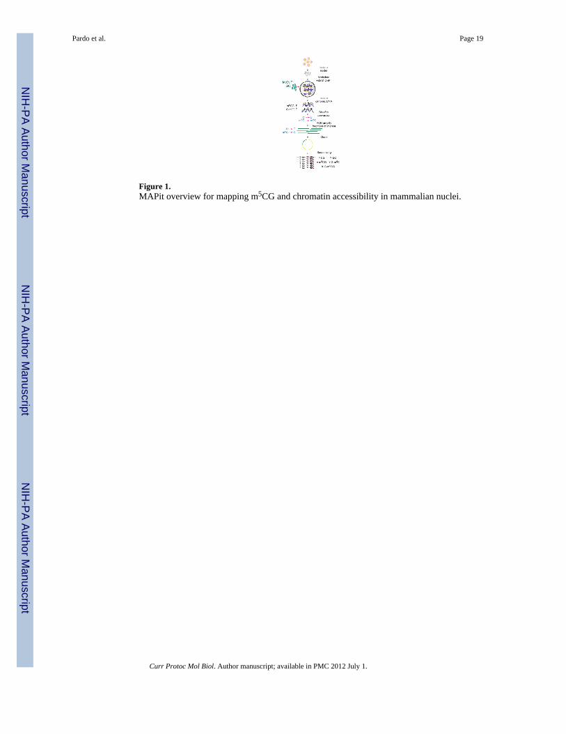
Figure 1.MAPit overview for mapping m5CG and chromatin accessibility in mammalian nuclei.
Pardo et al. Page 19
Curr Protoc Mol Biol. Author manuscript; available in PMC 2012 July 1.
NIH
-PA Author Manuscript
NIH
-PA Author Manuscript
NIH
-PA Author Manuscript
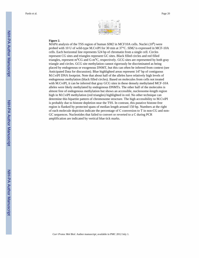
Figure 2.MAPit analysis of the TSS region of human SIM2 in MCF10A cells. Nuclei (106) wereprobed with 10 U of wild-type M.CviPI for 30 min at 37°C. SIM2 is expressed in MCF-10Acells. Each horizontal line represents 524 bp of chromatin from a single cell. Circlesrepresent CG sites and triangles represent GC sites. Black filled circles and red filledtriangles, represent m5CG and G-m5C, respectively. GCG sites are represented by both graytriangle and circles. GCG site methylation cannot rigorously be discriminated as beingplaced by endogenous or exogenous DNMT, but this can often be inferred from context (seeAnticipated Data for discussion). Blue highlighted areas represent 147 bp of contiguousM.CviPI DNA footprint. Note that about half of the alleles have relatively high levels ofendogenous methylation (black filled circles). Based on molecules from cells not treatedwith M.CviPI, it can be inferred that gray GCG sites in these densely methylated MCF-10Aalleles were likely methylated by endogenous DNMTs. The other half of the molecules isalmost free of endogenous methylation but shows an accessible, nucleosome-length regionhigh in M.CviPI methylation (red triangles) highlighted in red. No other technique candetermine this bipartite pattern of chromosome structure. The high accessibility to M.CviPIis probably due to histone depletion near the TSS. In contrast, this putative histone-freeregion is flanked by protected spans of median length around 150 bp. Numbers at the rightof each molecule depiction indicate the percentage of C conversion to T in non-CG and non-GC sequences. Nucleotides that failed to convert or reverted to a C during PCRamplification are indicated by vertical blue tick marks.
Pardo et al. Page 20
Curr Protoc Mol Biol. Author manuscript; available in PMC 2012 July 1.
NIH
-PA Author Manuscript
NIH
-PA Author Manuscript
NIH
-PA Author Manuscript
Related Documents