Simultaneous detection of molecular oxygen and water vapor in the tissue optical window using tunable diode laser spectroscopy Persson, Linda; Andersson, Mats; Lewander, Märta; Svanberg, Katarina; Svanberg, Sune Published in: Applied Optics DOI: 10.1364/AO.47.002028 Published: 2008-01-01 Link to publication Citation for published version (APA): Persson, L., Andersson, M., Lewander, M., Svanberg, K., & Svanberg, S. (2008). Simultaneous detection of molecular oxygen and water vapor in the tissue optical window using tunable diode laser spectroscopy. Applied Optics, 47(12), 2028-2034. DOI: 10.1364/AO.47.002028 General rights Copyright and moral rights for the publications made accessible in the public portal are retained by the authors and/or other copyright owners and it is a condition of accessing publications that users recognise and abide by the legal requirements associated with these rights. • Users may download and print one copy of any publication from the public portal for the purpose of private study or research. • You may not further distribute the material or use it for any profit-making activity or commercial gain • You may freely distribute the URL identifying the publication in the public portal

Welcome message from author
This document is posted to help you gain knowledge. Please leave a comment to let me know what you think about it! Share it to your friends and learn new things together.
Transcript

LUND UNIVERSITY
PO Box 117221 00 Lund+46 46-222 00 00
Simultaneous detection of molecular oxygen and water vapor in the tissue opticalwindow using tunable diode laser spectroscopy
Persson, Linda; Andersson, Mats; Lewander, Märta; Svanberg, Katarina; Svanberg, Sune
Published in:Applied Optics
DOI:10.1364/AO.47.002028
Published: 2008-01-01
Link to publication
Citation for published version (APA):Persson, L., Andersson, M., Lewander, M., Svanberg, K., & Svanberg, S. (2008). Simultaneous detection ofmolecular oxygen and water vapor in the tissue optical window using tunable diode laser spectroscopy. AppliedOptics, 47(12), 2028-2034. DOI: 10.1364/AO.47.002028
General rightsCopyright and moral rights for the publications made accessible in the public portal are retained by the authorsand/or other copyright owners and it is a condition of accessing publications that users recognise and abide by thelegal requirements associated with these rights.
• Users may download and print one copy of any publication from the public portal for the purpose of privatestudy or research. • You may not further distribute the material or use it for any profit-making activity or commercial gain • You may freely distribute the URL identifying the publication in the public portal

Simultaneous detection of molecular oxygen and watervapor in the tissue optical window using tunable
diode laser spectroscopy
Linda Persson,1,* Märta Lewander,1 Mats Andersson,1 Katarina Svanberg,2
and Sune Svanberg1
1Atomic Physics Division, Lund University, LTH, P.O. Box 118, SE-221 00 Lund, Sweden2Department of Oncology, Lund University Hospital, SE-221 85 Lund, Sweden
*Corresponding author: [email protected]
Received 4 September 2007; revised 8 February 2008; accepted 28 February 2008;posted 6 March 2008 (Doc. ID 87198); published 14 April 2008
We report on a dual-diode laser spectroscopic system for simultaneous detection of two gases. The tech-nique is demonstrated by performing gas measurements on absorbing samples such as an air distance,and on absorbing and scattering porous samples such as human tissue. In the latter it is possible to derivethe concentration of one gas by normalizing to a second gas of known concentration. This is possible if thescattering and absorption of the bulk material is equal or similar for the two wavelengths used, resultingin a common effective pathlength. Two pigtailed diode lasers are operated in a wavelength modulationscheme to detect molecular oxygen ∼760nm and water vapor ∼935nm within the tissue optical window(600nm to 1:3 μm). Different modulation frequencies are used to distinguish between the two wave-lengths. No crosstalk can be observed between the gas contents measured in the two gas channels.The system is made compact by using a computer board and performing software-based lock-in detection.The noise floor obtained corresponds to an absorption fraction of approximately 6 × 10�5 for both oxygenandwater vapor, yielding aminimumdetection limit of∼2mm for both gases in ambient air. The power ofthe technique is illustrated by the preliminary results of a clinical trial, nonintrusively investigating gasin human sinuses. © 2008 Optical Society of America
OCIS codes: 300.6260, 300.1030, 170.3890, 170.7050.
1. Introduction
Simultaneous detection of more than one gas hasbeen a hot topic in the tunable semiconductor laserspectroscopy field during past years. The rapid pro-gress in this field is mostly due to the invention ofquantum cascade lasers (QCLs) operating in themid-infrared region [1]. In this region many moleculessuch as CO, H2O, NO, and CO2 have strong absorp-tion bands that might overlap spectrally. Because ofthe wavelength tunability of the QCLs, it is possibleto scan over multiple absorption lines of differentspecies and thus detect more than one gas simulta-
neously [2,3]. However, in tissue the absorption ofconstituents strongly limits the penetration depthin the midinfrared region (1:5–4 μm). In the tissueoptics field it is common to refer to a tissue opticalwindow ranging from 600nm to 1:3 μm, where tissuecan be considered transparent due to the relativelyweak absorption of major species making light pene-tration possible. The lower wavelength limit is due toabsorption of hemoglobin and the upper limit is dueto absorption of water [4,5]. In this optical window,where conventional diode lasers operate, the absorp-tion lines of gases are weaker and fewer. This makesit difficult to achieve information about more thanone gas by the use of a single laser source. A fruitfulapproach to improve on this is to use sum- and dif-ference-frequency generation. An example of the for-
0003-6935/08/122028-07$15.00/0© 2008 Optical Society of America
2028 APPLIED OPTICS / Vol. 47, No. 12 / 20 April 2008

mer is given in [6], where water vapor, nitrogen di-oxide, and sulfur dioxide can be measured usingtwo diode lasers. The latter is illustrated in [7],where molecular oxygen, water vapor, and methanecan be studied. However, the drawback of these tech-niques is the low light intensity of the generated fre-quency (normally a factor of ∼1000 lower than theintensity of the light sources). Limited light intensitymay be adequate in the application of free beam pro-pagation in air, but is highly problematic, e.g., instrongly scattering media.In this paper we report on a newly developed
transportable system for dual-gas diode laser spec-troscopy in biological andmedical contexts. Since sig-nals are highly geometry dependent, it is importantto ensure measurement with identical conditions. Byusing two lasers operating spectrally close to eachother, similar effective pathlengths can be achievedunder the assumption that the absorption and scat-tering is the same for the bulk material at the twodifferent wavelengths. Then, by normalizing the sig-nal of a studied gas to the result obtained for a gaswith known concentration, an absolute concentrationvalue for the gas of interest can be determined. Thepresent system uses two pigtailed diode lasers to si-multaneously detect molecular oxygen at 760nm andwater vapor at 935nm. The light beams from the twolasers are brought together with fibers before enter-ing the sample. Different modulation frequencies ofthe lasers are used to distinguish between the twowavelengths. The system is not limited to these ab-sorption lines. Any gas can be detected with the as-sumption that lasers operating at the correspondingwavelengths of the absorption lines of the gases areavailable. The system employs a computer board andsoftware-based lock-in detection. The purpose of thispaper is to describe the equipment and the multiplexadvantages and to illustrate its applicability for clin-ical use. Examples of the detection of the two gases inthe human sinuses are provided. The analyzed re-sults of an ongoing clinical trial on human sinus cav-ities will be presented in future clinical publicationsfocusing on the medical aspects.
2. Experimental Setup
A schematic drawing of the setup is shown in Fig. 1.Two pigtailed distributed feedback (DFB) lasers(Nanoplus, Germany) are scanned across the molecu-lar oxygen absorption line R11Q12 at 760:445nm(vacuum wavelength) and the water vapor absorp-tion line at 935:685nm (vacuum wavelength, vibra-tion: ð000Þ → ð121Þ, rotation: J00 ¼ 3 → J0 ¼ 4, Ka
00 ¼0 → Ka
0 ¼ 0, Kc00 ¼ 3 → Kc
0 ¼ 4), respectively. ALabVIEW program controlling a computer board(NI-6120, National Instruments) creates the two in-put signals for the laser drivers. Each signal consistsof a ramp with a superimposed sinus wave. The tworamps have the same frequencies (4Hz), while theamplitude of the ramps differ due to the various re-sponses of the wavelength of the lasers with regard tothe driver current. The superimposed sinus waves on
the ramps make lock-in detection possible. The laserused to detect oxygen is modulated by a frequency of20:216kHz and the laser used to detect water vaporis modulated by a frequency of 9:016kHz. These fre-quencies are selected to avoid extensive noise at lowfrequencies and overlaps of the lower harmonics, andcoherent sampling is enabled. The modulation depthis selected to optimize the signal amplitude withoutcausing extensive line broadening [8].
The light beams from the lasers are each split intotwo arms, sample (90%) and reference (10%), by useof single-mode fiber-coupled beam splitters (La-ser2000, Sweden). The light from the two referencearms and the two sample arms are fiber-opticallybrought together. The low-intensity light is directlyguided to a photodiode (Hamamatsu S3590-01) pro-viding the reference signal. The light from the sam-ple arm is guided to the sample before being detectedby a photodiode (Hamamatsu S3204-08). The samplein this study is either an air path or a scatteringmedium containing absorbing gas. The intensitiesreaching the sample are ∼1mW for both laserwavelengths. The signals from the detectors are am-plified with two transimpedance amplifiers (FemtoDHPCA-100 and DHPCA-200) before being synchro-nously recorded by two input channels on the compu-ter board.
3. Data Analysis
The recorded signals are software-based lock-in de-tected using a LabVIEW program (Lock-in toolkitfor LabVIEW). One advantage of this approach isthe possibility to extract the different harmonicsboth in real time and after the data acquisition.All harmonics are available to process from the de-tected signals. A more detailed discussion of advan-tages using software-based lock-in detection isprovided in [9]. The reference sinus waves neededto perform lock-in detection are internally createdin LabVIEW. The 1f and 2f harmonics are computedfor both the sample and the reference detector at thetwo frequencies corresponding to the two gases. The2f signals of the two gases are normalized by divid-ing them with the offset of the corresponding 1f sig-
Fig. 1. Schematic of the experimental arrangement for simulta-neous detection of molecular oxygen (760nm) and water vapor(935nm) using tunable diode laser gas absorption spectroscopy.The sample can either be an absorbing sample such as an air dis-tance or an absorbing and scattering sample such as human tissue.
20 April 2008 / Vol. 47, No. 12 / APPLIED OPTICS 2029

nal. This is done since the fractional absorptionΔI=Idue to the gas has to be retrieved for concentrationanalysis with the Beer–Lambert law. The amplitudeof the 2f signal is proportional toΔI and the 1f signaloffset is proportional to I. Before analyzing the nor-malized 2f sample signal, balanced detection isperformed. This is done to suppress noise and pertur-bations like optical interference fringes. This proce-dure has been done in single-gas measurements andis described in detail elsewhere [10]. However, thesame procedure can be performed with the simulta-neous measurement since the two different wave-lengths are modulation frequency tagged. A set ofparameters is estimated by comparing the 2f refer-ence signal and the 2f sample signal for each gas atfrequency regions outside the expected gas imprint.A matched version of the reference signal is thensubtracted from the sample signal over the whole fre-quency scan. An ideal signal obtained from measure-ments over a long air path resulting in a recordedsignal with a gas imprint much larger than the noiseis then fitted to the balanced-detection signal.The gas content is obtained for each gas by evalu-
ating the amplitude of the fitted curve of the normal-ized balanced-detection 2f signal. From this value aquantity called the equivalent mean path length, Leq,is estimated by the use of the standard additionmethod [10]. The calibration curves were obtainedin an atmosphere of ∼30% relative humidity and atemperature of ∼27 °C. The Leq corresponds to howfar the light has to travel in ambient air to obtainthe same value.It should be noted that when investigating scatter-
ing samples the estimated Leq for each gas dependsnot only on the gas concentration but also on the op-tical properties of the sample such as scattering. Aquantity independent of the latter and thereby di-rectly proportional to the gas concentration can beestimated by forming the ratio between the two re-corded Leq for the different gases under the conditionthat similar optical properties for the two wave-lengths can be assumed. In this way the concentra-tion of one gas can be obtained if the concentration ofthe other gas is known.
4. Results and Discussion
A. Technique Performance Investigation
Typical recorded 1f and 2f signals from measure-ments over an air distance of ∼20mm are shownin Fig. 2. The signals have been averaged for ∼25 sby repeatedly ramping the laser at 4Hz and storingthe signals. The balanced-detection signals are alsoincluded together with fitted ideal signals. The am-plitude of the fitted signals correspond to an absorp-tion fraction of approximately 5 × 10⁻⁴ and 9 × 10⁻⁴
for molecular oxygen and water vapor, respectively.To investigate possible crosstalk between the two
gas channels, an air distance of∼25mmwas studied.A fast Fourier transform was performed on the de-tected signals (see Fig. 3). Signals were recorded with
both lasers on, one laser on at a time, and no laser on.To the right in the figure, frequency regions of inter-est for crosstalk investigation are presented. As canbe seen in the figure the modulation peaks occur onlyat multiples of the modulation frequencies of theindividual lasers and no detectable crosstalk canbe observed. To further study this issue ten measure-ments were recorded for each case. Signals wereaveraged for ∼25 s. In Fig. 4 typical obtained ba-lanced-detection signals can be seen, and theaverages of the measured Leq for each gas channeltogether with standard deviations are shown inTable 1. This test also verifies that no crosstalkoccurs between the two gas channels.
By recording signals with no added air distance,the detection limit of the setup could be estimated.The detected noise floor corresponded to an absorp-tion fraction of approximately 6 × 10⁻⁵ for both mole-cular oxygen and water vapor when the number ofdetected photons was not a limitation. This corre-sponds to an Leq of ∼2:5mm for oxygen and∼1:5mm for water vapor with calibration atmo-sphere. It was observed that the remaining patternhad a periodic structure for both cases. Hence theconclusion can be drawn that optical interferencefringes [10] set the limitation of detectable signals.
B. Initial Clinical Test
In order to demonstrate the performance of the sys-tem developed, we present the results from test mea-surements on human frontal and maxillary sinusesperformed in an initial clinical trial at the Lund Uni-versity Hospital in collaboration with the Ear, Noseand Throat Clinic, the Radiology Clinic, and the On-cology Clinic. The cavities located within the humanskull are surrounded by strongly scattering tissue,and gas signal recordings for such cavities have beenpreviously demonstrated with laboratory equipment[11,12]. However, then separate setups were used for
Fig. 2. Typical recorded oxygen and water vapor signals afterbackground subtraction for measurements over an air distanceof ∼20mm. Insets can be seen in the 1f signal plots enhancingthe intensity region of interest for signals due to gas imprint.The balanced-detection 2f signal is shown (black) together witha fitted experimental ideal line shape (gray line). The relative fre-quency range for both gases corresponds to 35GHz.
2030 APPLIED OPTICS / Vol. 47, No. 12 / 20 April 2008

molecular oxygen and water vapor measurements,and identical geometrical conditions for themeasure-ments performed with separate probes could clearlynot be ensured.For a healthy patient the sinuses should be air
filled, while in conditions like sinusitis (inflamma-tion and infection of the sinuses), the mucosa is swol-len and the sinuses may be filled with pus and liquid.The cause of sinusitis can in many cases be due topassage blockage between the nasal cavity and thesinuses preventing proper ventilation. The rationalebehind the clinical trial is to investigate the pos-
sibility of using noninvasive diode laser-basedabsorption measurements of the sinuses for charac-terization regarding gas composition. The gasmeasurements are performed directly after the pa-tients have undergone a computed tomography(CT) investigation of the skull, which is one of themost reliable diagnostic methods of sinusitis today.Unfortunately this method is expensive and uses io-nising radiation. Therefore complementary and al-ternative techniques are desirable.
1. Frontal Sinus Investigation
To investigate the human frontal sinuses the opticalfiber probe carrying the laser light to the sample isplaced on the frontal bone below the eyebrow (theroof of the orbita) pointing up toward the frontal bonewith the sinus cavity. The diffusely scattered lightemerging out through the frontal bone with a frac-tion crossing the frontal sinus is detected by placingthe detector on the frontal bone.
In Fig. 5(a) a CT image of the frontal sinuses can beseen of a patient participating in the clinical trial andin Fig. 5(b) balanced-detection signals for the leftfrontal sinus are shown. The signals correspond toan Leq of 22mm for oxygen and an Leq of 123mmfor water vapor, which agree with the expectedvalues reported in [12]. It was found that the ratiowas ∼0:18. For air with 21% oxygen and water vaporat 27 °C and 30% relative humidity, a ratio of 1.00 isexpected with the definition chosen. However, thetemperature within the body is ∼37 °C and thehumidity is 100% due to water in liquid phase inthe surrounding tissue. Both conditions (correspond-ing to enhancement factors of 1.8 and 3.3, respec-
Fig. 3. Recorded laser relative intensity noise (RIN) expressed in relative dB, to investigate possible crosstalk between the two modula-tion frequencies used to detect two gases simultaneously. The fiber and the sample detector have been separated with an air distance of∼25mm. To the right the frequency regions of interest are plotted. (a) Both lasers on. (b) Only laser for oxygen detection on. (c) Only laserfor water vapor detection on. (d) No laser on.
Fig. 4. Investigation of possible crosstalk between the two mod-ulation frequencies used to detect two gases simultaneously. Thebalanced-detection signals are recorded from measurements overan air distance of ∼25mm. The balanced-detection 2f signals areshown (black line) together with a fitted experimental ideal lineshape (gray line). The relative frequency range for both gases cor-responds to 35GHz.
20 April 2008 / Vol. 47, No. 12 / APPLIED OPTICS 2031

tively) will result in a reduction of the ratio by1:00=ð1:8 × 3:3Þ ¼ 0:17. This is close to the value ob-tained for the sinus cavity indicating a close to am-bient oxygen concentration (again, subject to thetissue property assumptions made).At each occasion of measurements on patients dur-
ing the clinical trial, a control recording is performed.The purpose of this control is to study any currentoffset of gas signals in the setup. During these mea-surements the fiber is placed on the inside of thecheek in the mouth and the light is detected onthe outside of the cheek. This should result in non-detectable signals since no free oxygen or watervapor are expected in the tissue. In Fig. 5(c) recordedbalanced-detection signals from the control per-formed just before the frontal sinus study are shown.As can be seen no offset of either gas could be de-tected.
2. Maxillary Sinus Investigation
For the maxillary sinus studies the optical fiberprobe carrying the laser light to the sample is placedinside the mouth in contact with the palate in thearea directly under the maxillary sinus investigated.The diffusely scattered light emerging out throughthe cheek bone with a fraction crossing the maxillarysinus is detected by placing the detector on the cheekbone [11,12].In Fig. 6(a) the CT image of the maxillary sinuses
can be seen for a different patient participating in theclinical trial, and in Fig. 6(b) the balanced-detectionsignals for the right maxillary sinus are shown. Thesignals correspond to an Leq of 30mm for oxygen andan Leq of 185mm for water vapor. The ratio of the gassignals are similar to the one recorded for the frontalsinus within the margin for error. The obtainedbalanced-detection control signals can be seen inFig. 6(c). No offset of either gas was obtained.Preliminary studies of the ventilation between the
nasal cavity and the maxillary sinuses have alsobeen performed. The nasal cavity on a healthy volun-teer was flushed with pure nitrogen through the nos-tril for ∼25 s while continuously measuring the gascomposition within the sinuses. Signals were re-corded before, during, and after termination of theflush. Each recorded signal was averaged for ∼12 s.In Figs. 7(a) and 7(b) the measured Leq for oxygenand water vapor, respectively, for one of themaxillarysinuses of the volunteer can be seen. In Fig. 7(c) the
ratio is shown and in Fig. 7(d) recorded oxygen andwater vapor balanced-detection signals correspond-ing to indicated numbers in Figs. 7(a) and 7(b) areprovided. A decrease of oxygen is recorded duringthe flush and the reinvasion of oxygen is observedafter its termination. However, for the water vaporsignal no noticeable change can be observed. Thisis not expected either since 100% humidity in thesinuses remains during the flush. For the recordedLeq for oxygen during the time period no considera-tions have to be given with regards to the opticalproperties of the surrounding tissue since they re-main unaffected. However, the water vapor signalis still advantageous to eliminate possible changesof recorded molecular oxygen signal due to move-ments of fiber and detector, and thereby differentsampling volumes during the measurements. Thusthe estimated ratio shown in Fig. 7(c) can be consid-ered as a more reliable picture of how the oxygen con-centration within the sinus changes than Leq for
Table 1. Average Leq from Ten Recordings over an Air Distanceof ~25mm to Investigate Possible Crosstalk between the Two
Modulation Frequencies Used to Study Two GasesSimultaneouslya
Leq (mm) O2 Leq (mm) H2O
Both Lasers On 23:5� 1:5 24� 1O2: Laser On 24� 1:5 —
H2O: Laser On — 26� 1
aExamples of the detected signals used are provided in Fig. 4.
Fig. 5. (a) CT image of human frontal sinuses of a patient parti-cipating in the clinical trial; transversal section. (b) Correspondingrecorded balanced-detection signals (black line) from the left fron-tal sinus of the patient with CT image shown in Fig. 5(a), togetherwith fitted ideal signal (gray line). The oxygen signal correspondstoLeq ¼ 22mmand the water vapor signal toLeq ¼ 123mm. (c) Re-corded balanced-detection control signal through the cheek. Therelative frequency range for both gases corresponds to 35GHz.
2032 APPLIED OPTICS / Vol. 47, No. 12 / 20 April 2008
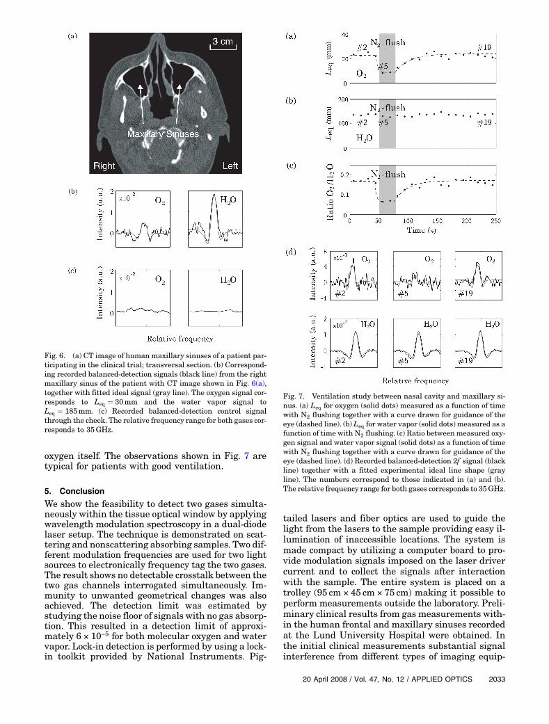
oxygen itself. The observations shown in Fig. 7 aretypical for patients with good ventilation.
5. Conclusion
We show the feasibility to detect two gases simulta-neously within the tissue optical window by applyingwavelength modulation spectroscopy in a dual-diodelaser setup. The technique is demonstrated on scat-tering and nonscattering absorbing samples. Two dif-ferent modulation frequencies are used for two lightsources to electronically frequency tag the two gases.The result shows no detectable crosstalk between thetwo gas channels interrogated simultaneously. Im-munity to unwanted geometrical changes was alsoachieved. The detection limit was estimated bystudying the noise floor of signals with no gas absorp-tion. This resulted in a detection limit of approxi-mately 6 × 10⁻⁵ for both molecular oxygen and watervapor. Lock-in detection is performed by using a lock-in toolkit provided by National Instruments. Pig-
tailed lasers and fiber optics are used to guide thelight from the lasers to the sample providing easy il-lumination of inaccessible locations. The system ismade compact by utilizing a computer board to pro-vide modulation signals imposed on the laser drivercurrent and to collect the signals after interactionwith the sample. The entire system is placed on atrolley (95 cm × 45 cm × 75 cm) making it possible toperform measurements outside the laboratory. Preli-minary clinical results from gas measurements with-in the human frontal and maxillary sinuses recordedat the Lund University Hospital were obtained. Inthe initial clinical measurements substantial signalinterference from different types of imaging equip-
Fig. 6. (a) CT image of human maxillary sinuses of a patient par-ticipating in the clinical trial; transversal section. (b) Correspond-ing recorded balanced-detection signals (black line) from the rightmaxillary sinus of the patient with CT image shown in Fig. 6(a),together with fitted ideal signal (gray line). The oxygen signal cor-responds to Leq ¼ 30mm and the water vapor signal toLeq ¼ 185mm. (c) Recorded balanced-detection control signalthrough the cheek. The relative frequency range for both gases cor-responds to 35GHz.
Fig. 7. Ventilation study between nasal cavity and maxillary si-nus. (a) Leq for oxygen (solid dots) measured as a function of timewith N2 flushing together with a curve drawn for guidance of theeye (dashed line). (b) Leq for water vapor (solid dots) measured as afunction of time with N2 flushing. (c) Ratio between measured oxy-gen signal and water vapor signal (solid dots) as a function of timewith N2 flushing together with a curve drawn for guidance of theeye (dashed line). (d) Recorded balanced-detection 2f signal (blackline) together with a fitted experimental ideal line shape (grayline). The numbers correspond to those indicated in (a) and (b).The relative frequency range for both gases corresponds to 35GHz.
20 April 2008 / Vol. 47, No. 12 / APPLIED OPTICS 2033

ment typical for a radiological clinic was occasionallyexperienced. It should be noted that such an environ-ment would not pertain to locations of intended fu-ture clinical use. A full analysis of the clinical datafor all patients included in the study will be pre-sented for the frontal and maxillary sinuses as wellas for the mastoid bones (behind the ears) in futurepublications.
The authors acknowledge the useful discussionswith Morgan Andersson, Karolina Falkenius-Schmidt, Kjell Jonsson, Sven Lindberg, KarinPrellner, Bo Paulsson, and Roger Siemund. We aregrateful for all help from the staff at the RadiologyClinic at the Lund University Hospital during theclinical measurements. This research was supportedby the Swedish Research Council, the MedicalFaculty Lund University, and the Knut and AliceWallenberg Foundation.
References1. J. Faist, F. Capasso, D. Sivco, C. Sirtori, A. Hutchinson, and A.
Cho, “Quantum cascade laser,” Science 264, 553–556 (1994).2. G. Wysocki, M. McCurdu, S. So, D. Weidmann, C. Roller, R.
Curl, and F. Tittel, “Pulsed quantum-cascade laser-based sen-sor for trace-gas detection of carbonyl sulfide,” Appl. Opt. 43,6040–6046 (2004).
3. M. Silva, D. Sonnenfroh, D. Rosen, M. Allen, and A. O’Keefe,“Integrated cavity output spectroscopy measurements ofnitric oxide levels in breath with a pulsed room-temperaturequantum cascade laser,” Appl. Phys. B 81, 705–710 (2005).
4. J. Parrish, “New concepts in therapeutic photomedicine:photochemistry, optical targeting and the therapeutic win-dow,” J. Investigative Dermatol. 77, 45–50 (1981).
5. J. Boulnois, “Photophysical processes in recent medical laserdevelopments: a review,” Lasers Med. Sci. 1, 47–66 (1986).
6. G. Somesfalean, Z. Zhang, M. Sjöholm, and S. Svanberg, “All-diode-laser ultraviolet absorption spectroscopy for sulfur diox-ide detection,” Appl. Phys. B 80, 1021–1025 (2005).
7. U. Gustafsson, J. Sandsten, and S. Svanberg, “Simultaneousdetection of methane, oxygen and water vapour utilizingnear-infrared diode lasers in conjunction with difference fre-quency generation,” Appl. Phys. B 71, 853–857 (2000).
8. J. Reid and D. Labrie, “Second-harmonic detection with tun-able diode lasers—comparison of experiment and theory,”Appl. Phys. B 26, 203–210 (1981).
9. M. Andersson, L. Persson, T. Svensson, and S. Svanberg,“Flexible lock-in detection system based on synchronized com-puter plug-in boards applied in sensitive gas spectroscopy,”Rev. Sci. Instrum. 78, 113107 (2007).
10. L. Persson, F. Andersson,M. Andersson, and S. Svanberg, “Ap-proach to optical interference fringes reduction in diode laserabsorption spectroscopy,” Appl. Phys. B 87, 523–530 (2007).
11. L. Persson, M. Andersson, T. Svensson, M. Cassel-Engquist,K. Svanberg, and S. Svanberg, “Non-intrusive optical studyof gas and its exchange in human maxillary sinuses,” Proc.SPIE 6628, 662804-1–7 (2007).
12. L. Persson, M. Andersson, M. Cassel-Engquist, K. Svanberg,and S. Svanberg, “Gas monitoring in human sinuses usingtunable diode laser spectroscopy,” J Biomed. Opt. 12, 054001(2007).
2034 APPLIED OPTICS / Vol. 47, No. 12 / 20 April 2008
Related Documents


