© 2013 Yang et al. This work is published by Dove Medical Press Ltd, and licensed under Creative Commons Attribution – Non Commercial (unported, v3.0) License. The full terms of the License are available at http://creativecommons.org/licenses/by-nc/3.0/. Non-commercial uses of the work are permitted without any further permission from Dove Medical Press Ltd, provided the work is properly attributed. Permissions beyond the scope of the License are administered by Dove Medical Press Ltd. Information on how to request permission may be found at: http://www.dovepress.com/permissions.php International Journal of Nanomedicine 2013:8 3333–3343 International Journal of Nanomedicine Dovepress submit your manuscript | www.dovepress.com Dovepress 3333 ORIGINAL RESEARCH open access to scientific and medical research Open Access Full Text Article http://dx.doi.org/10.2147/IJN.S50683 Silymarin-loaded solid nanoparticles provide excellent hepatic protection: physicochemical characterization and in vivo evaluation Kwan Yeol Yang 1, * Du Hyeong Hwang 1, * Abid Mehmood Yousaf 2 Dong Wuk Kim 2 Young-Jun Shin 2 Ok-Nam Bae 2 Yong-Il Kim 1 Jong Oh Kim 1 Chul Soon Yong 1 Han-Gon Choi 2 1 College of Pharmacy, Yeungnam University, Dae-Dong, Gyongsan, 2 College of Pharmacy and Institute of Pharmaceutical Science and Technology, Hanyang University, Sangnok-gu, Ansan, South Korea * These authors contributed equally to this work Correspondence: Han-Gon Choi College of Pharmacy and Institute of Pharmaceutical Science and Technology, Hanyang University, 55 Hanyangdaehak-ro, Sangnok-gu, Ansan 426-791, South Korea Tel +823 1400 5802 Fax +823 1400 5958 Email [email protected] Chul Soon Yong College of Pharmacy, Yeungnam University, 214-1, Dae-Dong, Gyongsan 712-749, South Korea Tel +825 3810 2812 Fax +825 3810 4654 Email [email protected] Background: The purpose of this study was to develop a novel silymarin-loaded solid nanoparticle system with enhanced oral bioavailability and an ability to provide excellent hepatic protection for poorly water-soluble drugs using Shirasu porous glass (SPG) membrane emulsification and a spray-drying technique. Methods: A silymarin-loaded liquid nanoemulsion was formulated by applying the SPG mem- brane emulsification technique. This was further converted into solid state nanosized particles by the spray-drying technique. The physicochemical characteristics of these nanoparticles were determined by scanning electron microscopy, differential scanning calorimetry, and powder X-ray diffraction. Their dissolution, bioavailability, and hepatoprotective activity in rats were assessed by comparison with a commercially available silymarin-loaded product. Results: Formulation of a silymarin-loaded nanoemulsion, comprising silymarin, castor oil, polyvinylpyrrolidone, Transcutol HP, Tween 80, and water at a weight ratio of 5/3/3/1.25/1.25/100 was accomplished using an SPG membrane emulsification technique at an agitator speed of 700 rpm, a feed pressure of 15 kPa, and a continuous phase temperature of 25°C. This resulted in generation of comparatively uniform emulsion globules with a narrow size distribution. Moreover, the silymarin-loaded solid nanoparticles, containing silymarin/castor oil/polyvi- nylpyrrolidone/Transcutol HP/Tween 80 at a weight ratio of 5/3/3/1.25/1.25, improved about 1,300-fold drug solubility and retained a mean size of about 210 nm. Silymarin was located in unaltered crystalline form in the nanoparticles. The drug dissolved rapidly from the nanopar- ticles, reaching nearly 80% within 15 minutes, indicating three-fold better dissolution than that of the commercial product. Further, the nanoparticles showed a considerably shorter time to peak concentration, a greater area under the concentration-time curve, and a higher maximum concentration of silymarin compared with the commercial product (P , 0.05). In particular, the area under the concentration-time curve of the drug provided by the nanoparticles was approxi- mately 1.3-fold greater than that of the commercial product. In addition, the silymarin-loaded nanoparticles significantly reduced carbon tetrachloride-induced hepatotoxicity, indicating improved bioactivity compared with silymarin powder and the commercial product. Conclusion: Silymarin-loaded nanoparticles developed using SPG membrane emulsification and spray-drying techniques could be a useful system for delivery of poorly water-soluble silymarin while affording excellent hepatic protection. Keywords: silymarin, nanoparticle, hepatoprotective activity, Shirasu porous glass membrane, enhanced oral bioavailability Introduction Silymarin is a purified mixture of four isomeric flavonoids extracted from the seeds and fruit of the milk thistle plant, Carduus marianus (L.) Gaertn. These isomeric

Welcome message from author
This document is posted to help you gain knowledge. Please leave a comment to let me know what you think about it! Share it to your friends and learn new things together.
Transcript
© 2013 Yang et al. This work is published by Dove Medical Press Ltd, and licensed under Creative Commons Attribution – Non Commercial (unported, v3.0) License. The full terms of the License are available at http://creativecommons.org/licenses/by-nc/3.0/. Non-commercial uses of the work are permitted without any further
permission from Dove Medical Press Ltd, provided the work is properly attributed. Permissions beyond the scope of the License are administered by Dove Medical Press Ltd. Information on how to request permission may be found at: http://www.dovepress.com/permissions.php
International Journal of Nanomedicine 2013:8 3333–3343
International Journal of Nanomedicine Dovepress
submit your manuscript | www.dovepress.com
Dovepress 3333
O r I g I N a l r e s e a r c h
open access to scientific and medical research
Open access Full Text article
http://dx.doi.org/10.2147/IJN.S50683
silymarin-loaded solid nanoparticles provide excellent hepatic protection: physicochemical characterization and in vivo evaluation
Kwan Yeol Yang1,*
Du hyeong hwang1,*
abid Mehmood Yousaf2
Dong Wuk Kim2
Young-Jun shin2
Ok-Nam Bae2
Yong-Il Kim1
Jong Oh Kim1
chul soon Yong1
han-gon choi2
1college of Pharmacy, Yeungnam University, Dae-Dong, gyongsan, 2college of Pharmacy and Institute of Pharmaceutical science and Technology, hanyang University, sangnok-gu, ansan, south Korea
*These authors contributed equally to this work
correspondence: han-gon choi college of Pharmacy and Institute of Pharmaceutical science and Technology, hanyang University, 55 hanyangdaehak-ro, sangnok-gu, ansan 426-791, south Korea Tel +823 1400 5802 Fax +823 1400 5958 email [email protected] chul soon Yong college of Pharmacy, Yeungnam University, 214-1, Dae-Dong, gyongsan 712-749, south Korea Tel +825 3810 2812 Fax +825 3810 4654 email [email protected]
Background: The purpose of this study was to develop a novel silymarin-loaded solid
nanoparticle system with enhanced oral bioavailability and an ability to provide excellent
hepatic protection for poorly water-soluble drugs using Shirasu porous glass (SPG) membrane
emulsification and a spray-drying technique.
Methods: A silymarin-loaded liquid nanoemulsion was formulated by applying the SPG mem-
brane emulsification technique. This was further converted into solid state nanosized particles
by the spray-drying technique. The physicochemical characteristics of these nanoparticles were
determined by scanning electron microscopy, differential scanning calorimetry, and powder
X-ray diffraction. Their dissolution, bioavailability, and hepatoprotective activity in rats were
assessed by comparison with a commercially available silymarin-loaded product.
Results: Formulation of a silymarin-loaded nanoemulsion, comprising silymarin, castor oil,
polyvinylpyrrolidone, Transcutol HP, Tween 80, and water at a weight ratio of 5/3/3/1.25/1.25/100
was accomplished using an SPG membrane emulsification technique at an agitator speed of
700 rpm, a feed pressure of 15 kPa, and a continuous phase temperature of 25°C. This resulted
in generation of comparatively uniform emulsion globules with a narrow size distribution.
Moreover, the silymarin-loaded solid nanoparticles, containing silymarin/castor oil/polyvi-
nylpyrrolidone/Transcutol HP/Tween 80 at a weight ratio of 5/3/3/1.25/1.25, improved about
1,300-fold drug solubility and retained a mean size of about 210 nm. Silymarin was located in
unaltered crystalline form in the nanoparticles. The drug dissolved rapidly from the nanopar-
ticles, reaching nearly 80% within 15 minutes, indicating three-fold better dissolution than that
of the commercial product. Further, the nanoparticles showed a considerably shorter time to
peak concentration, a greater area under the concentration-time curve, and a higher maximum
concentration of silymarin compared with the commercial product (P , 0.05). In particular, the
area under the concentration-time curve of the drug provided by the nanoparticles was approxi-
mately 1.3-fold greater than that of the commercial product. In addition, the silymarin-loaded
nanoparticles significantly reduced carbon tetrachloride-induced hepatotoxicity, indicating
improved bioactivity compared with silymarin powder and the commercial product.
Conclusion: Silymarin-loaded nanoparticles developed using SPG membrane emulsification
and spray-drying techniques could be a useful system for delivery of poorly water-soluble
silymarin while affording excellent hepatic protection.
Keywords: silymarin, nanoparticle, hepatoprotective activity, Shirasu porous glass membrane,
enhanced oral bioavailability
IntroductionSilymarin is a purified mixture of four isomeric flavonoids extracted from the seeds
and fruit of the milk thistle plant, Carduus marianus (L.) Gaertn. These isomeric
International Journal of Nanomedicine 2013:8submit your manuscript | www.dovepress.com
Dovepress
Dovepress
3334
Yang et al
flavonolignans include silyb, isosilybin, silychristin, and
silydianin. Silybin is the chief active constituent amongst
them.1–3 Silymarin has been used to treat liver disease and
for the supportive treatment of chronic active hepatitis
and hepatic cirrhosis.4,5 However, the bioavailability of
silymarin is quite low due to its poor aqueous solubility.6–8
Pharmacokinetic investigations have demonstrated that only
23%–47% of silymarin reaches the systemic circulation after
oral administration.9–11 Various techniques, such as complex-
ation of silymarin with phosphatidylcholine,12 lecithin,13 or
cyclodextrin clathrate14 and incorporation of silymarin into
a solid dispersion15 have been used to improve the aqueous
solubility of silymarin. Moreover, a commercial product
(Legalon®, Bukwang Pharmaceutical Company, Seoul, South
Korea), which is administered orally twice daily as a hard
capsule containing 70 mg silymarin, has been launched on
the market.
In this study, with the intention of developing a new
silymarin-loaded solid nanoparticle system with enhanced
oral bioavailability of the hydrophobic drug, a silymarin-
loaded nanoemulsion was formulated using a Shirasu porous
glass (SPG) membrane emulsification technique and then
spray-dried to obtain solid state nanoparticles. Scanning
electron microscopy, differential scanning calorimetry
(DSC), and powder X-ray diffraction techniques were used to
explore the physicochemical properties of the nanoparticles.
Their dissolution, bioavailability in rats, and hepatoprotec-
tive activity in carbon tetrachloride (CCl4)-treated rats were
assessed in comparison with the commercial product.
Although liquid nanoemulsions play an important role
in improving oral drug bioavailability and reducing various
side effects, few emulsion preparations are currently in use
when compared with other oral dosage formulations. Their
less frequent use is because of their poor physicochemical
stability and compliance issues. Emulsions are notorious
for their physical instability. One way to circumvent this
problem and render emulsions more attractive for use could
be to transform them into dry powdered nanoemulsions.
First described in the early 1960s, a dry powdered emulsion,
a type of nanoparticle, is a dispersion of an immiscible oil
phase within a solid phase, and is achieved by elimination
of the aqueous phase of a liquid emulsion by spray-drying,16
lyophilization,17 and later solvent evaporation.18,19 These
nanoparticles are lipid-based powder formulations from
which an oil/water (o/w) emulsion can be reconstituted in
vivo or in vitro. Like o/w emulsions, these nanoparticles
represent a potential oral dosage form for lipophilic and
poorly water-soluble drug entities.
In this study, the SPG membrane emulsification technique
was used because of its uniform-sized pores and the wide
range of mean pore diameters (0.05–50 µm) available. In
addition, it is capable of generating narrow droplet size dis-
tributions by expending low energy. Since the development
of uniform-sized kerosene-in-water and water-in-kerosene
emulsions, this technique has attracted the attention of many
investigators because of its ability to produce narrow droplet
size distributions with low energy consumption.18 SPG mem-
branes are potentially amenable to membrane emulsification
because they have uniform-sized pores and a wide range of
available mean pore diameters.20 When nanoparticles are
developed conventionally, irregularly sized nanoparticles
are produced due to lack of inclusion of an SPG membrane
emulsification process. In this study, SPG membrane emul-
sification yielded uniform emulsion droplets with a narrow
size distribution via spray-drying.21
Materials and methodsSilymarin was supplied by Kolon Life Science Company
(Kwacheon, South Korea). Transcutol HP, polysorbate 80
(Tween 80), castor oil, and polyvinylpyrrolidone (PVP K30)
were obtained from Gattefosse (Saint-Priest Cedex, France),
Duksan Chemical Company (Ansan, South Korea), Sigma-
Aldrich (St Louis, MO, USA) and Shin-Etsu Co (Tokyo,
Japan), respectively. The commercially available silymarin
product (Legalon®, in capsule form) was sourced from
Bukwang Pharmaceutical Company (Seoul, South Korea).
All other chemicals were of reagent grade and used without
further purification.
animalsAll animal care and procedures were conducted in accor-
dance with the Guiding Principles in the Use of Animals
in Toxicology, as adopted in 1989, revised in 1999, and
amended in 2008 by the Society of Toxicology.22 The
protocols used for the pharmacokinetic studies were also
sanctioned by the Institute of Laboratory Animal Resources
at Yeungnam University. Sixteen male Sprague Dawley rats
(aged 7–9 weeks, weighing 260–300 g) were purchased
from Charles River Company (Seoul, Korea). Further in
vivo animal experiments for hepatotoxicity were performed
using 25 male Sprague Dawley rats weighing 190–210 g
(Charles River Company) and performed in accordance with
the National Institutes of Health Policy and Animal Welfare
Act under the approval of the Institutional Animal Care and
Use Committee at Hanyang University. The rats were housed
at a temperature of 20°C to 23°C with a relative humidity of
International Journal of Nanomedicine 2013:8 submit your manuscript | www.dovepress.com
Dovepress
Dovepress
3335
silymarin-loaded solid nanoparticles with excellent hepatic protection
50% ± 5%, and fasted for 24–36 hours prior to the experi-
ments, but were allowed free access to water.
solubilityAn excess of silymarin powder (about 100 mg) was trans-
ferred to 1 mL of surfactant, oil, or aqueous solution contain-
ing 1% hydrophilic polymer in a conical tube. The tubes were
agitated in a water bath at 25°C for 7 days, centrifuged at
3,000g for 10 minutes using a 5415C centrifuge (Eppendorf,
Hauppauge, NY, USA) and filtered through a membrane
filter (0.45 µm). The silymarin in the filtrate was assayed
using a high-pressure liquid chromatography (HPLC) sys-
tem (Hitachi, Tokyo, Japan) equipped with Ezchrom elite
(version 318a) computer software, a Hitachi L-2130 pump,
and a Hitachi L-2400 ultraviolet-visible detector. An Inertsil
ODS-2 C18 column (GL Science, Tokyo, Japan; 0.5 µm,
15 cm × 0.46 cm internal diameter) was used in this analy-
sis. The mobile phase A consisted of a mixture of methanol,
acetic acid, and phosphate buffer (pH 3.0, 34/6/60, v/v/v),
and the mobile phase B, acetonitrile, was eluted as follows:
0 minutes (98:2), 0.1 minute (80:20), 12 minutes (98:2)
and 22 minutes (98:2). The eluent (1 mL per minute) was
monitored at 285 nm for detection of silymarin. The interday
and intraday variance of this HPLC method was within the
acceptable range (R2 = 0.999).
Pseudoternary phase diagramThe existence of emulsion fields forming emulsions on agita-
tion was identified from ternary phase diagrams of systems
comprising oil-surfactant-water. The influence of surfactant
(Transcutol HP, Tween 80, or a mixture of these two sur-
factants at a 1:1 w/w ratio) and water on the pseudoternary
phase diagram was carefully recorded at room temperature.
Subsequently, the oil phase (castor oil) was added in 5%
increments to each clear mixture of water and surfactant.
This mixture was vortexed for 5 minutes and placed in a
water bath at 25°C for 30 minutes. The mixture was then
visually inspected for formation of an emulsion. The points
from uniform turbid solution to separation were considered
as the emulsion-forming and nonemulsion-forming regions,
respectively.
experimental setup and procedureAn emulsif ication kit comprising an MPG module
(microporous glass, SPG®; Kiyomoto Iron Works Company,
Miyazaki, Japan) and a hydrophilic SPG membrane (outer
diameter 10 mm, thickness 0.75 mm, pore size 2.5 µm) was
used.21,23,24 Ten grams of dispersed (oil) phase was retained
in an air-tight vessel connected to a nitrogen gas inlet
connected in turn to a pressure gauge (PG-200-163GP-S,
Copal Electronics, Tokyo, Japan). The continuous phase,
100 g of water containing 2.5 g surfactant and 3 g polyvi-
nylpyrrolidone, was stirred gently with a magnetic bar (3 cm
long) in a beaker to avoid creaming of droplets. By exerting
an appropriate pressure of nitrogen gas, the oil phase (3 g
castor oil) was infused through the uniform pores of the
membrane into the aqueous phase to produce globules. These
dispersed globules were stabilized by polyvinylpyrrolidone
dissolved in aqueous phase. The experiments were conducted
over a wide range of agitator speeds (300–700 rpm), feed
pressures (15–35 kPa), and continuous phase temperatures
(25°C–45°C).
Preparation of silymarin-loaded solid nanoparticlesA Büchi 190 nozzle-type mini spray dryer (Flawil,
Switzerland) was used to form the silymarin-loaded
nanoparticles. The silymarin-loaded nanoemulsion was car-
ried to the spraying nozzle (0.7 mm diameter) at a flow rate
of 5 mL per minute by a peristaltic pump, and spray-dried at
an inlet temperature of 120°C and an outlet temperature of
75°C–80°C. The pressure of the sprayed air was 4 kg/cm2.
The flow rate of drying air was maintained at an aspirator
setting of 10, indicating that the pressure of the aspirator filter
vessel was -25 mbar. The sprayed products were delivered
in the direction of air flow.
Mean droplet size and distribution of silymarin-loaded nanoparticlesThe droplet size distribution and zeta potential of the sily-
marin nanoemulsion and solid nanoparticles were investi-
gated using a Zetasizer NanoZS light dispersing particle
size analyzer (Malvern Instruments Ltd, Malvern, UK) with
noninvasive back scatter (NIBS®) technology, which permit-
ted detection of globules within the range of 0.6–6,000 nm.
Acquisition of data on particle size distribution was done
using the DTS (nano) software (version 5.0) supplied with
the device. The z-average diameter, also referred to as the har-
monic intensity-weighted average hydrodynamic diameter,
of the emulsions was calculated from cumulated analysis
applying Automeasure software (Malvern Instruments Ltd).
For investigation of the silymarin-loaded nanoparticle size,
reconstitution was achieved prior to measurement as follows:
the nanoparticles (6 mg) were vortexed with 10 mL of dis-
tilled water in a conical tube for 10 seconds, and subsequently
incubated for 30 minutes at 25°C.
International Journal of Nanomedicine 2013:8submit your manuscript | www.dovepress.com
Dovepress
Dovepress
3336
Yang et al
Morphologic features of silymarin- loaded nanoparticlesThe morphologic manifestations associated with the shape
and surface of the silymarin powder and silymarin-loaded
solid nanoparticles were studied using a scanning electron
microscope (S-4100, Hitachi) equipped with an image analy-
sis system (ImageInside version 2.32). The powders were
stuck onto a brass specimen club using double-sided adhesive
tape and made electrically conductive by coating in a vacuum
(6 Pa) with platinum (6 nm per minute) using a Hitachi Ion
Sputter (E-1030) for 300 seconds at 15 mA.
Thermal and structural characterization of silymarin-loaded nanoparticlesThe thermal aspects of silymarin powder, polyvinylpyrrolidone,
the physical mixture, and silymarin-loaded solid nanoparticles
were evaluated using a differential scanning calorimeter (DSC-
2010, TA Instruments, New Castle, DE, USA). The physical
mixture was constituted by physically mingling silymarin and
polyvinylpyrrolidone at a weight ratio of 1:1. Approximately
2 mg of each sample was enclosed in a sealed aluminum pan
prior to heating under nitrogen flow (25 mL per minute) at a
heating rate of 10°C per minute in the range of 10°C–180°C.
The crystallinity of the drug in the nanoparticles was measured
by powder X-ray diffraction (MPD for bulk, PANalytical,
Almelo, The Netherlands) performed at room temperature using
monochromatic Cu-Kα radiation (λ = 1.54178 Å) at 30 mA
current and 40 kV voltage in the region of 10°# 2θ #40° with
an angular increment of 0.02º per second.
DissolutionDissolution studies were performed using a basket method
(USP XXI, apparatus I). The drug powder, silymarin-loaded
nanoparticles, and the commercial product, equivalent to 140 mg
of silymarin, were introduced into the basket and placed in the
dissolution tester (Shinseang Instrument Company, Seoul, South
Korea). This apparatus was fitted with an outer waterbath in order
to regulate temperature and sink conditions. The dissolution test
was conducted at 36.5°C using the rotating basket method at 100
rpm in 900 mL of water as the dissolution medium. At sched-
uled intervals, 2 mL of the dissolution medium was withdrawn
and filtered through a membrane filter (0.45 µm, nylon syringe
filter). The concentration of silymarin in the filtrate (50 µL) was
analyzed by HPLC, as mentioned above.
PharmacokineticsTwelve rats, divided into two groups, were orally administered
the commercial product or nanoparticles, each at a silymarin
dose of 10 mg/kg. Each rat, anesthetized in an ether-saturated
chamber, was fastened onto a surgical board in the supine
position with a thread. A polyethylene tube was introduced
into the right femoral artery of the rat. The nanoparticles and
commercial product, enclosed in small hard gelatin capsules
(#9, Suheung Capsule Company, Seoul, Korea), were orally
administered to the rats in each group. Afterwards, 0.2 mL
of blood was sampled from the right femoral artery at preset
time intervals and centrifuged at 3,000g for 10 minutes using
a 5415C centrifuge (Eppendorf).
Blood sample analysisPlasma (150 µL) was poured into 400 µL of pH 5.0 acetate
buffer solution, 50 µL of internal standard working solution
(ethylparaben 20 µg/mL of methanol), and 50 µL of purified
enzyme solution. The purified enzyme solution comprised
13.48 units of β-glucuronidase and 4.5 units of arylsulfatase
in 0.5 M acetate buffer (pH 5.0). This resulting solution was
kept at 37°C for 4 hours to promote cleavage of glucuronides
and sulfates of silybin. The solution was then cooled to room
temperature, 1.5 mL of acetonitrile was introduced, and the
solution was shaken vigorously for 15 minutes. After cen-
trifuging at 2,000 rpm for 10 minutes, the supernatant layer
was transferred into a microtube and evaporated. The residue
was reconstituted with 100 µL of the mobile phase, vortexed
for one minute, and centrifuged at 10,000 rpm for 5 minutes.
Next, 50 µL of the supernatant layer was assayed by HPLC
(Hitachi) equipped with an Inertsil ODS-2 C18 column and
an ultraviolet detector (Model L-7450). The mobile phase
was composed of methanol and pH 3.0 phosphate-buffered
solution (45/55, v/v). The eluent was monitored at 285 nm
with a flow rate of 1.0 mL per minute for drug detection.
hepatoprotective activityInduction of hepatotoxicityHepatotoxicity was induced by CCl
4 as previously described.25,26
Twenty-five rats were divided into five groups. Rats in one
group were not given the treatment, and those in the second
group were administered the treatment with 0.5% sodium
carboxymethylcellulose as a vehicle. To evaluate the protec-
tive activities of silymarin powder, commercial product, or
nanoparticles, a dose of 50 mg/kg was administered orally
to each rat in the other groups on days 1 and 2. In all groups,
hepatotoxicity was induced by treatment with CCl4 (50% in
corn oil, 0.5 mL/kg body weight, orally) on day 3. All animals
were fasted for 16 hours prior to administration of CCl4. One
hour after administration of CCl4, the rats were allowed access
to food and water ad libitum. Each formulation of silymarin
International Journal of Nanomedicine 2013:8
0100Oil
(castor oil) Water
90
80
70
Surfactant(Transcutol HP:Tween 80 = 1:1)
10 20 30 40 50 60 70 80 90 1000
10
20
30
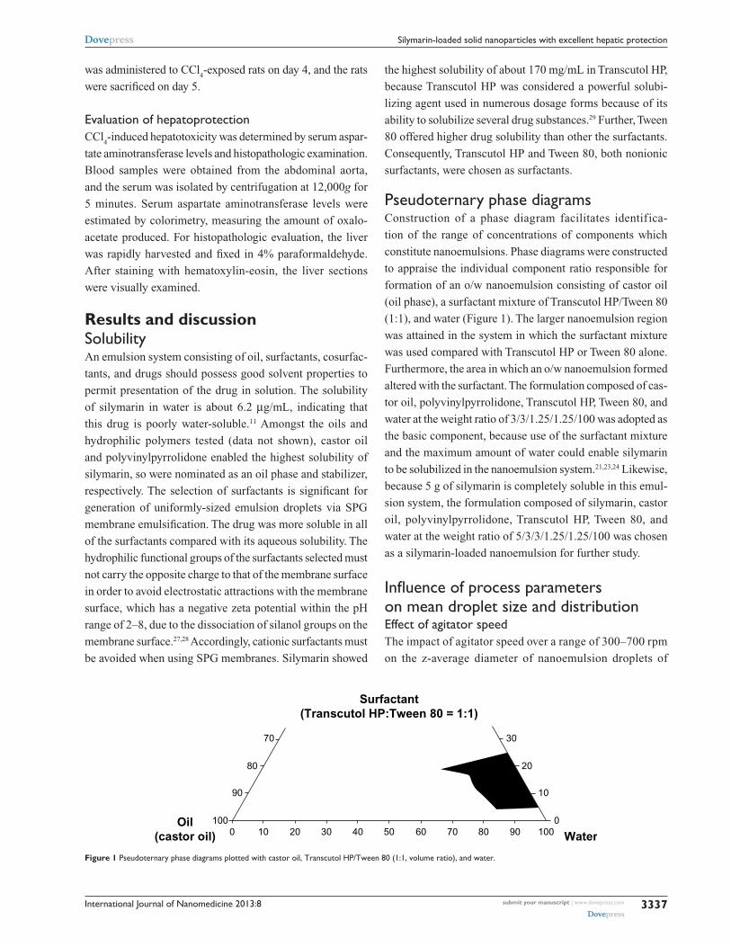
Figure 1 Pseudoternary phase diagrams plotted with castor oil, Transcutol hP/Tween 80 (1:1, volume ratio), and water.
submit your manuscript | www.dovepress.com
Dovepress
Dovepress
3337
silymarin-loaded solid nanoparticles with excellent hepatic protection
was administered to CCl4-exposed rats on day 4, and the rats
were sacrificed on day 5.
evaluation of hepatoprotectionCCl
4-induced hepatotoxicity was determined by serum aspar-
tate aminotransferase levels and histopathologic examination.
Blood samples were obtained from the abdominal aorta,
and the serum was isolated by centrifugation at 12,000g for
5 minutes. Serum aspartate aminotransferase levels were
estimated by colorimetry, measuring the amount of oxalo-
acetate produced. For histopathologic evaluation, the liver
was rapidly harvested and fixed in 4% paraformaldehyde.
After staining with hematoxylin-eosin, the liver sections
were visually examined.
Results and discussionsolubilityAn emulsion system consisting of oil, surfactants, cosurfac-
tants, and drugs should possess good solvent properties to
permit presentation of the drug in solution. The solubility
of silymarin in water is about 6.2 µg/mL, indicating that
this drug is poorly water-soluble.11 Amongst the oils and
hydrophilic polymers tested (data not shown), castor oil
and polyvinylpyrrolidone enabled the highest solubility of
silymarin, so were nominated as an oil phase and stabilizer,
respectively. The selection of surfactants is significant for
generation of uniformly-sized emulsion droplets via SPG
membrane emulsification. The drug was more soluble in all
of the surfactants compared with its aqueous solubility. The
hydrophilic functional groups of the surfactants selected must
not carry the opposite charge to that of the membrane surface
in order to avoid electrostatic attractions with the membrane
surface, which has a negative zeta potential within the pH
range of 2–8, due to the dissociation of silanol groups on the
membrane surface.27,28 Accordingly, cationic surfactants must
be avoided when using SPG membranes. Silymarin showed
the highest solubility of about 170 mg/mL in Transcutol HP,
because Transcutol HP was considered a powerful solubi-
lizing agent used in numerous dosage forms because of its
ability to solubilize several drug substances.29 Further, Tween
80 offered higher drug solubility than other the surfactants.
Consequently, Transcutol HP and Tween 80, both nonionic
surfactants, were chosen as surfactants.
Pseudoternary phase diagramsConstruction of a phase diagram facilitates identifica-
tion of the range of concentrations of components which
constitute nanoemulsions. Phase diagrams were constructed
to appraise the individual component ratio responsible for
formation of an o/w nanoemulsion consisting of castor oil
(oil phase), a surfactant mixture of Transcutol HP/Tween 80
(1:1), and water (Figure 1). The larger nanoemulsion region
was attained in the system in which the surfactant mixture
was used compared with Transcutol HP or Tween 80 alone.
Furthermore, the area in which an o/w nanoemulsion formed
altered with the surfactant. The formulation composed of cas-
tor oil, polyvinylpyrrolidone, Transcutol HP, Tween 80, and
water at the weight ratio of 3/3/1.25/1.25/100 was adopted as
the basic component, because use of the surfactant mixture
and the maximum amount of water could enable silymarin
to be solubilized in the nanoemulsion system.21,23,24 Likewise,
because 5 g of silymarin is completely soluble in this emul-
sion system, the formulation composed of silymarin, castor
oil, polyvinylpyrrolidone, Transcutol HP, Tween 80, and
water at the weight ratio of 5/3/3/1.25/1.25/100 was chosen
as a silymarin-loaded nanoemulsion for further study.
Influence of process parameters on mean droplet size and distributioneffect of agitator speedThe impact of agitator speed over a range of 300–700 rpm
on the z-average diameter of nanoemulsion droplets of
International Journal of Nanomedicine 2013:8
Agitator speed (rpm)
Z-a
vera
ge
size
(n
m)
100
200
300
400
500
600
700
800
900 MembraneNo membrane
A B C
Pressure (kPa)
Z-a
vera
ge
size
(n
m)
100
200
300
400
500
600
Temperature (°C)300 400 500 600 700 15 20 25 30 35 25 30 35 40 45
Z-a
vera
ge
size
(n
m)
100
200
300
400
500
600
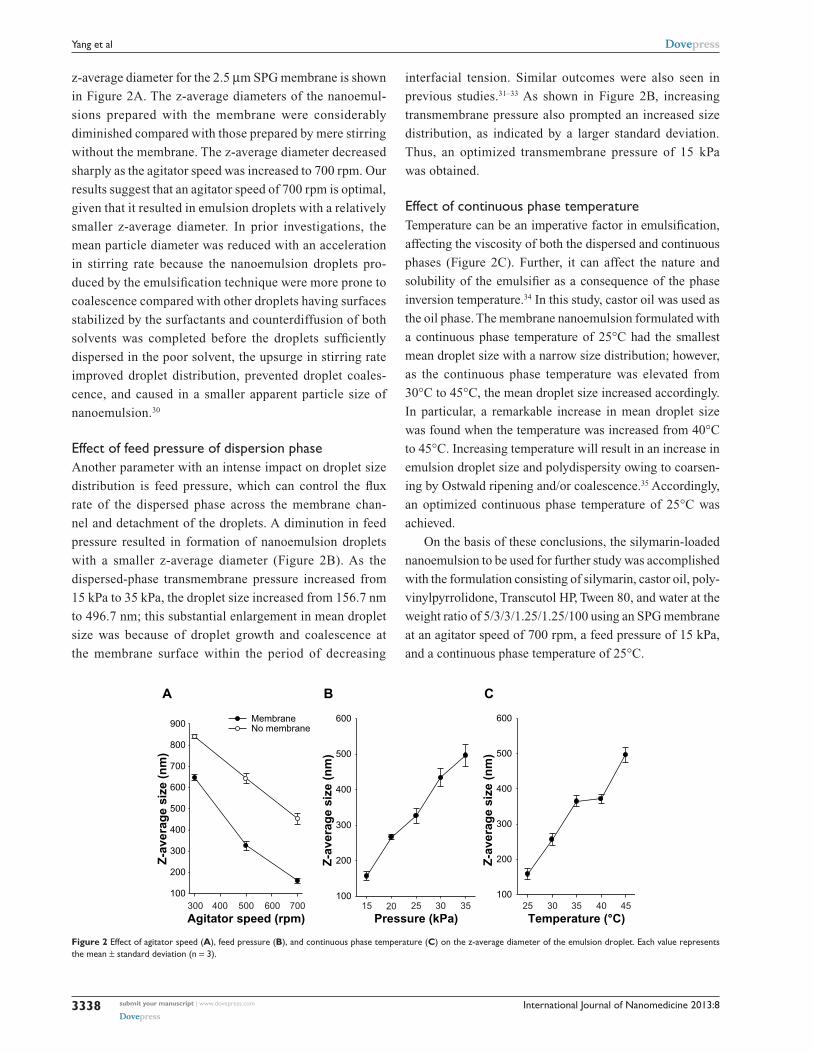
Figure 2 effect of agitator speed (A), feed pressure (B), and continuous phase temperature (C) on the z-average diameter of the emulsion droplet. each value represents the mean ± standard deviation (n = 3).
submit your manuscript | www.dovepress.com
Dovepress
Dovepress
3338
Yang et al
z-average diameter for the 2.5 µm SPG membrane is shown
in Figure 2A. The z-average diameters of the nanoemul-
sions prepared with the membrane were considerably
diminished compared with those prepared by mere stirring
without the membrane. The z-average diameter decreased
sharply as the agitator speed was increased to 700 rpm. Our
results suggest that an agitator speed of 700 rpm is optimal,
given that it resulted in emulsion droplets with a relatively
smaller z-average diameter. In prior investigations, the
mean particle diameter was reduced with an acceleration
in stirring rate because the nanoemulsion droplets pro-
duced by the emulsification technique were more prone to
coalescence compared with other droplets having surfaces
stabilized by the surfactants and counterdiffusion of both
solvents was completed before the droplets sufficiently
dispersed in the poor solvent, the upsurge in stirring rate
improved droplet distribution, prevented droplet coales-
cence, and caused in a smaller apparent particle size of
nanoemulsion.30
effect of feed pressure of dispersion phaseAnother parameter with an intense impact on droplet size
distribution is feed pressure, which can control the flux
rate of the dispersed phase across the membrane chan-
nel and detachment of the droplets. A diminution in feed
pressure resulted in formation of nanoemulsion droplets
with a smaller z-average diameter (Figure 2B). As the
dispersed-phase transmembrane pressure increased from
15 kPa to 35 kPa, the droplet size increased from 156.7 nm
to 496.7 nm; this substantial enlargement in mean droplet
size was because of droplet growth and coalescence at
the membrane surface within the period of decreasing
interfacial tension. Similar outcomes were also seen in
previous studies.31–33 As shown in Figure 2B, increasing
transmembrane pressure also prompted an increased size
distribution, as indicated by a larger standard deviation.
Thus, an optimized transmembrane pressure of 15 kPa
was obtained.
effect of continuous phase temperatureTemperature can be an imperative factor in emulsification,
affecting the viscosity of both the dispersed and continuous
phases (Figure 2C). Further, it can affect the nature and
solubility of the emulsifier as a consequence of the phase
inversion temperature.34 In this study, castor oil was used as
the oil phase. The membrane nanoemulsion formulated with
a continuous phase temperature of 25°C had the smallest
mean droplet size with a narrow size distribution; however,
as the continuous phase temperature was elevated from
30°C to 45°C, the mean droplet size increased accordingly.
In particular, a remarkable increase in mean droplet size
was found when the temperature was increased from 40°C
to 45°C. Increasing temperature will result in an increase in
emulsion droplet size and polydispersity owing to coarsen-
ing by Ostwald ripening and/or coalescence.35 Accordingly,
an optimized continuous phase temperature of 25°C was
achieved.
On the basis of these conclusions, the silymarin-loaded
nanoemulsion to be used for further study was accomplished
with the formulation consisting of silymarin, castor oil, poly-
vinylpyrrolidone, Transcutol HP, Tween 80, and water at the
weight ratio of 5/3/3/1.25/1.25/100 using an SPG membrane
at an agitator speed of 700 rpm, a feed pressure of 15 kPa,
and a continuous phase temperature of 25°C.
International Journal of Nanomedicine 2013:8
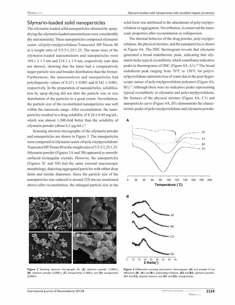
Figure 3 scanning electron micrographs for (A) silymarin powder (1,000×), (B), silymarin powder (5,000×), (C) nanoparticles (1,000×), and (D) nanoparticles (5,000×).
submit your manuscript | www.dovepress.com
Dovepress
Dovepress
3339
silymarin-loaded solid nanoparticles with excellent hepatic protection
silymarin-loaded solid nanoparticlesThe silymarin-loaded solid nanoparticles obtained by spray-
drying the silymarin-loaded nanoemulsion were considerably
dry and nonsticky. These nanoparticles comprised silymarin/
castor oil/polyvinylpyrrolidone/Transcutol HP/Tween 80
at a weight ratio of 5/3/3/1.25/1.25. The mean sizes of the
silymarin-loaded nanoemulsion and nanoparticles were
169.1 ± 1.5 nm and 214.3 ± 1.9 nm, respectively (raw data
not shown), showing that the latter had a comparatively
larger particle size and broader distribution than the former.
Furthermore, the nanoemulsion and nanoparticles had
polydispersity values of 0.211 ± 0.003 and 0.342 ± 0.001,
respectively. In the preparation of nanoparticles, solidifica-
tion by spray-drying did not alter the particle size or size
distribution of the particles to a great extent. Nevertheless,
the particle size of the reconstituted nanoparticles was well
within the nanoscale range. After reconstitution, the nano-
particles resulted in a drug solubility of 8.24 ± 0.49 mg/mL,
which was almost 1,300-fold better than the solubility of
silymarin powder (about 6.2 µg/mL).11
Scanning electron micrographs of the silymarin powder
and nanoparticles are shown in Figure 3. The nanoparticles
were composed of silymarin/castor oil/polyvinylpyrrolidone/
Transcutol HP/Tween 80 at the weight ratio of 5/3/3/1.25/1.25.
Silymarin powder (Figures 3A and 3B) appeared as smooth-
surfaced rectangular crystals. However, the nanoparticles
(Figures 3C and 3D) had the same external macroscopic
morphology, depicting aggregated particles with rather deep
dents and similar diameters. Since the particle size of the
nanoparticles was reduced to around 210 nm (as mentioned
above) after reconstitution, the enlarged particle size in the
solid form was attributed to the attachment of polyvinylpyr-
rolidone or aggregation. Nevertheless, it conserved the nano-
scale properties after reconstitution or redispersion.
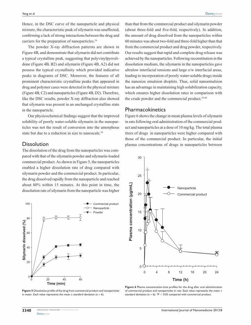
The thermal behavior of the drug powder, polyvinylpyr-
rolidone, the physical mixture, and the nanoparticles is shown
in Figure 4A. The DSC thermogram reveals that silymarin
generated a broad endothermic peak, indicating that sily-
marin lacks typical crystallinity which contributes indicative
peaks in thermograms of DSC (Figure 4A, A1).36 The broad
endotherm peak ranging from 70°C to 130°C for polyvi-
nylpyrrolidone represents loss of water due to the poor hygro-
scopic nature of polyvinylpyrrolidone polymers (Figure 4A,
B1).37 Although there were no indicative peaks representing
typical crystallinity in silymarin and polyvinylpyrrolidone,
the features of the physical mixture (Figure 4A, C1) and
nanoparticle curve (Figure 4A, D1) demonstrate the charac-
teristic peaks of polyvinylpyrrolidone and silymarin powder.
0
A
B
20 40 60 80 100
Temperature (˚C)120 140 160 180 200
A1
A2
B2
C2
D2
B1
C1
D1
5 10 15 20 252 theta(˚)
30 35 40 45
Figure 4 Differential scanning calorimetric thermograms (A) and powder X-ray diffraction (B). (A1 and B1), polyvinylpyrrolidone, (A2 and B2), silymarin powder, (C1 and C2), physical mixture, and (D1 and D2), nanoparticles.
International Journal of Nanomedicine 2013:8
Time (min)0 20 40 60
Sily
mar
in d
isso
lved
(%
)
0
20
40
60
80
100 Commercial product
Nanoparticle
Powder
Figure 5 Dissolution profile of the drug from commercial product and nanoparticles in water. each value represents the mean ± standard deviation (n = 6).
submit your manuscript | www.dovepress.com
Dovepress
Dovepress
3340
Yang et al
Hence, in the DSC curve of the nanoparticle and physical
mixture, the characteristic peak of silymarin was unaffected,
confirming a lack of strong interactions between the drug and
carriers for the preparation of nanoparticles.24
The powder X-ray diffraction patterns are shown in
Figure 4B, and demonstrate that silymarin did not contribute
a typical crystalline peak, suggesting that polyvinylpyrroli-
done (Figure 4B, B2) and silymarin (Figure 4B, A2) did not
possess the typical crystallinity which provided indicative
peaks in diagrams of DSC. Moreover, the features of all
prominent characteristic crystalline peaks that appeared in
drug and polymer cases were detected in the physical mixture
(Figure 4B, C2) and nanoparticles (Figure 4B, D2). Therefore,
like the DSC results, powder X-ray diffraction also showed
that silymarin was present in an unchanged crystalline state
in the nanoparticle.
Our physicochemical findings suggest that the improved
solubility of poorly water-soluble silymarin in the nanopar-
ticles was not the result of conversion into the amorphous
state but due to a reduction in size to nanoscale.38
DissolutionThe dissolution of the drug from the nanoparticles was com-
pared with that of the silymarin powder and silymarin-loaded
commercial product. As shown in Figure 5, the nanoparticles
enabled a higher dissolution rate of drug compared with
silymarin powder and the commercial product. In particular,
the drug dissolved rapidly from the nanoparticle and reached
about 80% within 15 minutes. At this point in time, the
dissolution rate of silymarin from the nanoparticle was higher
than that from the commercial product and silymarin powder
(about three-fold and five-fold, respectively). In addition,
the amount of drug dissolved from the nanoparticles within
60 minutes was about two-fold and three-fold higher than that
from the commercial product and drug powder, respectively.
Our results suggest that rapid and complete drug release was
achieved by the nanoparticles. Following reconstitution in the
dissolution medium, the silymarin in the nanoparticles gave
ultralow interfacial tensions and large o/w interfacial areas,
leading to incorporation of poorly water-soluble drugs inside
the nanosize emulsion droplets. Thus, solid nanoemulsion
has an advantage in maintaining high solubilization capacity,
which ensures higher dissolution rates in comparison with
the crude powder and the commercial product.39,40
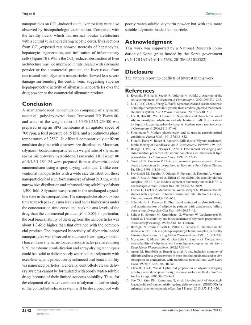
PharmacokineticsFigure 6 shows the change in mean plasma levels of silymarin
in rats following oral administration of the commercial prod-
uct and nanoparticles at a dose of 10 mg/kg. The total plasma
titers of drugs in nanoparticles were higher compared with
those of the commercial product. In particular, the initial
plasma concentrations of drugs in nanoparticles between
Time (h)
0 4 8 12 16 20 24
Pla
sma
con
cen
trat
ion
(µ
g/m
L)
0
5
10
15
20
25
30
Nanoparticle
Commercial product
*
*
*
*
Figure 6 Plasma concentration-time profiles for the drug after oral administration of commercial product and nanoparticles in rats. each value represents the mean ± standard deviation (n = 6). *P , 0.05 compared with commercial product.
International Journal of Nanomedicine 2013:8 submit your manuscript | www.dovepress.com
Dovepress
Dovepress
3341
silymarin-loaded solid nanoparticles with excellent hepatic protection
0.25 and 1.5 hours were impressively higher than those in
the commercial product (P , 0.05). Our results suggest that
the higher initial plasma concentrations of silymarin were
as a result of the increased dissolution rate of drug in the
nanoparticles.28
The pharmacokinetic parameters are shown in Table 1.
The nanoparticles provided an impressively greater area
under the concentration-time curve and higher peak plasma
levels of silymarin than the commercial product (1.27 and
1.44 times, respectively, P , 0.05). Thus, the bioavailability
of the drug achieved by the nanoparticles was about 1.3-fold
higher than that of the commercial product. On the other hand,
the time taken to reach peak drug levels for the nanoparticles
was significantly shorter than that for the commercial product
(P , 0.05). The plasma levels achieved by the nanoparticles
increased to 22.85 ± 3.59 µg/mL at Tmax
(time taken to reach
peak plasma drug concentration; 1 hour). Our results suggest
that a shorter time to peak plasma levels followed by a higher
peak plasma concentration was owing to more complete
absorption of silymarin in the nanoparticles. Therefore, the
enhanced oral bioavailability of silymarin in rats might be
associated with marked enhancement in the absorption rate of
silymarin as a result of the increased dissolution of drug in the
nanoparticles. Conversely, the Kel (elimination rate constant)
and t1/2
(elimination half-life) values of the drug did not vary
considerably between these two groups.21 Our results indicate
that nanoparticles could be worthwhile for the delivery of
drugs in a design that permits rapid absorption in the early
phase, resulting in complete and better absorption.23,24
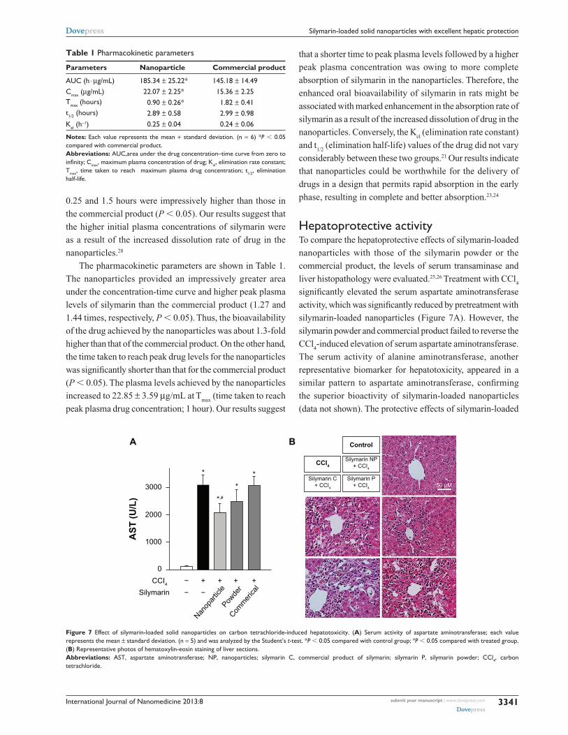
hepatoprotective activityTo compare the hepatoprotective effects of silymarin-loaded
nanoparticles with those of the silymarin powder or the
commercial product, the levels of serum transaminase and
liver histopathology were evaluated.25,26 Treatment with CCl4
significantly elevated the serum aspartate aminotransferase
activity, which was significantly reduced by pretreatment with
silymarin-loaded nanoparticles (Figure 7A). However, the
silymarin powder and commercial product failed to reverse the
CCl4-induced elevation of serum aspartate aminotransferase.
The serum activity of alanine aminotransferase, another
representative biomarker for hepatotoxicity, appeared in a
similar pattern to aspartate aminotransferase, confirming
the superior bioactivity of silymarin-loaded nanoparticles
(data not shown). The protective effects of silymarin-loaded
Table 1 Pharmacokinetic parameters
Parameters Nanoparticle Commercial product
aUc (h ⋅ µg/ml) 185.34 ± 25.22* 145.18 ± 14.49cmax (µg/ml) 22.07 ± 2.25* 15.36 ± 2.25Tmax (hours) 0.90 ± 0.26* 1.82 ± 0.41t1/2 (hours) 2.89 ± 0.58 2.99 ± 0.98Kel (h
-1) 0.25 ± 0.04 0.24 ± 0.06
Notes: each value represents the mean + standard deviation. (n = 6) *P , 0.05 compared with commercial product. Abbreviations: aUc,area under the drug concentration–time curve from zero to infinity; Cmax, maximum plasma concentration of drug; Kel, elimination rate constant; Tmax, time taken to reach maximum plasma drug concentration; t1/2, elimination half-life.
0
−
− −
Nanop
artic
lePow
der
Comm
erica
l
CCI4
Silymarin
+ + + +
50 µM
1000
2000
AS
T (
U/L
)
3000
A B
*
*,#
*
*
CCI4
Control
Silymarin C+ CCI4
Silymarin NP+ CCI4
Silymarin P+ CCI4
Figure 7 effect of silymarin-loaded solid nanoparticles on carbon tetrachloride-induced hepatotoxicity. (A) Serum activity of aspartate aminotransferase; each value represents the mean ± standard deviation. (n = 5) and was analyzed by the student’s t-test. *P , 0.05 compared with control group; #P , 0.05 compared with treated group. (B) representative photos of hematoxylin-eosin staining of liver sections. Abbreviations: asT, aspartate aminotransferase; NP, nanoparticles; silymarin C, commercial product of silymarin; silymarin P, silymarin powder; CCl4, carbon tetrachloride.
International Journal of Nanomedicine 2013:8submit your manuscript | www.dovepress.com
Dovepress
Dovepress
3342
Yang et al
nanoparticles on CCl4-induced acute liver toxicity were also
observed by histopathologic examination. Compared with
the healthy livers, which had normal lobular architecture
with a central vein and radiating hepatic cords, liver sections
from CCl4-exposed rats showed necrosis of hepatocytes,
hepatocyte degeneration, and infiltration of inflammatory
cells (Figure 7B). While the CCl4-induced destruction of liver
architecture was not improved in rats treated with silymarin
powder or the commercial product, the liver tissue from
rats treated with silymarin nanoparticles showed less severe
damage surrounding the central vein, suggesting superior
hepatoprotective activity of silymarin nanoparticles over the
drug powder or the commercial silymarin product.
ConclusionA silymarin-loaded nanoemulsion composed of silymarin,
castor oil, polyvinylpyrrolidone, Transcutol HP, Tween 80,
and water at the weight ratio of 5/3/3/1.25/1.25/100 was
prepared using an SPG membrane at an agitator speed of
700 rpm, a feed pressure of 15 kPa, and a continuous phase
temperature of 25°C. It offered comparatively uniform
emulsion droplets with a narrow size distribution. Moreover,
silymarin-loaded nanoparticles at a weight ratio of silymarin/
castor oil/polyvinylpyrrolidone/Transcutol HP/Tween 80
of 5/3/3/1.25/1.25 were prepared from a silymarin-loaded
nanoemulsion using a spray-drying technique. Unlike con-
ventional nanoparticles with a wide size distribution, these
nanoparticles had a uniform nanosize of about 210 nm, with a
narrow size distribution and enhanced drug solubility of about
1,300-fold. Silymarin was present in the unchanged crystal-
line state in the nanoparticles. The nanoparticles also took less
time to reach peak plasma levels and had a higher area under
the concentration-time curve and peak plasma levels of the
drug than the commercial product (P , 0.05). In particular,
the oral bioavailability of the drug from the nanoparticles was
about 1.3-fold higher than that obtained with the commer-
cial product. The improved bioactivity of silymarin-loaded
nanoparticles was observed in rat acute liver injury models.
Hence, these silymarin-loaded nanoparticles prepared using
SPG membrane emulsification and spray-drying techniques
could be useful to deliver poorly water-soluble silymarin with
excellent hepatic protection by enhanced oral bioavailability
via nanosized particles. Generally, controlled-release deliv-
ery systems cannot be formulated with poorly water-soluble
drugs because of their limited aqueous solubility. Thus, for
development of a better candidate of silymarin, further study
of the controlled-release system will be developed not with
poorly water-soluble silymarin powder but with this more
soluble silymarin-loaded nanoparticle.
AcknowledgmentThis work was supported by a National Research Foun-
dation of Korea grant funded by the Korea government
(N2012R1A2A2A01045658, 2013M4A1035382).
DisclosureThe authors report no conflicts of interest in this work.
References 1. Kvasnika F, Biba B, Sevcík R, Voldrich M, Krátká J. Analysis of the
active components of silymarin. J Chromatogr A. 2003;990:239–245. 2. Lu C, Lu Y, Chen J, Zhang W, Wu W. Synchronized and sustained release
of multiple components in silymarin from erodible glyceryl monostear-ate matrix system. Eur J Pharm Biopharm. 2007;66:210–219.
3. Lee JI, Hsu BH, Wu D, Barrett JS. Separation and characterization of silybin, isosilybin, silydianin and silychristin in milk thistle extract by liquid chromatography-electrospray tandem mass spectrometry. J Chromatogr A. 2006;1116:57–68.
4. Fintelmann V. Modern phytotherapy and its uses in gastrointestinal conditions. Planta Med. 1991;57:S48–S52.
5. Flora K, Hahn M, Rosen H, Benner K. Milk thistle (Silybum marianum) for the therapy of liver disease. Am J Gastroenterol. 1998;93: 139–143.
6. Basaga H, Poli G, Tekkaya C, Aras I. Free radical scavenging and anti-oxidative properties of ‘silibin’ complexes on microsomal lipid peroxidation. Cell Biochem Funct. 1997;15:27–33.
7. Skottová N, Krecman V. Dietary silymarin improves removal of low density lipoproteins by the perfused rat liver. Acta Univ Palacki Olomuc Fac Med. 1998;141:39–40.
8. Provinciali M, Papalini F, Orlando F, Pierpaoli S, Donnini A, Moraz-zoni P, Riva A, Smorlesi A. Effect of the silybin-phosphatidylcholine complex (IdB 1016) on the development of mammary tumors in HER-2/neu transgenic mice. Cancer Res. 2007;67:2022–2029.
9. Lorenz D, Lucker P, Mennicke W, Wetzelsberger N. Pharmacokinetic studies with silymarin in human serum and bile. Methods Find Exp Clin Pharmacol. 1984;6:655–661.
10. Schandalik R, Perucca E. Pharmacokinetics of silybin following oral administration of silipide in patients with extrahepatic biliary obstruction. Drugs Exp Clin Res. 1994;20:37–42.
11. Schulz H, Schurer M, Krumbiegel G, Wachter W, Weyhenmeyer R, Seidel G. The solubility and bioequivalence of silymarin preparations. Arzneimittelforschung. 1995;45:61–64. German.
12. Barzaghi N, Crema F, Gatti G, Pifferi G, Perucca E. Pharmacokinetic studies on IdB 1016, a silybin-phosphatidylcholine complex, in healthy human subjects. Eur J Drug Metab Pharmacokinet. 1990;15: 333–338.
13. Morazzoni P, Magistretti M, Giachetti C, Zanolo G. Comparative bioavailability of silipide, a new flavanolignan complex, in rats. Eur J Drug Metab Pharmacokinet. 1992;17:39–44.
14. Arcari M, Brambilla A, Brandt A, et al. A new inclusion complex of silibinin and beta-cyclodextrins: in vitro dissolution kinetics and in vivo absorption in comparison with traditional formulations. Boll Chim Farm. 1992;131:205–209. Italian.
15. Chen W, Xia H, Wu W. Optimized preparation of silymarin dripping pills by a central composite design response surface method. Chin Trad Herbal Drugs. 2005;36:679–683.
16. Seo YG, Kim DH, Ramasamy T, et al. Development of docetaxel-loaded solid self-nanoemulsifying drug delivery system (SNEDDS) for enhanced chemotherapeutic effect. Int J Pharm. 2013;452:412–420.
International Journal of Nanomedicine
Publish your work in this journal
Submit your manuscript here: http://www.dovepress.com/international-journal-of-nanomedicine-journal
The International Journal of Nanomedicine is an international, peer-reviewed journal focusing on the application of nanotechnology in diagnostics, therapeutics, and drug delivery systems throughout the biomedical field. This journal is indexed on PubMed Central, MedLine, CAS, SciSearch®, Current Contents®/Clinical Medicine,
Journal Citation Reports/Science Edition, EMBase, Scopus and the Elsevier Bibliographic databases. The manuscript management system is completely online and includes a very quick and fair peer-review system, which is all easy to use. Visit http://www.dovepress.com/ testimonials.php to read real quotes from published authors.
International Journal of Nanomedicine 2013:8 submit your manuscript | www.dovepress.com
Dovepress
Dovepress
Dovepress
3343
silymarin-loaded solid nanoparticles with excellent hepatic protection
17. Lladser M, Medrano C, Arancibia A. The use of supports in the lyophilization of oil-in-water emulsions. J Pharm Pharmacol. 1968;20: 450–455.
18. Myers S, Shively M. Solid-state emulsions: the effects of maltodextrin on microcrystalline aging. Pharm Res. 1993;10:1389–1391.
19. Kim GG, Poudel BK, Marasini N, et al. Enhancement of oral bioavail-ability of fenofibrate by solid selfmicroemulsifying drug delivery systems. Drug Dev Ind Pharm. 2013;39:1431–1438.
20. Nakashima T, Shimizu M, Kukizaki M. Particle control of emulsion by membrane emulsification and its applications. Adv Drug Deliv Rev. 2000;45:47–56.
21. Oh DH, Balakrishnan P, Oh YK, Kim DD, Yong CS, Choi HG. Effect of process parameters on nanoemulsion droplet size and distribution in SPG membrane emulsif ication. Int J Pharm. 2011;404:191–197.
22. Society of Toxicology. Guiding Principles in the Use of Animals in Toxicology. Reston, VA: Society of Toxicology; 2008. Available from: http://www.toxicology.org/AI/FA/guidingprinciples.pdf. Accessed August 6, 2013.
23. Choi YK, Poudel BK, Marasini N, et al. Enhanced solubility and oral bioavailability of itraconazole by combining membrane emulsification and spray drying technique. Int J Pharm. 2012;434:264–271.
24. Pradhan R, Lee DW, Choi HG, Yong CS, Kim JO. Fabrication of a uniformly sized fenofibrate microemulsion by membrane emulsification. J Microencapsul. 2013;30:42–48.
25. Fariss MW, Bryson KF, Hylton EE, Lippman HR, Stubin CH, Zhao XG. Protection against carbon tetrachloride-induced hepatotoxicity by pre-treating rats with the hemisuccinate esters of tocopherol and cholesterol. Environ Health Perspect. 1993;101:528–536.
26. Lee SN, Poudel BK, Tran TH, et al. A novel surface-attached carvedilol solid dispersion with enhanced solubility and dissolution. Arch Pharm Res. 2013;3:79–85.
27. Marasini N, Yan YD, Poudel BK, Choi HG, Yong CS, Kim JO. Develop-ment and optimization of self-nanoemulsifying drug delivery system with enhanced bioavailability by Box-Behnken design and desirability function. J Pharm Sci. 2012;101:4584–4596.
28. Vladisavljevic GT, Williams RA. Recent developments in manufacturing emulsions and particulate products using membranes. Adv Colloid Interface Sci. 2005;113:1–20.
29. Kim HJ, Yoon KA, Hahn M, Park ES, Chi SC. Preparation and in vitro evaluation of self-microemulsifying drug delivery systems containing idebenone. Drug Dev Ind Pharm. 2000;26:523–529.
30. Tsukada Y, Hara K, Bando Y, et al. Particle size control of poly (dl-lactide-co-glycolide) nanospheres for sterile applications. Int J Pharm. 2009;370:196–201.
31. Vladisavljevic GT, Williams RA. Manufacture of large uniform drop-lets using rotating membrane emulsification. J Colloid Interface Sci. 2006;299:396–402.
32. Vladisavljevic GT, Duncanson WJ, Shum HC, Weitz DA. Emulsion templating of poly(lactic acid) particles: droplet formation behavior. Langmuir. 2012;28:12948–12954.
33. Vladisavljevi G, Schubert H. Preparation of emulsions with a narrow particle size distribution using microporous α-alumina membranes. J Dispers Sci Technol. 2003;24:811–819.
34. Oh DH, Kang JH, Kim DW, Lee BJ, Kim JO, Yong CS, Choi HG. Comparison of solid self-microemulsifying drug delivery system (solid SMEDDS) prepared with hydrophilic and hydrophobic solid carrier. Int J Pharm. 2011;420:412-418.
35. Ee S, Duan X, Liew J, Nguyen Q. Droplet size and stability of nano-emulsions produced by the temperature phase inversion method. Chem Eng J. 2008;140:626–631.
36. Quan Q, Kim DW, Marasini N,et al. Physicochemical characteriza-tion and in vivo evaluation of solid self-nanoemulsifying drug deliv-ery system for oral administration of docetaxel. J Microencapsul. 2013;30:307–314.
37. Sethia S, Squillante E. Solid dispersion of carbamazepine in PVP K30 by conventional solvent evaporation and supercritical methods. Int J Pharm. 2004;272:1–10.
38. Grau MJ, Kayser O, Müller RH. Nanosuspensions of poorly soluble drugs – reproducibility of small scale production. Int J Pharm. 2000;196:155–157.
39. Albers J, Alles R, Matthe, K, Knop K, Nahrup J, Kleinebudde P. Mechanism of drug release from polymethacrylate-based extrudates and milled strands prepared by hot-melt extrusion. Eur J Pharm Biopharm. 2009;71:387–394.
40. Xi J, Chang Q, Chan CK, et al. Formulation development and bioavailability evaluation of a self-nanoemulsified drug delivery system of oleanolic acid. AAPS PharmSciTech. 2009;10:172–182.
Related Documents


![Silymarin as Supportive Treatment in Liver Diseases: A ... · specific treatment ‘‘against liver diseases’’ [10]. The first commercial preparation of silymarin was developed](https://static.cupdf.com/doc/110x72/60236bef98a17f3f9a1db02d/silymarin-as-supportive-treatment-in-liver-diseases-a-speciic-treatment-aaagainst.jpg)


