BioMed Central Page 1 of 19 (page number not for citation purposes) BMC Biotechnology Open Access Research article Silica-based cationic bilayers as immunoadjuvants Nilton Lincopan 1 , Mariana RA Santana 1 , Eliana Faquim-Mauro 2 , Maria Helena B da Costa 2 and Ana M Carmona-Ribeiro* 1 Address: 1 Departamento de Bioquímica, Instituto de Química, Universidade de São Paulo, São Paulo, Caixa Postal 26077, São Paulo-SP, Brazil and 2 Instituto Butantan, Av. Vital Brasil 1500, Butantan 05503-900, Brazil Email: Nilton Lincopan - [email protected]; Mariana RA Santana - [email protected]; Eliana Faquim- Mauro - [email protected]; Maria Helena B da Costa - [email protected]; Ana M Carmona-Ribeiro* - [email protected] * Corresponding author Abstract Background: Silica particles cationized by dioctadecyldimethylammonium bromide (DODAB) bilayer were previously described. This work shows the efficiency of these particulates for antigen adsorption and presentation to the immune system and proves the concept that silica-based cationic bilayers exhibit better performance than alum regarding colloid stability and cellular immune responses for vaccine design. Results: Firstly, the silica/DODAB assembly was characterized at 1 mM NaCl, pH 6.3 or 5 mM Tris.HCl, pH 7.4 and 0.1 mg/ml silica over a range of DODAB concentrations (0.001–1 mM) by means of dynamic light scattering for particle sizing and zeta-potential analysis. 0.05 mM DODAB is enough to produce cationic bilayer-covered particles with good colloid stability. Secondly, conditions for maximal adsorption of bovine serum albumin (BSA) or a recombinant, heat-shock protein from Mycobacterium leprae (18 kDa-hsp) onto DODAB-covered or onto bare silica were determined. At maximal antigen adsorption, cellular immune responses in vivo from delayed-type hypersensitivity reactions determined by foot-pad swelling tests (DTH) and cytokines analysis evidenced the superior performance of the silica/DODAB adjuvant as compared to alum or antigens alone whereas humoral response from IgG in serum was equal to the one elicited by alum as adjuvant. Conclusion: Cationized silica is a biocompatible, inexpensive, easily prepared and possibly general immunoadjuvant for antigen presentation which displays higher colloid stability than alum, better performance regarding cellular immune responses and employs very low, micromolar doses of cationic and toxic synthetic lipid. Background Over the last two decades novel assemblies obtained from particles and lipids have been introduced as important tools to novel applications in drug and vaccine delivery [1-4]. Particulates such as silica, latex or hydrophobic drugs have been coated by lipids and successfully employed in biomolecular recognition [5,6] drug delivery [7,8] and antigen presentation [9,10]. The systematic and quantitative evaluation of particle-lipid interaction has been realized by means of adsorption isotherms of lipids on particles, effects of lipids on particle size and zeta- potential from dynamic light scattering methods and Published: 19 January 2009 BMC Biotechnology 2009, 9:5 doi:10.1186/1472-6750-9-5 Received: 1 August 2008 Accepted: 19 January 2009 This article is available from: http://www.biomedcentral.com/1472-6750/9/5 © 2009 Lincopan et al; licensee BioMed Central Ltd. This is an Open Access article distributed under the terms of the Creative Commons Attribution License (http://creativecommons.org/licenses/by/2.0 ), which permits unrestricted use, distribution, and reproduction in any medium, provided the original work is properly cited.

Welcome message from author
This document is posted to help you gain knowledge. Please leave a comment to let me know what you think about it! Share it to your friends and learn new things together.
Transcript
BioMed CentralBMC Biotechnology
ss
Open AcceResearch articleSilica-based cationic bilayers as immunoadjuvantsNilton Lincopan1, Mariana RA Santana1, Eliana Faquim-Mauro2, Maria Helena B da Costa2 and Ana M Carmona-Ribeiro*1Address: 1Departamento de Bioquímica, Instituto de Química, Universidade de São Paulo, São Paulo, Caixa Postal 26077, São Paulo-SP, Brazil and 2Instituto Butantan, Av. Vital Brasil 1500, Butantan 05503-900, Brazil
Email: Nilton Lincopan - [email protected]; Mariana RA Santana - [email protected]; Eliana Faquim-Mauro - [email protected]; Maria Helena B da Costa - [email protected]; Ana M Carmona-Ribeiro* - [email protected]
* Corresponding author
AbstractBackground: Silica particles cationized by dioctadecyldimethylammonium bromide (DODAB)bilayer were previously described. This work shows the efficiency of these particulates for antigenadsorption and presentation to the immune system and proves the concept that silica-basedcationic bilayers exhibit better performance than alum regarding colloid stability and cellularimmune responses for vaccine design.
Results: Firstly, the silica/DODAB assembly was characterized at 1 mM NaCl, pH 6.3 or 5 mMTris.HCl, pH 7.4 and 0.1 mg/ml silica over a range of DODAB concentrations (0.001–1 mM) bymeans of dynamic light scattering for particle sizing and zeta-potential analysis. 0.05 mM DODABis enough to produce cationic bilayer-covered particles with good colloid stability. Secondly,conditions for maximal adsorption of bovine serum albumin (BSA) or a recombinant, heat-shockprotein from Mycobacterium leprae (18 kDa-hsp) onto DODAB-covered or onto bare silica weredetermined. At maximal antigen adsorption, cellular immune responses in vivo from delayed-typehypersensitivity reactions determined by foot-pad swelling tests (DTH) and cytokines analysisevidenced the superior performance of the silica/DODAB adjuvant as compared to alum orantigens alone whereas humoral response from IgG in serum was equal to the one elicited by alumas adjuvant.
Conclusion: Cationized silica is a biocompatible, inexpensive, easily prepared and possibly generalimmunoadjuvant for antigen presentation which displays higher colloid stability than alum, betterperformance regarding cellular immune responses and employs very low, micromolar doses ofcationic and toxic synthetic lipid.
BackgroundOver the last two decades novel assemblies obtained fromparticles and lipids have been introduced as importanttools to novel applications in drug and vaccine delivery[1-4]. Particulates such as silica, latex or hydrophobicdrugs have been coated by lipids and successfully
employed in biomolecular recognition [5,6] drug delivery[7,8] and antigen presentation [9,10]. The systematic andquantitative evaluation of particle-lipid interaction hasbeen realized by means of adsorption isotherms of lipidson particles, effects of lipids on particle size and zeta-potential from dynamic light scattering methods and
Published: 19 January 2009
BMC Biotechnology 2009, 9:5 doi:10.1186/1472-6750-9-5
Received: 1 August 2008Accepted: 19 January 2009
This article is available from: http://www.biomedcentral.com/1472-6750/9/5
© 2009 Lincopan et al; licensee BioMed Central Ltd. This is an Open Access article distributed under the terms of the Creative Commons Attribution License (http://creativecommons.org/licenses/by/2.0), which permits unrestricted use, distribution, and reproduction in any medium, provided the original work is properly cited.
Page 1 of 19(page number not for citation purposes)

BMC Biotechnology 2009, 9:5 http://www.biomedcentral.com/1472-6750/9/5
determination of colloid stability from turbidity kineticsor particle sedimentation over time [1-10]. Cationic lip-ids, in particular, are especially interesting to cover parti-cles, since cationic particles may electrostatically combinewith a vast variety of oppositely charged biomolecules,cells or other biological structures. Cationization, in gen-eral, has often been explored as a convenient approach totarget active biomolecules into cells [11].
The control of lipid assembly on particles turned out to bedependent on properties of the intervening medium, egionic strength, and on the proportion of surface areas forbilayer vesicles and particles in dispersion [12-15]. Fromequivalence of total surface areas for particles and cationiclipid bilayers, over a range of low ionic strength, a goodcolloid stability was reported for the bilayer-covered cati-onic particles [3,4,12,15].
In this work, the interaction between silica previouslycoated with cationic bilayers of dioctadecyldimethylam-monium bromide (DODAB) [15] and the model proteinbovine serum albumin (BSA) is investigated aiming atantigen presentation to the immune system by silica-based cationized particles. BSA choice was due to variousreasons: its extensively studied adsorption behaviour atinterfaces [16,17]; its utility to prevent nonspecific bind-ing in biosensing and proteomics applications [17-19], itsconformational adaptability as a "soft" globular protein[20], its thoroughly investigated adsorption onto hydro-phobic or hydrophilic particles sometimes resulting inexchange between the adsorbed and dissolved states [21-24] and its substantial adsorption onto cationic and largeDODAB vesicles [25].
The 18 kDa-hsp protein belongs to a conserved proteinfamily of M. leprae heat-shock proteins that display pro-nounced immunogenicity and are considered importanttargets of the immunoresponse to mycobacteria and, assuch, relevant to subunit vaccine design. Peripheral bloodmononuclear cells and T-cell lines from M. leprae vacci-nated subjects proliferated in response to this protein[26]. Furthermore, overexpression and scaling-up of 18kDa-hsp production in Saccharomyces cerevisae has alreadybeen described so that this protein is available in suffi-cient amount for a complete physico-chemical study ofthe adjuvant-antigen interaction [27-29].
The DODAB cationic lipid and its assemblies in water dis-persion have been established as effective immunoadju-vants able to stimulate dendritic cells and often employedto present antigens [29-34]. Silica particles are biocompat-ible, represent a reference adsorbent, offer a chemicallywell defined surface and are widely used as a chromato-graphic stationary phase [34,35].
We have recently combined the typical property of parti-cles that stimulate dendritic cells uptake with the adjuvanteffect of DODAB by using supported DODAB bilayers onlatex to present antigens [9]. Here we take advantage ofthe biocompatible character of silica [35,36] to produceDODAB-covered silica particles for further immobiliza-tion and presentation of two different model antigens:BSA and 18 kDa-hsp protein.
Results and discussionCoverage of silica particles with a cationic bilayer and BSA adsorptionCharge density on silica particles increases with pH andionic strength [37] so that electrostatic attraction betweenDODAB bilayer and silica is substantial over the 1–10 mMrange of monovalent salt concentration, and leads toDODAB bilayer deposition onto particles [15]. Oneshould notice that poor or none DODAB adsorption onsilica was previously reported for pure water as interven-ing media [13-15,38]. Therefore, the experiments in thiswork were designed either at 1 or at 5 mM monovalentsalt.
The DODAB bilayer in closed vesicles is in the rigid gelstate at room temperature. This represents an importantlimitation regarding deposition of bilayers onto particleshampering the occurrence of vesicle disruption which isessential for bilayer deposition. We have previouslyshown that closed vesicles with bilayers in the gel state donot disrupt upon contact with silica particles [38], thus, inthis work, bilayer deposition on silica is obtained byemploying disrupted vesicles or bilayer fragments.
Both at 1 mM NaCl, pH 6.3, and at 5.0 mM Tris.HCl, pH7.4, the dependence of size and zeta-potential of silicaparticles on DODAB concentration shows size minimiza-tion and zeta-potential maximization at and above 0.050mM DODAB (Figure 1). Therefore, 0.050 mM DODABwas the final concentration selected to cover silica parti-cles at 0.1 mg/ml with one DODAB bilayer. Theoretically,assuming 0.6 nm2 as the DODAB area per molecule at theair-water interface and 26–50 m2/g as silica specific sur-face area, the DODAB concentration required to cover sil-ica particles at 0.1 mg/ml with one bilayer would be0.014–0.028 mM DODAB, in reasonable agreement withthe 0.050 mM DODAB experimentally determined forminimization of particle size and maximal zeta-potential(Figure 1). The bell-shaped dependence of zeta-averagediameter of particles as a function of the logarithm ofDODAB concentration shows the occurrence of tworegions of high colloidal stability identified by low sizesand large zeta-potentials in modulus: one at negative andthe other at positive zeta-potentials both at mean sizessimilar to the one of bare silica and over a range of lowand high DODAB concentrations (Figure 1).
Page 2 of 19(page number not for citation purposes)
BMC Biotechnology 2009, 9:5 http://www.biomedcentral.com/1472-6750/9/5
The stability of the silica/DODAB system was sistemati-cally described in a previous work [15]. The adjuvant sys-tem is very reproducible yielding always the same meanDz and zeta-potential for the same final silica andDODAB concentrations. However, upon antigen addi-tion, zeta-potential may decrease to values close to zero sothat colloid stability will be low.
Table 1 shows the detailed physical description of silica,DODAB bilayer fragments (BF), silica/DODAB and silica/DODAB/BSA regarding mean particle size, zeta-potential
and polydispersity. In agreement with Tadros andLyklema data [37], silica in 1 mM NaCl at pH 6.3 was lesscharged (zeta-potential of -40 mV) than at pH 7.4 (zeta-potential of -47 mV). One should notice the aggregatedstate of BSA in solution and the large polydispersity ofthese aggregates at 0.5 mg/ml in 1 mM NaCl and pH 6.3or in 5 mM Tris.HCl, pH 7.4 (Table 1). Consistently, at pH7.4, the protein aggregates display a more negative zeta-potential than the one at pH 6.3, as expected from BSAisoelectric point at pH 5.0–5.5. When the protein wasadded to silica/DODAB particles to a final [BSA] of 0.025
The concentration of cationic lipid DODAB required to cover silica particles with a cationic lipid bilayer is 0.05 mMFigure 1The concentration of cationic lipid DODAB required to cover silica particles with a cationic lipid bilayer is 0.05 mM. Effect of DODAB concentration on zeta-average diameter (A, C) and zeta-potential (B, D) of SiO2 particles at 0.1 mg sil-ica/ml, 25°C, in 1 mM NaCl, pH 6.3 (A, B) or 5 mM Tris.HCl, pH 7.4 (C, D). Cationic bilayer fragments were adsorbed onto 0.1 mg/ml silica particles (AEROSIL OX-50). Measurements were performed after 1 h interaction. The arrows indicate 0.05 mM DODAB as the concentration of cationic lipid that is required to attain maximal positive zeta-potential and minimal sizes for particles in dispersion.
Page 3 of 19(page number not for citation purposes)
BMC Biotechnology 2009, 9:5 http://www.biomedcentral.com/1472-6750/9/5
mg/ml, different assemblies were obtained at pH 6.3 and7.4 (Table 1): the larger electrostatic attraction at pH 7.4drove BSA onto particles to yield a slightly negativelycharged particle whereas, at pH 6.3, the moderate electro-static attraction, moderately drove BSA onto particlesyielding a still positively charged particle with 24 mV ofzeta-potential (Table 1). The design chosen for evaluationof BSA presentation to the immune system was the one in5 mM Tris.HCl, pH 7.4 yielding mean diameters of ca. 800nm. All other results in Table 1, consistently agreed withprevious data published by our group.
Figure 2 shows size distributions for silica, DODAB BFand silica/DODAB for each dispersion both at 1 mMNaCl, pH 6.3 and at 5 mM Tris. HCl, pH 7.4. Under bothexperimental conditions, DODAB addition produced thesilica-based positively charged particles with final size dis-
tributions similar to the ones of bare silica. Quantificationof BSA amount adsorbed from solution onto bare silica(Figure 3) or onto silica/DODAB (Figure 4) in the twoquoted intervening media was carried out over a range ofadded BSA concentrations (0–150 μg/ml). Given the lang-muirian profile of the adsorption isotherms, we appliedthe Langmuir model in order to linearize adsorption iso-therms and obtain adsorption parameters prone to becompared with each other for different intervening mediaand silica or silica/DODAB as adsorbates. Table 2 summa-rizes the BSA adsorption parameters such as maximaladsorption and affinity constant for adsorption onto baresilica or onto silica/DODAB particles in the two differentintervening media. BSA adsorbs with the highest affinityonto bare silica, possibly driven by several hydrogenbonds between silanoils on particles and carbonyls on theprotein, but maximal adsorption is at lowest indicating
Table 1: Physical properties of SiO2, DODAB BF dispersion, BSA, 18 kDa-hsp leprae and their mixtures in 1 mM NaCl (pH 6.3) or in 5 mM TrisHCl (pH 7.4)
Dispersion SiO2(mg/ml)
DODAB(mM)
Protein(mg/ml)
Mean Diameter(nm)
Zeta Potencial(mV)
Polydispersity(Index)
SiO2(i) 0.1 - - 328 ± 7 -40 ± 2 0.240 ± 0.01
DODAB(i) - 1.0 - 67 ± 1 41 ± 3 0.120 ± 0.04BSA(i) - - 0.5 25 ± 4 -28 ± 1 0.450 ± 0.09DODAB/BSA(i) - 0.1 0.500 72 ± 1 3 ± 1 0.245 ± 0.01SiO2/DODAB(i) 0.1 0.05 - 304 ± 2 32 ± 1 0.260 ± 0.02SiO2/DODAB/BSA(i) 0.1 0.05 0.025 324 ± 8 24 ± 1 0.301 ± 0.01SiO2
(ii) 0.1 - - 294 ± 2 -47 ± 2 0.180 ± 0.02DODAB(ii) - 1.0 1.0 74 ± 1 33 ± 5 0.270 ± 0.04BSA(ii) - - 0.5 24 ± 1 -58 ± 18 0.397 ± 0.01SiO2/DODAB(ii) 0.1 0.05 - 373 ± 7 33 ± 2 0.260 ± 0.02SiO2/DODAB/BSA(ii) 0.1 0.05 0.025 851 ± 36 -1.3 ± 4 0.396 ± 0.0318 kDa-hsp M. leprae(i) - - 0.5 732 ± 188 -29 ± 2 0.35 ± 0.05DODAB/18 kDa-hsp(i) - 0.1 0.05 208 ± 8 39 ± 5 0.315 ± 0.0118 kDa-hsp M. leprae(ii) - - 0.05 360 ± 16 -43 ± 18 0.180 ± 0.09SiO2/DODAB/18 kDa-hsp(ii) 0.1 0.05 0.05 2132 ± 370 -2 ± 2 0.492 ± 0.04Al(OH3)(i) - - - 458 ± 3 28 ± 3 0.191 ± 0.04Al(OH3)(ii) - - - 702 ± 17 14 ± 2 0.317 ± 0.01Al(OH3)/18 kDa-hsp(i),(iii) - - 0.05 4888 ± 665 - 0.272 ± 0.07Al(OH3)/18 kDa-hsp(ii),(iii) - - 0.05 10607 ± 2998 - 0.480 ± 0.03
(i) 1 mM NaCl (pH 6.3). (ii) 5 mM Tris-HCl (pH 7.4). (iii) Al(OH3) at 0.1 mg/ml.
Table 2: Maximal adsorption, affinity constant (K) and area per adsorbed BSA molecule on silica (0.1 mg/ml) or on DODAB covered silica particles in two different media
Particles Maximal adsorption(molecules m-2)
Affinity Constant(K, M-1)
Area per adsorbed molecule(nm2)
SiO2/BSA(i) 4.14 × 10¹⁶ 1.19 × 10⁹ 24.15SiO2/BSA(ii) 4.75 × 10¹⁶ 8.19 × 10⁷ 21.05SiO2/DODAB/BSA(i) 7.20 × 10¹⁶ 5.18 × 10⁷ 13.88SiO2/DODAB/BSA(ii) 3.22 × 10¹⁷ 8.98 × 10⁸ 3.10SiO2/18 kDa-hsp(ii) 1.12 × 10¹⁷ - -SiO2/DODAB/18 kDa-hsp(ii) 10.00 × 10¹⁷ - -
(i) 1 mM NaCl (pH 6.3).(ii) 5 mM Tris.HCl (pH 7.4).Final DODAB concentration was 0.05 mM.
Page 4 of 19(page number not for citation purposes)
BMC Biotechnology 2009, 9:5 http://www.biomedcentral.com/1472-6750/9/5
the largest area per BSA molecule occupied at maximaladsorption among those in all media and adsorbatestested (Table 2). Changing the medium to 5 mM Tris.HCland pH 7.4, diminished BSA affinity for the negativelycharged bare silica since electrostatic repulsion wasincreased by increasing negative charge on the proteinthough maximal adsorption remained approximately thesame (Table 2). Offering the DODAB bilayer on silica tothe protein improved BSA maximal adsorption for both
intervening media. However, a large increase in the affin-ity constant such as the one obtained for BSA adsorptionon silica/DODAB in Tris.HCl pH 7.4 (Table 2) may not beso desirable in terms of molecular order of BSA on the par-ticle surface. While BSA appeared to be in a ordered end-on position occupying about 14 nm2 at maximal adsorp-tion (Table 2) under conditions of moderate electrostaticattraction between BSA and silica/DODAB (1 mM NaCl,pH 6.3) further increasing this attraction drove massive
Comparison of size distributions and zeta-potentials for silica and silica-based cationic bilayersFigure 2Comparison of size distributions and zeta-potentials for silica and silica-based cationic bilayers. Size distribution and zeta-potential (ζ) for 0.1 mg/ml silica, dispersion of 1 mM DODAB bilayer fragments and silica/DODAB particles prepared from 0.1 mg/ml silica and 0.05 mM DODAB. The intervening medium was 1 mM NaCl, pH 6.3 (A-C) or 5 mM Tris.HCl, pH 7.4 (D-F). Measurements were performed, after 1 h interaction. In each subfigure, mean zeta-potential and diameter ± SE are quoted.
Page 5 of 19(page number not for citation purposes)
BMC Biotechnology 2009, 9:5 http://www.biomedcentral.com/1472-6750/9/5
BSA adsorption onto the particle as detected from thesmall area occupied per protein on the silica/DODAB sur-face, about 3 nm2 (Table 2). It was previously reported byus that BSA maximal adsorption onto supported DODABbilayers deposited on polystyrene sulfate microspheres
with 1 mM NaCl, pH 6.3 as the intervening medium wasalso consistent with deposition of an end-on BSA mono-molecular layer [24]. This agreement for different sup-porting particles reconfirms not only DODAB bilayerdeposition on silica but also suggests the preferential end-
Low levels of maximal BSA adsorption onto bare silicaFigure 3Low levels of maximal BSA adsorption onto bare silica. BSA adsorption isotherms onto bare SiO2 particles in two dif-ferent media: 1 mM NaCl, pH 6.3 or 5 mM Tris.HCl, pH 7.4. Open and filled circles refer to two independent experiments with each mixture evaluated in duplicate. Interaction between silica particles and protein took place for 1 h at 25°C. Final silica concentration is 0.1 mg/ml. BSA adsorption was expressed either as adsorbed BSA concentration in mg/ml (A and C) or as number of BSA molecules adsorbed per m2 silica (B and D).
Page 6 of 19(page number not for citation purposes)
BMC Biotechnology 2009, 9:5 http://www.biomedcentral.com/1472-6750/9/5
on mode of BSA adsorption on DODAB bilayer at pH 6.3and 1 mM NaCl.
Size distributions for BSA aggregates, silica/DODAB andsilica/DODAB/BSA particles are on Figure 5 and Figure 6
for dispersions in 1 mM NaCl, pH 6.3 and in 5 mMTris.HCl, pH 7.4, respectively. Interestingly enough,increasing BSA concentration from 5 up to 30 μg/ml, col-loid stability rapidly decreased for the latter environmentyielding aggregates with ca. 2.5 μm mean diameter and
High levels of maximal BSA adsorption onto silica-based cationic bilayersFigure 4High levels of maximal BSA adsorption onto silica-based cationic bilayers. BSA adsorption isotherms onto silica/DODAB particles in two different media. 1 mM NaCl, pH 6.3 or 5 mM Tris.HCl, pH 7.4. Open and filled circles refer to two independent experiments with each mixture evaluated in duplicate. Interaction between silica/DODAB particles and protein took place for 1 h at 25°C. Final silica and DODAB concentrations are 0.1 mg/ml and 0.05 mM, respectively. BSA adsorption was expressed as adsorbed BSA concentration in mg/ml (A and C) or as number of BSA molecules adsorbed per m2 silica (B and D).
Page 7 of 19(page number not for citation purposes)
BMC Biotechnology 2009, 9:5 http://www.biomedcentral.com/1472-6750/9/5
Page 8 of 19(page number not for citation purposes)
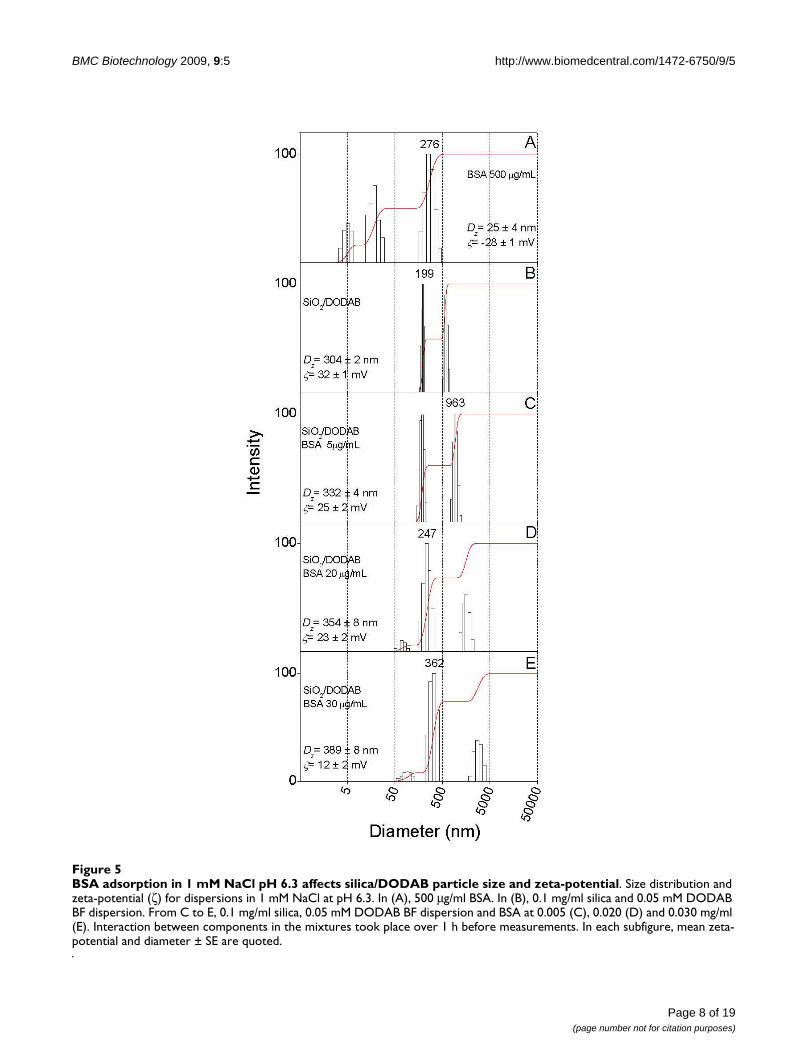
BSA adsorption in 1 mM NaCl pH 6.3 affects silica/DODAB particle size and zeta-potentialFigure 5BSA adsorption in 1 mM NaCl pH 6.3 affects silica/DODAB particle size and zeta-potential. Size distribution and zeta-potential (ζ) for dispersions in 1 mM NaCl at pH 6.3. In (A), 500 μg/ml BSA. In (B), 0.1 mg/ml silica and 0.05 mM DODAB BF dispersion. From C to E, 0.1 mg/ml silica, 0.05 mM DODAB BF dispersion and BSA at 0.005 (C), 0.020 (D) and 0.030 mg/ml (E). Interaction between components in the mixtures took place over 1 h before measurements. In each subfigure, mean zeta-potential and diameter ± SE are quoted.

BMC Biotechnology 2009, 9:5 http://www.biomedcentral.com/1472-6750/9/5
Page 9 of 19(page number not for citation purposes)
BSA adsorption in 5 mM Tris.HCl, pH 7.4 affects silica/DODAB particle size and zeta-potentialFigure 6BSA adsorption in 5 mM Tris.HCl, pH 7.4 affects silica/DODAB particle size and zeta-potential. Size distribution and zeta-potential (ζ) for dispersions in 5 mM Tris.HCl, pH 7.4. In (A), 500 μg/ml BSA. In (B), 0.1 mg/ml silica and 0.05 mM DODAB BF dispersion. From C to E, 0.1 mg/ml silica, 0.05 mM DODAB BF dispersion and BSA at 0.005 (C), 0.02 (D) and 0.03 mg/ml (E). Interaction between components in the mixtures took place over 1 h before measurements. In each subfigure, mean zeta-potential and diameter ± SE are quoted.
BMC Biotechnology 2009, 9:5 http://www.biomedcentral.com/1472-6750/9/5
zeta-potentials close to zero (Figure 6E). The moderateelectrostatic attraction in 1 mM NaCl, pH 6.3 producedhigher colloid stability for silica/DODAB/BSA assembliesgiven their positive charge even at the largest BSA concen-tration (30 μg/ml) (Figure 5E). However, we privilegedthe largest adsorbed amount of BSA obtained in Tris.HCl,pH 7.4 for testing immune response.
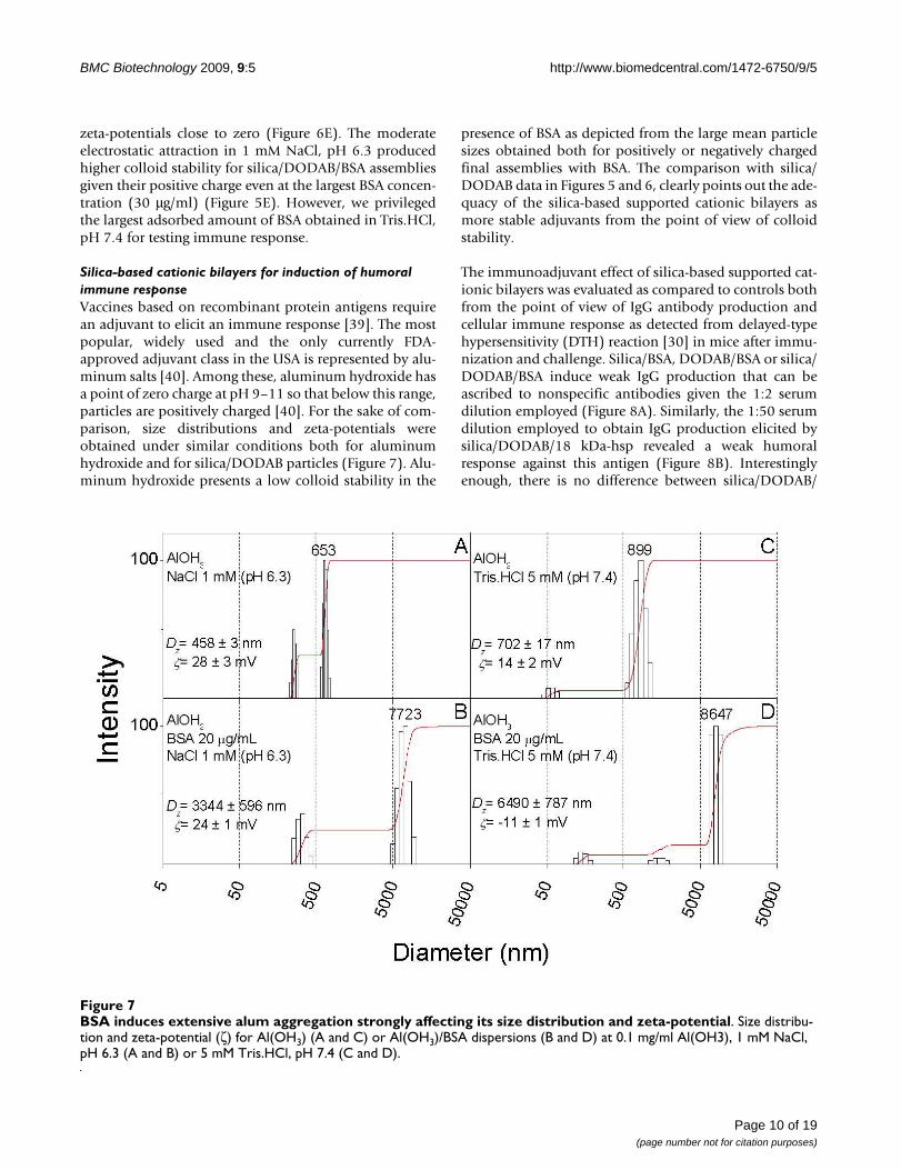
Silica-based cationic bilayers for induction of humoral immune responseVaccines based on recombinant protein antigens requirean adjuvant to elicit an immune response [39]. The mostpopular, widely used and the only currently FDA-approved adjuvant class in the USA is represented by alu-minum salts [40]. Among these, aluminum hydroxide hasa point of zero charge at pH 9–11 so that below this range,particles are positively charged [40]. For the sake of com-parison, size distributions and zeta-potentials wereobtained under similar conditions both for aluminumhydroxide and for silica/DODAB particles (Figure 7). Alu-minum hydroxide presents a low colloid stability in the
presence of BSA as depicted from the large mean particlesizes obtained both for positively or negatively chargedfinal assemblies with BSA. The comparison with silica/DODAB data in Figures 5 and 6, clearly points out the ade-quacy of the silica-based supported cationic bilayers asmore stable adjuvants from the point of view of colloidstability.
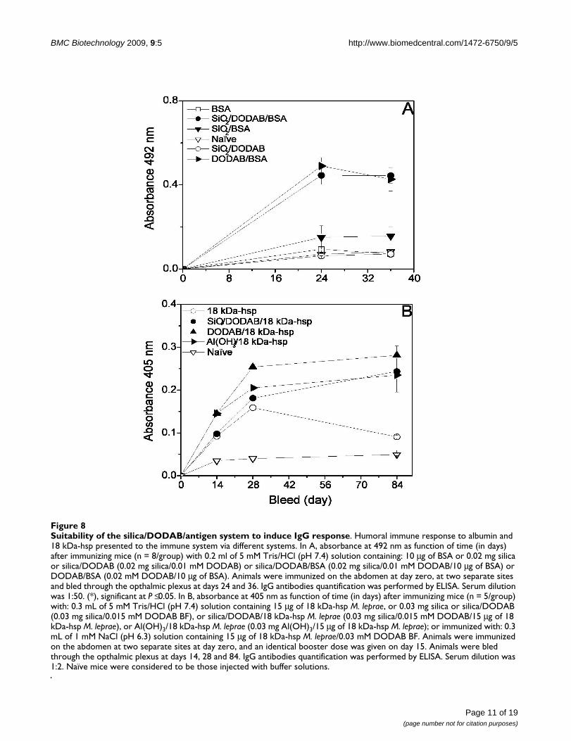
The immunoadjuvant effect of silica-based supported cat-ionic bilayers was evaluated as compared to controls bothfrom the point of view of IgG antibody production andcellular immune response as detected from delayed-typehypersensitivity (DTH) reaction [30] in mice after immu-nization and challenge. Silica/BSA, DODAB/BSA or silica/DODAB/BSA induce weak IgG production that can beascribed to nonspecific antibodies given the 1:2 serumdilution employed (Figure 8A). Similarly, the 1:50 serumdilution employed to obtain IgG production elicited bysilica/DODAB/18 kDa-hsp revealed a weak humoralresponse against this antigen (Figure 8B). Interestinglyenough, there is no difference between silica/DODAB/
BSA induces extensive alum aggregation strongly affecting its size distribution and zeta-potentialFigure 7BSA induces extensive alum aggregation strongly affecting its size distribution and zeta-potential. Size distribu-tion and zeta-potential (ζ) for Al(OH3) (A and C) or Al(OH3)/BSA dispersions (B and D) at 0.1 mg/ml Al(OH3), 1 mM NaCl, pH 6.3 (A and B) or 5 mM Tris.HCl, pH 7.4 (C and D).
Page 10 of 19(page number not for citation purposes)
BMC Biotechnology 2009, 9:5 http://www.biomedcentral.com/1472-6750/9/5
Page 11 of 19(page number not for citation purposes)
Suitability of the silica/DODAB/antigen system to induce IgG responseFigure 8Suitability of the silica/DODAB/antigen system to induce IgG response. Humoral immune response to albumin and 18 kDa-hsp presented to the immune system via different systems. In A, absorbance at 492 nm as function of time (in days) after immunizing mice (n = 8/group) with 0.2 ml of 5 mM Tris/HCl (pH 7.4) solution containing: 10 μg of BSA or 0.02 mg silica or silica/DODAB (0.02 mg silica/0.01 mM DODAB) or silica/DODAB/BSA (0.02 mg silica/0.01 mM DODAB/10 μg of BSA) or DODAB/BSA (0.02 mM DODAB/10 μg of BSA). Animals were immunized on the abdomen at day zero, at two separate sites and bled through the opthalmic plexus at days 24 and 36. IgG antibodies quantification was performed by ELISA. Serum dilution was 1:50. (*), significant at P ≤ 0.05. In B, absorbance at 405 nm as function of time (in days) after immunizing mice (n = 5/group) with: 0.3 mL of 5 mM Tris/HCl (pH 7.4) solution containing 15 μg of 18 kDa-hsp M. leprae, or 0.03 mg silica or silica/DODAB (0.03 mg silica/0.015 mM DODAB BF), or silica/DODAB/18 kDa-hsp M. leprae (0.03 mg silica/0.015 mM DODAB/15 μg of 18 kDa-hsp M. leprae), or Al(OH)3/18 kDa-hsp M. leprae (0.03 mg Al(OH)3/15 μg of 18 kDa-hsp M. leprae); or immunized with: 0.3 mL of 1 mM NaCl (pH 6.3) solution containing 15 μg of 18 kDa-hsp M. leprae/0.03 mM DODAB BF. Animals were immunized on the abdomen at two separate sites at day zero, and an identical booster dose was given on day 15. Animals were bled through the opthalmic plexus at days 14, 28 and 84. IgG antibodies quantification was performed by ELISA. Serum dilution was 1:2. Naïve mice were considered to be those injected with buffer solutions.
BMC Biotechnology 2009, 9:5 http://www.biomedcentral.com/1472-6750/9/5
antigen and DODAB/antigen in terms of antibodyresponses in the case of 18 kDa-hsp (Figure 8B). We haverecently explored this to systematically study the interest-ing property of DODAB bilayer fragments as antigennanocarriers by themselves. They are also able to inducegood cellular immune responses at very low DODAB con-centrations (unpublished results). The absorbance valuesin Figure 8 are very low and within the range of nonspe-cific responses to both antigens and might be related tothe antigen preference for self-aggregation in solutioninstead of adsorption onto the particles as depicted fromthe large sizes of proteic aggregates in solution (Table 1).The immune responses to the recombinant 18 kDa-hspfrom Mycobacterium leprae were studied in different pres-entations: free, copolymerized with bovine serum albu-min in aggregates (18 kDa-hsp-BSA), and either surfacelinked to liposomes or entrapped into liposomes [41].Measuring the antibody production of immunized genet-ically selected mice has compared the adjuvant effects ofliposomes and proteic copolymer. Among the two lipo-some preparations, the strongest response was obtainedwith the surface-exposed antigen-liposomes[41].
Table 3 shows the % footpad swelling for control mice incomparison to those immunized with DODAB BF carry-ing BSA or silica/DODAB/BSA. Although BSA is a poorantigen regarding elicitation of cellular immune responsedue to homology of structure with mice serum albumin,the silica/DODAB/BSA particulate was so efficient that asmall but significant DTH response could be detected(Table 3). On the other hand, 18 kDa-hsp was previously
shown to be a potent immune modulator in conjunctionwith DODAB large vesicles for DTH response [29]. There-fore, in the next section, 18 kDa-hsp immobilization andpresentation by silica/DODAB particles is described.
Silica-based cationic bilayers for induction of cellular immune responseThe adsorption of 18 kDa-hsp onto bare silica or onto sil-ica/DODAB particles was quantitatively evaluated over arange of 18 kDa-hsp concentrations in the mixtures (Fig-ure 9). It was interesting to notice the distinctive shape ofadsorption isotherms for both particulate systems:whereas the cationic particulate produced adsorption ofhigh affinity leading to a plateau maximum (Figure 9Aand Figure 9B), bare silica changed the adsorption curveto a typical competitive adsorption profile with protein-protein interaction inducing protein desorption from sil-ica and leading to null adsorption over the high proteinconcentration regimen (Figure 9C and Figure 9D). Thisresult emphasizes the importance of cationic character ofthe particulate for maintaining antigen adsorption on par-ticles.
The 18 kDa-hsp is similar to BSA in the sense that it tendsto form large aggregates in water solution (Figure 10). InFigure 10A, this aggregation is characterized at 0.5 mg/mlof 18 kDa-hsp concentration indicating negativelycharged aggregates (-43 mV of zeta-potential) with 360nm of mean hydrodynamic diameter. Adding protein at0.005 mg/ml of final 18 kDa-hsp concentration to the sil-ica/DODAB particles produced a positively charged par-
Table 3: Percentage footpad swelling (%fs) (delayed-type hypersensitivity reaction) to BSA or 18 kDa-hsp M. leprae antigens supported on DODAB-covered silica particles, or complexed with Al(OH3).
Systems for sensitization Ag/dose for sensitization (μg/mouse) Elicitation (μg/mouse)(i)
BSA 18 kDa-hsp M. leprae
4 40 3 30
% footpad swelling ± SEM
Silica/DODAB(ii) - 2 ± 2 2 ± 2 - -BSA 10 2 ± 2 2 ± 2 - -Silica/BSA(ii) 10 3 ± 2 2 ± 2 - -DODAB BF(iii)/BSA 10 3 ± 2 2 ± 2 - -Silica/DODAB/BSA(ii) 10 11 ± 2(iv) 14 ± 4(iv) - -18 kDa-hsp M. leprae 15 - - 4 ± 2 10 ± 2DODAB BF(iii)/18 kDa-hsp 15 56 ± 8(iv), (v) 95 ± 16(iv), (v)
Silica/DODAB/18 kDa-hsp(ii) 15 - - 60 ± 7(iv), (v) 89 ± 8(iv), (v)
Al(OH)3/18 kDa-hsp(ii) 15 - - 18 ± 4(iv) 33 ± 1(iv)
(i) Animals were sensitized with different systems and footpad swelling was elicited on the 5th day pos-immunization.(ii) Silica at final concentration of 0.1 mg/ml; DODAB at final concentration of 0.05 mM. Al(OH)3 at final concentration of 0.1 mg/ml.(iii) [DODAB] = 0.1 mM.(iv) P < 0.05, compared to animals that were BSA or 18 kDa-hsp M. leprae-pretreated and received the same elicitation dose.(v) P < 0.05, compared to animals that were Al(OH3)/18 kDa-hsp-pretreated and received the same elicitation dose.
Page 12 of 19(page number not for citation purposes)

BMC Biotechnology 2009, 9:5 http://www.biomedcentral.com/1472-6750/9/5
ticulate with 411 nm of mean diameter and zeta-potentialof 24 mV (Figure 10D). Increasing final 18 kDa-hsp by tentimes up to a final concentration of 0.05 mg/ml led tolarge colloidal instability and mean diameter (2132 nm)due to occurrence of a zeta-potential close to zero (-2 mV)(Figure 10E). Therefore, for assaying the DTH immuneresponse, the protein concentration chosen was interme-diary at 0.015 mg/ml (Table 3). The DTH responseinduced by silica/DODAB/18 kDa-hsp after sensitizingthe mice with 0.015 mg/ml 18 kDa-hsp was excellent and
higher than the one elicited by alum adjuvant under anal-ogous conditions (Table 3). In agreement with the DTHresponse, cytokines production, especially INF-γ , revealedthe superior character of the silica/DODAB adjuvant incomparison to alum (Figure 11). Therefore, the silica/DODAB/antigen system is prone to be used as a biocom-patible cationic adjuvant for antigen presentation andvaccine design. Much larger biomimetic crystals com-posed of hydroxyapatite or urate with several micrometersof mean diameter and diverse crystal shapes revealed their
Poor maximal adsorption of antigen on bare silica against sustained maximal adsorption onto cationized silicaFigure 9Poor maximal adsorption of antigen on bare silica against sustained maximal adsorption onto cationized silica. 18 kDa-hsp adsorption onto SiO2/DODAB particles (A-B) or bare SiO2 particles (C-D) in 5 mM Tris.HCl, pH 7.4. Interaction between particles and protein took place for 1 h at 25°C. Final silica and DODAB concentrations are 0.1 mg/ml and 0.05 mM, respectively. 18 kDa-hsp M. leprae adsorption was expressed either as adsorbed 18 kDa-hsp M. leprae concentration in mg/ml (A and C) or as number of 18 kDa-hsp M. leprae molecules adsorbed per m2 silica (B and D).
Page 13 of 19(page number not for citation purposes)
BMC Biotechnology 2009, 9:5 http://www.biomedcentral.com/1472-6750/9/5
robust effect on the expression of CD11b, MHC-class IIand CD 86 on peritoneal macrophages systems [42]. Thesystem described in this work is also expected to providea variety of shapes since silica particles are forming aggre-gates from primary particles of 50 nm mean diameter.Indeed manipulation of the physico-chemical features ofthe particulates provides means of controlling the innateimmune response.
ConclusionSupported cationic bilayers built on silica can effectivelyadsorb antigens to elicit superior immune responses invivo. They can be prepared from a tiny amount of cationicand inexpensive synthetic lipid, just enough for coveringsilica particles with a cationic layer. The main advantageof this adjuvant system is precisely this low amount ofcytotoxic cationic lipid employed in comparison to cati-onic liposomes usually used over a range of millimolarconcentrations. Regarding physical properties, silica/DODAB particulates are less polydisperse than alumallowing better antigen presentation and eliciting superiorcellular immune responses. Therefore, cationized silica isa biocompatible, inexpensive, easily prepared and possi-bly general immunoadjuvant for antigen presentationwhich displays higher colloid stability than alum and bet-ter performance regarding cellular immune responses.
MethodsLipids, silica particles and antigenDioctadecyldimethylammonium bromide (DODAB)99.9% pure was obtained from Sigma-Aldrich (St Louis,MO, USA). Silica (Aerosil OX-50) with a 50 nm meandiameter from transmission electron microscopy andnominally, 26 m2/g specific surface area was a gift fromDegussa (Degussa Co.). A stock silica dispersion at 4 mg/ml was prepared in 1 mM NaCl (pH 6.3) or 5 mMTris.HCl (pH 7.4) solutions, which provide adequateionic strengths to assemble DODAB as a single bilayeronto particles [15]. Bovine serum albumin (BSA) was pur-chased from Sigma-Aldrich, and prepared as a 1 mg/mlstock solution in 1 mM NaCl (pH 6.3) or 5 mM Tris.HCl5 mM (pH 7.4) and stored in a freezer in 1 ml aliquots forquick use. Recombinant 18 kDa-hsp Mycobacterium lepraeprotein (18 kDa-hsp) was prepared as previouslydescribed [28] and diluted in 1 mM NaCl (pH 6.3) or 5mM Tris.HCl (pH 7.3) to obtain a stock solution at 2 mg/ml. 18 kDa-hsp concentration was determined spectro-photometrically measuring the absorbance at λ = 230 nm,using a standard curve (5 – 160 μg/ml) of 18 kDa-hsp, aspreviously described [43]. BSA concentration was deter-mined by a protein microassay, based on the method ofLowry [44], using a standard curve (10 – 100 μg/ml) ofBSA. Aluminium hydroxide adjuvant Al(OH3) wasobtained from Merial do Brasil (Merial Ltda.). NaCl,Trizma base, and all other reagents were analytical grade.Water was Milli-Q quality.
Preparation of lipidic dispersions and analytical determination of lipid concentrationSmall DODAB bilayer fragments were prepared by sonica-tion with titanium macrotip probe in 1 mM NaCl (pH6.3) or 5 mM Tris.HCl (pH 7.4) water solution at ca. 2.0mM DODAB (nominal potency, 80 W/15 min of sonica-tion time) as previously described [45]. Following sonica-tion, the solutions were centrifuged (10.000 g/15°C/40min) to eliminate the titanium ejected from the tip. Meansize and ζ-potential for the DODAB dispersions areshown in Table 1. The DODAB concentration was deter-mined spectrophotometrically from Orange G/DODABsolubilization in neutral micelles [46] or from halidemicrotitration [47]. The silica powder was routinely dis-persed by sonication with a titanium tip (85 W/10 min)in 1 mM NaCl (pH 6.5) or 5 mM Tris.HCl (pH 7.5). Tita-nium particles ejected from the tip were allowed to pelletfor 1 h before the silica dispersion was withdrawn fromthe supernatant for further use.
Preparation of silica/DODAB and silica/DODAB/protein assembliesStock dispersions of silica particles at 4 mg/ml and stockDODAB bilayer fragments BF dispersions at 2.0 mMDODAB were dispersed either in 1 mM NaCl (pH 6.3) orin 5 mM Tris.HCl (pH 7.4) and diluted to the final desiredconcentration using this same solution of NaCl orTris.HCl. First, to obtain lipid-covered silica particles, sil-ica, at 0.1 mg/ml final concentration, and oppositelycharged DODAB BF solutions ranging from 0.1 μM to 1mM, interacted for 1 h/25°C. DODAB final co ncentra-tion for producing the assemblies was selected as 50 μM at0.1 mg/ml of silica since this concentration is the onerequired to cover each silica particle with a DODABbilayer [15]. In fact, experimentally it is shown in Figure 1that from this concentration cationic particles are indeedobtained. In a second experimental step, the stock BSA or18 kDa-hsp solutions were used to obtain final proteinconcentrations ranging from 5 to 50 μg/ml after additionto the silica/DODAB mixture, for 1 h/25°C interaction.Thereafter, sizes, zeta-potentials, and polydispersitieswere determined. Considering the total surface area of 2.6× 10⁻³ m2 the selected DODAB concentration of 0.05 mMwas more than sufficient to produce bilayer-covered parti-cles.
Determination of average zeta-diameter and zeta-potential for particles, bilayer fragments, or mixtures of bothParticle size (mean diameter Dz), size distribution, poly-dispersity and zeta-potential (ζ) in the presence orabsence of silica, DODAB and BSA or 18 kDa-hsp weredetermined using the ZetaPlus-ZetaPotential Analyzer(Brookhaven Instruments Corporation, Holtsville, NY),which was equipped with a 677 nm laser and dynamiclight scattering (PCS) at 90° for particle sizing. Mean d
Page 14 of 19(page number not for citation purposes)

BMC Biotechnology 2009, 9:5 http://www.biomedcentral.com/1472-6750/9/5
Page 15 of 19(page number not for citation purposes)
Antigen adsorption induces silica/DODAB aggregationFigure 10Antigen adsorption induces silica/DODAB aggregation. Size distribution and zeta-potential (ζ) for dispersions in 5 mM Tris.HCl, pH 7.4. In (A), 500 μg/ml 18 kDa-hsp M. leprae. In (B), 0.1 mg/ml silica. In (C), 0.1 mg/ml silica and 0.05 mM DODAB BF dispersion. From D to E, 0.1 mg/ml silica, 0.05 mM DODAB BF dispersion and 18 kDa-hsp M. leprae at 0.005 (D), and 0.05 mg/ml (E). Interaction between components in the mixtures took place over 1 h before measurements. In each subfigure, mean zeta-potential and diameter ± SE are quoted.
BMC Biotechnology 2009, 9:5 http://www.biomedcentral.com/1472-6750/9/5
iameters were obtained by fitting data to log-normal sizedistributions which do not discriminate between one,two, or more different populations and considers alwaysall scattering particles as belonging to one single Gaussianpopulation. On the other hand, for the size distributiondata, fitting was performed by the apparatus softwareusing the non-negatively constrained least squares(NNLS) algorithm, which is a model independent tech-nique allowing to achieve multimodal distributions [48].ζ was determined from electrophoretic mobility μ in 1mM NaCl and the Smoluchowski's equation: ζ = μη/ε ,where η is the medium viscosity and ε the medium dielec-tric constant.
Determination of BSA and 18 kDa-hsp adsorption isothermsBSA adsorption isotherms on silica alone, in 1 mM NaCl(pH 6.3) or 5 mM Tris.HCl (pH 7.4), were obtained bymixing 0.05 ml of silica solution (0.4 mg/ml) with 0.15ml of the appropriate BSA dilution in 1 mM NaCl or 5mM Tris.HCl. For BSA adsorption onto silica/DODABparticles, prior to protein addition, 0.05 ml of silica solu-tion was allowed to interact for 1 hour with 0.05 ml of a0.2 mM DODAB BF dispersion; thereafter, BSA solutionwas added to yield a final volume of 0.2 ml. Final concen-tration of BSA in the assays ranged from 0 – 150 μg/ml, ata fixed concentration of 0.1 mg silica/ml and 0.05 mMDODAB BF. After 1 h silica/BSA or silica/DODAB/BSAinteraction at 25°C, a clear supernatant was obtained bycentrifugation at 15,000 rpm for 1.5 h. The concentrationof protein in the supernatant was determined by Lowrymicroassay using a standard curve prepared from 10 – 100μg/ml BSA [38]. The method is sensitive over a proteinconcentration range of 0.005–0.100 mg/ml. A microplatereader equipped with a 655 nm filter (Ultramark, Model550 Bio-Rad, Hercules, CA, USA) was used for absorbancemeasurement.
18 kDa-hsp adsorption isotherms on silica alone, in 5 mMTris.HCl (pH 7.4), were obtained by mixing 0.025 ml ofstock silica dispersion (4 mg/ml) with 0.975 ml of theappropriate 18 kDa-hsp dilution in 5 mM Tris.HCl. For 18kDa-hsp adsorption onto silica/DODAB particles, prior toprotein addition, 0.025 ml of stock silica dispersion wasallowed to interact for 1 hour with 0.025 ml of a 2 mMDODAB BF dispersion; thereafter, 18 kDa-hsp solutionwas added to yield a final volume of 1 ml. Final concen-tration of 18 kDa-hsp in the assays ranged from 0 – 120μg/ml, at a fixed concentration of 0.1 mg silica/ml and0.05 mM DODAB BF. After 1 h silica/18 kDa-hsp or silica/DODAB/18 kDa-hsp interaction at 25°C, a clear superna-tant was obtained by centrifugation at 15,000 rpm for 1.5h. The concentration of protein in the supernatant wasdetermined spectrophotometrically by measuring theabsorbance at λ = 230 nm, using a standard curve (5 – 160
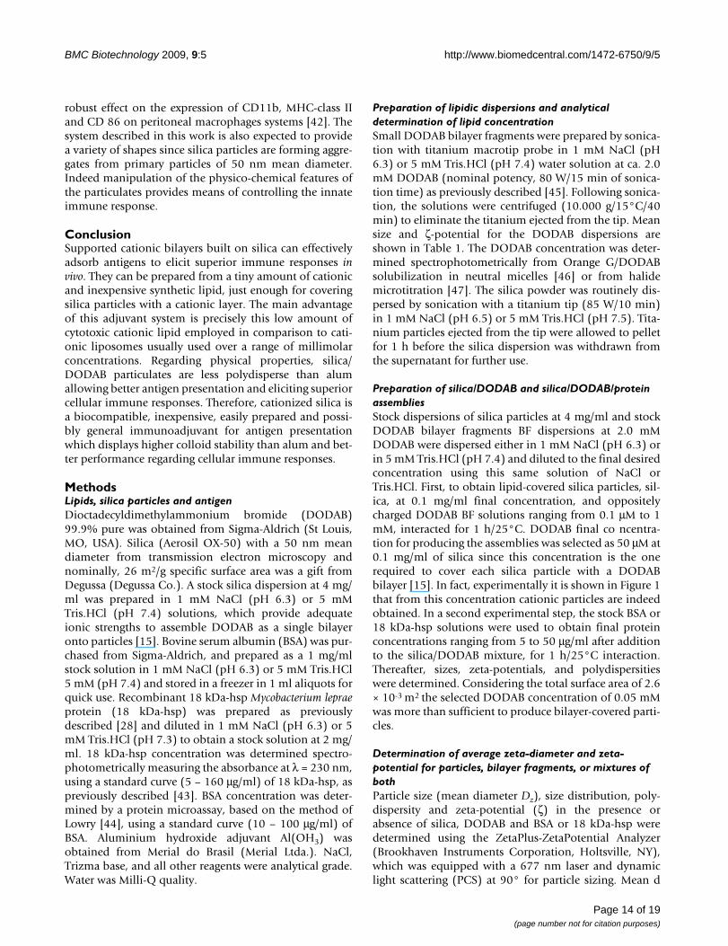
Suitability of the silica/DODAB/antigen system to induce improved cytokines productionFigure 11Suitability of the silica/DODAB/antigen system to induce improved cytokines production. Cytokine responses generated by SiO2/DODAB/18 kDa-hsp and Al(OH)3/18 kDa-hsp. Release of IL-10 (A), IL-12 (B), IL-13 (C) and IFN-γ (D) from inguinal and periaortic lymph node cells isolated from BALB/c mice immunized with 15 μg of 18 kDa-hsp M. leprae administered in silica/DODAB or Al(OH)3. Lymph node cells were isolated six days after immunization and re-stimulated in vitro with 250 μg/ml of 18 kDa-hsp or 2.5 μg/mL of ConA for 48 hours. The cytokine levels were measured by sandwich kit enzyme-linked immu-nosorbent assay (ELISA) as described in materials and meth-ods. The limit of detection was 156 pg/mL for IL-10 and IL-12, and 78 pg/mL for IL-13 and IFN-γ respectively.
Page 16 of 19(page number not for citation purposes)
BMC Biotechnology 2009, 9:5 http://www.biomedcentral.com/1472-6750/9/5
μg/ml) of 18 kDa-hsp, as previously described [37]. Aspectrophotometer Hitachi U-2000 was used for absorb-ance measurement.
For both proteins, the amount of adsorbed protein wasdetermined by the difference between the total proteinadded and the amount of protein recovered in the super-natant.
Adsorption was expressed as the number of moleculesadsorbed per square meter silica. Curves were fitted usingcubic polynomial regression. Wherever possible, theLangmuir model was employed for isotherms lineariza-tion and determination of adsorption constants such asaffinity constant (K, in M-1) and maximal adsorption (innumber of molecules per m2 silica) [49].
Subcutaneous immunization, assay for delayed-type hypersensitivity (DTH) and antigen-specific ELISABALB/c female mice 8–12-week old were purchased fromthe University of São Paulo, São Paulo, Brazil. Six groupsof 8 – 10 female mice were challenged subcutaneously (s.c.) in the abdomen at two separate sites. Total volumeinjected in each site was 0.1 or 0.15 mL for BSA or 18 kDa-hsp, respectively. The dispersion injected containedeither: (a) 10 μg BSA or 15 μg 18 kDa-hsp in 5 mMTrisHCl (pH 7.4); or (b) 10 μg BSA or 15 μg 18 kDa-hspin 1 mM NaCl and 0.1 mM DODAB; or (c) 10 μg BSA or15 μg 18 kDa-hsp in 0.1 mg silica/mL, 5 mM Tris.HCl; or(d) 10 μg BSA or 15 μg 18 kDa-hsp in silica/DODAB (0.1mg mL-1/0.05 mM) in 5 mM Tris.HCl; or (e) 0.1 mg silica/mL in 5 mM Tris.HCl. For DTH evaluation, the footpadswelling test was carried out essentially as previouslydescribed [9,26,27]. On the fifth day post subcutaneousimmunization, pre-immunized mice with BSA, 18 kDa-hsp, DODAB/BSA, DODAB/18 kDa-hsp, silica/BSA, sil-ica/18 kDa-hsp, silica/DODAB/BSA, silica/DODAB/18kDa-hsp or silica alone, were challenged in the left-hindfootpad with a total elicitation dose of 4 or 40 μg BSA or3 or 30 μg 18 kDa-hsp in 50 μL Tris.HCl 5 mM, respec-tively. Footpad swelling was measured 24 h later with aMitutoyo engineering micrometer. Depending on the ageof the animals, the thickness of uninjected hind footpadvaried from 1.60 to 1.70 mm. Percentage footpad swelling(%fs) is calculated according to the formula below withresults expressed as %fs ± standard error of the mean(SEM).
%fs = 100 [(left hind footpad thickness) - (right hind foot-pad thickness)]/(mean thickness uninjected left hind
footpad).
For evaluation of humoral immune response, the samegroups previously immunized and challenged with BSAabove were bled through the ophthalmic plexus in days
24 and 36, after immunization. The sera obtained wereanalyzed by ELISA. Each well of 96-well ELISA polystyrenehigh binding plates (Costar Corning Inc., Cambridge,Mass.) was coated with 100 μL of BSA (final concentrationof 0.05 μg/well) in 0.5 M carbonate-bicarbonate buffer(pH 9.6) for 18 h in an humidified chamber at 4°C. Thewells were blocked for 1 h with 5% milk in PBS contain-ing 0.05% Tween 20 (PBS/T), and then incubated for onehour with serum samples diluted 1:50 or 1:200, for spe-cific IgG antibody quantitation. In each well, 100 μL ofgoat anti-mouse IgG peroxidase-conjugate (Sigma)diluted 1:3000 was added and plates were incubated for 1h. After each incubation step, the plates were washedusing an automatic washer, with four cycles of PBS/T.Ortho-phenylenediamine (1 mg/mL) (Sigma) and H2O2(1 μL/mL) diluted in 0.2 M citrate buffer (pH 5.0) wereadded (in the dark) as chromogenic substrate and plateswere incubated for 10 min. The reactions were stopped byadding 100 μL of 2 M H2SO4. Color intensity was quanti-fied using an ELISA plate reader (Diagnostics Pasteur,Strassburg-Schiltigheim, France) at 492 nm. All incuba-tions were carried out at 37°C. Antibody titers remainedbelow 1/100 dilution, meaning that the dilution is nothigh enough to discriminate specific and non specificantibodies against BSA. Serum titration included a serumfrom naïve mice and a serum from mice immunised witha non relevant antigen towards induction of humoralimmune response, namely, hsp-18 kDa protein itself.
For evaluation of humoral immune response against 18kDa-hsp, the same groups previously immunized andchallenged with 18 kDa-hsp above were bled through theophthalmic plexus in days 14, 28, and 84 after immuniza-tion. The sera obtained were analyzed by ELISA. Each wellof 96-well ELISA polystyrene maxisorpt plates (Maxisorp,Nunc) was coated with 100 μL of 18 kDa-hsp (final con-centration of 40 μg/well) in 0.5 M carbonate-bicarbonatebuffer (pH 9.6) for 18 h in an humidified chamber at 4°C.The wells were blocked for 1 h with 5% milk in PBS con-taining 0.05% Tween 20 (PBS/T), and then incubated forone hour with serum samples diluted 1:2, for specific IgGantibody quantitation. In each well, 100 μL of goat anti-mouse IgG peroxidase-conjugate (Sigma) diluted 1:1000was added and plates were incubated for 1 h. After eachincubation step, the plates were washed using an auto-matic washer, with four cycles of PBS/T. Ortho-phenylen-ediamine (1 mg/mL) (Sigma) and H2O2 (1 μL/mL)diluted in 0.2 M citrate buffer (pH 5.0) were added (in thedark) as chromogenic substrate and plates were incubatedfor 10 min. The reactions were stopped by adding 100 μLof 2 M H2SO4. Color intensity was quantified using anELISA plate reader A microplate reader equipped with a405 nm filter (Ultramark, Model 550 Bio-Rad, Hercules,CA, USA) was used for absorbance measurement. All incu-bations were carried out at 37°C.
Page 17 of 19(page number not for citation purposes)
BMC Biotechnology 2009, 9:5 http://www.biomedcentral.com/1472-6750/9/5
Cell culture and cytokine analysisSix days after immunization, cell suspensions frominguinal and periaortic lymph nodes (LN) of 5 mice wereprepared in RPMI 1640 (GIBCO) supplemented with 10mM HEPES, 50 μM 2-Mercaptoethanol, 216 mg L-glutamine/L and 5 % FCS (GIBCO). The cell suspensions(8 × 10⁶ cells) were distributed into tissue culture 24-wellsplates (Costar) and incubated with medium containing250 μg/mL of 18 kDa-hsp M. leprae in a humidified CO2incubator for 48 hours. Wells containing medium only or2.5 μg/mL of ConA (Sigma Aldrich) were included in allexperiments as negative and positive controls, respec-tively. After this time, the plates were centrifuged for 8minutes at 500 × g and the supernatants collected forcytokine content. The levels of cytokines (IL-10, IL12, IL-13 and IFN-γ ) in the supernatants were assayed by sand-wich kit enzyme-linked immunosorbent assay (ELISA),using the following monoclonal antibodies: MAB417 andbiotinylated-BAF417 for IL-10; MAB419 and biotinylated-BAF419 for IL-12; MAB413 and biotinylated-BAF413 forIL-13; and XMG 1.2 and biotinylated-AN18 for IFN-γ ,according to the manufacturer's suggestion (R&D Systems– Minneapolis, MN). Binding of the biotinylated antibod-ies was determined using the streptavidin-peroxidase con-jugate (Sigma) and TMB (3,3',5,5'-Tetramethylbenzidine;Sigma) solution in citrate buffer plus Hydrogen peroxide.The plates were read (450 nm) in an automated ELISAreader (Dynatech MR5000). Samples were quantified bycomparison with standard curves of purified recombinantcytokines and the values expressed as ng/mL. The limit ofdetection was 156 pg/mL for IL-10 and IL-12, and 78 pg/mL for IL-13 and IFN-γ respectively.
Statistical analysisANOVA one-way multiple comparison tests and theKruskal-Wallis non-parametric test were used whenneeded. A P value of ≤ 0.05 was considered significant.
Competing interestsThe authors declare that they have no competing interests.
Authors' contributionsNL did most experiments and data analysis in the labora-tory, MRAS obtained adsorption isotherms and did somesizing and zeta-potential determinations, EFM performedcytokines experiments and analysis, MHBC scaled-up pro-duction of the 18 kDa-hsp recombinant protein from M.leprae and provided this protein for the experiments,AMCR coordinated the experiments, provided importantadvice for the experiments and financial support. Allauthors read and approved the final manuscript.
AcknowledgementsFAPESP and CNPq are gratefully acknowledged for financial support. NL and MRAS were recipients of a FAPESP postdoctoral and CNPq trainee technician fellowships, respectively.
References1. Al-Jamal WT, Kostarelos K: Liposome-nanoparticle hybrids for
multimodal diagnostic and therapeutic applications. Nanom-edicine 2007, 2:85-98.
2. Troutier AL, Ladavière C: An overview of lipid membrane sup-ported by colloidal particles. Adv Colloid Interface Sci 2007,133:1-21.
3. Carmona-Ribeiro AM: Biomimetic particles in drug and vaccinedelivery. J Liposome Res 2007, 17:165-172.
4. Petri DFS, Carmona-Ribeiro AM: Biomimetic particles. In Poly-meric Nanostructures and Their Applications Edited by: Nalwa HS. Ste-venson Ranch, American Scientific Publishers; 2007:485-530.
5. Sicchierolli SM, Carmona-Ribeiro AM: Biomolecular recognitionon phospholipid-covered polystyrene microspheres. J PhysChem 1996, 100:16771-16775.
6. Moura SP, Carmona-Ribeiro AM: Biomimetic particles for isola-tion and reconstitution of receptor function. Cell Biochem Bio-phys 2006, 44:446-452.
7. Pacheco LF, Carmona-Ribeiro AM: Effects of synthetic lipids onsolubilization and colloid stability of hydrophobic drugs. J Col-loid Interface Sci 2003, 258:146-154.
8. Lincopan N, Carmona-Ribeiro AM: Lipid-covered drug particles:combined action of dioctadecyldimethylammonium bro-mide and amphotericin B or miconazole. J Antimicrob Chem-other 2006, 58:66-75.
9. Lincopan N, Espíndola NM, Vaz AJ, Carmona-Ribeiro AM: Cationicsupported lipid bilayers for antigen presentation. Int J Pharm2007, 340:216-222.
10. Lincopan N, Rosa H, Carmona-Ribeiro AM: Biomimetic particles.Macromol Symp 2006, 245–246:485-490.
11. Blau S, Jubeh TT, Haupt SM, Rubinstein A: Drug targeting by sur-face cationization. Crit Rev Ther Drug Carrier Syst 2000, 17:425-465.
12. Pereira EMA, Vieira DB, Carmona-Ribeiro AM: Cationic bilayerson polymeric particles: effect of low NaCl concentration onsurface coverage. J Phys Chem B 2004, 108:11490-11495.
13. Rapuano R, Carmona-Ribeiro AM: Physical adsorption of bilayermembranes on silica. J Colloid Interface Sci 1997, 193:104-109.
14. Rapuano R, Carmona-Ribeiro AM: Supported bilayers on sílica. JColloid Interface Sci 2000, 226:299-307.
15. Moura SP, Carmona-Ribeiro AM: Cationic bilayer fragments onsilica at low ionic strength: competitive adsorption and col-loid stability. Langmuir 2003, 19:6664-6667.
16. Carter DC, Ho JX: Structure of serum albumin. Adv Protein Chem1994, 45:153-203.
17. Malmsten M: Biopolymers at interfaces New York, Marcel Dekker;2003.
18. Gray JJ: The interaction of proteins with solid surfaces. CurrOpin Struct Biol 2004, 14:110-115.
19. Bodzon-Kulakowska A, Bierczynska-Krzysik A, Dylag T, Drabik A,Suder P, Noga M, Jarzebinska J, Silberring J: Methods for samplespreparation in proteomic research. J Chromatogr B Analyt Tech-nol Biomed Life Sci 2007, 849:1-31.
20. Efimova YM, Haemers S, Wierczinski B, Norde W, van Well AA: Sta-bility of globular proteins in H2O and D2O. Biopolymers 2007,85:264-273.
21. Norde W, Buijs J, Lyklema H: Adsorption of globular proteins. InFundamentals of interface and colloid science Edited by: Lyklema J.Amsterdam, Elsevier; 2005:3.1-3.59.
22. Veen M Van der, Stuart MC, Norde W: Spreading of proteins andits effect on adsorption and desorption kinetics. Colloids Surf B:Biointerfaces 2007, 54:136-142.
23. Derand H, Malmsten M: Protein interfacial behavior in micro-fabricated analysis systems and microarrays. Surfactant Sci Ser2003, 110:773-810.
24. Lincopan N, Carmona-Ribeiro AM: Protein assembly onto cati-onic supported bilayers. J Nanoscience Nanotechnol 2009 in press.
25. Carvalho LA, Carmona-Ribeiro AM: Interactions between cati-onic vesicles and serum proteins. Langmuir 1998, 14:6077-6081.
26. Mustafa AS, Lundin KE, Oftung F: Human T cells recognize myco-bacterial heat shock proteins in the context of multipleHLA-DR molecules: studies with healthy subjects vaccinatedwith Mycobacterium bovis BCG and Mycobacterium leprae.Infect Immun 1993, 61:5294-5301.
27. Pinho JR, Cardi BA, Andrade HF Jr, Barr PJ, Bathurst IC, Vicente EJ,Schenberg AC: Immunogenic properties of the Mycobacteriumleprae recombinant 18-kDa antigen purified from Saccharo-
Page 18 of 19(page number not for citation purposes)
http://www.ncbi.nlm.nih.gov/entrez/query.fcgi?cmd=Retrieve&db=PubMed&dopt=Abstract&list_uids=9299094
http://www.ncbi.nlm.nih.gov/entrez/query.fcgi?cmd=Retrieve&db=PubMed&dopt=Abstract&list_uids=9299094
http://www.ncbi.nlm.nih.gov/entrez/query.fcgi?cmd=Retrieve&db=PubMed&dopt=Abstract&list_uids=8154369

BMC Biotechnology 2009, 9:5 http://www.biomedcentral.com/1472-6750/9/5
Publish with BioMed Central and every scientist can read your work free of charge
"BioMed Central will be the most significant development for disseminating the results of biomedical research in our lifetime."
Sir Paul Nurse, Cancer Research UK
Your research papers will be:
available free of charge to the entire biomedical community
peer reviewed and published immediately upon acceptance
cited in PubMed and archived on PubMed Central
yours — you keep the copyright
Submit your manuscript here:http://www.biomedcentral.com/info/publishing_adv.asp
BioMedcentral
myces cerevisiae; enhancement of delayed-type hypersensi-tivity after gamma-irradiation. Int J Lepr Other Mycobact Dis 1995,63:381-390.
28. Costa MHB, Ueda C, Sato RA, Liberman C, Raw I: Procedures forscaling up the recombinant 18 kDa-hsp lepra protein produc-tion. Biotechnol Tech 1995, 9:527-532.
29. Tsuruta LR, Quintilio W, Costa MH, Carmona-Ribeiro AM: Interac-tions between cationic liposomes and an antigenic protein:the physical chemistry of the immunoadjuvant action. J LipidRes 1997, 38:2003-2011.
30. Snippe H, Belder M, Willers JM: Dimethyl diotadecyl ammoniumbromide as adjuvant for delayed hypersensitivity in mice.Immunology 1977, 33:931-936.
31. Klinguer C, Beck A, De-Lys P, Bussat MC, Blaecke A, Derouet F, Bon-nefoy JY, Nguyen TN, Corvaïa N, Velin D: Lipophilic quaternaryammonium salt acts as a mucosal adjuvant when co-admin-istered by the nasal route with vaccine antigens. Vaccine 2001,19:4236-4244.
32. Davidsen J, Rosenkrands I, Christensen D, Vangala A, Kirby D, PerrieY, Agger EM, Andersen P: Characterization of cationic lipo-somes based on dimethyldioctadecylammonium and syn-thetic cord factor from M. tuberculosis (trehalose 6,6'-dibehenate)-a novel adjuvant inducing both strong CMI andantibody responses. Biochim Biophys Acta 2005, 1718:22-31.
33. Mohammed AR, Bramwell VW, Coombes AGA, Perrie Y: Lyophili-sation and sterilisation of liposomal vaccines to produce sta-ble and sterile products. Methods 2006, 40:30-38.
34. Vangala A, Bramwell VW, McNeil S, Christensen D, Agger EM, PerrieY: Comparison of vesicle based antigen delivery systems fordelivery of hepatitis B surface antigen. J Control Release 2007,119:102-110.
35. Iler RK: The chemistry of silica New York, Wiley-Interscience; 1979. 36. Parida SK, Dash S, Patel S, Mishra BK: Adsorption of organic mol-
ecules on silica surface. Adv Colloid Interface Sci 2006, 121:77-110.37. Tadros ThF, Lyklema J: Adsorption of potential-determining
ions at the silica-aqueous electrolyte interface and the roleof some cations. J Electroanal Chem 1968, 17:267-275.
38. Moura SP, Carmona-Ribeiro AM: Adsorption behavior ofDODAB/DPPC vesicles on silica. J Colloid Interface Sci 2007,313:519-526.
39. Callahan PM, Shorter AL, Hem SL: The importance of surfacecharge in the optimization of antigen-adjuvant interactions.Pharm Res 1991, 8:851-858.
40. Clausi AL, Merkley SA, Carpenter JF, Randolph TW: Inhibition ofaggregation of aluminum hydroxide adjuvant during freezingand drying. J Pharm Sci 2008, 97:2049-2061.
41. Costa MHB, Sant'Anna OA, de Araujo PS, Sato RA, Quintilio W, SilvaLV, Matos CR, Raw I: Conformational stability and antibodyresponse to the 18 kDa heat-shock protein formulated intodifferent vehicles. Appl Biochem Biotechnol 1998, 73:19-28.
42. Ramesh M, Turner LF, Yadav R, Rajan TV, Vella AT, Kuhn LT: Effectsof the physico-chemical nature of two biomimetic crystalson the innate immune response. Int Immunopharmacol 2007,7:1617-1629.
43. Costa MHB, Sato RA, Silveira A, Barratt G, Fattal E, Quintilio W: Mic-roquantification of proteins with low chromophore con-tents. Biotech Tech 1997, 11:697-670.
44. Lowry OH, Rosebrough NJ, Farr AL, Randall R: Protein measure-ment with the Folin phenol reagent. J Biol Chem 1951,193:265-275.
45. Vieira DB, Carmona-Ribeiro AM: Synthetic bilayer fragments forsolubilization of. amphotericin B. J Colloid Interface Sci 2001,117:427-431.
46. Stelmo M, Chaimovich H, Cuccovia IM: Quantitative determina-tion of alkylammonium amphiphiles using neutral micelles. JColloid Interface Sci 1987, 117:200-204.
47. Schales O, Schales SS: A simple and accurate method for thedetermination of chloride in biological fluids. J Biol Chem 1941,140:879-884.
48. Grabowski E, Morrison I: Particle size distribution from analysisof quasi-elastic light scattering data. In Measurements of Sus-pended Particles by Quasi-Elastic Light Scattering Edited by: Dahneke B.New York, Wiley-Interscience; 1983:199-236.
49. Carmona-Ribeiro AM: Bilayer vesicles and liposomes as inter-face agents. Chem Soc Rev 2001, 30:241-247.
Page 19 of 19(page number not for citation purposes)
http://www.ncbi.nlm.nih.gov/entrez/query.fcgi?cmd=Retrieve&db=PubMed&dopt=Abstract&list_uids=7594921
http://www.ncbi.nlm.nih.gov/entrez/query.fcgi?cmd=Retrieve&db=PubMed&dopt=Abstract&list_uids=7594921
http://www.ncbi.nlm.nih.gov/entrez/query.fcgi?cmd=Retrieve&db=PubMed&dopt=Abstract&list_uids=9374123
http://www.ncbi.nlm.nih.gov/entrez/query.fcgi?cmd=Retrieve&db=PubMed&dopt=Abstract&list_uids=9374123
http://www.ncbi.nlm.nih.gov/entrez/query.fcgi?cmd=Retrieve&db=PubMed&dopt=Abstract&list_uids=9374123
http://www.ncbi.nlm.nih.gov/entrez/query.fcgi?cmd=Retrieve&db=PubMed&dopt=Abstract&list_uids=1924135
http://www.ncbi.nlm.nih.gov/entrez/query.fcgi?cmd=Retrieve&db=PubMed&dopt=Abstract&list_uids=1924135
http://www.ncbi.nlm.nih.gov/entrez/query.fcgi?cmd=Retrieve&db=PubMed&dopt=Abstract&list_uids=9621407
http://www.ncbi.nlm.nih.gov/entrez/query.fcgi?cmd=Retrieve&db=PubMed&dopt=Abstract&list_uids=9621407
Related Documents