Signaling Pathways Mediating Cardiac Myocyte Gene Expression in Physiological and Stress Responses ANGELA CLERK, * TIMOTHY E. CULLINGFORD, STEPHEN J. FULLER, ALEJANDRO GIRALDO, THOMAIS MARKOU, SAMPSA PIKKARAINEN, AND PETER H. SUGDEN NHLI Division, Faculty of Medicine, Imperial College London, London, UK The contractile cells in the heart (the cardiac myocytes) are terminally differentiated. In response to pathophysiological stresses, cardiac myocytes undergo hypertrophic growth or apoptosis, responses associated with the development of cardiac pathologies. There has been much effort expended in gaining an understanding of the stimuli which promote these responses, and in identifying the intracellular signaling pathways which are activated and potentially involved. These signaling pathways presumably modulate gene and protein expression to elicit the end-stage response. For the regulation of gene expression, the signal may traverse the cytoplasm to modulate nuclear-localized transcription factors as occurs with the mitogen-activated protein kinase or protein kinase B/Akt cascades. Alternatively, the signal may promote translocation of transcription factors from the cytoplasm to the nucleus as is seen with the calcineurin/NFAT and JAK/STAT systems. We present an overview of the principal signaling pathways implicated in the regulation of gene expression in cardiac myocyte pathophysiology, and summarize the current understanding of these pathways, the transcription factors they regulate and the changes in gene expression associated with the development of cardiac pathologies. Finally, we discuss how intracellular signaling and gene expression may be integrated to elicit the overall change in cellular phenotype. J. Cell. Physiol. 212: 311–322, 2007. ß 2007 Wiley-Liss, Inc. Cardiac myocytes, the contractile cells of the heart, withdraw from the cell cycle in the perinatal period. Subsequent growth of the heart (maturational growth) primarily reflects an increase in size of individual cardiac myocytes in the absence of proliferation. The prevailing view is that differentiated cardiac myocytes in the adult heart do not divide (Nadal-Ginard et al., 2003). This renders the heart vulnerable to pathophysiological stresses such as hypoxia or ischemia which cause cardiac myocyte death by necrosis and/or apoptosis. Surviving cardiac myocytes undergo hypertrophic growth to maintain cardiac function (Dorn et al., 2003). Cardiac myocyte hypertrophy also occurs to accommodate an increase in workload, for example, in response to hypertension. This initial compensated hypertrophy is beneficial and fosters survival but, in the longer term, the response may become decompensated and lead to heart failure. Although stem cells may repopulate an adult heart with contractile cells (Nadal-Ginard et al., 2003), this is clearly insufficient to repair the damage sustained by pathophysiological stresses. Cardiac myocyte hypertrophy is associated with changes in gene expression including increased expression of immediate early genes (e.g. c-jun, c-fos and egr1) and re-expression of genes which are expressed early in development (the ‘‘fetal’’ pattern of gene expression; e.g. b-myosin heavy chain, atrial natriuretic factor), and these are often used as indices of hypertrophy (Hoshijima and Chien, 2002; Dorn et al., 2003). It is assumed that changes in gene expression are manifested in the protein profile to effect the changes in morphology (increased cell size and myofibrillogenesis) and physiology which are characteristic of a hypertrophic cardiac myocyte. The potential for changes in gene expression to modulate cardiac myocyte progression through apoptosis has received less consideration, but stimuli which promote apoptosis simultaneously modulate the expression of selected genes (Clerk et al., 2007). It seems probable that changes in the mRNA/protein profiles modulate the cell death response, either to rescue myocytes before commitment to cell death or to modulate the balance between apoptosis and necrosis. Over the last 20 years, much research has focused on identifying the stimuli which induce cardiac myocyte hypertrophy or apoptosis and the intracellular signaling pathways which are involved. Stimuli associated with cardiac myocyte hypertrophy include heterotrimeric G protein-coupled receptor agonists which signal principally through Gaq [e.g. endothelin-1 (ET-1) acting through ET A receptors, a-adrenergic agonists], peptide growth factors (e.g. platelet-derived growth factor, epidermal growth factor), agonists which signal through gp130 cytokine receptors (e.g. cardiotrophin-1; leukemia inhibitory factor, LIF), and mechanical stress/strain (Sugden and Clerk, 1998b; Molkentin and Dorn, 2001; Sugden, 2003; Clerk et al., 2006). Oxidative stresses are particularly implicated in cardiac myocyte death, whether applied directly (e.g. as H 2 O 2 (Aikawa et al., 1997; Cook et al., 1999; von Harsdorf et al., 1999; Aoki et al., 2002)), generated intracellularly as a consequence of drug treatments (e.g. doxorubicin or chelerythrine (Kumar et al., 1999; Yamamoto et al., 2001)) or resulting from hypoxia/reoxygenation (Kang et al., 2000). The type of response depends on the degree of stress such that high levels of oxidative stress induce cardiac myocyte necrosis whereas lower levels are permissive for regulated apoptosis (Sawyer et al., 2002). Subtoxic levels may be cytoprotective and, according to some groups, may even promote hypertrophy This article includes Supplementary Material available from the authors upon request or via the internet at http:// www.interscience.wiley.com/jpages/0021-9541/suppmat. *Correspondence to: Angela Clerk, NHLI Division, Faculty of Medicine, Imperial College London, Flowers Building, Armstrong Road, London SW7 2AZ, UK. E-mail: [email protected] Received 19 February 2007; Accepted 27 February 2007 DOI: 10.1002/jcp.21094 MINI REVIEW 311 Journal of Journal of Cellular Physiology Cellular Physiology ß 2007 WILEY-LISS, INC.

Welcome message from author
This document is posted to help you gain knowledge. Please leave a comment to let me know what you think about it! Share it to your friends and learn new things together.
Transcript
MINI REVIEW 311J o u r n a l o fJ o u r n a l o f
CellularPhysiologyCellularPhysiology
Signaling Pathways MediatingCardiac Myocyte Gene Expressionin Physiological and StressResponses
ANGELA CLERK,* TIMOTHY E. CULLINGFORD, STEPHEN J. FULLER, ALEJANDRO GIRALDO,THOMAIS MARKOU, SAMPSA PIKKARAINEN, AND PETER H. SUGDEN
NHLI Division, Faculty of Medicine, Imperial College London, London, UK
The contractile cells in the heart (the cardiac myocytes) are terminally differentiated. In response to pathophysiological stresses, cardiacmyocytes undergo hypertrophic growth or apoptosis, responses associated with the development of cardiac pathologies. There has beenmuch effort expended in gaining an understanding of the stimuli which promote these responses, and in identifying the intracellularsignaling pathways which are activated and potentially involved. These signaling pathways presumably modulate gene and proteinexpression to elicit the end-stage response. For the regulation of gene expression, the signal may traverse the cytoplasm to modulatenuclear-localized transcription factors as occurs with the mitogen-activated protein kinase or protein kinase B/Akt cascades.Alternatively, the signal may promote translocation of transcription factors from the cytoplasm to the nucleus as is seen with thecalcineurin/NFAT and JAK/STAT systems. We present an overview of the principal signaling pathways implicated in the regulation of geneexpression in cardiac myocyte pathophysiology, and summarize the current understanding of these pathways, the transcription factorsthey regulate and the changes in gene expression associated with the development of cardiac pathologies. Finally, we discuss howintracellular signaling and gene expression may be integrated to elicit the overall change in cellular phenotype.
J. Cell. Physiol. 212: 311–322, 2007. � 2007 Wiley-Liss, Inc.
This article includes Supplementary Material available from theauthors upon request or via the internet at http://www.interscience.wiley.com/jpages/0021-9541/suppmat.
*Correspondence to: Angela Clerk, NHLI Division, Faculty ofMedicine, Imperial College London, Flowers Building, Armstrong Road,London SW7 2AZ, UK. E-mail: [email protected]
Received 19 February 2007; Accepted 27 February 2007
DOI: 10.1002/jcp.21094
Cardiac myocytes, the contractile cells of the heart, withdrawfrom the cell cycle in the perinatal period. Subsequent growth ofthe heart (maturational growth) primarily reflects an increase insize of individual cardiac myocytes in the absence ofproliferation. The prevailing view is that differentiated cardiacmyocytes in the adult heart do not divide (Nadal-Ginard et al.,2003). This renders the heart vulnerable to pathophysiologicalstresses such as hypoxia or ischemia which cause cardiacmyocyte death by necrosis and/or apoptosis. Surviving cardiacmyocytes undergo hypertrophic growth to maintain cardiacfunction (Dorn et al., 2003). Cardiac myocyte hypertrophy alsooccurs to accommodate an increase in workload, for example,in response to hypertension. This initial compensatedhypertrophy is beneficial and fosters survival but, in the longerterm, the response may become decompensated and lead toheart failure. Although stem cells may repopulate an adult heartwith contractile cells (Nadal-Ginard et al., 2003), this is clearlyinsufficient to repair the damage sustained bypathophysiological stresses.Cardiac myocyte hypertrophy is associated with changes ingene expression including increased expression of immediateearly genes (e.g. c-jun, c-fos and egr1) and re-expression of geneswhich are expressed early in development (the ‘‘fetal’’ patternof gene expression; e.g. b-myosin heavy chain, atrial natriureticfactor), and these are often used as indices of hypertrophy(Hoshijima and Chien, 2002; Dorn et al., 2003). It is assumedthat changes in gene expression are manifested in the proteinprofile to effect the changes in morphology (increased cell sizeand myofibrillogenesis) and physiology which are characteristicof a hypertrophic cardiac myocyte. The potential for changes ingene expression to modulate cardiac myocyte progressionthrough apoptosis has received less consideration, but stimuliwhich promote apoptosis simultaneously modulate theexpression of selected genes (Clerk et al., 2007). It seemsprobable that changes in the mRNA/protein profiles modulatethe cell death response, either to rescue myocytes beforecommitment to cell death or to modulate the balance betweenapoptosis and necrosis.
� 2 0 0 7 W I L E Y - L I S S , I N C .
Over the last 20 years, much research has focused on identifyingthe stimuli which induce cardiac myocyte hypertrophy orapoptosis and the intracellular signaling pathways which areinvolved. Stimuli associated with cardiac myocyte hypertrophyinclude heterotrimeric G protein-coupled receptor agonistswhich signal principally through Gaq [e.g. endothelin-1 (ET-1)acting through ETA receptors, a-adrenergic agonists], peptidegrowth factors (e.g. platelet-derived growth factor, epidermalgrowth factor), agonists which signal through gp130 cytokinereceptors (e.g. cardiotrophin-1; leukemia inhibitory factor, LIF),and mechanical stress/strain (Sugden and Clerk, 1998b;Molkentin and Dorn, 2001; Sugden, 2003; Clerk et al., 2006).Oxidative stresses are particularly implicated in cardiacmyocyte death, whether applied directly (e.g. as H2O2 (Aikawaet al., 1997; Cook et al., 1999; von Harsdorf et al., 1999; Aokiet al., 2002)), generated intracellularly as a consequence ofdrug treatments (e.g. doxorubicin or chelerythrine (Kumaret al., 1999; Yamamoto et al., 2001)) or resulting fromhypoxia/reoxygenation (Kang et al., 2000). The type ofresponse depends on the degree of stress such that high levelsof oxidative stress induce cardiac myocyte necrosis whereaslower levels are permissive for regulated apoptosis (Sawyeret al., 2002). Subtoxic levels may be cytoprotective and,according to some groups, may even promote hypertrophy
312 C L E R K E T A L .
(Sawyer et al., 2002). Other stimuli, including pro-inflammatorycytokines [e.g. tumor necrosis factor a (TNFa), interleukin 1b(IL1b)] and death receptor ligands (e.g. Fas ligand) may promotecardiac myocyte death, particularly when defence mechanismsbecome compromised (e.g. following ischemia) (van Empelet al., 2005). The various hypertrophic or apoptotic stimuliactivate a range of intracellular signaling pathways in cardiacmyocytes and the heart (Sugden and Clerk, 1998b; Clerk et al.,2003), but the diversity of the signals, activation of similarpathways by hypertrophic and apoptotic stimuli, reports ofextensive cross-talk between pathways, and conflicting datafrom different experimental systems and/or research groupscauses much confusion. The mechanisms by which differentsignals are integrated to bring about changes in gene expressionare particularly unclear. Here, we present an overview of theprincipal signaling pathways implicated in the regulation of geneexpression in cardiac myocyte pathophysiology, and discusshow they may elicit the cellular response.
Signaling Pathways in Cardiac Myocytes and theTranscription Factors they Regulate
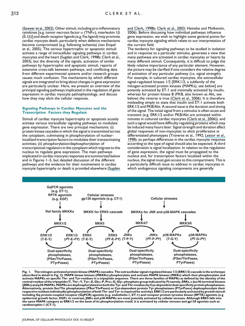
Stimuli of cardiac myocyte hypertrophy or apoptosis acutelyactivate various intracellular signaling pathways to modulategene expression. There are two principal mechanisms: (i)protein kinase cascades in which the signal is transmitted acrossthe cytoplasm, culminating in phosphorylation of nuclear-localized transcription factors to modulate their transactivatingactivities; (ii) phosphorylation/dephosphorylation oftranscriptional regulators in the cytoplasm which migrate to thenucleus to regulate gene expression. The main pathwaysimplicated in cardiac myocyte responses are summarized belowand in Figures 1–5, but detailed discussion of the differentpathways and the evidence for their involvement in cardiacmyocyte hypertrophy or death is provided elsewhere (Sugden
GqPCR agonists(e.g. ET-1),
RPTK agonists(e.g. EGF)
JN(T-
Raf family MKKKs
MKK1/2
ERK1/2(T-E-Y)
ERK1/2(PT-E-PY)
Dual-specificityphosphatases,
(PSer/ThrPases, PTyrPases)
MKKK for ERK5 cascade
MKK5
ERK5(T-E-Y)
ERK5(PT-E-PY)
Dual-specificityphosphatases,
(PSer/ThrPases, PTyrPases)
Cellular stressesgp130 agonists (e.g. CT-1)
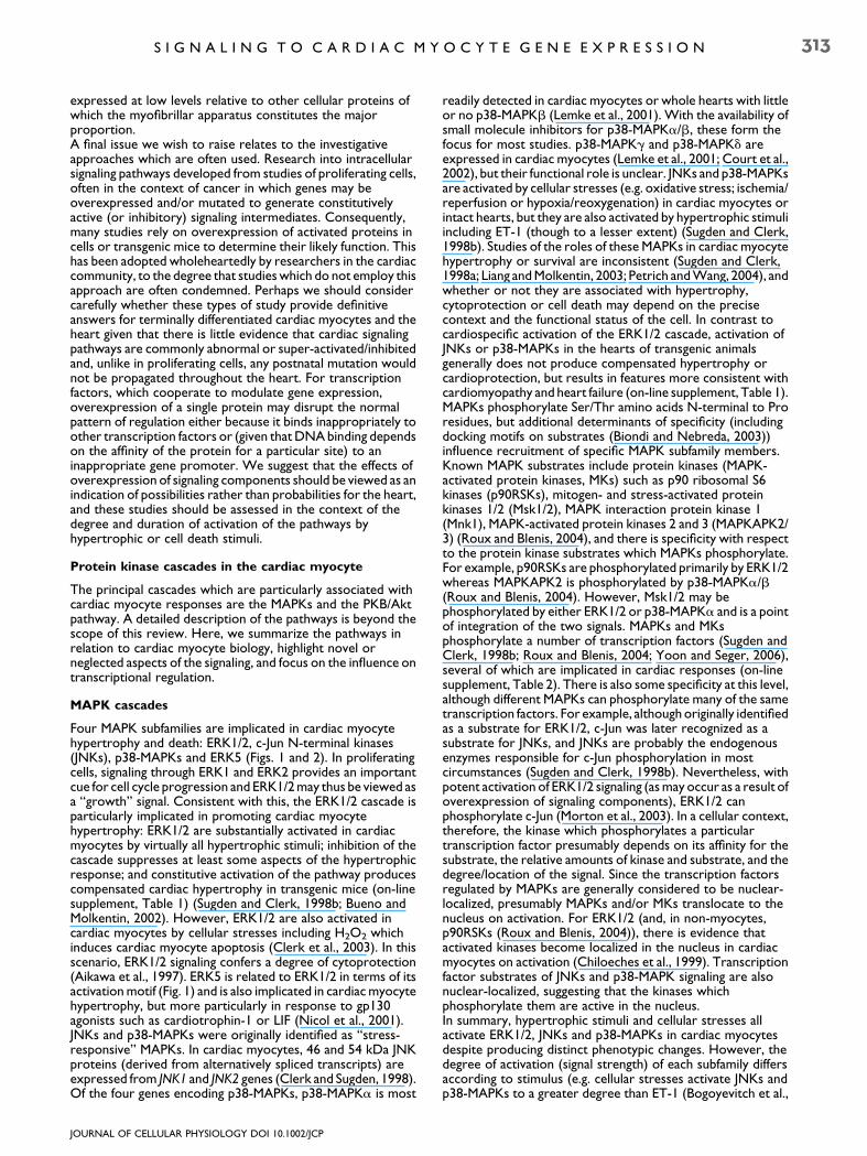
Fig. 1. Themitogen-activated protein kinase (MAPK) cascades. The extr(described in detail in Fig. 2). MAPK kinase kinases (MKKKs) phosphorylaactivate MAPKs on specific Thr- and Tyr-residues in a tripeptide sequenccentral residueof the tripeptide (T,Thr-; Y, Tyr-; E,Glu-; P, Pro-;G,Gly-; ph(JNKs)andp38-MAPKs.MAPKsaredephosphorylatedonboththeTyr-andAlternatively, protein Ser/Thr phosphatases (PSer/ThrPases) or Cys-deprespectiveresidues individually (phosphorylationofbothThr-andTyr- is reincluding Gq protein-coupled receptor (GqPCR) agonists (e.g. endothelinepidermal growth factor, EGF). In contrast, JNKs and p38-MAPKs are mothe same MAPK category as ERK1/2 on the basis of its phosphorylation mcardiotrophin-1 (CT-1).
JOURNAL OF CELLULAR PHYSIOLOGY DOI 10.1002/JCP
and Clerk, 1998b; Clerk et al., 2003; Heineke and Molkentin,2006). Before discussing how individual pathways influencegene expression, we wish to highlight some general points forcardiac myocyte signaling which relate to our interpretation ofthe current field.The tendency for signaling pathways to be studied in isolationand in response to a particular stimulus, generates a view thatmany pathways are activated in cardiac myocytes or hearts bymany different stimuli. Consequently, it is difficult to judge thelikely relative importance of any particular element. However,the picture may be clarified if one considers the relative degreeof activation of any particular pathway (i.e. signal strength).For example, in cultured cardiac myocytes, the extracellularsignal-regulated kinases 1/2 [ERK1/2, a subfamily of themitogen-activated protein kinases (MAPKs), see below] arepotently activated by ET-1 and minimally activated by insulin,whereas for protein kinase B (PKB, also known as Akt, seebelow) the reverse is true (Clerk et al., 2006). It is thereforemisleading simply to state that insulin and ET-1 activate bothERK1/2 and PKB/Akt. A second issue is the duration and timingof the signal. The initial signal from a stimulus is often acute andtransient (e.g. ERK1/2 and/or PKB/Akt are activated withinminutes in cultured cardiac myocytes (Clerk et al., 2006)), andsuch a signal would have difficulty regulating proteins which maybe induced many hours later. Signal strength and duration affectglobal responses of non-myocytes to elicit proliferative vsdifferentiated phenotypes (Traverse et al., 1992; Lessor et al.,1998), so perhaps differences in the cardiac myocyte responseaccording to the type of signal should also be expected. A thirdconsideration is signal localization. In relation to the regulationof gene expression, the signal must be propagated to thenucleus and, for transcription factors localized within thenucleus, the signal must gain access to this compartment. This isa particularly difficult issue to address in cardiac myocytes inwhich endogenous signaling components are generally
MKK4/7
Cellular stresses
MKKKs for JNK and p38-MAPK cascades
KsP-Y)
JNKs(PT-P-PY)
p38-MAPKs(T-G-Y)
p38-MAPKs(PT-G-PY)
MKK3/6
Dual-specificityphosphatases,
(PSer/ThrPases,PTyrPases)
Dual-specificityphosphatases,
(PSer/ThrPases,PTyrPases)
acellular signal-regulated kinase 1/2 (ERK1/2) cascade is the archetypete and activate MAPK kinases (MKKs) which then phosphorylate ande. There are three families of MAPKs as defined by the identity of theosphate group indicatedbyP) namely, ERKs, c-JunN-terminal kinasesThr-residuesbyCys-dependentdual-specificityproteinphosphatases.endent protein Tyr phosphatases (PTyrPases) dephosphorylate theirquired foractivity).ERK1/2areparticularlyactivatedbygrowthstimuli-1, ET-1) and receptor protein tyrosine kinase (RPTK) agonists (e.gst potently activated by cellular stresses. Although ERK5 falls intootif, it is activated by cellular stresses and gp130 agonists such as
S I G N A L I N G T O C A R D I A C M Y O C Y T E G E N E E X P R E S S I O N 313
expressed at low levels relative to other cellular proteins ofwhich the myofibrillar apparatus constitutes the majorproportion.A final issue we wish to raise relates to the investigativeapproaches which are often used. Research into intracellularsignaling pathways developed from studies of proliferating cells,often in the context of cancer in which genes may beoverexpressed and/or mutated to generate constitutivelyactive (or inhibitory) signaling intermediates. Consequently,many studies rely on overexpression of activated proteins incells or transgenic mice to determine their likely function. Thishas been adopted wholeheartedly by researchers in the cardiaccommunity, to the degree that studies which do not employ thisapproach are often condemned. Perhaps we should considercarefully whether these types of study provide definitiveanswers for terminally differentiated cardiac myocytes and theheart given that there is little evidence that cardiac signalingpathways are commonly abnormal or super-activated/inhibitedand, unlike in proliferating cells, any postnatal mutation wouldnot be propagated throughout the heart. For transcriptionfactors, which cooperate to modulate gene expression,overexpression of a single protein may disrupt the normalpattern of regulation either because it binds inappropriately toother transcription factors or (given that DNA binding dependson the affinity of the protein for a particular site) to aninappropriate gene promoter. We suggest that the effects ofoverexpression of signaling components should be viewed as anindication of possibilities rather than probabilities for the heart,and these studies should be assessed in the context of thedegree and duration of activation of the pathways byhypertrophic or cell death stimuli.
Protein kinase cascades in the cardiac myocyte
The principal cascades which are particularly associated withcardiac myocyte responses are the MAPKs and the PKB/Aktpathway. A detailed description of the pathways is beyond thescope of this review. Here, we summarize the pathways inrelation to cardiac myocyte biology, highlight novel orneglected aspects of the signaling, and focus on the influence ontranscriptional regulation.
MAPK cascades
Four MAPK subfamilies are implicated in cardiac myocytehypertrophy and death: ERK1/2, c-Jun N-terminal kinases(JNKs), p38-MAPKs and ERK5 (Figs. 1 and 2). In proliferatingcells, signaling through ERK1 and ERK2 provides an importantcue for cell cycle progression and ERK1/2 may thus be viewed asa ‘‘growth’’ signal. Consistent with this, the ERK1/2 cascade isparticularly implicated in promoting cardiac myocytehypertrophy: ERK1/2 are substantially activated in cardiacmyocytes by virtually all hypertrophic stimuli; inhibition of thecascade suppresses at least some aspects of the hypertrophicresponse; and constitutive activation of the pathway producescompensated cardiac hypertrophy in transgenic mice (on-linesupplement, Table 1) (Sugden and Clerk, 1998b; Bueno andMolkentin, 2002). However, ERK1/2 are also activated incardiac myocytes by cellular stresses including H2O2 whichinduces cardiac myocyte apoptosis (Clerk et al., 2003). In thisscenario, ERK1/2 signaling confers a degree of cytoprotection(Aikawa et al., 1997). ERK5 is related to ERK1/2 in terms of itsactivation motif (Fig. 1) and is also implicated in cardiac myocytehypertrophy, but more particularly in response to gp130agonists such as cardiotrophin-1 or LIF (Nicol et al., 2001).JNKs and p38-MAPKs were originally identified as ‘‘stress-responsive’’ MAPKs. In cardiac myocytes, 46 and 54 kDa JNKproteins (derived from alternatively spliced transcripts) areexpressed from JNK1 and JNK2 genes (Clerk and Sugden, 1998).Of the four genes encoding p38-MAPKs, p38-MAPKa is most
JOURNAL OF CELLULAR PHYSIOLOGY DOI 10.1002/JCP
readily detected in cardiac myocytes or whole hearts with littleor no p38-MAPKb (Lemke et al., 2001). With the availability ofsmall molecule inhibitors for p38-MAPKa/b, these form thefocus for most studies. p38-MAPKg and p38-MAPKd areexpressed in cardiac myocytes (Lemke et al., 2001; Court et al.,2002), but their functional role is unclear. JNKs and p38-MAPKsare activated by cellular stresses (e.g. oxidative stress; ischemia/reperfusion or hypoxia/reoxygenation) in cardiac myocytes orintact hearts, but they are also activated by hypertrophic stimuliincluding ET-1 (though to a lesser extent) (Sugden and Clerk,1998b). Studies of the roles of these MAPKs in cardiac myocytehypertrophy or survival are inconsistent (Sugden and Clerk,1998a; Liang and Molkentin, 2003; Petrich and Wang, 2004), andwhether or not they are associated with hypertrophy,cytoprotection or cell death may depend on the precisecontext and the functional status of the cell. In contrast tocardiospecific activation of the ERK1/2 cascade, activation ofJNKs or p38-MAPKs in the hearts of transgenic animalsgenerally does not produce compensated hypertrophy orcardioprotection, but results in features more consistent withcardiomyopathy and heart failure (on-line supplement, Table 1).MAPKs phosphorylate Ser/Thr amino acids N-terminal to Proresidues, but additional determinants of specificity (includingdocking motifs on substrates (Biondi and Nebreda, 2003))influence recruitment of specific MAPK subfamily members.Known MAPK substrates include protein kinases (MAPK-activated protein kinases, MKs) such as p90 ribosomal S6kinases (p90RSKs), mitogen- and stress-activated proteinkinases 1/2 (Msk1/2), MAPK interaction protein kinase 1(Mnk1), MAPK-activated protein kinases 2 and 3 (MAPKAPK2/3) (Roux and Blenis, 2004), and there is specificity with respectto the protein kinase substrates which MAPKs phosphorylate.For example, p90RSKs are phosphorylated primarily by ERK1/2whereas MAPKAPK2 is phosphorylated by p38-MAPKa/b(Roux and Blenis, 2004). However, Msk1/2 may bephosphorylated by either ERK1/2 or p38-MAPKa and is a pointof integration of the two signals. MAPKs and MKsphosphorylate a number of transcription factors (Sugden andClerk, 1998b; Roux and Blenis, 2004; Yoon and Seger, 2006),several of which are implicated in cardiac responses (on-linesupplement, Table 2). There is also some specificity at this level,although different MAPKs can phosphorylate many of the sametranscription factors. For example, although originally identifiedas a substrate for ERK1/2, c-Jun was later recognized as asubstrate for JNKs, and JNKs are probably the endogenousenzymes responsible for c-Jun phosphorylation in mostcircumstances (Sugden and Clerk, 1998b). Nevertheless, withpotent activation of ERK1/2 signaling (as may occur as a result ofoverexpression of signaling components), ERK1/2 canphosphorylate c-Jun (Morton et al., 2003). In a cellular context,therefore, the kinase which phosphorylates a particulartranscription factor presumably depends on its affinity for thesubstrate, the relative amounts of kinase and substrate, and thedegree/location of the signal. Since the transcription factorsregulated by MAPKs are generally considered to be nuclear-localized, presumably MAPKs and/or MKs translocate to thenucleus on activation. For ERK1/2 (and, in non-myocytes,p90RSKs (Roux and Blenis, 2004)), there is evidence thatactivated kinases become localized in the nucleus in cardiacmyocytes on activation (Chiloeches et al., 1999). Transcriptionfactor substrates of JNKs and p38-MAPK signaling are alsonuclear-localized, suggesting that the kinases whichphosphorylate them are active in the nucleus.In summary, hypertrophic stimuli and cellular stresses allactivate ERK1/2, JNKs and p38-MAPKs in cardiac myocytesdespite producing distinct phenotypic changes. However, thedegree of activation (signal strength) of each subfamily differsaccording to stimulus (e.g. cellular stresses activate JNKs andp38-MAPKs to a greater degree than ET-1 (Bogoyevitch et al.,
Diacylglycerol [+ Ins(1,4,5)P3]
Protein kinase C
Raf (c-Raf, A-Raf, B-Raf)
MKK1/2
ERK1/2
PD98059U0126
Phorbol esters
? ?
?
PDGF (RPTK agonist)
EGF (RPTK
agonist)
PLC PLC
Grb2/
Sos
GqPCR agonists (e.g. ET-1),
GqPCR, Gq activation,
[( GDP) (q.GTP) + ]
-
,
PtdIns(4,5)P2
hydrolysis
Ras activation
(Ras.GDP Ras.GTP)GEF?
}
?
q.
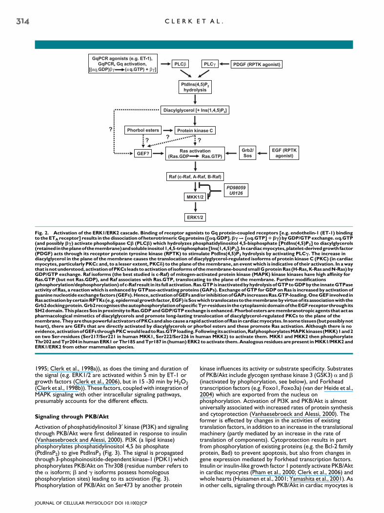
Fig. 2. Activation of the ERK1/ERK2 cascade. Binding of receptor agonists to Gq protein-coupled receptors [e.g. endothelin-1 (ET-1) bindingto theETAreceptor] results in thedissociation of heterotrimericGqproteins ([aq.GDP].bg! [aq.GTP]Rbg) byGDP/GTPexchange.aq.GTP(and possibly bg) activate phospholipase Cb (PLCb) which hydrolyzes phosphatidylinositol 4,5-bisphosphate [PtdIns(4,5)P2] to diacylglycerols(retainedintheplaneofthemembrane)andsolubleinositol1,4,5-trisphosphate[Ins(1,4,5)P3]. Incardiacmyocytes,platelet-derivedgrowthfactor(PDGF) acts through its receptor protein tyrosine kinase (RPTK) to stimulate PtdIns(4,5)P2 hydrolysis by activating PLCg. The increase indiacylglycerol in the plane of themembrane causes the translocation of diacylyglycerol-regulated isoforms of protein kinase C (PKC) (in cardiacmyocytes, particularly PKCe and, to a lesser extent, PKCd) to the plane of themembrane, an eventwhich is indicative of their activation. In awaythat isnotunderstood, activationofPKCs leads toactivationof isoformsof themembrane-boundsmallGproteinRas (H-Ras,K-RasandN-Ras) byGDP/GTP exchange. Raf isoforms (the best studied is c-Raf) of mitogen-activated protein kinase (MAPK) kinase kinases have high affinity forRas.GTP (but not Ras.GDP), and Raf associates with Ras.GTP, translocating to the plane of the membrane. Further modifications(phosphorylation/dephosphorylation)of c-Raf result in its full activation.Ras.GTP is inactivatedbyhydrolysisofGTPtoGDPbythe innateGTPaseactivity of Ras, a reactionwhich is enhanced byGTPase-activating proteins (GAPs). Exchange ofGTP forGDPonRas is increased by activation ofguaninenucleotideexchange factors (GEFs).Hence,activationofGEFsand/or inhibitionofGAPs increasesRas.GTP-loading.OneGEFinvolved inRasactivationbycertainRPTKs(e.g.epidermalgrowth factor,EGF) isSoswhichtranslocates tothemembranebyvirtueof itsassociationwiththeGrb2dockingprotein.Grb2recognisestheautophosphorylationofspecificTyr-residues inthecytoplasmicdomainoftheEGFreceptorthroughitsSH2domain.ThisplacesSos inproximity toRas.GDPandGDP/GTPexchange isenhanced.Phorbolesters aremembranotropicagents thatactaspharmacological mimetics of diacylglycerols and promote long-lasting translocation of diacylglycerol-regulated PKCs to the plane of themembrane.TheyarethuspowerfulactivatorsofPKCsandalsocausearapidactivationofRas incardiacmyocytes. Insometissues (butpossiblynotheart), there are GEFs that are directly activated by diacylglycerols or phorbol esters and these promote Ras activation. Although there is noevidence,activationofGEFsthroughPKCwould leadtoRas.GTPloading.Following itsactivation,RafphosphorylatesMAPKkinases(MKK)1and2on two Ser-residues (Ser217/Ser221 in human MKK1, Ser222/Ser226 in human MKK2) to activate them. MKK1 and MKK2 then phosphorylateThr202 andTyr204 in humanERK1orThr185 andTyr187 in (human)ERK2 toactivate them.Analogous residues arepresent inMKK1/MKK2andERK1/ERK2 from other mammalian species.
314 C L E R K E T A L .
1995; Clerk et al., 1998a)), as does the timing and duration ofthe signal (e.g. ERK1/2 are activated within 5 min by ET-1 orgrowth factors (Clerk et al., 2006), but in 15 -30 min by H2O2
(Clerk et al., 1998b)). These factors, coupled with integration ofMAPK signaling with other intracellular signaling pathways,presumably accounts for the different effects.
Signaling through PKB/Akt
Activation of phosphatidylinositol 30 kinase (PI3K) and signalingthrough PKB/Akt were first delineated in response to insulin(Vanhaesebroeck and Alessi, 2000). PI3K (a lipid kinase)phosphorylates phosphatidylinositol 4,5 bis phosphate(PtdInsP2) to give PtdInsP3 (Fig. 3). The signal is propagatedthrough 3-phosphoinositide-dependent kinase-1 (PDK1) whichphosphorylates PKB/Akt on Thr308 (residue number refers tothe a isoform; b and g isoforms possess homologousphosphorylation sites) leading to its activation (Fig. 3).Phosphorylation of PKB/Akt on Ser473 by another protein
JOURNAL OF CELLULAR PHYSIOLOGY DOI 10.1002/JCP
kinase influences its activity or substrate specificity. Substratesof PKB/Akt include glycogen synthase kinase 3 (GSK3) a and b(inactivated by phophorylation, see below), and Forkheadtranscription factors (e.g. Foxo1, Foxo3a) (van der Heide et al.,2004) which are exported from the nucleus onphosphorylation. Activation of PI3K and PKB/Akt is almostuniversally associated with increased rates of protein synthesisand cytoprotection (Vanhaesebroeck and Alessi, 2000). Theformer is effected by changes in the activities of existingtranslation factors, in addition to an increase in the translationalmachinery (partly mediated by an increase in the rate oftranslation of components). Cytoprotection results in partfrom phosphorylation of existing proteins (e.g. the Bcl-2 familyprotein, Bad) to prevent apoptosis, but also from changes ingene expression mediated by Forkhead transcription factors.Insulin or insulin-like growth factor 1 potently activate PKB/Aktin cardiac myocytes (Pham et al., 2000; Clerk et al., 2006) andwhole hearts (Huisamen et al., 2001; Yamashita et al., 2001). Asin other cells, signaling through PKB/Akt in cardiac myocytes is
S I G N A L I N G T O C A R D I A C M Y O C Y T E G E N E E X P R E S S I O N 315
strongly implicated in the regulation of protein synthesis and incytoprotection (Matsui and Rosenzweig, 2005). Thus, inhibitionof PI3K signaling suppresses the basal rate of protein synthesisin cardiac myocytes and inhibits the increase induced by insulin(Pham et al., 2000), and inhibition of the pathway in vivoincreases cardiac dysfunction and apoptosis in animal models ofheart failure (Matsui and Rosenzweig, 2005; Heineke andMolkentin, 2006). Because of these effects, PI3K and PKB/Aktsignaling is implicated in cardiac myocyte hypertrophy.Amongst other issues, it is argued that stimuli such as ET-1activate PKB/Akt to induce the response, and insulin (whichpotently activates the pathway) promotes hypertrophy.However, as discussed above, the relative degree of activation
JOURNAL OF CELLULAR PHYSIOLOGY DOI 10.1002/JCP
of PKB/Akt by ET-1 and the relative degree of hypertrophyinduced by insulin are minimal (Clerk et al., 2006).Nevertheless, cardiospecific overexpression of constitutivelyactivated PKB/Akt in transgenic animals generally protects theheart against damage induced by ischemia/reperfusion andincreases heart size, although there are some inconsistencies(on-line supplement, Table 3). The question is whether or notthis growth response represents hypertrophy or (as has beensuggested by Shiojima et al., 2002) is more akin to physiological/maturational growth. Given the general role of insulin in thebody and the fluctuating nature of insulin release, it would seemthat the effect of PKB/Akt signaling may be to modulatephysiological growth of the heart, perhaps to couple growth tonutritional status (Preedy et al., 1984), rather than pathologicalhypertrophy. Despite the cytoprotective and growth-promoting effects of PI3K and PKB/Akt in the heart, toxic levelsof oxidative stress activate PKB/Akt in cardiac myocytes to asimilar degree as insulin (Pham et al., 2000). The functionalconsequence is unclear but, since protein translation is inhibitedby oxidative stress, cytoprotection may be the important facetof the response in this context. Indeed, activation of acytoprotective signal may be required for apoptosis to proceedin a regulated manner. Alternatively, activation of the pathwaycould occur solely in surviving cells or it could represent a futileattempt at survival in the global population.For PKB/Akt, the location of the signal is of particularimportance. It is generally accepted that PtdInsP3 accumulationin the plasma membrane leads to activation of PKB/Akt in thecytoplasm, but PI3K and PKB/Akt may also operate at thenuclear membrane (Martelli et al., 2006). Consistent with thelatter, activated PKB/Akt accumulates in cardiac myocyte nuclei(Camper-Kirby et al., 2001) where it potentially regulates geneexpression. The roles of PKB/Akt in the cytoplasm versus
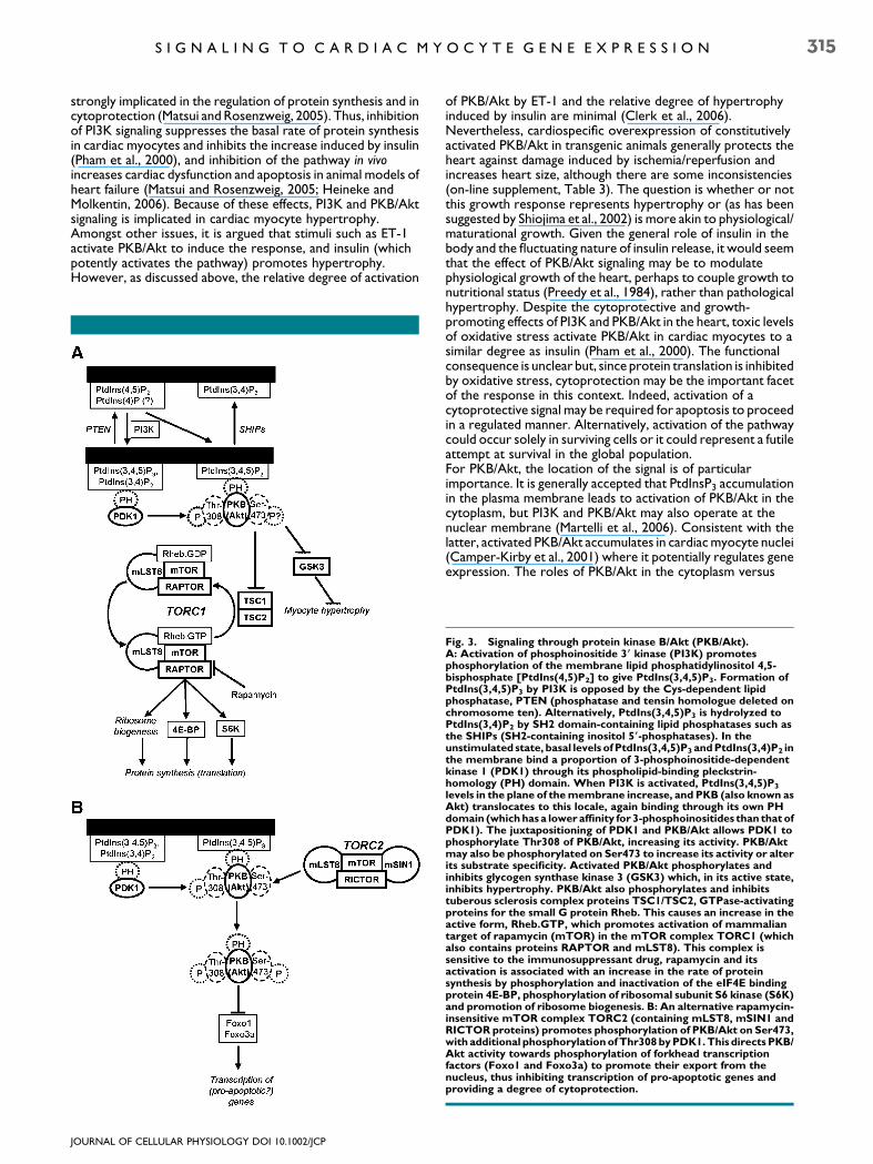
Fig. 3. Signaling through protein kinase B/Akt (PKB/Akt).A: Activation of phosphoinositide 3( kinase (PI3K) promotesphosphorylation of the membrane lipid phosphatidylinositol 4,5-bisphosphate [PtdIns(4,5)P2] to give PtdIns(3,4,5)P3. Formation ofPtdIns(3,4,5)P3 by PI3K is opposed by the Cys-dependent lipidphosphatase, PTEN (phosphatase and tensin homologue deleted onchromosome ten). Alternatively, PtdIns(3,4,5)P3 is hydrolyzed toPtdIns(3,4)P2 by SH2 domain-containing lipid phosphatases such asthe SHIPs (SH2-containing inositol 5(-phosphatases). In theunstimulated state, basal levels ofPtdIns(3,4,5)P3 andPtdIns(3,4)P2 inthe membrane bind a proportion of 3-phosphoinositide-dependentkinase 1 (PDK1) through its phospholipid-binding pleckstrin-homology (PH) domain. When PI3K is activated, PtdIns(3,4,5)P3
levels in the plane of themembrane increase, and PKB (also known asAkt) translocates to this locale, again binding through its own PHdomain (whichhasa loweraffinity for 3-phosphoinositides than that ofPDK1). The juxtapositioning of PDK1 and PKB/Akt allows PDK1 tophosphorylate Thr308 of PKB/Akt, increasing its activity. PKB/Aktmay also be phosphorylated on Ser473 to increase its activity or alterits substrate specificity. Activated PKB/Akt phosphorylates andinhibits glycogen synthase kinase 3 (GSK3) which, in its active state,inhibits hypertrophy. PKB/Akt also phosphorylates and inhibitstuberous sclerosis complex proteins TSC1/TSC2, GTPase-activatingproteins for the small G protein Rheb. This causes an increase in theactive form, Rheb.GTP, which promotes activation of mammaliantarget of rapamycin (mTOR) in the mTOR complex TORC1 (whichalso contains proteins RAPTOR and mLST8). This complex issensitive to the immunosuppressant drug, rapamycin and itsactivation is associated with an increase in the rate of proteinsynthesis by phosphorylation and inactivation of the eIF4E bindingprotein 4E-BP, phosphorylation of ribosomal subunit S6 kinase (S6K)and promotion of ribosome biogenesis. B: An alternative rapamycin-insensitive mTOR complex TORC2 (containing mLST8, mSIN1 andRICTOR proteins) promotes phosphorylation of PKB/Akt on Ser473,with additional phosphorylationofThr308byPDK1.This directsPKB/Akt activity towards phosphorylation of forkhead transcriptionfactors (Foxo1 and Foxo3a) to promote their export from thenucleus, thus inhibiting transcription of pro-apoptotic genes andproviding a degree of cytoprotection.

316 C L E R K E T A L .
nucleus appear to differ and, in transgenic mice, cardiacmyocyte growth is induced when PKB/Akt is tethered to theplasma membrane for constitutive activation, whereas nuclear-targeting of activated PKB/Akt fails to stimulate cardiac growthbut promotes cardioprotection (on-line supplement, Table 3).In non-myocytes, PKB/Akt associates with mammalian target ofrapamycin (mTOR) in different complexes (Polak and Hall,2006; Wullschleger et al., 2006). The mTORC1complexappears to be involved in modulating protein synthesis. Thiscomplex includes the protein Raptor and, here, mTOR isactivated downstream of PKB/Akt. mTORC2 contains proteinsRictor and Sin1, and this complex promotes phosphorylation ofPKB/Akt on Ser473. Here, PKB/Akt regulates Forkheadtranscription factors Foxo1/3a rather than GSK3 or othersubstrates, suggesting that mTORC2 is particularly involved inthe regulation of gene expression via Foxo1/3a. Whether thisoccurs in myocytes or the heart remains to be established.GSK3a and b are amongst the best characterized PKB/Aktsubstrates and phosphorylation of Ser21/Ser9 of GSK3a/b(e.g. in response to insulin) inhibits their activity (Cohen andFrame, 2001; Frame and Cohen, 2001). However, other proteinkinases including cAMP-dependent protein kinase and p90RSKalso phosphorylate these sites (Cohen and Frame, 2001; Frameand Cohen, 2001) placing GSK3 at a point of integration ofseveral signaling pathways. There are many potential substratesfor GSK3a/b including transcription factors [e.g.phosphorylation of c-Jun by GSK3 inhibits its binding to DNA;phosphorylation of nuclear factors of activated T cells (NFATs)promotes their nuclear export] (Frame and Cohen, 2001).In most cases, GSK3a/b requires substrates which arepre-phosphorylated at a priming site (the fourth amino acidC-terminal to the target residue) in order for the substrate todock. Indeed, the inhibitory phosphorylation on GSK3 foldsinto the substrate docking site to prevent substrate binding(Frame and Cohen, 2001). In the heart and in cardiac myocytes,most attention has focussed on GSK3b (although GSK3a is alsoexpressed), and its role in cardiac myocyte hypertrophy. Asexpected, insulin promotes phosphorylation and inhibition ofGSK3b, and GSK3a/b are implicated in the regulation ofglycogen synthase in the heart (Mora et al., 2005). Hypertrophicstimuli including ET-1 also promote phosphorylation of GSK3bin cardiac myocytes (Haq et al., 2000). This may occur viaactivation PKB/Akt as proposed, but phosphorylation may bemediated via ERK1/2 signaling through p90RSKs (Cohen andFrame, 2001; Frame and Cohen, 2001). GSK3 activity appearsto be anti-hypertrophic (Haq et al., 2000; Antos et al., 2002),and inhibition of the enzyme therefore potentially contributesto cardiac myocyte hypertrophy, possibly by allowingdephosphorylation and activation of transcription factors suchas c-Jun or NFATs.
Activation of transcription factors in the cytoplasm
Some DNA-binding transcription factors are regulated by theirsubcellular localization, and phosphorylation/dephophorylationevents modulate their migration into the nucleus where theycan regulate gene expression. Probably the best known of thesein relation to cardiac hypertrophy are the NFATs.Dephosphorylation of NFATs by the protein phosphatasecalcineurin A (CaNA) allows migration to the nucleus topromote gene expression (Fig. 4). This pathway was firstestablished in relation to T cell activation, and NFATs areimplicated in regulating expression of pro-inflammatory genes(e.g. interleukin 2). Most evidence to implicate CaNA/NFATs incardiac hypertrophy derives from transgenic mice in whichvarious components to activate or inhibit the pathway arecardiospecifically expressed. These studies (reviewedextensively in Wilkins and Molkentin (2004)) indicate thatactivation of CaNA can be associated with pathological
JOURNAL OF CELLULAR PHYSIOLOGY DOI 10.1002/JCP
hypertrophy leading to heart failure, and suggest that the bisoform of CaNA signals through NFAT to promote cardiachypertrophy induced by angiotensin II or isoproterenol in vivo.However, the precise input from this pathway remains to bedefined and the genes which are regulated by NFATs in cardiacmyocytes are still to be determined.Signal transducers and activators of transcription (STATs,activated primarily by cytokine receptors) are the other majorgroup of DNA-binding transcription factors which areregulated by their cytoplasmic/nuclear localization and areimplicated in cardiac myocyte hypertrophy. Dimerization oroligomerization of cytokine receptors promotesphosphorylation and activation of associated Janus kinases(JAKs) (Horvath, 2000). JAKs (protein tyrosine kinases)phosphorylate STATs which migrate to the nucleus as activedimers. Of relevance to the heart, ligands such ascardiotrophin-1 or LIF activate gp130 cytokine receptors incardiac myocytes and activate JAK/STAT signaling (Wollert andChien, 1997). These stimuli promote cardiac myocytehypertrophy in which enlargement results from laying down ofsarcomeres in series rather than myofibrils in parallel, resultingin myocyte elongation rather than cell thickening. Classically, itmight be expected that STATs promote this response (Muller-Newen, 2003), but LIF and cardiotrophin-1 also activate ERK5,and at least some of the signal may be mediated through thispathway (Nicol et al., 2001).Other DNA-binding transcription factors in this group includeNFkB (which may be activated by a range of stimuli(Tergaonkar, 2006)) and SMADs (activated principally by theTGFb family of receptors and thus particularly implicated infibrosis; Euler-Taimor and Heger, 2006). These are activated inthe heart by specific stimuli and potentially contribute tocardiac hypertrophy or modulate apoptosis (Valen et al., 2001;Euler-Taimor and Heger, 2006). In addition, a class oftranscriptional modulators may be activated in the cytoplasmwhich, on translocation to the nucleus, can affect geneexpression by interacting with DNA-binding transcriptionfactors. In cardiac myocytes and the heart, myocardin andrelated transcription factors are implicated in regulating theactivity of serum response factor (SRF) and, thus, cardiacspecific gene expression (Pipes et al., 2006). Thesetranscriptional modulators are particularly regulated by apathway which involves the small G protein RhoA, actinpolymerization and striated muscle activator of Rho signaling(STARS).
Modification of DNA structure
For any gene to be expressed, proteins which regulatedtranscription must gain access to the DNA. Modification ofcore histone proteins, particularly by acetylation, can ‘‘relax’’the coiled structure of the DNA allowing binding oftranscription factors. Thus, histone acetyltransferases (HATs)promote gene expression, whereas histone deacetylases(HDACs) suppress it. Probably the best characterized HATsare p300 and CBP (cAMP response element binding protein)which were known as transcriptional co-activators for sometime before they were identified as HATs (Kalkhoven, 2004).p300/CBP are recruited to specific promoters by DNA-bindingtranscription factors, and both the interactions and HATactivities may be influenced by phosphorylation state. p300 and/or CBP are required for cardiac myocyte hypertrophy(Gusterson et al., 2003). Although positive regulation of p300/CBP by phosphorylation does not appear to have beeninvestigated in the heart, hyperphosphorylation of p300 via p38-MAPK signaling may prime the protein for degradation incardiac myocytes (Poizat et al., 2005). To counteract HATactivities, HDACs (which comprise three classes) operate topromote DNA coiling and suppress gene expression. Class II
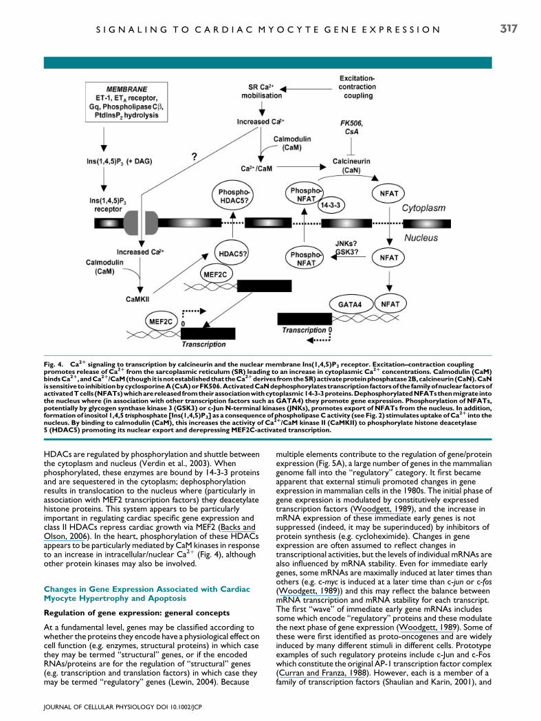
Fig. 4. Ca2R signaling to transcription by calcineurin and the nuclear membrane Ins(1,4,5)P3 receptor. Excitation–contraction couplingpromotes release of Ca2R from the sarcoplasmic reticulum (SR) leading to an increase in cytoplasmic Ca2R concentrations. Calmodulin (CaM)bindsCa2R,andCa2R/CaM(though it isnotestablishedthattheCa2Rderives fromtheSR)activateproteinphosphatase2B,calcineurin (CaN).CaNissensitiveto inhibitionbycyclosporineA(CsA)orFK506.ActivatedCaNdephosphorylates transcription factorsof the familyofnuclear factorsofactivatedTcells (NFATs)whicharereleased fromtheirassociationwithcytoplasmic14-3-3proteins.DephosphorylatedNFATsthenmigrate intothe nucleus where (in association with other transcription factors such as GATA4) they promote gene expression. Phosphorylation of NFATs,potentially by glycogen synthase kinase 3 (GSK3) or c-Jun N-terminal kinases (JNKs), promotes export of NFATs from the nucleus. In addition,formation of inositol 1,4,5 trisphosphate [Ins(1,4,5)P3] as a consequenceof phospholipaseCactivity (see Fig. 2) stimulates uptake ofCa2R into thenucleus. By binding to calmodulin (CaM), this increases the activity of Ca2R/CaM kinase II (CaMKII) to phosphorylate histone deacetylase5 (HDAC5) promoting its nuclear export and derepressing MEF2C-activated transcription.
S I G N A L I N G T O C A R D I A C M Y O C Y T E G E N E E X P R E S S I O N 317
HDACs are regulated by phosphorylation and shuttle betweenthe cytoplasm and nucleus (Verdin et al., 2003). Whenphosphorylated, these enzymes are bound by 14-3-3 proteinsand are sequestered in the cytoplasm; dephosphorylationresults in translocation to the nucleus where (particularly inassociation with MEF2 transcription factors) they deacetylatehistone proteins. This system appears to be particularlyimportant in regulating cardiac specific gene expression andclass II HDACs repress cardiac growth via MEF2 (Backs andOlson, 2006). In the heart, phosphorylation of these HDACsappears to be particularly mediated by CaM kinases in responseto an increase in intracellular/nuclear Ca2þ (Fig. 4), althoughother protein kinases may also be involved.
Changes in Gene Expression Associated with CardiacMyocyte Hypertrophy and Apoptosis
Regulation of gene expression: general concepts
At a fundamental level, genes may be classified according towhether the proteins they encode have a physiological effect oncell function (e.g. enzymes, structural proteins) in which casethey may be termed ‘‘structural’’ genes, or if the encodedRNAs/proteins are for the regulation of ‘‘structural’’ genes(e.g. transcription and translation factors) in which case theymay be termed ‘‘regulatory’’ genes (Lewin, 2004). Because
JOURNAL OF CELLULAR PHYSIOLOGY DOI 10.1002/JCP
multiple elements contribute to the regulation of gene/proteinexpression (Fig. 5A), a large number of genes in the mammaliangenome fall into the ‘‘regulatory’’ category. It first becameapparent that external stimuli promoted changes in geneexpression in mammalian cells in the 1980s. The initial phase ofgene expression is modulated by constitutively expressedtranscription factors (Woodgett, 1989), and the increase inmRNA expression of these immediate early genes is notsuppressed (indeed, it may be superinduced) by inhibitors ofprotein synthesis (e.g. cycloheximide). Changes in geneexpression are often assumed to reflect changes intranscriptional activities, but the levels of individual mRNAs arealso influenced by mRNA stability. Even for immediate earlygenes, some mRNAs are maximally induced at later times thanothers (e.g. c-myc is induced at a later time than c-jun or c-fos(Woodgett, 1989)) and this may reflect the balance betweenmRNA transcription and mRNA stability for each transcript.The first ‘‘wave’’ of immediate early gene mRNAs includessome which encode ‘‘regulatory’’ proteins and these modulatethe next phase of gene expression (Woodgett, 1989). Some ofthese were first identified as proto-oncogenes and are widelyinduced by many different stimuli in different cells. Prototypeexamples of such regulatory proteins include c-Jun and c-Foswhich constitute the original AP-1 transcription factor complex(Curran and Franza, 1988). However, each is a member of afamily of transcription factors (Shaulian and Karin, 2001), and
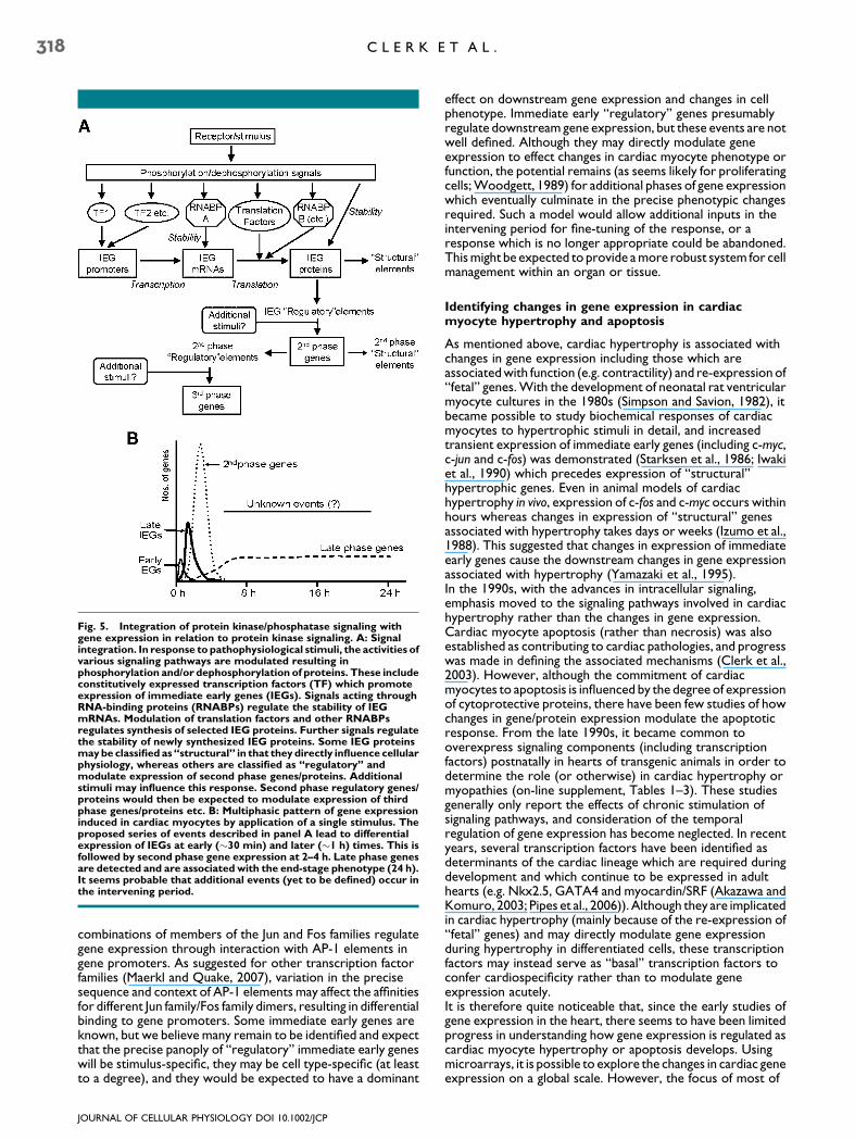
Fig. 5. Integration of protein kinase/phosphatase signaling withgene expression in relation to protein kinase signaling. A: Signalintegration. In response to pathophysiological stimuli, the activities ofvarious signaling pathways are modulated resulting inphosphorylation and/or dephosphorylation of proteins. These includeconstitutively expressed transcription factors (TF) which promoteexpression of immediate early genes (IEGs). Signals acting throughRNA-binding proteins (RNABPs) regulate the stability of IEGmRNAs. Modulation of translation factors and other RNABPsregulates synthesis of selected IEG proteins. Further signals regulatethe stability of newly synthesized IEG proteins. Some IEG proteinsmaybe classified as ‘‘structural’’ in that theydirectly influence cellularphysiology, whereas others are classified as ‘‘regulatory’’ andmodulate expression of second phase genes/proteins. Additionalstimuli may influence this response. Second phase regulatory genes/proteins would then be expected to modulate expression of thirdphase genes/proteins etc. B: Multiphasic pattern of gene expressioninduced in cardiac myocytes by application of a single stimulus. Theproposed series of events described in panel A lead to differentialexpression of IEGs at early (�30 min) and later (�1 h) times. This isfollowed by second phase gene expression at 2–4 h. Late phase genesare detected and are associated with the end-stage phenotype (24 h).It seems probable that additional events (yet to be defined) occur inthe intervening period.
318 C L E R K E T A L .
combinations of members of the Jun and Fos families regulategene expression through interaction with AP-1 elements ingene promoters. As suggested for other transcription factorfamilies (Maerkl and Quake, 2007), variation in the precisesequence and context of AP-1 elements may affect the affinitiesfor different Jun family/Fos family dimers, resulting in differentialbinding to gene promoters. Some immediate early genes areknown, but we believe many remain to be identified and expectthat the precise panoply of ‘‘regulatory’’ immediate early geneswill be stimulus-specific, they may be cell type-specific (at leastto a degree), and they would be expected to have a dominant
JOURNAL OF CELLULAR PHYSIOLOGY DOI 10.1002/JCP
effect on downstream gene expression and changes in cellphenotype. Immediate early ‘‘regulatory’’ genes presumablyregulate downstream gene expression, but these events are notwell defined. Although they may directly modulate geneexpression to effect changes in cardiac myocyte phenotype orfunction, the potential remains (as seems likely for proliferatingcells; Woodgett, 1989) for additional phases of gene expressionwhich eventually culminate in the precise phenotypic changesrequired. Such a model would allow additional inputs in theintervening period for fine-tuning of the response, or aresponse which is no longer appropriate could be abandoned.This might be expected to provide a more robust system for cellmanagement within an organ or tissue.
Identifying changes in gene expression in cardiacmyocyte hypertrophy and apoptosis
As mentioned above, cardiac hypertrophy is associated withchanges in gene expression including those which areassociated with function (e.g. contractility) and re-expression of‘‘fetal’’ genes. With the development of neonatal rat ventricularmyocyte cultures in the 1980s (Simpson and Savion, 1982), itbecame possible to study biochemical responses of cardiacmyocytes to hypertrophic stimuli in detail, and increasedtransient expression of immediate early genes (including c-myc,c-jun and c-fos) was demonstrated (Starksen et al., 1986; Iwakiet al., 1990) which precedes expression of ‘‘structural’’hypertrophic genes. Even in animal models of cardiachypertrophy in vivo, expression of c-fos and c-myc occurs withinhours whereas changes in expression of ‘‘structural’’ genesassociated with hypertrophy takes days or weeks (Izumo et al.,1988). This suggested that changes in expression of immediateearly genes cause the downstream changes in gene expressionassociated with hypertrophy (Yamazaki et al., 1995).In the 1990s, with the advances in intracellular signaling,emphasis moved to the signaling pathways involved in cardiachypertrophy rather than the changes in gene expression.Cardiac myocyte apoptosis (rather than necrosis) was alsoestablished as contributing to cardiac pathologies, and progresswas made in defining the associated mechanisms (Clerk et al.,2003). However, although the commitment of cardiacmyocytes to apoptosis is influenced by the degree of expressionof cytoprotective proteins, there have been few studies of howchanges in gene/protein expression modulate the apoptoticresponse. From the late 1990s, it became common tooverexpress signaling components (including transcriptionfactors) postnatally in hearts of transgenic animals in order todetermine the role (or otherwise) in cardiac hypertrophy ormyopathies (on-line supplement, Tables 1–3). These studiesgenerally only report the effects of chronic stimulation ofsignaling pathways, and consideration of the temporalregulation of gene expression has become neglected. In recentyears, several transcription factors have been identified asdeterminants of the cardiac lineage which are required duringdevelopment and which continue to be expressed in adulthearts (e.g. Nkx2.5, GATA4 and myocardin/SRF (Akazawa andKomuro, 2003; Pipes et al., 2006)). Although they are implicatedin cardiac hypertrophy (mainly because of the re-expression of‘‘fetal’’ genes) and may directly modulate gene expressionduring hypertrophy in differentiated cells, these transcriptionfactors may instead serve as ‘‘basal’’ transcription factors toconfer cardiospecificity rather than to modulate geneexpression acutely.It is therefore quite noticeable that, since the early studies ofgene expression in the heart, there seems to have been limitedprogress in understanding how gene expression is regulated ascardiac myocyte hypertrophy or apoptosis develops. Usingmicroarrays, it is possible to explore the changes in cardiac geneexpression on a global scale. However, the focus of most of
S I G N A L I N G T O C A R D I A C M Y O C Y T E G E N E E X P R E S S I O N 319
these studies is still the identification of changes in expressionassociated with end stage disease in human/rodent hearts or inhearts of transgenic mice with chronic overexpression ofsignaling components (on-line supplement, Table 4), rather thanthe changes which may lead to that state. In whole heart studies,it is difficult to know which changes are associated with cardiacmyocytes rather than other cells in the heart and, because thesystem is complex (with multiple factors acting on myocytes atdifferent stages) and asynchronous, any sequential nature ofgene expression which occurs in an individual cardiac myocytemay be obscured in the whole heart. Even studies in culturedcardiac myocytes often use chronic agonist treatment or longterm overexpression of signaling components. Although thesetypes of experiments provide information on how cardiacpathologies are manifest, there is little information on themechanisms associated with the development of thosepathologies. It is particularly difficult to link an acute, transientprotein kinase signal (for example) to the regulation of geneexpression.At this stage, perhaps more insight can be gained by using arelatively simple system such as cultured cardiac myocytes tostudy global changes in gene expression as a response to aparticular stimulus develops. This is not a perfect model foreither hypertrophy or apoptosis in a whole heart, but therelatively uniform nature of cultured cells and the fact that theycan be synchronized with respect to gene expression (e.g. byserum starvation) allows some assessment of what may occurduring the development of a particular response. Studiesconducted in this way suggest that the response is morecomplex than is perhaps being considered. Liu et al. (2001)demonstrated temporal regulation of gene expression incardiac myocytes exposed to IGF-1 over 12 h. The response isbiphasic (at least) with changes in expression of non-identicalsets of genes at different times. We have also found temporalregulation of gene expression in cardiac myocyte hypertrophyinduced by ET-1 or the a-adrenergic agonist, phenylephrine(A Clerk and PH Sugden, unpublished work), with greaterchanges in expression of greater numbers of genes at 2–4 h thanat 24 h by which time the hypertrophic phenotype is developed.Differential gene expression is observed even between 2 and4 h. This temporal regulation extends to subtoxic or apoptoticconcentrations of H2O2 (Clerk et al., 2007). Of the mRNAswith altered expression at early times, a large number are‘‘regulatory’’ (Clerk et al., 2007; unpublished data), and thesepotentially modulate gene expression at later times. The genesidentified at 2 and 4 h are not, generally, immediate early genesand are presumably regulated by immediate early ‘‘regulatory’’gene products. Cardiac myocyte hypertrophy or apoptosistherefore probably develop as a consequence of sequential‘‘waves’’ of gene expression that eventually culminate in theoverall phenotypic response (Fig. 5B), and much remains to belearnt about the underlying mechanisms associated with theseresponses. Perhaps, when we understand the responses ofspecific cells to individual stimuli, it may then become possibleto start to understand the complexities of a whole heart system.
Integrating Protein Kinase/Phosphatase Signaling WithTranscription Factor Regulation of Gene Expression
Whilst much work has been done to develop an understandingof the signaling pathways which are activated in cardiacmyocytes and the heart in response to pathophysiologicalstimuli, and to identify the genes which are regulated, questionsof signal integration remain. Key points relate to how signalsinitiated by different stimuli become integrated in the cell, howintracellular signaling pathways are integrated to elicit changesin gene expression, and how signaling and gene expression areintegrated together to generate an end-stage phenotype. Theseareas are not well studied in cardiac myocytes (nor are they
JOURNAL OF CELLULAR PHYSIOLOGY DOI 10.1002/JCP
much better studied in other cells), so much of the discussion ofthese issues is theoretical.
Integration of signals from different stimuli
Different receptors may operate through the same signalingpathway, or may initiate conflicting signals. In either case (unlessthere is spatial separation), there must be specific points forintegration. For example, ERK1/2 and its upstream activatorsMKK1/2 (Fig. 2) are activated in cardiac myocytes by variousstimuli including ET-1, growth factor receptors, H2O2 orphenylephrine. However, the upstream activators vary suchthat, for example, ET-1 and platelet-derived growth factorsignal through protein kinase C and c-Raf, epidermal growthfactor does not signal through protein kinase C (Sugden andClerk, 1998b; Clerk et al., 2006), and H2O2 or phenylephrinerequire PI3K (Clerk et al., 2004). In addition, increased cAMP(a prime regulator of myocyte contractility) inhibits Raf activityin cardiac myocytes (Chiloeches et al., 1999), so simultaneousactivation of, for example, b-adrenergic receptors in cardiacmyocytes has the potential to inhibit ERK1/2 signaling. Thereare other examples of possible cross-talk between signalingpathways which generate a highly complex view of intracellularsignaling events. For a non-cancerous cell in which signalingcomponents are expressed at ‘‘normal’’ stoichiometry, with‘‘normal’’ levels of activity and in the appropriate spatialenvironment, we suggest that the picture may not be quite ascomplicated.Another consideration is that most studies utilize agonists attheir maximally effective concentrations. It is not possible toknow the local concentrations of receptor ligands in vivo, but itmay be expected that most will be present at considerablylower levels than in experimental studies. It may be interestingto determine whether different stimuli at low concentrationsact in a synergistic or additive manner to activate, for example,the ERK1/2 cascade. This would dictate whether a pathway mayoperate in a bistable manner (i.e. the signal is essentially on oroff) or whether there is a graded response, either of which hasimplications for the regulation of gene expression. For a bistableresponse, presumably a threshold has to be reached for thesignal to be activated but, once activated, there is a single output(e.g. to gene expression), whereas a graded response mayprovide for a greater range in terms of effect. The type ofresponse may depend on the context rather than the pathway.MAPKs operate in a bistable fashion in Xenopus oocytematuration (Ferrell and Machleder, 1998), but a gradedresponse is preferred in proliferating fibroblasts (MacKeiganet al., 2005). Perhaps a graded response might be expected incardiac myocytes.
Integrating signals for gene expression
Transcription of a particular gene is regulated by multipletranscription factors which bind to specific DNA elementsoften in the 50 promoter regions, and these interact with thebasal transcriptional machinery to modulate expression. Thepromoter is therefore a key point for signal integration whetherthis is a consequence of direct phosphorylation/dephosphorylation of transcription factors downstream ofprotein kinase/phosphatase signaling pathways, or a result ofaltered concentrations of transcription factors in the nucleus.As far as we are aware, no individual promoter for anyparticular gene has been mapped with respect to the specifictranscription factors which bind or the signaling pathwayswhich regulate their activity. This is partly because the full rangeof transcription factors expressed in any cell is not yet fullydefined. Thus, even though (for example) the rat c-junpromoter contains multiple elements with proteins bound tothem (determined by DNA footprinting) (Rozek and Pfeifer,
320 C L E R K E T A L .
1993), it is not possible to be certain exactly which proteins arepresent in a particular context.Of the techniques available, chromatin immunoprecipitation(ChIP) is perhaps the only way to establish experimentallywhether a transcription factor is bound to a particularpromoter, but (currently) this requires prior knowledge ofwhich transcription factors may be present, and bioinformaticsapproaches are necessary to predict possible transcriptionfactor binding sites. This is not an easy task, given that manytranscription factors exist as families of proteins which might beexpected to exhibit differential affinities for variant sequences.Although core sequences for different transcription factorsappear similar, individual bases probably do not contributeindependently in an additive fashion to binding affinities andflanking sequences surrounding the core may also influencetranscription factor binding (Maerkl and Quake, 2007). Thus,even for the Jun (c-Jun, JunB and JunD) and Fos (c-Fos, FosB,Fra1 and Fra2) families, the different Jun dimers or Jun/Fosdimers display different affinities for the very similar AP-1(TGACTCA) and CRE (TGACGTCA) consensus sequences(Ryseck and Bravo, 1991). A further consideration relating towhether or not a particular transcription factor family membermay bind to a promoter element is the affinity andconcentration of that specific protein in relation to the affinitiesand concentrations of other family members.In relation to cardiac myocyte gene expression, until the geneswhich are regulated during hypertrophy or apoptosis (ratherthan at the end stage of the response) are identified, it will bedifficult to start to link the signaling pathways through thetranscription factors to any particular gene. In the context ofET-1, using microarrays, we demonstrated that a largeproportion of genes (�70%) with altered expression at 2–4 happear to do so as a consequence of some degree of ERK1/2signaling (Kennedy et al., 2006). However, these are notimmediate early genes and not likely to be regulated directly bythe initial protein kinase signaling pathway. It is worthconsidering that immediate early gene transcription factorssuch as c-Jun and c-Fos act as dimers on an individual element,and multiple elements may operate in a single promoter. Aninput from the ERK1/2 cascade at the level of one of thesetranscription factors could therefore be widely propagated tomany downstream genes, and the proportion of genes requiringan element of ERK1/2 signaling may therefore be greater at latertimes than within the immediate early gene phase.Apart from transcription, other levels of signal integration areemerging for the co-ordinated regulation of gene and proteinexpression but for which there has been little consideration incardiac myocytes or cardiac pathologies. For example, in T cells,as much as 50% of the changes in gene expression result fromchanges in mRNA stability rather than alterations intranscriptional rates (Cheadle et al., 2005). Regulation ofmRNA stability may be just as important in cardiac myocytessince signaling through p38-MAPKs promotes mRNA stabilityin other cells (Dean et al., 2004), and p38-MAPK is activated incardiac myocytes by hypertrophic stimuli or cellular stresses(Sugden and Clerk, 1998b). Another issue remaining to beaddressed in cardiac myocytes is the regulation of translation ofspecific mRNAs. In other cells, signaling through the ERK1/2cascade or PKB/Akt can influence the translation of specificpools of mRNAs (Rajasekhar et al., 2003; Tominaga et al., 2005;Spence et al., 2006), presumably through phosphorylation ofRNA-binding proteins which modulate recruitment toribosomes for translation. Regulation of the association ofRNA-binding proteins with a particular pool of mRNAs toregulate translation has led to the concept of post-transcriptional operons in mammalian cells for theco-ordination of gene/protein expression (Keene and Lager,2005). Perhaps we should expect that cardiac myocytesco-ordinate expression of related genes/proteins which
JOURNAL OF CELLULAR PHYSIOLOGY DOI 10.1002/JCP
promote phenotypic changes. Finally, even after proteins havebeen synthesized, further phosphorylation may be required forstabilization. In other cells, immediate early gene productsincluding c-Fos may be phosphorylated as a consequence ofsustained activation of ERK1/2 signaling, and stabilization of theproteins can alter the cellular response (Murphy and Blenis,2006). In cardiac myocytes, phosphorylation of c-Jun by JNKsstabilizes the protein and allows efficient upregulation (Clerket al., 2002), but the consequences of high level expression ofc-Jun (in the context of ET-1 stimulation) compared with lowlevel c-Jun expression (with, for example, phorbol ester) areunknown. Clearly, with respect to signal integration forgene/protein expression in the heart, many issues remain to beexplored.
Protein Kinase Signaling, Gene Expression and theDevelopment of Cardiac Pathologies
As we have discussed, considerable progress has been made inidentifying the stimuli and protein kinase signaling pathwayswhich are likely to be involved in regulating cardiac myocyteresponses, and it is assumed that these stimuli and pathwaysregulate gene/protein expression to influence the changes thatare seen. How these observations relate to the development ofhuman cardiac hypertrophy or heart failure, which may takeseveral decades to develop, is not clear. Others propose thatprocessive activation of individual signaling pathways, each ofwhich modulates a particular phase of the response associatedwith specific changes in gene expression, leads to the end stagecardiac pathology (Hoshijima and Chien, 2002). Since anindividual stimulus causes phasic ‘‘waves’’ of gene expression(Fig. 5; Clerk et al., 2007) and many of the genes which aremodulated encode ‘‘regulators’’ of gene/protein expression, wesuggest that this model should be expanded to incorporate theregulatory transcriptional network as an integral part of thesignal. Thus, a signal through ERK1/2 to immediate early genetranscription factors c-Fos and c-Jun continues through theregulatory genes they modulate to the downstream genesbeyond, and eventually this leads (perhaps) to a hypertrophiccardiac myocyte. In the context of a whole heart, not allmyocytes will behave similarly since the response is influencedby the immediate environment of each individual cell. As aresponse is triggered in any individual cell, that myocyte rapidlychanges its responsiveness to the environment and producesfactors that may act in a paracrine fashion to modulate theresponse of adjacent cells (e.g. stimulation of myocytes withET-1 increases expression of interleukin1 receptor-like 1 andamphiregulin (Kennedy et al., 2006)). Understanding thecomplexity of this type of response in the whole heart isprobably beyond current capabilities. Though it is tempting totry to invoke a particular signaling pathway or transcriptionalregulator in the development of cardiac pathologies, in order todo this, we first need to understand how cardiac myocytes (andother cells in the heart) respond to different stimuli and howsignals are integrated in this (relatively) simple system. It maythen become possible to develop computational models topredict how individual cells react to pathophysiological stressesin the whole heart within spatial and temporal constraints. It ishoped that, eventually, these studies may be linked to thechanges in gene and protein expression observed in cardiachypertrophy and/or heart failure (on-line supplement, Table 4)which develops as a consequence of pathophysiologicalstresses.
Acknowledgments
Our work is supported by the British Heart Foundation, theMedical Research Council, the NHLI Foundation and theFondation Leducq.
S I G N A L I N G T O C A R D I A C M Y O C Y T E G E N E E X P R E S S I O N 321
Literature Cited
Aikawa R, Komuro I, Yamazaki T, Zou Y, Kudoh S, Tanaka M, Shiojima I, Hiroi Y, Yazaki Y.1997. Oxidative stress activates extracellular signal-regulated kinases through Src and Rasin cultured cardiac myocytes of neonatal rats. J Clin Invest 100:1813–1821.
Akazawa H, Komuro I. 2003. Roles of transcription factors in cardiac hypertrophy. Circ Res92:1079–1088.
Antos CL, McKinsey TA, Frey N, Kutschke W, McAnally J, Shelton JM, Richardson JA, Hill JA,Olson EN. 2002. Activated glycogen synthase-3b suppresses cardiac hypertrophy in vivo.Proc Natl Acad Sci USA 99:907–912.
Aoki H, Kang PM, Hampe J, Yoshimura K, Noma T, Matsuzaki M, Izumo S. 2002. Directactivation of mitochondrial apoptosis machinery by c-Jun N-terminal kinase in adult cardiacmyocytes. J Biol Chem 277:10244–10250.
Backs J, Olson EN. 2006. Control of cardiac growth by histone acetylation/deacetylation. CircRes 98:15–24.
Biondi RM, Nebreda AR. 2003. Signalling specificity of Ser/Thr protein kinases throughdocking-site-mediated interactions. Biochem J 372:1–13.
Bogoyevitch MA, Ketterman AJ, Sugden PH. 1995. Cellular stresses differentially activate c-Jun N-terminal protein kinases and extracellular signal-regulated protein kinases incultured ventricular myocytes. J Biol Chem 270:29710–29717.
Bueno OF, Molkentin JD. 2002. Involvement of extracellular signal-regulated kinases 1/2 incardiac hypertrophy and cell death. Circ Res 91:776–781.
Camper-Kirby D, Welch S, Walker A, Shiraishi I, Setchell KDR, Schaefer E, Kajstura J,Anversa P, Sussman MA. 2001. Myocardial Akt activation and gender: Increased nuclearactivity in females versus males. Circ Res 88:1020–1027.
Cheadle C, Fan J, Cho-Chung YS, Werner T, Ray J, Do L, Gorospe M, Becker KG. 2005.Control of gene expression during T cell activation: Alternate regulation of mRNAtranscription and mRNA stability. BMC Genom 6:75.
Chiloeches A, Paterson HF, Marais R, Clerk A, Marshall CJ, Sugden PH. 1999. Regulation ofRas.GTP loading and Ras-Raf association in neonatal rat ventricular myocytes by G protein-coupled receptor agonists and phorbol ester. Activation of the ERK cascade by phorbolester is mediated by Ras. J Biol Chem 274:19762–19770.
Clerk A, Sugden PH. 1998. The p38-MAPK inhibitor, SB203580, inhibits cardiac stress-activated protein kinases/c-Jun N-terminal kinases (SAPKs/JNKs). FEBS Lett 426:93–96.
Clerk A, Michael A, Sugden PH. 1998a. Stimulation of the p38 mitogen-activated proteinkinase pathway in neonatal rat ventricular myocytes by the G protein-coupled receptoragonists, endothelin-1 and phenylephrine: A role in cardiac myocyte hypertrophy? J CellBiol 142:523–535.
Clerk A, Michael A, Sugden PH. 1998b. Stimulation of multiple mitogen-activated proteinkinase sub-families by oxidative stress and phosphorylation of the small heat shock protein,HSP25/27, in neonatal ventricular myocytes. Biochem J 333:581–589.
Clerk A, Kemp TJ, Harrison JG, Mullen AJ, Barton PJ, Sugden PH. 2002. Up-regulation of c-junmRNA in cardiac myocytes requires the extracellular signal-regulated kinase cascade, butc-Jun N-terminal kinases are required for efficient up-regulation of c-Jun protein. Biochem J368:101–110.
Clerk A, Cole SM, Cullingford TE, Harrison JG, Jormakka M, Valks DM. 2003. Regulation ofcardiac myocyte cell death. Pharmacol Ther 97:223–261.
Clerk A, Kemp TJ, Harrison JG, Pham FH, Sugden PH. 2004. Integration of protein kinasesignaling pathways in cardiac myocytes: Signaling to and from the extracellular signal-regulated kinases. Adv Enzyme Regul 44:233–248.
Clerk A, Aggeli I-K, Stathopoulou K, Sugden PH. 2006. Peptide growth factors signaldifferentially through protein kinase C to extracellular signal-regulated kinase in neonatalcardiomyocytes. Cell Signal 18:225–235.
Clerk A, Kemp TJ, Zoumpoulidou G, Sugden PH. 2007. Cardiac myocyte gene expressionprofiling during H2O2-induced apoptosis. Physiol Genomics E-pub ahead of print.
Cohen P, Frame S. 2001. The renaissance of G SK3. Nat Rev Mol Cell Biol 2:769–776.Cook SA, Sugden PH, Clerk A. 1999. Regulation of Bcl-2 family proteins during development
and in response to oxidative stress in cardiac myocytes: Association with changes inmitochondrial membrane potential. Circ Res 85:940–949.
Court NW, dos Remedios CG, Cordell J, Bogoyevitch MA. 2002. Cardiac expression andsubcellular localization of the p38 mitogen-activated protein kinase member, stress-activated protein kinase-3 (SAPK3). J Mol Cell Cardiol 34:413–426.
Curran T, Franza BRJr. 1988. Fos and Jun: The AP-1 connection. Cell 55:395–397.Dean JL, Sully G, Clark AR, Saklatvala J. 2004. The involvement of AU-rich element-binding
proteins in p38 mitogen-activated protein kinase pathway-mediated mRNA stabilisation.Cell Signal 16:1113–1121.
Dorn GWII, Robbins J, Sugden PH. 2003. Phenotyping hypertrophy. Eschew obfuscation. CircRes 92:1171–1175.
Euler-Taimor G, Heger J. 2006. The complex pattern of SMAD signaling in the cardiovascularsystem. Cardiovasc Res 69:15–25.
Ferrell JE, Machleder EM. 1998. The biochemical basis of an all-or-none cell fate switch inXenopus oocytes. Science 280:895–898.
Frame S, Cohen P. 2001. GSK3 takes centre stage more than 20 years after its discovery.Biochem J 359:1–16.
Gusterson RJ, Jazrawi E, Adcock IM, Latchman DS. 2003. The transcriptional co-activatorsCREB-binding protein (CBP) and p300 play a critical role in cardiac hypertrophy that isdependent on their histone acetyltransferase activity. J Biol Chem 278:6838–6847.
Haq S, Choukroun G, Kang ZB, Ranu H, Matsui T, Rosenzweig A, Molkentin JD, AlessandriniA, Woodgett J, Hajjar R, Michael A, Force T. 2000. Glycogen synthase kinase-3b is anegative regulator of cardiomyocyte hypertrophy. J Cell Biol 151:117–129.
Heineke J, Molkentin JD. 2006. Regulation of cardiac hypertrophy by intracellular signallingpathways. Nat Rev Mol Cell Biol 7:589–600.
Horvath CM. 2000. STAT proteins and transcriptional responses to extracellular signals.Trends Biochem Sci 25:496–502.
Hoshijima M, Chien KR. 2002. Mixed signals in heart failure: Cancer rules. J Clin Invest109:849–855.
Huisamen B, van Zyl M, Keyser A, Lochner A. 2001. The effects of insulin and b-adrenergic stimulation on glucose transport, glut 4 and PKB activation in themyocardium of lean and obese non-insulin dependent diabetes mellitus rats.Mol Cell Biochem 223: 15–25.
Iwaki K, Sukhatme VP, Shubeita HE, Chien KR. 1990.a- andb-Adrenergic stimulation inducesdistinct patterns of immediate early gene expression in neonatal rat myocardial cells. fos/junexpression is associated with sarcomere assembly; Egr-1 induction is primarily an a1-mediated response. J Biol Chem 265:13809–13817.
Izumo S, Nadal-Ginard B, Mahdavi V. 1988. Protooncogene induction and reprogramming ofcardiac gene expression produced by pressure overload. Proc Natl Acad Sci U. S. A.85:339–343.
JOURNAL OF CELLULAR PHYSIOLOGY DOI 10.1002/JCP
Kalkhoven E. 2004. CBP and p300: HATs for different occasions. Biochem Pharmacol68:1145–1155.
Kang PM, Haunstetter A, Aoki H, Usheva A, Izumo S. 2000. Morphological and molecularcharacterization of adult cardiomyocyte apoptosis during hypoxia and reoxygenation. CircRes 87:118–125.
Keene JD, Lager PJ. 2005. Post-transcriptional operons and regulons co-ordinating geneexpression. Chromosome Res 13:327–337.
Kennedy RA, Kemp TJ, Sugden PH, Clerk A. 2006. Using U0126 to dissect the role of theextracellular signal-regulated kinase 1/2 (ERK1/2) cascade in the regulation of geneexpression by endothelin-1 in cardiac myocytes. J Mol Cell Cardiol 41:236–247.
Kumar D, Kirshenbaum L, Li T, Danelisen I, Singal P. 1999. Apoptosis in isolated adultcardiomyocytes exposed to adriamycin. Ann NY Acad Sci 874:156–168.
Lemke LE, Bloem LJ, Fouts R, Esterman M, Sandusky G, Vlahos CJ. 2001. Decreased p38MAPK activity in end-stage failing human myocardium: p38 MAPK a is the predominantisoform expressed in human heart. J Mol Cell Cardiol 33:1527–1540.
Lessor T, Yoo JY, Davis M, Hamburger AW. 1998. Regulation of heregulin b1-induceddifferentiation in a human breast carcinoma cell line by the extracellular-regulated kinase(ERK) pathway. J Cell Biochem 70:587–595.
Lewin B. 2004. The operon. In: Carlson G, Challice J, editors. Genes VIII. London: PearsonEducation Ltd. p 279–299.
Liang Q, Molkentin JD. 2003. Redefining the roles of p38 and JNK signaling in cardiachypertrophy: Dichotomy between cultured myocytes and animal models. J Mol CellCardiol 35:1385–1394.
Liu T, Lai H, Wu W, Chinn S, Wang PH. 2001. Developing a strategy to define the effects ofinsulin-like growth factor-1 on gene expression profile in cardiomyocytes. Circ Res88:1231–1238.
MacKeigan JP, Murphy LO, Dimitri CA, Blenis J. 2005. Graded mitogen-activated proteinkinase activity precedes switch-like c-Fos induction in mammalian cells. Mol Cell Biol25:4676–4682.
Maerkl SJ, Quake SR. 2007. A systems approach to measuring the binding energy landscapesof transcription factors. Science 315:233–237.
Martelli AM, Faenza I, Billi AM, Manzoli L, Evangelisti C, Fala F, Cocco L. 2006. Intranuclear 30-phosphoinositide metabolism and Akt signaling: New mechanisms for tumorigenesis andprotection against apoptosis. Cell Signal 18:1101–1107.
Matsui T, Rosenzweig A. 2005. Convergent signal transduction pathways controllingcardiomyocyte survival and function: The role of PI 3-kinase and Akt. J Mol Cell Cardiol38:63–71.
Molkentin JD, Dorn GII. 2001. Cytoplasmic signaling pathways that regulate cardiachypertrophy. Annu Rev Physiol 63:391–426.
Mora A, Sakamoto K, McManus EJ, Alessi DR. 2005. Role of the PDK1-PKB-GSK3 pathwayin regulating glycogen synthase and glucose uptake in the heart. FEBS Lett 579:3632–3638.
Morton S, Davis RJ, McLaren A, Cohen P. 2003. A reinvestigation of the multisitephosphorylation of the transcription factor c-Jun. EMBO J 22:3876–3886.
Muller-Newen G. 2003. The cytokine receptor gp130: Faithfully promiscuous. Sci STKE201:PE40.
Murphy LO, Blenis J. 2006. MAPK signal specificity: the right place at the right time. TrendsBiochem Sci 31:268–275.
Nadal-Ginard B, Kajstura J, Leri A, Anversa P. 2003. Myocyte death, growth, and regenerationin cardiac hypertrophy and failure. Circ Res 92:139–150.
Nicol RL, Frey N, Pearson G, Cobb M, Richardson J, Olson EN. 2001. Activated MEK5induces serial assembly of sarcomeres and eccentric cardiac hypertrophy. EMBO J20:2757–2767.
Petrich BG, Wang Y. 2004. Stress-activated MAP kinases in cardiac remodeling and heartfailure; new insights from transgenic studies. Trends Cardiovasc Med 14:50–55.
Pham FH, Sugden PH, Clerk A. 2000. Regulation of protein kinase B and 4E-BP1 by oxidativestress in cardiac myocytes. Circ Res 86:1252–1258.
Pipes GC, Creemers EE, Olson EN. 2006. The myocardin family of transcriptionalcoactivators: Versatile regulators of cell growth, migration, and myogenesis. Genes Dev20:1545–1556.
Poizat C, Puri PL, Bai Y, Kedes L. 2005. Phosphorylation-dependent degradation of p300 bydoxorubicin-activated p38 mitogen-activated protein kinase in cardiac cells. Mol Cell Biol25:2673–2687.
Polak P, Hall MN. 2006. mTORC2 caught in a SINful Akt. Dev Cell 11:583–589.Preedy VR, Smith DM, Kearney NF, Sugden PH. 1984. Rates of protein turnover in vivo
and in vitro in ventricular muscle of hearts from fed and starved rats. Biochem J 222:395–400.
Rajasekhar VK, Viale A, Socci ND, Wiedmann M, Hu X, Holland EC. 2003. Oncogenic Ras andAkt signaling contribute to glioblastoma formation by differential recruitment of existingmRNAs to polysomes. Mol Cell 12:889–901.
Roux PP, Blenis J. 2004. ERK and p38 MAPK-activated protein kinases: A family of proteinkinases with diverse biological functions. Microbiol Mol Biol Rev 68:320–344.
Rozek D, Pfeifer GP. 1993. In vivo protein–DNA interactions at the c-jun promoter:Preformed complexes mediate the UV response. Mol Cell Biol 13:5490–5499.
Ryseck RP, Bravo R. 1991. c-JUN, JUN B and JUN D differ in their binding affinities to AP-1 andCRE consensus sequences: Effect of FOS proteins. Oncogene 6:533–542.
Sawyer DB, Siwik DA, Xiao L, Pimentel DR, Singh K, Colucci WS. 2002. Role of oxidativestress in myocardial hypertrophy and failure. J Mol Cell Cardiol 34:379–388.
Shaulian E, Karin M. 2001. AP-1 in cell proliferation and survival. Oncogene 20:2390–2400.Shiojima I, Yefremashvili M, Luo Z, Kureishi Y, Takahashi A, Tao J, Rosenzweig A, Kahn CR,
Abel ED, Walsh K. 2002. Akt signaling mediates postnatal heart growth in response toinsulin and nutritional status. J Biol Chem 277:37670–37677.
Simpson P, Savion S. 1982. Differentiation of rat myocytes in single cell cultures with andwithout proliferating nonmyocardial cells: Cross-striations, ultrastructure, andchronotropic response to isoproterenol. Circ Res 50:101–116.
Spence J, Duggan BM, Eckhardt C, McClelland M, Mercola D. 2006. Messenger RNAsunder differential translational control in Ki-ras-transformed cells. Mol Cancer Res4:47–60.
Starksen NF, Simpson PC, Bishopric N, Coughlin SR, Lee WMF, Escobedo JA, Williams LT.1986. Cardiac myocyte hypertrophy is associated with c-myc protooncogene expression.Proc Natl Acad Sci U. S. A. 83:8348–8350.
Sugden PH. 2003. Ras, Akt, and mechanotransduction in the cardiac myocyte. Circ Res93:1179–1192.
Sugden PH, Clerk A. 1998a. ‘‘Stress-responsive’’ mitogen-activated protein kinases (c-Jun N-terminal kinases and p38 mitogen-activated protein kinases) in the myocardium. Circ Res83:345–352.
Sugden PH, Clerk A. 1998b. Cellular mechanisms of cardiac hypertrophy. J Mol Med 76:725–746.
322 C L E R K E T A L .
Tergaonkar V. 2006. NFkB: A good signaling paradigm and therapeutic target. Int J BiochemCell Biol 38:1647–1653.
Tominaga Y, Tanguney T, Kolesnichenko M, Bilanges B, Stokoe D. 2005. Translationalderegulation in PDK-1�/� embryonic stem cells. Mol Cell Biol 25:8465–8475.
Traverse S, Gomez N, Paterson H, Marshall C, Cohen P. 1992. Sustained activation of themitogen-activated protein (MAP) kinase cascade may be required for differentiation ofPC12 cells. Comparison of the effects of nerve growth factor and epidermal growth factor.Biochem J 288:351–355.
Valen G, Yan Z, Hansson GK. 2001. Nuclear factor k-B and the heart. J Am Coll Cardiol38:307–314.
van der Heide LP, Hoekman MF, Smidt MP. 2004. The ins and outs of FoxOshuttling: Mechanisms of FoxO translocation and transcriptional regulation. Biochem J380:297–309.
van Empel VP, Bertrand AT, Hofstra L, Crijns HJ, Doevendans PA, De Windt LJ. 2005.Myocyte apoptosis in heart failure. Cardiovasc Res 67:21–29.
Vanhaesebroeck B, Alessi DR. 2000. The PI3K-PDK1 connection: More than just a road toPKB. Biochem J 346:561–576.
Verdin E, Dequiedt F, Kasler HG. 2003. Class II histone deacetylases: Versatile regulators.Trends Genet 19:286–295.
JOURNAL OF CELLULAR PHYSIOLOGY DOI 10.1002/JCP
von Harsdorf R, Li P-F, Dietz R. 1999. Signaling pathways in reactive oxygen species-inducedcardiomyocyte apoptosis. Circulation 99:2934–2941.
Wilkins BJ, Molkentin JD. 2004. Calcium-calcineurin signaling in the regulation of cardiachypertrophy. Biochem Biophys Res Commun 322:1178–1191.
Wollert KC, Chien KR. 1997. Cardiotrophin-1 and the role of gp130-dependent signalingpathways in cardiac growth and development. J Mol Med 75:492–501.
Woodgett JR. 1989. Early gene induction by growth factors. Br Med Bull 45:529–540.Wullschleger S, Loewith R, Hall MN. 2006. TOR signaling in growth and metabolism. Cell
124:471–484.Yamamoto S, Seta K, Morisco C, Vatner SF, Sadoshima J. 2001. Chelerythrine rapidly induces
apoptosis through generation of reactive oxygen species in cardiac myocytes. J Mol CellCardiol 33:1829–1848.
Yamashita K, Kajstura J, Discher DJ, Wasserlauf BJ, Bishopric NH, Anversa P, Webster KA.2001. Reperfusion-activated Akt kinase prevents apoptosis in mouse heartsoverexpressing insulin-like growth factor 1. Circ Res 88:609–614.
Yamazaki T, Komuro I, Yazaki Y. 1995. Molecular mechanism of cardiac cellular hypertrophyby mechanical stress. J Mol Cell Cardiol 27:133–140.
Yoon S, Seger R. 2006. The extracellular signal-regulated kinase: Multiple substrates regulatediverse cellular functions. Growth Factors 24:21–44.
Related Documents


![In Vitro Rat Myocyte Cardiotoxicity Model for …...(CANCER RESEARCH 48. 5222-5227, September 15. 1988] In Vitro Rat Myocyte Cardiotoxicity Model for Antitumor Antibiotics Using Adenosine](https://static.cupdf.com/doc/110x72/5f801929f00b6a5fb7561c08/in-vitro-rat-myocyte-cardiotoxicity-model-for-cancer-research-48-5222-5227.jpg)