1 Supporting Information SI Materials and Methods Plasma IgG adsorptions on SF162 gp120- and SF162 gp120 D368R -coated beads. Plasma adsorptions were performed as previously described (1, 3), with some modifications, and the flow of the adsorptions is outlined in Figure S1. Briefly, total IgG was isolated by protein A chromatography (Pierce, Rockford Il, USA) from VC10042 plasma drawn 22 years post-infection. The purified total IgG was serially adsorbed onto SF162 gp120-coated beads (MyOne Tosylactivated Dynabeads, Invitrogen, Carlsbad, CA, USA), and the flow through collected. The antibodies bound to the gp120 coated beads were eluted by vortexing in increasingly acidic 0.1M glycine solutions, followed by buffer exchange into PBS. The anti-gp120 Ab fraction was then serially adsorbed onto SF162 gp120 D368R -coupled beads to remove Abs that do not bind the CD4-BS. Each fraction described above was tested for residual neutralizing activity against 4 clade B, 3 clade C, and 2 clade A isolates in the TZM-bl neutralization assay. The depleted total IgG and the gp120 D368R depleted anti-gp120 fractions were tested for the presence of anti-CD4-BS antibodies, and the absence of non-CD4-BS gp120 Abs by Luminex assay (Luminex Corporation, Austin, TX, USA) against both wild type SF162 gp120 and SF162 gp120 D368R .

Welcome message from author
This document is posted to help you gain knowledge. Please leave a comment to let me know what you think about it! Share it to your friends and learn new things together.
Transcript

1
Supporting Information SI Materials and Methods Plasma IgG adsorptions on SF162 gp120- and SF162 gp120D368R-coated beads. Plasma adsorptions were performed as previously described (1, 3), with some modifications, and the flow of the adsorptions is outlined in Figure S1. Briefly, total IgG was isolated by protein A chromatography (Pierce, Rockford Il, USA) from VC10042 plasma drawn 22 years post-infection. The purified total IgG was serially adsorbed onto SF162 gp120-coated beads (MyOne Tosylactivated Dynabeads, Invitrogen, Carlsbad, CA, USA), and the flow through collected. The antibodies bound to the gp120 coated beads were eluted by vortexing in increasingly acidic 0.1M glycine solutions, followed by buffer exchange into PBS. The anti-gp120 Ab fraction was then serially adsorbed onto SF162 gp120D368R-coupled beads to remove Abs that do not bind the CD4-BS. Each fraction described above was tested for residual neutralizing activity against 4 clade B, 3 clade C, and 2 clade A isolates in the TZM-bl neutralization assay. The depleted total IgG and the gp120D368R depleted anti-gp120 fractions were tested for the presence of anti-CD4-BS antibodies, and the absence of non-CD4-BS gp120 Abs by Luminex assay (Luminex Corporation, Austin, TX, USA) against both wild type SF162 gp120 and SF162 gp120D368R.

Figu
Figusubjeadsobeadisolaafterand tbindNAbgp12showSF16
ure S1
ure S1. Conect VC10042
orbed onto SFds, the flow tates. The var depletion wthen serially
d to the CD4-bs) were test20 antibody wn in the sec62 gp120D36
ntribution of 2. IgG was F162 gp120-through was lues in the fi
with gp120. y adsorbed on-BS. The aned against thfraction afte
cond column8R.
anti-CD4-Bfirst isolated-coated magtested for re
irst column rAnti-gp120 nto magnetic
nti-gp120 anthe 9 isolates er depletion wn. All deplet
S NAbs to thd from heat-tgnetic beads.esidual neutrrepresent theantibodies wc beads coattibodies anddescribed ab
with gp120Dtions were co
2
he overall netreated plasm After 6 rouralizing active percent reswere eluted fted with SF1d the gp120Dbove. The p
D368R (the neuonfirmed by
eutralizing pma, and thenunds of seriavity against 4sidual neutrafrom the SF162 gp120D36
D368R flow thrpercent residutralizing ac Luminex as
potential of pn antibodies tal adsorption4 clade B, 3
alizing activi162 gp120-c68R to removrough (conta
dual neutraliztivity due tossay against
plasma isolatthat bind to
n with gp120clade C, and
ity against eacoated magnve antibodiesaining the anzing activity
o anti-CD4-BSF162 gp12
ted from gp120 were
0-coated d 2 clade A ach isolate
netic beads, s that do not nti-CD4-BS y of the anti-BS NAbs) is 20 and
Figu
FiguCD4
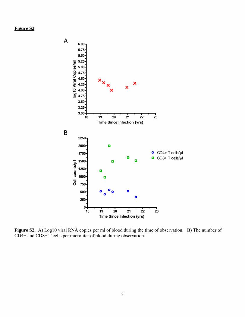
ure S2
ure S2. A) L4+ and CD8+
Log10 viral R+ T cells per
RNA copies r microliter o
per ml of blof blood duri
3
lood during ing observat
the time of otion.
observation.
B) The nuumber of

Figu
Figucollecladevariaacid circl
ure S3
ure S3. Full ected approxe B sequencable loops Vresidues tha
les.
length gp16ximately 19 ye using Clus
V1-V5. Sitesat are known
60 amino aciyears post instal W. The s that are varn to contact C
id alignmentnfection. Repgp120, gp41
riable amongCD4 are mar
4
t of the ‘earlypresentative1 and signal g the VC100rked with red
y’ clones isoe Env clones
peptide port042 clones ard circles and
olated from p were alignetions of Envre highlighted VRC01 are
plasma that wed with the cv are labeled,ed in yellow.e marked wit
was consensus , as are the . Amino th blue

Figu
Figuapprsequloopresid
ure S4
ure S4. Full roximately 2uence using Cs V1-V5. S
dues that are
length gp162 years postClustal W. Tites that are known to co
60 amino acit infection. RThe gp120, gvariable amontact CD4 a
id alignmentRepresentatigp41 and sigong the clonare marked w
5
t of the ‘late’ve Env clon
gnal peptide nes from VCwith red circ
’ clones isolnes were alig
portions of EC10042 are hcles and VRC
ated from plgned with theEnv are labe
highlighted inC01 are mar
lasma that we consensus eled, as are tn yellow. Arked with blu
was collectedclade B
the variable Amino acid ue circles.
d

Figu
FigusubjewereMEG(JTTis locmapp100-19 y
ure S5
ure S5. Phyect VC10042e tested for thGA5.1(4) usT) substitutiocated in the ped, with no-250 coloredears post-inf
ylogenetic tr2 at 19 yearsheir neutraliing Maximu
on model andbottom left.
on-neutralized in orange, afection. (B) N
ree showing s (A) and 22zation susce
um Likelihood utilizing un The potenc
ed isolates leand IC50 titeNeutralizatio
the relations2 years (B) peptibility of Vod with 100 niform ratescy with whiceft uncoloreders above 25on with plas
6
ship betweenost infectionVC10042 plbootstrap rep. Bootstrap
ch the plasmad, IC50 titers50 colored inma isolated
n the autologn and env clolasma. Phyloplications, uvalues are lia is able to ns below 100 n red. (A) Ne
at 22 years p
gous env cloones from hogenies were
utilizing the Jisted at eachneutralize a gcolored blue
eutralization post-infectio
ones circulatiheterologous e reconstrucJones-Taylo
h node and thgiven isolatee, IC50 titerwith plasma
on.
ing in isolates that
cted using or-Thornton he scale bar e is heat s between a isolated at
t

Figu
Figuin Vmapprelev
ure S6
ure S6. The C10042. Reped on to thevant features
regions on gesidues R373e HXB2 cors.
gp120 that m3 and N386 re molecule i
mediate escapreside outsidin the b12-bo
7
pe from the de the CD4 bound state (2
neutralizingbinding pock2). Inset is s
g activity of tket (inset). Aslightly rotat
the anti-CD4All residues ted to better
4-BS NAbs were view
Figu
Figumonmuta(♦).
ure S7
ure S7. Neunoclonal broaations were iA double m
utralization seadly neutraliintroduced in
mutant, H364
ensitivity of zing antibodnto the wild
4s.R373M (o
f the clone Vdies b12 (A)type VC100
o) was also g
8
VC10042.y22
, VRC01 (B042.y22.05 (generated.
2.05 bearing ), and NIH4
(■) clone: H3
reversion m45-46G54W (C364S(▲) , R
mutations to tC). Three revR373M (●), a
the version and N386D

9
References: 1. Li, Y., S. A. Migueles, B. Welcher, K. Svehla, A. Phogat, M. K. Louder, X. Wu, G. M. Shaw, M.
Connors, R. T. Wyatt, and J. R. Mascola. 2007. Broad HIV-1 neutralization mediated by CD4-binding site antibodies. Nat Med 13:1032-4.
2. Liu, J., A. Bartesaghi, M. J. Borgnia, G. Sapiro, and S. Subramaniam. 2008. Molecular architecture of native HIV-1 gp120 trimers. Nature 455:109-13.
3. Sather, D. N., J. Armann, L. K. Ching, A. Mavrantoni, G. Sellhorn, Z. Caldwell, X. Yu, B. Wood, S. Self, S. Kalams, and L. Stamatatos. 2009. Factors associated with the development of cross-reactive neutralizing antibodies during human immunodeficiency virus type 1 infection. J Virol 83:757-69.
4. Tamura, K., D. Peterson, N. Peterson, G. Stecher, M. Nei, and S. Kumar. 2011. MEGA5: molecular evolutionary genetics analysis using maximum likelihood, evolutionary distance, and maximum parsimony methods. Mol Biol Evol 28:2731-9.
Related Documents