BioMed Central Page 1 of 10 (page number not for citation purposes) Journal of Translational Medicine Open Access Research Should tumor VEGF expression influence decisions on combining low-dose chemotherapy with antiangiogenic therapy? Preclinical modeling in ovarian cancer David O Holtz 1,2 , Robert T Krafty 4 , Alisha Mohamed-Hadley 1 , Lin Zhang 1 , Ioannis Alagkiozidis 1 , Benjamin Leiby 4 , Wensheng Guo 4,5 , Phyllis A Gimotty 4,5 and George Coukos* 1,2,3 Address: 1 Center for Research on Early Detection and Cure of Ovarian Cancer, University of Pennsylvania, Philadelphia, USA, 2 Division of Gynecologic Oncology, Department of Obstetrics and Gynecology, University of Pennsylvania, Philadelphia, USA, 3 Abramson Family Cancer Research Institute, University of Pennsylvania, Philadelphia, USA, 4 Center for Clinical Epidemiology and Biostatistics, University of Pennsylvania, Philadelphia, USA and 5 Department of Biostatistics and Epidemiology, University of Pennsylvania, Philadelphia, USA Email: David O Holtz - [email protected]; Robert T Krafty - [email protected]; Alisha Mohamed-Hadley - [email protected]; Lin Zhang - [email protected]; Ioannis Alagkiozidis - [email protected]; Benjamin Leiby - [email protected]; Wensheng Guo - [email protected]; Phyllis A Gimotty - [email protected]; George Coukos* - [email protected] * Corresponding author Abstract Because of its low toxicity, low-dose (LD) chemotherapy is ideally suited for combination with antiangiogenic drugs. We investigated the impact of tumor vascular endothelial growth factor A (VEGF-A) expression on the efficacy of LD paclitaxel chemotherapy and its interactions with the tyrosine kinase inhibitor SU5416 in the ID8 and ID8-Vegf models of ovarian cancer. Functional linear models using weighted penalized least squares were utilized to identify interactions between Vegf, LD paclitaxel and antiangiogenic therapy. LD paclitaxel yielded additive effects with antiangiogenic therapy against tumors with low Vegf expression, while it exhibited antagonism to antiangiogenic therapy in tumors with high Vegf expression. This is the first preclinical study that models interactions of LD paclitaxel chemotherapy with antiangiogenic therapy and tumor VEGF expression and offers important lessons for the rational design of clinical trials. Introduction Expansion of vasculature is critical for tumor growth. Tumors cannot grow beyond few millimeters in the absence of angiogenic support provided by vascular endothelial growth factor-A (VEGF-A or VEGF) and other soluble factors [1]. Approaches to block tumor angiogen- esis have therefore attracted significant attention, and combination of antiangiogenic therapy targeting VEGF with standard cytotoxic chemotherapy has provided proof of principle in the clinic [2-5]. Low-dose (LD) or metro- nomic chemotherapy was designed to damage tumor endothelial cells through the close, regular administration of low, nontoxic doses of chemotherapeutic drugs with short drug-free intervals [6-8]. Additionally, LD chemo- therapy suppresses angiogenic factors and inhibits the recruitment and function of circulating endothelial pro- genitor cells and/or circulating endothelial cells [9-11]. Based on preclinical studies and early phase clinical trials, Published: 8 January 2008 Journal of Translational Medicine 2008, 6:2 doi:10.1186/1479-5876-6-2 Received: 8 November 2007 Accepted: 8 January 2008 This article is available from: http://www.translational-medicine.com/content/6/1/2 © 2008 Holtz et al; licensee BioMed Central Ltd. This is an Open Access article distributed under the terms of the Creative Commons Attribution License (http://creativecommons.org/licenses/by/2.0 ), which permits unrestricted use, distribution, and reproduction in any medium, provided the original work is properly cited.

Welcome message from author
This document is posted to help you gain knowledge. Please leave a comment to let me know what you think about it! Share it to your friends and learn new things together.
Transcript

BioMed CentralJournal of Translational Medicine
ss
Open AcceResearchShould tumor VEGF expression influence decisions on combining low-dose chemotherapy with antiangiogenic therapy? Preclinical modeling in ovarian cancerDavid O Holtz1,2, Robert T Krafty4, Alisha Mohamed-Hadley1, Lin Zhang1, Ioannis Alagkiozidis1, Benjamin Leiby4, Wensheng Guo4,5, Phyllis A Gimotty4,5 and George Coukos*1,2,3Address: 1Center for Research on Early Detection and Cure of Ovarian Cancer, University of Pennsylvania, Philadelphia, USA, 2Division of Gynecologic Oncology, Department of Obstetrics and Gynecology, University of Pennsylvania, Philadelphia, USA, 3Abramson Family Cancer Research Institute, University of Pennsylvania, Philadelphia, USA, 4Center for Clinical Epidemiology and Biostatistics, University of Pennsylvania, Philadelphia, USA and 5Department of Biostatistics and Epidemiology, University of Pennsylvania, Philadelphia, USA
Email: David O Holtz - [email protected]; Robert T Krafty - [email protected]; Alisha Mohamed-Hadley - [email protected]; Lin Zhang - [email protected]; Ioannis Alagkiozidis - [email protected]; Benjamin Leiby - [email protected]; Wensheng Guo - [email protected]; Phyllis A Gimotty - [email protected]; George Coukos* - [email protected]
* Corresponding author
AbstractBecause of its low toxicity, low-dose (LD) chemotherapy is ideally suited for combination withantiangiogenic drugs. We investigated the impact of tumor vascular endothelial growth factor A(VEGF-A) expression on the efficacy of LD paclitaxel chemotherapy and its interactions with thetyrosine kinase inhibitor SU5416 in the ID8 and ID8-Vegf models of ovarian cancer. Functionallinear models using weighted penalized least squares were utilized to identify interactions betweenVegf, LD paclitaxel and antiangiogenic therapy. LD paclitaxel yielded additive effects withantiangiogenic therapy against tumors with low Vegf expression, while it exhibited antagonism toantiangiogenic therapy in tumors with high Vegf expression. This is the first preclinical study thatmodels interactions of LD paclitaxel chemotherapy with antiangiogenic therapy and tumor VEGFexpression and offers important lessons for the rational design of clinical trials.
IntroductionExpansion of vasculature is critical for tumor growth.Tumors cannot grow beyond few millimeters in theabsence of angiogenic support provided by vascularendothelial growth factor-A (VEGF-A or VEGF) and othersoluble factors [1]. Approaches to block tumor angiogen-esis have therefore attracted significant attention, andcombination of antiangiogenic therapy targeting VEGFwith standard cytotoxic chemotherapy has provided proof
of principle in the clinic [2-5]. Low-dose (LD) or metro-nomic chemotherapy was designed to damage tumorendothelial cells through the close, regular administrationof low, nontoxic doses of chemotherapeutic drugs withshort drug-free intervals [6-8]. Additionally, LD chemo-therapy suppresses angiogenic factors and inhibits therecruitment and function of circulating endothelial pro-genitor cells and/or circulating endothelial cells [9-11].Based on preclinical studies and early phase clinical trials,
Published: 8 January 2008
Journal of Translational Medicine 2008, 6:2 doi:10.1186/1479-5876-6-2
Received: 8 November 2007Accepted: 8 January 2008
This article is available from: http://www.translational-medicine.com/content/6/1/2
© 2008 Holtz et al; licensee BioMed Central Ltd. This is an Open Access article distributed under the terms of the Creative Commons Attribution License (http://creativecommons.org/licenses/by/2.0), which permits unrestricted use, distribution, and reproduction in any medium, provided the original work is properly cited.
Page 1 of 10(page number not for citation purposes)
Journal of Translational Medicine 2008, 6:2 http://www.translational-medicine.com/content/6/1/2
despite lower cumulative doses, clinical efficacy of LDchemotherapy may be superior to maximally tolerateddose regimens [12-18]. Because of its low toxicity, LDchemotherapy is ideally suited for combination withother drugs, including antiangiogenic drugs. LD chemo-therapy enhanced the effects of antiangiogenic therapy inpreclinical models [19] and has yielded encouragingresults in combination with antiangiogenic drugs in theclinic [6,20,21].
In spite of its obvious promise, translation of LD chemo-therapy to the clinic faces numerous challenges, includingdefining a biologically optimal dose, and identifying theoptimal drug for combination schemes in specific diseasesettings, since combination therapy with antiangiogenicdrugs has proven beneficial in some patients but not inothers. To date it is unclear what factors determine thelikelihood of success of LD chemotherapy with antiang-iogenic therapy.
VEGF is a critical angiogenic factor in advanced ovariancarcinoma [22]. Expression of VEGF varies considerablyamong tumors of similar origin, and VEGF overexpressionat the mRNA level or increased serum levels portend poorsurvival [22-24]. Upregulation of VEGF by tumor cellsmay be in response to metabolic starvation or hypoxia[25], but may also be constitutive as a result of oncogeneamplification [26]. To date, the effect of VEGF expressionon tumor response to LD chemotherapy has not beeninvestigated. Furthermore, it remains unknown whethertumor VEGF overexpression influences tumor response tocombination of antiangiogenic therapy and LD chemo-therapy. Yet, these are important questions that may affectclinical decisions.
We investigated the impact of tumor Vegf on the efficacyof LD chemotherapy, and examined whether tumor Vegfaffects the interactions of metronomic chemotherapy withan antiangiogenic drug. As examples of LD chemotherapywe used paclitaxel, a drug commonly used in ovarian andother cancers, while as an example of antiangiogenic ther-apy we used SU5416, a tyrosine kinase inhibitor withactivity against VEGF receptor-2 (VEGFR-2) [27,28],which has been used in the clinic in combinations [6]. Weused the ID8 and ID8-Vegf mouse models of ovarian can-cer to address the above questions [29]. ID8 cells expressconstitutively low levels of Vegf-A, while ID8-Vegf cellswere retrovirally transduced to express constitutively highlevels of Vegf164 isoform. This model recapitulates closelyhuman ovarian cancer. We have shown that ID8-Vegftumors maintain significantly higher levels of Vegf-Aexpression in vivo; exhibit increased angiogenesis andgrowth; and are associated with significantly shorter sur-vival relative to ID8 tumors. Importantly, tumor, ascitesand serum levels of Vegf-A protein in animals bearing
ID8-VEGF tumors were significantly higher (approxi-mately 28, 13 and 3-fold, respectively) than in animalsbearing control ID8 tumors, but both were within therange described in human ovarian cancer [29]. To analyzethe interactions between tumor Vegf, LD chemotherapyand SU5416, we used a novel method of statistical mode-ling that involves fitting functional linear models usingweighted penalized least squares [30,31]. This approachenabled us to investigate interactions between LD chemo-therapy and antiangiogenic therapy through simple exper-iments.
We found a significant difference in tumor responsedepending on Vegf expression. LD chemotherapy yieldedadditive effects with antiangiogenic therapy only againsttumors with low Vegf expression, while it exhibited antag-onism to antiangiogenic therapy in tumors with high Vegfexpression. This is the first preclinical study that modelsinteractions of LD chemotherapy with antiangiogenictherapy and tumor Vegf expression and offers importantlessons for the rational design of clinical trials.
Materials and methodsCell culture and reagentsThe development and characterization of ID8-Vegf cellline was described elsewhere in detail [29]. ID8 and ID8-Vegf cells were maintained in DMEM media (Invitrogen,Carlsbad, CA) supplemented with 10% fetal bovineserum (FBS), 100 U/ml penicillin, and 100 μg/ml strepto-mycin (Roche, Indianapolis, IN) in a 5% CO2 atmosphereat 37°C.
Mice and treatmentsSix to eight week old female C57BL/6 mice (Charles RiverLaboratories, Wilmington, MA) were used in protocolsapproved by the IACUC of the University of Pennsylvania.Mice were treated with intraperitoneal (i.p.) bolus injec-tions of pacitaxel, SU5416 or dimethyl sulfoxide(DMSO). Paclitaxel (7.5 mg/kg in 0.2 ml 0.9% saline) orsaline alone were given i.p. on days 1 and 4 every week.This dose is approximately one-fourth of maximally toler-ated doses (MTD) for mice [32,33] and are within metro-nomic range. SU5416 (20 mg/kg in 25 μl DMSO, Sigma-Aldrich, St. Louis, MO) or DMSO (25 μl) alone were giveni.p. on days 1, 3, and 5 every week. This dose of SU5416and DMSO are MTD for C57BL/6 mice, as were identifiedby dose-defining experiments in healthy 6 week oldfemale C57BL/6 mice (not shown). Higher doses ofSU5416 or DMSO resulted in significant weight loss ormortality.
Tumor-free Matrigel™ experimentsIn some experiments, mice (n = 3 mice/group) were anes-thetized with 20 μl/gm tribromoethanol/tert-amyl alco-hol solution (Avertin) and injected with 0.5 ml Matrigel™
Page 2 of 10(page number not for citation purposes)

Journal of Translational Medicine 2008, 6:2 http://www.translational-medicine.com/content/6/1/2
(BD Biosciences, Bedford, MA) containing recombinantmouse (rm)Vegf164 (100 ng/ml). One day after Matrigel™injection, mice were treated with i.p. paclitaxel and/orSU5416 or DMSO at the above doses. Paclitaxel or salinewere given i.p. on days 1 and 4. SU5416 or DMSO weregiven on days 1, 3, and 5. Matrigel™ plugs were removedunder anesthesia 7 days later and were snap frozen in liq-uid nitrogen.
TumorsSubconfluent ID8 or ID8-VEGF cultures were trypsinized,washed twice, and cells harvested by centrifugation at1,000 g for 5 min. A single-cell suspension was preparedin PBS mixed with an equal volume of cold Matrigel at 10mg/ml. A total volume of 0.5 ml containing 1 × 10⁶ ID8or ID8-Vegf cells was injected subcutaneously (s.c.) intothe flank of 6-week old C57BL6 mice (n = 10). Tumorswere detectable two weeks later and tumor size was meas-ured weekly thereafter using a Vernier caliper. Tumor vol-umes were calculated by the formula V = 1/2 (L × W)2,where L is length (longest dimension) and W is width(shortest dimension) [34]. Treatments were initiated four-teen days after tumor inoculation and were carried out for6 weeks. After listed time periods, mice were euthanized,and tumors were removed and snap frozen in liquid nitro-gen.
Tumor microvascular densitySnap frozen tumors and Matrigel plugs were stored at -80°C; embedded in OCT compound (Sakura Finetek, Tor-rance, CA); frozen in liquid nitrogen; and cut with a cryo-stat into 8 μm sections. For immunofluorescent staining,sections were sequentially incubated in 5% horse serum;biotin-labeled anti-mouse CD31 antibody (1:400, BDBioscience); and avidin-FITC or avidin-Cy5 (BD Bio-science). All sections were imaged using an upright Nikon(Augusta, GA) E-600 Eclipse microscope equipped with aBio-Rad (Hercules, CA) 1024-ES confocal system. Imageswere acquired through Cool SNAP Pro® color digital cam-era (Media Cybernetics). All tumors were viewed at ×200magnification. For Matrigel plugs, CD31 staining was ana-lyzed using Image-Pro® Plus 4.1 software (Media Cyber-netics). For microvessel density measurements, slideswere scanned at low power (x40) to identify areas of high-est vascularity. Ten high-powered (x200) fields were thenselected randomly within these areas and microvesseldensities were calculated based on the number of CD31-positive structures. At least three tumors from each groupwere examined in three sections. Sections from eachtumor were separated by at least 200 μm.
Statistical methodsA fixed effects analysis of variance was used for betweengroup comparisons with the in vitro data and with theECM data observed after 6 weeks of treatment. Means are
reported with their standard errors. To assess interactionsbetween LD paclitaxel and antiangiogenic therapy with-out assuming any specific functional form for tumor vol-umes over time (such as assuming that the naturallogarithm of tumor volume over time is linear), we fit afunctional linear model via weighted penalized leastsquares that included covariates for treatment with paclit-axel, treatment with SU5416, ID8-Vegf, and all subse-quent two- and three-way interactions [30,31]. By not apriori assuming a form for the shape characterizing thechanges in tumor volume over time, this smoothingspline based approach differs from a standard linearregression by avoiding the bias induced by deviations ofthe data from a nice parametric form and by allowing forthe assessment of interactions over time Smoothingparameters used in the algorithm to estimate the parame-ters in the functional linear model were selected usinggeneralized maximum likelihood, while 95% Bayesianconfidence intervals and likelihood ratio tests were usedto perform inference [35-37].
ResultsLow-dose paclitaxel inhibits neovasculature formation in tumor-free MatrigelThe tumor-free Matrigel model has been used as a suitabletool to measure the efficacy of antiangiogenic therapy invivo. We tested whether paclitaxel suppress new blood ves-sel formation at one-fourth of MTD, a dose similar tothose used in the clinic. Healthy mice were inoculated onday 0 with tumor-free Matrigel containing rmVegf164.Mice were treated on days 1 and 4 with i.p. paclitaxel at 1/4 MTD. Control mice were inoculated with tumor-freeMatrigel and treated with SU5416 at MTD or PBS plusDMSO on days 1, 3, and 5. Matrigel plugs were analyzedon day 7 for microvascular density. The average microvas-cular density at 7 days was 42 microvessels per high powerfield (200×) in control mice treated with PBS/DMSO (Fig-ure 1). LD paclitaxel as well as SU5416 at MTD resulted insignificant suppression of vascular development (both, p< 0.0001). Thus paclitaxel at 1/4 MTD twice per week sup-pressed vessel formation in vivo.
Therapeutic efficacy of low-dose paclitaxel on ID8 and ID8-Vegf tumorsNext, we tested the effects of LD paclitaxel on tumorgrowth in the ID8 and ID8-Vegf model (Figure 2). Low-dose paclitaxel at the above dose and schedule had a sig-nificant inhibitory effect on the growth of ID8 tumors(Figure 2, left). The volume of ID8 tumors treated withpaclitaxel (26.6 ± 13.2) was significantly smaller thancontrol ID8 tumors treated with PBS (88.1 ± 9.6 p =0.0029). Low-dose paclitaxel had also significant inhibi-tory effect on the growth of ID8-Vegf tumors (Figure 2,right). The volume of ID8-Vegf tumors treated with pacli-taxel was significantly smaller (189.2 ± 20.6 than control
Page 3 of 10(page number not for citation purposes)

Journal of Translational Medicine 2008, 6:2 http://www.translational-medicine.com/content/6/1/2
ID8-Vegf tumors treated with PBS (271.2 ± 27.2 p =0.026). These results show that both ID8 and ID8-Vegftumors respond to paclitaxel.
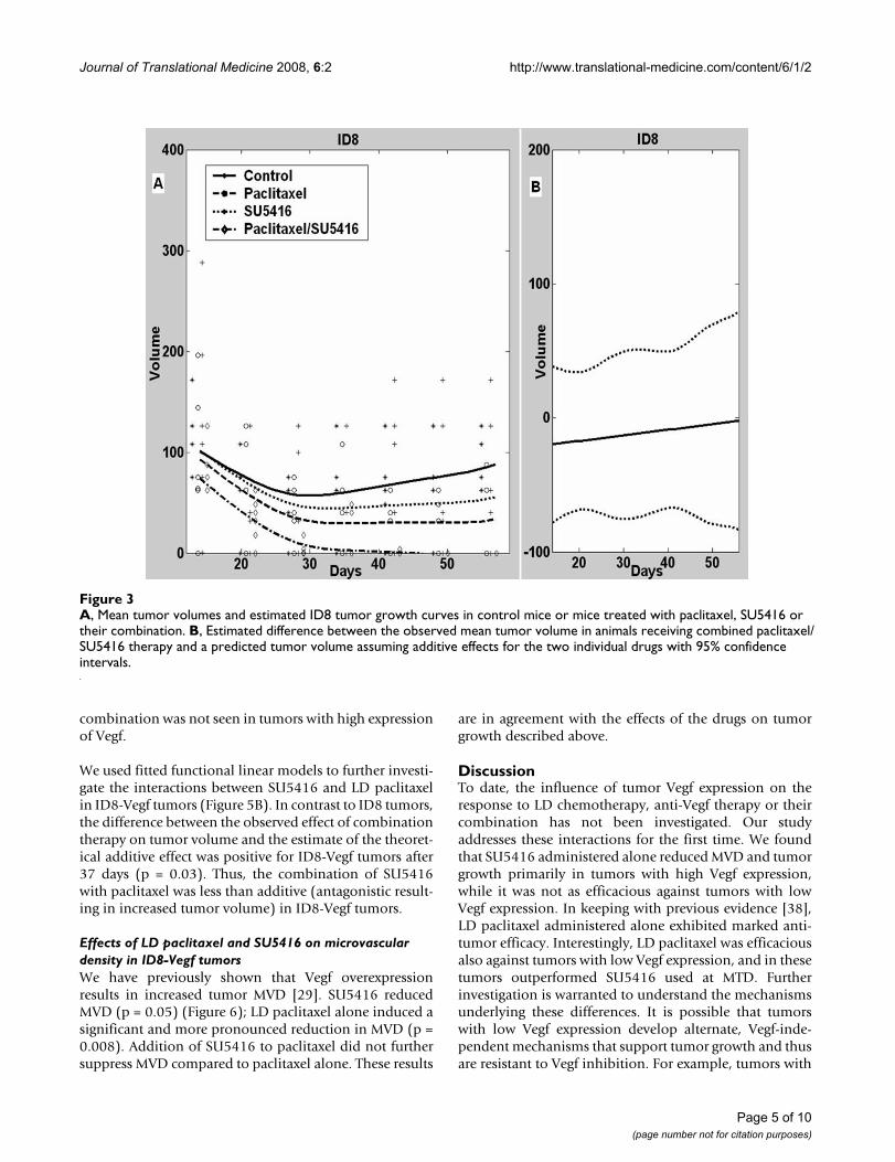
Interactions of SU5416 with LD paclitaxel in ID8 tumorsTo evaluate the interaction between low-dose paclitaxeland SU5416 on tumor growth, we treated animals bearingID8 tumors with LD paclitaxel (plus DMSO), SU5416
(plus saline), or paclitaxel plus SU5416 (Figure 3A). Con-trol mice were treated with saline plus DMSO. As previ-ously noted, LD paclitaxel delayed the growth of ID8tumors (p = 0.003), while SU5416 had a nonsignificantinhibitory effect on ID8 tumors (p = 0.62). The combina-tion of paclitaxel and SU5416 resulted in significant sup-pression of tumor growth where ID8 tumors treated withLD paclitaxel plus SU5416 were 13-fold smaller than con-trol tumors (p < 0.001) and tumors became undetectablein many animals. Thus, LD paclitaxel was more effica-cious than SU5416 against tumors with low Vegf expres-sion. Furthermore, combination of SU5416 and paclitaxelwas quite efficacious against ovarian carcinoma thatexpressed low levels of Vegf resulting in substantial tumorregression.
We used the fitted functional linear models to furthercharacterize the interactions between SU5416 and LDpaclitaxel in ID8 tumors (Figure 3B). The estimate of thedifference between actual effect of combination therapyand the estimated theoretical additive effect of the twodrugs on ID8 tumor volumes was approximately zero forall time points (p = 0.81), indicating that SU5416 whencombined with paclitaxel had an additive effect on ID8tumors.
Effects of LD paclitaxel and SU5416 on microvascular density in ID8 tumorsTo better understand the interaction of paclitaxel andSU5416, we sought to define the effects of each drug ontumor microvasculature in vivo. Microvascular density(MVD) was assessed by CD31 immunostaining (Figure4). SU5416 alone resulted in mild but not significantdecrease in MVD in ID8 tumors (p = 0.4 both). Paclitaxelalone resulted in significant decrease in MVD (p = 0.05).Importantly, paclitaxel plus SU5416 resulted in marked(>90%) reduction in MVD (p = 0.03), indicating a potentdrug interaction at the level of the vasculature. Theseresults are in agreement with the effects of the drugs ontumor growth described above. Thus, LD paclitaxel alonesuppressed significantly MVD and growth of tumors withlow Vegf expression, and its combination with SU5416produced more dramatic results on MVD and tumorgrowth.
Interactions of SU5416 with LD paclitaxel in ID8-Vegf tumorsPaclitaxel alone had a significant inhibitory effect on thegrowth of ID8-Vegf tumors (p = 0.026) (Figure 5A).SU5416 alone was similarly efficacious in inhibiting thegrowth of ID8-Vegf tumors (p < 0.001). The combinationof paclitaxel and SU5416 resulted in significant suppres-sion of ID8 tumor growth (p < 0.001), but was slightly lesseffective than SU5416 alone. Thus, the benefit of drug
Effect of low dose paclitaxel on ID8 or ID8-Vegf tumorsFigure 2Effect of low dose paclitaxel on ID8 or ID8-Vegf tumors. Mice were treated for 6 weeks, starting 2 weeks after tumor inoculation. Tumor volumes were recorded at completion of therapy. * indicates p ≤ 0.05.
Average microvessel count in tumor-free Matrigel plugs in mice treated with phosphate buffered saline and DMSO (control); low dose paclitaxel; or SU5416Figure 1Average microvessel count in tumor-free Matrigel plugs in mice treated with phosphate buffered saline and DMSO (control); low dose paclitaxel; or SU5416. Matrigel plugs were enriched with recombinant mouse Vegf164 (100 ng/ml). * indicates p ≤ 0.05.
Page 4 of 10(page number not for citation purposes)
Journal of Translational Medicine 2008, 6:2 http://www.translational-medicine.com/content/6/1/2
combination was not seen in tumors with high expressionof Vegf.
We used fitted functional linear models to further investi-gate the interactions between SU5416 and LD paclitaxelin ID8-Vegf tumors (Figure 5B). In contrast to ID8 tumors,the difference between the observed effect of combinationtherapy on tumor volume and the estimate of the theoret-ical additive effect was positive for ID8-Vegf tumors after37 days (p = 0.03). Thus, the combination of SU5416with paclitaxel was less than additive (antagonistic result-ing in increased tumor volume) in ID8-Vegf tumors.
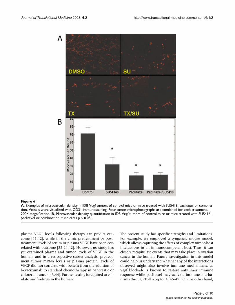
Effects of LD paclitaxel and SU5416 on microvascular density in ID8-Vegf tumorsWe have previously shown that Vegf overexpressionresults in increased tumor MVD [29]. SU5416 reducedMVD (p = 0.05) (Figure 6); LD paclitaxel alone induced asignificant and more pronounced reduction in MVD (p =0.008). Addition of SU5416 to paclitaxel did not furthersuppress MVD compared to paclitaxel alone. These results
are in agreement with the effects of the drugs on tumorgrowth described above.
DiscussionTo date, the influence of tumor Vegf expression on theresponse to LD chemotherapy, anti-Vegf therapy or theircombination has not been investigated. Our studyaddresses these interactions for the first time. We foundthat SU5416 administered alone reduced MVD and tumorgrowth primarily in tumors with high Vegf expression,while it was not as efficacious against tumors with lowVegf expression. In keeping with previous evidence [38],LD paclitaxel administered alone exhibited marked anti-tumor efficacy. Interestingly, LD paclitaxel was efficaciousalso against tumors with low Vegf expression, and in thesetumors outperformed SU5416 used at MTD. Furtherinvestigation is warranted to understand the mechanismsunderlying these differences. It is possible that tumorswith low Vegf expression develop alternate, Vegf-inde-pendent mechanisms that support tumor growth and thusare resistant to Vegf inhibition. For example, tumors with
A, Mean tumor volumes and estimated ID8 tumor growth curves in control mice or mice treated with paclitaxel, SU5416 or their combinationFigure 3A, Mean tumor volumes and estimated ID8 tumor growth curves in control mice or mice treated with paclitaxel, SU5416 or their combination. B, Estimated difference between the observed mean tumor volume in animals receiving combined paclitaxel/SU5416 therapy and a predicted tumor volume assuming additive effects for the two individual drugs with 95% confidence intervals.
Page 5 of 10(page number not for citation purposes)

Journal of Translational Medicine 2008, 6:2 http://www.translational-medicine.com/content/6/1/2
mature vessels respond less to Vegf inhibition [39]. Of rel-evance, we have previously reported that ID8-Vegf tumorsexhibit immature vessels, while ID8 tumors exhibit moremature vessels surrounded by pericytes [40]. LD paclitaxelmay circumvent this limitation as it may exert toxicity ontumor vascular cells independently of maturity.
We used a statistical approach that enabled us to first eval-uate the effect of combinations of therapy on tumor vol-
ume over time with simple experiments and then assessthe difference between the observed effect of combinationtherapy and what would be expected if the two drugs hadan additive effect. To the best of our knowledge, this is thefirst application of semiparametric functional linear mod-els in preclinical tumor studies. This approach betterapproximates growth curves that do not satisfy assump-tions inherent in the standard models. The examples pro-vided in this work illustrate the power inherent in this
A, Examples of microvascular density in ID8 tumors of control mice or mice treated with SU5416, paclitaxel or combinationFigure 4A, Examples of microvascular density in ID8 tumors of control mice or mice treated with SU5416, paclitaxel or combination. Vessels were visualized with CD31 immunostaining. Four tumor microphotographs are combined for each treatment. 200× magnification. B, Microvascular density quantification in ID8 tumors of control mice or mice treated with SU5416, paclitaxel or combination. * indicates p ≤ 0.05.
Page 6 of 10(page number not for citation purposes)

Journal of Translational Medicine 2008, 6:2 http://www.translational-medicine.com/content/6/1/2
approach in estimating the ability of two therapies to actadditively or not. The additive effect of two therapiesoccurs when the difference between the observed effect onthe tumor when both therapies are given and the pre-dicted effect on the tumor, based on the theoretical addi-tive effect of the two therapies given alone, approximateszero. In the case of tumor volume, a positive difference isevidence for a less-than-additive (negative) interaction orantagonism, while a negative difference indicates agreater-than-additive (positive) interaction or synergism.This analytical model has the potential to provingextremely useful in preclinical screening of drug combina-tions and dose optimization of metronomic chemother-apy when characteristics of the tumor microenvironmentare important factors in therapeutic outcome.
Our findings indicate that combination of antiangiogenicdrugs with LD paclitaxel provides therapeutic advantagein tumors expressing low levels of Vegf, while in tumorswith high Vegf expression the combination did not pro-vide any benefit or was rather antagonistic. Lack of addi-
tive effect between two drugs in tumors with high Vegfexpression may signify that both drugs act through thesame pathways, thus their concomitant use cannot pro-duce more effects than either one alone. Why is howeverthis lack of cooperation seen only in tumors with highVegf expression and not in tumors with low Vegf expres-sion? It is possible that only in tumors with high Vegfexpression the action of each drug can be maximized. It isinteresting that antiangiogenic therapy alone performedsimilar to LD paclitaxel in tumors with high Vegf expres-sion. Thus, clinically either therapeutic approach could bechosen based on the desired toxicity profile.
The present results suggest that clinical investigators test-ing combinations of LD chemotherapy and antiang-iogenic therapy should make an attempt to measurepretreatment tumor Vegf expression. However, the bestway to measure VEGF in the clinic is unclear. Tissue levelsof VEGF-A protein would be ideal, but this requires inva-sive procedures. Plasma VEGF might be a reasonable sur-rogate. Results from xenograft studies have indicated that
A, Mean tumor volumes and estimated ID8-Vegf tumor growth curves in control mice or mice treated with paclitaxel, SU5416 or combinationFigure 5A, Mean tumor volumes and estimated ID8-Vegf tumor growth curves in control mice or mice treated with paclitaxel, SU5416 or combination. B, Estimated difference between the observed mean tumor volume in animals receiving combined paclitaxel/SU5416 therapy and a predicted tumor volume assuming additive effects to the two individual drugs with 95% confidence inter-vals.
Page 7 of 10(page number not for citation purposes)
Journal of Translational Medicine 2008, 6:2 http://www.translational-medicine.com/content/6/1/2
plasma VEGF levels following therapy can predict out-come [41,42], while in the clinic pretreatment or post-treatment levels of serum or plasma VEGF have been cor-related with outcome [22-24,42]. However, no study hasyet examined plasma and tumor levels of VEGF in thehuman, and in a retrospective subset analysis, pretreat-ment tumor mRNA levels or plasma protein levels ofVEGF did not correlate with benefit from the addition ofbevacizumab to standard chemotherapy in pancreatic orcolorectal cancer [43,44]. Further testing is required to val-idate our findings in the human.
The present study has specific strengths and limitations.For example, we employed a syngeneic mouse model,which allows capturing the effects of complex tumor-hostinteractions in an immunocompetent host. Thus, it canclosely recapitulate events that may take place in ovariancancer in the human. Future investigation in this modelcould help us understand whether any of the interactionsobserved might also involve immune mechanisms, asVegf blockade is known to restore antitumor immuneresponse while paclitaxel may activate immune mecha-nisms through Toll receptor 4 [45-47]. On the other hand,
A, Examples of microvascular density in ID8-Vegf tumors of control mice or mice treated with SU5416, paclitaxel or combina-tionFigure 6A, Examples of microvascular density in ID8-Vegf tumors of control mice or mice treated with SU5416, paclitaxel or combina-tion. Vessels were visualized with CD31 immunostaining. Four tumor microphotographs are combined for each treatment. 200× magnification. B, Microvascular density quantification in ID8-Vegf tumors of control mice or mice treated with SU5416, paclitaxel or combination. * indicates p ≤ 0.05.
Page 8 of 10(page number not for citation purposes)

Journal of Translational Medicine 2008, 6:2 http://www.translational-medicine.com/content/6/1/2
in this study we used a flank model rather than an ortho-topic tumor model. This was mainly because we sought toevaluate drug interactions on microvascular density,which can be reliably interpreted only in this model, andwe sought to report observations that are possibly appli-cable to many other solid tumor models. Future studieswill address the effect of drugs on the intraperitonealmodel.
ConclusionOur work provides the first evidence that tumor Vegfexpression influences the interactions between LD chem-otherapy and antiangiogenic therapy. Our results suggestthat tumor Vegf expression should be measured in clinicaltrials testing the above approaches. LD paclitaxel is bestcombined with antiangiogenic therapy targeting Vegfagainst tumors with low Vegf expression, where such com-bination could achieve dramatic responses without majortoxicity. Tumors with high Vegf expression, on the otherhand, may likely benefit from antiangiogenic therapy asmuch as from LD chemotherapy, and alternate combina-tions need to be evaluated, including high dose chemo-therapy.
Competing interestsThe author(s) declare that they have no competing inter-ests.
Authors' contributionsDOH carried out the in vitro and in vivo studies anddrafted the manuscript and figures. RTK developed thestatistical algorithms and performed the statistical analy-ses and drafted the statistical sections of the manuscriptand some of the figures. AMH assisted with the in vivoexperiments. LZ generated the mouse model used in thesestudies and optimized norphology methodologies. IAassisted with the in vivo experiments and data analysis. BLparticipated in the statistical analyses. WG oversaw devel-opment of the statistical algorithms. PAG developed sta-tistical approach and oversaw the statistical analyses. GCconceived of the study, participated in its design and coor-dination, and finalized the manuscript.
AcknowledgementsThis study was supported by USAMRC OC-050314 and the Ovarian Can-cer Research Fund. PAG was supported by NCI Ovarian Cancer SPORE P50-CA083638. RTK was supported by NIH T32-CA-093283.
References1. Folkman J: Fundamental concepts of the angiogenic process.
Curr Mol Med 2003, 3(7):643-651.2. Hurwitz H, Fehrenbacher L, Novotny W, Cartwright T, Hainsworth
J, Heim W, Berlin J, Baron A, Griffing S, Holmgren E, Ferrara N, FyfeG, Rogers B, Ross R, Kabbinavar F: Bevacizumab plus irinotecan,fluorouracil, and leucovorin for metastatic colorectal can-cer. The New England journal of medicine 2004, 350(23):2335-2342.
3. Kabbinavar FF, Hambleton J, Mass RD, Hurwitz HI, Bergsland E,Sarkar S: Combined analysis of efficacy: the addition of beva-
cizumab to fluorouracil/leucovorin improves survival forpatients with metastatic colorectal cancer. J Clin Oncol 2005,23(16):3706-3712.
4. Laskin JJ, Sandler AB: First-line treatment for advanced non-small-cell lung cancer. Oncology (Williston Park, NY 2005,19(13):1671-6; discussion 1678-80.
5. Miller KD, Chap LI, Holmes FA, Cobleigh MA, Marcom PK, Fehren-bacher L, Dickler M, Overmoyer BA, Reimann JD, Sing AP, LangmuirV, Rugo HS: Randomized phase III trial of capecitabine com-pared with bevacizumab plus capecitabine in patients withpreviously treated metastatic breast cancer. J Clin Oncol 2005,23(4):792-799.
6. Gasparini G: Metronomic scheduling: the future of chemo-therapy? Lancet Oncol 2001, 2(12):733-740.
7. Browder T, Butterfield CE, Kraling BM, Shi B, Marshall B, O'Reilly MS,Folkman J: Antiangiogenic scheduling of chemotherapyimproves efficacy against experimental drug-resistant can-cer. Cancer research 2000, 60(7):1878-1886.
8. Bocci G, Nicolaou KC, Kerbel RS: Protracted low-dose effects onhuman endothelial cell proliferation and survival in vitroreveal a selective antiangiogenic window for various chemo-therapeutic drugs. Cancer research 2002, 62(23):6938-6943.
9. Kerbel RS, Kamen BA: The anti-angiogenic basis of metro-nomic chemotherapy. Nat Rev Cancer 2004, 4(6):423-436.
10. Bertolini F, Paul S, Mancuso P, Monestiroli S, Gobbi A, Shaked Y, Ker-bel RS: Maximum tolerable dose and low-dose metronomicchemotherapy have opposite effects on the mobilization andviability of circulating endothelial progenitor cells. Cancerresearch 2003, 63(15):4342-4346.
11. Shaked Y, Emmenegger U, Man S, Cervi D, Bertolini F, Ben-David Y,Kerbel RS: Optimal biologic dose of metronomic chemother-apy regimens is associated with maximum antiangiogenicactivity. Blood 2005, 106(9):3058-3061.
12. Lam T, Hetherington JW, Greenman J, Maraveyas A: From totalempiricism to a rational design of metronomic chemother-apy phase I dosing trials. Anti-cancer drugs 2006, 17(2):113-121.
13. Orlando L, Cardillo A, Rocca A, Balduzzi A, Ghisini R, Peruzzotti G,Goldhirsch A, D'Alessandro C, Cinieri S, Preda L, Colleoni M: Pro-longed clinical benefit with metronomic chemotherapy inpatients with metastatic breast cancer. Anti-cancer drugs 2006,17(8):961-967.
14. Correale P, Cerretani D, Remondo C, Martellucci I, Marsili S, La PlacaM, Sciandivasci A, Paolelli L, Pascucci A, Rossi M, Di Bisceglie M,Giorgi G, Gotti G, Francini G: A novel metronomic chemother-apy regimen of weekly platinum and daily oral etoposide inhigh-risk non-small cell lung cancer patients. Oncology reports2006, 16(1):133-140.
15. Young SD, Whissell M, Noble JC, Cano PO, Lopez PG, Germond CJ:Phase II clinical trial results involving treatment with low-dose daily oral cyclophosphamide, weekly vinblastine, androfecoxib in patients with advanced solid tumors. Clin CancerRes 2006, 12(10):3092-3098.
16. Bocci G, Tuccori M, Emmenegger U, Liguori V, Falcone A, Kerbel RS,Del Tacca M: Cyclophosphamide-methotrexate 'metronomic'chemotherapy for the palliative treatment of metastaticbreast cancer. A comparative pharmacoeconomic evalua-tion. Ann Oncol 2005, 16(8):1243-1252.
17. Ramalingam S, Belani CP: Taxanes for advanced non-small celllung cancer. Expert opinion on pharmacotherapy 2002,3(12):1693-1709.
18. Kerbel RS, Klement G, Pritchard KI, Kamen B: Continuous low-dose anti-angiogenic/ metronomic chemotherapy: from theresearch laboratory into the oncology clinic. Ann Oncol 2002,13(1):12-15.
19. Klement G, Huang P, Mayer B, Green SK, Man S, Bohlen P, Hicklin D,Kerbel RS: Differences in therapeutic indexes of combinationmetronomic chemotherapy and an anti-VEGFR-2 antibodyin multidrug-resistant human breast cancer xenografts. ClinCancer Res 2002, 8(1):221-232.
20. Shah MA, Ramanathan RK, Ilson DH, Levnor A, D'Adamo D, O'ReillyE, Tse A, Trocola R, Schwartz L, Capanu M, Schwartz GK, Kelsen DP:Multicenter Phase II Study of Irinotecan, Cisplatin, and Bev-acizumab in Patients With Metastatic Gastric or Gastro-esophageal Junction Adenocarcinoma. J Clin Oncol 2006,24(33):5201-5206.
Page 9 of 10(page number not for citation purposes)

Journal of Translational Medicine 2008, 6:2 http://www.translational-medicine.com/content/6/1/2
Publish with BioMed Central and every scientist can read your work free of charge
"BioMed Central will be the most significant development for disseminating the results of biomedical research in our lifetime."
Sir Paul Nurse, Cancer Research UK
Your research papers will be:
available free of charge to the entire biomedical community
peer reviewed and published immediately upon acceptance
cited in PubMed and archived on PubMed Central
yours — you keep the copyright
Submit your manuscript here:http://www.biomedcentral.com/info/publishing_adv.asp
BioMedcentral
21. Wright JD, Hagemann A, Rader JS, Viviano D, Gibb RK, Norris L,Mutch DG, Powell MA: Bevacizumab combination therapy inrecurrent, platinum-refractory, epithelial ovarian carci-noma: A retrospective analysis. Cancer 2006, 107(1):83-89.
22. Zhang L, Yang N, Conejo-Garcia JR, Katsaros D, Mohamed-Hadley A,Fracchioli S, Schlienger K, Toll A, Levine B, Rubin SC, Coukos G:Expression of endocrine gland-derived vascular endothelialgrowth factor (EG-VEGF) in ovarian carcinoma. Clin CancerRes 2003, 9:264-272.
23. Hefler LA, Zeillinger R, Grimm C, Sood AK, Cheng WF, Gadducci A,Tempfer CB, Reinthaller A: Preoperative serum vascularendothelial growth factor as a prognostic parameter in ovar-ian cancer. Gynecologic oncology 2006, 103(2):512-517.
24. Li L, Wang L, Zhang W, Tang B, Zhang J, Song H, Yao D, Tang Y, ChenX, Yang Z, Wang G, Li X, Zhao J, Ding H, Reed E, Li QQ: Correla-tion of serum VEGF levels with clinical stage, therapy effi-cacy, tumor metastasis and patient survival in ovariancancer. Anticancer research 2004, 24(3b):1973-1979.
25. Zhang L, Conejo-Garcia JR, Yang N, Huang W, Mohamed-Hadley A,Yao W, Benencia F, Coukos G: Different effects of glucose star-vation on expression and stability of VEGF mRNA isoformsin murine ovarian cancer cells. Biophys Biochem Res Commun2002, 292:860-868.
26. Zhang L, Yang N, Wang W, Katsaros D, Mohamed-Hadley A, RubinSC, Coukos G: Oncogene phosphatidyl inositol kinase-3 alphasubunit promotes angiogenesis in ovarian carcinoma viaVEGF. Cancer Res 2003, in Press:.
27. Mendel DB, Laird AD, Smolich BD, Blake RA, Liang C, Hannah AL,Shaheen RM, Ellis LM, Weitman S, Shawver LK, Cherrington JM:Development of SU5416, a selective small molecule inhibi-tor of VEGF receptor tyrosine kinase activity, as an anti-ang-iogenesis agent. Anticancer Drug Des 2000, 15(1):29-41.
28. Stopeck A, Sheldon M, Vahedian M, Cropp G, Gosalia R, Hannah A:Results of a Phase I Dose-escalating Study of the Antiang-iogenic Agent, SU5416, in Patients with Advanced Malignan-cies. Clin Cancer Res 2002, 8(9):2798-2805.
29. Zhang L, Yang N, Conejo-Garcia JR, Mohamed A, Benencia F, RubinSC, Allman D, Coukos G: Generation of a Syngeneic MouseModel to Study the Effects of Vascular Endothelial GrowthFactor in Ovarian Carcinoma. Am J Pathol 2002, 161:2295-2309.
30. Ramsay JO, Silverman BW: Functional Data Analysis. New York, Springer-Verlag; 1997.
31. Eubank R: Nonparametric Regression and Spline Smoothing.New York , Marcel Dekker; 1999.
32. Zhang X, Burt HM, Mangold G, Dexter D, Von Hoff D, Mayer L,Hunter WL: Anti-tumor efficacy and biodistribution of intra-venous polymeric micellar paclitaxel. Anti-cancer drugs 1997,8(7):696-701.
33. Sparreboom A, Wolff AC, Verweij J, Zabelina Y, van Zomeren DM,McIntire GL, Swindell CS, Donehower RC, Baker SD: Disposition ofdocosahexaenoic acid-paclitaxel, a novel taxane, in blood: invitro and clinical pharmacokinetic studies. Clin Cancer Res2003, 9(1):151-159.
34. Tomayko MM, Reynolds CP: Determination of subcutaneoustumor size in athymic (nude) mice. Cancer chemotherapy andpharmacology 1989, 24(3):148-154.
35. Nychka D: Bayesian confidence intervals for smoothingsplines. J Amer Stat Assoc 1988, 83:1134-1143.
36. Eubank RL, Huang C, Maldonado YM, Wang N, Wang S, Buchanan RJ:Smoothing spline estimation in varying-coefficient models. JRoyal Stat Soc 2004, 66:653-667.
37. Guo W: Functional Mixed Effects Models. Biometrics 2002,58:121-128.
38. Marchetti P, Urien S, Cappellini GA, Ronzino G, Ficorella C: Weeklyadministration of paclitaxel: theoretical and clinical basis.Critical reviews in oncology/hematology 2002, 44 Suppl:S3-13.
39. Bergers G, Song S, Meyer-Morse N, Bergsland E, Hanahan D: Bene-fits of targeting both pericytes and endothelial cells in thetumor vasculature with kinase inhibitors.[comment]. Journalof Clinical Investigation 2003, 111(9):1287-1295.
40. Zhang L, Yang N, Park JW, Katsaros D, Fracchioli S, Cao G, O'Brien-Jenkins A, Randall TC, Rubin SC, Coukos G: Tumor-derived vas-cular endothelial growth factor up-regulates angiopoietin-2in host endothelium and destabilizes host vasculature, sup-porting angiogenesis in ovarian cancer. Cancer research 2003,63(12):3403-3412.
41. Bocci G, Man S, Green SK, Francia G, Ebos JM, du Manoir JM, Wein-erman A, Emmenegger U, Ma L, Thorpe P, Davidoff A, Huber J, Hick-lin DJ, Kerbel RS: Increased plasma vascular endothelialgrowth factor (VEGF) as a surrogate marker for optimaltherapeutic dosing of VEGF receptor-2 monoclonal antibod-ies. Cancer research 2004, 64(18):6616-6625.
42. Lee L, Sharma S, Morgan B, Allegrini P, Schnell C, Brueggen J, CozensR, Horsfield M, Guenther C, Steward WP, Drevs J, Lebwohl D, WoodJ, McSheehy PM: Biomarkers for assessment of pharmacologicactivity for a vascular endothelial growth factor (VEGF)receptor inhibitor, PTK787/ZK 222584 (PTK/ZK): transla-tion of biological activity in a mouse melanoma metastasismodel to phase I studies in patients with advanced colorectalcancer with liver metastases. Cancer chemotherapy and pharma-cology 2006, 57(6):761-771.
43. Jubb AM, Hurwitz HI, Bai W, Holmgren EB, Tobin P, Guerrero AS,Kabbinavar F, Holden SN, Novotny WF, Frantz GD, Hillan KJ, Koep-pen H: Impact of vascular endothelial growth factor-Aexpression, thrombospondin-2 expression, and microvesseldensity on the treatment effect of bevacizumab in meta-static colorectal cancer. J Clin Oncol 2006, 24(2):217-227.
44. Kindler HL, Friberg G, Singh DA, Locker G, Nattam S, Kozloff M,Taber DA, Karrison T, Dachman A, Stadler WM, Vokes EE: Phase IItrial of bevacizumab plus gemcitabine in patients withadvanced pancreatic cancer. J Clin Oncol 2005,23(31):8033-8040.
45. Byrd-Leifer CA, Block EF, Takeda K, Akira S, Ding A: The role ofMyD88 and TLR4 in the LPS-mimetic activity of Taxol. Euro-pean journal of immunology 2001, 31(8):2448-2457.
46. Mullins DW, Burger CJ, Elgert KD: Paclitaxel enhances macro-phage IL-12 production in tumor-bearing hosts throughnitric oxide. J Immunol 1999, 162(11):6811-6818.
47. Muenchen HJ, Aggarwal SK: Activation of murine peritonealmacrophages after cisplatin and taxol combination. Anti-can-cer drugs 1997, 8(8):784-789.
Page 10 of 10(page number not for citation purposes)
http://www.ncbi.nlm.nih.gov/entrez/query.fcgi?cmd=Retrieve&db=PubMed&dopt=Abstract&list_uids=9311446
http://www.ncbi.nlm.nih.gov/entrez/query.fcgi?cmd=Retrieve&db=PubMed&dopt=Abstract&list_uids=9311446
http://www.ncbi.nlm.nih.gov/entrez/query.fcgi?cmd=Retrieve&db=PubMed&dopt=Abstract&list_uids=9396623
Related Documents


![Antiangiogenic Scheduling of Chemotherapy Improves ...[CANCER RESEARCH 60, 1878–1886, April 1, 2000] Antiangiogenic Scheduling of Chemotherapy Improves Efficacy against Experimental](https://static.cupdf.com/doc/110x72/5f0d25fa7e708231d438e9b7/antiangiogenic-scheduling-of-chemotherapy-improves-cancer-research-60-1878a1886.jpg)