ORIGINAL RESEARCH ARTICLE published: 02 April 2014 doi: 10.3389/fpsyg.2014.00274 Short-term plasticity of visuo-haptic object recognition Tanja Kassuba 1,2,3 *, Corinna Klinge 2,4 , Cordula Hölig 2,5 , Brigitte Röder 5 and Hartwig R. Siebner 1,2,3 1 Danish Research Centre for Magnetic Resonance, Copenhagen University Hospital Hvidovre, Hvidovre, Denmark 2 NeuroImageNord/Department of Systems Neuroscience, University Medical Center Hamburg-Eppendorf, Hamburg, Germany 3 Department of Neurology, Christian-Albrechts-University, Kiel, Germany 4 Department of Psychiatry, Warneford Hospital, Oxford, UK 5 Biological Psychology and Neuropsychology, University of Hamburg, Hamburg, Germany Edited by: Chris Fields, Retired, USA Reviewed by: Ryo Kitada, National Institute for Physiological Sciences, Japan Jacqueline Clare Snow, University of Nevada, USA *Correspondence: Tanja Kassuba, Princeton Neuroscience Institute, Princeton University, Princeton, NJ 08544, USA e-mail: [email protected] Functional magnetic resonance imaging (fMRI) studies have provided ample evidence for the involvement of the lateral occipital cortex (LO), fusiform gyrus (FG), and intraparietal sulcus (IPS) in visuo-haptic object integration. Here we applied 30 min of sham (non-effective) or real offline 1Hz repetitive transcranial magnetic stimulation (rTMS) to perturb neural processing in left LO immediately before subjects performed a visuo-haptic delayed-match-to-sample task during fMRI. In this task, subjects had to match sample (S1) and target (S2) objects presented sequentially within or across vision and/or haptics in both directions (visual-haptic or haptic-visual) and decide whether or not S1 and S2 were the same objects. Real rTMS transiently decreased activity at the site of stimulation and remote regions such as the right LO and bilateral FG during haptic S1 processing. Without affecting behavior, the same stimulation gave rise to relative increases in activation during S2 processing in the right LO, left FG, bilateral IPS, and other regions previously associated with object recognition. Critically, the modality of S2 determined which regions were recruited after rTMS. Relative to sham rTMS, real rTMS induced increased activations during crossmodal congruent matching in the left FG for haptic S2 and the temporal pole for visual S2. In addition, we found stronger activations for incongruent than congruent matching in the right anterior parahippocampus and middle frontal gyrus for crossmodal matching of haptic S2 and in the left FG and bilateral IPS for unimodal matching of visual S2, only after real but not sham rTMS. The results imply that a focal perturbation of the left LO triggers modality-specific interactions between the stimulated left LO and other key regions of object processing possibly to maintain unimpaired object recognition. This suggests that visual and haptic processing engage partially distinct brain networks during visuo-haptic object matching. Keywords: multisensory interactions, visual perception, haptic perception, object recognition, repetitive transcranial magnetic stimulation, functional magnetic resonance imaging INTRODUCTION An object’s geometrical structure (shape) and surface can be extracted by both using vision and haptics. Integrating shape information across senses can facilitate object recognition (Stein and Stanford, 2008). In vision, the lateral occipital complex (LOC), consisting of subregions in the lateral occipital cortex (LO) and in the fusiform gyrus (FG) (Malach et al., 2002), has long been known to show a preferential response to images of objects as opposed to their scrambled counterparts or other tex- tures (Malach et al., 1995; Grill-Spector et al., 1999; Kourtzi and Kanwisher, 2001). Subsequent neuroimaging studies and stud- ies using transcranial magnetic stimulation (TMS) have linked object- or shape-specific brain responses in the LOC to individ- ual performance during visual object recognition (Grill-Spector et al., 2000; Bar et al., 2001; Ellison and Cowey, 2006; Williams et al., 2007; Pitcher et al., 2009). The functional relevance of the LOC has been further substantiated by patients with lesions in the occipito-temporal cortex suffering from visual agnosia, that is, a severe deficit in visually recognizing objects despite otherwise intact intelligence (Goodale et al., 1991; Karnath et al., 2009). Object-specific responses in the LOC, particularly in the left LO, have also been found when comparing brain responses during the haptic exploration of objects and texture stimuli (Amedi et al., 2001, 2002; Kassuba et al., 2011) or when testing for haptic shape adaptation (Snow et al., 2013). Accordingly, lesions in occipito- temporal cortex can lead to haptic object agnosia (Morin et al., 1984; Feinberg et al., 1986) but see (Snow et al., 2012). Since both vision and haptics provide shape information, it has been pro- posed that the left LO comprises multisensory representations of object shape that are accessed by the different senses (Amedi et al., 2002). Accordingly, the left LO is typically not recruited by audi- tory object stimuli which do not provide any shape information unless subjects have learned to extract shape information from soundscapes produced by visual-to-auditory sensory substitution devices (Amedi et al., 2002, 2007). Previous studies had neglected potential intrinsic differences in the relative contributions of vision and haptics to visuo-haptic shape or object recognition. Since vision provides information www.frontiersin.org April 2014 | Volume 5 | Article 274 | 1

Welcome message from author
This document is posted to help you gain knowledge. Please leave a comment to let me know what you think about it! Share it to your friends and learn new things together.
Transcript

ORIGINAL RESEARCH ARTICLEpublished: 02 April 2014
doi: 10.3389/fpsyg.2014.00274
Short-term plasticity of visuo-haptic object recognitionTanja Kassuba1,2,3*, Corinna Klinge2,4, Cordula Hölig2,5, Brigitte Röder5 and Hartwig R. Siebner1,2,3
1 Danish Research Centre for Magnetic Resonance, Copenhagen University Hospital Hvidovre, Hvidovre, Denmark2 NeuroImageNord/Department of Systems Neuroscience, University Medical Center Hamburg-Eppendorf, Hamburg, Germany3 Department of Neurology, Christian-Albrechts-University, Kiel, Germany4 Department of Psychiatry, Warneford Hospital, Oxford, UK5 Biological Psychology and Neuropsychology, University of Hamburg, Hamburg, Germany
Edited by:
Chris Fields, Retired, USA
Reviewed by:
Ryo Kitada, National Institute forPhysiological Sciences, JapanJacqueline Clare Snow, University ofNevada, USA
*Correspondence:
Tanja Kassuba, PrincetonNeuroscience Institute, PrincetonUniversity, Princeton, NJ 08544,USAe-mail: [email protected]
Functional magnetic resonance imaging (fMRI) studies have provided ample evidencefor the involvement of the lateral occipital cortex (LO), fusiform gyrus (FG), andintraparietal sulcus (IPS) in visuo-haptic object integration. Here we applied 30 min of sham(non-effective) or real offline 1 Hz repetitive transcranial magnetic stimulation (rTMS) toperturb neural processing in left LO immediately before subjects performed a visuo-hapticdelayed-match-to-sample task during fMRI. In this task, subjects had to match sample(S1) and target (S2) objects presented sequentially within or across vision and/or hapticsin both directions (visual-haptic or haptic-visual) and decide whether or not S1 and S2 werethe same objects. Real rTMS transiently decreased activity at the site of stimulation andremote regions such as the right LO and bilateral FG during haptic S1 processing. Withoutaffecting behavior, the same stimulation gave rise to relative increases in activation duringS2 processing in the right LO, left FG, bilateral IPS, and other regions previously associatedwith object recognition. Critically, the modality of S2 determined which regions wererecruited after rTMS. Relative to sham rTMS, real rTMS induced increased activationsduring crossmodal congruent matching in the left FG for haptic S2 and the temporal polefor visual S2. In addition, we found stronger activations for incongruent than congruentmatching in the right anterior parahippocampus and middle frontal gyrus for crossmodalmatching of haptic S2 and in the left FG and bilateral IPS for unimodal matching of visualS2, only after real but not sham rTMS. The results imply that a focal perturbation of theleft LO triggers modality-specific interactions between the stimulated left LO and otherkey regions of object processing possibly to maintain unimpaired object recognition. Thissuggests that visual and haptic processing engage partially distinct brain networks duringvisuo-haptic object matching.
Keywords: multisensory interactions, visual perception, haptic perception, object recognition, repetitive
transcranial magnetic stimulation, functional magnetic resonance imaging
INTRODUCTIONAn object’s geometrical structure (shape) and surface can beextracted by both using vision and haptics. Integrating shapeinformation across senses can facilitate object recognition (Steinand Stanford, 2008). In vision, the lateral occipital complex(LOC), consisting of subregions in the lateral occipital cortex(LO) and in the fusiform gyrus (FG) (Malach et al., 2002), haslong been known to show a preferential response to images ofobjects as opposed to their scrambled counterparts or other tex-tures (Malach et al., 1995; Grill-Spector et al., 1999; Kourtzi andKanwisher, 2001). Subsequent neuroimaging studies and stud-ies using transcranial magnetic stimulation (TMS) have linkedobject- or shape-specific brain responses in the LOC to individ-ual performance during visual object recognition (Grill-Spectoret al., 2000; Bar et al., 2001; Ellison and Cowey, 2006; Williamset al., 2007; Pitcher et al., 2009). The functional relevance of theLOC has been further substantiated by patients with lesions inthe occipito-temporal cortex suffering from visual agnosia, thatis, a severe deficit in visually recognizing objects despite otherwise
intact intelligence (Goodale et al., 1991; Karnath et al., 2009).Object-specific responses in the LOC, particularly in the left LO,have also been found when comparing brain responses during thehaptic exploration of objects and texture stimuli (Amedi et al.,2001, 2002; Kassuba et al., 2011) or when testing for haptic shapeadaptation (Snow et al., 2013). Accordingly, lesions in occipito-temporal cortex can lead to haptic object agnosia (Morin et al.,1984; Feinberg et al., 1986) but see (Snow et al., 2012). Since bothvision and haptics provide shape information, it has been pro-posed that the left LO comprises multisensory representations ofobject shape that are accessed by the different senses (Amedi et al.,2002). Accordingly, the left LO is typically not recruited by audi-tory object stimuli which do not provide any shape informationunless subjects have learned to extract shape information fromsoundscapes produced by visual-to-auditory sensory substitutiondevices (Amedi et al., 2002, 2007).
Previous studies had neglected potential intrinsic differencesin the relative contributions of vision and haptics to visuo-hapticshape or object recognition. Since vision provides information
www.frontiersin.org April 2014 | Volume 5 | Article 274 | 1

Kassuba et al. Short-term plasticity of visuo-haptic object recognition
about several object features in parallel and even if the objectis outside the reaching space, there might be an overall dom-inance of vision in object recognition, at least if objects haveto be recognized predominantly based on their shape. In linewith this notion, we have recently found an asymmetry in theprocessing of crossmodal information during visual and hap-tic object recognition (Kassuba et al., 2013a). Using a visuo-haptic delayed-match-to-sample task during functional magneticresonance imaging (fMRI), the direction of delayed matching(visual-haptic vs. haptic-visual) influenced the activation profilesin bilateral LO, FG, anterior (aIPS) and posterior intraparietalsulcus (pIPS), that is, in regions which have previously been asso-ciated with visuo-haptic object integration (Grefkes et al., 2002;Saito et al., 2003; Stilla and Sathian, 2008; Kassuba et al., 2011;for review see Lacey and Sathian, 2011). Only when a haptic tar-get was matched to a previously presented visual sample but notin the reverse order (i.e., when a visual target was matched toa haptic sample) we found activation profiles in these regionssuggesting multisensory interactions. In line with the maximumlikelihood account of multisensory integration (Ernst and Banks,2002; Helbig and Ernst, 2007), we attributed this asymmetry tothe fact that haptic exploration is less efficient than vision whenrecognizing objects based on their shape (given highly reliableinput from both modalities) and gains more from integratingadditional crossmodal information than vision.
To further explore the role of left LO in visuo-haptic objectintegration, we here examined how repetitive TMS (rTMS) ofthe left LO affects crossmodal object matching. Specifically,we applied real or sham (non-effective) offline 1 Hz rTMS tothe left LO immediately before subjects performed a visuo-haptic delayed-match-to-sample task during fMRI. The pub-lished results reported above (Kassuba et al., 2013a) present theresults after sham rTMS, the current paper focuses on how thesemultisensory interaction effects were modulated by real rTMS.During fMRI, a visual or haptic sample object (S1) and a visual orhaptic target object (S2) were presented sequentially, and subjectshad to indicate whether the identity of both objects was the same(congruent) or not (incongruent). Thus, the event of matching(processing S2 and matching it to previously presented S1) wasmanipulated by three orthogonal factors: (1) S1 and S2 werefrom the same (unimodal) or different modalities (crossmodal),(2) their identity was congruent or incongruent, and (3) S2 waspresented either in the visual or the haptic modality. We assumedthat crossmodal integration occurs only when the visual and hap-tic object inputs are semantically congruent (Laurienti et al.,2004). Multisensory interactions were defined as an increasedactivation during crossmodal vs. unimodal matching (crossmodalmatching effects, cf. Grefkes et al., 2002) that were stronger forcongruent than incongruent object pairs (crossmodal matchingby semantic congruency interaction; Kassuba et al., 2013a,b).The rTMS-induced changes in task-related activity were investi-gated with blood-oxygenated-level-dependent (BOLD) fMRI. Wehypothesized that real compared to sham rTMS of the left LOwould trigger compensatory increases in activity not only at thesite of stimulation but additionally in remote key regions of visuo-haptic object integration such as the right LO, bilateral FG, andIPS. Based on previous work (Kassuba et al., 2013a), we predicted
that real rTMS would particularly affect visuo-haptic interactionsduring matching of haptic as opposed to visual S2.
MATERIALS AND METHODSSUBJECTSThe description of the subjects is reproduced from (Kassuba et al.,2013a: Participants, p. 60) and adjusted to include rTMS-specificinformation. Nineteen healthy right-handed volunteers took partin this study. In one female subject, real rTMS caused uncom-fortable sensations on her skull and the experiment was aborted.Data acquisition was successfully completed in 18 participants (9females, 22–33 years of age, average 25.72 ± 2.87). All subjectsreported normal or corrected-to-normal vision, normal tactileand hearing ability, and none had a history of psychiatric or neu-rological disorders. Handedness was assessed with the short formof the Edinburgh Handedness Inventory (Oldfield, 1971). All sub-jects were right-handed [Laterality Index ≥ 0.78; scaling adaptedfrom Annett (1970)]. Written informed consent was obtainedfrom each subject prior to the experiment. The study proto-col was approved by the local ethics committee (ÄrztekammerHamburg).
EXPERIMENTAL PROCEDURESThe description of the experimental procedures is reproducedfrom Kassuba et al. (2013a: Experimental design and procedure,p. 60) with slight changes in phrasing. All subjects took part infour experimental sessions which were conducted on separatedays (Figure 1). First, subjects attended a behavioral training ses-sion. An epoch-related fMRI localizer session was performed aday later. The last two sessions each consisted of a 40 min runof event-related fMRI, preceded by either 30 min of sham orreal rTMS. The order of the real and sham rTMS sessions wascounterbalanced across subjects and separated by at least oneweek.
In the initial training session, subjects were trained outside theMRI scanner room to recognize 24 object stimuli by viewing pho-tographs and by haptic exploration with an appropriate speed(without ever viewing the real objects themselves). The trainingwas repeated until the object stimuli were identified with an accu-racy of 100% (0-1 repetitions per subject and object). In addition,subjects were familiarized with the visual and haptic texture stim-uli used in the localizer fMRI session to be presented on the nextday. We ran this training to avoid confounding effects due to dif-ferences in familiarity and recognition performance between thetwo modalities.
EPOCH-RELATED fMRI LOCALIZERThe description of the localizer is reproduced from (Kassubaet al., 2013a: Visuo-haptic fMRI localizer, p. 60) with slightchanges in phrasing. The left LO (rTMS target region) andfurther regions of interest (ROIs) were identified by means ofan fMRI localizer. The paradigm determined the convergenceof brain activation during unimodal processing of visual andhaptic object stimuli as compared to non-object control stim-uli of the same modality. In different blocks, we presentedvisual, haptic or auditory object or corresponding texture stim-uli, resulting in six different block conditions: visual-object,
Frontiers in Psychology | Perception Science April 2014 | Volume 5 | Article 274 | 2

Kassuba et al. Short-term plasticity of visuo-haptic object recognition
FIGURE 1 | Experimental procedure. One day after a behavioral trainingsession (not illustrated here), subjects took part in an fMRI localizer in orderto identify the rTMS target area in the left lateral occipital cortex (LO). TherTMS target area was defined by the convergence of visual (V) and haptic (H)
processing of objects vs. textures in the left LO. Then, the subjectsunderwent two 40 min event-related fMRI sessions on separate days (atleast one week apart), both preceded by an off-line session of either real orsham 1 Hz rTMS to the left LO. RMT, motor resting threshold.
haptic-object, auditory-object, visual-texture, haptic-texture, andauditory-texture. Within each block condition, subjects had topress a button whenever an identical stimulus was presented intwo consecutive trials (1-back task, responses in 12.5% of trials).Each stimulation block lasted 30 s during which 8 stimuli fromthe respective condition were presented (2 s stimuli + 2 s inter-stimulus-interval). The subjects were informed 2.8 s before eachblock by a visual instruction (0.8 s) about the upcoming blockand whether they would see (picture of an eye), touch (pictureof a hand) or hear (picture of an ear) stimuli. Each stimulationblock was followed by 11.5 s of rest, and each blocked condi-tion was presented six times. The left LO was destined as thepeak of the group mean BOLD response in the conjunction con-trast (visual-object > visual-texture) ∩ (haptic-object > haptic-texture) at p < 0.001, uncorrected (MNI coordinates in mm: x =−42, y = −63, z = −3). The auditory stimuli were used in thecontext of a different research question (these results have beenpreviously published in Kassuba et al., 2011).
EVENT-RELATED fMRI EXPERIMENTThe description of the event-related fMRI experiment is repro-duced from (Kassuba et al., 2013a: Event-related fMRI experi-ment, pp. 60–62) with slight changes in phrasing. The main fMRIexperiment entailed two experimental sessions that used an iden-tical event-related fMRI paradigm (except for differences due topseudorandomization of the conditions and stimuli). Each exper-iment started with a short practice session, consisting of a shortrecall of the initial training, and then subjects were familiarized
with the subsequent fMRI task. Thereafter, real or sham 1 HzrTMS was applied to the left LO for 30 min followed by the event-related fMRI experiment (for details on rTMS see RepetitiveTMS).
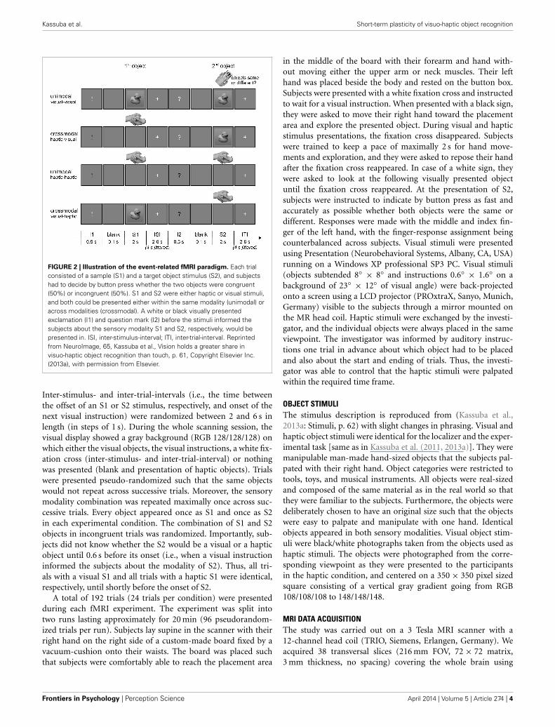
Example trials of the event-related fMRI paradigm are shownin Figure 2. Each trial consisted of a sample object stimulus (S1)and a target object stimulus (S2) presented successively, and thesubjects’ task was to decide whether or not both stimuli referredto the same object (50% congruent and 50% incongruent). Theobject stimuli were presented either haptically (actively palpat-ing an object) or visually (seeing a black-and-white photographof an object; for a detailed description of the objects, see ObjectStimuli), and S1 and S2 were both presented either within thesame modality (unimodal) or across modalities (crossmodal).With respect to the event of matching (i.e., processing of S2 andrelating it to S1), the experiment resulted in a 2 × 2 × 2 design.The first factor was the mode of sensory matching (unimodal orcrossmodal). The second factor related to congruency in objectidentity between S1 and S2 (congruent or incongruent). The sen-sory modality of the S2 (visual or haptic) constituted the thirdexperimental factor.
A visual instruction was presented before each stimulus whichspecified the type of upcoming stimulus (S1 or S2) and whethersubjects would see or touch it. An exclamation mark announcedan S1, a question mark an S2, a white font a visual stimulus,and a black font a haptic stimulus. The instruction was presentedfor 0.5 s. A short blank screen of 0.1 s separated instruction andstimulus presentation. S1 and S2 were both presented for 2 s.
www.frontiersin.org April 2014 | Volume 5 | Article 274 | 3
Kassuba et al. Short-term plasticity of visuo-haptic object recognition
FIGURE 2 | Illustration of the event-related fMRI paradigm. Each trialconsisted of a sample (S1) and a target object stimulus (S2), and subjectshad to decide by button press whether the two objects were congruent(50%) or incongruent (50%). S1 and S2 were either haptic or visual stimuli,and both could be presented either within the same modality (unimodal) oracross modalities (crossmodal). A white or black visually presentedexclamation (I1) and question mark (I2) before the stimuli informed thesubjects about the sensory modality S1 and S2, respectively, would bepresented in. ISI, inter-stimulus-interval; ITI, inter-trial-interval. Reprintedfrom NeuroImage, 65, Kassuba et al., Vision holds a greater share invisuo-haptic object recognition than touch, p. 61, Copyright Elsevier Inc.(2013a), with permission from Elsevier.
Inter-stimulus- and inter-trial-intervals (i.e., the time betweenthe offset of an S1 or S2 stimulus, respectively, and onset of thenext visual instruction) were randomized between 2 and 6 s inlength (in steps of 1 s). During the whole scanning session, thevisual display showed a gray background (RGB 128/128/128) onwhich either the visual objects, the visual instructions, a white fix-ation cross (inter-stimulus- and inter-trial-interval) or nothingwas presented (blank and presentation of haptic objects). Trialswere presented pseudo-randomized such that the same objectswould not repeat across successive trials. Moreover, the sensorymodality combination was repeated maximally once across suc-cessive trials. Every object appeared once as S1 and once as S2in each experimental condition. The combination of S1 and S2objects in incongruent trials was randomized. Importantly, sub-jects did not know whether the S2 would be a visual or a hapticobject until 0.6 s before its onset (i.e., when a visual instructioninformed the subjects about the modality of S2). Thus, all tri-als with a visual S1 and all trials with a haptic S1 were identical,respectively, until shortly before the onset of S2.
A total of 192 trials (24 trials per condition) were presentedduring each fMRI experiment. The experiment was split intotwo runs lasting approximately for 20 min (96 pseudorandom-ized trials per run). Subjects lay supine in the scanner with theirright hand on the right side of a custom-made board fixed by avacuum-cushion onto their waists. The board was placed suchthat subjects were comfortably able to reach the placement area
in the middle of the board with their forearm and hand with-out moving either the upper arm or neck muscles. Their lefthand was placed beside the body and rested on the button box.Subjects were presented with a white fixation cross and instructedto wait for a visual instruction. When presented with a black sign,they were asked to move their right hand toward the placementarea and explore the presented object. During visual and hapticstimulus presentations, the fixation cross disappeared. Subjectswere trained to keep a pace of maximally 2 s for hand move-ments and exploration, and they were asked to repose their handafter the fixation cross reappeared. In case of a white sign, theywere asked to look at the following visually presented objectuntil the fixation cross reappeared. At the presentation of S2,subjects were instructed to indicate by button press as fast andaccurately as possible whether both objects were the same ordifferent. Responses were made with the middle and index fin-ger of the left hand, with the finger-response assignment beingcounterbalanced across subjects. Visual stimuli were presentedusing Presentation (Neurobehavioral Systems, Albany, CA, USA)running on a Windows XP professional SP3 PC. Visual stimuli(objects subtended 8◦ × 8◦ and instructions 0.6◦ × 1.6◦ on abackground of 23◦ × 12◦ of visual angle) were back-projectedonto a screen using a LCD projector (PROxtraX, Sanyo, Munich,Germany) visible to the subjects through a mirror mounted onthe MR head coil. Haptic stimuli were exchanged by the investi-gator, and the individual objects were always placed in the sameviewpoint. The investigator was informed by auditory instruc-tions one trial in advance about which object had to be placedand also about the start and ending of trials. Thus, the investi-gator was able to control that the haptic stimuli were palpatedwithin the required time frame.
OBJECT STIMULIThe stimulus description is reproduced from (Kassuba et al.,2013a: Stimuli, p. 62) with slight changes in phrasing. Visual andhaptic object stimuli were identical for the localizer and the exper-imental task [same as in Kassuba et al. (2011, 2013a)]. They weremanipulable man-made hand-sized objects that the subjects pal-pated with their right hand. Object categories were restricted totools, toys, and musical instruments. All objects were real-sizedand composed of the same material as in the real world so thatthey were familiar to the subjects. Furthermore, the objects weredeliberately chosen to have an original size such that the objectswere easy to palpate and manipulate with one hand. Identicalobjects appeared in both sensory modalities. Visual object stim-uli were black/white photographs taken from the objects used ashaptic stimuli. The objects were photographed from the corre-sponding viewpoint as they were presented to the participantsin the haptic condition, and centered on a 350 × 350 pixel sizedsquare consisting of a vertical gray gradient going from RGB108/108/108 to 148/148/148.
MRI DATA ACQUISITIONThe study was carried out on a 3 Tesla MRI scanner with a12-channel head coil (TRIO, Siemens, Erlangen, Germany). Weacquired 38 transversal slices (216 mm FOV, 72 × 72 matrix,3 mm thickness, no spacing) covering the whole brain using
Frontiers in Psychology | Perception Science April 2014 | Volume 5 | Article 274 | 4
Kassuba et al. Short-term plasticity of visuo-haptic object recognition
a fast gradient echo T2∗-weighted echo planar imaging (EPI)sequence (TR 2480 ms, TE 30 ms, 80◦ flip angle). High-resolutionT1-weighted anatomical images were additionally acquired afterthe localizer fMRI scan using an MPRAGE (magnetization-prepared, rapid acquisition gradient echo) sequence (256 mmFOV, 256 × 192 matrix, 240 transversal slices, 1 mm thickness,50% spacing, TR 2300 ms, TE 2.98 ms).
REPETITIVE TMSFocal rTMS was applied off-line outside the MR scanner roomusing a figure-of-eight coil attached to a Magstim Rapid stim-ulator (Magstim Company, Dyfeld, UK). The coil was centeredover the left LO using Brainsight frameless stereotaxy (RogueResearch, Montreal, Canada). The center of the eight-shaped coiltargeted the MNI coordinates in mm: x = −42, y = −63, z = −3as determined by the localizer (for details see Epoch-related fMRILocalizer). For each subject, the group peak LO coordinates weretransformed into individual anatomical MRI native space coor-dinates, and the site of rTMS stimulation was verified and tracedthroughout the conditioning with the frameless stereotaxy device.
Subjects received continuous 1 Hz rTMS for 30 min (1800stimuli). Stimulation intensity was set to 110% of the indi-vidual resting motor threshold (RMT) of the right first dor-sal interosseous muscle. Mean stimulation intensity during realrTMS was 53.00 ± 7.50% of total stimulator output. The RMTwas defined as the lowest stimulus intensity that evoked a motorevoked potential (MEP) of 50 µV in five out of ten stimuli givenover the motor hot spot. Besides the stimulation intensity, therTMS protocol was identical the protocol used by Siebner et al.(2003) which had resulted in a suppression of neuronal activityin the stimulated left dorsal premotor region that was measur-able for at least 1 h after the end of stimulation. In the currentstudy, stimulation intensity was increased to account for thegreater scalp-cortex distance of the target region compared toprimary motor cortex (Stokes et al., 2005). Repetitive TMS waswell tolerated by all participants apart from one female subjectwho aborted the real rTMS session because of uncomfortablesensations on her skull. Four of the remaining 18 subjects dis-played slight twitches in neck and jaw muscles during real rTMS.Repetitive TMS of the left LO did not produce phosphenes in anysubject.
MEPs were recorded from the first dorsal interosseous musclewith Ag-AgCl electrodes attached to the skin using a tendon-bellymontage. Electromyographic responses were amplified, filtered,and sampled using a D360 eight-channel amplifier (Digitimer,Welwyn Garden City, UK), a CED 1401 analog-to-digital con-verter (Cambridge Electronics Design, Cambride, UK), and a per-sonal computer running Signal software (Cambridge ElectronicDesign). The sampling rate was 5 kHz, and signals were band-passfiltered between 5 and 1000 Hz.
An air-cooled figure-of-eight coil (double 70 mm cooled coilsystem; Magstim Company) was used for real rTMS. The coil wasplaced tangential to the skull with the handle pointing backward,parallel to the horizontal and the mid-sagittal plane (Ellison andCowey, 2006). For sham rTMS, a non-charging standard figure-of-eight coil (double 70 mm coil; Magstim Company) was placedat the skull instead, and the charging coil was placed 90◦ tilted on
top of the non-charging coil. In order to provide a comparableacoustic stimulus, intensity of the charging coil was increased for15% of the total stimulator output. In analogy to the sham rTMScondition, the non-charging coil was placed 90◦ tilted on top ofthe charging coil during real rTMS in order to keep the real andsham rTMS conditions as similar as possible.
Repetitive TMS conditioning was performed offline beforefMRI but after the short object recognition and task training ses-sion. On average, it took 10 ± 2 min from the end of rTMS untilfMRI data acquisition was started. This time was needed to movethe subjects from the TMS lab to the MR scanner, bed them, set upthe board for haptic stimulus presentation, and localize the FOV.Since previous neuroimaging studies have shown that 1Hz rTMSconditioning can produce effects on regional neuronal activitythat last for up to 1 h after the end of stimulation (Lee et al., 2003;Siebner et al., 2003), fMRI lasted 40 min and was, thus, within thetime limits for capturing reorganizational effects.
BEHAVIORAL DATA ANALYSISThe description of the behavioral data analysis is reproducedfrom (Kassuba et al., 2013a: Behavioral analysis, p. 62) andadjusted to include rTMS-specific analysis steps. For each sub-ject and for each trial condition, mean RTs relative to the onsetof S2, and response accuracies were calculated. Only correctresponses were considered for further analyses (trials excludeddue to errors: 0–5 per subject/condition, overall Median = 0;M ± SD sham rTMS session 0.69 ± 0.84 trials, real rTMSsession 0.81 ± 1.07 trials, p = 0.32). Haptic trials in whichparticipants did not palpate the object, dropped the object, ormade premature or late palpations, as well as palpations lastinglonger than 2 s were excluded from analysis (sham rTMS ses-sion 0.04 ± 0.08 trials, real rTMS session 0.01 ± 0.03 trials,p = 0.41). Within each participant and condition, RTs that dif-fered ±3 standard deviations from the preliminary mean weredefined as outliers and excluded from further analyses (shamrTMS session 0.29 ± 0.16 trials, real rTMS session 0.29 ±0.19 trials). Mean RTs of the adjusted data were entered intoa repeated-measures ANOVA (PASW Statistics 18) with RTMS(real/sham), S2-MODALITY (visual/haptic), CONGRUENCY(congruent/incongruent), and SENSORY-MATCHING (uni-modal/crossmodal) as within-subject factors. In order to capturetransient effects of rTMS conditioning on behavior, RTs withineach condition were divided into four time bins of about 10 mineach (∼4-7 trials/bin). Additional ANOVAs with the factorsTIME and RTMS were run for each S1-S2 condition. Each of theseANOVAs tested for a linear trend in the factor Time, and whetherthis trend interacted with rTMS. Statistical effects at p < 0.05were considered significant. Post-hoc Bonferroni corrected pairedt-tests were used to test for differences between single conditions.
FUNCTIONAL MRI DATA ANALYSISThe basic steps of the fMRI analysis is reproduced fromKassuba et al. (2013a: Functional image analysis, pp. 62–63) withslight changes in phrasing and adjustments to include rTMS-specific analysis steps. Image processing and statistical analyseswere performed using SPM8 (statistical parametric mapping 8;www.fil.ion.ucl.ac.uk/spm). The first five volumes of each time
www.frontiersin.org April 2014 | Volume 5 | Article 274 | 5

Kassuba et al. Short-term plasticity of visuo-haptic object recognition
series were discarded to account for T1 equilibrium effects. Dataprocessing consisted of slice timing (correction for differences inslice acquisition time), realignment (rigid body motion correc-tion) and unwarping (accounting for susceptibility by movementinteractions), spatial normalization to MNI standard space asimplemented in SPM8, thereby resampling to a voxel size of3 × 3 × 3 mm3, and smoothing with an 8 mm full-width athalf-maximum isotropic Gaussian kernel.
Statistical analyses were carried out using a general linearmodel approach. The time jitter between the onsets of S1 andS2 allowed us to model the effects of rTMS on sample encoding(response to S1) and target matching (response to S2) indepen-dently. At the individual level (fixed effects), we defined separateregressors for the onsets of S1 and S2 in each session (i.e., aftersham and real rTMS): two different S1 regressors (one for visualS1 and one for haptic S1; Vx and Hx) and eight different S2regressors (one for each matching condition: V, visual; H, haptic;c, congruent; i, incongruent: VVc, HVc, VVi, HVi, HHc, VHc,HHi, VHi) for each rTMS condition. Only onsets of S1 and S2 incorrect trials withstanding the same inclusion criteria as appliedfor RT analyses were included. An additional regressor modeledthe onsets of S1 and S2 in all excluded trials (errors, improperhaptic exploration, and outliers) combined over all conditions.All onset vectors were modeled by convolving delta functions witha canonical hemodynamic response function as implemented inSPM8 and their first derivative. Low frequency drifts in the BOLDsignal were removed by a high-pass filter with a cut-off period of128 s. On the group level, we evaluated effects of rTMS on sampleencoding (onset S1), target matching (onset S2) as well as timedependent effects.
Sample encoding (onset S1)In order to determine the modulation of visual (Vx) and hap-tic (Hx) S1 encoding by rTMS on the group level (randomeffects), a flexible factorial design with the within-subject factorsMODALITY (Vx/Hx) and RTMS (r/s) was configured. The modelalso included the estimation of the subjects’ constants in form ofa SUBJECT factor, and accounted for a possible non-sphericityof the error term (dependences and possible unequal variancesbetween conditions in the within-subject factors).
Target matching (onset S2)Given the complexity of the design (RTMS × S2-MODALITY× CONGRUENCY × SENSORY-MATCHING: 2 × 2 × 2 ×2), we aggregated the S2 matching conditions (S2-MODALITY× CONGRUENCY × SENSORY-MATCHING) into one S2-Condition factor (SPM does not allow a specification ofmore than 3 factors in a factorial model). In order toevaluate the modulation of S2 processing in a randomeffects group analysis, we configured a flexible factorialdesign with the within-subject factors RTMS (r/s) and S2-CONDITION (VVc/HVc/VVi/HVi/HHc/VHc/HHi/VHi). Themodel also included the estimation of the subjects’ constants inform of a SUBJECT factor, and accounted for a possible non-sphericity of the error term (dependences and possible unequalvariances between conditions in the within-subject factors). Notethat in order to evaluate S2 matching effects, we first calculated
contrasts of interest for visual and haptic S2 conditions separately(e.g., crossmodal > unimodal × congruent > incongruent forhaptic S2: [VHc - HHc] > [VHi - HHi], for visual S2: [HVc -VVc] > [HVi - VVi]). This enabled us to eliminate modality-specific confounding factors such as residual effects of the cueon S2 processing, eye movements or potential visual imageryand motor activations during haptic but not visual exploration.In a next step, we compared these modality-specific differentialeffects across modalities (instead of comparing visual and hapticS2 processing directly).
Time-dependent effects of rTMSTime-dependent effects on the processing of S1 and matching ofS2 were also investigated in order to capture transient effects ofrTMS on task-related neuronal processing which gradually recov-ered during the ∼40 min fMRI session. In each session, each ofthe two S1 processing conditions (Vx, Hx) was divided into 10time bins (5 time bins per run) of about 4 min each (∼7-10 tri-als/bin). In the single subject analysis, we defined a regressor foreach time bin in each condition. For each condition, we definedcontrasts that represented a linear or an exponential modulationover time (i.e., across successive time bins). The exponential func-tion we modeled was y = a + (b · 2−x), where y is the BOLDsignal and x is time. The beta images of these contrasts of allsubjects in the real rTMS and the sham rTMS sessions were thenentered into a random effects flexible factorial model [cf. SampleEncoding (Onset S1)] in order to compare time-dependenteffects between real and sham rTMS sessions on the grouplevel.
We applied the same approach to the analysis of S2 responses.Here, each S2 matching condition was divided into four time binsof about 10 min each (∼4-7 trials/bin) and fitted to a linear func-tion. A division into more than four time bins was not reasonablegiven the limited number of trials. Given only four time bins forthe S2 matching conditions, non-linear time-dependent effectswere not modeled here.
Regions of interestThe description of the regions of interest is reproduced fromKassuba et al. (2013a: Functional image analysis, p. 63) withslight changes in phrasing and adjustments. We report voxel-wise family wise error rate (FWE) corrected p-values as obtainedfrom small volume correction in visuo-haptic regions of inter-est (ROIs; p < 0.05). Four brain regions were predefined as ROIs:LO, FG, aIPS, and pIPS. The ROIs in left and right LO and FGwere delineated from the localizer. Images of the localizer datawere preprocessed and analyzed as reported previously (Kassubaet al., 2011). Converging object-specific processing across visionand haptics was calculated with a conjunction of the respectiveobject > texture contrasts within each modality. Only voxels thatshowed an absolute increase during object processing vs. base-line fixation were included. Small volume correction was basedon spheres of 8 mm radius centered at the group-based peakcoordinates obtained from the conjunction contrast thresholdedat p < 0.001, uncorrected: x = −42, y = −63, z = −3 for theleft LO (rTMS target), x = 48, y = −69, z = −9 for the rightLO, x = −36, y = −39, z = −21 for the left FG, and x = 36,y = −45, z = −27 for the right FG.
Frontiers in Psychology | Perception Science April 2014 | Volume 5 | Article 274 | 6
Kassuba et al. Short-term plasticity of visuo-haptic object recognition
Four additional ROIs in the left and right aIPS and pIPS werederived from previous studies applying a crossmodal matchingtask. Correction was based on spheres of 8 mm radius centered atgroup-based peak coordinates reported by the previous studies.Talairach coordinates (Talairach and Tournoux, 1988) from pre-vious studies were transformed into MNI standard space (mm)as implemented in SPM8 using a MATLAB code provided byBrainMap (http://brainmap.org/icbm2tal/index.html; Lancasteret al., 2007). The spherical ROIs were centered over the stereo-tactic coordinates x = −42, y = −40, z = 40 for the left aIPS(Grefkes et al., 2002), x = −28, y = −65, z = 49 for the left pIPS(Saito et al., 2003), and x = 31, y = −62, z = 50 for the rightpIPS (Saito et al., 2003). We also included the right hemispherichomolog of the left aIPS as a region of interest (x = 42, y = −40,z = 40). Whole-brain voxel-wise FWE correction was appliedfor all other voxels in the brain (p < 0.05). Activations derivedfrom the whole-brain analyses were anatomically labeled usingthe probabilistic stereotaxic cytoarchitectonic atlas implementedin the SPM Anatomy Toolbox version 1.8 (Eickhoff et al., 2005),adjusted based on anatomical landmarks in the average structuralT1-weighted image of all subjects. Percent signal changes used forvisualization of the results were extracted using the SPM toolboxrfxplot (Gläscher, 2009).
RESULTSBEHAVIORAL PERFORMANCETask performance after sham rTMS has been reported in a pre-vious paper (Kassuba et al., 2013a). In short, RTs were longerfor incongruent than for congruent trials [F(1,17) = 31.43, p <
0.001], indicating that incongruent matching was in general moredemanding than congruent matching. RTs decreased linearly dur-ing the fMRI session in all conditions [F(1,17) = 14.37, p < 0.01].Response accuracies were nearly perfect irrespectively of condi-tion (on average 96.76 ± 0.97% correct). Neither response accu-racies nor RTs (time-dependent and time-independent effects)were affected by rTMS conditioning (p > 0.10, see Figure 3 andSupplementary Table S1).
FUNCTIONAL MRIThe fMRI results after sham rTMS have been reported in aprevious paper (Kassuba et al., 2013a).
Sample encoding (response to S1)Bilateral LO, FG, aIPS, and pIPS were all activated duringvisual and haptic S1 encoding both after sham and real rTMS[t(51) ≥ 5.30, p < 0.001, corrected]. This mean response to S1was increased in an inferior portion of bilateral FG [left: −33,−46, −23, t(51) = 2.86, p = 0.052, corrected; right: 33, −43, −23,t(51) = 3.46, p < 0.05, corrected; see Figures 4A,B] after real asopposed to sham rTMS but otherwise did not differ between thetwo sessions (p > 0.01, uncorrected).
Real TMS affected the activity at the site of stimulation (leftLO) mainly during S1 encoding and in a time-dependent fashion.After real rTMS, the BOLD response at the left LO to haptic S1 wasinitially attenuated and exponentially recovered until ∼30 minpost rTMS [−42, −67, −11; t(51) = 3.49, p < 0.05, corrected;see Figure 4C]. The regional BOLD response to haptic S1 stim-uli displayed opposite temporal dynamics after sham rTMS with
a higher initial level of S1-induced activity which quickly atten-uated during continuous task performance. Relative to shamrTMS, real rTMS additionally caused a transient attenuation ofhaptic S1 processing in the right LO [45, −73, −5; t(51) = 3.37],a superior portion of bilateral FG [left: −36, −46, −20, t(51) =3.74; right: 36, −43, −20, t(51) = 3.16], and bilateral posteriorsuperior temporal sulcus and adjacent middle temporal gyrus[pSTS/MTG; left: −66, −40, 1; t(51) = 6.10; right: 54, −40, −8,t(51) = 5.44; all p < 0.05, corrected; see Figures 4A,C]. Similarbut weaker (p < 0.05, uncorrected) transient decreases in activa-tion were found for visual S1 encoding as well. The effects forhaptic S1 were not significant different from the effects for visualS1 (p > 0.05, corrected).
Target matching (response to S2)Effects of real rTMS on crossmodal congruent matching. We
expected rTMS to evoke the strongest reorganizational effects forcrossmodal matching of semantically congruent stimulus pairs(i.e., in the crossmodal matching by semantic congruency inter-action contrast as indication for multisensory interactions). Aftersham rTMS, we had found such multisensory interaction effectsin bilateral LO, FG, aIPS, and pIPS which were more pronouncedfor haptic than visual S2 (Kassuba et al., 2013a). Based on thesefindings, we proposed that multisensory interactions are morelikely for haptic than visual object recognition, and we, there-fore, expected stronger effects of real rTMS for the matching ofhaptic as opposed to visual S2. After real rTMS, we found com-parable multisensory interaction effects in our ROIs that werestronger pronounced for haptic as opposed to visual S2 condi-tions (see Figure 5 and Supplementary Tables S2–S4). We did notobserve any significant effects of rTMS on multisensory interac-tions (rTMS x crossmodal > unimodal × congruent > incongru-ent) nor on crossmodal matching effects (rTMS × crossmodal >
unimodal), neither for visual nor haptic S2.However, real rTMS altered the temporal dynamics of event-
related activity during crossmodal matching compared to shamrTMS. Several regions in left temporal cortex showed initialincreases in activations after real rTMS during crossmodal match-ing of congruent onjects (see Table 1). These effects of real rTMSwere transient and decreased gradually during the fMRI session,resulting in a negative linear modulation of the BOLD response.For congruent crossmodal matching of haptic S2 (VHc), the leftFG showed an initial relative enhancement of the BOLD responseto S2 after real rTMS with a subsequent linear decay over time. Incontrast, for congruent crossmodal matching of visual S2 (HVc),the left temporal pole and pSTS/MTG displayed an initial increasein S2-related activation after real rTMS (see Table 1). Direct com-parisons between the two modalities (r-VHc > s-VHc × time vs.r-HVc > s-HVc × time) showed that these effects were modal-ity specific. No consistent effects of real rTMS were found duringunimodal matching in these regions. Yet, the effects found forcrossmodal matching did not differ significantly from the effectsfor unimodal matching.
Effects of real rTMS on incongruent matching. Longer responselatencies suggested that matching of incongruent objects wasbehaviorally more challenging than matching of congruent
www.frontiersin.org April 2014 | Volume 5 | Article 274 | 7

Kassuba et al. Short-term plasticity of visuo-haptic object recognition
FIGURE 3 | Mean reaction times for visual and haptic S2 for all four
delayed matching conditions (unimodal/crossmodal ×congruent/incongruent) and after real (red bars) and sham rTMS (green
bars) conditions. Error bars indicate the standard error of the mean.
Reaction times were recorded from S2 onset onwards. Repetitive TMS didnot have any effects on reaction times. Sample-target (S1–S2) conditions: V,visual; H, haptic. Cong, congruent; Incong, incongruent. ∗p < 0.05,∗∗p < 0.01, ∗∗∗p < 0.001, Bonferroni corrected.
objects (see Figure 3). Since behavioral performance was notimpaired by rTMS, we next asked whether we could find reor-ganizational effects on the neuronal level related to incongru-ent matching, that is, triggered by task difficulty. We foundrTMS-induced increases in activations related to matching ofincongruent objects for both haptic and visual S2. These effectswere found transiently for crossmodal matching of haptic S2and lastingly (i.e., temporally stable for the whole durationif the experiment) for unimodal matching of visual S2 (seeTable 2). When a haptic S2 was matched to an incongruent visualS1 (r-VHi > s-VHi), real rTMS-induced transient increases inactivation were found in bilateral parahippocampus, right LO,bilateral pSTS/MTG, IPS, and in the right middle and adja-cent superior frontal gyrus. On the other hand, when a visualS2 was matched to an incongruent visual S1, temporarily sta-ble increases in activation were found in the left FG and pIPS.No other incongruent matching condition was affected by realrTMS.
Incongruency effects (incongruent > congruent) after realrTMS. The time-dependent effects in the right anterior parahip-pocampus and middle frontal gyrus and adjacent precentral gyrusfound for crossmodal matching of haptic S2 were significantlymore pronounced for incongruent than congruent conditions(real > sham × VHi > VHc × time, see Table 3 and Figure 6).Thus in these regions, real rTMS conditioning induced incon-gruency effects, that is, stronger activations during incongruentthan congruent matching, that were not evident after shamrTMS. Such rTMS by incongruency interactions (real > sham× incongruent > congruent) were found for unimodal visual(VV) matching as well. For unimodal visual matching, tem-porarily stable rTMS-induced incongruency effects were found
the left superior medial gyrus extending to the right hemi-sphere, left FG, and bilateral pIPS (see Table 3 and Figure 7). Adirect comparison of visual and haptic S2 conditions showed thatthese time-dependent ([r-VHi > s-VHi × time] > [r-HVi > s-HVi × time]) and time-independent effects ([r-VVi > s-VVi] >
[r-HHi > s-HHi]) were modality-specific. Unimodal matching ofhaptic S2 and crossmodal matching of visual S2 did not show realrTMS-induced incongruency effects.
Exclusion of subjects with low LO activations in the localizerOne concern with respect to the null findings regarding multisen-sory interactions could be that we used the peak coordinates fromthe localizer group analysis as rTMS target instead of individualpeaks. Yet theoretically, the group peak coordinates represent thepeak responses across subjects, and indeed, the Eucledian distancebetween individual peaks and the group peak were smaller than1 cm in all subjects. However, 5 out of the 18 subjects showed veryweak activations in the localizer contrast and peaks in the left LOcould only be localized at very low thresholds (p > 0.05, uncor-rected). In these subjects, the group peak coordinates provided amore objective guide for placing the TMS coil. To test whetherthese subjects had biased our results, we repeated our analyseswithout these 5 subjects. There were still no significant effects ofrTMS on multisensory interactions.
DISCUSSIONWe probed short-term plasticity of visuo-haptic object recog-nition by conditioning neuronal processing in left LO withlow-frequency offline rTMS. Compared to sham rTMS, realrTMS led to a dynamic redistribution of brain activity duringvisuo-haptic object matching. Changes in task-related activitywere not only triggered in the stimulated left and contralateral
Frontiers in Psychology | Perception Science April 2014 | Volume 5 | Article 274 | 8

Kassuba et al. Short-term plasticity of visuo-haptic object recognition
FIGURE 4 | Effects of real rTMS on S1 processing. (A) Activation mapshowing temporally stable increases in activation (blue) and transientdecreases in activation (yellow) in bilateral FG after real compared to shamrTMS (p < 0.01, uncorrected). (B) Temporarily stable increases in activationin bilateral FG (blue portion in (A), MNI coordinates x, y, z; left: −33, −46,−23; right: 33, −43, −23) to both visual S1 (Vx) and haptic S1 (Hx) after real(red) relative to sham rTMS (green). (C) Transient rTMS-induced decreasesin activation during haptic S1 encoding. Regional activity in bilateral LO,FG, and pSTS/MTG showed an interaction of exponential time-dependenteffects by rTMS condition when haptic S1 were processed: Whereas
regional activity was initially decreased and exponentially increased overtime after real rTMS (red), the reversed pattern was found after sham rTMS(green). Similar but weaker effects were found for visual S1 processing(p < 0.05, uncorrected). Each time bin represents ∼4 min and 7–10 trials.FG, fusiform gyrus [yellow portion in (A), left: −36, −46, −20; right: 36,−43, −20]; LO, lateral occipital cortex (left, i.e., rTMS target area: −42,−67, −11; right: 45, −73, −5); pSTS/MTG, posterior superior temporalsulcus /middle temporal gyrus (left: −66, −40, 1; right: 54, −40, −8).L, left; R, right. ∗p < 0.05, small volume corrected, (∗)p = 0.052, smallvolume corrected, #p < 0.05, whole brain corrected.
LO but also in remote temporal and parietal regions previ-ously associated with object recognition. While LO, FG, aIPS,pIPS have been implicated in visuo-haptic object recognition(Amedi et al., 2001; Grefkes et al., 2002; Saito et al., 2003;Kassuba et al., 2011), the pSTS/MTG seems to participate inaudio-visual and audio-haptic object recognition (Beauchampet al., 2004, 2008; Kassuba et al., 2011, 2013b), and the tem-poral pole appears to support semantic memory (Martin andChao, 2001; Rogers et al., 2006). Since behavioral performancewas not impaired, the real rTMS-induced changes in task-relatedbrain activity likely indicate compensatory processes preserving
behavior after neuronal challenge. Importantly, the pattern of realrTMS-induced changes in regional activity differed as a functionof the stage of the delayed-match-to-sample task (S1 encoding vs.S2 matching) and the target modality.
Since various previous studies have implicated the left LOin visuo-haptic integration of object information (Lacey andSathian, 2011), we predicted that rTMS of the left LO would par-ticularly affect multisensory interactions as defined by crossmodalmatching by semantic congruency interactions and particularlyfor haptic S2 conditions (Kassuba et al., 2013a,b). Contrary toour expectations, rTMS had no impact on crossmodal matching
www.frontiersin.org April 2014 | Volume 5 | Article 274 | 9
Kassuba et al. Short-term plasticity of visuo-haptic object recognition
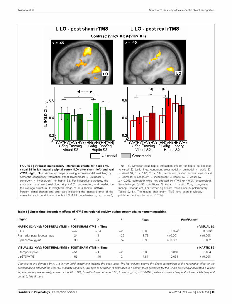
FIGURE 5 | Stronger multisensory interaction effects for haptic vs.
visual S2 in left lateral occipital cortex (LO) after sham (left) and real
rTMS (right). Top: Activation maps showing a crossmodal matching bysemantic congruency interaction effect (crossmodal > unimodal ×congruent > incongruent) for haptic S2. For illustrative purposes, thestatistical maps are thresholded at p < 0.01, uncorrected, and overlaid onthe average structural T1-weighted image of all subjects. Bottom:
Percent signal change and error bars indicating the standard error of themean for each condition at the left LO (MNI coordinates: x, y, z = −45,
−70, −5). Stronger visuo-haptic interaction effects for haptic as opposedto visual S2 (solid lines: congruent crossmodal > unimodal × haptic S2> visual S2, ∗p < 0.05, ∗∗p < 0.01, corrected; dashed arrows: crossmodal> unimodal x congruent > incongruent × haptic S2 > visual S2,p ≤ 0.063, corrected) were not affected by rTMS (p > 0.01, uncorrected).Sample-target (S1-S2) conditions: V, visual; H, haptic; Cong, congruent;Incong, incongruent. For further significant results see SupplementaryTables S2–S4. The results after sham rTMS have been previouslypublished in Kassuba et al. (2013a).
Table 1 | Linear time-dependent effects of rTMS on regional activity during crossmodal congruent matching.
Region x y z tpeak pcorr(puncorr)
HAPTIC S2 (VHc): POST-REAL rTMS > POST-SHAM rTMS × Time >VISUAL S2
L FG −42 −34 −20 3.03 0.024§ 0.066§
R anterior parahippocampus 24 −1 −29 3.76 (<0.001) (<0.001)
R precentral gyrus 39 −7 52 3.95 (<0.001) 0.032
VISUAL S2 (HVc): POST-REAL rTMS > POST-SHAM rTMS × Time >HAPTIC S2
L temporal pole −51 −4 −29 5.85 0.001 0.004
L pSTS/MTG −66 −40 −2 4.87 0.034 (<0.001)
Coordinates are denoted by x, y, z in mm (MNI space) and indicate the peak voxel. The last column shows the direct comparison of the respective effect to the
corresponding effect of the other S2 modality condition. Strength of activation is expressed in t- and p-values corrected for the whole brain and uncorrected p-values
in parentheses, respectively, at peak voxel (df = 119), §small volume corrected. FG, fusiform gyrus; pSTS/MTG, posterior superior temporal sulcus/middle temporal
gyrus. L, left; R, right.
Frontiers in Psychology | Perception Science April 2014 | Volume 5 | Article 274 | 10
Kassuba et al. Short-term plasticity of visuo-haptic object recognition
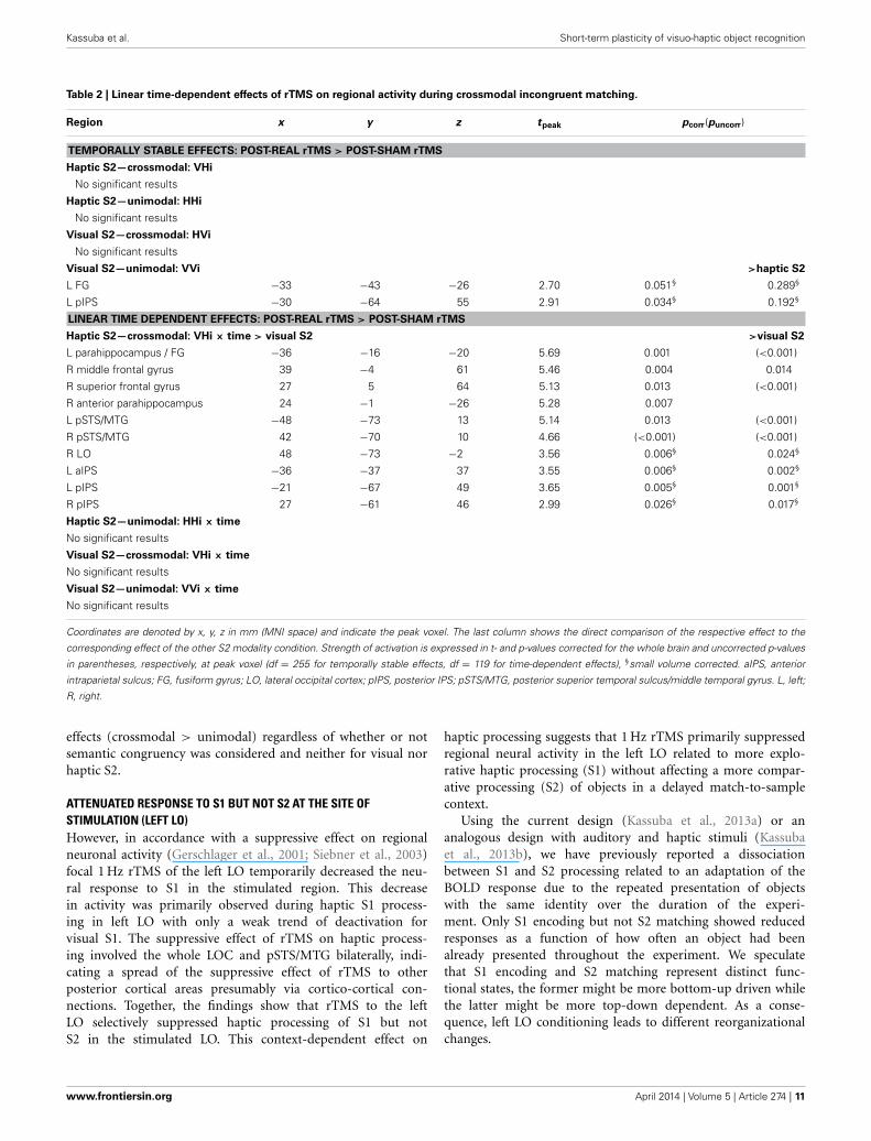
Table 2 | Linear time-dependent effects of rTMS on regional activity during crossmodal incongruent matching.
Region x y z tpeak pcorr(puncorr)
TEMPORALLY STABLE EFFECTS: POST-REAL rTMS > POST-SHAM rTMS
Haptic S2—crossmodal: VHi
No significant results
Haptic S2—unimodal: HHi
No significant results
Visual S2—crossmodal: HVi
No significant results
Visual S2—unimodal: VVi >haptic S2
L FG −33 −43 −26 2.70 0.051§ 0.289§
L pIPS −30 −64 55 2.91 0.034§ 0.192§
LINEAR TIME DEPENDENT EFFECTS: POST-REAL rTMS > POST-SHAM rTMS
Haptic S2—crossmodal: VHi × time > visual S2 >visual S2
L parahippocampus / FG −36 −16 −20 5.69 0.001 (<0.001)
R middle frontal gyrus 39 −4 61 5.46 0.004 0.014
R superior frontal gyrus 27 5 64 5.13 0.013 (<0.001)
R anterior parahippocampus 24 −1 −26 5.28 0.007
L pSTS/MTG −48 −73 13 5.14 0.013 (<0.001)
R pSTS/MTG 42 −70 10 4.66 (<0.001) (<0.001)
R LO 48 −73 −2 3.56 0.006§ 0.024§
L aIPS −36 −37 37 3.55 0.006§ 0.002§
L pIPS −21 −67 49 3.65 0.005§ 0.001§
R pIPS 27 −61 46 2.99 0.026§ 0.017§
Haptic S2—unimodal: HHi × time
No significant results
Visual S2—crossmodal: VHi × time
No significant results
Visual S2—unimodal: VVi × time
No significant results
Coordinates are denoted by x, y, z in mm (MNI space) and indicate the peak voxel. The last column shows the direct comparison of the respective effect to the
corresponding effect of the other S2 modality condition. Strength of activation is expressed in t- and p-values corrected for the whole brain and uncorrected p-values
in parentheses, respectively, at peak voxel (df = 255 for temporally stable effects, df = 119 for time-dependent effects), §small volume corrected. aIPS, anterior
intraparietal sulcus; FG, fusiform gyrus; LO, lateral occipital cortex; pIPS, posterior IPS; pSTS/MTG, posterior superior temporal sulcus/middle temporal gyrus. L, left;
R, right.
effects (crossmodal > unimodal) regardless of whether or notsemantic congruency was considered and neither for visual norhaptic S2.
ATTENUATED RESPONSE TO S1 BUT NOT S2 AT THE SITE OFSTIMULATION (LEFT LO)However, in accordance with a suppressive effect on regionalneuronal activity (Gerschlager et al., 2001; Siebner et al., 2003)focal 1 Hz rTMS of the left LO temporarily decreased the neu-ral response to S1 in the stimulated region. This decreasein activity was primarily observed during haptic S1 process-ing in left LO with only a weak trend of deactivation forvisual S1. The suppressive effect of rTMS on haptic process-ing involved the whole LOC and pSTS/MTG bilaterally, indi-cating a spread of the suppressive effect of rTMS to otherposterior cortical areas presumably via cortico-cortical con-nections. Together, the findings show that rTMS to the leftLO selectively suppressed haptic processing of S1 but notS2 in the stimulated LO. This context-dependent effect on
haptic processing suggests that 1 Hz rTMS primarily suppressedregional neural activity in the left LO related to more explo-rative haptic processing (S1) without affecting a more compar-ative processing (S2) of objects in a delayed match-to-samplecontext.
Using the current design (Kassuba et al., 2013a) or ananalogous design with auditory and haptic stimuli (Kassubaet al., 2013b), we have previously reported a dissociationbetween S1 and S2 processing related to an adaptation of theBOLD response due to the repeated presentation of objectswith the same identity over the duration of the experi-ment. Only S1 encoding but not S2 matching showed reducedresponses as a function of how often an object had beenalready presented throughout the experiment. We speculatethat S1 encoding and S2 matching represent distinct func-tional states, the former might be more bottom-up driven whilethe latter might be more top-down dependent. As a conse-quence, left LO conditioning leads to different reorganizationalchanges.
www.frontiersin.org April 2014 | Volume 5 | Article 274 | 11

Kassuba et al. Short-term plasticity of visuo-haptic object recognition
Table 3 | Real rTMS induced incongruency effects (incongruent > congruent × real rTMS > sham rTMS).
Region x y z tpeak pcorr(puncorr)
TEMPORALLY STABLE EFFECTS: POST-REAL rTMS > POST-SHAM rTMS
Haptic S2—crossmodal: VHi > VHc
No significant results
Haptic S2—unimodal: HHi > HHc
No significant results
Visual S2—crossmodal: HVi > VVc
No significant results
Visual S2—unimodal: VVi > VVc >haptic S2
L superior medial gyrus 3 32 52 4.92 0.014 (<0.001)
L FG −36 −37 −26 4.55 <0.001§ 0.039§
L pIPS −30 −61 55 3.81 0.002§ 0.092§
R pIPS 27 −58 52 3.73 0.003§ 0.050§
LINEAR TIME DEPENDENT EFFECTS: POST-REAL rTMS > POST-SHAM rTMS
Haptic S2—crossmodal: VHi > VHc × time > visual S2 >visual S2
R anterior parahippocampus 24 −1 −29 5.60 0.002 (<0.001)
R middle frontal gyrus 39 −1 58 5.61 0.002 0.004
Haptic S2—unimodal: HHi > HHc × time
No significant results
Visual S2—crossmodal: VHi > VHc × time
No significant results
Visual S2—unimodal: VVi > VVc × time
No significant results
Coordinates are denoted by x, y, z in mm (MNI space) and indicate the peak voxel. The last column shows the direct comparison of the respective effect to the
corresponding effect of the other S2 modality condition. Strength of activation is expressed in t- and p-values corrected for the whole brain and uncorrected p-values
in parentheses, respectively, at peak voxel (df = 255 for temporally stable effects, df = 119 for time-dependent effects), § small volume corrected. FG, fusiform
gyrus, pIPS, posterior intraparietal sulcus. L, left; R, right.
INCREASED RESPONSES TO S2 IN REMOTE REGIONSWhile processing of S2 was unchanged at the site of stimulation,transient increases in activation emerged in remote regions afterreal relative to sham rTMS in congruent crossmodal matchingtrials, that is, when object concepts were most likely integratedacross the senses (Laurienti et al., 2004). These putatively com-pensatory increases in activation were found in temporal regionssuch as the left temporal pole and pSTS/MTG for crossmodalmatching of visual S2 (HVc) and the left FG and right anteriorparahippocampus for crossmodal matching of haptic S2 (VHc)and were specific for the respective S2 modality. It has beenpreviously proposed that the temporal cortex integrates objectinformation (e.g., object motion, shape, use-associated motormovements) with increasing convergence and abstraction alongthe posterior to anterior axis (Martin and Chao, 2001; Martin,2007). For instance, studies that used dynamic visual and audi-tory object stimuli suggested that the pSTS/MTG is tuned tofeatures of motion associated with different objects (Beauchampet al., 2002, 2004). We have previously shown that the sameleft FG region as found here shows object-specific responsesindependent of whether objects were seen, heard, or touched,suggesting more abstract or conceptual representations of objectinformation (Kassuba et al., 2011, 2013b; see also Martin, 2007).Patient studies suggest that the anterior temporal pole is criti-cal for semantic memory (Rogers et al., 2006) and particularlyfor retrieving object information about unique entities (Damasio,
1989; Damasio et al., 1996). We, therefore, propose that in thepresence of a functional perturbation of the left LO, regions ofa semantic object recognition network are increasingly activatedwhen the same objects are matched across vision and haptics.These enhanced activations might reflect a compensatory strategyinvolving semantic memory. Critically, retrieving haptic objectinformation and matching it to the same object processed visuallyactivated different nodes of this putative network than retriev-ing visual object information and matching it to the same objectprocessed haptically.
One likely explanation for the null findings with respect to realrTMS effects on multisensory interactions is that the delayed-match-to-sample task was not challenging enough. Even afterreal rTMS, task accuracy was nearly perfect (≥95%). We foundreal rTMS-induced increases in activations in LOC and IPSrelated to matching of incongruent objects, which was behav-iorally more difficult than matching of congruent objects. Someof these increased brain activations were specifically stronger dur-ing incongruent than congruent matching (incongruency effect),only after real but not sham rTMS. Again, these rTMS-inducedincongruency effects differed based on the S2 modality: Theeffects were limited to the first 30 min post rTMS in the rightanterior parahippocampus for crossmodal haptic matching ofhaptic S2 (VH) but remained stable throughout the session inleft FG and bilateral pIPS for unimodal matching of visual S2(VV). Even though left LO rTMS had no effects on multisensory
Frontiers in Psychology | Perception Science April 2014 | Volume 5 | Article 274 | 12

Kassuba et al. Short-term plasticity of visuo-haptic object recognition
FIGURE 6 | Transient incongruency effects for crossmodal matching of
haptic S2 (VH) evoked by rTMS conditioning. Regional activity in theright parahippocampus (MNI coordinates: x, y, z = 24, −1, 29) and rightmiddle frontal gyrus (39, −1, 58) showed an interaction of lineartime-dependent effects by rTMS condition that was stronger forincongruent than congruent trials: Whereas regional activity was initiallyincreased and linearly decreased over time after real rTMS (red,real-congruent; dark red, real-incongruent), no significant linear
time-dependent increases in activations (or rather decreases) were foundafter sham rTMS (green, sham-congruent; dark green, sham-incongruent),and these differential effects were stronger for incongruent thancongruent conditions (real > sham × incongruent > congruent × time).For illustrative purposes, the statistical maps are thresholded atp < 0.001, uncorrected, and overlaid on the average structuralT1-weighted image of all subjects. Each time bin represents ∼10 min and4–7 trials. L, left; R, right. ∗p < 0.05, corrected.
FIGURE 7 | Incongruency effects for unimodal visual matching (VV)
evoked by rTMS conditioning. Only after real but not sham rTMS,regional activity in the L superior medial gyrus (MNI coordinates: x, y,z = 3, 32, 52) (left), L FG (−36, −37, −26) (middle), and R pIPS (27, −58,52) (right) was increased when a visual S2 was matched to anincongruent (i) as compared to a congruent (c) visual S1 (real > sham ×incongruent > congruent). The same effects were found in the left pIPS.
For illustrative purposes, the statistical maps are thresholded at p < 0.001,uncorrected, and overlaid on the average structural T1-weighted image ofall subjects. Bars represent percent signal change and error bars thestandard error of the mean for each VV condition after real and shamrTMS (red, real-congruent; dark red, real-incongruent; green,sham-congruent; dark green, sham-incongruent). FG, fusiform gyrus; pIPS,posterior intraparietal sulcus; L, left; R, right. ∗p < 0.05, corrected.
interactions, these results suggest a functional relevance of left LOfor evaluating visual and haptic object information.
Since S1 and S2 were presented sequentially, incongruencyeffects (incongruent > congruent) could also be interpreted asrepetition suppression or fMRI-adaptation (fMRI-A) effects (i.e.,decreased activity in the congruent condition due to the repeatedpresentation of objects with the same identity). Thus, incongru-ency effects found for crossmodal matching could be interpretedas crossmodal adaptation and might indicate multisensory inte-gration (cf. Tal and Amedi, 2009; Doehrmann et al., 2010; VanAtteveldt et al., 2010). However, we argue that the task demandsin our paradigm have overruled general effects of stimulus habit-uation (for a detailed discussion of this issue, see Kassuba et al.,2013a,b). First, the stimulus onset asynchronies between S1 andS2 in the present study were rather long and favored a semantic
encoding of S1. Second, while other studies showing adaptationeffects typically used a task orthogonal to the effect of interestsuch as a detection task (Doehrmann et al., 2010; Van Atteveldtet al., 2010; Snow et al., 2013) or passive recognition (Tal andAmedi, 2009), our task required an explicit semantic decision onthe identity of S1 and S2. In addition, using this delayed-match-to-sample paradigm, we did not find any general adaptationof the BOLD response to S2 due to repeated presentations ofthe same objects throughout the experiment (independent ofmatching condition), neither when using visual and haptic stim-uli (Kassuba et al., 2013a), nor when using auditory and hapticstimuli (Kassuba et al., 2013b). Consistent with our findings,other studies employing longer delays in visuo-haptic priming(James et al., 2002) or using a delayed-match-to-sample task(Grefkes et al., 2002) have found enhanced instead of decreased
www.frontiersin.org April 2014 | Volume 5 | Article 274 | 13

Kassuba et al. Short-term plasticity of visuo-haptic object recognition
BOLD responses in LO and IPS to crossmodal matching. Thus,the transient rTMS-induced incongruency effects for crossmodalmatching of haptic S2 most likely reflect an increased responseto incongruent stimuli after real rTMS. We speculate that thisincrease is due to compensatory activations that help to maintaintask performance in the behaviorally most challenging condition.
METHODOLOGICAL CONSIDERATIONSThe null effects of rTMS with respect to behavioral performanceand multisensory interactions have to be interpreted in light ofthe stimulus paradigm and applied rTMS stimulation protocol.In addition to semantic congruency, temporal and spatial coher-ence are important factors for multisensory integration (Steinand Stanford, 2008). We presented crossmodal stimuli sequen-tially (instead of simultaneously) and in different positions withrespect to the subjects’ egocentric spaces (visual: mirror on headcoil, haptic: on the subjects waist). The delayed-match-to-sampletask enabled us to identify a differential contribution of vision andhaptics to visuo-haptic interactions and guaranteed that objectswere processed conceptually. Therefore, our paradigm ratherprobed visuo-haptic interactions in higher-order object recogni-tion than basic visuo-haptic integration. In addition, behavioralperformance was at ceiling. Thus, the delayed-match-to-sampletask might have not been sensitive enough to identify rTMSeffects on multisensory interactions, behaviorally or neurally.
Previous studies in which LO TMS had been found toimpair visual object processing have used different tasks andapplied TMS “online” (i.e., while participants performed thetask). For example, Ellison and Cowey (2006) used discrimina-tion tasks with simultaneously presented shapes and applied ahigh-frequency five-pulse train at stimulus onset. In the studyby Pitcher et al. (2009), subjects performed a delayed-match-to-sample task as well, although with shorter presentation times(500ms S1 + 500 ms mask + 500 ms S2) than in the present studyand TMS was applied to the right LO. In that study, the onlineadministration of a 10 Hz TMS train was aligned with S2. Thesestudies have applied TMS online during the task and not as aconditioning offline protocol as we did in the present study.
It is important to recall that the effects of online and offlinerTMS are not the same (Siebner and Rothwell, 2003; Siebneret al., 2009b). With its prolonged effects on cortical excitability,offline rTMS induces a complex reorganization and re-weightingof the involvement of cortical structures in task relevant networks(Siebner et al., 2009a). The system may adapt to the rTMS-induced changes to maintain functional homeostasis. Effects ofrTMS conditioning on behavior are typically reported in the first15 min post rTMS (cf. Rounis et al., 2006; O’Shea et al., 2007;Mancini et al., 2011), while effects on neuronal activity can bemeasured up to 1 h post rTMS (Siebner et al., 2003). Our fMRImeasurement started on average 10 min post rTMS. There areprevious studies that found changes in neuronal activity at thestimulated region and in remote regions after 1 Hz rTMS condi-tioning without affecting behavior later than 10 min post rTMS(Lee et al., 2003; O’Shea et al., 2007). Therefore, the lack ofbehavioral impairment but task-related changes in cortical activ-ity found in the current study could be interpreted as functionalreorganization preserving behavior after neuronal challenge.
It is possible that we would have found behavioral effects iffMRI had started earlier within the first 10 min post rTMS. TherTMS-related effects might have been stronger if we had usedindividual activations from the localizer as rTMS target regionsinstead of the peak response from the group analysis. However,individual peak responses were close to the group peak. Further,results did not change when we excluded 5 subjects from the anal-yses that showed only weak visuo-haptic convergence in the leftLO during the localizer.
CONCLUSIONSThe fact that we found distinct effects for different S2 match-ing conditions supports the idea that these reflect compensatorymechanisms provoked by task demands rather than mere transsy-naptic spreading of rTMS conditioning. Together, the resultssupport the notion that the left LO is functionally relevant forboth visual and haptic object recognition but to a different extent.Our data suggest that visuo-haptic object recognition involves anetwork of regions comprising the bilateral LO, FG, aIPS, pIPS,pSTS/MTG, and anterior temporal regions, which can be flexiblyrecruited if the system is challenged. How compensatory process-ing is allocated depends on the target modality (visual vs. haptic)and task demands (S1 encoding vs. S2 matching).
AUTHOR CONTRIBUTIONSTanja Kassuba and Hartwig R. Siebner conceived the experiment.Tanja Kassuba, Cordula Hölig, and Hartwig R. Siebner designedthe experiment. Tanja Kassuba and Corinna Klinge collected thedata. Tanja Kassuba analyzed the data. All authors interpreted theresults and wrote the paper.
ACKNOWLEDGMENTSThe authors would like to thank Gesine Müller, Katrin Müller,Kathrin Wendt, and Karolina Müller for their support withacquiring the MRI data and Mareike Menz for helpful sugges-tions on the data analysis. The study was funded by BMBF(grant 01GW0562 to Hartwig R. Siebner). Tanja Kassuba was sup-ported by the Janggen-Pöhn Stiftung, the Jubiläumsstiftung derBLKB, and by a postdoctoral fellowship from the Swiss NationalScience Foundation. Hartwig R. Siebner was supported by aGrant of Excellence on the control of actions “ContAct” from theLundbeck Foundation (grant no. R59 A5399).
SUPPLEMENTARY MATERIALThe Supplementary Material for this article can be found onlineat: http://www.frontiersin.org/journal/10.3389/fpsyg.2014.
00274/abstract
REFERENCESAmedi, A., Jacobson, G., Hendler, T., Malach, R., and Zohary, E. (2002).
Convergence of visual and tactile shape processing in the human lateral occipitalcomplex. Cereb. Cortex 12, 1202–1212. doi: 10.1093/cercor/12.11.1202
Amedi, A., Malach, R., Hendler, T., Peled, S., and Zohary, E. (2001). Visuo-haptic object-related activation in the ventral visual pathway. Nat. Neurosci. 4,324–330. doi: 10.1038/85201
Amedi, A., Stern, W. M., Camprodon, J. A., Bermpohl, F., Merabet, L., Rotman,S., et al. (2007). Shape conveyed by visual-to-auditory sensory substitu-tion activates the lateral occipital complex. Nat. Neurosci. 10, 687–689. doi:10.1038/nn1912
Frontiers in Psychology | Perception Science April 2014 | Volume 5 | Article 274 | 14
Kassuba et al. Short-term plasticity of visuo-haptic object recognition
Annett, M. (1970). A classification of hand preference by association analysis. Br. J.Psychol. 61, 303–321. doi: 10.1111/j.2044-8295.1970.tb01248.x
Bar, M., Tootell, R. B., Schacter, D. L., Greve, D. N., Fischl, B., Mendola, J. D.,et al. (2001). Cortical mechanisms specific to explicit visual object recognition.Neuron 29, 529–535. doi: 10.1016/S0896-6273(01)00224-0
Beauchamp, M. S., Lee, K. E., Argall, B. D., and Martin, A. (2004). Integrationof auditory and visual information about objects in superior temporal sulcus.Neuron 41, 809–823. doi: 10.1016/S0896-6273(04)00070-4
Beauchamp, M. S., Lee, K. E., Haxby, J. V., and Martin, A. (2002). Parallel visualmotion processing streams for manipulable objects and human movements.Neuron 34, 149–159. doi: 10.1016/S0896-6273(02)00642-6
Beauchamp, M. S., Yasar, N. E., Frye, R. E., and Ro, T. (2008). Touch, sound andvision in human superior temporal sulcus. Neuroimage 41, 1011–1020. doi:10.1016/j.neuroimage.2008.03.015
Damasio, A. R. (1989). Time-locked multiregional retroactivation: a systems-levelproposal for the neural substrates of recall and recognition. Cognition 33, 25–62.doi: 10.1016/0010-0277(89)90005-X
Damasio, H., Grabowski, T. J., Tranel, D., Hichwa, R. D., and Damasio, A. R. (1996).A neural basis for lexical retrieval. Nature 380, 499–505. doi: 10.1038/380499a0
Doehrmann, O., Weigelt, S., Altmann, C. F., Kaiser, J., and Naumer, M. J. (2010).Audiovisual functional magnetic resonance imaging adaptation reveals multi-sensory integration effects in object-related sensory cortices. J. Neurosci. 30,3370–3379. doi: 10.1523/JNEUROSCI.5074-09.2010
Eickhoff, S. B., Stephan, K. E., Mohlberg, H., Grefkes, C., Fink, G. R., Amunts,K., et al. (2005). A new SPM toolbox for combining probabilistic cytoarchi-tectonic maps and functional imaging data. Neuroimage 25, 1325–1335. doi:10.1016/j.neuroimage.2004.12.034
Ellison, A., and Cowey, A. (2006). TMS can reveal contrasting functions of thedorsal and ventral visual processing streams. Exp. Brain Res. 175, 618–625. doi:10.1007/s00221-006-0582-8
Ernst, M. O., and Banks, M. S. (2002). Humans integrate visual and hap-tic information in a statistically optimal fashion. Nature 415, 429–433. doi:10.1038/415429a
Feinberg, T. E., Rothi, L. J., and Heilman, K. M. (1986). Multimodalagnosia after unilateral left hemisphere lesion. Neurology 36, 864–867. doi:10.1212/WNL.36.6.864
Gerschlager, W., Siebner, H. R., and Rothwell, J. C. (2001). Decreased corti-cospinal excitability after subthreshold 1 Hz rTMS over lateral premotor cortex.Neurology 57, 449–455. doi: 10.1212/WNL.57.3.449
Gläscher, J. (2009). Visualization of group inference data in functional neuroimag-ing. Neuroinformatics 7, 73–82. doi: 10.1007/s12021-008-9042-x
Goodale, M. A., Milner, A. D., Jakobson, L. S., and Carey, D. P. (1991). A neuro-logical dissociation between perceiving objects and grasping them. Nature 349,154–156. doi: 10.1038/349154a0
Grefkes, C., Weiss, P. H., Zilles, K., and Fink, G. R. (2002). Crossmodal process-ing of object features in human anterior intraparietal cortex: an fMRI studyimplies equivalencies between humans and monkeys. Neuron 35, 173–184. doi:10.1016/S0896-6273(02)00741-9
Grill-Spector, K., Kushnir, T., Edelman, S., Avidan, G., Itzchak, Y., and Malach, R.(1999). Differential processing of objects under various viewing conditions inthe human lateral occipital complex. Neuron 24, 187–203. doi: 10.1016/S0896-6273(00)80832-6
Grill-Spector, K., Kushnir, T., Hendler, T., and Malach, R. (2000). The dynamics ofobject-selective activation correlate with recognition performance in humans.Nat. Neurosci. 3, 837–843. doi: 10.1038/77754
Helbig, H. B., and Ernst, M. O. (2007). Optimal integration of shape informationfrom vision and touch. Exp. Brain Res. 179, 595–606. doi: 10.1007/s00221-006-0814-y
James, T. W., Humphrey, G. K., Gati, J. S., Servos, P., Menon, R. S., and Goodale,M. A. (2002). Haptic study of three-dimensional objects activates extrastriatevisual areas. Neuropsychologia 40, 1706–1714. doi: 10.1016/S0028-3932(02)00017-9
Karnath, H. O., Ruter, J., Mandler, A., and Himmelbach, M. (2009). Theanatomy of object recognition–visual form agnosia caused by medial occipi-totemporal stroke. J. Neurosci. 29, 5854–5862. doi: 10.1523/JNEUROSCI.5192-08.2009
Kassuba, T., Klinge, C., Hölig, C., Menz, M. M., Ptito, M., Röder, B., et al. (2011).The left fusiform gyrus hosts trisensory representations of manipulable objects.Neuroimage 56, 1566–1577. doi: 10.1016/j.neuroimage.2011.02.032
Kassuba, T., Klinge, C., Holig, C., Roder, B., and Siebner, H. R. (2013a). Visionholds a greater share in visuo-haptic object recognition than touch. Neuroimage65, 59–68. doi: 10.1016/j.neuroimage.2012.09.054
Kassuba, T., Menz, M. M., Roder, B., and Siebner, H. R. (2013b). Multisensoryinteractions between auditory and haptic object recognition. Cereb. Cortex 23,1097–1107. doi: 10.1093/cercor/bhs076
Kourtzi, Z., and Kanwisher, N. (2001). Representation of perceived object shape bythe human lateral occipital complex. Science 293, 1506–1509. doi: 10.1126/sci-ence.1061133
Lacey, S., and Sathian, K. (2011). Multisensory object representation: insights fromstudies of vision and touch. Prog. Brain Res. 191, 165–176. doi: 10.1016/B978-0-444-53752-2.00006-0
Lancaster, J. L., Tordesillas-Gutierrez, D., Martinez, M., Salinas, F., Evans, A.,Zilles, K., et al. (2007). Bias between MNI and Talairach coordinates analyzedusing the ICBM-152 brain template. Hum. Brain Mapp. 28, 1194–1205. doi:10.1002/hbm.20345
Laurienti, P. J., Kraft, R. A., Maldjian, J. A., Burdette, J. H., and Wallace, M. T.(2004). Semantic congruence is a critical factor in multisensory behavioralperformance. Exp. Brain Res. 158, 405–414. doi: 10.1007/s00221-004-1913-2
Lee, L., Siebner, H. R., Rowe, J. B., Rizzo, V., Rothwell, J. C., Frackowiak,R. S., et al. (2003). Acute remapping within the motor system induced bylow-frequency repetitive transcranial magnetic stimulation. J. Neurosci. 23,5308–5318. Available online at: http://www.jneurosci.org/content/23/12/5308.
long.Malach, R., Levy, I., and Hasson, U. (2002). The topography of high-order human
object areas. Trends Cogn. Sci. 6, 176–184. doi: 10.1016/S1364-6613(02)01870-3Malach, R., Reppas, J. B., Benson, R. R., Kwong, K. K., Jiang, H., Kennedy, W. A.,
et al. (1995). Object-related activity revealed by functional magnetic resonanceimaging in human occipital cortex. Proc. Natl. Acad. Sci. U.S.A. 92, 8135–8139.doi: 10.1073/pnas.92.18.8135
Mancini, F., Bolognini, N., Bricolo, E., and Vallar, G. (2011). Cross-modal process-ing in the occipito-temporal cortex: a TMS study of the Muller-Lyer illusion.J. Cogn. Neurosci. 23, 1987–1997. doi: 10.1162/jocn.2010.21561
Martin, A. (2007). The representation of object concepts in the brain. Annu. Rev.Psychol. 58, 25–45. doi: 10.1146/annurev.psych.57.102904.190143
Martin, A., and Chao, L. L. (2001). Semantic memory and the brain: struc-ture and processes. Curr. Opin. Neurobiol. 11, 194–201. doi: 10.1016/S0959-4388(00)00196-3
Morin, P., Rivrain, Y., Eustache, F., Lambert, J., and Courtheoux, P. (1984). [Visualand tactile agnosia]. Rev. Neurol. (Paris). 140, 271–277.
Oldfield, R. C. (1971). The assessment and analysis of handedness: the Edinburghinventory. Neuropsychologia 9, 97–113. doi: 10.1016/0028-3932(71)90067-4
O’Shea, J., Johansen-Berg, H., Trief, D., Gobel, S., and Rushworth, M. F. (2007).Functionally specific reorganization in human premotor cortex. Neuron 54,479–490. doi: 10.1016/j.neuron.2007.04.021
Pitcher, D., Charles, L., Devlin, J. T., Walsh, V., and Duchaine, B. (2009). Tripledissociation of faces, bodies, and objects in extrastriate cortex. Curr. Biol. 19,319–324. doi: 10.1016/j.cub.2009.01.007
Rogers, T. T., Hocking, J., Noppeney, U., Mechelli, A., Gorno-Tempini, M. L.,Patterson, K., et al. (2006). Anterior temporal cortex and semantic memory: rec-onciling findings from neuropsychology and functional imaging. Cogn. Affect.Behav. Neurosci. 6, 201–213. doi: 10.3758/CABN.6.3.201
Rounis, E., Stephan, K. E., Lee, L., Siebner, H. R., Pesenti, A., Friston, K. J., et al.(2006). Acute changes in frontoparietal activity after repetitive transcranialmagnetic stimulation over the dorsolateral prefrontal cortex in a cued reactiontime task. J. Neurosci. 26, 9629–9638. doi: 10.1523/JNEUROSCI.2657-06.2006
Saito, D. N., Okada, T., Morita, Y., Yonekura, Y., and Sadato, N. (2003). Tactile-visual cross-modal shape matching: a functional MRI study. Brain Res. Cogn.Brain Res. 17, 14–25. doi: 10.1016/S0926-6410(03)00076-4
Siebner, H. R., Bergmann, T. O., Bestmann, S., Massimini, M., Johansen-Berg,H., Mochizuki, H., et al. (2009a). Consensus paper: combining transcranialstimulation with neuroimaging. Brain Stimul. 2, 58–80. doi: 10.1016/j.brs.2008.11.002
Siebner, H. R., Filipovic, S. R., Rowe, J. B., Cordivari, C., Gerschlager, W., Rothwell,J. C., et al. (2003). Patients with focal arm dystonia have increased sensitiv-ity to slow-frequency repetitive TMS of the dorsal premotor cortex. Brain 126,2710–2725. doi: 10.1093/brain/awg282
Siebner, H. R., Hartwigsen, G., Kassuba, T., and Rothwell, J. C. (2009b).How does transcranial magnetic stimulation modify neuronal activity in
www.frontiersin.org April 2014 | Volume 5 | Article 274 | 15
Kassuba et al. Short-term plasticity of visuo-haptic object recognition
the brain? Implications for studies of cognition. Cortex 45, 1035–1042. doi:10.1016/j.cortex.2009.02.007
Siebner, H. R., and Rothwell, J. (2003). Transcranial magnetic stimulation: newinsights into representational cortical plasticity. Exp. Brain Res. 148, 1–16. doi:10.1007/s00221-002-1234-2
Snow, J. C., Goodale, M. A., and Culham, J. C. (2012). “The lateral occipital com-plex is not necessary for haptic shape representation,” in Talk Presented at theAnnual Meeting of the Society for Neuroscience (New Orleans, LA).
Snow, J. C., Strother, L., and Humphreys, G. W. (2013). Haptic shape processing invisual cortex. J. Cogn. Neurosci. 26, 1–14. doi: 10.1162/jocn_a_00548
Stein, B. E., and Stanford, T. R. (2008). Multisensory integration: current issuesfrom the perspective of the single neuron. Nat. Rev. Neurosci. 9, 255–266. doi:10.1038/nrn2331
Stilla, R., and Sathian, K. (2008). Selective visuo-haptic processing of shape andtexture. Hum. Brain Mapp. 29, 1123–1138. doi: 10.1002/hbm.20456
Stokes, M. G., Chambers, C. D., Gould, I. C., Henderson, T. R., Janko, N.E., Allen, N. B., et al. (2005). Simple metric for scaling motor thresh-old based on scalp-cortex distance: application to studies using transcranialmagnetic stimulation. J. Neurophysiol. 94, 4520–4527. doi: 10.1152/jn.00067.2005
Tal, N., and Amedi, A. (2009). Multisensory visual-tactile object related networkin humans: insights gained using a novel crossmodal adaptation approach. Exp.Brain Res. 198, 165–182. doi: 10.1007/s00221-009-1949-4
Talairach, J., and Tournoux, P. (1988). Co-planar Stereotaxic Atlas of the HumanBrain. New York, NY: Thieme.
Van Atteveldt, N. M., Blau, V. C., Blomert, L., and Goebel, R. (2010). fMR-adaptation indicates selectivity to audiovisual content congruency in distributedclusters in human superior temporal cortex. BMC Neurosci. 11:11. doi:10.1186/1471-2202-11-11
Williams, M. A., Dang, S., and Kanwisher, N. G. (2007). Only some spatial patternsof fMRI response are read out in task performance. Nat. Neurosci. 10, 685–686.doi: 10.1038/nn1900
Conflict of Interest Statement: The authors declare that the research was con-ducted in the absence of any commercial or financial relationships that could beconstrued as a potential conflict of interest.
Received: 16 January 2014; accepted: 14 March 2014; published online: 02 April 2014.Citation: Kassuba T, Klinge C, Hölig C, Röder B and Siebner HR (2014) Short-termplasticity of visuo-haptic object recognition. Front. Psychol. 5:274. doi: 10.3389/fpsyg.2014.00274This article was submitted to Perception Science, a section of the journal Frontiers inPsychology.Copyright © 2014 Kassuba, Klinge, Hölig, Röder and Siebner. This is an open-access article distributed under the terms of the Creative Commons AttributionLicense (CC BY). The use, distribution or reproduction in other forums is permit-ted, provided the original author(s) or licensor are credited and that the originalpublication in this journal is cited, in accordance with accepted academic prac-tice. No use, distribution or reproduction is permitted which does not comply withthese terms.
Frontiers in Psychology | Perception Science April 2014 | Volume 5 | Article 274 | 16
Related Documents


![[DEMO] Comprehensive Workspace Calibration for …imd.naist.jp/imdweb/pub/eck_ismar14/paper.pdfTitle [DEMO] Comprehensive Workspace Calibration for Visuo-Haptic Augmented Reality Author](https://static.cupdf.com/doc/110x72/5edf3438ad6a402d666a8dd1/demo-comprehensive-workspace-calibration-for-imdnaistjpimdwebpubeckismar14paperpdf.jpg)