0270~6474/81/0105-0493$02.00/O Copyright 0 Society for Neuroscience Printed in U.S.A. The Journal of Neuroscience Vol. 1, No. 5, pp. 493-504 May 1981 SHORT AND LONG TERM CHANGES IN TYROSINE HYDROXYLASE ACTIVITY IN RAT BRAIN AFTER SUBTOTAL DESTRUCTION OF CENTRALNORADRENERGICNEURONS ANN LINN ACHESON AND MICHAEL J. ZIGMOND Department of Biological Sciences, University of Pittsburgh, Pittsburgh, Pennsylvania 15260 Abstract The administration of 6-hydroxydopamine into the cerebroventricles of the rat produced a rapid and permanent decrease of norepinephrine in hippocampus due to an apparent degeneration of central catecholaminergic nerve terminals. The decrease in norepinephrine levels was accompanied by a decrease in the activity of the rate-limiting biosynthetic enzyme, tyrosine hydroxylase. However, the decrease in enzyme activity was less pronounced than the decrease in norepinephrine levels, resulting in an increase in the ratio of tyrosine hydroxylase activity to norepinephrine content. This relative increase in enzyme activity was shown to result from two processes. Within 36 hr after the lesion, the apparent V,,,,, had decreased in parallel to the norepinephrine loss. However, there was an apparent activation of the remaining enzyme molecules. This activation was only detectable in the presence of subsaturating cofactor concentrations and at a pH above the pH optimum. The activation resembled that produced in control samples by in vitro adenosine 3’:5’-monophosphate- dependent protein-phosphorylating conditions, and incubation under these conditions had no further effect on enzyme activity. The activation was followed by a gradual increase in the apparent V,,, of tyrosine hydroxylase toward control values. This increase was preceded by a 2-fold rise in the amount of enzyme present in the region of the locus coeruleus, an area rich in noradrenergic cell bodies. The time course of the increased V ,,,aX in terminal fields appeared to be related to their proximity to the locus coeruleus, since it was more rapid for cerebellum (peak activity, 7 days) than for hippocampus (21 days) and probably represented a 3- to 4-fold increase in the amount of tyrosine hydroxylase per residual terminal. The increase in the V max was accompanied by a return to a basal activation state of the enzyme molecules and a restoration of the ability of in vitro protein- phosphorylating conditions to increase enzyme activity. These short and long term alterations in tyrosine hydroxylase activity after 6-hydroxydopamine treatment may represent adaptive responses to the lesion. Pharmacological treatments which reduce the activa- 1975). In longer term studies (days), an increase in the tion of catecholamine receptors often are followed by an amount of TH protein is seen (Mueller et al., 1969; Reis increase in the activity of the rate-limiting enzyme, ty- et al., 1975). These two types of alterations in TH activity rosine hydroxylase (TH). In short term experiments may be part of the homeostatic capacity of catechol- (minutes), changes in TH activity appear to be due to an aminergic neurons, permitting an increase in transmitter increase in the affinity of existing molecules for pterin synthesis and release under conditions of decreased re- cofactor (Zivkovic et al., 1974; Lovenberg and Bruckwick, ceptor activity. ’ Support for this research was provided by United States Public Health Service Grant MH-20620. M. J. Z. is a Research Career Scientist Awardee (MH-00058). Dr. Edward M. Stricker participated in discus- sions which led to many of the ideas presented in this paper. Helpful comments were provided by Dr. Linda Toth Kennedy. Dr. Tong H. Joh provided the tyrosine hydroxylase antibody and assisted in the design of the immunoinhibition protocol. We thank Mr. Michael West- fall for preparation of the figures, and Ms. Karol Tinsley for assisting in the preparation of the manuscript. Preliminary reports of some of these results were presented at the Ninth and Tenth Annual Meetings of the Society for Neuroscience, 1979 and 1980. Subtotal destruction of catecholaminergic terminals often produces relatively few gross physiological or be- havioral impairments, and even when severe disruptions occur, they are frequently temporary (Laverty and Tay- lor, 1970; Uretsky and Iversen, 1970; Ungerstedt, 1971a; Gauthier et al., 1972; Zigmond and Stricker, 1972). The effects of pharmacological antagonists on TH activity suggested to us that the lack of major functional deficits following such lesions might be due in part to similar changes in TH activity within residual catecholaminergic nerve terminals brought about by a lesion-induced de-

Welcome message from author
This document is posted to help you gain knowledge. Please leave a comment to let me know what you think about it! Share it to your friends and learn new things together.
Transcript
0270~6474/81/0105-0493$02.00/O Copyright 0 Society for Neuroscience Printed in U.S.A.
The Journal of Neuroscience Vol. 1, No. 5, pp. 493-504
May 1981
SHORT AND LONG TERM CHANGES IN TYROSINE HYDROXYLASE ACTIVITY IN RAT BRAIN AFTER SUBTOTAL DESTRUCTION OF CENTRALNORADRENERGICNEURONS
ANN LINN ACHESON AND MICHAEL J. ZIGMOND
Department of Biological Sciences, University of Pittsburgh, Pittsburgh, Pennsylvania 15260
Abstract
The administration of 6-hydroxydopamine into the cerebroventricles of the rat produced a rapid and permanent decrease of norepinephrine in hippocampus due to an apparent degeneration of central catecholaminergic nerve terminals. The decrease in norepinephrine levels was accompanied by a decrease in the activity of the rate-limiting biosynthetic enzyme, tyrosine hydroxylase. However, the decrease in enzyme activity was less pronounced than the decrease in norepinephrine levels, resulting in an increase in the ratio of tyrosine hydroxylase activity to norepinephrine content. This relative increase in enzyme activity was shown to result from two processes. Within 36 hr after the lesion, the apparent V,,,,, had decreased in parallel to the norepinephrine loss. However, there was an apparent activation of the remaining enzyme molecules. This activation was only detectable in the presence of subsaturating cofactor concentrations and at a pH above the pH optimum. The activation resembled that produced in control samples by in vitro adenosine 3’:5’-monophosphate- dependent protein-phosphorylating conditions, and incubation under these conditions had no further effect on enzyme activity. The activation was followed by a gradual increase in the apparent V,,, of tyrosine hydroxylase toward control values. This increase was preceded by a 2-fold rise in the amount of enzyme present in the region of the locus coeruleus, an area rich in noradrenergic cell bodies. The time course of the increased V ,,,aX in terminal fields appeared to be related to their proximity to the locus coeruleus, since it was more rapid for cerebellum (peak activity, 7 days) than for hippocampus (21 days) and probably represented a 3- to 4-fold increase in the amount of tyrosine hydroxylase per residual terminal. The increase in the V max was accompanied by a return to a basal activation state of the enzyme molecules and a restoration of the ability of in vitro protein- phosphorylating conditions to increase enzyme activity. These short and long term alterations in tyrosine hydroxylase activity after 6-hydroxydopamine treatment may represent adaptive responses to the lesion.
Pharmacological treatments which reduce the activa- 1975). In longer term studies (days), an increase in the tion of catecholamine receptors often are followed by an amount of TH protein is seen (Mueller et al., 1969; Reis increase in the activity of the rate-limiting enzyme, ty- et al., 1975). These two types of alterations in TH activity rosine hydroxylase (TH). In short term experiments may be part of the homeostatic capacity of catechol- (minutes), changes in TH activity appear to be due to an aminergic neurons, permitting an increase in transmitter increase in the affinity of existing molecules for pterin synthesis and release under conditions of decreased re- cofactor (Zivkovic et al., 1974; Lovenberg and Bruckwick, ceptor activity.
’ Support for this research was provided by United States Public Health Service Grant MH-20620. M. J. Z. is a Research Career Scientist
Awardee (MH-00058). Dr. Edward M. Stricker participated in discus- sions which led to many of the ideas presented in this paper. Helpful comments were provided by Dr. Linda Toth Kennedy. Dr. Tong H.
Joh provided the tyrosine hydroxylase antibody and assisted in the design of the immunoinhibition protocol. We thank Mr. Michael West- fall for preparation of the figures, and Ms. Karol Tinsley for assisting in the preparation of the manuscript. Preliminary reports of some of these results were presented at the Ninth and Tenth Annual Meetings
of the Society for Neuroscience, 1979 and 1980.
Subtotal destruction of catecholaminergic terminals often produces relatively few gross physiological or be- havioral impairments, and even when severe disruptions occur, they are frequently temporary (Laverty and Tay- lor, 1970; Uretsky and Iversen, 1970; Ungerstedt, 1971a; Gauthier et al., 1972; Zigmond and Stricker, 1972). The effects of pharmacological antagonists on TH activity suggested to us that the lack of major functional deficits following such lesions might be due in part to similar changes in TH activity within residual catecholaminergic nerve terminals brought about by a lesion-induced de-
494 Acheson and Zigmond Vol. 1, No. 5, May 1981
crease in receptor activation. Such changes might result in an adaptive increase in the capacity of residual ter- minals to synthesize and release transmitter (Zigmond and Stricker, 1974; Stricker and Zigmond, 1976). In an effort to examine this hypothesis, we destroyed norepi- nephrine (NE)-containing nerve terminals in rat brain using the neurotoxin, 6-hydroxydopamine (GHDA). We then examined TH activity in brain areas rich in norad- renergic cell bodies (locus coeruleus) or their terminals (hippocampus and cerebellum). Our observations sup- port the concept of adaptive changes in TH underlying recovery from subtotal destruction of catecholaminergic systems.
Materials and Methods
Materials. Except where indicated below or in the references provided, all reagents were purchased from Fisher Scientific Co., Pittsburgh, PA, and were of the highest obtainable purity.
Animals. Male Sprague-Dawley rats (Zivic Miller Lab- oratories, Allison Park, PA), weighing 150 to 250 gm, were allowed free access to Purina Rodent Laboratory Chow (Ralston Purina, St. Louis, MO) and tap water and were housed separately in hanging wire cages illuminated by fluorescent lights from 7 AM to 7 PM. Animals were allowed to adapt to their home cages for at least 3 days prior to lesioning. Animals were anesthetized with ether and then received 6-hydroxydopamine hydrobromide (Sigma, St. Louis, MO) or vehicle (20 ~1 of a 0.9% NaCl solution containing 0.1% ascorbic acid) into the cerebro- spinal fluid by way of the lateral ventricle. Unless other- wise stated, lesioned animals received 250 pg of 6HDA (doses indicated are of the free base). In one experiment, animals were pretreated with desmethylimipramine (25 mg/kg, i.p., Merrell-National Laboratories, Cincinnati, OH) 30 min prior to anesthetization. Animals were killed by decapitation 36 hr to 21 days after 6HDA treatment and their brains were removed rapidly and placed on ice. An area containing locus coeruleus (average weight, 2 mg; R. E. Zigmond et al., 1974), the hippocampus (aver- age weight, 120 mg), and the cerebellum (average weight, 155 mg) were removed bilaterally. Tissues were frozen immediately on dry ice and stored at -70°C for up to several weeks prior to assay.
Determination of TH actiuity. The soluble TH activity of crude homogenates was measured as previously de- scribed (Kapatos and Zigmond, 1979; Acheson et al., 1981). Tissues were homogenized in 50 mM Tris-HCl buffer, pH 6.0, and centrifuged at 40,000 x g for 30 min. Aliquots of the supernatant were incubated in the pres- ence of L-[l-14C]tyrosine (54 mCi/mmol, New England Nuclear, Boston, MA) and 6-methyl-5,6,7,8-tetrahydro- pterin-HCl (GMPH,, Calbiochem-Behring Corp., La Jolla, CA). The concentration of tyrosine (75 PM) was subsaturating, since higher concentrations resulted in inhibition of TH activity in our assay system (Kapatos and Zigmond, 1979). The reaction was carried out in the presence of air (21% 02); thus, the concentration of O2 was subsaturating also.
In several experiments, the apparent Michaelis con- stant, K,,,, for 6MPH4 and V,,, were determined. In these studies, the concentrations of O2 and tyrosine were held
constant and the concentration of 6MPH4 was varied between 0.2 and 4.0 mM. K,,, and V,,, values were deter- mined by the method of Wilkinson (1961) using a Texas Instruments T159 programmable calculator (Barnes and Waring, 1980). This method is based on statistical weight- ing of the data followed by a transformation to the reciprocal values l/u and l/s and linear regression anal- ysis. The weighting factor used is the velocity to the fourth power (u4). The standard error of the apparent K, and V,,, values also were determined from this analysis. Since neither tyrosine nor 02 were saturating, true max- imal velocities and K,,, values were not determined in this study.
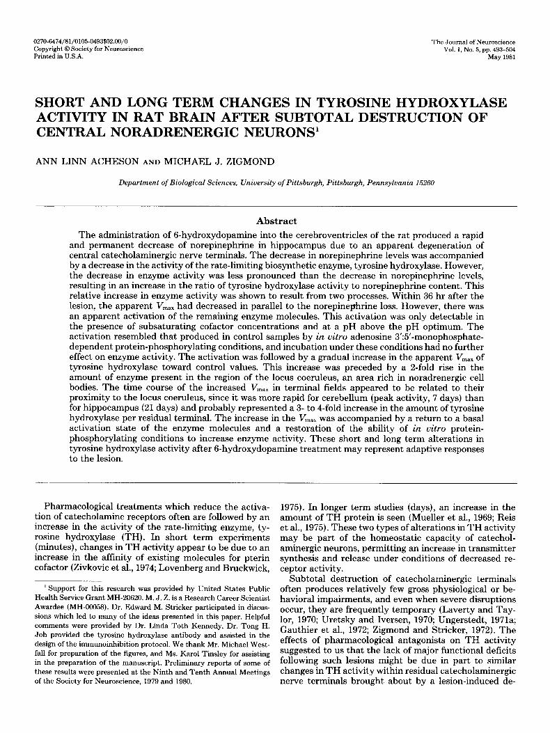
A Tris/acetate buffer was used, the pH of which was either optimal for the preparation (hippocampus, pH 6.2; cerebellum, 6.1; locus coeruleus, pH 5.9 to 6.3; Fig. 1) or was above this pH optimum. Tris/acetate was used de- spite the fact that it does not have pK, values within this range, because it results in higher TH activity than buffers which have the correct pK, values (Acheson et al., 1981), presumably because Tris serves to inhibit the conversion of 6MPH4 to a form which is not reducible by dihydropteridine reductase (Nielsen et al., 1962). The pH of the reaction mixture was not significantly different from that of the Tris/acetate buffer.
The L-[ l-‘4C]dihydroxyphenylalanine (DOPA) formed during the initial reaction was decarboxylated subse- quently by addition of an excess of aromatic L-amino- acid decarboxylase and a Tris/acetate buffer, pH 7.2 to 7.5, selected to bring the final pH of the reaction mixture to 6.8. Finally, 14C02 was trapped and quantified using liquid scintillation spectrometry. The specificity of the assay for tyrosine hydroxylation and its linearity with
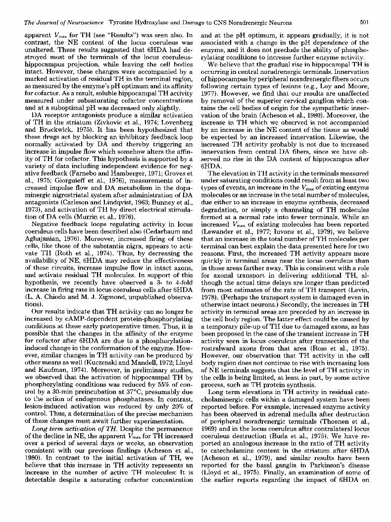
30 1 L I 1 I I J
58 60 62 64 66
OH
Figure 1. pH optimum of soluble TH activity from hippo- campus, cerebellum, and locus coeruleus. Enzyme activity was measured in Tris/acetate buffer with 0.7 to 1.5 mM 6MPH4 and 75 PM r.,-[l-‘4C]tyrosine as a function of assay pH. Enzyme activity was expressed as a percentage of maximal activity for hippocampus (O), cerebellum (A), and locus coeruleus (O), respectively. The 40,000 x g supernatant from a homogenate served as the source of soluble TH. Each point represents the mean of five animals.
The Journal of Neuroscience Tyrosine Hydroxylase and Damage to CNS Noradrenergic Neurons 495
time and amount of protein were determined within the ranges used (Acheson et al., 1980, 1981). The sensitivity of the assay was such that all values were at least twice the value of blank controls.
Adenosine 3’:5’-monophosphate (CAMP)-dependent protein-phosphorylating conditions. Samples were preincubated for 10 min at 30°C in the presence of 1 pg/ ~1 of CAMP-dependent protein kinase (partially purified from bovine heart; Miyamoto et al., 1969), 0.5 mM aden- osine 5’-triphosphate (ATP), 0.2 mM CAMP, 1.0 mM MgC12, and 2.0 mM theophylline (Sigma) in 75 mM Tris/ acetate, pH 6.0. Control samples were preincubated with 75 mM Tris/acetate buffer, pH 6.0, containing 1 pg/pl of bovine serum albumin (BSA, Fraction V, Sigma). The preincubation was terminated by placing the sample on ice. TH activity in the samples was assayed then in the presence of 2 mM disodium ethylenediaminetetra-acetic acid (EDTA; Fisher Scientific Co.) to inhibit further phosphorylation.
Determination of amount of THprotein. Animals were treated with 6HDA or vehicle and killed 5 to 21 days later. The area containing locus coeruleus was removed, and two locus coeruleus pairs were pooled and homoge- nized in 450 ~1 of 50 mM Tris-HCl buffer, pH 6.0, contain- ing 0.1% bovine serum albumin. Homogenates were cen- trifuged at 40,000 x g for 30 min, and the supernatant served as the source of TH. Antibodies specific to TH were a gift from Dr. T. H. Joh and were raised in rabbits from enzyme purified from rat adrenal glands by the method of Joh et al. (1973). The IgG fraction was precip- itated from the serum at 50% saturation with ammonium sulfate and resuspended in 0.9% NaCl. A lo-p1 aliquot of 0.9% NaCl containing various amounts of antibody was added to a 20-~1 aliquot of tissue supernatant. Samples then were incubated at room temperature for 1 hr with occasional shaking and centrifuged at 10,000 x g for 10 min. A 10-p aliquot of the resulting supernatant was assayed for TH activity as described above. The amount of antibody required to inhibit TH activity completely (the equivalence point) was estimated by graphical anal- ysis.
Catecholamine assay. NE and dopamine (DA) levels were measured as previously described (Saller and Zig- mond, 1978). Endogenous catecholamines were methyl- ated by catechol-0-methyltransferase, using radiolabeled S-adenosyl-L-[methyZ-3H]methionine (80 Ci/mmol, New England Nuclear) as a methyl donor. The methylated compounds then were separated using silica gel thin layer chromatography and eluted with borate buffer, pH 10.0. The compounds were separated further by solvent ex- traction and quantified using liquid scintillation spec- trometry. Final values were determined using internal standards carried through the entire assay. The assay was determined to be linear with the concentration of catecholamine within the ranges used. The average sen- sitivity of the assay was 10 pg for DA and 20 pg for NE. All values were at least three times blank for DA and twice blank for NE.
Determination of sample protein content. Soluble pro- tein content in each of the brain regions was determined using the Bio-Rad Protein Assay Kit (Bio-Rad Labora- tories, Richmond, CA) with BSA as the standard. This
assay is based on the binding of Coomassie brilliant blue G-250 dye to protein, followed by spectrophotometric quantification.
Results
NE content in hippocampus and locus coeruleus after GHDA. Rats were lesioned with 250 pg of 6HDA admin- istered intraventricularly. Thirty-six hours or 21 days later, the animals were killed and the hippocampus, cerebellum, and locus coeruleus region were removed for analysis of NE content. 6HDA treatment resulted in an immediate and apparently permanent decrease in the NE content of noradrenergic terminal fields (hippocam- pus and cerebellum) to approximately 20% of control values (Table I). However, this depletion was not accom- panied by any change in the NE content of the locus coeruleus region (Table I). Using a synaptosome-rich P2 fraction prepared from hippocampus, we also have mea- sured high affinity NE uptake, a specific characteristic of noradrenergic terminals, and found an equivalent de- crease in the initial transport velocity (Acheson et al., 1980). Collectively these observations suggest that 6HDA caused a degeneration of noradrenergic nerve terminals without significant loss of cell bodies and are consistent with previous reports (Jonsson, 1980).
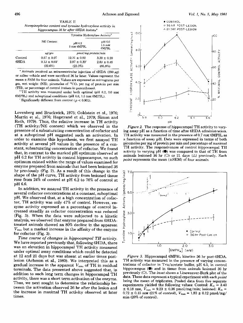
TH activity 36 hr after 6HDA treatment. Rats given 250 pg of 6HDA were killed 36 hr later and hippocampal TH activity was measured. When enzyme activity was measured at the pH optimum (pH 6.2) and in the pres- ence of a saturating concentration of cofactor (3.0 mM), activity was reduced to 23% of control, an amount com- parable to the loss of NE. In contrast, when TH activity was assayed at a suboptimal pH (pH 6.6) and in the presence of a subsaturating concentration of cofactor (1.5 mM), TH activity was reduced to only 86% of control (Table II).
Short term activation of TH is usually characterized by an increase in the affinity of the enzyme for its pterin cofactor and in its pH optimum (Zivkovic et al., 1974;
TABLE I
Norepinephrine content of terminal and cell body regions 36 hr or 21 days after GHDA lesions”
NE Content
6HDA Control
36 Hr 21 Days
P&v&?m
Hippocampus 0.54 + 0.04 0.14 + O.Olh 0.13 Y.!z O.Olh
(26.2%) (24.7%)
Cerebellum 0.25 f 0.05 0.07 f O.Olh 0.06 f 0.02h (23.0%) (24.0%)
Locus coeruleus’ 46.5 f 3.7 45.7 * 3.1 47.4 + 4.0
” Animals received an intraventricular injection of 6HDA (250 pg) or saline vehicle and were killed 36 hr or 21 days later. Control values for
animals killed 36 hr or 21 days after GHDA administration were not significantly different and were pooled. Values represent the mean + SEM for eight animals (controls) or for four animals (GHDA-treated).
Data are expressed as micrograms per gm, wet weight (NE) or per- centage of control (values in parentheses).
’ Significantly different from control (p < 0.001). ‘NE values for locus coeruleus are expressed as micrograms per
pair.
496 Acheson and Zigmond Vol. 1, No. 5, May 1981
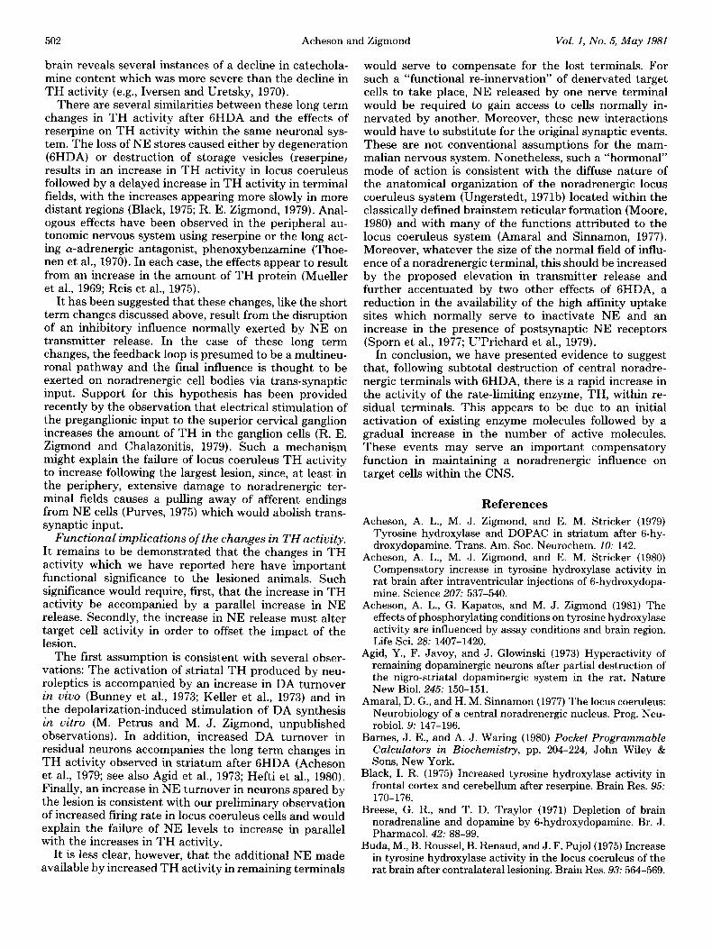
TABLE II . CONTROL
Norepinephrine content and tyrosine hydroxylase activity in
hippocampus 36 hr after 6HDA lesions” I
0 36 HR POST-LESION
A 21 DAY POST-LESION T
Tyrosine Hydroxylase Activityh
NE Content pH 6.2
pH 6.6
3 mh! GMPH, 1.5 InM GMPHI
PeT/&m pmol/mg protein/min
Control 0.57 + 0.07 12.31 + 0.92 3.30 + 0.20
6HDA 0.12 + 0.03’ 2.87 + 0.33’ 2.83 -c 0.45
(22.6%) (23.3%) (85.8%)
” Animals received an intraventricular injection of 6HDA (250 pg) or saline vehicle and were sacrificed 36 hr later. Values represent the mean f SEM for four animals. Values are expressed as micrograms per gm, wet weight (NE), picomoles of 14C02 per mg of protein per min
(TH), or percentage of control (values in parentheses). h TH activity was measured under both optimal (pH 6.2, 3.0 mM
GMPH,) and suboptimal conditions (pH 6.6, 1.5 mM GMPH,). ’ Significantly different from control (p < 0.001).
y 2.0
z t
,” >
Lovenberg and Bruckwick, 1975; Goldstein et al., 1976; Murrin et al., 1976; Hegstrand et al., 1979; Simon and 58 60 62 64 66
Roth, 1979). Thus, the relative increase in TH activity PH
(TH activity/NE content) which we observed in the presence of a subsaturating concentration of cofactor and
Figure 2. The response of hippocampal TH activity to vary- ing assay pH as a function of time after 6HDA administration.
at a suboptimal pH suggested such an activation. In TH activity was measured in the presence of 0.7 mM GMPH, as order to examine this hypothesis, we first assayed TH a function of assay pH. Data were expressed in terms of both activity at several pH values in the presence of a con- picomoles per mg of protein per min and percentage of maximal
stant, subsaturating concentration of cofactor. We found TH activity. The responsiveness of control hippocampal TH
that, in contrast to the marked pH optimum observed at activity to varying pH (0) was compared to that of TH from
pH 6.2 for TH activity in control hippocampus, no such animals lesioned 36 hr (0) or 21 days (A) previouslv. Each
optimum existed within the range of values examined for point represents the mean (GEM) of four animals. ”
enzyme prepared from animals that had been lesioned 36 hr previously (Fig. 2). As a result of this change in the shape of the pH curve, TH activity from lesioned tissue
r; 7.01
rose from 24% of control at pH 6.2 to 76% of control at pH 6.6.
In addition, we assayed TH activity in the presence of several cofactor concentrations at a constant, suboptimal pH. We observed that, at a high concentration of cofac- tor, TH activity was only 47% of control. However, en- zyme activity expressed as a percentage of control in- creased steadily as cofactor concentration was reduced (Fig. 3). When the data were subjected to a kinetic analysis, we observed that enzyme prepared from GHDA- *̂ treated animals showed an 80% decline in the apparent
E -_- -- ---e-------y m P V,,, but a marked increase in the affinity of the enzyme 2 for cofactor (Fig. 3). g 10
l Control
. - 6
0 36 Hr Post-Lesion Time course of changes in hippocampal TH activity. L
x We have reported previously that, following GHDA, there k 1 I I I
was an elevation in hippocampal TH activity measured 1.0 2.0 3.0
under optimal assay conditions which could be detected [GMPH,] CmM)
at 12 and 21 days but was absent at earlier times post- Figure 3. Hippocampal 6MPH4 kinetics 36 hr post-GHDA. lesion (Acheson et al., 1980). We interpreted this as a TH activity was measured in the presence of varying concen- gradual increase in the apparent V,,, of TH in residual trations of cofactor in Tris/acetate buffer, pH 6.5, in control
terminals. The data presented above suggested that, in hippocampus (0) and in tissue from animals lesioned 36 hr
addition to such long term changes in hippocampal TH previously (0). The inset shows a Lineweaver-Burk plot of the
activity, there was a short term activation of the enzyme. data. These data represent a typical experiment with eachpoint
Thus, we next sought to determine the relationship be- being the mean of triplicates. Pooled data from five separate
tween the activation observed 36 hr after the lesion and experiments yielded the following values: Control: K,,, = 3.43
the increase in maximal TH activity observed at later f 0.18 mM, V,., = 9.23 f 0.36 pmol/mg/min; lesioned: K, =
times. 0.72 f 0.15 mM (21% of control), V,,,., = 1.83 f 0.12 pmol/mg/ min (20% of control).
The Journal of Neuroscience Tyrosine Hydroxylase and Damage to CNS Noradrenergic Neurons 497
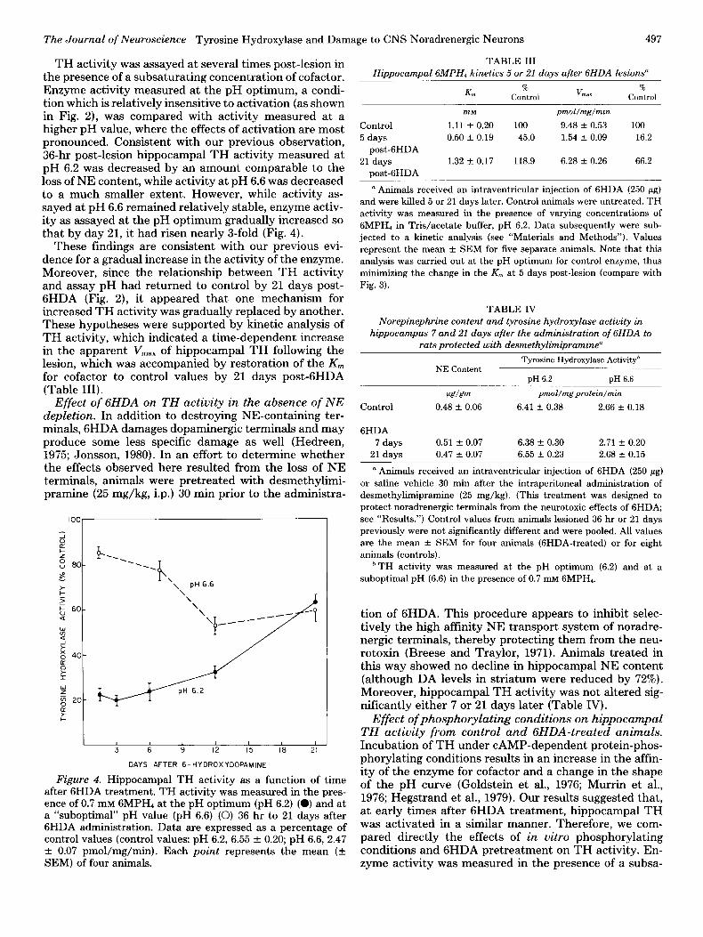
TH activity was assayed at several times post-lesion in the presence of a subsaturating concentration of cofactor. Enzyme activity measured at the pH optimum, a condi- tion which is relatively insensitive to activation (as shown in Fig. 2), was compared with activity measured at a higher pH value, where the effects of activation are most pronounced. Consistent with our previous observation, 36-hr post-lesion hippocampal TH activity measured at pH 6.2 was decreased by an amount comparable to the loss of NE content, while activity at pH 6.6 was decreased to a much smaller extent. However, while activity as- sayed at pH 6.6 remained relatively stable, enzyme activ- ity as assayed at the pH optimum gradually increased so that by day 21, it had risen nearly 3-fold (Fig. 4).
These findings are consistent with our previous evi- dence for a gradual increase in the activity of the enzyme. Moreover, since the relationship between TH activity and assay pH had returned to control by 21 days post- 6HDA (Fig. 2), it appeared that one mechanism for increased TH activity was gradually replaced by another. These hypotheses were supported by kinetic analysis of TH activity, which indicated a time-dependent increase in the apparent V,,,,, of hippocampal TH following the lesion, which was accompanied by restoration of the K,,, for cofactor to control values by 21 days post-6HDA (Table III).
Effect of 6HDA on TH activity in the absence of NE depletion. In addition to destroying NE-containing ter- minals, 6HDA damages dopaminergic terminals and may produce some less specific damage as well (Hedreen, 1975; Jonsson, 1980). In an effort to determine whether the effects observed here resulted from the loss of NE terminals, animals were pretreated with desmethylimi- pramine (25 mg/kg, i.p.) 30 min prior to the administra-
100 1
1 I 1
3 6 9 12 15 IS 21
DAYS AFTER 6.HYDROXYDOPAMINE
Figure 4. Hippocampal TH activity as a function of time after 6HDA treatment. TH activity was measured in the pres- ence of 0.7 mM 6MPH4 at the pH optimum (pH 6.2) (0) and at a “suboptimal” pH value (pH 6.6) (0) 36 hr to 21 days after 6HDA administration. Data are expressed as a percentage of control values (control values: pH 6.2, 6.55 + 0.20; pH 6.6, 2.47 f 0.07 pmol/mg/min). Each point represents the mean (k SEM) of four animals.
TABLE III Hippocampal GMPH, kinetics 5 or 21 days after GHDA lesions”
K”l %
V %
Control “IaX Control
Control
5 days post-6HDA
21 days post-6HDA
InM
1.11 f 0.20 0.50 * 0.19
1.32 + 0.17
100 45.0
118.9
pmol/mg/min
9.48 + 0.53 1.54 + 0.09
6.28 + 0.26
100 16.2
66.2
” Animals received an intraventricular injection of 6HDA (250 pg) and were killed 5 or 21 days later. Control animals were untreated. TH
activity was measured in the presence of varying concentrations of GMPH, in Tris/acetate buffer, pH 6.2. Data subsequently were sub-
jected to a kinetic analysis (see “Materials and Methods”). Values represent the mean f SEM for five separate animals. Note that this
analysis was carried out at the pH optimum for control enzyme, thus minimizing the change in the K, at 5 days post-lesion (compare with
Fig. 3).
TABLE IV Norepinephrine content and tyrosine hydroxylase activity in
hippocampus 7 and 21 days after the administration of GHDA to rats protected with desmethylimipramine”
Tyrosine Hydroxylase Activityh NE Content
pH 6.2 pH 6.6
de pmol/mgprotein/min
Control 0.48 f 0.06 6.41 + 0.38 2.66 f 0.18
6HDA 7 days 0.51 f 0.07 6.38 -t 0.30 2.71 f 0.20
21 days 0.47 Tk 0.07 6.55 k 0.23 2.68 f 0.15
n Animals received an intraventricular injection of 6HDA (250 pg)
or saline vehicle 30 min after the intraperitoneal administration of desmethylimipramine (25 mg/kg). (This treatment was designed to protect noradrenergic terminals from the neurotoxic effects of GHDA; see “Results.“) Control values from animals lesioned 36 hr or 21 days previously were not significantly different and were pooled. All values
are the mean + SEM for four animals (GHDA-treated) or for eight animals (controls).
* TH activity was measured at the pH optimum (6.2) and at a
suboptimal pH (6.6) in the presence of 0.7 mM GMPH,.
tion of GHDA. This procedure appears to inhibit selec- tively the high affinity NE transport system of noradre- nergic terminals, thereby protecting them from the neu- rotoxin (Breese and Traylor, 1971). Animals treated in this way showed no decline in hippocampal NE content (although DA levels in striatum were reduced by 72%). Moreover, hippocampal TH activity was not altered sig- nificantly either 7 or 21 days later (Table IV).
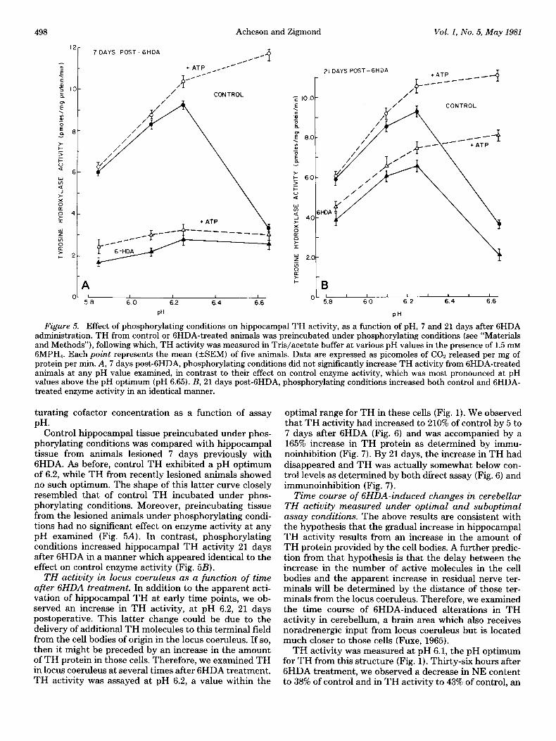
Effect of phosphorylating conditions on hippocampal TH activity from control and GHDA-treated animals. Incubation of TH under CAMP-dependent protein-phos- phorylating conditions results in an increase in the affin- ity of the enzyme for cofactor and a change in the shape of the pH curve (Goldstein et al., 1976; Murrin et al., 1976; Hegstrand et al., 1979). Our results suggested that, at early times after 6HDA treatment, hippocampal TH was activated in a similar manner. Therefore, we com- pared directly the effects of in vitro phosphorylating conditions and 6HDA pretreatment on TH activity. En- zyme activity was measured in the presence of a subsa-
498 Acheson and Zigmond Vol. I, No. 5, May 1981
12- 7 DAYS POST- 6HDA
/M /x
l ATP _c~-~
CONTROL
0 I I 1 I 58 60 6.2 6.4 6.6
PH
I 1 I 1 I 60 6.2 6.4 6.6
PH
Figure 5. Effect of phosphorylating conditions on hippocampal TH activity, as a function of pH, 7 and 21 days after 6HDA administration. TH from control or GHDA-treated animals was preincubated under phosphorylating conditions (see “Materials and Methods”), following which, TH activity was measured in Tris/acetate buffer at various pH values in the presence of 1.5 mM GMPH,. Each point represents the mean (ASEM) of five animals. Data are expressed as picomoles of COn released per mg of protein per min. A, 7 days post-GHDA, phosphorylating conditions did not significantly increase TH activity from GHDA-treated animals at any pH value examined, in contrast to their effect on control enzyme activity, which was most pronounced at pH values above the pH optimum (nH 6.65). B. 21 davs nest-GHDA, phosphorylating conditions increased both control and GHDA- treated enzyme activity in an identical manner. ” -
turating cofactor concentration as a function of assay PH.
Control hippocampal tissue preincubated under phos- phorylating conditions was compared with hippocampal tissue from animals lesioned 7 days previously with GHDA. As before, control TH exhibited a pH optimum of 6.2, while TH from recently lesioned animals showed no such optimum. The shape of this latter curve closely resembled that of control TH incubated under phos- phorylating conditions. Moreover, preincubating tissue from the lesioned animals under phosphorylating condi- tions had no significant effect on enzyme activity at any pH examined (Fig. 5A). In contrast, phosphorylating conditions increased hippocampal TH activity 21 days after 6HDA in a manner which appeared identical to the effect on control enzyme activity (Fig. 5B).
TH activity in locus coeruleus as a function of time after 6HDA treatment. In addition to the apparent acti- vation of hippocampal TH at early time points, we ob- served an increase in TH activity, at pH 6.2, 21 days postoperative. This latter change could be due to the delivery of additional TH molecules to this terminal field from the cell bodies of origin in the locus coeruleus. If so, then it might be preceded by an increase in the amount of TH protein in those cells. Therefore, we examined TH in locus coeruleus at several times after 6HDA treatment. TH activity was assayed at pH 6.2, a value within the
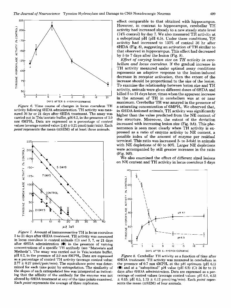
optimal range for TH in these cells (Fig. 1). We observed that TH activity had increased to 210% of control by 5 to 7 days after 6HDA (Fig. 6) and was accompanied by a 165% increase in TH protein as determined by immu- noinhibition (Fig. 7). By 21 days, the increase in TH had disappeared and TH was actually somewhat below con- trol levels as determined by both direct assay (Fig. 6) and immunoinhibition (Fig. 7).
Time course of GHDA-induced changes in cerebellar TH activity measured under optimal and suboptimal assay conditions. The above results are consistent with the hypothesis that the gradual increase in hippocampal TH activity results from an increase in the amount of TH protein provided by the cell bodies. A further predic- tion from that hypothesis is that the delay between the increase in the number of active molecules in the cell bodies and the apparent increase in residual nerve ter- minals will be determined by the distance of those ter- minals from the locus coeruleus. Therefore, we examined the time course of GHDA-induced alterations in TH activity in cerebellum, a brain area which also receives noradrenergic input from locus coeruleus but is located much closer to those cells (Fuxe, 1965).
TH activity was measured at pH 6.1, the pH optimum for TH from this structure (Fig. 1). Thirty-six hours after 6HDA treatment, we observed a decrease in NE content to 38% of control and in TH activity to 43% of control, an
The Journal of Neuroscience Tyrosine Hydroxylase and Damage to CNS Noradrenergic Neurons 499
+
1 50 I I 1 I I I
0 3 6 9 12 15 I8 21
DAYS AFTER 6-HYOROXYOOPAMlNE
Figure 6. Time course of changes in locus coeruleus TH activity following 6HDA administration. TH activity was mea- sured 36 hr or 21 days after 6HDA treatment. The assay was carried out in Tris/acetate buffer, pH 6.2, in the presence of 3.0 mM GMPH,. Data are expressed as a percentage of control values (average control value: 2.43 f 0.21 pmol/pair/min). Each point represents the mean (+SEM) of at least three animals.
I I I\ I. 1 0 5 IO 15 20’ 25 30 35 40
Figure 7. Amount of immunoreactive TH in locus coeruleus 5 to 21 days after 6HDA treatment. TH activity was measured in locus coeruleus in control animals (0) and 5, 7, or 21 days after 6HDA administration (0) in the presence of varying concentrations of a specific TH antibody (see “Materials and Methods”). The assay was carried out in Tris/acetate buffer, pH 6.2, in the presence of 3.0 mM GMPH,. Data are expressed as a percentage of control TH activity (average control value: 2.77 + 0.27 pmol/pair/min). The equivalence point was deter- mined for each time point by extrapolation. The similarity of the slopes of each extrapolated line was interpreted as indicat- ing that the affinity of the antibody for the enzyme was not altered by 6HDA treatment at any of the time points examined. Each point represents the average of three replicates.
effect comparable to that obtained with hippocampus. However, in contrast to hippocampus, cerebellar TH activity had increased already to a new steady state level (74% control) by day 7. We also measured TH activity at a suboptimal pH (pH 6.5). Under these conditions, TH activity had increased to 133% of control 36 hr after 6HDA (Fig. 8), suggesting an activation of TH similar to that observed in hippocampus. This effect had decreased by 5 to 7 days after the lesion (Fig. 8).
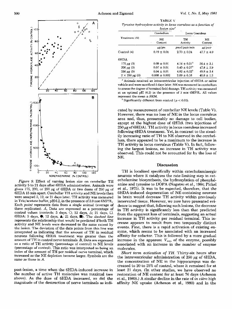
Effect of varying lesion size on TH activity in cere- bellum and locus coeruleus. If the gradual increase in TH activity measured under optimal assay conditions represents an adaptive response to the lesion-induced decrease in receptor activation, then the extent of the increase should be proportional to the size of the lesion. To examine the relationship between lesion size and TH activity, animals were given different doses of 6HDA and killed 5 to 21 days later, times when the apparent increase in the amount of TH in cerebellum was at or near maximum. Cerebellar TH was assayed in the presence of a saturating concentration of 6MPH4. We observed that, in GHDA-lesioned animals, TH activity was significantly higher than the value predicted from the NE content of the structure. Moreover, the extent of the deviation increased with increasing lesion size (Fig. 9A). This phe- nomenon is seen most clearly when TH activity is ex- pressed as a ratio of enzyme activity to NE content, a possible index of the amount of enzyme per residual terminal. This ratio was increased 2- to 3-fold in animals with NE depletions of 60 to 80%. Larger NE depletions were accompanied by still greater increases in the ratio (Fig. 9B).
We also examined the effect of different sized lesions on NE content and TH activity in locus coernleus 5 days
140
DAYS AFTER 6- HYDROXYDOPAMINE
Figure 8. Cerebellar TH activity as a function of time after 6HDA treatment. TH activity was measured in cerebellum in the presence of 0.7 mM GMPH, at the pH optimum (pH 6.1) (0) and at a “suboptimal” pH value (pH 6.5) (0) 36 hr to 21 days after 6HDA administration. Data are expressed as a per- centage of control values (average control values: pH 6.1, 4.33 f 0.15; pH 6.5, 1.73 + 0.12 pmol/mg/min). Each point repre- sents the mean (+SEM) of four animals.
500 Acheson and Zigmond Vol. I, No. 5, May 1981
E lool ,+-?yyTi 0 20 40 60 SC 100 120
NOREPINEPHRINE (% CONTROL)
Figure 9. Effect of varying lesion size on cerebellar TH activity 5 to 21 days after 6HDA administration. Animals were given 175, 200, or 250 pg of 6HDA or two doses of 250 pg of 6HDA 15 min apart. Cerebellar TH activity and NE levels then were assayed 5, 12, or 21 days later. TH activity was measured in Tris/acetate buffer, pH 6.2, in the presence of 3.0 mM GMPH,. Each point represents data from a single animal (average of three replicates). A, Data are expressed as a percentage of control values (controls: 5 days, 0, 12 days, a; 21 days, q . GHDA: 5 days, e 12 days, A; 21 days, W). The dashed line
represents the relationship that would be predicted if both TH activity and NE levels were decreased to the same extent by the lesion. The deviation of the data points from this line was interpreted as indicating that the amount of TH in residual neurons following 6HDA treatment was greater than the amount of TH in control nerve terminals. B, Data are expressed as a ratio of TH activity (percentage of control) to NE levels (percentage of control). This ratio was interpreted as being an index of the amount of TH per residual nerve terminal, which increased as the NE depletion became larger. Symbols are the same as those in A.
post-lesion, a time when the GHDA-induced increase in the number of active TH molecules was maximal (see above). As the dose of 6HDA increased, so did the magnitude of the destruction of nerve terminals as indi-
TABLE V
Tyrosine hydroxylase activity in locus coeruleus as a function of lesion size”
Treatment (iVJ
Control (4)
Cerebellum Locus Coeruleus
NE TH NE content Activity Content
~~Lg/g~ pmol/pair/min w/PaLr
0.19 + 0.01 2.70 k 0.24 47.7 f. 4.0
6HDA
175 pg (3) 0.09 + 0.01 4.14 f 0.31h 50.4 + 3.1
200 pg (3) 0.07 f 0.01 5.40 + 0.37h 47.8 -c 3.9
250 pg (5) 0.04 * 0.01 4.82 f 0.32& 49.8 -t 3.4
2 x 250 pg (3) 0.008 + 0.002 2.89 f 0.16 40.8 + 1.3
n Animals received an intraventricular injection of 6HDA or saline vehicle and were sacrificed 5 days later. NE was measured in cerebellum to assess the degree of terminal field damage. TH activity was measured at an optimal pH (6.2) in the presence of 3 InM GMPH,. All values represent the mean + SEM.
’ Significantly different from control (p < 0.01).
cated by measurement of cerebellar NE levels (Table V). However, there was no loss of NE in the locus coeruleus area and, thus, presumably no damage to cell bodies, except at the highest dose of 6HDA (two injections of 250 pg of GHDA). TH activity in locus coeruleus increased following 6HDA treatment. Yet, in contrast to the stead- ily increasing ratio of TH to NE observed in the cerebel- lum, there appeared to be a maximum to the increase in TH activity in locus coeruleus (Table V). In fact, follow- ing the largest lesions, no increase in TH activity was observed. This could not be accounted for by the loss of NE.
Discussion
TH is localized specifically within catecholaminergic neurons where it catalyzes the rate-limiting step in cat- echolamine biosynthesis, the hydroxylation of phenylal- anine and tyrosine to DOPA (Nagatsu et al., 1964; Pickel et al., 1975). It was to be expected, therefore, that the GHDA-induced degeneration of NE-containing neurons in brain would decrease TH activity within previously innervated tissue. However, we now have presented evi- dence to suggest that, following such lesions, the decrease in TH activity is significantly less than that predicted from the apparent loss of terminals, suggesting an actual increase in TH activity per residual terminal. This in- crease appears to result from two temporally distinct events. First, there is a rapid activation of existing en- zyme, which seems to be associated with an increased affinity for cofactor. This is followed by a more gradual increase in the apparent V,,,,, of the enzyme, possibly associated with an increase in the number of enzyme molecules.
Short term activation of TH. Thirty-six hours after the intraventricular administration of 250 pg of GHDA, the concentration of NE in the hippocampus was de- creased to 20 to 25% of control, where it remained for at least 21 days. (In other studies, we have observed no restoration of NE content for at least 76 days (Acheson et al., 1980).) A similar decline in the rate of in vitro high affinity NE uptake (Acheson et al., 1980) and in the
The Journal of Neuroscience Tyrosine Hydroxylase and Damage to CNS Noradrenergic Neurons 501
apparent V,,, for TH (see “Results”) was seen also. In contrast, the NE content of the locus coeruleus was unaltered. These results suggested that 6HDA had de- stroyed most of the terminals of the locus coeruleus- hippocampus projection, while leaving the cell bodies intact. However, these changes were accompanied by a marked activation of residual TH in the terminal region, as measured by the enzyme’s pH optimum and its affinity for cofactor. As a result, soluble hippocampal TH activity measured under subsaturating cofactor concentrations and at a suboptimal pH was decreased only slightly.
DA receptor antagonists produce a similar activation of TH in the striatum (Zivkovic et al., 1974; Lovenberg and Bruckwick, 1975). It has been hypothesized that these drugs act by blocking an inhibitory feedback loop normally activated by DA and thereby triggering an increase in impulse flow which somehow alters the aflin- ity of TH for cofactor. This hypothesis is supported by a variety of data including independent evidence for neg- ative feedback (Farnebo and Hamberger, 1971; Groves et al., 1975; Giorguieff et al., 1976), measurements of in- creased impulse flow and DA metabolism in the dopa- minergic nigrostriatal system after administration of DA antagonists (Carlsson and Lindqvist, 1963; Bunney et al., 1973), and activation of TH by direct electrical stimula- tion of DA cells (Murrin et al., 1976).
Negative feedback loops regulating activity in locus coeruleus cells have been described also (Cedarbaum and Aghajanian, 1976). Moreover, increased firing of these cells, like those of the substantia nigra, appears to acti- vate TH (Roth et al., 1974). Thus, by decreasing the availability of NE, 6HDA may reduce the effectiveness of these circuits, increase impulse flow in intact axons, and activate residual TH molecules. In support of this hypothesis, we recently have observed a 3- to 4-fold increase in firing rate in locus coeruleus cells after 6HDA (L. A. Chiodo and M. J. Zigmond, unpublished observa- tions) .
Our results indicate that TH activity can no longer be increased by CAMP-dependent protein-phosphorylating conditions at these early postoperative times. Thus, it is possible that the changes in the affinity of the enzyme for cofactor after 6HDA are due to a phosphorylation- induced change in the conformation of the enzyme. How- ever, similar changes in TH activity can be produced by other means as well (Kuczenski and Mandell, 1972; Lloyd and Kaufman, 1974). Moreover, in preliminary studies, we observed that the activation of hippocampal TH by phosphorylating conditions was reduced by 55% of con- trol by a 30-min preincubation at 37”C, presumably due to the action of endogenous phosphatases. In contrast, lesion-induced activation was reduced by only 20% of control. Thus, a determination of the precise mechanism of these changes must await further experimentation.
Long term activation of TH. Despite the permanence of the decline in NE, the apparent V,,, for TH increased over a period of several days or weeks, an observation consistent with our previous findings (Acheson et al., 1980). In contrast to the initial activation of TH, we believe that this increase in TH activity represents an increase in the number of active TH molecules: It is detectable despite a saturating cofactor concentration
and at the pH optimum, it appears gradually, it is not associated with a change in the pH dependence of the enzyme, and it does not preclude the ability of phospho- rylating conditions to increase further enzyme activity.
We believe that the gradual rise in hippocampal TH is occurring in central noradrenergic terminals. Innervation of hippocampus by peripheral noradrenergic fibers occurs following certain types of lesions (e.g., Loy and Moore, 1977). However, we find that our results are unaffected by removal of the superior cervical ganglion which con- tains the cell bodies of origin for the sympathetic inner- vation of the brain (Acheson et al., 1980). Moreover, the increase in TH which we observed is not accompanied by an increase in the NE content of the tissue as would be expected by an increased innervation. Likewise, the increased TH activity probably is not due to increased innervation from central DA fibers, since we have ob- served no rise in the DA content of hippocampus after GHDA.
The elevation in TH activity in the terminals measured under saturating conditions could result from at least two types of events, an increase in the V,,, of existing enzyme molecules or an increase in the total number of molecules, due either to an increase in enzyme synthesis, decreased degradation, or simply a channeling of TH molecules formed at a normal rate into fewer terminals. While an increased V max of existing molecules has been reported (Lewander et al., 1977; Iuvone et al., 1979), we believe that an increase in the total number of TH molecules per terminal can best explain the data presented here for two reasons. First, the increased TH activity appears more quickly in terminal areas near the locus coeruleus than in those areas farther away. This is consistent with a role for axonal transport in delivering additional TH, al- though the actual time delays are longer than predicted from most estimates of the rate of TH transport (Levin, 1978). (Perhaps the transport system is damaged even in otherwise intact neurons.) Secondly, the increases in TH activity in terminal areas are preceded by an increase in the cell body region. The latter effect could be caused by a temporary pile-up of TH due to damaged axons, as has been proposed in the case of the transient increase in TH activity seen in locus coeruleus after transection of the rostralward axons from that area (Ross et al., 1975). However, our observation that TH activity in the cell body region does not continue to rise with increasing loss of NE terminals suggests that the level of TH activity in the cells is being limited, at least in part, by some active process, such as TH protein synthesis.
Long term elevations in TH activity in residual cate- cholaminergic cells within a damaged system have been reported before. For example, increased enzyme activity has been observed in adrenal medulla after destruction of peripheral noradrenergic terminals (Thoenen et al., 1969) and in the locus coeruleus after contralateral locus coeruleus destruction (Buda et al., 1975). We have re- ported an analogous increase in the ratio of TH activity to catecholamine content in the striatum after 6HDA (Acheson et al., 1979), and similar results have been reported for the basal ganglia in Parkinson’s disease (Lloyd et al., 1975). Finally, an examination of some of the earlier reports regarding the impact of 6HDA on
502 Acheson and Zigmond Vol. 1, No. 5, May 1981
brain reveals several instances of a decline in catechola- mine content which was more severe than the decline in TH activity (e.g., Iversen and Uretsky, 1970).
There are several similarities between these long term changes in TH activity after 6HDA and the effects of reserpine on TH activity within the same neuronal sys- tem. The loss of NE stores caused either by degeneration (GHDA) or destruction of storage vesicles (reserpine, results in an increase in TH activity in locus coeruleus followed by a delayed increase in TH activity in terminal fields, with the increases appearing more slowly in more distant regions (Black, 1975; R. E. Zigmond, 1979). Anal- ogous effects have been observed in the peripheral au- tonomic nervous system using reserpine or the long act- ing a-adrenergic antagonist, phenoxybenzamine (Thoe- nen et al., 1970). In each case, the effects appear to result from an increase in the amount of TH protein (Mueller et al., 1969; Reis et al., 1975).
It has been suggested that these changes, like the short term changes discussed above, result from the disruption of an inhibitory influence normally exerted by NE on transmitter release. In the case of these long term changes, the feedback loop is presumed to be a multineu- ronal pathway and the final influence is thought to be exerted on noradrenergic cell bodies via trans-synaptic input. Support for this hypothesis has been provided recently by the observation that electrical stimulation of the preganglionic input to the superior cervical ganglion increases the amount of TH in the ganglion cells (R. E. Zigmond and Chalazonitis, 1979). Such a mechanism might explain the failure of locus coeruleus TH activity to increase following the largest lesion, since, at least in the periphery, extensive damage to noradrenergic ter- minal fields causes a pulling away of afferent endings from NE cells (Purves, 1975) which would abolish trans- synaptic input.
Functional implications of the changes in TH activity. It remains to be demonstrated that the changes in TH activity which we have reported here have important functional significance to the lesioned animals. Such significance would require, first, that the increase in TH activity be accompanied by a parallel increase in NE release. Secondly, the increase in NE release must alter target cell activity in order to offset the impact of the lesion.
The first assumption is consistent with several obser- vations: The activation of striatal TH produced by neu- roleptics is accompanied by an increase in DA turnover in vivo (Bunney et al., 1973; Keller et al., 1973) and in the depolarization-induced stimulation of DA synthesis in vitro (M. Petrus and M. J. Zigmond, unpublished observations). In addition, increased DA turnover in residual neurons accompanies the long term changes in TH activity observed in striatum after 6HDA (Acheson et al., 1979; see also Agid et al., 1973; Hefti et al., 1980). Finally, an increase in NE turnover in neurons spared by the lesion is consistent with our preliminary observation of increased firing rate in locus coeruleus cells and would explain the failure of NE levels to increase in parallel with the increases in TH activity.
It is less clear, however, that the additional NE made available by increased TH activity in remaining terminals
would serve to compensate for the lost terminals. For such a “functional re-innervation” of denervated target cells to take place, NE released by one nerve terminal would be required to gain access to cells normally in- nervated by another. Moreover, these new interactions would have to substitute for the original synaptic events. These are not conventional assumptions for the mam- malian nervous system. Nonetheless, such a “hormonal” mode of action is consistent with the diffuse nature of the anatomical organization of the noradrenergic locus coeruleus system (Ungerstedt, 1971b) located within the classically defined brainstem reticular formation (Moore, 1980) and with many of the functions attributed to the locus coeruleus system (Amaral and Sinnamon, 1977). Moreover, whatever the size of the normal field of influ- ence of a noradrenergic terminal, this should be increased by the proposed elevation in transmitter release and further accentuated by two other effects of GHDA, a reduction in the availability of the high affinity uptake sites which normally serve to inactivate NE and an increase in the presence of postsynaptic NE receptors (Sporn et al., 1977; U’Prichard et al., 1979).
In conclusion, we have presented evidence to suggest that, following subtotal destruction of central noradre- nergic terminals with GHDA, there is a rapid increase in the activity of the rate-limiting enzyme, TH, within re- sidual terminals. This appears to be due to an initial activation of existing enzyme molecules followed by a gradual increase in the number of active molecules. These events may serve an important compensatory function in maintaining a noradrenergic influence on target cells within the CNS.
References
Acheson, A. L., M. J. Zigmond, and E. M. Stricker (1979) Tyrosine hydroxylase and DOPAC in striatum after 6-hy- droxydopamine. Trans. Am. Sot. Neurochem. 10: 142.
Acheson, A. L., M. J. Zigmond, and E. M. Stricker (1980) Compensatory increase in tyrosine hydroxylase activity in rat brain after intraventricular injections of 6-hydroxydopa- mine. Science 207: 537-540.
Acheson, A. L., G. Kapatos, and M. J. Zigmond (1981) The effects of phosphorylating conditions on tyrosine hydroxylase activity are influenced by assay conditions and brain region. Life Sci. 28: 1407-1420.
Agid, Y., F. Javoy, and J. Glowinski (1973) Hyperactivity of remaining dopaminergic neurons after partial destruction of the nigro-striatal dopaminergic system in the rat. Nature New Biol. 245: 150-151.
Amaral, D. G., and H. M. Sinnamon (1977) The locus coeruleus: Neurobiology of a central noradrenergic nucleus. Prog. Neu- robiol. 9: 147-196.
Barnes, J. E., and A. J. Waring (1980) Pocket Programmable Calculators in Biochemistry, pp. 204-224, John Wiley & Sons, New York.
Black, I. R. (1975) Increased tyrosine hydroxylase activity in frontal cortex and cerebellum after reserpine. Brain Res. 95: 170-176.
Breese, G. R., and T. D. Traylor (1971) Depletion of brain noradrenaline and dopamine by 6-hydroxydopamine. Br. J. Pharmacol. 42: 88-99.
Buda, M., B. Roussel, B. Renaud, and J. F. Pujol(1975) Increase in tyrosine hydroxylase activity in the locus coeruleus of the rat brain after contralateral lesioning. Brain Res. 93: 564-569.
The Journal of Neuroscience Tyrosine Hydroxylase and Damage to CNS Noradrenergic Neurons 503
Bunney, B. S., J. R. Walters, R. H. Roth, and G. K. Aghajanian (1973) Dopaminergic neurons: Effect of antipsychotic drugs and amphetamine on single cell activity. J. Pharmacol. Exp. Ther. 185: 560-568.
Carlsson, A., and M. Lindqvist (1963) Effect of chlorpromazine and haloperidol on formation of 3-methoxytyramine and normetanephrine in mouse brain. Acta Pharmacol. Toxicol. (Copenh.) 20: 140-144.
Cedarbaum, J. M., and G. K. Aghajanian (1976) Noradrenergic neurons of the locus coeruleus: Inhibition by epinephrine and activation by the a-antagonist piperoxane. Brain Res. 112: 413-419.
Farnebo, L. O., and B. Hamberger (1971) Drug-induced changes in the release of ‘H-monoamines from field stimulated rat brain slices. Acta. Physiol. Stand. Suppl. 371: 34-44.
Fuxe, K. (1965) Evidence for the existence of monoamine neu- rons in the central nervous system. II. The distribution of monoamine terminals in the central nervous system. Acta Physiol. Stand. Suppl. 247, 64: 37-84.
Gauthier, P., R. A. Nadeau, and J. dechamplain (1972) Acute and chronic cardiovascular effects of 6-hydroxydopamine in dogs. Circ. Res. 31: 207-217.
Giorguieff, M. F., M. L. LeFloch, T. C. Westfall, J. Glowinski, and M. J. Besson (1976) Nicotinic effect of acetylcholine on the release of newly synthesized “H-dopamine in rat striatal slices and cat caudate nucleus. Brain Res. 106: 117-131.
Goldstein, M., R. L. Bronaugh, B. Ebstein, and C. Roberge (1976) Stimulation of tyrosine hydroxylase activity by cyclic AMP in synaptosomes and in soluble striatal enzyme prepa- rations. Brain Res. 109: 563-574.
Groves, P., C. J. Wilson, S. J. Young, and G. V. Rebec (1975) Self-inhibition by dopaminergic neurons. Science 100: 522- 529.
Hedreen, J. (1975) Increased nonspecific damage after lateral ventricle injection of 6-OHDA compared with fourth ventri- cle injection in rat brain. In Chemical Tools in Catechola- mine Research, G. Jonsson, T. Malmfors, and C. Sachs, eds., Vol. I., pp. 91-100, North-Holland Publishing Co., Amster- dam.
Hefti, F., E. Melamed, and R. J. Wurtman (1980) Partial lesions of the dopaminergic nigrostriatal system in rat brain: Bio- chemical characterization. Brain Res. 195: 123-138.
Hegstrand, L. R., J. R. Simon, and R. H. Roth (1979) Tyrosine hydroxylase: Examination of conditions influencing activity in pheochromocytoma, adrenal medulla, and striatum. Bio- them. Pharmacol. 28: 519-523.
Iuvone, P. M., T. H. Joh, and N. H. Neff (1979) Regulation of retinal tyrosine hydroxylase: Long-term exposure to light increased the apparent V,,, without a concomitant increase of immunotitratable enzyme molecules. Brain Res. 178: 191- 195.
Iversen, L. L., and N. J. Uretsky (1970) Regional effects of 6- hydroxydopamine on catecholamine containing neurones in rat brain and spinal cord. Brain Res. 24: 364-367.
Joh, T. H., C. Geghman, and D. J. Reis (1973) Immunochemical demonstration of increased accumulation of tyrosine hydrox- ylase protein in sympathetic ganglia and adrenal medulla elicited by reserpine. Proc. Natl. Acad. Sci. U. S. A. 70: 2767- 2771.
Jonsson, G. (1980) Chemical neurotoxins as denervation tools in neurobiology. Annu. Rev. Neurosci. 3: 169-187.
Kapatos, G., and M. J. Zigmond (1979) Effect of haloperidol on dopamine synthesis and tyrosine hydroxylase in striatal syn- aptosomes. J. Pharmacol. Exp. Ther. 208: 468-475.
Keller, H. H., G. Bartholini, and A. Pletscher (1973) Increase in 3-methoxy-4-hydroxyphenylethylene glycol in rat brain by neuroleptic drugs. Eur. J. Pharmacol. 23: 183-189.
Kuczenski, R. T., and A. J. Mandell (1972) Allosteric activation
of hypothalamic tyrosine hydroxylase by ions and sulphated mucopolysaccharides. J. Neurochem. 19: 131-137.
Laverty, R., and K. Taylor (1970) Effects of intraventricular 2,4,5-trihydroxyphenylethylamine (6-hydroxydopamine) on rat behavior and brain catecholamine metabolism. Br. J. Pharmacol. 40: 836-846.
Levin, B. E. (1978) Axonal transport of [“HI proteins in a noradrenergic system of the rat brain. Brain Res. 150: 55-68.
Lewander, T., T. H. Joh, and D. J. Reis (1977) Tyrosine hydroxylase: Delayed activation in central noradrenergic neurons and induction in adrenal medulla elicited by stimu- lation of central cholinergic receptors. J. Pharmacol. Exp. Ther. 200: 523-534.
Lloyd, T., and S. Kaufman (1974) The stimulation of partially purified bovine caudate tyrosine hydroxylase by phospha- tidyl-L-serine. Biochem. Biophys. Res. Commun. 59: 1262- 1269.
Lloyd, K. G., L. Davidson, and 0. Hornykiewicz (1975) The neurochemistry of Parkinson’s disease: Effect of L-DOPA therapy. J. Pharmacol. Exp. Ther. 195: 453-464.
Lovenberg, W., and E. A. Bruckwick (1975) Mechanisms of receptor mediated regulation of catecholamine synthesis in brain. In Pre- and Postsynaptic Receptors, E. Usdin and W. E. Bunney, eds., pp. 149-169, Marcel Dekker, New York.
Loy, R., and R. Y. Moore (1977) Anomalous innervation of the hippocampal formation by peripheral sympathetic axons fol- lowing mechanical injury. Exp. Neurol. 57: 645-650.
Miyamoto, E., J. F. Kuo, and P. Greengard (1969) Cyclic nucleotide-dependent protein kinase. III. Purification and properties of 3’,5’-monophosphate-dependent protein kinase from bovine brain. J. Biol. Chem. 244: 6395-6402.
Moore, R. Y. (1980) The reticular formation: Monoamine neu- ron systems. In The Reticular Formation Reuisited, J. A. Hobson and M. A. B. Brazier, eds., pp. 67-81, Raven Press, New York.
Mueller, R. A., H. Thoenen, and J. Axelrod (1969) Inhibition of trans-synaptically increased tyrosine hydroxylase activity by cycloheximide and actinomycin-D. Mol. Pharmacol. 5: 463- 469.
Murrin, L. C., V. H. Morgenroth, and R. H. Roth (1976) Dopaminergic neurons: Effects of electrical stimulation on tyrosine hydroxylase. Mol. Pharmacol. 12: 1070-1081.
Nagatsu, T., M. Levitt, and S. Udenfriend (1964) The initial step in norepinephrine biosynthesis. J. Biol. Chem. 239: 2910- 2917.
Nielsen, K. H., V. Simonsen, and K. E. Lind‘ (1962) Dihydro- pteridine reductase-A method for the measurement of ac- tivity and investigations of the specificity for NADH and NADPH. Eur. J. Biochem. 9: 497-502.
Pickel, V. M., T. H. Joh, and D. J. Reis (1975) Ultrastructural localization of tyrosine hydroxylase in noradrenergic neurons of brain. Proc. Natl. Acad. Sci. U. S. A. 72: 659-666.
Purves, D. (1975) Functional and structural changes in mam- malian sympathetic neurons following interruption of their axons. J. Physiol. (Lond.) 252: 429-463.
Reis, D. J., T. H. Joh, and R. A. Ross (1975) Effects of reserpine on activities and amounts of tyrosine hydroxylase and dopa- mine-p-hydroxylase in catecholamine neuronal systems in rat brain. J. Pharmacol. Exp. Ther. 193: 775-784.
Ross, R. A., T. H. Joh, and D. J. Reis (1975) Reversible changes in the accumulation and activities of tyrosine hydroxylase and dopamine+hydroxylase in neurons of nucleus locus coeruleus during the retrograde reaction. Brain Res. 92: 57- 72.
Roth, R. H., P. M. Salzman, and V. H. Morgenroth (1974) Noradrenergic neurons: Allosteric activation of hippocampal tyrosine hydroxylase by stimulation of the locus coeruleus. Biochem. Pharmacol. 23: 2779-2784.
504 Acheson and Zigmond Vol. I, No. 5, May 1981
SaIIer, C. F., and M. J. Zigmond (1978) A radioenzymatic assay for catecholamines and dihydroxyphenylacetic acid. Life Sci. 23: 1117-1130.
Simon, J. R., and R. H. Roth (1979) Striatal tyrosine hydrox- ylase: Comparison of the activation produced by depolariza- tion and dibutyryl-CAMP. Mol. Pharmacol. 16: 224-233.
Sporn, J. R., B. B. Wolfe, T. K. Harden, and P. B. Molinoff (1977) Supersensitivity in rat cortex: Pre- and post-synaptic effects of 6-hydroxydopamine at noradrenergic synapses. Mol. Pharmacol. 13: 1170-1180.
Stricker, E. M., and M. J. Zigmond (1976) Recovery of function after damage to central catecholamine-containing neurons: A neurochemical model for the lateral hypothalamic syndrome. In Progress in Psychobiology and Physiological Psychology, Vol. 6., pp. 121-188, Academic Press Inc., New York.
Thoenen, H., R. A. Mueller, and J. Axelrod (1969) Trans- synaptic induction of adrenal tyrosine hydroxylase. J. Phar- macol. Exp. Ther. 169: 249-254.
Thoenen, H., R. A. Mueller, and J. Axehod (1970) Phase differences in the induction of tyrosine hydroxylase in cell body and nerve terminals of sympathetic neurons. Proc. Natl. Acad. Sci. U. S. A. 65: 58-62.
Ungerstedt, U. (1971a) Adipsia and aphagia after 6-hydroxy- dopamine induced degeneration of the nigro-striatal dopa- mine system. Acta Physiol. Stand. Suppl. 367, 82: 95-122.
Ungerstedt, U. (1971b) Stereotaxic mapping of the monoamine pathways in the rat brain. Acta Physiol. Stand. Suppl. 367, 82: l-29.
U’Pritchard, D. C., W. D. Bechtel, B. M. Roust, and S. H. Snyder (1979) Multiple apparent alpha-adrenergic receptor
binding sites in rat brain: Effect of 6-hydroxydopamine. Mol. Pharmacol. 16: 47-60.
Uretsky, N. J., and L. L. Iversen (1970) Effects of 6-hydroxy- dopamine on catecholamine containing neurones in the rat brain. J. Neurochem. 17: 269-278.
Wilkinson, G. N. (19,61) Statistical estimations in enzyme ki- netics. Biochem. J. 80: 324-332.
Zigmond, M. J., and E. M. Stricker (1972) Deficits in feeding behavior after intraventricular injections of 6-hydroxydopa- mine in rats. Science 177: 1211-1214.
Zigmond, M. J., and E. M. Stricker (1974) Ingestive behavior following damage to central dopamine neurons: Implications for homeostasis and recovery of function. In Neurophysiology of Monoamines and Their Regulatory Enzymes, E. Usdin, ed., pp. 385-402, Raven Press, New York.
Zigmond, R. E. (1979) Tyrosine hydroxylase activity in norad- renergic neurons of the locus coeruleus after reserpine ad- ministration: Sequential increase in cell bodies and nerve terminals. J. Neurochem. 32: 23-29.
Zigmond, R. E., and A. Chalazonitis (1979) Long-term effects of preganglionic nerve stimulation on tyrosine hydroxylase ac- tivity in the rat superior cervical ganglion. Brain Res. 164: 137-152.
Zigmond, R. E., F. Schon, and L. L. Iversen (1974) Increased tyrosine hydroxylase activity in the locus coeruleus of rat brainstem after reserpine treatment and cold stress. Brain Res. 70: 547-552.
Zivkovic, B., A. Guidotti, and E. Costa (1974) Effects of neuro- leptics on striatal tyrosine hydroxylase: Changes in affinity for the pteridine cofactor. Mol. Pharmacol. 10: 727-735.
Related Documents