Accepted Manuscript Title: Sexual behavior and dendritic spine density of posterodorsal medial amygdala neurons in oxytocin knockout female mice Author: Roberta Oriques Becker Virg´ ınia Meneghini Lazzari Itiana Castro Menezes Mariana Morris Katya Rigatto Aldo B. Lucion Alberto A. Rasia-Filho M´ arcia Giovenardi<ce:footnote id="fn0005"><ce:note-para id="npar0005">These authors contributed equally to the present study.</ce:note-para></ce:footnote> PII: S0166-4328(13)00433-6 DOI: http://dx.doi.org/doi:10.1016/j.bbr.2013.07.034 Reference: BBR 8404 To appear in: Behavioural Brain Research Received date: 16-5-2013 Revised date: 17-7-2013 Accepted date: 21-7-2013 Please cite this article as: Becker RO, Lazzari VM, Menezes IC, Morris M, Rigatto K, Lucion AB, Rasia-Filho AA, Giovenardi M, Sexual behavior and dendritic spine density of posterodorsal medial amygdala neurons in oxytocin knockout female mice, Behavioural Brain Research (2013), http://dx.doi.org/10.1016/j.bbr.2013.07.034 This is a PDF file of an unedited manuscript that has been accepted for publication. As a service to our customers we are providing this early version of the manuscript. The manuscript will undergo copyediting, typesetting, and review of the resulting proof before it is published in its final form. Please note that during the production process errors may be discovered which could affect the content, and all legal disclaimers that apply to the journal pertain.

Welcome message from author
This document is posted to help you gain knowledge. Please leave a comment to let me know what you think about it! Share it to your friends and learn new things together.
Transcript

Accepted Manuscript
Title: Sexual behavior and dendritic spine density of
posterodorsal medial amygdala neurons in oxytocin knockout
female mice
Author: Roberta Oriques Becker Virgınia Meneghini Lazzari
Itiana Castro Menezes Mariana Morris Katya Rigatto Aldo B.
Lucion Alberto A. Rasia-Filho Marcia
Giovenardi<ce:footnote id="fn0005"><ce:note-para
id="npar0005">These authors contributed equally to the
present study.</ce:note-para></ce:footnote>
PII: S0166-4328(13)00433-6
DOI: http://dx.doi.org/doi:10.1016/j.bbr.2013.07.034
Reference: BBR 8404
To appear in: Behavioural Brain Research
Received date: 16-5-2013
Revised date: 17-7-2013
Accepted date: 21-7-2013
Please cite this article as: Becker RO, Lazzari VM, Menezes IC, Morris M, Rigatto
K, Lucion AB, Rasia-Filho AA, Giovenardi M, Sexual behavior and dendritic spine
density of posterodorsal medial amygdala neurons in oxytocin knockout female mice,
Behavioural Brain Research (2013), http://dx.doi.org/10.1016/j.bbr.2013.07.034
This is a PDF file of an unedited manuscript that has been accepted for publication.
As a service to our customers we are providing this early version of the manuscript.
The manuscript will undergo copyediting, typesetting, and review of the resulting proof
before it is published in its final form. Please note that during the production process
errors may be discovered which could affect the content, and all legal disclaimers that
apply to the journal pertain.

Page 1 of 29
Acc
epte
d M
anus
crip
t
Sexual behavior and dendritic spine density of posterodorsal medial amygdala
neurons in oxytocin knockout female mice
Roberta Oriques Beckera, Virgínia Meneghini Lazzaria, Itiana Castro Menezesb,
Mariana Morrisc, Katya Rigattoa, Aldo B. Lucione, Alberto A. Rasia-Filhob,d,*, Márcia
Giovenardia,*
a Programa de Pós-Graduação em Ciências da Saúde, Universidade Federal de
Ciências da Saúde de Porto Alegre, Porto Alegre, RS, Brazil.
b Programa de Pós-Graduação em Neurociências, Universidade Federal do Rio
Grande do Sul, Porto Alegre, RS, Brazil.
c Pharmacology and Toxicology Department, Wright State University, Dayton, OH,
United States of America.
d Programa de Pós-Graduação em Patologia e em Ciências da Reabilitação,
Universidade Federal de Ciências da Saúde de Porto Alegre, RS, Brazil.
e Departamento de Fisiologia, Universidade Federal do Rio Grande do Sul, Porto
Alegre, RS, Brazil.
* These authors contributed equally to the present study.
Corresponding Author: Profª. Márcia Giovenardi, Departamento de Ciências Básicas
da Saúde, Universidade Federal de Ciências da Saúde de Porto Alegre, Sarmento
Leite 245, sala 308, Porto Alegre, RS, 90050-170, Brazil.
Page 2 of 29
Acc
epte
d M
anus
crip
t
2
Phone: +55 51 33038751, Fax: +55 51 33038718
E-mail: [email protected]
ABSTRACT
Central oxytocin (OT) and arginine-vasopressin (AVP) have been shown to
play an important role in sexual behavior and neuroendocrine secretion in rodents.
The results of exogenous OT administration on sexual behaviors in male and female
mice are controversial. This study aimed to analyze the role of OT in sexual behavior,
the number of oocytes and the density of dendritic spines in the posterodorsal medial
amygdala (MePD) of female mice with selective deletion of the OT gene (OTKO).
Female C57BL/6 mice were genotyped and divided into control (WT) and OTKO
groups (n= 11 each). All experiments were performed in the proestrus phase.
Compared to WT data, our results showed that the OTKO group had a significant
increase in the latency for the display of lordosis behavior (490.8±113.8 and
841.9±53.9, respectively) and a decrease in both the frequency (6.3±2.4 and 0.5±0.4)
and duration (49.3±19.9 and 7.2±7.1) of lordosis and a reduction in the number of
oocytes (12.2±0.8 and 9.9± 0.6). However, the OTKO group showed a higher density
of proximal dendritic spines in the MePD compared to the WT group (2.4±0.1 and
1.9±0.1 spines/dendritic µm, respectively). No significant difference was observed in
the plasma levels of AVP between the groups (OTKO: 617.1±96.0 and WT:
583.3±112.0 pg/mL). Our data suggest that OT plays a crucial role in the sexual
behavior display, number of released oocytes and density of dendritic spines in the
MePD of female mice. The AVP plasma concentration was not affected in the OTKO
animals.
Keywords: Extended amygdala, OTKO mice, ovulation, reproductive behavior,
vasopressin.
Page 3 of 29
Acc
epte
d M
anus
crip
t
3
1. INTRODUCTION
Oxytocin (OT) and arginine-vasopressin (AVP) play a pivotal role in the
regulation of social behaviors in rodents [1]. These hormones are synthesized in the
magno- and parvocellular neurons of the paraventricular nucleus (PVN) and
supraoptic nucleus (SON) of the hypothalamus. Magnocellular neurons project to the
neurohypophysis and release these peptides into peripheral circulation. Parvocellular
neurons project to several brain areas, including the amygdaloidal nuclei,
hippocampus, nucleus of the solitary tract, dorsal motor nucleus of the vagus, area
postrema, hypothalamic ventromedial nucleus (VMH) and medial preoptic area
(MPOA), ventral tegmental area, nucleus accumbens and bed nucleus of the stria
terminalis (BnST) [2-4]. Thus, OT and AVP appear to be key regulators of the
evolution and expression of different types of social systems, including maternal
care, aggression, pair bonding, sexual behavior and social memory [5-7].
The recognition of conspecifics is an initial and crucial condition for the
establishment of social [8] and sexual behavior. Chemical cues, such as odor, scent
and pheromones, mediate sexual and competitive interactions and are important in
individual and kin recognition and mate selection [9-11]. Odor signals are processed
by two systems: the main olfactory and vomeronasal pathways [12]. Both pathways
have heavy direct and indirect projections to the medial nucleus of the amygdala
(MeA) [12, 13] with a relatively minor input to the BnST [14]. The MeA sends large
projections to the BnST and MPOA, which in turn project to the lateral septum and
hippocampus [13, 14]. This odor investigation/recognition circuit involves two relevant
neuropeptides: OT and AVP [15, 16]. OT-deficient mice are not able to recognize a
previously encountered, familiar conspecific during subsequent trials [1, 17], possibly
because of a defect in the processing of chemosensory information involving the
Page 4 of 29
Acc
epte
d M
anus
crip
t
4
MeA [18]. In fact, this social recognition behavior can be restored with a direct
microinjection of OT into the MeA of OT knockout (OTKO) mice [17].
The MeA is composed of 4 subnuclei and modulates social and reproductive
behaviors [22-25], among other functions [19-21]. Several studies indicate that sex
steroids can alter the morphology and function of the posterodorsal medial amygdala
(MePD) neurons and glial cells [26-28], thus making the MePD rat sexually dimorphic
or modifiable by naturally occurring variations in the level of circulating ovarian
steroids [23-31]. The MePD neurons from intact adult male rats have a higher density
of proximal dendritic spines than females in proestrus, estrus or metaestrus but not
diestrus [23, 25]. Thus, acting locally in the MePD and/or interconnected sex steroid
sensitive regions, gonadal hormones can alter the amount of dendritic spines in the
MePD to establish and/or maintain a higher quantity of spines in males and to induce
a numerical variation across the estrous cycle in females. Dendritic spines have
crucial properties for synaptic strength and plasticity and affect neuronal activity in
integrated circuits [32-35]. For example, the rat MePD is connected to hypothalamic
nuclei that control reproduction [e.g., the MPOA and anteroventral periventricular
nucleus; 36, 37], modulate timely hypothalamic gonadotropin releasing hormone
(GnRH) secretion and sexual behavior display [38, 39], process olfactory/pheromonal
[40, 41] and vaginocervical stimuli [38], and induce long-term changes in prolactin
secretion that are necessary for pregnancy/pseudopregnancy or mnemonic events at
the time of mating [42, 43].
Moreover, brain OT plays an important role in the regulation of male and
female sexual behavior. In male rodents, OT is implicated in erectile function,
copulatory activity and ejaculation [44, 45]. In female rats, other copulatory behavior
regulations occur through interactions between estrogen (E), progesterone (P) and

Page 5 of 29
Acc
epte
d M
anus
crip
t
5
OT [44]. OT induces female sexual behavior, primarily by its action in the MPOA and
VMH to control lordosis displays [46, 47]. In females primed with E, the OT receptor
(OTR) antisense oligodeoxynucleotides infusion into the VMH blocks female
receptivity to male rats [48-50]. Furthermore, experimental evidence demonstrates
that OT can regulate GnRH cell activity, thus suggesting a possible modulation of the
luteinizing hormone (LH) peak and, consequently, ovulation [51]. Compared to OT,
AVP has been reported to exert opposite effects on female sexual behavior in rats.
When administered centrally, AVP inhibits, but AVP receptor (V1a) antagonists
stimulate, sexual receptivity [52, 53]. These antagonistic effects can occur in the
MPOA, which suggests that the OT and AVP interaction may contribute to the
regulation of sexual behavior in females [53].
The present study aimed to analyze the role of OT in the sexual behavior,
number of oocytes and density of dendritic spines in the MePD of female mice. The
basal plasma concentration of AVP was also measured in these animals.
2. MATERIALS AND METHODS
2.1 GENERAL METHODS
2.1.1 Animals
The mice in this study were the offspring of a backcrossed stock obtained from
Dr. W. Scott Young (B6; 129S-Oxttm1Wsy/J; NIMH, USA). All animals were
littermates from heterozygous breeders (C57BL/6 mice). Thirty-four females and 10
males, weighing 25 to 35 g and between 5 and 8 months old, were raised in the
animal housing facility of the Universidade Federal de Ciências da Saúde de

Page 6 of 29
Acc
epte
d M
anus
crip
t
6
Porto Alegre (UFCSPA, Brazil). The mice were housed in ventilated transparent
acrylic cages (37 × 24 × 24 cm) and grouped with up to five same-sex individuals.
The room temperature was controlled (22 ± 1ºC), and a 12:12 light-dark cycle (lights
off at 5 pm) was adopted. The mice had free access to chow (Nuvilab, Brazil) and
water.
All procedures conformed to the international regulations for the Care and Use
of Laboratory Animals (National Institutes of Health Publication No. 85-23, reviewed
1985, USA) and Brazilian Society for Neuroscience and Behavior Guidelines. The
protocols were approved by the local ethics committee (UFCSPA, Brazil, protocol No.
920/09).
To determine the regularity of the estrous cycle, vaginal smears were obtained
from virgin female mice 2 weeks before the beginning of the experiment. After the
occurrence of three regular estrous cycles (cf, 4), the experiments were performed
the night of the proestrus phase (54, 55).
2.1.2 Genotyping
The colony founders were developed by Young et al. [56]. The gene was
deleted by crossing a genetic construct with the WT mouse OT allele in a manner
that replaced the final 2 exons. Genotyping was performed as previously described
[54]. Briefly, the genomic DNA was isolated from the mouse tail samples and used as
a template for a polymerase chain reaction. The primer sequences for amplification
of the WT alleles involved the forward primer 5’-CTT GGC TTA CTG GCT CTG
ACCT-3’ and reverse primer 5’-GTC AAG AGG GAG CCT AAC ACT TC-3’. To
amplify the targeted allele, an additional forward primer (NEO) was used: 5’-TGC
CCC AAA GGC CTA CCC GCT TCC-3’.
Page 7 of 29
Acc
epte
d M
anus
crip
t
7
After genotyping, the mice were divided into WT and OTKO groups and
randomly assigned into the following two experiments.
2.2 EXPERIMENT 1
2.2.1 Sexual Behavior
The females from the WT control (n=11) and OTKO groups (n=11) were tested
with sexually experienced males. In this experiment, we used sexually experienced
males because they exhibited higher frequencies and shorter latencies for the
behavioral components of copulation, including mounting, intromission and
ejaculation [57]. The male was habituated for 10 minutes in the test apparatus. After
the adaptation time, a female was placed in the same observation box, and the
behavioral test began. The test was performed during the dark cycle in an
observation room illuminated by a dim red light, and the behaviors were recorded
with a video camera for 15 minutes [58].
The following parameters were evaluated: latency, frequency and duration of
lordosis behavior. The lordosis response was scored on a 4-point scale (0-3), as
described by Hardy and DeBold [59]. For each female mouse, a lordosis quotient
was calculated by dividing the number of lordosis scores of 2 or 3 by the total number
of mounts x 100. The test was videotaped using a video camera and recorded using
“Observer” software (Noldus®, Holland).
2.2.2 Hormonal Measures and Counting of Oocytes
On the day following the sexual behavior test (i.e., on estrus day during the
light cycle at 8 a.m.), the female mice were decapitated and trunk blood samples
Page 8 of 29
Acc
epte
d M
anus
crip
t
8
(n=8 for both the WT and OTKO groups) and ovaries (n=11 for both the WT and
OTKO groups) were collected for further analysis.
The blood samples were placed in heparinized test tubes and centrifuged for
15 minutes at 1600g at 4°C; the plasma was separated and stored at -80°C. AVP
enzyme-linked immunosorbent assays (ELISA) were performed according to the
manufacturer's protocol (Enzo Life Sciences, USA) using the Arg8-Vasopressin EIA
kit. Briefly, 100 L of plasma was compared to other known concentrations, an
optical density reading was obtained at 405 nm with a correction at 570 nm, and a
standard curve was generated. The accepted intra-assay variability was 5.9%.
The oviducts were dissected and squashed between 2 glass slides. The
number of oocytes of both oviductal ampullae was counted under an optical
microscope as previously described [60].
2.3 EXPERIMENT 2
2.3.1 Histological Procedure and Data Acquisition
Females from the WT and OTKO groups (n=6 each) were anesthetized with a
single intraperitoneal injection of ketamine (80 mg/kg) and xylazine (10 mg/kg). A
transcardial perfusion with heparinized 4% paraformaldehyde and 2% picric acid
diluted in 0.1 M phosphate buffer solution (pH=7.4) was then performed. The brains
were removed and sectioned using a vibratome (Leica, Germany). The coronal
sections (150 µm thick) were received in a 3% potassium dichromate (Merck,
Germany) solution for 24 h and subsequently impregnated in 1.5% silver nitrate
(Merck, Germany) solution for 48 h at room temperature. The sections were
dehydrated, cleared with xylene, mounted on slides and covered with non-acidic
Page 9 of 29
Acc
epte
d M
anus
crip
t
9
synthetic balsam and coverslips [adapted from 21, 31, 61, 62].
The sections containing the MePD corresponded to a distance of 1.46 to 1.94
mm posterior to the bregma [62]. In both hemispheres, the MePD was located
laterally to the optic tract and “molecular layer” and ventrally to the stria terminalis.
Microscopic images of each brain slice were compared with the schematic drawings
of an atlas [62; Figure 1A]. Neurons were selected based on the following criteria: (a)
undoubtedly being located within the boundaries of the MePD and relatively distant
from its ultimate borders; (b) being relatively isolated from the neighboring
impregnated cells to avoid “tangled” dendrites; (c) the dendrites should have well-
impregnated and defined borders; and (d) the spines should be obviously
distinguishable from the background [based on 21, 26, 61]. Because the number of
impregnated neurons varied between sections, both sides of the brain were used
[also see 61].
For each female, the first dendrites that fulfilled these aforementioned
inclusion criteria had their spines drawn using a camera lucida (2000x; i.e., 100x oil-
immersion objective lens and 20x ocular lens) coupled to an optic microscope
(Olympus BX-41, Japan). The dendritic spines in the different microscopic focal
planes were counted on the proximal branches. Eight different dendrites were
studied from each female with 1 dendrite per sampled neuron. The dendritic spine
data were then obtained from a total of 48 dendrites per experimental group. After
this procedure, three-dimensional dendritic lengths were measured from the identical
microscopic images (400x; Olympus BX-61, Japan), and the images of the selected
dendrites were captured by a high-resolution digital camera (CCD DP72, Japan) and
analyzed with Image Pro Plus 7.0 computer software (Media Cybernetics, USA). The
sampled dendritic length varied from 30-60 µm in both groups [means ± standard
Page 10 of 29
Acc
epte
d M
anus
crip
t
10
deviation (SD) values of 41 + 5 µm and 39 + 7 µm for the WT and OTKO mice,
respectively]. Spine density was defined as the number of spines per unit length of
dendritic segment [µm; 26, 31, 61].
2.4 STATISTICAL ANALYSIS
For the sexual behavioral test, the latency, frequency, duration and lordosis
quotients were analyzed by the Mann-Whitney test because all parameters showed a
non-parametric distribution. The counting of oocytes was also analyzed using a
Mann-Whitney test.
The mean values for the dendritic spine density from each mouse were
calculated for further comparisons. The results showed a normal distribution
(Kolmogorov-Smirnov test) and equal SD and therefore fulfilled the formal
requirements for the use of parametrical analyses. The dendritic spine density data
from the two experimental groups were submitted to an unpaired two-tailed Student’s
t-test. The identical test was used to compare the plasma concentration of AVP of
WT and OTKO mice. In all cases, P<0.05 was considered statistically significant.
3. RESULTS
The OTKO group had a significant increase in the latency and decrease in the
frequency and duration of female sexual behavior compared to WT females (Table
1).
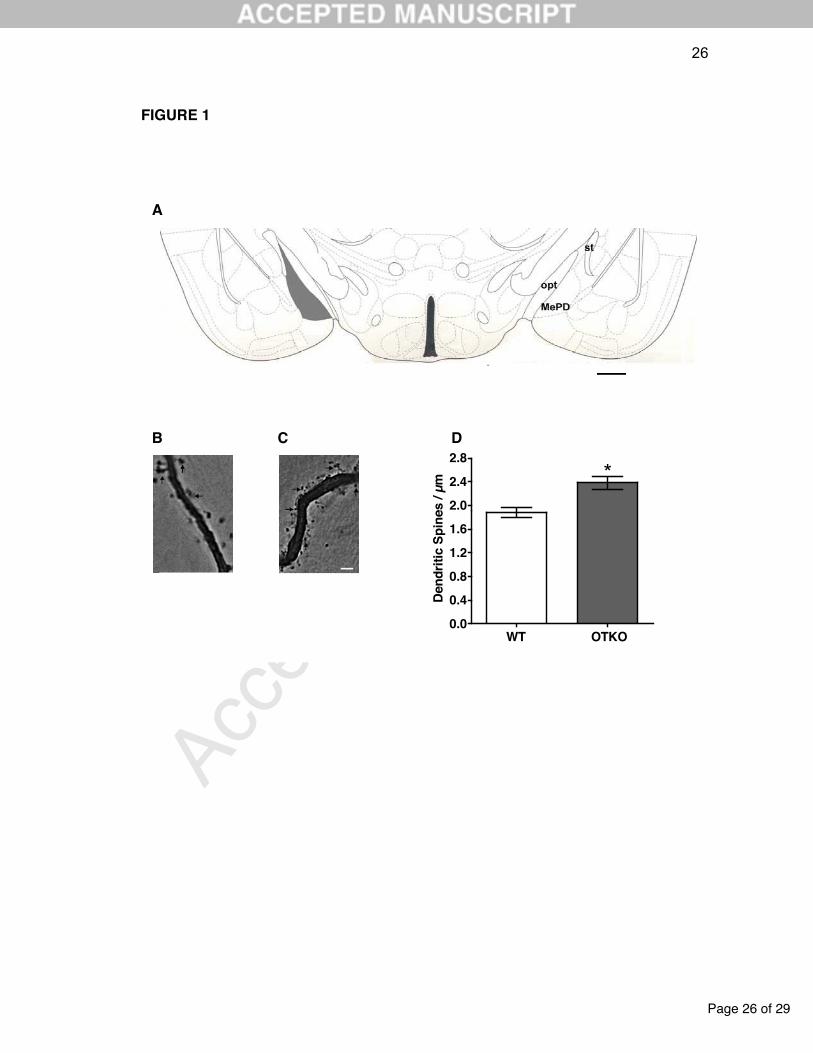
The OTKO females had a significant decrease in the number of oocytes at the
oviductal ampullae (P=0.03) compared to the WT group (Figure 2).
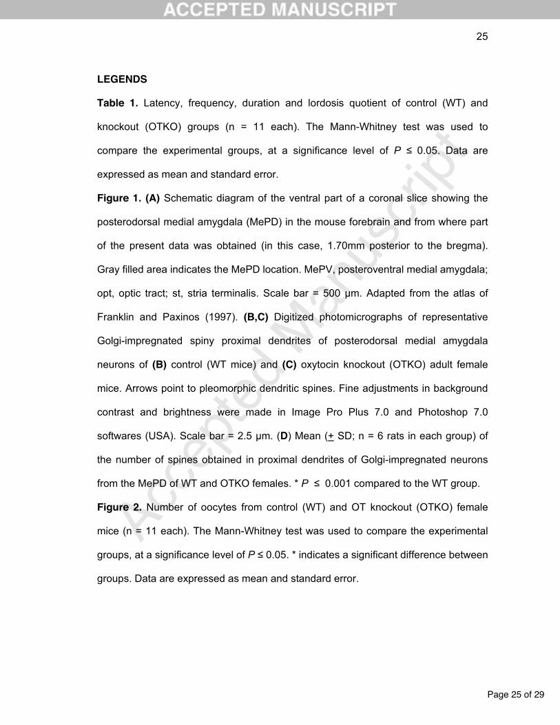
Proximal dendritic spine density showed a highly significant difference
between the groups. Minimum to maximum ranges for the MePD spine density
Page 11 of 29
Acc
epte
d M
anus
crip
t
11
observed for the WT and OTKO groups were 1.7-1.9 and 2.2-2.5 spines/dendritic
µm, respectively. The OTKO group showed consistently higher values than the WT
group (P < 0.01; Figure 1B-D).
Finally, there was no significant difference in the plasma concentration of AVP
between the WT and OTKO groups (means standard errors: 583.3±112.0 and
617.1±96.03 pg/mL, respectively; P=0.82).
4. DISCUSSION
There are currently differing perspectives on the role of OT in the sexual
behavior of male and female rodents [7, 56, 63-67]. Recently, we showed that the
sexual behavior of male mice was not affected by the absence of OT [68]. Therefore,
other hormones and neurochemical mechanisms could be more critically involved in
the sexual behavior of males (e.g., see 55, 69-70). In this study, our results showed
that knocking out the OT gene notably decreased the sexual behavior of female
mice.
Various central effects of OT on sexual behavior were obtained after
intracerebroventricular microinjections with some showing that OT facilitates socio-
sexual interactions [66, 69, 71]. In ovariectomized female rats under E and P
replacement therapy, OT increased lordosis behavior in response to mounting
attempts [44]. Lordosis also increased when either E and OT [48] or P and OT [72]
were administered concomitantly. However, the use of an OT antagonist reduced or
inhibited the expression of sexual behavior in female rats [44, 48, 50, 53].
The lordosis response, the typical expression of female receptivity, is a
complex phenomenon regulated by excitatory and inhibitory neural systems in the
brain [73, 74]. The induction of female sexual behavior is mediated primarily by the
Page 12 of 29
Acc
epte
d M
anus
crip
t
12
MPOA and VMH [47]. McCarthy et al. [50] demonstrated that a microinjection of
antisense oligodeoxynucleotides to OTR into the VMH of females primed with E
blocked female receptivity to male rats. Similarly, an OT antagonist injected into the
MPOA prior to treatment with P significantly decreased lordosis behavior and
increased fighting durations with males [75]. Our results also indicated that OT was
significantly involved in female sexual behavior, and together with a previous study
[76], these data showed that OT is an essential modulator of lordosis behavior in
mice.
Our oocyte quantification results showed that the OTKO group had a
significant decrease in the number of oocytes compared to the WT group. Several
studies show that OT modulates GnRH neuronal activity and that OT constitutes the
final output pathway of a neuronal network that controls the pre-ovulatory LH peak
and ovulation [51, 77-80]. Additionally, the central administration of an OT antiserum
abolishes the proestrus LH peak [81], whereas OT induces GnRH release from
hypothalamic explants on the afternoon of proestrus [82], thus supporting the pivotal
role of OT in GnRH release. Caligioni et al. [51] demonstrated that female rats in
metaestrus or proestrus had a double-labeled immunofluorescence in approximately
10% of GnRH neurons co-expressing OTRs in the MPOA, and few OT fibers could
be observed in the vicinity of these GnRH neurons. These data suggest that OT may
partially control neuronal activity in a subpopulation of GnRH neurons. Moreover, our
results allowed us to infer that ovulation was influenced by the absence of OT, but
further investigations should be conducted to clarify the OT interaction in the
multifactor neural network that controls GnRH neurons during the estrous cycle. In
our study, OTKO females showed normal cycles, as assessed by vaginal cytology,
and could be studied in the proestrus phase.
Page 13 of 29
Acc
epte
d M
anus
crip
t
13
Our results also showed that OTKO mice had a higher number (~25% more)
of dendritic spines in the MePD than WT controls. Again, both groups were studied
during proestrus, the cycle phase when there are estradiol and progesterone peaks
in circulation. It is noteworthy that ovarian hormones and their receptors correlate
with OT and OTR expression in socially relevant mouse brain regions [83, 84]. The
MePD expresses one of the highest concentrations of and estrogen receptors
[29, 30, 85], local neurons also co-express progesterone receptors [29], and OT and
OTR can be observed in the subnucleus [86]. In addition, the bilateral administration
of an OTR antagonist into the MeA impairs social memory in adult females [87]. It is
highly conceivable that female mouse MePD neurons are affected by sex steroid
fluctuations and the local release of OT and dynamically modulate
reproductive/social behaviors. The OTKO mice showed a disruption of sexual
receptiveness during proestrus, and MePD dendritic spine density was also higher. In
the rat MePD, there is a significant decrease in the number of spines when females
cycle from di- to proestrus [24, 26]. Considering that direct and indirect projections
from the MePD to reproductive behavior-related hypothalamic areas are GABAergic,
it is likely that the reduction in the MePD dendritic spines during proestrus would
reduce the output inhibition for the occurrence of female sexual behavior [further
elaborated in 21, 31; also see relevant data in 88]. It is possible that OTKO female
mice have an impaired sex steroid-mediated influence on the plastic number of
MePD dendritic spines and/or an altered neural circuitry for sexual behavior, although
the cyclic hypothalamic GnRH neuroendocrine secretion is spared. Other possibilities
include the pheromonal recognition of conspecific males and emotional/social
behavior processing by the MePD neurons of OTKO females. These working
hypotheses are emphasized by the present results and should be tested with further
Page 14 of 29
Acc
epte
d M
anus
crip
t
14
experimental approaches specifically aimed to evaluate the MePD.
The involvement of OT and AVP in social recognition and numerous social
behaviors, including parental and sexual behaviors [2, 7], suggests a fundamental
role of these hormones in the evolution of animal sociality. In contrast to OT, AVP is
likely to be more important for social recognition in males than females [89]. The
expression of AVP is greater in male than female brains in various species [63], and
AVP is typically associated with male-typical social behaviors, such as male
reproduction, aggression and territoriality [90].
The AVP and OT genes are highly homologous and closely linked (tail-to-tail)
in the mouse genome and are separated by an intragenic region of 3 kbp. Both of
these genes are transcribed toward one another from opposite strands of the DNA
duplex (91). Some studies (56, 92) demonstrated that AVP transcript levels are
decreased in the PVN and SON of OTKO mice. These results suggest that OT might
be involved in the regulation of AVP gene expression. Here, we observed that
knocking out the OT gene did not change the basal AVP plasma concentration when
comparing OTKO and WT female mice, and it could be inferred that lordosis behavior
was only influenced by the absence of OT.
In conclusion, our data suggest that OT plays a crucial role in sexual behavior
displays, the number of released oocytes and the density of dendritic spines in the
MePD of female mice. The AVP plasma concentration was not affected in OTKO
animals.
ACKNOWLEDGMENTS
We thank Maria Beatriz Kohek, Aline Gasparotto and Josi Maria Zimmermann
for collaboration on the genotyping. Additionally, we thank Dinara Jaqueline Moura
Page 15 of 29
Acc
epte
d M
anus
crip
t
15
for help with the ELISA assays and CAPES (Brazil) for the financial support.
CONFLICT OF INTEREST
The authors declare no actual or potential conflict of interest for the present work.
REFERENCES
[1] Choleris E, Clipperton-Allen AE, Phan A, Kavaliers M. Neuroendocrinology of
social information processing in rats and mice. Front Neuroendocrinol 2009;
30:442-459.
[2] Gimpl G, Fahrenholz F. The oxytocin receptor system: structure, function, and
regulation. Physiol Rev 2001; 81:629-682.
[3] Tom N, Stephen JA. Oxytocin in health and disease. Int J Biochem Cell B
2010; 42:202-205
[4] Lim MM, Young LJ. Neuropeptidergic regulation of affiliative behavior and
social bonding in animals. Horm Behav 2006; 50:506-517.
[5] Argiolas A, Meles MR, Vargiu L, Gessa GL. D(CH2)5Tyr(Me)-[Orn8]vasotocin,
a potent oxytocin antagonist, antagonizes penile erection and yawning
induced by oxytocin and apomorphine, but not by ACTH-(1-24). Eur J
Pharmacol 1987; 134:221-224.
[6] Lee HJ, Macbeth AH, Pagani JH, Young III WS. Oxytocin: the great facilitator
of life. Prog Neurobiol 2009; 88:127-151.
[7] Neumann ID. Brain oxytocin: a key regulator of emotional and social
behaviours in both females and males. J Endocrinol 2008; 20:858-865.
[8] Achiraman S, Ponmanickam P, Ganesh DS, Archunan G. Detection of estrus
by male mice: Synergistic role of olfactory–vomeronasal system. Neurosci Lett
2010; 477:144-148.
Page 16 of 29
Acc
epte
d M
anus
crip
t
16
[9] Nevison CM, Barnard CJ, Beynon RJ, Hurst JL. The consequences of
inbreeding for recognizing competitors. Proc Roy Soc B 2000; 267:687-694.
[10] Kavaliers M, Colwell DD , Choleris E , Agmo A, Muglia LJ, Ogawa S, Pfaff
DW. Impaired discrimination of and aversion to parasitized male odors by
female oxytocin knockout mice. Genes Brain Behav 2003; 2:220-230.
[11] Kavaliers M, Agmos A, Choleris E, Gustafsson JA, Korach KS, Muglia LJ,
Pfaff DW, Ogawa S. Oxytocin and estrogen receptor a and b knockout
miceprovide discriminably different odor cues in behavioral assays. Genes
Brain Behav 2004; 3:189-195.
[12] Brennan PA, Keverne EB. Something in the air? New insights into
mammalian pheromones. Curr Biol 2004; 14:81-89.
[13] Keller M, Michael JB, Brock O, Brennan PA, Bakker J. The main and the
accessory olfactory systems interact in the control of mate recognition and
sexual behavior. Behav Brain Res 2009; 200:268-276.
[14] Wacker WD, Ludwig M. Vasopressin, oxytocin, and social odor recognition.
Horm Behav 2012; 61:259-265.
[15] Kevetter GA, Winans SS. Connections of the corticomedial amygdala in the
golden hamster. II. Efferents of the “olfactory amygdala”. J Comp Neurol 1981;
197:99-111.
[16] Young LJ. The neurobiology of social recognition, approach, and avoidance.
Biol Psychiatry 2002; 51:18-26.
[17] Ferguson JN, Adag JM, Insel TR, Young LJ. Oxytocin in the medial amygdala
is essential for social recognition in the mouse. J Neurosci 2001; 21:8278-
8285.
[18] Samuelsen CL, Meredith M. Oxytocin antagonist disrupts male mouse medial
amygdala response to chemical-communication signals. Neuroscience 2011;
180:96-104.
[19] De Olmos J, Beltramino CA, Alheid GF. Amydala and extended amydala of
the rat: cytoarchitectonical, fibroarchitectonical and chemoarchitectonical
Page 17 of 29
Acc
epte
d M
anus
crip
t
17
survey. The Rat Nervous System. Amsterdam. Elsevier Academic Press:
2004; 1:509-603.
[20] Choi GB, Dong HW, Murphy AJ, Valenzuela DM, Yancopoulos GD, Swanson
LW, Anderson DJ. Lhx6 delineates a pathway mediating innate reproductive
behaviors from the amygdala to the hypothalamus. Neuron 2005; 46:647-660.
[21] Dall'Oglio A, Gehlen G, Achaval M, Rasia-Filho AA. Dendritic branching
feature s of posterodorsal medial amygdala neurons of adult male and female
rats: further data based on the Golgi method. Neurosci Lett 2008; 430:151-
156.
[22] Newman SW. The medial extended amygdala in male reproductive behavior.
A node in the mammalian social behavior network. Ann NY Acad Sci 1999;
877:242-257.
[23] Sheehan TP, Paul M, Amaral E, Numa MJ, Numan M. Evidence that the
medial amygdala projects to the anterior/ventromedial hypothalamic nuclei to
inhibit maternal behavior in rats. Neuroscience 2001; 106:341-356.
[24] Rasia-Filho AA, Dalpian F, Menezes IC, Brusco J, Moreira JE, Cohen RS.
Dendritic spines of the medial amygdala: plasticity, density, shape, and
subcellular modulation by sex steroids. Histol Histopathol 2012; 27:985-1011.
[25] Rasia-Filho AA, Haas D, de Oliveira AP, de Castilhos J, Frey R, Stein D,
Lazzari VM, Back F, Pires GN, Pavesi E, Winkelmann-Duarte EC, Giovenardi
M. Morphological and functional features of the sex steroid-responsive
posterodorsal medial amygdala of adult rats. Mini Rev Med Chem 2012;
12:1090-106.
[26] Rasia-Filho AA, Fabian C, Rigoti K, Achaval M. Influence of sex, estrous
cycle and motherhood in dendritic spine density in the rat medial amygdala
revealed by the Golgi method. Neuroscience 2004; 126:839-847.
[27] Johnson RT, Breedlove SM, Jordan CL. Sex differences and laterality in
astrocyte number and complexity in the adult rat medial amygdala. J Comp
Neurol 2008; 511:599-609.
Page 18 of 29
Acc
epte
d M
anus
crip
t
18
[28] Morris JA, Jordan CL, Breedlove SM. Sexual dimorphism in neuronal number
of the posterodorsal medial amygdala is independent of circulating androgens
and regional volume in adult rats. J Comp Neurol 2008; 506:851-859.
[29] Gréco B, Allegretto EA, Tetel MJ, Blaustein JD. Coexpression of ER beta with
ER alpha and progestin receptor proteins in the female rat forebrain: effects of
estradiol treatment. Endocrinology 2001; 142:5172-5181.
[30] Gréco B, Blasberg ME, Kosinski E C, Blaustein JD. Response of ER·-IR and
ER‚-IR cells in the forebrain of female rats to mating stimuli. Horm Behav
2003; 43:444-453.
[31] De Castilhos J, Forti CD, Achaval M, Rasia-Filho AA. Dendritic spine density
of posterodorsal medial amygdala neurons can be affected by gonadectomy
and sex steroid manipulations in adult rats: a Golgi study. Brain Res 2008;
1240:73-81.
[32] Segal M. Dendritic spines, synaptic plasticity and neuronal survival: activity
shapes dendritic spines to enhance neuronal viability. Eur J Neurosci 2010;
31:2178-2184.
[33] Nuriya M, Jiang J, Nemet B, Eisenthal KB, Yuste R. Imaging membrane
potential in dendritic spines . Proc Natl Acad Sci U. S. A. 2006; 103:786-790.
[34] Chen Y, Sabatini BL. Signaling in dendritic spines and spine microdomains.
Curr Opin Neurobiol 2012; 22:389-396.
[35] Rochefort NL, Konnerth A. Dendritic spines: from structure to in vivo function.
EMBO Rep 2012; 13:699-708.
[36] Yoshida M, Suga S, Sakuma Y. Estrogen reduces the excitability of the
female rat medial amygdala afferents from the medial preoptic area but not
those from the lateral septum. Exp Brain Res 1994; 101:1-7
[37] Polston EK, Gu G, Simerly RB. Neurons in the principal nucleus of the bed
nuclei of the stria terminalis provide a sexually dimorphic GABAergic input to
the anteroventral periventricular nucleus of the hypothalamus. Neuroscience
2004; 123:793-803.
Page 19 of 29
Acc
epte
d M
anus
crip
t
19
[38] Pfaus JG, Hebb MM. Implications of immediate–early gene induction in the
brain following sexual stimulation of female and male rodents. Brain Res Bull
1997; 44: 397-407.
[39] Simerly RB. Anatomical substrates of hypothalamic integration. The rat
nervous system. Amsterdam: Elsevier Academic Press 2004; 1:335-369.
[40] Meredith M, Westberry JM. Distinctive responses in the medial amygdala to
same-species and different-species pheromones. J Neurosci 2004; 24:5719-
5725.
[41] Pereno GL, Balaszczuk V, Beltramino CA. Detection of conspecific
pheromones elicits FOS expression in GABA and calcium-binding cells of the
rat vomeronasal system-medial extended amygdala. J Physiol Biochem 2011;
67:71-85.
[42] Polston EK, Heitz M, Barnes W, Cardamone K, Erskine MS. NMDA-mediated
activation of the medial amygdala initiates a downstream neuroendocrine
memory responsible for pseudopregnancy in the female rat. J Neurosci 2001;
21:4104-4110
[43] Lehmann ML, McKellar H, Erskine MS. Coding for the initiation of
pseudopregnancy by temporally patterned activation of amygdalar NMDA
receptors. J Neurosci 2005; 25:8696-8703.
[44] Witt DM. Oxytocin and rodent sociosexual responses: from behavior to gene
expression. Neurosci Biobehav Rev 1995; 19:315-24.
[45] Argiolas A, Collu M, Gessa GL, Melis MR, Serra G. The oxytocin antagonist
d(CH2)5Tyr(Me)-Orn8-vasotocin inhibits male copulatory behaviour in rats.
Eur J Pharmacol 1988; 149:389–92.
[46] Arletti R, Bertolini A. Oxytocin stimulates lordosis behavior in female rats.
Neuropeptides 1985; 6:247-253.
[47] Kow LM, Pfaff DW. Mapping of neural and signal transduction pathways for
lordosis in the search for estrogen actions on the central nervous system.
Behav Brain Res 1998; 92:169-180.
Page 20 of 29
Acc
epte
d M
anus
crip
t
20
[48] Caldwell JD. Central oxytocin and female sexual behavior. Annals of the New
York Academy of Sciences 1992; 652:166-179.
[49] Lee H-J, Pagani J, Young III W S. Using transgenic mouse models to study
oxytocin's role in the facilitation of species propagation. Brain Res 2010;
1364:216-224.
[50] McCarthy MM, Kleopoulous CV, Mobbs CV, Pfaff DW. Infusion of antisense
oligodeoxynucleotides to the oxytocin receptor in the ventromedial
hypothalamus reduces estrogen-induced sexual receptivity and oxytocin
receptor binding in the female rat. Neuroendocrinology 1994; 59:432-440.
[51] Caligioni CS, Oliver C, Jamur MC, Franci CR. Presence of oxytocin receptors
in the gonadotrophin-releasing hormone (GnRH) neurones in female rats: a
possible direct action of oxytocin on GnRH neurones. J Neuroendocrinol 2007;
19:439-448.
[52] Södersten P, DeVries GJ, Buijs RM, Melin P. A daily rhythm in behavioural
vasopressin sensitivity and brain vasopressin concentrations. Neurosci Lett
1985; 58:37-41.
[53] Caldwell JD, Barakat AS, Smith DD, Hruby VJ, Pedersen CA. A uterotonic
antagonist blocks the oxytocin-induced facilitation of female sexual receptivity.
Brain Res 1990; 512:291-296.
[54] Antunes IB, Da Silva A, Kawakami R, Andersen ML. The Female Rat. In:
Andersen ML & Tufik S. Animal Models as Tools in Ethical Biomedical
Research 2010; 103-120.
[55] Rasia-Filho AA, Lucion AB. Effects of 8-OH-DPAT on sexual behavior of
male rats castrated at different ages. Horm Behav 1996; 30:251-8.
[56] Young III WS, Shepard E, Amico J, Hennighausen L, Wagner KU, Lamarca
MU, Mckinney C, Ginns E. Deficiency in mouse oxytocin prevents milk
ejection, but not fertility or parturition. J Neuroendocrinol 1996; 8:847-853.

Page 21 of 29
Acc
epte
d M
anus
crip
t
21
[57] Swaney WT, Dubose BN, Curley JP, Champagne FA. Sexual experience
affects reproductive behavior and preoptic androgen receptors in male mice.
Horm Behav 2012; 61:472–478.
[58] Jonhansen JA, Clemens LG, Nunez AA. Characterization of copulatory
behavior in female mice: evidence for paced mating. Physiol Behav 2008;
95:425-429.
[59] Hardy DF, DeBold JF. Effects of mounts without intromission upon the
behavior of female rats during the onset of estrogen-induced heat. Physiol
Behav 1971; 7:643-645.
[60] Gomez CM, Frantz PJ, Sanvitto GL, Anselmo-Franci JA, Lucion AB. Neonatal
handling induces anovulatory estrous cycles in rats. Braz J Med Biol Res
1999; 32:1239-1242.
[61] Arpini M, Menezes I T, Dall’Oglio A, Rasia-Filho AA. The density of Golgi-
impregnated dendritic spines from adult rat posterodorsal medial amygdala
neurons displays no evidence of hemispheric or dorsal/ventral differences.
Neurosci Lett 2010; 469:209-221.
[62] Franklin KBJ, Paxinos G. The Mouse Brain in Stereotaxic Coordinates,
Academic Press, San Diego, 1997.
[63] DeVries AC, Young Jr WS, Nelson RJ. Reduced aggressive behaviour in
mice with targeted disruption of the oxytocin gene. J Neuroendocrinol 1997;
9:363-368.
[64] Nishimori K, Young LJ, Guo Q, Wang Z, Insel TR, Matzuk MM. Oxytocin is
required for nursing but not essential for parturition or reproductive behavior.
Proc Natl Acad Sci USA 1996; 93:11699-11704.
[65] Insel TR, Gelhard R, Shapiro LE. The comparative distribution of forebrain
receptors for neurohypophyseal peptides inmonogamous and polygamous
mice. Neuroscience 1991; 43:623-630.
[66] Pedersen CA, Caldwell JD, Jirikowski GF, Insel TR. Oxytocin in maternal,
sexual, and social behaviors. Ann NY Acad Sci 1992; 652:1-492.
Page 22 of 29
Acc
epte
d M
anus
crip
t
22
[67] Borrow AP, Cameron NM. The role of oxytocin in mating and pregnancy.
Horm Behav 2012; 61:266-276.
[68] Lazzari V M, Becker R O, Azevedo MS, Morris M, Rigatto K, Almeida S,
Lucion AB, Giovenardi M. Oxytocin modulates social interaction but is not
essential for sexual behavior in male mice. Behav Brain Res 2013; 244:130-
136.
[69] Carter CS, Lederhendler II, Kirkpatrick B. The interactive neurobiology of
affiliation. Ann NY Acad Sci 1997; 807:13-18.
[70] Argiolas A, Melis MR. The role of oxytocin and the paraventricular nucleus in
the sexual behaviour of male mammals. Physiol Behav 2004; 83:309-317.
[71] Ivell R, Russel JA. Oxytocin: cellular and molecular approaches in medicine
and research. Adv Exp Med Bio 1995; 1:13-18.
[72] Gorzalka BB; Lester GL. Oxytocin-induced facilitation of lordosis behavior in
rats in progesterone-dependent. Neuropeptides 1987; 10:55-56.
[73] Pfaff DW, Schwartz-Giblin S, McCarthy MM, Kow LM. Cellular and molecular
mechanisms of female reproductive behaviors. The physiology of reproduction
1994; 1:107-220.
[74] Segovia S, Garcia-Falgueras A, Perez-Lago C, Pinos H, Carrillo B, Collado
P, Claro P, Guillamon A. The effects of partial and complete masculinization
on the sexual differentiation of nuclei that control lordotic behavior in the male
rat. Behav Brain Res 2009; 196:261-267.
[75] Caldwell JD, Johns JM, Faggin BM, Senger MA, Pedersen CA. Infusion of an
oxytocin antagonist into the medial preoptic area prior to progesterone inhibits
sexual receptivity and increases rejection in female rats. Horm Behav 1994;
28:288-302.
[76] Ferri-Kolwicz, SL, Flanagan-Cato LM. Oxytocin and dendrite remodeling in
the hypothalamus. Horm Behav 2012; 61:251-258.
[77] Sarkar DK, Chiappa SA, Fink G, Sherwood NM. Gonadotropin-releasing
hormone surge in proestrous rats. Nature 1976; 264: 461-463.
Page 23 of 29
Acc
epte
d M
anus
crip
t
23
[78] Robinson G, Evans JJ. Oxytocin has a role in gonadotrophin regulation in
rats. J Endocrinol 1990; 125:425-432.
[79] Levine JE, Bauer-Dantoin AC, Besecke LM, Conaghan LA, Legan SJ,
Meredith JM, Strobl FJ, Urban JH, Vogelsong KM, Wolfe AM. Neuroendocrine
regulation of luteinizing hormone pulse generator in the rat. Recent Prog Horm
Res 1991; 47:97-153.
[80] Freeman ME. The neuroendocrine control of the estrous cycle in the rat. In:
Knobil E, Neill JD, editors. The physiology of reproduction. 2nd edition New
York: Raven Press; 1994; 613-658.
[81] Johnston CA, Lopez F, Samson WK, Negro-Vilar A. Physiologically important
role for central oxytocin in the preovulatory release of luteinizing hormone.
Neuroscience Lett 1990; 120:256-258.
[82] Selvage D, Johnston CA. Central stimulatory influence of oxytocin on
preovulatory gonadotropin-releasing hormone requires more than the median
eminence. Neuroendocrinol 2001; 74:129-134.
[83] Patisaul HB, Scordalakes EM, Young LJ, Rissman EF. Oxytocin, but not
oxytocin receptor, is regulated by oestrogen receptor beta in the female
mouse hypothalamus. J Neuroendocrinol 2003; 15:787-793.
[84] Clipperton-Allen AE, Lee AW, Reyes A, Devidze N, Phan A, Pfaff DW,
Choleris E. Oxytocin, vasopressin and estrogen receptor gene expression in
relation to social recognition in female mice. Physiol Behav 2012; 105:915-
924.
[85] Simerly RB, Chang C, Muramatsu M, Swanson LW. Distribution of androgen
and estrogen receptor mRNA-containing cells in the rat brain: an in situ
hybridization study. J Comp Neurol 1990; 294:76-95.
[86] Allen Mouse Brain Atlas [Internet]. ©2012 Allen Institute for Brain Science.
Available from: http://mouse.brain-map.org/.
[87] Lukas M, Veenema AH, Neumann ID. Oxytocin mediates rodent social
memory within the lateral septum and the medial amygdala depending on the
Page 24 of 29
Acc
epte
d M
anus
crip
t
24
relevance of the social stimulus: Male juvenile versus female adult
conspecifics. Psychoneuroendocrinol 2012; 61:50-56.
[88] Carrer HF, Whitmoyer DI, Sawyer CH. Effects of hippocampal and
amygdaloid stimulation on the firing of preoptic neurons in the proestrous
female rat. Brain Res 1978; 142:363-367.
[89] Gabor CS, Phan A,Clipperton-Allen AE, Kavaliers M, Choleris E. Interplay of
oxytocin, vasopressin, and sex hormones in the regulation of social
recognition. Behav Neurosci 2012; 126:97-109
[90] Donaldson ZR, Young LJ. Oxytocin, vasopressin and the neurogenetics of
sociality. Science 2008; 322:900-904.
[91] Ratty AK, Jeong SW, Nagle JW, Chin H, Gainer H, Murphy D. A systematic
survey of the intragenic region between the murine oxytocin- and vasopressin-
encoding genes. Gene 1996; 174:71-8.
[92] Ozaki Y, Nomura M, Saito J, Luedke CE, Muglia LJ, Matsumoto T, Ogawa S,
Ueta Y, Pfaff DW. Expression of the arginine vasopressin gene in response to
salt loading in oxytocin gene knockout mice. Journal of Neuroendocrinology
2004; 16:39-44.
Page 25 of 29
Acc
epte
d M
anus
crip
t
25
LEGENDS
Table 1. Latency, frequency, duration and lordosis quotient of control (WT) and
knockout (OTKO) groups (n = 11 each). The Mann-Whitney test was used to
compare the experimental groups, at a significance level of P 0.05. Data are
expressed as mean and standard error.
Figure 1. (A) Schematic diagram of the ventral part of a coronal slice showing the
posterodorsal medial amygdala (MePD) in the mouse forebrain and from where part
of the present data was obtained (in this case, 1.70mm posterior to the bregma).
Gray filled area indicates the MePD location. MePV, posteroventral medial amygdala;
opt, optic tract; st, stria terminalis. Scale bar = 500 µm. Adapted from the atlas of
Franklin and Paxinos (1997). (B,C) Digitized photomicrographs of representative
Golgi-impregnated spiny proximal dendrites of posterodorsal medial amygdala
neurons of (B) control (WT mice) and (C) oxytocin knockout (OTKO) adult female
mice. Arrows point to pleomorphic dendritic spines. Fine adjustments in background
contrast and brightness were made in Image Pro Plus 7.0 and Photoshop 7.0
softwares (USA). Scale bar = 2.5 µm. (D) Mean (+ SD; n = 6 rats in each group) of
the number of spines obtained in proximal dendrites of Golgi-impregnated neurons
from the MePD of WT and OTKO females. * P 0.001 compared to the WT group.
Figure 2. Number of oocytes from control (WT) and OT knockout (OTKO) female
mice (n = 11 each). The Mann-Whitney test was used to compare the experimental
groups, at a significance level of P 0.05. * indicates a significant difference between
groups. Data are expressed as mean and standard error.
Page 26 of 29
Acc
epte
d M
anus
crip
t
26
FIGURE 1
0.0
0.4
0.8
1.2
1.6
2.0
2.4
2.8
*
WT OTKO
De
nd
riti
c S
pin
es / µ
m
A
DB C
2,5µm
Page 27 of 29
Acc
epte
d M
anus
crip
t
27
0
5
10
15
*
Counting of Oocytes
WT OTKO
Nu
mb
er
of
Oo
cyt
es
Page 28 of 29
Acc
epte
d M
anus
crip
t
28
TABLE 1. Sexual Behavior of Female Mice
Parameters/Groups
WT
OTKO
P
Latency of Lordosis (s)
490.8 ± 113.8
841.9 ± 53.9
0.01
Frequency of Lordosis
6.2 ± 2.3
0.5 ± 0.4
0.02
Duration of Lordosis (s)
49.2 ± 19.9
7.1 ± 7.0
0.02
Quotient of Lordosis
30.0 ± 11.6
2.8 ± 2.6
0.02
Frequency of Mounts
23.3 ± 3.0
14.5 ± 4.3
0.07

Page 29 of 29
Acc
epte
d M
anus
crip
t
29
Highlights OTKO female mice had a reduced sexual behavior OTKO mice had an impared release of oocytes The dendritic spine density in the MePD is high in OTKO females AVP plasma concentration is not affect in these OTKO mice.
Related Documents


![Self-Regulation of Amygdala Activation Using Real-Time ...€¦ · amygdala participates in more detailed and elaborate stimulus evaluation [20,26,27]. The involvement of the amygdala](https://static.cupdf.com/doc/110x72/5fa8a495e8acaa50d8405bd2/self-regulation-of-amygdala-activation-using-real-time-amygdala-participates.jpg)