Sex modifies exercise and cardiac adaptation in mice John P. Konhilas 1,* , Alexander H. Maass 1,* , Stephen W. Luckey 1 , Brian L. Stauffer 1 , Eric N. Olson 2 , and Leslie A. Leinwand 1 1Department of Molecular, Cellular, and Developmental Biology, University of Colorado, Boulder, Colorado 80309-0347 2Department of Molecular Biology, University of Texas Southwestern Medical Center, Dallas, Texas 75390 Abstract How an individual’s sex and genetic background modify cardiac adaptation to increased workload is a topic of great interest. We systematically evaluated morphological and physiological cardiac adaptation in response to voluntary and forced exercise. We found that sex/gender is a dominant factor in exercise performance (in two exercise paradigms and two mouse strains) and that females of one of these strains have greater capacity to increase their cardiac mass in response to similar amounts of exercise. To explore the biochemical mechanisms for these differences, we examined signaling pathways previously implicated in cardiac hypertrophy. Ca 2+ /calmodulin-dependent protein kinase (CaMK) activity was significantly greater in males compared with females and increased after voluntary cage-wheel exposure in both sexes, but the proportional increase in CaMK activity was twofold higher in females compared with males. Phosphorylation of glycogen synthase kinase-3β (GSK-3β) was evident after 7 days of cage-wheel exposure in both sexes and remained elevated in females only by 21 days of exercise. Despite moderate increases in myocyte enhancer factor-2 (a downstream effector of CaMK) transcriptional activity and phosphorylation of Akt with exercise, there were no sex differences. Mitogen-activated protein kinase signaling components (p38 mitogen-activated protein kinase and extracellular regulated kinase 1/2) were not different between male and female mice and were not affected by exercise. We conclude that females have increased exercise capacity and increased hypertrophic response to exercise. We have also identified sex- specific differences in hypertrophic signaling within the cardiac myocyte that may contribute to sexual dimorphism in exercise and cardiac adaptation to exercise. Keywords hypertrophy; myocyte signaling; workload; glycogen; calmodulin Despite increasing knowledge of the mechanisms of cardiac adaptation to increased workload, there are significant sex/gender differences that remain poorly understood. For example, in response to pathological stimuli such as systemic hypertension or aortic stenosis, women respond with more left ventricular hypertrophy and augmented contractility than men (51, 54), whereas men progress to poor contractility, chamber dilation, and wall thinning (7,11, 24,39,50). Yet in the face of ischemic heart disease, females fare worse than their male counterparts (12,14,21). Address for reprint requests and other correspondence: L. A. Leinwand, Dept. of Molecular, Cellular, and Developmental Biology, Univ. of Colorado, Campus Box 347, Boulder, CO 80309-0347 (E-mail: [email protected]). * J. P. Konhilas and A. H. Maass contributed equally to this work. NIH Public Access Author Manuscript Am J Physiol Heart Circ Physiol. Author manuscript; available in PMC 2009 February 6. Published in final edited form as: Am J Physiol Heart Circ Physiol. 2004 December ; 287(6): H2768–H2776. doi:10.1152/ajpheart. 00292.2004. NIH-PA Author Manuscript NIH-PA Author Manuscript NIH-PA Author Manuscript

Welcome message from author
This document is posted to help you gain knowledge. Please leave a comment to let me know what you think about it! Share it to your friends and learn new things together.
Transcript
Sex modifies exercise and cardiac adaptation in mice
John P. Konhilas1,*, Alexander H. Maass1,*, Stephen W. Luckey1, Brian L. Stauffer1, Eric N.Olson2, and Leslie A. Leinwand1
1Department of Molecular, Cellular, and Developmental Biology, University of Colorado, Boulder, Colorado80309-0347
2Department of Molecular Biology, University of Texas Southwestern Medical Center, Dallas, Texas 75390
AbstractHow an individual’s sex and genetic background modify cardiac adaptation to increased workloadis a topic of great interest. We systematically evaluated morphological and physiological cardiacadaptation in response to voluntary and forced exercise. We found that sex/gender is a dominantfactor in exercise performance (in two exercise paradigms and two mouse strains) and that femalesof one of these strains have greater capacity to increase their cardiac mass in response to similaramounts of exercise. To explore the biochemical mechanisms for these differences, we examinedsignaling pathways previously implicated in cardiac hypertrophy. Ca2+/calmodulin-dependentprotein kinase (CaMK) activity was significantly greater in males compared with females andincreased after voluntary cage-wheel exposure in both sexes, but the proportional increase in CaMKactivity was twofold higher in females compared with males. Phosphorylation of glycogen synthasekinase-3β (GSK-3β) was evident after 7 days of cage-wheel exposure in both sexes and remainedelevated in females only by 21 days of exercise. Despite moderate increases in myocyte enhancerfactor-2 (a downstream effector of CaMK) transcriptional activity and phosphorylation of Akt withexercise, there were no sex differences. Mitogen-activated protein kinase signaling components (p38mitogen-activated protein kinase and extracellular regulated kinase 1/2) were not different betweenmale and female mice and were not affected by exercise. We conclude that females have increasedexercise capacity and increased hypertrophic response to exercise. We have also identified sex-specific differences in hypertrophic signaling within the cardiac myocyte that may contribute tosexual dimorphism in exercise and cardiac adaptation to exercise.
Keywordshypertrophy; myocyte signaling; workload; glycogen; calmodulin
Despite increasing knowledge of the mechanisms of cardiac adaptation to increased workload,there are significant sex/gender differences that remain poorly understood. For example, inresponse to pathological stimuli such as systemic hypertension or aortic stenosis, womenrespond with more left ventricular hypertrophy and augmented contractility than men (51,54), whereas men progress to poor contractility, chamber dilation, and wall thinning (7,11,24,39,50). Yet in the face of ischemic heart disease, females fare worse than their malecounterparts (12,14,21).
Address for reprint requests and other correspondence: L. A. Leinwand, Dept. of Molecular, Cellular, and Developmental Biology, Univ.of Colorado, Campus Box 347, Boulder, CO 80309-0347 (E-mail: [email protected]).*J. P. Konhilas and A. H. Maass contributed equally to this work.
NIH Public AccessAuthor ManuscriptAm J Physiol Heart Circ Physiol. Author manuscript; available in PMC 2009 February 6.
Published in final edited form as:Am J Physiol Heart Circ Physiol. 2004 December ; 287(6): H2768–H2776. doi:10.1152/ajpheart.00292.2004.
NIH
-PA Author Manuscript
NIH
-PA Author Manuscript
NIH
-PA Author Manuscript
Physiological stimuli such as exercise can also elicit a sexually dimorphic cardiac response.Despite conflicting reports on the functional impact of exercise on the heart, it is apparent thatthere is differential remodeling between the sexes in response to aerobic exercise (16,49).Moreover, the sexually dimorphic cardiac response to exercise likely depends on the type ofactivity performed (aerobic vs. anaerobic, chronic vs. acute) as demonstrated in exercise-trained male and female rats. Male and female animals exercised by treadmill running havesimilar heart mass compared with sedentary controls (42,44). In both sexes, run training causeshypertrophy when heart weight is normalized for body weight. However, the hearts of malerats adapt to treadmill training with improved intrinsic performance, whereas the hearts offemale rats do not (42). Compared with males, swim-trained female rats exhibit a substantialincrease in absolute heart mass that is associated with an increase in tension development inboth an isolated papillary preparation and a whole heart model (31).
Although the mechanisms that underlie these phenotypical variations between the sexes arepresently unresolved, it is clear that there are strong genetic modifiers to cardiac adaptationand exercise capacity (17,25,46). Candidates for these modifiers are likely to lie in knownhypertrophic signaling pathways within the cardiac myocyte. For example, the serinethreonineprotein kinase Akt [or protein kinase B (PKB)] when expressed as a transgene in constitutivelyactive form in the heart can induce hypertrophy that is greater in females than males whennormalized to body weight (27). The active, phosphorylated form of Akt (phospho-Akt) isincreased in hearts of young women compared with age-matched men (5-fold) andpostmenopausal women (16-fold). A twofold increase in nuclear myocardial phospho-Akt (butnot total Akt) is seen in female mice compared with age-matched males and is associated witha concomitant increase in Akt kinase activity (6). Moreover, there is interplay between sex-specific factors such as estrogen and signaling pathways. This is illustrated by a study in whichnuclear localization of Akt was induced by an estrogen receptor agonist in vitro (6). Recently,it was shown that the α1-adrenergic system is critical in determining heart size and in the abilityof the heart to respond to both pathological and physiological stimuli, but only in male mice.Intriguingly, this sex difference does not disappear after ovariectomy of females (38).
A coherent analysis of the potential mediators of sex differences in the heart is stronglyjustified. We wished to determine whether sex was the dominant factor over type of exercise,age, and strain. Sex- and strain-specific differences in exercise performance and cardiacadaptation are demonstrated here under conditions of increased physiological workload usingboth voluntary and involuntary running paradigms. We employed these different paradigms todecrease the impact of behavior on exercise performance. We show that sex is a dominantfactor (over exercise paradigm, age, and genetic background) in exercise performance. Weexplored the basis for these differences by examining cardiac hypertrophic signaling moleculesthat have been implicated in physiological, pathological, or sex-specific regulation in sedentaryand exercised wild-type male and female mice (3,6,27,38). These include mitogen-activatedprotein kinases (MAPKs), glycogen synthase kinase-3β, and Akt. Because Ca2+ handling playsa central role in normal myocyte function, we also measured Ca2+/calmodulin-dependentprotein kinase (CaMK) activity and one of the many targets of CaMK, myocyte enhancerfactor-2 transcription factor (MEF-2). Accordingly, we have identified signaling pathways thatmay contribute to these observed differences.
METHODSAll experiments were performed according to institutional guidelines concerning the care anduse of experimental animals.
Konhilas et al. Page 2
Am J Physiol Heart Circ Physiol. Author manuscript; available in PMC 2009 February 6.
NIH
-PA Author Manuscript
NIH
-PA Author Manuscript
NIH
-PA Author Manuscript
Voluntary cage-wheel exerciseVoluntary running was performed by inbred male and female C57BL/6J and FVB/NJ micethat were 12 wk old at the start of cage-wheel exposure. Individual animals were housed in acage (47 × 26 × 14.5 cm) that contained a free wheel for 7, 10, or 21 days. The exercise wheelsused have been previously described (1). Briefly, this system consists of an 11.5-cm-diameterwheel with a 5.0-cm-wide running surface (model 6208; PetsMart; Phoenix, AZ) equippedwith a digital magnetic counter (model BC 600; Sigma Sport; Olney, IL) that is activated bywheel rotation. For a given litter, mice were randomly assigned to the particular exerciseduration or sedentary littermate control. All animals were given water and standard pelletedrodent feed ad libitum. Daily exercise values for time and distance run were recorded for eachexercised animal throughout the duration of the exercise period. At the end of the specificexercise period, exercised and sedentary control mice were euthanized by cervical dislocationunder inhaled anesthesia. Body mass was measured and hearts were rapidly excised and washedwith a modified ice-cold PBS solution that contained (in mmol/l) 136.9 NaCl, 3.35 KCl, 12NaH2PO4, and 1.84 KH2PO4 (pH 7.4). Hearts were frozen in isopentane cooled in liquidnitrogen or used fresh for biochemical assays.
Treadmill exercise testsMice were exercised in the absence of light on an eight-lane treadmill with adjustable beltspeed (0–50 m/min) as previously described (25). The treadmill apparatus was equipped withadjustable amperage (0–2 mA) shock bars at the rear of the belt to stimulate each mouse torun, and an air gun was used as an additional stimulus when mice attempted to rest on the shockgrid. A double-beam infrared photon detector located above the shock grid allowed forquantification of the number of shock stimuli received by each mouse. Over a 2-wk period,mice were acclimated to the treadmill via three 15-min running sessions at a 7° incline asfollows: 1) no shock activation and 2 m/min belt speed; 2) mild shock stimulation and 5 m/min belt speed; 3) high shock stimulation and 15 m/min belt speed. After acclimation, twoexercise tests were performed: an endurance exercise tolerance test and an exercise stress test.The endurance exercise test consisted of a 30-min treadmill run at 20 m/min with a 7° incline.During the test, the number of beam breaks per minute was recorded and used as an indicationof a mouse’s ability to sustain the required workload. Thus for this test, a high number of beambreaks per minute is indicative of poor performance. This test was used as an indicator ofmuscle endurance and the ability of the physiological systems to contend with a prolonged,constant exercise stimulus. Each mouse was tested three times (2–3 days between each test),and the average values across all exercise sessions for each mouse were calculated. Aftercompletion of the exercise tolerance tests, mice were subjected to a graded exercise stress test.Such tests consist of an incremental protocol with increasing workloads and are commonlyused to screen for cardiovascular disease and evaluate cardiovascular fitness. This test beganwith a 7° incline and a 20-m/min belt speed. Belt speed was then increased linearly by 1.5 m/min every 2 min until 45 m/min were reached. “Failure” was defined as the mouse’s inabilityto continue regular treadmill running despite the extra stimulus of pressurized nitrogen froman air gun. Upon completion of the treadmill tests, the mice were killed as described above.
CaMK II and LacZ activity assaysFreshly excised or frozen hearts were mechanically disrupted in ice-cold buffer solutions. Forthe CaMK assays, buffer contained (in mmol/l) 50 Tris(hydroxymethyl)-aminomethane, 0.5EGTA, 1 EDTA, and 0.5 dithiothreitol (pH 7.0); for the β-galactosidase (β-gal) assays, 40 Tris(hydroxymethyl)-aminomethane, 150 NaCl, and 1 EDTA. Each of the buffers also contained(in mmol/l) 0.1 leupeptin, 0.1 pepstatin, and 0.1 phenylmethylsulfonyl fluoride to preventnonspecific proteolysis. The homogenized tissue was then centrifuged at 10,000 g (BeckmanJ2-HS centrifuge) for 5 min at 4°C. CaMK II activity was performed by means of SignaTECT
Konhilas et al. Page 3
Am J Physiol Heart Circ Physiol. Author manuscript; available in PMC 2009 February 6.
NIH
-PA Author Manuscript
NIH
-PA Author Manuscript
NIH
-PA Author Manuscript
CaMK II assay system (Promega; Madison, WI). β-Galactosidase assays (β-gal; Galacto-Star; Tropix; Bedford, MA) were performed on cardiac extracts from MEF-2-indicator miceas described previously (30) under conditions of linearity with respect to time and proteinconcentration.
Western blot analysis and gel electrophoresisPreparation of heart samples for SDS-PAGE and subsequent Western immunoblotting fordetection of proteins and particular sites of phosphorylation on each protein began byhomogenization of whole hearts (minus the atria) in a protein extraction buffer that contained137 mmol/l NaCl, 20 mmol/l Tris(hydroxymethyl)-aminomethane, 10% (vol/vol) glycerol,and 1% (vol/vol) Nonidet P-40 (pH 7.4) (48). The buffer also contained (in mmol/l) 0.1leupeptin, 0.1 pepstatin, and 0.1 phenylmethylsulfonyl fluoride (to prevent nonspecificproteolysis) and 1 sodium pyrophosphate and 1 sodium vanadate (to prevent nonspecificphosphorylation or dephosphorylation, respectively). The homogenized tissue was thencentrifuged at 12,000–14,000 g (Beckman J2-HS centrifuge) for 10 min at 4°C. The supernatantwas removed, and protein concentration was determined using the Bradford method. SDS-PAGE was performed on the heart extracts followed by the transfer to a membrane support(polyvinylidene difluoride). The membranes were probed according to specifications withantibodies specific for Akt, GSK-3β, and MAPK components [extracellular regulated kinase1/2 (ERK1/2), c-Jun NH2-terminal kinase, and p38 MAPK] including the phosphorylated formof each respective protein. All antibodies were obtained commercially from Cell SignalingTechnology (Beverly, MA) except the antibodies specific for GSK-3β and phosphorylatedGSK-3β (phospho-GSK-3β), which were purchased from Santa Cruz Biotechnology (SantaCruz, CA).
Data and statistical analysisResults are presented as means ± SE. The percent change in cardiac mass with exercise wasdetermined by comparing the mass of each exercised animal to the mean cardiac mass of thesedentary group. The difference in cardiac mass was then expressed as a percent change fromsedentary animals for each respective animal. Similarly, cardiac mass normalized to cage-wheel activity was determined by subtracting the cardiac mass of each exercised animal fromthe mean cardiac mass of the sedentary group; this value was then normalized to the cage-wheel activity of each corresponding animal. The differences between male and female groupswere analyzed with one-way ANOVA; to determine age-specific differences between maleand female mice, two-way ANOVA was performed with sex and age as independent factors.P < 0.05 was considered as significant.
RESULTSExercise performance
Female C57BL/6J (4 mo old) mice exhibited enhanced wheel-running performance over theirmale counterparts as measured by both distance and duration. As illustrated in Fig. 1A, femaleC57BL/6J mice (4 mo old) ran an average of 5.26 ± 0.4 km, whereas male mice ran 3.43 ± 0.4km during each 24-h period. To complete these distances, females spent 4.11 ± 0.2 h on thewheel, which is a significantly longer duration than the 2.99 ± 0.3 h spent on the wheels by themales during each 24-h period (data not shown). This resulted in significantly elevated wheel-running speeds (1.22 ± 0.04 km/h) in female mice over male mice (1.10 ± 0.03 km/h; Fig.1B) when averaged over the 3-wk time period. The enhanced performance of females comparedwith males remained evident in older but still premenopausal animals (12–15 mo old), whichsuggests that the sex/gender difference in exercise performance is not an age-specificphenomenon (Fig. 1A). It should be noted that performance in 12- to 15-mo-old animals wasless than that of 4-mo-old animals of both sexes, yet the proportional differences in both time
Konhilas et al. Page 4
Am J Physiol Heart Circ Physiol. Author manuscript; available in PMC 2009 February 6.
NIH
-PA Author Manuscript
NIH
-PA Author Manuscript
NIH
-PA Author Manuscript
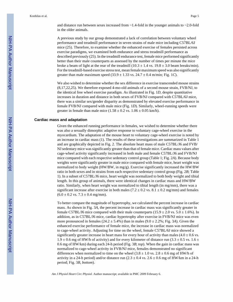
and distance run between sexes increased from ~1.4-fold in the younger animals to ~2.0-foldin the older animals.
A previous study by our group demonstrated a lack of correlation between voluntary wheelperformance and treadmill performance in seven strains of male mice including C57BL/6Jmice (25). Therefore, to examine whether the enhanced exercise of females persisted acrossexercise paradigms, we examined both endurance and stress treadmill performance asdescribed previously (25). In the treadmill endurance test, female mice performed significantlybetter than their male counterparts as assessed by the number of times per minute the micebroke a beam of light at the rear of the treadmill (10.3 ± 1.4 vs. 19.8 ± 3.0 beam breaks/min).For the treadmill-based exercise stress test, mean female maximum speed was also significantlygreater than male maximum speed (33.9 ± 1.33 vs. 24.7 ± 0.4 m/min; Fig. 1C).
We also wished to determine whether the sex difference in exercise transcended mouse strains(8,17,22,25). We therefore exposed 4-mo-old animals of a second mouse strain, FVB/NJ, tothe identical free wheel exercise paradigm. As illustrated in Fig. 1D, despite quantitativeincreases in duration and distance in both sexes of FVB/NJ compared with C57BL/6J mice,there was a similar sex/gender disparity as demonstrated by elevated exercise performance infemale FVB/NJ compared with male mice (Fig. 1D). Similarly, wheel-running speeds weregreater in female than male mice (1.58 ± 0.2 vs. 1.06 ± 0.05 km/h).
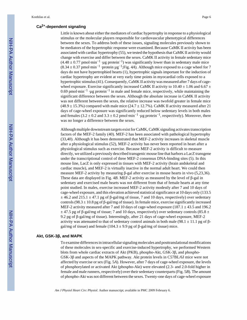
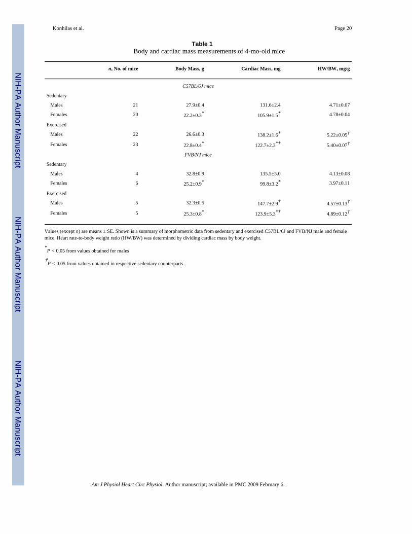
Cardiac mass and adaptationGiven the enhanced running performance in females, we wished to determine whether therewas also a sexually dimorphic adaptive response to voluntary cage-wheel exercise in themyocardium. The adaptation of the mouse heart to voluntary cage-wheel exercise is noted byan increase in cardiac mass (1). The results of these investigations are summarized in Table 1and are graphically depicted in Fig. 2. The absolute heart mass of male C57BL/J6 and FVB/NJ sedentary mice was significantly greater than that of female mice. Cardiac mass values aftercage-wheel activity significantly increased in both male and female C57BL/J6 and FVB/NJmice compared with each respective sedentary control group (Table 1; Fig. 2A). Because bodyweights were significantly greater in male mice compared with female mice, heart weight wasnormalized to body weight (HW/BW, in mg/g). Exercise significantly increased the HW/BWratio in both sexes and in strains from each respective sedentary control group (Fig. 2B; Table1). In a subset of C57BL/J6 mice, heart weight was normalized to both body weight and tibiallength. In this group of animals, there were identical changes in cardiac mass and HW/BWratio. Similarly, when heart weight was normalized to tibial length (in mg/mm), there was asignificant increase after exercise in both males (7.2 ± 0.2 vs. 8.1 ± 0.2 mg/mm) and females(6.0 ± 0.2 vs. 7.3 ± 0.4 mg/mm).
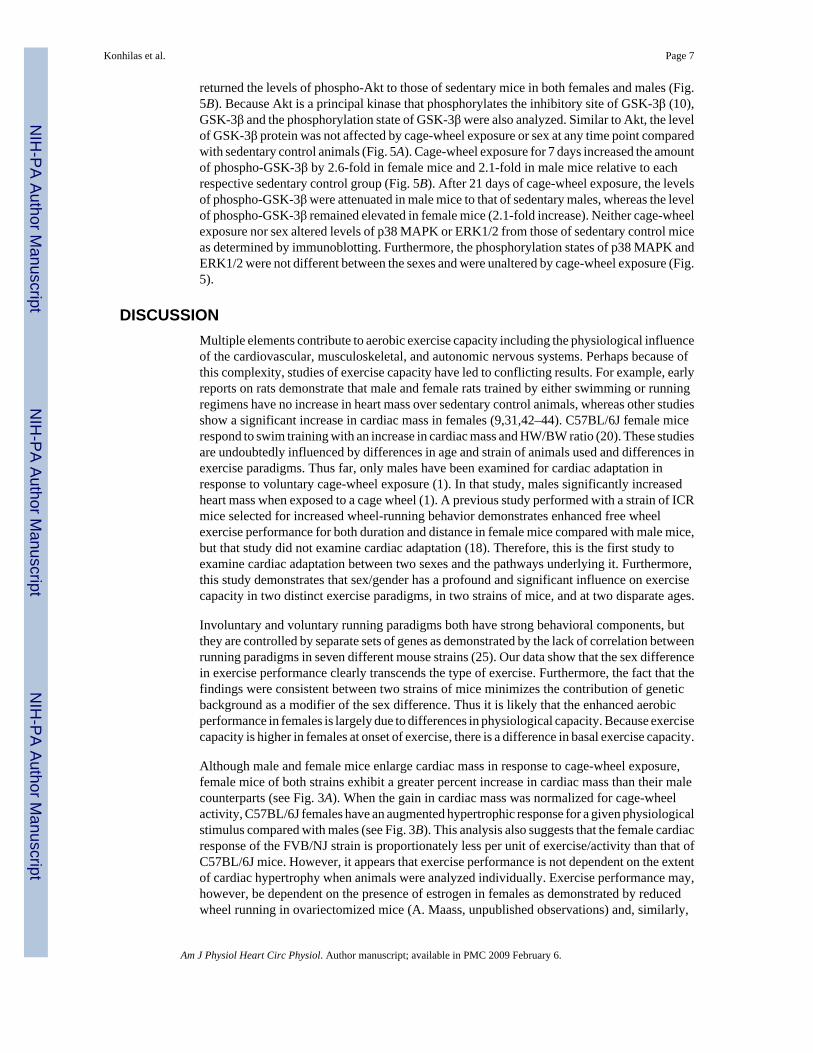
To better compare the magnitude of hypertrophy, we calculated the percent increase in cardiacmass. As shown in Fig. 3A, the percent increase in cardiac mass was significantly greater infemale C57BL/J6 mice compared with their male counterparts (15.9 ± 2.0 vs. 5.0 ± 1.6%). Inaddition, as in C57BL/J6 mice, cardiac hypertrophy after exercise in FVB/NJ mice was evenmore pronounced in females (24.2 ± 5.4%) than in males (9.0 ± 2.2%; Fig. 3A). Given theenhanced exercise performance of female mice, the increase in cardiac mass was normalizedto cage-wheel activity. Adjusting for time on the wheel, female C57BL/6J mice showed asignificantly greater increase in heart mass for every hour of activity than males (4.0 ± 0.6 vs.1.9 ± 0.6 mg of HW/h of activity) and for every kilometer of distance run (3.3 ± 0.5 vs. 1.6 ±0.6 mg of HW/km) during each 24-h period (Fig. 3B; top). When the gain in cardiac mass wasnormalized to cage-wheel activity in FVB/NJ mice, females demonstrated no significantdifferences when normalized to time on the wheel (3.8 ± 1.0 vs. 2.8 ± 0.6 mg of HW/h ofactivity in a 24-h period) and/or distance run (2.3 ± 0.4 vs. 2.6 ± 0.6 mg of HW/km in a 24-hperiod; Fig. 3B, bottom).
Konhilas et al. Page 5
Am J Physiol Heart Circ Physiol. Author manuscript; available in PMC 2009 February 6.
NIH
-PA Author Manuscript
NIH
-PA Author Manuscript
NIH
-PA Author Manuscript
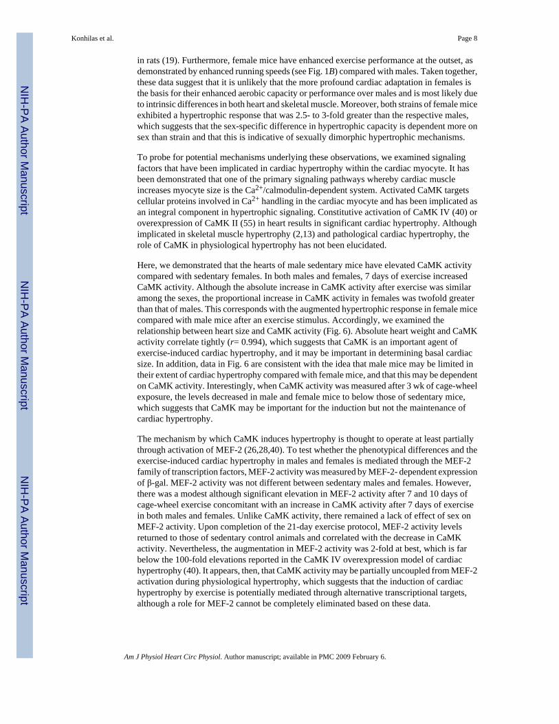
Ca2+-dependent signalingLittle is known about either the mediators of cardiac hypertrophy in response to a physiologicalstimulus or the molecular players responsible for cardiovascular phenotypical differencesbetween the sexes. To address both of these issues, signaling molecules previously shown tobe mediators of the hypertrophic response were examined. Because CaMK II activity has beenassociated with cardiac hypertrophy (55), we tested the hypothesis that CaMK II activity wouldchange with exercise and differ between the sexes. CaMK II activity in female sedentary mice(4.48 ± 0.77 pmol·min−1 ·µg protein−1) was significantly lower than in sedentary male mice(8.34 ± 0.37 pmol·min−1 ·protein µg−1;Fig. 4A). Although mice exposed to a cage wheel for 7days do not have hypertrophied hearts (1), hypertrophic signals important for the induction ofcardiac hypertrophy are evident at very early time points in myocardial cells exposed to ahypertrophic stimulus (41). Consequently, CaMK II activity was measured after 7 days of cage-wheel exposure. Exercise significantly increased CaMK II activity to 10.40 ± 1.06 and 6.67 ±0.69 pmol·min−1 ·µg protein−1 in male and female mice, respectively, while maintaining thesignificant difference between the sexes. Although the absolute increase in CaMK II activitywas not different between the sexes, the relative increase was twofold greater in female mice(48.9 ± 15.3%) compared with male mice (24.7 ± 12.7%). CaMK II activity measured after 21days of cage-wheel exposure was significantly reduced below sedentary levels in both malesand females (3.2 ± 0.2 and 3.3 ± 0.2 pmol·min−1 ·µg protein−1, respectively). Moreover, therewas no longer a difference between the sexes.
Although multiple downstream targets exist for CaMK, CaMK signaling activates transcriptionfactors of the MEF-2 family (40). MEF-2 has been associated with pathological hypertrophy(33,40). Although it has been demonstrated that MEF-2 activity increases in skeletal muscleafter a physiological stimulus (52), MEF-2 activity has never been reported in heart after aphysiological stimulus such as exercise. Because MEF-2 activity is difficult to measuredirectly, we utilized a previously described transgenic mouse line that harbors a LacZ transgeneunder the transcriptional control of three MEF-2 consensus DNA-binding sites (5). In thismouse line, LacZ is only expressed in tissues with MEF-2 activity (brain andskeletal andcardiac muscle), and MEF-2 is virtually inactive in the normal adult heart. We could thenmeasure MEF-2 activity by measuring β-gal after exercise in mouse hearts in vivo (5,23,36).These data are displayed in Fig. 4B. MEF-2 activity as measured by the level of β-gal insedentary and exercised male hearts was not different from that of female hearts at any timepoint studied. In males, exercise increased MEF-2 activity modestly after 7 and 10 days ofcage-wheel exposure, and this elevation achieved statistical significance at 10 days only (133.5± 46.2 and 215.1 ± 47.1 pg of β-gal/mg of tissue, 7 and 10 days, respectively) over sedentarycontrols (98.3 ± 10.8 pg of β-gal/mg of tissue). In female mice, exercise significantly increasedMEF-2 activity measured after 7 and 10 days of cage-wheel exposure (187.1 ± 43.5 and 196.2± 47.5 pg of β-gal/mg of tissue; 7 and 10 days, respectively) over sedentary controls (85.8 ±9.2 pg of β-gal/mg of tissue). Interestingly, after 21 days of cage-wheel exposure, MEF-2activity was attenuated to that of sedentary control animals in both male (98.1 ± 11.1 pg of β-gal/mg of tissue) and female (104.3 ± 9.9 pg of β-gal/mg of tissue) mice.
Akt, GSK-3β, and MAPKTo examine differences in intracellular signaling molecules and posttranslational modificationsof these molecules in sex-specific and exercise-induced hypertrophy, we performed Westernblots from whole cardiac extracts of Akt (PKB), phospho-Akt, GSK-3β, and phospho-GSK-3β and aspects of the MAPK pathway. Akt protein levels in C57BL/6J mice were notaffected by exercise or sex (Fig. 5A). However, after 7 days of cage-wheel exposure, the levelsof phosphorylated or activated Akt (phospho-Akt) were elevated (2.3- and 2.0-fold higher infemale and male runners, respectively) over their sedentary counterparts (Fig. 5B). The amountof phospho-Akt was not different between the sexes. Twenty-one days of cage-wheel exposure
Konhilas et al. Page 6
Am J Physiol Heart Circ Physiol. Author manuscript; available in PMC 2009 February 6.
NIH
-PA Author Manuscript
NIH
-PA Author Manuscript
NIH
-PA Author Manuscript
returned the levels of phospho-Akt to those of sedentary mice in both females and males (Fig.5B). Because Akt is a principal kinase that phosphorylates the inhibitory site of GSK-3β (10),GSK-3β and the phosphorylation state of GSK-3β were also analyzed. Similar to Akt, the levelof GSK-3β protein was not affected by cage-wheel exposure or sex at any time point comparedwith sedentary control animals (Fig. 5A). Cage-wheel exposure for 7 days increased the amountof phospho-GSK-3β by 2.6-fold in female mice and 2.1-fold in male mice relative to eachrespective sedentary control group (Fig. 5B). After 21 days of cage-wheel exposure, the levelsof phospho-GSK-3β were attenuated in male mice to that of sedentary males, whereas the levelof phospho-GSK-3β remained elevated in female mice (2.1-fold increase). Neither cage-wheelexposure nor sex altered levels of p38 MAPK or ERK1/2 from those of sedentary control miceas determined by immunoblotting. Furthermore, the phosphorylation states of p38 MAPK andERK1/2 were not different between the sexes and were unaltered by cage-wheel exposure (Fig.5).
DISCUSSIONMultiple elements contribute to aerobic exercise capacity including the physiological influenceof the cardiovascular, musculoskeletal, and autonomic nervous systems. Perhaps because ofthis complexity, studies of exercise capacity have led to conflicting results. For example, earlyreports on rats demonstrate that male and female rats trained by either swimming or runningregimens have no increase in heart mass over sedentary control animals, whereas other studiesshow a significant increase in cardiac mass in females (9,31,42–44). C57BL/6J female micerespond to swim training with an increase in cardiac mass and HW/BW ratio (20). These studiesare undoubtedly influenced by differences in age and strain of animals used and differences inexercise paradigms. Thus far, only males have been examined for cardiac adaptation inresponse to voluntary cage-wheel exposure (1). In that study, males significantly increasedheart mass when exposed to a cage wheel (1). A previous study performed with a strain of ICRmice selected for increased wheel-running behavior demonstrates enhanced free wheelexercise performance for both duration and distance in female mice compared with male mice,but that study did not examine cardiac adaptation (18). Therefore, this is the first study toexamine cardiac adaptation between two sexes and the pathways underlying it. Furthermore,this study demonstrates that sex/gender has a profound and significant influence on exercisecapacity in two distinct exercise paradigms, in two strains of mice, and at two disparate ages.
Involuntary and voluntary running paradigms both have strong behavioral components, butthey are controlled by separate sets of genes as demonstrated by the lack of correlation betweenrunning paradigms in seven different mouse strains (25). Our data show that the sex differencein exercise performance clearly transcends the type of exercise. Furthermore, the fact that thefindings were consistent between two strains of mice minimizes the contribution of geneticbackground as a modifier of the sex difference. Thus it is likely that the enhanced aerobicperformance in females is largely due to differences in physiological capacity. Because exercisecapacity is higher in females at onset of exercise, there is a difference in basal exercise capacity.
Although male and female mice enlarge cardiac mass in response to cage-wheel exposure,female mice of both strains exhibit a greater percent increase in cardiac mass than their malecounterparts (see Fig. 3A). When the gain in cardiac mass was normalized for cage-wheelactivity, C57BL/6J females have an augmented hypertrophic response for a given physiologicalstimulus compared with males (see Fig. 3B). This analysis also suggests that the female cardiacresponse of the FVB/NJ strain is proportionately less per unit of exercise/activity than that ofC57BL/6J mice. However, it appears that exercise performance is not dependent on the extentof cardiac hypertrophy when animals were analyzed individually. Exercise performance may,however, be dependent on the presence of estrogen in females as demonstrated by reducedwheel running in ovariectomized mice (A. Maass, unpublished observations) and, similarly,
Konhilas et al. Page 7
Am J Physiol Heart Circ Physiol. Author manuscript; available in PMC 2009 February 6.
NIH
-PA Author Manuscript
NIH
-PA Author Manuscript
NIH
-PA Author Manuscript
in rats (19). Furthermore, female mice have enhanced exercise performance at the outset, asdemonstrated by enhanced running speeds (see Fig. 1B) compared with males. Taken together,these data suggest that it is unlikely that the more profound cardiac adaptation in females isthe basis for their enhanced aerobic capacity or performance over males and is most likely dueto intrinsic differences in both heart and skeletal muscle. Moreover, both strains of female miceexhibited a hypertrophic response that was 2.5- to 3-fold greater than the respective males,which suggests that the sex-specific difference in hypertrophic capacity is dependent more onsex than strain and that this is indicative of sexually dimorphic hypertrophic mechanisms.
To probe for potential mechanisms underlying these observations, we examined signalingfactors that have been implicated in cardiac hypertrophy within the cardiac myocyte. It hasbeen demonstrated that one of the primary signaling pathways whereby cardiac muscleincreases myocyte size is the Ca2+/calmodulin-dependent system. Activated CaMK targetscellular proteins involved in Ca2+ handling in the cardiac myocyte and has been implicated asan integral component in hypertrophic signaling. Constitutive activation of CaMK IV (40) oroverexpression of CaMK II (55) in heart results in significant cardiac hypertrophy. Althoughimplicated in skeletal muscle hypertrophy (2,13) and pathological cardiac hypertrophy, therole of CaMK in physiological hypertrophy has not been elucidated.
Here, we demonstrated that the hearts of male sedentary mice have elevated CaMK activitycompared with sedentary females. In both males and females, 7 days of exercise increasedCaMK activity. Although the absolute increase in CaMK activity after exercise was similaramong the sexes, the proportional increase in CaMK activity in females was twofold greaterthan that of males. This corresponds with the augmented hypertrophic response in female micecompared with male mice after an exercise stimulus. Accordingly, we examined therelationship between heart size and CaMK activity (Fig. 6). Absolute heart weight and CaMKactivity correlate tightly (r= 0.994), which suggests that CaMK is an important agent ofexercise-induced cardiac hypertrophy, and it may be important in determining basal cardiacsize. In addition, data in Fig. 6 are consistent with the idea that male mice may be limited intheir extent of cardiac hypertrophy compared with female mice, and that this may be dependenton CaMK activity. Interestingly, when CaMK activity was measured after 3 wk of cage-wheelexposure, the levels decreased in male and female mice to below those of sedentary mice,which suggests that CaMK may be important for the induction but not the maintenance ofcardiac hypertrophy.
The mechanism by which CaMK induces hypertrophy is thought to operate at least partiallythrough activation of MEF-2 (26,28,40). To test whether the phenotypical differences and theexercise-induced cardiac hypertrophy in males and females is mediated through the MEF-2family of transcription factors, MEF-2 activity was measured by MEF-2- dependent expressionof β-gal. MEF-2 activity was not different between sedentary males and females. However,there was a modest although significant elevation in MEF-2 activity after 7 and 10 days ofcage-wheel exercise concomitant with an increase in CaMK activity after 7 days of exercisein both males and females. Unlike CaMK activity, there remained a lack of effect of sex onMEF-2 activity. Upon completion of the 21-day exercise protocol, MEF-2 activity levelsreturned to those of sedentary control animals and correlated with the decrease in CaMKactivity. Nevertheless, the augmentation in MEF-2 activity was 2-fold at best, which is farbelow the 100-fold elevations reported in the CaMK IV overexpression model of cardiachypertrophy (40). It appears, then, that CaMK activity may be partially uncoupled from MEF-2activation during physiological hypertrophy, which suggests that the induction of cardiachypertrophy by exercise is potentially mediated through alternative transcriptional targets,although a role for MEF-2 cannot be completely eliminated based on these data.
Konhilas et al. Page 8
Am J Physiol Heart Circ Physiol. Author manuscript; available in PMC 2009 February 6.
NIH
-PA Author Manuscript
NIH
-PA Author Manuscript
NIH
-PA Author Manuscript
It is likely, however, that signaling pathways in addition to CaMK are contributing tophysiological cardiac adaptation. Previous studies have highlighted the importance of theMAPKs as positive regulators of cardiac hypertrophy (for reviews, see Refs. 15 and 32).Several studies have examined the MAPK pathways in exercised skeletal muscles. Marathonrunning in men was shown to significantly increase phosphorylated ERK1/2 and p38 MAPKlevels (downstream effectors of MAPK signaling) in skeletal muscle (53). Studies thatexamined these pathways in rodent heart have shown that treadmill exercise induced activationof p38 MAPK and ERK1/2 (35), whereas swimming induced phosphorylation of Akt but notp38 MAPK or ERK1/2 (29). However, in the aforementioned studies, the mice that wereexercised by treadmill did not undergo cardiac hypertrophy, whereas mice that were subjectedto swim training had significant cardiac hypertrophy. Moreover, female mice were notexamined in either study. In the present study, cardiac hypertrophy induced by voluntary cage-wheel exposure led to no obvious alterations in these components of the MAPK pathway.
Growth factors and their downstream targets have also been implicated in cardiac hypertrophy(15,37,45,47). Phosphatidylinositol 3-kinases (PI3-kinases) are downstream of many receptortyrosine kinases to which growth factors bind. A downstream target of PI3-kinase is Akt (PKB).Expression of the catalytic subunit of PI3-kinase-α (P110α) in an activated form in heart resultsin increased phospho-Akt, significant hypertrophy, and normal contractility (48). Moreover,Akt may impart phenotypical differences between males and females (6). Here, although cage-wheel exposure increased Akt activity as measured by the increase in phospho-Akt, there wasno difference between male and female mice. However, sex differences in Akt activation maybe limited to the nuclear localization of phospho-Akt as demonstrated in a previous report(6); this distinction was not made in the present study. The present study, however, is consistentwith a role for Akt in physiological hypertrophy as indicated in swim-trained mice (29).
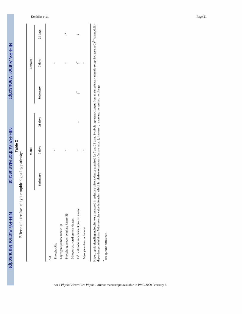
GSK-3β, a negative regulator of cardiac hypertrophy, is one of the downstream effectors ofAkt. It is inactivated through phosphorylation by Akt (3,4,34). As detailed (see Akt, GSK-3β,and MAPK), cage-wheel exposure for 7 days doubled the relative amount of the inactive(phosphorylated) form of GSK-3β in both male and female mice. Thus the antihypertrophiceffects of GSK-3β are relieved, and prohypertrophic pathways are promoted. Moreover, theinactivation of GSK-3β by exercise may be mediated through the activation of Akt. After 21days of cage-wheel exposure, phospho-GSK-3β levels remained elevated only in femalesdespite the attenuation of phospho-Akt levels to those of sedentary control animals. Themechanism of the sustained deactivation of GSK-3β was not addressed in this study.Nevertheless, these findings suggest two important sex dimorphisms as follows: 1)Aktactivation by phosphorylation can be uncoupled from GSK-3β inactivation by phosphorylationonly in females; 2) the prohypertrophic effects of phospho-GSK-3β persist after a prolongedphysiological stimulus only in females. Table 2 summarizes the data on the signaling factorsexamined in this study.
Here, we have identified potential targets, molecules, and pathways that may be important inimparting the phenotypical differences between male and female mice and in the sexuallydimorphic cardiac adaptation to exercise. The observed differences between the sexes may bepartially explained by disparities in the levels of certain hypertrophic signaling molecules. Itappears that the initial pathways may involve CaMK, Akt, and GSK-3β in both sexes and thatthe persistence of phospho-GSK-3β in females may be permissive to further cardiac growth.Clearly, multiple systems contribute to the aerobic exercise capacity of male and female mice,and numerous signaling pathways affect cardiac adaptation.
Konhilas et al. Page 9
Am J Physiol Heart Circ Physiol. Author manuscript; available in PMC 2009 February 6.
NIH
-PA Author Manuscript
NIH
-PA Author Manuscript
NIH
-PA Author Manuscript
ACKNOWLEDGMEMENTSPresent address of A. H. Maass: Department of Medicine, University of Wuerzburg, Josef-Schneider-Str. 2, 97080Wuerzburg, Germany.
GRANTS
This work was supported by National Institutes of Health (NIH) Grant HL-56510 (to L. A. Leinwand), DeutscheForschungsgemeinschaft Grant Ma2185/1-1 (to A. Maass), and by NIH National Research Service Awards F32HL-70509 (to J. P. Konhilas), F32 HL-72565 (to S. W. Luckey), and F32 HL-67543 (to B. L. Stauffer).
REFERENCES1. Allen DL, Harrison BC, Maass A, Bell ML, Byrnes WC, Leinwand LA. Cardiac and skeletal muscle
adaptations to voluntary wheel running in the mouse. J Appl Physiol 2001;90:1900–1908. [PubMed:11299284]
2. Antipenko A, Frias JA, Parra J, Cadefau JA, Cusso R. Effect of chronic electrostimulation of rabbitskeletal muscle on calmodulin level and protein kinase activity. Int J Biochem Cell Biol 1999;31:303–310. [PubMed: 10216962]
3. Antos CL, McKinsey TA, Frey N, Kutschke W, McAnally J, Shelton JM, Richardson JA, Hill JA,Olson EN. Activated glycogen synthase-3 beta suppresses cardiac hypertrophy in vivo. Proc Natl AcadSci USA 2002;99:907–912. [PubMed: 11782539]
4. Badorff C, Ruetten H, Mueller S, Stahmer M, Gehring D, Jung F, Ihling C, Zeiher AM, Dimmeler S.Fas receptor signaling inhibits glycogen synthase kinase 3 beta and induces cardiac hypertrophyfollowing pressure overload. J Clin Invest 2002;109:373–381. [PubMed: 11827997]
5. Black BL, Olson EN. Transcriptional control of muscle development by myocyte enhancer factor-2(MEF2) proteins. Annu Rev Cell Dev Biol 1998;14:167–196. [PubMed: 9891782]
6. Camper-Kirby D, Welch S, Walker A, Shiraishi I, Setchell KDR, Schaefer E, Kajstura J, Anversa P,Sussman MA. Myocardial Akt activation and gender: increased nuclear activity in females versusmales. Circ Res 2001;88:1020–1027. [PubMed: 11375271]
7. Carroll JD, Carroll EP, Feldman T, Ward DM, Lang RM, McGaughey D, Karp RB. Sex-associateddifferences in left ventricular function in aortic stenosis of the elderly. Circulation 1992;86:1099–1107.[PubMed: 1394918]
8. Chen J, Feller GM, Barbato JC, Periyasamy S, Xie ZJ, Koch LG, Shapiro JI, Britton SL. Cardiacperformance in inbred rat genetic models of low and high running capacity. J Physiol 2001;535:611–617. [PubMed: 11533149]
9. Crews J, Aldinger EE. Effect of chronic exercise on myocardial function. Am Heart J 1967;74:536–542. [PubMed: 4227758]
10. Cross DA, Alessi DR, Cohen P, Andjelkovich M, Hemmings BA. Inhibition of glycogen synthasekinase-3 by insulin mediated by protein kinase B. Nature 1995;378:785–789. [PubMed: 8524413]
11. de Simone G. Gender differences in left ventricular growth. Hypertension 1995;26:979–983.[PubMed: 7490158]
12. Dittrich H, Gilpin E, Nicod P, Cali G, Henning H, Ross J Jr. Acute myocardial infarction in women:influence of gender on mortality and prognostic variables. Am J Cardiol 1988;62:1–7. [PubMed:3381727]
13. Fluck M, Booth FW, Waxham MN. Skeletal muscle CaMK II enriches in nuclei and phosphorylatesmyogenic factor SRF at multiple sites. Biochem Biophys Res Commun 2000;270:488–494.[PubMed: 10753652]
14. Greenberg MA, Mueller HS. Why the excess mortality in women after PTCA? Circulation1993;87:1030–1032. [PubMed: 8443879]
15. Hefti MA, Harder BA, Eppenberger HM, Schaub MC. Signaling pathways in cardiac myocytehypertrophy. J Mol Cell Cardiol 1997;29:2873–2892. [PubMed: 9405163]
16. Higginbotham MB, Morris KG, Coleman RE, Cobb FR. Sex-related differences in the normal cardiacresponse to upright exercise. Circulation 1984;70:357–366. [PubMed: 6744540]
Konhilas et al. Page 10
Am J Physiol Heart Circ Physiol. Author manuscript; available in PMC 2009 February 6.
NIH
-PA Author Manuscript
NIH
-PA Author Manuscript
NIH
-PA Author Manuscript
17. Hoit BD, Kiatchoosakun S, Restivo J, Kirkpatrick D, Olszens K, Shao H, Pao YH, Nadeau JH.Naturally occurring variation in cardiovascular traits among inbred mouse strains. Genomics2002;79:679–685. [PubMed: 11991717]
18. Houle-Leroy P, Garland T Jr, Swallow JG, Guderley H. Effects of voluntary activity and geneticselection on muscle metabolic capacities in house mice Mus domesticus. J Appl Physiol2000;89:1608–1616. [PubMed: 11007602]
19. Kadi F, Karlsson C, Larsson B, Eriksson J, Larval M, Billig H, Jonsdottir IH. The effects of physicalactivity and estrogen treatment on rat fast and slow skeletal muscles following ovariectomy. J MuscleRes Cell Motil 2002;23:335–339. [PubMed: 12630708]
20. Kaplan ML, Cheslow Y, Vikstrom K, Malhotra A, Geenen DL, Nakouzi A, Leinwand LA, ButtrickPM. Cardiac adaptations to chronic exercise in mice. Am J Physiol Heart Circ Physiol1994;267:H1167–H1173.
21. Khan SS, Nessim S, Gray R, Czer LS, Chaux A, Matloff J. Increased mortality of women in coronaryartery bypass surgery: evidence for referral bias. Ann Intern Med 1990;112:561–567. [PubMed:2327676]
22. Koch LG, Britton SL, Barbato JC, Rodenbaugh DW, DiCarlo SE. Phenotypic differences incardiovascular regulation in inbred rat models of aerobic capacity. Physiol Genomics 1999;1:63–69.[PubMed: 11015562]
23. Kothary R, Clapoff S, Darling S, Perry MD, Moran LA, Rossant J. Inducible expression of an hsp68-lacZ hybrid gene in transgenic mice. Development 1989;105:707–714. [PubMed: 2557196]
24. Krumholz HM, Larson M, Levy D. Sex differences in cardiac adaptation to isolated systolichypertension. Am J Cardiol 1993;72:310–313. [PubMed: 8342510]
25. Lerman I, Harrison BC, Freeman K, Hewett TE, Allen DL, Robbins J, Leinwand LA. Geneticvariability in forced and voluntary endurance exercise performance in seven inbred mouse strains. JAppl Physiol 2002;92:2245–2255. [PubMed: 12015333]
26. Lu J, McKinsey TA, Nicol RL, Olson EN. Signal-dependent activation of the MEF2 transcriptionfactor by dissociation from histone deacetylases. Proc Natl Acad Sci USA 2000;97:4070–4075.[PubMed: 10737771]
27. Matsui T, Li L, Wu JC, Cook SA, Nagoshi T, Picard MH, Liao R, Rosenzweig A. Phenotypic spectrumcaused by transgenic overexpression of activated Akt in the heart. J Biol Chem 2002;277:22896–22901. [PubMed: 11943770]
28. McKinsey TA, Zhang CL, Olson EN. Activation of the myocyte enhancer factor-2 transcription factorby calcium/calmodulin-dependent protein kinase-stimulated binding of 14-3-3 to histone deacetylase5. Proc Natl Acad Sci USA 2000;97:14400–14405. [PubMed: 11114197]
29. McMullen JR, Shioi T, Zhang L, Tarnavski O, Sherwood MC, Kang PM, Izumo S. Phosphoinositide3-kinase(p110alpha) plays a critical role for the induction of physiological, but not pathological,cardiac hypertrophy. Proc Natl Acad Sci USA 2003;100:12355–12360. [PubMed: 14507992]
30. Miller, JM. Assays for β-galactosidase. In: Miller, JM., editor. Molecular Genetics. Cold SpringHarbor, NY: Cold Spring Harbor Laboratory Press; 1972. p. 352-355.
31. Mole PA. Increased contractile potential of papillary muscles from exercise-trained rat hearts. Am JPhysiol Heart Circ Physiol 1978;234:H421–H425.
32. Molkentin JD, Dorn GW 2nd. Cytoplasmic signaling pathways that regulate cardiac hypertrophy.Annu Rev Physiol 2001;63:391–426. [PubMed: 11181961]
33. Molkentin JD, Markham BE. Myocyte-specific enhancer-binding factor (MEF-2) regulates alpha-cardiac myosin heavy chain gene expression in vitro and in vivo. J Biol Chem 1993;268:19512–19520. [PubMed: 8366095]
34. Morisco C, Zebrowski D, Condorelli G, Tsichlis P, Vatner SF, Sadoshima J. The Akt-glycogensynthase kinase 3beta pathway regulates transcription of atrial natriuretic factor induced by beta-adrenergic receptor stimulation in cardiac myocytes. J Biol Chem 2000;275:14466–14475. [PubMed:10799529]
35. Nakamura A, Yoshida K, Takeda S, Dohi N, Ikeda S. Progression of dystrophic features and activationof mitogen-activated protein kinases and calcineurin by physical exercise, in hearts of mdx mice.FEBS Lett 2002;520:18–24. [PubMed: 12044863]
Konhilas et al. Page 11
Am J Physiol Heart Circ Physiol. Author manuscript; available in PMC 2009 February 6.
NIH
-PA Author Manuscript
NIH
-PA Author Manuscript
NIH
-PA Author Manuscript
36. Naya FJ, Wu C, Richardson JA, Overbeek P, Olson EN. Transcriptional activity of MEF2 duringmouse embryogenesis monitored with a MEF2-dependent transgene. Development 1999;126:2045–2052. [PubMed: 10207130]
37. Neri Serneri GG, Boddi M, Modesti PA, Cecioni I, Coppo M, Padeletti L, Michelucci A, Colella A,Galanti G. Increased cardiac sympathetic activity and insulin-like growth factor-I formation areassociated with physiological hypertrophy in athletes. Circ Res 2001;89:977–982. [PubMed:11717153]
38. O’Connell TD, Ishizaka S, Nakamura A, Swigart PM, Rodrigo MC, Simpson GL, Cotecchia S,Rokosh DG, Grossman W, Foster E, Simpson PC. The alpha(1A/C)- and alpha(1B)-adrenergicreceptors are required for physiological cardiac hypertrophy in the double-knockout mouse. J ClinInvest 2003;111:1783–1791. [PubMed: 12782680]
39. Olivetti G, Giordano G, Corradi D, Melissari M, Lagrasta C, Gambert SR, Anversa P. Genderdifferences and aging: effects on the human heart. J Am Coll Cardiol 1995;26:1068–1079. [PubMed:7560601]
40. Passier R, Zeng H, Frey N, Naya FJ, Nicol RL, McKinsey TA, Overbeek P, Richardson JA, GrantSR, Olson EN. CaM kinase signaling induces cardiac hypertrophy and activates the MEF2transcription factor in vivo. J Clin Invest 2000;105:1395–1406. [PubMed: 10811847]
41. Sadoshima J, Izumo S. The cellular and molecular response of cardiac myocytes to mechanical stress.Annu Rev Physiol 1997;59:551–571. [PubMed: 9074777]
42. Schaible TF, Penpargkul S, Scheuer J. Cardiac responses to exercise training in male and female rats.J Appl Physiol 1981;50:112–117. [PubMed: 6259097]
43. Schaible TF, Scheuer J. Cardiac function in hypertrophied hearts from chronically exercised femalerats. J Appl Physiol 1981;50:1140–1145. [PubMed: 6455402]
44. Schaible TF, Scheuer J. Effects of physical training by running or swimming on ventricularperformance of rat hearts. J Appl Physiol 1979;46:854–860. [PubMed: 457566]
45. Scheinowitz M, Kessler-Icekson G, Freimann S, Zimmermann R, Schaper W, Golomb E, Savion N,Eldar M. Short- and long-term swimming exercise training increases myocardial insulin-like growthfactor-I gene expression. Growth Horm IGF Res 2003;13:19–25. [PubMed: 12550078]
46. Semsarian C, Healey MJ, Fatkin D, Giewat M, Duffy C, Seidman CE, Seidman JG. A polymorphicmodifier gene alters the hypertrophic response in a murine model of familial hypertrophiccardiomyopathy. J Mol Cell Cardiol 2001;33:2055–2060. [PubMed: 11708849]
47. Serneri GG, Modesti PA, Boddi M, Cecioni I, Paniccia R, Coppo M, Galanti G, Simonetti I, VanniS, Papa L, Bandinelli B, Migliorini A, Modesti A, Maccherini M, Sani G, Toscano M. Cardiac growthfactors in human hypertrophy. Relations with myocardial contractility and wall stress. Circ Res1999;85:57–67. [PubMed: 10400911]
48. Shioi T, Kang PM, Douglas PS, Hampe J, Yballe CM, Lawitts J, Cantley LC, Izumo S. The conservedphosphoinositide 3-kinase pathway determines heart size in mice. EMBO J 2000;19:2537–2548.[PubMed: 10835352]
49. Sullivan MJ, Cobb FR, Higginbotham MB. Stroke volume increases by similar mechanisms duringupright exercise in normal men and women. Am J Cardiol 1991;67:1405–1412. [PubMed: 2042572]
50. Tamura T, Said S, Gerdes AM. Gender-related differences in myocyte remodeling in progression toheart failure. Hypertension 1999;33:676–680. [PubMed: 10024326]
51. Vriz O, Lu H, Visentin P, Nicolosi L, Mos L, Palatini P. Gender differences in the relationship betweenleft ventricular size and ambulatory blood pressure in borderline hypertension. The HARVESTStudy. Eur Heart J 1997;18:664–670. [PubMed: 9129899]
52. Wu H, Rothermel B, Kanatous S, Rosenberg P, Naya FJ, Shelton JM, Hutcheson KA, DiMaio JM,Olson EN, Bassel-Duby R, Williams RS. Activation of MEF2 by muscle activity is mediated througha calcineurin-dependent pathway. EMBO J 2001;20:6414–6423. [PubMed: 11707412]
53. Yu M, Blomstrand E, Chibalin AV, Krook A, Zierath JR. Marathon running increases ERK1/2 andp38 MAP kinase signalling to downstream targets in human skeletal muscle. J Physiol 2001;536:273–282. [PubMed: 11579175]
54. Zabalgoitia M, Ur Rahman SN, Haley WE, Oneschuk L, Yarrows S, Yunis C, Lucas C, Linn W,Krause L, Amerena J. Gender dimorphism in cardiac adaptation to hypertension is unveiled by priortreatment and efficacy. Am J Cardiol 1996;78:838–840. [PubMed: 8857496]
Konhilas et al. Page 12
Am J Physiol Heart Circ Physiol. Author manuscript; available in PMC 2009 February 6.
NIH
-PA Author Manuscript
NIH
-PA Author Manuscript
NIH
-PA Author Manuscript
55. Zhang T, Johnson EN, Gu Y, Morissette MR, Sah VP, Gigena MS, Belke DD, Dillmann WH, RogersTB, Schulman H, Ross J Jr, Brown JH. The cardiac-specific nuclear delta(B) isoform of Ca2+/cal-modulin-dependent protein kinase II induces hypertrophy and dilated cardiomyopathy associatedwith increased protein phosphatase 2A activity. J Biol Chem 2002;277:1261–1267. [PubMed:11694533]
Konhilas et al. Page 13
Am J Physiol Heart Circ Physiol. Author manuscript; available in PMC 2009 February 6.
NIH
-PA Author Manuscript
NIH
-PA Author Manuscript
NIH
-PA Author Manuscript
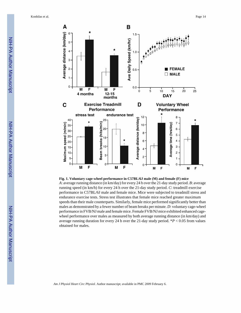
Fig. 1. Voluntary cage-wheel performance in C57BL/6J male (M) and female (F) miceA: average running distance (in km/day) for every 24 h over the 21-day study period. B: averagerunning speed (in km/h) for every 24 h over the 21-day study period. C: treadmill exerciseperformance in C57BL/6J male and female mice. Mice were subjected to treadmill stress andendurance exercise tests. Stress test illustrates that female mice reached greater maximumspeeds than their male counterparts. Similarly, female mice performed significantly better thanmales as demonstrated by a fewer number of beam breaks per minute. D: voluntary cage-wheelperformance in FVB/NJ male and female mice. Female FVB/NJ mice exhibited enhanced cage-wheel performance over males as measured by both average running distance (in km/day) andaverage running duration for every 24 h over the 21-day study period. *P < 0.05 from valuesobtained for males.
Konhilas et al. Page 14
Am J Physiol Heart Circ Physiol. Author manuscript; available in PMC 2009 February 6.
NIH
-PA Author Manuscript
NIH
-PA Author Manuscript
NIH
-PA Author Manuscript
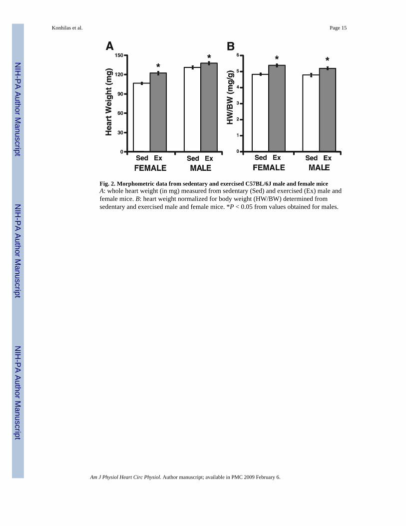
Fig. 2. Morphometric data from sedentary and exercised C57BL/6J male and female miceA: whole heart weight (in mg) measured from sedentary (Sed) and exercised (Ex) male andfemale mice. B: heart weight normalized for body weight (HW/BW) determined fromsedentary and exercised male and female mice. *P < 0.05 from values obtained for males.
Konhilas et al. Page 15
Am J Physiol Heart Circ Physiol. Author manuscript; available in PMC 2009 February 6.
NIH
-PA Author Manuscript
NIH
-PA Author Manuscript
NIH
-PA Author Manuscript
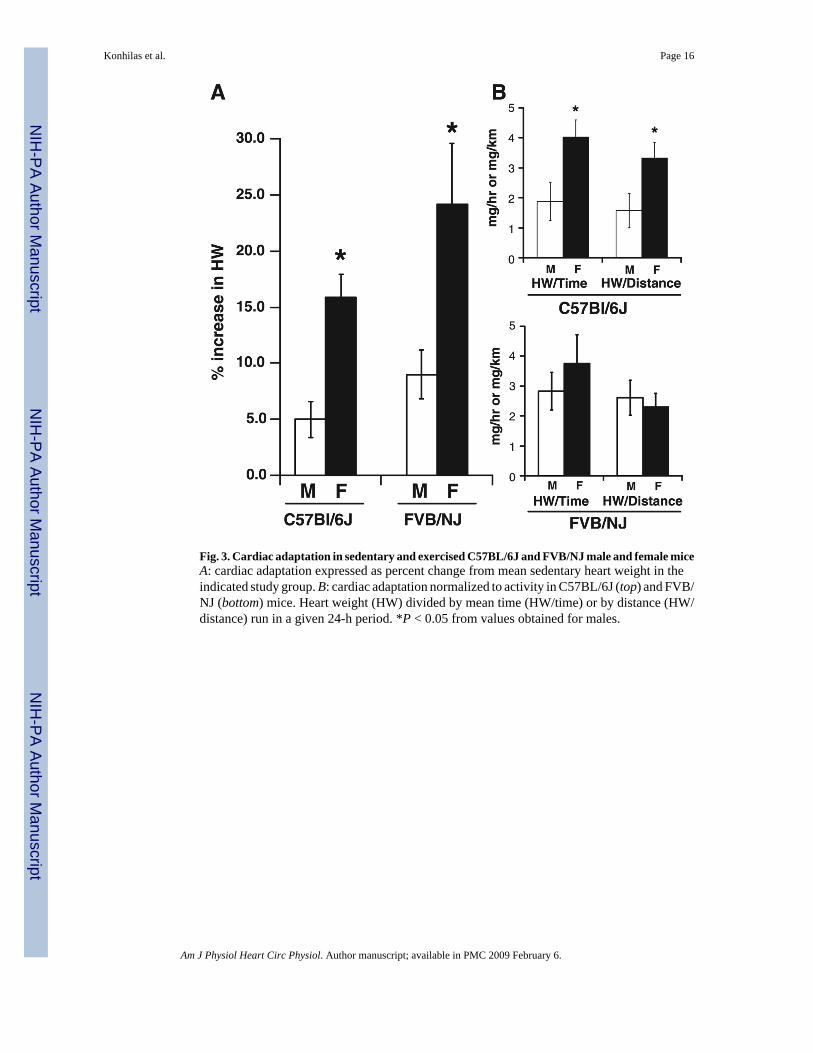
Fig. 3. Cardiac adaptation in sedentary and exercised C57BL/6J and FVB/NJ male and female miceA: cardiac adaptation expressed as percent change from mean sedentary heart weight in theindicated study group. B: cardiac adaptation normalized to activity in C57BL/6J (top) and FVB/NJ (bottom) mice. Heart weight (HW) divided by mean time (HW/time) or by distance (HW/distance) run in a given 24-h period. *P < 0.05 from values obtained for males.
Konhilas et al. Page 16
Am J Physiol Heart Circ Physiol. Author manuscript; available in PMC 2009 February 6.
NIH
-PA Author Manuscript
NIH
-PA Author Manuscript
NIH
-PA Author Manuscript
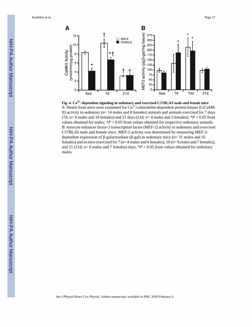
Fig. 4. Ca2+-dependent signaling in sedentary and exercised C57BL/6J male and female miceA: Hearts from mice were examined for Ca2+/calmodulin-dependent protein kinase II (CaMKII) activity in sedentary (n= 14 males and 8 females) animals and animals exercised for 7 days(7d; n= 8 males and 10 females) and 21 days (21d; n= 4 males and 3 females). *P < 0.05 fromvalues obtained for males; †P < 0.05 from values obtained for respective sedentary animals.B: myocyte enhancer factor-2 transcription factor (MEF-2) activity in sedentary and exercisedC57BL/6J male and female mice. MEF-2 activity was determined by measuring MEF-2-dependent expression of β-galactosidase (β-gal) in sedentary mice (n= 31 males and 35females) and in mice exercised for 7 (n= 8 males and 6 females), 10 (n= 8 males and 7 females),and 21 (21d; n= 8 males and 7 females) days. *P < 0.05 from values obtained for sedentarymales.
Konhilas et al. Page 17
Am J Physiol Heart Circ Physiol. Author manuscript; available in PMC 2009 February 6.
NIH
-PA Author Manuscript
NIH
-PA Author Manuscript
NIH
-PA Author Manuscript
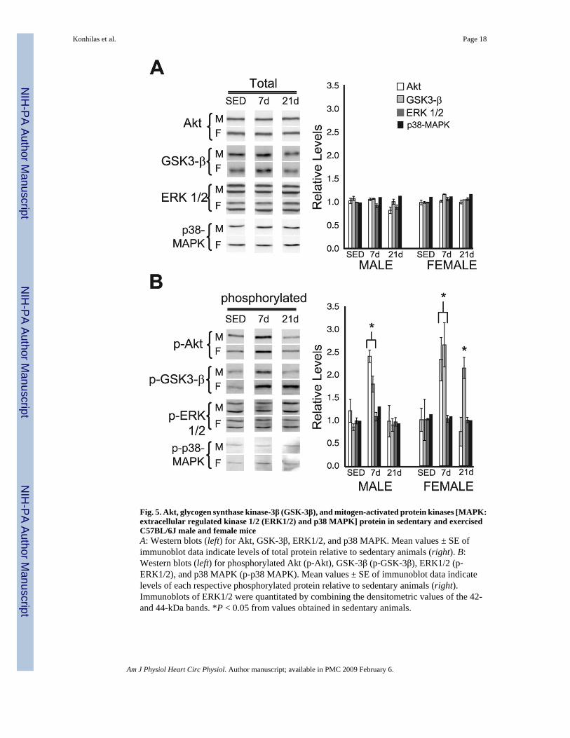
Fig. 5. Akt, glycogen synthase kinase-3β (GSK-3β), and mitogen-activated protein kinases [MAPK:extracellular regulated kinase 1/2 (ERK1/2) and p38 MAPK] protein in sedentary and exercisedC57BL/6J male and female miceA: Western blots (left) for Akt, GSK-3β, ERK1/2, and p38 MAPK. Mean values ± SE ofimmunoblot data indicate levels of total protein relative to sedentary animals (right). B:Western blots (left) for phosphorylated Akt (p-Akt), GSK-3β (p-GSK-3β), ERK1/2 (p-ERK1/2), and p38 MAPK (p-p38 MAPK). Mean values ± SE of immunoblot data indicatelevels of each respective phosphorylated protein relative to sedentary animals (right).Immunoblots of ERK1/2 were quantitated by combining the densitometric values of the 42-and 44-kDa bands. *P < 0.05 from values obtained in sedentary animals.
Konhilas et al. Page 18
Am J Physiol Heart Circ Physiol. Author manuscript; available in PMC 2009 February 6.
NIH
-PA Author Manuscript
NIH
-PA Author Manuscript
NIH
-PA Author Manuscript
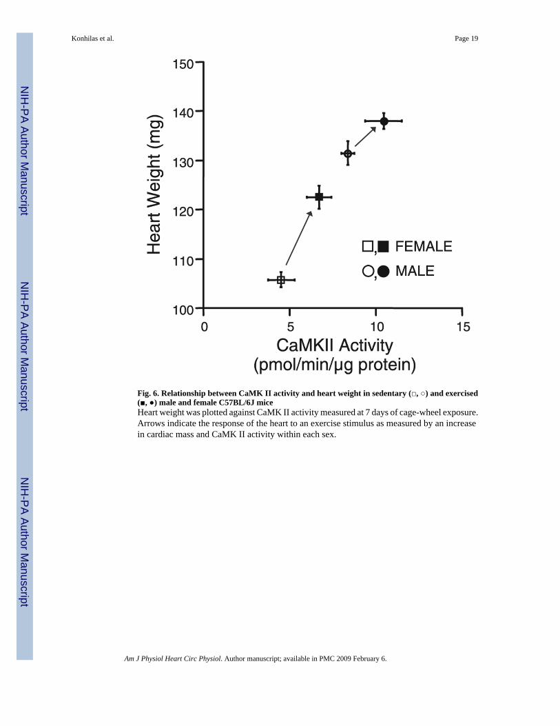
Fig. 6. Relationship between CaMK II activity and heart weight in sedentary (□, ○) and exercised(■, ●) male and female C57BL/6J miceHeart weight was plotted against CaMK II activity measured at 7 days of cage-wheel exposure.Arrows indicate the response of the heart to an exercise stimulus as measured by an increasein cardiac mass and CaMK II activity within each sex.
Konhilas et al. Page 19
Am J Physiol Heart Circ Physiol. Author manuscript; available in PMC 2009 February 6.
NIH
-PA Author Manuscript
NIH
-PA Author Manuscript
NIH
-PA Author Manuscript
NIH
-PA Author Manuscript
NIH
-PA Author Manuscript
NIH
-PA Author Manuscript
Konhilas et al. Page 20
Table 1Body and cardiac mass measurements of 4-mo-old mice
n, No. of mice Body Mass, g Cardiac Mass, mg HW/BW, mg/g
C57BL/6J mice
Sedentary
Males 21 27.9±0.4 131.6±2.4 4.71±0.07
Females 20 22.2±0.3* 105.9±1.5* 4.78±0.04
Exercised
Males 22 26.6±0.3 138.2±1.6† 5.22±0.05†
Females 23 22.8±0.4* 122.7±2.3*† 5.40±0.07†
FVB/NJ mice
Sedentary
Males 4 32.8±0.9 135.5±5.0 4.13±0.08
Females 6 25.2±0.9* 99.8±3.2* 3.97±0.11
Exercised
Males 5 32.3±0.5 147.7±2.9† 4.57±0.13†
Females 5 25.3±0.8* 123.9±5.3*† 4.89±0.12†
Values (except n) are means ± SE. Shown is a summary of morphometric data from sedentary and exercised C57BL/6J and FVB/NJ male and femalemice. Heart rate-to-body weight ratio (HW/BW) was determined by dividing cardiac mass by body weight.
*P < 0.05 from values obtained for males
†P < 0.05 from values obtained in respective sedentary counterparts.
Am J Physiol Heart Circ Physiol. Author manuscript; available in PMC 2009 February 6.
NIH
-PA Author Manuscript
NIH
-PA Author Manuscript
NIH
-PA Author Manuscript
Konhilas et al. Page 21Ta
ble
2Ef
fect
s of e
xerc
ise
on h
yper
troph
ic si
gnal
ing
path
way
s
Mal
esFe
mal
es
Sede
ntar
y7
days
21 d
ays
Sede
ntar
y7
days
21 d
ays
Akt
Pho
spho
-Akt
↑↑
Gly
coge
n sy
ntha
se k
inas
e-3β
Pho
spho
-gly
coge
n sy
ntha
se k
inas
e-3β
↑↑
↑*
Mito
gen-
activ
ated
pro
tein
kin
ases
Ca2+
cal
mod
ulin
-dep
ende
nt p
rote
in k
inas
e↑
↓↓*
↑*↓
Myo
cyte
enh
ance
r fac
tor-
2↑
↑
Hyp
ertro
phic
sign
alin
g m
olec
ules
wer
e m
easu
red
in se
dent
ary
mic
e an
d m
ice
exer
cise
d fo
r 7 a
nd 2
1 da
ys. S
ymbo
ls re
pres
ent c
hang
es fr
om m
ale
sede
ntar
y an
imal
s exc
ept i
ncre
ase
in C
a2+ /
calm
odul
in-
depe
nden
t pro
tein
kin
ase
7-da
y-ex
erci
se v
alue
in fe
mal
es, w
hich
is re
lativ
e to
sede
ntar
y fe
mal
e m
ice.
↑, i
ncre
ase;
↓, d
ecre
ase;
no
sym
bol,
no c
hang
e
* sex-
spec
ific
diff
eren
ce.
Am J Physiol Heart Circ Physiol. Author manuscript; available in PMC 2009 February 6.
Related Documents


