IOTC-2021-WPB19-19_Rev1 Sex identification of swordfish using a low cost genetic method Helary Louise 1 , Chevrier Thomas 1 , Roumagnac Marie 1 , Chanut Jérémie 2 , Nieblas Anne Elise 2 , Dominique Cowart 2 , Padron Mariana 1,3 , Brisset Blandine 1 , Evano Hugues 1 , Guiguen Yann 4 , Bernard Serge 5 , Kerzerho Vincent 5 , Rouyer Tristan 6 , Bonhommeau Sylvain 1 1 IFREMER, DOI, La Réunion, France 2 Company for Open Ocean Observations and Logging, La Réunion, France 3 Versailles, France 4 INRAE - BEAULIEU - Rennes, France 5 LIRMM/CNRS - Université de Montpellier, France 6 MARBEC/IFREMER, Sète, France Corresponding authors [email protected] [email protected] [email protected] Abstract Sex identification of animal species is a critical piece of information to derive parameters for population dynamic models. In the context of stock assessment (SA) for marine population, sex identification provides information about the sex-ratio of the population which is subsequently used to calculate the stock spawning biomass. In these SA models, sex-ratio can be set to a constant value throughout the lives of individuals (e.g. 0.5) or age-structured to account for changes linked to the physiology of individuals (e.g. females may live longer and represent a larger proportion of the population) or the selectivity of the fishery (e.g. a gender may be more accessible to the fishery at specific stages). The most common methods to identify sex are derived from direct observation of gonads. However, scientists must access the whole fish, which is rarely the case for large pelagic species in the IOTC fishery as fish that are landed have been gutted. Here we show the preliminary results of sex identification of swordfish (Xiphias gladius) using a genetic method: Amplified Fragment Length Polymorphism (AFLP). This polymerase chain reaction (PCR)- based genetic tool is a highly sensitive method for detecting polymorphisms in DNA. We tested the hypothesis that male and female swordfish present a genetic polymorphism linked to sex. Using a multiplexing technique (i.e. a combination of different restriction enzymes and PCR primers), we identified potential locations in the swordfish genome where polymorphism could occur. Comparing results between 3 males and 3 females of swordfish, we were able to identify 2 markers over 64 combinations of primers that led to potential sex-specific polymorphism identification. Higher sampling (40 males and 40 females) will be performed to confirm these results. This method has the advantage of being low cost, simple to develop, requires few genetic laboratory analysis or preparation (DNA extraction, PCR, electrophoresis and a capillary sequencer for DNA fragment analysis) and the genetic material required is minimal and can be taken from a living or dead animal.

Welcome message from author
This document is posted to help you gain knowledge. Please leave a comment to let me know what you think about it! Share it to your friends and learn new things together.
Transcript

IOTC-2021-WPB19-19_Rev1
Sex identification of swordfish using a low cost genetic method
Helary Louise1, Chevrier Thomas1, Roumagnac Marie1, Chanut Jérémie2, Nieblas Anne Elise2, Dominique
Cowart2, Padron Mariana1,3, Brisset Blandine1, Evano Hugues1, Guiguen Yann4, Bernard Serge5, Kerzerho
Vincent5, Rouyer Tristan6, Bonhommeau Sylvain1
1 IFREMER, DOI, La Réunion, France 2 Company for Open Ocean Observations and Logging, La Réunion, France 3 Versailles, France 4 INRAE - BEAULIEU - Rennes, France 5 LIRMM/CNRS - Université de Montpellier, France 6 MARBEC/IFREMER, Sète, France
Corresponding authors
Abstract Sex identification of animal species is a critical piece of information to derive parameters for population
dynamic models. In the context of stock assessment (SA) for marine population, sex identification provides
information about the sex-ratio of the population which is subsequently used to calculate the stock
spawning biomass. In these SA models, sex-ratio can be set to a constant value throughout the lives of
individuals (e.g. 0.5) or age-structured to account for changes linked to the physiology of individuals (e.g.
females may live longer and represent a larger proportion of the population) or the selectivity of the
fishery (e.g. a gender may be more accessible to the fishery at specific stages). The most common methods
to identify sex are derived from direct observation of gonads. However, scientists must access the whole
fish, which is rarely the case for large pelagic species in the IOTC fishery as fish that are landed have been
gutted. Here we show the preliminary results of sex identification of swordfish (Xiphias gladius) using a
genetic method: Amplified Fragment Length Polymorphism (AFLP). This polymerase chain reaction (PCR)-
based genetic tool is a highly sensitive method for detecting polymorphisms in DNA. We tested the
hypothesis that male and female swordfish present a genetic polymorphism linked to sex. Using a
multiplexing technique (i.e. a combination of different restriction enzymes and PCR primers), we identified
potential locations in the swordfish genome where polymorphism could occur. Comparing results
between 3 males and 3 females of swordfish, we were able to identify 2 markers over 64 combinations of
primers that led to potential sex-specific polymorphism identification. Higher sampling (40 males and 40
females) will be performed to confirm these results. This method has the advantage of being low cost,
simple to develop, requires few genetic laboratory analysis or preparation (DNA extraction, PCR,
electrophoresis and a capillary sequencer for DNA fragment analysis) and the genetic material required is
minimal and can be taken from a living or dead animal.

IOTC-2021-WPB19-19_Rev1
Abstract 1
Introduction 3
Material and Method 4
Biological samples 4
Genetic analyses 5
Data analyses 10
Results 10
Discussion 15
Appendix 18

IOTC-2021-WPB19-19_Rev1
Introduction
Sex is a fundamental biological parameter used in stock assessments. Sex determination and
differentiation in fishes are highly variable processes, with genetic, environmental (e.g. temperature, pH),
and even social factors having a strong influence on them (Devlin and Nagahama, 2002; Guerrero-Estévez
and Moreno-Mendoza, 2010). The ability to correctly identify sex is an important and sometimes complex
task, particularly for species that lack clear sexual dimorphism or sex-specific characters. Sex
determination is most of the time impossible at landings for IOTC fisheries as fish are gutted onboard for
preservation purposes.
For stock assessment modeling, sex is a critical information as it is used to estimate the population
sex-ratio. For an aged-structured model, sex-ratio combined with fecundity at age or stock-recruitment
relationship define the renewal capacity of the population. This source of information about sex has hence
critical impacts on the stock assessment projections.
The identification of sex-linked DNA markers is key genetic information allowing the development
of molecular sexing tools (Gamble and Zarkower, 2014; Lai et al., 2014; Robertson and Gemmell, 2006).
This approach is non-lethal and offers an opportunity to study endangered species, and/or to work in
situations (e.g. mark-recapture programmes and/or experiments) where the sex of living individuals needs
to be determined.
Even though most fish species do not possess heteromorphic sex chromosomes, genetic sex
determination has been found in a wide variety of fish species using alternative techniques to those based
on karyotyping. Using the Amplified Fragment Length Polymorphism (AFLP) technique, Agawa et al.,
(2014) identified one male-specific marker in the Pacific Bluefin tuna (Thunnus orientalis). Sex-linked AFLP
markers were also isolated from the three-spined stickleback Gasterosteus aculeatus (Griffiths et al.,
2000), and the rainbow trout Oncorhynchus mykiss (Felip et al., 2005). A sex-linked microsatellite marker

IOTC-2021-WPB19-19_Rev1
was identified in the nine-spined stickleback Pungitius pungitius (Shikano et al., 2011) and Chen et al.,
(2007), successfully isolated seven female-specific AFLP markers in the half-smooth tongue sole,
Cynoglossus semilaevis. However, the variability in the number and type of loci involved in fish sex
determination implies that sex-linked genetic markers are typically species- or population-specific.
Swordfish, Xiphias gladius, is one of the most widely distributed marine pelagic fish species
worldwide. It is a highly commercial species that constitutes an important fishery resource in the Pacific
and Indian Ocean, and it is managed internationally by the International Commission for the Conservation
of Atlantic Tunas (ICCAT), Indian Ocean Tuna Commission (IOTC), and the Inter-American Tropical Tuna
Commission (IATTC).
Sex determination markers have not been yet identified for this species, and swordfish individuals
do not exhibit morphological characters enabling to distinguish males from females, although females do
tend to be larger (Palko et al., 1981). Given its economic importance and in order to help elucidate genetic
sex determination mechanisms in the species, the aim of this study was to identify sex-specific markers in
X. gladius based on cDNA samples from known male and female individuals, using the Amplified Fragment
Length Polymorphism (AFLP) technique (Vos et al., 1995).
Material and Method
Biological samples
Swordfish individuals of various lengths (LJFL: 50 cm to 350 cm) were obtained from commercial
fisheries in the Indian Ocean (Table A1). A total of 6 individuals of known sex from 2 different locations
was used, 4 individuals (2 females named F5 and F7 and 2 males named M6 and M7) from the island of La
Réunion, and another 2 individuals (1 female named F6 and 1 male named M5) from the Gulf of Bengal.

IOTC-2021-WPB19-19_Rev1
Phenotypic sex was determined by examining gonadal morphology. Muscle tissues were collected and
preserved in Ethanol (95%), and stored at -20 °C.
Genetic analyses
● DNA extraction
DNA extraction from 30±2 mg of muscle was performed using a Phenol/Chloroform/Isoamyl alcohol (PCI)
method. It is based on a liquid-liquid DNA isolation and allows separating the nucleic acids (DNA and RNA)
to the proteins thanks to their different solubility. Before that, a chemical lysis was induced using a lysis
buffer (NaCl 120mM, Tris-HCl pH8 10 mM, EDTA 10 mM, Urée 4M and SDS 5%) for 3h at 60°C. It attacks
the lipid layers and membranes of the samples and initiate protein denaturation:
- NaCl prevents DNA denaturation.
- Tris-HCl plays a role in maintaining the pH of the solution and will permeabilize membrane cells.
- EDTA is a chelating agent that blocks enzymatic activities such as DNases.
- SDS is an anionic detergent that assists in the denaturation of protein membrane cells.
- Proteinase K is an enzyme (endopeptidase), which digests proteins, extracts nucleic acids (DNA
or RNA) and removes contaminants from the nucleic acid preparation. It inactivates DNases and RNases.
And finally, its activity is stimulated by denaturing agents such as SDS.
RNase (4 mg/mL, Qiagen) was then added 30 min at 37°C to destroy RNA. PCI was added and centrifuged
(14 000 rpm at 4°C, 5 min). The aqueous supernatant phase was recovered and chloroform was added
and centrifuged at the same conditions as before. This step was realized twice. To precipitate DNA,
absolute ethanol was incorporated. Another centrifugation was applied to the solution to put down all
nucleic acid and create a DNA pellet. The solution was eliminated and 70% ethanol was added to wash

IOTC-2021-WPB19-19_Rev1
the pellet. After removing the 70% ethanol and air-drying the pellet, DNA was eluted in buffer AE (Qiagen).
All steps of DNA extraction by the PCI method are shown in Figure 1.
Figure 1: DNA extraction using PCI method
DNA was quantified using Qubit™ dsDNA BR assay kit and quality ratio A260/280 was obtained by
using NanoDrop™Lite Spectrophotometer. All DNA samples were then diluted to 10 ng/µL.
● AFLP technique
AFLP analysis was carried out as described by Vos et al., 1995 with minor modifications. DNA
samples were digested with EcoRI and MseI restriction enzymes (Takara Bio Inc.). Restriction enzymes are
enzymes that cut, or digests, DNA at specific sites (restriction site). Recognition sequences in DNA differ

IOTC-2021-WPB19-19_Rev1
for each restriction enzyme, producing differences in length, sequence and strand orientation (5' end or
3' end). Here two restriction enzymes were used: a rare cutter (EcoRI) and a frequent one (MseI). The
sequences recognized by EcoRI and MseI are presented in Table 1.
Table 1 : Restriction enzymes and their restriction sites (arrows) according to DNA strand orientation.
DNA digestion was performed at the same time as adapter ligation. The EcoRI and MseI adapters
(Table 2) were ligated to the DNA fragments to generate template DNA for amplifications, using T4 DNA
ligase (Takara Bio Inc.) during 2 hours at 37°C.
Table 2 : Adaptators sequences
Adaptator’s name Sequence (5’ → 3’)
EcoRI-A1 CTC GTA GAC TGC GTA CC
EcoRI-A2 AAT TGG TAC GCA GTC TAC
MseI-A1 GAC GAT GAG TCC TGA G
MseI-A2 TAC TCA GGA CTC AT
Once the ligation was completed, two DNA amplifications were done, the pre-selective and the
selective amplification. In the pre-selective amplification step, adapter common primers were used (Table
3). Polymerase Chain Reaction (PCR) amplifications were performed for 20 cycles with the following cycle
profile: a 30s DNA denaturation step at 94 °C, a 30s annealing step at 56 °C and a 1 min extension step at

IOTC-2021-WPB19-19_Rev1
72 °C. Pre-selective amplification allows a few fragments to be selectively amplified.
Table 3: Primer sequences designed for AFLP
Selective amplification was performed using 64 different primers combinations from 8 EcoRI
primers and 8 MseI primers (table 3) with five-times diluted pre-selective PCR product as template.
Primers were the same as pre-selective primers but with 3 extra nucleotides to the 3’ ends to reduce the
number of fragments amplified by the DNA polymerase. EcoRI primers were labelled with one of the
fluorescent dyes Yakima Yellow™, DragonFly Orange™, ATTO 565 or 6-FAM (Table 3). PCR amplifications
were performed with the following cycle profile: 30s DNA denaturation step at 94 °C, a 30s annealing step
of variable temperature and a 2 min extension step at 72 °C. The annealing temperature in the first cycle
was 65 °C, and was subsequently reduced each cycle by 0.7 °C for the next 12 cycles. The profile was

IOTC-2021-WPB19-19_Rev1
continued with a 30 s DNA denaturation step at 94 °C, a 30s annealing step at 56 °C and a 2 min extension
step at 72 °C for the next 24 cycles. Fluorescently labelled fragments for each primer combination were
analysed on an ABI 3730XL DNA sequencer by capillary electrophoresis. The resulting genotyping data
were scanned in fsa files. All steps of AFLP techniques are shown in Figure 2.
Figure 2 : AFLP principle
● Electrophoresis
At the end of each amplification, a small DNA portion was put into a 2% agarose gel for
electrophoresis analysis to check if PCR was correctly achieved (Fig. 2). Gel electrophoresis is a method
used to separate macromolecules like DNA, RNA or proteins by applying an electric field to move the
molecules through an agarose matrix to the positive charge. DNA fragments, globally negatively charged,

IOTC-2021-WPB19-19_Rev1
migrate more or less far in the agarose gel matrix depending on their size. A DNA molecular weight marker
(SmartLadder, Eurogentec) was added in the agarose gel to identify migrated DNA fragments according
to their size. A DNA intercalant (Midori) was incorporated in the agarose gel in order to visualize the
different DNA bands by UV exposure once migration completed.
Data analyses
All the analyses were done with CRAN R (version 3.5.3) and the {Fragman} package (Covarrubias-
Pazaran et al., 2016). One of the most important things to perform was to import the correct ladder to
find the real peaks corresponding to the expected weights using the ladder.info.attach function. In our
case, it was GeneScan™ 600 LIZ™, all peaks for this ladder are presented in the appendix (Fig. A2). All
samples with a correlation for the ladder under 0.99 were removed.
Results
A total of 64 combinations of selective primers were tested resulting in thousands of scorable peaks across
individuals. Over these peaks, three were of interest to differentiate males and females. They are
presented below with the primers combination that allowed them to be obtained.
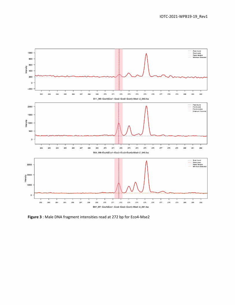
● Combination Eco4 - Mse2:
The first potential AFLP marker for X. gladius was observed with the Eco4-Mse2 primers
combination. Here, all samples have a correlation with ladder higher than 0.99. For the males M6 and M7
, a peak at 272 bp is observed with an intensity between 500 and 1000 (Fig. 3). A peak seems also present
at 272 bp for male M5 but it is not clear. On the other hand, for the 3 females this peak does not exist. A
peak is detected by the software for the female F7 but it is certainly due to the high threshold (Fig. 4).
IOTC-2021-WPB19-19_Rev1
Figure 3 : Male DNA fragment intensities read at 272 bp for Eco4-Mse2

IOTC-2021-WPB19-19_Rev1
Figure 4 : Female DNA fragment intensities read at 272 bp for Eco4-Mse2
● Combination Eco4 - Mse4:
Another potential AFLP marker can be observed using the Eco4-Mse4 combination. In this case,
sample F5 has a correlation of 0.9616 with the ladder whereas for M5 no correlation was found. Both of
them were removed for the marker's determination. For the two other male (M6 & M7) a peak with high

IOTC-2021-WPB19-19_Rev1
intensity between 2000 and 2600 is observed at 165bp (Fig. 5). This peak was observed for only one female
(F7) with a lower intensity around 536 which seems to be artefactual (Fig. 6).
Figure 5 : Male DNA fragment intensities read at 165 bp for Eco4-Mse4

IOTC-2021-WPB19-19_Rev1
Figure 6: Female DNA fragment intensities read at 165 bp for Eco4-Mse4

IOTC-2021-WPB19-19_Rev1
Discussion
The preliminary analyses for sex determination of swordfish using a genetic method (AFLP) are
very promising. We have identified 2 potential markers that could be used to easily identify the sex of
swordfish individuals. This work is a first step to investigate the potential application of AFLP for sex
identification of swordfish. We plan to extend the number of samples to 40 females and 40 males to
confirm the results and identify robust markers of sex.
The samples from the Bengal Gulf showed some issues in quality as the correlation between the
DNA fragment lengths and the reference ladder has a low correlation. This implies potential issues in DNA
extraction. This issue should be solved by increasing the sample size of good DNA quality.
When AFLP will be done on more samples and sex specific loci will be clearly identified, AFLP
fragments will be isolated and sequenced. As swordfish genome had been sequenced in February 2021
(scaffold assembly) (Wu et al., 2021) it would be possible to design specific primers in order to easily
determine sex of swordfish by using only one PCR.
If this genetic technique for sex identification can be validated, it can provide an efficient tool for
getting sex information about individuals at landings (while fish are gutted) or on live fish (e.g. tagging)
with genetic sex determination (GSD). This technique also has the advantage of being relatively low-cost
(~ 4,20 $ per sample from DNA extraction to electrophoresis) and to give rapid results (~ 2-3h from DNA
extraction to electrophoresis for 96 samples depending on laboratory equipment). This opens the
opportunity for large-scale sampling programs with low lab equipment requirements.

IOTC-2021-WPB19-19_Rev1
References
Agawa, Y., Iwaki, M., Komiya, T., Honryo, T., Tamura, K., Okada, T., Yagishita, N., Kobayashi, T.,
Sawada, Y., 2014. Identification of male sex-linked DNA sequence of the cultured Pacific bluefin tuna
Thunnus orientalis. Fish Sci 81, 113–121. doi:10.1371/journal.pgen.1002798
Chen, S.-L., Li, J., Deng, S.-P., Tian, Y.-S., Wang, Q.-Y., Zhuang, Z.-M., Sha, Z.-X., Xu, J.-Y., 2007. Isolation
of Female-Specific AFLP Markers and Molecular Identification of Genetic Sex in Half-Smooth Tongue
Sole (Cynoglossus semilaevis). Mar Biotechnol 9, 273–280. doi:10.1007/s10126-006-6081-x
Covarrubias-Pazaran G, Diaz-Garcia L, Schlautman B, Salazar W, Zalapa J. Fragman: An R package for
fragment analysis. 2016. BMC Genetics 17(62):1-8.
Devlin, R., Nagahama, Y., 2002. Sex determination and sex differentiation in fish: an overview of
genetic, physiological, and environmental influences. Aquaculture 191–364.
Felip, A., Young, W.P., Wheeler, P.A., Thorgaard, G.H., 2005. An AFLP-based approach for the
identification of sex-linked markers in rainbow trout (Oncorhynchus mykiss). Aquaculture 247, 35–43.
doi:10.1016/j.aquaculture.2005.02.014
Gamble, T., Zarkower, D., 2014. Identification of sex-specific molecular markers using restriction site-
associated DNA sequencing. Molecular Ecology Resources 477. doi:10.1093/bioinformatics/btr346
Griffiths, R., Orr, K., Adam, A., Barber, I., 2000. DNA sex identification in the three-spined stickleback.
J Fish Biology 57, 1331–1334. doi:10.1006/jfbi.2000.1386
Guerrero-Estévez, S., Moreno-Mendoza, N., 2010. Sexual determination and differentiation in teleost
fish. Rev Fish Biol Fisheries 20, 101–121. doi:10.1139/f76-122
Lai, X., Gao, H., Yan, B., Yan, W., Shen, S., 2014. Identification of sex-linked DNA markers by AFLP in
Portunus trituberculatus. Gene 544, 216–219. doi:10.1016/j.gene.2014.04.034
Morozova, O., & Marra, M. A. (2008). Applications of next-generation sequencing technologies in

IOTC-2021-WPB19-19_Rev1
functional genomics. Genomics, 92(5), 255–264. https://doi.org/10.1016/j.ygeno.2008.07.001
Palko, B.J., Beardsley, G.L., Richards, W.J., 1981. Synopsis of the biology of the swordfish, Xiphias
gladius Linnaeaus. NOAA Technical Report NMFS Circular 441 1–28.
Robertson, B.C., Gemmell, N.J., 2006. PCR-based sexing in conservation biology: Wrong answers from
an accurate methodology? Conserv Genet 7, 267–271. doi:10.1007/s10592-005-9105-6
Shikano, T., Herczeg, G., Merilä, J., 2011. Molecular sexing and population genetic inference using a
sex-linked microsatellite marker in the nine-spined stickleback (Pungitius pungitius). BMC Research
Notes 4, 1–6. doi:10.1186/1756-0500-4-119
Vos, P., Hogers, R., Bleeker, M., Reijans, M., van de Lee, T., Hornes, M., Frijters, A., Pot, J., Peleman,
J., Kuiper, M., Zabeau, M., 1995. AFLP: a new technique for DNA fingerprinting. Nucleic Acids Research
23, 4407–4414.
Wu, B., Feng, C., Zhu, C., Xu, W., Yuan, Y., Hu, M., ... & Wang, K. (2021). The Genomes of Two Billfishes
Provide Insights into the Evolution of Endothermy in Teleosts. Molecular biology and evolution, 38(6),
2413-2427.

IOTC-2021-WPB19-19_Rev1
Appendix
Table A1: Samples retained for AFLP
Name Sex Localisation
F5 Female RUN
F6 Female GBE
F7 Female RUN
M5 Male GBE
M6 Male RUN
M7 Male RUN
GBE : Gulf of Bengal, RUN : Réunion

IOTC-2021-WPB19-19_Rev1
Figure A2 : Electropherogram of the GeneScanTM - 600 LIZ Size Standard
Related Documents