Severe traumatic brain injury - clinical course and prognostic factors Maud Stenberg Department of Community Medicine and Rehabilitation, Rehabilitation Medicine, Umeå University, Umeå, Sweden. Umeå 2016

Welcome message from author
This document is posted to help you gain knowledge. Please leave a comment to let me know what you think about it! Share it to your friends and learn new things together.
Transcript
Severe traumatic brain injury -clinical course and prognostic factors Maud Stenberg
Department of Community Medicine and Rehabilitation, Rehabilitation Medicine, Umeå University, Umeå, Sweden. Umeå 2016

Responsible publisher under Swedish law: the Dean of the Medical Faculty This work is protected by the Swedish Copyright Legislation (Act 1960:729) ISBN: 978-91-7601-416-5 ISSN: 0346-6612 New series nr:1792 Omslagsbild: Saltoluokta, Ultevis Elektronisk version tillgänglig på http://umu.diva-portal.org/
Printed by: Print & Media, Umeå university SE-901 87 Umeå Sweden, 2016

To my beloved family ”Memories of our lives, of our works and our deeds will continue in others”. Words from a civil rights legend, Rosa Parks.
I

II

III

TABLE OF CONTENTS
ABSTRACT 3 ABBREVATIONS 5 SAMMANFATTNING PÅ SVENSKA 7 PREFACE 9 LIST OF ORIGINAL PAPERS 11 INTRODUCTION 12 Definition of traumatic brain injury (TBI) 12 Mechanism 12 Level of consciousness (LOC) 13 Structural damage (neuroimaging) 13 Epidemiology 14 Rehabilitation process after TBI 16 Care pathways 16 Care pathways, NHR in Sweden 16 Neurointensive care 17 Neurorehabilitation 17 Disorders of consciousness (DOC) after S-TBI 20 Cognitive Impairment after S-TBI 25 Global outcome after S-TBI 26 RATIONALE 27 AIMS OF THESIS 29 MATERIALS AND METHODS 30 Design 30 Patients 30 Data collection 31 PAPER I 33 PAPER II 34 PAPER III 35 PAPERS IV-V 36 INSTRUMENTS 38 Glasgow Coma Scale (GCS) 38 The Swedish Reaction Scale (RLS85) 39 JFK Coma Recovery Scale Revised (CRS-R) 40 The Barrow Neurological Institute Screen (BNIS) 41 The Hospital Anxiety and Depression Scale (HADS) 42 CT-findings 42 The Marshall CT classification 43 The Rotterdam CT Score 43 Outcome assessment 44
1
Glasgow outcome Scale Extended (GOSE) 45 Rancho Los Amigos Scale Revised (RLAS-R) 47 Acute prognostic model 49 Lund Concept 50 ICPMAX 51 STATISTICAL ANALYSES 51 ETHICAL CONSIDERATIONS 52 RESULTS 53 PAPERS I –V 55 DISCUSSION 73 Summery of the Thesis 90 Strengths and limitations 91 Conclusions 93 Future considerations 94 ACKNOWLEDGEMENTS 96 REFERENCES 99
2

ABSTRACT
Traumatic brain injury (TBI) constitutes a major health problem and is a leading cause of long-term disability and death. Patients with severe traumatic brain injury, S-TBI, comprise a heterogeneous group with varying complexity and prognosis. The primary aim of this thesis was to increase knowledge about clinical course and outcome with regard to prognostic factors. Papers I, II and III were based on data from a prospective multicentre observational study from six neurotrauma centers (NCs) in Sweden and Iceland of patients (n=103-114), 18-65 years with S-TBI requiring neurosurgical intensive care or collaborative care with a neurosurgeon (the “PROBRAIN” study). Paper IV and V were performed on a regional subset (n=37). In Paper I, patients with posttraumatic disorders of consciousness (DOC) were assessed as regards relationship between conscious state at 3 weeks and outcomes at 1 year. The number of patients who emerged from minimally conscious state (EMCS) 1 year after injury according to status at 3 weeks were: coma (0/6), unresponsive wakeful syndrome (UWS) (9/17), minimally conscious state (MCS) (13/13), anaesthetized (9/11). Outcome at 1 year was good (Glasgow Outcome Scale Extended (GOSE>4) in half of the patients in MCS (or anaesthetized) at 3 weeks, but not for any of the patients in coma or UWS. In Paper II, the relationships between clinical care descriptors and outcome at 1 year were assessed. A longer length of stay in intensive care, and longer time between discharge from intensive care and admission to inpatient rehabilitation, were both associated with a worse outcome on the GOSE. The number of intervening care units between intensive care and rehabilitation, was not significantly associated with outcome at 1 year. In Paper III, the clinical course of cognitive and emotional impairments as reflected in the Barrow Neurological Institute Screen for Higher Cerebral Functions (BNIS) and the Hospital Anxiety and Depression Scale (HADS) were assessed from 3 weeks to 1 year together with associations with outcomes GOSE and Rancho Los Amigos Cognitive Scale-Revised (RLAS-R) at 1 year. Cognition improved over time and appeared to be stable from 3 months to 1 year.
3
In Paper IV, clinical parameters, the clinical pathways from injury to 3 months after discharge from the NC in relation to outcomes 3 months post-injury. Ratings on the RLAS-R improved significantly over time. Eight patients had both “superior cognitive functioning” on the RLAS-R and “favourable outcome” on the GOSE. Acute transfers to the one regional NC was direct and swift, transfers for postacute rehabilitation scattered patients to many hospitals/hospital departments, not seldom by several transitional stages. In Paper V, an initial computerized tomography of the brain (CTi) and a further posttraumatic brain CT after 24 hours (CT24) were evaluated according to protocols for standardized assessment, the Marshall and Rotterdam classifications. The CT scores only correlated with clinical outcome measures (GOSE and RLAS-R) at 3 months, but failed to yield prognostic information regarding outcome at 1 year. A prognostic model was also implemented, based on acute data (CRASH model). This model predicted unfavourable outcomes for 81% of patients with bad outcome and for 85% of patients with favourable outcome according to GOSE at 1 year. When assessing outcomes per se, both GOSE and RLAS-R improved significantly from 3 months to 1 year. The papers in this study point both to the generally favourable outcomes that result from active and aggressive management of S-TBI, while also underscore our current lack of reliable instruments for outcome prediction. In the absence of an ability to select patients based on prognostication, the overall favourable prognosis lends support for providing active rehabilitation to all patients with S-TBI. The results of these studies should be considered in conjunction with the prognosis of long-term outcomes and the planning of rehabilitation and care pathways. The results demonstrate the importance of a combination of active, acute neurotrauma care and intensive specialized neurorehabilitation with follow-up for these severely injured patients.
Key words: Severe traumatic brain injury, outcome, rehabilitation, prognosis
4

ABBREVIATIONS
ATV all-terrain vehicle BAC blood alcohol concentration BNIS Barrow Neurological Institute Screen for Higher Cerebral Functions CI confidence interval CPP cerebral perfusion pressure CRASH Corticosteroid randomisation after significant head injury CRS-R Coma Recovery Scale Revised CT Computed tomography CTi Initial Computed tomography CT24 Computed tomography nearest 24 hours after trauma DAI Diffuse axonal injury DOC disorders of consciousness DT Datortomografi EDH epidural haematoma EMCS emerging from the minimally conscious state fMRI functional magnetic resonance imaging GCS Glasgow Coma Scale GOS Glasgow Outcome Scale GOSE Glasgow Outcome Scale Extended HADS Hospital Anxiety and Depression Scale ICF International Classification of Functioning, Disability and Health ICP Intracranial pressure IMPACT International Mission for Prognosis and Clinical Trial database of traumatic brain injury LOC Level of consciousness LOSIC Length of stay in intensive care LSS The law on support and service for certain people with disabilities MAP mean arterial blood pressure MCS minimally conscious state MRI magnetic resonance imaging NC Neurotrauma Center NHR Northern Health Region NMDA N-methyl-D-aspartate OR odds ratio PET positron emission tomography POCON Prospective Observational Cohort Neurotrauma study PTA Posttraumatic amnesia PT-DOC post-traumatic disorders of consciousness
5

PTV Persistent vegetative state RLS85 Swedish Reaction level scale RLAS-R Rancho Los Amigos Cognitive Scale Revised RPAP Rivermead Post-traumatic Amnesia Protocol SPSS Statistical Package for the Social Sciences SD standard deviation SDH Subdural haematoma TBI Traumatic brain injury S-TBI severe traumatic brain injury UWS unresponsive wakefulness syndrome VS vegetative state
6

SAMMANFATTNING PÅ SVENSKA Traumatisk hjärnskada (Traumatic brain injury/TBI) är en av de vanligaste orsakerna till funktionsnedsättning hos personer i arbetsför ålder. Skadan medför stort personligt lidande, drabbar även anhöriga, och innebär stora kostnader såväl för den enskilde som för samhället. Patienter med svår traumatisk hjärnskada (severe traumatic brain injury S-TBI), utgör en heterogen grupp med varierande komplexitet och prognos. Det primära målet med denna avhandling är att öka kunskapen om kliniskt förlopp och prognostiska faktorer vid S-TBI. Studierna I, II och III baseras på data från en prospektiv multicenter observationsstudie (PROBRAIN studien) från sex neurotraumacenter (NCs) i Sverige och Island. Inkluderade patienter (n=103-114) var i åldrarna 18-65 år, med svår traumatisk hjärnskada, som vårdats på neurointensivvårdsavdelning eller annan intensivvårdsavdelning i samråd med neurokirurg. Exklusionskriterier var patienter som avled inom 3 veckor efter skadan. Studierna IV och V baseras på en regional subpopulation i multicenterstudien PROBRAIN (n=37), och omfattar patienter från norra Sverige. I studie I var patienter med medvetandestörning vid 3 veckor upp till 1 år undersökta och jämförda beträffande medvetandegrad vid 3 månader och utfall vid 1 år. De patienter som förbättrades till bättre än minimalt medvetande tillstånd (”emerged from minimally conscious state”, EMCS) 1 år efter skadan jämfört med medvetandetillstånd vid 3 veckor var för koma: 0/6, för vegetativt tillstånd/icke-responsivt vakenhets tillstånd (unresponsive wakeful syndrome UWS) (9/17), för minimalt medvetande tillstånd (minimally conscious state MSC) (13/13), och för sederade/sövda patienter (9/11). Gott utfall vid 1 år på skalan (Glasgow Outcome Scale Extended GOSE>4) skattades för hälften av patienterna som bedömdes vara i ett minimalt medvetande tillstånd eller varit sövda vid 3 veckor men inte för de som bedömdes vara komatösa eller bedömdes vara i ett vegetativt/icke-responsivt vakenhetstillstånd. I studie II undersöktes relationen mellan vårdvägar, vårdtid inom neurointensivvård, tiden mellan neurointensivvård och intag på rehabiliteringsavdelning och utfall vid 1 år. Längre vårdtid vid neurointensivvård och längre tid mellan neurointensivvård och intag på vårdavdelning för rehabilitering var faktorer som var associerade med ett sämre utfall enligt GOSE. Antalet förflyttningar mellan olika vårdavdelningar under tiden mellan utskrivning från neurointensivvård och rehabilitering var inte signifikant associerat med utfall vid 1 år.
7

I studie III, undersöktes det kliniska förloppet av kognitiva och emotionella funktionsnedsättningar med Barrow Neurological Institute Screen for Higher Cerebral Functions (BNIS) och Hospital Anxiety and Depression Scale (HADS) från 3 veckor till 1 år jämfört med utfallet vid 1 år på GOSE och Ranchos Los Amigos Cognitive Scale-Revised (RLAS-R). Kognitiv funktionsnivå förbättrades över tid och föreföll vara stabil från 3 månader till 1 år. BNIS delskalor ”orientering” och ”visuospatial och visuell problemlösning” var associerade med GOSE och RLAS-R vid 1 år. I studie IV, studerades kliniska parametrar, vårdvägar från skadetillfället fram till 3 månader efter skadan för 37 patienter från Norra sjukvårdsregionen i Sverige i relation till utfall vid 3 månader efter skadan. Utfall enligt RLAS-R förbättrades signifikant över tid. Utmärkande var att akut transport till det enda neurotraumacentret i regionen fungerade väl emedan postakut förflyttning fram till rehabilitering kunde ske dels via olika sjukhus och ibland via olika avdelningar med olika vårdnivåer. I studie V undersöktes hjärnan med datortomografi (DT) initialt och efter 24 timmar enligt Marshall och Rotterdam DT klassifikationer i relation till utfall på GOSE och RLAS-R vid 3 månader och 1 år. Dessa klassifikationer för DT var bara relaterade till GOSE och RLAS-R vid 3 månader. GOSE och RLAS-R förbättrades signifikant från 3 månader till 1 år. En prognostisk modell baserad på akuta data (CRASH) predicerade dåligt utfall för 81% av patienter med dåligt utfall och för 85% av patienterna med gott utfall enligt GOSE vid 1 år. Sammanfattningsvis skattades gott utfall på GOSE 1 år efter S-TBI hos majoriteten av patienterna. Vid prognostisering av långtidsutfall, rehabiliteringsplanering och planering av vårdvägar bör resultaten från dessa studier beaktas. Resultaten pekar på vikten av en kombination av aktiv akut neurotraumavård och intensiv neurorehabilitering med uppföljningar av dessa svårt skadade patienter.
8

PREFACE For two decades, I have worked as a physician in rehabilitation medicine and neurorehabilitation at Umeå University hospital. My main interest has been rehabilitation after acquired brain injury. After some unexpectedly interesting and fundamentally instructive years in the early 1990s devoted to cognitive impairment at the Department of Psycho-geriatric Care at the Geriatric Center at Umeå University Hospital, I started my employment at the Center for Neurorehabilitation. Some of the patients at the psycho-geriatric department with dementia, progressive cognitive impairment and behavioural disorders had a previous history of severe traumatic brain injury (S-TBI), many years earlier. These patients had participated in rehabilitation programmes and improved but had then suffered a progression of cognitive disorders. Neurorehabilitation in Sweden offers specialized rehabilitation after spinal cord injury, acquired brain injury and for patients with neurological disease, primarily to patients of working age. Patients with acquired brain injury after trauma, stroke, infections, tumours, hypoxia/anoxia and metabolic causes are assessed and treated at center of neurorehabilitation. Umeå University Hospital provides specialized care to the Northern Health Region (NHR), a region that covers almost half the total area of Sweden (136,373 km2), with a total of 900,000 inhabitants. As the NHR comprises mainly rural districts with geographically large but sparsely populated areas, with long distances between hospitals, the clinical setting in this part of the country differs substantially from the more urbanised, southern half of the country. It is a challenge to offer equal care to persons in the NHR. Specialized rehabilitation in the NHR after brain injury is offered at three county hospitals in addition to the neurorehabilitation department at Umeå University Hospital. My aim as a physician over the years has been to focus on the importance of rehabilitation and especially on rehabilitation after acquired brain injury. There are areas for improvement in brain injury rehabilitation for county councils and regions but resources are limited. It is important to identify current conditions and compare with other brain injury rehabilitation departments in Sweden and abroad. The “PROBRAIN” study was an excellent chance for me to be part of a Swedish-Icelandic multicentre study of patients with S-TBI and therefore I devoted all my strength and time to pursuing and implementing this project. The aim of the multicentre study for S-TBI is to increase knowledge about clinical course and outcome with regard to prognostic factors. With knowledge from this survey of a patient group that is already well-known as heterogeneous, my
9

personal contribution was to have a basis for the further improvement of the rehabilitation of patients with S-TBI in clinical practice at our department and in the NHR. I also hoped that the studies could bring valuable knowledge of how to improve information to persons with S-TBI and their relatives for better planning of care pathways, use of resources and the evaluation of treatment effects. My goals for the future are studies which focus on rehabilitation and the long-term follow-up of patients with S-TBI and their relatives, if possible, from a lifetime perspective.
10

LIST OF ORIGINAL PAPERS
I. Godbolt AK, DeBoussard CN, Stenberg M, Lindgren M, Ulfarsson T, Borg J. Disorders of consciousness after severe traumatic brain injury: a Swedish-Icelandic study of incidence, outcomes and implications for optimizing care pathways. J Rehabil Med. 2013 Sep;45(8):741-8.
II. Godbolt AK, Stenberg M, Lindgren M, Ulfarsson T, Lannsjö M, Stålnacke BM. Borg J, DeBoussard CN. Associations between care pathways and outcome 1 year after severe traumatic brain injury. J Head Trauma Rehabil. 2015 May-Jun;30(3):E41-51.
III. Stenberg M, Godbolt AK, Nygren DeBoussard CN, Levi R, Stålnacke BM. Cognitive Impairment after Severe Traumatic Brain Injury, Clinical Course and Impact on Outcome: A Swedish-Icelandic Study. Behav Neurol. 2015;2015:680308.
IV. Stenberg M, Koskinen LO D, Levi R, Stålnacke BM. Severe traumatic brain injuries in Northern Sweden: a prospective 2-year study. J Rehabil Med. 2013 Sep;45(8):792-800.
V. Stenberg M, Koskinen LO D, Jonasson P, Levi R, Stålnacke
BM. Computed Tomography and clinical outcome in patients with severe traumatic brain injury. Submitted.
Papers I, II, III, IV are reprinted with kind permission from the publishers.
11
INTRODUCTION
Definition of traumatic brain injury (TBI)
Traumatic brain injury (TBI) occurs when “direct or indirect external
destructive, mechanical force causes brain dysfunction with impaired
consciousness. Typically focal change, coup and contrecoup injuries
includes contusion and hematoma formation whereas diffuse
microvascular change, occur over a more widespread area includes
diffuse axonal injury (DAI), and each includes multiple types of
subcellular, cellular and physiologic dysfunction. [1]. TBI can lead to a
broad range of temporary or permanent impairments of a cognitive,
physical or psychosocial nature. Pathology and severity of TBI can be
defined or classified in different ways: e.g. by i) mechanism, ii) level
of consciousness (LOC) or by iii) structural damage (neuroimaging).
Mechanism
If the skull remains intact after trauma, the head injury is described
as a “closed head injury”. If, by contrast, penetration of the skull
occurs, the head injury is described as being “open”. In most cases,
the brain remains enclosed in the skull cavity and any intracranial
volume expansion, for example due to haematoma or oedema, will
increase the intracranial pressure, thereby causing further brain
injury. Closed head injury is caused by rotational and/or
decelerational forces and resulting brain damage is categorized as
being focal or diffuse. Focal brain injury comprises hematoma and/or
contusions of different sizes, in one or several locations. Diffuse brain
injury, by contrast, is widespread, a result of microscopic damage,
typically in the subcortical white substance. Such damage may be
impossible to visualize by ordinary neuroimaging but may
nevertheless have disastrous consequences. Focal and diffuse
12
pathologies often coexist and contribute to morbidity [2]. Diffuse
changes also include diffuse axonal injury (DAI) caused by rapid
rotational movement, acceleration or deceleration force, which causes
axonal disruption, leading to impaired function and also to diffuse
microvascular damage with leakage of chemicals, further contributing
to brain damage [3,4,5,6]. More recent studies indicate more
generalized abnormalities after S-TBI, involving widespread neuro-
excitation and metabolic changes that ultimately may prove to be of
therapeutic importance [1].
Level of consciousness (LOC)
Level of consciousness (LOC), typically and historically assessed by
Glasgow Coma Scale score (GCS) on admission [7] is the most widely
used clinical instrument for assessment of severity of TBI. It consists
of the sum score 3-15 of three different responses by eye, motor and
verbal reaction and three different levels of sum scores describing
three different levels of severity. Lower sum score GCS 3-8 indicates
worst reaction: severe traumatic brain injury (S-TBI). The Reaction
Level Scale (RLS85) is another 8-level hierarchic scale of reaction and
this scale is widely used in Sweden [8]. RLS 4-8 assesses worst
responsiveness: the patient is unconscious and classified as S-TBI.
RLS scale can be translated to GCS score [9].
Structural damage (neuroimaging)
The third way to describe severity and pathology after TBI is by
structural damage and this can be assessed by neuroimaging;
computer tomography (CT) or magnetic resonance imaging (MRI).
Acute CT scan of the brain is the most commonly used neuroimaging
after TBI and it is used for acute survey and for deciding the further
planning of acute care. Different classification systems have been
13

developed in an attempt to predict outcome, for example Marshall
[10,85] and Rotterdam classification [11].
Epidemiology
Traumatic brain injury (TBI) is a global major health problem,
predicted year 2020 to be the third leading cause of death and
disability in the world [12]. Patients with TBI are a heterogeneous
population and their subsequent state can vary from death or severe
disability to full recovery. A study from Northern Sweden six to fifteen
years after TBI reported a high degree of motor and cognitive function
but also disability related to community reintegration and social
participation even several years after injury [13]. Primary preventions
such as seat belts, helmets and strict limitations for alcohol and drug
use for motor vehicle drivers have reduced the number of TBI.
However, the number of patients who survive S-TBI has increased
due to improved chains of acute care, acute transportation systems,
access to neurosurgery and modern neurointensive care. As a benefit
of improved acute neurosurgical care and improved survival rates,
there is an increased need for qualified neurorehabilitation [14].
Every year 15,000 – 20,000 persons in Sweden are hospitalized after
TBI [15,16]. In a previous study 74% of hospital days were less then
two days [16]. In a systematic review by Tagliaferri et al (2006) [17],
the incidence of TBI in Europe was estimated at about
235/100,000/year. In a study from northern Sweden (2007), the
incidence of TBI in all ages was reported as being even higher,
354/100,000/year and out of these, only 2% were classified as S-TBI
[18]. Despite its relative rarity, S-TBI with an incidence of 3-
12/100,000 per year [16,19] defined by acute Glasgow Coma Scale
(GCS), total score 3-8, is the most common cause of death and long-
14

term disability in Western countries for young people and those of
working age [20,21]. The mortality rate has been reported as 15-
17/100,000/year [17]. In a European multicentre study, mortality for
persons (>16 years) after six months with S-TBI was 40% [22] and in
the Prospective Observational Cohort Neurotrauma (POCON), a
study executed in 5 out of 11 specialized (Level I) trauma centres in
the Netherlands with mortality 46% in patients (16-72 years) with
S-TBI at 6-month post-injury follow-up [23]. TBI epidemiology and
injury patterns have changed but case fatality rates remain high. [23].
Falls and motor vehicle related injuries are the leading causes of TBI
[16,18,23,24]. Influence of alcohol or drugs at the time of injury is
clearly indicated for persons with TBI; some studies have shown an
incidence of 25-50% [17,25]. Injuries with S-TBI often involve great
personal suffering and a reduced quality of life for patients and their
relatives [26]. TBI also causes high societal costs [27]. Moreover, S-
TBI may be associated with a higher risk (4.5 times) of Alzheimer’s
disease or dementia in general in a lifetime perspective [28,29].
15

Rehabilitation process after TBI
Care pathways
There are multiple logistical challenges presented at the acute stage of
transfer of patients and rapid admission to Neuro trauma centers
(NCs) and later after discharge from the NC transfer to intensive care
if necessary or rehabilitation at a local hospital. It is also important to
study care pathways after neuro-intensive care all the way to the
person’s home after discharge or to suitable accommodation.
Admission to rehabilitation units and length of stay are usually
decided by rehabilitation physicians according to local criteria.
Previous studies have shown that delays between discharge from
intensive care and admission to a rehabilitation unit are negatively
associated with outcome one year after S-TBI [30]. Patients with
S-TBI require hospitalization with different levels of care from acute
care to a rehabilitation unit and are often discharged without a
planned continuous care pathway. Acute care and rehabilitation come
under different organizations which may affect treatment times and
coordination.
Care pathways, NHR in Sweden
Outcome studies after S-TBI are mainly focused on injury severity;
few studies have considered the effect of geographical factors [31].
The Northern Health Region (NHR) in Sweden which comprises
mainly rural districts is a geographically large area that covers almost
half of the total area of Sweden (136,373 km²). It differs substantially
from the more urbanized southern half of the country. In the NHR,
there are 900,000 inhabitants and a total of 13 hospitals (one of these
is a single neuro trauma center, NC) that are very far apart.
16

Emergency transport is in many cases carried out by helicopters to
the local hospital and then on to NC by helicopter, plane or car.
Neurointensive care
After admission to an NC with initial neurosurgery, neurointensive
care is required to avoid ongoing brain damage and prevent
secondary injuries after S-TBI such as brain swelling, increased
intracranial pressure (ICP) and intracranial haemorrhage, and to
provide the best conditions for the brain to recover after trauma.
Neurosurgery and neurointensive care, observation and treatment in
specialized neurotrauma centres are of importance. Secondary brain
injury associated with lack of oxygen and pathological processes
involving blood-brain barrier, oedema, release of chemicals factors
with cell injury or death and swelling which can affects cerebrospinal
circulation from the skull. The Lund concept is a modern protocol-
driven concept for volume regulation of the brain and an aggressive
neurointensive treatment after S-TBI, reducing brain swelling and
improving oxygenation of the damaged brain, keeping intracerebral
pressure (ICP) under control. [32,33]. After intensive care, admission
to a rehabilitation ward is to be expected but delays and time for
admission can differ, these patients can be dispersed among many
different wards, each; of which rehabilitated only one or a few
patients with very S-TBI per year [36].
Neurorehabilitation
The International Classification of Functioning, Disability and Health
(ICF) from 2001 is a classification of health and health-related
domains of a person in a context with environmental factors. It is the
WHO framework for measuring health and disability from an
individual and population perspective and it is very important in
17

neurorehabilitation. The ICF model shows five different components
where body function and body structures is one of them. Activity and
participation are separate. State of health is multifactorial and
everything affects each other as well as environmental and personal
factors. (Figure 1)
Figure 1.
There are available evidence that multidisciplinary specialized
rehabilitation (multidisciplinary teams in departments with a defined
responsibility for patients with S-TBI) programs for patients with
S-TBI have beneficial effects when applied early or late post-injury as
reported in recent reviews [34, 135]. A study from Southern Sweden
reported that early formalized rehabilitation and an effective chain of
medical and rehabilitation efforts resulted in shorter hospital stays
and a good outcome after S-TBI [35]. A study from Denmark reported
that centralized rehabilitation after S-TBI resulted in better
18

outcome compared with historical data from decentralized
rehabilitation [36]. In a Norwegian quasi-experimental study, one of
only a few prospective studies which compared two different
treatment approaches of patients (aged 16-55 years) with S-TBI
reported that patients who received early comprehensive
rehabilitation to stimulate neuronal reorganization and functional
recovery with a continuous chain of treatment showed better
functional outcomes 12 months post-injury then patients in ordinairy
rehabilitation programs [30]. Borg et al. [37] recently reported that
continued access to rehabilitation competencies after acute
management for S-TBI is not standard procedure in Sweden. Data
available from stroke studies have demonstrated recovery of function
and functional reorganization of brain networks [38] this should
principally be true also after TBI [37]. Several studies have reported
that brain plasticity is activity driven and recovery is probably greater
early after injury even though it can have some effect later post-injury
[39]. Early onset of rehabilitation refers to medical stability, give time
for spontaneous recovery with resolution of oedema, inflammatory
infiltrate and reduction of disruption to functional networks. It is also
of importance to minimize serious side effects such as pressure sores,
malnutrition, focal spasticity, or contractures and making use of
effective interventions such as for example beneficial effects of
amantadine in patients with DOC [52,53]. There are a number of
aspects to consider: the assessment of consciousness, awareness,
neurological and cognitive functioning, regular medical mapping,
radiological and neurophysiological conditions and treating disorders
if necessary such as hydrocephalus and epilepsy after S-TBI. There is
a need of rehabilitation programmes with specialized early
interventions; like the description in a Danish study according to
earlier recommendations like sensory stimulation, functional training
19

with guidance of movements in daily activity, early mobilization,
supported sitting and standing even for comatose patients and
inserting different rehabilitation interventions for different patients
in an appropriate chain [36]. There is substantial evidence to support
interventions for attention, memory, social communication skills,
executive function, and for comprehensive-holistic
neuropsychological rehabilitation after TBI [40]. There is some
evidence for multimodal rehabilitation for persons with severe
disorders of consciousness (DOC) [41].
Disorders of consciousness (DOC) after S-TBI
Medical care has improved greatly and the number of persons who
survive S-TBI has increased. Lives are saved. If the brain damage is
very severe, the patient can have different levels of “disorders of
consciousness” (DOC), initially a “coma state” and then recovery to a
“vegetative state” (VS). Although in some non-traumatic cases
patients may become in VS after a day or so, or without an initial
period of coma. Jennett B et al 1972 [42] called this state “persistent
vegetative state” (PVS) and after a month in this state the probability
to recovery diminishes [42]. VS as a syndrome in search of a name
have been described and named many times for example “the apallic
syndrome” [43] and as early as 1899 Rosenblath reported about a
young tightrope walker following a fall recovered after two weeks in
coma “to become strangely awake” [44]. Persistent vegetative state
(PVS) was recommended as the term of choice in the 1993 report of
the American Neurological Association [45] and in the 1994
statement of the Multi-Society Task Force [46]. ”Unresponsive
wakefulness syndrome” (UWS) is a new proposed term for persistent
vegetative state (VS) by Laureys et al 2010 [47] as changing the
pejorative image to a descriptive term that indicates clinical signs
20

such as unresponsiveness and wakefulness with eye opening. UWS is
the term that will be used instead of vegetative state (VS) continues.
William James in 1890 defined the term “consciousness” as patients
aware of themselves and the environment with two dimensions:
wakefulness and awareness. Wakefulness can be present without
awareness but awareness requires wakefulness [48]. The prevalence
of UWS or for patients with a better awareness a “minimally
conscious state” (MCS) [50] (See figure 2.) is not known because of a
lack of earlier accepted diagnostic criteria. There are no codes of DOC
in the International Classification of Diseases (ICD) 10th edition but
these codes will be added in ICD-11 beta [64], which will be of
importance for medical care planning in the future. For patients with
DOC, it is important to differentiate patients in MCS from patients in
UWS in order to offer specialized interventions and to plan further
rehabilitation. Some patients with S-TBI who are initially assessed as
being in a “coma state” do not survive more than about two-five
weeks without a respirator [49]. Patients in “coma state”do not open
their eyes and the best observation is some reflex movement of the
limbs. Problems following S-TBI vary. Some of the patients with
S-TBI could have a fast recovery, while others could remain in DOC
entering UWS or MCS. When long-term (>4 weeks) pronounced
disturbance of consciousness occurs, it is important to differentiate
different levels of unconsciousness and awareness with active
assessment as with JFK Coma Recovery Scale Revised (CRS-R) [82]
an instrument that was established in acute specialized
neurorehabilitation programmes. Patients assessed as being in a UWS
are characterised by independent breathing, periods of sleep and
wakefulness, giving spontaneous sound or movements and
being able to open their eyes but there is no evidence of awareness of
themselves or their environment or consciousness and they cannot
21

obey commands or make purposeful movements [48]. Recent studies
about functional neuroimaging and cognitive evoked potential studies
have shown new findings regarding awareness in some patients
without behavioural responses to command [47]. Patients who
improve to the MCS are able to follow simple prompts, gestures or
verbally mediated yes or no responses, simple verbalization but have
no functional communication and adequate affective behaviour to
presented stimuli, contingent crying or laughing. They are partially
conscious, localise noxious stimuli, can locate sound, can reach for
objects and automatic movement such as scratching [50].
Misdiagnosis of UWS may occur and in a study from 2009 from
Belgium, 41% of patients in UWS were found to be in MCS, when
standardized assessment instruments were used such as Coma
Recovery Scale Revised (CSR-R) [51]. However, most patients recover
completely after S-TBI.
Figure 2. Content of consciousness: Awareness and level of consciousness Wakefulness Laureys
[Laureys S. Eyes open, brain shut. Sci Am. 2007;296:84-9] Permission to print from the author.
There is strong evidence that active rehabilitation interventions and
more intensive rehabilitation programmes for patients with S-TBI
Awareness
Lucid dream-ing
REM- sleep
Conscious Wakefulness
»Locked-in syndrome« Drowsiness
Light sleep
Minimally Deep Sleep Conscious
State
Anesthesia Vegetative
Coma State
Wakefulness
22

who are already in rehabilitation are associated with better function
and that multidisciplinary rehabilitation affects outcome and there is
strong evidence for a milieu-oriented model for patients with S-TBI
[34]. Several studies have evaluated pharmacological treatment in
patients with S-TBI. To optimize awareness and response to stimuli in
patients with very S-TBI, a dopamine agonist and NMDA (N-methyl-
D-aspartate) antagonist (amantadine) combined with
interdisciplinary rehabilitation was used in a multicentre study. A
clear effect of the amantadine in speeding improvement was noted
without any side-effects. [52]. Amantadine is also considered to have
a neuroprotective effect early after brain injury [53]. Other
pharmacological treatments with the same purpose but without the
same level of evidence are bromocriptine [54,55] and zolpidem [56].
For agitation that could not be managed by interpersonal intervention
alone, antiepileptic drugs, especially carbamazepine, are
recommended [57,58]. Benzodiazepines is considered to inhibit
functional cerebral plasticity after brain injury [59]. A new area in
neurorehabilitation is the knowledge of reorganization in the adult
central nervous system after brain injury. Neuroplasticity can be
influenced by different, specifically directed, active rehabilitation
interventions [34,60]. This can be assessed by neuroimaging methods
such as functional magnetic resonance imaging (fMRI), diffusion
tensor imaging and positron emission tomography (PET).
Neurorehabilitation has the overall aim of an independent life as
possible through improving a person´s ability to cope from their own
perspective and family member´s goals with as full and independent
life as possible through increased activity and better possibilities for
participation. Successful rehabilitation has been determined on the
basis of the patient’s return to work. Mauriel Lezak summarizes in her
book “Neuropsychological Assessment” (seen as a kind of standard
23

work) that employment is important because it leads to life structure,
stability and gives the ability to live independently [61]. In contrast,
Kersel et al 2001 [62] reported that return to work reduces the
possibility of developing social contacts, increases isolation and
entails higher levels of depression. Moreover, McCrimmon et al 2005
[61] found that patients with moderate to severe TBI who had not
returned to work reported significantly higher levels of fatigue,
depression and self-reported symptoms in comparison with patients
who had returned to work. These different findings about factors
related to return to work can be seen in the perspective of state of
health in the ICF model. The Swedish and Icelandic insurance and
healthcare systems for patients with S-TBI aim to offer all patients
with S-TBI the medical care and rehabilitation needed when it is
medically indicated. In 2012, for the first time, the National Board of
Health and Welfare in Sweden [63] did a survey on the county and
regional rehabilitation for people with moderate TBI and S-TBI based
on a questionnaire to healthcare providers. This survey revealed a
number of areas for improvement. In several counties, there were no
guidelines for individuals with TBI, neither in terms of priority nor
who should be offered rehabilitation. There is a lack of care
programmes for rehabilitation after TBI and if there is a care
programme, it does not cover the entire continuum of care. County
Council directors are recommended to improve the management of
rehabilitation for persons with TBI. The Swedish National Board of
Health and Welfare stated that what determines whether a person is
entitled to rehabilitation is whether he or she can benefit from
rehabilitation and should not depend on whether the person is of
working age. In 1997, the National Board of Health in Denmark [36]
completed a review of the national state of rehabilitation for patients
with TBI. Health insurance in Sweden gives access to assessment and
24
rehabilitation for patients with disorder of consciousness (DOC) and
in December 2014, national recommendations were published in the
Swedish medical journal [64] on the request of the Swedish
Rehabilitation Physician Association. However, there are no national
guidelines for rehabilitation after S-TBI, and admission to
rehabilitation units and length of stay are usually decided by
rehabilitation physicians according to local criteria. Patients with S-
TBI have different problems and need different interventions and
combinations of interventions and they benefit from routine follow-
up so their needs for rehabilitation can be assessed [34].
Cognitive Impairment after S-TBI
Cognitive impairment is a common sequela of S-TBI. Most
commonly, cognitive deficits are disorders of memory, attention [65]
and speed of information-processing [66]. The demand for reliable
screening instruments has increased so as to enable decisions to be
made early in order to facilitate the further planning of care and
rehabilitation. Chapman et al 1959 [67] described that cognitive
impairment is not the only problem; frustration, inappropriate
affective reactions, lack of spontaneity and avoiding challenges is also
common. When affective disturbances are assessed, it is often done by
questionnaires or rating scales [68,69,70,71]. Both thinking and
feeling is important to maximize adaptive problem solving [72] as
well as self-awareness [73,74]. It is important to distinguish between
“mood” - a person’s subjective experience of feeling - and “affect”,
described as an external manifestation of an individual´s feelings,
thus physical and behavioural expression of mood [75]. Still there is a
demand for cognitive retraining after rehabilitation programmes but
many patients with S-TBI have emotional and motivational problems
which require a different type of rehabilitation. These personality
25
difficulties do mostly not correlate to the specific brain tissue damage
or level of severity of the TBI [76]. A patient with emotional distress
in the rehabilitation process is a factor to be aware of; this could
decrease with a holistic approach, intensive cognitive retraining and
psychotherapeutic intervention and possibly maximize, if necessary,
psychosocial recovery. An intensive programme for 6 hours a day, 5
days a week for 6 months showed that patients with self-awareness
and acceptance of their disability after S-TBI was the best match for
this program and that such patients need constant rehabilitation
attention [77,78].
Global outcome after S-TBI
Patients with S-TBI are heterogeneous with varying complexity and
prognosis, problems and outcome. In different studies, global
outcome like survival/death, Glasgow Outcome Scale (GOS) [79 ] or
Glasgow Outcome Scale Extended (GOSE) are used [80]. GOSE is an
extended version of GOS and allows a more finely tuned
categorization of post-traumatic disability. The Rancho Los Amigos
Cognitive Scale (RLAS-R) is a clinical outcome scale for assessing
cognitive improvement and recovery [81]. Parameters such as acute
care, post–acute complications, level of function and interventions on
neuroplasticity, which can be influenced by active rehabilitation [34]
all have the potential to impact on outcome. Environmental factors
and circumstances related to the patient are also important for
outcome. Different instruments are used to evaluate activity,
participation, sense of coherence, health-related quality of life, life
satisfaction and self-awareness. All these identifications, clinical
assessment, acute parameters, acute prognostic factors and outcome
are of importance for a knowledge bank that is relevant for the design
of appropriate rehabilitation programmes.
26

RATIONALE
In a recent Cochrane report about rehabilitation after S-TBI (2015),
the authors concluded it would be beneficial to have a routine follow-
up for the assessment of the needs for rehabilitation [34]. Problems
vary after injury and different interventions and combinations of
interventions are required. In the Cochrane report, there was strong
evidence for better function from formal interventions and for active
rehabilitation interventions with more intensive rehabilitation
programmes for patients with S-TBI (already in rehabilitation).
However, the context of multidisciplinary rehabilitation affected
outcome. Multidisciplinary neurorehabilitation facility has been
found to be more effective than rehabilitation in a nonspecialized
facility in earlier studies [34,145]. Limited evidence in the Cochrane
report indicated that early rehabilitation results in better outcome
and there was strong evidence for milieu-oriented rehabilitation for
patients with S-TBI and comprehensive cognitive interventions in a
therapeutic environment [34]. In a review from 2011 Cicerone et al
[40] reported that there is substantial evidence to support
inteventions for attention, memory, executive function, social
communication skills and for comprensive-holistic neuropsychologic
rehabilitation after TBI. There is some evidence for multimodal
rehabilitation for persons with severe disorders of consciousness
(DOC) [41]. Recommendations from a concensus conference 1999
[143] were that all patients with S-TBI and in need of systematic
assessment and rehabilitation should be offered this and with an early
onset. A Norweigian study [141] from 2016 indicated that clinical
pathways in wich specialized neurorehabilitation departments and
interventions according to evidence based recommendations and
guidelines for the management of S-TBI [144] may contribute to
27

enhance indepence in S-TBI patients [141]. Although this evidence
was shown, there are no national guidelines for rehabilitation after S-
TBI in Sweden. Admission to rehabilitation units and length of stay
are usually decided according to local criteria, different priority or
limited numbers of beds. There are no standards for care pathways
after acute care. It is therefore important to increase knowledge about
the clinical course and outcome of this heterogeneous group of
patients with S-TBI and a subgroup, namely, patients with disorders
of consciousness (DOC) with regard to acute prognostic factors and
care pathways.
This thesis could contribute to better knowledge about level of
function and progress of function at different points in time with
follow-ups up to 1 year.
28

AIMS OF THE THESIS
The overall aim of this thesis was to increase knowledge about the
clinical course and outcome in patients with S-TBI with regard to
prognostic factors.
The specific aims were:
Paper I: To assess the rates of disorder of consciousness at three
weeks, three months and one year after S-TBI, and to relate conscious
state three weeks after the injury to outcome at one year.
Paper II: To investigate prospectively the relationship between care
pathways for patients with S-TBI in the first year after the injury, and
outcome at one year.
Paper III: To assess the clinical course of cognitive and emotional
impairments in patients with S-TBI from three weeks to one year
after trauma and to study associations with outcomes at one year.
Paper IV: To evaluate the clinical characteristics, injury descriptors
and the care pathways from injury to three months after discharge in
patients with S-TBI in Northern Sweden and to assess outcomes at
three months post-injury.
Paper V: To investigate the relationships between CT scans as
assessed by the Marshall and Rotterdam protocols and clinical
outcomes at three months and one year post S-TBI and to evaluate
the prognostic value of the CRASH model.
29

MATERIALS AND METHODS
Design
This thesis includes prospective observational studies conducted in a
clinical setting with follow-up three months and one year after the
injury. The first three papers are multicentre prospective,
observational studies. Papers IV and V are population-based cohort
studies.
Patients
Patients in Papers I-III were from the Swedish-Icelandic, multicentre
study of patients with S-TBI, the “PROBRAIN” study, and included
patients from 6 of 7 neurotrauma centers (NCs). Papers IV and V
included patients from the Northern Health Region (NHR) treated at
the NC at Umeå University Hospital (included patients as part of the
“PROBRAIN” study). For a flowchart, see Figures 3-5.
Inclusion criteria were severe, non-penetrating, traumatic brain
injury, with a lowest non-sedated Glasgow Coma Score (GCS) [7] of
3–8 or Reaction Level Scale score (RLS85) [8] of 4–8 in the first 24
hours after injury, age at injury was 18–65 years, with an injury
requiring neurosurgical intensive care, or collaborative care with a
neurosurgeon in another intensive care unit. Exclusion criteria were
death or expected death within 3 weeks of injury. The participating
NCs provide neurosurgical care to more than 80% of the population
in Sweden and 100% in Iceland. The population of Sweden and
Iceland aged 18-65 years comprises ∼4.7 million persons (Papers I-
III) and for the NHR, 525000 persons (Papers III-V). Patients were
included from January 2010 to June 2011 in Paper I with extended
recruitment until December 2011 at 2 centres (Papers II-V).
30

Data collection
Patients were recruited after contact with NCs on a weekly basis to
identify eligible patients by rehabilitation physicians and then they
underwent prospective clinical assessment at 3 points in time: 3
weeks (18-24 days), 3 months (75-105 days), and 1 year (350-420
days) after injury. The patient gave informed consent in cases where
he or she had the capacity to do so. In the majority of cases, the
patient lacked the capacity and the patient’s nearest relative gave
consent to inclusion. When the patient improved and at all follow-up
occasions, patients gave a new mandate if they wanted to continue
participation. After inclusion, acute prognostic and socioeconomic
data were obtained from medical records. Additional background
socioeconomic data and medical history were collected through
interviews of relatives (if the patient was still unable to participate) as
soon as possible after inclusion. Patients were considered to have a
coexisting medical problem at the time of injury if any of the
following were present: hypertension, diabetes, cardiac disorder,
psychiatric disorder, renal failure, chronic obstructive airways
disease, other significant medical problem. Data on care pathways
were updated in conjunction with each follow-up to gather complete
care pathway data during the first year after injury, as far as possible.
Assessments took place in the patient’s current care setting if possible
(which in some cases was in the patient’s home) or in a local
outpatient department. Inclusion and follow-up were therefore
designed to be independent of any decisions regarding care pathways
and of any decision regarding admission to inpatient rehabilitation.
Assessments were performed by rehabilitation physicians with
assistance from rehabilitation nurses, psychologists, physiotherapists
and occupational therapists. Assessments at each of the 3 points in
time included both clinical examination and a battery of standardized
31

instruments, allowing description of the patient’s condition according
to the framework of the International Classification of Functioning,
Disability and Health (ICF): bodily structure and function, activities
and participation.
32

PAPER I
“Disorders of consciousness” (DOC) after S-TBI was assessed at three
weeks, three months and one year to relate conscious state three
weeks after injury to outcome at one year. The instruments relevant
to this sub-study included the JFK Coma Recovery Scale Revised
(CRS-R) [82], and the Glasgow Outcome Scale Extended (GOSE)
[80]. The JFK CRS-R was used for all patients where a DOC was
suspected on the basis of lack of functional communication and/or
functional object use, with the exception of patients who remained
sedated or anaesthetized. The CRASH prognostic model was used
(available at: http://www.crash2.lshtm.ac.uk/Risk%20
calculator/index.html) to calculate the percentage risk of an
unfavourable outcome (equivalent to GOSE 1–4) at 6 months, for
each patient, after conversion of RLS scores for those patients not
assessed with the GCS.
33

PAPER II
The care pathways and their relationship to outcome one year after S-
TBI were prospectively assessed with the presence or absence of
complications that were recorded at each point of time in the study.
Complications present three weeks after injury were considered in
relation to possible delays in transfer to rehabilitation and outcome.
The following possible complications were recorded: infection
(meningitis, sepsis, wound infection, urinary tract infection,
pneumonia, other stated infection), hydrocephalus, deep vein
thrombosis, pulmonary embolism, heterotopic ossification, new
fracture or new brain injury since the incident injury, other defined
complication. The presence of tracheostomy, ongoing artificial
ventilation and administration of oxygen three weeks after injury
were considered as surrogates for respiratory complications in terms
of difficulties in weaning from ventilation and/or persisting
respiratory difficulties and were therefore also coded as representing
complications. Bad outcome was assessed as GOSE 2-4 for patients
alive and followed up 1 year after injury.
34

PAPER III In this study, the clinical course of cognitive and emotional
impairments in patients with S-TBI from three weeks to one year
after trauma was assessed at three points in time and related to
outcomes at one year. The data regarding education and earlier
cognitive problems were obtained by interviews with patients and/or
significant others. Patients were interviewed and administered the
Barrow Neurological Institute Screen for Higher Cerebral Functions
(BNIS) [68,89,90] for assessment of cognitive function, either by a
clinical neuropsychologist or a physician who was a specialist in
rehabilitation medicine. Pre-screening was performed initially to
evaluate whether it was meaningful to attempt further testing. The
BNIS was assessed at 3 weeks, 3 months, and 1 year after injury. The
Hospital Anxiety and Depression Scale (HADS) [94] self-reporting
instrument was used for screening of depression and anxiety. The
HADS was assessed at 3 weeks, 3 months, and 1 year after injury.
Outcome variables were GOSE [80] at 1 year and RLAS-R [81] at 3
weeks, 3 months, and 1 year. GOSE 1-4 was assessed as unfavourable
and inferior function as RLAS-R 1-8.
35

PAPERS IV-V
Enrolled patients were treated at the NC at Umeå University Hospital
NHR according to the “Lund” concept, which is standard protocol at
this center [32]. For details, see [83,84]. The primary hospital
performed an initial computed tomography (CT) scan of the brain.
This investigation was often repeated upon arrival to the NC. Pictures
were transferred electronically to the NC where a neuro-radiologist
assessed the images (Papers IV-V).
In Paper IV, the clinical characteristics and injury descriptors of
patients with S-TBI from the NHR were assessed together with care
pathways from injury to three months after discharge and compared
with outcomes at three months. The first CT scans were classified
according to CRASH protocol and the Marshall [10,85] classification.
Outcomes were assessed by GOSE [80] at 3 months after injury and
RLAS-R [81] at 3 weeks and 3 months. GOSE 1-6 was assessed as
unfavourable outcome and inferior functioning as RLAS-R I-VIII.
In Paper V, prospectively a senior neuro-radiologist (PJ), and a senior
neuro-rehabilitationist (MS) assessed the first CT scan and
subsequent CT scan nearest twenty-four hours after trauma according
to the Marshall [10,85] and Rotterdam classification [11]. The
relationships between CT scans assessed by the Marshall and
Rotterdam protocols and clinical outcomes were investigated at three
months and one year post injury on the GOSE and RLAS-R. The
CRASH acute prognostic model [101] was used to predict the risk of
unfavourable outcome at six months (used in Papers I-II). GOSE 1-4
was assessed as unfavourable outcome and inferior outcome as RLAS-
R 1-6.
36

All the gathering of clinical outcome data was performed by one of the
authors (MS) through patient assessment at 3 weeks and 3 months
and 1 year post-injury. Socio-demographic data and data regarding
pre-morbid health were gathered by interviews with patients and/or
significant others, also performed by MS. Data regarding injury
characteristics and length of stay at the NC were retrieved from the
medical records.
37

INSTRUMENTS
Table 1. Overview of instruments.
Study I Study II Study III Study IV Study V Glasgow Outcome Scale Extended, GOSE
x x x
x
x
Rancho Los Amigos Scale of cognitive functioning-revised, RLAS-R
x x x
Coma Recovery Scale revised, CRS-R
x
CRASH acute prognostic model
x x x
The Barrow Neurological Institute Screen for higher cerebral functions BNIS
x
The Hospital Anxiety and Depression Scale HADS
x
The Marshall CT classification
x x
The Rotterdam CT classification
x
Glasgow Coma Scale (GCS) [7] is the most widely used scale for
assessing patients’ responses at admission and consists of eye-
opening, motor and verbal responses. It is of importance to assess
patient without sedation or intubation and note if they are under the
influence of drugs or alcohol. Acute intensive care starts at the place
of the accident and even before emergency transportation; patients
are therefore often sedated at admission and a new assessment of
LOC is necessary. GCS have different scores for different responses
and consist of a sum score of 3 to 15. Higher scores indicate better
responses. GCS scores of 13 to 15 correspond to mild TBI, GCS scores
of 9 to 12 moderate TBI, and GCS scores of 3 to 8 S-TBI. See Table 2.
The incidence of TBI severity are as follows: mild/moderate/severe;
22:1.5:1.1 [17].
38

The Swedish Reaction Level Scale (RLS85) [8] is another
classification of LOC. This scale is an 8-point hierarchic scale where
scores of 4 to 8 indicate worst responsiveness, corresponding to S-
TBI. At some NCs in Sweden, RLS is the most commonly used scale.
RLS can be converted to GCS in order to be compared in worldwide
studies. Conversion studies of these scales have been carried out,
thus, RLS 8 = GCS 3, RLS 7 = GCS 4, RLS 6 = GCS 5, RLS 5 = GCS 6,
RLS 4 = GCS 7 [9,86]. In Paper I-V, GCS is used. See Table 3.
Table 2. Glasgow Coma Scale (GCS) Eye response Open spontaneously 4
Open to verbal command 3
Open in response to pain 2
No response 1 Verbal response
Talking/Orientated
5
Confused speech/Disorientated 4
Inappropriate words 3
Incomprehensible sounds 2
No response 1
Motor response Obeys commands 6
Localizes to pain 5
Flexion/withdrawal 4
Abnormal flexion 3
Extension 2
No response 1
TOTAL SCORE 3-15
Teasdale G. & Jennett B. Assessment of coma and impaired consciousness. A practical scale. Lancet 1974;2:81-84.
39

Table 3. Reaction Level Scale (RLS85) Clinical descriptor Responsiveness Score Alert No delay in response 1
Drowsy or confused Responsive to light stimulation 2
Very drowsy or confused Responsive to strong stimulation 3
Unconscious Localizes but does not ward off pain 4
Unconscious Withdrawing movements on pain stimulation 5
Unconscious Stereotype flexion movements on pain stimulation 6
Unconscious Stereotype extension movements on pain stimulation 7
Unconscious No response on pain stimulation 8 Starmark JE, Stålhammar D, Holmgren E. The Reaction Level Scale (RLS85). Manual and guidelines. Acta Neurochir (Wien). 1988;91(1-2):12-20.
JFK Coma Recovery Scale –Revised, (CRS-R) is an instrument
used to assess DOC [82]. This instrument was first described by
Giacino and colleagues in 1991 and was restructured at 2004. The
purpose of using the CRS-R is to help assessment of persons with
DOC as a prognostic assessment and for the further planning of
treatment. The CRS-R was recently recommended by the American
Congress of Rehabilitation Medicine for the assessment of possible
“disorders of consciousness” (DOC) and has good reliability and
validity [87,88].
This scale consists of 23 items with six subscales; auditory (0-4),
visual (0-5), motor (0-6), oral (0-3), communication (0-2), arousal
(0-3). Total score is 0-23; estimated time is 25 minutes. This subscale
consists of hierarchically-arranged items associated with brain stem,
subcortical and cortical processes where lowest items correspond to
reflex activity and highest items purposeful response. The CRS-R has
been used in TBI outcome research. The Swedish version was
produced in 2008-2009 by Godbolt AK, Jonasson, Sörbo A, Tengvar
C and Borg J.
40

The Barrow Neurological Institute Screen for Higher
Cerebral Functions (BNIS) [68,89,90] is a cognitive screening
test with seven subscales for speech and language functions,
orientation, attention/concentration, visuospatial and visual problem
solving, memory, affect and the patient’s own ability to perceive
cognitive ability and awareness of their abilities with 1-10 tasks after
different types of brain injury early post-injury. The instrument was
developed by Prigatano GP et al during the nineties with a focus on
cognitive function, affective disturbances and self-awareness in early
stages of brain injury. This instrument was used in Paper III. It was
translated and validated in Sweden through cooperation with
scientists in Malmö and Göteborg, Sweden. BNIS takes 15-20 minutes
to perform, preferably acute or subacute at bedside. This survey
should if necessary be followed by a detailed neuropsychological
assessment. The BNIS test comprises a pre-screen test (level of
arousal 3 p, basic communication 3 p, and cooperation 3 p) to judge if
the person is testable. The three items in the BNIS pre-screening
must be assessed and the patients must score at least two points on
each of the items in order for it to be meaningful to continue. Lower
scores indicate that the patient will not be able to do the BNIS
[68,89,90]. Total score for pre-screen and the screening test is 6 to 50
p and total for the seven subscales 41 p. BNIS has good reliability and
validity [91]. The total score (maximum 50 points) represents the
results from the pre-screen plus the 7 subscale scores (speech and
language 15 p, orientation 3 p, attention/concentration 3 p,
visuospatial and visual problem solving 8 p, memory and learning 7 p,
affect (generating happy versus angry affect, perception of facial
affect, affect control, and ability to generate spontaneity) 4 p, and
awareness of own performance 1 p). A total subscale score can be
obtained, as well as a total BNIS raw score that is converted to an age-
41
corrected standard 𝑇𝑇-score. Higher scores reflect a higher level of
functioning. If the total BNIS score is below 47 points, further
cognitive investigation is recommended [92]. The BNIS has been
validated for a Swedish population [91,93].
The Hospital Anxiety and Depression Scale (HADS) was
used to screen for presence and degree of anxiety and depression. It
consists of 14 items (7 items in each subscale) which are assessed on a
4-point Likert scale (range 0–3), where the total score is the sum of
each subscale (range 0– 21) [94]. Cut-offs for both subscales of 8 or
higher were used to determine “caseness” [95]. The HADS is an
established screening tool for anxiety and depression and it has been
used previously in patients with TBI [96]. The HADS has acceptable
reliability, sensitivity and specificity in assessing symptom severity in
anxiety and depression in various populations [97]. The HADS was
assessed at 3 weeks, 3 months and 1 year after injury.
CT findings
Computerized tomography (CT) assesses findings such as focal lesion,
mass lesion or diffuse brain injury in patients with S-TBI [98,
99,100]. CT scan of the brain is an important assessment in the acute
setting for further decisions about surgical planning and neuro-
intensive care. A prognostic model such as “Corticosteroid
randomisation after significant head injury”, the CRASH online
model [101], includes CT findings with indirect signs of increased
intracranial pressure (ICP) and major radiological indicators of poor
outcome such as midline shift, obliteration of the third ventricle or
basal cisterns and diffuse hemispheric swelling [102]. Different
classification systems such as the Marshall [10, 85] and Rotterdam
classifications [11] are two examples of useful structured
42

investigations and these classification systems have been used in
several studies such as prognostic [103] and mortality studies after S-
TBI [104].
The Marshall CT classification [10,85] is a descriptive classification
of morphological abnormalities as depicted by CT scanning, see Table
4. Marshall CT classifications I-IV comprise a diffuse injury severity
rating scale and V-VI reflect a mass lesion. This classification is a
commonly used predictor of clinical outcome. In Paper V, we
dichotomized Marshall CT scores into two groups (in accordance with
Andelic et al. [30]) Marshall classifications I-II defined as “less severe
brain injury” and Marshall classifications III-VI defined as “more
severe brain injury” [30].
Table 4. Marshall CT classification
Marshall CT classification Diffuse injury I Diffuse injury, no visible intracranial
pathologic change seen on CT Diffuse injury II Cisterns are present with shift 0-5 mm
and/or lesion densities present No high or mixed density lesion >25 ml. May include bone fragments and foreign bodies.
Diffuse injury (swelling) III Cisterns compressed or absent with shift 0-5 mm. No high or mixed density lesion >25ml
Diffuse injury (shift) IV Shift >5 mm No high or mixed density lesion > 25 ml
Evacuated mass lesion (EML) V Any lesion surgically evacuated. Non-evacuated mass lesion (NEML) VI
High or mixed density lesion > 25 ml, not surgically evacuated
Marshall LF, Bowers Marshall S, Klauber MR, van Berkum Clark M, Eisenberg H M, Jane JA, et al. A new classification of head injury based on computerized tomography. J Neurosurg 1991;75: S14–S20 The Rotterdam CT score. This classification is used for clinical
application for individual patients. Maas et al (2005) [11] translated a
43

logistic regression model into a score chart with a prognostic score
according to CT characteristics and for the probability of mortality in
patients with moderate or S-TBI brain injury and includes 6 points
that are consistent with the motor score of the GCS and the Marshall
classification. Rotterdam CT scores were used in Paper V. The
presence of traumatic subarachnoid haemorrhage (tSAH) is a strong
predictor of outcome and mortality in S-TBI [105,106,107,108,109],
intraventricular blood and status of the basal cisterns while epidural
mass lesion is a favourable predictor [11]. See Table 5. Table 5. Rotterdam CT score Score Basal cisterns Normal 0 Compressed 1 Absent 2 Midline shift No shift or shift < 5 mm 0 Shift > 5 mm 1 Epidural mass lesion Present 0 Absent 1 Intraventricular blood or subarachnoid haemorrhage
Absent 0 Present 1 Sum score +1 Maas AI, Hukkelhoven CW, Marshall LF, Steyerberg EW. Prediction of outcome in traumatic brain injury with computed tomographic characteristics: a comparison between the computed tomographic classification and combinations of computed tomographic predictors. Neurosurgery. 2005 Dec;57(6):1173-82; discussion 1173-1182.
Outcome assessment
Outcome variables were survival/death, GOSE [80] and RLAS-R [81]
at 3 months and 1 year after trauma. In previous studies, different
dichotomization has been used for these two scales for
44

good/favourable or bad/unfavourable outcome, for example, global
outcome as “unfavourable outcome” (GOSE 1-4) in accordance to
unfavourable outcome used in the CRASH study [101] and in
accordance with previous GOS classification [103]. In four of the
Papers (I-III and V), GOSE 1-4 were dichotomized as ”unfavourable
outcome”. In Paper IV, GOSE 1-6 were dichotomized as
“unfavourable outcome”/”bad recovery” in accordance with some
other earlier studies [30,110,111,112]. There were different cut-offs on
the RLAS-R scale: RLAS-R IX–X (Papers III and IV) were
dichotomized as “superior functioning” while RLAS-R I–VIII were
dichotomized as “inferior outcome”. RLAS-R classification in Paper V
were RLAS-R VII–X as ”favourable outcome” (minimal assistance for
daily living to modified independent) while RLAS-R I–VI represented
”unfavourable outcome” (with two different levels: I-III total
assistance and IV-VI maximum to moderate assistance).
Glasgow Outcome Scale Extended (GOSE) [80] extends the 5
categories of the previously developed GOS [113] to 8, thereby
increasing its sensitivity. With a structured interview, to identifying of
specific criteria. GOSE has been developed for a more detailed
categorization and has good interrater reliability [80] and validity
[114] and is an established measure of global outcome after traumatic
brain injury. The patient´s overall rating is based on the lowest
outcome category indicated on the scale. GOSE 1 corresponds to
death. GOSE scores 2 -4 (“vegetative state” – “lower and upper severe
disability”) are considered as a “bad” outcome. GOSE scores 2-4, are
described as dependent on others for activities of daily living. GOSE
scores 5-8 are often described as “good” outcome. Some
45

characteristics for patients assessed as GOSE score 5 (lower moderate
disability): are able to work only in a sheltered workshop or non-
competitive job, or currently unable to work and unable to
participate; rarely, if ever, take part in social and leisure activities and
have constant daily and intolerable quick temper, irritability, anxiety,
insensitivity to others, mood swings, depression and unreasonable or
childish behaviour. Those with GOSE scores 5 to 8 are independent at
home: individuals with GOSE scores 5 to 6 lack or have a reduced
ability to work while those with GOSE score 7 have some impact on
social life and leisure activities and symptoms that are similar to
those of patients with post-concussion, GOSE 8 indicates recovery .
See Table 6.
Table 6. Glasgow Outcome Scale Extended (GOSE) 1 Death D 2 Vegetative state VS 3 Lower severe disability SD- 4 Upper severe disability SD+ 5 Lower moderate disability MD- 6 Upper moderate disability MD+ 7 Lower good recovery GR- 8 Upper good recovery GR+ Lindsay Wilson JT, Laura EL, Pettigrew, Graham, Teasdale M. Structured Interviews for Glasgow Outcome Scale and the Extended Glasgow Outcome Scale: Guidelines for their use. J Neurotrauma 1998; 15:5 73–585.
46

Rancho Los Amigos Cognitive Scale Revised, Levels of Cognitive
Functioning (RLAS-R) [81] is a medical scale with scores from 1 to 10,
representing 10 states of cognitive and behavioural functioning
through which patients with TBI typically progress, see Table 7.
Typical progress in recovery from S-TBI is a period of impaired
consciousness to a posttraumatic confusional state with amnesia and
then improvement of attention, memory, and executive capacities [1].
Coma, UWS and MCS to a high degree correspond to the three first
levels RLAS-R I-III. The posttraumatic confusional state and
posttraumatic amnesia correspond to the next three levels RLAS-R
IV-VI and the post-confusional period corresponds to levels VII-VIII
[115]. The RLAS originally had 8 levels but the revision added levels 9
and 10 to better reflect the highest levels of recovery. Higher scores
indicate improved functioning. The bottom level is “No Response,
Total Assistance”, and the top level is “Purposeful, Appropriate:
Modified Independent”. Patients are thus assessed by reaction to
stimuli, ability to follow instructions, presence of confusion,
behaviour with and without meaning, cooperation, attention, ability
to maintain attention to the environment, verbal ability, memory,
orientation and higher cognitive ability.
47

Table 7. Rancho Los Amigos Scale of Cognitive Functioning-Revised (RLAS-R) Level I No Response: Total Assistance II Generalized Response: Total Assistance III Localized Response: Total Assistance IV Confused/Agitated: Maximum Assistance V Confused, Inappropriate Non-Agitated: Maximum Assistance VI Confused, Appropriate: Moderate Assistance VII Automatic, Appropriate: Minimal Assistance for Daily Living VIII Purposeful, Appropriate: Stand-By Assistance IX Purposeful, Appropriate: Stand-By Assistance on Request X Purposeful, Appropriate: Modified Independent Hagen C, Malkmus D, Durham P. Levels of cognitive functioning. In: Professional Staff Association of Rancho Los Amigos Hospital, editors. Rehabilitation of the head injured adult: comprehensive physical management. Downey, CA: Rancho Los Amigos Hospital Inc.; 1987.
48

Acute prognostic model
The CRASH (Corticosteroid Randomization After Significant Head
Injury) acute prognostic model is a model based on data from a study
with 10,008 patients [101] and is validated with another big study, the
IMPACT study, with over 8,686 patients. A prognosis calculator has
been developed and is available online for risk of mortality at 14 days
and risk of unfavourable outcome at 6 months. This model has been
used in Papers I, II and V and the presented parameters were country,
age, acute GCS, pupils react to light, major extracranial injury, acute
CT findings as presence of petechial haemorrhages, obliteration of the
third ventricle or basal cisterns, subarachnoid bleeding and non-
evacuated hematoma. This prognostic model and calculator have
been used in a previous study at NC at Umeå University Hospital. The
CRASH prognosis calculator was found to overestimate the risk of
mortality and unfavourable outcome at six month in a population of
47 patients with S-TBI and ICP-targeted therapy based on the Lund
concept. Assessment and decisions in individual patients are
therefore considered to be doubtful in this study [116]. We used the
online calculator for the CRASH prognostic model (available at
http://www .crash2.lshtm.ac.uk/Risk%20calculator/index.html) to
calculate the percentage risk of an unfavourable outcome equivalent
to GOSE 1-4 at 6 months, for each patient, after conversion of RLS
scores for those patients not assessed with the GCS. In Paper V, the
CRASH model predicted risk for unfavourable outcome at 6 months
cut-off >50%.
49

Lund Concept
A modern protocol-driven concept for volume regulation of the brain
and an aggressive neurointensive treatment after S-TBI, the “Lund
concept” focuses on reducing brain swelling and improving
oxygenation of the damaged brain, keeping intracerebral pressure
(ICP) under control. Prompt removal of intracranial hematomas if
necessary after head trauma is an important acute measure but
besides that avoiding secondary damage by elevated intracerebral
pressure (ICP) is a priority. The cause of death after a head trauma is
often impaction of the brain stem because of brain swelling.
Vasogenic oedema due to a damaged blood brain barrier is what
neurointensive care focuses on. In Sweden, state-of-the-art medical
treatment of patients with S-TBI comprises this standardized
protocol-driven therapy, according to an intracranial pressure (ICP)
oriented protocol such as the “Lund concept” [32,33,117,118]. The
Lund concept has been evaluated in a number of outcome studies that
have shown favourable results and was offered to patients at the
neurotrauma center (NC) in our region NHR [14,83,119]. Patients are
sedated with Midazolam, receive continuous fentanyl for analgesia,
are mechanically normo-ventilated (PaCO2 4.5–5.5 kPa, PaO2 kept ≥
12 kPa) and initially nursed in a supine position with no head
elevation. Normovolaemia is maintained with preferably albumin
infusion (Serum albumin ≥ 40 g/l) and packed red blood cells
(haemoglobin ≥ 110 g/l). These levels are maintained and a neutral to
slightly negative fluid balance is achieved by using furosemide as
needed. The limits for blood glucose and serum sodium are normal (≥
135 mmol/l). Normovolaemia is kept by infusions of metoprolol and
clonidine. It is desirable to normalize mean arterial blood pressure
(MAP), minimize fluid leakage through the capillary membrane, and
reduce stress mediated by the sympathetic nervous system. A
50

minimum cerebral perfusion pressure (CPP) of 50 mmHg is accepted
but the aim is to preserve CPP at 60-70 mmHg. To reduce an
elevated/ rising ICP (> 20 mmHg), possible additional interventions
are low-dose barbiturates, ventriculostomy with intermittent
drainage, and/or decompressive craniectomy.
ICPMax
Hourly mean ICPs were calculated by using all the minute-to-minute
ICP values during the first 5 days. ICPMax was defined as the mean ICP
of the hour with the highest ICP during the five first days and was
measured with the intention to assess the potential secondary damage
of the brain. The mean ICPMax for the first 5 days were also calculated
reported in Paper V.
Statistical analysis
Data are reported as frequencies or medians and IQR and means. In
all the five studies, differences between groups were analysed using
non-parametric tests, Mann-Whitney test (Papers I-V). For the study
of paired observations, Wilcoxon’s signed rank test was used (Papers
III, IV, V). For the analyses of bivariate correlations, Spearman’s
correlation coefficient was used (Papers II and III). The Chi-square
test was used for the comparison of proportions (Papers III, IV, V).
Logistic regression analysis was performed to study relationships
between several variables and outcomes (Papers II and III). The
statistical significant level was set at p <0.05 in Papers I, II, IV and V
and p<0.01 in Paper III.
The statistical analysis in Papers I and II was performed using
Statistical Package for the Social Sciences (SPSS) version 20.0, in
Paper III SPSS version 21.0, in Paper IV SPSS version 19.0 and in
Paper V SPSS version 22.0.
51
Ethical considerations
In all the studies, the patients gave written informed consent in cases
where he or she had the capacity to do so. When the patient lacked
capacity, the patient’s nearest relative gave consent to inclusion. No
adverse events occurred during any of the tests. The studies were
approved by the regional Ethical Review Board of Stockholm, Sweden
(no 2009/1644-31/3).
52

RESULTS
Papers I, II and III were based on data from the prospective
multicenter observational study for S-TBI (the “PROBRAIN” study)
from six of seven neurotrauma centers (NCs) in Sweden and Iceland
of patients (n=103-114), 18-65 years with S-TBI requiring
neurosurgical intensive care or collaborative care with a
neurosurgeon. Papers IV and V were part of this multicenter study
(n=37). Falls was the most common cause of injury 44%- 54%, for an
overview of recruitment and clinical data in Papers I-V, see Table 8.
53

Table 8. Overview of recruitment and clinical data.
Paper I Paper II Paper III Paper IV Paper V Recruited, n (%) 103 (90) 114 (100) 114 (100) 37 (33) 37 (33) 3 weeks, n (HD) 102 (0) 111 (1) 111 (1) 37 (1) - 3 months, n (HD) 96 (3) 105 (5) 105 (5) 32 (5) 32 (5) 1 year, n (HD) 78 (5) 100 (7) 100 (7) - 31 (6) Follow-up 1 year 1 year 1 year 3 months 1 year Approximated number of adults aged 18-65 years
4,700,000* 4,700,000* 4,700,000* 525,000** 525,000**
Age at injury, years, median, (range) 41 (17–65) 42 (17-65) 42 (17-65) 45 (17-64) 45 (17-64) Worst unsedated GCS score during first 24 h, median (range)
5 (3-8)
5 (3-8)
5 (3-8)
5 (3-8)
5 (3-8)
Gender Male/female/missing %
67/24/9
66/23/11
66/23/11
70/30/0
70/30/0
Previous brain injury requiring Hospitalization, n (%)
15 (15)
18 (16)
18 (16)
12 (32)
12 (32)
Known drug or alcohol abuse at time of injury n (%)
27 (26)
34 (28)
34 (28)
11 (30)
11 (30)
Length of stay in intensive care, LOSIC, days, median (range)
17.5 (1-54)
17 (1-78)
17 (1-78)
17(2-54) n=34
16 (2-54)
GOSE 1 at 1 year, n (%) 5 (5) 7 (6) 7 (6) 5 (14) 6 (16) GOSE 2 at 1 year, n (%) 6 (6) 7 (6) 7 (6) 2 (5) 1 (3) GOSE 3 at 1 year, n (%) 22 (22) 23 (20) 23 (20) 9 (24) 5 (14) GOSE 4 at 1 year, n (%) 6 (6) 6 (5) 6 (5) 2 (5) 1 (3) GOSE 5 at 1 year, n (%) 10 (10) 12 (11) 12 (11) 7 (19) 2 (5)
GOSE 6 at 1 year, n (%) 0 (0) 12 (11) 12 (11) 3 (8) 0 (0) GOSE 7 at 1 year, n (%) 19 (19) 21 (18) 21 (18) 2 (5) 9 (24) GOSE 8 at 1 year, n (%) 12 (12) 17 (15) 17 (15) 6 (16) 13 (35) Missing n (%) 3 (3) 2 (2) 2 (2) 1 (3) 0 (0) Withdraw n (%) 18 (17) 7 (6) 7 (6) 0 (0) 0 (0) Total n 101 114 114 37 37
HD= Hospital Death, *Sweden and Iceland, **Northern Health Region in Sweden.
54

PAPER I
In Paper I, 103 patients were recruited from 6 neurosurgical
intensive care units in Sweden and Iceland, and acute data were
entered for 102 patients (one patient withdrew consent). At three
months, 3 patients had died and 4 withdrawn from the study, 96
patients continued (93%). One year post-injury, 5 patients had died
and 18 withdrawn (17%), 78 continued (76%) and study status was
missing for one person. Median length of intensive care of 17.5 days
indicated that these patients had the most “severe” brain injuries.
Most injuries were due to transport accidents and falls. Data on the
relationship between conscious state at 3 weeks and outcome at 1 year
are shown in Figure 3. The percentage of patients with DOC at 3
weeks (n=36) and who emerged from a minimally conscious state
(EMCS) at 1 year was 61% (n=22) and 82% of the sedated
anaesthetized patients at 3 weeks were conscious at 1 year.
55
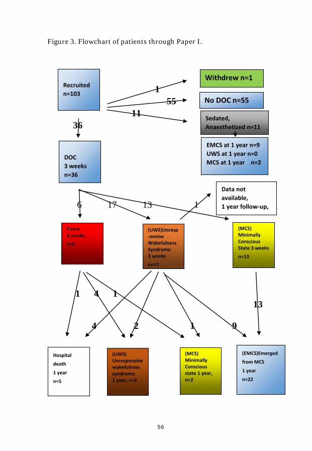
Figure 3. Flowchart of patients through Paper I.
1 55 11 36
6 17 13 1 1 4 1 13 4 2 1 9
Recruited n=103
Withdrew n=1
DOC 3 weeks n=36
Coma 3 weeks,
n=6
(UWS)Unresp-onsive Wakefulness Syndrome 3 weeks
n=17
(MCS) Minimally Conscious State 3 weeks
n=13
(UWS) Unresponsive wakefulness syndrome. 1 year, n=6
(MCS) Minimally Conscious state 1 year, n=2
(EMCS)Emerged
from MCS
1 year
n=22
Hospital
death
1 year
n=5
Data not available, 1 year follow-up,
No DOC n=55
Sedated, Anaesthetized n=11
EMCS at 1 year n=9 UWS at 1 year n=0 MCS at 1 year n=2
56

Patients in unresponsive wakefulness syndrome (UWS) 3
weeks after injury
Of the 17 patients in UWS at 3 weeks, by 3 months, 5 remained in
UWS, 6 had improved to MCS, 4 had emerged from MCS, and 2 were
dead, missing data for one. The outcome 1 year after injury for these
patients, according to the GOSE, was 1 (dead, n = 4), 2 (vegetative
state, n = 3), 3 (lower severe disability, n = 7), 4 (upper severe
disability, n = 2), missing data (n = 1). Note, that GOSE level 2,
associated with the description “vegetative state” actually includes
some patients in MCS, explaining the apparent discrepancy. At first
assessment 3 weeks after injury, scores on the CRS-R (maximum 23),
for patients found to be in UWS ranged from 0 to 7. Correlation
between CRS-R score at 3 weeks and outcome at 1 year for these
patients, according to the GOSE, was poor, with a correlation
coefficient of 0.29.
Patients in minimally conscious state 3 weeks after injury
Of the patients in a minimally conscious state (MCS) at 3 weeks, all 13
had emerged from MCS at 3 months. These patients scored median 12
on the CRS-R (range 6-19) at 3 weeks. GOSE for these 13 patients one
year after injury varied from 3-7. Correlation between CRS-R score at
3 weeks and outcome at 1 year for these patients, according to the
GOSE, was weak (r= –0.19). One year after injury, 4 of these patients
were living at home without assistance, 8 were at home with
assistance, and 1 was in a nursing home. One patient was working
full-time (and could also drive).
57

Patients in coma or sedated/anaesthetized 3 weeks after
injury
Of the 6 patients in coma at 3 weeks, by 3 months, 4 were in UWS, 1
was in MCS, none were better than MCS, and 1 was dead. The same
figures were shown at one year follow-up. Of the 11 patients who were
sedated/anaesthetized 3 weeks after injury, by 3 months, 1 was in
UWS, 3 were in MCS, and 7 were better than MCS. One year after
injury, none of these initially sedated patients remained in UWS, 2
were in MCS, and 8 were better than MCS. Only a few patients were
treated with dopaminergic drugs at the time of study assessments. Of
patients in UWS at any point during the study, none were being given
such drugs at the 3-week assessment; at the 3-month assessment 1
patient (in UWS at 3 weeks, MCS at 3 months, and EMCS at 1 year)
was being given Madopark (levodopa/benserazide combination), and
one patient (coma at 3 weeks, UWS at 3 months, UWS at 1 year) was
being given amantadine at 3 months but not at 1 year. One other
patient (UWS at all study time-points) was being given amantadine at
the 1-year assessment but not earlier.
Admission to specialized rehabilitation units
Of the 15 patients in UWS at 3 weeks who survived at least to 3
months, 14 were admitted to an inpatient specialized rehabilitation
unit. Admission to a rehabilitation unit occurred on average 62 days
after injury (standard deviation (SD) 46, range (26–198 days). All of
the 13 patients in MCS 3 weeks after injury were admitted to inpatient
rehabilitation units, a mean of 44 days after injury (SD 18, range 17–
78).
58

Figure 4. Flowchart of patients Papers II and III
PATIENTS BNIS PAPERS II-III PAPER III
Recruited, n=114
Withdrew, n=1
Acute data entered,
n=113
Withdrew, n=1 Hospital death, n=1
Three-weeks follow-up,
n=111
Eligible patients for BNIS at 3 weeks, n=42 Data not available, n=10 Not assessable, n=59
Three-month follow-up,
n=105
Eligible patients for BNIS, at 3 months, n=75 Data not available, n=1 Not assessable, n=29
One-year follow-up, n=100
Eligible patients for BNIS at 1 year, n=78 Data not available, n=3 Not assessable, n=19
Withdrew, n=2 Hospital death, n=4
Withdrew, n=3 Hospital death, n=2
59

Figure 4 shows a flowchart of follow-up, withdrawals and deaths. For
demographic details and summary statistics, see Table 8. Patients
who withdrew were similar to those who continued in terms of age
and median GCS and RLS.
60
PAPER II
Care pathways
Ninety-seven patients were transferred to an inpatient brain injury
rehabilitation unit at some point during the first year after injury. Of
these, 90 were alive and followed up at 1 year, 2 patients died after
admitted to inpatient rehabilitation but before follow-up at 1 year, 4
had withdrawn from the study, and data were missing for 1. Another 5
patients died without having been transferred to a rehabilitation unit.
Their median time from injury to first admission to inpatient
rehabilitation was 28 days (range 9-198 days). Time from first
discharge from intensive care to admission to inpatient rehabilitation
was median 13 days (range, 0-176 days), and a substantial proportion
of patients had to wait for several weeks. Eight surviving patients
(7%) were known not to have been transferred to an inpatient brain
injury rehabilitation service, 4 received rehabilitation (early
outpatient rehabilitation, nursing home or a geriatric unit within a
neurology facility) and 4 did not receive rehabilitation. Length of
intensive care was shorter for those not receiving rehabilitation
(median = 6 days; range, 5-17 days) than those receiving
rehabilitation (median=17 days; range, 1-78 days). The most common
care pathways for those patients who were transferred to inpatient
rehabilitation (n = 97) was from intensive care to a neurosurgical
ward and then to a rehabilitation unit (26%). Twenty-three patients
(24%), were transferred directly from intensive care to a
rehabilitation unit. Twenty patients (20%) were transferred from
intensive care to a surgical ward and then to a rehabilitation unit. The
remaining 29 patients (30%) received care at between 1 and 5
different intervening care units after intensive care discharge and
before eventual transfer to a rehabilitation unit. The number of
61

intervening care units was not significantly associated with outcomes
at 1 year.
Outcomes
Of the 100 patients that were alive and followed up one year after
injury (including those who did not receive inpatient rehabilitation),
36 had a bad outcome on the GOSE (score 2-4), and data on GOSE
were missing for 2. The CRASH acute prognostic model correlated
poorly with actual outcome at 1 year (r =−0.12). Length of stay in
intensive care (r= −0.49) and length of time between intensive care
and admission to rehabilitation (r= −0.30) were more strongly
correlated with outcomes.
A logistic regression model demonstrated that length of stay in
intensive care, length of time between intensive care and
rehabilitation admission, and the presence of post–acute
complications contributed significantly to the variation in outcome
and together explained 52% of the variation in the model. The CRASH
model and the presence of pre-existing medical problems were not
significantly related to outcome. Time between intensive care and
rehabilitation admission was not significantly different for patients
with and without complications at 3 weeks (p = 0.11), or for patients
with and without major extracranial injury (p = 0.59), or for patients
with and without pre-existing medical conditions (p = 0.64).
Length of inpatient rehabilitation stay was significantly inversely
related to outcome, a bad outcome being associated with a longer stay
in inpatient rehabilitation (p = 0.0001). Length of inpatient
rehabilitation stay was median 34 days (range, 3-127 days) for
patients with a good outcome and 64 days (range, 2-315 days) for
patients with a bad outcome.
62

PAPER III
Figure 4 shows a flowchart depicting the study process and recruited
and included patients in Paper III and patients who completed BNIS
at 3 weeks, 3 months and 1 year. For demographic details and
summary statistics, see Table 8.
The Barrow Neurological Institute Screen for Higher
Cerebral Functions
It was possible to use the Barrow Neurological Institute Screen for
Higher Cerebral Functions (BNIS) for assessment at three weeks after
injury in 42 patients. 59 patients could not be assessed due to
preescreen, ongoing disorders of consciousness (DOC) or sedation. At
3 months, 75 patients were assessed and at one-year follow-up, 78
patients were assessed.
Both the BNIS total raw scores and 𝑇𝑇-scores improved significantly
from 3 weeks to 3 months after injury (raw score: 𝑝𝑝 < 0.001, 𝑇𝑇- score:
𝑝𝑝 < 0.001) and from 3 months to 1 year on the raw score only (𝑝𝑝 =
0.004) and 𝑇𝑇-score (𝑝𝑝 = 0.086).
From 3 months to 1 year, no further significant improvements were
found. At 3 months and 1 year, patients with more than 12 years of
education had statistically significant higher scores than patients with
less education on the subscales speech/language, orientation
attention/concentration, memory affect and awareness (p<0.01).
The Hospital Anxiety and Depression Scale (HADS).
Scores above cut-off for HADS anxiety occurred in 16 of 75 assessable
patients at 3 months and in 16 of 74 patients at 1 year after injury.
63
HADS depression scores above cut-off occurred in 11 of 75 assessable
patients at 3 months and in 15 of 74 patients at 1 year. Significant
correlations were found between HADS depression and BNIS total at
3 months (𝑟𝑟 = −0.302, 𝑝𝑝 = 0.009) and at one year (𝑟𝑟 = −0.361, 𝑝𝑝 =
0.002).
Outcomes
The majority of patients (83%) who completed the BNIS at 3 weeks
had favourable outcome on the GOSE at one year. Out of the patients
who completed the BNIS at 3 months and at one year, “favourable”
outcomes were reported for 80% and 76%, respectively.
Out of the patients who completed the BNIS at 3 weeks, “superior
functioning” on the RLAS-R at 1 year was shown for 81%. The
corresponding proportion on the RLAS-R at 1 year for patients
completing the BNIS at 3 months was 73% and for patients
completing the BNIS at 1 year 69%.
In a multivariate model, statistically significant associations were
obtained for the BNIS subscales orientation and visuospatial and
visual problem solving and “favourable outcome” on the GOSE at one
year. The same subscales were also significantly associated with
“superior functioning” on the RLAS-R at one year.
64

PAPER IV Figure 5 shows a flowchart depicting the study process and recruited
patients. A total of 37 patients were identified during the study period
and included in the analyses and no patient withdrew. Falls were the
most frequent cause of injury (54%) and 70% were males. Males had
less education than the females. Education less then 12 years was
reported for 62% of included patients and 65 % had an employment or
as a student for more than 50%. More than half were married or
cohabitating with or without children. The number of patients 18-25
years was nearly the same as for patients aged 26-49 and 50-65 years.
Eighteen patients (49%) were under the influence of alcohol and/or
drugs (as demonstrated by clinical assessment, anamnestic
information and/or blood test) at the time of injury. Known current
drug and/or alcohol abuse was present in 11 patients (30%). For
demographic details and summary statistics, see Table 8.
65

Figure 5. Flowchart of patients through Papers IV and V PAPERS IV-V
Recruited n=37
Follow-up 3 weeks n=36
Follow-up 3 months n=32
Follow-up 1 year n=31
Hospital death n=1
Hospital death n=4
Hospital death n=1
66

Figure 6. Acute care pathways from the Northern Health Region (NHR) to the neurotrauma center (NC) at Umeå University hospital.
67
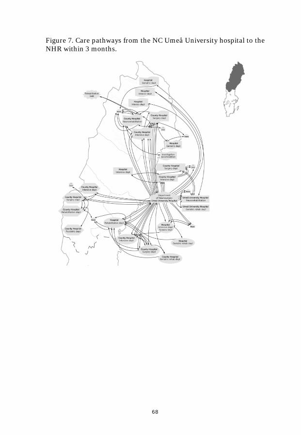
Figure 7. Care pathways from the NC Umeå University hospital to the NHR within 3 months.
68

Computed tomography scan
Time to first CT scan was less than 1 h for 11%, less than 3 h for 55%
and less than 4 h for 82%. The first CT scan of the brain showed
traumatic subdural hematoma in 73% of the patients, brain contusion
(s) in 76% and traumatic subarachnoidal haemorrhage in 78% of the
patients. The first CT was classified as Marshall I-II in 43% of the
patients and Marshall III-VI in 57%.
Clinical outcomes
The RLAS-R scores improved significantly from 3 weeks (5.26 ± 3.07)
to 3 months (8.0 ± 2.45) (p < 0.001), and 19 patients had “superior
functioning” on the RLAS-R IX–X. Eight patients had both “superior
functioning” on the RLAS-R and a “favourable outcome” on the GOSE
7–8.
Clinical care pathways.
Most patients (92%) were admitted directly to the regional
neurotrauma centre (Figure 6). After discharge, patients were
typically transferred back to one of several county or local hospitals
(Figure 7). It was also common for them to be transferred between
different departments within a given hospital.
69

PAPER V
See Figure 5 and the flowchart depicting the study process and
recruited included patients in Paper V and for patient characteristics
see table 8.
Hospital deaths occurred in 6 patients. One of these patients died at
the NC due to respiratory complications. One of the fatalities suffered
from multiple illnesses at the time of injury, two patients with very
severe brain injury (GCS 3) died because of respiratory complications,
one died because of inoperable traumatic intracranial aneurysm and
one patient died from intracerebral bleeding after transportation
from NC to the local hospital.
Fatalities (16%) had more severe injuries GCS median 3 (3-6)
compared with survivors GCS median 5 (3-8) and a significantly
higher mean age in comparison patients who survived (52.8 +17.8 vs.
41.3 +15.1, p =0.048). GCS 3 was seen in 24 % (n=9) of the included
patients and hospital deaths occurred in 44% (n=4) of these patients.
A review at the NC to identify possibly missed patients revealed an
additional 6 patients, all males, mean age 49.8 (+9.6) age, GCS
median 6.5 (4-7). These patients could not be included in the study,
as they were identified later than 3 weeks post-injury (which is the
latest time of inclusion as stipulated by the study protocol).
CTi was assessed in all patients. A subsequent CT24, according to the
study protocol, was assessed in 34 patients. In this study, 41% of
patients performed CTi within 2 hours post-trauma. The mean time
from trauma to CTi was 2.7 ±1.6 h (n=31). One patient was excluded
because of delayed admission to hospital (15.2h). For 5 patients, the
70

exact elapsed time post-trauma could not be established, but was
deduced to be within 22 hours. The mean time from trauma to CT24
was 25.4 ± 12.4 h (n=30). Two patients were investigated by CT24 after
60 hours due to clinical reasons. For CT characteristics, see Table 3.
The median (range) score of CTi according to Marshall was 3 (1-6)
and the corresponding results of CT24 was 5 (1-6). The median (range)
Rotterdam score of CTi was 4 (2-6) and of CT24 3 (1-6). See Figures 1,2
and Table 4.
Non-evacuated mass lesion on the CTi. was seen in 19% (n=7/37)
compared with 9% (n=3/34) on CT24. Out of 27 patients with available
CTi and CT24 and with detectable diffuse injury (Marshall I-IV) on the
initial scan, 48% subsequently developed a mass lesion on CT24,
which was then evacuated. One patient who sustained high-energy
trauma displayed findings of no pathological according to Marshall
CTi and CT24. Nevertheless, the patient presented GCS 6 at admission
and diffuse axonal injury on magnetic resonance imaging and GOSE 5
at one year. According to Rotterdam, 16 patients out of 34 (47%)
showed improvement from CTi to CT24, whereas four patients (12%)
deteriorated.
There was a positive correlation between Marshall CTi and Rotterdam
CTi (r= 0.716, p<0.001) but no significant correlation between
Marshall CT24 and Rotterdam CT24 (r=0.077, p=0.667). Rotterdam
CT24 showed a negative correlation to GOSE at 3 months (r= -0.421,
p= 0.015). There were negative correlations between Marshall CTi and
CT24 and RLAS-R at 3 months (CTi r= -0.364 p=0.044; CT24 r= -
0.425, p=0.024). However, Marshall and Rotterdam scores of CTi
and CT24 did not correlate with the GOSE and RLAS-R scores at 1
year, this being the study endpoint as regards outcome.
71

GOSE improved significantly from 3 months (median 4.5 (1-8),
mean 4.4±2.3) to 1 year (median 7 (1-8); mean 5.5±2.7, p=0.003.) At
3 months, GOSE 1-4 was seen in 50% and GOSE 5-8 in 50%. At 1
year, GOSE 1-4 was seen in 36% and GOSE 5-8 in 64%. One patient
was in a vegetative state at 1 year. Good recovery (GOSE 7-8) was seen
in 59 % at 1 year. RLAS-R also improved significantly from 3 months
(median 9 (2-1); mean 8.0±2.4) to 1 year (median 10 (3-10); mean
8.9±1.9, p=0.003). At 1 year, RLAS-R 1-6 was seen in 10% and RLAS-
R 7-10 in 90%, and 77% reached the highest level, i.e. “Stand-by
assistance on request” and “Modified independent” (RLAS-R 9-10).
One patient who was classified on CTi as Marshall I (i.e. no visible
intracranial pathologic change) had an initial score of GCS 6 and was
classified as GOSE 5 at 1 year due to diffuse axonal injury. GCS on
admission correlated with GOSE at 1 year (r=0.366, p=0.026). There
were negative correlations between in-hospital total days and GOSE
at 3 months and 1 year (r= -0.419, p= 0.011 and r= -0.429, p= 0.008)
and between in-hospital total days and RLAS-R at 3 months and 1
year (r= -0.738, p<0.001 and r= - 0.713, p<0.001). The proportion of
unfavorable outcome (GOSE 1-4) at 1 year was 31% for men and 46%
for women. There were 4 patients with hospital death (GCS 3, GOSE
1) and unfavorable outcome ( >95%) according to CRASH.
72

DISCUSSION
Main findings
This thesis describes patients of working age with S-TBI in Sweden
and Iceland, a total of 114 patients recruited in a 2–year period,
follow-up at 3 weeks, 3 months and 1 year post-injury, with further
studies of three subgroups: i) 36 patients with DOC and 11 patients in
sedation/anaesthetized at three weeks, ii) 42-78 patients who
managed to complete cognitive screening, iii) 37 patients in the NHR,
Sweden. This thesis is a result of collaboration with the “PROBRAIN”
multicenter study, primarily descriptive. The number of included
patients was relatively small although 80% of the population in
Sweden and 100% of the population in Iceland were recruited.
Therefore, some caution in interpretation is necessary. Global
outcome (for all patients in the study/patients in the Northern Health
Region) at 1 year was good and nearly two thirds (55%/64%) of these
patients were assessed as GOSE 5-8, (33%/59% GOSE 7-8) but some
patients died or ended up with severe disabilities GOSE 1-2
(12%/19%). Ninety-seven patients of the remaining 100 assessed
patients were transferred to an inpatient brain injury rehabilitation
unit at some point during the first year after injury. However, it is
worth noting that rehabilitation was offered at different units, not
only in a neurorehabilitation unit. Delays to rehabilitation unit were
identified and delayed transfer was almost as common as patients
transferred directly to rehabilitation and some waited longer than a
month. The percentage of patients who received care at between 1 and
5 different intervening care units after intensive care discharge and
before eventual transfer to a rehabilitation unit was 30%. The number
of intervening care units was not significantly associated with
outcome at 1 year. Sedated and anaesthetized patients or patients in
73

MCS at three weeks had a better prognosis than patients in coma or
UWS. No patient in UWS was assessed better than GOSE 4 at one
year (one missing). In accordance with an earlier Norwegian study, a
delay from discharge from intensive care and admission to a
rehabilitation unit was associated with worse outcome one year after
injury [30]. The routines for transferring patients with severe TBI
from the geographically large NHR Sweden to NC seemed to work
very well but transfer from NC Umeå University hospital differed
from one patient to another and no structured continuous care
pathway was found. Cognition improved over time after the injury
and appeared to be relatively stable from three months to one year.
For the relatively small population from the Northern Health Region,
neither an acute prognostic model (outcome at six months) nor acute
repeated CT protocols could prove clinically useful correlation with
outcome at one year.
74

Paper I
An understanding of the natural course of unresponsive wakeful
syndrome (UWS) after S-TBI and recovery is of importance. The
Multi-Society Task Force on persistent vegetative status (PVS) (1994)
[48] performed a meta-analysis on recovery from UWS in 434
patients. The included studies had no standardized neurobehavioral
assessment which was shown to be a more sensitive tool than clinical
consensus for the establishment of differential diagnoseis in patients
with DOC, as misdiagnosis of UWS may occur in up to 40% of
patients [51]. At that time, when the definition of minimally conscious
state (MCS) [50] had not been established, it is likely that a number
of patients who were judged as vegetative state in the Task Force
report at one month were in MCS and therefore some of these
patients had a better prognosis. Reported data in Paper I were
assessed at three weeks postinjury and the relatively small group of
patients, even though they represented 80% of the population of
Sweden and 100% of the population of Iceland, meant that caution
had to be applied to interpretation. A comparison of reported data
from Paper I and the Multi-Society Task Force from 1994 showed that
patients with UWS at 3 weeks who emerged to full consciousness
(EMCS) at 3 months were quite similar, namely, 24% (Task Force
33%), and 53% (52% Task Force) at 1 year but a comparison of the
patients who recovered consciousness (EMCS) at 3 months in Task
Force with patients in UWS and MCS in our study at 3 weeks then
there was a higher share of recovery in our study 57% (Task Force
33%) and 73% (52% Task Force) at 1 year. It is problematic to
compare results with studies published more than twenty years ago.
However, this may reflect the consequence of better access to NCs
today and improved neurosurgical, modern neuro-intensive and
neuro-rehabilitative care. The percentage of patients in UWS who
75

died or were still in UWS at three months in Paper I was 41% (67%
Task Force). Long-term outcome for patients in conscious state UWS
three weeks after injury was assessed at 1-year follow-up, the best
level on the GOSE being 4 (upper severe disability), 13 % fulfilled this
rating. GOSE 1-4 is described as unfavourable outcome. GOSE 2-4
with a need for assistance and further interventions of rehabilitation.
Outcomes were also poor for patients in coma: 25% were dead and
75% in UWS which indicates that patients in coma state at three
weeks have a poorer outcome. Reports have recently appeared in the
literature on outcomes for selected groups of patients with disorders
of consciousness from the point at which they are admitted to
specialized rehabilitation programmes. Katz et al. [115] reported in a
retrospective review of outcomes in 36 patients admitted to a slow-to-
recover rehabilitation unit, of whom the 22 patients with S- TBI (8 in
UWS at admission, 14 in MCS) were admitted a mean of 37 days after
injury. Seven of the 8 UWS patients improved to MCS and 45%
(number not stated) later emerged from MCS. Although follow-up
periods differ, the figure of 45% who improved to better than MCS is
not dissimilar from our figure of 53% 1 year after injury. Katz et al
[115], reported that all of their patients admitted in MCS after TBI
emerged from MCS during rehabilitation similar to our findings. It
should be emphasized that such estimates are necessarily based on
small numbers of patients. In a retrospective study from Italy of 259
patients admitted consecutively to a neurorehabilitation facility with
acquired severe brain injury and admitted at different points in time
75 patients (29%) presented a UWS and 107 (41%) a MCS according
to CRS-R. Thirty-six months after the acute event, 17 persons were
still in UWS, 30 had died and 26 had regained some consciousness
[120]. This reflects the need for a longer follow-up period even though
this report did not only present data from S-TBI. In Paper I, outcome
76

was better for patients in MCS, 3 weeks after injury: 25% were
assessed as lower good recovery (GOSE 7) and 25% as lower moderate
disability (GOSE 5) at one year. Of the 15 patients in UWS at 3 weeks
who survived at least to 3 months, 14 were admitted to an inpatient
specialized rehabilitation unit. All patients with MCS at 3 weeks after
injury were later admitted to inpatient rehabilitation units. The
ongoing “PROBRAIN” study could have an impact on this because of
the fact that patients were recruited by rehabilitation physicians who
also underwent prospective clinical assessment at each point in time.
A study from Norway [30] has shown that early initiation of an
unbroken chain of rehabilitation improves outcomes after S-TBI.
Post-traumatic disorders of consciousness (PT-DOC) occurred at one
year after S-TBI in this paper with an incidence of approximately 1.4
per million people of working age. Because Sweden is a geographically
large country, national standards are needed. Such standards already
exist in some European countries, for example Scotland (2009) and
Denmark (2006) [36,121]. Subacute rehabilitation and care with
special standards and/or development of a DOC network and to
promote further research are of importance. The development of a
continuous chain of rehabilitation after S-TBI, which has been shown
to improve outcomes but which was not in place for any patients in
this study, should be prioritized.
Paper II
Patients with S-TBI may require a lengthy hospital stay and S-TBI can
cause long-term disability. Paper II reported that delays between
discharge from intensive care and admission to a rehabilitation unit
were negatively associated with outcome one year after injury.
Similar findings were also reported in an earlier study from Norway
2012 [30] and in a study from Norway 2016 [141] findings indicate
77

that clinical pathways without delay of admission to specialized
department for rehabilitation may contribute to enhance
independence. Medical stability is of importance before admission to
rehabilitation but our data in Paper II could not determine whether a
short period of a day or two between discharge from neuro-intensive
care had a negative effect on outcome. Delays identified in Paper II
were not short: the number of patients who had to wait longer than a
month (n=22) was nearly as large as those transferred directly (n =
23). During the time between intensive care and inpatient
rehabilitation, nearly a third of the patients received care at units that
would not be expected to have specific knowledge of recovery after
TBI. Some patients even received a short period of care in short-stay
nursing homes before the initial rehabilitation stay. Delay in
rehabilitation can depend of medical problems, extracranial injuries
but often because of a lack of beds because of delays in discharge from
rehabilitation to appropriate social care. These delays are well-known
by rehabilitation professionals but not documented in a transparent
way which would be of significance for creating a more smooth flow of
patients and increasing access to rehabilitation. The presence of
coexisting medical problems and major extracranial injury did not
have a significant link to outcome and were not significantly related to
the time between intensive care discharge and rehabilitation
admission. However, our findings did provide support to the role of
post–acute complications in contributing to poorer outcome. Patients
with complications did not have a significantly longer time between
intensive care discharge and rehabilitation admission than those
without complications. The relationship between longer length of stay
in intensive care (LOSIC) and worse outcome can be understood as
complications during the intensive care period and secondary brain
injury. Similar findings were reported in the PariS-TBI Study (2013),
78

that also found LOSIC to be an independent predictor of outcome at 1
year [122] and nearly a third of patients were discharged without
inpatient rehabilitation [142]. LOSIC can differ because of variation of
intensive care and treatment guidelines but standardized protocols
such as the “Lund concept” and pressure on intensive care beds is
extremely high at all centres and would reduce any difference. Our
hypothesis that acute prognostic factors would be associated with
outcome at 1 year has face validity and has been insufficiently
considered in previous studies focusing on rehabilitation. We
incorporated such acute prognostic variables into our data collection
primarily to allow the evaluation of any additional contribution of
delayed rehabilitation admission. We used the CRASH model to
predict the risk of unfavourable outcome from acute prognostic
variables. CRASH was not significantly related to actual outcome in
our patients. Several factors may explain this apparent paradox.
Assessment of outcome was at 6 months in the CRASH model but at 1
year in our study. Recovery continues after 6 months post injury [115]
and the CRASH model may be missing improvements that have had
an important long-term impact on patients’ functioning. Another
factor is that the CRASH model included patients who died in the
group with unfavourable outcome, and because our study evaluated
the impact of rehabilitation care pathways, it was not meaningful to
include patients who died before any rehabilitation was received. In
addition, the CRASH study omitted any consideration of
rehabilitation interventions. The CRASH model has also recently
been shown to overestimate rates of unfavourable outcome in
patients receiving intracerebral pressure–targeted neurosurgical
treatment [116] according to the Lund concept [123] which is
common in Sweden. Given the evidence for the effectiveness of
rehabilitation [34], it is positive that the majority of patients did
79

eventually receive inpatient brain injury rehabilitation. A previous
retrospective study [124] of a comparable group of patients with S-
TBI receiving care in 2003-2004 at 3 neurosurgical centres in Sweden
found that 17% were never admitted to rehabilitation compared with
7% of patients in Paper II. A Cochrane review reported the
importance of rehabilitation for recovery of function after S-TBI
depending on neuroplasticity, which can be influenced by active
rehabilitation interventions. [34]. As regards access to rehabilitation
for patients surviving S-TBI in Sweden and Iceland, the general
health care insurance system in these countries gives access to
rehabilitation for almost all patients but it is common for there to be
delays in admission to several county hospitals, not only to
specialized rehabilitation units. There is no planned continuous
pathway and a lack of structured programmes. An quasirandomized
study of S-TBI in Norway (2012) [30] because of a limited number of
beds showed benefits of a continuous chain of care after S-TBI from
neurosurgical intensive care to inpatient rehabilitation to discharge,
with a better outcome for those who received early continuous
rehabilitation starting in the intensive care compared with those who
received ordinary rehabilitation without continuous pathways. [30].
A defined chain of care for all patients suffering from S-TBI would
contribute to the optimization of care for all patients and support
difficult discharge decisions and facilitate adequate follow-up.
Paper III
Our results in Paper III indicate that cognition improves over time
after S-TBI and appears to be relatively stable from three months to 1
year. Since cognitive function was associated with outcomes, it seems
that early screening of cognitive function could be of importance for
rehabilitation planning in a clinical setting.
80

In this population of patients with S-TBI, it was feasible to use the
BNIS instrument for the screening of cognitive functions as early as
three weeks after injury in 42% of the patients. The screening made it
possible to individualize interventions at the stage of recovery where
neuroplasticity is maximal, with potential outcome benefits.
In Paper III, the patients who died had lower GCS while the patients
who withdrew were younger and were less severely injured according
to the GCS which could have had an influence on the result in this
study. Falls were the most frequent cause in our population of
working age adults while transport accidents were the second most
common cause, just like those reported in some earlier Scandinavian
studies [18,110] while motor vehicle injuries dominated, for example,
in southern Europe, the USA and Australia [23,125,126]. The BNIS
scores of the patients who completed the test at three weeks improved
substantially at three months and further improvement was shown at
1 year, but the number of patients who completed the BNIS was
relatively stable from 3 months to 1 year. There were only a few of the
very severely injured patients who improved so much they were able
to complete BNIS at the last follow-up at 1 year. When the BNIS total
scores at three weeks were compared with the results reported by
Borgaro and Prigatano [68] of a small population of S-TBI patients
early after the injury (around 20 days), the patients in our study
performed better and the scores were in fact higher than a group of
patients with moderate TBI, but lower than a control group. This
finding can in part be explained by differences regarding study
populations and a large variation in the ranges of post injury time in
the Borgaro and Prigatano study [68]. The BNIS scores at 1 year were
in a range that was similar to another Swedish study [91], indicating
that the long-term results are probably relatively consistent and in the
same study, the BNIS was validated in a patient group from a
81

neurorehabilitation clinic where less than 9 years of schooling was
defined as low education, as in the manual. In our study, low
education was defined as being less than 12 years (in this study 35-
40% of the patients) because the majority of the Swedish population
continue to study at upper secondary school. Regardless of where the
education level limit is set, it seems that the results in our study
confirm earlier results of a link between education level and cognition
[68,91]. The BNIS is a screening instrument that is made for practical
clinical use and which indicates if comprehensive cognitive
neuropsychological assessment should be proposed. It should be
noted that according to the Swedish BNIS manual [127], the majority
of our assessed patients at all the points in time gained scores that
were below the cut-off (less than 47 points) which means they were
recommended further testing but this proportion decreased over time
from 84% to 74%.
In a clinical context, it is important to consider this in order to
optimize the setting of realistic rehabilitation goals for each individual
patient. When comparing the scores of the subscales at the different
points in time, significant improvements in our study were only
shown from three weeks to three months. The results at three months
and at one year were in line with the previous Swedish results by
Hofgren et al. [91]. Moreover, the majority of patients who completed
the BNIS at all three points in time experienced “favourable outcome”
on the GOSE and “superior functioning” on the RLAS. Higher scores
on the orientation and visuospatial and visual problem-solving
subscales at three months were also associated with good outcomes.
Disorientation, a key component of posttraumatic amnesia, has often
been studied in patients in the acute phase after TBI and it has been
reported as a predictor of cognitive impairments after injury [128].
Borgaro et al [129] examined the usefulness of the BNIS for assessing
82

orientation in patients with TBI and concluded that the instrument
was shown to be a sensitive measure of disorientation in these
patients. The orientation, visuospatial and visual problem-solving
subscales include basic domains of importance for independence
inside and outside the patients’ homes. It was therefore not surprising
that these subscales were associated with outcome in the present
study. In our study, awareness on the BNIS subscale was associated
with the GOSE. This result is in line with earlier studies which have
reported a relationship between self-awareness and long-term
outcome in TBI patients [130]. In a study by Kelley et al. [131]
impaired awareness was shown more than 5 years after TBI and
awareness of cognitive function was found to predict return to work.
Although awareness may improve over time, it seems to be a complex
construct including varying aspects. Studies have reported depression
and anxiety as a major cause of disability after TBI [132,133]. In the
present study, there were negative relationships between the BNIS
total score and the HADS anxiety and depression scores at one-year
follow-up, indicating that patients with a cognitive dysfunction may
also suffer from anxiety and depression symptoms over time. These
findings confirm earlier results which have shown an association
between self-reported depression and anxiety and poor performance
on cognitive tests [134].
Paper IV
In Paper IV, 37 patients were included, suvivers more than three
weeks with S-TBI, from the NHR during 2 years. Injury
characteristics of patients, their clinical pathways and outcome after
three months were studied. In this rural area, which covers almost
half of Sweden, most patients were nevertheless shown to be swiftly
transported direct to the regional Neurotrauma Center (NC). Thus,
83

routines for pre-acute care seem to be well-established. Acute
transportation to the NC was made by aircraft or helicopter in over
70% of cases and 82% of the included patients had made their first CT
scan within 4 hours. By contrast, post-acute care after discharge from
NC seemed to lack a structured care pathway since patients were
transferred back to local hospitals at a fairly early stage, moved
between departments and to different kinds of rehabilitation
departments. The medical rationale of this dispersion is not clear.
There were probably several reasons behind the differences between
the acute and post-acute logistics. Although standardized treatment
and specialized rehabilitation are also likely to be needed, the
individual patient’s differences and needs are factors that tend to
grow in importance as the patient gradually becomes medically
stabilized [34,135] and such aspects may have played a role in the
choice of diverging pathways of the patients in the present study.
Costs may be another operative factor, as each county has its own
budget and has to cover the costs for patient care outside its
jurisdiction. In addition, the severity of residual disability and
projected prognosis was also likely to be a factor that determines the
choice of post-acute clinical pathway. Well-organized pre-hospital
transportation systems for patients with S-TBI have also been
reported from rural regions of Norway [30]. In these areas,
rehabilitation in the early phases is based on close collaboration
between the neurosurgical departments and rehabilitation units, but
capacity problems may delay inpatient rehabilitation [37]. Since
similar difficulties with insufficient management routines in Sweden
and Norway have been observed, researchers recently proposed a
Scandinavian organization model that integrates neurointensive care
and qualified rehabilitation, and ensures an effective chain of
rehabilitation activities after S-TBI [37]. Differences in post-acute
84

pathways after S-TBI have also been demonstrated from other
countries. In studies that have evaluated patients with S-TBI from
rural and urban areas, poorer outcomes for rural residents have often
been reported [31]. However, with an integrated acute and post-acute
network of services, similar results have been shown for rural and
urban groups in Australia [136]. These findings underline the
importance of structured interventions in the early rehabilitation
process.
The male patients in our study had a lower education level in
comparison with the females, and more males than females were
intoxicated at the time of injury. Alcohol use at the time of injury has
been shown to be a risk factor for TBI [137,138]. In the present study,
significantly more patients who were under the influence of alcohol at
the time of injury had a history of previous TBI and were more often
injured by high-energy trauma in comparison with the non-
intoxicated patients. However, there was no significant difference
between these groups on the outcomes three months post injury.
For assessment of outcomes, the GOSE and RLAS-R scales were used.
Overall outcomes were surprisingly good in this group of severely
injured patients, all patients improved significantly on the RLAS from
3 weeks to 3 months. At 3 months, 3 of the 19 patients in the 2 highest
RLAS-R categories and 2 of the 8 patients with the highest GOSE
levels had the lowest GCS score of 3 during the first 24 hours in the
acute stage. Thus, the majority of the assessed patients experienced
good recovery as regards cognitive and behavioural functioning, and
around one quarter were considered as having both “superior
cognitive functioning” [81] and a “favourable outcome” [80].
However, it is worth noting that even if positive results on the GOSE
and the RLAS were measured, patients may still not be fully recovered
at 3 months after the injury and may experience subtle deficits not
85

covered by these instruments. Therefore, it seems reasonable to
assume that some of the patients were in need of further
rehabilitation interventions and follow-up.
Paper V
To our knowledge, there are no previous studies using both
Rotterdam and Marshall for study of outcome of comprehensive
management and rehabilitation of S-TBI. In previous prognostic
studies on mortality and outcome in TBI, Marshall or Rotterdam were
utilized with a main focus on neurointensive care [9].
In this study, we found a negative correlation between Marshall and
Rotterdam and the clinical outcome according to GOSE and RLAS-R
at 3 months. However, there was no correlation between CT scores
and GOSE or RLAS-R at one year post injury, indicating that analysis
of CT acutely and within 24-48 hours lack predictive ability as regards
long-term clinical outcome in S-TBI. Likewise, CRASH failed to
predict outcome in this S-TBI population. Similar findings were
reported by Olivecrona & Olivecrona [29], who used CRASH for
prediction after S-TBI at six months. In previous research from our
hospital, Marshall and Rotterdam CTi and Marshall CT24 correlated
with the disability outcome GOS both at 3 months and at one year [9].
However, since GOSE is an extended version of GOS, these
instruments are not completely comparable. It might be that
prognostic prediction based on CT protocols lack sufficient sensitivity
to provide more fine grained outcome assessments, particularly
within a TBI subgroup comprising the most severe injuries. Another
possible reason may be related to the inclusion criteria. The
PROBRAIN study included patients who survived at three weeks, this
was not a criteria in the previous study [9].
86

It is of particular clinical relevance that overall outcome among
patients with severe TBI in our study was encouragingly favorable
(GOSE 5-8, 64%) (GOSE 7-8, 59%), while instruments for
prognostication failed to predict favourable/unfavourable outcome at
one year. When interpreting data from this study, some
distinguishing factors pertaining to this study population and design
should be emphasized.
First, patients were somewhat older (mean age +6 years) than in
some previous studies [5,9] on this topic. Second, in comparison with
a prior study from our center [9], patients on average had lower GCS
(5 vs 6), indicating more severe injury. Fatalities (16%) suffered more
severe injuries (GCS 3) compared with survivors (GCS 5) and were
also older (approximately +10 years). Third, this study was limited by
the relatively small study population. However, S-TBI is rare in
comparison with mild and moderate TBI. Also, the included patients
in fact comprised a near-total population of incident S-TBI cases
fulfilling selection criteria during two years. Furthermore, all data
were collected by one of the authors, who also personally examined all
patients during the course of the study, minimizing the amount of
missing or secondary data. One fourth of the patients were initially
classified as severely injured and with a minimal GCS score (3).
Nevertheless, at 1 year, 44% of this subgroup was classified as “good
recovery” on the GOSE (7-8), pointing to the importance of providing
active care for all S-TBI patients [34,36,40,139].
Both a history of previous brain injury and indications of alcohol use
at the time of injury have been shown to be risk factors for TBI
[34,35]. Over one third of patients in our study had been hospitalized
previously for TBI, and almost half were under the influence of
87

alcohol and/or drugs at the time of injury. This is a much higher rate
of alcohol use in S-TBI patients than that recently reported in a
Norwegian study (32%) [30]. These findings highlight the concept of
high-risk populations and high-risk situations in conjunction with
sTBI, and thus the need and potential for preventative measures.
CT of the brain remains a standard diagnostic tool for assessing TBI,
and is also used for prediction of outcome. Since studies have shown
that pathological intracranial changes in the brain often progress
during the first 24 and even 48 hours, routine repeated CT scans have
been proposed to capture intracranial dynamics [33]. In the present
study, the proportion of “less severely injured” patients based on
Marshall CTi was higher than in some previous studies [9,31].
However, when comparing our results on CT24, the percentage of
severely injured patients was similar to these studies, as the severely
injured group increased by more than 50% from CTi to CT24. Thus, it
is to be emphasized that intracranial pathology after S-TBI commonly
progress, so that repeat CT scans in the early stage often may be
implicated, especially in light of clinical deterioration.
The majority of patients in our study experienced good recovery as
regards disability and cognitive and behavioral functioning, and about
two thirds were assessed as having good outcomes on both GOSE and
RLAS-R. Those patients were independent as regards activities of
daily living and did not need another person’s assistance at one year
post injury.
In conclusion, the findings of this study proved negative as regards
the predictive ability of CT and CRASH protocol on outcome
prognostication at one year post injury in S-TBI. At the same time,
good outcomes were found in about two thirds of survivors. The study
88

support to the notion that, as a rule, patients with S-TBI should be
offered a combination of active and aggressive neurosurgical and
neurointensive care and active and intensive neurorehabilitation, as a
majority of these seriously injured patients showed favorable outcome
by such management, and as our possibilities for early
prognostication in the TBI subpopulation fails to identify who will
benefit from aggressive management or not.
89

SUMMARY OF THE THESIS In this thesis, we found that acute and repetitive CT of the brain
assessed with classification scales could not serve as a prognostic
factor for long-term outcome 1 year after injury. However, an initial
CT of the brain and a CT within 24 hours is of importance for acute
care planning. Patients recovered well, including patients in MCS and
those who were sedated/anaesthetized at three weeks. Patients with
S-TBI is a well-known heterogeneous group; this was confirmed in
these papers. Patients who died were older and had more severe brain
damage. There were only a few patients who were given
pharmacological treatment to optimize awareness and response to
stimuli, for example, Amantadine until follow-up at 1 year. Among
the S-TBI patients, there was a relatively large group of patients with
known drug or alcohol abuse. In comparison with previous studies, a
large proportion had signs of influence of alcohol and/or drug use at
the time of the injury compared with other studies. In this population
of patients with S-TBI, it was feasible to use the BNIS instrument for
the screening of cognitive functions as early as three weeks after
injury in 38% of available patients. In order to find prognostic factors
that describe the complexity of these patients and their needs for
different interventions after acute care, this overall clinical study gave
an opportunity to get better knowledge in several respects and
provide a basis for new studies. A structured chain of care and
specialized rehabilitation for patients with S-TBI, new guidelines for
equal healthcare in urban and rural areas, and better information to
care-providers, patients and their relatives are needed. The results
from these papers can hopefully contribute to better information to
patients and their relatives and thereby facilitate better planning of
care pathways and use of resources.
90

Strengths and limitations
The studies have several strengths, such as a prospective multicenter
observational design of S-TBI in Sweden and Iceland. Furthermore,
in Papers IV and V, one of the authors examined all patients during
the course of the studies and ensured that data were precisely and
completely documented. The number of patients in these studies was
rather small but comprised the total or near-total regional population
of S-TBI patients injured during a two-year period.
However, the studies are based on clinical populations and have some
limitations. Although the authors in Papers I-III had weekly contact
with intensive care units, some eligible patients may have been
missed from the recruitment process if they were admitted to and
discharged from intensive care between contacts.
Confirming a diagnosis of unresponsive wakeful syndrome (UWS) or
minimally conscious state (MCS) requires repeated assessment over
time which was not possible within the design of Paper I, given that
patients were assessed in whatever care setting was current at the
study time-points. However, the use of the JFK Coma Recovery Scale
Revised CRS-R is a strength in diagnostic accuracy.
Completeness of follow-up in Paper I of 81% patients (76% alive, 5%
dead) and in Paper II of 94% (88% alive, 6% dead) one year after
injury is acceptable, considering the necessity of obtaining consent
from relatives at the start of the study. In Paper III, the follow-up rate
of 69% completing the BNIS is satisfactory. Only 19 patients could not
complete the BNIS at the one-year follow. In Papers IV and V, all
patients who survived at three months and at one year participated in
the follow-up.
91

Clinicians who assessed outcome at one year in the studies were not
systematically blinded to acute data, which is a source of potential
bias. Within reasonable study resources, it was not possible to
arrange blinded follow-up at all locations and at the same time ensure
completeness of follow-up and inter-rater reliability. The time
interval between assessment at 3 months and one year can reasonably
be expected to protect against this bias, as the relatively long period
would make it unlikely that examiners would remember data from the
acute phase at the time of follow-up.
The CRASH prognostic model was used in Papers I, II and V. The
CRASH model predicts outcome 6 months after injury. We assessed
outcome one year after injury as recovery may continue at least until
this point in time for severely injured patients. These differing time-
frames could explain why differences in outcome between patients in
UWS and MCS 3 weeks after injury were not predicted by CRASH in
Paper I and why the CRASH model failed to predict outcome for the
population in Paper V.
Another limitation of the studies was that blood alcohol concentration
was not measured in all patients, thus decreasing the accuracy in
determining the contribution of alcohol to the early clinical picture
and the presumed effects on outcome. Both alcohol and drug intake at
the time of injury may depress the level of consciousness which could
affect the classification of TBI.
However, longer follow-up is of importance. Additional descriptions
of the complexity of these patients could provide better information
for decision-makers and rehabilitation planning, and could be used in
clinical practice and for further studies.
92

Conclusions
This thesis describes the clinical course and outcomes in patients with
severe TBI with regard to prognostic factors.
• The patients in minimally conscious state or anaesthetized
three weeks after injury were found to have a better prognosis
than patients in coma or unresponsive wakefulness syndrome.
This was not explained by acute prognostic models.
• A delay from discharge from intensive care and admission to a
rehabilitation unit was associated with worse outcome one
year after injury.
• Cognition improved over time after the injury and appeared to
be relatively stable from three months to one year.
• The routines for transferring patients with severe TBI from
the geographically large Northern Health Region in northern
Sweden so they can be given well-monitored surgical care
seemed to work very well. In contrast, the post-acute clinical
pathways did not reflect as clearly an optimized medical and
rehabilitative strategy.
• Neither acute CT protocols nor an acute prognostic model
proved clinically useful correlations with outcomes one year
after injury. At the same time, good outcomes were found in
about two thirds of the patients.
93

Future considerations
During the course of work on this thesis, some further considerations
and suggestions for further research have arisen.
• The small numbers of patients with disorders of consciousness
indicate that a development of national standards for post-
acute care for these patients is necessary to ensure good
standards of care for everyone.
• Although outcomes were assessed at one year after injury,
recovery may continue for longer than that. Further studies
are needed with longer follow-up time.
• Validated questionnaires were used for the assessments.
However, there is also a need for qualitative studies in order to
get knowledge about the views of the patients and their
nearest relatives concerning their situation after the injury.
• A high proportion of patients were reported to have a known
drug or alcohol abuse at the time of injury. It is necessary to
measure blood alcohol concentrations to determine the
contribution of alcohol to the clinical picture and to the effects
on outcomes.
• Health-economic studies to study costs of both the acute care
but also longer-term care requirements would be of interest.
• Well-organized routines for admission to rehabilitation after
severe TBI and for evidence-based treatment in the post-acute
94

stage should be considered as well as strategies to ensure
standardized rehabilitation care in both rural and urban areas.
• Because of the specific epidemiology of S-TBI, it is important
to inform of drug or alcohol use and its relation to S-TBI,
prevent accidents, provide effective emergency care and
neurorehabilitation, and there is a need for further studies of
functioning and disability after S-TBI.
95

ACKNOWLEDGEMENTS
I would like to thank the patients, their relatives and the clinical staff at the Department of Neurorehabilitation, Umeå University Hospital, Umeå.
I extend my thanks to my colleagues at the Neurotrauma Center and intensive care unit for helping to recruit patients, and my colleagues at the rehabilitation departments in the Northern Health Region.
I would like to thank all the people who made the collection of patient data possible from all participating neurocenters, my colleagues and all the people who have contributed in some way to this study. My special thanks to:
Anna-Lisa Nilsson who helped me with the collection and assessment of data and who was my travel companion by car, bus, and train in the Northern Health Region. Thank you for always supporting me and being a true friend and knowledgeable colleague.
Britt-Marie Stålnacke, my main supervisor and co-author, for your purposeful guidance which enabled me to complete this work. Thank you for your extensive knowledge of the various parts of rehabilitation research. Thank you for your patience, positive spirit and encouragement, your belief in me and my ability to create the prerequisites for this work. Thank you for introducing me to this field of research and your profound knowledge of science and its processes. Richard Levi, my co-supervisor and co-author, because you prioritised clinical research and research addressing S-TBI and care processes, rehabilitation and the outcome of these patients. Thank you for your personal philosophical interest, wise support and for sharing your own research experience with me. Thank you for your excellent knowledge of rehabilitation and your guidance about structured writing and written English. Lars-Ove Koskinen, my co-author, for your faithful support and your contribution to the writing of two of the manuscripts. Thank you for your excellent knowledge of S-TBI and for fruitful round-table discussions. Thank you for your wide experience of research and for much honest and useful advice and for giving me the courage to continue and to believe in myself.
96

Per Jonasson, my co-author, for your experience, time, kindness and excellent knowledge of neuroradiology and meticulous and tireless review of current CT brain studies. Catharina Nygren DeBoussard, my co-author, for your courteous manner, your sense of order and your profound knowledge of these patients, cooperation in the “PROBRAIN” study, as well as kind invitations to dinner and help with accommodation and administrative measures at Danderyd Hospital. Alison K. Godbolt, my co-author, for your immense knowledge, working capacity, good teamwork in the “PROBRAIN” study and invitation to different kinds of collaboration, your excellent eye for structure your ability to complete research work with excellent results. Jörgen Borg, my co-author, for initiating this multicentre study and of course for inviting me and your excellent support and cooperation with a new generation of female researchers and rehabilitation physicians at Danderyd Hospital. Thank you for your well-known, long-standing and deep knowledge of this field of research. My dear colleagues and friends, Trandur Ulfarsson, Marie Lindgren, Marianne Lannsjö and Gudrun Karlsdottir Reykjavik, my co-authors and thank you for our collaboration in the “PROBRAIN” multicenter study. Hans Stenlund for your calm, knowledgeable guidance and your advice on statistical matters. Hans Lindsten Head of the Neurocenter Clinical Center in Umeå Hospital for kindly support and understanding for fulfilling this thesis. My mother and father Svea and Sven-Erik Broström for your support and for always believing in me. Thank you for raising me to be a free-thinking person and for teaching me that “nothing is impossible” and “everything will be all right”! Thank you for giving me the ability to think innovatively, to question and draw new conclusions from facts that are presented and never to abandon my fundamental values. Thank you for your everyday support whenever necessary, for example, when my father travelled 600 kilometres by car with me
97

during the “PROBRAIN” study, from Umeå to Kiruna in winter conditions. I love you. To all my friends - you know who you are - and especially Lena Renlund, Marguerite Olofsson, Anette Keisu and all my cousins who have supported me in different ways during these years. I am so grateful that I knew you were always there if necessary for example, when you offered me accommodation when I travelled for the “PROBRAIN” study. Of course I have to mention two strong women who have been part of my life; my grandmothers Hilma Töyrä Broström (born 1909 in Karesuando) and Inga Kajsa Rimpi Nordquist (born 1893 in Kaltisluokta, Storlule). Finally, my thanks to my beloved husband and best friend Roger for always believing in me. Thank you for your love and tireless support and patience during these years. You never complained even when there was “late work” or “working weekends” and thanks also to my three beloved children Jonas, Nina and Lotta with their respective girlfriend and boyfriend Hanna and Kristoffer for giving me all that love and support. You are all a great joy to me and Roger. I am so thankful for all of you and you are always in my heart! I love you all.
98
REFERENCES 1. Povlishock JT, Katz DI. Update of neuropathology and neurological recovery after traumatic brain injury. J Head Trauma Rehabil. 2005 Jan-Feb;20:76-94.
2. Graham DI, Gennarelli TA, McIntosh TK. Trauma. Greenfields Neuropathol. 2002;14:823– 898.
3. Smith DH, Meaney DF. Axonal damage in traumatic brain injury. The Neuroscientist. 2000;6 :483– 495, Povlishock JT, Christman CW. Diffuse axonal injury. In: Waxman SG, Kocsis J, Stys P, eds. The Axon. New York: Oxford University Press; 1995:504–529. 4. Povlishock JT, Jenkins LW. Are the pathobiological changes evoked by traumatic brain injury immediate and irreversible? Brain Pathol. 1995;5:415–426.
5. Povlishock JT, Stone JR. Traumatic axonal injury. Head Trauma Basic Preclin Clin Dir. 2001;13: 281–302. 6. Smith DH, Meaney DF, Shull WH. Diffuse axonal injury in head trauma. J Head Trauma Rehabil. 2003;18:307–316.
7. Teasdale G, Jennett B. Assessment of coma and impaired consciousness. A practical scale. Lancet. 1974 Jul 13;2:81-84. 8. Starmark JE, Stålhammar D, Holmgren E. The Reaction Level Scale (RLS85). Manual and guidelines. Acta Neurochir 1988;91: 12–20. 9. Starmark JE, Stålhammar D, Holmgren E, & Rosander B. A comparisation of the Glasgow Coma Scale and the Reaction Level Scale (RLS85). J Neurosurg 1988; 69:699-706. 10. Marshall LF, Marshall SB, Klauber MR, Van Berkum Clark M, Eisenberg H, Jane JA, Luerssen TG, Marmarou A, Foulkes MA. The diagnosis of head injury requires a classification based on computed axial tomography. J Neurotrauma. 1992 Mar;9 Suppl 1:S287-292. 11. Maas AI, Hukkelhoven CW, Marshall LF, Steyerberg EW. Prediction of outcome in traumatic brain injury with computed tomographic characteristics: a comparison between the computed tomographic classification and combinations of computed tomographic predictors. Neurosurgery. 2005 Dec;57:1173-1182; discussion 1173-1182. 12. Murray CJL, Lopez AD. Alternative projections of mortality and disability by cause 1990–2020: Global Burden of Disease Study. The Lancet. 1997; 349:1498–1504. 13. Jacobsson LJ, Westerberg M, Söderberg S, Lexell J. Functioning and disability 6-15 years after traumatic brain injuries in northern Sweden. Acta Neurol Scand. 2009 Dec;120:389-395.
99

14. Eker C, Schalén W, Asgeirsson B, Grände PO, Ranstam J, Nordström CH. Reduced mortality after severe head injury will increase the demands for rehabilitation services. Brain Inj. 2000 Jul;14:605-619. 15. Socialstyrelsen. Official statistics of Sweden’s health care and medical services. Hospitalisation due to injuries and poisoning in Sweden 2011. 16. Kleiven S, Peloso PM, von Holst H. The epidemiology of head injuries in Sweden from 1987 to 2000. Inj Control Saf Promot. 2003 Sep;10:173-180. 17. Tagliaferri F, Compagnone C, Korsic M, Servadei F, Kraus J. A systematic review of brain injury epidemiology in Europe. Acta Neurochir (Wien). 2006 Mar;148:255-268. 18. Styrke J, Stålnacke BM, Sojka P, Björnstig U. Traumatic brain injuries in a well-defined population: epidemiological aspects and severity. J Neurotrauma. 2007 Sep;24:1425-1436. 19. Bruns J Jr, Hauser WA. The epidemiology of traumatic brain injury: a review. Epilepsia. 2003;44 Suppl 10:2-10. 20. Polinder S, Meerding WJ, Van Baar ME, et al. Cost estimation of injury-related hospital admissions in 10 European countries. J Trauma. 2005;59:1283–1290. 21. Centers for Disease Control and prevention (CDC) Traumatic Brain Injury in the United States. 1999 Jul. p. 1-28. 22. Murray GD, Teasdale GM, Braakman R, Cohadon F, Dearden M, Iannotti F, Karimi A, Lapierre F, Maas A, Ohman J, Persson L, Servadei F, Stocchetti N, Trojanowski T, Unterberg A. The European Brain Injury Consortium survey of head injuries. Acta Neurochir (Wien). 1999;141:223-236. 23. Andriessen TM, Horn J, Franschman G, van der Naalt J, Haitsma I, Jacobs B, Steyerberg EW, Vos PE. Epidemiology, severity classification, and outcome of moderate and severe traumatic brain injury: a prospective multicenter study. J Neurotrauma. 2011 Oct;28:2019-2031. doi: 10.1089/neu.2011.2034. Epub 2011 Sep 27. 24. Andersson EH, Björklund R, Emanuelson I, Stålhammar D. Epidemiology of traumatic brain injury: a population based study in western Sweden. Acta Neurol Scand. 2003;107:256-259. 25. Parry-Jones BL, Vaughan FL, Miles Cox W. Traumatic brain injury and substance misuse: a systematic review of prevalence and outcomes research (1994-2004). Neuropsychol Rehabil. 2006 Oct;16:537-560. 26. Jacobsson LJ, Westerberg M, Malec JF, Lexell J. Sense of coherence and disability and the relationship with life satisfaction 6-15 years after traumatic brain injury in northern Sweden. Neuropsychol Rehabil. 2011;21:383-400. 27. Thurman DJ, Alverson C, Dunn KA, Guerrero J, Sniezek JE. Traumatic brain injury in the United States: A public health perspective. J Head Trauma Rehabil. 1999;14:602-615.
100
28. Plassman BL, Havlik RJ, Steffens DC, Helms MJ, Newman TN, Drosdick D, Phillips C, Gau BA, Welsh-Bohmer KA, Burke JR, Guralnik JM, Breitner JC. Documented head injury in early adulthood and risk of Alzheimer's disease and other dementias. Neurology. 2000 ;55:1158-1166. 29. Langlois JA, Rutland-Brown W, Wald MM. The epidemiology and impact of traumatic brain injury: a brief overview. J Head Trauma Rehabil. 2006 ;21:375-378. 30. Andelic N, Bautz-Holter E, Ronning P, Olafsen K, Sigurdardottir S, Schanke AK, Sveen U, Tornas S, Sandhaug M, Roe C. Does an early onset and continuous chain of rehabilitation improve the long-term functional outcome of patients with severe traumatic brain injury? J Neurotrauma. 2012;29:66-74. 31. Schootman M, Fuortes L. Functional status following traumatic brain injuries: population-based rural-urban differences. Brain Inj. 1999;13:995-1004. 32 Asgeirsson B, Grände PO, Nordström CH. A new therapy of post-trauma brain oedema based on haemodynamic principles for brain volume regulation. Intensive Care Med. 1994;20:260-7. 33. Grände PO. The "Lund Concept" for the treatment of severe head trauma--physiological principles and clinical application. Intensive Care Med. 2006 Oct;32:1475-1484. Epub 2006 Review. Erratum in: Intensive Care Med. 2007 ;33:205. 34. Turner-Stokes L, Pick A, Nair A, Disler PB, Wade DT. Multi-disciplinary rehabilitation for acquired brain injury in adults of working age. Cochrane Database Syst Rev. 2015 Dec 22;12:CD004170. 35. Sörbo A, Rydenhag B, Sunnerhagen KS, Blomqvist M, Svensson S, Emanuelson I. Outcome after severe brain damage, what makes the difference? Brain Inj. 2005 ;19:493-503. 36. Engberg AW, Liebach A, Nordenbo A. Centralized rehabilitation after severe traumatic brain injury--a population-based study. Acta Neurol Scand. 2006;113:178-184. 37. Borg J, Röe C, Nordenbo A, Andelic N, de Boussard C, af Geijerstam JL. Trends and challenges in the early rehabilitation of patients with traumatic brain injury: a Scandinavian perspective. Am J Phys Med Rehabil. 2011;90:65-73. 38. Johnston MV: Plasticity in the developing brain: Implications for rehabilitation. Dev Disabil Res Rev 2009;15:94-101. 39. Lindberg PG, Schmitz C, Engardt M, et al: Usedependent up- and down-regulation of sensorimotor brain circuits in stroke patients. Neurorehabil Neural Repair 2007;21:315-326. 40. Cicerone KD, Langenbahn DM, Braden C, Malec JF, Kalmar K, Fraas M, Felicetti T, Laatsch L, Harley JP, Bergquist T, Azulay J, Cantor J, Ashman T. Evidence-based cognitive rehabilitation: updated review of the literature from 2003 through 2008. Arch Phys Med Rehabil. 2011 Apr;92:519-530.
101

41. Seel RT, Douglas J, Dennison AC, et al. Specialized early treatment for persons with disorders of consciousness: program components and outcomes. Arch Phys Med Rehabil. 2013;94:1908-23, DeFina PA, Fellus J, Thompson JWG, et al. Improving outcomes of severe disorders of consciousness. Restor Neurol Neurosci. 2010;28:769-780. 42. Jennett B, Plum F. Persistent vegetative state after brain damage. A syndrome in search of a name. Lancet 1972; 1: 734–737. 43. Kretschmer E. Das apallische syndrom. Z Gesante Neurol Psychiatr, Berlin 1940; 169: 576–579. 44. Rosenblath W. Uber einen bemerkenswerten Fall von Hirnerschutterung. Dtsch Arch Klin Med 1899; 64: 406–424. 45. American Neurological Association Committee on Ethical AVairs. Persistent vegetative state. Ann Neurol 1993; 33: 386–390. 46. The Multi-Society Task Force on the Persistent Vegetative State. Statement on medical aspects of the persistent vegetative state. N Engl J Med 1994; 330: 1499–508.
47. Laureys S, Celesia GG, Cohadon F, Lavrijsen J, León-Carrión J, Sannita WG, Sazbon L, Schmutzhard E, von Wild KR, Zeman A, Dolce G; European Task Force on Disorders of Consciousness. Unresponsive wakefulness syndrome: a new name for the vegetative state or apallic syndrome. BMC Med. 2010;8:68. 48. The Multi-Society Task Force on PVS. Medical aspects of the persistent vegetative state (2). N Engl J Med 1994; 330: 1572–1579. 49. Laureys S. Eyes open, brain shut. Sci Am. 2007;296:84-89. 50. Giacino J, Ashwal S, Childs N, Cranford R, Jennett B, Katz DJ, et al. The minimally conscious state: Definition and diagnostic criteria. Neurology 2002; 58: 349–353. 51. Schnakers C, Vanhaudenhuyse A, Giacino J, Ventura M, Boly M, Majerus S, et al. Diagnostic accuracy of the vegetative and minimally conscious state: clinical consensus versus standardized neurobehavioral assessment. BMC Neurol 2009; 9: 35. 52. Giacino JT, Whyte J, Bagiella E, et al. Placebo-controlled trial of amantadine for severe traumatic brain injury. N Engl J Med. 2012;366:819-826. 53. Zafonte RD, Lexell J, Cullen N. Possible applications for dopaminergic agents following traumatic brain injury: Part 2. J Head Trauma Rehabil. 2001;16:112-116. 54. Laureys S, Schiff ND. Coma and consciousness: paradigms (re)framed by neuroimaging. Neuroimage. 2012;61:478-491. 55. Lee HB, Lyketsos CG, Rao V. Pharmacological management of the psychiatric aspects of traumatic brain injury. Int Rev Psychiatry. 2003;15:359-370.
102

56. Whyte J, Myers R. Incidence of clinically significant responses to zolpidem among patients with disorders of consciousness: a preliminary placebo controlled trial. Am J Phys Med Rehabil. 2009;88:410-418. 57. Azouvi P, Jokic C, Attal N, Denys P, Markabi S, Bussel B. Carbamazepine in agitation and aggressive behaviour following severe closed head-injury: results of an open trial. Brain Injury 1996;10:849–860. 58. Deb S, Crownshaw T. Review of subject. The role of pharmacotherapy in the management of behaviour disorders in traumatic brain injury patients. Brain Inj. 2004;18:1-31. 59. Goldstein LB. Basic and clinical studies of pharmacologic effects on recovery from brain injury. J Neural Transplant Plast. 1993;4:175- 192. 60. Chen H, Epstein J, Stern E. Neural plasticity after acquired brain injury: evidence from functional neuroimaging. PM R. 2010;2(12 Suppl 2):S306-12. 61. McCrimmon S, Oddy M. Return to work following moderate-to-severe traumatic brain injury. Brain Inj. 2006 Sep;20:1037-1046. 62. Kersel DA, Marsh NV, Havill JH, Sleigh JW. Psychosocial functioning during the year following severe traumatic brain injury. Brain Inj. 2001 Aug;15:683-696. 63. Socialstyrelsen. Rehabilitering för personer med traumatisk hjärnskada [Rehabilitation for people with traumatic brain injury]—Landstingens rehabiliteringsinsatser. http://www.socialstyrelsen.se/publikationer2012/2012-12-27. Accessed May 30, 2013. 64. Godbolt A, Lindgren M, Stenberg M, Cronberg T, Tengvar C, Sörbo A[Prolonged disorder of consciousness after severe acquired brain injury in adults. New recommendations provide a basis for investigation and rehabilitation]. Läkartidningen. 2014;111(49-50):2230-4. Article in Swedish. 65. Oddy M, Coughlan T, Tyerman A, Jenkins D. Social adjustment after closed head injury: a further follow-up seven years after injury. J Neurol Neurosurg Psychiatry. 1985 Jun;48:564-568. 66. Mathias JL, Wheaton P. Changes in attention and information-processing speed following severe traumatic brain injury: a meta-analytic review. Neuropsychology. 2007 Mar;21:212-223. 67. Chapman L, Wolff H. The cerebral hemispheres and the highest integrative functions of man. AMA Arch Neurol. 1959;1:357–424. 68. Borgaro SR, Prigatano GP. Early cognitive and affective sequelae of traumatic brain injury: a study using the BNI Screen for Higher Cerebral Functions. J Head Trauma Rehabil. 2002 Dec;17:526-534. 69. Lezak M Neuropsychological assessment. New York Oxford University, 1995. 70. McCleary C, Satz P, Forney D, et al. Depression after traumatic brain injury as a function of Glasgow Outcome Score. J Clin Exp Neuropsychol. 1998;20:270– 279.
103

71. Mathias J, Coats J. Emotional and cognitive sequelae to mild traumatic brain injury. J Clin Exp Neuropsychol. 1999;21:200–215. 72. Chapman L, Wolff H. The cerebral hemispheres and the highest integrative functions of man. AMA Arch Neurol. 1959;1:357–424, Damasio AR, Descartes' Error 1994. 73. Prigatano GP, Wong JL. Cognitive and affective improvement in brain dysfunctional patients who achieve inpatient rehabilitation goals. Arch Phys Med Rehabil. 1999 Jan;80:77-84. 74. Ezrachi O, Ben-Yishay Y, Kay T, et al. Predicting employment in traumatic brain injury following neuropsychological rehabilitation. J Head Trauma Rehabil. 1991;6:71–84. 75. Stuss DT, Gow CA, Hetherington CR. "No longer Gage": frontal lobe dysfunction and emotional changes. J Consult Clin Psychol. 1992 Jun;60:349-59. 76. Lishman's Organic Psychiatry: A Textbook of Neuropsychiatry, Lishman WA 1968. 77. Prigatano GP, Fordyce DJ, Zeiner HK, Roueche JR, Pepping M, Wood BC. Neuropsychological rehabilitation after closed head injury in young adults. J Neurol Neurosurg Psychiatry. 1984 May;47:505-513. 78. Prigatano GP. Disturbances of self-awareness and rehabilitation of patients with traumatic brain injury: a 20-year perspective. J Head Trauma Rehabil. 2005 Jan-Feb;20:19-29. 79. Jennett B, Bond M. Assessment of outcome after severe brain damage. Lancet. 1975 Mar 1;1:480-484. 80. Wilson JT, Pettigrew LE, Teasdale GM. Structured interviews for the Glasgow Outcome Scale and the extended Glasgow Outcome Scale: guidelines for their use. J Neurotrauma. 1998 Aug;15:573-585. 81. Hagen C, Malkmus D, Durham P. Levels of cognitive functioning. In: Professional Staff Association of Rancho Los Amigos Hospital, editors. Rehabilitation of the head injured adult: comprehensive physical management. Downey, CA: Rancho Los Amigos Hospital Inc.; 1987. 82. Giacino JT, Kalmar K, Whyte J. The JFK Coma Recovery Scale- Revised: measurement characteristics and diagnostic utility. Arch Phys Med Rehabil 2004; 85: 2020–2029. 83. Olivecrona M, Rodling-Wahlström M, Naredi S, Koskinen LO. Effective ICP reduction by decompressive craniectomy in patients with severe traumatic brain injury treated by an ICP-targeted therapy. J Neurotrauma. 2007 ;24:927-935. 84. Stenberg M, Koskinen LO, Levi R, Stålnacke BM. Severe traumatic brain injuries in Northern Sweden: a prospective 2-year study. J Rehabil Med. 2013 ;45:792-800.
104

85. Marshall LF, Marshall SB, Klauber MR, Clark MvB, Eisenberg HM, Jane JA, Luerssen TG, Marmarou A, Foulkes MA: A new classification of head injury based on computerized tomography. J Neurosurg 75:S14–S20, 1991. 86. Tesseris J, Pantazidis N, Routsi C, Fragoulakis D. A comparative study of the Reaction Level Scale (RLS85) with Glasgow Coma Scale (GCS) and Edinburgh-2 Coma Scale (modified) (E2CS(M)) Acta Neurochir (Wien) 1991; 110: 65–76. 87. Seel RT, Sherer M, Whyte J, Katz DI, Giacino JT, Rosenbaum AM, et al. Assessment scales for disorders of consciousness: Evidence based recommendations for clinical practice and research. Arch Phys Med Rehabil 2010; 91: 1795–1813. 88. Schnakers C, Majerus S, Giacino J, Vanhaudenhuyse A, Bruno MA, Boly M, Moonen G, Damas P, Lambermont B, Lamy M, Damas F, Ventura M, Laureys S. A French validation study of the Coma Recovery Scale-Revised (CRS-R). Brain Inj. 2008 Sep;22:786-792. 89. G. P. Prigatano, “Screen for higher cerebral functions:Rational and initial validation,” BNI Quarterly, vol. 7, pp. 2–9, 1991. 90. Prigatano GP, Montreuil M, Chapple K, Tonini A, Toron J, Paquet C, Dumurgier J, Hugon J, Truelle JL. Screening for cognitive and affective dysfunction in patients suspected of mild cognitive impairment. Int J Geriatr Psychiatry. 2014;29:936-42. 91. Hofgren C, Esbjörnsson E, Aniansson H, Sunnerhagen KS. Application and validation of the barrow neurological institute screen for higher cerebral functions in a control population and in patient groups commonly seen in neurorehabilitation. J Rehabil Med. 2007 Sep;39:547-53. 92. Esbjörnsson EHC, Larsson J, Denvall V. BNI Screen for Higher Cerebral Functions, Manual, Svensk Version, Katarina Tryck AB, Stockholm, Sweden, 2008. 93. Denvall V, Elmståhl S, Prigatano GP. Replication and construct validation of the Barrow Neurological Institute Screen for Higher Cerebral Function with a Swedish population. J Rehabil Med. 2002;34:153-157. 94. Zigmond AS, Snaith RP. The hospital anxiety and depression scale. Acta Psychiatr Scand. 1983;67:361–370. 95. Hung CI, Liu CY, Wang SJ, Yao YC, Yang CH. The cut-off points of the Depression and Somatic Symptoms Scale and the Hospital Anxiety and Depression Scale in detecting non-full remission and a current major depressive episode. Int J Psychiatry Clin Pract. 2012; 16:33–40. 96. Whelan-Goodinson R, Ponsford J, Schönberger M. “Validity of the Hospital Anxiety and Depression Scale to assess depression and anxiety following traumatic brain injury as compared with the Structured Clinical Interview for DSM-IV. J Affect Disord. 2009; 114: 94–102. 97. Bjelland I, Dahl AA, Haug TT, Neckelmann D. The validity of the Hospital Anxiety and Depression Scale: an updated literature review. J Psychosom Res.2002; 52:69–77.
105

98. Lobato RD, Gomez PA, Alday R, Rivas JJ, Dominguez J, Cabrera A, Turanzas FS, Benitez A, Rivero B. Sequential computerized tomography changes and related final outcome in severe head injury patients. Acta Neurochir. 1997;139:385-391. 99. Gennarelli TA, Spielman GM, Langfitt TW, Gildenberg PL, Harrington T, Jane JA, Marshall LF, Miller JD, Pitts LH. Influence of the type of intracranial lesion on outcome from severe head injury. J Neurosurg. 1982;56:26-32. 100. Lubillo S, Bolaños J, Carreira L, Cardeñosa J, Arroyo J, Manzano J. Prognostic value of early computerized tomography scanning following craniotomy for traumatic hematoma. J Neurosurg. 1999 Oct;91:581-587. 101. MRC CRASH Trial Collaborators, Perel P, Arango M, Clayton T, Edwards P, Komolafe E, Poccock S, Roberts I, Shakur H, Steyerberg E, Yutthakasemsunt S. Predicting outcome after traumatic brain injury: practical prognostic models based on large cohort of international patients. BMJ. 2008;336:425-429. 102. Eisenberg HM, Gary HE Jr, Aldrich EF, Saydjari C, Turner B, Foulkes MA, Jane JA, Marmarou A, Marshall LF, Young HF. Initial CT findings in 753 patients with severe head injury. A report from the NIH Traumatic Coma Data Bank. J Neurosurg. 1990;73:688-698. 103. Bobinski L, Olivecrona M, Koskinen LO. Dynamics of brain tissue changes induced by traumatic brain injury assessed with the Marshall, Morris-Marshall, and the Rotterdam classifications and its impact on outcome in a prostacyclin placebo-controlled study. Acta Neurochir. 2012;154:1069-1079. 104. Mata-Mbemba D, Mugikura S, Nakagawa A, Murata T, Ishii K, Li L, Takase K, Kushimoto S, Takahashi S. Early CT findings to predict early death in patients with traumatic brain injury: Marshall and Rotterdam CT scoring systems compared in the major academic tertiary care hospital in northeastern Japan. Acad Radiol. 2014 May;21:605-611. 105. Parchani A, El-Menyar A, Al-Thani H, El-Faramawy A, Zarour A, Asim M, Latifi R. Traumatic subarachnoid hemorrhage due to motor vehicle crash versus fall from height: a 4-year epidemiologic study. World Neurosurg. 2014 Nov;82:e639-644. 106. Mattioli C, Beretta L, Gerevini S, Veglia F, Citerio G, Cormio M, Stocchetti N. Traumatic subarachnoid hemorrhage on the computerized tomography scan obtained at admission: a multicenter assessment of the accuracy of diagnosis and the potential impact on patient outcome. J Neurosurg. 2003;98:37-42. 107. Greene KA, Marciano Ff, Johnson BA, Jacobowitz R, Spetzler RF, Harrington TR: Impact of traumatic subarachnoid hemorrhage on outcome in nonpenetrating head injury. Part I: A proposed computerized tomography grading scale. J Neurosurg. 1995;83:445–452. 108. Greene KA, Jacobowitz R, Marciano FF: Impact of traumatic subarachnoid hemorrhage on outcome in nonpenetrating head injury. Part II: Relationship to clinical course and outcome variables during acute hospitalization. J Neurotrauma.1996; 41:964-971. 109. Servadei F, Murray GD, Teasdale GM, Dearden M, Iannotti F, Lapierre F, Maas AJ, Karimi A, Ohman J, Persson L, Stocchetti N, Trojanowski T Unterberg A:
106

Traumatic Subarachnoid Hemorrhage: Demographic and clinical study of 750 patients from the European Brain Injury Consortium Survey of Head Injuries. Neurosurgery.2002; 50:261–269. 110. Andelic N, Anke A, Skandsen T, Sigurdardottir S, Sandhaug M, Ader T, Roe C. Incidence of hospital-admitted severe traumatic brain injury and in-hospital fatality in Norway: a national cohort study. Neuroepidemiology. 2012;38:259-267. 111. Jacobs B, Beems T, van der Vliet TM, van Vugt AB, Hoedemaekers C, Horn J, Franschman G, Haitsma I, van der Naalt J, Andriessen TM, Borm GF, Vos PE. Outcome prediction in moderate and severe traumatic brain injury: a focus on computed tomography variables. Neurocrit Care. 2013;19:79-89. 112. Jacobs B, Beems T, Stulemeijer M, van Vugt AB, van der Vliet TM, Borm GF, Vos PE. Outcome prediction in mild traumatic brain injury: age and clinical variables are stronger predictors than CT abnormalities. J Neurotrauma. 2010;27:655-668. 113. Jennett B, Bond M. Assessment of outcome after severe brain damage. Lancet. 1975 Mar 1;1:480-484. 114. Levin HS, Boake C, Song J et al. Validity and sensitivity to change of the extended Glasgow Outcome Scale in mild to moderate traumatic brain injury. J Neurotrauma. 2001;18: 575–584.. 115. Katz DI, Polyak M, Coughlan D, Nichols M, Roche A. Natural history of recovery from brain injury after prolonged disorders of consciousness: outcome of patients admitted to inpatient rehabilitation with 1-4 year follow-up. Prog Brain Res. 2009;177:73-88. 116. Olivecrona M, Olivecrona Z. Use of the CRASH study prognosis calculator in patients with severe traumatic brain injury treated with an intracranial pressure-targeted therapy. J Clin Neurosci. 2013;20:996-1001. 117. Grände PO, Asgeirsson B, Nordström CH. Physiologic principles for volume regulation of a tissue enclosed in a rigid shell with application to the injured brain. J Trauma. 1997;42:S23-31. 118. Grände PO, Asgeirsson B, Nordström CH. Volume-targeted therapy of increased intracranial pressure: the Lund concept unifies surgical and non-surgical treatments. Acta Anaesthesiol Scand. 2002 Sep;46:929-941. 119. Olivecrona M, Rodling-Wahlström M, Naredi S, Koskinen LO. Prostacyclin treatment in severe traumatic brain injury: a microdialysis and outcome study. J Neurotrauma. 2009 Aug;26:1251-1262. 120. Antoniono Elena, Lamberti Gianfranco Recovery From Vegetative State: A Four-Years Follow Up International Journal of Health Sciences March 2015, Vol. 3, No. 1, pp. 211-224 ISSN: 2372-5060 (Print), 2372-5079 (Online) URL: http://dx.doi.org/10.15640/ijhs.v3n1a13 121. NHS Scotland, Acquired Brain Injury National Managed Clinical Network. Traumatic Brain Injury (TBI) in Adults, Standards [Internet]. 2009 [Accessed 10 Oct 2012. (Standard 5 on vegetative and minimally conscious state). Available from: http://www.sabin. scot.nhs.uk/files/standards.pdf.
107

122. Jourdan C, Bosserelle V, Azerad S, et al. Predictive factors for 1-year outcome of a cohort of patients with severe traumatic brain injury (TBI): results from the Paris-TBI study. Brain Inj. 2013;27:1000–1007. 123. Grande PO. The Lund concept for the treatment of patients with severe traumatic brain injury. JNeurosurg Anesthesiol. 2011;23:358– 362. 124. Krakau K, Hansson A, Karlsson T, de Boussard CN, Tengvar C, Borg J. Nutritional treatment of patients with severe traumatic brain injury during the first six months after injury. Nutrition. 2007;23:308–317. 125. Mellick, K. A. Gerhart, and G. G. Whiteneck. Understanding outcomes based on the post-acute hospitalization pathways followed by persons with traumatic brain injury. Brain Injury.2003; 17: 55–71. 126. Harradine PG, Winstanley JB, Tate R, Cameron ID, Baguley IJ, Harris RD. Severe traumatic brain injury in New South Wales: comparable outcomes for rural and urban Residents. Med J Aust. 2004;181:130–134. 127. Esbjörnsson EHC, Larsson J, Denvall V. BNI Screen for Higher Cerebral Functions, Manual, Svensk Version, Katarina Tryck AB, Stockholm, Sweden, 2008. 128. Dowler RN, Bush BA, Novack TA, Jackson WT. Cognitive orientation in rehabilitation and neuropsychological outcome after traumatic brain injury. Brain Injury.2000;14:117–123. 129. Borgaro SR, Kwasnica C, Cutter N, Alcott S. The use of the BNI screen for higher cerebral functions in assessing disorientation after traumatic brain injury. J Head Trauma Rehabil. 2003;18:284–291. 130. Robertson K, Schmitter-Edgecombe M. Self-awareness and traumatic brain injury outcome. Brain Injury.2015;29:848–858. 131. Kelley E, Sullivan C, Loughlin JK et al. Self-awareness and neurobehavioral outcomes, 5 years or more after moderate to severe brain injury. J Head Trauma Rehabil.2014;29: 147–152. 132. Bornhofen C, McDonald S. Emotion perception deficits following traumatic brain injury: a review of the evidence and rationale for intervention. J Int Neuropsychol Soci. 2008;14:511–525. 133. Dahm J, Wong D, Ponsford J. Validity of the depression anxiety stress scales in assessing depression and anxiety following traumatic brain injury. J Affect Disord. 2013;151:392–396.
108

134. G. Spitz, M. Schönberger, and J. Ponsford. The relations among cognitive impairment, coping style, and emotional adjustment following traumatic brain injury. J Head Trauma Rehabil. 2013;28: 116–125. 135. Chesnut RM, Carney N, Maynard H, Mann NC, Patterson P, Helfand M. Summary report: evidence for the effectiveness of rehabilitation for persons with traumatic brain injury. J Head Trauma Rehabil 1999; 14: 176–188. 136. Harradine PG, Winstanley JB, Tate R, Cameron ID, Baguley IJ, Harris RD. Severe traumatic brain injury in New South Wales: comparable outcomes for rural and urban residents. Med J Aust 2004; 181: 130–134. 137. Opreanu RC, Kuhn D, Basson MD. Influence of alcohol on mortality in traumatic brain injury. J Am Coll Surg 2010; 210: 997–1007. 138. Chen CM, Yi HY, Yoon YH, Dong C. Alcohol use at time of injury and survival following traumatic brain injury: results from National Trauma Data Bank. J Stud Alcohol Drugs 2012; 73: 531–541. 139. Nudo RJ. Adaptive plasticity in motor cortex: implications for rehabilitation after brain injury. J Rehabil Med. 2003;(41 Suppl):7-10. 140. Ding J, Yuan F, Guo Y, Chen SW, Gao WW, Wang G, Cao HL, Ju SM, Chen H, Zhang PQ, Tian HL. A prospective clinical study of routine repeat computed tomography (CT) after traumatic brain injury (TBI). Brain Inj. 2012;26:1211-1216. 141. Sveen U, Røe C, Sigurdardottir S, Skandsen T, Andelic N, Manskow U, Berntsen SA, Soberg HL, Anke A. Rehabilitation pathways and functional independence one year after severe traumatic brain injury. Eur J Phys Rehabil Med. 2016 Apr 6. [Epub ahead of print] 142. Jourdan C, Bayen E, Bosserelle V, Azerad S, Genet F, Fermanian C, Aegerter P, Pradat-Diehl P, Weiss JJ, Azouvi P; Members of the Steering Committee of the PariS-TBI Study. Referral to rehabilitation after severe traumatic brain injury: results from the PariS-TBI Study. Neurorehabil Neural Repair. 2013;27:35-44. 143. Consensus conference. Rehabilitation of persons with traumatic brain injury. NIH Consensus Development Panel on Rehabilitation of Persons With Traumatic Brain Injury. JAMA. 1999;282:974-983. 144. Guidelines for the management of severe traumatic brain injury. J Neurotrauma. 2007;24 Suppl 1:S1-106. 145. Semlyen JK, Summers SJ, Barnes MP. Traumatic brain injury: efficacy of multidisciplinary rehabilitation. Arch Phys Med Rehabil. 1998;79:678-683.
109
Related Documents