Severe Acute Respiratory Syndrome Coronavirus ORF7a Inhibits Bone Marrow Stromal Antigen 2 Virion Tethering through a Novel Mechanism of Glycosylation Interference Justin K. Taylor, a Christopher M. Coleman, a Sandra Postel, b Jeanne M. Sisk, a John G. Bernbaum, c Thiagarajan Venkataraman, a Eric J. Sundberg, a,b,d Matthew B. Frieman a Department of Microbiology and Immunology, University of Maryland at Baltimore, Baltimore, Maryland, USA a ; Institute of Human Virology, University of Maryland School of Medicine, Baltimore, Maryland, USA b ; Integrated Research Facility, National Institutes of Health, Frederick, Maryland, USA c ; Department of Medicine, University of Maryland School of Medicine, Baltimore, Maryland, USA d ABSTRACT Severe acute respiratory syndrome (SARS) emerged in November 2002 as a case of atypical pneumonia in China, and the caus- ative agent of SARS was identified to be a novel coronavirus, severe acute respiratory syndrome coronavirus (SARS-CoV). Bone marrow stromal antigen 2 (BST-2; also known as CD317 or tetherin) was initially identified to be a pre-B-cell growth promoter, but it also inhibits the release of virions of the retrovirus human immunodeficiency virus type 1 (HIV-1) by tethering budding virions to the host cell membrane. Further work has shown that BST-2 restricts the release of many other viruses, including the human coronavirus 229E (hCoV-229E), and the genomes of many of these viruses encode BST-2 antagonists to overcome BST-2 restriction. Given the previous studies on BST-2, we aimed to determine if BST-2 has the ability to restrict SARS-CoV and if the SARS-CoV genome encodes any proteins that modulate BST-2’s antiviral function. Through an in vitro screen, we identified four potential BST-2 modulators encoded by the SARS-CoV genome: the papain-like protease (PL Pro ), nonstructural protein 1 (nsp1), ORF6, and ORF7a. As the function of ORF7a in SARS-CoV replication was previously unknown, we focused our study on ORF7a. We found that BST-2 does restrict SARS-CoV, but the loss of ORF7a leads to a much greater restriction, confirming the role of ORF7a as an inhibitor of BST-2. We further characterized the mechanism of BST-2 inhibition by ORF7a and found that ORF7a localization changes when BST-2 is overexpressed and ORF7a binds directly to BST-2. Finally, we also show that SARS- CoV ORF7a blocks the restriction activity of BST-2 by blocking the glycosylation of BST-2. IMPORTANCE The severe acute respiratory syndrome coronavirus (SARS-CoV) emerged from zoonotic sources in 2002 and caused over 8,000 infections and 800 deaths in 37 countries around the world. Identifying host factors that regulate SARS-CoV pathogenesis is crit- ical to understanding how this lethal virus causes disease. We have found that BST-2 is capable of restricting SARS-CoV release from cells; however, we also identified a SARS-CoV protein that inhibits BST-2 function. We show that the SARS-CoV protein ORF7a inhibits BST-2 glycosylation, leading to a loss of BST-2’s antiviral function. S evere acute respiratory syndrome coronavirus (SARS-CoV) was identified to be the causative agent of a 2002 to 2004 out- break of severe respiratory disease that emerged from the Guang- dong province of China, resulting in 8,096 cases and 774 deaths across 37 countries (1, 2). SARS-CoV is an enveloped virus with a positive-sense, single-stranded RNA genome of roughly 30,000 nucleotides encoding four structural proteins: the spike (S), enve- lope (E), membrane (M), and nucleocapsid (N) proteins (3). N protein forms the nucleocapsid, while E and M are minor virion membrane proteins. SARS-CoV entry into the cell is mediated by S-protein binding to angiotensin-converting enzyme 2 (ACE2) on the cell surface (4). In addition to the structural proteins, the SARS-CoV genome encodes several nonstructural and accessory proteins that promote SARS-CoV replication and virulence (5). Some of the nonstructural and accessory proteins function out- side of replication as type I interferon antagonists (6–8). ORF7a is a SARS-CoV genome-encoded accessory protein that is composed of a type I transmembrane protein that localizes pri- marily to the Golgi apparatus but can be found on the cell surface (9, 10). SARS-CoV ORF7a overlaps ORF7b in the viral genome, where they share a transcriptional regulatory sequence (TRS). ORF7a has a 15-amino-acid (aa) N-terminal signal peptide, an 81-aa luminal domain, a 21-aa transmembrane domain, and a 5-aa cytoplasmic tail (9, 10). To investigate the role of ORF7a in SARS-CoV replication, an ORF7ab deletion virus that replicated to a titer similar to that of wild-type (WT) SARS-CoV in vitro and in vivo was produced (10–12). Characterization of ORF7a in vitro demonstrated the ORF7a-dependent induction of apoptosis in a caspase-dependent pathway (13–15). Analysis of ORF7a evolu- tion during the SARS-CoV outbreak identified several residues in ORF7a that were under positive selection as SARS-CoV evolved Received 4 September 2015 Accepted 4 September 2015 Accepted manuscript posted online 16 September 2015 Citation Taylor JK, Coleman CM, Postel S, Sisk JM, Bernbaum JG, Venkataraman T, Sundberg EJ, Frieman MB. 2015. Severe acute respiratory syndrome coronavirus ORF7a inhibits bone marrow stromal antigen 2 virion tethering through a novel mechanism of glycosylation interference. J Virol 89:11820 –11833. doi:10.1128/JVI.02274-15. Editor: A. García-Sastre Address correspondence to Matthew B. Frieman, [email protected]. Copyright © 2015, American Society for Microbiology. All Rights Reserved. 11820 jvi.asm.org December 2015 Volume 89 Number 23 Journal of Virology on May 5, 2016 by HEALTH SCIENCES & HUMAN http://jvi.asm.org/ Downloaded from

Welcome message from author
This document is posted to help you gain knowledge. Please leave a comment to let me know what you think about it! Share it to your friends and learn new things together.
Transcript
Severe Acute Respiratory Syndrome Coronavirus ORF7a Inhibits BoneMarrow Stromal Antigen 2 Virion Tethering through a NovelMechanism of Glycosylation Interference
Justin K. Taylor,a Christopher M. Coleman,a Sandra Postel,b Jeanne M. Sisk,a John G. Bernbaum,c Thiagarajan Venkataraman,a
Eric J. Sundberg,a,b,d Matthew B. Friemana
Department of Microbiology and Immunology, University of Maryland at Baltimore, Baltimore, Maryland, USAa; Institute of Human Virology, University of Maryland Schoolof Medicine, Baltimore, Maryland, USAb; Integrated Research Facility, National Institutes of Health, Frederick, Maryland, USAc; Department of Medicine, University ofMaryland School of Medicine, Baltimore, Maryland, USAd
ABSTRACT
Severe acute respiratory syndrome (SARS) emerged in November 2002 as a case of atypical pneumonia in China, and the caus-ative agent of SARS was identified to be a novel coronavirus, severe acute respiratory syndrome coronavirus (SARS-CoV). Bonemarrow stromal antigen 2 (BST-2; also known as CD317 or tetherin) was initially identified to be a pre-B-cell growth promoter,but it also inhibits the release of virions of the retrovirus human immunodeficiency virus type 1 (HIV-1) by tethering buddingvirions to the host cell membrane. Further work has shown that BST-2 restricts the release of many other viruses, including thehuman coronavirus 229E (hCoV-229E), and the genomes of many of these viruses encode BST-2 antagonists to overcome BST-2restriction. Given the previous studies on BST-2, we aimed to determine if BST-2 has the ability to restrict SARS-CoV and if theSARS-CoV genome encodes any proteins that modulate BST-2’s antiviral function. Through an in vitro screen, we identifiedfour potential BST-2 modulators encoded by the SARS-CoV genome: the papain-like protease (PLPro), nonstructural protein 1(nsp1), ORF6, and ORF7a. As the function of ORF7a in SARS-CoV replication was previously unknown, we focused our study onORF7a. We found that BST-2 does restrict SARS-CoV, but the loss of ORF7a leads to a much greater restriction, confirming therole of ORF7a as an inhibitor of BST-2. We further characterized the mechanism of BST-2 inhibition by ORF7a and found thatORF7a localization changes when BST-2 is overexpressed and ORF7a binds directly to BST-2. Finally, we also show that SARS-CoV ORF7a blocks the restriction activity of BST-2 by blocking the glycosylation of BST-2.
IMPORTANCE
The severe acute respiratory syndrome coronavirus (SARS-CoV) emerged from zoonotic sources in 2002 and caused over 8,000infections and 800 deaths in 37 countries around the world. Identifying host factors that regulate SARS-CoV pathogenesis is crit-ical to understanding how this lethal virus causes disease. We have found that BST-2 is capable of restricting SARS-CoV releasefrom cells; however, we also identified a SARS-CoV protein that inhibits BST-2 function. We show that the SARS-CoV proteinORF7a inhibits BST-2 glycosylation, leading to a loss of BST-2’s antiviral function.
Severe acute respiratory syndrome coronavirus (SARS-CoV)was identified to be the causative agent of a 2002 to 2004 out-
break of severe respiratory disease that emerged from the Guang-dong province of China, resulting in 8,096 cases and 774 deathsacross 37 countries (1, 2). SARS-CoV is an enveloped virus with apositive-sense, single-stranded RNA genome of roughly 30,000nucleotides encoding four structural proteins: the spike (S), enve-lope (E), membrane (M), and nucleocapsid (N) proteins (3). Nprotein forms the nucleocapsid, while E and M are minor virionmembrane proteins. SARS-CoV entry into the cell is mediated byS-protein binding to angiotensin-converting enzyme 2 (ACE2) onthe cell surface (4). In addition to the structural proteins, theSARS-CoV genome encodes several nonstructural and accessoryproteins that promote SARS-CoV replication and virulence (5).Some of the nonstructural and accessory proteins function out-side of replication as type I interferon antagonists (6–8).
ORF7a is a SARS-CoV genome-encoded accessory protein thatis composed of a type I transmembrane protein that localizes pri-marily to the Golgi apparatus but can be found on the cell surface(9, 10). SARS-CoV ORF7a overlaps ORF7b in the viral genome,where they share a transcriptional regulatory sequence (TRS).ORF7a has a 15-amino-acid (aa) N-terminal signal peptide, an
81-aa luminal domain, a 21-aa transmembrane domain, and a5-aa cytoplasmic tail (9, 10). To investigate the role of ORF7a inSARS-CoV replication, an ORF7ab deletion virus that replicatedto a titer similar to that of wild-type (WT) SARS-CoV in vitro andin vivo was produced (10–12). Characterization of ORF7a in vitrodemonstrated the ORF7a-dependent induction of apoptosis in acaspase-dependent pathway (13–15). Analysis of ORF7a evolu-tion during the SARS-CoV outbreak identified several residues inORF7a that were under positive selection as SARS-CoV evolved
Received 4 September 2015 Accepted 4 September 2015
Accepted manuscript posted online 16 September 2015
Citation Taylor JK, Coleman CM, Postel S, Sisk JM, Bernbaum JG, Venkataraman T,Sundberg EJ, Frieman MB. 2015. Severe acute respiratory syndrome coronavirusORF7a inhibits bone marrow stromal antigen 2 virion tethering through a novelmechanism of glycosylation interference. J Virol 89:11820 –11833.doi:10.1128/JVI.02274-15.
Editor: A. García-Sastre
Address correspondence to Matthew B. Frieman, [email protected].
Copyright © 2015, American Society for Microbiology. All Rights Reserved.
11820 jvi.asm.org December 2015 Volume 89 Number 23Journal of Virology
on May 5, 2016 by H
EA
LTH
SC
IEN
CE
S &
HU
MA
Nhttp://jvi.asm
.org/D
ownloaded from
during transmission from bat to palm civet to humans (16). Thesedata suggest that ORF7a is vital for SARS-CoV biology and has ayet unidentified role in pathogenesis and disease.
Bone marrow stromal antigen 2 (BST-2; also known as CD317or tetherin) was initially identified to be a pre-B-cell growth pro-moter (17, 18). However, BST-2 is also a marker of type I inter-feron-producing cells (IPC) and is broadly expressed in many celltypes when treated with type I interferon (19). BST-2 has an un-usual structure, with an N-terminal transmembrane domain, aC-terminal glycosylphosphatidylinositol (GPI) anchor, and twoN-linked glycosylation sites in its extracellular domain, and existsas a disulfide-linked homodimer (20, 21). BST-2 traffics throughthe endoplasmic reticulum (ER) and Golgi apparatus, eventuallylocalizing to the surface and trans-Golgi network (20). Studieshave shown evolutionary conservation in three major surfacepatches of BST-2: near each of the two N-linked glycosylation sitesand in the C-terminal region (22).
The antiviral effect of BST-2 was first identified when it wasshown that BST-2 inhibits the release of virions of the retrovirushuman immunodeficiency virus type 1 (HIV-1) by directly teth-ering budding virions to the host cell. BST-2 also restricts therelease of many other viruses, including alphaviruses, arenavi-ruses, herpesviruses, paramyxoviruses, and other retroviruses(23–26). BST-2 is thought to restrict virus release by physicallytethering the budding enveloped virion to the plasma membrane(27), and a number of mechanism models have been proposed(28, 29). All of the BST-2 restriction models predict that BST-2functions as a dimer, interfacing through ectodomains that incor-porate into both the viral envelope and plasma membrane; how-ever, the models vary in regard to the orientation of the GPI an-chor and transmembrane domain. BST-2 has not been shown tointeract with any specific viral surface protein but, rather, has beenshown to function as an embedded intermembrane physicaltether. Therefore, BST-2 is thought to be able to restrict any mem-brane-budding enveloped virus (28, 29). Previous studies haveshown that the ability to form cysteine-linked dimers is necessaryfor BST-2 function, while conflicting results concerning the im-portance of the N-linked glycosylation have been reported (29,30). More recently, it has been suggested that BST-2 is a virussensor during HIV-1 infection and induces a proinflammatoryresponse through NF-�B (31).
Given the lack of virus specificity in BST-2 restriction, the ge-nomes of numerous viruses encode BST-2 antagonists to allow therelease of virions. The first such antagonist was identified to beHIV-1 accessory protein Vpu (27). HIV-1 Vpu binds BST-2 andcauses the �-TrCP2-dependent degradation of BST-2 and the ef-ficient release of HIV-1 virions, although it is not clear whetherdegradation occurs in the lysosome or proteasome (32–34). Otherviral antagonists of BST-2 include Chikungunya virus nonstruc-tural protein 1 (nsp1), Ebola virus glycoprotein GP1,2, herpessimplex virus glycoprotein M, HIV-2 envelope glycoprotein, Sen-dai virus glycoproteins, and simian immunodeficiency virus (SIV)Nef and envelope glycoproteins (23–26, 35–38). HIV-2 and SIVare closely related to HIV-1; however, the envelope glycoproteinsfrom HIV-2 and SIV antagonize BST-2 by sequestration withinthe trans-Golgi network rather than degradation, suggesting thatdifferent mechanisms of BST-2 antagonism exist for different vi-ruses, even within the same virus genus (35, 36). Another exampleis Ebola virus GP1,2, which antagonizes BST-2 through an un-
known mechanism that does not involve surface removal but stillleads to BST-2 functional inhibition (39).
Unlike many enveloped viruses, which bud from the cellplasma membrane, coronaviruses bud in the ER-Golgi apparatusintermediate compartment (ERGIC) and are transported to theplasma membrane inside vesicles (40). However, it has recentlybeen shown that BST-2 restricts the release of human coronavirus229E (hCoV-229E), suggesting that BST-2 can also restrict virusesthat bud in the ERGIC and are then released from the cell viavesicle fusion (37).
In this study, we found that BST-2 restricts SARS-CoV virionegress by tethering virions to the plasma membrane. We also iden-tified several SARS-CoV proteins that are putative modulators ofBST-2 function. Focusing on ORF7a, we found that ORF7a di-rectly binds BST-2 and, when coexpressed with BST-2, ORF7alocalizes to the plasma membrane rather than the ER and Golgiapparatus. Additionally, we demonstrate that the interaction ofORF7a and BST-2 results in inhibition of BST-2 glycosylation,leading to a reduced tethering function in cells and the subsequentloss of BST-2 antiviral function. Together, these data indicate anovel role for SARS-CoV ORF7a as an inhibitor of BST-2, as wellas reveal a novel mechanism for altering the function of BST-2.
MATERIALS AND METHODSViruses and cells. icSARS-CoV and SARS-CoV with an ORF7ab deletion(icSARS-ORF7ab�-CoV) were constructed using the SARS-CoV infec-tious clone (icSARS-CoV) as previously described (41, 42). All virusstocks were stored at �80°C until they were ready to use. Vero E6 cellswere purchased from ATCC (catalog number CRL-1586; Manassas, VA)and were grown in minimal essential medium (MEM; Invitrogen, Carls-bad, CA) with 10% fetal bovine serum (FBS; Atlanta Biologicals, Law-renceville, GA), 2 mM L-glutamine (Life Technologies, Grand Island,NY), and 1% penicillin-streptomycin (Gemini Bioproducts, West Sacra-mento, CA). HEK293T cells were grown in Dulbecco’s minimal essentialmedium (DMEM; Invitrogen, Carlsbad, CA) with 10% FBS (Atlanta Bi-ologicals, Lawrenceville, GA), 2 mM L-glutamine (Life Technologies,Grand Island, NY), and 1% penicillin-streptomycin (Gemini Bioprod-ucts, West Sacramento, CA). HEK293T cells expressing ACE2(HEK293T/ACE2 cells) were a gift from David Wentworth (J. Craig Ven-ter Institute) and were grown in HEK293T medium supplemented with 1mg/ml G418 (Corning, Manassas, VA).
Plasmids. We received pCAGGS carrying BST-2–Flag as a gift fromSina Bavari (USAMRIID) (24). We received ORF7a with an Fc fusion tag(ORF7a-Fc) as a gift from Andrew Pekosz (Johns Hopkins University,Baltimore, MD). Plasmids carrying SARS-CoV ORF3a, ORF3b, ORF6,ORF7a, ORF8a, S, E, membrane protein, N, and papain-like protease(PLPro) were produced as described in previous work (6, 7). The non-structural proteins were cloned into the pCAGGS-green fluorescent pro-tein (GFP) or pCAGGS-hemagglutinin (HA) vector for expression inHEK293T cells as previously described (7). Amplicons were producedusing the primers shown in (Table 1). For each construct, an ATG startcodon was added as the first codon, but no stop codon was included at the3= terminus of each open reading frame (ORF). Rather, an HA or GFP tagwas fused in frame to each ORF. The amplicons and vector were digestedwith EcoRI/XmaI fragments for cloning, and all constructs were verifiedby sequence analysis.
SARS-CoV growth curve. HEK293T human ACE2 (hACE2) cellswere plated in a 24-well plate and grown overnight at 37°C. Cells weretransfected, using 2 �l of Lipofectamine LTX (Invitrogen, Carlsbad, CA)according to the manufacturer’s instructions, with 700 ng of pCAGGScarrying BST-2–Flag or ORF7a-HA or a control plasmid carrying MissionpLKO.1-puro nonmammalian short hairpin RNA (shRNA) (Sigma-Al-drich, St. Louis, MO). For the glycosylation mutant experiments, pCR3.1-
BST-2 Restricts SARS Coronavirus
December 2015 Volume 89 Number 23 jvi.asm.org 11821Journal of Virology
on May 5, 2016 by H
EA
LTH
SC
IEN
CE
S &
HU
MA
Nhttp://jvi.asm
.org/D
ownloaded from
EXN-tetherin-HA and pCR3.1-EXN-tetherin(N65A/N92A)-HA werekindly provided by Paul Bieniasz (29). At 24 h posttransfection,HEK293T/ACE2 cells were infected with icSARS-CoV or icSARS-GFP-CoV at a multiplicity of infection (MOI) of 0.1. Supernatant was taken at12, 24, and 36 h postinfection to measure the SARS-CoV titer by plaqueassay on Vero E6 cells. The supernatant and cell lysate were also analyzedby Western blotting. The growth curve experiments were repeated twicewith 6 replicates of each sample.
The products of SARS-CoV RNA replication were assessed by reversetranscription-PCR (RT-PCR). RNA was isolated from cells that had beeninfected with SARS-CoV for 24 h using the TRIzol reagent (Ambion)according to the manufacturer’s instructions. RNA was converted tocDNA using a RevertAid RT-PCR (Thermo Scientific) according to themanufacturer’s instructions and treated with RNase H (New EnglandBioLabs) according to the manufacturer’s instructions. The levels ofSARS-CoV pp1a mRNA (forward primer, GCCGTAGTGTCAGTATCATCACC; reverse primer, AATAGGACCAATCTCTGTAAGAGCC) andN protein mRNA (forward primer, CTCTTGTAGATCTGTTCTCTAAACGAAC; reverse primer, TTACTGTACTAGCAAAGCAATATTGTCG)were quantified using Sybr green PCR master mix (Applied Biosystems)according to the manufacturer’s instructions and a 7500 Fast Dx real-timePCR instrument (Applied Biosystems). The levels of SARS-CoV RNAwere quantified using the ��CT threshold cycle (CT) method. Means andstandard deviations were calculated from 3 independent infections.
Electron microscopy. Vero E6 cells were plated in a 24-well plate andgrown overnight at 37°C. Cells were transfected, using Lipofectamine LTX(Invitrogen, Carlsbad, CA) according to the manufacturer’s instructions,with pCAGGS carrying BST-2–Flag or a control plasmid carrying MissionpLKO.1-puro nonmammalian shRNA (Sigma-Aldrich, St. Louis, MO).At 24 h posttransfection, Vero cells were infected with icSARS-CoV oricSARS-GFP-CoV at an MOI of 10. At 24 h postinfection, the cells werefixed and analyzed by electron microscopy. For conventional ultrastruc-tural investigations, infected Vero E6 cells were fixed with 2.5% glutaral-dehyde (Electron Microscopy Sciences, Warrington, PA) at 24 h postin-fection. After fixation for 72 h, the preserved cells were postfixed in 1.0%osmium tetroxide (Electron Microscopy Sciences), stained en bloc with2.0% uranyl acetate, dehydrated in a series of graded ethanol, and infil-trated and embedded in Spurr plastic resin (Tousimis Research, Rockville,MD). Embedded blocks were sectioned using a Leica UC7 ultrami-crotome, collected thin sections were mounted on 200-mesh copper grids,lead citrate was added as a contrast reagent, and the sections were subse-quently viewed at 80 kV with an FEI Tecnai Twin transmission electronmicroscope.
BST-2–SARS-CoV accessory protein cotransfections. HEK293Tcells were transfected, using Lipofectamine LTX (Invitrogen, Carlsbad,CA) according to the manufacturer’s instructions, with 500 ng total DNA.One hundred nanograms of pCAGGS carrying BST-2–Flag, 200 ng or 400
ng of a plasmid carrying GFP- or HA-tagged SARS-CoV proteins, and acontrol plasmid carrying Mission pLKO.1-puro nonmammalian shRNA(Sigma-Aldrich, St. Louis, MO) were cotransfected into HEK293T cells.After 18 h of expression, the cells were lysed in lysis buffer (20 mM Tris-HCl [pH 7.6], 150 mM NaCl, 1% NP-40, 0.5% SDS, 5 mM EDTA, 1protease inhibitor tablet). Lysate was combined with 2� Laemmli samplebuffer (Bio-Rad, Hercules, CA) before boiling and electrophoresis usingMini-Protean TGX gels (Bio-Rad, Hercules, CA). Protein expression wasassessed using rabbit anti-HA antibody (Sigma-Aldrich, St. Louis, MO),rabbit anti-GFP antibody (Sigma-Aldrich, St. Louis, MO), mouse anti-Flag M2 antibody (Sigma-Aldrich, St. Louis, MO), and mouse anti-�-tubulin antibody (Sigma-Aldrich, St. Louis, MO). For the inhibitionexperiments, cells were transfected as described above, and at 4 h post-transfection, medium was removed and replaced with 20 nM concanamy-cin A (ConA; Sigma-Aldrich, St. Louis, MO) or 500 nM MG-132 (Sigma-Aldrich, St. Louis, MO). Cell lysate was collected after 18 h of drugtreatment. For time course experiments, HEK293T cells were transfected,using Lipofectamine LTX according to the manufacturer’s instructions(Invitrogen, Carlsbad, CA), with 500 ng of a plasmid carrying ORF7a or acontrol plasmid. After 6 h of expression, medium was replaced with freshHEK293T medium. At 22 h posttransfection, the cells were transfected,using Lipofectamine LTX (Invitrogen, Carlsbad, CA) according to themanufacturer’s instructions, with 500 ng of DNA, 100 ng of a plasmidcarrying BST-2, and 400 ng of a control plasmid. Cell lysate was collectedas described above at 4, 8, 12, and 16 h after the second transfection.Expression was analyzed as described above. Deglycosylation was per-formed using glycopeptidase F (TaKaRa, Mountain View, CA) accordingto the manufacturer’s instructions for deglycosylating denatured pro-teins. The ratio of glycosylated to unglycosylated proteins was calculatedby measuring the density of the bands with ImageJ software (NationalInstitute of Mental Health, Bethesda, MD). All of the transfection exper-iments were repeated at least two times.
Anti-Flag immunoprecipitations. HEK293T cells were transfected,using Lipofectamine LTX (Invitrogen, Carlsbad, CA), with 1,000 ng totalDNA. Five hundred nanograms of a plasmid carrying Flag-tagged BST-2and 500 ng of a plasmid carrying SARS-CoV PLPro-GFP, nsp1-GFP,ORF6-GFP, or ORF7a-HA or a control plasmid carrying MissionpLKO.1-puro nonmammalian shRNA (Sigma-Aldrich, St. Louis, MO)were cotransfected into HEK293T cells. After 18 h of expression, the cellswere lysed in lysis buffer (20 mM Tris-HCl [pH 7.6], 150 mM NaCl, 1%NP-40, 0.5% SDS, 5 mM EDTA, 1 protease inhibitor tablet), the extractwas centrifuged for 10 min at 4°C, and the supernatant was removed. EZView Red anti-Flag M2 affinity gel beads (catalog number F2426; Sigma,St. Louis, MO) were added to each extract, and the mixture was rotatedovernight at 4°C. The extract was then washed twice with lysis buffer andeluted using 0.1 M glycine (pH 3.5). The eluate was combined with 2�Laemmli sample buffer (Bio-Rad, Hercules, CA) before boiling and elec-
TABLE 1 SARS-CoV nonstructural protein cloning primers used in this study
SARS-CoVgene Forward primer/EcoRI site Reverse primer/XmaI site
nsp1 5=-GATCGAATTCACCATGGAGAGCCTTGTTCTTGGTGTCA-3= 5=-GATCCCCGGGACCTCCATTGAGCTCACGAGTGAGT-3=nsp4 5=-GATCGAATTCACCATGAAGATTGTTAGTACTTGTTTTA-3= 5=-GATCCCCGGGCTGCAGAACAGCAGAAGTGATTGAT-3=nsp5 5=-GATCGAATTCACCATGAGTGGTTTTAGGAAAATGGCAT-3= 5=-GATCCCCGGGTTGGAAGGTAACACCAGAGCATTGT-3=nsp6 5=-GATCGAATTCACCATGGGTAAGTTCAAGAAAATTGTTA-3= 5=-GATCCCCGGGCTGTACAGTAGCAACCTTGATACAT-3=nsp7 5=-GATCGAATTCACCATGTCTAAAATGTCTGACGTAAAGT-3= 5=-GATCCCCGGGCTGAAGAGTAGCACGGTTATCGAGC-3=nsp8 5=-GATCGAATTCACCATGGCTATTGCTTCAGAATTTAGTT-3= 5=-GATCCCCGGGCTGTAGTTTAACAGCTGAGTTGGCT-3=nsp9 5=-GATCGAATTCACCATGAATAATGAACTGAGTCCAGTAG-3= 5=-GATCCCCGGGCTGAAGACGTACTGTAGCAGCTAAA-3=nsp10 5=-GATCGAATTCACCATGGCTGGAAATGCTACAGAAGTAC-3= 5=-GATCCCCGGGCTGCATCAAGGGTTCGCGGAGTTGG-3=nsp13 5=-GATCGAATTCACCATGAGGCTGTAGGTGCTTGTGTATTGT-3= 5=-GATCCCCGGGTTGTAATGTAGCCACATTGCGACGTGGTAT-3=nsp14 5=-GATCGAATTCACCATGGCAGAAAATGTAACTGGACTTTTT-3= 5=-GATCCCCGGGCTGTAACCTGGTAAATGTATTCCACAGGTT-3=nsp15 5=-GATCGAATTCACCATGAGTTTAGAAAATGTGGCTTATAAT-3= 5=-GATCCCCGGGTTGTAGTTTTGGGTAGAAGGTTTCAACATG-3=nsp16 5=-GATCGAATTCACCATGGCAAGTCAAGCGTGGCAACCAG-3= 5=-GATCCCCGGGGTTGTTAACAAGAATATCACTTGAA-3=
Taylor et al.
11822 jvi.asm.org December 2015 Volume 89 Number 23Journal of Virology
on May 5, 2016 by H
EA
LTH
SC
IEN
CE
S &
HU
MA
Nhttp://jvi.asm
.org/D
ownloaded from
trophoresis using Mini-Protean TGX gels (Bio-Rad, Hercules, CA). Pro-tein levels were assessed using rabbit anti-HA antibody (Sigma-Aldrich,St. Louis, MO) and mouse anti-Flag M2 antibody (Sigma-Aldrich, St.Louis, MO). Coimmunoprecipitation experiments were performed twice.
Confocal microscopy. HEK293T cells were seeded onto a microscopecover glass (Fisher Scientific, Pittsburg, PA) that had been pretreated withfibronectin (Sigma-Aldrich, St. Louis, MO) for 30 min. HEK293T cellswere transfected, using Lipofectamine LTX (Invitrogen, Carlsbad, CA)according to the manufacturer’s instructions, with 500 ng total DNA. Twohundred fifty nanograms of pCAGGS carrying BST-2–Flag or ORF7a-HAand/or a control plasmid carrying Mission pLKO.1-puro nonmammalianshRNA (Sigma-Aldrich, St. Louis, MO) was transfected. At 24 h post-transfection, cells were fixed with 4% formaldehyde overnight at 4°C andthen incubated in cold phosphate-buffered saline (PBS) for 10 min atroom temperature. Each sample was permeabilized with permeabilizationbuffer (PBS, 0.1% Triton X-100, 0.5% bovine serum albumin [BSA]) for15 min at room temperature and then blocked for 5 min using blockingbuffer (PBS, 5% BSA). The cells were washed using wash buffer (PBS, 1%BSA, 0.05% NP-40) and then stained for protein expression. The primaryantibodies used were rabbit anti-HA antibody (Sigma-Aldrich, St. Louis,MO) and mouse anti-Flag M2 antibody (Sigma-Aldrich, St. Louis, MO).The cells were incubated with primary antibodies diluted in antibodydilution buffer (PBS, 1% BSA, 0.05% NP-40, 2% normal goat serum) for45 min at room temperature. The cells were washed three times with washbuffer and then incubated while rocking for 30 min at room temperaturewith goat anti-rabbit IgG conjugated with aminomethylcoumarin
(AMCA) (Vector Laboratories, Burlingame, CA) and/or horse anti-mouse immunoglobulin conjugated with Texas Red (Vector Laborato-ries, Burlingame, CA). Cells were then washed with wash buffer 3 timesand a final time with PBS for 30 min at room temperature. For the ORF7alocalization experiments, the ER was stained with concanavalin A andAlexa Fluor 594 conjugate (Invitrogen, Carlsbad, CA) and the Golgi ap-paratus was stained with Bodipy TR ceramide complexed to BSA (Invit-rogen, Carlsbad, CA) according to the manufacturer’s instructions. Thecoverslips were then mounted on slides using Vectashield hard-setmounting medium with DAPI (4=,6-diamidino-2-phenylindole; VectorLaboratories, Burlingame, CA). The slides were analyzed by confocal mi-croscopy using a Zeiss LSM 510 microscope. Images were collated andadjusted using ImageJ software (National Institute of Mental Health, Be-thesda, MD).
Flow cytometry. For experiments to determine BST-2 surface expres-sion, HEK293T cells were transfected with BST-2 and ORF7a as describedabove. After 18 h of expression, the cells were washed with PBS and dis-sociated with 0.05% trypsin–EDTA (1�), phenol red (Invitrogen, Carls-bad, CA). The cells were washed in HEK293T medium to inactivate thetrypsin, resuspended in fluorescence-activated cell sorting (FACS) buffer(PBS with 1% fetal bovine serum), and stained for 20 min with allophy-cocyanin (APC)-conjugated anti-human CD317 clone RS38E (BioLegend,San Diego, CA) or APC-conjugated mouse IgG1 (Becton Dickinson,Franklin Lakes, NJ) as a control. The cells were then washed, resuspendedin FACS buffer, and analyzed on an LSRII flow cytometer (Becton Dick-
icSARS-CoV
Hours post infection
BST-2Control
12 24 36103
104
105
106
PFU
/mL
**
N m
RN
A ex
pres
sion
(rela
tive
to e
mpt
y ve
ctor
con
trol)
icSARS icSARS-ORF7abΔ-CoV
0.0
0.5
1.0
1.5
2.0Empty vectorBST-2
pp1a
mR
NA
expr
essi
on(re
lativ
e to
em
pty
vect
or c
ontro
l)
icSARS icSARS-ORF7abΔ-CoV
0.0
0.5
1.0
1.5
2.0
2.5Empty vectorBST-2
icSARS-ORF7abΔ-CoV
12 24 36103
104
105
106
PFU
/mL
BST-2Control*
***
Hours post infection
A. B.
C. D.
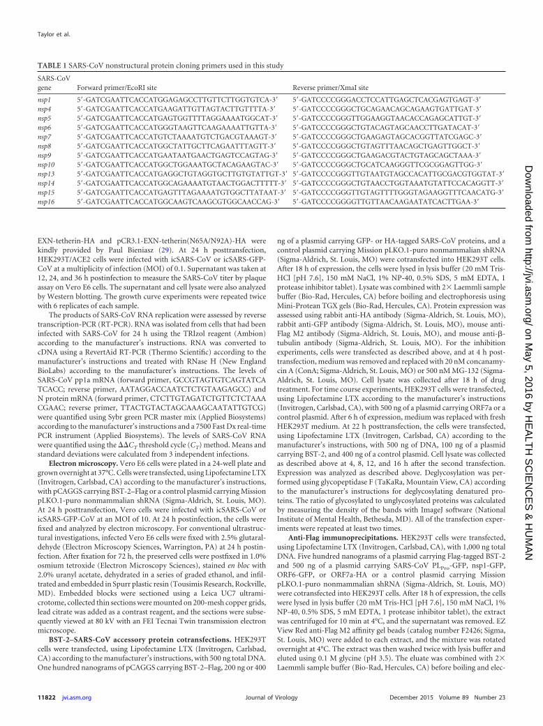
FIG 1 icSARS-CoV and icSARS-ORF7ab�-CoV infection of cells with and without BST-2 expression. (A and B) HEK293T/ACE2 cells were transfected withpCAGGS carrying BST-2–Flag or a control plasmid. At 24 h posttransfection, HEK293T/ACE2 cells were infected with icSARS-CoV (A) or icSARS-ORF7ab�-CoV (B) at an MOI of 0.1. Supernatant and cell lysate were taken at 12, 24, and 36 h postinfection. The virus titer in supernatants taken at 12, 24, and 36 h wasdetermined. (C and D) RNA extracted from icSARS-CoV- and icSARS-ORF7ab�-CoV-infected HEK293T/ACE2 cells was analyzed by real-time PCR forgenomic RNA (C) or leader-containing N mRNA (D) levels as a signature of replicating virus. *, significant at a P value of �0.05; **, significant at a P value of�0.01; ***, significant at a P value of �0.001. The data shown are representative of those from two independent experiments.
BST-2 Restricts SARS Coronavirus
December 2015 Volume 89 Number 23 jvi.asm.org 11823Journal of Virology
on May 5, 2016 by H
EA
LTH
SC
IEN
CE
S &
HU
MA
Nhttp://jvi.asm
.org/D
ownloaded from
inson, Franklin Lakes, NJ). The data were analyzed using FlowJo software(TreeStar, Ashland, OR).
For experiments to determine mutant BST-2 surface expression,HEK293T cells were plated at 250,000 cells per well in 6-well plates, grownovernight at 37°C with 5% CO2, and then transfected with 1 �g of eachDNA (empty vector, empty vector plus WT BST-2, empty vector plusmutant BST-2, empty vector plus ORF7a, WT BST-2 plus ORF7a, mutantBST-2 plus ORF7a). All the following steps were performed at room tem-perature. At 24 h after transfection, cells were harvested using cell disso-ciation buffer (Invitrogen). The contents of duplicate transfected wellswere pooled, and samples were transferred to a 96-well plate. The cellswere pelleted by centrifugation at 2,000 rpm for 2 min, fixed in 4% para-formaldehyde for 5 min, and then washed with 10% FBS in PBS andpelleted as described above. Samples were divided into 2 aliquots and thenblocked with 10% FBS in PBS or blocked/permeabilized with 10% sapo-nin in 10% FBS in PBS for 30 min. The cells were pelleted as describedabove and incubated in primary antibodies for 1 h (HA antibody [1:1,000;catalog number H6908; Sigma] and Flag antibody [1:1,000; catalog num-ber F3165; Sigma]). Cells were washed 2 times with 10% FBS in PBS andpelleted as described above. Secondary antibodies (fluorescein isothiocya-nate-conjugated anti-rabbit immunoglobulin [1:1,000; Vector Laborato-ries] and Alexa Fluor 405-conjugated anti-mouse immunoglobulin [1:1,000; Thermo Fisher]) were added to the cells, and the mixture wasincubated for 1 h, followed by washing and pelleting as described above.Cells were resuspended in PBS, and cell surface localization as well as thetotal cell expression of BST-2 and ORF7a was determined using an LSRII
flow cytometer. Compensation controls consisted of cells transfected withno DNA and cells transfected with BST2 or ORF7a alone. Data were ana-lyzed using FlowJo software, and statistical analysis was performed by a ttest, with standard errors being based on the results from three individualexperiments.
CD of BST-2 and ORF7a-Fc. The circular dichroism (CD) spectra of10 �M BST-2 expressed in HEK293T cells, 12 �M BST-2 expressed inEscherichia coli cells, and 8 �M ORF7a-Fc expressed in HEK239T cellswere recorded at wavelengths ranging from 200 to 260 nm in 10 mMsodium phosphate buffer (pH 7.5) using a Jasco J-810 instrument. CDmelting curves were analyzed at 222 nm by increasing the temperature by1°C/min starting at 20°C.
SPR analysis. Twenty-one micrograms of pCAGGS-T7/ORF7a-Fcwas transfected into HEK293T cells seeded in 100-mm dishes, using Li-pofectamine LTX (Invitrogen, Carlsbad, CA) according to the manufac-turer’s instructions. After 48 h of expression, the supernatant was col-lected and purified using a HiTrap protein A column (GE Healthcare,Little Chalfont, Buckinghamshire, United Kingdom) according to themanufacturer’s instructions. Purified ORF7a-Fc was subsequently dia-lyzed into PBS. The codon-optimized sequence of the extracellular do-main (residues 47 to 161) of BST-2 with an N-terminal His6 tag and aC-terminal Flag tag was cloned into pET28b. The protein was expressed at19°C overnight in BL21(DE3)pLysS cells induced at an optical density at600 nm of 0.6 with 0.4 mM isopropyl-�-D-thiogalactopyranoside. Thefusion protein was purified by nickel affinity chromatography (ThermoScientific, Pittsburg, PA) using a Mono Q 5/50GL anion-exchange col-
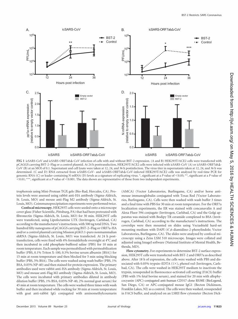
FIG 2 BST-2 tethers SARS-CoV to the plasma membrane. BST-2 or control plasmid was transfected into Vero E6 cells and infected with either icSARS-CoV oricSARS-ORF7ab�-CoV at an MOI of 10. At 24 h postinfection, cells were fixed and imaged by electron microscopy. Transfection of BST-2 results in a largeincrease in the amount of icSARS-ORF7ab�-CoV virions retained at the surface compared to that for control transfected cells. Bars, 500 nm.
Taylor et al.
11824 jvi.asm.org December 2015 Volume 89 Number 23Journal of Virology
on May 5, 2016 by H
EA
LTH
SC
IEN
CE
S &
HU
MA
Nhttp://jvi.asm
.org/D
ownloaded from
umn (GE Healthcare, Little Chalfont, Buckinghamshire, United King-dom) and finally separated on a Superdex 200 10/300 GL gel filtrationcolumn (GE Healthcare, Little Chalfont, Buckinghamshire, United King-dom). The extracellular domain (residues 47 to 161) of BST-2 was alsocloned into the plGplus vector (R&D Systems, Minneapolis, MN). Theresulting BST-2 protein includes a C-terminal His6-Flag tag and, due tothe stop codon carried by the protein, was not expressed as an Fc fusionprotein. HEK293T cells in suspension culture were transfected with thisconstruct using polyethylenimine (Polysciences, Inc., Warrington, PA).The cell culture supernatant was harvested at 96 h posttransfection andpurified using nickel affinity chromatography (Thermo Scientific, Pitts-burg, PA), followed by gel filtration on a Superdex 200 10/300 GL column(GE Healthcare, Little Chalfont, Buckinghamshire, United Kingdom).Direct binding of the extracellular domain of ORF7a-Fc expressed inHEK293T cells to the extracellular domain of BST-2 expressed in E. coli orHEK293T cells was measured by surface plasmon resonance (SPR) anal-ysis using a Biacore T100 instrument (GE Healthcare, Little Chalfont,Buckinghamshire, United Kingdom). Protein A (1,000 resonance units
[RU]) was immobilized by amine coupling on the surface of a CM5 sensorchip. Approximately 170 RU of human IgG (Sigma-Aldrich, St. Louis,MO) as a negative control and also 170 RU of ORF7a-Fc were captured onflow cells 1 and 2, respectively. In single-cycle kinetics experiments, 2-folddilutions from 80 to 5 �M BST-2 were injected over the surfaces, and theresponse was recorded after subtraction of the response of the control.HBS-EP was used as a running buffer, and the surfaces were regeneratedwith 20 mM HCl after each cycle. Steady-state analysis of the data wasperformed using Biacore T100 evaluation software (version 2.0.3). All ofthe SPR experiments were repeated at least three times.
Statistical analysis. Growth curve titers were analyzed by t test usingthe Holm-Sidak method, with alpha being equal to 5.0%. Prism software(GraphPad Software Inc., La Jolla, CA) was used to perform the analysis.
RESULTSSARS-CoV proteins antagonize BST-2 expression in vitro. Thegenomes of many enveloped viruses, including hCoV-229E, en-
100 101 102 103 104
APC-A
0
20
40
60
80
100
% o
f Max
α-Flag
α-HA
α-Tubulin
100 ng BST-2/Flag
ORF7a/HA
25 kDa30 kDa
50 kDa
15 kDa
A.
B.Mock 0.0
BST-2 88.6200 ng ORF7a/HA BST-2 82.1400 ng ORF7a/HA BST-2 84.1
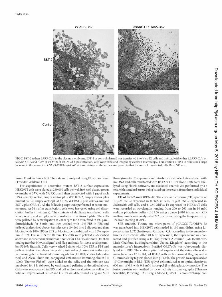
FIG 3 ORF7a expression leads to lower-molecular-mass BST-2 within the cells but not reduced BST-2 surface expression. BST-2 was transfected into HEK293Tcells with increasing amounts of ORF7a. At 18 h posttransfection, the cells were either lysed and analyzed by Western blotting or stained with an anti-BST-2antibody conjugated to APC (APC-A) and analyzed using flow cytometry. Untransfected cells did not express BST-2. Cells transfected with BST-2 showed highlevels of BST-2 expression, and cotransfection of ORF7a and BST-2 led to decreased levels of the high-molecular-mass BST-2 band and increasing levels of alower-molecular-mass BST-2 band in a dose-dependent manner (A) but did not lead to reduced surface expression of BST-2 (B). The data shown arerepresentative of those from two independent experiments.
BST-2 Restricts SARS Coronavirus
December 2015 Volume 89 Number 23 jvi.asm.org 11825Journal of Virology
on May 5, 2016 by H
EA
LTH
SC
IEN
CE
S &
HU
MA
Nhttp://jvi.asm
.org/D
ownloaded from
code proteins that counteract BST-2 (24, 27, 37). We hypothe-sized that the highly pathogenic SARS-CoV may also inhibitBST-2 function. To investigate this hypothesis, HA- and GFP-tagged SARS-CoV proteins and BST-2 were cotransfected intoHEK293T cells and BST-2 expression levels were assessed byWestern blotting. Four SARS-CoV proteins, nonstructural pro-tein 1 (nsp1), the papain-like protease (PLPro) domain of nsp3,ORF6, and ORF7a altered BST-2 protein expression or the BST-2molecular mass. Several proteins encoded by the SARS-CoV ge-nome have been shown to alter other antiviral response pathwaysduring infection (6–8, 43–50). Three of the proteins, PLPro, ORF6,and nsp1, have previously been shown to be interferon antagonists(7, 8, 45). PLPro inhibits interferon regulatory factor 3 (IRF3) andNF-�B activation (45), ORF6 blocks STAT1 nuclear import, andnsp1 blocks beta interferon induction by degrading host mRNAs(6, 7, 45, 48). Because the function of ORF7a is unclear, we de-cided to further study the interactions between BST-2 and ORF7a.
icSARS-ORF7ab�-CoV shows defects in replication com-pared to icSARS-CoV replication when BST-2 is overexpressed.Since ORF7a affects the BST-2 protein, we hypothesized thatSARS-CoV with an ORF7ab deletion (icSARS-ORF7ab�-CoV)would show a greater defect in replication than WT SARS-CoVwhen BST-2 is overexpressed. Since ORF7a and ORF7b have over-lapping open reading frames, a virus with the ORF7ab doubledeletion was used for the infection experiments. No effect onBST-2 was found in transfection screens when ORF7b was ex-pressed alone in the assays. We transfected HEK293T/ACE2 cellswith BST-2 or a control plasmid, and at 24 h posttransfection weinfected the cells with either icSARS-CoV or icSARS-ORF7ab�-CoV at an MOI of 0.1. HEK293T cells do not express endogenousBST-2 (27), so we were able to ensure that any effect was from thetransfected BST-2 and not endogenous BST-2 expression. ic-SARS-CoV replicated to 1.10 � 105 PFU/ml, while in BST-2-ex-pressing cells, icSARS-CoV replicated to significantly lower titersof 3.43 � 104 PFU/ml (Fig. 1A; P � 0.01). icSARS-ORF7ab�-CoVwas also significantly restricted by BST-2 expression at 24 and 36h. In control transfected cells, icSARS-ORF7ab�-CoV replicatedto 7.37 � 104 PFU/ml and 4.80 � 104 PFU/ml at 24 and 36 h,respectively, while in BST-2-transfected cells, icSARS-ORF7ab�-CoV replicated to significantly lower titers of 4.00 � 103 PFU/ml(P � 0.05) and 1.10 � 104 PFU/ml (P � 0.001) at 24 and 36 h,respectively (Fig. 1B). While BST-2 restricts SARS-CoV by a smallbut significant amount, BST-2 restricts icSARS-ORF7ab�-CoVby a much larger amount, suggesting that ORF7a antagonizesBST-2.
We confirmed that BST-2 does not affect another step in theSARS-CoV replication cycle by assessing the accumulation ofSARS-CoV RNA products of replication in the presence of BST-2at 24 h postinfection. There was no significant effect of BST-2expression on SARS-CoV pp1a mRNA (Fig. 1C) or SARS-CoV NmRNA (Fig. 1D), regardless of ORF7a expression. These data sug-gest that BST-2 does not affect SARS-CoV RNA accumulation,even in the absence of ORF7a expression.
The defect in icSARS-ORF7ab�-CoV replication is due to di-rect tethering of SARS-CoV virions to the plasma membrane.Since BST-2 has been shown to restrict virus replication by di-rectly tethering HIV-1 virions to the plasma membrane (27, 51),we sought to determine if overexpression of BST-2 was preventingthe release of SARS-CoV and if icSARS-ORF7ab�-CoV was moresusceptible to BST-2 restriction. To determine if BST-2 was affect-
ing release, we transfected either BST-2 or a control plasmid intoVero E6 cells and subsequently infected the cells with either ic-SARS-CoV or icSARS-ORF7ab�-CoV at an MOI of 10. At 24 hpostinfection, the cells were fixed and imaged using electron mi-croscopy. When the Vero E6 cells were transfected with the con-trol plasmid, the minimal accumulation of both icSARS-CoV andicSARS-ORF7ab�-CoV virions was found at the plasma mem-brane (Fig. 2). Transfection of BST-2 led to the accumulation of asmall amount of icSARS-CoV on the plasma membrane (Fig. 2,top left). BST-2 transfection showed a much greater effect on ic-SARS-ORF7ab�-CoV, with a large amount of virions accumulat-ing at the plasma membrane (Fig. 2, top right). These results con-firm that, as in many other viruses, BST-2 restricts SARS-CoV bypreventing virus release. The increased effect of BST-2 on icSARS-
BST-2/Flag
ORF7a/HA ORF7a/HA ORF7a/HA
Con AMG132
25 30
50
15
α-Flag
α-HA
α-Tubulin
Con AMG132
++
α-Ub
250
10
15
25
35
55
100
150
Con AMG132
++
α-LC3B
α-Tub
15
55
A. B.
C.
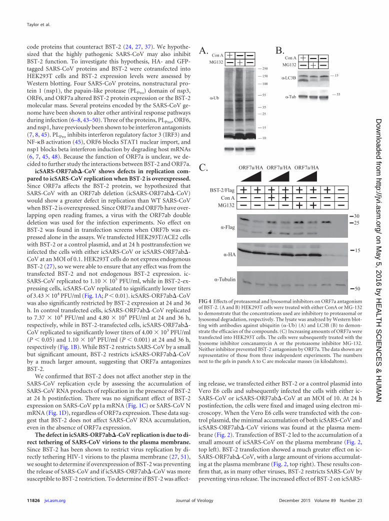
FIG 4 Effects of proteasomal and lysosomal inhibitors on ORF7a antagonismof BST-2. (A and B) HEK293T cells were treated with either ConA or MG-132to demonstrate that the concentrations used are inhibitory to proteasomal orlysosomal degradation, respectively. The lysate was analyzed by Western blot-ting with antibodies against ubiquitin (-Ub) (A) and LC3B (B) to demon-strate the efficacies of the compounds. (C) Increasing amounts of ORF7a weretransfected into HEK293T cells. The cells were subsequently treated with thelysosome inhibitor concanamycin A or the proteasome inhibitor MG-132.Neither inhibitor prevented BST-2 antagonism by ORF7a. The data shown arerepresentative of those from three independent experiments. The numbersnext to the gels in panels A to C are molecular masses (in kilodaltons).
Taylor et al.
11826 jvi.asm.org December 2015 Volume 89 Number 23Journal of Virology
on May 5, 2016 by H
EA
LTH
SC
IEN
CE
S &
HU
MA
Nhttp://jvi.asm
.org/D
ownloaded from
ORF7ab�-CoV further suggests that ORF7a acts as an inhibitor ofBST-2-mediated restriction of SARS-CoV.
ORF7a expression leads to lower-molecular-mass BST-2within the cells but not reduced BST-2 surface expression. SinceORF7a appears to be a BST-2 antagonist, we aimed to determine ifSARS-CoV ORF7a causes BST-2 surface removal and subsequentdegradation, as seen in HIV-1 Vpu protein antagonism (33, 51).SARS-CoV ORF7a was cotransfected with increasing amounts ofBST-2 to assay the effect of ORF7a on BST-2 expression. Increas-ing the amount of ORF7a cotransfected with BST-2 led to de-creased levels of BST-2 expression and lower-molecular-mass
products, suggesting that the BST-2 protein is affected by ORF7aexpression (Fig. 3A). Next, we sought to determine if, like HIV-1Vpu, the expression of ORF7a leads to a reduction in BST-2 sur-face expression (51). To assay the effect of ORF7a on BST-2 sur-face expression, we transfected BST-2 either alone or in combina-tion with an ORF7a expression plasmid to compare BST-2 surfaceexpression by flow cytometry. Untransfected cells exhibited littleto no expression of surface BST-2. Cells transfected with BST-2alone were 88.2% positive for surface BST-2 expression, with themajority of cells being in a population expressing larger amountsand a smaller percentage being in a population expressing smaller
egreMa7FRO Golgi
egreMREORF7a
A.
Alone
BST-2
Cotransfected
ORF7a
Merge
B.
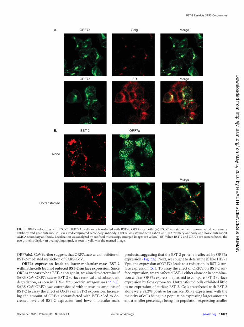
FIG 5 ORF7a colocalizes with BST-2. HEK293T cells were transfected with BST-2, ORF7a, or both. (A) BST-2 was stained with mouse anti-Flag primaryantibody and goat anti-mouse Texas Red-conjugated secondary antibody. ORF7a was stained with rabbit anti-HA primary antibody and horse anti-rabbitAMCA secondary antibody. Localization was analyzed by confocal microscopy (merged images are yellow). (B) When BST-2 and ORF7a are cotransfected, thetwo proteins display an overlapping signal, as seen in yellow in the merged image.
BST-2 Restricts SARS Coronavirus
December 2015 Volume 89 Number 23 jvi.asm.org 11827Journal of Virology
on May 5, 2016 by H
EA
LTH
SC
IEN
CE
S &
HU
MA
Nhttp://jvi.asm
.org/D
ownloaded from
amounts (Fig. 3B). Interestingly, increasing amounts of ORF7ahad no effect on the surface expression of BST-2 (Fig. 3B). Thesedata demonstrate that ORF7a coexpression leads to lower-molec-ular-mass BST-2 within cells but does not lead to the removal ofBST-2 from the surface, suggesting that ORF7a may antagonizeBST-2 through a mechanism other than removal from the surface.
Lysosomal and proteasomal inhibitors do not affect BST-2antagonism by ORF7a. While we did not observe the ORF7a-dependent removal of surface BST-2, we did observe the appear-ance of lower-molecular-mass bands of BST-2, suggesting degra-dation of intracellular BST-2. Many other viruses, such as HIV-1,antagonize BST-2 by degradation through either the lysosome orthe proteasome, and thus, we assessed whether lysosomal or pro-teasomal inhibitors could block BST-2 antagonism by ORF7a(32–34). First, to demonstrate that concanamycin A (ConA) andMG-132 inhibit proteasome and lysosomal degradation, respec-tively, at the concentrations used in HEK293T cells, we treatedcells and assayed for ubiquitin and LC3B levels by Western blot-ting (Fig. 4A and B). As expected, MG-132 treatment increasedtotal ubiquitin levels in the cell (Fig. 4A) and ConA treatmentblocked lysosomal degradation, as shown by an increase in thelower-molecular-mass LC3B product. To test for the effect of pro-teosomal or lysosomal effects on BST-2 antagonism, we trans-fected HEK293T cells with plasmids carrying BST-2 and ORF7a ora control plasmid and at 4 h posttransfection replaced the mediumwith medium containing either 20 nM concanamycin A (to in-hibit lysosomal degradation) or 500 nM MG-132 (to inhibit pro-teasome function). At 18 h posttransfection, the cells were lysedand analyzed by Western blotting to determine if BST-2 was de-graded. After treatment, lower-molecular-mass bands were stillobserved. Treatment with neither concanamycin A nor MG-132blocked the ability of ORF7a to antagonize BST-2 (Fig. 4C). The 2lanes on the far right in Fig. 4C contain a background band with amolecular mass similar to that of HA-tagged ORF7a that did notaffect the experiment. These data demonstrate that the appear-ance of lower-molecular-mass bands of BST-2 is not due to lyso-somal or proteasomal degradation and suggest that ORF7a antag-onizes BST-2 through an alternative mechanism.
BST-2 colocalizes with and alters the localization of SARS-CoV ORF7. Since icSARS-ORF7ab�-CoV is more susceptible toBST-2 restriction and ORF7a appears to cause the appearance of alow-molecular-mass BST-2 band, we hypothesized that BST-2may alter the ORF7a localization within the cell. ORF7a was trans-fected into HEK293T cells, and the cells were stained for ORF7a aswell as the ER and Golgi apparatus (9, 10). ORF7a normally local-
izes to the Golgi apparatus and was also detectable in the ER, aswould be expected for a protein that passes through the ER to theGolgi apparatus (Fig. 5A). To determine if BST-2 and ORF7a co-localize, we performed confocal microscopy. When transfectedalone, ORF7a localized primarily to the Golgi apparatus, whereasBST-2 localized to the plasma membrane (Fig. 5B). When BST-2and ORF7a were cotransfected, ORF7a appeared to localize to theplasma membrane, coincident with BST-2 (Fig. 5B). These datasuggest that BST-2 and ORF7a may be interacting in cells.
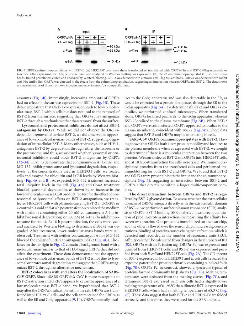
SARS-CoV ORF7a coimmunoprecipitates with BST-2. Hav-ing shown that ORF7a both alters protein mobility and localizes tothe plasma membrane when coexpressed with BST-2, we soughtto determine if there is a molecular interaction between the twoproteins. We cotransfected BST-2 and ORF7a into HEK293T cells,and at 18 h posttransfection the cells were lysed. We immunopre-cipitated proteins from the transfected cells and performed im-munoblotting for both BST-2 and ORF7a. We found that BST-2and ORF7a were present in both the input and the coimmunopre-cipitate (Fig. 6), suggesting an interaction between BST-2 andORF7a either directly or within a larger multicomponent com-plex.
The direct interaction between ORF7a and BST-2 is regu-lated by BST-2 glycosylation. To assess whether the extracellulardomain of ORF7a interacts directly with the extracellular domainof BST-2, we performed surface plasmon resonance (SPR) analy-sis of ORF7a–BST-2 binding. SPR analysis allows direct quantita-tion of protein-protein interactions by measuring the affinity be-tween two proteins. One protein is immobilized on a sensor chip,and the other is flowed over the sensor chip in increasing concen-trations. Binding of proteins causes changes in refraction, which isdetected and recorded as the number of resonance units (RU).Affinity can then be calculated from changes in the numbers of RU(52). ORF7a with an Fc fusion tag (ORF7a-Fc) was expressed andpurified from HEK293T cells, and BST-2 was expressed and puri-fied from both E. coli and HEK293T cells (Fig. 7A). The CD spectraof BST-2 expressed in both HEK239T and E. coli cells revealed theexpected pattern for a protein primarily containing -helical folds(Fig. 7B). ORF7a-Fc, in contrast, showed a spectrum typical ofproteins formed dominantly by � sheets (Fig. 7B). Melting tem-peratures were deduced from the melting curves (Fig. 7C), andtetrameric BST-2 expressed in E. coli cells had a slightly lowermelting temperature of 61.95°C than dimeric BST-2 expressed inHEK293T cells, which had a melting temperature of 65.3°C (Fig.7C). These data suggest that both BST-2 and ORF7a-Fc are foldedcorrectly, and therefore, they were used for the SPR analysis.
FIG 6 ORF7a coimmunoprecipitates with BST-2. (A) HEK293T cells were sham transfected or transfected with ORF7a-HA and BST-2–Flag separately ortogether. After expression for 18 h, cells were lysed and analyzed by Western blotting for expression. (B) BST-2 was immunoprecipitated (IP) with anti-Flagbeads. Bound protein was eluted and analyzed by Western blotting. BST-2 was detected with a mouse anti-Flag M2 antibody. ORF7a was detected with rabbitanti-HA antibodies. ORF7a was detected in the eluate from the coimmunoprecipitation, suggesting an interaction between ORF7a and BST-2. The data shownare representative of those from two independent experiments. *, a nonspecific band.
Taylor et al.
11828 jvi.asm.org December 2015 Volume 89 Number 23Journal of Virology
on May 5, 2016 by H
EA
LTH
SC
IEN
CE
S &
HU
MA
Nhttp://jvi.asm
.org/D
ownloaded from
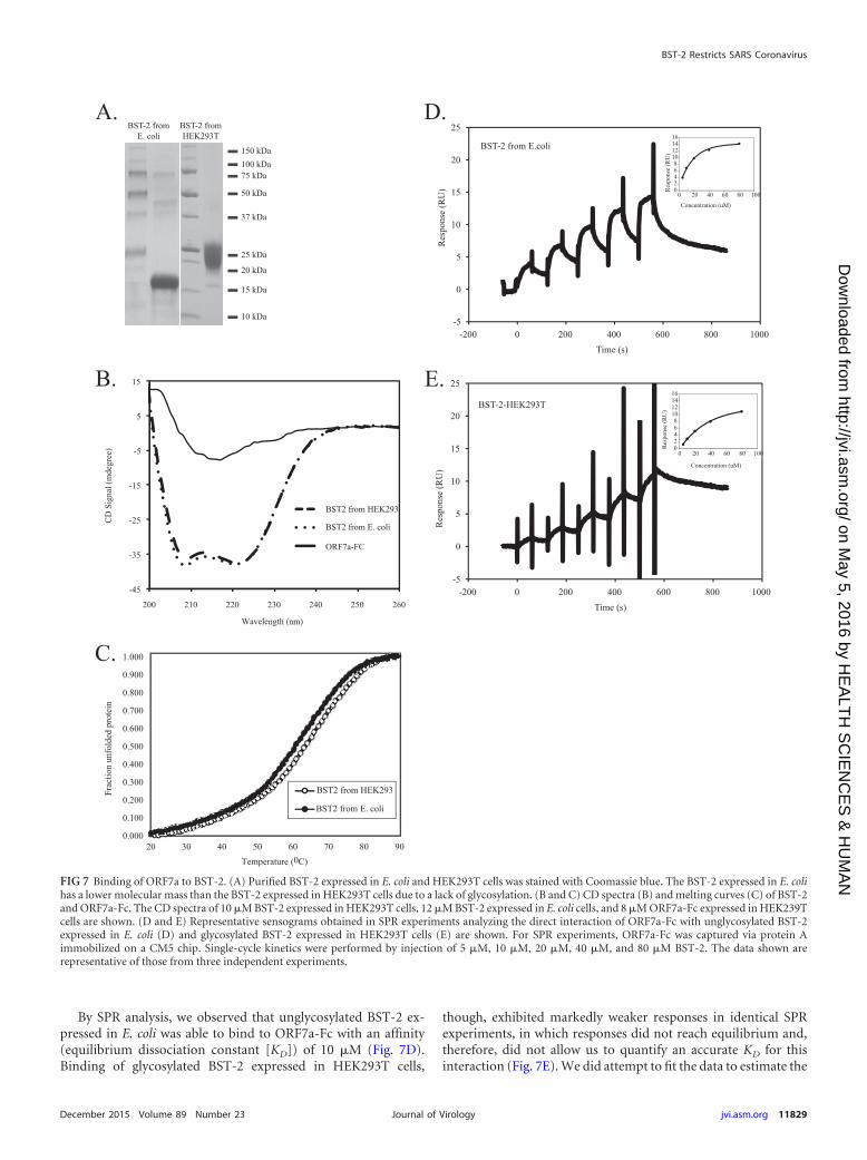
By SPR analysis, we observed that unglycosylated BST-2 ex-pressed in E. coli was able to bind to ORF7a-Fc with an affinity(equilibrium dissociation constant [KD]) of 10 �M (Fig. 7D).Binding of glycosylated BST-2 expressed in HEK293T cells,
though, exhibited markedly weaker responses in identical SPRexperiments, in which responses did not reach equilibrium and,therefore, did not allow us to quantify an accurate KD for thisinteraction (Fig. 7E). We did attempt to fit the data to estimate the
25 kDa
37 kDa
50 kDa
20 kDa
75 kDa100 kDa150 kDa
15 kDa
10 kDa
BST-2 from E. coli
BST-2 fromHEK293T
-200 0 200 400 600 800 1000-5
0
5
10
15
20
25
0 20 40 60 80 10002468
10121416
-200 0 200 400 600 800 1000-5
0
5
10
15
20
25
BST-2-HEK293T
BST-2 from E.coli
Time (s)
Time (s)
Res
pons
e (R
U)
0 20 40 60 80 10002468
10121416
Res
pons
e (R
U)
Res
pons
e (R
U) R
espo
nse
(RU
)
Concentration (uM)
-45
-35
-25
-15
-5
5
15
200 210 220 230 240 250 260
CD
Sig
nal (
mde
gree
)
Wavelength (nm)
BST2 from HEK293
BST2 from E. coli
ORF7a-FC
0.000
0.100
0.200
0.300
0.400
0.500
0.600
0.700
0.800
0.900
1.000
20 30 40 50 60 70 80 90
Frac
tion
unfo
lded
pro
tein
Temperature (0C)
BST2 from HEK293
BST2 from E. coli
Concentration (uM)
A.
B.
C.
D.
E.
FIG 7 Binding of ORF7a to BST-2. (A) Purified BST-2 expressed in E. coli and HEK293T cells was stained with Coomassie blue. The BST-2 expressed in E. colihas a lower molecular mass than the BST-2 expressed in HEK293T cells due to a lack of glycosylation. (B and C) CD spectra (B) and melting curves (C) of BST-2and ORF7a-Fc. The CD spectra of 10 �M BST-2 expressed in HEK293T cells, 12 �M BST-2 expressed in E. coli cells, and 8 �M ORF7a-Fc expressed in HEK239Tcells are shown. (D and E) Representative sensograms obtained in SPR experiments analyzing the direct interaction of ORF7a-Fc with unglycosylated BST-2expressed in E. coli (D) and glycosylated BST-2 expressed in HEK293T cells (E) are shown. For SPR experiments, ORF7a-Fc was captured via protein Aimmobilized on a CM5 chip. Single-cycle kinetics were performed by injection of 5 �M, 10 �M, 20 �M, 40 �M, and 80 �M BST-2. The data shown arerepresentative of those from three independent experiments.
BST-2 Restricts SARS Coronavirus
December 2015 Volume 89 Number 23 jvi.asm.org 11829Journal of Virology
on May 5, 2016 by H
EA
LTH
SC
IEN
CE
S &
HU
MA
Nhttp://jvi.asm
.org/D
ownloaded from
KD for this interaction, and the binding of ORF7a to glycosylatedBST-2 was at least 4 times weaker than that to unglycosylatedBST-2. These data indicate that ORF7a binds directly to unglyco-sylated BST-2 with an affinity at micromolar concentrations andthat the presence of N-linked glycosylation at positions 65 and 92of BST-2 significantly weakens this interaction.
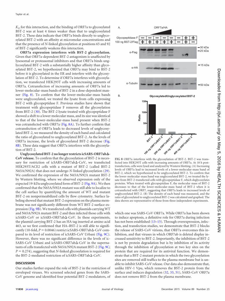
ORF7a expression interferes with BST-2 glycosylation.Given that ORF7a-dependent BST-2 antagonism is unaffected bylysosomal or proteasomal inhibitors and that ORF7a binds ung-lycosylated BST-2 with a substantially higher affinity than glyco-sylated BST-2, we hypothesized that ORF7a may bind to BST-2before it is glycosylated in the ER and interfere with the glycosy-lation of BST-2. To determine if ORF7a interferes with glycosyla-tion, we transfected HEK293T cells with increasing amounts ofORF7a. Cotransfection of increasing amounts of ORF7a led tolower-molecular-mass bands of BST-2 in a dose-dependent man-ner (Fig. 8). To confirm that the lower-molecular-mass bandswere unglycosylated, we treated the lysate from cells expressingBST-2 with glycopeptidase F. Previous studies have shown thattreatment with glycopeptidase F removes all the glycosylationfrom BST-2 (30). The BST-2 lysate treated with glycopeptidase Fshowed a shift to a lower molecular mass, and its size was identicalto that of the lower-molecular-mass band present when BST-2was cotransfected with ORF7a (Fig. 8A). To further confirm thatcotransfection of ORF7a leads to decreased levels of unglycosy-lated BST-2, we measured the density of each band and calculatedthe ratio of glycosylated to unglycosylated BST-2. As the levels ofORF7a increase, the levels of glycosylated BST-2 decrease (Fig.8B). These data suggest that ORF7a interferes with the glycosyla-tion of BST-2.
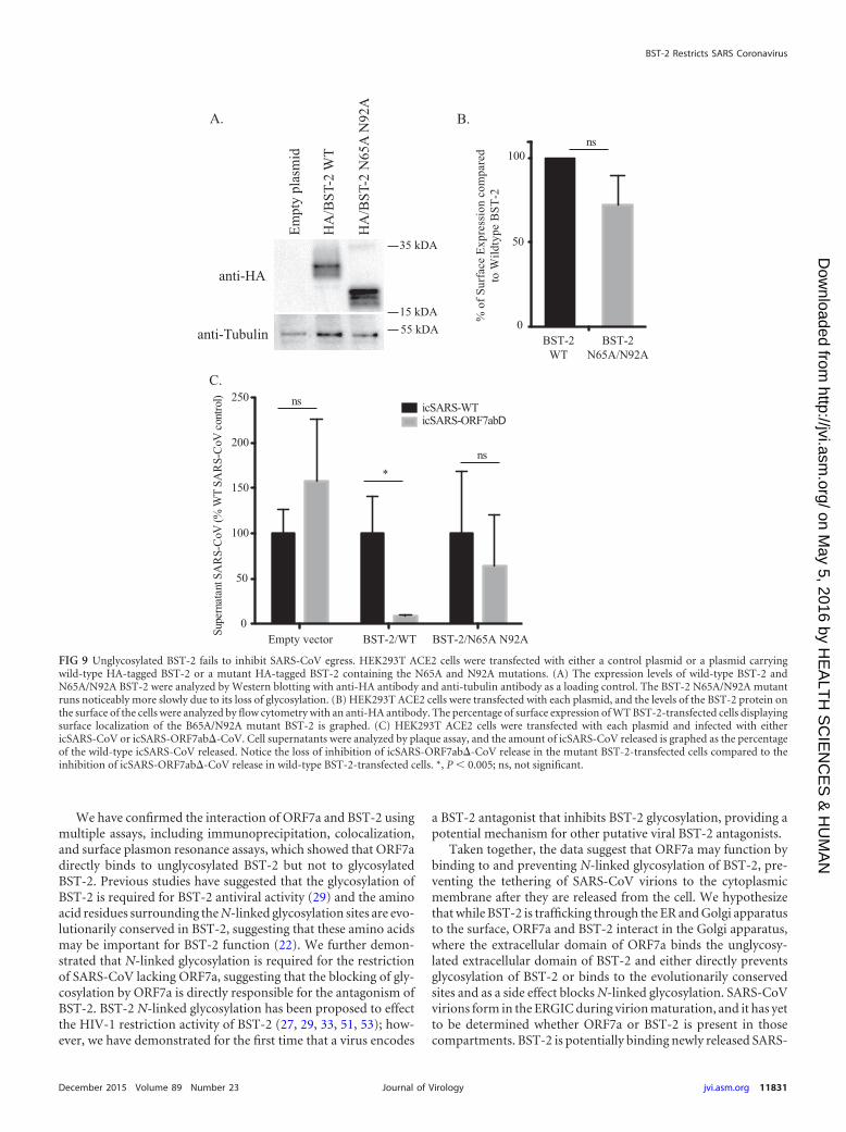
Unglycosylated BST-2 no longer restricts icSARS-ORF7ab�-CoV release. To confirm that the glycosylation of BST-2 is neces-sary for restriction of icSARS-ORF7ab�-CoV, we transfectedHEK293T/ACE2 cells with a mutant of BST-2 (called BST-2N65A/N92A) that does not undergo N-linked glycosylation (29).We confirmed the expression of the N65A/N92A mutant BST-2by Western blotting, where we observed expression only of theexpected 19-kDa unglycosylated form of BST-2 (Fig. 9A). We thenconfirmed that the N65A/N92A mutant was still able to localize tothe cell surface by quantifying the amount of WT and mutantBST-2 on nonpermeabilized cells by flow cytometry. Surface la-beling showed that mutant BST-2 expression on the plasma mem-brane was not significantly different from WT BST-2 surface ex-pression (Fig. 9B). We transfected cells with plasmids carrying WTand N65A/N92A mutant BST-2 and then infected those cells withicSARS-CoV or icSARS-ORF7ab�-CoV. In these experiments,the plasmid carrying BST-2 has an HA tag inserted at amino acid463, so we first confirmed that HA–BST-2 is still able to signifi-cantly (10-fold, P 0.0046) restrict icSARS-ORF7ab�-CoV com-pared to its level of restriction of icSARS-CoV Urbani (Fig. 9C).However, there was no significant difference in the levels of ic-SARS-CoV Urbani and icSARS-ORF7ab�-CoV in the superna-tants of cells transfected with N65A/N92A mutant BST-2 (Fig. 9C;P 0.274), suggesting that N-linked glycosylation is required forthe BST-2-mediated restriction of icSARS-ORF7ab�-CoV.
DISCUSSION
Our studies further expand the role of BST-2 in the restriction ofenveloped viruses. We screened selected genes from the SARS-CoV genome and identified four potential BST-2 modulators, of
which one was SARS-CoV ORF7a. While ORF7a has been shownto induce apoptosis, a definitive role for ORF7a during infectionhas not been established (13–15). Through overexpression, infec-tion, and transfection studies, we demonstrate that BST-2 blocksthe release of SARS-CoV virions, that ORF7a overcomes this in-hibition, and that viruses in which ORF7ab is deleted display in-creased sensitivity to BST-2. Importantly, the inhibition of BST-2is not by protein degradation but is by inhibition of its activitythrough the inhibition of glycosylation at two key sites on theprotein that are required for its antiviral function. We demon-strate that a BST-2 mutant protein in which the two glycosylationsites are removed still traffics to the plasma membrane but is un-able to inhibit SARS-CoV release. Our data also demonstrate that,unlike HIV-1 Vpu, which removes the BST-2 protein from thesurface and induces degradation (32, 33, 51), SARS-CoV ORF7adoes not remove BST-2 from the plasma membrane.
A.
100 ng BST-2/Flag
ORF7a/HA
α-Flag
α-HA
α-Tubulin
25 kDa30 kDa
50 kDa
15 kDa
Glycopeptidase F
BST-2
100 n
g ORF7a
/BST-2
200 n
g ORF7a
/BST-2
300 n
g ORF7a
/BST-2
400 n
g ORF7a
/BST-2
0
50
100
150
Glycosylated/Unglycosylated BST-2
Rat
io
B.
FIG 8 ORF7a interferes with the glycosylation of BST-2. BST-2 was trans-fected into HEK293T cells with increasing amounts of ORF7a. At 18 h post-transfection, cells were lysed and analyzed by Western blotting. (A) Increasinglevels of ORF7a lead to increased levels of a lower-molecular-mass band ofBST-2, which we hypothesized to be unglycosylated BST-2. To confirm thatthe lower-molecular-mass band was unglycosylated BST-2, we treated the ly-sate from BST-2-transfected cells with glycopeptidase F, which deglycosylatesproteins. When treated with glycopeptidase F, the molecular mass of BST-2decreases to that of the lower-molecular-mass band of BST-2 when it iscotransfected with ORF7, suggesting that ORF7a leads to increased levels ofunglycosylated BST-2. (B) The density of each band was measured, and theratio of glycosylated to unglycosylated BST-2 was calculated and graphed. Thedata shown are representative of those from three independent experiments.
Taylor et al.
11830 jvi.asm.org December 2015 Volume 89 Number 23Journal of Virology
on May 5, 2016 by H
EA
LTH
SC
IEN
CE
S &
HU
MA
Nhttp://jvi.asm
.org/D
ownloaded from
We have confirmed the interaction of ORF7a and BST-2 usingmultiple assays, including immunoprecipitation, colocalization,and surface plasmon resonance assays, which showed that ORF7adirectly binds to unglycosylated BST-2 but not to glycosylatedBST-2. Previous studies have suggested that the glycosylation ofBST-2 is required for BST-2 antiviral activity (29) and the aminoacid residues surrounding the N-linked glycosylation sites are evo-lutionarily conserved in BST-2, suggesting that these amino acidsmay be important for BST-2 function (22). We further demon-strated that N-linked glycosylation is required for the restrictionof SARS-CoV lacking ORF7a, suggesting that the blocking of gly-cosylation by ORF7a is directly responsible for the antagonism ofBST-2. BST-2 N-linked glycosylation has been proposed to effectthe HIV-1 restriction activity of BST-2 (27, 29, 33, 51, 53); how-ever, we have demonstrated for the first time that a virus encodes
a BST-2 antagonist that inhibits BST-2 glycosylation, providing apotential mechanism for other putative viral BST-2 antagonists.
Taken together, the data suggest that ORF7a may function bybinding to and preventing N-linked glycosylation of BST-2, pre-venting the tethering of SARS-CoV virions to the cytoplasmicmembrane after they are released from the cell. We hypothesizethat while BST-2 is trafficking through the ER and Golgi apparatusto the surface, ORF7a and BST-2 interact in the Golgi apparatus,where the extracellular domain of ORF7a binds the unglycosy-lated extracellular domain of BST-2 and either directly preventsglycosylation of BST-2 or binds to the evolutionarily conservedsites and as a side effect blocks N-linked glycosylation. SARS-CoVvirions form in the ERGIC during virion maturation, and it has yetto be determined whether ORF7a or BST-2 is present in thosecompartments. BST-2 is potentially binding newly released SARS-
anti-HA
anti-Tubulin
Empt
y pl
asm
id
HA
/BST
-2 W
T
HA
/BST
-2 N
65A
N92
A
A.
35 kDA
15 kDA55 kDA
Supe
rnat
ant S
AR
S-C
oV (%
WT
SAR
S-C
oV c
ontro
l)
Empty vector BST-2/WT BST-2/N65A N92A0
50
100
150
200
250icSARS-WTicSARS-ORF7abD
ns
*ns
C.
% o
f Sur
face
Exp
ress
ion
com
pare
dto
Wild
type
BST
-2
0
50
100
BST-2WT
BST-2N65A/N92A
B.ns
FIG 9 Unglycosylated BST-2 fails to inhibit SARS-CoV egress. HEK293T ACE2 cells were transfected with either a control plasmid or a plasmid carryingwild-type HA-tagged BST-2 or a mutant HA-tagged BST-2 containing the N65A and N92A mutations. (A) The expression levels of wild-type BST-2 andN65A/N92A BST-2 were analyzed by Western blotting with anti-HA antibody and anti-tubulin antibody as a loading control. The BST-2 N65A/N92A mutantruns noticeably more slowly due to its loss of glycosylation. (B) HEK293T ACE2 cells were transfected with each plasmid, and the levels of the BST-2 protein onthe surface of the cells were analyzed by flow cytometry with an anti-HA antibody. The percentage of surface expression of WT BST-2-transfected cells displayingsurface localization of the B65A/N92A mutant BST-2 is graphed. (C) HEK293T ACE2 cells were transfected with each plasmid and infected with eithericSARS-CoV or icSARS-ORF7ab�-CoV. Cell supernatants were analyzed by plaque assay, and the amount of icSARS-CoV released is graphed as the percentageof the wild-type icSARS-CoV released. Notice the loss of inhibition of icSARS-ORF7ab�-CoV release in the mutant BST-2-transfected cells compared to theinhibition of icSARS-ORF7ab�-CoV release in wild-type BST-2-transfected cells. *, P � 0.005; ns, not significant.
BST-2 Restricts SARS Coronavirus
December 2015 Volume 89 Number 23 jvi.asm.org 11831Journal of Virology
on May 5, 2016 by H
EA
LTH
SC
IEN
CE
S &
HU
MA
Nhttp://jvi.asm
.org/D
ownloaded from
CoV virions at the plasma membrane; however, most models ofBST-2 function predict that BST-2 is inserted into the membraneas the virion forms (20), so we would predict that BST-2 firstinteracts with SARS-CoV virions in the ERGIC.
While the genomes of a variety of enveloped viruses encodeBST-2 antagonists, those antagonists function by different mech-anisms. Both HIV-1 Vpu and Kaposi’s sarcoma-associated her-pesvirus K5 ubiquitinate BST-2, leading to surface removal andsubsequent lysosomal degradation (27, 51, 54). HIV-2 Env alsoremoves BST-2 from the surface, but rather than being degraded,BST-2 is relocated to the trans-Golgi network and cannot functionas a cytoplasmic membrane tether (36). SIV Env removes BST-2from the surface through BST-2 internalization by endocytosis(38, 55). Ebola virus GP1,2 does not remove BST-2 from the sur-face but antagonizes BST-2 through an as yet unknown mecha-nism (39). The diverse mechanisms of known BST-2 antagonistsdemonstrate that viruses have independently evolved many differ-ent ways of antagonizing BST-2, an important restriction factorfor any enveloped virus. It is possible that the genomes of all en-veloped viruses encode BST-2 antagonists that act by a variety ofmechanisms, but in most viruses these remain undiscovered.
In this study, we have identified BST-2 to be a potential inhib-itor of SARS-CoV release. Our studies suggest that SARS-CoVORF7a antagonizes the function of BST-2 by interfering with itsN-linked glycosylation while binding it in the Golgi apparatus andthen trafficking with it from the Golgi apparatus to the plasmamembrane. From this we predict that therapeutics designed toinhibit the interaction between BST-2 and ORF7a may inhibitvirus growth in vitro and in vivo.
ACKNOWLEDGMENTS
This work was supported by the Division of Intramural Research of theNational Institute of Allergy and Infectious Diseases (NIAID) with grantRO1AI1095569 (to M.B.F.) and by NIAID grant R01AI087452 (to E.J.S.).
REFERENCES1. World Health Organization. 2003. WHO summary of probable SARS
cases with onset of illness from 1 November 2002 to 31 July 2003. WorldHealth Organization, Geneva, Switzerland.
2. Drosten C, Preiser W, Günther S, Schmitz H, Doerr HW. 2003. Severeacute respiratory syndrome: identification of the etiological agent. TrendsMol Med 9:325–327. http://dx.doi.org/10.1016/S1471-4914(03)00133-3.
3. Marra MA, Jones SJM, Astell CR, Holt RA, Brooks-Wilson A, ButterfieldYSN, Khattra J, Asano JK, Barber SA, Chan SY, Cloutier A, Coughlin SM,Freeman D, Girn N, Griffith OL, Leach SR, Mayo M, McDonald H,Montgomery SB, Pandoh PK, Petrescu AS, Robertson AG, Schein JE,Siddiqui A, Smailus DE, Stott JM, Yang GS, Plummer F, Andonov A,Artsob H, Bastien N, Bernard K, Booth TF, Bowness D, Czub M, DrebotM, Fernando L, Flick R, Garbutt M, Gray M, Grolla A, Jones S, FeldmannH, Meyers A, Kabani A, Li Y, Normand S, Stroher U, Tipples GA, Tyler S,et al. 2003. The genome sequence of the SARS-associated coronavirus. Sci-ence 300:1399–1404. http://dx.doi.org/10.1126/science.1085953.
4. Li W, Moore MJ, Vasilieva N, Sui J, Wong SK, Berne MA, So-masundaran M, Sullivan JL, Luzuriaga K, Greenough TC, Choe H,Farzan M. 2003. Angiotensin-converting enzyme 2 is a functional recep-tor for the SARS coronavirus. Nature 426:450 – 454. http://dx.doi.org/10.1038/nature02145.
5. Rota PA, Oberste MS, Monroe SS, Nix WA, Campagnoli R, Icenogle JP,Peñaranda S, Bankamp B, Maher K, Chen M-H, Tong S, Tamin A,Lowe L, Frace M, DeRisi JL, Chen Q, Wang D, Erdman DD, Peret TCT,Burns C, Ksiazek TG, Rollin PE, Sanchez A, Liffick S, Holloway B,Limor J, McCaustland K, Olsen-Rasmussen M, Fouchier R, Günther S,Osterhaus ADME, Drosten C, Pallansch MA, Anderson LJ, Bellini WJ.2003. Characterization of a novel coronavirus associated with severe acuterespiratory syndrome. Science 300:1394 –1399. http://dx.doi.org/10.1126/science.1085952.
6. Frieman M, Yount B, Heise M, Kopecky-Bromberg SA, Palese P, BaricRS. 2007. Severe acute respiratory syndrome coronavirus ORF6 antago-nizes STAT1 function by sequestering nuclear import factors on the roughendoplasmic reticulum/Golgi membrane. J Virol 81:9812–9824. http://dx.doi.org/10.1128/JVI.01012-07.
7. Kopecky-Bromberg SA, Martínez-Sobrido L, Frieman M, Baric RA, PaleseP. 2007. Severe acute respiratory syndrome coronavirus open reading frame(ORF) 3b, ORF 6, and nucleocapsid proteins function as interferon antago-nists. J Virol 81:548–557. http://dx.doi.org/10.1128/JVI.01782-06.
8. Wathelet MG, Orr M, Frieman MB, Baric RS. 2007. Severe acute respi-ratory syndrome coronavirus evades antiviral signaling: role of nsp1 andrational design of an attenuated strain. J Virol 81:11620 –11633. http://dx.doi.org/10.1128/JVI.00702-07.
9. Nelson CA, Pekosz A, Lee CA, Diamond MS, Fremont DH. 2005.Structure and intracellular targeting of the SARS-coronavirus Orf7a ac-cessory protein. Structure 13:75– 85. http://dx.doi.org/10.1016/j.str.2004.10.010.
10. Schaecher SR, Touchette E, Schriewer J, Buller M, Pekosz A. 2007.Severe acute respiratory syndrome coronavirus gene 7 products contrib-ute to virus-induced apoptosis. J Virol 81:11054 –11068. http://dx.doi.org/10.1128/JVI.01266-07.
11. Frieman MB, Yount B, Sims AC, Deming DJ, Morrison TE, Sparks J,Denison M, Heise M, Baric RS. 2006. SARS coronavirus accessory ORFsencode luxury functions. Adv Exp Med Biol 581:149 –152. http://dx.doi.org/10.1007/978-0-387-33012-9_26.
12. Yount B, Roberts RS, Sims AC, Deming D, Frieman MB, Sparks J,Denison MR, Davis N, Baric RS. 2005. Severe acute respiratory syn-drome coronavirus group-specific open reading frames encode nonessen-tial functions for replication in cell cultures and mice. J Virol 79:14909 –14922. http://dx.doi.org/10.1128/JVI.79.23.14909-14922.2005.
13. Schaecher SR, Touchette E, Schriewer J, Buller RM, Pekosz A. 2007.Severe acute respiratory syndrome coronavirus gene 7 products contrib-ute to virus-induced apoptosis. J Virol 81:11054 –11068. http://dx.doi.org/10.1128/JVI.01266-07.
14. Tan Y-J, Fielding BC, Goh P-Y, Shen S, Tan THP, Lim SG, Hong W.2004. Overexpression of 7a, a protein specifically encoded by the severeacute respiratory syndrome coronavirus, induces apoptosis via a caspase-dependent pathway. J Virol 78:14043–14047. http://dx.doi.org/10.1128/JVI.78.24.14043-14047.2004.
15. Tan Y-X, Tan THP, Lee MJR, Tham P-Y, Gunalan V, Druce J, Birch C,Catton M, Fu NY, Yu VC, Tan Y-J. 2007. Induction of apoptosis by thesevere acute respiratory syndrome coronavirus 7a protein is dependent onits interaction with the Bcl-XL protein. J Virol 81:6346 – 6355. http://dx.doi.org/10.1128/JVI.00090-07.
16. Tang X, Li G, Vasilakis N, Zhang Y, Shi Z, Zhong Y, Wang L-F, ZhangS. 2009. Differential stepwise evolution of SARS coronavirus functionalproteins in different host species. BMC Evol Biol 9:52. http://dx.doi.org/10.1186/1471-2148-9-52.
17. Goto T, Kennel SJ, Abe M, Takishita M, Kosaka M, Solomon A, SaitoS. 1994. A novel membrane antigen selectively expressed on terminallydifferentiated human B cells. Blood 84:1922–1930.
18. Ishikawa J, Kaisho T, Tomizawa H, Lee BO, Kobune Y, Inazawa J,Oritani K, Itoh M, Ochi T, Ishihara K, Hirano T. 1995. Molecularcloning and chromosomal mapping of a bone marrow stromal cell surfacegene, BST2, that may be involved in pre-B-cell growth. Genomics 26:527–534. http://dx.doi.org/10.1016/0888-7543(95)80171-H.
19. Blasius AL, Giurisato E, Cella M, Schreiber RD, Shaw AS, Colonna M.2006. Bone marrow stromal cell antigen 2 is a specific marker of type IIFN-producing cells in the naive mouse, but a promiscuous cell surfaceantigen following IFN stimulation. J Immunol 177:3260 –3265. http://dx.doi.org/10.4049/jimmunol.177.5.3260.
20. Kupzig S, Korolchuk V, Rollason R, Sugden A, Wilde A, Banting G.2003. Bst-2/HM1.24 is a raft-associated apical membrane protein withan unusual topology. Traffic 4:694 –709. http://dx.doi.org/10.1034/j.1600-0854.2003.00129.x.
21. Ohtomo T, Sugamata Y, Ozaki Y, Ono K, Yoshimura Y, Kawai S,Koishihara Y, Ozaki S, Kosaka M, Hirano T, Tsuchiya M. 1999. Mo-lecular cloning and characterization of a surface antigen preferentiallyoverexpressed on multiple myeloma cells. Biochem Biophys Res Commun258:583–591. http://dx.doi.org/10.1006/bbrc.1999.0683.
22. Swiecki M, Scheaffer SM, Allaire M, Fremont DH, Colonna M, Brett TJ.2011. Structural and biophysical analysis of BST-2/tetherin ectodomains
Taylor et al.
11832 jvi.asm.org December 2015 Volume 89 Number 23Journal of Virology
on May 5, 2016 by H
EA
LTH
SC
IEN
CE
S &
HU
MA
Nhttp://jvi.asm
.org/D
ownloaded from
reveals an evolutionary conserved design to inhibit virus release. J BiolChem 286:2987–2997. http://dx.doi.org/10.1074/jbc.M110.190538.
23. Jones PH, Maric M, Madison MN, Maury W, Roller RJ, Okeoma CM.2013. BST-2/tetherin-mediated restriction of chikungunya (CHIKV) VLPbudding is counteracted by CHIKV non-structural protein 1 (nsP1). Vi-rology 438:37– 49. http://dx.doi.org/10.1016/j.virol.2013.01.010.
24. Radoshitzky SR, Dong L, Chi X, Clester JC, Retterer C, Spurgers K,Kuhn JH, Sandwick S, Ruthel G, Kota K, Boltz D, Warren T, KranzuschPJ, Whelan SPJ, Bavari S. 2010. Infectious Lassa virus, but not filoviruses,is restricted by BST-2/tetherin. J Virol 84:10569 –10580. http://dx.doi.org/10.1128/JVI.00103-10.
25. Blondeau C, Pelchen-Matthews A, Mlcochova P, Marsh M, Milne RSB,Towers GJ. 2013. Tetherin restricts herpes simplex virus type 1 and isantagonised by glycoprotein M. J Virol 87:13124 –13133. http://dx.doi.org/10.1128/JVI.02250-13.
26. Bampi C, Rasga L, Roux L. 2013. Antagonism to human BST-2/tetherinby Sendai virus glycoproteins. J Gen Virol 94:1211–1219. http://dx.doi.org/10.1099/vir.0.051771-0.
27. Neil SJD, Zang T, Bieniasz PD. 2008. Tetherin inhibits retrovirus releaseand is antagonized by HIV-1 Vpu. Nature 451:425– 430. http://dx.doi.org/10.1038/nature06553.
28. Fitzpatrick K, Skasko M, Deerinck TJ, Crum J, Ellisman MH, GuatelliJ. 2010. Direct restriction of virus release and incorporation of the inter-feron-induced protein BST-2 into HIV-1 particles. PLoS Pathog6:e1000701. http://dx.doi.org/10.1371/journal.ppat.1000701.
29. Perez-Caballero D, Zang T, Ebrahimi A, McNatt MW, Gregory DA,Johnson MC, Bieniasz PD. 2009. Tetherin inhibits HIV-1 release bydirectly tethering virions to cells. Cell 139:499 –511. http://dx.doi.org/10.1016/j.cell.2009.08.039.
30. Andrew AJ, Miyagi E, Kao S, Strebel K. 2009. The formation of cysteine-linked dimers of BST-2/tetherin is important for inhibition of HIV-1 virusrelease but not for sensitivity to Vpu. Retrovirology 6:80. http://dx.doi.org/10.1186/1742-4690-6-80.
31. Galão RP, Le Tortorec A, Pickering S, Kueck T, Neil SJD. 2012. Innatesensing of HIV-1 assembly by tetherin induces NF�B-dependent proin-flammatory responses. Cell Host Microbe 12:633– 644. http://dx.doi.org/10.1016/j.chom.2012.10.007.
32. Douglas JL, Viswanathan K, McCarroll MN, Gustin JK, Früh K, MosesAV. 2009. Vpu directs the degradation of the human immunodeficiencyvirus restriction factor BST-2/tetherin via a �TrCP-dependent mecha-nism. J Virol 83:7931–7947. http://dx.doi.org/10.1128/JVI.00242-09.
33. Mangeat B, Gers-Huber G, Lehmann M, Zufferey M, Luban J, Piguet V.2009. HIV-1 Vpu neutralizes the antiviral factor tetherin/BST-2 by bind-ing it and directing its beta-TrCP2-dependent degradation. PLoS Pathog5:e1000574. http://dx.doi.org/10.1371/journal.ppat.1000574.
34. Mitchell RS, Katsura C, Skasko MA, Fitzpatrick K, Lau D, Ruiz A,Stephens EB, Margottin-Goguet F, Benarous R, Guatelli JC. 2009. Vpuantagonizes BST-2-mediated restriction of HIV-1 release via �-TrCP andendo-lysosomal trafficking. PLoS Pathog 5:e1000450. http://dx.doi.org/10.1371/journal.ppat.1000450.
35. Gupta RK, Mlcochova P, Pelchen-Matthews A, Petit SJ, Mattiuzzo G,Pillay D, Takeuchi Y, Marsh M, Towers GJ. 2009. Simian immunode-ficiency virus envelope glycoprotein counteracts tetherin/BST-2/CD317by intracellular sequestration. Proc Natl Acad Sci U S A 106:20889 –20894.http://dx.doi.org/10.1073/pnas.0907075106.
36. Le Tortorec A, Neil SJ. 2009. Antagonism to and intracellular sequestra-tion of human tetherin by the human immunodeficiency virus type 2envelope glycoprotein. J Virol 83:11966 –11978. http://dx.doi.org/10.1128/JVI.01515-09.
37. Wang S-M, Huang K-J, Wang C-T. 2014. BST2/CD317 counteractshuman coronavirus 229E productive infection by tethering virions at thecell surface. Virology 449:287–296. http://dx.doi.org/10.1016/j.virol.2013.11.030.
38. Zhang F, Wilson SJ, Landford WC, Virgen B, Gregory D, Johnson MC,Munch J, Kirchhoff F, Bieniasz PD, Hatziioannou T. 2009. Nef proteinsfrom simian immunodeficiency viruses are tetherin antagonists. Cell HostMicrobe 6:54 – 67. http://dx.doi.org/10.1016/j.chom.2009.05.008.
39. Kaletsky RL, Francica JR, Agrawal-Gamse C, Bates P. 2009. Tetherin-mediated restriction of filovirus budding is antagonized by the Ebola gly-coprotein. Proc Natl Acad Sci U S A 106:2886 –2891. http://dx.doi.org/10.1073/pnas.0811014106.
40. Stertz S, Reichelt M, Spiegel M, Kuri T, Martínez-Sobrido L, García-
Sastre A, Weber F, Kochs G. 2007. The intracellular sites of early repli-cation and budding of SARS-coronavirus. Virology 361:304 –315. http://dx.doi.org/10.1016/j.virol.2006.11.027.
41. Yount B, Curtis KM, Fritz EA, Hensley LE, Jahrling PB, Prentice E,Denison MR, Geisbert TW, Baric RS. 2003. Reverse genetics with afull-length infectious cDNA of severe acute respiratory syndrome corona-virus. Proc Natl Acad Sci U S A 100:12995–13000. http://dx.doi.org/10.1073/pnas.1735582100.
42. Sims AC, Baric RS, Yount B, Burkett SE, Collins PL, Pickles RJ. 2005.Severe acute respiratory syndrome coronavirus infection of human cili-ated airway epithelia: role of ciliated cells in viral spread in the conductingairways of the lungs. J Virol 79:15511–15524. http://dx.doi.org/10.1128/JVI.79.24.15511-15524.2005.
43. Pewe L, Zhou H, Netland J, Tangudu C, Olivares H, Shi L, Look D,Gallagher T, Perlman S. 2005. A severe acute respiratory syndrome-associated coronavirus-specific protein enhances virulence of an attenu-ated murine coronavirus. J Virol 79:11335–11342. http://dx.doi.org/10.1128/JVI.79.17.11335-11342.2005.
44. Tangudu C, Olivares H, Netland J, Perlman S, Gallagher T. 2007. Severeacute respiratory syndrome coronavirus protein 6 accelerates murinecoronavirus infections. J Virol 81:1220 –1229. http://dx.doi.org/10.1128/JVI.01515-06.
45. Frieman M, Ratia K, Johnston RE, Mesecar AD, Baric RS. 2009. Severeacute respiratory syndrome coronavirus papain-like protease ubiquitin-likedomain and catalytic domain regulate antagonism of IRF3 and NF-kappaBsignaling. J Virol 83:6689–6705. http://dx.doi.org/10.1128/JVI.02220-08.
46. Barretto N, Jukneliene D, Ratia K, Chen Z, Mesecar AD, Baker SC.2005. The papain-like protease of severe acute respiratory syndrome coro-navirus has deubiquitinating activity. J Virol 79:15189 –15198. http://dx.doi.org/10.1128/JVI.79.24.15189-15198.2005.
47. Clementz MA, Chen Z, Banach BS, Wang Y, Sun L, Ratia K, Baez-Santos YM, Wang J, Takayama J, Ghosh AK, Li K, Mesecar AD, BakerSC. 2010. Deubiquitinating and interferon antagonism activities of coro-navirus papain-like proteases. J Virol 84:4619 – 4629. http://dx.doi.org/10.1128/JVI.02406-09.
48. Kamitani W, Narayanan K, Huang C, Lokugamage K, Ikegami T, Ito N,Kubo H, Makino S. 2006. Severe acute respiratory syndrome coronavirusnsp1 protein suppresses host gene expression by promoting host mRNAdegradation. Proc Natl Acad Sci U S A 103:12885–12890. http://dx.doi.org/10.1073/pnas.0603144103.
49. Devaraj SG, Wang N, Chen Z, Chen Z, Tseng M, Barretto N, Lin R,Peters CJ, Tseng CT, Baker SC, Li K. 2007. Regulation of IRF-3-dependent innate immunity by the papain-like protease domain of thesevere acute respiratory syndrome coronavirus. J Biol Chem 282:32208 –32221. http://dx.doi.org/10.1074/jbc.M704870200.
50. Basu D, Walkiewicz MP, Frieman M, Baric RS, Auble DT, Engel DA.2009. Novel influenza virus NS1 antagonists block replication and restoreinnate immune function. J Virol 83:1881–1891. http://dx.doi.org/10.1128/JVI.01805-08.
51. Van Damme N, Goff D, Katsura C, Jorgenson RL, Mitchell R, JohnsonMC, Stephens EB, Guatelli J. 2008. The interferon-induced proteinBST-2 restricts HIV-1 release and is downregulated from the cell surfaceby the viral Vpu protein. Cell Host Microbe 3:245–252. http://dx.doi.org/10.1016/j.chom.2008.03.001.
52. Hoa XD, Kirk AG, Tabrizian M. 2007. Towards integrated and sensitivesurface plasmon resonance biosensors: a review of recent progress. BiosensBioelectron 23:151–160. http://dx.doi.org/10.1016/j.bios.2007.07.001.
53. McNatt MW, Zang T, Hatziioannou T, Bartlett M, Ben Fofana I,Johnson WE, Neil SJD, Bieniasz PD. 2009. Species-specific activity ofHIV-1 Vpu and positive selection of tetherin transmembrane domainvariants. PLoS Pathog 5:e1000300. http://dx.doi.org/10.1371/journal.ppat.1000300.
54. Mansouri M, Viswanathan K, Douglas JL, Hines J, Gustin J, Moses AV,Früh K. 2009. Molecular mechanism of BST2/tetherin downregulation byK5/MIR2 of Kaposi’s sarcoma-associated herpesvirus. J Virol 83:9672–9681. http://dx.doi.org/10.1128/JVI.00597-09.
55. Jia HP, Look DC, Shi L, Hickey M, Pewe L, Netland J, Farzan M,Wohlford-Lenane C, Perlman S, McCray PB, Jr. 2005. ACE2 receptorexpression and severe acute respiratory syndrome coronavirus infectiondepend on differentiation of human airway epithelia. J Virol 79:14614 –14621. http://dx.doi.org/10.1128/JVI.79.23.14614-14621.2005.
BST-2 Restricts SARS Coronavirus
December 2015 Volume 89 Number 23 jvi.asm.org 11833Journal of Virology
on May 5, 2016 by H
EA
LTH
SC
IEN
CE
S &
HU
MA
Nhttp://jvi.asm
.org/D
ownloaded from
Related Documents