BioMed Central Page 1 of 24 (page number not for citation purposes) Respiratory Research Open Access Review Serum biomarkers in interstitial lung diseases Argyris Tzouvelekis 1 , George Kouliatsis 2 , Stavros Anevlavis 2 and Demosthenes Bouros* 2 Address: 1 Interstitial Lung Disease Unit, Royal Brompton Hospital, Imperial College, Faculty of Medicine, London, UK and 2 Department of Pneumonology, Medical School, Democritus University of Thrace, Greece Email: Argyris Tzouvelekis - [email protected]; George Kouliatsis - [email protected]; Stavros Anevlavis - [email protected]; Demosthenes Bouros* - [email protected] * Corresponding author Serum biomarkersinterstitial lung diseasesidiopathic pulmonary fibrosissystemic sclerosissarcoidosisKL-6surfactant proteinscytokines Abstract The use of biomarkers in medicine lies in their ability to detect disease and support diagnostic and therapeutic decisions. New research and novel understanding of the molecular basis of the disease reveals an abundance of exciting new biomarkers who present a promise for use in the everyday clinical practice. The past fifteen years have seen the emergence of numerous clinical applications of several new molecules as biologic markers in the research field relevant to interstitial lung diseases (translational research). The scope of this review is to summarize the current state of knowledge about serum biomarkers in interstitial lung diseases and their potential value as prognostic and diagnostic tools and present some of the future perspectives and challenges. Introduction The use of biomarkers in medicine lies in their ability to detect disease and support diagnostic and therapeutic decisions. New research and novel understanding of the molecular basis of the disease reveals an abundance of exciting new biomarkers who present a promise for use in the everyday clinical practice. The initial evaluation of a serum biomarker concerns its expression in patients with the disease and in normal individuals in order to define sensitivity and specificity. The sensitivity of a test is defined as the proportion of patients with disease having a positive test whereas the specificity is the proportion of patients without the disease who have a negative or nor- mal test (Table 1). Consequently the serum level of an ideal marker should: 1) increase pathologically in the presence of the disease (high sensitivity), 2) not increase in the absence of the disease (high specificity), 3) add information about the risk or prognosis 4) change in accordance with the clinical evolution, reflecting the cur- rent status of disease, or better 5) anticipate clinical changes, i.e. indicating the presence of relapse before it becomes obvious at a clinical level and finally 6) relate to disease burden and extent 7) be reproducible (as deter- mined by the low coefficient of variation), 8) be of easy and cheap determination [1,2]. Very few markers present a threshold at which the risk suddenly rises. The interplay between sensitivity and spe- cificity and the nature of the disease under prediction assigns suitable cut-off points. Sensitivity and specificity calculated at various cut-off points give rise to a receiver- operating-characteristic (ROC) curve [2]. A clinically Published: 21 July 2005 Respiratory Research 2005, 6:78 doi:10.1186/1465-9921-6-78 Received: 01 February 2005 Accepted: 21 July 2005 This article is available from: http://respiratory-research.com/content/6/1/78 © 2005 Tzouvelekis et al; licensee BioMed Central Ltd. This is an Open Access article distributed under the terms of the Creative Commons Attribution License (http://creativecommons.org/licenses/by/2.0 ), which permits unrestricted use, distribution, and reproduction in any medium, provided the original work is properly cited.

Welcome message from author
This document is posted to help you gain knowledge. Please leave a comment to let me know what you think about it! Share it to your friends and learn new things together.
Transcript

BioMed CentralRespiratory Research
ss
Open AcceReviewSerum biomarkers in interstitial lung diseasesArgyris Tzouvelekis1, George Kouliatsis2, Stavros Anevlavis2 and Demosthenes Bouros*2Address: 1Interstitial Lung Disease Unit, Royal Brompton Hospital, Imperial College, Faculty of Medicine, London, UK and 2Department of Pneumonology, Medical School, Democritus University of Thrace, Greece
Email: Argyris Tzouvelekis - [email protected]; George Kouliatsis - [email protected]; Stavros Anevlavis - [email protected]; Demosthenes Bouros* - [email protected]
* Corresponding author
Serum biomarkersinterstitial lung diseasesidiopathic pulmonary fibrosissystemic sclerosissarcoidosisKL-6surfactant proteinscytokines
AbstractThe use of biomarkers in medicine lies in their ability to detect disease and support diagnostic andtherapeutic decisions. New research and novel understanding of the molecular basis of the diseasereveals an abundance of exciting new biomarkers who present a promise for use in the everydayclinical practice. The past fifteen years have seen the emergence of numerous clinical applicationsof several new molecules as biologic markers in the research field relevant to interstitial lungdiseases (translational research). The scope of this review is to summarize the current state ofknowledge about serum biomarkers in interstitial lung diseases and their potential value asprognostic and diagnostic tools and present some of the future perspectives and challenges.
IntroductionThe use of biomarkers in medicine lies in their ability todetect disease and support diagnostic and therapeuticdecisions. New research and novel understanding of themolecular basis of the disease reveals an abundance ofexciting new biomarkers who present a promise for use inthe everyday clinical practice. The initial evaluation of aserum biomarker concerns its expression in patients withthe disease and in normal individuals in order to definesensitivity and specificity. The sensitivity of a test isdefined as the proportion of patients with disease havinga positive test whereas the specificity is the proportion ofpatients without the disease who have a negative or nor-mal test (Table 1). Consequently the serum level of anideal marker should: 1) increase pathologically in thepresence of the disease (high sensitivity), 2) not increase
in the absence of the disease (high specificity), 3) addinformation about the risk or prognosis 4) change inaccordance with the clinical evolution, reflecting the cur-rent status of disease, or better 5) anticipate clinicalchanges, i.e. indicating the presence of relapse before itbecomes obvious at a clinical level and finally 6) relate todisease burden and extent 7) be reproducible (as deter-mined by the low coefficient of variation), 8) be of easyand cheap determination [1,2].
Very few markers present a threshold at which the risksuddenly rises. The interplay between sensitivity and spe-cificity and the nature of the disease under predictionassigns suitable cut-off points. Sensitivity and specificitycalculated at various cut-off points give rise to a receiver-operating-characteristic (ROC) curve [2]. A clinically
Published: 21 July 2005
Respiratory Research 2005, 6:78 doi:10.1186/1465-9921-6-78
Received: 01 February 2005Accepted: 21 July 2005
This article is available from: http://respiratory-research.com/content/6/1/78
© 2005 Tzouvelekis et al; licensee BioMed Central Ltd. This is an Open Access article distributed under the terms of the Creative Commons Attribution License (http://creativecommons.org/licenses/by/2.0), which permits unrestricted use, distribution, and reproduction in any medium, provided the original work is properly cited.
Page 1 of 24(page number not for citation purposes)

Respiratory Research 2005, 6:78 http://respiratory-research.com/content/6/1/78
useful biomarker will be one with the largest area underthe ROC curve. A number of novel blood biomarkers oflung disease including cytokines, enzymes, adhesion mol-ecules, collagen relevant products and products of type IIepithelial cells, have been studied for their clinicalapplicability.
The need for a biomarker in Interstitial Lung DiseasesIn diffuse lung disease, there has been confusion for dec-ades because of the term "disease activity" that variablymeans a) inflammation and responsiveness to therapy;and b) progressiveness. In diffuse lung disease, there aretwo separate therapeutic goals: a) a short term response asjudged by improvements in pulmonary function test(PFT) and regression of imaging abnormalities as judgedmainly by high resolution computed tomography(HRCT); and b) slowing or prevention of decline. It fol-lows logically that a useful biomarker might conceivablypredict either responsiveness (i.e. the inflammatory com-ponent) or the risk of progression of fibrotic disease.Therefore, the clinical utility of a biomarker is criticallydependant upon whether clinicians have tools in diffuselung disease that accurately predict: a) responsiveness andb) progression of disease. There is much less need to pre-dict responsiveness than progressiveness. HRCT is reason-ably accurate in the separation between a group ofpatients in which disease is clearly irreversible and a groupof patients in whom responsiveness is reasonably likely[3]. Hunninghake et al. [4,5] reported that a confidentHRCT pattern diagnostic of UIP can predict outcome aswell as a histologic diagnosis of usual interstitial pneumo-nia (UIP). They also concluded that, when a HRCT patterndiagnostic of UIP is present, the extent of disease is the
best predictor of mortality. However, the change in extentof disease is not yet confirmed to be predictive of survival.Moreover, even in cases in which HRCT is indeterminatein this regard the clinician has an answer very quickly witha trial of corticosteroids. By contrast, there is no reliableroutine tool with which to predict the likelihood of pro-gression of fibrotic disease with or without therapy. Evenin the usually progressive disorder, (UIP), there is signifi-cant heterogeneity in the risk of progression and in otherdisorders. HRCT may be quite good at distinguishingbetween individual disorders but it does not have anytrack-record at all in predicting the rapidity of decline,within individual diseases. Moreover, the value of bron-choalveolar lavage (BAL) in assessment of prognosis andtreatment in the strictly defined UIP subset is still an openquestion [6]. Thereby, clinicians need a biomarker withwhich to predict decline; a fibrogenetic biomarker.
Another key benefit a biomarker might provide is moreaccurate prognostic information from change in thatbiomarker, especially if the information can be obtainedrapidly (i.e. short term change with treatment). The cur-rent serial tests including serial HRCT and PFT providescattered statements regarding the prediction of long-termoutcome. Flaherty et al. [7] stated that short term changesin baseline PFT are strongly predictive of long term sur-vival in patients with well defined UIP and non-specificinterstitial pneumonia (NSIP), whereas serial changes inHRCT were of limited value. In addition, Latsi et al. [8]have recently demonstrated that serial PFT is probably thebest so far for this role in UIP/ NSIP patients (predictssurvival and longitudinal behaviour of the disease moreaccurately than baseline variables) but represents only an
Table 1: Definition of sensitivity, specificity, and predictive values of a discrete test for the presence of a disease
Sensitivity, Specificity, and Predictive Value of a Discrete Test for the Presence of a Disease
Disease Present Disease AbsentPositive Test a (true positive) b (false positive)Negative Test c (false negative) d (true negative)
Sensitivity = a
a + c
Specificity = d
b + d
Positive Predictive Value = a
a + b
Negative Predictive Value = d
c + d
Page 2 of 24(page number not for citation purposes)
Respiratory Research 2005, 6:78 http://respiratory-research.com/content/6/1/78
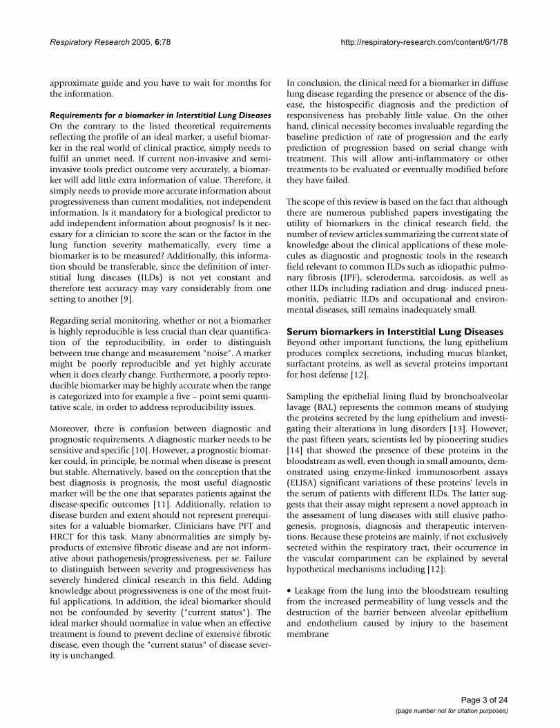
approximate guide and you have to wait for months forthe information.
Requirements for a biomarker in Interstitial Lung DiseasesOn the contrary to the listed theoretical requirementsreflecting the profile of an ideal marker, a useful biomar-ker in the real world of clinical practice, simply needs tofulfil an unmet need. If current non-invasive and semi-invasive tools predict outcome very accurately, a biomar-ker will add little extra information of value. Therefore, itsimply needs to provide more accurate information aboutprogressiveness than current modalities, not independentinformation. Is it mandatory for a biological predictor toadd independent information about prognosis? Is it nec-essary for a clinician to score the scan or the factor in thelung function severity mathematically, every time abiomarker is to be measured? Additionally, this informa-tion should be transferable, since the definition of inter-stitial lung diseases (ILDs) is not yet constant andtherefore test accuracy may vary considerably from onesetting to another [9].
Regarding serial monitoring, whether or not a biomarkeris highly reproducible is less crucial than clear quantifica-tion of the reproducibility, in order to distinguishbetween true change and measurement "noise". A markermight be poorly reproducible and yet highly accuratewhen it does clearly change. Furthermore, a poorly repro-ducible biomarker may be highly accurate when the rangeis categorized into for example a five – point semi quanti-tative scale, in order to address reproducibility issues.
Moreover, there is confusion between diagnostic andprognostic requirements. A diagnostic marker needs to besensitive and specific [10]. However, a prognostic biomar-ker could, in principle, be normal when disease is presentbut stable. Alternatively, based on the conception that thebest diagnosis is prognosis, the most useful diagnosticmarker will be the one that separates patients against thedisease-specific outcomes [11]. Additionally, relation todisease burden and extent should not represent prerequi-sites for a valuable biomarker. Clinicians have PFT andHRCT for this task. Many abnormalities are simply by-products of extensive fibrotic disease and are not inform-ative about pathogenesis/progressiveness, per se. Failureto distinguish between severity and progressiveness hasseverely hindered clinical research in this field. Addingknowledge about progressiveness is one of the most fruit-ful applications. In addition, the ideal biomarker shouldnot be confounded by severity ("current status"). Theideal marker should normalize in value when an effectivetreatment is found to prevent decline of extensive fibroticdisease, even though the "current status" of disease sever-ity is unchanged.
In conclusion, the clinical need for a biomarker in diffuselung disease regarding the presence or absence of the dis-ease, the histospecific diagnosis and the prediction ofresponsiveness has probably little value. On the otherhand, clinical necessity becomes invaluable regarding thebaseline prediction of rate of progression and the earlyprediction of progression based on serial change withtreatment. This will allow anti-inflammatory or othertreatments to be evaluated or eventually modified beforethey have failed.
The scope of this review is based on the fact that althoughthere are numerous published papers investigating theutility of biomarkers in the clinical research field, thenumber of review articles summarizing the current state ofknowledge about the clinical applications of these mole-cules as diagnostic and prognostic tools in the researchfield relevant to common ILDs such as idiopathic pulmo-nary fibrosis (IPF), scleroderma, sarcoidosis, as well asother ILDs including radiation and drug- induced pneu-monitis, pediatric ILDs and occupational and environ-mental diseases, still remains inadequately small.
Serum biomarkers in Interstitial Lung DiseasesBeyond other important functions, the lung epitheliumproduces complex secretions, including mucus blanket,surfactant proteins, as well as several proteins importantfor host defense [12].
Sampling the epithelial lining fluid by bronchoalveolarlavage (BAL) represents the common means of studyingthe proteins secreted by the lung epithelium and investi-gating their alterations in lung disorders [13]. However,the past fifteen years, scientists led by pioneering studies[14] that showed the presence of these proteins in thebloodstream as well, even though in small amounts, dem-onstrated using enzyme-linked immunosorbent assays(ELISA) significant variations of these proteins' levels inthe serum of patients with different ILDs. The latter sug-gests that their assay might represent a novel approach inthe assessment of lung diseases with still elusive patho-genesis, prognosis, diagnosis and therapeutic interven-tions. Because these proteins are mainly, if not exclusivelysecreted within the respiratory tract, their occurrence inthe vascular compartment can be explained by severalhypothetical mechanisms including [12]:
• Leakage from the lung into the bloodstream resultingfrom the increased permeability of lung vessels and thedestruction of the barrier between alveolar epitheliumand endothelium caused by injury to the basementmembrane
Page 3 of 24(page number not for citation purposes)
Respiratory Research 2005, 6:78 http://respiratory-research.com/content/6/1/78
• Increased production by the alveolar type II cells cou-pled with an increase in total type II cells per lung due todiffuse hyperplasia and
• Diminished clearance rates from the circulation.
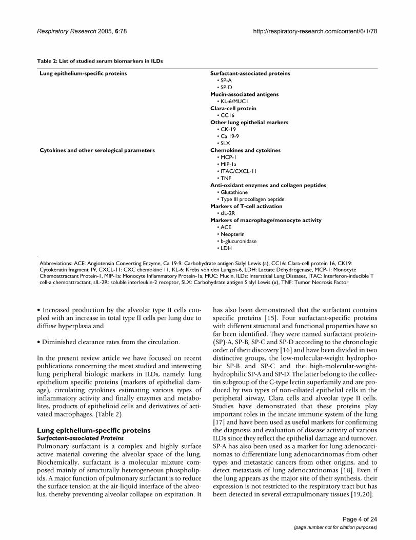
In the present review article we have focused on recentpublications concerning the most studied and interestinglung peripheral biologic markers in ILDs, namely: lungepithelium specific proteins (markers of epithelial dam-age), circulating cytokines estimating various types ofinflammatory activity and finally enzymes and metabo-lites, products of epithelioid cells and derivatives of acti-vated macrophages. (Table 2)
Lung epithelium-specific proteinsSurfactant-associated ProteinsPulmonary surfactant is a complex and highly surfaceactive material covering the alveolar space of the lung.Biochemically, surfactant is a molecular mixture com-posed mainly of structurally heterogeneous phospholip-ids. A major function of pulmonary surfactant is to reducethe surface tension at the air-liquid interface of the alveo-lus, thereby preventing alveolar collapse on expiration. It
has also been demonstrated that the surfactant containsspecific proteins [15]. Four surfactant-specific proteinswith different structural and functional properties have sofar been identified. They were named surfactant protein-(SP)-A, SP-B, SP-C and SP-D according to the chronologicorder of their discovery [16] and have been divided in twodistinctive groups, the low-molecular-weight hydropho-bic SP-B and SP-C and the high-molecular-weight-hydrophilic SP-A and SP-D. The latter belong to the collec-tin subgroup of the C-type lectin superfamily and are pro-duced by two types of non-ciliated epithelial cells in theperipheral airway, Clara cells and alveolar type II cells.Studies have demonstrated that these proteins playimportant roles in the innate immune system of the lung[17] and have been used as useful markers for confirmingthe diagnosis and evaluation of disease activity of variousILDs since they reflect the epithelial damage and turnover.SP-A has also been used as a marker for lung adenocarci-nomas to differentiate lung adenocarcinomas from othertypes and metastatic cancers from other origins, and todetect metastasis of lung adenocarcinomas [18]. Even ifthe lung appears as the major site of their synthesis, theirexpression is not restricted to the respiratory tract but hasbeen detected in several extrapulmonary tissues [19,20].
Table 2: List of studied serum biomarkers in ILDs
Lung epithelium-specific proteins Surfactant-associated proteins• SP-A• SP-D
Mucin-associated antigens• KL-6/MUC1
Clara-cell protein• CC16
Other lung epithelial markers• CK-19• Ca 19-9• SLX
Cytokines and other serological parameters Chemokines and cytokines• MCP-1• MIP-1a• ITAC/CXCL-11• TNF
Anti-oxidant enzymes and collagen peptides• Glutathione• Type III procollagen peptide
Markers of T-cell activation• sIL-2R
Markers of macrophage/monocyte activity• ACE• Neopterin• b-glucuronidase• LDH
Abbreviations: ACE: Angiotensin Converting Enzyme, Ca 19-9: Carbohydrate antigen Sialyl Lewis (a), CC16: Clara-cell protein 16, CK19: Cytokeratin fragment 19, CXCL-11: CXC chemokine 11, KL-6: Krebs von den Lungen-6, LDH: Lactate Dehydrogenase, MCP-1: Monocyte Chemoattractant Protein-1, MIP-1a: Monocyte Inflammatory Protein-1a, MUC: Mucin, ILDs: Interstitial Lung Diseases, ITAC: Interferon-inducible T cell-a chemoattractant, sIL-2R: soluble interleukin-2 receptor, SLX: Carbohydrate antigen Sialyl Lewis (x), TNF: Tumor Necrosis Factor
Page 4 of 24(page number not for citation purposes)

Respiratory Research 2005, 6:78 http://respiratory-research.com/content/6/1/78
Mucin-associated AntigensMucins are major components of the mucus layer cover-ing the airway epithelium. They consist of high-molecu-lar-weight glycoproteins belonging to a broad family ofmucin peptides [12]. Mucins are either associated withmembranes or secreted at the surface of the respiratorytract [12]. Krebs von den Lungen-(KL)-6 is mainly associ-ated with cellular membranes. It was initially described byKohno et al. [14] as a high-molecular-weight glycoproteinand was classified as human MUC1 mucin. Immunohis-tochemistry has mainly detected KL-6 in alveolar type IIand epithelial cells of the respiratory bronchioles.Although KL-6 is predominantly expressed by airway cells,however is not entirely lung specific, since it is alsopresent on other somatic cells, such as pancreatic cells,eosophageal cells and fundic cells of the stomach [21].Functionally KL-6 has been demonstrated to be induciblefor the migration of human lung and skin fibroblasts, sug-gesting a potential role in the lung fibrogenic process [22].Additionally KL-6 is a sensitive indicator of damage toalveolar type II cells, which strongly express this mucin attheir surface. Type II pneumonocytes are regenerated overthe alveolar basement membrane after the death of type Ipneumonocytes over the first stage of lung injury. There-fore, its raise would theoretically represent the destructionof the normal lung parenchyma and architecture, theincreased permeability of the air-blood barrier as long asthe regenerating process as expressed by type II pneu-monocytes' activity. Towards this direction KL-6 has beenreported by several studies some of them cited in thisreview article as a sensitive marker for ILDs such as IPF,collagen vascular disease-associated interstitial pneumo-nia (CVD-IP), radiation pneumonitis, hypersensitivitypneumonitis and pulmonary sarcoidosis.
Clara Cell Protein (CC16)The Clara Cell secretory protein-(CC)-16 is a low-molecu-lar-weight protein of 16 kDa is secreted in large amountsinto the lumen of the respiratory tract by nonciliatedbronchiolar Clara cells in humans and rodents [23].Immunohistochemical studies have shown that CC16 isnot an entirely specific and exclusive product of Clara cellsor even the lung [24,25]. The exact functions of CC16 arestill elusive but there is increased knowledge that CC16serves as an important immunosuppressive and anti-inflammatory mediator in the lung [12]. AdditionallyCC16 can inhibit production of interferon-γ (IFN-γ) byperipheral blood mononuclear cells [12]. Serum CC16has been demonstrated to elevate in several conditionsknown to be related with an impairment of the air-bloodbarrier, including pulmonary fibrosis [26] or lung injurycaused by firesmoke [27]. Thus, several group of scientistsestimated CC16 blood levels to determine whether theseare associated with the degree of lung involvement in ILDs
and thereby can serve as a useful biomarker of the diseaseactivity and severity.
Other lung-epithelial markersCytokeratin is a specific cytoskeletal structure expressed inepithelial cells, including bronchial epithelia [28,29]. Ofthe 27 known subunits of cytokeratin, cytokeratin frag-ment 19 (CK19) has been found soluble in serum and itslevels have already been evaluated as a useful tumourmarker for lung cancer [30]. Since it has been suggestedthat CK19 is released from injured bronchial epithelium[31], the past ten years it has been hypothesized and even-tually indicated by several reports in the literature thatCK19 serum levels are elevated in IPF and other ILDs andcan be well correlated with the disease prognosis anddiagnosis. Thus, in nowadays, there is an ongoing attemptto scrutinize the value of CK19 in evaluating the severityof lung injury as reflected by the increasing number ofpublished papers investigating the role of this biologicmarker in ILDs.
The cancer-associated antigens sialyl Lewis (a) (Ca 19-9)and sialyl Lewis (x) (SLX) are carbohydrate structures usedas markers of cell differentiation and embryonic develop-ment [32]. It has been reported that a variety of these anti-gens is expressed on the cell surface during tumorprogression [33]. Elevated circulating levels of these anti-gens have been associated with neoplastic transformationand metastases and therefore been used as tumor markersin the diagnosis of lung adenocarcinoma [34,35]. Serumlevels of carbohydrate antigens have been found, how-ever, also raised in some patients with non-malignantlung diseases such as IPF [36], tuberculosis [37] and dif-fuse parabronchiolitis [38] and moreover immunohisto-chemical analysis demonstrated their presence in thehyperplastic bronchiolar epithelium, on the surface epi-thelium cells and on exudates in air space. Repeated dam-age to the lungs may force these antigens into the bloodcirculation resulting to the elevated serum levels of thesemarkers detected in these patients. Hence, it has beenspeculated that their elevation could mirror the extent oflung injury and serve as a valuable prognosticator of dis-ease progression.
Cytokines and other serological parametersA number of cytokines probing different aspects of theimmunopathogenesis of ILDs have been tested for theirclinical usefulness as serum biomarkers for monitoringdisease severity and predicting response to treatment andtherefore leading to early diagnosis of progressive diseaseand determination of therapeutic interventions.
Monocyte chemoattractant protein-(MCP)-1 and mono-cyte inflammatory protein- (MIP)-1a belong to the C-Csubfamily of the chemokine family and appear to be
Page 5 of 24(page number not for citation purposes)
Respiratory Research 2005, 6:78 http://respiratory-research.com/content/6/1/78
important factors in the monocyte/macrophage-mediatedinflammatory process in the ILDs [39,40]. It has beenreported that epithelial cells, macrophages and vascularendothelial cells are the major-MCP-1-producing cells inIPF lung tissue [41]. Moreover, an elevation of MCP-1 andMIP-1a concentrations in BAL from patients with IPF andsarcoidosis has recently been demonstrated [40,42].Tempted by the latter observation several group of scien-tists aimed to measure the levels of MCP-1 and MIP-1a inthe serum and determine their clinical significance as areliable and easily repeatable serological parameters forthe differential diagnosis of ILDs and the monitoring oftheir clinical course.
IFN-inducible T cell-a chemoattractant-ITAC/CXCL-11 isa chemoattractant CXC chemokine with versatile proper-ties including immunomodulatory, definsin-like antimi-crobial, antiangiogenic and potentially antifibroticactivities. It has been reported to inhibit angiogenesis andthereby to reduce aberrant vascular remodeling resultingin diminished fibrosis [43,44]. Studies have demon-strated a direct association of this molecule with the plei-otropic cytokine IFN-γ1b [43,44] evidence that capturedthe interest of both clinicians and researchers to investi-gate the effects of this newly applied treatment on biologicmarkers such as ITAC/CXCL-11 associated with fibrosis,angiogenesis and immunomodulation in the plasma ofpatients with ILDs and ultimately reveal novel molecularpathways which support IFN-γ1b therapeutic utilities.
In addition, the activity and release of a large body ofinflammatory mediators including tumor necrosis factor(TNF), antioxidant enzymes (glutathione), procollagenpeptides (type III) and markers of cell damage such as lac-tate dehydrogenase (LDH) have been evaluated as prog-nostic and monitoring tools of the disease development,activity and progression in a variety of occupational andenvironmental studies.
Other delineated serological parameters that have beenscrutinized for their clinical efficacy as serum biologicmarkers of the disease severity and the activation of sev-eral inflammatory cells contributing to the immun-opathogenesis of the disease include markers of T-cellactivation and markers for the evaluation of macrophage/monocyte activity.
Soluble IL-2 receptor (sIL-2R) represents a biomarker ofthe T-cell activation and can be found in BAL fluid andserum of sarcoidosis patients and it is released by acti-vated alveolar immune cells. In addition to lymphocytes,activated sarcoid alveolar macrophages are capable ofexpressing increased numbers of sIL-2R upon activation[45]. There are several reports in the literature, some ofthem are reviewed here, that reveal an intimate relation-
ship between this parameter and the clinical activity of thedisease, providing further evidence for the close linkagebetween the course of sarcoidosis and the activated stateof T-cells [45].
Parameters suitable for gauging the activity of the macro-phage lineage in ILDs and specifically sarcoidosis andoccupational diseases have been delineated and comprisethe angiotensin converting enzyme (ACE) a product ofepithelioid cells that reflect the granuloma burden of theentire body, neopterin a metabolite of the guanosinetri-phosphate pathway that is released by activated macro-phages and monocytes under the control of IFN-γproduced by T-cells [45] and b-glucuronidase a lysosomalenzyme, associated with increased phagocytic activity[46]. Serum and BALF levels of these molecules have beenfound elevated in patients with sarcoidosis and environ-mental lung disorders and have been used in the everydayclinical practice as markers for the clinical assessment andfollow-up of granulomatous inflammation and dust-induced inflammatory response.
1. Serum biomarkers in Idiopathic Pulmonary Fibrosis and Collagen Vascular Disease – associated Interstitial Pneumonia (Tables 3 and 4)Idiopathic pulmonary fibrosis (IPF) is a refractory andlethal ILD characterized by fibroblast proliferation, extra-cellular matrix (ECM) deposition and progressive lungscarring. The incidence of IPF is estimated at 15–40 casesper 100.000 per year, and the mean survival from the timeof diagnosis is 3–5 yr regardless of treatment [47]. On theother hand, scleroderma (progressive systemic sclerosis-SSc), is a systemic disease characterized by a progressivedermatologic abnormality. Systemic involvement mayinclude among others, restrictive lung disease whichdevelops in 30–60% of patients with scleroderma andprogresses to severe restrictive lung disease and pulmo-nary fibrosis (a major cause of death in scleroderma) in15% of these patients [48]. However, predicting the pro-gression of IPF and SSc as well as their prognosis stillremains elusive. To evaluate the activity and monitor thecourse of the disease HRCT, PFT, BAL and histologic fea-tures are clinically used [48]. Nonetheless, there are prob-lems with the sensitivity, effort-dependability and mainlythe ease of repetition of these examinations.
One of the pioneering studies that set the foundations forthe development of a new research field with massive clin-ical implications was published ten years ago by Honda etal [49]. Authors were the first reported the potential use-fulness of SP-D serum levels in reflecting the diseaseactivity in a group of patients with different types of ILDsincluding IPF and collagen vascular disease – associatedinterstitial pneumonia (CVD-IP).
Page 6 of 24(page number not for citation purposes)
Respiratory Research 2005, 6:78 http://respiratory-research.com/content/6/1/78
Moreover, KL-6 serological levels have been tested byKobayashi et al. [50] whether they can reflect the activityof pneumonitis seen in different types of ILD and there-fore used as a tool for the differential diagnosis of thislarge set of diseases and the assessment of their responseto treatment. Data derived from this analysis utilizingROC curves and cut-off values showed a distinct differen-tiation between ILDs and non-ILDs based on the periph-eral KL-6 concentrations as well as a clear correlation ofKL-6 levels with the clinical activity of the ILDs as definedby a series of conventional criteria. Although these resultscreate major expectations in the diagnostic field of ILDs,
however it should be noted that an elevation of KL-6 andSP-D serum levels suggests diagnosis of ILD and does notestablish a specific diagnosis. For these markers to becometruly specific, large evaluation and comparative studies arerequired to determine their usefulness in the differentialdiagnosis of ILDs.
The first and to best of our knowledge the only so far com-parative study testing the sensitivity and specificity ofthree groups of molecules reported to serve as sensitivemarkers for ILDs was conducted by Ohnishi et al [51].They generated ROC curves and performed a comparative
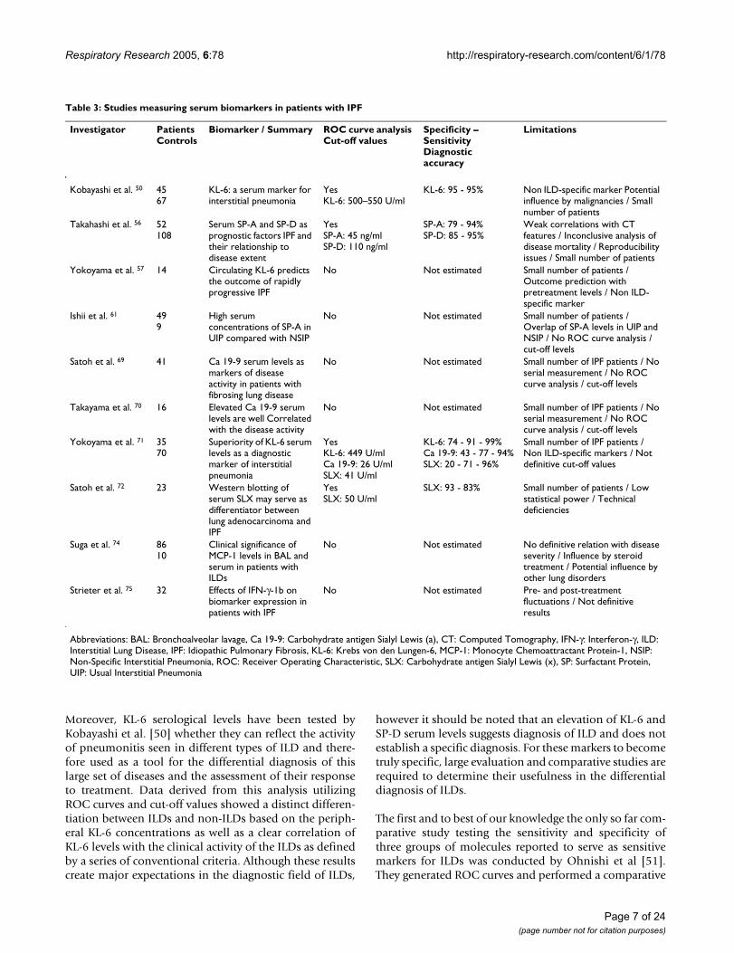
Table 3: Studies measuring serum biomarkers in patients with IPF
Investigator Patients Controls
Biomarker / Summary ROC curve analysis Cut-off values
Specificity – Sensitivity Diagnostic accuracy
Limitations
Kobayashi et al. 50 4567
KL-6: a serum marker for interstitial pneumonia
YesKL-6: 500–550 U/ml
KL-6: 95 - 95% Non ILD-specific marker Potential influence by malignancies / Small number of patients
Takahashi et al. 56 52108
Serum SP-A and SP-D as prognostic factors IPF and their relationship to disease extent
YesSP-A: 45 ng/mlSP-D: 110 ng/ml
SP-A: 79 - 94%SP-D: 85 - 95%
Weak correlations with CT features / Inconclusive analysis of disease mortality / Reproducibility issues / Small number of patients
Yokoyama et al. 57 14 Circulating KL-6 predicts the outcome of rapidly progressive IPF
No Not estimated Small number of patients / Outcome prediction with pretreatment levels / Non ILD-specific marker
Ishii et al. 61 499
High serum concentrations of SP-A in UIP compared with NSIP
No Not estimated Small number of patients / Overlap of SP-A levels in UIP and NSIP / No ROC curve analysis / cut-off levels
Satoh et al. 69 41 Ca 19-9 serum levels as markers of disease activity in patients with fibrosing lung disease
No Not estimated Small number of IPF patients / No serial measurement / No ROC curve analysis / cut-off levels
Takayama et al. 70 16 Elevated Ca 19-9 serum levels are well Correlated with the disease activity
No Not estimated Small number of IPF patients / No serial measurement / No ROC curve analysis / cut-off levels
Yokoyama et al. 71 3570
Superiority of KL-6 serum levels as a diagnostic marker of interstitial pneumonia
YesKL-6: 449 U/mlCa 19-9: 26 U/mlSLX: 41 U/ml
KL-6: 74 - 91 - 99%Ca 19-9: 43 - 77 - 94%SLX: 20 - 71 - 96%
Small number of IPF patients / Non ILD-specific markers / Not definitive cut-off values
Satoh et al. 72 23 Western blotting of serum SLX may serve as differentiator between lung adenocarcinoma and IPF
YesSLX: 50 U/ml
SLX: 93 - 83% Small number of patients / Low statistical power / Technical deficiencies
Suga et al. 74 8610
Clinical significance of MCP-1 levels in BAL and serum in patients with ILDs
No Not estimated No definitive relation with disease severity / Influence by steroid treatment / Potential influence by other lung disorders
Strieter et al. 75 32 Effects of IFN-γ-1b on biomarker expression in patients with IPF
No Not estimated Pre- and post-treatment fluctuations / Not definitive results
Abbreviations: BAL: Bronchoalveolar lavage, Ca 19-9: Carbohydrate antigen Sialyl Lewis (a), CT: Computed Tomography, IFN-γ: Interferon-γ, ILD: Interstitial Lung Disease, IPF: Idiopathic Pulmonary Fibrosis, KL-6: Krebs von den Lungen-6, MCP-1: Monocyte Chemoattractant Protein-1, NSIP: Non-Specific Interstitial Pneumonia, ROC: Receiver Operating Characteristic, SLX: Carbohydrate antigen Sialyl Lewis (x), SP: Surfactant Protein, UIP: Usual Interstitial Pneumonia
Page 7 of 24(page number not for citation purposes)
Respiratory Research 2005, 6:78 http://respiratory-research.com/content/6/1/78
analysis of the diagnostic values of KL-6, SP-A, SP-D andMCP-1 in a pool of serum samples derived from patientswith IPF and CVD-IP. Intriguingly, this study demon-strated for the first time a clear superiority of KL-6 as adiagnostic marker of ILDs in terms of diagnostic accuracy,sensitivity, specificity and likelihood ratio. Interestingly,results from the statistical analysis confirmed that all fourserum markers are specific markers for IPF and at leastsuperior to previously described markers such as lactatedehydrogenase and other collagen products [52].Although this study adds knowledge of high scientificrigidity about the status of these peripheral markers asdiagnostic tools, however there are substantial weaknessesarising from the origin disadvantages of these moleculesas specific biologic markers. More specifically, serum lev-
els of all the four markers can be influenced by the pres-ence of ILDs other than IPF as well as lung disorders otherthan ILDs such as malignancies, systemic inflammationsand fibrosing lung infections [12,53,54]. Nonetheless,these observations are not to diminish their value as diag-nostic tools but to illuminate the need for their furtherinvestigation in the context of more detailed studiessearching potential correlation of these markers with theclinical and radiological findings and adding more spe-cific information on the differential diagnosis of IPFamong the other 200 members of the ILD group.
Towards this direction Takahashi et al. [55,56] were thefirst who attempted to prove a correlation between the SP-A and SP-D circulating concentrations and the radiologic
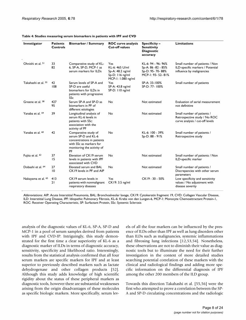
Table 4: Studies measuring serum biomarkers in patients with IPF and CVD
Investigator Patients Controls
Biomarker / Summary ROC curve analysis Cut-off values
Specificity – Sensitivity Diagnostic accuracy
Limitations
Ohnishi et al. 51 3382
Comparative study of KL-6, SP-A, SP-D, MCP-1 as serum markers for ILDs
YesKL-6: 465 U/mlSp-A: 48.2 ng/mlSp-D: 116 ng/mlMCP-1: 1.080 ng/ml
KL-6: 94 - 96- 96%Sp-A: 86 -82 - 85%Sp-D: 95- 70- 88%MCP-1: 93- 52- 81%
Small number of patients / Non ILD-specific markers / Potential influence by malignancies
Takahashi et al. 55 42108
Serum levels of SP-A and SP-D are useful biomarkers for ILDs in patients with progressive SSc
YesSP-A: 43.8 ng/mlSP-D: 110 ng/ml
SP-A: 33-100%SP-D: 77- 100%
Small number of patients
Greene et al. 58 42795
Serum SP-A and SP-D as biomarkers in PF of different etiologies
No Not estimated Evaluation of serial measurement not definitive
Yanaba et al. 59 39 Longitudinal analysis of serum KL-6 levels in patients with SSc: association with the activity of PF
No Not estimated Small number of patients / Retrospective study / No ROC curve analysis / cut-off levels
Yanaba et al. 60 42 Comparative study of serum SP-D and KL-6 concentrations in patients with SSc as markers for monitoring the activity of PF
No KL-6: 100 - 39%Sp-D: 88 - 91%
Small number of patients / Retrospective study
Fujita et al. 62 3715
Elevation of CK19 serum levels in patients with IPF associated with CVD
No Not estimated Small number of patients / Non ILD-specific marker
Dobashi et al. 64 2710
Elevated serum and BAL CK19 levels in PF and AIP
No Not estimated Small number of patients / Discrepancies with other serum parameters
Nakayama et al. 65 41321
CK19 serum levels in patients with nonmalignant respiratory diseases
YesCK19: 3.5 ng/ml
CK19 : 30 - 50% Low specificity and sensitivity values / No adjustment with disease severity
Abbreviations: AIP: Acute Interstitial Pneumonia, BAL: Bronchoalveolar lavage, CK19: Cytokeratin fragment 19, CVD: Collagen Vascular Disease, ILD: Interstitial Lung Disease, IPF: Idiopathic Pulmonary Fibrosis, KL-6: Krebs von den Lungen-6, MCP-1: Monocyte Chemoattractant Protein-1, ROC: Receiver Operating Characteristic, SP: Surfactant Protein, SSc: Systemic Sclerosis
Page 8 of 24(page number not for citation purposes)

Respiratory Research 2005, 6:78 http://respiratory-research.com/content/6/1/78
findings as well as the disease extent in patients with CVD-IP and IPF. Authors published two really well done andheavily informative papers, both of them are reviewedhere.
In the first study [55], results derived by a statistical anal-ysis using ROC curves and cut-off levels demonstratedthat elevated levels of SP-A and SP-D in patients with pro-gressive SSc closely reflect in terms of sensitivity and spe-cificity the presence of ILD on the basis of CT diagnosis.Moreover these markers increase even in patients withmild alveolitis not detectable by compatible chest X-ray.Additionally an interesting finding with major clinicalapplications pointed out by authors is that the combina-tion assay of SP-D circulating concentrations and chest X-ray is almost equivalent to CT in the detection of ILD andcan potentially serve as a contributor to reduce the risk ofclinicians overlooking ILD complicated by progressiveSSc.
Furthermore, Takahashi et al. [56] estimated the serologi-cal levels of SP-A and SP-D in IPF patients and correlatedthem with the radiological and functional parameters.Interestingly, accumulated findings from this analysisdemonstrated a positive correlation between serum levelsof both markers and the extent of alveolitis whereas nocorrelation was observed with the progression of fibrosis.Moreover authors showed a statistically significant linkbetween increased concentrations of SP-D and rapiddecline in pulmonary function tests as well as with higherrates of mortality. The above findings suggest that theseproteins and especially SP-D may help scientists to shedfurther light in the pathologic characteristics of IPF seen inHRCT and assist as prognostic tool for the final outcomein applying optimal therapeutic approaches. However,caveats that should be taken under consideration includeweak correlations of SP-A and SP-D serum levels with theCT features and inconclusive analysis of the disease mor-tality and the biomarker serum levels.
Several biologic markers have been studied the past fewyears not only to evaluate the disease activity and for dif-ferential diagnosis of ILDs but also for prediction of thedisease outcome and the effectiveness of the commonlyapplied treatment during the accelerated phase of pulmo-nary fibrosis.
One of the most detailed and informative study in thisfield was published by Yokoyama et al [57]. Scrutinizingfor early predictive markers of the therapeutic effects ofhigh-dose corticosteroids on patients with rapidly deteri-orating IPF, authors demonstrated that circulating levelsof KL-6 could predict the efficacy of corticosteroids at anearlier time-point than other studied non-specific mark-ers, when overall clinical effect may not yet be evident.
However, potential criticisms of this study including thesmall number of a specific group of IPF patients studied,the finding that the KL-6 levels in the pretreatment periodcould not be used to predict outcome as well as the evi-dence that KL-6 concentrations are affected by certainmalignancies [12,53] pose major limitations to theseobservations and highlight the necessity for further inves-tigation and studies.
Currently, the hurdle faced by young physicians that lim-its the application of blood biomarkers in the daily clini-cal practice of IPF patients is whether alterations of theircirculating concentrations are specific for IPF or they alsoreflect other lung parenchyma abnormalities seen in dif-ferent ILDs. Several studies have tested the efficacy ofblood biomarkers in differentiating IPF from other ILDs.Some of them are reviewed here.
One of the first and most extensive studies addressing thatimportant issue was conducted by Greene et al. [58] in thecontext of a large cohort multivariate analysis includingover 200 patients with IPF and progressive SSc andapproximately 200 patients with other ILDs. Notably,authors showed an important correlation between ele-vated SP-A and SP-D serological levels in IPF and SScpatients compared to patients with sarcoidosis or beryl-lium disease. Furthermore, SP-D levels found to bestrongly related to radiographic abnormalities in patientswith IPF. The most intriguing result of this study was thatboth increasing SP-A and SP-D levels were highly predic-tive of mortality in patients with IPF and progressive SScand this finding robust (especially SP-D levels) afteradjustment for many measures of disease severity. How-ever, a major limitation of this analysis was that serialmeasurement of SP-A and SP-D levels during the clinicalcourse of patients with diffuse lung disease was not defin-itively evaluated. Hence, an important issue highlightedby authors that needs further investigation is the useful-ness of serum alterations in reflecting the disease activityand extent during different time-points of the diseasecourse.
To this end Yanaba et al. [59] carried out a longitudinalretrospective study in a relatively small number of subjectswith SSc and found that the majority of patients with nor-mal baseline serum KL-6 levels exhibited no deteriorationor new onset of PF whereas patients with dramaticallyincreased KL-6 levels showed a parallel progression of PF.
The latter observation concerning the utility of KL-6 as abiomarker in monitoring the clinical course of PF was fur-ther strengthened by the same group of scientists {Yanabaet al. [60]} in the context of a comparative study of SP-Dand KL-6 serum levels in a limited number of patientswith SSc. Moreover, by generating ROC curve analysis,
Page 9 of 24(page number not for citation purposes)
Respiratory Research 2005, 6:78 http://respiratory-research.com/content/6/1/78
authors concluded that the combined use of these twoblood markers could be of higher diagnostic and monitor-ing value for the activity of PF than the single use of eachmarker. However, data derived from the last two studieswas inconclusive mainly due to the fact that it was pro-duced by retrospective analyses and the number ofpatients was inadequately small for any meaningfuloutcome.
Alternatively, Ishii et al. [61] estimated the diagnostic val-ues of five lung peripheral biomarkers including SP-A, SP-D, KL-6 and two tumour markers in discriminatingpatients with ILD of various histopathologic patterns suchas usual interstitial pneumonia (UIP) and non-specificinterstitial pneumonia (NSIP). Ultimately authors dem-onstrated differential SP-A serum concentrations betweenpatients with UIP and NSIP indicating a potential role ofthis protein for the discrimination of these two membersof ILD group with sometimes similar pathologic and radi-ologic characteristics but with totally different prognosisand response to treatment. Although this study adds criti-cal information on the field of differential diagnosis ofILDs, however it is of high risk to adapt this evidence inthe everyday clinical practice and make the differentialdiagnosis based only on non-invasive methods. It is ofhigh importance to note that there are substantial weak-nesses including the relatively small number of patientsused and the overlap in the serum SP-A levels betweenUIP and NSIP patients that limit the further application ofthese results and illuminate the need for a more extensiveanalysis to determine its diagnostic cut-off levels.
To gain a more comprehensive understanding on theactivity of lung epithelial cell damage and repair charac-terizing IPF and other ILDs and to clear out whether theseare related to the disease prognosis, investigators studiedthe significance of other lung specific epithelial proteinsinitially reported as tumour markers, in reflecting thesepathological abnormalities. Some of the most extensivelyreported markers were CK19 and carbohydrate antigenssialyl Lewis (Ca 19-9, SLX).
There have been several published papers reporting ele-vated levels of CK19 in the serum of patients with varioustypes of ILD [62,63]. Nevertheless, the first report in theliterature demonstrating elevated CK19 serum levels inpatients with different types of ILDs and a significant asso-ciation with several clinical parameters known to bestrong predictors of the disease clinical course was madeby Dobashi et al [64]. This data combined with resultsfrom immunohistochemical analysis indicate that CK19is a marker of lung injury. However, it is inconclusivegiven the small sample of patients recruited and the majordiscrepancies between levels of CK19 and other serologi-cal predictive parameters. Therefore, the efficacy of CK19
in the differential diagnosis of IPF from other ILDs as wellas in the prediction of the disease prognosis should be fur-ther evaluated.
Fueled by this prospect, Nakayama et al. [65] were the firstthat revealed a positive linkage between increased CK19serum levels and low survival rate in patients with IPF andCVD-IP suggesting a potential role for this tumour markerin reflecting the severity of the lung injury and the diseaseprognosis. Additionally, this study clearly documented byusing ROC curves and cut-off levels the ineffectiveness ofthis marker to differentiate IPF from other non-malignantrespiratory diseases characterized by marked epithelialcell damage. This observation coupled with substantialweaknesses including the lack of adjustment of CK19serum levels with the disease severity, allow us to makeonly speculations on the exact role of this biomarker as aprognostic factor and warrant consideration.
Elevated plasma levels of Ca 19-9 were first demonstratedby Bungo et al. [66] in 1988. In addition, Mukae et al. [67]reported two cases of interstitial pneumonitis withmarked increase of carbohydrate antigens in serum andBALF, the level of which changed in accordance to theirclinical course. Immunohistochemical analysis showedthe expression of these antigens on bronchiolar epithelialcells and regenerating epithelial cells which covered thesurface of fibrosing alveolar septi or remodeling septalstructures finding that was further corroborated byShimizu et al. [68] who stated elevated serum levels ofcancer-associated antigens (Ca 19-9, SLX) primarily local-ized in the hyperplastic bronchiolar epithelium and thesurface epithelium cells of microscopic honeycombing intwo different patients with IPF. However, the first studythat tested the clinical usefulness of elevated blood levelsof Ca 19-9 in a series of patients with fibrosing lung dis-ease was conducted by Satoh et al [69]. In consistencywith previous reports in the literature {Takayama et al.[70]}, authors demonstrated a strong correlation of serumlevels with the degree of disease activity indicating apotential role of these molecules as prognostic markers ofdisease severity and therapeutic response. Furthermore,Yokoyama et al. [71] carried out the first comparativeevaluation of the diagnostic values of three serum carbo-hydrate antigens (KL-6, Ca 19-9, and SLX). They plottedROC curves and demonstrated a clear superiority in termsof specificity, sensitivity and diagnostic accuracy of KL-6plasma levels in discriminating interstitial pneumoniafrom alveolar pneumonia and healthy volunteers. Inanother study, Satoh et al. [72] coupled SLX serum cut-offlevels with Western blotting analysis and documentedthat this combination is of high diagnostic power in dif-ferentiating IPF from lung adenocarcinoma.
Page 10 of 24(page number not for citation purposes)
Respiratory Research 2005, 6:78 http://respiratory-research.com/content/6/1/78
Evidence presented give credence to the view that cancer-associated antigens could serve as valuable and reliablediagnostic and prognostic markers of ILDs. On the otherhand, none of the studies provided us with establisheddiagnostic cut-off values since they exhibited low statisti-cal power and in most of them no adjustment with thedisease severity was performed. Moreover, KL-6 hasproven to have higher discriminative power than carbohy-drate antigens. In addition, potential arguments includethe lack of knowledge regarding the role of these markersin the pathogenetic pathway of IPF. So far, the only studyaddressed this crucial issue was published by Obayashi etal. [73] who reported a direct correlation of elevated BALFCa 19-9 levels with the percentage of neutrophils suggest-ing a potential role of carbohydrate antigens as neu-trophilic chemoattractants and subsequently ascontributors in the process of lung injury. Further analysesin the context of large prospective studies are warranted toelucidate this role and establish their clinical usefulness asmarkers of disease severity and activity.
The past few years led by the same perspective idea to finda reliable and reproducible biologic marker for the diag-nosis and prediction of ILDs, several group of scientistshave tested the usefulness of a slew of cytokines to playthat role.
One of these reports was published by Suga et al [74].Authors concluded that elevated serum and BAL levels ofMCP-1 could be useful to discriminate IPF among othermembers of the ILD group. Interestingly, in circulatingMCP-1 concentrations of patients with IPF underwentsteroid therapy no trend towards fall with treatment wasnoticed whereas a major fluctuation in pre-and post-treat-ment levels was readily identifiable. However, theseresults do not prove neither the reproducibility of thisbiormarker nor its relationship with the disease behav-iour. The latter observations coupled with the evidencethat MCP-1 levels are markedly influenced by corticoster-oid treatment as well as other lung disorders since it is amediator of inflammation also produced in other parts ofbody, limit its specificity in the differential diagnosis ofILDs and underline the need for re-evaluation of its pre-dictive value.
One of the most exciting roles cytokines can play is theiruse as indicators of the therapeutic effects of several drugsagainst IPF providing investigators with useful knowledgeon the potential mechanisms of these drugs in terms ofphysiology and molecular biology highlighting noveltherapeutic targets.
The first study in humans to characterize the effects ofIFN-γ1b on plasma and lung biomarkers, speculated toserve as critical mediators in the pathogenesis of IPF was
recently published by Strieter et al [75]. Authors docu-mented indefinite pre- and post-treatment alterations inthe peripheral concentrations of several biologic markersassociated with fibrosis, aberrant vascular modelling andimmunomodulatory activity that can be used only forgenerating hypotheses for future research. However, thestrongest finding in this study was the differential eleva-tion of blood and BAL before and after treatment levels ofITAC/CXCL-11, a chemokine with multifunctional activi-ties including antiangiogenic and antimicrobial. Inferen-tially, this study comprises evidence that mortality inpatients with IPF could be potentially improved throughthe versatile protective properties of IFN-γ1b supportingits therapeutic utility (Tables 3 and 4).
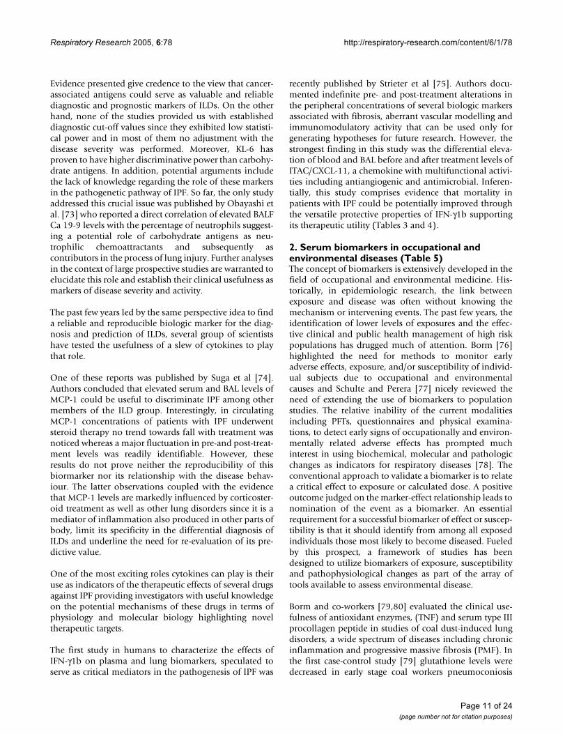
2. Serum biomarkers in occupational and environmental diseases (Table 5)The concept of biomarkers is extensively developed in thefield of occupational and environmental medicine. His-torically, in epidemiologic research, the link betweenexposure and disease was often without knowing themechanism or intervening events. The past few years, theidentification of lower levels of exposures and the effec-tive clinical and public health management of high riskpopulations has drugged much of attention. Borm [76]highlighted the need for methods to monitor earlyadverse effects, exposure, and/or susceptibility of individ-ual subjects due to occupational and environmentalcauses and Schulte and Perera [77] nicely reviewed theneed of extending the use of biomarkers to populationstudies. The relative inability of the current modalitiesincluding PFTs, questionnaires and physical examina-tions, to detect early signs of occupationally and environ-mentally related adverse effects has prompted muchinterest in using biochemical, molecular and pathologicchanges as indicators for respiratory diseases [78]. Theconventional approach to validate a biomarker is to relatea critical effect to exposure or calculated dose. A positiveoutcome judged on the marker-effect relationship leads tonomination of the event as a biomarker. An essentialrequirement for a successful biomarker of effect or suscep-tibility is that it should identify from among all exposedindividuals those most likely to become diseased. Fueledby this prospect, a framework of studies has beendesigned to utilize biomarkers of exposure, susceptibilityand pathophysiological changes as part of the array oftools available to assess environmental disease.
Borm and co-workers [79,80] evaluated the clinical use-fulness of antioxidant enzymes, (TNF) and serum type IIIprocollagen peptide in studies of coal dust-induced lungdisorders, a wide spectrum of diseases including chronicinflammation and progressive massive fibrosis (PMF). Inthe first case-control study [79] glutathione levels weredecreased in early stage coal workers pneumoconiosis
Page 11 of 24(page number not for citation purposes)
Respiratory Research 2005, 6:78 http://respiratory-research.com/content/6/1/78
(CWP) but increased in patients with PMF indicating apotential role of anti-oxidant enzymes in the early detec-tion of inflammatory response to mineral dust exposure.However, serum alterations of glutathione failed to pre-dict individual susceptibility since they most likely reflecta consequence of the disease. With this aim in mind,authors carried out a second case-control study [80] whereTNF was studied as a potential risk factor and concludedthat TNF release from peripheral blood monocytesexposed coal mine dust was a marker of individual suscep-
tibility to dust-induced lung fibrosis. The latter observa-tion corroborated earlier findings [81,82] and supportedthe notion that TNF release from monocytes or TNF inplasma are not associated with actual or cumulative expo-sure. However, definitive proof must come from a pro-spective analysis in the context of carefully designedfollow-up study. The only follow-up study that has beenconducted so far {Schins et al. [83]} showed that the min-ers that had disease progression (PMF) during 5 yearsalready had high levels of dust-induced monocyte TNF
Table 5: Studies measuring serum biomarkers in patients with occupational and environmental diseases
Investigator Patients Controls
Biomarker / Summary ROC curve analysis Cut-off values
Specificity – Sensitivity Diagnostic accuracy
Limitations
Borm et al. 80 3358
Glutathione levels reflect early inflammatory response but are not a predictive parameter for individual susceptibility in CWP
No Not estimated Retrospective analysis / No serial measurement / Small number of patients / Scarce data on the interrelationships among antioxidant enzymes
Borm et al. 81 3927
TNF is a marker of individual susceptibility to dust-induced lung fibrosis in coal-workers
No Not estimated Retrospective analysis / Small number of patients / Indefinite data on the acquired or genetically controlled differences of TNF release
Schins et al. 83 10429
A 5-year follow-up study reveals that TNF is a reliable prognosticator of CWP
No Not estimated No linkage to disease behaviour / Indefinite data on the effect of exposure / No ROC curve analysis / cut-off levels
Schins et al. 84 10429
Serum PIIIP has a poor value as a biomarker to screen for CWP
No Not estimated Studied population: retired miners / Diverse exposure to coal-dust / Heterogeneity of disease-fibrotic activity / Indefinite data on the effect of exposure and time variation in serum PIIP
Cobben et al. 86 20148
Serum LDH levels are elevated in coalminers but are not associated with clinical variables of disease severity
No Not estimated Retrospective study / No serial measurements / No ROC curve analysis / cut-off levels
Cobben et al. 87 19148
Serum b-glucuronidase levels may be a useful biomarker in monitoring lung inflammation following coal dust exposure
No Not estimated Retrospective study / No serial measurements / No ROC curve analysis / cut-off levels
Takahashi et al. 88 35237
Elevated serum KL-6 levels in patients with FLD
YesKL-6: 410 - 450 U/ml
KL-6: 80 - 73% Small number of patients / No serial measurements
Harris et al. 89 10820
Elevated serum neopterin levels in CBD
YesNeopterin: 1.27 ng/ml
Neopterin: 88 - 58% No serial measurements / No correlation with clinical and/or radiological parameters
Maier et al. 90 3134
Neopterin is a useful diagnostic tool in differentiating CBD from BeS
YesNeopterin: 2.5 ng/ml
Neopterin: 100 - 80% No serial measurements Poor prognostic value Indefinite cut-off levels
Abbreviations: BeS: Beryllium Sensitization, CBD: Chronic Beryllium Disease, CWP: Coal-Workers Pneumoconiosis, FLD: Farmer's Lung Disease, KL-6: Krebs von den Lungen-6, LDH: Lactate Dehydrogenase, PIIIP: type III procollagen peptide, ROC: Receiver Operating Characteristic, TNF: Tumor Necrosis Factor
Page 12 of 24(page number not for citation purposes)

Respiratory Research 2005, 6:78 http://respiratory-research.com/content/6/1/78
release at the beginning and no alterations during follow-up were noticed. This highly informative study excludedTNF as an exposure marker and suggested that TNF releaseis a constitutional marker of the disease prognosis and isnot highly affected by the disease itself. In a fourth study,Schins and Borm [84] conducted a 5-year prospectiveanalysis and estimated whether type III procollagen pep-tide could serve as a valuable predictor of the fibrotic lungdisease progression, outcome or activity. Since no differ-ences between procollagen serum levels in coal-minersand non-dust-exposed controls were observed, the inves-tigators stated that type III procollagen peptide is not areliable marker of early effect.
To characterize the nature and extent of coal dust inducedairway injury there is a need for biomarkers useful tomonitor exposure effects. The potential of many cellmediators as monitoring tools i.e. CC16 [85], antioxi-dants [79] and several cytokines [80] has been raisedmany times. Towards this direction Cobben et al. [86]studied the role of LDH (a marker of cell damage) asmarker of lung tissue injury. Authors conducted the firsthuman study describing a considerable elevation of thisenzyme in a group of ex-coalminers and a further associa-tion with other clinical variables. Additionally, the samegroup of investigators [87] evaluated the role of b-glu-curonidase as marker of phagocytic activity and reportedincreased plasma concentrations in a group of coal-min-ers after 20 years of exposure whereas no correlation withclinical parameters was observed. The latter data is in linewith the hypothesis that LDH and b-glucuronidase activ-ity are conceivable markers of disease activity. Potentialcriticisms include the retrospective analysis, the lack ofserial measurement and link to disease behaviour. Thus,these results should be interpreted with care. To deter-mine the significance of these biological indicators withregard to the development or progression of CWP a longi-tudinal prospective design is necessary.
Hypersensitivity pneumonitis also called extrinsic allergicalveolitis is an ILD that may be due to a wide variety ofinhalative antigenic stimuli. To date, the conventionaldiagnosis of hypersensitivity pneumonitis is usually madeon the basis of history of periodically recurring or perma-nent complaints upon exposure to a specific inhalativeantigen, interstitial abnormalities in both lungs by chestradiography or HRCT and detection of precipitating anti-bodies. However, at present, there are few diagnostic pro-cedures available to confirm the diagnosis and to estimatethe disease activity and most of them are too invasive andcostly for widespread and daily use. With this aim inmind, Takahashi et al. [88] evaluated serum KL-6 meas-urement as a biologic marker for farmer's lung disease(FLD), a type of hypersensitivity pneumonitis caused bythe inhalation of moldy antigens. Authors conducted a
large cohort study and clearly documented significanthigher blood KL-6 levels in patients with FLD comparedto the levels of farmers with or without precipitating anti-bodies. In addition, major findings in this study previ-ously unknown, include the consistency between KL-6serum levels in FLD patients and the activity of the diseaseas well as the indication that elevated KL-6 concentrationscoupled with conventional diagnostic criteria may detectsubclinical FLD and determine early and effective thera-peutic interventions. However, to validate whether serumKL-6 levels reflect the disease activity a larger evaluation ofthe specificity and sensitivity of this marker against thedisease behaviour in combination with a definitively esti-mated serial measurement of its plasma levels arerequired.
While its direct role in pathogenesis of immune andinflammatory responses remains unclear, neopterin'sability to reflect monocyte and macrophage activation hasbeen exploited and yet postulated as a marker of the beryl-lium-specific cell-mediated immune response that leadsto a chronic granulomatous disease, a hypersensitivity dis-order named chronic beryllium disease (CBD). In thestudy of Harris et al. [89] ROC curves were generated toevaluate the optimum diagnostic accuracy of neopterin'scut-off levels. Interestingly, authors have found that thecombination of elevated neopterin's serum levels with theconventional screening test for CBD, beryllium lym-phocyte proliferation test exhibited an optimized positivepredictive value suggesting neopterin as a valuablebiomarker in discriminating workers with CBD fromthese that are only sensitized to beryllium rendering lungbiopsy unnecessary. These results were substantiated byin-vitro studies of peripheral blood mononuclear cellsderived by patients with CBD or beryllium exposed work-ers [90] and an association between serum levels of neop-terin and clinicolaboratory parameters of the diseaseseverity was found. However, further confirmatory tests inlarge cohorts of patients including serial measurementsand correlation of results with clinical and radiologicalfindings are essential to determine reliable cut-off valuesfor the diagnosis of the disease and the assessment of theprogression likelihood (Table 5).
3. Serum biomarkers in other interstitial lung diseases (Table 6)Since lung specific-epithelium proteins reflect the epithe-lial damage and turnover it has been hypothesized andultimately demonstrated that they can be used as effectivecirculating markers for the diagnosis and prognosis of theclinical course of various types of interstitial pneumonitisincluding drug-associated, radiation-induced and hyper-sensitivity pneumonitis. Furthermore, pneumoproteinshave already been introduced as potential valuablebiomarkers in the research field of pediatric ILDs.
Page 13 of 24(page number not for citation purposes)
Respiratory Research 2005, 6:78 http://respiratory-research.com/content/6/1/78
Numerous agents including cytotoxic and non-cytotoxicdrugs exert pulmonary toxicity including interstitial pneu-monitis which several times culminates to a fatal outcome[91]. Therefore early diagnosis is crucial, since withdrawalof the implicated drug is usually the most sufficient treat-ment for drug toxicities, whereas undiagnosed toxicity canbe progressive and fatal.
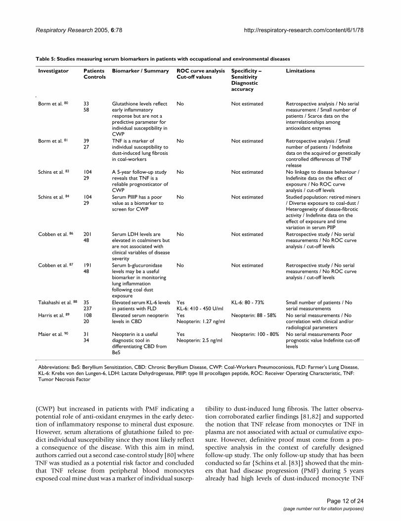
Ohnishi et al. [92] in their attempt to introduce a specificand reproducible hallmark for the recognition of differenttypes of drug-induced lung injury and the prediction oftheir clinical course, estimated the plasma concentrationsof KL-6 in 30 patients with drug-associated pneumonitisclassified into four different predominant radiographicpatterns. The remarkable ascertainment of this study wasthe demonstration of a high sensitivity relation betweenelevated serum KL-6 levels and particular types of lunginjury as well as with their clinical course. However, thesmall number of patients included in this study coupledwith discrepancies between serum KL-6 levels and the dis-ease extent as defined by CT findings comprise majorcaveats and illuminate the need for further prospectivestudies to determine whether measurement of KL-6 levelswould be beneficial in the monitoring and early detectionof drug-induced ILDs.
Radiation pneumonitis is the most common complica-tion for thoracic tumours and is classified as an ILD. SinceCT scanning that represents the gold standard diagnosticprocedure for radiation pneumonitis is either not fre-quently repeatable or often exhibits non-specific findingshardly to be differentiated from the lung tumour manifes-tations, a lung specific laboratory test easily repeatableand reproducible is highly required for the early detectionof radiation-associated lung injury. On the basis of thisconception, Kohno et al. [93] were the first reported thatserum KL-6 levels are a sensitive marker for detectingsevere radiation pneumonitis. Nevertheless, becauseauthors measured KL-6 serum levels at a few time-pointsin each patient's clinical course, data derived from thisstudy is inconclusive and cannot be applied to firmly cor-relate circulating KL-6 concentrations with the clinicalcourse of radiation pneumonitis. Therefore, Goto et al.[94] to further streamline these observations retrospec-tively monitored at shorter intervals blood KL-6 levels inpatients with lung cancer who had received radiotherapywith or without chemotherapy and showed a correlationwith the clinical course of radiation pneumonitis and theresponse to treatment. Interestingly, the finding thatserum KL-6 levels in some patients were increased prioryto the clinical and radiological diagnosis of radiationpneumonitis is particularly noteworthy and should bekept in mind.
Table 6: Studies measuring serum biomarkers in patients with other ILDs
Investigator Patients Controls
Biomarker / Summary ROC curve analysis Cut-off values
Specificity – Sensitivity Diagnostic accuracy
Limitations
Ohnishi et al. 92 30 Elevated circulating KL-6 levels in patients with drug induced pneumonitis
NoKL-6: 520 U/ml
Sensitivity: 53 – 89% Small number of patients / Discrepancies with CT features
Kohno et al. 93 15 Circulating antigen KL-6 and LDH for monitoring irradiated patients with lung cancer
No Not estimated Small number of patients / Retrospective study / Evaluation of serial measurement not definitive
Goto et al. 94 16 Serum levels of KL-6 are useful biomarkers for severe radiation pneumonitis
No Not estimated Small number of patients / Retrospective study / Chemotherapy influence
Takahashi et al.95 25 Diagnostic significance of SP-A and SP-D in sera from patients with radiation pneumonitis
No Sp-A: 83 - 85%Sp-D: 83 - 85%
Small number of patients / Chemotherapy influence / No long term follow-up / Non ILD-specific markers
Al-Salmi et al. 97 1010
Elevated serum KL-6 and SP-A and SP-D in pediatric ILDs
No Not estimated Small number of patients / Diversity of the diseases studied / Poor correlation with functional and radiological parameters
Abbreviations: BAL: Bronchoalveolar lavage, CBD: Chronic Beryllium Disease CT: Computed Tomography, FLD: Farmer's Lung Disease, ILDs: Interstitial Lung Diseases IPF: Idiopathic Pulmonary Fibrosis, KL-6: Krebs von den Lungen-6, LDH: Lactate Dehydrogenase, ROC: Receiver Operating Characteristic, SP: Surfactant Protein
Page 14 of 24(page number not for citation purposes)
Respiratory Research 2005, 6:78 http://respiratory-research.com/content/6/1/78
Another study searching for potential indicators for theearly detection of radiation pneumonitis and for monitor-ing its clinical course was conducted by Takahashi et al[95]. Remarkably almost all of the patients with radiationpneumonitis detected by HRCT exhibited significantincreases in both SP-A and SP-D levels which showed highsensitivity and specificity for the early diagnosis of radia-tion pneumonitis compared with other conventional hae-matological laboratory indices. Additionally, anagreement of SP-A and SP-D plasma levels with the clini-cal response to steroid therapy was also noted. This datasuggest a possible role of lung-specific epithelial proteinsas a diagnostic tool for the recognition of radiation-induced lung injury even when its radiographic change isfaint.
However, these studies exhibit major caveats that must beaddressed prior to their further application in the routineclinical practice. Briefly, we report the small number ofpatients studied, the application of chemotherapy prior toradiotherapy, the absence of serial measurements throughthe clinical course of the disease and the evidence thatthese proteins (SP-A and KL-6) are neither organ nor dis-ease specific since they have also been used as markers forlung adenocarcinoma [18,53]. To confirm the assump-tions arising from these concerns, further studies arerequired.
As mentioned above the spectrum of ILDs includes a largeheterogeneous group of disorders calculating over 200members with IPF to be the most common form of idio-pathic ILD. In contrast ILD in children occurs far less fre-quently and there are no dominant forms [96]. Althoughthis research field is newly developed a recently publishedstudy by Al-Salmi et al. [97] revealed for the first time ele-vated serum levels of three candidate biomarkers of thedisease activity and severity including KL-6, SP-A and SP-D in a group of children with ILDs of various histopatho-logic patterns. These results corroborate earlier findings byKobayashi et al. [98] who reported elevated KL-6 serumlevels in three children with ILD associated with dermato-myositis. Nonetheless it should be noted that these stud-ies are deficient because of the small number of patientsrecruited and the diversity of the diseases studied. Further-more, data described here is inconclusive and it ascertainspoor correlation with the functional and the radiologicalparameters. More in depth analysis in a large cohort ofpatients is required for any meaningful outcome (Table6).
4. Serum biomarkers in sarcoidosis (Table 7)Sarcoidosis is a chronic systemic disorder characterized bythe presence of non-caseating granulomas and accumula-tion of T-lymphocytes and macrophages in multipleorgans [99]. The mechanisms leading to the persistent
accumulation of inflammatory cells and maintain thealveolitis which may lead to irreversible organ damage arenot fully understood. The classical parameters used in themanagement of pulmonary involvement are mainly radi-ological methods and PFTs which do not gauge alveolitisbut rather pulmonary impairment. Consequently theseparameters have been proved ineffective both for earlydiagnosis of the disease and prediction of the response totreatment. The last twenty years immunological studiesperformed with cells obtained by BAL have shed furtherlight into the pathogenetic mechanisms of sarcoidosis[100,101] and formed the basis of concepts of its immun-opathogenesis. To further streamline these concepts andameliorate hardships generating from their clinical appli-cation, several serological parameters including cytokines,soluble cytokine receptors, enzymes and other serumcomponents including lung epithelium-specific proteinswere delineated to probe different aspects of inflamma-tory activity and therefore reflect the disease severity andhelp the early diagnosis of progressive disease [45].Although levels of these markers are closely associatedwith the pathogenesis of the disease [102,103], howeverthe availability of sufficient information as to which ofthem is valuable for assessment of the disease severity andthe prediction of clinical deterioration still represents abottleneck. From a clinical point of view it is even moreimportant to know whether sarcoidosis is severe, ratherthan active. The latter observation represents the key evi-dence determining the initiation of treatment. Some ofthe most extensive and informative studies addressing thiscrucial issue are presented and criticized in this reviewarticle.
One of the first studies attempted to circumvent this prob-lem was published by Ziegenhagen et al [104]. Authorsscrutinized the efficacy of both BAL and serum parametersin indicating likelihood of progression in patients withsarcoidosis and revealed that almost half of the patientswith no indications for steroid therapy who had elevatedsIL-2R plasma levels experienced disease deteriorationwhereas none with normal values did. The aforemen-tioned observation strongly suggests that this immuneparameter could serve as a prognostic guide leading to theidentification of patients with greater risk to relapse andmaybe associated with clinical findings such as the diseasecourse.
The effectiveness of sIL-2R in evaluating sarcoidosis sever-ity even in the early stages of the disease was further con-firmed by another study of the same group of scientists[105] who clearly reported significantly elevated sIL-2Rconcentrations in patients with progressive sarcoidosis.On the contrary, this evidence was followed by a surpris-ing finding regarding the ACE serum concentrationswhich did not differ significantly between patients with
Page 15 of 24(page number not for citation purposes)

Respiratory Research 2005, 6:78 http://respiratory-research.com/content/6/1/78
stable or progressing disease indicating a poor predictivevalue of this biomarker in sarcoidosis.
In the study of Grutters et al. [106] authors evaluated theserological concentrations of sIL-2R as marker of diseaseactivity, severity and prognosis in a well-defined group ofpatients with sarcoidosis. Data derived from this studypartially consistent with other studies [107] asserts a pos-itive correlation between sIL-2R serum levels and the dis-ease activity and severity and furthermore claims a
predictive value for this serum biomarker. However, thisstudy was retrospective and major discrepancies betweenthe latter findings and results derived from older studies[108] were also notable.
To establish a more reliable association between sIL-2Rand severity of sarcoidosis Rothkrantz-Kos et al. [109]applied ROC curve analysis and estimated the potentialprognostic and diagnostic value of four inflammatorymarkers to predict respiratory severity in a large number of
Table 7: Studies measuring serum biomarkers in patients with sarcoidosis
Investigator Patients Controls
Biomarker / Summary
ROC curve analysis Cut-off values
Specificity / Sensitivity Diagnostic accuracy
Limitations
Ziegenhagen et al.104 7750
TNF-a release from alveolar macrophages and serum level of sIL-2R are prognostic markers for sarcoidosis patients
No Not estimated No serial measurements / No ROC curve analysis / cut-off values
Ziegenhagen et al. 105
7348
BAL and serological parameters reflect the severity of sarcoidosis
No Not estimatedHigh predictive value for neopterin+ sIL-2R
No serial measurements / No ROC curve analysis / cut-off values
Grutters et al. 106 47 Positive correlation between sIL-2R serum levels and the disease activity and severity in patients with sarcoidosis
No Not estimated Small number of patients with serial measurement / No ROC curve analysis / cut-off values
Rothkrantz-Kos et al.109
144282
Potential usefulness of inflammatory markers to monitor respiratory functional impairment in sarcoidosis
YessIL-2R: 750 U/ mlsIL-2R: 1300 U/ml
sIL-2R: 94 - 82%sIL-2R: 82 - 94%
Discrepancies between treated and untreated patients / No serial measurements / Retrospective study / No correlation with radiological findings
Hashimoto et al. 117 26 Correlation of plasma MCP-1 and MIP-1a levels with disease activity and clinical course of sarcoidosis patients
No Not estimated Small number of patients / No ROC curve analysis / cut-off values / No determination of the cytokines' cellular sources
Iyonaga et al. 118 4710
MIP-1 serum levels estimate the activity of granuloma formation in sarcoidosis
No Not estimated Small number of patients / No ROC curve analysis / cut-off values / No correlation with radiological findings
Kobayashi et al. 119 47 Serum KL-6 for the evaluation of active pneumonitis in pulmonary sarcoidosis
No Not estimated Small number of patients / No follow-up laboratory data
Hermans et al. 120 117117
Serum CC16 levels, a marker of the integrity of the air-blood barrier in sarcoidosis
No Not estimated Non ILD- specific marker / Potential influence by tobacco smoking / Poor discriminative value
Janssen et al. 122 7938
Elevated serum CC16, KL-6, and SP-D levels reflect pulmonary disease severity and prognosis in sarcoidosis patients
YesCC16: 12.7 ng/mlKL-6: 223 U/mlSP-D: 91.7 ng/ml
CC16: 84 - 73- 73%KL-6: 84 - 86 - 86%SP-D: 84 - 66 - 66%
Non ILD- specific markers / Retrospective study / No serial measurement
Abbreviations: CC16: Clara Cell protein 16, ILD: Interstitial Lung Disease, KL-6: Krebs von den Lungen-6, MCP-1: Monocyte Chemoattractant Protein-1 MIP-1a: Monocyte Inflammatory Protein-1a, ROC: Receiver Operating Characteristic, sIL-2R: soluble Interleukin-2 Receptor, SP: Surfactant Protein, TNF-a: Tumour Necrosis Factor-alpha
Page 16 of 24(page number not for citation purposes)

Respiratory Research 2005, 6:78 http://respiratory-research.com/content/6/1/78
sarcoidosis patients. Areas under curve indicated that sIL-2R showed the greatest ability to determine pulmonaryseverity as assessed by the highest sensitivity, specificity,positive and negative predictive values among theevaluated markers, including C-reactive protein, serumamyloid A and ACE. Nonetheless, discrepancies betweenserum levels of all markers in treated and untreatedpatients coupled with the lack of serial measurementthrough patients' clinical course pose major limitations tothe reported data which is unable to determine thepotential efficacy of the evaluated markers to reflectrespiratory severity in sarcoidosis patients under treat-ment in general.
Accumulated evidence from the data presented reveal thatthe predictive values of serum ACE in sarcoid patients arestill under consideration and findings derived from thesestudies are controversial concerning its usefulness inassessing the disease severity and predicting the clinicaloutcome. Although some investigators suggest that arising ACE serum level can predict radiographic relapsesof sarcoidosis [110] others have clearly demonstrated thatACE appears to be of poor prognostic value [111,112]. Inthe aforementioned studies [104,105,109] ACE does notyield prognostic information. However, several studies inthe literature have addressed this issue and have reportedan extraordinary high variability in serum ACE concentra-tion in health, which is provoked by numerous factors.The most important factor, responsible for approximately25% of this variation is a deletion/insertion polymor-phism in intron 16 of the ACE gene [113-116]. Thequestion whether genotype corrected normal valuesmight be able to increase the reliability of ACE serologicalconcentrations as a marker of disease activity in sarcoido-sis remains to be determined by further studies.
The role of chemokines in monitoring the disease behav-iour and in reflecting the activity of granuloma formationin sarcoidosis patients has been evaluated and postulated.Hashimoto et al. [117] were the first who carried out alongitudinal evaluation of plasma MCP-1 and MIP-1a lev-els in a rather small number of sarcoid subjects and dem-onstrated a closely relation between serum levels of thestudied biomarkers and the clinical course of the diseaseas defined by radiological and laboratory parameters.However, further light should be shed onto the role ofthese chemokines as markers of the disease activity andseverity as well as their cellular sources and their relation-ship with the granuloma formation.
To this end Iyonaga et al. [118] in parallel with the eleva-tion of MCP-1 serum levels in sarcoid individuals andtheir linkage with radiographic abnormalities showed byboth immunohistochemistry and in situ hybridizationthat the origin of the increased MCP-1 plasma levels were
the macrophages peripheral to the granulomas suggestinga role for this molecule as an indicator in estimating theactivity of granuloma formation and subsequently of thesarcoidosis activity.
Apart from their application as prognostic and diagnostictools of IPF and other ILDs the utility of lung-epitheliumspecific proteins including KL-6, SP-D and CC16 as bio-logical markers of the disease activity has been separatelyevaluated in sarcoidosis patients. To date, several studieshave addressed this issue and much good work has beendone towards this direction.
The potential usefulness of pneumoproteins and morespecifically KL-6 as true marker in the assessment of sar-coidosis was initially suggested by Kobayashi et al [119].Authors were the first demonstrating linkage betweenincreased KL-6 plasma levels and consistent changes inconventional clinical and laboratory parameters for theevaluation of alveolitis activity in sarcoidosis patients sug-gesting a potential role of KL-6 as an indicator of the dis-ease severity.
Moreover Hermans et al. [120] scrutinized the role ofCC16 as a valuable tool for the non-invasive evaluation ofthe damage and consequently the integrity of the air-blood barrier associated with sarcoidosis, sheddingfurther light into pathogenetic mechanisms underlyingthe disease severity. Among other findings authors dem-onstrated that CC16 serum levels are influenced by thepulmonary extent of the disease as defined by the radio-logical abnormalities indicating a potential role of thismolecule as a non-invasive and easily reproducibleparameter for the assessment of the disease severity. Onthe other hand, blood levels of this protein reflect notonly the rate of entry into the circulation, but also the rateof clearance since it has been reported that CC16 is rapidlyeliminated by glomerular filtration [121]. Authorsaddressed this issue and found that the increase occurredindependently of the impaired renal function, attributingthe elevation to the leakage of the protein into the blood-stream across the air-blood barrier. Further studies arewarranted to establish this notion.
Although these markers have been studied separately[41,119,120] no study so far had performed a compara-tive analysis of their diagnostic and prognostic accuracy.Triggered by the latter perspective idea Janssen et al. [122]recently published a really well done and informativepaper comparing the ability of these three markers toreflect pulmonary disease severity and prognosis in sar-coidosis patients. They performed a ROC curve analysiswhich revealed a better sensitivity for KL-6 in discriminat-ing sarcoidosis patients, results consistent with previousfindings [51]. However, as it pointed out from the authors
Page 17 of 24(page number not for citation purposes)
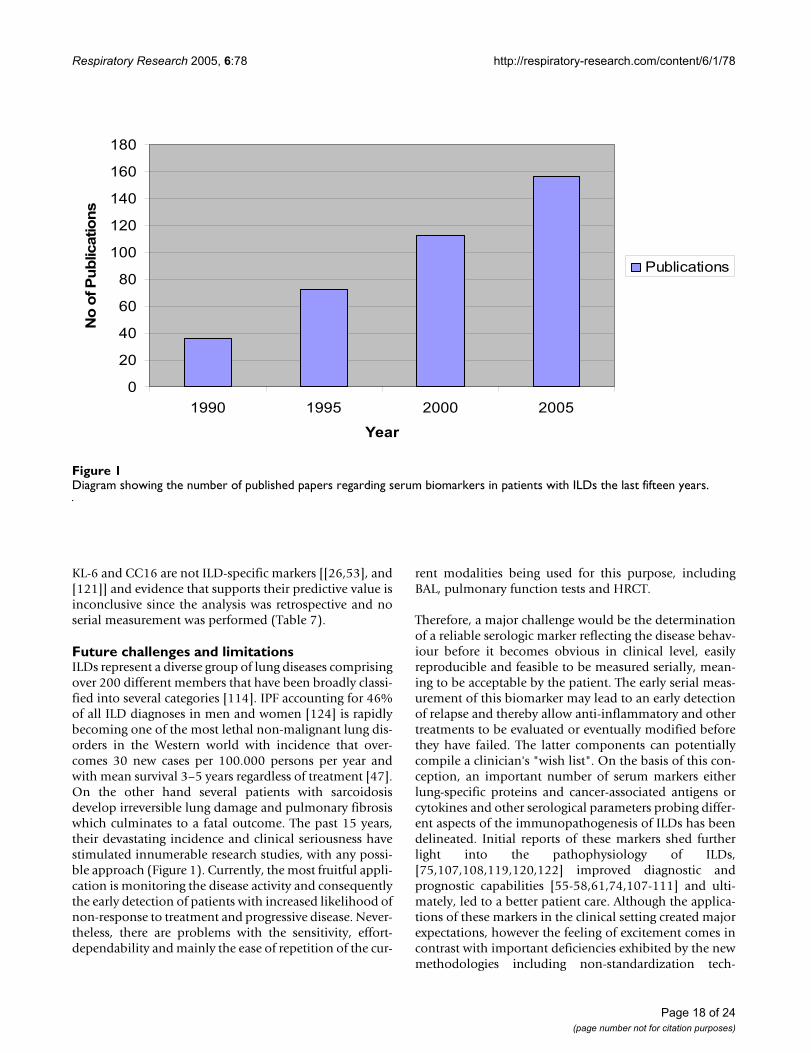
Respiratory Research 2005, 6:78 http://respiratory-research.com/content/6/1/78
KL-6 and CC16 are not ILD-specific markers [[26,53], and[121]] and evidence that supports their predictive value isinconclusive since the analysis was retrospective and noserial measurement was performed (Table 7).
Future challenges and limitationsILDs represent a diverse group of lung diseases comprisingover 200 different members that have been broadly classi-fied into several categories [114]. IPF accounting for 46%of all ILD diagnoses in men and women [124] is rapidlybecoming one of the most lethal non-malignant lung dis-orders in the Western world with incidence that over-comes 30 new cases per 100.000 persons per year andwith mean survival 3–5 years regardless of treatment [47].On the other hand several patients with sarcoidosisdevelop irreversible lung damage and pulmonary fibrosiswhich culminates to a fatal outcome. The past 15 years,their devastating incidence and clinical seriousness havestimulated innumerable research studies, with any possi-ble approach (Figure 1). Currently, the most fruitful appli-cation is monitoring the disease activity and consequentlythe early detection of patients with increased likelihood ofnon-response to treatment and progressive disease. Never-theless, there are problems with the sensitivity, effort-dependability and mainly the ease of repetition of the cur-
rent modalities being used for this purpose, includingBAL, pulmonary function tests and HRCT.
Therefore, a major challenge would be the determinationof a reliable serologic marker reflecting the disease behav-iour before it becomes obvious in clinical level, easilyreproducible and feasible to be measured serially, mean-ing to be acceptable by the patient. The early serial meas-urement of this biomarker may lead to an early detectionof relapse and thereby allow anti-inflammatory and othertreatments to be evaluated or eventually modified beforethey have failed. The latter components can potentiallycompile a clinician's "wish list". On the basis of this con-ception, an important number of serum markers eitherlung-specific proteins and cancer-associated antigens orcytokines and other serological parameters probing differ-ent aspects of the immunopathogenesis of ILDs has beendelineated. Initial reports of these markers shed furtherlight into the pathophysiology of ILDs,[75,107,108,119,120,122] improved diagnostic andprognostic capabilities [55-58,61,74,107-111] and ulti-mately, led to a better patient care. Although the applica-tions of these markers in the clinical setting created majorexpectations, however the feeling of excitement comes incontrast with important deficiencies exhibited by the newmethodologies including non-standardization tech-
Diagram showing the number of published papers regarding serum biomarkers in patients with ILDs the last fifteen yearsFigure 1Diagram showing the number of published papers regarding serum biomarkers in patients with ILDs the last fifteen years.
0
20
40
60
80
100
120
140
160
180
1990 1995 2000 2005
Year
No
of P
ub
licatio
ns
Publications
Page 18 of 24(page number not for citation purposes)
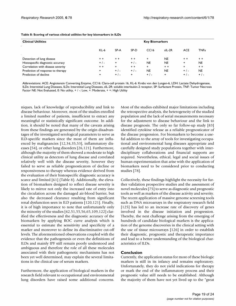
Respiratory Research 2005, 6:78 http://respiratory-research.com/content/6/1/78
niques, lack of knowledge of reproducibility and link todisease behaviour. Moreover, most of the studies enrolleda limited number of patients, insufficient to extract anymeaningful or statistically significant outcome. In addi-tion, it should be noted that many of the caveats arisingfrom these findings are generated by the origin disadvan-tages of the investigated serological parameters to serve asILD-specific markers since the most of them are influ-enced by malignancies [12,34,35,53], inflammatory dis-eases [54], or other lung disorders [26,121]. Furthermore,although the majority of them showed a moderate to highclinical utility as detectors of lung disease and correlatedrelatively well with the disease severity, however theyfailed to serve as reliable prognosticators of decline orresponsiveness to therapy whereas evidence derived fromthe evaluation of their histospecific diagnostic accuracy isscarce and limited [61] (Table 8). Additionally, the eleva-tion of biomarkers designed to reflect disease severity islikely to mirror not only the increased rate of entry intothe circulation across the damaged air-blood barrier, butalso the decreased clearance resulting from significantrenal dysfunction seen in ILD patients [120,121]. Finally,it is of high importance to note that unfortunately onlythe minority of the studies [42,51,55,56,65,109,122] clar-ified the effectiveness and the diagnostic accuracy of thebiomarkers by applying ROC curve analysis which isessential to estimate the sensitivity and specificity of amarker and moreover to define its discriminative cut-offlevels. The aforementioned observations coupled with theevidence that the pathogenesis or even the definitions ofILDs and mainly IPF still remain poorly understood andambiguous and therefore the role of all these moleculesassociated with their pathogenetic mechanisms has notbeen yet well determined, may explain the several limita-tions in the clinical use of serum markers.
Furthermore, the application of biological markers in theresearch field relevant to occupational and environmentallung disorders have raised some additional concerns.
Most of the studies exhibited major limitations includingthe retrospective analysis, the heterogeneity of the studiedpopulation and the lack of serial measurements necessaryfor the adjustment to disease behaviour and the link todisease prognosis. The only so far follow-up study [83]identified cytokine release as a reliable prognosticator ofthe disease progression. For biomarkers to become a use-ful addition to the array of tools for investigating occupa-tional and environmental lung diseases appropriate andcarefully designed study populations together with inter-disciplinary collaborations and financial supports arerequired. Nevertheless, ethical, legal and social issues ofhuman experimentation that arise with the application ofbiomarkers need to be considered prior to conductingstudies [78].
Collectively, these findings highlight the necessity for fur-ther validation prospective studies and the assessment ofnovel molecules [75] to serve as diagnostic and prognostictools as well as markers of the disease activity and severity.The recent application of massive genome screening toolssuch as DNA microarrays in the respiratory research field[125] has led to an increase rate of discovery of genesinvolved in the disease initiation and progression.Thereby, the next challenge arising from the emerging ofhundreds of candidate biological markers is the applica-tion of the genome discoveries in the clinical setting withthe use of tissue microarrays [126] in order to establishtheir diagnostic, prognostic and therapeutic importanceand lead to a better understanding of the biological char-acteristics of ILDs.
ConclusionCurrently, the application status for most of these biologicmarkers is still in its infancy and remains exploratory.Unfortunately, they do not yield indications for therapyor mark the end of the inflammatory process and theirprognostic value still needs to be established. Althoughthe majority of them have not yet lived up to the "great
Table 8: Scoring of various clinical utilities for key biomarkers in ILDs
Clinical Utilities Key Biomarkers
KL-6 SP-A SP-D CC16 sIL-2R ACE TNFa
Detection of lung disease + + + + + + + NE + + + +Histospecific diagnostic accuracy + / - + + / - NE NE + NECorrelation with disease severity + + + + + + + + + + +Prediction of response to therapy + + / - + / - NE NE + / - NEPrediction of decline + + / - + + / - + + / - + / -
Abbreviations: ACE: Angiotensin Converting Enzyme, CC16: Clara-cell protein 16, KL-6: Krebs von den Lungen-6, LDH: Lactate Dehydrogenase, ILDs: Interstitial Lung Diseases, ILDs: Interstitial Lung Diseases, sIL-2R: soluble interleukin-2 receptor, SP: Surfactant Protein, TNF: Tumor Necrosis Factor NE: Not Evaluated, 0: No utility, + / -: Low, +: Moderate, + +: High Utility
Page 19 of 24(page number not for citation purposes)
Respiratory Research 2005, 6:78 http://respiratory-research.com/content/6/1/78
hype" that was generated, lung-epithelium specific pro-teins and mostly KL-6 and SP-D show the greatest promisein IPF and other ILDs whereas serum cytokines seem to benot ready for routine monitoring. As for sarcoidosis,despite the controversial aspects regarding the usefulnessof ACE in reflecting the disease activity, it still remains themost reliable and well defined marker whereas sIL-2R rep-resents one of the most fruitful applications. Finally, inoccupational and environmental lung disorders the onlyso far follow-up study supports TNF release as a valuablepredictor of the fibrotic disease progression, outcome andactivity (Table 8).
Further prospective investigations, technical improve-ments and introduction of novel markers are warranted inorder to elevate the association of serum biomarkers withthe pathogenesis of ILDs in the same status as for tumourmarkers with lung cancer. Nonetheless, crossing theboundary from research to clinical application requiresvalidation in multiple settings, experimental evidencesupporting a pathophysiologic role, and ideally interven-tion trials showing that modification improves the out-come. The emergence of pioneering technologiesincluding DNA and tissue microarrays which have alreadybeen applied with great success in the respiratory researchfield can help scientists to circumvent this problem andbridge this boundary. In the interim, these markers can bequite useful to supplement the clinical, radiological andphysiological monitoring of the disease and identify high-risk patients who would benefit from aggressive manage-ment of established risk factors.
List of AbbreviationsAngiotensin converting enzyme (ACE)
Bronchoalveolar lavage (BAL)
Carbohydrate antigen sialyl Lewis a (Ca 19-9)
Chronic beryllium disease (CBD)
Clara Cell Protein (CC16)
Collagen vascular disease-associated interstitial pneumo-nia (CVD-IP)
Cytokeratin fragment 19 (CK19)
Coal Worker's Pneumoconiosis (CWP)
Enzyme-linked immunosorbent assays (ELISA)
Extracellular matrix (ECM)
Farmer's lung disease (FLD)
High resolution computed tomography (HRCT)
Idiopathic pulmonary fibrosis (IPF)
Interferon-γ (IFN-γ)
IFN-inducible T cell-a chemoattractant (ITAC)
Interstitial lung diseases (ILDs)
Krebs von den Lungen-(KL)-6
Lactate Dehydrogenase (LDH)
Monocyte chemoattractant protein-(MCP)-1
Monocyte inflammatory protein- (MIP)-1a
Non-specific interstitial pneumonia (NSIP)
Progressive Massive Fibrosis (PMF)
Pulmonary Function Test (PFT)
Receiver-operating-characteristic (ROC)
Sialyl Lewis x (SLX)
Soluble IL-2 receptor (sIL-2R)
Surfactant protein-(SP)
Systemic sclerosis (SSc)
Tumor Necrosis Factor (TNF)
Usual interstitial pneumonia (UIP)
Competing interestsThe author(s) declare that they have no competinginterests.
Authors' contributionsAT and DB were involved with the study conception. AT,GK and SA performed the data acquisition and interpreta-tion. AT prepared the manuscript. DB was involved inrevising the article for important intellectual content. Allauthors read and approved the final manuscript.
AcknowledgementsThe authors thank Katerina Antoniou (M.D) for her constructive com-ments. The authors are also grateful to Dr. Nikolaos Gouvas and Dr. Kon-stantinos Lasithiotakis for their valuable assistance in collecting the data and designing the figure.
Page 20 of 24(page number not for citation purposes)

Respiratory Research 2005, 6:78 http://respiratory-research.com/content/6/1/78
References1. Ferrigno D, Buccheri G, Biggi A: Serum tumour markers in lung
cancer: history, biology and clinical applications. Eur Respir J1994, 7:186-97.
2. Manolio T: Novel risk markers and clinical practice. N Engl JMed 2003, 349:1587-9.
3. Flaherty KR, Thwaite EL, Kazerooni EA, Gross BH, Toews GB, ColbyTV, Travis WD, Mumford JA, Murray S, Flint A, Lynch JP 3rd, MartinezFJ: Radiological versus histological diagnosis in UIP and NSIP:survival implications. Thorax 2003, 58:143-8.
4. Hunninghake GW, Zimmerman MB, Schwartz DA, King TE Jr, LynchJ, Hegele R, Waldron J, Colby T, Muller N, Lynch D, Galvin J, GrossB, Hogg J, Toews G, Helmers R, Cooper JA Jr, Baughman R, StrangeC, Millard M: Utility of a lung biopsy for the diagnosis of idio-pathic pulmonary fibrosis. Am J Respir Crit Care Med 2001,164:193-6.
5. Hunninghake GW, Lynch DA, Galvin JR, Gross BH, Muller N,Schwartz DA, King TE Jr, Lynch JP 3rd, Hegele R, Waldron J, ColbyTV, Hogg JC: Radiologic findings are strongly associated witha pathologic diagnosis of usual interstitial pneumonia. Chest2003, 124:1215-23.
6. Costabel U, Guzman J: Bronchoalveolar lavage in interstitiallung disease. Curr Opin Pulm Med 2001, 7:255-61.
7. Flaherty KR, Mumford JA, Murray S, Kazerooni EA, Gross BH, ColbyTV, Travis WD, Flint A, Toews GB, Lynch JP 3rd, Martinez FJ: Prog-nostic implications of physiologic and radiographic changesin idiopathic interstitial pneumonia. Am J Respir Crit Care Med2003, 168:543-8.
8. Latsi PI, du Bois RM, Nicholson AG, Colby TV, Bisirtzoglou D, Nikola-kopoulou A, Veeraraghavan S, Hansell DM, Wells AU: Fibrotic idi-opathic interstitial pneumonia: the prognostic value oflongitudinal functional trends. Am J Respir Crit Care Med 2003,168:531-7.
9. Irwig L, Bossuyt P, Glasziou P, Gatsonis C, Lijmer J: Designing stud-ies to ensure that estimates of test accuracy aretransferable. BMJ 2002, 324:669-71.
10. Knottnerus JA, van Weel C, Muris JW: Evaluation of diagnosticprocedures. BMJ 2002, 324:477-80.
11. Hansell DM, Wells AU: Towards complete and accuratereporting of studies of diagnostic accuracy: the STARDinitiative. Clin Radiol 2003, 58:573-4.
12. Hermans C, Bernard A: Lung epithelium-specific proteins: char-acteristics and potential applications as markers:. Am J RespirCrit Care Med 1999, 159:646-78.
13. Reynolds HY, Newball HH: Analysis of proteins and respiratorycells obtained from human lungs by bronchial lavage:. J LabClin Med 1974, 84:559-73.
14. Kohno N, Kyoizumi S, Awaya Y, Fukuhara H, Yamakido M, AkiyamaM: New serum indicator of interstitial pneumonitis activity.Sialylated carbohydrate antigen KL-6. Chest 1989, 96:68-73.
15. King RJ, Klass DJ, Gikas EG, Clements JA: Isolation of apoproteinsfrom canine surface active material:. Am J Physiol 1973,224:788-95.
16. Possmayer F: A proposed nomenclature for pulmonary sur-factant-associated proteins:. Am Rev Respir Dis 1988, 138:990-8.
17. Griese M: Pulmonary surfactant in health and human lung dis-eases: state of the art:. Eur Respir J 1999, 13:1455-76.
18. Kuroki Y, Takahashi H, Chiba H, Akino T: Surfactant proteins Aand D: disease markers. Biochim Biophys Acta 1998, 1408:334-45.
19. Rubio S, Lacaze-Masmonteil T, Chailley-Heu B, Kahn A, Bourbon JR,Ducroc R: Pulmonary surfactant protein A (SP-A) isexpressed by epithelial cells of small and large intestine. J BiolChem 1995, 270:12162-9.
20. Fisher JH, Mason R: Expression of pulmonary surfactant pro-tein D in rat gastric mucosa:. Am J Respir Cell Mol Biol 1995,12:13-8.
21. Kohno N, Akiyama M, Kyoizumi S, Hakoda M, Kobuke K, YamakidoM: Detection of soluble tumor-associated antigens in seraand effusions using novel monoclonal antibodies, KL-3 andKL-6, against lung adenocarcinoma. Jpn J Clin Oncol 1988,18:203-16.
22. Hirasawa Y, Kohno N, Yokoyama A, Inoue Y, Abe M, Hiwada K: KL-6, a human MUC1 mucin, is chemotactic for humanfibroblasts. Am J Respir Cell Mol Biol 1997, 17:501-7.
23. Bernard A, Marchandise FX, Depelchin S, Lauwerys R, Sibille Y: Claracell protein in serum and bronchoalveolar lavage. Eur Respir J1992, 5:1231-8.
24. Broers JL, Jensen SM, Travis WD, Pass H, Whitsett JA, Singh G, KatyalSL, Gazdar AF, Minna JD, Linnoila RI: Expression of surfactantassociated protein-A and Clara cell 10 kilodalton mRNA inneoplastic and non-neoplastic human lung tissue as detectedby in situ hybridization. Lab Invest 1992, 66:337-46.
25. Peri A, Cordella-Miele E, Miele L, Mukherjee AB: Tissue-specificexpression of the gene coding for human Clara cell 10-kDprotein, a phospholipase A2-inhibitory protein. J Clin Invest1993, 92:2099-109.
26. Lesur O, Bernard A, Arsalane K, Lauwerys R, Begin R, Cantin A, LaneD: Clara cell protein (CC-16) induces a phospholipase A2-mediated inhibition of fibroblast migration in vitro. Am JRespir Crit Care Med 1995, 152:290-7.
27. Bernard A, Hermans C, Van Houte G: Transient increase ofserum Clara cell protein (CC16) after exposure to smoke.Occup Environ Med 1997, 54:63-5.
28. Moll R, Franke WW, Schiller DL, Geiger B, Krepler R: The catalogof human cytokeratins: patterns of expression in normal epi-thelia, tumors and cultured cells. Cell 1982, 31:11-24.
29. Coulombe PA, Kopan R, Fuchs E: Expression of keratin K14 inthe epidermis and hair follicle: insights into complex pro-grams of differentiation. J Cell Biol 1989, 109:2295-312.
30. Pujol JL, Grenier J, Daures JP, Daver A, Pujol H, Michel FB: Serumfragment of cytokeratin subunit 19 measured by CYFRA 21-1 immunoradiometric assay as a marker of lung cancer. Can-cer Res 1993, 53:61-6.
31. Nakamura H, Abe S, Shibata Y, Yuki H, Suzuki H, Saito H, Sata M,Kato S, Tomoike H: Elevated levels of cytokeratin 19 in thebronchoalveolar lavage fluid of patients with chronic airwayinflammatory diseases – a specific marker for bronchial epi-thelial injury. Am J Respir Crit Care Med 1997, 155:1217-21.
32. Childs RA, Gregoriou M, Scudder P, Thorpe SJ, Rees AR, Feizi T:Blood group-active carbohydrate chains on the receptor forepidermal growth factor of A431 cells. EMBO J 1984,3:2227-33.
33. Itzkowitz SH, Yuan M, Fukushi Y, Lee H, Shi ZR, Zurawski V Jr, Hako-mori S, Kim YS: Immunohistochemical comparison of Lea,monosialosyl Lea (CA 19-9), and disialosyl Lea antigens inhuman colorectal and pancreatic tissues. Cancer Res 1988,48:3834-42.
34. Del Villano BC, Brennan S, Brock P, Bucher C, Liu V, McClure M,Rake B, Space S, Westrick B, Schoemaker H, Zurawski VR Jr: Radio-immunometric assay for a monoclonal antibody-definedtumor marker, CA 19-9. Clin Chem 1983, 29:549-52.
35. Chia D, Terasaki PI, Suyama N, Galton J, Hirota M, Katz D: Use ofmonoclonal antibodies to sialylated Lewisx and sialylatedLewisa for serological tests of cancer. Cancer Res 1985,45:435-7.
36. Satoh H, Kamma H, Ogata T, Yano H, Ohtsuka M, Hasegawa S: Clin-ical significance of serum levels of a carbohydrate antigen,sialyl SSEA-1, in patients with fibrosing lung disease. Am RevRespir Dis 1991, 144:1177-81.
37. Ishiura Y, Fujimura M, Minami S, Ueda A, Iwata M, Watanabe K, Shi-nagawa T, Yasui M, Matsuda T, Kitagawa M: Increased CA19-9level in serum and bronchoalveolar lavage fluid from apatient with pulmonary tuberculosis. Nihon Kyobu ShikkanGakkai Zasshi 1996, 34:477-81.
38. Mukae H, Hirota M, Kohno S, Komori K, Fukushima K, Hiratani K,Kadota J, Hara K: Elevation of tumor-associated carbohydrateantigens in patients with diffuse panbronchiolitis. Am RevRespir Dis 1993, 148:744-51.
39. Wolpe SD, Davatelis G, Sherry B, Beutler B, Hesse DG, Nguyen HT,Moldawer LL, Nathan CF, Lowry SF, Cerami A: Macrophagessecrete a novel heparin-binding protein with inflammatoryand neutrophil chemokinetic properties. J Exp Med 1988,167:570-81.
40. Car BD, Meloni F, Luisetti M, Semenzato G, Gialdroni-Grassi G, WalzA: Elevated IL-8 and MCP-1 in the bronchoalveolar lavagefluid of patients with idiopathic pulmonary fibrosis and pul-monary sarcoidosis. Am J Respir Crit Care Med 1994, 149:655-9.
41. Smith RE, Strieter RM, Zhang K, Phan SH, Standiford TJ, Lukacs NW,Kunkel SL: A role for C-C chemokines in fibrotic lung disease.J Leukoc Biol 1995, 57:782-7.
Page 21 of 24(page number not for citation purposes)
http://www.ncbi.nlm.nih.gov/entrez/query.fcgi?cmd=Retrieve&db=PubMed&dopt=Abstract&list_uids=8143820
http://www.ncbi.nlm.nih.gov/entrez/query.fcgi?cmd=Retrieve&db=PubMed&dopt=Abstract&list_uids=8143820
http://www.ncbi.nlm.nih.gov/entrez/query.fcgi?cmd=Retrieve&db=PubMed&dopt=Abstract&list_uids=9927386
http://www.ncbi.nlm.nih.gov/entrez/query.fcgi?cmd=Retrieve&db=PubMed&dopt=Abstract&list_uids=9927386
http://www.ncbi.nlm.nih.gov/entrez/query.fcgi?cmd=Retrieve&db=PubMed&dopt=Abstract&list_uids=4412011
http://www.ncbi.nlm.nih.gov/entrez/query.fcgi?cmd=Retrieve&db=PubMed&dopt=Abstract&list_uids=4412011
http://www.ncbi.nlm.nih.gov/entrez/query.fcgi?cmd=Retrieve&db=PubMed&dopt=Abstract&list_uids=2661160
http://www.ncbi.nlm.nih.gov/entrez/query.fcgi?cmd=Retrieve&db=PubMed&dopt=Abstract&list_uids=2661160
http://www.ncbi.nlm.nih.gov/entrez/query.fcgi?cmd=Retrieve&db=PubMed&dopt=Abstract&list_uids=4633419
http://www.ncbi.nlm.nih.gov/entrez/query.fcgi?cmd=Retrieve&db=PubMed&dopt=Abstract&list_uids=4633419
http://www.ncbi.nlm.nih.gov/entrez/query.fcgi?cmd=Retrieve&db=PubMed&dopt=Abstract&list_uids=3059887
http://www.ncbi.nlm.nih.gov/entrez/query.fcgi?cmd=Retrieve&db=PubMed&dopt=Abstract&list_uids=3059887
http://www.ncbi.nlm.nih.gov/entrez/query.fcgi?cmd=Retrieve&db=PubMed&dopt=Abstract&list_uids=9813383
http://www.ncbi.nlm.nih.gov/entrez/query.fcgi?cmd=Retrieve&db=PubMed&dopt=Abstract&list_uids=9813383
http://www.ncbi.nlm.nih.gov/entrez/query.fcgi?cmd=Retrieve&db=PubMed&dopt=Abstract&list_uids=7744866
http://www.ncbi.nlm.nih.gov/entrez/query.fcgi?cmd=Retrieve&db=PubMed&dopt=Abstract&list_uids=7744866
http://www.ncbi.nlm.nih.gov/entrez/query.fcgi?cmd=Retrieve&db=PubMed&dopt=Abstract&list_uids=7811466
http://www.ncbi.nlm.nih.gov/entrez/query.fcgi?cmd=Retrieve&db=PubMed&dopt=Abstract&list_uids=7811466
http://www.ncbi.nlm.nih.gov/entrez/query.fcgi?cmd=Retrieve&db=PubMed&dopt=Abstract&list_uids=3411786
http://www.ncbi.nlm.nih.gov/entrez/query.fcgi?cmd=Retrieve&db=PubMed&dopt=Abstract&list_uids=3411786
http://www.ncbi.nlm.nih.gov/entrez/query.fcgi?cmd=Retrieve&db=PubMed&dopt=Abstract&list_uids=3411786
http://www.ncbi.nlm.nih.gov/entrez/query.fcgi?cmd=Retrieve&db=PubMed&dopt=Abstract&list_uids=9376125
http://www.ncbi.nlm.nih.gov/entrez/query.fcgi?cmd=Retrieve&db=PubMed&dopt=Abstract&list_uids=9376125
http://www.ncbi.nlm.nih.gov/entrez/query.fcgi?cmd=Retrieve&db=PubMed&dopt=Abstract&list_uids=9376125
http://www.ncbi.nlm.nih.gov/entrez/query.fcgi?cmd=Retrieve&db=PubMed&dopt=Abstract&list_uids=1486970
http://www.ncbi.nlm.nih.gov/entrez/query.fcgi?cmd=Retrieve&db=PubMed&dopt=Abstract&list_uids=1486970
http://www.ncbi.nlm.nih.gov/entrez/query.fcgi?cmd=Retrieve&db=PubMed&dopt=Abstract&list_uids=1311398
http://www.ncbi.nlm.nih.gov/entrez/query.fcgi?cmd=Retrieve&db=PubMed&dopt=Abstract&list_uids=1311398
http://www.ncbi.nlm.nih.gov/entrez/query.fcgi?cmd=Retrieve&db=PubMed&dopt=Abstract&list_uids=1311398
http://www.ncbi.nlm.nih.gov/entrez/query.fcgi?cmd=Retrieve&db=PubMed&dopt=Abstract&list_uids=8227325
http://www.ncbi.nlm.nih.gov/entrez/query.fcgi?cmd=Retrieve&db=PubMed&dopt=Abstract&list_uids=8227325
http://www.ncbi.nlm.nih.gov/entrez/query.fcgi?cmd=Retrieve&db=PubMed&dopt=Abstract&list_uids=8227325
http://www.ncbi.nlm.nih.gov/entrez/query.fcgi?cmd=Retrieve&db=PubMed&dopt=Abstract&list_uids=7541278
http://www.ncbi.nlm.nih.gov/entrez/query.fcgi?cmd=Retrieve&db=PubMed&dopt=Abstract&list_uids=7541278
http://www.ncbi.nlm.nih.gov/entrez/query.fcgi?cmd=Retrieve&db=PubMed&dopt=Abstract&list_uids=9072037
http://www.ncbi.nlm.nih.gov/entrez/query.fcgi?cmd=Retrieve&db=PubMed&dopt=Abstract&list_uids=9072037
http://www.ncbi.nlm.nih.gov/entrez/query.fcgi?cmd=Retrieve&db=PubMed&dopt=Abstract&list_uids=6186379
http://www.ncbi.nlm.nih.gov/entrez/query.fcgi?cmd=Retrieve&db=PubMed&dopt=Abstract&list_uids=6186379
http://www.ncbi.nlm.nih.gov/entrez/query.fcgi?cmd=Retrieve&db=PubMed&dopt=Abstract&list_uids=6186379
http://www.ncbi.nlm.nih.gov/entrez/query.fcgi?cmd=Retrieve&db=PubMed&dopt=Abstract&list_uids=2478566
http://www.ncbi.nlm.nih.gov/entrez/query.fcgi?cmd=Retrieve&db=PubMed&dopt=Abstract&list_uids=2478566
http://www.ncbi.nlm.nih.gov/entrez/query.fcgi?cmd=Retrieve&db=PubMed&dopt=Abstract&list_uids=2478566
http://www.ncbi.nlm.nih.gov/entrez/query.fcgi?cmd=Retrieve&db=PubMed&dopt=Abstract&list_uids=7677981
http://www.ncbi.nlm.nih.gov/entrez/query.fcgi?cmd=Retrieve&db=PubMed&dopt=Abstract&list_uids=7677981
http://www.ncbi.nlm.nih.gov/entrez/query.fcgi?cmd=Retrieve&db=PubMed&dopt=Abstract&list_uids=7677981
http://www.ncbi.nlm.nih.gov/entrez/query.fcgi?cmd=Retrieve&db=PubMed&dopt=Abstract&list_uids=9105057
http://www.ncbi.nlm.nih.gov/entrez/query.fcgi?cmd=Retrieve&db=PubMed&dopt=Abstract&list_uids=9105057
http://www.ncbi.nlm.nih.gov/entrez/query.fcgi?cmd=Retrieve&db=PubMed&dopt=Abstract&list_uids=9105057
http://www.ncbi.nlm.nih.gov/entrez/query.fcgi?cmd=Retrieve&db=PubMed&dopt=Abstract&list_uids=6094178
http://www.ncbi.nlm.nih.gov/entrez/query.fcgi?cmd=Retrieve&db=PubMed&dopt=Abstract&list_uids=6094178
http://www.ncbi.nlm.nih.gov/entrez/query.fcgi?cmd=Retrieve&db=PubMed&dopt=Abstract&list_uids=6094178
http://www.ncbi.nlm.nih.gov/entrez/query.fcgi?cmd=Retrieve&db=PubMed&dopt=Abstract&list_uids=3288336
http://www.ncbi.nlm.nih.gov/entrez/query.fcgi?cmd=Retrieve&db=PubMed&dopt=Abstract&list_uids=3288336
http://www.ncbi.nlm.nih.gov/entrez/query.fcgi?cmd=Retrieve&db=PubMed&dopt=Abstract&list_uids=3288336
http://www.ncbi.nlm.nih.gov/entrez/query.fcgi?cmd=Retrieve&db=PubMed&dopt=Abstract&list_uids=6825270
http://www.ncbi.nlm.nih.gov/entrez/query.fcgi?cmd=Retrieve&db=PubMed&dopt=Abstract&list_uids=6825270
http://www.ncbi.nlm.nih.gov/entrez/query.fcgi?cmd=Retrieve&db=PubMed&dopt=Abstract&list_uids=6825270
http://www.ncbi.nlm.nih.gov/entrez/query.fcgi?cmd=Retrieve&db=PubMed&dopt=Abstract&list_uids=3965149
http://www.ncbi.nlm.nih.gov/entrez/query.fcgi?cmd=Retrieve&db=PubMed&dopt=Abstract&list_uids=3965149
http://www.ncbi.nlm.nih.gov/entrez/query.fcgi?cmd=Retrieve&db=PubMed&dopt=Abstract&list_uids=3965149
http://www.ncbi.nlm.nih.gov/entrez/query.fcgi?cmd=Retrieve&db=PubMed&dopt=Abstract&list_uids=1683194
http://www.ncbi.nlm.nih.gov/entrez/query.fcgi?cmd=Retrieve&db=PubMed&dopt=Abstract&list_uids=1683194
http://www.ncbi.nlm.nih.gov/entrez/query.fcgi?cmd=Retrieve&db=PubMed&dopt=Abstract&list_uids=1683194
http://www.ncbi.nlm.nih.gov/entrez/query.fcgi?cmd=Retrieve&db=PubMed&dopt=Abstract&list_uids=8691672
http://www.ncbi.nlm.nih.gov/entrez/query.fcgi?cmd=Retrieve&db=PubMed&dopt=Abstract&list_uids=8691672
http://www.ncbi.nlm.nih.gov/entrez/query.fcgi?cmd=Retrieve&db=PubMed&dopt=Abstract&list_uids=8691672
http://www.ncbi.nlm.nih.gov/entrez/query.fcgi?cmd=Retrieve&db=PubMed&dopt=Abstract&list_uids=8103656
http://www.ncbi.nlm.nih.gov/entrez/query.fcgi?cmd=Retrieve&db=PubMed&dopt=Abstract&list_uids=8103656
http://www.ncbi.nlm.nih.gov/entrez/query.fcgi?cmd=Retrieve&db=PubMed&dopt=Abstract&list_uids=3279154
http://www.ncbi.nlm.nih.gov/entrez/query.fcgi?cmd=Retrieve&db=PubMed&dopt=Abstract&list_uids=3279154
http://www.ncbi.nlm.nih.gov/entrez/query.fcgi?cmd=Retrieve&db=PubMed&dopt=Abstract&list_uids=3279154
http://www.ncbi.nlm.nih.gov/entrez/query.fcgi?cmd=Retrieve&db=PubMed&dopt=Abstract&list_uids=8118632
http://www.ncbi.nlm.nih.gov/entrez/query.fcgi?cmd=Retrieve&db=PubMed&dopt=Abstract&list_uids=8118632

Respiratory Research 2005, 6:78 http://respiratory-research.com/content/6/1/78
42. Standiford TJ, Rolfe MW, Kunkel SL, Lynch JP 3rd, Burdick MD, Gil-bert AR, Orringer MB, Whyte RI, Strieter RM: Macrophage inflam-matory protein-1 alpha expression in interstitial lungdisease. J Immunol 1993, 151:2852-63.
43. Cole AM, Ganz T, Liese AM, Burdick MD, Liu L, Strieter RM: Cuttingedge: IFN-inducible ELR- CXC chemokines display defensin-like antimicrobial activity. J Immunol 2001, 167:623-7.
44. Strieter RM, Belperio JA, Phillips RJ, Dubinett SM, Keane MP: Chem-okines in lung cancer. In Chemokines in the lung Edited by: StrieterRM, Kunkel SL, Standiford TJ. New York: Marcel Dekker;2003:297-324.
45. Muller-Quernheim J: Serum markers for the staging of diseaseactivity of sarcoidosis and other interstitial lung diseases ofunknown etiology. Sarcoidosis Vasc Diffuse Lung Dis 1998, 15:22-37.
46. Fishman WH: The 1993 ISOBM Abbott Award Lecture. Iso-zymes, tumor markers and oncodevelopmental biology.Tumour Biol 1995, 16:394-402.
47. Costabel U, King TE: International consensus statement on idi-opathic pulmonary fibrosis. Eur Respir J 2001, 17:163-7.
48. Schwarz MI, King TE: Interstitial Lung Disease 4th edition. London: BCDecker; 2003:405.
49. Honda Y, Kuroki Y, Matsuura E, Nagae H, Takahashi H, Akino T, AbeS: Pulmonary surfactant protein D in sera and bronchoalveo-lar lavage fluids. Am J Respir Crit Care Med 1995, 152:1860-6.
50. Kobayashi J, Kitamura S: KL-6: a serum marker for interstitialpneumonia. Chest 1995, 108:311-5.
51. Ohnishi H, Yokoyama A, Kondo K, Hamada H, Abe M, Nishimura K,Hiwada K, Kohno N: Comparative study of KL-6, surfactantprotein-A, surfactant protein-D, and monocyte chemoat-tractant protein-1 as serum markers for interstitial lungdiseases. Am J Respir Crit Care Med 2002, 165:378-81.
52. Kohno N, Yokoyama A, Hirasawa Y, Kondo K, Fujino S, Abe M,Hiwada K: Comparative studies of circulating KL-6, type IIIprocollagen N-terminal peptide and type IV collagen 7S inpatients with interstitial pneumonitis and alveolarpneumonia. Respir Med 1997, 91:558-61.
53. Kohno N, Akiyama M, Kyoizumi S, Hakoda M, Kobuke K, YamakidoM: Detection of soluble tumor-associated antigens in seraand effusions using novel monoclonal antibodies, KL-3 andKL-6, against lung adenocarcinoma. Jpn J Clin Oncol 1988,18:203-16.
54. Inoue Y, Nishimura K, Shiode M, Akutsu H, Hamada H, Fujioka S,Fujino S, Yokoyama A, Kohno N, Hiwada K: Evaluation of serumKL-6 levels in patients with pulmonary tuberculosis. TuberLung Dis 1995, 76:230-3.
55. Takahashi H, Kuroki Y, Tanaka H, Saito T, Kurokawa K, Chiba H,Sagawa A, Nagae H, Abe S: Serum levels of surfactant proteinsA and D are useful biomarkers for interstitial lung disease inpatients with progressive systemic sclerosis. Am J Respir CritCare Med 2000, 162:258-63.
56. Takahashi H, Fujishima T, Koba H, Murakami S, Kurokawa K, ShibuyaY, Shiratori M, Kuroki Y, Abe S: Serum surfactant proteins A andD as prognostic factors in idiopathic pulmonary fibrosis andtheir relationship to disease extent. Am J Respir Crit Care Med2000, 162:1109-14.
57. Yokoyama A, Kohno N, Hamada H, Sakatani M, Ueda E, Kondo K,Hirasawa Y, Hiwada K: Circulating KL-6 predicts the outcomeof rapidly progressive idiopathic pulmonary fibrosis. Am JRespir Crit Care Med 1998, 158:1680-4.
58. Greene KE, King TE Jr, Kuroki Y, Bucher-Bartelson B, HunninghakeGW, Newman LS, Nagae H, Mason RJ: Serum surfactant pro-teins-A and -D as biomarkers in idiopathic pulmonaryfibrosis. Eur Respir J 2002, 19:439-46.
59. Yanaba K, Hasegawa M, Hamaguchi Y, Fujimoto M, Takehara K, SatoS: Longitudinal analysis of serum KL-6 levels in patients withsystemic sclerosis: association with the activity of pulmonaryfibrosis. Clin Exp Rheumatol 2003, 21:429-36.
60. Yanaba K, Hasegawa M, Takehara K, Sato S: Comparative study ofserum surfactant protein-D and KL-6 concentrations inpatients with systemic sclerosis as markers for monitoringthe activity of pulmonary fibrosis. J Rheumatol 2004, 31:1112-20.
61. Ishii H, Mukae H, Kadota J, Kaida H, Nagata T, Abe K, Matsukura S,Kohno S: High serum concentrations of surfactant protein Ain usual interstitial pneumonia compared with non-specificinterstitial pneumonia. Thorax 2003, 58:52-7.
62. Fujita J, Dobashi N, Ohtsuki Y, Yamadori I, Yoshinouchi T, Kamei T,Tokuda M, Hojo S, Okada H, Takahara J: Elevation of anti-cytok-eratin 19 antibody in sera of the patients with idiopathic pul-monary fibrosis and pulmonary fibrosis associated withcollagen vascular disorders. Lung 1999, 177:311-9.
63. Matsubara Y, Iwashita T, Ishimatsu Y, Shikuwa C, Kadota J, Kohno S:Evaluation of CYFRA 21-1 and ProGRP in serum and bron-choalveolar lavage fluid of patients with benign lung disease.Nihon Kokyuki Gakkai Zasshi 2000, 38:659-64.
64. Dobashi N, Fujita J, Ohtsuki Y, Yamadori I, Yoshinouchi T, Kamei T,Takahara J: Elevated serum and BAL cytokeratin 19 fragmentin pulmonary fibrosis and acute interstitial pneumonia. EurRespir J 1999, 14:574-8.
65. Nakayama M, Satoh H, Ishikawa H, Fujiwara M, Kamma H, OhtsukaM, Sekizawa K: Cytokeratin 19 fragment in patients with non-malignant respiratory diseases. Chest 2003, 123:2001-6.
66. Bungo M, Yamaji Y, Futami H, Shiotani T, Irino S, Okino T, OhmoriM: A case of idiopathic interstitial pneumonia with markedincrease of serum CA-19-9. Nihon Kyobu Shikkan Gakkai Zasshi1988, 26:185-9.
67. Mukae H, Sakito O, Oda H, Senju R, Fukushima K, Hiratani K, KadotaJ, Komori K, Hirota M, Hara K: Two cases of interstitial pneumo-nitis with marked increase of tumor-associated carbohy-drate antigens in serum. Nihon Kyobu Shikkan Gakkai Zasshi 1991,29:611-7.
68. Shimizu Y, Hamada T, Tanaka Y, Sasaki A, Nemoto T: Colocaliza-tion of CA19-9 and KL-6 to epithelial cells in dilated bronchi-oles in a patient with idiopathic pulmonary fibrosiscomplicated by diffuse alveolar damage. Respirology 2002,7:281-4.
69. Satoh H, Kamma H, Ogata T, Yano H, Ohtsuka M, Hasegawa S: Clin-ical significance of serum levels of a carbohydrate antigen,sialyl SSEA-1, in patients with fibrosing lung disease. Am RevRespir Dis 1991, 144:1177-81.
70. Takayama S, Kataoka N, Usui Y, Inase N, Natori Y, Nakayama M,Miura H, Kimula Y: CA 19-9 in patients with benign pulmonarydiseases. Nihon Kyobu Shikkan Gakkai Zasshi 1990, 28:1326-31.
71. Yokoyama A, Kohno N, Kondo K, Ueda S, Hirasawa Y, Watanabe K,Takada Y, Hiwada K: Comparative evaluation of sialylated car-bohydrate antigens, KL-6, CA19-9 and SLX as serum mark-ers for interstitial pneumonia. Respirology 1998, 3:199-202.
72. Satoh H, Ishikawa H, Yamashita YT, Ohtsuka M, Sekizawa K: Serumsialyl Lewis X-i antigen in lung adenocarcinoma and idio-pathic pulmonary fibrosis. Thorax 2002, 57:263-6.
73. Obayashi Y, Fujita J, Nishiyama T, Yoshinouchi T, Kamei T, YamadoriI, Hojo S, Ohtsuki Y, Hirashima M, Takahara J: Role of carbohy-drate antigens sialyl Lewis (a) (CA19-9) in bronchoalveolarlavage in patients with pulmonary fibrosis. Respiration 2000,67:146-52.
74. Suga M, Iyonaga K, Ichiyasu H, Saita N, Yamasaki H, Ando M: Clinicalsignificance of MCP-1 levels in BALF and serum in patientswith interstitial lung diseases. Eur Respir J 1999, 14:376-82.
75. Strieter RM, Starko KM, Enelow RI, Noth I, Valentine VG: IdiopathicPulmonary Fibrosis Biomarkers Study Group. Effects ofinterferon-gamma 1b on biomarker expression in patientswith idiopathic pulmonary fibrosis. Am J Respir Crit Care Med2004, 170:133-40.
76. Borm PJ: Biological markers and occupational lung disease:mineral dust-induced respiratory disorders. Exp Lung Res 1994,20:457-70.
77. Schulte PA, Perera FP: Validation. In: Schulte PA, Perera FP,eds. Molecular epidemiology:principles and practices. SanDiego:Academic press; 1993:79-109.
78. Schulte PA: Use of biomarkers to investigate occupational andenvironmental lung disorders. Chest 1996, 109:9-12.
79. Borm PJ: Biological markers and occupational lung disease:mineral dust-induced respiratory disorders. Exp Lung Res 1994,20:457-70.
80. Borm PJ, Meijers JM, Swaen GM: Molecular epidemiology of coalworker's pneumoconiosis: application to risk assessment ofoxidant and monokine generation by mineral dusts. Exp LungRes 1990, 16:57-71.
81. Borm PJ, Palmen N, Engelen JJ, Buurman WA: Spontaneous andstimulated release of tumor necrosis factor-alpha (TNF)from blood monocytes of miners with coal workers'pneumoconiosis. Am Rev Respir Dis 1988, 138:1589-94.
Page 22 of 24(page number not for citation purposes)
http://www.ncbi.nlm.nih.gov/entrez/query.fcgi?cmd=Retrieve&db=PubMed&dopt=Abstract&list_uids=8360496
http://www.ncbi.nlm.nih.gov/entrez/query.fcgi?cmd=Retrieve&db=PubMed&dopt=Abstract&list_uids=8360496
http://www.ncbi.nlm.nih.gov/entrez/query.fcgi?cmd=Retrieve&db=PubMed&dopt=Abstract&list_uids=8360496
http://www.ncbi.nlm.nih.gov/entrez/query.fcgi?cmd=Retrieve&db=PubMed&dopt=Abstract&list_uids=9571999
http://www.ncbi.nlm.nih.gov/entrez/query.fcgi?cmd=Retrieve&db=PubMed&dopt=Abstract&list_uids=9571999
http://www.ncbi.nlm.nih.gov/entrez/query.fcgi?cmd=Retrieve&db=PubMed&dopt=Abstract&list_uids=9571999
http://www.ncbi.nlm.nih.gov/entrez/query.fcgi?cmd=Retrieve&db=PubMed&dopt=Abstract&list_uids=7569686
http://www.ncbi.nlm.nih.gov/entrez/query.fcgi?cmd=Retrieve&db=PubMed&dopt=Abstract&list_uids=7569686
http://www.ncbi.nlm.nih.gov/entrez/query.fcgi?cmd=Retrieve&db=PubMed&dopt=Abstract&list_uids=8520747
http://www.ncbi.nlm.nih.gov/entrez/query.fcgi?cmd=Retrieve&db=PubMed&dopt=Abstract&list_uids=8520747
http://www.ncbi.nlm.nih.gov/entrez/query.fcgi?cmd=Retrieve&db=PubMed&dopt=Abstract&list_uids=7634858
http://www.ncbi.nlm.nih.gov/entrez/query.fcgi?cmd=Retrieve&db=PubMed&dopt=Abstract&list_uids=7634858
http://www.ncbi.nlm.nih.gov/entrez/query.fcgi?cmd=Retrieve&db=PubMed&dopt=Abstract&list_uids=9415357
http://www.ncbi.nlm.nih.gov/entrez/query.fcgi?cmd=Retrieve&db=PubMed&dopt=Abstract&list_uids=9415357
http://www.ncbi.nlm.nih.gov/entrez/query.fcgi?cmd=Retrieve&db=PubMed&dopt=Abstract&list_uids=9415357
http://www.ncbi.nlm.nih.gov/entrez/query.fcgi?cmd=Retrieve&db=PubMed&dopt=Abstract&list_uids=3411786
http://www.ncbi.nlm.nih.gov/entrez/query.fcgi?cmd=Retrieve&db=PubMed&dopt=Abstract&list_uids=3411786
http://www.ncbi.nlm.nih.gov/entrez/query.fcgi?cmd=Retrieve&db=PubMed&dopt=Abstract&list_uids=3411786
http://www.ncbi.nlm.nih.gov/entrez/query.fcgi?cmd=Retrieve&db=PubMed&dopt=Abstract&list_uids=7548906
http://www.ncbi.nlm.nih.gov/entrez/query.fcgi?cmd=Retrieve&db=PubMed&dopt=Abstract&list_uids=7548906
http://www.ncbi.nlm.nih.gov/entrez/query.fcgi?cmd=Retrieve&db=PubMed&dopt=Abstract&list_uids=9817725
http://www.ncbi.nlm.nih.gov/entrez/query.fcgi?cmd=Retrieve&db=PubMed&dopt=Abstract&list_uids=9817725
http://www.ncbi.nlm.nih.gov/entrez/query.fcgi?cmd=Retrieve&db=PubMed&dopt=Abstract&list_uids=3165481
http://www.ncbi.nlm.nih.gov/entrez/query.fcgi?cmd=Retrieve&db=PubMed&dopt=Abstract&list_uids=3165481
http://www.ncbi.nlm.nih.gov/entrez/query.fcgi?cmd=Retrieve&db=PubMed&dopt=Abstract&list_uids=1886302
http://www.ncbi.nlm.nih.gov/entrez/query.fcgi?cmd=Retrieve&db=PubMed&dopt=Abstract&list_uids=1886302
http://www.ncbi.nlm.nih.gov/entrez/query.fcgi?cmd=Retrieve&db=PubMed&dopt=Abstract&list_uids=1886302
http://www.ncbi.nlm.nih.gov/entrez/query.fcgi?cmd=Retrieve&db=PubMed&dopt=Abstract&list_uids=1683194
http://www.ncbi.nlm.nih.gov/entrez/query.fcgi?cmd=Retrieve&db=PubMed&dopt=Abstract&list_uids=1683194
http://www.ncbi.nlm.nih.gov/entrez/query.fcgi?cmd=Retrieve&db=PubMed&dopt=Abstract&list_uids=1683194
http://www.ncbi.nlm.nih.gov/entrez/query.fcgi?cmd=Retrieve&db=PubMed&dopt=Abstract&list_uids=2273661
http://www.ncbi.nlm.nih.gov/entrez/query.fcgi?cmd=Retrieve&db=PubMed&dopt=Abstract&list_uids=2273661
http://www.ncbi.nlm.nih.gov/entrez/query.fcgi?cmd=Retrieve&db=PubMed&dopt=Abstract&list_uids=9767620
http://www.ncbi.nlm.nih.gov/entrez/query.fcgi?cmd=Retrieve&db=PubMed&dopt=Abstract&list_uids=9767620
http://www.ncbi.nlm.nih.gov/entrez/query.fcgi?cmd=Retrieve&db=PubMed&dopt=Abstract&list_uids=9767620
http://www.ncbi.nlm.nih.gov/entrez/query.fcgi?cmd=Retrieve&db=PubMed&dopt=Abstract&list_uids=7843079
http://www.ncbi.nlm.nih.gov/entrez/query.fcgi?cmd=Retrieve&db=PubMed&dopt=Abstract&list_uids=7843079
http://www.ncbi.nlm.nih.gov/entrez/query.fcgi?cmd=Retrieve&db=PubMed&dopt=Abstract&list_uids=8549226
http://www.ncbi.nlm.nih.gov/entrez/query.fcgi?cmd=Retrieve&db=PubMed&dopt=Abstract&list_uids=8549226
http://www.ncbi.nlm.nih.gov/entrez/query.fcgi?cmd=Retrieve&db=PubMed&dopt=Abstract&list_uids=7843079
http://www.ncbi.nlm.nih.gov/entrez/query.fcgi?cmd=Retrieve&db=PubMed&dopt=Abstract&list_uids=7843079
http://www.ncbi.nlm.nih.gov/entrez/query.fcgi?cmd=Retrieve&db=PubMed&dopt=Abstract&list_uids=2307148
http://www.ncbi.nlm.nih.gov/entrez/query.fcgi?cmd=Retrieve&db=PubMed&dopt=Abstract&list_uids=2307148
http://www.ncbi.nlm.nih.gov/entrez/query.fcgi?cmd=Retrieve&db=PubMed&dopt=Abstract&list_uids=2307148
http://www.ncbi.nlm.nih.gov/entrez/query.fcgi?cmd=Retrieve&db=PubMed&dopt=Abstract&list_uids=2849351

Respiratory Research 2005, 6:78 http://respiratory-research.com/content/6/1/78
82. Lassalle P, Gosset P, Aerts C, Fournier E, Lafitte JJ, Degreef JM, Wal-laert B, Tonnel AB, Voisin C: Abnormal secretion of interleukin-1 and tumor necrosis factor alpha by alveolar macrophagesin coal worker's pneumoconiosis: comparison between sim-ple pneumoconiosis and progressive massive fibrosis. ExpLung Res 1990, 16:73-80.
83. Schins RP, Borm PJ: Epidemiological evaluation of release ofmonocyte TNF-alpha as an exposure and effect marker inpneumoconiosis: a five year follow up study of coal workers.Occup Environ Med 1995, 52:441-50.
84. Schins RP, Borm PJ: Serum procollagen type III peptide in coalworkers' pneumoconiosis: a five-year follow-up study. ExpLung Res 1994, 20:445-55.
85. Bernard AM, Gonzalez-Lorenzo JM, Siles E, Trujillano G, Lauwerys R:Early decrease of serum Clara cell protein in silica-exposedworkers. Eur Respir J 1994, 7:1932-7.
86. Cobben NA, Drent M, Schols AM, Lamers RJ, Wouters EF, Van Die-ijen-Visser MP: Serum lactate dehydrogenase and its isoen-zyme pattern in ex-coalminers. Respir Med 1997, 91:616-23.
87. Cobben NA, Drent M, De Vries J, Wouters EF, Van Dieijen-VisserMP, Henderson RF: Serum beta-glucuronidase activity in apopulation of ex-coalminers. Clin Biochem 1999, 32:659-64.
88. Takahashi T, Munakata M, Ohtsuka Y, Satoh-Kamachi A, Sato R,Homma Y, Kawakami Y: Serum KL-6 concentrations in dairyfarmers. Chest 2000, 118:445-50.
89. Harris J, Bartelson BB, Barker E, Balkissoon R, Kreiss K, Newman LS:Serum neopterin in chronic beryllium disease. Am J Ind Med1997, 32:21-6.
90. Maier LA, Kittle LA, Mroz MM, Newman LS: Beryllium-stimulatedneopterin as a diagnostic adjunct in chronic berylliumdisease. Am J Ind Med 2003, 43:592-601.
91. Foucher P, Biour M, Blayac JP, Godard P, Sgro C, Kuhn M, VergnonJM, Vervloet D, Pfitzenmeyer P, Ollagnier M, Mayaud C, Camus P:Drugs that may injure the respiratory system. Eur Respir J1997, 10:265-79.
92. Ohnishi H, Yokoyama a, Yasuhara Y, Watanabe A, Naka T, HamadaH, Abe M, Nishimura K, Higaki J, Ikezoe J, Kohno N: Circulating KL-6 levels in patients with drug induced pneumonitis. Thorax2003, 58:872-5.
93. Kohno N, Hamada H, Fujioka S, Hiwada K, Yamakido M, Akiyama M:Circulating antigen KL-6 and lactate dehydrogenase formonitoring irradiated patients with lung cancer. Chest 1992,102:117-22.
94. Goto K, Kodama T, Sekine I, Kakinuma R, Kubota K, Hojo F, Mat-sumoto T, Ohmatsu H, Ikeda H, Ando M, Nishiwaki Y: Serum levelsof KL-6 are useful biomarkers for severe radiationpneumonitis. Lung Cancer 2001, 34:141-8.
95. Takahashi H, Imai Y, Fujishima T, Shiratori M, Murakami S, Chiba H,Kon H, Kuroki Y, Abe S: Diagnostic significance of surfactantproteins A and D in sera from patients with radiationpneumonitis. Eur Respir J 2001, 17:481-7.
96. Fan LL, Langston C: Pediatric interstitial lung disease: childrenare not small adults. Am J Respir Crit Care Med 2002, 165:1466-7.
97. Al-Salmi QA, Walter JN, Colasurdo GN, Sockrider MM, Smith EO,Takahashi H, Fan LL: Serum KL-6 and Surfactant Proteins Aand D in Pediatric Interstitial Lung Disease. Chest 2005,127:403-407.
98. Kobayashi I, Ono S, Kawamura N, Okano M, Miyazawa K, Shibuya H,Kobayashi K: KL-6 is a potential marker for interstitial lungdisease associated with juvenile dermatomyositis. J Pediatr2001, 138:274-6.
99. Joint Statement of the American Thoracic Society (ATS),the European Respiratory Society and the World Associa-tion of Sarcoidosisand Other Granulomatous Disorders(WASOG) Statement on Sarcoidosis. Am Respir Crit Care Med1999, 160:736-755.
100. Hunninghake GW, Crystal RG: Pulmonary sarcoidosis: a disor-der mediated by excess helper T-lymphocyte activity at sitesof disease activity. N Engl J Med 1981, 305:429-34.
101. Newman LS, Rose CS, Maier LA: Sarcoidosis. N Engl J Med 1997,336:1224-34.
102. Keicho N, Kitamura K, Takaku F, Yotsumoto HL: Serum concen-tration of soluble interleukin-2 receptor as a sensitiveparameter of disease activity in sarcoidosis. Chest 1990,98:1125-9.
103. Lawrence EC, Brousseau KP, Berger MB, Kurman CC, Marcon L, Nel-son DL: Elevated concentrations of soluble interleukin-2receptors in serum samples and bronchoalveolar lavage flu-ids in active sarcoidosis. Am Rev Respir Dis 1988, 137:759-64.
104. Ziegenhagen MW, Benner UK, Zissel G, Zabel P, Schlaak M, Muller-Quernheim J: Sarcoidosis: TNF-alpha release from alveolarmacrophages and serum level of sIL-2R are prognosticmarkers. Am J Respir Crit Care Med 1997, 156:1586-92.
105. Ziegenhagen MW, Rothe ME, Schlaak M, Muller-Quernheim J: Bron-choalveolar and serological parameters reflecting the sever-ity of sarcoidosis. Eur Respir J 2003, 21:407-13.
106. Grutters JC, Fellrath JM, Mulder L, Janssen R, van den Bosch JM, vanVelzen-Blad H: Serum soluble interleukin-2 receptor measure-ment in patients with sarcoidosis: a clinical evaluation. Chest2003, 124:186-95.
107. Tsutsumi T, Nagai S, Imai K, Setoyama Y, Uchiyama T, Izumi T: Sol-uble interleukin-2 receptor in blood from patients with sar-coidosis and idiopathic pulmonary fibrosis. Sarcoidosis 1994,11:102-9.
108. Muller-Quernheim J, Pfeifer S, Strausz J, Ferlinz R: Correlation ofclinical and immunologic parameters of the inflammatoryactivity of pulmonary sarcoidosis. Am Rev Respir Dis 1991,144:1322-9.
109. Rothkrantz-Kos S, van Dieijen-Visser MP, Mulder PG, Drent M:Potential usefulness of inflammatory markers to monitorrespiratory functional impairment in sarcoidosis. Clin Chem2003, 49:1510-7.
110. Liebermann J, Schleissner LA, Nosal A, Sasfre A, Mishkin FS: Clinicalcorrelations of serum angiotensin-converting enzyme (ACE)in sarcoidosis. Chest 1983, 84:522-528.
111. Ainslie GM, Poulter LW, du Bois RM: Relation between immuno-cytological features of bronchoalveolar lavage fluid and clin-ical indices in sarcoidosis. Thorax 1989, 44:501-509.
112. Prior C, Barbee RA, Evans PM, Townsend PJ, Primett ZS, Fyhrquist F,Gronhagen-Riska C, Haslam PL: Lavage versus serum measure-ments of lysozyme, angiotensin converting enzyme andother inflammatory markers in sarcoidosis. Eur Respir J 1990,3:1146-1154.
113. Sharma P, Smith I, Maguire G, Stewart S, Shneerson J, Brown MJ:Clinical value of ACE genotyping in diagnosis of sarcoidosis.Lancet 1997, 349:1602-1603.
114. Tomita H, Ina Y, Sugiura Y, Sato S, Kawaguchi H, Morishita M,Yamamoto M, Ueda R: Polymorphism in the angiotensin-con-verting enzyme (ACE) gene and sarcoidosis. Am J Respir CritCare Med 1997, 156:255-259.
115. Papadopoulos KI, Melander O, Orho-Melander M, Groop LC, Carls-son M, Hallengren B: Angiotensin converting enzyme (ACE)gene polymorphism in sarcoidosis in relation to associatedautoimmune diseases. J Intern Med 2000, 247:71-7.
116. McGrath DS, Foley PJ, Petrek M, Izakovicova-Holla L, Kolek V, Veera-raghavan S, Lympany PA, Pantelidis P, Vasku A, Wells AU, Welsh KI,Du Bois RM: Ace gene I/D polymorphism and sarcoidosis pul-monary disease severity. Am J Respir Crit Care Med 2001,164:197-201.
117. Hashimoto S, Nakayama T, Gon Y, Hata N, Koura T, Maruoka S, Mat-sumoto K, Hayashi S, Abe Y, Horie T: Correlation of plasmamonocyte chemoattractant protein-1 (MCP-1) and mono-cyte inflammatory protein-1alpha (MIP-1alpha) levels withdisease activity and clinical course of sarcoidosis. Clin ExpImmunol 1998, 111:604-10.
118. Iyonaga K, Suga M, Ichiyasu H, Yamamoto T, Hiraga Y, Ando M:Measurement of serum monocyte chemoattractant protein-1 and its clinical application for estimating the activity ofgranuloma formation in sarcoidosis. Sarcoidosis Vasc Diffuse LungDis 1998, 15:165-72.
119. Kobayashi J, Kitamura S: Serum KL-6 for the evaluation of activepneumonitis in pulmonary sarcoidosis. Chest 1996,109:1276-82.
120. Hermans C, Petrek M, Kolek V, Weynand B, Pieters T, Lambert M,Bernard A: Serum Clara cell protein (CC16), a marker of theintegrity of the air-blood barrier in sarcoidosis. Eur Respir J2001, 18:507-14.
121. Doyle IR, Hermans C, Bernard A, Nicholas TE, Bersten AD: Clear-ance of Clara cell secretory protein 16 (CC16) and sur-factant proteins A and B from blood in acute respiratoryfailure. Am J Respir Crit Care Med 1998, 158:1528-35.
Page 23 of 24(page number not for citation purposes)
http://www.ncbi.nlm.nih.gov/entrez/query.fcgi?cmd=Retrieve&db=PubMed&dopt=Abstract&list_uids=2307149
http://www.ncbi.nlm.nih.gov/entrez/query.fcgi?cmd=Retrieve&db=PubMed&dopt=Abstract&list_uids=2307149
http://www.ncbi.nlm.nih.gov/entrez/query.fcgi?cmd=Retrieve&db=PubMed&dopt=Abstract&list_uids=2307149
http://www.ncbi.nlm.nih.gov/entrez/query.fcgi?cmd=Retrieve&db=PubMed&dopt=Abstract&list_uids=7670618
http://www.ncbi.nlm.nih.gov/entrez/query.fcgi?cmd=Retrieve&db=PubMed&dopt=Abstract&list_uids=7670618
http://www.ncbi.nlm.nih.gov/entrez/query.fcgi?cmd=Retrieve&db=PubMed&dopt=Abstract&list_uids=7843078
http://www.ncbi.nlm.nih.gov/entrez/query.fcgi?cmd=Retrieve&db=PubMed&dopt=Abstract&list_uids=7843078
http://www.ncbi.nlm.nih.gov/entrez/query.fcgi?cmd=Retrieve&db=PubMed&dopt=Abstract&list_uids=7875262
http://www.ncbi.nlm.nih.gov/entrez/query.fcgi?cmd=Retrieve&db=PubMed&dopt=Abstract&list_uids=7875262
http://www.ncbi.nlm.nih.gov/entrez/query.fcgi?cmd=Retrieve&db=PubMed&dopt=Abstract&list_uids=7875262
http://www.ncbi.nlm.nih.gov/entrez/query.fcgi?cmd=Retrieve&db=PubMed&dopt=Abstract&list_uids=9488895
http://www.ncbi.nlm.nih.gov/entrez/query.fcgi?cmd=Retrieve&db=PubMed&dopt=Abstract&list_uids=9488895
http://www.ncbi.nlm.nih.gov/entrez/query.fcgi?cmd=Retrieve&db=PubMed&dopt=Abstract&list_uids=9131208
http://www.ncbi.nlm.nih.gov/entrez/query.fcgi?cmd=Retrieve&db=PubMed&dopt=Abstract&list_uids=9131208
http://www.ncbi.nlm.nih.gov/entrez/query.fcgi?cmd=Retrieve&db=PubMed&dopt=Abstract&list_uids=9042620
http://www.ncbi.nlm.nih.gov/entrez/query.fcgi?cmd=Retrieve&db=PubMed&dopt=Abstract&list_uids=9042620
http://www.ncbi.nlm.nih.gov/entrez/query.fcgi?cmd=Retrieve&db=PubMed&dopt=Abstract&list_uids=1320562
http://www.ncbi.nlm.nih.gov/entrez/query.fcgi?cmd=Retrieve&db=PubMed&dopt=Abstract&list_uids=1320562
http://www.ncbi.nlm.nih.gov/entrez/query.fcgi?cmd=Retrieve&db=PubMed&dopt=Abstract&list_uids=1320562
http://www.ncbi.nlm.nih.gov/entrez/query.fcgi?cmd=Retrieve&db=PubMed&dopt=Abstract&list_uids=6454846
http://www.ncbi.nlm.nih.gov/entrez/query.fcgi?cmd=Retrieve&db=PubMed&dopt=Abstract&list_uids=6454846
http://www.ncbi.nlm.nih.gov/entrez/query.fcgi?cmd=Retrieve&db=PubMed&dopt=Abstract&list_uids=6454846
http://www.ncbi.nlm.nih.gov/entrez/query.fcgi?cmd=Retrieve&db=PubMed&dopt=Abstract&list_uids=9110911
http://www.ncbi.nlm.nih.gov/entrez/query.fcgi?cmd=Retrieve&db=PubMed&dopt=Abstract&list_uids=2171882
http://www.ncbi.nlm.nih.gov/entrez/query.fcgi?cmd=Retrieve&db=PubMed&dopt=Abstract&list_uids=2171882
http://www.ncbi.nlm.nih.gov/entrez/query.fcgi?cmd=Retrieve&db=PubMed&dopt=Abstract&list_uids=2171882
http://www.ncbi.nlm.nih.gov/entrez/query.fcgi?cmd=Retrieve&db=PubMed&dopt=Abstract&list_uids=3128148
http://www.ncbi.nlm.nih.gov/entrez/query.fcgi?cmd=Retrieve&db=PubMed&dopt=Abstract&list_uids=3128148
http://www.ncbi.nlm.nih.gov/entrez/query.fcgi?cmd=Retrieve&db=PubMed&dopt=Abstract&list_uids=3128148
http://www.ncbi.nlm.nih.gov/entrez/query.fcgi?cmd=Retrieve&db=PubMed&dopt=Abstract&list_uids=9372680
http://www.ncbi.nlm.nih.gov/entrez/query.fcgi?cmd=Retrieve&db=PubMed&dopt=Abstract&list_uids=9372680
http://www.ncbi.nlm.nih.gov/entrez/query.fcgi?cmd=Retrieve&db=PubMed&dopt=Abstract&list_uids=9372680
http://www.ncbi.nlm.nih.gov/entrez/query.fcgi?cmd=Retrieve&db=PubMed&dopt=Abstract&list_uids=7809492
http://www.ncbi.nlm.nih.gov/entrez/query.fcgi?cmd=Retrieve&db=PubMed&dopt=Abstract&list_uids=7809492
http://www.ncbi.nlm.nih.gov/entrez/query.fcgi?cmd=Retrieve&db=PubMed&dopt=Abstract&list_uids=7809492
http://www.ncbi.nlm.nih.gov/entrez/query.fcgi?cmd=Retrieve&db=PubMed&dopt=Abstract&list_uids=1741545
http://www.ncbi.nlm.nih.gov/entrez/query.fcgi?cmd=Retrieve&db=PubMed&dopt=Abstract&list_uids=1741545
http://www.ncbi.nlm.nih.gov/entrez/query.fcgi?cmd=Retrieve&db=PubMed&dopt=Abstract&list_uids=1741545
http://www.ncbi.nlm.nih.gov/entrez/query.fcgi?cmd=Retrieve&db=PubMed&dopt=Abstract&list_uids=6313303
http://www.ncbi.nlm.nih.gov/entrez/query.fcgi?cmd=Retrieve&db=PubMed&dopt=Abstract&list_uids=6313303
http://www.ncbi.nlm.nih.gov/entrez/query.fcgi?cmd=Retrieve&db=PubMed&dopt=Abstract&list_uids=6313303
http://www.ncbi.nlm.nih.gov/entrez/query.fcgi?cmd=Retrieve&db=PubMed&dopt=Abstract&list_uids=2669224
http://www.ncbi.nlm.nih.gov/entrez/query.fcgi?cmd=Retrieve&db=PubMed&dopt=Abstract&list_uids=2669224
http://www.ncbi.nlm.nih.gov/entrez/query.fcgi?cmd=Retrieve&db=PubMed&dopt=Abstract&list_uids=2669224
http://www.ncbi.nlm.nih.gov/entrez/query.fcgi?cmd=Retrieve&db=PubMed&dopt=Abstract&list_uids=1965306
http://www.ncbi.nlm.nih.gov/entrez/query.fcgi?cmd=Retrieve&db=PubMed&dopt=Abstract&list_uids=1965306
http://www.ncbi.nlm.nih.gov/entrez/query.fcgi?cmd=Retrieve&db=PubMed&dopt=Abstract&list_uids=1965306
http://www.ncbi.nlm.nih.gov/entrez/query.fcgi?cmd=Retrieve&db=PubMed&dopt=Abstract&list_uids=9174568
http://www.ncbi.nlm.nih.gov/entrez/query.fcgi?cmd=Retrieve&db=PubMed&dopt=Abstract&list_uids=9174568
http://www.ncbi.nlm.nih.gov/entrez/query.fcgi?cmd=Retrieve&db=PubMed&dopt=Abstract&list_uids=9230757
http://www.ncbi.nlm.nih.gov/entrez/query.fcgi?cmd=Retrieve&db=PubMed&dopt=Abstract&list_uids=9230757
http://www.ncbi.nlm.nih.gov/entrez/query.fcgi?cmd=Retrieve&db=PubMed&dopt=Abstract&list_uids=9528906
http://www.ncbi.nlm.nih.gov/entrez/query.fcgi?cmd=Retrieve&db=PubMed&dopt=Abstract&list_uids=9528906
http://www.ncbi.nlm.nih.gov/entrez/query.fcgi?cmd=Retrieve&db=PubMed&dopt=Abstract&list_uids=9528906
http://www.ncbi.nlm.nih.gov/entrez/query.fcgi?cmd=Retrieve&db=PubMed&dopt=Abstract&list_uids=9789895
http://www.ncbi.nlm.nih.gov/entrez/query.fcgi?cmd=Retrieve&db=PubMed&dopt=Abstract&list_uids=9789895
http://www.ncbi.nlm.nih.gov/entrez/query.fcgi?cmd=Retrieve&db=PubMed&dopt=Abstract&list_uids=9789895
http://www.ncbi.nlm.nih.gov/entrez/query.fcgi?cmd=Retrieve&db=PubMed&dopt=Abstract&list_uids=8625680
http://www.ncbi.nlm.nih.gov/entrez/query.fcgi?cmd=Retrieve&db=PubMed&dopt=Abstract&list_uids=8625680
http://www.ncbi.nlm.nih.gov/entrez/query.fcgi?cmd=Retrieve&db=PubMed&dopt=Abstract&list_uids=9817704

Respiratory Research 2005, 6:78 http://respiratory-research.com/content/6/1/78
Publish with BioMed Central and every scientist can read your work free of charge
"BioMed Central will be the most significant development for disseminating the results of biomedical research in our lifetime."
Sir Paul Nurse, Cancer Research UK
Your research papers will be:
available free of charge to the entire biomedical community
peer reviewed and published immediately upon acceptance
cited in PubMed and archived on PubMed Central
yours — you keep the copyright
Submit your manuscript here:http://www.biomedcentral.com/info/publishing_adv.asp
BioMedcentral
122. Janssen R, Sato H, Grutters JC, Bernard A, van Velzen-Blad H, du BoisRM, van den Bosch JM: Study of Clara cell 16, KL-6, and sur-factant protein-D in serum as disease markers in pulmonarysarcoidosis. Chest 2003, 124:2119-25.
123. Green FH: Overview of pulmonary fibrosis. Chest 2002,122:334-339.
124. Coultas DB, Zumwalt RE, Black WC, Sobonya RE: The epidemiol-ogy of interstitial lung diseases. Am J Respir Crit Care Med 1994,150:967-72.
125. Tzouvelekis A, Patlakas G, Bouros D: Application of microarraytechnology in pulmonary diseases. Respir Res 2004, 5:26.
126. Rubin MA: Use of laser capture microdissection, cDNA micro-arrays, and tissue microarrays in advancing our understand-ing of prostate cancer. J Pathol 2001, 195:80-6.
Page 24 of 24(page number not for citation purposes)
http://www.ncbi.nlm.nih.gov/entrez/query.fcgi?cmd=Retrieve&db=PubMed&dopt=Abstract&list_uids=7921471
Related Documents