Serum Amyloid A Promotes Lung Neutrophilia by Increasing IL-17A Levels in the Mucosa and gd T Cells Desiree Anthony 1 , Huei Jiunn Seow 1 , Mohib Uddin 2 , Michelle Thompson 3 , Lovisa Dousha 1 , Ross Vlahos 1 , Louis B. Irving 3 , Bruce D. Levy 2 , Gary P. Anderson 1 , and Steven Bozinovski 1 1 Department of Pharmacology and Therapeutics, The University of Melbourne, Parkville, Victoria, Australia; 2 Pulmonary and Critical Care Medicine, Brigham and Women’s Hospital and Harvard Medical School, Boston, Massachusetts; and 3 Department of Respiratory Medicine, Royal Melbourne Hospital, Parkville, Victoria, Australia Rationale: Neutrophilic inflammation is an important pathologic feature of chronic obstructive pulmonary disease (COPD) and infec- tious exacerbations of COPD. Serum amyloid A (SAA) promotes neu- trophilic inflammation by its interaction with lung mucosal ALX/ FPR2 receptors. However, little is known about how this endogenous mediator regulates IL-17A immunity. Objectives: To determine whether SAA causes neutrophilic inflam- mation by IL-17A–dependent mechanisms. Methods: The relationship between SAA and neutrophils was inves- tigated in lung sections from patients with COPD and a chronic mouse model of SAA exposure. A neutralizing antibody to IL-17A was used to block SAA responses in vivo, and a cell-sorting strategy was used to identify cellular sources. Measurements and Main Results: SAA mRNA expression was positively associated with tissue neutrophils in COPD (P , 0.05). SAA pre- dominately promoted expression of the T H 17 polarizing cytokine IL-6, which was opposed by 15-epi-lipoxin A 4 , a counter-regulatory mediator, and ALX/FPR2 ligand. SAA-induced inflammation was markedly reduced by a neutralizing antibody to IL-17A in vivo. Cellular sources of IL-17A induced by SAA include CD4 1 T cells, gd T cells, and an Epcam 1 CD45 2 population enriched for epithelial cells. SAA promotes expression of IL-17A in gd T cells and this innate cell proportionally expressed higher levels of IL-17A transcript than CD4 1 T cells or epithelial cells. Conclusions: The SAA–IL-17A axis represents an important innate de- fense network that may underlie persistent neutrophilic airway inflam- mation in COPD and modulating the ALX/FPR2 receptor represents a novel approach to targeting aberrant IL-17A–mediated lung immunity. Keywords: inflammation; neutrophils; chronic obstructive pulmonary disease; innate immunity Chronic lung diseases, such as chronic obstructive pulmonary disease (COPD) and severe asthma, are characterized by an exaggerated inflammatory profile involving accumulation of neutrophils with disease progression (1). Neutrophilic inflam- mation increases with COPD severity despite escalating use of glucocorticosteroids (2, 3), which contributes to excessive proteinase release and host tissue damage (4). Respiratory infec- tions also trigger acute exacerbations of COPD (AECOPD), where airway neutrophilia increases with severity (5). AECOPDs have a major impact because they lead to impaired health-related quality of life (6) and a more rapid decline in lung function (7). We have previously shown that circulating serum amyloid A (SAA) levels acutely rise during AECOPD, where its levels were predic- tive of event severity (8). Furthermore, we have demonstrated elevated SAA immunoreactivity in the submucosa of COPD lung sections in close proximity to the basal epithelium and show a pos- itive correlation between secreted SAA and the neutrophil activa- tion marker, neutrophil elastase (9). SAA promotes expression of inflammatory mediators and neutrophil chemotaxis and survival by the ALX-FPR2 receptor in a manner that is opposed by the en- dogenous proresolving lipid mediator lipoxin A 4 (LXA 4 ) (9–12). SAA is also a potent endogenous ligand that stimulates ex- pression of T H 17 polarizing mediators (13, 14), and influences in vitro T H 17 differentiation of CD4 1 T cells (15). IL-17A pro- motes inflammation by coordinating granulopoiesis and neutro- phil mobilization through its regulation of leukocyte growth factors and cytokines. IL-17A is particularly central to lung immunity because innate host defenses to respiratory pathogens are compromised in mice lacking this cytokine or its receptor (IL- 17RA), leading to reduced neutrophil recruitment and increased bacterial burden (16, 17). An increase in IL-17A 1 immunoreac- tive cells has been identified in the submucosa of patients with COPD (18). IL-17A is also elevated in a chronic model of ciga- rette smoke exposure, where mice lacking IL-17RA were pro- tected from developing emphysema (19). In asthma, IL-17A expressing CD4 1 T cells are present in tissue biopsies and levels (Received in original form November 28, 2012; accepted in final form April 14, 2013) Supported in part by National Health Medical Research Council of Australia and National Institutes of Health grants HL068669 and GM095467. Author Contributions: D.A. and S.B. designed the study, performed the experi- ments and data analysis, and prepared the manuscript. H.J.S., M.U., M.T., L.D., and R.V. performed the experiments and data analysis. L.B.I., B.D.L., and G.P.A. designed the study and provided critical revision of the manuscript. Correspondence and requests for reprints should be addressed to Steven Bozinovski, Ph.D., Department of Pharmacology, The University of Melbourne, Parkville, VIC 3010, Australia. E-mail: [email protected] This article has an online supplement, which is accessible from this issue’s table of contents at www.atsjournals.org Am J Respir Crit Care Med Vol 188, Iss. 2, pp 179–186, Jul 15, 2013 Copyright ª 2013 by the American Thoracic Society Originally Published in Press as DOI: 10.1164/rccm.201211-2139OC on April 29, 2013 Internet address: www.atsjournals.org AT A GLANCE COMMENTARY Scientific Knowledge on the Subject Serum amyloid A (SAA) and IL-17A are present in the submucosa of chronic obstructive pulmonary disease (COPD) airways. However, it is not known how SAA regulates neu- trophilic inflammation and whether IL-17A is pivotal in this process. What This Study Adds to the Field This study demonstrates a direct relationship between neu- trophils and SAA levels in COPD airways and that neutral- izing IL-17A prevents neutrophilic inflammation caused by SAA challenge in vivo. In addition, SAA increased the T H 17 regulating cytokine IL-6 and IL-17A expression by ALX- FPR2–dependent mechanisms, and proportionally gd T cells expressed the highest levels of IL-17A transcript relative to CD4 1 T cells and Epcam 1 CD45 2 cells.

Welcome message from author
This document is posted to help you gain knowledge. Please leave a comment to let me know what you think about it! Share it to your friends and learn new things together.
Transcript
Serum Amyloid A Promotes Lung Neutrophiliaby Increasing IL-17A Levels in the Mucosaand gd T Cells
Desiree Anthony1, Huei Jiunn Seow1, Mohib Uddin2, Michelle Thompson3, Lovisa Dousha1, Ross Vlahos1,Louis B. Irving3, Bruce D. Levy2, Gary P. Anderson1, and Steven Bozinovski1
1Department of Pharmacology and Therapeutics, The University of Melbourne, Parkville, Victoria, Australia; 2Pulmonary and Critical Care Medicine,
Brigham and Women’s Hospital and Harvard Medical School, Boston, Massachusetts; and 3Department of Respiratory Medicine, Royal Melbourne
Hospital, Parkville, Victoria, Australia
Rationale: Neutrophilic inflammation is an important pathologicfeature of chronic obstructive pulmonary disease (COPD) and infec-tious exacerbations of COPD. SerumamyloidA (SAA) promotes neu-trophilic inflammation by its interaction with lung mucosal ALX/FPR2 receptors.However, little is knownabouthowthis endogenousmediator regulates IL-17A immunity.Objectives: To determine whether SAA causes neutrophilic inflam-mation by IL-17A–dependent mechanisms.Methods: The relationship between SAA and neutrophils was inves-tigated in lung sections from patients with COPD and a chronicmouse model of SAA exposure. A neutralizing antibody to IL-17Awas used to block SAA responses in vivo, and a cell-sorting strategywas used to identify cellular sources.Measurements andMain Results: SAAmRNA expressionwas positivelyassociated with tissue neutrophils in COPD (P , 0.05). SAA pre-dominately promoted expression of the TH17 polarizing cytokineIL-6, which was opposed by 15-epi-lipoxin A4, a counter-regulatorymediator, and ALX/FPR2 ligand. SAA-induced inflammation wasmarkedly reduced by a neutralizing antibody to IL-17A in vivo.Cellular sources of IL-17A induced by SAA include CD41 T cells, gdT cells, and an Epcam1CD452 population enriched for epithelialcells. SAA promotes expression of IL-17A in gd T cells and this innatecell proportionally expressed higher levels of IL-17A transcriptthan CD41 T cells or epithelial cells.Conclusions: The SAA–IL-17A axis represents an important innate de-fense network that may underlie persistent neutrophilic airway inflam-mation in COPD and modulating the ALX/FPR2 receptor representsanovelapproachtotargetingaberrant IL-17A–mediated lungimmunity.
Keywords: inflammation; neutrophils; chronic obstructive pulmonary
disease; innate immunity
Chronic lung diseases, such as chronic obstructive pulmonarydisease (COPD) and severe asthma, are characterized by anexaggerated inflammatory profile involving accumulation ofneutrophils with disease progression (1). Neutrophilic inflam-mation increases with COPD severity despite escalating use ofglucocorticosteroids (2, 3), which contributes to excessive
proteinase release and host tissue damage (4). Respiratory infec-tions also trigger acute exacerbations of COPD (AECOPD),where airway neutrophilia increases with severity (5). AECOPDshave a major impact because they lead to impaired health-relatedquality of life (6) and a more rapid decline in lung function (7). Wehave previously shown that circulating serum amyloid A (SAA)levels acutely rise during AECOPD, where its levels were predic-tive of event severity (8). Furthermore, we have demonstratedelevated SAA immunoreactivity in the submucosa of COPD lungsections in close proximity to the basal epithelium and show a pos-itive correlation between secreted SAA and the neutrophil activa-tion marker, neutrophil elastase (9). SAA promotes expression ofinflammatory mediators and neutrophil chemotaxis and survival bythe ALX-FPR2 receptor in a manner that is opposed by the en-dogenous proresolving lipid mediator lipoxin A4 (LXA4) (9–12).
SAA is also a potent endogenous ligand that stimulates ex-pression of TH17 polarizing mediators (13, 14), and influencesin vitro TH17 differentiation of CD41 T cells (15). IL-17A pro-motes inflammation by coordinating granulopoiesis and neutro-phil mobilization through its regulation of leukocyte growthfactors and cytokines. IL-17A is particularly central to lungimmunity because innate host defenses to respiratory pathogensare compromised in mice lacking this cytokine or its receptor (IL-17RA), leading to reduced neutrophil recruitment and increasedbacterial burden (16, 17). An increase in IL-17A1 immunoreac-tive cells has been identified in the submucosa of patients withCOPD (18). IL-17A is also elevated in a chronic model of ciga-rette smoke exposure, where mice lacking IL-17RA were pro-tected from developing emphysema (19). In asthma, IL-17Aexpressing CD41 T cells are present in tissue biopsies and levels
(Received in original form November 28, 2012; accepted in final form April 14, 2013)
Supported in part by National Health Medical Research Council of Australia and
National Institutes of Health grants HL068669 and GM095467.
Author Contributions: D.A. and S.B. designed the study, performed the experi-
ments and data analysis, and prepared the manuscript. H.J.S., M.U., M.T., L.D.,
and R.V. performed the experiments and data analysis. L.B.I., B.D.L., and G.P.A.
designed the study and provided critical revision of the manuscript.
Correspondence and requests for reprints should be addressed to Steven Bozinovski,
Ph.D., Department of Pharmacology, The University of Melbourne, Parkville, VIC
3010, Australia. E-mail: [email protected]
This article has an online supplement, which is accessible from this issue’s table of
contents at www.atsjournals.org
Am J Respir Crit Care Med Vol 188, Iss. 2, pp 179–186, Jul 15, 2013
Copyright ª 2013 by the American Thoracic Society
Originally Published in Press as DOI: 10.1164/rccm.201211-2139OC on April 29, 2013
Internet address: www.atsjournals.org
AT A GLANCE COMMENTARY
Scientific Knowledge on the Subject
Serum amyloid A (SAA) and IL-17A are present in thesubmucosa of chronic obstructive pulmonary disease (COPD)airways. However, it is not known how SAA regulates neu-trophilic inflammation and whether IL-17A is pivotal inthis process.
What This Study Adds to the Field
This study demonstrates a direct relationship between neu-trophils and SAA levels in COPD airways and that neutral-izing IL-17A prevents neutrophilic inflammation caused bySAA challenge in vivo. In addition, SAA increased the TH17regulating cytokine IL-6 and IL-17A expression by ALX-FPR2–dependent mechanisms, and proportionally gd T cellsexpressed the highest levels of IL-17A transcript relativeto CD41 T cells and Epcam1CD452 cells.
of IL-17A are elevated in severe asthma where neutrophils aremore prominent (20, 21). Although IL-17A is implicated in airwayneutrophilia associated with chronic lung diseases, such as COPDand severe asthma (reviewed in [22]), there is a poor understand-ing of disease-related endogenous mediators that can regulate thiscytokine network.
In this study, we demonstrate a direct relationship between SAAand neutrophils in COPD lung tissue and that blocking IL-17Aactivity in vivo with a neutralizing antibody significantly reducedneutrophil recruitment in response to SAA challenge. Furthermore,we identify CD41 T cells, gd T cells, and Epcam1CD452 cells ascellular sources of IL-17A in the lung, where gd T cells proportion-ally express higher levels of IL-17A transcript relative to CD41 Tcells. These data demonstrate that SAA represents a potent endog-enous innate molecule that drives neutrophilic airway inflammationby ALX-FPR2–dependent regulation of IL-17A.
METHODS
Mice and Animal Ethics Statement
The experiments described in this manuscript were approved by the An-imal Experimentation Ethics Committee of The University of Mel-bourne and performed according to the guidelines of the NationalHealth and Medical Research Council of Australia. Full details are pro-vided in the online supplement.
Reagents and SAA Administration
Recombinant human Apo SAA (Peprotech, Rocky Hill, NJ), anti–IL-17,and rat IgG2A (R&D Systems, Inc, Minneapolis, MN) were reconstituted in0.01 M phosphate-buffered saline and stored at 2808C according to themanufacturer’s instructions. SAA was confirmed to be endotoxin freeby neutralizing the SAA preparation with polymyxin B, which did notalter neutrophilic responses (see Figure E1 in the online supplement).Mice were challenged intranasally as previously described (23) and asdetailed in the online supplement.
Immunohistochemistry and Granulocyte Staining
Immunohistochemical analysis was performed as previously described,where the SAA tissue scoring has previously been determined (9).Briefly, lung tissue from resection surgery for treatment of a solitary
peripheral carcinoma was collected from 13 subjects with Global Initiativefor Chronic Obstructive Lung Disease stage I–II COPD (mean age, 70 65 yr; mean smoking history, 49 6 25 packs per year) and from 4 subjectswith no airflow obstruction (mean age, 64 6 8) that were used as controlsubjects in this study. Tissue blocks of subpleural parenchyma avoidingareas involved by tumor and mouse lungs were fixed in 10% neutralbuffered formalin, embedded in paraffin, and 5-mm sections were pre-pared. Mouse lung sections were stained with hematoxylin and eosin tovisualize cell infiltrate. Detection of neutrophils and granulocytes wasperformed using the Naphthol AS-D Chloroacetate (Specific Esterase) Kit(SigmaAldrich, St. Louis, MO) according to the manufacturer’s instructions.Areas containing muscle and vessels were excluded and esterase-positiveneutrophils were blinded counted at3200 magnification, with at least 20fields being captured per sample for analysis using ImagePro software.
Cell Culture
Human lung type II alveolar A549 epithelial cells, which do not expressALX-FPR2, were transfected to constitutively express full-length recombi-nant human ALX-FPR2 receptors as previously described (24). Fulldetails of all other cell lines are provided in the online supplement.
Bronchoalveolar Lavage and Preparation of
Single–Lung-Cell Suspensions
Mice were culled by an intraperitoneal overdose of ketamine-xylazine(Parnell Laboratories, Australia). Bronchoalveolar lavage (BAL) wasperformed by tracheotomy and total and differential BAL cell countsdetermined as previously described (3). Single-cell suspensions wereprepared as detailed in the online supplement.
Quantitative Real-Time Polymerase Chain Reaction
For extraction of RNA from human lung tissue sections, the RNeasyFFPE kit was used in accordance to the manufacturer’s instructions(Qiagen, GmbH, Hilden). All threshold cycle values (Ct) were normal-ized to control (glyceraldehyde phosphate dehydrogenase for humantissue sections and 18S rRNA for mouse tissue and cells) and therelative fold change determined by the DDCt value (25). Full detailsare provided in the online supplement.
Antibodies and Flow Cytometry
A strict gating strategy was used to determine different immune cellpopulations as fully detailed in the online supplement.
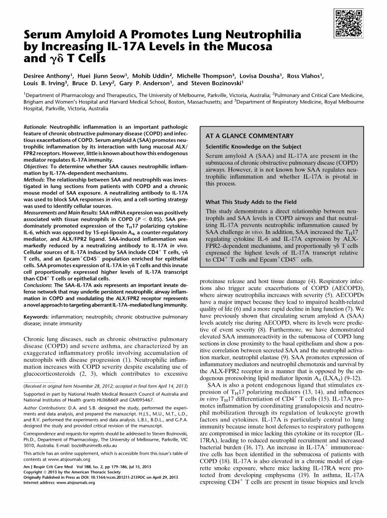
Figure 1. The relationship between tissue neutrophils and
serum amyloid A (SAA) levels in chronic obstructive pul-monary disease (COPD) lung. Representative tissue stain-
ing for the granulocyte marker esterase in (A) control and
(B) COPD lung sections. (C) Esterase-positive tissue neu-
trophils were quantified and grouped according to the degreeof SAA tissue staining, demonstrating a significant increase in
neutrophils numbers in lung sections that displayed intense
SAA tissue staining. (D) Correlation between SAA expression
as determined by quantitative polymerase chain reaction andthe number of neutrophils in serial COPD lung sections. n¼ 13
individual patient samples, #P , 0.05, Spearman correlation.
GADPH ¼ glyceraldehyde phosphate dehydrogenase.
180 AMERICAN JOURNAL OF RESPIRATORY AND CRITICAL CARE MEDICINE VOL 188 2013
Cytokine Measurements and Enzyme-linked
Immunospot Assay
The CBA Flex Systems Kit (BD Biosciences, San Jose, CA) was used tomeasure cytokines in BAL fluid (BALF). Secreted IL-17A in the super-natant of A549 cells was determined using the ELISA kit (eBiosciences,San Diego, CA) in accordance with the manufacturer’s instructions.An enzyme-linked immunospot (ELISPOT) assay was used to detectIL-17A–secreting cell populations as detailed in the online supplement.
Statistical Analysis
Full details are provided in the online supplement.
RESULTS
SAA in COPD Lung Is Associated with
Neutrophil Accumulation
The relationship between SAAexpression and airway neutrophilaccumulation in stable COPD was investigated by quantifyingesterase-positive neutrophils in serial sections, where SAAexpression was determined by quantitative polymerase chainreaction (QPCR) and compared with matching tissue sectionspreviously stained for SAA by immunohistochemistry (9). Rep-resentative images of esterase-positive staining neutrophils inlung sections from a control subject (Figure 1A) and a patientwith COPD (Figure 1B) with elevated neutrophil numbers areshown. In these sections, the number of neutrophils significantlyincreased (P , 0.05) with the intensity of SAA staining (Figure1C). In addition, there was a positive correlation (r ¼ 0.62; P ,0.05) between SAA mRNA expression and the numbers ofneutrophils in matching serial lung sections (Figure 1D).
Chronic Administration of SAA Induced the Accumulation
of Neutrophils In Vivo
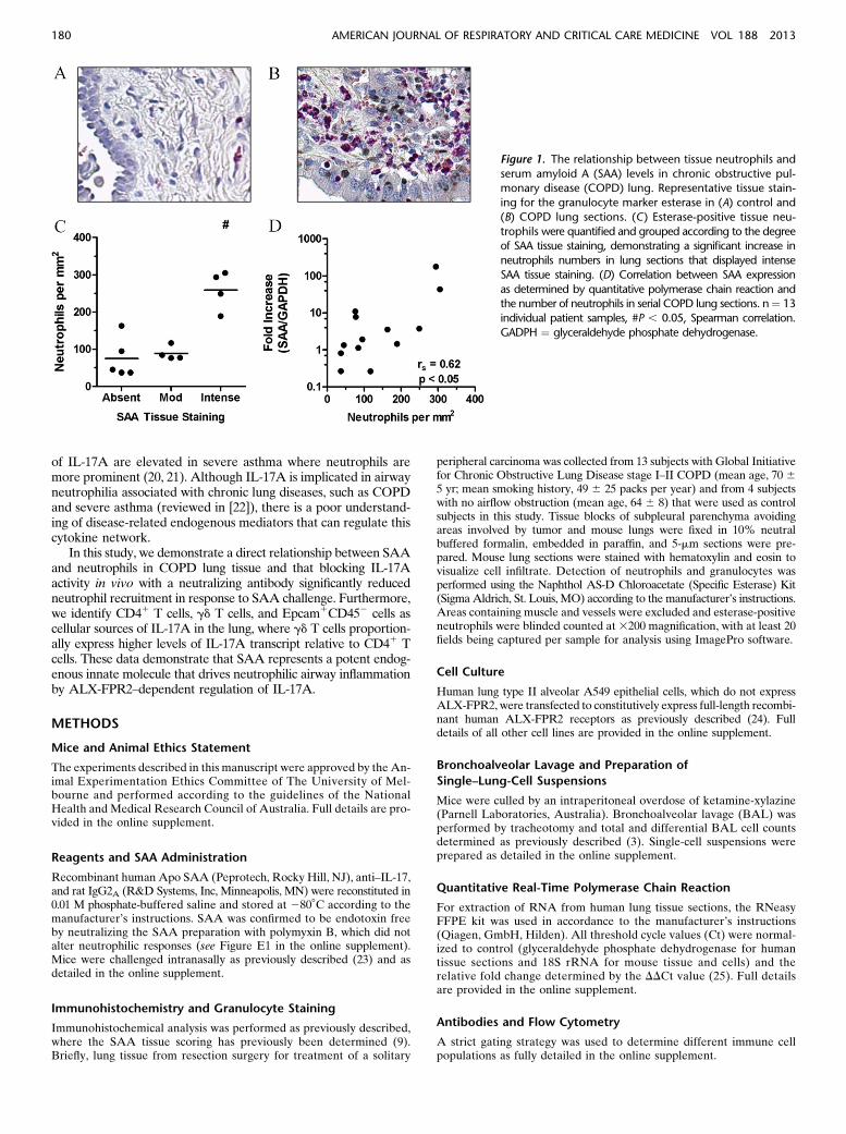
To further explore the direct relationship between SAA and lungneutrophil numbers in the chronic setting, a mouse model of re-peated SAA challenge was established. Mice were exposed tointranasal SAA or saline once a week for 5 weeks and immunecells were characterized by fluorescence-activated cell sorteranalysis and histology. Hematoxylin and eosin staining revealedminimal cell infiltrate in the lung of mice treated with saline (Fig-ure 2A) compared with SAA where there was a large influx ofimmune cells into the mouse lung (Figure 2B). Further analysisby fluorescence-activated cell sorter revealed that neutrophilnumbers were significantly increased in the BALF (saline 0.11 60.06 vs. SAA 0.60 6 0.09 3 104) (Figure 2C) and lung tissue
Figure 2. Chronic administration of serum amyloid A (SAA) promotes
neutrophil accumulation in the mouse lung. Mice were challenged
with SAA (2 mg) or saline once a week for 5 weeks and lung tissue
and bronchoalveolar lavage (BAL) fluid were harvested 24 hours afterthe final intranasal treatment of SAA. Representative hematoxylin and
eosin staining of sections from (A) saline- or (B) SAA-treated mice. The
hematoxylin and eosin staining demonstrates increased immune cell
infiltrate in the lung sections after SAA treatment. The number of neu-trophils was determined by fluorescence-activated cell sorter and pre-
gated according to a single cell; viable (PI exclusion), large-granular cell
gate and neutrophils were determined to be CD11C2 and Ly6G1 (Gr-1) in the (C) BAL and (D) lung. n ¼ 3–4 mice, *P , 0.001.
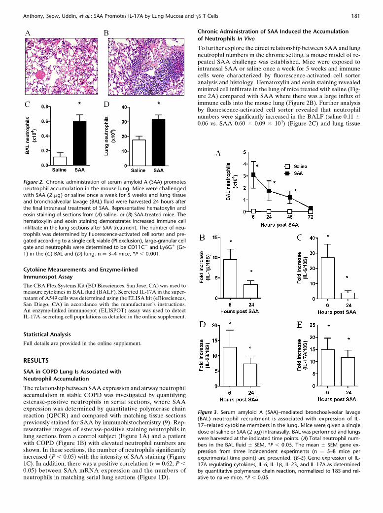
Figure 3. Serum amyloid A (SAA)–mediated bronchoalveolar lavage(BAL) neutrophil recruitment is associated with expression of IL-
17–related cytokine members in the lung. Mice were given a single
dose of saline or SAA (2 mg) intranasally. BAL was performed and lungs
were harvested at the indicated time points. (A) Total neutrophil num-bers in the BAL fluid 6 SEM, *P , 0.05. The mean 6 SEM gene ex-
pression from three independent experiments (n ¼ 5–8 mice per
experimental time point) are presented. (B–E) Gene expression of IL-
17A regulating cytokines, IL-6, IL-1b, IL-23, and IL-17A as determinedby quantitative polymerase chain reaction, normalized to 18S and rel-
ative to naive mice. *P , 0.05.
Anthony, Seow, Uddin, et al.: SAA Promotes IL-17A by Lung Mucosa and gd T Cells 181
(saline 17.62 6 0.27 vs. SAA 31.86 6 0.28 3 104) (Figure 2D) inresponse to SAA.
SAA Promotes the IL-17A Cytokine Network via
the ALX-FPR2 Receptor
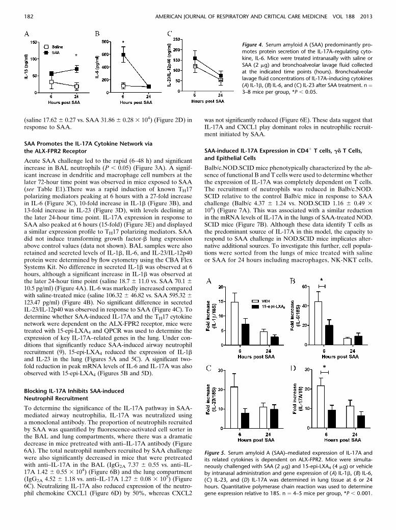
Acute SAA challenge led to the rapid (6–48 h) and significantincrease in BAL neutrophils (P , 0.05) (Figure 3A). A signif-icant increase in dendritic and macrophage cell numbers at thelater 72-hour time point was observed in mice exposed to SAA(see Table E1).There was a rapid induction of known TH17polarizing mediators peaking at 6 hours with a 27-fold increasein IL-6 (Figure 3C), 10-fold increase in IL-1b (Figure 3B), and13-fold increase in IL-23 (Figure 3D), with levels declining atthe later 24-hour time point. IL-17A expression in response toSAA also peaked at 6 hours (15-fold) (Figure 3E) and displayeda similar expression profile to TH17 polarizing mediators. SAAdid not induce transforming growth factor-b lung expressionabove control values (data not shown). BAL samples were alsoretained and secreted levels of IL-1b, IL-6, and IL-23/IL-12p40protein were determined by flow cytometry using the CBA FlexSystems Kit. No difference in secreted IL-1b was observed at 6hours, although a significant increase in IL-1b was observed atthe later 24-hour time point (saline 18.7 6 11.0 vs. SAA 70.1 610.5 pg/ml) (Figure 4A). IL-6 was markedly increased comparedwith saline-treated mice (saline 106.32 6 46.82 vs. SAA 595.32 6123.47 pg/ml) (Figure 4B). No significant difference in secretedIL-23/IL-12p40 was observed in response to SAA (Figure 4C). Todetermine whether SAA-induced IL-17A and the TH17 cytokinenetwork were dependent on the ALX-FPR2 receptor, mice weretreated with 15-epi-LXA4 and QPCR was used to determine theexpression of key IL-17A–related genes in the lung. Under con-ditions that significantly reduce SAA-induced airway neutrophilrecruitment (9), 15-epi-LXA4 reduced the expression of IL-1band IL-23 in the lung (Figures 5A and 5C). A significant two-fold reduction in peak mRNA levels of IL-6 and IL-17A was alsoobserved with 15-epi-LXA4 (Figures 5B and 5D).
Blocking IL-17A Inhibits SAA-induced
Neutrophil Recruitment
To determine the significance of the IL-17A pathway in SAA-mediated airway neutrophilia, IL-17A was neutralized usinga monoclonal antibody. The proportion of neutrophils recruitedby SAA was quantified by fluorescence-activated cell sorter inthe BAL and lung compartments, where there was a dramaticdecrease in mice pretreated with anti–IL-17A antibody (Figure6A). The total neutrophil numbers recruited by SAA challengewere also significantly decreased in mice that were pretreatedwith anti–IL-17A in the BAL (IgG2A 7.37 6 0.55 vs. anti–IL-17A 1.42 6 0.55 3 104) (Figure 6B) and the lung compartment(IgG2A 4.52 6 1.18 vs. anti–IL-17A 1.27 6 0.08 3 105) (Figure6C). Neutralizing IL-17A also reduced expression of the neutro-phil chemokine CXCL1 (Figure 6D) by 50%, whereas CXCL2
was not significantly reduced (Figure 6E). These data suggest thatIL-17A and CXCL1 play dominant roles in neutrophilic recruit-ment initiated by SAA.
SAA-induced IL-17A Expression in CD41 T cells, gd T Cells,
and Epithelial Cells
Balb/c.NOD.SCID mice phenotypically characterized by the ab-sence of functional B and T cells were used to determine whetherthe expression of IL-17A was completely dependent on T cells.The recruitment of neutrophils was reduced in Balb/c.NOD.SCID relative to the control Balb/c mice in response to SAAchallenge (Balb/c 4.37 6 1.24 vs. NOD.SCID 1.16 6 0.49 3104) (Figure 7A). This was associated with a similar reductionin the mRNA levels of IL-17A in the lungs of SAA-treated NOD.SCID mice (Figure 7B). Although these data identify T cells asthe predominant source of IL-17A in this model, the capacity torespond to SAA challenge in NOD.SCID mice implicates alter-native additional sources. To investigate this further, cell popula-tions were sorted from the lungs of mice treated with salineor SAA for 24 hours including macrophages, NK-NKT cells,
Figure 4. Serum amyloid A (SAA) predominantly pro-
motes protein secretion of the IL-17A–regulating cyto-kine, IL-6. Mice were treated intranasally with saline or
SAA (2 mg) and bronchoalveolar lavage fluid collected
at the indicated time points (hours). Bronchoalveolarlavage fluid concentrations of IL-17A–inducing cytokines
(A) IL-1b, (B) IL-6, and (C) IL-23 after SAA treatment. n ¼3–8 mice per group, *P , 0.05.
Figure 5. Serum amyloid A (SAA)–mediated expression of IL-17A andits related cytokines is dependent on ALX-FPR2. Mice were simulta-
neously challenged with SAA (2 mg) and 15-epi-LXA4 (4 mg) or vehicle
by intranasal administration and gene expression of (A) IL-1b, (B) IL-6,(C) IL-23, and (D) IL-17A was determined in lung tissue at 6 or 24
hours. Quantitative polymerase chain reaction was used to determine
gene expression relative to 18S. n ¼ 4–5 mice per group, *P , 0.001.
182 AMERICAN JOURNAL OF RESPIRATORY AND CRITICAL CARE MEDICINE VOL 188 2013
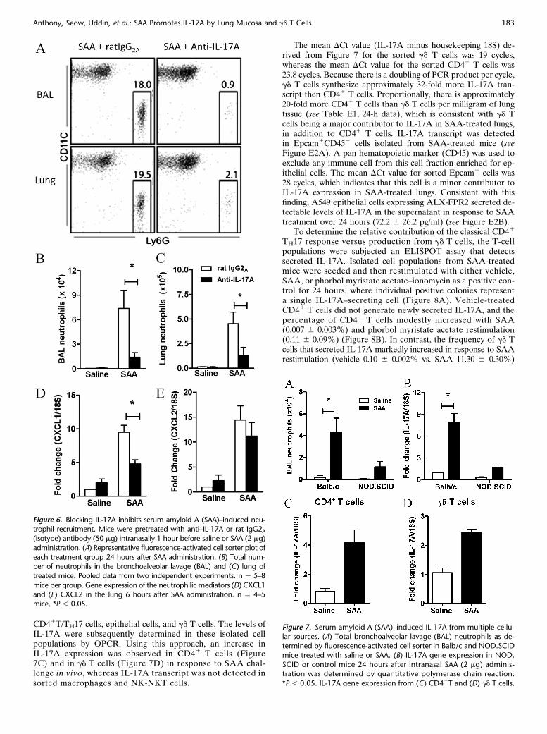
CD41T/TH17 cells, epithelial cells, and gd T cells. The levels ofIL-17A were subsequently determined in these isolated cellpopulations by QPCR. Using this approach, an increase inIL-17A expression was observed in CD41 T cells (Figure7C) and in gd T cells (Figure 7D) in response to SAA chal-lenge in vivo, whereas IL-17A transcript was not detected insorted macrophages and NK-NKT cells.
The mean DCt value (IL-17A minus housekeeping 18S) de-rived from Figure 7 for the sorted gd T cells was 19 cycles,whereas the mean DCt value for the sorted CD41 T cells was23.8 cycles. Because there is a doubling of PCR product per cycle,gd T cells synthesize approximately 32-fold more IL-17A tran-script then CD41 T cells. Proportionally, there is approximately20-fold more CD41 T cells than gd T cells per milligram of lungtissue (see Table E1, 24-h data), which is consistent with gd Tcells being a major contributor to IL-17A in SAA-treated lungs,in addition to CD41 T cells. IL-17A transcript was detectedin Epcam1CD452 cells isolated from SAA-treated mice (seeFigure E2A). A pan hematopoietic marker (CD45) was used toexclude any immune cell from this cell fraction enriched for ep-ithelial cells. The mean DCt value for sorted Epcam1 cells was28 cycles, which indicates that this cell is a minor contributor toIL-17A expression in SAA-treated lungs. Consistent with thisfinding, A549 epithelial cells expressing ALX-FPR2 secreted de-tectable levels of IL-17A in the supernatant in response to SAAtreatment over 24 hours (72.2 6 26.2 pg/ml) (see Figure E2B).
To determine the relative contribution of the classical CD41
TH17 response versus production from gd T cells, the T-cellpopulations were subjected an ELISPOT assay that detectssecreted IL-17A. Isolated cell populations from SAA-treatedmice were seeded and then restimulated with either vehicle,SAA, or phorbol myristate acetate–ionomycin as a positive con-trol for 24 hours, where individual positive colonies representa single IL-17A–secreting cell (Figure 8A). Vehicle-treatedCD41 T cells did not generate newly secreted IL-17A, and thepercentage of CD41 T cells modestly increased with SAA(0.007 6 0.003%) and phorbol myristate acetate restimulation(0.11 6 0.09%) (Figure 8B). In contrast, the frequency of gd Tcells that secreted IL-17A markedly increased in response to SAArestimulation (vehicle 0.10 6 0.002% vs. SAA 11.30 6 0.30%)
Figure 6. Blocking IL-17A inhibits serum amyloid A (SAA)–induced neu-trophil recruitment. Mice were pretreated with anti–IL-17A or rat IgG2A(isotype) antibody (50 mg) intranasally 1 hour before saline or SAA (2 mg)
administration. (A) Representative fluorescence-activated cell sorter plot of
each treatment group 24 hours after SAA administration. (B) Total num-ber of neutrophils in the bronchoalveolar lavage (BAL) and (C) lung of
treated mice. Pooled data from two independent experiments. n ¼ 5–8
mice per group. Gene expression of the neutrophilic mediators (D) CXCL1and (E) CXCL2 in the lung 6 hours after SAA administration. n ¼ 4–5
mice, *P , 0.05.
Figure 7. Serum amyloid A (SAA)–induced IL-17A from multiple cellu-lar sources. (A) Total bronchoalveolar lavage (BAL) neutrophils as de-
termined by fluorescence-activated cell sorter in Balb/c and NOD.SCID
mice treated with saline or SAA. (B) IL-17A gene expression in NOD.
SCID or control mice 24 hours after intranasal SAA (2 mg) adminis-tration was determined by quantitative polymerase chain reaction.
*P , 0.05. IL-17A gene expression from (C) CD41T and (D) gd T cells.
Anthony, Seow, Uddin, et al.: SAA Promotes IL-17A by Lung Mucosa and gd T Cells 183
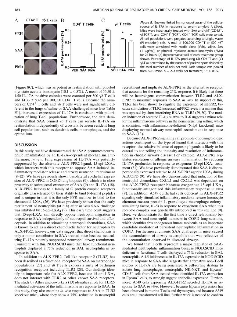
(Figure 8C), which was as potent as restimulation with phorbolmyristate acetate–ionomycin (10.1 6 0.5%). A mean of 56.50 61.50 IL-17A–positive colonies were counted per 500 gd T cellsand 14.33 6 5.45 per 100,000 CD41 T cells. Because the num-bers of CD41 T cells and gd T cells were not significantly dif-ferent in the lungs of saline or SAA-challenged mice (see TableE1), increased expression of IL-17A is consistent with polari-zation of lung T-cell populations. Furthermore, the data dem-onstrate that SAA primed gd T cells can secrete IL-17A onrestimulation independently of crosstalk between resident lungcell populations, such as dendritic cells, macrophages, and theepithelium.
DISCUSSION
In this study, we have demonstrated that SAA promotes neutro-philic inflammation by an IL-17A–dependent mechanism. Fur-thermore, in vivo lung expression of IL-17A was potentlysuppressed by the alternate ALX-FPR2 ligand, 15-epi-LXA4,which interacts with this receptor to oppose SAA-induced in-flammatory mediator release and airway neutrophil recruitment(9–12). We have previously shown basolateral epithelial expres-sion of ALX-FPR2 in COPD lung biopsies (9), which is in closeproximity to submucosal expression of SAA (9) and IL-17A (18).ALX-FPR2 belongs to a family of G protein coupled receptorsoriginally characterized by their ability to bind N-formyl peptidesand is unique in that it is the only FPR member to bind to theeicosanoid, LXA4 (26). We have previously shown that the earlyrecruitment of neutrophils (at 6 h) after in vivo SAA challengewas inhibited by 15-epi-LXA4 (9). This early time point suggeststhat 15-epi-LXA4 can directly oppose neutrophil migration inresponse to SAA independently of neutrophil survival and effer-ocytosis. In addition to stimulating neutrophil chemokines, SAAis known to act as a direct chemotactic factor for neutrophils byALX-FPR2; however, our data suggest that direct chemotaxis isonly a minor contributor in SAA-treated mice because neutral-izing IL-17A potently suppressed neutrophil airway recruitment.Consistent with this, NOD.SCID mice that have functional neu-trophils displayed a 75% reduction in BAL neutrophilia in re-sponse to SAA.
In addition to ALX-FPR2, Toll-like receptor-2 (TLR2) hasbeen described as a functional receptor for SAA on macrophagepopulations (27) and gd T cells express a suite of pathogenrecognition receptors including TLR2 (28). Our findings iden-tify an important role for ALX-FPR2, because 15-epi-LXA4
does not interact with TLR2 or other known SAA receptors.The study by Ather and coworkers (13) identifies a role for TLR2-mediated activation of the inflammasome in response to SAA. Inthis study, they also examine in vivo responses to SAA in TLR2knockout mice, where they show a 75% reduction in neutrophil
recruitment and implicate ALX-FPR2 as the alternative receptorthat accounts for the remaining 25% response. It is likely that therewill be heterologous communication between TLR2 and ALX-FPR2 to maximize responses to SAA in vivo. In support of this,TLR2 has been shown to regulate the expression of mFPR2, be-cause stimulation of TLR2 increasedmFPR2 levels in a manner thatwas opposed by short interfering RNA to TLR2 (29, 30). The mod-est induction of secreted IL-1b relative to IL-6 suggests a minor rolefor the inflammasome pathway in the nonallergic lung setting, whichis consistent with inflammasome-deficient (Nrlp3 knockout) micedisplaying normal airway neutrophil recruitment in responseto SAA (13).
Because ALX-FPR2 signaling can promote opposing biologicactions contingent on the type of ligand that interacts with thisreceptor, the relative balance of opposing ligands is likely to becentral to controlling the intensity and resolution of inflamma-tion in chronic airways disease. For example, ALX-FPR2 reg-ulates resolution of allergic airways inflammation by reducingIL-17A production in response to exogenous 15-epi-LXA4 treat-ment (31). We have previously demonstrated that SAA is dispro-portionally expressed relative to ALX-FPR2 agonist LXA4 duringAECOPD (9). We have also demonstrated that induction of theneutrophil chemokines CXCL1 and CXCL2 was dependent onthe ALX-FPR2 receptor because exogenous 15-epi-LXA4
functionally antagonized this inflammatory response in vivo(9). In addition, A549 epithelial cells deficient in ALX-FPR2signaling only stimulated inflammatory mediator release (monocytechemoattractant protein-1, granulocyte-macrophage colony–stimulating factor, IL-8) in response to exogenous SAA when thisreceptor complex was genetically introduced in this cell line (9).Here, we demonstrate for the first time a direct relationship be-tween SAA and neutrophil numbers in COPD lung sections,which identifies this endogenous innate molecule as an importantcandidate mediator of persistent neutrophilic inflammation inCOPD. Furthermore, chronic SAA challenge in mice causedthe accumulation of airway neutrophils that was reflective ofthe accumulation observed in diseased airways.
We found that T cells represent a major regulator of SAA-mediated neutrophilic inflammation because NOD.SCID micedeficient in functional T cells displayed a 75% reduction in BALneutrophils. A 4.5-fold increase in IL-17A expression inNOD.SCIDmice in response to SAA also suggests that alternative non–T-cellsources of IL-17A are being generated. A cell-sorting strategy toisolate lung macrophages, neutrophils, NK-NKT, and Epcam1
CD452 cells from SAA-treated mice identified IL-17A expressionin Epcam1 cells, to strongly suggest epithelial expression. Further-more, A549 cells expressing ALX-FPR2 secreted IL-17A in re-sponse to SAA in vitro. However, because Epcam expression hasbeen observed in murine T cells and thymocytes, and because A549cells are a transformed cell line, further work is needed to confirm
Figure 8. Enzyme-linked immunospot assay of the cellular
source of IL-17A in response to serum amyloid A (SAA).Mice were intranasally treated with SAA and gdT (CD451,
gdTCR1), and CD41T (TCR1, CD41 TCR) cells were sorted.
All cell populations were pregated according to single, viable(PI exclusion) cells. A total of 100,000 CD41T or 500 gdT
cells were stimulated with media alone (Veh), saline, SAA
(1 mg/ml), or phorbol myristate acetate–ionomycin (PMA)
for 24 hours. (A) Representative well of each treatment groupshown. Percentage of IL-17A–producing (B) CD41T and (C)
gdT as determined by the number of positive spots divided by
the total number of cells per well. Each sample was pooled
from 8–10 mice. n ¼ 2–3 wells per treatment, *P , 0.05.
184 AMERICAN JOURNAL OF RESPIRATORY AND CRITICAL CARE MEDICINE VOL 188 2013
epithelial expression in primary epithelial cells, which is beyond thescope of this study. In addition, sorted CD41 T cells isolated fromthe lungs of SAA-challenged mice expressed increased IL-17Atranscript levels. Ex vivo restimulation with SAA demonstrateda very low frequency (0.007%) of CD41 IL-17A–secreting T cells.Our finding that isolated CD41 T cells respond poorly to restim-ulation with SAA is consistent with the expression of IL-17A beingdependent on coculture with other innate immune cells capable ofinducing IL-17A–related genes, such as IL-6 and IL-23 (15). Levelsof secreted IL-6 in response to SAA were significantly higherthan IL-1b or IL-23 in vivo, and no increase in transforming growthfactor-b lung expression was detected. Using 15-epi-LXA4, wedemonstrate that increased IL-6 expression in response to SAAchallenge is dependent on the ALX-FPR2 receptor complex. Al-though IL-6 is known to promote expression of SAA from hepaticand epithelial cells (32, 33), we propose a novel positive feedbackmechanism, whereby SAA initiates expression of IL-6 from airwayepithelial cells and macrophages, and this promotes polarization ofCD41 T cell populations to express IL-17A.
Our study is the first to identify gd T cells as a major producerof IL-17A in response to SAA. Furthermore, the secretion ofIL-17A by isolated gd T cells in the absence of coculture withdendritic cells or macrophages demonstrates a direct mecha-nism by which gd T cells can produce IL-17A independentlyof the classic TΗ17 paradigm. It has been shown that gd T cellscan selectively expand in response to pathogen products in a man-ner that is amplified by IL-23 (28). Here, we did not observe anexpansion of gd T cell numbers in the airways after SAA challenge,which is consistent with the observation that gd T cells numbers arenot elevated in COPD (34). Although the role of gd T cells inCOPD pathogenesis remains poorly defined, direct interactions be-tween the innate endogenous mediator SAA and gd T cells re-presents a novel mechanism by which neutrophilic recruitment issustained in chronic lung diseases, such as COPD. Furthermore,therapeutically targeting the ALX-FPR2 receptor using the prore-solving mediator 15-epi-LXA4 represents a novel approach todampening IL-17A–dependent immunity responsible for excessiveneutrophilic inflammation in an environment where SAA is abun-dantly present.
Author disclosures are available with the text of this article at www.atsjournals.org.
References
1. Hogg JC, Chu F, Utokaparch S, Woods R, Elliott WM, Buzatu L,
Cherniack RM, Rogers RM, Sciurba FC, Coxson HO, et al. The
nature of small-airway obstruction in chronic obstructive pulmonary
disease. N Engl J Med 2004;350:2645–2653.
2. Culpitt SV, Maziak W, Loukidis S, Nightingale JA, Matthews JL, Barnes
PJ. Effect of high dose inhaled steroid on cells, cytokines, and proteases
in induced sputum in chronic obstructive pulmonary disease. Am J
Respir Crit Care Med 1999;160:1635–1639.
3. Vlahos R, Wark PA, Anderson GP, Bozinovski S. Glucocorticosteroids
differentially regulate MMP-9 and neutrophil elastase in COPD. PLoS
ONE 2012;7:e33277.
4. Taggart CC, Greene CM, Carroll TP, O’Neill SJ, McElvaney NG.
Elastolytic proteases: inflammation resolution and dysregulation in
chronic infective lung disease. Am J Respir Crit Care Med 2005;171:
1070–1076.
5. Papi A, Bellettato CM, Braccioni F, Romagnoli M, Casolari P, Caramori
G, Fabbri LM, Johnston SL. Infections and airway inflammation in
chronic obstructive pulmonary disease severe exacerbations. Am J
Respir Crit Care Med 2006;173:1114–1121.
6. Donaldson GC, Wilkinson TM, Hurst JR, Perera WR, Wedzicha JA.
Exacerbations and time spent outdoors in chronic obstructive pulmonary
disease. Am J Respir Crit Care Med 2005;171:446–452.
7. Donaldson GC, Seemungal TA, Bhowmik A, Wedzicha JA. Relationship
between exacerbation frequency and lung function decline in chronic
obstructive pulmonary disease. Thorax 2002;57:847–852.
8. Bozinovski S, Hutchinson A, Thompson M, Macgregor L, Black J, Giannakis
E, Karlsson AS, Silvestrini R, Smallwood D, Vlahos R, et al. Serum
amyloid a is a biomarker of acute exacerbations of chronic obstructive
pulmonary disease. Am J Respir Crit Care Med 2008;177:269–278.
9. Bozinovski S, Uddin M, Vlahos R, Thompson M, McQualter JL, Merritt
AS, Wark PA, Hutchinson A, Irving LB, Levy BD, et al. Serum
amyloid A opposes lipoxin A4 to mediate glucocorticoid refractory
lung inflammation in chronic obstructive pulmonary disease. Proc Natl
Acad Sci USA 2012;109:935–940.
10. El Kebir D, József L, Khreiss T, Pan W, Petasis NA, Serhan CN, Filep
JG. Aspirin-triggered lipoxins override the apoptosis-delaying action
of serum amyloid A in human neutrophils: a novel mechanism for
resolution of inflammation. J Immunol 2007;179:616–622.
11. He R, Sang H, Ye RD. Serum amyloid A induces IL-8 secretion through
a G protein-coupled receptor, FPRL1/LXA4R. Blood 2003;101:
1572–1581.
12. Su SB, Gong W, Gao JL, Shen W, Murphy PM, Oppenheim JJ, Wang
JM. A seven-transmembrane, G protein-coupled receptor, FPRL1,
mediates the chemotactic activity of serum amyloid A for human
phagocytic cells. J Exp Med 1999;189:395–402.
13. Ather JL, Ckless K, Martin R, Foley KL, Suratt BT, Boyson JE, Fitzgerald
KA, Flavell RA, Eisenbarth SC, Poynter ME. Serum amyloid A activates
the NLRP3 inflammasome and promotes Th17 allergic asthma in mice.
J Immunol 2011;187:64–73.
14. He R, Shepard LW, Chen J, Pan ZK, Ye RD. Serum amyloid A is an
endogenous ligand that differentially induces IL-12 and IL-23. J Immunol
2006;177:4072–4079.
15. Ivanov II, Atarashi K, Manel N, Brodie EL, Shima T, Karaoz U, Wei D,
Goldfarb KC, Santee CA, Lynch SV, et al. Induction of intestinal
Th17 cells by segmented filamentous bacteria. Cell 2009;139:485–498.
16. Wu Q, Martin RJ, Rino JG, Breed R, Torres RM, Chu HW. IL-23-
dependent IL-17 production is essential in neutrophil recruitment
and activity in mouse lung defense against respiratory Mycoplasma
pneumoniae infection. Microbes Infect 2007;9:78–86.
17. Ye P, Rodriguez FH, Kanaly S, Stocking KL, Schurr J, Schwarzenberger
P, Oliver P, HuangW, Zhang P, Zhang J, et al. Requirement of interleukin
17 receptor signaling for lung CXC chemokine and granulocyte colony-
stimulating factor expression, neutrophil recruitment, and host defense.
J Exp Med 2001;194:519–527.
18. Di Stefano A, Caramori G, Gnemmi I, Contoli M, Vicari C, Capelli A,
Magno F, D’Anna SE, Zanini A, Brun P, et al. T helper type 17-
related cytokine expression is increased in the bronchial mucosa of stable
chronic obstructive pulmonary disease patients. Clin Exp Immunol 2009;
157:316–324.
19. Chen K, Pociask DA, McAleer JP, Chan YR, Alcorn JF, Kreindler JL,
Keyser MR, Shapiro SD, Houghton AM, Kolls JK, et al. IL-17RA is
required for CCL2 expression, macrophage recruitment, and em-
physema in response to cigarette smoke. PLoS ONE 2011;6:e20333.
20. Al-Ramli W, Préfontaine D, Chouiali F, Martin JG, Olivenstein R,
Lemière C, Hamid Q. T(H)17-associated cytokines (IL-17A and IL-17F)
in severe asthma. J Allergy Clin Immunol 2009;123:1185–1187.
21. Chakir J, Shannon J, Molet S, Fukakusa M, Elias J, Laviolette M, Boulet
LP, Hamid Q. Airway remodeling-associated mediators in moderate
to severe asthma: effect of steroids on TGF-beta, IL-11, IL-17, and
type I and type III collagen expression. J Allergy Clin Immunol 2003;
111:1293–1298.
22. Alcorn JF, Crowe CR, Kolls JKT. TH17 cells in asthma and COPD.
Annu Rev Physiol 2010;72:495–516.
23. Bozinovski S, Jones JE, Vlahos R, Hamilton JA, Anderson GP. Granulocyte/
macrophage-colony-stimulating factor (GM-CSF) regulates lung innate
immunity to lipopolysaccharide through Akt/Erk activation of NFkappa B
and AP-1 in vivo. J Biol Chem 2002;277:42808–42814.
24. Bonnans C, Fukunaga K, Levy MA, Levy BD. Lipoxin A(4) regulates
bronchial epithelial cell responses to acid injury. Am J Pathol 2006;
168:1064–1072.
25. Bozinovski S, Seow HJ, Crack PJ, Anderson GP, Vlahos R. Glutathione
peroxidase-1 primes pro-inflammatory cytokine production after LPS
challenge in vivo. PLoS ONE 2012;7:e33172.
26. Ye RD, Boulay F, Wang JM, Dahlgren C, Gerard C, Parmentier M, Serhan
CN, Murphy PM. International Union of Basic and Clinical Pharmacology.
LXXIII. Nomenclature for the formyl peptide receptor (FPR) family.
Pharmacol Rev 2009;61:119–161.
Anthony, Seow, Uddin, et al.: SAA Promotes IL-17A by Lung Mucosa and gd T Cells 185
27. Cheng N, He R, Tian J, Ye PP, Ye RD. Cutting edge: TLR2 is
a functional receptor for acute-phase serum amyloid A. J Immunol
2008;181:22–26.
28. Martin B, Hirota K, Cua DJ, Stockinger B, Veldhoen M. Interleukin-17-
producing gammadelta T cells selectively expand in response to
pathogen products and environmental signals. Immunity 2009;31:
321–330.
29. Chen K, Iribarren P, Hu J, Chen J, Gong W, Cho EH, Lockett S, Dunlop
NM, Wang JM. Activation of Toll-like receptor 2 on microglia pro-
motes cell uptake of Alzheimer disease-associated amyloid beta
peptide. J Biol Chem 2006;281:3651–3659.
30. Chen K, Zhang L, Huang J, Gong W, Dunlop NM, Wang JM. Cooperation
between NOD2 and Toll-like receptor 2 ligands in the up-regulation of
mouse mFPR2, a G-protein-coupled Abeta42 peptide receptor, in micro-
glial cells. J Leukoc Biol 2008;83:1467–1475.
31. Haworth O, Cernadas M, Yang R, Serhan CN, Levy BD. Resolvin E1
regulates interleukin 23, interferon-gamma and lipoxin A4 to promote
the resolution of allergic airway inflammation. Nat Immunol 2008;9:
873–879.
32. Thorn CF, Lu Z-Y, Whitehead AS. Regulation of the human acute phase
serum amyloid A genes by tumour necrosis factor-alpha, interleukin-6
and glucocorticoids in hepatic and epithelial cell lines. Scand J
Immunol 2004;59:152–158.
33. Thorn CF, Whitehead AS. Differential glucocorticoid enhancement of
the cytokine-driven transcriptional activation of the human acute
phase serum amyloid A genes, SAA1 and SAA2. J Immunol 2002;169:
399–406.
34. Pons J, Sauleda J, Ferrer JM, Barceló B, Fuster A, Regueiro V, JuliàMR,Agustí AG. Blunted g d T-lymphocyte response in chronic obstructive
pulmonary disease. Eur Respir J 2005;25:441–446.
186 AMERICAN JOURNAL OF RESPIRATORY AND CRITICAL CARE MEDICINE VOL 188 2013
Related Documents


![Case Report Primary cutaneous γδ-T-cell lymphoma … cutaneous γδ-T-cell lymphoma (CGD-TCL) ... TCL [3]. Some other study reports that allogenic ... we reported a case of CGD-TCL](https://static.cupdf.com/doc/110x72/5ae360cf7f8b9a495c8d272b/case-report-primary-cutaneous-t-cell-lymphoma-cutaneous-t-cell-lymphoma.jpg)