FINAL ACCEPTED VERSION; LCMP-00477-2004 Sequential recruitment of neutrophils into lung and bronchoalveolar lavage fluid in LPS-induced acute lung injury Jörg Reutershan 1) 4) , Abdul Basit 2) , Elena V. Galkina 1)3) , Klaus Ley 1) 2) 3) 1) Cardiovascular Research Center, 2) Department of Physiology and Biological Physics, and 3) Biomedical Engineering; University of Virginia; Charlottesville, Virginia, USA, and 4) Department of Anesthesiology and Intensive Care Medicine, University of Tübingen, Tübingen, Germany Requests for reprints and corresponding author Klaus Ley University of Virginia Health System Cardiovascular Research Center P.O. Box 801394 Charlottesville, VA 22908-1394, USA phone +1 (434) 243-9966 fax +1 (434) 924-2828 [email protected] Running head: Neutrophil migration in the lung Articles in PresS. Am J Physiol Lung Cell Mol Physiol (June 10, 2005). doi:10.1152/ajplung.00477.2004 Copyright © 2005 by the American Physiological Society.

Welcome message from author
This document is posted to help you gain knowledge. Please leave a comment to let me know what you think about it! Share it to your friends and learn new things together.
Transcript
FINAL ACCEPTED VERSION; LCMP-00477-2004
Sequential recruitment of neutrophils into lung and
bronchoalveolar lavage fluid in LPS-induced acute lung
injury
Jörg Reutershan1) 4), Abdul Basit 2), Elena V. Galkina 1)3), Klaus Ley1) 2) 3)
1) Cardiovascular Research Center, 2) Department of Physiology and Biological
Physics, and 3) Biomedical Engineering; University of Virginia; Charlottesville,
Virginia, USA, and 4) Department of Anesthesiology and Intensive Care Medicine,
University of Tübingen, Tübingen, Germany
Requests for reprints and corresponding author
Klaus Ley
University of Virginia Health System
Cardiovascular Research Center
P.O. Box 801394
Charlottesville, VA 22908-1394, USA
phone +1 (434) 243-9966
fax +1 (434) 924-2828
Running head: Neutrophil migration in the lung
Articles in PresS. Am J Physiol Lung Cell Mol Physiol (June 10, 2005). doi:10.1152/ajplung.00477.2004
Copyright © 2005 by the American Physiological Society.
Reutershan et al.; LCMP-00477-2004
1
Abstract
Infiltration of activated neutrophils (polymorphonuclear leukocytes; PMN) into the
lung is an important component of the inflammatory response in acute lung
injury. The signals required to direct PMN into the different compartments of the
lung have not been fully elucidated. In a murine model of LPS-induced lung
injury, we investigated the sequential recruitment of PMN into the pulmonary
vasculature, lung interstitium, and alveolar space. Mice were exposed to
aerosolized LPS and bronchoalveolar lavage fluid (BAL) and lungs were
harvested at different time points. We developed a flow cytometry-based
technique to assess in-vivo trafficking of PMN in the intravascular and
extravascular lung compartments. Aerosolized LPS induced consistent PMN
migration into all lung compartments. We found that sequestration in the
pulmonary vasculature occurred within the first hour. Transendothelial migration
into the interstitial space started one hour after LPS-exposure and increased
continuously until a plateau was reached between twelve and 24 hours.
Transepithelial migration into the alveolar airspace was delayed, as the first PMN
did not appear until two hours after LPS, reaching a peak at 24 hours.
Transendothelial migration was partially and transepithelial migration was
completely inhibited by pertussis toxin, indicating involvement of Gαi-coupled
receptors. These findings confirm LPS-induced migration of PMN into the lung.
For the first time, distinct transmigration steps into the different lung
compartments are characterized in vivo.
Reutershan et al.; LCMP-00477-2004
2
Key words: Acute lung injury; Polymorphonuclear Leukocytes; Pulmonary
circulation; Chemokines
Reutershan et al.; LCMP-00477-2004
3
Introduction
Acute lung injury (ALI) and acute respiratory distress syndrome (ARDS) are
characterized by a disturbance of the alveolar-capillary barrier associated with
several clinical disorders. There is no specific therapy, and the mortality of this
disease is still high. Our current understanding of the molecular mechanisms of
ALI/ARDS has recently been described as “embryonic at best” (27).
Migration of activated PMN plays a key role in development of ALI and ARDS (1).
Here, we investigate the sequential migration steps from blood to air space
(intravascular sequestration – transendothelial migration – transepithelial
migration).
A variety of stimuli induce PMN migration into the lung. Endotoxin of Gram-
negative bacteria (lipopolysaccharide; LPS) induces a range of inflammatory
responses. Toll-like receptor 4 (TLR4) is the most important cellular receptor for
LPS. LPS stimulates the response to chemoattractants and increases PMN
migration at sites of inflammation (14). TLR4 is essential for LPS-induced PMN
migration into the lung as shown by the absence of a response in TLR4-deficient
mice (3). In the lung, the response to LPS is regulated by radioresistant cells,
most likely endothelial cells (2) or alveolar macrophages (28).
The administration of LPS alone might not reflect the whole complexity of the
human disease, because it does not consider preexisting diseases, fluid
resuscitation, or mechanical ventilation (36). However, infections with gram-
Reutershan et al.; LCMP-00477-2004
4
negative bacteria and exposure to their predominant pathogenic component play
a key role in both development and outcome of ARDS (26).
PMN trafficking into the vascular compartment of the lung, also known as
“margination” (9), and into the bronchoalveolar space has been studied
extensively in various models of acute lung injury. Adhesion molecules and CXC
chemokines have been shown to be involved. CXC chemokines, such as CXCL8
(IL-8), promote PMN migration into the alveolar compartment (29), and pertussis
toxin-dependent chemokine receptors are essential for PMN infiltration in the
lung (4; 42). Selectins and integrins are thought to be required for PMN
sequestration into the vascular compartment (6). However, results from studies
using monoclonal antibodies and mutant mice have yielded conflicting results
(10). The importance of investigating each step of PMN migration in the lung has
been emphasized recently (8).
Methodological limitations complicate the assessment of PMN trafficking in the
lung. Most studies employ indirect parameters to assess PMN trafficking in the
lung. For instance, the drop in circulating PMN counts in response to an
inflammatory stimulus was used to estimate PMN sequestration in the lung
vasculature (6; 31). However, recruitment to other organs might occur at the
same time. Ex-vivo labeling of murine PMN might result in neutrophil activation
that makes results uninterpretable (7). Myeloperoxidase activity in the lung is
often measured to estimate PMN infiltration, but this technique is not able to
distinguish between intravascular and interstitial PMN. Intravital microscopy of
the pulmonary microcirculation has recently become available in mice (35; 39)
Reutershan et al.; LCMP-00477-2004
5
and will promote insights into the interactions between leukocytes and
endothelium. However, this technique is technically challenging because of the
respiratory movement, requires mechanical ventilation, and allows observation of
only the most superficial lung capillaries, which may not be representative of the
whole lung. Morphometric analysis, such as electron microscopy (16), is useful,
but remain semi-quantitative, time-consuming and expensive.
In this study, we developed a flow cytometry based approach to assess the
different steps of PMN trafficking in a murine model of LPS-induced acute lung
injury. PMN accumulation in the pulmonary vasculature, transendothelial
migration into the interstitium, and transepithelial migration from the interstitium
into the airspace occurred as a sequential process in a time dependent manner.
Our findings improve the current understanding of neutrophil recruitment into the
inflamed lung and airways in a model that mimics some aspects of ALI/ARDS.
Methods
Mice
Wild type male C57Bl/6 mice were obtained from Jackson Labs (Bar Harbor,
ME). All animal experiments were approved by the Animal Care and Use
Committee of the University of Virginia. Mice were eight to twelve weeks of age.
Antibodies for flow cytometry
Rat anti-mouse antibodies for flow cytometry were obtained from Pharmingen
(anti-CD45; clone 30-F11), Caltag (anti-7/4; recognizing the 7/4 antigen on
Reutershan et al.; LCMP-00477-2004
6
murine neutrophils), and eBioscience (anti-TER-119; recognizing glycophorin A-
associated TER-119 on cells of the erythroid lineage). Anti-mouse GR-1 antibody
was purified from supernatant of the GR-1 hybridoma (ATCC) by the
biomolecular facility of University of Virginia. GR-1 was labeled with a staining kit
following the manufacturer’s directions (Alexa Fluor 633, Molecular Probes).
Appropriate rat anti-mouse IgG2a and IgG2b (Pharmingen) were used as isotype
controls.
Murine model of acute lung injury
Aerosolized LPS was utilized to induce PMN-infiltration in the lung (40). Besides
PMN-migration, LPS-inhalation is known to induce the expression of various
inflammatory mediators such as chemokines and adhesion molecules. LPS has
also been shown to increase airway resistance (24). Up to four mice were
exposed simultaneously to aerosolized LPS in a custom-built cylindrical chamber
(20cm in length; 9cm in diameter) connected to an air nebulizer (MicroAir, Omron
Healthcare, Vernon Hills, IL). This system produced particles in the range of one
to five µm. LPS from Salmonella enteritidis (Sigma Co., St. Louis, MO) was
dissolved in 0.9% saline (500µg/ml) and mice were allowed to inhale LPS for 30
minutes. One side of the chamber was connected to a vacuum pump and a
constant flow rate of 15 ml/min was ensured using a flow meter (Gilmont
Instruments, Barrington, Illinois).

Reutershan et al.; LCMP-00477-2004
7
PMN counts in bronchoalveolar lavage fluid (BAL) and lung tissue
At different time after LPS exposure, mice were anesthetized with an
intraperitoneal injection of ketamine (125 mg/kg; Sanofi Winthrop
Pharmaceuticals, New York, NY), xylazine (12.5 mg/kg; Phoenix Scientific, St.
Joseph, MO) and atropine sulfate (0.025 mg/kg; Fujisawa, Deerfield, IL). The
pulmonary circulation was rinsed by injection of 10ml of PBS at 25 cmH2O into
the beating right ventricle after the inferior vena cava had been cut to allow
exsanguination. This was done to remove non-adherent PMN from the
pulmonary vessels. The trachea was cannulated (22 GA Insyte, Becton
Dickinson) and 1ml of PBS was infused intratracheally and withdrawn. This
procedure was repeated six times, resulting in a total volume of 7 ml. BAL fluid
was centrifuged for 5 minutes at 300g. The pellet was resuspended in 1ml buffer
(1% BSA and 0.1% sodium azide in PBS), and a 10µl aliquot was used for cell
count with a hemocytometer (Trypan Blue exclusion).
After performing BAL, lungs were harvested en bloc. Mediastinal tissue was
removed, lungs were minced and digested with 125 U/ml collagenase type XI, 60
U/ml hyaluronidase type I-s and 60 U/ml DNAse1 (all Sigma) at 37°C for 30 min.
Digested lungs were passed through a 70 µm cell strainer (BD Falcon, Bedford,
MA) and the resulting cell suspension was centrifuged for 10 minutes at 300g.
The pellet was lyzed using 0.83% NH4Cl to remove erythrocytes, and centrifuged
again. Pellet was resuspended in buffer and cells were counted with a
hemocytometer.
Reutershan et al.; LCMP-00477-2004
8
PMN were identified by 1) their typical appearance in the FSC/SSC, 2) by their
expression of CD45+, and 3) by two independent PMN-markers, 7/4 and GR-1
(19), and the absolute numbers of leukocytes (CD45+) and PMN were calculated.
Appropriate isotypes were used to set the gates. All studies were performed on a
FACS Calibur, Becton Dickinson (San Jose, CA), and data were analyzed with
FlowJo software (Tree Star, Ashland, OR). To confirm the presence of PMN
within the different populations as defined by flow cytometry, we sorted both
7/4+GR-1+ and 7/4+GR-1- cells (FACS Vantage, Becton Dickinson) and
characterized them morphologically by cytospin (Diff Quick staining; IMEB Inc,
IL).
In-vivo trafficking experiments
Dialyzed Alexa 633-labeled rat anti-mouse GR-1 (10µg) antibody was injected
i.v. and allowed to circulate for five minutes to bind to intravascular PMN. After
five minutes, mice were euthanized. BAL was obtained as described above, and
the lungs were homogenized in the presence of excess unlabeled anti-GR-1 to
prevent possible binding of excess Alexa 633-GR-1 to extravascular PMN. Cell
suspensions from BAL and lung tissue were made and cells were counted in a
hemocytometer.
Non-perfused or occluded vessels in the lung might result in trapped neutrophils,
not accessible for the injected antibody. This would lead to an underestimation of
PMN counts in the intravascular compartment. Non-perfused / occluded vessels
contain white and red blood cells. To assess the significance of this phenomenon
in our model, a monoclonal antibody to the TER-119 antigen on erythrocytes was

Reutershan et al.; LCMP-00477-2004
9
injected i.v. (TER-119; eBioscience, San Diego, CA). This 52-kDa molecule is
associated with glycophorin A on cells of the erythroid lineage (22). Five minutes
after injection, blood and lungs were harvested. Erythrocytes were defined by
their typical appearance in the forward-sideward scatter and the amount of TER-
119+ erythrocytes in each organ was expressed as percentage of total
erythrocytes by flow cytometry. To assess the effect of i.v. injection of anti-GR-1
on peripheral PMN counts, in some experiments, blood was withdrawn from the
tail vein and blood counts were performed before and ten minutes after antibody
injection using an automatic cell counter (Hemavet, Drew Scientific, Dallas, TX).
In all experiments, animals exposed to aerosolized saline served as control.
Cytospin of BAL
In some experiments, cytospins of the BAL (without LPS-treatment and 24 hours
after LPS-inhalation) were performed using a cytocentrifuge (Shandon, Southern
Sewickley, PA). The cytospun cells were Giemsa stained, air-dried and
coverslipped.
Lung permeability
To confirm lung injury in our LPS inhalation model, we determined microvascular
permeability using the Evans blue dye extravasation technique. Evans blue
(20mg/kg; Sigma-Aldrich) was injected i.v. 30 minutes prior to euthanasia. Lungs
were perfused through the spontaneously beating right ventricle. Lungs were
removed and Evans blue was extracted as described previously (32). The
absorption of Evans blue was measured at 620 nm and corrected for the
Reutershan et al.; LCMP-00477-2004
10
presence of heme pigments: A620 (corrected) = A620 - (1.426 x A740 + 0.030) (45).
Extravasated Evans blue was determined 6 hours after LPS or saline inhalation
and calculated against a standard curve (micrograms Evans blue dye per gram
lung).
PTx pretreatment
Chemokines have been shown to regulate PMN migration in the lung (42). To
block chemokine-mediated PMN migration, some mice received tail vein
injections of 4 µg of Pertussis toxin (PTx) from Bordetella pertussis (Lyophilized
powder, Sigma) 30 minutes prior to LPS exposure. This dose completely inhibits
Gα1-mediated signaling (41). PTx was dissolved in physiological saline. PMN in
lung and BAL were assessed 14 hours after LPS exposure. In addition, a dose-
response-curve was established for each lung compartment 12 hours after LPS
exposure (0, 0.04, 0.4, or 4 µg PTx / mouse).
Statistical analysis
Data were analyzed using Excel software package (Microsoft). PMN counts were
compared with the paired Student’s t-Test. P < 0.05 was considered statistically
significant. Data were expressed as the mean ± SEM.

Reutershan et al.; LCMP-00477-2004
11
Results
LPS-induced PMN recruitment into lung and BAL
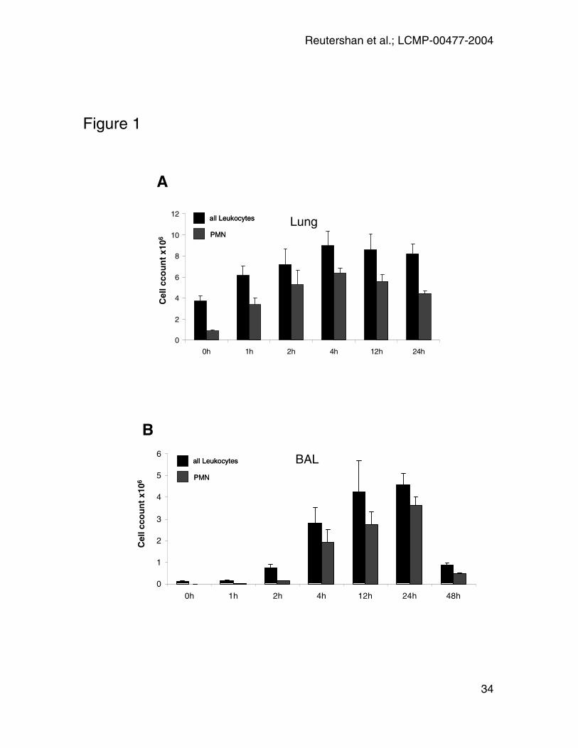
LPS-inhalation induced a time-dependent PMN recruitment into lung and BAL
(Figure 1). In the lung, significant numbers of leukocytes (CD45+ cells) were
present even before LPS administration, and their numbers increased only
moderately from four to a maximum of nine million cells at four hours after LPS.
Neutrophils were also present in resting lungs (approximately one million per
mouse), consistent with the concept of a physiologically marginated pool in the
pulmonary vasculature. Lung neutrophil numbers reached more than six million
cells/mouse at four hours of LPS administration, which is several-fold more than
the total number of all circulating neutrophils consistent with a previously
described release of PMN from the bone marrow (37; 46). At the peak of the
response, neutrophils accounted for 74 ± 7% of all leukocytes in the lung.
No PMN were observed in the BAL at 0h. PMN recruitment into the airspace was
delayed, and the first PMN did not appear until two hours. Between two and four
hours, neutrophil recruitment was very pronounced. After 48 hours, cell counts in
BAL were reduced, but did not reach baseline.
In-vivo GR-1 labeling
In the lung homogenate, GR-1 labeling was utilized to distinguish between PMN
derived from the pulmonary vasculature (GR-1+7/4+) and PMN derived from
interstitial space (GR-1-7/4+). We assessed the PMN labeling five minutes after
injection of anti-GR-1 antibody. We found that almost all blood PMN
Reutershan et al.; LCMP-00477-2004
12
(99.2 ± 0.4%) had been stained with GR-1 five minutes after antibody injection
(Figure 2A and B; table 1). To test whether GR-1 antibody was leaking into the
BAL, GR-1 labeling of PMN in BAL (CD45+7/4+) was assessed at different time
points after LPS-exposure, five minutes after antibody injection. No GR-1+ cells
were found in BAL. When GR-1 antibody was added after BAL harvesting, all
PMN were GR-1+ (positive control) (Figure 2C and D; table 1). GR-1+ PMN did
not appear in the BAL until 4h after antibody injection (data not shown).
Although all circulating PMN were shown to be GR-1+, potential non- or poorly
perfused areas of the pulmonary vasculature might be inaccessible for an
intravenously injected antibody. This would result in an underestimation of the
intravascular or an overestimation of the interstitial PMN concentration in the
lung, respectively. This effect might occur particularly in the injured lung. We
therefore labeled erythrocytes by i.v. injection of an antibody to the TER-119
antigen of red blood cells intravenously, 24 hours after LPS exposure. Red blood
cells are not found in the lung interstitium or BAL (5). The amount of TER-119+
cells in all erythrocytes were then determined in both blood and lung
homogenate. Five minutes after injection, we found 98.8% of all blood
erythrocytes to be TER-119+. At the same time, 97.7% of all erythrocytes were
TER-119+ in the lung homogenate indicating that the injected antibody is able to
bind to almost all red cells in the pulmonary vasculature.
Effect of anti-GR-1 antibody on PMN blood counts
GR-1 antibody can induce a severe neutropenia when given at high doses (33).
Therefore, we performed peripheral blood counts before and ten minutes after

Reutershan et al.; LCMP-00477-2004
13
antibody injection. GR-1 injection did not affect blood PMN counts at the
concentration used in our study (0.89 ± 0.14x10³/µl before, 0.86 ± 0.16 x10³/µl
after injection; p = 0.90).
In-vivo trafficking experiments
We assessed the concentration of PMN in the different lung compartments at 0,
1, 2, 4, 12, and 24 hours after LPS-exposure. At each time point, anti-GR-1
antibody was injected five minutes prior to euthanasia. PMN were identified by
flow cytometry (FSC/SSC-gate; CD45+7/4+) and GR-1 was utilized to distinguish
between intravascular (GR-1+) and interstitial (GR-1-) PMN (Figure 3). Both
populations predominantly consisted of PMN as confirmed morphologically by
cytospins (7/4+/GR-1+: 99% PMN; 7/4+GR-1-: 97% PMN; data not shown). At 0h,
86% of all PMN were found in the vascular compartment. LPS induced PMN
accumulation in the pulmonary vasculature. Both absolute and relative PMN
counts increased rapidly until a peak was reached after four hours. After four
hours, PMN counts in the vasculature decreased and returned to baseline at 24
hours after LPS-exposure (Figure 3A).
PMN concentration in both interstitium and bronchoalveolar lavage was
negligible at 0h. After 4h, 33% of all pulmonary PMN were found in the
interstitium. At the same time, PMN represented 91% of all cells in the BAL
(Figure3B). After 24h, the majority of PMN (78%) in the lung were found in the
extravascular space. Note that PMN in BAL appear GR-1- as the GR-1 antibody
injected five minutes before euthanasia remains confined to the vasculature. In
control animals, saline inhalation induced a mild PMN accumulation in the
Reutershan et al.; LCMP-00477-2004
14
pulmonary vasculature. No migration into the interstitium or into the alveolar
airspace was observed in these mice (data not shown).
Kinetics of transendothelial and transepithelial PMN migration
Interstitium and airspace were free of PMN at 0h. Transendothelial migration into
the interstitial space started one hour after LPS-exposure and increased
continuously. After twelve hours, the majority of PMN in the lung were now found
extravascular (interstitium and airspace) (Figure 4). Until two hours, the
intravascular accumulation outpaced the neutrophil accumulation in the
extravascular space so that the proportion of extravascular PMN did not change.
By contrast, at four hours the rate of accumulation of intravascular neutrophils
slowed and extravascular PMN accounted for 33% of all neutrophils in the lung.
The kinetics of neutrophil recruitment into the BAL was different in that no cells
were found at one hour and only a very small number (130.000 per mouse) at
two hours, after which time neutrophil numbers increased rapidly and then
followed the number of interstitial neutrophils with a delay of about two hours.
The appearance of PMN in the BAL was confirmed by cytospin. Cytospins of
BAL were analyzed in untreated mice and in mice 24 hours after LPS-inhalation.
The predominant cells in BAL of untreated mice were alveolar macrophages
(Figure 4, left inset). As expected, PMN dominated the cell population 24 hours
after LPS-exposure (Figure 4, right inset).

Reutershan et al.; LCMP-00477-2004
15
Lung permeability
Vascular leakage was determined to confirm lung injury in our LPS inhalation
model. 6 hours after LPS inhalation, Evans blue extravasation was significantly
higher compared to saline inhalation (66.2 ± 6.2 vs. 29.3 ± 3.7 µg per g lung;
p = 0.002) (Figure 5).
PTx treatment
In some mice, 4µg of PTx (41) was injected i.v. 30 minutes prior to LPS
challenge. Lungs and BAL were harvested 14 hours after LPS-exposure, and
anti-GR-1 was injected five minutes prior to euthanasia to distinguish between
intravascular and interstitial PMN. LPS induced a significant PMN migration into
all three lung compartments. Mice pretreated with PTx exhibited a normal PMN
sequestration into the pulmonary vasculature but showed a reduced PMN
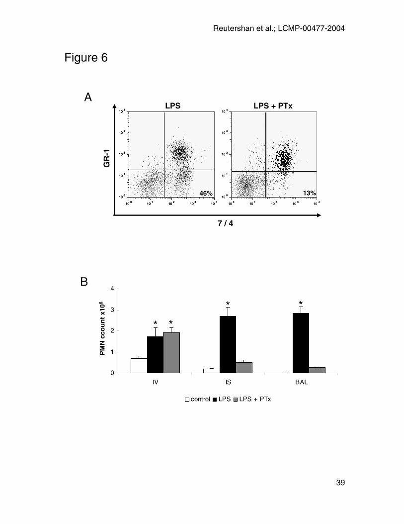
migration into the lung interstitium (0.5 ± 0.1 x10⁶ vs. 2.7 ± 0.4 x 10⁶; p < 0.01)
(Figure 6A and B). Accordingly, almost no PMN were found in the alveolar
airspace at 14 hours after LPS (0.3 ± 0.01 x10⁶ vs. 2.8 ± 0.3 x 10⁶; p < 0.01)
(Figure 6B). The inhibitory effect of PTx was dose-dependent as shown in Figure
7. This suggests that Gαi is involved at least in transendothelial migration from
blood space to interstitium and possibly also in transepithelial migration into the
alveolar airspace.

Reutershan et al.; LCMP-00477-2004
16
Discussion
In a LPS-induced model of acute lung injury, the sequential migration of PMN
into the different compartments of the lung was explored. Using a new and
quantitative flow cytometry-based technique, we show that LPS-inhalation
induced a rapid PMN sequestration in the pulmonary vasculature. Migration into
the lung interstitium was observed within one hour after LPS-exposure while
transepithelial migration was delayed. Pertussis toxin sharply reduced migration
into the interstitium and into the alveolar airspace but did not affect vascular
accumulation.
Methodological considerations
When using antibody injection to identify intravascular neutrophils in the lung, the
antibody is required to reach all PMN in this compartment. We found a complete
GR-1 labeling of all PMN in the systemic circulation. Additionally, the accessibility
of the pulmonary vasculature was successfully tested using a marker for
erythrocytes.
Even within five minutes, the GR-1 antibody might leak and stain extravascular
PMN. In our studies, GR-1+ PMN did not appear until four hours after antibody
injection in the BAL, indicating that the antibody did not reach alveolar airspace.
To test for antibody leakage into the interstitium, we injected a higher (10-fold)
dose of GR-1 that should increase the amount of leaked antibody and lead to an
increased fraction of labeled PMN. No such increase was observed in our
experiments (data not shown). Finally, a possible endothelial leakage should
Reutershan et al.; LCMP-00477-2004
17
increase as the LPS-induced alveolo-capillary damage proceeds over time
leading to a continuously rising overestimation of the intravascular PMN-fraction.
However, intravascular PMN-concentration peaks at 4h after LPS, while PMN-
concentration in the interstitium increases at this time (Figure 4). Taken together,
antibody leakage into the interstitium does not appear to play a major role in our
study.
Alveolar PMN are removed by bronchoalveolar lavage. Inaccessible airways
might exist, particularly in the inflamed lung, leaving (GR-1-) PMN in the airspace.
This might result in an overestimation of the PMN counts in the interstitial
compartment. However, significant contribution of alveolar PMN to the fraction of
GR-1- PMN would be reflected by an either constant or increased ratio between
interstitial and total extravascular (interstitial + alveolar) PMN over time. In fact,
the fraction of interstitial PMN in all extravascular PMN decreases over time
(Figure 4). In addition, PMN counts in the interstitium reach a plateau after twelve
hours, while the PMN concentration in the airspace still arises. Therefore, this
effect does not significantly contribute to the results.
Investigating PMN migration in the lung
Although the lung offers a unique system to study cell migration, molecular
mechanisms are still largely unknown.
Transendothelial migration from the vasculature into the lung interstitium was not
measured in earlier studies. Indirect measurements, such as a drop in PMN
blood counts or lung MPO activity are not suitable to study the first important
migration step. Current knowledge about the interaction between PMN and
Reutershan et al.; LCMP-00477-2004
18
pulmonary endothelium derives mostly from in-vitro studies using endothelial cell
lines (25; 30). Attempts were recently made to mimic the alveolo-capillary barrier
in an in-vitro system more realistically (20). It remains to be shown whether this
method will provide insight into pulmonary PMN migration.
Our flow cytometry based approach is able to reflect many aspects of PMN
trafficking in vivo, including the presence of a physiological marginated pool (9),
accumulation after challenge as well as the different migration steps into the
alveolar airspace and the LPS-induced release of PMN from the bone marrow
(37; 46).
Kinetics of PMN trafficking
Several studies addressed the kinetics of PMN trafficking in the lung. Most of
them focused on the initial retention in the pulmonary capillaries. Mathematical
models (18; 21), multiple-indicator techniques (38), injection of labeled PMN (17),
as well as isolated lung models have been developed to describe PMN retention
in the lung. PMN sequestration in the pulmonary vasculature in response to
various inflammatory stimuli, such as live bacteria (13), complement fragments
(31), MIP-2 (16), or LPS (2) occurs rapidly within a few minutes. PMN migration
into the lung was detected as early as one to two hours after injection of C5-
fragment or Escherichia coli as assessed by radiolabeled PMN (11; 15). PMN
infiltration into the BAL has been shown to be delayed for up to six hours (12; 13;
43; 44). We found evidence of PMN migration into the alveolar airspace starting
after two hours with a peak between 12 and 24 hours. At 4h, vascular PMN
accumulation reached its maximum indicating that PMN recruitment from the
Reutershan et al.; LCMP-00477-2004
19
peripheral circulation was balanced by migration into the interstitium and the
alveolar airspace at an equal rate. The interstitial space was holding a significant
number of PMN during the migration process indicating that this space functions
as a discrete compartment after an inflammatory stimulus.
Leukocyte-endothelial interactions are essential for the PMN recruitment to the
lung (34). The engagement of molecules required for PMN recruitment, such as
adhesion molecules or chemokines, varies among different inflammation models.
There is good evidence that distinct signals are required for PMN to migrate
through the different barriers and even one single mediator can affect the
migration steps differentially. For instance, nitric oxide induces vascular PMN
sequestration in a murine model of sepsis but attenuates migration into the
alveolar airspace (35). In our study, pertussis toxin was able to block both
transendothelial and transepithelial migration. However, the vascular
accumulation was largely unaffected, indicating that chemokine receptor
signaling is not required for neutrophil arrest in the pulmonary circulation. It has
been previously suggested that chemokines and adhesion molecules contribute
both equally to PMN arrest in the systemic microcirculation (41). It remains to be
shown whether this mechanism applies in the lung as well.
The delay of transepithelial PMN migration in PTx-treated mice supports the
hypothesis that a distinct signal is required for PMN to advance from the lung
interstitium into the alveolar airspace. Interestingly, PMN crossing the epithelial
barrier seem to be pivotal for inducing lung damage associated with an increase
in mortality (23).
Reutershan et al.; LCMP-00477-2004
20
Our data establish the first quantitative method to monitor neutrophil migration
from blood to lung interstitium to alveolar airspace. Vascular sequestration
occurred immediately after LPS challenge, while transendothelial and
transepithelial migration into the airspace were delayed. In acute lung injury, the
lung interstitium holds a significant amount of PMN during the migration process.
Distinguishing intravascular and interstitial PMN in-vivo facilitates new
opportunities to study the regulation of PMN migration in the lung.
This study was supported by fortüne grant of the Medical Faculty of the
University of Tübingen (1099-1-0) to J.R. and by National Institutes of Health
(NIH) grant HL73361 to K.L.
Reutershan et al.; LCMP-00477-2004
21
References
1. Abraham E. Neutrophils and acute lung injury. Crit Care Med 31: S195-
S199, 2003.
2. Andonegui G, Bonder CS, Green F, Mullaly SC, Zbytnuik L, Raharjo E
and Kubes P. Endothelium-derived Toll-like receptor-4 is the key molecule
in LPS-induced neutrophil sequestration into lungs. J Clin Invest 111: 1011-
1020, 2003.
3. Andonegui G, Goyert SM and Kubes P. Lipopolysaccharide-induced
leukocyte-endothelial cell interactions: a role for CD14 versus toll-like
receptor 4 within microvessels. J Immunol 169: 2111-2119, 2002.
4. Belperio JA, Keane MP, Burdick MD, Londhe V, Xue YY, Li K, Phillips
RJ and Strieter RM. Critical role for CXCR2 and CXCR2 ligands during the
pathogenesis of ventilator-induced lung injury. J Clin Invest 110: 1703-1716,
2002.
5. Burns AR, Smith CW and Walker DC. Unique structural features that
influence neutrophil emigration into the lung. Physiol Rev 83: 309-336,
2003.
6. Burns JA, Issekutz TB, Yagita H and Issekutz AC. The beta2, alpha4,
alpha5 integrins and selectins mediate chemotactic factor and endotoxin-
enhanced neutrophil sequestration in the lung. Am J Pathol 158: 1809-1819,
2001.
Reutershan et al.; LCMP-00477-2004
22
7. Cotter MJ, Norman KE, Hellewell PG and Ridger VC. A novel method for
isolation of neutrophils from murine blood using negative immunomagnetic
separation. Am J Pathol 159: 473-481, 2001.
8. Doerschuk CM. NO and Neutrophils during Sepsis: NO says "Yes" to
Sequestration but "No" to Migration. Am J Respir Crit Care Med 170: 205-
206, 2004.
9. Doerschuk CM, Allard MF, Martin BA, MacKenzie A, Autor AP and
Hogg JC. Marginated pool of neutrophils in rabbit lungs. J Appl Physiol 63:
1806-1815, 1987.
10. Doerschuk CM, Quinlan WM, Doyle NA, Bullard DC, Vestweber D,
Jones ML, Takei F, Ward PA and Beaudet AL. The role of P-selectin and
ICAM-1 in acute lung injury as determined using blocking antibodies and
mutant mice. J Immunol 157: 4609-4614, 1996.
11. Doherty DE, Downey GP, Worthen GS, Haslett C and Henson PM.
Monocyte retention and migration in pulmonary inflammation. Requirement
for neutrophils. Lab Invest 59: 200-213, 1988.
12. Downey GP, Worthen GS, Henson PM and Hyde DM. Neutrophil
sequestration and migration in localized pulmonary inflammation. Capillary
localization and migration across the interalveolar septum. Am Rev Respir
Dis 147: 168-176, 1993.
Reutershan et al.; LCMP-00477-2004
23
13. Doyle NA, Bhagwan SD, Meek BB, Kutkoski GJ, Steeber DA, Tedder TF
and Doerschuk CM. Neutrophil margination, sequestration, and emigration
in the lungs of L-selectin-deficient mice. J Clin Invest 99: 526-533, 1997.
14. Fan J and Malik AB. Toll-like receptor-4 (TLR4) signaling augments
chemokine-induced neutrophil migration by modulating cell surface
expression of chemokine receptors. Nat Med 9: 315-321, 2003.
15. Gao X, Xu N, Sekosan M, Mehta D, Ma SY, Rahman A and Malik AB.
Differential role of CD18 integrins in mediating lung neutrophil sequestration
and increased microvascular permeability induced by Escherichia coli in
mice. J Immunol 167: 2895-2901, 2001.
16. Gupta S, Feng L, Yoshimura T, Redick J, Fu SM and Rose CE, Jr. Intra-
alveolar macrophage-inflammatory peptide 2 induces rapid neutrophil
localization in the lung. Am J Respir Cell Mol Biol 15: 656-663, 1996.
17. Han L, Saito H, Fukatsu K, Inoue T, Yasuhara H, Furukawa S, Matsuda
T, Lin MT and Ikeda S. Ex vivo fluorescence microscopy provides simple
and accurate assessment of neutrophil-endothelial adhesion in the rat lung.
Shock 16: 143-147, 2001.
18. Hanger CC, Wagner WW, Jr., Janke SJ, Lloyd TC, Jr. and Capen RL.
Computer simulation of neutrophil transit through the pulmonary capillary
bed. J Appl Physiol 74: 1647-1652, 1993.
Reutershan et al.; LCMP-00477-2004
24
19. Henderson RB, Hobbs JAR, Mathies M and Hogg N. Rapid recruitment of
inflammatory monocytes is independent of neutrophil migration. Blood 102:
328-335, 2003.
20. Hermanns MI, Unger RE, Kehe K, Peters K and Kirkpatrick CJ. Lung
epithelial cell lines in coculture with human pulmonary microvascular
endothelial cells: development of an alveolo-capillary barrier in vitro. Lab
Invest 84: 736-752, 2004.
21. Huang Y, Doerschuk CM and Kamm RD. Computational modeling of RBC
and neutrophil transit through the pulmonary capillaries. J Appl Physiol 90:
545-564, 2001.
22. Kina T, Ikuta K, Takayama E, Wada K, Majumdar AS, Weissman IL and
Katsura Y. The monoclonal antibody TER-119 recognizes a molecule
associated with glycophorin A and specifically marks the late stages of
murine erythroid lineage. Br J Haematol 109: 280-287, 2000.
23. Li Q, Park PW, Wilson CL and Parks WC. Matrilysin shedding of
syndecan-1 regulates chemokine mobilization and transepithelial efflux of
neutrophils in acute lung injury. Cell 111: 635-646, 2002.
24. Lorenz E, Jones M, Wohlford-Lenane C, Meyer N, Frees KL, Arbour NC
and Schwartz DA. Genes other than TLR4 are involved in the response to
inhaled LPS. Am J Physiol Lung Cell Mol Physiol 281: L1106-L1114, 2001.
25. Mackarel AJ, Russell KJ, Ryan CM, Hislip SJ, Rendall JC, FitzGerald
MX and O'Connor CM. CD18 dependency of transendothelial neutrophil
Reutershan et al.; LCMP-00477-2004
25
migration differs during acute pulmonary inflammation. J Immunol 167:
2839-2846, 2001.
26. Markowicz P, Wolff M, Djedaini K, Cohen Y, Chastre J, Delclaux C,
Merrer J, Herman B, Veber B, Fontaine A and Dreyfuss D. Multicenter
prospective study of ventilator-associated pneumonia during acute
respiratory distress syndrome. Incidence, prognosis, and risk factors. ARDS
Study Group. Am J Respir Crit Care Med 161: 1942-1948, 2000.
27. Matthay MA, Zimmerman GA, Esmon C, Bhattacharya J, Coller B,
Doerschuk CM, Floros J, Gimbrone MA, Jr., Hoffman E, Hubmayr RD,
Leppert M, Matalon S, Munford R, Parsons P, Slutsky AS, Tracey KJ,
Ward P, Gail DB and Harabin AL. Future research directions in acute lung
injury: summary of a National Heart, Lung, and Blood Institute working
group. Am J Respir Crit Care Med 167: 1027-1035, 2003.
28. Maus UA, Waelsch K, Kuziel WA, Delbeck T, Mack M, Blackwell TS,
Christman JW, Schlondorff D, Seeger W and Lohmeyer J. Monocytes
are potent facilitators of alveolar neutrophil emigration during lung
inflammation: role of the CCL2-CCR2 axis. J Immunol 170: 3273-3278,
2003.
29. Miller EJ, Cohen AB, Nagao S, Griffith D, Maunder RJ, Martin TR,
Weiner-Kronish JP, Sticherling M, Christophers E and Matthay MA.
Elevated levels of NAP-1/interleukin-8 are present in the airspaces of
patients with the adult respiratory distress syndrome and are associated
with increased mortality. Am Rev Respir Dis 146: 427-432, 1992.
Reutershan et al.; LCMP-00477-2004
26
30. Moreland JG, Bailey G, Nauseef WM and Weiss JP. Organism-specific
neutrophil-endothelial cell interactions in response to Escherichia coli,
Streptococcus pneumoniae, and Staphylococcus aureus. J Immunol 172:
426-432, 2004.
31. Olson TS, Singbartl K and Ley K. L-selectin is required for fMLP- but not
C5a-induced margination of neutrophils in pulmonary circulation. Am J
Physiol Regul Integr Comp Physiol 282: R1245-R1252, 2002.
32. Peng X, Hassoun PM, Sammani S, McVerry BJ, Burne MJ, Rabb H,
Pearse D, Tuder RM and Garcia JGN. Protective Effects of Sphingosine 1-
Phosphate in Murine Endotoxin-induced Inflammatory Lung Injury. Am J
Respir Crit Care Med 169: 1245-1251, 2004.
33. Pruijt JFM, Verzaal P, van Os R, de Kruijf EJ, van Schie MLJ,
Mantovani A, Vecchi A, Lindley IJD, Willemze R, Starckx S,
Opdenakker G and Fibbe WE. Neutrophils are indispensable for
hematopoietic stem cell mobilization induced by interleukin-8 in mice. PNAS
99: 6228-6233, 2002.
34. Puneet P, Moochhala S and Bhatia M. Chemokines in acute respiratory
distress syndrome. Am J Physiol Lung Cell Mol Physiol 288: L3-L15, 2005.
35. Razavi HM, Wang LF, Weicker S, Rohan M, Law C, McCormack DG and
Mehta S. Pulmonary Neutrophil Infiltration in Murine Sepsis: Role of
Inducible Nitric Oxide Synthase. Am J Respir Crit Care Med 170: 227-233,
2004.
Reutershan et al.; LCMP-00477-2004
27
36. Reutershan J and Ley K. Bench-to-bedside review: Acute respiratory
distress syndrome - how neutrophils migrate into the lung. Crit Care 8: 453-
461, 2004.
37. Sato Y, Van Eeden SF, English D and Hogg JC. Bacteremic
pneumococcal pneumonia: bone marrow release and pulmonary
sequestration of neutrophils. Crit Care Med 26: 501-509, 1998.
38. Schwab AJ, Salamand A, Merhi Y, Simard A and Dupuis J. Kinetic
analysis of pulmonary neutrophil retention in vivo using the multiple-
indicator-dilution technique. J Appl Physiol 95: 279-291, 2003.
39. Sikora L, Johansson AC, Rao SP, Hughes GK, Broide DH and
Sriramarao P. A murine model to study leukocyte rolling and intravascular
trafficking in lung microvessels. Am J Pathol 162: 2019-2028, 2003.
40. Skerrett SJ, Liggitt HD, Hajjar AM, Ernst RK, Miller SI and Wilson CB.
Respiratory epithelial cells regulate lung inflammation in response to inhaled
endotoxin. Am J Physiol Lung Cell Mol Physiol 287: L143-L152, 2004.
41. Smith ML, Olson TS and Ley K. CXCR2- and E-Selectin-induced
Neutrophil Arrest during Inflammation In Vivo. J Exp Med 200: 935-939,
2004.
42. Sue RD, Belperio JA, Burdick MD, Murray LA, Xue YY, Dy MC, Kwon
JJ, Keane MP and Strieter RM. CXCR2 is critical to hyperoxia-induced
lung injury. J Immunol 172: 3860-3868, 2004.
Reutershan et al.; LCMP-00477-2004
28
43. Tasaka S, Qin L, Saijo A, Albelda SM, DeLisser HM and Doerschuk CM.
Platelet endothelial cell adhesion molecule-1 in neutrophil emigration during
acute bacterial pneumonia in mice and rats. Am J Respir Crit Care Med
167: 164-170, 2003.
44. Wagner JG, Harkema JR and Roth RA. Pulmonary leukostasis and the
inhibition of airway neutrophil recruitment are early events in the
endotoxemic rat. Shock 17: 151-158, 2002.
45. Wang LF, Patel M, Razavi HM, Weicker S, Joseph MG, McCormack DG
and Mehta S. Role of Inducible Nitric Oxide Synthase in Pulmonary
Microvascular Protein Leak in Murine Sepsis. Am J Respir Crit Care Med
165: 1634-1639, 2002.
46. Yamada M, Kubo H, Kobayashi S, Ishizawa K, Numasaki M, Ueda S,
Suzuki T and Sasaki H. Bone marrow-derived progenitor cells are
important for lung repair after lipopolysaccharide-induced lung injury. J
Immunol 172: 1266-1272, 2004.
Reutershan et al.; LCMP-00477-2004
29
Figure 1: LPS-inhalation induces a time dependent recruitment of leukocytes
and PMN into the lung (A) and BAL (B). X-axis indicates time after LPS
exposure. Absolute numbers of leukocytes (CD45+) and PMN (CD45+GR-1+7/4+)
were obtained as the product of flow cytometry percentage and hemocytometer-
based total cell counts. Data are presented as mean ± SEM of n = 4 experiments
at each time point.
Figure 2: In-vivo anti-GR-1 labeling. 10µg rat anti-mouse GR-1 was injected and
blood was taken five minutes later. PMN were identified by their typical
appearance in the forward-sideward scatter (A) and by the expression of 7/4
antigen. GR-1 labeling in these cells was determined (B). Five minutes after
antibody injection, almost all PMN (>99%) were GR-1+. Plot shows a
representative result of n = 6 experiments. Panel C and D: GR-1 labeling in BAL.
At different time points after LPS-exposure, 10µg rat anti-mouse GR-1 was
injected and BAL performed five minutes thereafter. PMN were identified by
expression of CD45 and 7/4. No GR-1+ cells were found in the BAL (C). Panel D
shows the positive control (BAL stained with anti-GR-1 and anti-7/4 after
harvesting). Graphs show representative dot plot 24h after LPS-exposure.
Figure 3: In-vivo PMN trafficking. A: PMN numbers in the different lung
compartments 0, 1, 2, 4, 12, and 24 hours after LPS-exposure. GR-1 antibody
was injected five minutes prior to euthanasia. PMN were identified as 7/4+. GR-1
labeling was used to distinguish between intravascular (GR-1+; right upper
Reutershan et al.; LCMP-00477-2004
30
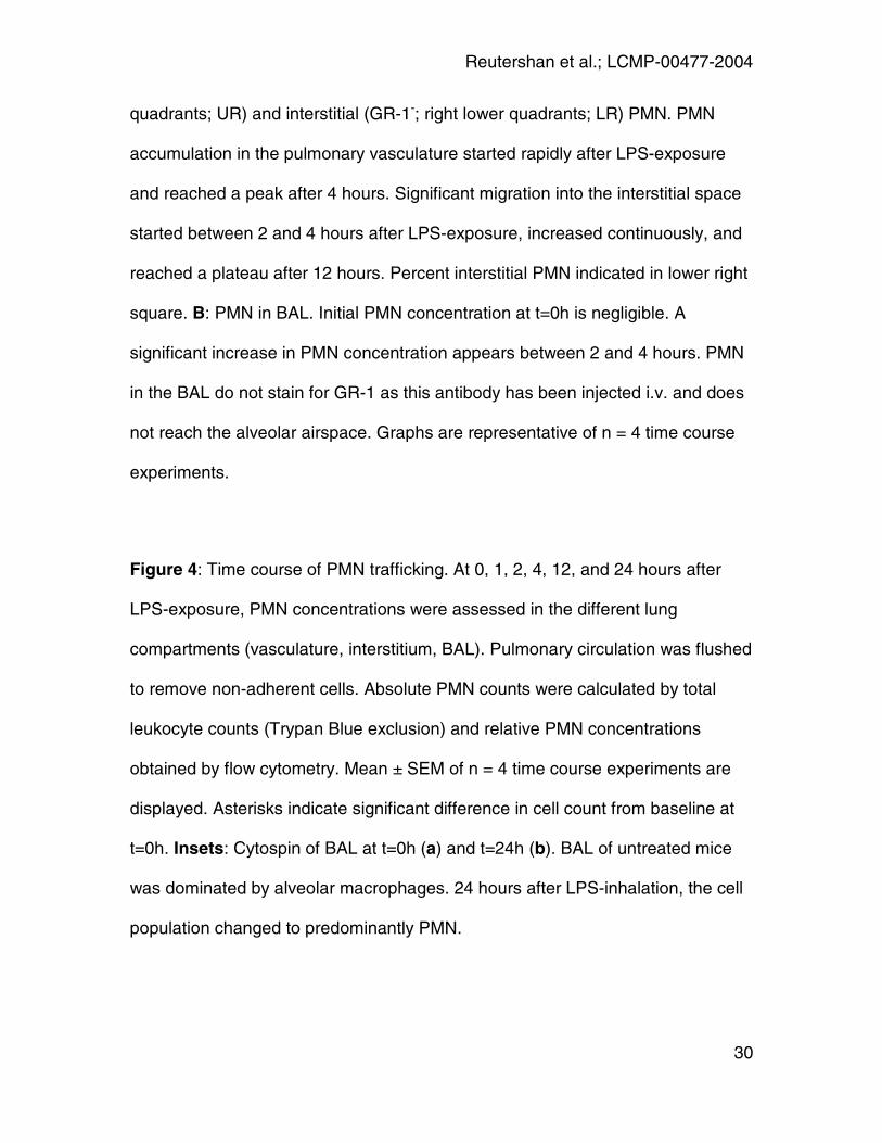
quadrants; UR) and interstitial (GR-1-; right lower quadrants; LR) PMN. PMN
accumulation in the pulmonary vasculature started rapidly after LPS-exposure
and reached a peak after 4 hours. Significant migration into the interstitial space
started between 2 and 4 hours after LPS-exposure, increased continuously, and
reached a plateau after 12 hours. Percent interstitial PMN indicated in lower right
square. B: PMN in BAL. Initial PMN concentration at t=0h is negligible. A
significant increase in PMN concentration appears between 2 and 4 hours. PMN
in the BAL do not stain for GR-1 as this antibody has been injected i.v. and does
not reach the alveolar airspace. Graphs are representative of n = 4 time course
experiments.
Figure 4: Time course of PMN trafficking. At 0, 1, 2, 4, 12, and 24 hours after
LPS-exposure, PMN concentrations were assessed in the different lung
compartments (vasculature, interstitium, BAL). Pulmonary circulation was flushed
to remove non-adherent cells. Absolute PMN counts were calculated by total
leukocyte counts (Trypan Blue exclusion) and relative PMN concentrations
obtained by flow cytometry. Mean ± SEM of n = 4 time course experiments are
displayed. Asterisks indicate significant difference in cell count from baseline at
t=0h. Insets: Cytospin of BAL at t=0h (a) and t=24h (b). BAL of untreated mice
was dominated by alveolar macrophages. 24 hours after LPS-inhalation, the cell
population changed to predominantly PMN.
Reutershan et al.; LCMP-00477-2004
31
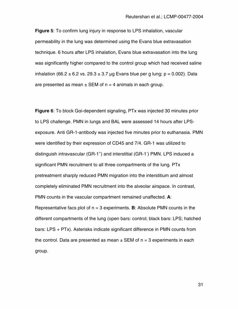
Figure 5: To confirm lung injury in response to LPS inhalation, vascular
permeability in the lung was determined using the Evans blue extravasation
technique. 6 hours after LPS inhalation, Evans blue extravasation into the lung
was significantly higher compared to the control group which had received saline
inhalation (66.2 ± 6.2 vs. 29.3 ± 3.7 µg Evans blue per g lung; p = 0.002). Data
are presented as mean ± SEM of n = 4 animals in each group.
Figure 6: To block Gαi-dependent signaling, PTx was injected 30 minutes prior
to LPS challenge. PMN in lungs and BAL were assessed 14 hours after LPS-
exposure. Anti GR-1-antibody was injected five minutes prior to euthanasia. PMN
were identified by their expression of CD45 and 7/4. GR-1 was utilized to
distinguish intravascular (GR-1+) and interstitial (GR-1-) PMN. LPS induced a
significant PMN recruitment to all three compartments of the lung. PTx
pretreatment sharply reduced PMN migration into the interstitium and almost
completely eliminated PMN recruitment into the alveolar airspace. In contrast,
PMN counts in the vascular compartment remained unaffected. A:
Representative facs plot of n = 3 experiments. B: Absolute PMN counts in the
different compartments of the lung (open bars: control; black bars: LPS; hatched
bars: LPS + PTx). Asterisks indicate significant difference in PMN counts from
the control. Data are presented as mean ± SEM of n = 3 experiments in each
group.
Reutershan et al.; LCMP-00477-2004
32
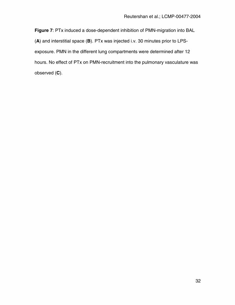
Figure 7: PTx induced a dose-dependent inhibition of PMN-migration into BAL
(A) and interstitial space (B). PTx was injected i.v. 30 minutes prior to LPS-
exposure. PMN in the different lung compartments were determined after 12
hours. No effect of PTx on PMN-recruitment into the pulmonary vasculature was
observed (C).
Reutershan et al.; LCMP-00477-2004
33
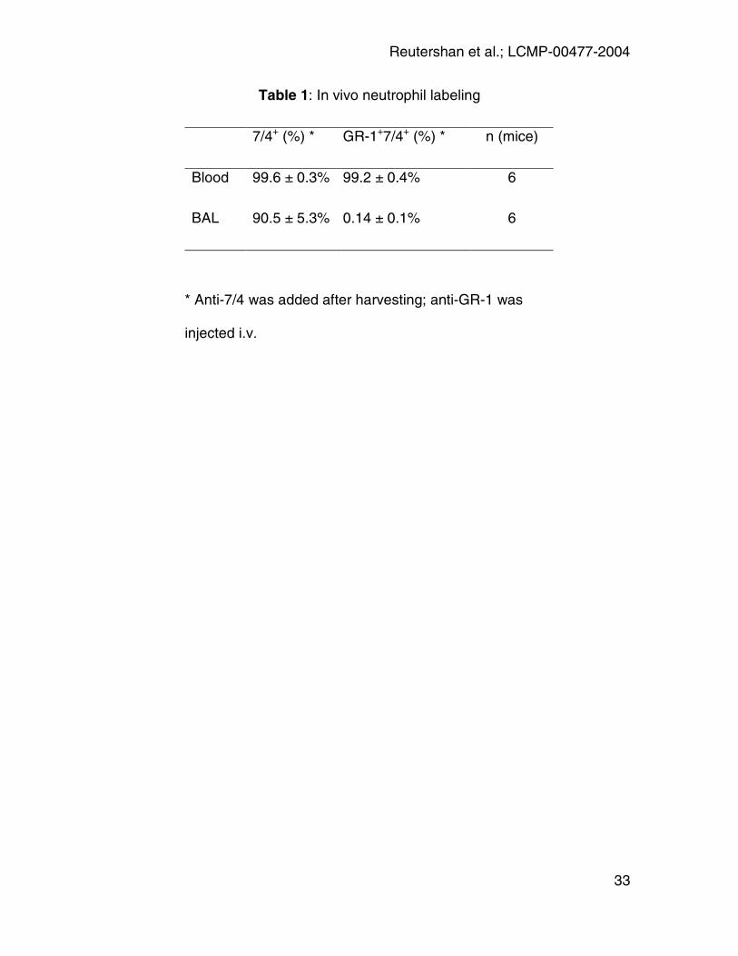
Table 1: In vivo neutrophil labeling
7/4+ (%) * GR-1+7/4+ (%) * n (mice)
Blood 99.6 ± 0.3% 99.2 ± 0.4% 6
BAL 90.5 ± 5.3% 0.14 ± 0.1% 6
* Anti-7/4 was added after harvesting; anti-GR-1 was
injected i.v.
Reutershan et al.; LCMP-00477-2004
34
AC
ell c
cou
nt
x106
Cel
l cco
un
tx1
06
B
all Leukocytes
PMN
all Leukocytes
PMN
0
2
4
6
8
10
12
0h 1h 2h 4h 12h 24h
Lung
BAL
0
1
2
3
4
5
6
0h 1h 2h 4h 12h 24h 48h
all Leukocytes
PMN
all Leukocytes
PMN
Figure 1
Reutershan et al.; LCMP-00477-2004
35
A
C
FSC
SS
C
0 200 400 600 800 10000
200
400
600
800
1000
PMN
FSC
SS
C
0 200 400 600 800 10000 200 400 600 800 10000
200
400
600
800
1000
0
200
400
600
800
1000
PMN
7 / 4
GR
-1
100 101 102 103 104100
101
102
103
104
0.093 99.2
0.650.093
100 101 102 103 104100
101
102
103
104
0.093 99.2
0.650.093
7 / 4
GR
-1
BAL
B
D
BloodBlood Blood
BAL
Figure 2
100 101 102 103 104100
101
102
103
104
0.046 0.57
89.210.2
100 101 102 103 104100 101 102 103 104100
101
102
103
104
100
101
102
103
104
0.046 0.57
89.210.2
100 101 102 103 104100
101
102
103
104
0.16 73.9
3.2722.7
100 101 102 103 104100 101 102 103 104100
101
102
103
104
100
101
102
103
104
0.16 73.9
3.2722.7
Reutershan et al.; LCMP-00477-2004
36
A: Lung LPS
100 101 102 103 104100
101
102
103
104
100 101 102 103 104100
101
102
103
104
100 101 102 103 104100
101
102
103
104
100 101 102 103 104100
101
102
103
104
100 101 102 103 104100
101
102
103
104
100 101 102 103 104100
101
102
103
104
100 101 102 103 104100
101
102
103
104
100 101 102 103 104100
101
102
103
104
100 101 102 103 104100
101
102
103
104
100 101 102 103 104100
101
102
103
104
7/4
GR
-1
0h 1h 2h 4h 12h 24h
Figure 3
14% 15% 10% 60% 78%
B: BAL LPS
100 101 102 103 104100
101
102
103
104
100 101 102 103 104100
101
102
103
104
100 101 102 103 104100
101
102
103
104
100 101 102 103 104100
101
102
103
104
100 101 102 103 104100
101
102
103
104
100 101 102 103 104100
101
102
103
104
100 101 102 103 104100
101
102
103
104
100 101 102 103 104100
101
102
103
104
100 101 102 103 104100
101
102
103
104
100 101 102 103 104100
101
102
103
104
100 101 102 103 104100
101
102
103
104
100 101 102 103 104100
101
102
103
104
100 101 102 103 104100
101
102
103
104
100 101 102 103 104100 101 102 103 104100
101
102
103
104
100
101
102
103
104
33%
Reutershan et al.; LCMP-00477-2004
37
Figure 4
0
1
2
3
4
5
0 4 12 24h
PM
N c
ou
nts
x10
6
BALvascular interstitial BALBALvascular interstitial
21
*
**
**
* *
*
**
*
*
a
b

Reutershan et al.; LCMP-00477-2004
38
Figure 5
Eva
ns
Blu
e E
xtra
vasa
tio
n(µ
g p
er g
lun
g t
issu
e)
*
0
20
40
60
80
Saline LPS
Figure 5
Eva
ns
Blu
e E
xtra
vasa
tio
n(µ
g p
er g
lun
g t
issu
e)
*
0
20
40
60
80
Saline LPS
Reutershan et al.; LCMP-00477-2004
39
0
1
2
3
4
IV IS BAL
control LPS LPS + PTx
Figure 6
7 / 4
GR
-1
100
101
102
103
104
100
10 1
10 2
103
104
46%10
010
110
210
310
410
010
110
210
310
4
100
10 1
10 2
103
104
100
10 1
10 2
103
104
46%10
010
110
210
310
410
010
110
210
310
4
100
10 1
10 2
103
104
100
10 1
10 2
103
104
13%
LPS + PTxA
B
PM
Ncc
ou
nt
x106
* *
* *
LPS

Reutershan et al.; LCMP-00477-2004
40
Figure 7
ABAL
0
1
2
3
4
5
0 0.04 0.4 4
PM
Ns
x106
ABAL
0
1
2
3
4
5
0 0.04 0.4 4
PM
Ns
x106
B IS
0
1
2
3
4
5
0 0.04 0.4 4
PM
Ns
x106
B IS
0
1
2
3
4
5
0 0.04 0.4 4
PM
Ns
x106
CIV
0
1
2
3
4
5
0 0.04 0.4 4
PM
Ns
x106
PTx (µg / mouse)
CIV
0
1
2
3
4
5
0 0.04 0.4 4
PM
Ns
x106
PTx (µg / mouse)
Related Documents


![Neutrophils in Tissue Trauma of the Skin, Bone, and Lung ...downloads.hindawi.com/journals/jir/2018/8173983.pdf · bone regeneration [56], but did mitigate pulmonary damage [17, 57].](https://static.cupdf.com/doc/110x72/5f0ba75d7e708231d4319051/neutrophils-in-tissue-trauma-of-the-skin-bone-and-lung-bone-regeneration-56.jpg)


![Bronchoalveolar Lavage and Lung Tissue Digestion [Abstract] … · 2017. 10. 19. · 11. Cytospin cytocentrifuge (Thermo Fisher Scientific/Shandon, model: A7830002) 12. Microscope](https://static.cupdf.com/doc/110x72/60d0b4531ab8432f5309d74e/bronchoalveolar-lavage-and-lung-tissue-digestion-abstract-2017-10-19-11.jpg)
