Published Ahead of Print 11 November 2013. 2014, 82(3):924. DOI: 10.1128/IAI.00866-13. Infect. Immun. Beeson N. Hodder, Ross L. Coppel, Robin F. Anders and James G. Michelle J. Boyle, Christine Langer, Jo-Anne Chan, Anthony Invasion by Plasmodium falciparum Proteins during and after Erythrocyte Sequential Processing of Merozoite Surface http://iai.asm.org/content/82/3/924 Updated information and services can be found at: These include: SUPPLEMENTAL MATERIAL Supplemental material REFERENCES http://iai.asm.org/content/82/3/924#ref-list-1 at: This article cites 82 articles, 36 of which can be accessed free CONTENT ALERTS more» articles cite this article), Receive: RSS Feeds, eTOCs, free email alerts (when new http://journals.asm.org/site/misc/reprints.xhtml Information about commercial reprint orders: http://journals.asm.org/site/subscriptions/ To subscribe to to another ASM Journal go to: on March 18, 2014 by guest http://iai.asm.org/ Downloaded from on March 18, 2014 by guest http://iai.asm.org/ Downloaded from

Welcome message from author
This document is posted to help you gain knowledge. Please leave a comment to let me know what you think about it! Share it to your friends and learn new things together.
Transcript
Published Ahead of Print 11 November 2013. 2014, 82(3):924. DOI: 10.1128/IAI.00866-13. Infect. Immun.
BeesonN. Hodder, Ross L. Coppel, Robin F. Anders and James G. Michelle J. Boyle, Christine Langer, Jo-Anne Chan, Anthony Invasion by Plasmodium falciparumProteins during and after Erythrocyte Sequential Processing of Merozoite Surface
http://iai.asm.org/content/82/3/924Updated information and services can be found at:
These include:
SUPPLEMENTAL MATERIAL Supplemental material
REFERENCEShttp://iai.asm.org/content/82/3/924#ref-list-1at:
This article cites 82 articles, 36 of which can be accessed free
CONTENT ALERTS more»articles cite this article),
Receive: RSS Feeds, eTOCs, free email alerts (when new
http://journals.asm.org/site/misc/reprints.xhtmlInformation about commercial reprint orders: http://journals.asm.org/site/subscriptions/To subscribe to to another ASM Journal go to:
on March 18, 2014 by guest
http://iai.asm.org/
Dow
nloaded from
on March 18, 2014 by guest
http://iai.asm.org/
Dow
nloaded from

Sequential Processing of Merozoite Surface Proteins during and afterErythrocyte Invasion by Plasmodium falciparum
Michelle J. Boyle,a,b,c* Christine Langer,a Jo-Anne Chan,a Anthony N. Hodder,b,c Ross L. Coppel,d Robin F. Anders,e James G. Beesona,d
The Burnet Institute for Medical Research and Public Health, Melbourne, Victoria, Australiaa; Department of Medical Biology, University of Melbourne, Melbourne, Victoria,Australiab; Walter and Eliza Hall Institute of Medical Research, Parkville, Victoria, Australiac; Department of Microbiology, Monash University, Melbourne, Victoria, Australiad;Department of Biochemistry, La Trobe University, Melbourne, Victoria, Australiae
Plasmodium falciparum causes malaria disease during the asexual blood stages of infection when merozoites invade erythro-cytes and replicate. Merozoite surface proteins (MSPs) are proposed to play a role in the initial binding of merozoites to erythro-cytes, but precise roles remain undefined. Based on electron microscopy studies of invading Plasmodium merozoites, it is pro-posed that the majority of MSPs are cleaved and shed from the surface during invasion, perhaps to release receptor-ligandinteractions. In this study, we demonstrate that there is not universal cleavage of MSPs during invasion. Instead, there is sequen-tial and coordinated cleavage and shedding of proteins, indicating a diversity of roles for surface proteins during and after inva-sion. While MSP1 and peripheral surface proteins such as MSP3, MSP7, serine repeat antigen 4 (SERA4), and SERA5 are cleavedand shed at the tight junction between the invading merozoite and erythrocyte, the glycosylphosphatidylinositol (GPI)-anchoredproteins MSP2 and MSP4 are carried into the erythrocyte without detectable processing. Following invasion, MSP2 rapidly de-grades within 10 min, whereas MSP4 is maintained for hours. This suggests that while some proteins that are shed upon invasionmay have roles in initial contact steps, others function during invasion and are then rapidly degraded, whereas others are inter-nalized for roles during intraerythrocytic development. Interestingly, anti-MSP2 antibodies did not inhibit invasion and insteadwere carried into erythrocytes and maintained for approximately 20 h without inhibiting parasite development. These findingsprovide new insights into the mechanisms of invasion and knowledge to advance the development of new drugs and vaccinesagainst malaria.
Plasmodium falciparum is the causative agent of the majority ofmalaria morbidity and mortality worldwide. Pathogenesis re-
sults from blood-stage infection, where the merozoite stage of theparasite invades and then replicates in red blood cells (RBCs).Invasion occurs through a series of stepwise mechanisms: initialcontact, reorientation, commitment to invasion, tight-junctionformation, and invasion driven by an actin-myosin motor (1–5).Initial contact is thought to be mediated by merozoite surfaceproteins (MSPs) via multiple weak interactions with receptors onthe surface of RBCs, but the roles of individual merozoite surfaceproteins remain largely undefined (6). Following initial contact,commitment to invasion occurs with the reorientation of the par-asite to the apical pole, allowing the formation of a tight junctionbetween the merozoite and the RBC surface via interactions be-tween AMA1 and RON2 (3, 7). Invasion into RBCs is then drivenby the parasite actin-myosin motor (8), with the tight junctionprogressing rearwards as the merozoite enters the RBC. Studies ofinvasion by Plasmodium knowlesi, P. berghei, P. gallinaceum, andP. falciparum using electron microscopy (EM) have suggested thatthe entire surface coat of the merozoite is shed during invasionaround the point of the tight junction (4, 9–11). The shedding ofsurface proteins is thought to be required to disrupt receptor-ligand interactions, allowing invasion to proceed. This phenome-non also occurs in other Apicomplexa parasites, including Toxo-plasma, Neospora, Eimeria, and Cryptosporidium (12). Based onthese studies, it is widely assumed that the majority of, if not all,merozoite surface proteins are cleaved and shed during invasionof the P. falciparum merozoite (1, 13, 14). Indeed, to the best ofour knowledge, it has not been proposed in the literature thatsome surface proteins may not be shed and instead may be inter-nalized into RBCs without processing.
This concept of surface coat shedding is supported by studiesshowing the cleavage and shedding of MSP1 and the associatedproteins MSP6 and MSP7 during invasion (15–17) and reports ofseveral other merozoite surface proteins being detected in culturesupernatants after invasion, suggesting that they are shed from thesurface (17–20). However, due to a lack of methods to studymerozoites during invasion, shedding of merozoite surface pro-teins at the tight junction between the merozoite and RBC hasbeen directly visualized only with MSP1 (3, 21, 22). MSP1 is foundon the surface of the merozoite via the C-terminal glycosylphos-phatidylinositol (GPI)-anchored MSP1-42 fragment (15–17).During invasion, MSP1-42 is cleaved by a subtilisin protease,PfSUB2, which also cleaves AMA1 (23), releasing the majority ofthe complex at the point of the tight junction with the RBC (1–5,22). The remaining MSP1-19 fragment, representing �10% of theprotein, is carried into the RBC (6, 15), where it is thought to beinvolved with formation of the food vacuole in ring-stage para-
Received 12 July 2013 Returned for modification 19 August 2013Accepted 23 October 2013
Published ahead of print 11 November 2013
Editor: J. H. Adams
Address correspondence to James G. Beeson, [email protected].
* Present address: Michelle J. Boyle, University of California, San Francisco,California, USA.
Supplemental material for this article may be found at http://dx.doi.org/10.1128/IAI.00866-13.
Copyright © 2014 Boyle et al.
doi:10.1128/IAI.00866-13
924 iai.asm.org Infection and Immunity p. 924 –936 March 2014 Volume 82 Number 3
on March 18, 2014 by guest
http://iai.asm.org/
Dow
nloaded from

sites (24). Cleavage and shedding of MSP1 are essential, since in-hibitors, including antibodies, that block these processes disruptinvasion (12, 15, 25–28). MSP1 is believed to mediate initial con-tact with RBCs via binding to surface receptor band 3 and possiblyheparin-like molecules (29–31). It is possible that the cleavage andshedding of MSP1 during invasion are required to release the re-ceptor-ligand interactions between this protein and the RBC sur-face. To date, there are no published reports of more global anal-yses of merozoite surface proteins to assess the loss of the surfacecoat during invasion.
Merozoite surface proteins have long been considered attrac-tive targets for malaria vaccine development, and several proteinshave progressed to phase 1 and 2 clinical trials (32, 33). Merozoitesurface proteins also appear to be important targets of acquiredhuman immunity, particularly antibodies (34–38). Therefore, anunderstanding of their role, localization, and fate during invasionis important in the context of identifying key immune targets andprioritizing candidates for vaccine development and understand-ing how specific candidates can be targeted by vaccine-inducedresponses. Furthermore, while it is thought that surface proteinsare likely to be involved in initial contact with RBCs, specific re-ceptor-ligand interactions for merozoite surface proteins otherthan MSP1 have yet to be identified. Characterization of process-ing and shedding patterns of other merozoite surface proteinsduring invasion may also inform the roles of these proteins duringthe invasion process.
Here, we aimed to conduct a broader characterization of sur-face shedding of merozoite proteins by studying two GPI-an-chored proteins, MSP2 and MSP4, and four peripherally associ-ated proteins, MSP3, MSP7, serine repeat antigen 4 (SERA4), andSERA5, during and immediately after invasion. All proteins ex-cept SERA4 have been considered malaria vaccine candidates;MSP2 and MSP3 have progressed to phase 2 clinical trials, andSERA5 has progressed to phase 1 clinical trials (39–41). MSP2 isthe second most abundant merozoite surface protein in terms ofcopy number (42), and while a function for MSP2 has not beenidentified, this protein is refractory to genetic deletion and isthought to be essential (43). MSP2 is highly polymorphic; how-ever, all alleles can be grouped as either 3D7-like or FC27-likebased on tandem sequence repeats and flanking nonrepetitive di-morphic sequences, with both alleles sharing N- and C-terminalregions (44–46). Currently, the fate of MSP2 during invasion isunclear, and there is no evidence of MSP2 in culture supernatants(20, 47). MSP4, like MSP1-19, contains an epidermal growth fac-tor (EGF) domain adjacent to the C-terminal GPI anchor (48)that is essential for the conformation and immunogenicity of theprotein (49). MSP4 has been tested in multiple vaccine trials inmouse models, with significant efficacy (50–55). MSP4 is refrac-tory to genetic deletion in vitro and is assumed to be essential forasexual replication (43), but its role and localization during inva-sion have not been defined. MSP7 is bound to MSP1 on the mero-zoite surface and is thought to be shed during invasion along withthe majority of MSP1 (16). The binding partners of the peripheralsurface proteins MSP3, SERA5, and SERA4 are not established.
New methods to isolate viable P. falciparum merozoites pro-vide valuable approaches to imaging of merozoites and importantproteins during the process of invasion by immunofluorescence(IF) microscopy (3, 6, 22). Here, we used these approaches tolocalize a number of merozoite surface proteins during and im-mediately after invasion and to characterize the cleavage and shed-
ding of these proteins. We aimed to investigate whether the shed-ding of the merozoite surface coat during invasion involves thespecific processing of individual proteins, reflecting the differentroles and functions of merozoite surface proteins during invasionrather than shedding being a nonspecific global process resultingin the complete loss of the merozoite surface protein coat. A de-tailed understanding of invasion is important because merozoiteproteins are leading vaccine candidates (32–34), and targeting ofinvasion may be an effective strategy in antimalarial drug devel-opment (6, 56). We show that loss of the merozoite surface coat isnot a global event and instead involves specific processes resultingin the sequential cleavage of individual proteins both during andafter invasion, indicating the diversity of roles of different pro-teins.
MATERIALS AND METHODSParasite culture and merozoite isolation. P. falciparum isolates were cul-tured as described previously (57, 58), in culture medium of RPMI-HEPES (pH 7.4) supplemented with 50 �g/ml hypoxanthine, 20 �g/mlgentamicin, 25 mM sodium bicarbonate (NaHCO3), and 0.5% AlbumaxII (Gibco). RBCs from group O� blood donors were used to culture par-asites. Cultures were gassed with 1% O2, 4% CO2, and 95% N2 and incu-bated at 37°C. Parasites were initially synchronized by using 5% D-sorbitoltreatment for 5 min, as described previously (59). Following sorbitoltreatment, cultures were further synchronized by using the invasion-in-hibitory properties of heparin (29). Parasite strains 3D7 and D10 wereused as indicated. Viable merozoites were isolated as described previously(6, 22). Highly synchronized late-stage schizonts were magnet purified viaa MACS magnet separation column (Miltenyi Biotec) and treated withE64 until mature merozoites were formed. Merozoites were isolated bymembrane filtration, incubated with uninfected RBCs, and prepared forimaging for IF microscopy or EM as described previously (3, 6, 22).
Immunofluorescence microscopy and electron microscopy. IF mi-croscopy and EM were performed as described previously (3, 6, 22). Cellswere fixed with 4% formaldehyde– 0.0075% glutaraldehyde coated ontoglass slides and labeled with antibodies as indicated. Labeling was detectedby an Alexa 594/488-conjugated secondary antibody (Molecular Probes).Slides were mounted in VectaShield (Vector Laboratories) with 0.1 ng/ml4=,6-diamidino-2-phenylindole (DAPI) (Invitrogen) to label the parasitenucleus. Images were obtained by using a Plan-Apochromat (100�/1.40)oil immersion phase-contrast lens (Carl Zeiss) on an AxioVert 200M mi-croscope (Carl Zeiss) equipped with an AxioCam Mrm camera (CarlZeiss). Images were processed by using Photoshop CS4 (Adobe). In caseswhere brightness and contrast were changed, processing was applied towhole images and controls equally. For EM, invading merozoites werefixed in 1% glutaraldehyde in RPMI-HEPES on ice for 30 min. Sampleswere pelleted in low-melt agarose before being transferred into water,dehydrated in ethanol, and embedded in LR Gold resin (ProSciTech).Following polymerization by benzoyl peroxide (SPI-Chem), 100-nm sec-tions were prepared by using an Ultracut R ultramicrotome (Leica). Sec-tions were poststained with saturated aqueous uranyl-acetate and then5% triple lead and observed at 120 kV on a CM120 BioTWIN transmis-sion electron microscope (Philips).
To test for the internalization of MSP2 antibodies, MSP2 monoclonalantibodies (MAbs) were incubated with merozoites and uninfected RBCsfor 10 min. Cultures were washed twice in cold phosphate-buffered saline(PBS) containing complete protease inhibitors and then fixed and pro-cessed for IF microscopy. Control labeling was carried on in the absence ofprimary or secondary antibodies for all antigens. In all cases, laser inten-sity used to image positive samples showed no fluorescence in controls.
Antibodies. MSP2-3D7 rabbit polyclonal antibodies for N-terminalpurification were raised against full-length 3D7 recombinant MSP2.Monoclonal antibodies to MSP2 were generated by vaccination with full-length 3D7 or FC27 MSP2 (60). Rabbit and mouse anti-PfRON4 antibod-
Processing of Merozoite Proteins during Invasion
March 2014 Volume 82 Number 3 iai.asm.org 925
on March 18, 2014 by guest
http://iai.asm.org/
Dow
nloaded from

ies (61) and rabbit anti-MSP3 antibodies (62) were kindly provided byAlan Cowman, Walter and Eliza Hall Institute. MSP4 antibodies to dif-ferent regions and full-length MSP4 were generated as described previ-ously (49). MSP7 polyclonal rabbit antibodies were raised in rabbits (63).Rabbit antibodies to MSP1-19 (64) were kindly provided by BrendanCrabb, Burnet Institute. The SERA4 central domain was prepared in amanner similar to that for the central domain of SERA5 (65, 66), and thein vitro refolded protein was used to produce antibodies in rabbits. Thecysteine-rich region of the C-terminal fragment of SERA5 (R914 andV997) was oxidatively refolded in a manner similar to that for the SERA5central domain. The monoclonal antibody used in this study was found totarget a disulfide bond-stabilized conformational epitope in this domainfragment (data not shown). Antibodies were generated by immunizationof experimental animals with purified recombinant proteins at the Walterand Eliza Hall Institute Antibody Service. Procedures were consistent withthe policies of the National Health and Medical Research Council, Aus-tralia (83), and studies were approved by the Animal Ethics Committee ofthe Walter and Eliza Hall Institute, Australia (protocol AEC 2011.009r0).
Purification of MSP2 N-terminal specific antibodies. MSP2 N-ter-minal specific antibodies were purified from polyclonal 3D7 MSP2 rabbitserum raised by vaccination with full-length 3D7 MSP2 recombinant pro-tein. A peptide corresponding to the first 25 amino acids of the N terminus ofMSP2 (conserved between 3D7 and FC27 sequences) and the next 4 addi-tional amino acids of the FC27-specific region (H-IKNESKYSNTFINNAYNMSIRRSMANEGSN-NH2) was purchased from Mimotopes (90% mini-mum purity by high-performance liquid chromatography [HPLC]).Peptide was solubilized in 50% dimethyl sulfoxide (DMSO) at 2.75 �g/mland then diluted to 687.5 �g/ml and coupled to AminoLink Plus columnresin overnight at 4°C. Unbound peptide was collected, the column waswashed 2 times in PBS, and the coupling reaction was completed with 50mM NaCNBH3. The column was blocked according to the manufactur-er’s instructions with quenching buffer and 50 mM NaCNBH3 and thenwashed extensively with PBS. Polyclonal MSP2 3D7 rabbit serum wasincubated on the column overnight. The column was washed 12 timeswith PBS, and the antibodies were then eluted with 0.1 M glycine (pH 2.6)into tubes containing 100 �l of 1 M Tris (pH 8.8). Eluted antibodies weredialyzed against PBS overnight by using SnakeSkin dialysis tubing(Thermo Scientific) and then concentrated 8-fold through Amicon mi-crospin columns with a molecular mass cutoff of 10,000 kDa. Reactivity ofrabbit polyclonal serum before and after purification was tested by anenzyme-linked immunosorbent assay (ELISA) using an MSP2 peptidearray.
MSP2 peptide ELISA array. An MSP2 peptide array of 84 biotinylated13-mer peptides overlapping by 8 amino acids, covering the entire 3D7and FC27 MSP2 sequence, was purchased from Mimotopes (60). Peptideswere solubilized in 80% DMSO to a concentration of 2 to 6 mg/ml. Nine-ty-six-well Nunc flat-bottom plates were coated overnight with 1 �g/mlstreptavidin in PBS. Blocking was performed for 2 h with 1% casein at37°C, followed by incubation with peptides diluted in PBS to 4 to 12�g/ml. MSP2 MAbs at a 1:1,000 serum dilution were incubated for 1 h atroom temperature, and binding was detected with anti-mouse horserad-ish peroxidase (HRP)-conjugated antibody (Invitrogen) at a 1:2,500dilution. Plates were washed 3 times with PBS– 0.05% Tween betweenincubations, the ELISA plate was exposed with 2,2-azinobis(3-ethylbenz-thiazolinesulfonic acid) (ABTS) (Sigma-Australia) for 30 min and thenstopped with 1% SDS, and the absorbance was read at 405 nm.
Growth inhibition assays and invasion inhibition assays. High-throughput growth inhibition assays (GIAs) were performed as describedpreviously (58, 67–69). Duplicate suspensions of synchronized parasitesat 0.2 or 2% parasitemia (one- or two-cycle assays; 32-h trophozoite stage)and 1% hematocrit were incubated with antibodies in 96-well sterile U-bottom plates (Falcon). Plates were incubated as described above for par-asite culture for 44 h for one-cycle assays or 72 h for two-cycle assays andanalyzed by flow cytometry with staining of parasites with 10 �g/mlethidium bromide (EtBr) (Bio-Rad) for 1 h in darkness. Parasitemia was
measured by using a BD FACSCalibur or BD FACSCantoII flow cytome-ter. Samples were analyzed by using FlowJo (Tree Star) gating on intacterythrocytes and then determining parasitemia by EtBr-positive erythro-cytes. Inhibitory effects of compounds were normalized as percent growthof controls for each assay.
Invasion inhibition assays (IIAs) with isolated merozoites were con-ducted as described previously (6, 22). Merozoites were isolated from D10PfPHG (68) and incubated with MSP2 MAbs and uninfected RBCs. Fol-lowing invasion, cultures were washed twice and returned to culture me-dium. Parasites were analyzed by flow cytometry at 40 h postinvasion asdescribed above for GIAs, with parasites being gated as EtBr- and greenfluorescent protein (GFP)-positive cells. To investigate the ability ofMAbs to induce a growth delay, parasites were analyzed for mean fluores-cence intensity (MFI) of GFP and EtBr at 30 and 40 h postinvasion (70).
Protein extraction and Western blotting. Synchronized schizont-stage (40 to 48 h), or early-ring-stage (0 to 6 h postinvasion) parasitecultures were used for protein extraction. Uninfected erythrocytes werelysed with 0.15% saponin (Kodak) on ice for 10 min. The remainingpellets were washed three times with cold PBS. Culture supernatant pro-teins were collected from spent media from synchronized schizont para-site cultures after reinvasion of RBCs following schizont rupture. At allstages of protein extraction, Complete protease inhibitor (Roche) wasadded at an excess concentration to ensure minimization of nonspecificproteolysis. Proteins were solubilized in nonreducing sample buffer andseparated by SDS-PAGE. Proteins were transferred onto polyvinylidenedifluoride (PVDF) membranes and analyzed by Western blotting on achemiluminescence system.
RESULTSPeripherally associated merozoite surface proteins MSP3,MSP7, SERA4, and SERA5 are shed at the tight junction duringinvasion. To assess the shedding of the merozoite surface coat, welocalized both peripherally associated and GPI-anchored proteinsduring invasion. To assess the shedding of individual merozoitesurface proteins, invading P. falciparum merozoites were fixed formicroscopy and visualized at attachment to the RBC membrane,during the process of invasion, and immediately postinvasion byorientation with the P. falciparum rhoptry neck protein PfRON4,which is a marker of the tight junction (3). Using these ap-proaches, we have previously visualized the shedding of the N-ter-minal regions of MSP1 (3, 22). As a first step, we localized theperipherally associated proteins MSP3, MSP7, SERA4, and SERA5during invasion (Fig. 1). As expected, these proteins were shedfrom the surface of merozoites during invasion at the point oftight junction with RBCs. This was seen with positive labeling ofmerozoites at the initial stages of invasion with polyclonal anti-bodies specific to these proteins. As invasion progressed, labelingwas lost from the region of the merozoite that had already passedthrough the tight junction into the RBC, indicating the sheddingof individual proteins (Fig. 1). The shedding of MSP3, MSP7,SERA4, and SERA5 at the point of tight junction with the RBC isconsistent with the presence of these proteins in culture superna-tants following invasion.
The GPI-anchored protein MSP2 is carried into invadedRBCs and then rapidly degraded. To localize MSP2 during andafter invasion, we used merozoites from 3D7 or FC27 (D10 clone)parasites in order to include the two major allelic forms of MSP2and labeled invading merozoites with MAbs to distinct regions ofMSP2; MAbs were described in detail previously (60). Theepitopes recognized by these MAbs were distributed among dif-ferent regions of MSP2, including the 3D7 allele-specific region(MAbs 111, 9D11, and 2F2), the FC27 allele-specific region (MAb
Boyle et al.
926 iai.asm.org Infection and Immunity
on March 18, 2014 by guest
http://iai.asm.org/
Dow
nloaded from

8G10), and two regions of the conserved C-terminal region(MAbs 6C9, 9H4, 1F7, 9G8, and 4D11) (Fig. 2; see also Table S1 inthe supplemental material). The specificity of the MAbs to MSP2was previously confirmed via Western blotting; all MAbs werespecific to MSP2, with no cross-reactivity to other parasite pro-teins, except for MAb 9G8, which has some cross-reactivity tospectrin (60). 8G10 was reported previously in other studies to becross-reactive to some extent with other parasite proteins, whichhave not been identified (71). As we lacked a MAb against theN-terminal region of MSP2 that reacted with the native surface-located antigen by IF microscopy, N-terminally specific antibod-ies were purified from rabbit polyclonal serum that had been gen-erated by vaccination with full-length 3D7 MSP2. When tested byELISA using an overlapping peptide array covering the full-lengthMSP2 protein, the affinity-purified N-terminal polyclonal anti-bodies reacted only with the N-terminal peptides of MSP2 (seeFig. S1A and S1B in the supplemental material). These antibodieswere not cross-reactive with other parasite proteins; on Westernblots of parasite lysates, they labeled a single 40-kDa band indica-tive of MSP2 (see Fig. S1C in the supplemental material).
Labeling of MSP2 during invasion indicated that MSP2 wasnot cleaved and shed during invasion, in contrast to MSP1 and theperipheral surface proteins described above. Antibodies to N-ter-minal, central variable, and C-terminal regions of MSP2 labeledthe parasite evenly on both sides of the tight junction at early,middle, and late stages of invasion. These striking findings indi-
cate that no part of MSP2 was lost during invasion, suggesting thatthis protein does not undergo processing during invasion and isinstead internalized during invasion (Fig. 3A; see also Fig. S2 in thesupplemental material), which was unexpected based on thewidely described paradigm of surface protein shedding. The fateof MSP2 in the immediate period following invasion was investi-gated by fixing newly invaded parasites 10 min after mixing iso-lated merozoites with RBCs. Parasites were colabeled with anti-bodies to MSP1-19, which is known to remain on the parasitepostinvasion, and labeling with these antibodies was clearly seenin newly invaded parasites, as expected. However, little or noMSP2 labeling of newly invaded parasites was seen by using any ofthe MSP2 antibodies, suggesting that MSP2 was rapidly lost post-invasion (Fig. 3B). In contrast, the MSP2 antibodies labeled extra-cellular merozoites in the same IF microscopy preparations, con-firming antibody reactivity (for example, see merozoite labelingwith N-terminal MSP2 rabbit antibodies in Fig. 3B, top). The lossof MSP2 labeling in newly invaded parasites, despite the labelingof merozoites during invasion and immediately postinvasion,suggests that MSP2 is carried into RBCs but is then rapidly de-graded. In a minor proportion of invaded cells, MSP2 antibodyfluorescence was seen, but labeling was limited to discrete dotsthat had no clear indication of colocalization with MSP1-19. Thisminimal MSP2 labeling observed was above background fluores-cence levels, as assessed by labeling with secondary antibodiesalone or control antibodies (data not shown). 8G10, which is re-active with the FC27 central variable region, labeled newly in-vaded rings to a higher degree than other antibodies, but thislabeling was still minimal compared to the labeling of merozoitesprior to, during, or immediately after invasion. This may indicatethat the epitopes recognized by this antibody are more accessibleor degraded more slowly than other epitopes, or the labeling seenmay reflect the cross-reactivity of this MAb, which was reportedpreviously (71).
MSP4 is carried into invaded RBCs and maintained in earlyring forms. The same experimental approach was used to assessthe fate of MSP4, another essential GPI-anchored merozoite sur-face protein, during invasion. To study MSP4, we used rabbitpolyclonal antibodies that were raised against different regions ofMSP4 with recombinant protein, as described previously (49)(Fig. 2B). The specificity of the antibodies was investigated byusing Western blots of 3D7 and D10 schizont protein extracts.MSP4 full-length antibodies were specific for MSP4 in 3D7 para-sites reacting only to a single band of approximately 40kDa. Thisantibody showed some cross-reactivity with a non-MSP4 proteinin D10 parasites, as seen with the labeling of a protein at approx-imately 140 kDa. MSP4A antibodies were specific to MSP4 in D10parasites but showed some cross-reactivity in 3D7 parasites withan approximately 55-kDa protein. MSP4B and MSP4D antibodieswere specific to MSP4 in both 3D7 and D10 parasite protein ex-tracts, while MSP4C antibodies were cross-reactive to non-MSP4proteins in both parasite strains, as determined by the multiplebands seen in the Western blot (Fig. 2C).
As seen for MSP2, labeling of merozoites with antibodies tofull-length MSP4 was clearly seen at all stages of invasion (early,middle, and late), indicating that at least part of MSP4 is carriedinto RBCs (Fig. 4A). Subsequently, invading D10 merozoites werelabeled with antibodies raised against the four regions of MSP4.All antibodies labeled invading merozoites on both the intra- andextracellular surfaces in the mid-invasion stage, indicating that
FIG 1 Peripherally associated proteins MSP3, MSP7, SERA4, and SERA5 areshed during invasion. Invading merozoites were labeled with PfRON4 as amarker of the tight junction (green) and colabeled with antibodies directedagainst MSP3 (A), MSP7 (B), SERA4 (C), or SERA5 (D). All proteins were shedat the point of tight junction, with antibody reactivity to the merozoite surfacebeing observed only on the proportion of the merozoite external to the RBC.All primary antibodies were used at a 1:100 serum dilution, and secondaryantibodies were used at a 1:500 dilution.
Processing of Merozoite Proteins during Invasion
March 2014 Volume 82 Number 3 iai.asm.org 927
on March 18, 2014 by guest
http://iai.asm.org/
Dow
nloaded from
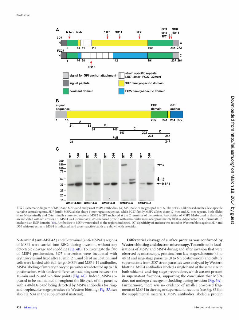
N-terminal (anti-MSP4A) and C-terminal (anti-MSP4D) regionsof MSP4 were carried into RBCs during invasion, without anydetectable cleavage and shedding (Fig. 4B). To investigate the fateof MSP4 postinvasion, 3D7 merozoites were incubated witherythrocytes and fixed after 10 min, 2 h, and 5 h of incubation, andcells were labeled with full-length MSP4 and MSP1-19 antibodies.MSP4 labeling of intraerythrocytic parasites was detected up to 5 hpostinvasion, with no clear difference in staining seen between the10-min and 2- and 5-h time points (Fig. 4C). Indeed, MSP4 ap-peared to be maintained throughout the life cycle of the parasite,with a 40-kDa band being detected by MSP4 antibodies for ring-and trophozoite-stage parasites via Western blotting (Fig. 5A; seealso Fig. S3A in the supplemental material).
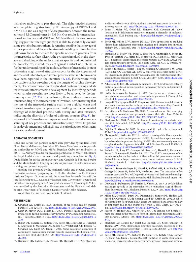
Differential cleavage of surface proteins was confirmed byWestern blotting and electron microscopy. To confirm the local-izations of MSP2 and MSP4 during and after invasion that wereobserved by microscopy, proteins from late-stage schizonts (44 to48 h) and ring-stage parasites (0 to 6 h postinvasion) and culturesupernatants from 3D7 strain parasites were analyzed by Westernblotting. MSP4 antibodies labeled a single band of the same size inboth schizont- and ring-stage preparations, which was not presentin supernatant fractions, supporting the conclusion that MSP4does not undergo cleavage or shedding during invasion (Fig. 5A).Furthermore, there was no evidence of smaller processed frag-ments of MSP4 in the ring or supernatant fractions (see Fig. S3B inthe supplemental material). MSP2 antibodies labeled a protein
FIG 2 Schematic diagram of MSP2 and MSP4 and analysis of MSP4 antibodies. (A) MSP2 alleles are grouped as 3D7-like or FC27-like based on the allele-specificvariable central regions. 3D7 family MSP2 alleles share 4-mer repeat sequences, while FC27 family MSP2 alleles share 12-mer and 32-mer repeats. Both allelesshare N-terminally and C-terminally conserved regions. MSP2 is GPI anchored at the C terminus of the protein. Reactivities of MSP2 MAbs used in this studyare indicated with red arrows. (B) MSP4 is a C-terminally GPI-anchored protein with a molecular mass of approximately 40 kDa. Adjacent to the C-terminal GPIanchor is an EGF domain (45). Antibodies to MSP4 were raised to the regions indicated. (C) Specificity of antisera was tested in Western blots against 3D7 andD10 schizont extracts. MSP4 is indicated, and cross-reactive bands are shown with asterisks.
Boyle et al.
928 iai.asm.org Infection and Immunity
on March 18, 2014 by guest
http://iai.asm.org/
Dow
nloaded from

only in schizont protein extracts, confirming the lack of MSP2cleavage and shedding into the supernatant and the degradation ofMSP2 following invasion (noted by the absence of MSP2 labelingin ring-stage protein extracts) (Fig. 5A). In control experiments,EBA175 was detected in both schizont extracts and supernatantfractions, which indicated that the supernatants did contain par-asite antigens and that the ring-stage preparations were relativelyfree of schizont contamination (see Fig. S3B in the supplementalmaterial).
Differential cleavage of surface proteins was also explored withelectron microscopy. Preparations of 3D7 merozoites and eryth-rocytes were fixed to capture invading merozoites, similar to IFmicroscopy described above. Merozoites before, during, and im-mediately after invasion were positively labeled with MSP2 N-ter-minal rabbit antibodies (Fig. 5B), supporting the IF microscopyresults showing that MSP2 protein is not cleaved and shed duringinvasion. Attempts to label electron micrographs of invadingmerozoites with MSP4 antibodies were hampered by poor stain-ing and high background labeling.
MSP2 MAbs but not rabbit antibodies to MSP4 are internal-ized during merozoite invasion and maintained for approxi-mately 24 h. It was previously reported that antibodies toMSP1-19 are able to be internalized during invasion along withthe MSP1-19 protein (24, 25, 70). To assess whether this was alsothe case for the internalized proteins MSP2 and MSP4, MSP2MAbs and MSP4 rabbit antibodies were incubated with merozo-ites and RBCs in invasion assays. The MSP2 MAbs had little or no
inhibitory activity in standard growth inhibition assays (GIAs)(58) performed over one or two cycles of replication using isolatesthat express either the FC27 or 3D7 form of MSP2 and in directinvasion inhibition assays (IIA) with purified merozoites (22)(11E1 and 6D8, 700 �g/ml; 9G8, 420 �g/ml; 1F7, 2F2, 4D11, 6C9,9D11, and 8H4, 250 �g/ml for GIA and 500 �g/ml for IIA) (Fig.6A to C). Furthermore, we found no evidence that the MAbs de-layed intraerythrocytic parasite development; this was assessed bythe mean fluorescence intensity of parasites invading in the pres-ence of MAb compared to the control (Fig. 6D). The noninhibi-tory activity of MSP2 MAbs is consistent with previous studies ofantibodies targeting this protein (40, 72). MSP4 rabbit antibodieshave been tested for activity in GIAs previously, with full-lengthMSP4 and MSP4A fragment antibodies showing modest, but sig-nificant, growth-inhibitory activity (73). The activity of MSP4rabbit antibodies was confirmed here by a direct IIA, with a mod-est inhibition of D10 parasite strains being observed (Fig. 6E).
To examine the internalization of MSP2 MAbs and MSP4 rab-bit antibodies during invasion, invading merozoites were incu-bated with antibodies and RBCs for 10 min before cells were pro-cessed for IF microscopy. For MSP2 MAbs, parasites werecolabeled with polyclonal rabbit MSP1-19 antibodies to visualizethe developing parasites postinvasion, and MSP2 MAbs were de-tected with anti-mouse secondary antibodies. Both extracellularmerozoites and newly invaded ring-stage parasites were positivelylabeled by anti-mouse antibodies, showing that MSP2 MAbsbound extracellular merozoites and were then internalized during
FIG 3 GPI-anchored MSP2 is carried into invaded RBCs and rapidly degraded. (A) Invading merozoites were labeled with the tight junction marker PfRON4(green) and colabeled with antibodies to different regions of MSP2 (red). All regions of MSP2 were carried through the tight junction into the RBC, with labelingbeing visible on both sides of PfRON4 at the tight junction. (B) Newly invaded rings (10 min postinvasion) were labeled with antibodies to MSP1-19 (green) andantibodies to different regions of MSP2 (red). MSP2 was rapidly degraded postinvasion, with little or no labeling being visible in rings. Images are single slicesfrom deconvoluted stacks. Representative images of antibodies to the different regions of MSP2; other antibodies tested showed the same labeling patterns (seeFig. S2 in the supplemental material). The MSP2 allele of the parasite strain used in each row of images is indicated as 3D7 parasite or FC27 parasite (D10 parasitestrain). Antibody dilutions used were 1:500 for secondary antibodies, 1:250 for 8G10, 1:100 for PfRON4, and 1:50 for N-terminal purified antibodies, 9D11, and9G8.
Processing of Merozoite Proteins during Invasion
March 2014 Volume 82 Number 3 iai.asm.org 929
on March 18, 2014 by guest
http://iai.asm.org/
Dow
nloaded from
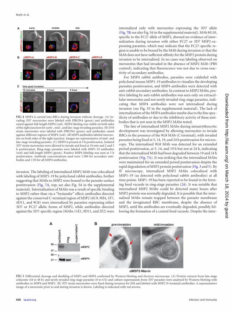
invasion. The labeling of internalized MSP2 MAb was colocalizedwith labeling of MSP1-19 by polyclonal rabbit antibodies, furthersuggesting that MAbs to MSP2 were bound to the parasite surfacepostinvasion (Fig. 7A, top; see also Fig. S4 in the supplementalmaterial). Internalization of MAbs was a result of specific bindingto MSP2 rather than via a “bystander” effect; antibodies directedagainst the conserved C-terminal region of MSP2 (6C9, 9H4, 1F7,4D11, and 9G8) were internalized by parasites expressing either3D7 or FC27 allelic forms of MSP2, while antibodies directedagainst the 3D7-specific region (MAbs 11E1, 9D11, and 2F2) were
internalized only with merozoites expressing the 3D7 allele(Fig. 7B; see also Fig. S4 in the supplemental material). MAb 8G10,specific to the FC27 allele of MSP2, showed no evidence of inter-nalization during invasion with either FC27 or 3D7 MSP2-ex-pressing parasites, which may indicate that the FC27-specific re-gion is unable to be bound by the MAb during invasion or that theMAb does not have sufficient affinity for the MSP2 protein duringinvasion to be internalized. In no cases was labeling observed onmerozoites that had invaded in the absence of MSP2 MAb (PBScontrol), indicating that fluorescence was not due to cross-reac-tivity of secondary antibodies.
For MSP4 rabbit antibodies, parasites were colabeled withpolyclonal mouse MSP1-19 antibodies to visualize the developingparasites postinvasion, and MSP4 antibodies were detected withanti-rabbit secondary antibodies. In contrast to MSP2 MAbs, pos-itive labeling by anti-rabbit antibodies was seen only on extracel-lular merozoites and not newly invaded ring-stage parasites, indi-cating that MSP4 antibodies were not internalized duringinvasion (see Fig. S5 in the supplemental material). The lack ofinternalization of the MSP4 antibodies maybe due to the fine spec-ificity of antibodies or due to the inhibitory activity of these anti-bodies that is not seen in the MSP2 MAbs tested.
The fate of internalized MSP2 MAbs during intraerythrocyticdevelopment was investigated by allowing merozoites to invadeRBCs in the presence of the 9G8 MAb (C terminal), with invadedparasites being fixed at 5, 14, 19, and 24 h postinvasion for micros-copy. The internalized 9G8 MAb was detected for an extendedperiod postinvasion, at 5, 14, and 19 h but not at 24 h, indicatingthat the internalized MAb had been degraded between 19 and 24 hpostinvasion (Fig. 7A). It was striking that the internalized MAbswere maintained for an extended period postinvasion despite therapid degradation of MSP2 protein postinvasion (Fig. 3 and 5). ByIF microscopy, internalized MSP2 MAbs colocalized withMSP1-19 (as detected with polyclonal rabbit antibodies) at alltime points. MSP1-19 has been reported to be found in the form-ing food vacuole in ring-stage parasites (24). It was notable thatinternalized MSP2 MAbs could be detected many hours afterMSP2 protein was normally degraded. It is possible that the inter-nalized MAbs remain trapped between the parasite membraneand the invaginated RBC membrane, despite the absence ofMSP2, until the antibodies are eventually degraded, possibly fol-lowing the formation of a central food vacuole. Despite the inter-
FIG 4 MSP4 is carried into RBCs during invasion without cleavage. (A) In-vading 3D7 merozoites were labeled with PfRON4 (green) and antibodiesraised against full-length MSP4 (red). MSP4 labeling was visible on both sidesof the tight junction for early-, mid-, and late-stage invading parasites. (B) D10strain merozoites were labeled with PfRON4 (green) and antibodies raisedagainst different regions of MSP4 (red). All MSP4 antibodies labeled merozo-ites on both sides of the tight junction. Images are representative of mid- andlate-stage invading parasites. (C) MSP4 is present at 5 h postinvasion. Isolated3D7 strain merozoites were allowed to invade and fixed at 10 min and 2 and 5h postinvasion. Ring-stage parasites were labeled with MSP1-19 antibodies(red) and full-length MSP4 (green). Positive MSP4 labeling was seen at 5 hpostinvasion. Antibody concentrations used were 1:500 for secondary anti-bodies and 1:50 for all MSP4 antibodies.
FIG 5 Differential cleavage and shedding of MSP2 and MSP4 confirmed by Western blotting and electron microscopy. (A) Protein extracts from late-stageschizonts (44 to 48 h) and newly invaded ring-stage parasites (0 to 6 h) and culture supernatants from 3D7 parasites were analyzed by Western blotting withantibodies to MSP4 and MSP2. (B) 3D7 strain merozoites were fixed during invasion for EM and labeled with MSP2 N-terminal antibodies. A representativeimage of a merozoite prior to and during invasion is shown. Labeling is indicated with red arrows.
Boyle et al.
930 iai.asm.org Infection and Immunity
on March 18, 2014 by guest
http://iai.asm.org/
Dow
nloaded from

nalization of MAbs, no growth inhibition or growth delay wasdetected (Fig. 6). This is in contrast to previous reports showingthat antibodies to MSP1-19 can be internalized and result in de-layed or inhibited intraerythrocytic parasite development in somecases (70).
DISCUSSION
Here, we provide evidence that there is a coordinated process ofsequential cleavage of merozoite surface proteins during invasionrather than the complete shedding of all surface proteins at thetight junction, which was previously proposed based on EM stud-ies (4, 9–11). Shedding is instead a specific process, with differen-tial cleavage and loss of merozoite surface proteins during andafter invasion. Our novel findings suggest that individual mero-zoite surface proteins have different roles during invasion andduring intraerythrocytic development (Fig. 8). Some merozoitesurface proteins are shed during invasion, suggesting that theseproteins may play a role in initial invasion events (e.g., N-terminalMSP1, MSP7, MSP3, SERA4, and SERA5). Other proteins, such asMSP2, may function during invasion and are then rapidly de-graded postinvasion, indicating that they do not function in fur-ther intraerythrocytic development. Finally, other proteins are in-ternalized without processing and persist postinvasion (e.g.,MSP4) or persist postinvasion as a processed form (e.g., MSP1-19) and may have important roles in intraerythrocytic develop-ment of parasites. These findings suggest that the merozoite sur-face is gradually remodeled by processing events commencingwith initiation of invasion, through immediate postinvasion pro-cesses, and subsequently through intraerythrocytic development,which significantly advances our understanding of erythrocyte in-
vasion and merozoite surface protein processing. We believe thatthese studies are the first to identify proteins (MSP2 and MSP4)that remain on the surface of the merozoite during invasion with-out processing, and we demonstrate the striking postinvasion pro-cessing of a merozoite antigen (MSP2).
Our study has shown that at least two GPI-anchored proteins,MSP2 and MSP4, appear to be carried into RBCs intact, in con-trast to the peripheral surface proteins (MSP3, MSP7, SERA4, andSERA5) and the MSP1 complex (3, 22, 25), which are shed duringinvasion, at the point of the tight junction. The shedding of pe-ripheral surface proteins is likely due to the cleavage and subse-quent shedding of the membrane-bound binding partners ofthese proteins; for example, MSP7 is part of the MSP1 complex,which is shed due to cleavage of MSP1-42 to the MSP1-19 form byPfSUB2 (23). In contrast, for MSP2 and MSP4, antibodies to theseproteins used for IF microscopy of invading merozoites labeledthe merozoite surface equally on both sides of the tight junction,showing that these proteins were carried into the RBC duringinvasion. Importantly, this was confirmed with a number of anti-bodies for different regions of both MSP2 and MSP4, indicatingthat these proteins are not cleaved. Along with these proteins, theGPI-anchored fragment of MSP1, MSP1-19, is well characterizedas being carried into RBCs and can be detected on ring-stage par-asites. It is possible that the GPI anchor of these proteins is impor-tant for the passage of these proteins through the tight junctionduring invasion, and any shedding of GPI-anchored proteins re-quires specific cleavage and processing.
Interestingly, MSP2 was immediately degraded postinvasion,with little or no protein being detectable by IF microscopy inparasites fixed for imaging at 10 min postinvasion. This result is
FIG 6 MSP2 MAbs have no invasion inhibition or growth delay activity. (A and B) MSP2 MAbs were tested for growth inhibition activity by a one-cycle (A) ora two-cycle (B) GIA against parasites expressing 3D7 MSP2 or FC27 MSP2 at the following concentrations: 700 �g/ml for 11E1 and 6D8; 420 �g/ml for 9G8; and250 �g/ml for 1F7, 2F2, 4D11, 6C9, 9D11, and 8H4. MAb 1F9 directed against AMA1 was used as a positive control for inhibition. Data are from one assay induplicate and are shown as mean growth and range. (C) MSP2 MAbs were tested in invasion inhibition assays against FC27 MSP2-expressing parasites at 500�g/ml. No invasion inhibition activity was observed. Data are means � standard errors of the means for 2 to 4 assays in duplicate. (D) Mean fluorescence of GFPand EtBr is known to increase with the age of parasite cultures, so comparison of mean fluorescence intensities between cultures invaded in the presence of MSP2MAbs and control cultures can detect a growth delay. No growth delay was detected at 30 or 40 h, as shown by the identical mean fluorescence intensity (MFI)of parasites labeled with ethidium bromide or GFP following invasion inhibition assays. No growth delay phenotype was observed for all MSP2 MAbs. The graphis a representative plot of MFIs of parasites incubated with MAb 4D11 (red) compared to the PBS control (blue), stained with EtBr. (E) MSP4 rabbit antibodies(MSP4-full, MSP4A, and MSP4D) were tested in invasion inhibition assays at a 1:10 serum dilution. Data are mean invasions and ranges of two assays induplicate.
Processing of Merozoite Proteins during Invasion
March 2014 Volume 82 Number 3 iai.asm.org 931
on March 18, 2014 by guest
http://iai.asm.org/
Dow
nloaded from

consistent with previous reports that MSP2 is not detectable inring-stage parasites (47, 74, 75) and is not found in culture super-natant fractions (20, 47). Importantly, both the 3D7 and FC27allelic forms of MSP2 showed the same phenotype of being carried
into RBCs followed by rapid degradation. The 3D7 and FC27 al-leles of MSP2 share N- and C-terminal regions but vary signifi-cantly in the central strain-specific region of the protein (46, 76–78). The observation that both MSP2 variants show the samelocalization and very precise timing of degradation postinvasionsuggests that the functions of the 3D7 and FC27 forms of MSP2are the same, despite any structural differences. The internaliza-tion of MSP2 protein during invasion observed by IF microscopywas confirmed by EM of invading merozoites and by demonstrat-ing the specific internalization of MSP2 MAbs during invasion. Incontrast to the rapid degradation of MSP2, MSP4 was maintainedfor at least 5 h in the developing ring parasite, and the presence ofuncleaved protein postinvasion was confirmed by Western blot-ting. Furthermore, the MSP4 protein was detected in trophozoite-stage parasites via Western blotting. A previous report failed todetect MSP4 by IF microscopy on early-ring-stage parasites (48).However, the precise age of the rings examined was not given inthat study, and it may be that as the parasite matures, MSP4 isdegraded and may not be detected via IF microscopy.
Merozoite surface proteins have been implicated in initial con-tact events between the merozoite and erythrocyte surface (1, 6).Indeed, shed proteins such as MSP1 (aside from the internalizedGPI-anchored MSP1-19 component), MSP3, MSP7, SERA4, andSERA5 may have roles at this stage, and the shedding of theseproteins may indicate that the role of these proteins in invasion iscomplete. In contrast, nonshed proteins may have roles distinctfrom initial contact; the localization of MSP2 during and afterinvasion suggests that MSP2 may function during the invasionprocess, while the localization of MSP4 suggest that this protein,like MSP1-19, may function postinvasion during early intraeryth-rocytic development of the parasite. While both MSP2 and MSP4are thought to be essential blood-stage antigens (43), only MSP2 isunder extensive immune selection pressure (39, 79) and appearsto be an important target of human immunity (37, 80). The pres-ence of polymorphisms for immune evasion provides additionalsupport for an important role of MSP2 during or around the timeof invasion and its significance as a target of human immunity.Our findings are also significant for understanding immunity andvaccine development because they indicate that MSP2 is seen bythe immune system only as it is presented on the merozoite sur-face, and it is not found as a processed or secreted form. Thisemphasizes the importance of understanding the native structureof MSP2 and the interaction with antibodies (60). Several otherGPI-anchored merozoite surface proteins have been identified;the localization of these proteins during invasion to ascertaincleavage and shedding profiles may inform our understanding ofthe roles of these specific proteins during invasion.
This work has also demonstrated that antibodies to MSP2 canbind the merozoite surface and then be internalized during mero-zoite invasion. The ability of antibodies to be carried into invadedRBCs was observed previously for MSP1-19, with internalizationof noninhibitory monoclonal antibodies (24, 25) as well as inhib-itory polyclonal rabbit antibodies, which can result in a specificgrowth delay (70). Other studies have reported that antibodies toMSP1 are able to disrupt intraerythrocytic development as well asinhibit invasion (81), a process that may be due to the internaliza-tion of antibodies during invasion. Although internalized MSP2antibodies were maintained within the parasite for between 19and 24 h, no invasion-inhibitory or growth-inhibitory activity wasobserved in either one-cycle direct invasion inhibition assays or
FIG 7 Antibodies to MSP2 are internalized during invasion and maintainedfor approximately 24 h. Isolated merozoites expressing the 3D7 or FC27 formof MSP2 were incubated with each of the different MSP2 MAbs and RBCs andallowed to invade. Following 10 min of incubation, parasites were washed toremove excess antibody and fixed for immunofluorescence assays postinva-sion. (A) Parasites were labeled with polyclonal rabbit antibodies to MSP1-19and secondary antibodies (anti-rabbit Alexa594 to detect MSP1-19 labelingand anti-mouse Alexa488 to detect internalized MSP2 MAbs, with anti-MSP1-19 at a 1:200 dilution and secondary antibodies at a 1:500 dilution).Internalized MSP2 MAbs were detected up to 19 h postinvasion. Images arefrom representative antibodies to the C-terminally conserved region. (B) Pos-itive labeling for internalized MAbs was quantified in parasites expressing the3D7 and FC27 forms of MSP2 with the panel of MSP2 MAbs. Percent positivemerozoites and standard errors of the proportion are graphed for each parasitestrain. The region of reactivity of the MAb is represented below the graph. Nolabeling was detected in merozoites that had invaded RBCs in the absence of aspecific MSP2 MAb (PBS control), and labeling was detected only for 3D7-specific MSP2 MAbs in parasites expressing the 3D7 and not the FC27 form ofMSP2, indicating that internalization was specific.
Boyle et al.
932 iai.asm.org Infection and Immunity
on March 18, 2014 by guest
http://iai.asm.org/
Dow
nloaded from

one- and two-cycle standard growth inhibition assays. It is possi-ble that in vivo, internalized antibodies would have some negativeimpact on parasite growth that is not measurable in the in vitroassays used here. However, it is also possible that internalizedMSP2 antibodies have no role in inhibiting intraerythrocytic de-velopment, as MSP2 is rapidly degraded and presumably has norole in the subsequent development of the parasite postinvasion.This is in contrast to MSP1-19, which has been hypothesized tohave a role in food vacuole formation, with internalized antibod-ies to MSP1-19 having the potential to disrupt function postinva-sion (24, 70). It is interesting to speculate that the ability of themerozoite to carry antibodies bound to surface proteins, such asMSP2, into the invaded RBC without apparent disruption of in-vasion or growth may represent an adaption of the parasite torender antibodies to the merozoite surface nonfunctional, therebycontributing to immune evasion. Although antibodies to MSP2do not inhibit invasion directly, they do function to opsonizemerozoites and promote antibody-dependent cellular inhibitionmediated by monocytes (40, 72), which is thought to be an impor-tant mechanism contributing to acquired immunity (82). In con-trast to MSP2, the internalization of rabbit antibodies to MSP4was not detected. It is possible that MSP4 antibodies are unable tobe internalized due to the mild inhibitory activity of these anti-bodies, as seen in growth inhibition and direct invasion inhibitionassays (73). However, it is also possible that the position of MSP4during invasion prevents the internalization of antibodies target-ing this protein or that this observation is due simply to the spe-
cific antibodies tested. Further studies on the internalization ofmultiple antibodies targeting specific proteins are needed to fur-ther understand the mechanisms mediating antibody internaliza-tion. Likewise, while the observations presented here indicate thatinternalized MSP2 antibodies had no impact on parasite develop-ment, more in-depth analysis is needed to fully understand theimplications of internalized antibodies for protein processing,function, and localization.
Differential cleavage and shedding of merozoite surface pro-teins and the ability of antibodies to be internalized during inva-sion raise questions about the mechanisms of shedding and thenature of the tight junction. While PfSUB2 processing is requiredfor the cleavage and subsequent shedding of some surface proteins(23), the timing of these events remains unclear. It was hypothe-sized previously that the cleavage of MSP1-42 would occur at thetight junction and occur concurrently with shedding of MSP1(23). However, the localization of PfSUB2 during this period ap-pears to be different from the point of shedding; PfSUB2 appearsto track to the posterior of the invading merozoite before MSP1shedding occurs (3). It is clear that the tight junction alone doesnot force the shedding of proteins, as some merozoite surfaceproteins and antibodies can be internalized during the invasionprocess. Antibodies are approximately 150 kDa; for internaliza-tion to occur, the tight junction between the merozoite and RBCmust not be a tight seal of proteins but instead must consist ofeither transient interactions which can allow passage of antibodiesand surface proteins or gaps between protein-protein interactions
FIG 8 Coordinated cleavage and shedding of major merozoite surface proteins during invasion. At initial attachment and reorientation of the merozoite, themerozoite surface coat includes numerous proteins (1). The most abundant of these is MSP1, which is found in a complex with other antigens (indicated in greenand purple). MSP2 is indicated in blue, and MSP4 is indicated in yellow. AMA1 and the tight junction are represented by pink circles. During invasion, some butnot all merozoite surface proteins are cleaved and shed (2). Shed proteins include the majority of the MSP1 complex, with MSP1-19 being carried into the RBCalong with MSP3 and SERA4/5 (not indicated). MSP2 and MSP4 are not cleaved and shed at the point of the tight junction and are instead carried into the RBC.Shedding continues to occur as the merozoite invades the RBC (3). Between late invasion and mature invasion, MSP2 protein is lost; however, MSP4 andMSP1-19 are maintained into early intraerythrocytic development (4).
Processing of Merozoite Proteins during Invasion
March 2014 Volume 82 Number 3 iai.asm.org 933
on March 18, 2014 by guest
http://iai.asm.org/
Dow
nloaded from
that allow molecules to pass through. The tight junction appearsas a complete ring structure by IF microscopy of PfRON4 andAMA1 (3) and as a region of close proximity between the mero-zoite and RBC membranes by EM (4). Our results for internaliza-tion of antibodies, and MSP2 and MSP4 without obvious process-ing, suggest that the tight junction is able to specifically excludesome proteins but not others. It remains possible that cleavage ofsurface proteins and the mechanisms of shedding require a furtherunknown factor to mediate the specific release of proteins fromthe merozoite surface. Clearly, the processes involved in the cleav-age and shedding of the surface coat are specific and not universalor nonselective; instead, they act against a subset of proteins. Afurther understanding of the molecular basis of merozoite proteinprocessing might reveal attractive targets for the development ofantimalarial inhibitors, and several proteases that inhibit invasionhave been reported in the literature (6, 13). Furthermore, withmerozoite surface proteins being the targets of vaccine develop-ment, clear characterization of individual proteins during and af-ter invasion informs vaccine development by identifying periodswhen parasite proteins are most likely to be targeted by the im-mune system (32, 33). In conclusion, these studies advance ourunderstanding of the mechanisms of invasion, demonstrating thatthe loss of the merozoite surface coat is not a global event andinstead involves specific processes resulting in the sequentialcleavage of individual proteins both during and after invasion,indicating the diversity of roles of different proteins (Fig. 8). In-vasion of RBCs involves a complex series of events, and an under-standing of key processes and interactions may reveal targets fordrug development and will facilitate the prioritization of antigensfor vaccine development.
ACKNOWLEDGMENTS
RBCs and serum for parasite culture were provided by the Red CrossBlood Bank (Melbourne, Australia). We thank Alan Cowman for provid-ing antibodies to RON2 and MSP6, Brendan Crabb for antibodies toMSP1-19, Anthony Holder for providing antibodies to MSP7, Jake Baumfor helpful advice and comments and providing a template for Fig. 8,David Riglar for advice on microscopy, and Candida da Fonseca Pereiraand the Monash Micro Imaging Facility for provision of instrumentation,training, and general support.
Funding was provided by the National Health and Medical ResearchCouncil of Australia (program grant to J.G.B.; Infrastructure for ResearchInstitutes Support Scheme grant), the Australian Research Council (fu-ture fellowship to J.G.B.), and a Victorian State Government operationalinfrastructure support grant. A postgraduate research fellowship to M.J.B.was provided by the Australian Government and the University of Mel-bourne Department of Medicine, Dentistry and Health Sciences.
We declare that we have no conflicts of interest.
REFERENCES1. Cowman AF, Crabb BS. 2006. Invasion of red blood cells by malaria
parasites. Cell 124:755–766. http://dx.doi.org/10.1016/j.cell.2006.02.006.2. Gaur D, Mayer DCG, Miller LH. 2004. Parasite ligand-host receptor
interactions during invasion of erythrocytes by Plasmodium merozoites.Int. J. Parasitol. 34:1413–1429. http://dx.doi.org/10.1016/j.ijpara.2004.10.010.
3. Riglar DT, Richard D, Wilson DW, Boyle MJ, Dekiwadia C, TurnbullL, Angrisano F, Marapana DS, Rogers KL, Whitchurch CB, Beeson JG,Cowman AF, Ralph SA, Baum J. 2011. Super-resolution dissection ofcoordinated events during malaria parasite invasion of the human eryth-rocyte. Cell Host Microbe 9:9 –20. http://dx.doi.org/10.1016/j.chom.2010.12.003.
4. Bannister LH, Butcher GA, Dennis ED, Mitchell GH. 1975. Structure
and invasive behaviour of Plasmodium knowlesi merozoites in vitro. Par-asitology 71:483– 491. http://dx.doi.org/10.1017/S0031182000047247.
5. Baum J, Maier AG, Good RT, Simpson KM, Cowman AF. 2005.Invasion by P. falciparum merozoites suggests a hierarchy of molecularinteractions. PLoS Pathog. 1:e37. http://dx.doi.org/10.1371/journal.ppat.0010037.
6. Boyle MJ, Wilson DW, Beeson JG. 2013. New approaches to studyingPlasmodium falciparum merozoite invasion and insights into invasionbiology. Int. J. Parasitol. 43:1–10. http://dx.doi.org/10.1016/j.ijpara.2012.11.002.
7. Srinivasan P, Beatty WL, Diouf A, Herrera R, Ambroggio X, Moch JK,Tyler JS, Narum DL, Pierce SK, Boothroyd JC, Haynes JD, Miller LH.2011. Binding of Plasmodium merozoite proteins RON2 and AMA1 trig-gers commitment to invasion. Proc. Natl. Acad. Sci. U. S. A. 108:13275–13280. http://dx.doi.org/10.1073/pnas.1110303108.
8. Baum J, Richard D, Healer J, Rug M, Krnajski Z, Gilberger T-W, GreenJL, Holder AA, Cowman AF. 2006. A conserved molecular motor drivescell invasion and gliding motility across malaria life cycle stages and otherapicomplexan parasites. J. Biol. Chem. 281:5197–5208. http://dx.doi.org/10.1074/jbc.M509807200.
9. Aikawa M, Miller LH, Johnson J, Rabbege J. 1978. Erythrocyte entry bymalarial parasites. A moving junction between erythrocyte and parasite. J.Cell Biol. 77:72– 82.
10. Ladda R, Aikawa M, Sprinz H. 1969. Penetration of erythrocytes bymerozoites of mammalian and avian malarial parasites. J. Parasitol. 55:633– 644. http://dx.doi.org/10.2307/3277308.
11. Langreth SG, Nguyen-Dinh P, Trager W. 1978. Plasmodium falciparum:merozoite invasion in vitro in the presence of chloroquine. Exp. Parasitol.46:235–238. http://dx.doi.org/10.1016/0014-4894(78)90136-4.
12. Carruthers VB, Blackman MJ. 2005. A new release on life: emergingconcepts in proteolysis and parasite invasion. Mol. Microbiol. 55:1617–1630. http://dx.doi.org/10.1111/j.1365-2958.2005.04483.x.
13. Blackman MJ. 2004. Proteases in host cell invasion by the malaria para-site. Cell. Microbiol. 6:893–903. http://dx.doi.org/10.1111/j.1462-5822.2004.00437.x.
14. Fujioka H, Aikawa M. 2002. Structure and life cycle. Chem. Immunol.80:1–26. http://dx.doi.org/10.1159/000058837.
15. Blackman MJ, Holder AA. 1992. Secondary processing of the Plasmodium fal-ciparum merozoite surface protein-1 (MSP1) by a calcium-dependent mem-brane-bound serine protease: shedding of MSP133 as a noncovalently associatedcomplexwithotherfragmentsoftheMSP1.Mol.Biochem.Parasitol.50:307–315.http://dx.doi.org/10.1016/0166-6851(92)90228-C.
16. Pachebat JA, Ling IT, Grainger M, Trucco C, Howell S, Fernandez-Reyes D, Gunaratne R, Holder AA. 2001. The 22 kDa component of theprotein complex on the surface of Plasmodium falciparum merozoites isderived from a larger precursor, merozoite surface protein 7. Mol.Biochem. Parasitol. 117:83– 89. http://dx.doi.org/10.1016/S0166-6851(01)00336-X.
17. Trucco C, Fernandez-Reyes D, Howell S, Stafford WH, Scott-Finnigan TJ,Grainger M, Ogun SA, Taylor WR, Holder AA. 2001. The merozoite surfaceprotein 6 gene codes for a 36 kDa protein associated with the Plasmodium falcip-arummerozoitesurfaceprotein-1complex.Mol.Biochem.Parasitol.112:91–101.http://dx.doi.org/10.1016/S0166-6851(00)00350-9.
18. Delplace P, Dubremetz JF, Fortier B, Vernes A. 1985. A 50 kilodaltonexoantigen specific to the merozoite release-reinvasion stage of Plasmo-dium falciparum. Mol. Biochem. Parasitol. 17:239 –251. http://dx.doi.org/10.1016/0166-6851(85)90021-0.
19. Miller SK, Good RT, Drew DR, Delorenzi M, Sanders PR, Hodder AN,Speed TP, Cowman AF, de Koning-Ward TF, Crabb BS. 2002. A subsetof Plasmodium falciparum SERA genes are expressed and appear to playan important role in the erythrocytic cycle. J. Biol. Chem. 277:47524 –47532. http://dx.doi.org/10.1074/jbc.M206974200.
20. Pearce JA, Hodder AN, Anders RF. 2004. The alanine-rich heptad re-peats are intact in the processed form of Plasmodium falciparum MSP3.Exp. Parasitol. 108:186 –189. http://dx.doi.org/10.1016/j.exppara.2004.07.017.
21. Blackman MJ, Dennis ED, Hirst EM, Kocken CH, Scott-Finnigan TJ,Thomas AW. 1996. Plasmodium knowlesi: secondary processing of themalaria merozoite surface protein-1. Exp. Parasitol. 83:229 –239. http://dx.doi.org/10.1006/expr.1996.0069.
22. Boyle MJ, Wilson DW, Richards JS, Riglar DT, Tetteh KKA, ConwayDJ, Ralph SA, Baum J, Beeson JG. 2010. Isolation of viable Plasmodiumfalciparum merozoites to define erythrocyte invasion events and advance
Boyle et al.
934 iai.asm.org Infection and Immunity
on March 18, 2014 by guest
http://iai.asm.org/
Dow
nloaded from

vaccine and drug development. Proc. Natl. Acad. Sci. U. S. A. 107:14378 –14383. http://dx.doi.org/10.1073/pnas.1009198107.
23. Harris PK, Yeoh S, Dluzewski AR, O’Donnell RA, Withers-Martinez C,Hackett F, Bannister LH, Mitchell GH, Blackman MJ. 2005. Molecularidentification of a malaria merozoite surface sheddase. PLoS Pathog.1:241–251. http://dx.doi.org/10.1371/journal.ppat.0010029.
24. Dluzewski AR, Ling IT, Hopkins JM, Grainger M, Margos G, MitchellGH, Holder AA, Bannister LH. 2008. Formation of the food vacuole inPlasmodium falciparum: a potential role for the 19 kDa fragment of mero-zoite surface protein 1 (MSP1(19)). PLoS One 3:e3085. http://dx.doi.org/10.1371/journal.pone.0003085.
25. Blackman MJ, Scott-Finnigan TJ, Shai S, Holder AA. 1994. Antibodiesinhibit the protease-mediated processing of a malaria merozoite surfaceprotein. J. Exp. Med. 180:389 –393. http://dx.doi.org/10.1084/jem.180.1.389.
26. Fleck SL, Birdsall B, Babon J, Dluzewski AR, Martin SR, Morgan WD,Angov E, Kettleborough CA, Feeney J, Blackman MJ, Holder AA. 2003.Suramin and suramin analogues inhibit merozoite surface protein-1 sec-ondary processing and erythrocyte invasion by the malaria parasite Plas-modium falciparum. J. Biol. Chem. 278:47670 – 47677. http://dx.doi.org/10.1074/jbc.M306603200.
27. Singh S, Miura K, Zhou H, Muratova O, Keegan B, Miles A, Martin LB,Saul AJ, Miller LH, Long CA. 2006. Immunity to recombinant Plasmo-dium falciparum merozoite surface protein 1 (MSP1): protection in Aotusnancymai monkeys strongly correlates with anti-MSP1 antibody titer andin vitro parasite-inhibitory activity. Infect. Immun. 74:4573– 4580. http://dx.doi.org/10.1128/IAI.01679-05.
28. Woehlbier U, Epp C, Hackett F, Blackman MJ, Bujard H. 2010. Antibodiesagainst multiple merozoite surface antigens of the human malaria parasitePlasmodium falciparum inhibit parasite maturation and red blood cell inva-sion. Malar. J. 9:77. http://dx.doi.org/10.1186/1475-2875-9-77.
29. Boyle MJ, Richards JS, Gilson PR, Chai W, Beeson JG. 2010. Interac-tions with heparin-like molecules during erythrocyte invasion by Plasmo-dium falciparum merozoites. Blood 115:4559 – 4568. http://dx.doi.org/10.1182/blood-2009-09-243725.
30. Goel VK, Li X, Chen H, Liu S-C, Chishti AH, Oh SS. 2003. Band 3 is ahost receptor binding merozoite surface protein 1 during the Plasmodiumfalciparum invasion of erythrocytes. Proc. Natl. Acad. Sci. U. S. A. 100:5164 –5169. http://dx.doi.org/10.1073/pnas.0834959100.
31. Li X, Chen H, Oo TH, Daly TM, Bergman LW, Liu S-C, Chishti AH, OhSS. 2004. A co-ligand complex anchors Plasmodium falciparum merozo-ites to the erythrocyte invasion receptor band 3. J. Biol. Chem. 279:5765–5771. http://dx.doi.org/10.1074/jbc.M308716200.
32. Richards JS, Beeson JG. 2009. The future for blood-stage vaccines againstmalaria. Immunol. Cell Biol. 87:377–390. http://dx.doi.org/10.1038/icb.2009.27.
33. Chauhan VS, Yazdani SS, Gaur D. 2010. Malaria vaccine developmentbased on merozoite surface proteins of Plasmodium falciparum. Hum.Vaccin. 6:757–762. http://dx.doi.org/10.4161/hv.6.9.12468.
34. Richards JS, Arumugam TU, Reiling L, Healer J, Hodder AN, FowkesFJI, Cross N, Langer C, Takeo S, Uboldi AD, Thompson JK, Gilson PR,Coppel RL, Siba PM, King CL, Torii M, Chitnis CE, Narum DL,Mueller I, Crabb BS, Cowman AF, Tsuboi T, Beeson JG. 2013. Identi-fication and prioritization of merozoite antigens as targets of protectivehuman immunity to Plasmodium falciparum malaria for vaccine and bio-marker development. J. Immunol. 191:795– 809. http://dx.doi.org/10.4049/jimmunol.1300778.
35. Fowkes FJI, Richards JS, Simpson JA, Beeson JG. 2010. The relationshipbetween anti-merozoite antibodies and incidence of Plasmodium falcip-arum malaria: a systematic review and meta-analysis. PLoS Med.7:e1000218. http://dx.doi.org/10.1371/journal.pmed.1000218.
36. Egan AF, Morris J, Barnish G, Allen S, Greenwood BM, Kaslow DC,Holder AA, Riley EM. 1996. Clinical immunity to Plasmodium falcipa-rum malaria is associated with serum antibodies to the 19-kDa C-terminalfragment of the merozoite surface antigen, PfMSP-1. J. Infect. Dis. 173:765–769. http://dx.doi.org/10.1093/infdis/173.3.765.
37. Taylor RR, Allen SJ, Greenwood BM, Riley EM. 1998. IgG3 antibodiesto Plasmodium falciparum merozoite surface protein 2 (MSP2): increas-ing prevalence with age and association with clinical immunity to malaria.Am. J. Trop. Med. Hyg. 58:406 – 413.
38. Roussilhon C, Oeuvray C, Müller-Graf C, Tall A, Rogier C, Trape J-F,Theisen M, Balde A, Pérignon J-L, Druilhe P. 2007. Long-term clinicalprotection from falciparum malaria is strongly associated with IgG3 anti-
bodies to merozoite surface protein 3. PLoS Med. 4:e320. http://dx.doi.org/10.1371/journal.pmed.0040320.
39. Genton B, Betuela I, Felger I, Al-Yaman F, Anders RF, Saul A, Rare L,Baisor M, Lorry K, Brown GV, Pye D, Irving DO, Smith TA, Beck H-P,Alpers MP. 2002. A recombinant blood-stage malaria vaccine reducesPlasmodium falciparum density and exerts selective pressure on parasitepopulations in a phase 1-2b trial in Papua New Guinea. J. Infect. Dis.185:820 – 827. http://dx.doi.org/10.1086/339342.
40. McCarthy JS, Marjason J, Elliott S, Fahey P, Bang G, Malkin E, TierneyE, Aked-Hurditch H, Adda C, Cross N, Richards JS, Fowkes FJI, BoyleMJ, Long C, Druilhe P, Beeson JG, Anders RF. 2011. A phase 1 trial ofMSP2-C1, a blood-stage malaria vaccine containing 2 isoforms of MSP2formulated with Montanide ISA 720. PLoS One 6:e24413. http://dx.doi.org/10.1371/journal.pone.0024413.
41. Sirima SB, Cousens S, Druilhe P. 2011. Protection against malaria byMSP3 candidate vaccine. N. Engl. J. Med. 365:1062–1064. http://dx.doi.org/10.1056/NEJMc1100670.
42. Gilson PR, Nebl T, Vukcevic D, Moritz RL, Sargeant T, Speed TP,Schofield L, Crabb BS. 2006. Identification and stoichiometry of glyco-sylphosphatidylinositol-anchored membrane proteins of the human ma-laria parasite Plasmodium falciparum. Mol. Cell. Proteomics 5:1286 –1299. http://dx.doi.org/10.1074/mcp.M600035-MCP200.
43. Sanders PR, Kats LM, Drew DR, O’Donnell RA, O’Neill M, Maier AG, CoppelRL, Crabb BS. 2006. A set of glycosylphosphatidyl inositol-anchored membraneproteins of Plasmodium falciparum is refractory to genetic deletion. Infect. Im-mun. 74:4330–4338. http://dx.doi.org/10.1128/IAI.00054-06.
44. Fenton B, Clark JT, Khan CM, Robinson JV, Walliker D, Ridley R,Scaife JG, McBride JS. 1991. Structural and antigenic polymorphism ofthe 35- to 48-kilodalton merozoite surface antigen (MSA-2) of the malariaparasite Plasmodium falciparum. Mol. Cell. Biol. 11:963–971.
45. Smythe JA, Coppel RL, Day KP, Martin RK, Oduola AM, Kemp DJ,Anders RF. 1991. Structural diversity in the Plasmodium falciparummerozoite surface antigen 2. Proc. Natl. Acad. Sci. U. S. A. 88:1751–1755.http://dx.doi.org/10.1073/pnas.88.5.1751.
46. Adda CG, Murphy VJ, Sunde M, Waddington LJ, Schloegel J, TalboGH, Vingas K, Kienzle V, Masciantonio R, Howlett GJ, Hodder AN,Foley M, Anders RF. 2009. Plasmodium falciparum merozoite surfaceprotein 2 is unstructured and forms amyloid-like fibrils. Mol. Biochem.Parasitol. 166:159 –171. http://dx.doi.org/10.1016/j.molbiopara.2009.03.012.
47. Clark JT, Donachie S, Anand R, Wilson CF, Heidrich HG, McBride JS.1989. 46-53 kilodalton glycoprotein from the surface of Plasmodium fal-ciparum merozoites. Mol. Biochem. Parasitol. 32:15–24. http://dx.doi.org/10.1016/0166-6851(89)90125-4.
48. Marshall VM, Silva A, Foley M, Cranmer S, Wang L, McColl DJ, KempDJ, Coppel RL. 1997. A second merozoite surface protein (MSP-4) ofPlasmodium falciparum that contains an epidermal growth factor-likedomain. Infect. Immun. 65:4460 – 4467.
49. Wang L, Black CG, Marshall VM, Coppel RL. 1999. Structural andantigenic properties of merozoite surface protein 4 of Plasmodium falcip-arum. Infect. Immun. 67:2193–2200.
50. Goschnick MW, Black CG, Kedzierski L, Holder AA, Coppel RL. 2004.Merozoite surface protein 4/5 provides protection against lethal challengewith a heterologous malaria parasite strain. Infect. Immun. 72:5840 –5849. http://dx.doi.org/10.1128/IAI.72.10.5840-5849.2004.
51. Kedzierski L, Black CG, Stowers AW, Goschnick MW, Kaslow DC,Coppel RL. 2001. Comparison of the protective efficacy of yeast-derivedand Escherichia coli-derived recombinant merozoite surface protein 4/5against lethal challenge by Plasmodium yoelii. Vaccine 19:4661– 4668.http://dx.doi.org/10.1016/S0264-410X(01)00244-4.
52. Kedzierski L, Black CG, Goschnick MW, Stowers AW, Coppel RL. 2002.Immunization with a combination of merozoite surface proteins 4/5 and1 enhances protection against lethal challenge with Plasmodium yoelii.Infect. Immun. 70:6606 – 6613. http://dx.doi.org/10.1128/IAI.70.12.6606-6613.2002.
53. Kedzierski L, Black CG, Coppel RL. 2000. Immunization with recombi-nant Plasmodium yoelii merozoite surface protein 4/5 protects miceagainst lethal challenge. Infect. Immun. 68:6034 – 6037. http://dx.doi.org/10.1128/IAI.68.10.6034-6037.2000.
54. Logan GJ, Wang L, Zheng M, Cunningham SC, Coppel RL, AlexanderIE. 2007. AAV vectors encoding malarial antigens stimulate antigen-specific immunity but do not protect from parasite infection. Vaccine25:1014 –1022. http://dx.doi.org/10.1016/j.vaccine.2006.09.072.
Processing of Merozoite Proteins during Invasion
March 2014 Volume 82 Number 3 iai.asm.org 935
on March 18, 2014 by guest
http://iai.asm.org/
Dow
nloaded from

55. Rainczuk A, Smooker PM, Kedzierski L, Black CG, Coppel RL, SpithillTW. 2003. The protective efficacy of MSP4/5 against lethal Plasmodiumchabaudi adami challenge is dependent on the type of DNA vaccine vectorand vaccination protocol. Vaccine 21:3030 –3042. http://dx.doi.org/10.1016/S0264-410X(03)00116-6.
56. Wilson DW, Langer C, Goodman CD, McFadden GI, Beeson JG. 2013.Defining the timing of action of antimalarial drugs against Plasmodiumfalciparum. Antimicrob. Agents Chemother. 57:1455–1467. http://dx.doi.org/10.1128/AAC.01881-12.
57. Beeson JG, Brown GV, Molyneux ME, Mhango C, Dzinjalamala F,Rogerson SJ. 1999. Plasmodium falciparum isolates from infected preg-nant women and children are associated with distinct adhesive and anti-genic properties. J. Infect. Dis. 180:464 – 472. http://dx.doi.org/10.1086/314899.
58. Persson KEM, Lee CT, Marsh K, Beeson JG. 2006. Development andoptimization of high-throughput methods to measure Plasmodium falcip-arum-specific growth inhibitory antibodies. J. Clin. Microbiol. 44:1665–1673. http://dx.doi.org/10.1128/JCM.44.5.1665-1673.2006.
59. Lambros C, Vanderberg JP. 1979. Synchronization of Plasmodium fal-ciparum erythrocytic stages in culture. J. Parasitol. 65:418 – 420. http://dx.doi.org/10.2307/3280287.
60. Adda CG, MacRaild CA, Reiling L, Wycherley K, Boyle MJ, Kienzle V,Masendycz P, Foley M, Beeson JG, Norton RS, Anders RF. 2012.Antigenic characterization of an intrinsically unstructured protein, Plas-modium falciparum merozoite surface protein 2. Infect. Immun. 80:4177– 4185. http://dx.doi.org/10.1128/IAI.00665-12.
61. Richard D, MacRaild CA, Riglar DT, Chan J-A, Foley M, Baum J, RalphSA, Norton RS, Cowman AF. 2010. Interaction between Plasmodiumfalciparum apical membrane antigen 1 and the rhoptry neck protein com-plex defines a key step in the erythrocyte invasion process of malaria par-asites. J. Biol. Chem. 285:14815–14822. http://dx.doi.org/10.1074/jbc.M109.080770.
62. Mills KE, Pearce JA, Crabb BS, Cowman AF. 2002. Truncation ofmerozoite surface protein 3 disrupts its trafficking and that of acidic-basicrepeat protein to the surface of Plasmodium falciparum merozoites. Mol.Microbiol. 43:1401–1411. http://dx.doi.org/10.1046/j.1365-2958.2002.02834.x.
63. Kadekoppala M, O’Donnell RA, Grainger M, Crabb BS, Holder AA.2008. Deletion of the Plasmodium falciparum merozoite surface protein 7gene impairs parasite invasion of erythrocytes. Eukaryot. Cell 7:2123–2132. http://dx.doi.org/10.1128/EC.00274-08.
64. O’Donnell RA, de Koning-Ward TF, Burt RA, Bockarie M, Reeder JC,Cowman AF, Crabb BS. 2001. Antibodies against merozoite surface pro-tein (MSP)-1(19) are a major component of the invasion-inhibitory re-sponse in individuals immune to malaria. J. Exp. Med. 193:1403–1412.http://dx.doi.org/10.1084/jem.193.12.1403.
65. Hodder AN, Drew DR, Epa VC, Delorenzi M, Bourgon R, Miller SK, MoritzRL, Frecklington DF, Simpson RJ, Speed TP, Pike RN, Crabb BS. 2003.Enzymic,phylogenetic,andstructuralcharacterizationof theunusualpapain-likeprotease domain of Plasmodium falciparum SERA5. J. Biol. Chem. 278:48169–48177. http://dx.doi.org/10.1074/jbc.M306755200.
66. Hodder AN, Malby RL, Clarke OB, Fairlie WD, Colman PM, Crabb BS,Smith BJ. 2009. Structural insights into the protease-like antigen Plasmo-dium falciparum SERA5 and its noncanonical active-site serine. J. Mol.Biol. 392:154 –165. http://dx.doi.org/10.1016/j.jmb.2009.07.007.
67. McCallum FJ, Persson KEM, Mugyenyi CK, Fowkes FJI, Simpson JA,Richards JS, Williams TN, Marsh K, Beeson JG. 2008. Acquisition ofgrowth-inhibitory antibodies against blood-stage Plasmodium falciparum.PLoS One 3:e3571. http://dx.doi.org/10.1371/journal.pone.0003571.
68. Wilson DW, Crabb BS, Beeson JG. 2010. Development of fluorescentPlasmodium falciparum for in vitro growth inhibition assays. Malar. J.9:152. http://dx.doi.org/10.1186/1475-2875-9-152.
69. Drew DR, Hodder AN, Wilson DW, Foley M, Mueller I, Siba PM, DentAE, Cowman AF, Beeson JG. 2012. Defining the antigenic diversity ofPlasmodium falciparum apical membrane antigen 1 and the requirementsfor a multi-allele vaccine against malaria. PLoS One 7:e51023. http://dx.doi.org/10.1371/journal.pone.0051023.
70. Moss DK, Remarque EJ, Faber BW, Cavanagh DR, Arnot DE, ThomasAW, Holder AA. 2012. Plasmodium falciparum 19-kilodalton merozoitesurface protein 1 (MSP1)-specific antibodies that interfere with parasitegrowth in vitro can inhibit MSP1 processing, merozoite invasion, andintracellular parasite development. Infect. Immun. 80:1280 –1287. http://dx.doi.org/10.1128/IAI.05887-11.
71. Saul A, Lord R, Jones G, Geysen HM, Gale J, Mollard R. 1989. Cross-reactivity of antibody against an epitope of the Plasmodium falciparumsecond merozoite surface antigen. Parasite Immunol. 11:593– 601. http://dx.doi.org/10.1111/j.1365-3024.1989.tb00923.x.
72. Flueck C, Frank G, Smith T, Jafarshad A, Nebie I, Sirima SB, OlugbileS, Alonso P, Tanner M, Druilhe P, Felger I, Corradin G. 2009. Evalu-ation of two long synthetic merozoite surface protein 2 peptides as malariavaccine candidates. Vaccine 27:2653–2661. http://dx.doi.org/10.1016/j.vaccine.2009.02.081.
73. de Silva HD, Saleh S, Kovacevic S, Wang L, Black CG, Plebanski M,Coppel RL. 2011. The antibody response to Plasmodium falciparummerozoite surface protein 4: comparative assessment of specificity andgrowth inhibitory antibody activity to infection-acquired and immuniza-tion-induced epitopes. Malar. J. 10:266. http://dx.doi.org/10.1186/1475-2875-10-266.
74. Barron C. 1992. Transcription of the gene for the merozoite surface an-tigen MSA2 of the human malaria parasite Plasmodium falciparum dur-ing the asexual cycle. FEBS Lett. 300:77– 81. http://dx.doi.org/10.1016/0014-5793(92)80168-G.
75. Ramasamy R. 1987. Studies on glycoproteins in the human malaria par-asite Plasmodium falciparum. Identification of a myristilated 45kDamerozoite membrane glycoprotein. Immunol. Cell Biol. 65(Part 5):419 –424.
76. Low A, Chandrashekaran IR, Adda CG, Yao S, Sabo JK, Zhang X,Soetopo A, Anders RF, Norton RS. 2007. Merozoite surface protein 2 ofPlasmodium falciparum: expression, structure, dynamics, and fibril for-mation of the conserved N-terminal domain. Biopolymers 87:12–22. http://dx.doi.org/10.1002/bip.20764.
77. Yang X, Adda CG, MacRaild CA, Low A, Zhang X, Zeng W, JacksonDC, Anders RF, Norton RS. 2010. Identification of key residues involvedin fibril formation by the conserved N-terminal region of Plasmodiumfalciparum merozoite surface protein 2 (MSP2). Biochimie 92:1287–1295.http://dx.doi.org/10.1016/j.biochi.2010.06.001.
78. Yang X, Adda CG, Keizer DW, Murphy VJ, Rizkalla MM, Perugini MA,Jackson DC, Anders RF, Norton RS. 2007. A partially structured regionof a largely unstructured protein, Plasmodium falciparum merozoite sur-face protein 2 (MSP2), forms amyloid-like fibrils. J. Pept. Sci. 13:839 – 848.http://dx.doi.org/10.1002/psc.910.
79. Benet A, Tavul L, Reeder JC, Cortés A. 2004. Diversity of Plasmodiumfalciparum vaccine candidate merozoite surface protein 4 (MSP4) in anatural population. Mol. Biochem. Parasitol. 134:275–280. http://dx.doi.org/10.1016/j.molbiopara.2003.12.005.
80. Stanisic DI, Richards JS, McCallum FJ, Michon P, King CL, SchoepflinS, Gilson PR, Murphy VJ, Anders RF, Mueller I, Beeson JG. 2009.Immunoglobulin G subclass-specific responses against Plasmodium fal-ciparum merozoite antigens are associated with control of parasitemiaand protection from symptomatic illness. Infect. Immun. 77:1165–1174.http://dx.doi.org/10.1128/IAI.01129-08.
81. Woehlbier U, Epp C, Kauth CW, Lutz R, Long CA, Coulibaly B,Kouyaté B, Arevalo-Herrera M, Herrera S, Bujard H. 2006. Analysis ofantibodies directed against merozoite surface protein 1 of the human ma-laria parasite Plasmodium falciparum. Infect. Immun. 74:1313–1322.http://dx.doi.org/10.1128/IAI.74.2.1313-1322.2006.
82. Bouharoun-Tayoun H, Attanath P, Sabchareon A, Chongsuphajaisid-dhi T, Druilhe P. 1990. Antibodies that protect humans against Plasmo-dium falciparum blood stages do not on their own inhibit parasite growthand invasion in vitro, but act in cooperation with monocytes. J. Exp. Med.172:1633–1641. http://dx.doi.org/10.1084/jem.172.6.1633.
83. National Health and Medical Research Council. 2013. Australian codefor the care and use of animals for scientific purposes, 8th ed. NationalHealth and Medical Research Council, Canberra, Australia.
Boyle et al.
936 iai.asm.org Infection and Immunity
on March 18, 2014 by guest
http://iai.asm.org/
Dow
nloaded from
Related Documents