University of Massachusetts Medical School University of Massachusetts Medical School eScholarship@UMMS eScholarship@UMMS Open Access Publications by UMMS Authors 2021-01-26 Separation and identification of permethylated glycan isomers by Separation and identification of permethylated glycan isomers by reversed phase nanoLC-NSI-MS(n) reversed phase nanoLC-NSI-MS(n) Simone Kurz University of Georgia Et al. Let us know how access to this document benefits you. Follow this and additional works at: https://escholarship.umassmed.edu/oapubs Part of the Amino Acids, Peptides, and Proteins Commons, Biochemistry Commons, Chemistry Commons, Molecular Biology Commons, and the Structural Biology Commons Repository Citation Repository Citation Kurz S, Sheikh MO, Lu S, Wells L, Tiemeyer M. (2021). Separation and identification of permethylated glycan isomers by reversed phase nanoLC-NSI-MS(n). Open Access Publications by UMMS Authors. https://doi.org/10.1074/mcp.RA120.002266. Retrieved from https://escholarship.umassmed.edu/ oapubs/4513 Creative Commons License This work is licensed under a Creative Commons Attribution 4.0 License. This material is brought to you by eScholarship@UMMS. It has been accepted for inclusion in Open Access Publications by UMMS Authors by an authorized administrator of eScholarship@UMMS. For more information, please contact [email protected].

Welcome message from author
This document is posted to help you gain knowledge. Please leave a comment to let me know what you think about it! Share it to your friends and learn new things together.
Transcript
University of Massachusetts Medical School University of Massachusetts Medical School
eScholarship@UMMS eScholarship@UMMS
Open Access Publications by UMMS Authors
2021-01-26
Separation and identification of permethylated glycan isomers by Separation and identification of permethylated glycan isomers by
reversed phase nanoLC-NSI-MS(n) reversed phase nanoLC-NSI-MS(n)
Simone Kurz University of Georgia
Et al.
Let us know how access to this document benefits you. Follow this and additional works at: https://escholarship.umassmed.edu/oapubs
Part of the Amino Acids, Peptides, and Proteins Commons, Biochemistry Commons, Chemistry
Commons, Molecular Biology Commons, and the Structural Biology Commons
Repository Citation Repository Citation Kurz S, Sheikh MO, Lu S, Wells L, Tiemeyer M. (2021). Separation and identification of permethylated glycan isomers by reversed phase nanoLC-NSI-MS(n). Open Access Publications by UMMS Authors. https://doi.org/10.1074/mcp.RA120.002266. Retrieved from https://escholarship.umassmed.edu/oapubs/4513
Creative Commons License
This work is licensed under a Creative Commons Attribution 4.0 License. This material is brought to you by eScholarship@UMMS. It has been accepted for inclusion in Open Access Publications by UMMS Authors by an authorized administrator of eScholarship@UMMS. For more information, please contact [email protected].

Separation and Identification of PermethylatedGlycan Isomers by Reversed Phase NanoLC-NSI-MSn
AuthorsSimone Kurz, M. Osman Sheikh, Shan Lu, Lance Wells, and Michael Tiemeyer
Correspondence Graphical [email protected];[email protected]
In BriefStructural analysis ofglycoprotein N-linked and O-linked glycans is greatlyenhanced by permethylationfollowing release from theprotein backbone. HPLC ofpermethylated glycans resolvesisomeric glycans that are difficultto discern by direct infusionmethodologies. An approach isdescribed that utilizes standardpeptide separation columns andaccessible instrumentation. Themethodology is easily adoptableby laboratories already familiarwith LC-based peptideseparations to achieve isomericresolution of glycans releasedfrom biological materials.
Highlights
• LC separation of N- and O-linked glycans as their permethylated derivatives.
• Glycan isomers resolved on reversed phase columns commonly used for peptides.
• Analytes identified by exact mass and retention time relative to glycan standards.
• Structural topologies validated by on-the-fly MSn using accessible instrumentation.
2021, Mol Cell Proteomics 20, 100045© 2021 THE AUTHORS. Published by Elsevier Inc on behalf of American Society for Biochemistry andMolecular Biology. This is an open access article under the CC BY license (http://creativecommons.org/licenses/by/4.0/).https://doi.org/10.1074/mcp.RA120.002266
RESEARCH Special Issue: Glycoproteomics
Separation and Identification of PermethylatedGlycan Isomers by Reversed Phase NanoLC-NSI-MSn
Simone Kurz1,‡ , M. Osman Sheikh1,‡ , Shan Lu2, Lance Wells1,3,* , andMichael Tiemeyer1,3,*
HPLC has been employed for decades to enhancedetection sensitivity and quantification of complex analy-tes within biological mixtures. Among these analytes,glycans released from glycoproteins and glycolipids havebeen characterized as underivatized or fluorescently tag-ged derivatives by HPLC coupled to various detectionmethods. These approaches have proven extremely usefulfor profiling the structural diversity of glycoprotein andglycolipid glycosylation but require the availability ofglycan standards and secondary orthogonal degradationstrategies to validate structural assignments. A robustmethod for HPLC separation of glycans as their per-methylated derivatives, coupled with in-line multidimen-sional ion fragmentation (MSn) to assign structuralfeatures independent of standards, would significantlyenhance the depth of knowledge obtainable from biolog-ical samples. Here, we report an optimized workflow forLC-MS analysis of permethylated glycans that includessample preparation, mobile phase optimization, and MSn
method development to resolve structural isomers on-the-fly. We report baseline separation and MSn of isomeric N-and O-glycan structures, aided by supplementing mobilephases with Li+, which simplifies adduct heterogeneityand facilitates cross-ring fragmentation to obtain valuablemonosaccharide linkage information. Our workflow hasbeen adapted from standard proteomics-based work-flows and, therefore, provides opportunities for labora-tories with expertise in proteomics to acquire glycomicdata with minimal deviation from existing buffer systems,chromatography media, and instrument configurations.Furthermore, our workflow does not require a massspectrometer with high-resolution/accurate mass capa-bilities. The rapidly evolving appreciation of the biologicalsignificance of glycans for human health and disease re-quires the implementation of high-throughput methods toidentify and quantify glycans harvested from sample setsof sufficient size to achieve appropriately powered
statistical significance. The LC-MSn approach we reportgenerates glycan isomeric separations and robust struc-tural characterization and is amenable to autosamplingwith associated throughput enhancements.
Every living cell is covered in a dense and complex array ofglycoconjugates that modulate various biological processessuch as molecular recognition, cell–cell, and cell–matrix in-teractions. The structural diversity of glycans arises from dif-ferences in monosaccharide compositions, anomericconfirmation, glycosidic linkages, branching, and their attach-ment to protein or lipid (1). A hallmark of protein glycosylation isthe phenomenon of microheterogeneity or the property thatany single glycosylation site may be unmodified or modified bymore than one glycan structure when comparing molecules ofthe same protein, even if produced in the same cell type. Thisdiversity presents unique challenges for characterizing glyco-protein glycosylation and creates vital needs for technologiesthat robustly capture glycan structural details (2). Thebiochemical and analytical tools currently deployed for glyco-mic analysis are almost as diverse as the biological roles ofglycans and are highly influenced by the research purpose.Given the complexity of glycan structures, with special
reference to the broad range of isomeric possibilities, glycanseparation by LC has emerged as a state-of-the-art approachin glycomics. Almost all previous LC work has been done onunderivatized or fluorescently tagged glycans, allowing highsensitivity of detection and high resolution separations basedon size, charge, and hydrophobicity (3). These approachesrequire previously characterized glycan standards as refer-ence materials or sequential enzymatic (e.g., exoglycosidasesequencing) and/or chemical treatments (e.g., hydrofluoricacid) for complete glycan characterization. While offline oronline MS-based methods can be combined with tagged-
From the 1Complex Carbohydrate Research Center, University of Georgia, Athens, Georgia, USA; 2Department of Medicine, University ofMassachusetts Medical School, Worcester, Massachusetts, USA; and 3Department of Biochemistry and Molecular Biology, University ofGeorgia, Athens, Georgia, USA
This article contains supporting information.‡These authors contributed equally to this work.*For correspondence: Michael Tiemeyer, [email protected]; Lance Wells, [email protected].
RESEARCH Special Issue: Glycoproteomics
Mol Cell Proteomics (2021) 20 100045 1© 2021 THE AUTHORS. Published by Elsevier Inc on behalf of American Society for Biochemistry and Molecular Biology.This is an open access article under the CC BY license (http://creativecommons.org/licenses/by/4.0/). https://doi.org/10.1074/mcp.RA120.002266

glycan separations, the sensitivity and information content ofthe resulting data is significantly influenced by the ionizationcharacteristics and chemical nature of the tag. In that respect,permethylation has emerged as a routine derivatization pro-cedure that improves sensitivity by enhancing MS ionization.Glycan permethylation converts the oxygen of all hydroxylgroups, including the sialic acid carboxylates, into an O-methyl groups (O-CH3). Because glycans generally have alarge number of free hydroxyls, the O-methyl groups of per-methylated glycans dominate the chemical behavior of themolecule, enhancing and equalizing ionization across diverseglycan structures (4). Permethylation thereby allows for theanalysis and quantification of acidic and neutral structures inthe mass spectrometer's positive ion detection mode.In sequential fragmentation MS/MS and MSn analysis,
“scars” of a free hydroxyl group instead of O-methyl aredetected at the original position of glycosidic linkages, therebyproviding valuable linkage information through cross-ringcleavages. Permethylation and heavy metal cations facilitatethese cross-ring fragments enabling in-depth structural char-acterization of glycans by direct infusion into the massspectrometer without LC separation (5–8). However, structuralelucidation of isomeric permethylated glycans poses analyticchallenges in direct infusion MS because they are detected atthe same m/z. This complication can be conquered bycombining LC separation of permethylated glycans withintelligent MSn in realtime. Various LC-MS methods foranalyzing permethylated glycans have been publishedrecently (9, 10). Here, we report our solution, which isdesigned to be rapidly adaptable by laboratories familiar withstandard proteomic workflows. Using an Ultimate 3000 RSLCHPLC system coupled to a Velos Pro Dual Pressure Linear IonTrap, we describe parameters of sample preparation, mobilephase and gradient optimization, as well as postacquisitiondata analysis of permethylated chemically synthesized stan-dards and glycans released from either standard proteins orbiological sample mixtures. We have achieved baseline sep-aration and MSn of isomeric N- and O-glycan structures to aidin confident identification and quantification by pushing theionization into a single adduct form, namely lithium. Ourworkflow simplifies postacquisition data analyses and en-hances throughput of glycomic data collection without dras-tically changing chromatography media used for proteomicsor requiring a mass spectrometer with high-resolution/accurate mass capabilities.
EXPERIMENTAL PROCEDURES
Materials and Reagents
PNGase F (N-glycanase) was obtained from the Complex Carbo-hydrate Research Center, University of Georgia, (Dr Kelley Moremen).PNGase A was acquired from NEB. N-Glycan standards were ob-tained from the Consortium for Functional Glycomics (CFG) and fromThe Scripps Research Institute (Dr James Paulson). O-Glycan
standards were obtained as part of an NIH Common Fund grant(R21AI123161) from the University of Georgia (Dr Christopher M.West). Sodium hydroxide (NaOH) (50%) was purchased from FisherScientific. Sep-Pak C18 disposable extraction columns were obtainedfrom Waters Corporation. AG-50W-X8 cation exchange resin (H+ form)was purchased from Bio-Rad, and trifluoroacetic acid from Pierce.Malto-series oligosaccharides were obtained from Wako Chemicals.Trypsin, Chymotrypsin, bovine pancreatic ribonuclease B (RNaseB),Fetuin, Dextran (Leuconostoc spp., Cat# 31388), and all otherchemical reagents were purchased from Sigma-Aldrich. Fly powder, adried preparation of Drosophila embryo glycoproteins, was preparedas previously described and used as a source of pauci-mannoseglycans (11). Mouse brain extracts were prepared as previouslydescribed for analysis of brain O-glycans (12). HIV gp120 wasrecombinantly expressed in glyco-engineered Pichia pastoris strainsobtained from GlycoFi to generate proteins with restricted glycan di-versity (13, 14).
Preparation of protein-rich powder and subsequent O-glycanrelease
Primary mouse tracheal epithelial cells (mTECs), mouse brain (Pel-Freez Biologicals), or HEK293 cells (kind gift from Dr Henrik Clausen,University of Copenhagen, Denmark) were homogenized in ice-cold50% (v/v) aqueous methanol and delipidated with chloroform/meth-anol/water (4:8:3, v/v/v) as described previously (11, 15). Insolubleproteins were precipitated by centrifugation, and protein pellets werewashed twice with ice-cold acetone before drying under a gentle Ni-trogen stream to produce fine protein powder. Two to 3 mg of proteinpowder was then subjected to reductive β-elimination, and thereleased O-glycan alditols were purified as described previously (16).Briefly, protein powder was resuspended in 100 mM NaOH containing1 M sodium borohydride and incubated for 18 h at 45 ◦C in a glasstube sealed with a Teflon-lined screw top. After incubation, the re-action mixture was neutralized with 10% acetic acid (AcOH) on iceand desalted using a AG-50W-X8 (H+ form) column (1 ml bed volume)before borate removal and Sep-pack C18 cartridge clean-up.
Release and reduction of N-glycans
Preparation of glycopeptides and release of N-glycans was per-formed as described previously (11). Briefly, 20 to 75 μg of glyco-proteins (RNAse B, Fetuin, gp120) were dried and resuspended intrypsin buffer (0.1 M Tris-HCl, pH 8.2, containing 1 mM CaCl2) bysonication and boiled for 5 min before addition of trypsin andchymotrypsin solutions. After incubation for 18 h at 37 ◦C, the (glyco)peptide mixture was boiled for 5 min and adjusted to 5% AcOH beforea Sep-Pak C18 cartridge column clean up. Glycopeptides were elutedstepwise in 20% isopropyl alcohol in 5% AcOH and 40% isopropylalcohol in 5% AcOH. Both, the 20 and 40% isopropyl alcohol fractionswere pooled and evaporated to dryness. Dried glycopeptides wereresuspended in 25 mM sodium phosphate buffer, pH 7.5, for digestionwith PNGaseF (fetuin, ribonuclease B) or in 50 mM ammonium acetatebuffer, pH 4.5, for digestion with PNGaseA (fly powder) before incu-bation for 18 h at 37 ◦C. PNGase-released oligosaccharides wereseparated from residual (glyco)peptides by another round of Sep-PakC18 cartridge clean-up.
For the reduction of PNGase-released N-glycans and standards,dried glycans were resuspended in 100 μl of 50 mM NaOH, vortexed,and ultrasonic water bath sonicated before the addition of 100 μl 2%(w/v) sodium borohydride in 50 mM NaOH. After incubation for at least4 h at RT, 200 μl of 10% (v/v) AcOH were added to neutralize thereaction. Samples were dried down under a gentle nitrogen streambefore borate removal by adding 400 μl of 10% (v/v) AcOH in methanoland repeated evaporation (up to three times).
nanoLC-NSI-MSn of permethylated glycans
2 Mol Cell Proteomics (2021) 20 100045

Permethylation
All released N- and O-linked glycans as well as standards werepermethylated before MS analysis according to the method by Anu-mula and Taylor (17). Dextran, malto-series oligosaccharide standardsand CFG N-glycan standards were permethylated with 13C methyl-iodide (13C-MeI).
nanoLC-MS/MS of permethylated glycans
Dried permethylated glycans and standards were dissolved in100% methanol, and an aliquot (usually 5–10% of the total sample)was combined with an internal standard mix (13C-permethylated de-gree of polymerization [DP]4) and mobile phase A. For each nanoLC-MS/MS analysis, 3 to 6 μl of the prepared sample mix was injected forLC separation at 60 ◦C and a constant flow rate of 300 nl/min using anUltimate 3000 RSLC (Thermo Fisher Scientific/Dionex) equipped witha PepMap Acclaim analytical C18 column (75 μm × 15 cm, 2 μm poresize) coupled to a Thermo Fisher Scientific Velos Pro Dual-PressureLinear Ion Trap mass spectrometer (ITMS), ending with sample ioni-zation via a stainless steel emitter. The three solvent systems usedwere either 0.1% formic acid in water for mobile phase A and 0.1%formic acid in 80% acetonitrile (ACN) for mobile phase B (“Proteomicsbuffers”); 0.1% AcOH containing 0.1 mM lithium acetate (LiOAc) formobile phase A and 0.1% AcOH in 80% ACN containing 0.1 mMLiOAc for mobile phase B (“Low lithium buffers”); or 0.02% AcOHcontaining 1 mM LiOAc for mobile phase A and 0.02% AcOH in 80%ACN containing 1 mM LiOAc for mobile phase B (“High lithiumbuffers”).
For O-glycans, after equilibrating the column in 99% mobile phaseA for 5 min, separation was achieved using a linear gradient from 30%to 70% mobile phase B over 150 min. For N-glycans, after equili-brating the column in 99% mobile phase A for 5 min, separation wasachieved using a linear gradient from 45% to 70% mobile phase Bover 150 min. The analytical column was regenerated after each run byramping to 99% mobile phase B for 10 min and then returning to 99%mobile phase A to re-equilibrate.
The Velos Pro Dual Pressure Linear ITMS was operated in positiveion mode with a spray voltage of 1.8 to 2.2 kV and capillary temper-ature set at 210 ◦C. The MS method consisted of first collecting a FullITMS (MS1) survey scan in profile mode and m/z mass range from 500to 2000 with automatic gain control Target set to 30,000.00, followedby data-dependent MS2 fragmentation of the top 3 to 5 most intensepeaks using collision-induced dissociation at 40 to 42% collisionenergy and an isolation window of 2 m/z. Dynamic exclusion param-eters were set to exclude ions for fragmentation for 15 s if they weredetected and fragmented five times in 15 s. For targeted MSn ap-proaches, intelligent fragmentation methods were generated usingneutral loss function and product dependent MS3-5 acquisition todetermine sialic acid linkage of terminally sialylated N-glycan struc-tures. To normalize elution times on the RP-column in terms of g.u.,13C-permethylated dextran (4–10 g.u.) was analyzed before and after asequence of samples. After the completion of a sample set, in prep-aration for limited HPLC use for an extended period of time, thepostcolumn-fused silica lines were manually flushed with 50%methanol in water to avoid clogging of the stainless steel emitter withlithium salts. All data were processed manually using the Xcalibursoftware package 2.0. GRITS Toolbox; a freely available tool forsemiautomated annotation of glycomic MS data was also employed togenerate candidates for structural assignment (18). The GRITSToolbox parameters used for annotations were set as follows: allglycosidic and cross-ring cleavage types enabled (B, Y, C, Z, A, X);maximum number of cleavages and cross-ring cleavages, 2; masstype, average; mass accuracy for full MS, 1 Da; mass accuracy forMSn, 250 ppm; precursor intensity cut-off, 5%; database, “N-glycans
(topology)” with prefilter human monosaccharides enabled; reducingend, reduced; PerDeriv type, perMe; derivative mass and adduct wereset according to experimental conditions, isotopically heavy methyl(13C-MeI) or Na/Li as appropriate. Raw mass spectrometry data fileshave been deposited at GlycoPOST (https://glycopost.glycosmos.org/) under the Accession ID number GPST000123.
RESULTS
Sample preparation
Our workflow can be applied to any biological startingmaterial. In the first preparation steps, glycosphingolipids areextracted from the biological sample before acetone precipi-tation to obtain protein rich powder (Fig. 1). Starting with 1 to2 mg of protein powder each, we can prepare N- and O-gly-cans. For N-glycans, the glycoproteins are proteolyticallycleaved into glycopeptides (usually by trypsin and/or chymo-trypsin) and purified using a C18 solid-phase extraction col-umn. N-glycans are then enzymatically released with PNGaseF. For liquid chromatography-nanospray ionization-massspectrometry (LC-NSI-MS) analysis, released N-glycans can
FIG. 1. General workflow for preparation of N- and O-glycans.Using virtually any biological source as starting material, protein-richpowder can be generated via organic delipidation as described pre-viously. N-linked glycans can be released enzymatically (PNGase F orPNGase A) followed by mild reduction of the free reducing terminus.O-linked glycans can be released chemically via reductive β-elimina-tion. Released glycans of either class are subjected to permethylationfollowed by direct analysis by LC-NSI-MS. Symbol and Text nomen-clature for representation of glycan structures is displayed accordingto the Symbol Nomenclature for Glycans (SNFG) (39). LC-NSI-MS,liquid chromatography-nanospray ionization-mass spectrometry.
nanoLC-NSI-MSn of permethylated glycans
Mol Cell Proteomics (2021) 20 100045 3
also be reduced in mild sodium borate conditions beforeborate removal and permethylation for improved chromato-graphic separation (see supplemental Fig. S1). O-glycans arechemically released by reductive β-elimination, followed bydesalting, borate removal, and C18 clean up. Following per-methylation and phase partitioning, we subjected the organicphase, which contains neutral and nonsulfated glycans, toanalysis by LC-NSI-MS using various buffer and gradientconditions.
O-glycan separation and mobile phase optimization
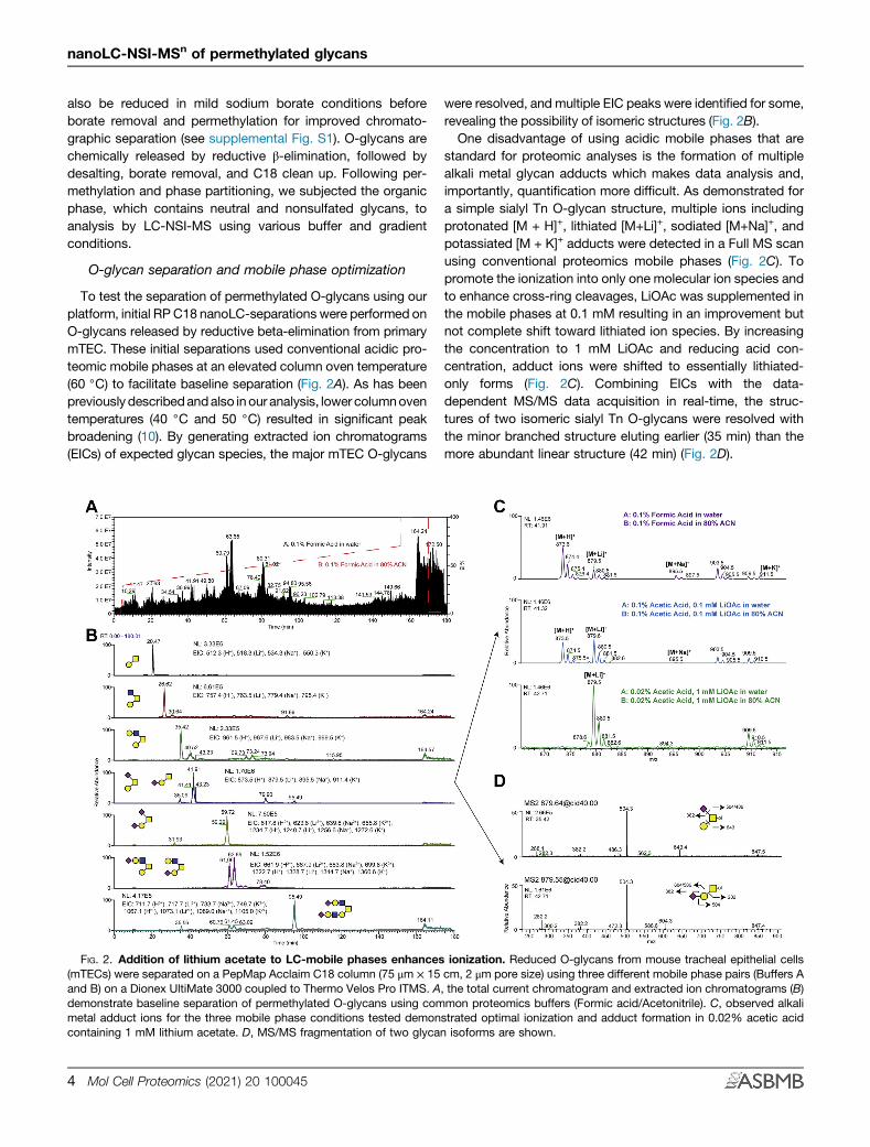
To test the separation of permethylated O-glycans using ourplatform, initial RP C18 nanoLC-separations were performed onO-glycans released by reductive beta-elimination from primarymTEC. These initial separations used conventional acidic pro-teomic mobile phases at an elevated column oven temperature(60 ◦C) to facilitate baseline separation (Fig. 2A). As has beenpreviously describedandalso inour analysis, lower columnoventemperatures (40 ◦C and 50 ◦C) resulted in significant peakbroadening (10). By generating extracted ion chromatograms(EICs) of expected glycan species, the major mTEC O-glycans
were resolved, and multiple EIC peaks were identified for some,revealing the possibility of isomeric structures (Fig. 2B).One disadvantage of using acidic mobile phases that are
standard for proteomic analyses is the formation of multiplealkali metal glycan adducts which makes data analysis and,importantly, quantification more difficult. As demonstrated fora simple sialyl Tn O-glycan structure, multiple ions includingprotonated [M + H]+, lithiated [M+Li]+, sodiated [M+Na]+, andpotassiated [M + K]+ adducts were detected in a Full MS scanusing conventional proteomics mobile phases (Fig. 2C). Topromote the ionization into only one molecular ion species andto enhance cross-ring cleavages, LiOAc was supplemented inthe mobile phases at 0.1 mM resulting in an improvement butnot complete shift toward lithiated ion species. By increasingthe concentration to 1 mM LiOAc and reducing acid con-centration, adduct ions were shifted to essentially lithiated-only forms (Fig. 2C). Combining EICs with the data-dependent MS/MS data acquisition in real-time, the struc-tures of two isomeric sialyl Tn O-glycans were resolved withthe minor branched structure eluting earlier (35 min) than themore abundant linear structure (42 min) (Fig. 2D).
FIG. 2. Addition of lithium acetate to LC-mobile phases enhances ionization. Reduced O-glycans from mouse tracheal epithelial cells(mTECs) were separated on a PepMap Acclaim C18 column (75 μm × 15 cm, 2 μm pore size) using three different mobile phase pairs (Buffers Aand B) on a Dionex UltiMate 3000 coupled to Thermo Velos Pro ITMS. A, the total current chromatogram and extracted ion chromatograms (B)demonstrate baseline separation of permethylated O-glycans using common proteomics buffers (Formic acid/Acetonitrile). C, observed alkalimetal adduct ions for the three mobile phase conditions tested demonstrated optimal ionization and adduct formation in 0.02% acetic acidcontaining 1 mM lithium acetate. D, MS/MS fragmentation of two glycan isoforms are shown.
nanoLC-NSI-MSn of permethylated glycans
4 Mol Cell Proteomics (2021) 20 100045
N-glycan separation and gradient optimization
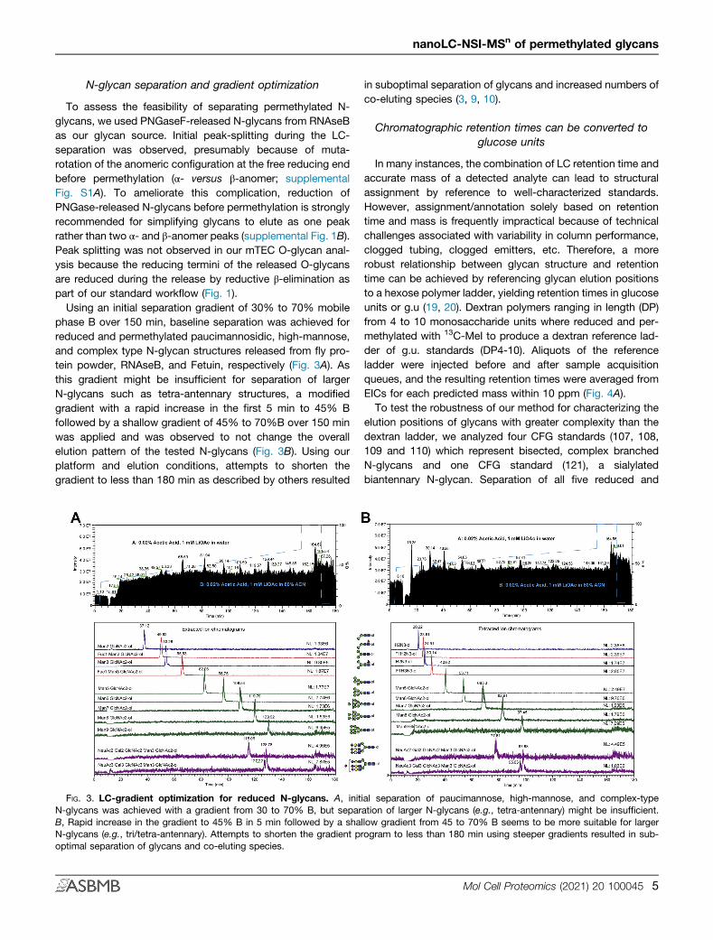
To assess the feasibility of separating permethylated N-glycans, we used PNGaseF-released N-glycans from RNAseBas our glycan source. Initial peak-splitting during the LC-separation was observed, presumably because of muta-rotation of the anomeric configuration at the free reducing endbefore permethylation (α- versus β-anomer; supplementalFig. S1A). To ameliorate this complication, reduction ofPNGase-released N-glycans before permethylation is stronglyrecommended for simplifying glycans to elute as one peakrather than two α- and β-anomer peaks (supplemental Fig. 1B).Peak splitting was not observed in our mTEC O-glycan anal-ysis because the reducing termini of the released O-glycansare reduced during the release by reductive β-elimination aspart of our standard workflow (Fig. 1).Using an initial separation gradient of 30% to 70% mobile
phase B over 150 min, baseline separation was achieved forreduced and permethylated paucimannosidic, high-mannose,and complex type N-glycan structures released from fly pro-tein powder, RNAseB, and Fetuin, respectively (Fig. 3A). Asthis gradient might be insufficient for separation of largerN-glycans such as tetra-antennary structures, a modifiedgradient with a rapid increase in the first 5 min to 45% Bfollowed by a shallow gradient of 45% to 70%B over 150 minwas applied and was observed to not change the overallelution pattern of the tested N-glycans (Fig. 3B). Using ourplatform and elution conditions, attempts to shorten thegradient to less than 180 min as described by others resulted
in suboptimal separation of glycans and increased numbers ofco-eluting species (3, 9, 10).
Chromatographic retention times can be converted toglucose units
In many instances, the combination of LC retention time andaccurate mass of a detected analyte can lead to structuralassignment by reference to well-characterized standards.However, assignment/annotation solely based on retentiontime and mass is frequently impractical because of technicalchallenges associated with variability in column performance,clogged tubing, clogged emitters, etc. Therefore, a morerobust relationship between glycan structure and retentiontime can be achieved by referencing glycan elution positionsto a hexose polymer ladder, yielding retention times in glucoseunits or g.u (19, 20). Dextran polymers ranging in length (DP)from 4 to 10 monosaccharide units where reduced and per-methylated with 13C-MeI to produce a dextran reference lad-der of g.u. standards (DP4-10). Aliquots of the referenceladder were injected before and after sample acquisitionqueues, and the resulting retention times were averaged fromEICs for each predicted mass within 10 ppm (Fig. 4A).To test the robustness of our method for characterizing the
elution positions of glycans with greater complexity than thedextran ladder, we analyzed four CFG standards (107, 108,109 and 110) which represent bisected, complex branchedN-glycans and one CFG standard (121), a sialylatedbiantennary N-glycan. Separation of all five reduced and
FIG. 3. LC-gradient optimization for reduced N-glycans. A, initial separation of paucimannose, high-mannose, and complex-typeN-glycans was achieved with a gradient from 30 to 70% B, but separation of larger N-glycans (e.g., tetra-antennary) might be insufficient.B, Rapid increase in the gradient to 45% B in 5 min followed by a shallow gradient from 45 to 70% B seems to be more suitable for largerN-glycans (e.g., tri/tetra-antennary). Attempts to shorten the gradient program to less than 180 min using steeper gradients resulted in sub-optimal separation of glycans and co-eluting species.
nanoLC-NSI-MSn of permethylated glycans
Mol Cell Proteomics (2021) 20 100045 5

FIG. 4. Elution of permethylated gly-cans expressed in terms of glucoseunits. A, reduced and 13C-permethylatedDextrans (isomaltooligosaccharide se-ries, DP4 to 10) were separated using aPepMap Acclaim C18 column in 1 mMlithium acetate-containing mobile phases(45–70% B over 150 min). Extracted ionchromatogram for each predicted massis shown (within 10 ppm). Retention timesof DP standards, run both before andafter sample acquisition queue, wereaveraged and plotted using a linearregression fitting. Error bars representstandard deviation (See inset).B, reduced and 13C-permethylated CFGN-Glycan standards (107–110, 121) wereanalyzed as described in (A). C, summarytable of analyzed CFG standards withretention times converted to reversedphase glucose units (RP g.u.) using linearregression fitting shown in A, inset. CFG,Consortium for Functional Glycomics;DP, degree of polymeriation.
nanoLC-NSI-MSn of permethylated glycans
6 Mol Cell Proteomics (2021) 20 100045
13C-permethylated CFG standards was achieved resulting instandard 107 (bisected, biantennary N-glycan) eluting beforestandard 108 (bisected, triantennary N-glycan with twoGlcNAc residues attached to the core α1,3 mannose). Inter-estingly, a gap of 6 min in retention time was detected be-tween the elution of the isomeric structure of standard 108,which is standard 109 (bisected, triantennary N-glycan withtwo GlcNAc residues attached to the core α1,6 mannose)demonstrating the power of our modified gradient to achieveseparations based on size and hydrophobicity (Fig. 4B). CFGstandard 110 (bisected, tetra-antennary N-glycan) elutedapproximately 2.6 min after standard 109 and before the finalsialylated, biantennary N-glycan standard 121. Using thelinear regression fitting from the dextran ladder runs beforeand after the sample queue, the observed retention times wereconverted to reverse phase glucose units (RP g.u.) to expressthe elution positions of the analyzed CFG standards (Fig. 4C).Dextran ladder was analyzed before a large sample acquisitionqueue and subsequently after 25 days of continuous opera-tion (supplemental Fig. S2). Retention times of isomaltosepolymers after nearly 1 month of continuous use wereconsistent within 2 to 3%. Furthermore, retention times wereconsistent regardless of amount injected, in a dynamic rangeof 15.6 to 1000 fmol of injected standard (supplementalFig. S3).Similar to the N-glycan standards, we tested the ability of
our method to separate chemically synthesized, reduced, and13C-permethylated O-glycan isomeric standards derived fromthe human pathogen Trypanosoma cruzi, the causative agentfor Chagas disease in South America (21, 22). The four tet-rasaccharide O-glycan isomers analyzed only differ by eitherthe cyclic form of a single monosaccharide (pyranose orfuranose) or a single linkage. Owing to the very small differ-ences in isomeric confirmation between these four mono-saccharides, a modified gradient was applied (35–40% B over170 min) (Fig. 5). The four standards were analyzed separately(Fig. 5, A–D) or as a mixture (Fig. 5E) and demonstrated nearlybaseline separation using the modified gradient program. Ofthe four isomeric O-glycan structures, the pyranose structureseluted over 20 min earlier than furanose-containing isoforms.As described previously, the observed retention times wereconverted to RP g.u. using the linear regression fitting fromdextran ladder runs before and after the sample queue usingthe same gradient program (Fig. 5F).
Characterization of N-glycans released from recombinantHIV gp120 expressed in glyco-engineered P. pastoris
The data described above demonstrates the ability of ourmethod to resolve N- and O-glycan structures derived fromabundant glycoproteins or prepared as synthetic standards.To assess the applicability of our method for detecting bio-logically relevant glycosylation patterns, we analyzed N-gly-cans released from recombinantly expressed HIV gp120envelope glycoproteins harvested from six different glyco-
engineered strains of P. pastoris (13, 14). Each of thesestrains lack glycosyltransferase activities that result in thegeneration of restricted cellular N-glycan profiles predicted tobe to predominated by specific high-mannose or complextype glycans. After release, reduction, and permethylation, N-glycans from each gp120 preparation were first separated andanalyzed individually, demonstrating that the detected N-gly-cans are consistent with the targeted glycosyltransferases ineach glyco-engineered strain (Fig. 6, A–F). For example, N-glycans of the composition Hex3HexNAc2-ol were onlydetected in the strain engineered to generate Man3GlcNAc2structures on gp120 glycoprotein (Fig. 6A) and Hex5HexNAc2-ol in the Man5GlcNAc2 engineered strain (Fig. 6B). A similarpattern was observed in engineered strains producing onlyMan8GlcNAc2 and Man9GlcNAc2 as well as Hex8HexNAc2-Hex10HexNAc2 structures (Fig. 6, C and D). Pichia strainsengineered to synthesize N-glycan structures with terminalgalactose residues or terminal sialic acid modifications alsoexhibited Man5GlcNAc2 structures in addition to the expectedcomplex type structures (Fig. 6, E and F). To assess the ca-pacity of our LC-MS system to resolve the range of glycansreleased from these expression platforms, we mixed equalamounts of each sample and analyzed as described above.We were able to accurately detect and resolve all of thevarious N-glycan structures derived from the recombinantgp120 glycoforms that were detected when each wereanalyzed individually (Fig. 6G). Furthermore, we applied anautomated MSn workflow which targeted the neutral loss ofpermethylated sialic acid (Δm/z = 375) to trigger productdependent MS3-5 acquisitions. Because lithium was includedin the LC separation mobile phases, this workflow revealedthat the terminal sialic acids of the N-glycan structuresreleased from these gp120 preparations are α2,6-linked(Fig. 6, H and I).
Analysis of O-glycan isomers from biological samples
O-linked glycans linked to Ser/Thr residues can be dividedinto various subtypes depending on the initiating mono-saccharide linked directly to the polypeptide backbone. Themost abundant class is the mucin-type O-GalNAc-initiatedstructures. Other types of O-glycans include O-Mannose,O-Fucose, O-Glucose, O-GlcNAc, O-Xylose, all of which havediverse biological functions (23, 24). Previous studies havedocumented the detection of both O-GalNAc andO-Mannose-initiated disaccharides and Galβ3GalNAc andGlcNAcβ2Man, respectively, in mouse brain (12, 25). In directinfusion MS, these glycans are detected at the same m/zbecause they have the same Hex1HexNAc1 composition.While MS/MS fragmentation of the permethylated di-saccharides can detect the presence of these two glycans, thefragmentation patterns of the isolated ions can be compli-cated as they are a mix of the two species and quantificationof each individual disaccharide is problematic. To identify thepresence or absence of Hex1HexNAc1 isomers in mouse
nanoLC-NSI-MSn of permethylated glycans
Mol Cell Proteomics (2021) 20 100045 7
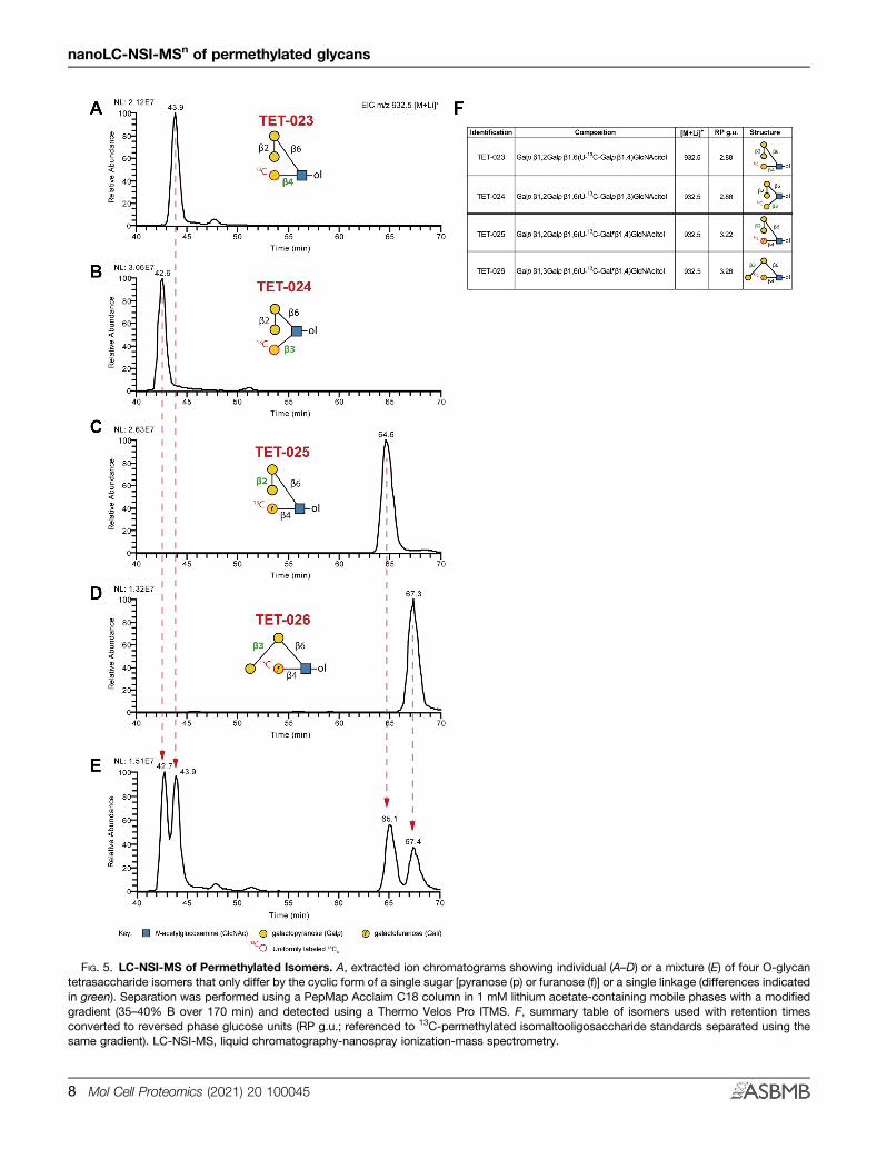
FIG. 5. LC-NSI-MS of Permethylated Isomers. A, extracted ion chromatograms showing individual (A–D) or a mixture (E) of four O-glycantetrasaccharide isomers that only differ by the cyclic form of a single sugar [pyranose (p) or furanose (f)] or a single linkage (differences indicatedin green). Separation was performed using a PepMap Acclaim C18 column in 1 mM lithium acetate-containing mobile phases with a modifiedgradient (35–40% B over 170 min) and detected using a Thermo Velos Pro ITMS. F, summary table of isomers used with retention timesconverted to reversed phase glucose units (RP g.u.; referenced to 13C-permethylated isomaltooligosaccharide standards separated using thesame gradient). LC-NSI-MS, liquid chromatography-nanospray ionization-mass spectrometry.
nanoLC-NSI-MSn of permethylated glycans
8 Mol Cell Proteomics (2021) 20 100045

brain, we analyzed the released and permethylated glycans byour LC-MS workflow (Fig. 7A). An EIC of the expected singlycharged, lithiated structures (m/z 518.3) revealed two peaks ofdiffering relative abundance at 17.8 and 19.2 min. MS2 frag-mentation of m/z 518 at the respective times is consistent withthe HexNAc-initiated disaccharide (O-GalNAc) eluting earlier
than the Hexose-initiated structure (O-mannose), as deter-mined by unique fragments at the reducing terminus (Fig. 7A,insets).We also analyzed O-glycans harvested from cells that were
glyco-engineered to be deficient in the O-Mannosylationpathway implicated in congenital muscular dystrophies
FIG. 6. LC-NSI-MSn analysis of N-glycans released from recombinant gp120 expressed in Pichia pastoris strains defective inN-glycosylation machinery. A, extracted ion chromatograms of all predicted PNGaseF-released, reduced, and permethylated N-glycans fromvarious gp120 expressing cell lines, either individually (A–F) or as a mixture (G). H, automated, intelligent MSn defines sialic acid linkage positionin realtime from Structure #10. I, summary table of gp120 N-glycans with retention times converted to reversed phase glucose units (RP g.u.;referenced to 13C-permethylated isomaltooligosaccharide standards separated using the same gradient). LC-NSI, liquid chromatography-nanospray ionization; MSn, multidimensional ion fragmentation.
nanoLC-NSI-MSn of permethylated glycans
Mol Cell Proteomics (2021) 20 100045 9

(26–28). The glycosyltransferase POMGNT1 (Protein O-LinkedMannose N-Acetyl-glucosaminyltransferase 1) transfers β2-linked GlcNAc to an underlying α-mannose linked to Ser or
Thr to generate Core M1-type glycans which can be furtherextended by B4GALT1 with β4-linked galactose (29). O-gly-cans were released from glycoproteins harvested from
FIG. 7. Analysis of reduced and permethylated O-glycans released from mouse brain proteins and human cell lines defective in theO-mannosylation pathway. A, extracted ion chromatogram of Hex1HexNAc1 structures released from mouse brain proteins indicates twostructural isomers. MS2 fragmentation of m/z 518.3 at the indicated retention times are shown as insets. B, LC-MS analysis of O-glycansreleased from HEK293 WT and ΔPOMGNT1 mutant cell lines. Respective EICs and annotated MS2 spectra are shown. EICs, extracted ionchromatograms.
nanoLC-NSI-MSn of permethylated glycans
10 Mol Cell Proteomics (2021) 20 100045

HEK293 wildtype cells or HEK293 cells harboring a Crispr-Cas9-mediated global disruption of the POMGNT1 locus (30).As a control, a non-O-Mannose structure (Xylα3Xylα3Glc-ol),which modifies a consensus found in the EGF repeats of arelatively small number of proteins (31–33), was detected (m/z593.3 [M+Li]+) in both samples (Fig. 7B). An EIC of m/z 722.1([M+Li]+) which corresponds to the composition Hex2HexNAc1(O-mannose Core M1 structure, Galβ4GlcNAcβ2Man-ol)demonstrates the presence of the glycan in HEK293 WT butnot in the POMGNT1-deficient cell line.
DISCUSSION
The field of glycomics has historically been eager to engagenew analytic platforms based on their promise to delivergreater sensitivity and enhanced detection of structural fea-tures (34–37). Despite tremendous advances in various ana-lytic technologies, it remains true that no single technique,besides NMR, is capable of routinely and completely definingglycan structures. Thus, the field will continue to benefit fromthe development of an expanding array of analytic ap-proaches. We have combined two powerful technologies,glycan permethylation and LC, in a manner that should beadoptable by analytic laboratories familiar with standard LC-MS/MS proteomic techniques. To facilitate adoption of ournanoLC-NSI-MSn approach, we have optimized run condi-tions and buffer compositions for analysis of N-linked orO-linked glycans. Using these conditions, we achievedbaseline separations of challenging isomeric glycans and alsodemonstrated the power of on-the-fly MSn to distinguishstructural features such as the linkage position of sialic acids.Other investigators have previously described LC-based
separations of glycans and, more particularly, of per-methylated glycans (3, 9, 10). These approaches are all valu-able contributions to the arsenal of glycomic technology. Inmany cases, these techniques make use of glycan standardsto define the retention times of known structures, generating adatabase of RT values that, when combined with accuratemass detection, can provide confident identification of com-ponents in complex mixtures. Glycans with novel RT valuescan be at least partially characterized using these methods byapplying off-line exoglycosidase digestions (sequencing) andre-analysis (37). Our approach is also amenable to off-lineexoglycosidase digestion with re-analysis, takes advantageof RT and accurate mass, and will, over-time build a library ofRP g.u. values to help define structural features of novel gly-cans. The resolving power of our method is most clearlydemonstrated by its ability to generate near-baseline separa-tion of four tetrasaccharides that share the same mono-saccharide composition and topology but differ only in asingle linkage position (see Fig. 5). To this resolving power, weadd the capability of capturing MSn data on-the-fly to helpassign structures with novel RT values in a single analytic run(see Figs. 6 and 7). The inclusion of lithium in the mobile phase
enhances this capability by facilitating the generation of cross-ring fragments indicative of specific structural features (5).We optimized the lithium concentration in the mobile phase
so that we detect glycans as a single adduct species. Thissimplification allows quantification to be done from peak areasdetected in MS1 for a single adduct, enhancing sensitivity andresolution of glycan species across the chromatogram. Asmentioned above, our method incorporates the inclusion ofRT standards (DP4-10) which allows the calculation of relativeretention times in Glc units (RP g.u.), facilitating cross-platformand run-to-run identification of glycans of interest. We havepreviously demonstrated that permethylated glycans yieldnearly the same signal intensity regardless of their mass orcomplexity, unless modified with sulfate or phosphate (4).Thus, known amounts of DP standards can not only serve asRP g.u. references but may facilitate quantification of molaramounts of analyte. However, additional work is required todetermine whether this approach can be validated as semi-quantitative or can provide absolute quantitation acrossvarious classes of glycans. A current limitation of ourapproach is the need for well-defined glycan standards to fullycharacterize complex mixtures of unknown structures. Ideally,a standard would be available for every glycan in the analyteand, over time, extensive tables of RP g.u. values would begenerated to facilitate rapid identification of components ofinterest. Confidence in the usefulness of RP g.u. tablespopulated by our approach will be heightened by the MSn datathat are generated to support structural features of standardsor mixtures of standards.Previously published glycomic studies from our laboratories
and many others, more than can be reasonably cited, haverelied heavily on direct infusion of permethylated glycans.These experiments, while valuable for in-depth structuralanalysis, require the presence of the operator for sample load,run, and subsequent wash before load of next sample. ThenanoLC-NSI-MSn method we describe here, like other LC-based methods, is amenable to autosampling, greatlyincreasing analytic throughput. Furthermore, data collection isalmost entirely automated compared with more labor-intensivedirect infusion experiments. This marked benefit also highlightsa bottleneck in all glycomic analysis, but especially for LC-based analysis, namely, facile analysis of large data sets.Software platforms capable of handling and annotating largeglycan data sets generated by nanoLC-NSI-MSn approachesare only in their infancy (38). We made use of our in-housedeveloped GRITS Toolbox to sieve through the data byproviding structural candidates based on MS/MS data (18).However, neither this platformnor anyother is currently capableof integrating MSn data for permethylated glycans across anentire LC run. Furthermore, software packages developed forLC-based proteomic applications are not ideal for interpretingLC of permethylated glycans because the underlying as-sumptions regarding isotopic distributions of amino acids arenot transferable to permethylated monosaccharides.
nanoLC-NSI-MSn of permethylated glycans
Mol Cell Proteomics (2021) 20 100045 11

Fully automated annotation packages for handling per-methylated glycan LC-separation data are a significant need forexpanding the adoption of this powerful approach for glyco-mics. Here, we have provided a useful and robust separationplatform that will facilitate the generation of data essential fordeveloping and testing such tools. In the meantime, as thesenew software tools are developed and implemented, theexpanded ability of our nanoLC-NSI-MSn platform to resolveand quantify glycan isomers, combined with the capacity forhigh-throughput analysis, will provide new opportunities forinvestigating the diversity of glycosylation at scales capable ofanswering biologically important questions.
DATA AVAILABILITY
Raw mass spectrometry data files have been deposited atGlycoPOST (https://glycopost.glycosmos.org/) under theaccession ID number GPST000123.
Acknowledgments—We wish to gratefully acknowledgeBrent Weatherly for his graciously given advice, encourage-ment, and assistance regarding computational analysis andinformatic considerations over the course of this project andothers, before his untimely death. We also thank Rob Bridgerfor his technical support of our mass spectrometry instru-mentation, Dr Christopher M. West (University of Georgia) forproviding the T. cruzi O-Glycan isomeric standards and DrHenrik Clausen (University of Copenhagen) for providingHEK293 cell lines. We are grateful for technical advice fromDrs. Christa Feasley, Julian Saba, and Sergei Snovida (Ther-moFisher Scientific). Four N-linked glycan standards (stan-dards 107–110) were received from the Consortium forFunctional Glycomics (CFG), and an additional N-linkedglycan standard (standard 121) was received as a gift from DrJames Paulson (The Scripps Research Institute). The instru-mentation utilized in this analysis was provided by Thermo-Fisher in partial support of the ThermoFisher Center ofExcellence for Glycomics and Glycoproteomics at the CCRC,Lance Wells and Michael Tiemeyer, Co-Directors.
Funding and additional information—This work was sup-ported in part by grants from National Institute of Health/Na-tional Institute of General Medical Sciences (R01GM111939 toL. W., P41GM103490 to M. T. and L. W., R21AI123161 to L.W., and R01GM130915 to L. W.) and National Institutes ofHealth/National Institute of Allergy and Infectious Disease(R21AI123161 to L. W.). The content is solely the responsibilityof the authors and does not necessarily represent the officialviews of the National Institutes of Health.
Author contributions—S. K., M. O. S, M. T., and L. W.designed research; S. K. and M. O. S performed research; S.L. contributed key reagents; S. K., M. O. S, M. T., and L. W.analyzed data; S. K., M. O. S, M. T., and L. W. wrote the paper.
Conflict of interest—The authors declare no competinginterests.
Abbreviations—The abbreviations used are: ACN, acetoni-trile; AcOH, acetic acid; CFG, Consortium for FunctionalGlycomics; DP, degree of polymerization; EIC, extracted ionchromatogram; ITMS, ion trap mass spectrometer; LiOAc,lithium acetate; NaOH, sodium hydroxide; MeI, methyl iodide;mTEC, mouse tracheal epithelial cell; nanoLC-NSI-MS,nanoflow Liquid Chromatography-Nanospray Ionization-MassSpectrometry; MSn, multidimensional ion fragmentation; RP,reverse phase.
Received August 3, 2020, and in revised from, December 21, 2020Published, MCPRO Papers in Press, January 26, 2021, https://doi.org/10.1074/mcp.RA120.002266
REFERENCES
1. Werz, D. B., Ranzinger, R., Herget, S., Adibekian, A., von der Lieth, C. W.,and Seeberger, P. H. (2007) Exploring the structural diversity ofmammalian carbohydrates ("glycospace") by statistical databank anal-ysis. ACS Chem. Biol. 2, 685–691
2. Marino, K., Bones, J., Kattla, J. J., and Rudd, P. M. (2010) A systematicapproach to protein glycosylation analysis: A path through the maze. Nat.Chem. Biol. 6, 713–723
3. Vreeker, G. C., and Wuhrer, M. (2017) Reversed-phase separation methodsfor glycan analysis. Anal. Bioanal. Chem. 409, 359–378
4. Mehta, N., Porterfield, M., Struwe, W. B., Heiss, C., Azadi, P., Rudd, P. M.,Tiemeyer, M., and Aoki, K. (2016) Mass spectrometric quantification of N-linked glycans by reference to exogenous standards. J. Proteome Res.15, 2969–2980
5. Ashline, D., Singh, S., Hanneman, A., and Reinhold, V. (2005) Congruentstrategies for carbohydrate sequencing. 1. Mining structural details byMSn. Anal. Chem. 77, 6250–6262
6. Lapadula, A. J., Hatcher, P. J., Hanneman, A. J., Ashline, D. J., Zhang, H.,and Reinhold, V. N. (2005) Congruent strategies for carbohydratesequencing. 3. OSCAR: An algorithm for assigning oligosaccharide to-pology from MSn data. Anal. Chem. 77, 6271–6279
7. Reinhold, V., Zhang, H., Hanneman, A., and Ashline, D. (2013) Toward aplatform for comprehensive glycan sequencing. Mol. Cell Proteomics 12,866–873
8. Zhang, H., Singh, S., and Reinhold, V. N. (2005) Congruent strategies forcarbohydrate sequencing. 2. FragLib: An MSn spectral library. Anal.Chem. 77, 6263–6270
9. Hsiao, C. T., Wang, P. W., Chang, H. C., Chen, Y. Y., Wang, S. H., Chern,Y., and Khoo, K. H. (2017) Advancing a high throughput glycotope-centric glycomics workflow based on nanoLC-MS(2)-product depen-dent-MS(3) analysis of permethylated glycans. Mol. Cell Proteomics 16,2268–2280
10. Zhou, S., Hu, Y., and Mechref, Y. (2016) High-temperature LC-MS/MS ofpermethylated glycans derived from glycoproteins. Electrophoresis 37,1506–1513
11. Aoki, K., Perlman, M., Lim, J. M., Cantu, R., Wells, L., and Tiemeyer, M.(2007) Dynamic developmental elaboration of N-linked glycan complexityin the Drosophila melanogaster embryo. J. Biol. Chem. 282, 9127–9142
12. Stalnaker, S. H., Aoki, K., Lim, J. M., Porterfield, M., Liu, M., Satz, J. S.,Buskirk, S., Xiong, Y., Zhang, P., Campbell, K. P., Hu, H., Live, D., Tie-meyer, M., and Wells, L. (2011) Glycomic analyses of mouse models ofcongenital muscular dystrophy. J. Biol. Chem. 286, 21180–21190
13. Hamilton, S. R., Davidson, R. C., Sethuraman, N., Nett, J. H., Jiang, Y.,Rios, S., Bobrowicz, P., Stadheim, T. A., Li, H., Choi, B. K., Hopkins, D.,Wischnewski, H., Roser, J., Mitchell, T., Strawbridge, R. R., et al. (2006)Humanization of yeast to produce complex terminally sialylated glyco-proteins. Science 313, 1441–1443
14. Hamilton, S. R., and Gerngross, T. U. (2007) Glycosylation engineering inyeast: The advent of fully humanized yeast. Curr. Opin. Biotechnol. 18,387–392
nanoLC-NSI-MSn of permethylated glycans
12 Mol Cell Proteomics (2021) 20 100045
15. You, Y., Richer, E. J., Huang, T., and Brody, S. L. (2002) Growth and dif-ferentiation of mouse tracheal epithelial cells: Selection of a proliferativepopulation. Am. J. Physiol. Lung Cell Mol. Physiol 283, L1315–1321
16. Aoki, K., Porterfield, M., Lee, S. S., Dong, B., Nguyen, K., McGlamry, K. H.,and Tiemeyer, M. (2008) The diversity of O-linked glycans expressedduring Drosophila melanogaster development reflects stage- and tissue-specific requirements for cell signaling. J. Biol. Chem. 283, 30385–30400
17. Anumula, K. R., and Taylor, P. B. (1992) A comprehensive procedure forpreparation of partially methylated alditol acetates from glycoproteincarbohydrates. Anal. Biochem. 203, 101–108
18. Weatherly, D. B., Arpinar, F. S., Porterfield, M., Tiemeyer, M., York, W. S.,and Ranzinger, R. (2019) GRITS Toolbox-a freely available software forprocessing, annotating and archiving glycomics mass spectrometry data.Glycobiology 29, 452–460
19. Lee, Y. C., Lee, B. I., Tomiya, N., and Takahashi, N. (1990) Parameterizationof contribution of sugar units to elution volumes in reverse-phase HPLCof 2-pyridylaminated oligosaccharides. Anal. Biochem. 188, 259–266
20. Takahashi, N., Wada, Y., Awaya, J., Kurono, M., and Tomiya, N. (1993) Two-dimensional elution map of GalNAc-containing N-linked oligosaccha-rides. Anal. Biochem. 208, 96–109
21. Sheikh, M. O., Gas-Pascual, E., Glushka, J. N., Bustamante, J. M., Wells, L.,and West, C. M. (2019) Trypanosoma cruzi 13C-labeled O-Glycan stan-dards for mass spectrometry. Glycobiology 29, 280–284
22. Tarleton, R. L. (2016) Chagas disease: A solvable problem, ignored. TrendsMol. Med. 22, 835–838
23. Joshi, H. J., Narimatsu, Y., Schjoldager, K. T., Tytgat, H. L. P., Aebi, M.,Clausen, H., and Halim, A. (2018) SnapShot: O-glycosylation pathwaysacross kingdoms. Cell 172, 632–632.e632
24. Varki, A., and Kornfeld, S. (2015) Historical background and overview. In:Varki, A., Cummings, R. D., Esko, J. D., Stanley, P., Hart, G. W., Aebi, M.,Darvill, A. G., Kinoshita, T., Packer, N. H., Prestegard, J. H., Schnaar, R.L., Seeberger, P. H., eds. Essentials of Glycobiology, Cold Spring HarborLaboratory Press, Cold Spring Harbor, NY: 1–18
25. Stalnaker, S. H., Hashmi, S., Lim, J. M., Aoki, K., Porterfield, M., Gutierrez-Sanchez, G., Wheeler, J., Ervasti, J. M., Bergmann, C., Tiemeyer, M., andWells, L. (2010) Site mapping and characterization of O-glycan structureson alpha-dystroglycan isolated from rabbit skeletal muscle. J. Biol. Chem.285, 24882–24891
26. Falsaperla, R., Pratico, A. D., Ruggieri, M., Parano, E., Rizzo, R., Corsello,G., Vitaliti, G., and Pavone, P. (2016) Congenital muscular dystrophy:From muscle to brain. Ital. J. Pediatr. 42, 78
27. Liu, J., Ball, S. L., Yang, Y., Mei, P., Zhang, L., Shi, H., Kaminski, H. J.,Lemmon, V. P., and Hu, H. (2006) A genetic model for muscle-eye-braindisease in mice lacking protein O-mannose 1,2-N-acetylglucosaminyl-transferase (POMGnT1). Mech. Dev. 123, 228–240
28. Sheikh, M. O., Halmo, S. M., and Wells, L. (2017) Recent advancements inunderstanding mammalian O-mannosylation. Glycobiology 27, 806–819
29. Praissman, J. L., and Wells, L. (2014) Mammalian O-mannosylationpathway: Glycan structures, enzymes, and protein substrates.Biochemistry 53, 3066–3078
30. Narimatsu, Y., Joshi, H. J., Nason, R., Van Coillie, J., Karlsson, R., Sun, L.,Ye, Z., Chen, Y. H., Schjoldager, K. T., Steentoft, C., Furukawa, S.,Bensing, B. A., Sullam, P. M., Thompson, A. J., Paulson, J. C., et al.(2019) An atlas of human glycosylation pathways enables display of thehuman glycome by gene engineered cells. Mol. Cell 75, 394–407.e395
31. Hase, S., Kawabata, S., Nishimura, H., Takeya, H., Sueyoshi, T., Miyata, T.,Iwanaga, S., Takao, T., Shimonishi, Y., and Ikenaka, T. (1988) A newtrisaccharide sugar chain linked to a serine residue in bovine blood-coagulation factor-Vii and factor-Ix. J. Biochem-tokyo 104, 867–868
32. Hase, S., Nishimura, H., Kawabata, S. I., Iwanaga, S., and Ikenaka, T. (1990)The structure of (Xylose)2glucose-O-serine 53 found in the 1st epidermalgrowth factor-like domain of bovine blood-clotting factor-Ix. J. Biol.Chem. 265, 1858–1861
33. Nishimura, H., Kawabata, S., Kisiel, W., Hase, S., Ikenaka, T., Takao, T.,Shimonishi, Y., and Iwanaga, S. (1989) Identification of a disaccharide(Xyl-Glc) and a trisaccharide (Xyl2-Glc) O-glycosidically linked to a serineresidue in the 1st epidermal growth factor-like domain of human factor-Viiand factor-Ix and protein-Z and bovine protein-Z. J. Biol. Chem. 264,20320–20325
34. Li, Q., Xie, Y., Wong, M., and Lebrilla, C. B. (2019) Characterization of cellglycocalyx with mass spectrometry methods. Cells 8
35. Mulloy, B., Dell, A., Stanley, P., and J, H. P. (2015) Structural analysis ofglycans. In: rd, Varki, A., Cummings, R. D., Esko, J. D., Stanley, P.,Hart, G. W., Aebi, M., Darvill, A. G., Kinoshita, T., Packer, N. H., Pre-stegard, J. H., Schnaar, R. L., Seeberger, P. H., eds. Essentials ofGlycobiology, Cold Spring Harbor Laboratory Press, Cold Spring Har-bor, NY: 639–652
36. Reiding, K. R., Bondt, A., Hennig, R., Gardner, R. A., O'Flaherty, R., Trbo-jevic-Akmacic, I., Shubhakar, A., Hazes, J. M. W., Reichl, U., Fernandes,D. L., Pucic-Bakovic, M., Rapp, E., Spencer, D. I. R., Dolhain, R., Rudd,P. M., et al. (2019) High-throughput serum N-glycomics: Method com-parison and application to study rheumatoid arthritis and pregnancy-associated changes. Mol. Cell Proteomics 18, 3–15
37. Rudd, P. M., Guile, G. R., Kuster, B., Harvey, D. J., Opdenakker, G., andDwek, R. A. (1997) Oligosaccharide sequencing technology. Nature 388,205–207
38. Abrahams, J. L., Campbell, M. P., and Packer, N. H. (2018) Building a PGC-LC-MS N-glycan retention library and elution mapping resource. Glyco-conj. J. 35, 15–29
39. Varki, A., Cummings, R. D., Aebi, M., Packer, N. H., Seeberger, P. H., Esko,J. D., Stanley, P., Hart, G., Darvill, A., Kinoshita, T., Prestegard, J. J.,Schnaar, R. L., Freeze, H. H., Marth, J. D., Bertozzi, C. R., et al. (2015)Symbol nomenclature for graphical representations of glycans. Glycobi-ology 25, 1323–1324
nanoLC-NSI-MSn of permethylated glycans
Mol Cell Proteomics (2021) 20 100045 13
Related Documents