Separable Bilayer Microfiltration Device for Viable Label-free Enrichment of Circulating Tumour Cells Ming-Da Zhou 1 *, Sijie Hao 1 *, Anthony J. Williams 2,3,5 *, Ramdane A. Harouaka 1 , Brett Schrand 3,4 , Siddarth Rawal 2,5 , Zheng Ao 2,3,5 , Randall Brennaman 3,4 , Eli Gilboa 4 , Bo Lu 6 , Shuwen Wang 7 , Jiyue Zhu 7 , Ram Datar 2,5 , Richard Cote 2,5 , Yu-Chong Tai 6 & Si-Yang Zheng 1 1 Micro & Nano Integrated Biosystem (MINIBio) Laboratory, Department of Biomedical Engineering and Materials Research Institute, Pennsylvania State University, University Park, PA 16802, U.S.A., 2 Department of Pathology, University of Miami – Miller School of Medicine, Miami, FL 33136, U.S.A., 3 Sheila and David Fuente Graduate Program in Cancer Biology, University of Miami – Miller School of Medicine, Miami, FL 33136, U.S.A., 4 Department of Microbiology and Immunology, University of Miami – Miller School of Medicine, Miami, FL 33136, U.S.A., 5 Dr John T Macdonald Foundation Biomedical Nanotechnology Institute, University of Miami – Miller School of Medicine, Miami, FL 33136, U.S.A., 6 Caltech Micromachining Laboratory, California Institute of Technology, MC 136-93, Pasadena, CA 91125, U.S.A., 7 Department of Pharmaceutical Sciences, Washington State University College of Pharmacy, Spokane, WA 99210, U.S.A. The analysis of circulating tumour cells (CTCs) in cancer patients could provide important information for therapeutic management. Enrichment of viable CTCs could permit performance of functional analyses on CTCs to broaden understanding of metastatic disease. However, this has not been widely accomplished. Addressing this challenge, we present a separable bilayer (SB) microfilter for viable size-based CTC capture. Unlike other single-layer CTC microfilters, the precise gap between the two layers and the architecture of pore alignment result in drastic reduction in mechanical stress on CTCs, capturing them viably. Using multiple cancer cell lines spiked in healthy donor blood, the SB microfilter demonstrated high capture efficiency (78–83%), high retention of cell viability (71–74%), high tumour cell enrichment against leukocytes (1.7–2 3 10 3 ), and widespread ability to establish cultures post-capture (100% of cell lines tested). In a metastatic mouse model, SB microfilters successfully enriched viable mouse CTCs from 0.4–0.6 mL whole mouse blood samples and established in vitro cultures for further genetic and functional analysis. Our preliminary studies reflect the efficacy of the SB microfilter device to efficiently and reliably enrich viable CTCs in animal model studies, constituting an exciting technology for new insights in cancer research. M etastatic disease represents the ability of solid tumour cells to intravasate from their site of origin, travel through the haematogenous and/or lymphatic circulatory systems and extravasate at distant secondary sites where new tumours are colonized. In contrast to early stage disease, treatment strategies in the metastatic setting are largely palliative rather than curable, where over 90% of cancer-related mortality can be attributed to disease outgrowth beyond the primary site 1 . To improve the survival rates among late stage and recurrent cancer patients, 1) enhanced diagnostic tools for earlier detection, 2) treatment monitoring strategies that detect the underlying cellular and molecular changes arising from selective pressure posed by the treatment process, and 3) therapies that better target the metastatic cells directly are in urgent need. Numerous studies suggest circulating tumour cells (CTCs), the tumour subpopulation responsible for invasion and colonization of distant sites, to be a candidate biomarker for prognosis, diagnosis and treatment monitoring of metastatic disease 2–5 . Assaying for CTCs requires only a simple, minimally invasive blood draw, providing a unique opportunity for repeated sampling in patients to monitor both metastatic disease as well as therapeutic response in real-time. Although promising for their diagnostic and prognostic potential, detecting and analysing CTCs is thwarted by their paucity, with only a few tumour cells occurring among billions of non-tumour cells in peripheral blood 4,6 . Thus highly efficient enrichment strategies are a prerequisite and a technical limitation for CTC analysis 7 . In recent years, a number of CTC enrichment systems have been developed 8,9 . Immunological approaches depend on cell surface antigen expression 10–12 . Epithelial cell surface markers expressed predominantly on CTCs, such as epithelial cell adhesion molecule (EpCAM), are widely used 13–23 . The CellSearchH system represents the OPEN SUBJECT AREAS: BIOMEDICAL ENGINEERING TUMOUR BIOMARKERS Received 17 June 2014 Accepted 20 November 2014 Published 9 December 2014 Correspondence and requests for materials should be addressed to Y.-C.T. (yctai@mems. caltech.edu) or S.-Y.Z. ([email protected]) * These authors contributed equally to this work. SCIENTIFIC REPORTS | 4 : 7392 | DOI: 10.1038/srep07392 1

Welcome message from author
This document is posted to help you gain knowledge. Please leave a comment to let me know what you think about it! Share it to your friends and learn new things together.
Transcript
Separable Bilayer Microfiltration Devicefor Viable Label-free Enrichment ofCirculating Tumour CellsMing-Da Zhou1*, Sijie Hao1*, Anthony J. Williams2,3,5*, Ramdane A. Harouaka1, Brett Schrand3,4,Siddarth Rawal2,5, Zheng Ao2,3,5, Randall Brennaman3,4, Eli Gilboa4, Bo Lu6, Shuwen Wang7, Jiyue Zhu7,Ram Datar2,5, Richard Cote2,5, Yu-Chong Tai6 & Si-Yang Zheng1
1Micro & Nano Integrated Biosystem (MINIBio) Laboratory, Department of Biomedical Engineering and Materials Research Institute,Pennsylvania State University, University Park, PA 16802, U.S.A., 2Department of Pathology, University of Miami – Miller School ofMedicine, Miami, FL 33136, U.S.A., 3Sheila and David Fuente Graduate Program in Cancer Biology, University of Miami – MillerSchool of Medicine, Miami, FL 33136, U.S.A., 4Department of Microbiology and Immunology, University of Miami – Miller Schoolof Medicine, Miami, FL 33136, U.S.A., 5Dr John T Macdonald Foundation Biomedical Nanotechnology Institute, University ofMiami – Miller School of Medicine, Miami, FL 33136, U.S.A., 6Caltech Micromachining Laboratory, California Institute ofTechnology, MC 136-93, Pasadena, CA 91125, U.S.A., 7Department of Pharmaceutical Sciences, Washington State UniversityCollege of Pharmacy, Spokane, WA 99210, U.S.A.
The analysis of circulating tumour cells (CTCs) in cancer patients could provide important information fortherapeutic management. Enrichment of viable CTCs could permit performance of functional analyses onCTCs to broaden understanding of metastatic disease. However, this has not been widely accomplished.Addressing this challenge, we present a separable bilayer (SB) microfilter for viable size-based CTC capture.Unlike other single-layer CTC microfilters, the precise gap between the two layers and the architecture ofpore alignment result in drastic reduction in mechanical stress on CTCs, capturing them viably. Usingmultiple cancer cell lines spiked in healthy donor blood, the SB microfilter demonstrated high captureefficiency (78–83%), high retention of cell viability (71–74%), high tumour cell enrichment againstleukocytes (1.7–2 3 103), and widespread ability to establish cultures post-capture (100% of cell lines tested).In a metastatic mouse model, SB microfilters successfully enriched viable mouse CTCs from 0.4–0.6 mLwhole mouse blood samples and established in vitro cultures for further genetic and functional analysis. Ourpreliminary studies reflect the efficacy of the SB microfilter device to efficiently and reliably enrich viableCTCs in animal model studies, constituting an exciting technology for new insights in cancer research.
Metastatic disease represents the ability of solid tumour cells to intravasate from their site of origin, travelthrough the haematogenous and/or lymphatic circulatory systems and extravasate at distant secondarysites where new tumours are colonized. In contrast to early stage disease, treatment strategies in the
metastatic setting are largely palliative rather than curable, where over 90% of cancer-related mortality can beattributed to disease outgrowth beyond the primary site1. To improve the survival rates among late stage andrecurrent cancer patients, 1) enhanced diagnostic tools for earlier detection, 2) treatment monitoring strategiesthat detect the underlying cellular and molecular changes arising from selective pressure posed by the treatmentprocess, and 3) therapies that better target the metastatic cells directly are in urgent need.
Numerous studies suggest circulating tumour cells (CTCs), the tumour subpopulation responsible for invasionand colonization of distant sites, to be a candidate biomarker for prognosis, diagnosis and treatment monitoringof metastatic disease2–5. Assaying for CTCs requires only a simple, minimally invasive blood draw, providing aunique opportunity for repeated sampling in patients to monitor both metastatic disease as well as therapeuticresponse in real-time. Although promising for their diagnostic and prognostic potential, detecting and analysingCTCs is thwarted by their paucity, with only a few tumour cells occurring among billions of non-tumour cells inperipheral blood4,6. Thus highly efficient enrichment strategies are a prerequisite and a technical limitation forCTC analysis7.
In recent years, a number of CTC enrichment systems have been developed8,9. Immunological approachesdepend on cell surface antigen expression10–12. Epithelial cell surface markers expressed predominantly on CTCs,such as epithelial cell adhesion molecule (EpCAM), are widely used13–23. The CellSearchH system represents the
OPEN
SUBJECT AREAS:BIOMEDICAL
ENGINEERING
TUMOUR BIOMARKERS
Received17 June 2014
Accepted20 November 2014
Published9 December 2014
Correspondence andrequests for materials
should be addressed toY.-C.T. (yctai@mems.
caltech.edu) or S.-Y.Z.([email protected])
* These authorscontributed equally to
this work.
SCIENTIFIC REPORTS | 4 : 7392 | DOI: 10.1038/srep07392 1
most prominently used platform by this approached, which enrichesCTCs from whole blood using an EpCAM-based immunomagneticseparation process24–26. Currently, it is the only FDA cleared clinicalCTC analysis system for metastatic breast, prostate and colorectalcancers. In addition to immunological approaches, unique physicalproperties of cancer cells have also been exploited for CTC enrich-ment, including cell size, deformability, electrical and acousticproperties18,27–31.
Beyond the enumeration and molecular characterization of CTCs,the development of the enrichment of viable CTCs enables theirfunctional characterization that are critical for disease interrogationand target therapy32–34. Viable CTCs in metastatic breast cancer havebeen enriched using multi-marker FACS technology, where in onestudy a metastasis-initiating subpopulation of primary luminalbreast cancer CTCs were used to generate a mouse xenograft modelthat gives rise to bone, lung and brain metastases29. In another study,a microfluidic device combined live CTC capture and ‘‘on-chip’’treatment with taxol drugs to demonstrate microtubule organizationalterations in CTCs35.
Our group has previously described the development of a 2Dround pore-shaped microfilter fabricated on a single 10 mm-thickparylene-C membrane by photolithography36. Pre-fixation of bloodsamples are needed to prevent cell lysis during filtration, whichmakes it unusable for viable CTC enrichment. To alleviate this lim-itation, we developed a 3D membrane microfilter, where blood sam-ples are processed for CTC enrichment with no prior fixation37.While we demonstrated an ability to capture viable tumour cells inmodel systems, trapping of cells inside of the pores restricts cellproliferation and cell release.
Herein, we report the new design of a separable bilayer (SB) micro-filter. The SB microfilter has fundamentally different device structureand filtration principles. These improvements have significantlyenhanced our ability to enrich and characterize viable CTCs. Weevaluated the performance of the SB microfilter device using bothin vitro and in vivo systems, where we demonstrate (1) high sensitiv-
ity and efficiency of viable tumour cell capture, (2) the ability toenrich tumour cells from multiple types of cancer cell lines for theirproliferation directly on the surface of the microfilter or on second-ary culture surfaces, (3) the ability to enrich viable CTCs in a breastcancer mouse model system for subsequent cell culture and func-tional characterization, and (4) the ability to enrich viable CTCs froma clinical blood sample. The SB microfilter is a new, high perform-ance viable CTC enrichment device with the potential to haveimportant utility in both research and clinical applications.
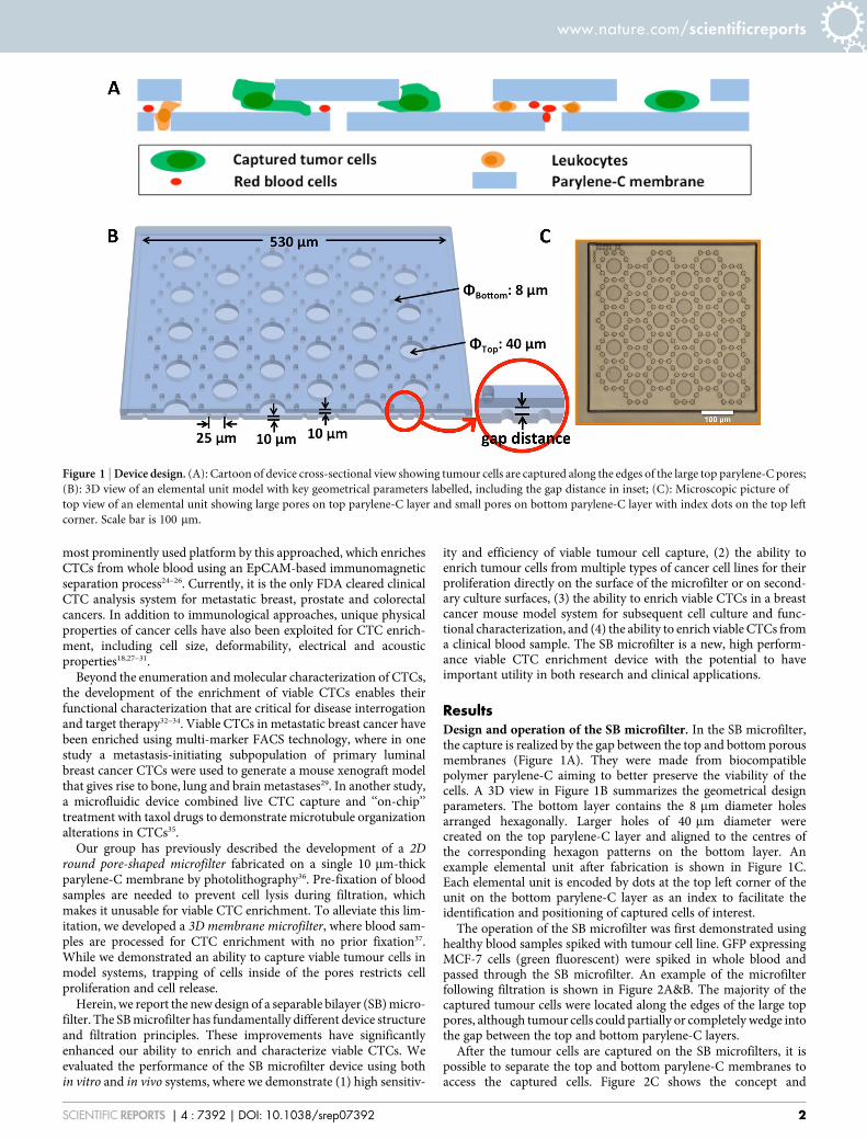
ResultsDesign and operation of the SB microfilter. In the SB microfilter,the capture is realized by the gap between the top and bottom porousmembranes (Figure 1A). They were made from biocompatiblepolymer parylene-C aiming to better preserve the viability of thecells. A 3D view in Figure 1B summarizes the geometrical designparameters. The bottom layer contains the 8 mm diameter holesarranged hexagonally. Larger holes of 40 mm diameter werecreated on the top parylene-C layer and aligned to the centres ofthe corresponding hexagon patterns on the bottom layer. Anexample elemental unit after fabrication is shown in Figure 1C.Each elemental unit is encoded by dots at the top left corner of theunit on the bottom parylene-C layer as an index to facilitate theidentification and positioning of captured cells of interest.
The operation of the SB microfilter was first demonstrated usinghealthy blood samples spiked with tumour cell line. GFP expressingMCF-7 cells (green fluorescent) were spiked in whole blood andpassed through the SB microfilter. An example of the microfilterfollowing filtration is shown in Figure 2A&B. The majority of thecaptured tumour cells were located along the edges of the large toppores, although tumour cells could partially or completely wedge intothe gap between the top and bottom parylene-C layers.
After the tumour cells are captured on the SB microfilters, it ispossible to separate the top and bottom parylene-C membranes toaccess the captured cells. Figure 2C shows the concept and
Figure 1 | Device design. (A): Cartoon of device cross-sectional view showing tumour cells are captured along the edges of the large top parylene-C pores;
(B): 3D view of an elemental unit model with key geometrical parameters labelled, including the gap distance in inset; (C): Microscopic picture of
top view of an elemental unit showing large pores on top parylene-C layer and small pores on bottom parylene-C layer with index dots on the top left
corner. Scale bar is 100 mm.
www.nature.com/scientificreports
SCIENTIFIC REPORTS | 4 : 7392 | DOI: 10.1038/srep07392 2
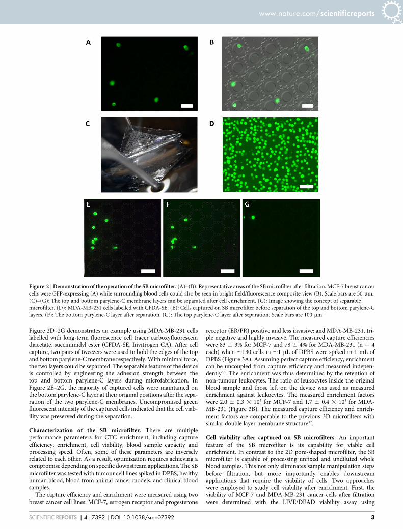
Figure 2D–2G demonstrates an example using MDA-MB-231 cellslabelled with long-term fluorescence cell tracer carboxyfluoresceindiacetate, succinimidyl ester (CFDA-SE, Invitrogen CA). After cellcapture, two pairs of tweezers were used to hold the edges of the topand bottom parylene-C membrane respectively. With minimal force,the two layers could be separated. The separable feature of the deviceis controlled by engineering the adhesion strength between thetop and bottom parylene-C layers during microfabrication. InFigure 2E–2G, the majority of captured cells were maintained onthe bottom parylene-C layer at their original positions after the sepa-ration of the two parylene-C membranes. Uncompromised greenfluorescent intensity of the captured cells indicated that the cell viab-ility was preserved during the separation.
Characterization of the SB microfilter. There are multipleperformance parameters for CTC enrichment, including captureefficiency, enrichment, cell viability, blood sample capacity andprocessing speed. Often, some of these parameters are inverselyrelated to each other. As a result, optimization requires achieving acompromise depending on specific downstream applications. The SBmicrofilter was tested with tumour cell lines spiked in DPBS, healthyhuman blood, blood from animal cancer models, and clinical bloodsamples.
The capture efficiency and enrichment were measured using twobreast cancer cell lines: MCF-7, estrogen receptor and progesterone
receptor (ER/PR) positive and less invasive; and MDA-MB-231, tri-ple negative and highly invasive. The measured capture efficiencieswere 83 6 3% for MCF-7 and 78 6 4% for MDA-MB-231 (n 5 4each) when ,130 cells in ,1 mL of DPBS were spiked in 1 mL ofDPBS (Figure 3A). Assuming perfect capture efficiency, enrichmentcan be uncoupled from capture efficiency and measured indepen-dently38. The enrichment was thus determined by the retention ofnon-tumour leukocytes. The ratio of leukocytes inside the originalblood sample and those left on the device was used as measuredenrichment against leukocytes. The measured enrichment factorswere 2.0 6 0.3 3 103 for MCF-7 and 1.7 6 0.4 3 103 for MDA-MB-231 (Figure 3B). The measured capture efficiency and enrich-ment factors are comparable to the previous 3D microfilters withsimilar double layer membrane structure37.
Cell viability after captured on SB microfilters. An importantfeature of the SB microfilter is its capability for viable cellenrichment. In contrast to the 2D pore-shaped microfilter, the SBmicrofilter is capable of processing unfixed and undiluted wholeblood samples. This not only eliminates sample manipulation stepsbefore filtration, but more importantly enables downstreamapplications that require the viability of cells. Two approacheswere employed to study cell viability after enrichment. First, theviability of MCF-7 and MDA-MB-231 cancer cells after filtrationwere determined with the LIVE/DEAD viability assay using
Figure 2 | Demonstration of the operation of the SB microfilter. (A)–(B): Representative areas of the SB microfilter after filtration. MCF-7 breast cancer
cells were GFP-expressing (A) while surrounding blood cells could also be seen in bright field/fluorescence composite view (B). Scale bars are 50 mm.
(C)–(G): The top and bottom parylene-C membrane layers can be separated after cell enrichment. (C): Image showing the concept of separable
microfilter. (D): MDA-MB-231 cells labelled with CFDA-SE. (E): Cells captured on SB microfilter before separation of the top and bottom parylene-C
layers. (F): The bottom parylene-C layer after separation. (G): The top parylene-C layer after separation. Scale bars are 100 mm.
www.nature.com/scientificreports
SCIENTIFIC REPORTS | 4 : 7392 | DOI: 10.1038/srep07392 3
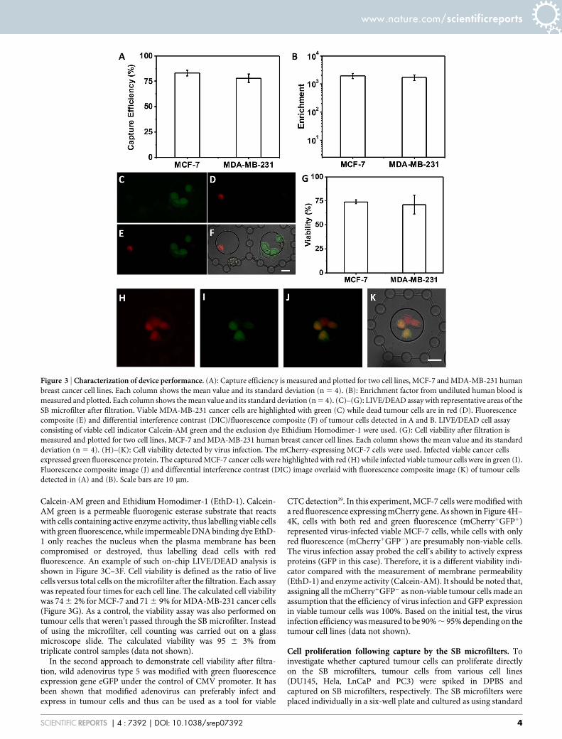
Calcein-AM green and Ethidium Homodimer-1 (EthD-1). Calcein-AM green is a permeable fluorogenic esterase substrate that reactswith cells containing active enzyme activity, thus labelling viable cellswith green fluorescence, while impermeable DNA binding dye EthD-1 only reaches the nucleus when the plasma membrane has beencompromised or destroyed, thus labelling dead cells with redfluorescence. An example of such on-chip LIVE/DEAD analysis isshown in Figure 3C–3F. Cell viability is defined as the ratio of livecells versus total cells on the microfilter after the filtration. Each assaywas repeated four times for each cell line. The calculated cell viabilitywas 74 6 2% for MCF-7 and 71 6 9% for MDA-MB-231 cancer cells(Figure 3G). As a control, the viability assay was also performed ontumour cells that weren’t passed through the SB microfilter. Insteadof using the microfilter, cell counting was carried out on a glassmicroscope slide. The calculated viability was 95 6 3% fromtriplicate control samples (data not shown).
In the second approach to demonstrate cell viability after filtra-tion, wild adenovirus type 5 was modified with green fluorescenceexpression gene eGFP under the control of CMV promoter. It hasbeen shown that modified adenovirus can preferably infect andexpress in tumour cells and thus can be used as a tool for viable
CTC detection39. In this experiment, MCF-7 cells were modified witha red fluorescence expressing mCherry gene. As shown in Figure 4H–4K, cells with both red and green fluorescence (mCherry1GFP1)represented virus-infected viable MCF-7 cells, while cells with onlyred fluorescence (mCherry1GFP2) are presumably non-viable cells.The virus infection assay probed the cell’s ability to actively expressproteins (GFP in this case). Therefore, it is a different viability indi-cator compared with the measurement of membrane permeability(EthD-1) and enzyme activity (Calcein-AM). It should be noted that,assigning all the mCherry1GFP2 as non-viable tumour cells made anassumption that the efficiency of virus infection and GFP expressionin viable tumour cells was 100%. Based on the initial test, the virusinfection efficiency was measured to be 90% , 95% depending on thetumour cell lines (data not shown).
Cell proliferation following capture by the SB microfilters. Toinvestigate whether captured tumour cells can proliferate directlyon the SB microfilters, tumour cells from various cell lines(DU145, Hela, LnCaP and PC3) were spiked in DPBS andcaptured on SB microfilters, respectively. The SB microfilters wereplaced individually in a six-well plate and cultured as using standard
Figure 3 | Characterization of device performance. (A): Capture efficiency is measured and plotted for two cell lines, MCF-7 and MDA-MB-231 human
breast cancer cell lines. Each column shows the mean value and its standard deviation (n 5 4). (B): Enrichment factor from undiluted human blood is
measured and plotted. Each column shows the mean value and its standard deviation (n 5 4). (C)–(G): LIVE/DEAD assay with representative areas of the
SB microfilter after filtration. Viable MDA-MB-231 cancer cells are highlighted with green (C) while dead tumour cells are in red (D). Fluorescence
composite (E) and differential interference contrast (DIC)/fluorescence composite (F) of tumour cells detected in A and B. LIVE/DEAD cell assay
consisting of viable cell indicator Calcein-AM green and the exclusion dye Ethidium Homodimer-1 were used. (G): Cell viability after filtration is
measured and plotted for two cell lines, MCF-7 and MDA-MB-231 human breast cancer cell lines. Each column shows the mean value and its standard
deviation (n 5 4). (H)–(K): Cell viability detected by virus infection. The mCherry-expressing MCF-7 cells were used. Infected viable cancer cells
expressed green fluorescence protein. The captured MCF-7 cancer cells were highlighted with red (H) while infected viable tumour cells were in green (I).
Fluorescence composite image (J) and differential interference contrast (DIC) image overlaid with fluorescence composite image (K) of tumour cells
detected in (A) and (B). Scale bars are 10 mm.
www.nature.com/scientificreports
SCIENTIFIC REPORTS | 4 : 7392 | DOI: 10.1038/srep07392 4
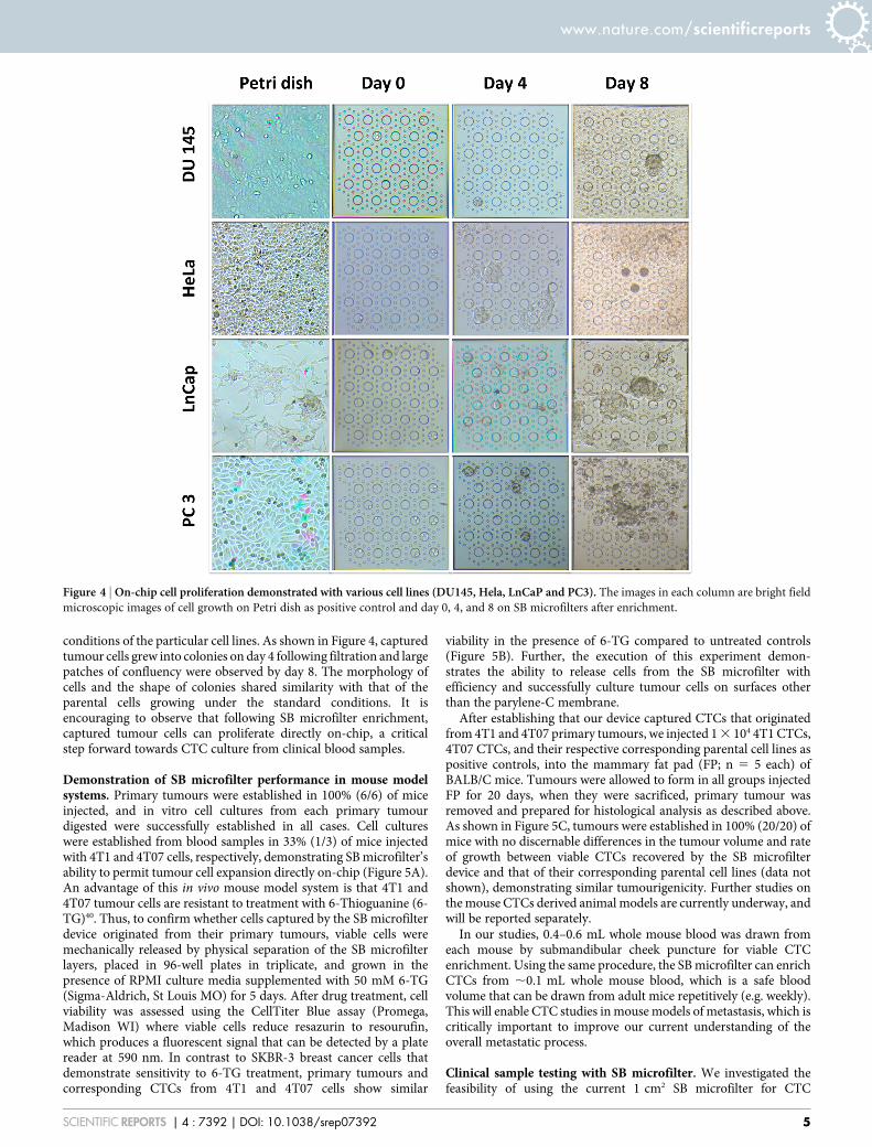
conditions of the particular cell lines. As shown in Figure 4, capturedtumour cells grew into colonies on day 4 following filtration and largepatches of confluency were observed by day 8. The morphology ofcells and the shape of colonies shared similarity with that of theparental cells growing under the standard conditions. It isencouraging to observe that following SB microfilter enrichment,captured tumour cells can proliferate directly on-chip, a criticalstep forward towards CTC culture from clinical blood samples.
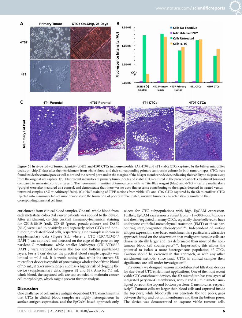
Demonstration of SB microfilter performance in mouse modelsystems. Primary tumours were established in 100% (6/6) of miceinjected, and in vitro cell cultures from each primary tumourdigested were successfully established in all cases. Cell cultureswere established from blood samples in 33% (1/3) of mice injectedwith 4T1 and 4T07 cells, respectively, demonstrating SB microfilter’sability to permit tumour cell expansion directly on-chip (Figure 5A).An advantage of this in vivo mouse model system is that 4T1 and4T07 tumour cells are resistant to treatment with 6-Thioguanine (6-TG)40. Thus, to confirm whether cells captured by the SB microfilterdevice originated from their primary tumours, viable cells weremechanically released by physical separation of the SB microfilterlayers, placed in 96-well plates in triplicate, and grown in thepresence of RPMI culture media supplemented with 50 mM 6-TG(Sigma-Aldrich, St Louis MO) for 5 days. After drug treatment, cellviability was assessed using the CellTiter Blue assay (Promega,Madison WI) where viable cells reduce resazurin to resourufin,which produces a fluorescent signal that can be detected by a platereader at 590 nm. In contrast to SKBR-3 breast cancer cells thatdemonstrate sensitivity to 6-TG treatment, primary tumours andcorresponding CTCs from 4T1 and 4T07 cells show similar
viability in the presence of 6-TG compared to untreated controls(Figure 5B). Further, the execution of this experiment demon-strates the ability to release cells from the SB microfilter withefficiency and successfully culture tumour cells on surfaces otherthan the parylene-C membrane.
After establishing that our device captured CTCs that originatedfrom 4T1 and 4T07 primary tumours, we injected 1 3 104 4T1 CTCs,4T07 CTCs, and their respective corresponding parental cell lines aspositive controls, into the mammary fat pad (FP; n 5 5 each) ofBALB/C mice. Tumours were allowed to form in all groups injectedFP for 20 days, when they were sacrificed, primary tumour wasremoved and prepared for histological analysis as described above.As shown in Figure 5C, tumours were established in 100% (20/20) ofmice with no discernable differences in the tumour volume and rateof growth between viable CTCs recovered by the SB microfilterdevice and that of their corresponding parental cell lines (data notshown), demonstrating similar tumourigenicity. Further studies onthe mouse CTCs derived animal models are currently underway, andwill be reported separately.
In our studies, 0.4–0.6 mL whole mouse blood was drawn fromeach mouse by submandibular cheek puncture for viable CTCenrichment. Using the same procedure, the SB microfilter can enrichCTCs from ,0.1 mL whole mouse blood, which is a safe bloodvolume that can be drawn from adult mice repetitively (e.g. weekly).This will enable CTC studies in mouse models of metastasis, which iscritically important to improve our current understanding of theoverall metastatic process.
Clinical sample testing with SB microfilter. We investigated thefeasibility of using the current 1 cm2 SB microfilter for CTC
Figure 4 | On-chip cell proliferation demonstrated with various cell lines (DU145, Hela, LnCaP and PC3). The images in each column are bright field
microscopic images of cell growth on Petri dish as positive control and day 0, 4, and 8 on SB microfilters after enrichment.
www.nature.com/scientificreports
SCIENTIFIC REPORTS | 4 : 7392 | DOI: 10.1038/srep07392 5
enrichment from clinical blood samples. One mL whole blood fromeach metastatic colorectal cancer patients was applied to the device.After enrichment, on-chip cocktail immunocytochemical stainingfor CK 8/18/19 (red), CD-45 (green, pseudo-colour) and DAPI(blue) were used to positively and negatively select CTCs and non-tumour, nucleated blood cells, respectively. One example is shown inSupplementary data (Figure S1), where a CTC (CK1/CD452/DAPI1) was captured and detected on the edge of the pore on topparylene-C membrane, while smaller leukocytes (CK2/CD451/DAPI1) were trapped between the top and bottom parylene-Clayers. For a 1 cm2 device, the practical blood sample capacity waslimited to ,1.5 mL. It is worth noting that, while the current SBmicrofilter device is capable of processing a whole tube of fresh bloodof 7.5 mL, it takes much longer and has a higher risk of clogging thedevice (Supplementary data, Figures S2 and S3). Also for 7.5 mLwhole blood, the captured cells are too crowded to maintain cancercell morphology, which might prevent further analysis.
DiscussionOne challenge of cell surface antigen dependent CTC enrichment isthat CTCs in clinical blood samples are highly heterogeneous insurface antigen expression, and the EpCAM-based approach only
selects for CTC subpopulations with high EpCAM expression.Further, EpCAM expression is absent from ,15–30% solid tumoursand down-regulated in many CTCs, especially those believed to haveundergone epithelial-mesenchymal transition (EMT) or those har-bouring stem/progenitor phenotypes41–44. Independent of surfaceantigen expression, size-based enrichment is a particularly attractiveapproach based on the observation that malignant tumour cells arecharacteristically larger and less deformable than most of the non-tumour blood cell counterparts45,46. Importantly, this allows thepotential to isolate a more heterogeneous population of CTCs.Caution should be exercised in this approach, as with any otherenrichment methods, since small CTCs in clinical samples theirsignificance are still under investigation17.
Previously we designed various microfabricated filtration devicesfor size-based CTC enrichment applications. One of the most recentviable CTC enrichment devices, the 3D microfilter, has two layers ofintegrated parylene-C membranes, with 9 and 8 mm diameter una-ligned pores on the top and bottom parylene-C membranes, respect-ively37. Tumour cells are larger than blood cells and captured insidethe top pore, while blood cells can penetrate the top pores, gapsbetween the top and bottom membranes and then the bottom pores.The device was demonstrated to capture viable tumour cells.
Figure 5 | In vivo study of tumourigenicity of 4T1 and 4T07 CTCs in mouse models. (A): 4T07 and 4T1 viable CTCs captured by the bilayer microfilter
device on-chip 21 days after their enrichment from whole blood, and their corresponding primary tumours in culture. In both tumour types, CTCs were
found inside the central pore as well as around the central pore and in the margins of the bilayer membrane device, indicating their ability to migrate away
from the original site capture. (B): Fluorescent intensities of primary tumour cells and viable CTCs cultured in the presence of 6-TG treatment (orange)
compared to untreated controls (green). The fluorescent intensities of tumour cells with no TiterBlue reagent (blue) and 6-TG 1 culture media alone
(purple) were also measured as a control, and demonstrate that there was no auto fluorescence contributing to the signals detected in treated versus
untreated samples. (AU 5 Arbitrary Units). (C): H&E staining of FFPE sections from viable 4T1 and 4T07 CTCs captured by the SB microfilter. CTCs
injected into mammary fads of mice demonstrate the formation of poorly differentiated, invasive tumours characteristically similar to their
corresponding parental cell lines.
www.nature.com/scientificreports
SCIENTIFIC REPORTS | 4 : 7392 | DOI: 10.1038/srep07392 6
However, cells did not proliferate presumably due to the limitedspace provided by the top pores (9 mm diameter) where cells werecaptured and immobilized tightly without extra space.
In this study, we designed, microfabricated and characterized anew three-dimensional microfilter for viable CTC enrichment basedon physical properties of the cells. Compared with the fabricationprocess of the previous 3D microfilter37, there are several majorchanges and the overall process has a much higher yield. First, theSB microfilter was completely constructed with parylene-C, a trans-parent and biocompatible polymer, without a silicon support as the3D microfilter. Although the device is still made on silicon substrate,the wafer is only served as a device carrier. As a result, the SB devicefabrication process eliminates the demanding fabrication steps ofdouble side alignment and through wafer etching as in the 3D micro-filter. Secondly, the pores on the top parylene layer have a largediameter of 40 mm, thus CTCs are not captured inside the top poreson the SB microfilter, but instead trapped in the gaps at the edge oflarge top pores and the bottom parylene-C layer. This design spe-cification allows greater freedom for enriched tumour cells toexpand, migrate, and proliferate on the surface of the device. Thekey fabrication parameter to be controlled during fabrication is thethickness of the sacrificial photoresist, which determines the gapdistance between the top and bottom parylene-C membranes andcan be controlled down to ,0.1 mm resolution. Another importantdesign characteristic of the SB microfilter is that the adhesionbetween the top and bottom parylene layers are controlled duringmicrofabrication to make it strong enough to hold in-place duringoperation, but they can be separated easily with tweezers afterwards.The exposed cells are thus more accessible to chemicals that for
examples disrupt cell adhesion for more efficient release, stain cellsfor imaging or lyse cells for genetic analysis. The critical innovationswe have made to our multilayer technology enabled us to extend thefundamental working principles and advantages of size-based CTCenrichment to viable cell capture and functional characterization.
We demonstrated the viability and proliferative capacity of cap-tured cells by the SB microfilter using multiple cancer cell lines invitro. The efficacy of the SB microfilter device was further validatedusing whole blood samples from a breast cancer mouse model systemin vivo, demonstrating our ability to capture and culture viable CTCsfrom mice injected with tumour cells of varying metastatic capacity.The SB microfilter succeeded in enrichment and in vitro culture ofCTCs from hundreds of microliters of mouse blood. Considering thecurrent 1 cm2 SB microfilter process whole blood volume in therange of 0.1–1.5 mL, which perfectly fit the total bleed out volumefor adult mouse of 0.6–1.4 mL47, thus this device is a highly valuabletool for CTC studies in pre-clinical mouse models.
In a feasibility study, the SB microfilter was shown to captureCTCs from an unfixed metastatic cancer patient blood sample.Since it is desirable to assess larger blood volumes (e.g. a full tubeof blood of 7.5 mL) in a clinical setting, future study will increasesample-processing capacity by expanding the effective filtration area.Further, the maintenance of CTC cultures from human blood sam-ples is a more complicated task than in mouse model systems.However, recent report on CTC culture after enrichment by CTC-iChip demonstrated the feasibility48. In addition to viable CTC isola-tion, the optimal culture conditions for CTC expansion will need tobe determined. An important advantage of the SB microfilter is itsability to simultaneously act as a capture, analysis, and culture plat-
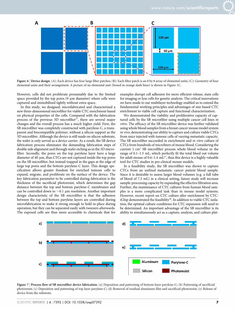
Figure 6 | Device design. (A): Each device has four large filter patches; (B): Each filter patch is an 8 by 8 array of elemental units; (C): Geometry of four
elemental units and their arrangement. A picture of an elemental unit (boxed in orange dash lines) is shown in Figure 1C.
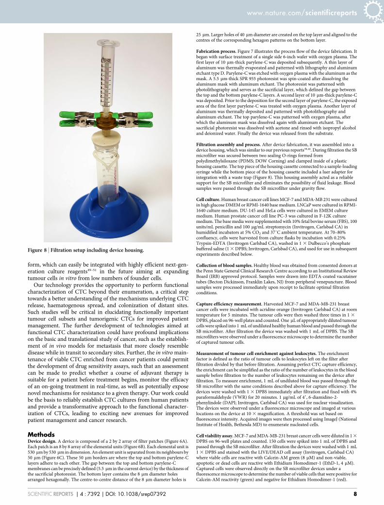
Figure 7 | Process flow of SB microfilter device fabrication. (a) Deposition and patterning of bottom layer parylene-C; (b) Patterning of sacrificial
photoresist; (c) Deposition and patterning of top layer parylene-C; (d) Removal of residual aluminum film and sacrificial photoresist; (e) Release of
device from the substrate.
www.nature.com/scientificreports
SCIENTIFIC REPORTS | 4 : 7392 | DOI: 10.1038/srep07392 7
form, which can easily be integrated with highly efficient next-gen-eration culture reagents49–51 in the future aiming at expandingtumour cells in vitro from low numbers of founder cells.
Our technology provides the opportunity to perform functionalcharacterization of CTC beyond their enumeration, a critical steptowards a better understanding of the mechanisms underlying CTCrelease, haematogenous spread, and colonization of distant sites.Such studies will be critical in elucidating functionally importanttumour cell subsets and tumorigenic CTCs for improved patientmanagement. The further development of technologies aimed atfunctional CTC characterization could have profound implicationson the basic and translational study of cancer, such as the establish-ment of in vivo models for metastasis that more closely resembledisease while in transit to secondary sites. Further, the in vitro main-tenance of viable CTC enriched from cancer patients could permitthe development of drug sensitivity assays, such that an assessmentcan be made to predict whether a course of adjuvant therapy issuitable for a patient before treatment begins, monitor the efficacyof an on-going treatment in real-time, as well as potentially exposenovel mechanisms for resistance to a given therapy. Our work couldbe the basis to reliably establish CTC cultures from human patientsand provide a transformative approach to the functional character-ization of CTCs, leading to exciting new avenues for improvedpatient management and cancer research.
MethodsDevice design. A device is composed of a 2 by 2 array of filter patches (Figure 6A).Each patch is an 8 by 8 array of the elemental units (Figure 6B). Each elemental unit is530 mm by 530 mm in dimension. An element unit is separated from its neighbours by50 mm (Figure 6C). These 50 mm borders are where the top and bottom parylene-Clayers adhere to each other. The gap between the top and bottom parylene-Cmembranes can be precisely defined (5.5 mm in the current device) by the thickness ofthe sacrificial photoresist. The bottom layer contains the 8 mm diameter holesarranged hexagonally. The centre-to-centre distance of the 8 mm diameter holes is
25 mm. Larger holes of 40 mm diameter are created on the top layer and aligned to thecentres of the corresponding hexagon patterns on the bottom layer.
Fabrication process. Figure 7 illustrates the process flow of the device fabrication. Itbegan with surface treatment of a single side 6-inch wafer with oxygen plasma. Thefirst layer of 10 mm-thick parylene-C was deposited subsequently. A thin layer ofaluminum was thermally evaporated and patterned with lithography and aluminumetchant type D. Parylene-C was etched with oxygen plasma with the aluminum as themask. A 5.5 mm-thick SPR 955 photoresist was spin-coated after dissolving thealuminum mask with aluminum etchant. The photoresist was patterned withphotolithography and serves as the sacrificial layer, which defined the gap betweenthe top and the bottom parylene-C layers. A second layer of 10 mm-thick parylene-Cwas deposited. Prior to the deposition for the second layer of parylene-C, the exposedarea of the first layer parylene-C was treated with oxygen plasma. Another layer ofaluminum was thermally deposited and patterned with photolithography andaluminum etchant. The top parylene-C was patterned with oxygen plasma, afterwhich the aluminum mask was dissolved again with aluminum etchant. Thesacrificial photoresist was dissolved with acetone and rinsed with isopropyl alcoholand deionized water. Finally the device was released from the substrate.
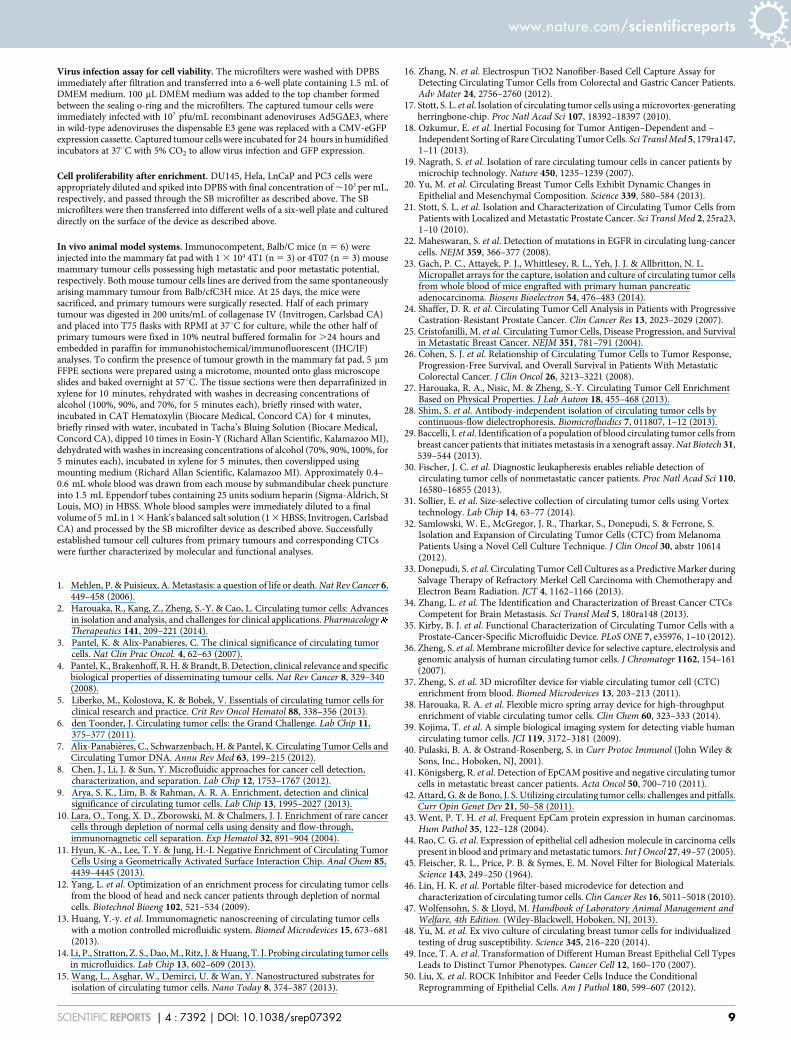
Filtration assembly and process. After device fabrication, it was assembled into adevice housing, which was similar to our previous reports38,46. During filtration the SBmicrofilter was secured between two sealing O-rings formed frompolydimethylsiloxane (PDMS; DOW Corning) and clamped inside of a plastichousing cassette. The top piece of the housing cassette connected to a sample-loadingsyringe while the bottom piece of the housing cassette included a luer adapter forintegration with a waste trap (Figure 8). This housing assembly acted as a reliablesupport for the SB microfilter and eliminates the possibility of fluid leakage. Bloodsamples were passed through the SB microfilter under gravity flow.
Cell culture. Human breast cancer cell lines MCF-7 and MDA-MB 231 were culturedin high glucose DMEM or RPMI-1640 base medium. LNCaP were cultured in RPMI-1640 culture medium. DU-145 and HeLa cells were cultured in EMEM culturemedium. Human prostate cancer cell line PC-3 was cultured in F-12K culturemedium. The base media were supplemented with 10% fetal bovine serum (FBS), 100units/mL penicillin and 100 mg/mL streptomycin (Invitrogen, Carlsbad CA) inhumidified incubators at 5% CO2 and 37uC ambient temperature. At 70–80%confluency, cells were harvested from culture flasks by incubation with 0.25%Trypsin-EDTA (Invitrogen Carlsbad CA), washed in 1 3 Dulbecco’s phosphatebuffered saline (1 3 DPBS; Invitrogen, Carlsbad CA), and used for use in subsequentexperiments described below.
Collection of blood samples. Healthy blood was obtained from consented donors atthe Penn State General Clinical Research Centre according to an Institutional ReviewBoard (IRB) approved protocol. Samples were drawn into EDTA-coated vacutainertubes (Becton Dickinson, Franklin Lakes, NJ) from peripheral venepuncture. Bloodsamples were processed immediately upon receipt to facilitate optimal filtrationconditions.
Capture efficiency measurement. Harvested MCF-7 and MDA-MB-231 breastcancer cells were incubated with acridine orange (Invitrogen Carlsbad CA) at roomtemperature for 5 minutes. The tumour cells were then washed three times in 1 3
DPBS, placed on 96-well plates and counted. One mL of appropriately diluted tumourcells were spiked into 1 mL of undiluted healthy human blood and passed through theSB microfilter. After filtration the device was washed with 1 mL of DPBS. The SBmicrofilters were observed under a fluorescence microscope to determine the numberof captured tumour cells.
Measurement of tumour cell enrichment against leukocytes. The enrichmentfactor is defined as the ratio of tumour cells to leukocytes left on the filter afterfiltration divided by that before filtration. Assuming perfect CTC capture efficiency,the enrichment can be simplified as the ratio of the number of leukocytes in the bloodsample before filtration to the number of leukocytes remaining on the device afterfiltration. To measure enrichment, 1 mL of undiluted blood was passed through theSB microfilter with the same conditions described above for capture efficiency. Thedevices were washed with 1 3 DPBS immediately after filtration and fixed with 4%paraformaldehyde (VWR) for 20 minutes. 1 mg/mL of 49, 6-diamidino-2-phenylindole (DAPI; Invitrogen, Carlsbad CA) was used for nuclear visualization.The devices were observed under a fluorescence microscope and imaged at variouslocations on the device at 10 3 magnification. A threshold was set based onfluorescence intensity. Acquired images were then processed using ImageJ (NationalInstitute of Health, Bethesda MD) to enumerate nucleated cells.
Cell viability assay. MCF-7 and MDA-MB-231 breast cancer cells were diluted in 1 3
DPBS on 96-well plates and counted. 150 cells were spiked into 1 mL of DPBS andpassed through the SB microfilter. After filtration the devices were washed with 1 mL1 3 DPBS and stained with the LIVE/DEAD cell assay (Invitrogen, Carlsbad CA)where viable cells are reactive with Calcein-AM green (8 mM) and non-viable,apoptotic or dead cells are reactive with Ethidium Homodimer-1 (EthD-1, 4 mM).Captured cells were observed directly on the SB microfilter devices under afluorescence microscope to determine the number of viable cells that were positive forCalcein-AM reactivity (green) and negative for Ethidium Homodimer-1 (red).
Figure 8 | Filtration setup including device housing.
www.nature.com/scientificreports
SCIENTIFIC REPORTS | 4 : 7392 | DOI: 10.1038/srep07392 8
Virus infection assay for cell viability. The microfilters were washed with DPBSimmediately after filtration and transferred into a 6-well plate containing 1.5 mL ofDMEM medium. 100 mL DMEM medium was added to the top chamber formedbetween the sealing o-ring and the microfilters. The captured tumour cells wereimmediately infected with 107 pfu/mL recombinant adenoviruses Ad5GDE3, wherein wild-type adenoviruses the dispensable E3 gene was replaced with a CMV-eGFPexpression cassette. Captured tumour cells were incubated for 24 hours in humidifiedincubators at 37uC with 5% CO2 to allow virus infection and GFP expression.
Cell proliferability after enrichment. DU145, Hela, LnCaP and PC3 cells wereappropriately diluted and spiked into DPBS with final concentration of ,103 per mL,respectively, and passed through the SB microfilter as described above. The SBmicrofilters were then transferred into different wells of a six-well plate and cultureddirectly on the surface of the device as described above.
In vivo animal model systems. Immunocompetent, Balb/C mice (n 5 6) wereinjected into the mammary fat pad with 1 3 104 4T1 (n 5 3) or 4T07 (n 5 3) mousemammary tumour cells possessing high metastatic and poor metastatic potential,respectively. Both mouse tumour cells lines are derived from the same spontaneouslyarising mammary tumour from Balb/cfC3H mice. At 25 days, the mice weresacrificed, and primary tumours were surgically resected. Half of each primarytumour was digested in 200 units/mL of collagenase IV (Invitrogen, Carlsbad CA)and placed into T75 flasks with RPMI at 37uC for culture, while the other half ofprimary tumours were fixed in 10% neutral buffered formalin for .24 hours andembedded in paraffin for immunohistochemical/immunofluorescent (IHC/IF)analyses. To confirm the presence of tumour growth in the mammary fat pad, 5 mmFFPE sections were prepared using a microtome, mounted onto glass microscopeslides and baked overnight at 57uC. The tissue sections were then deparrafinized inxylene for 10 minutes, rehydrated with washes in decreasing concentrations ofalcohol (100%, 90%, and 70%, for 5 minutes each), briefly rinsed with water,incubated in CAT Hematoxylin (Biocare Medical, Concord CA) for 4 minutes,briefly rinsed with water, incubated in Tacha’s Bluing Solution (Biocare Medical,Concord CA), dipped 10 times in Eosin-Y (Richard Allan Scientific, Kalamazoo MI),dehydrated with washes in increasing concentrations of alcohol (70%, 90%, 100%, for5 minutes each), incubated in xylene for 5 minutes, then coverslipped usingmounting medium (Richard Allan Scientific, Kalamazoo MI). Approximately 0.4–0.6 mL whole blood was drawn from each mouse by submandibular cheek punctureinto 1.5 mL Eppendorf tubes containing 25 units sodium heparin (Sigma-Aldrich, StLouis, MO) in HBSS. Whole blood samples were immediately diluted to a finalvolume of 5 mL in 1 3 Hank’s balanced salt solution (1 3 HBSS; Invitrogen, CarlsbadCA) and processed by the SB microfilter device as described above. Successfullyestablished tumour cell cultures from primary tumours and corresponding CTCswere further characterized by molecular and functional analyses.
1. Mehlen, P. & Puisieux, A. Metastasis: a question of life or death. Nat Rev Cancer 6,449–458 (2006).
2. Harouaka, R., Kang, Z., Zheng, S.-Y. & Cao, L. Circulating tumor cells: Advancesin isolation and analysis, and challenges for clinical applications. PharmacologyTherapeutics 141, 209–221 (2014).
3. Pantel, K. & Alix-Panabieres, C. The clinical significance of circulating tumorcells. Nat Clin Prac Oncol. 4, 62–63 (2007).
4. Pantel, K., Brakenhoff, R. H. & Brandt, B. Detection, clinical relevance and specificbiological properties of disseminating tumour cells. Nat Rev Cancer 8, 329–340(2008).
5. Liberko, M., Kolostova, K. & Bobek, V. Essentials of circulating tumor cells forclinical research and practice. Crit Rev Oncol Hematol 88, 338–356 (2013).
6. den Toonder, J. Circulating tumor cells: the Grand Challenge. Lab Chip 11,375–377 (2011).
7. Alix-Panabieres, C., Schwarzenbach, H. & Pantel, K. Circulating Tumor Cells andCirculating Tumor DNA. Annu Rev Med 63, 199–215 (2012).
8. Chen, J., Li, J. & Sun, Y. Microfluidic approaches for cancer cell detection,characterization, and separation. Lab Chip 12, 1753–1767 (2012).
9. Arya, S. K., Lim, B. & Rahman, A. R. A. Enrichment, detection and clinicalsignificance of circulating tumor cells. Lab Chip 13, 1995–2027 (2013).
10. Lara, O., Tong, X. D., Zborowski, M. & Chalmers, J. J. Enrichment of rare cancercells through depletion of normal cells using density and flow-through,immunomagnetic cell separation. Exp Hematol 32, 891–904 (2004).
11. Hyun, K.-A., Lee, T. Y. & Jung, H.-I. Negative Enrichment of Circulating TumorCells Using a Geometrically Activated Surface Interaction Chip. Anal Chem 85,4439–4445 (2013).
12. Yang, L. et al. Optimization of an enrichment process for circulating tumor cellsfrom the blood of head and neck cancer patients through depletion of normalcells. Biotechnol Bioeng 102, 521–534 (2009).
13. Huang, Y.-y. et al. Immunomagnetic nanoscreening of circulating tumor cellswith a motion controlled microfluidic system. Biomed Microdevices 15, 673–681(2013).
14. Li, P., Stratton, Z. S., Dao, M., Ritz, J. & Huang, T. J. Probing circulating tumor cellsin microfluidics. Lab Chip 13, 602–609 (2013).
15. Wang, L., Asghar, W., Demirci, U. & Wan, Y. Nanostructured substrates forisolation of circulating tumor cells. Nano Today 8, 374–387 (2013).
16. Zhang, N. et al. Electrospun TiO2 Nanofiber-Based Cell Capture Assay forDetecting Circulating Tumor Cells from Colorectal and Gastric Cancer Patients.Adv Mater 24, 2756–2760 (2012).
17. Stott, S. L. et al. Isolation of circulating tumor cells using a microvortex-generatingherringbone-chip. Proc Natl Acad Sci 107, 18392–18397 (2010).
18. Ozkumur, E. et al. Inertial Focusing for Tumor Antigen–Dependent and –Independent Sorting of Rare Circulating Tumor Cells. Sci Transl Med 5, 179ra147,1–11 (2013).
19. Nagrath, S. et al. Isolation of rare circulating tumour cells in cancer patients bymicrochip technology. Nature 450, 1235–1239 (2007).
20. Yu, M. et al. Circulating Breast Tumor Cells Exhibit Dynamic Changes inEpithelial and Mesenchymal Composition. Science 339, 580–584 (2013).
21. Stott, S. L. et al. Isolation and Characterization of Circulating Tumor Cells fromPatients with Localized and Metastatic Prostate Cancer. Sci Transl Med 2, 25ra23,1–10 (2010).
22. Maheswaran, S. et al. Detection of mutations in EGFR in circulating lung-cancercells. NEJM 359, 366–377 (2008).
23. Gach, P. C., Attayek, P. J., Whittlesey, R. L., Yeh, J. J. & Allbritton, N. L.Micropallet arrays for the capture, isolation and culture of circulating tumor cellsfrom whole blood of mice engrafted with primary human pancreaticadenocarcinoma. Biosens Bioelectron 54, 476–483 (2014).
24. Shaffer, D. R. et al. Circulating Tumor Cell Analysis in Patients with ProgressiveCastration-Resistant Prostate Cancer. Clin Cancer Res 13, 2023–2029 (2007).
25. Cristofanilli, M. et al. Circulating Tumor Cells, Disease Progression, and Survivalin Metastatic Breast Cancer. NEJM 351, 781–791 (2004).
26. Cohen, S. J. et al. Relationship of Circulating Tumor Cells to Tumor Response,Progression-Free Survival, and Overall Survival in Patients With MetastaticColorectal Cancer. J Clin Oncol 26, 3213–3221 (2008).
27. Harouaka, R. A., Nisic, M. & Zheng, S.-Y. Circulating Tumor Cell EnrichmentBased on Physical Properties. J Lab Autom 18, 455–468 (2013).
28. Shim, S. et al. Antibody-independent isolation of circulating tumor cells bycontinuous-flow dielectrophoresis. Biomicrofluidics 7, 011807, 1–12 (2013).
29. Baccelli, I. et al. Identification of a population of blood circulating tumor cells frombreast cancer patients that initiates metastasis in a xenograft assay. Nat Biotech 31,539–544 (2013).
30. Fischer, J. C. et al. Diagnostic leukapheresis enables reliable detection ofcirculating tumor cells of nonmetastatic cancer patients. Proc Natl Acad Sci 110,16580–16855 (2013).
31. Sollier, E. et al. Size-selective collection of circulating tumor cells using Vortextechnology. Lab Chip 14, 63–77 (2014).
32. Samlowski, W. E., McGregor, J. R., Tharkar, S., Donepudi, S. & Ferrone, S.Isolation and Expansion of Circulating Tumor Cells (CTC) from MelanomaPatients Using a Novel Cell Culture Technique. J Clin Oncol 30, abstr 10614(2012).
33. Donepudi, S. et al. Circulating Tumor Cell Cultures as a Predictive Marker duringSalvage Therapy of Refractory Merkel Cell Carcinoma with Chemotherapy andElectron Beam Radiation. JCT 4, 1162–1166 (2013).
34. Zhang, L. et al. The Identification and Characterization of Breast Cancer CTCsCompetent for Brain Metastasis. Sci Transl Med 5, 180ra148 (2013).
35. Kirby, B. J. et al. Functional Characterization of Circulating Tumor Cells with aProstate-Cancer-Specific Microfluidic Device. PLoS ONE 7, e35976, 1–10 (2012).
36. Zheng, S. et al. Membrane microfilter device for selective capture, electrolysis andgenomic analysis of human circulating tumor cells. J Chromatogr 1162, 154–161(2007).
37. Zheng, S. et al. 3D microfilter device for viable circulating tumor cell (CTC)enrichment from blood. Biomed Microdevices 13, 203–213 (2011).
38. Harouaka, R. A. et al. Flexible micro spring array device for high-throughputenrichment of viable circulating tumor cells. Clin Chem 60, 323–333 (2014).
39. Kojima, T. et al. A simple biological imaging system for detecting viable humancirculating tumor cells. JCI 119, 3172–3181 (2009).
40. Pulaski, B. A. & Ostrand-Rosenberg, S. in Curr Protoc Immunol (John Wiley &Sons, Inc., Hoboken, NJ, 2001).
41. Konigsberg, R. et al. Detection of EpCAM positive and negative circulating tumorcells in metastatic breast cancer patients. Acta Oncol 50, 700–710 (2011).
42. Attard, G. & de Bono, J. S. Utilizing circulating tumor cells: challenges and pitfalls.Curr Opin Genet Dev 21, 50–58 (2011).
43. Went, P. T. H. et al. Frequent EpCam protein expression in human carcinomas.Hum Pathol 35, 122–128 (2004).
44. Rao, C. G. et al. Expression of epithelial cell adhesion molecule in carcinoma cellspresent in blood and primary and metastatic tumors. Int J Oncol 27, 49–57 (2005).
45. Fleischer, R. L., Price, P. B. & Symes, E. M. Novel Filter for Biological Materials.Science 143, 249–250 (1964).
46. Lin, H. K. et al. Portable filter-based microdevice for detection andcharacterization of circulating tumor cells. Clin Cancer Res 16, 5011–5018 (2010).
47. Wolfensohn, S. & Lloyd, M. Handbook of Laboratory Animal Management andWelfare, 4th Edition. (Wiley-Blackwell, Hoboken, NJ, 2013).
48. Yu, M. et al. Ex vivo culture of circulating breast tumor cells for individualizedtesting of drug susceptibility. Science 345, 216–220 (2014).
49. Ince, T. A. et al. Transformation of Different Human Breast Epithelial Cell TypesLeads to Distinct Tumor Phenotypes. Cancer Cell 12, 160–170 (2007).
50. Liu, X. et al. ROCK Inhibitor and Feeder Cells Induce the ConditionalReprogramming of Epithelial Cells. Am J Pathol 180, 599–607 (2012).
www.nature.com/scientificreports
SCIENTIFIC REPORTS | 4 : 7392 | DOI: 10.1038/srep07392 9
51. Yuan, H. et al. Use of Reprogrammed Cells to Identify Therapy for RespiratoryPapillomatosis. NEJM 367, 1220–1227 (2012).
AcknowledgmentsS.-Y. Zheng thanks the Penn State Materials Research Institute, Nanofabrication laboratoryand Microscopy and Cytometry facility, and Penn State Hershey Cancer Institute for theirsupport. This work was partially supported by the Pennsylvania State University start-upfund and the National Cancer Institute of the National Institutes of Health under AwardNumber DP2CA174508. Additionally, the authors would like to acknowledge researchsupport provided by the Sylvester Comprehensive Cancer Centre at the University ofMiami through their Braman Foundation Breast Cancer Developmental Grant. Fundingsupport for A. Williams was provided through a fellowship award from the UNCF-MerckScience Initiative.
Author contributionsM.-D.Z. designed, fabricated, optimized the device. S.H. performed in vitro devicecharacterization, clinical sample testing, and data analysis. A.W. performed animal modelexperiments and related data analysis. R.H. constructed of the flow control system andprovided technical support of in vitro device characterization and clinical sample testing.B.S., R.B. and E.G. provided animal model system design and technical support for animalmodel system. S.R. and Z.A. provided technical Support for viable CTC capture and cultureand performed data analysis. B.L. performed on-chip cell culture of multiple cell lines on
device. S.W. and J.Z. designed, constructed and characterized the virus used for the on-chipcell viability assay. R.D. and R.C. provided project mentoring and animal model systemdesign. Y.-C.T. contributed to the initial device concept and design, and provided projectmentoring. S.-Y.Z. provided overall project mentoring, contribute to device concept,design, fabrication process development, experimental design, and data analysis. S.-Y.Z.,S.H. and A.W. wrote the manuscript.
Additional informationSupplementary information accompanies this paper at http://www.nature.com/scientificreports
Competing financial interests: The authors declare no competing financial interests.
How to cite this article: Zhou, M.-D. et al. Separable Bilayer Microfiltration Device forViable Label-free Enrichment of Circulating Tumour Cells. Sci. Rep. 4, 7392; DOI:10.1038/srep07392 (2014).
This work is licensed under a Creative Commons Attribution-NonCommercial-ShareAlike 4.0 International License. The images or other third party material in thisarticle are included in the article’s Creative Commons license, unless indicatedotherwise in the credit line; if the material is not included under the CreativeCommons license, users will need to obtain permission from the license holderin order to reproduce the material. To view a copy of this license, visit http://creativecommons.org/licenses/by-nc-sa/4.0/
www.nature.com/scientificreports
SCIENTIFIC REPORTS | 4 : 7392 | DOI: 10.1038/srep07392 10
Related Documents