Sensory representation abnormalities that parallel focal hand dystonia in a primate model David T. Blake 1 , Nancy N. Byl 2 , Steven Cheung 1 , Purvis Bedenbaugh 3 , Srikantan Nagarajan 4 , Michelle Lamb 2 , and Michael Merzenich 1 1 Coleman Laboratory, Keck Center for Integrative Neuroscience, and Dept. of Otolaryngology, University of California, San Francisco 2 Dept. of Physical Therapy, University of California, San Francisco 3 Dept of Neuroscience, University of Florida 4 Dept. of Bioengineering, University of Utah Abstract In our hypothesis of focal dystonia, attended repetitive behaviors generate aberrant sensory representations. Those aberrant representations interfere with motor control. Abnormal motor control strengthens sensory abnormalities. The positive feedback loop reinforces the dystonic condition. Previous studies of primates with focal hand dystonia have demonstrated multi-digit or hairy- glabrous responses at single sites in area 3b, receptive fields that average ten times larger than normal, and high receptive field overlap as a function of horizontal distance. In this study, we strengthen and elaborate these findings. One animal was implanted with an array of micro-electrodes that spanned the border between the face and digits. After the animal developed hand dystonia, responses in the initial hand representation increasingly responded to low threshold stimulation of the face in a columnar substitution. The hand-face border that is normally sharp became patchy and smeared over one millimeter of cortex within six weeks. Two more trained animals developed a focal hand dystonia variable in severity across the hand. Receptive field size, presence of multi-digit or hairy-glabrous receptive fields, and columnar overlap covaried with the animal’s ability to use specific digits. A fourth animal performed the same behaviors without developing dystonia. Many of its physiological measures were similar to the dystonic animals, but receptive field overlap functions were minimally abnormal, and no sites shared response properties that are normally segregated such as hairy-glabrous combined fields, or multi-digit fields. Thalamic mapping demonstrated proportionate levels of abnormality in thalamic representations as was found in cortical representations. Keywords cerebral cortex; focal dystonia; cortical representation; somatosensory; cortical column Introduction Our hypothesis on the genesis and remediation of focal hand dystonia has centered on the abnormal hand representation observed in owl monkeys, Aotus nancymae, which developped the disorder in the lab (Byl et al., 1996; Byl et al., 1997). Attended repetitive sterotypical behaviors cause simultaneous stimulation of large skin surfaces. This simultaneous activation Address Correspondence to: Dr. David T. Blake, 513 Parnassus Ave S-877, SF, CA, 94143-0732, Phone: 415-564-3241, Fax: 415-476-1941, [email protected]. NIH Public Access Author Manuscript Somatosens Mot Res. Author manuscript; available in PMC 2010 February 23. Published in final edited form as: Somatosens Mot Res. 2002 ; 19(4): 347–357. doi:10.1080/0899022021000037827. NIH-PA Author Manuscript NIH-PA Author Manuscript NIH-PA Author Manuscript

Welcome message from author
This document is posted to help you gain knowledge. Please leave a comment to let me know what you think about it! Share it to your friends and learn new things together.
Transcript

Sensory representation abnormalities that parallel focal handdystonia in a primate model
David T. Blake1, Nancy N. Byl2, Steven Cheung1, Purvis Bedenbaugh3, SrikantanNagarajan4, Michelle Lamb2, and Michael Merzenich11 Coleman Laboratory, Keck Center for Integrative Neuroscience, and Dept. of Otolaryngology,University of California, San Francisco2 Dept. of Physical Therapy, University of California, San Francisco3 Dept of Neuroscience, University of Florida4 Dept. of Bioengineering, University of Utah
AbstractIn our hypothesis of focal dystonia, attended repetitive behaviors generate aberrant sensoryrepresentations. Those aberrant representations interfere with motor control. Abnormal motor controlstrengthens sensory abnormalities. The positive feedback loop reinforces the dystonic condition.Previous studies of primates with focal hand dystonia have demonstrated multi-digit or hairy-glabrous responses at single sites in area 3b, receptive fields that average ten times larger than normal,and high receptive field overlap as a function of horizontal distance. In this study, we strengthen andelaborate these findings. One animal was implanted with an array of micro-electrodes that spannedthe border between the face and digits. After the animal developed hand dystonia, responses in theinitial hand representation increasingly responded to low threshold stimulation of the face in acolumnar substitution. The hand-face border that is normally sharp became patchy and smeared overone millimeter of cortex within six weeks. Two more trained animals developed a focal hand dystoniavariable in severity across the hand. Receptive field size, presence of multi-digit or hairy-glabrousreceptive fields, and columnar overlap covaried with the animal’s ability to use specific digits. Afourth animal performed the same behaviors without developing dystonia. Many of its physiologicalmeasures were similar to the dystonic animals, but receptive field overlap functions were minimallyabnormal, and no sites shared response properties that are normally segregated such as hairy-glabrouscombined fields, or multi-digit fields. Thalamic mapping demonstrated proportionate levels ofabnormality in thalamic representations as was found in cortical representations.
Keywordscerebral cortex; focal dystonia; cortical representation; somatosensory; cortical column
IntroductionOur hypothesis on the genesis and remediation of focal hand dystonia has centered on theabnormal hand representation observed in owl monkeys, Aotus nancymae, which developpedthe disorder in the lab (Byl et al., 1996; Byl et al., 1997). Attended repetitive sterotypicalbehaviors cause simultaneous stimulation of large skin surfaces. This simultaneous activation
Address Correspondence to: Dr. David T. Blake, 513 Parnassus Ave S-877, SF, CA, 94143-0732, Phone: 415-564-3241, Fax:415-476-1941, [email protected].
NIH Public AccessAuthor ManuscriptSomatosens Mot Res. Author manuscript; available in PMC 2010 February 23.
Published in final edited form as:Somatosens Mot Res. 2002 ; 19(4): 347–357. doi:10.1080/0899022021000037827.
NIH
-PA Author Manuscript
NIH
-PA Author Manuscript
NIH
-PA Author Manuscript

of receptor sheets in a learning context has been demonstrated to cause receptive fieldenlargement (Wang et al., 1995). As receptive fields enlarge and begin to occupy more thansingle phalanges, cortical sensory feedback for motor control becomes less independent, andeven ambiguous. Animals, including humans, commonly compensate for difficulties in motorcontrol by applying more repetition, attention, and stereotypy to the task performance. At somepoint the reduction in the dimensionality of the feedback crosses a stability point, and the resultis a pathologic movement dysfunction called focal hand dystonia. Glabrous and hairy skinrepresentations are not well differentiated in animals with focal hand dystonia, nor areresponses restricted to single phalanges or single digits.
The exact pathway by which this abnormal feedback occurs is not known. The handrepresentation in primary somatosensory cortex, area 3b (Merzenich et al., 1978), is dominatedby the inputs from low threshold mechanoreceptors (Mountcastle, 1977). Area 3b neuronsproject to posterior parietal somatosensory areas 1, 2, 5, and 7, along with proprioceptiveafferent input from area 3a. These areas all project to the striatum which is one possible siteof interference in motor control (Alexander et al., 1986; Tempel and Perlmutter, 1993). Thereare also projections directly from the somatosensory strip, primarily area 3a and area 2, to area4, primary motor cortex (Jones et al., 1978). Any of these pathways, or some combination ofthem, may be responsible for carrying the abnormal sensory feedback cues in dystonia. Area3b is necessary for hand functionality (Randolph and Semmes, 1974), whereas selectivepharmacological lesions of area 2 cause specific motor control problems with similarities todystonia (Hikosaka et al., 1985).
In prior studies (Byl et al., 1996; Byl et al., 1997), hand dystonia rendering the entire handdysfunctional has been induced by operant behaviors. In one case (Byl et al., 1996), passiveand rapid sequential hand opening and closing induced hand dystonias characterized by failureto achieve the stereotyped hand positioning required to initiate the task. In a second case (Bylet al., 1997), one animal developed dystonia in a palmar grasp task. In the current study, area3b was physiologically monitored using implant recording throughout the development of focalhand dystonia. The changes in receptive field size and representational maps were monitoredin the awake behaving monkey as they occurred. Later, thalamic maps of the implanted animalexamined the representation in the ventral posterolateral cutaneous representation.
In new acute mapping physiological studies, the variability in sensory abnormalities wascompared with the variability in individual motor use. Dystonia can variably degrade specificaspects of motor function, and the target task can usually be continued more slowly withalternative postures or motor strategies. Two of the animals induced to develop focal handdystonia had large differences in degraded ability to use different digits at a target task. Somedigits were functionally normal; the motor control of others was severely impaired. Thesebehavioral changes were systematically compared to the sensory maps. A behavioral controlanimal performed all the dystonia inducing tasks, and shared some but not all representationalabnormalities with the other dystonia animals.
Materials and MethodsBehavioral training
Four owl monkeys, Aotus nancymae, were used in this study. One animal developed dystoniaon a repetitive operant task. This animal was implanted in cortical area 3 before behavioraltraining as described later in Materials and Methods. The task required the animal to positionits right dominant hand in a hand mold and place its first and second digits on two metalcontacts, each one millimeter in diameter and positioned under the natural conformation of thehand shown in Figure 1B. The animal was required to hold its hand in place for several hundredmilliseconds before release could result in liquid reward. After focal dystonia developed, the
Blake et al. Page 2
Somatosens Mot Res. Author manuscript; available in PMC 2010 February 23.
NIH
-PA Author Manuscript
NIH
-PA Author Manuscript
NIH
-PA Author Manuscript
animal was trained on tasks that delivered 100–200 μm taps to its fingertips. This task was across-digit interval discrimination task. It was part of planned experiments not initially intendedfor studies of focal hand dystonia. The task used a limited hold reaction time paradigm. In thistype of task, a trial begins with an orienting response, which was the animal placing its handin a hand mold, and making electrical contact with the tips of two motors on its thumb andindex fingers. Then, a series of stimuli began. In all cases each element of the series was a pairof taps separated by an interval. In the pair of taps, a tap to the index finger occurred before atap on the thumb. A new pair of taps occurred after an onset to onset 500 millisecond interval.After at least two standard interval tap pairs, the series elements changed to the target, whichdiffered from the standards only in inter-tap interval length. Initial standard interval lengthswere 250 milliseconds, and shorter standards were used later in the task, as described inResults. The animal was rewarded with a few drops of Tang for releasing after the intervallength changed to the target of 100 milliseconds, and time-outs followed releases at other times.After two months of performance at this task, the animal’s performance dropped below 50%of its starting performance. The cortical implant was explanted and a mapping experiment wasperformed.
Two of the animals developed dystonia using an established experimental method (Byl et al.,1997) shown in Figure 1A. Each animal held two vertical rods with a palmar grasp in itsdominant right hand and closed its grip. A light signaled adequate contact, and a 20 Hertzvibration began. The end of the vibration signaled the end of the hold period, and the animalwas rewarded with food or liquid for releasing within reaction time limits. Each rod had to beindependently pushed together, and the force between the rods was adjusted to challenge theanimals’ abilities. Animals typically performed several hundred to two thousand trials per dayat a rate of approximately one trial per three seconds. Each dystonic animal’s performancedropped to less than 50% of starting performance before the animals were mapped.
A fourth behavioral control animal used its dominant left hand to perform the first task, thepalmar grasp task, for over a year without developing a dystonia. At that point it was trainedto touch two small metal rods in a hand mold, as the implanted animal did before developingfocal hand dystonia. The behavioral control performed the hand positioning task for more thansix months without developing a dystonia. At that point it was mapped.
Diagnosis of focal hand dystoniaThe clinical diagnosis of focal hand dystonia involves a confluence of a range of symptoms.For focal dystonia, the symptoms include intermittent arrythmic, involuntary contractions ofthe muscles of the hand while performing a target task (Cohen et al., 1989; Lockwood, 1989;Marsden and Sheehy, 1990; Newmark and Hochberg, 1987; Rothwell et al., 1983; Sheehy andMarsden, 1982; Wilson, 1989), co-contraction of agonist and antagonist muscle groups alongwith inaccurate, inappropriately timed and sequenced movements between adjacent fingersand wrist during task performance (Chen et al., 1995), and poor hand posture outside taskperformance. We have documented decrements in performance levels in number of trials perminute, number of trials per day, and hit percentage. Co-contraction of antagonist musclegroups is difficult to document objectively without EMG recordings which were not performed.However, abnormal stiffness of the digits was noted in some animals. Each training sessionwas videotaped, and video analysis was carried out during food retrieval from food tray (Bylet al., 1996).
Mapping experimentsHigh density microelectrode mapping experiments were performed at the end of each studyunder areflexic barbiturate anesthesia. A craniotomy contralateral to the affected hand exposedSI cortex and permitted thalamic recording access for mapping procedures. All mapping was
Blake et al. Page 3
Somatosens Mot Res. Author manuscript; available in PMC 2010 February 23.
NIH
-PA Author Manuscript
NIH
-PA Author Manuscript
NIH
-PA Author Manuscript

done using MAP (Peterson and Merzenich, 1995), and penetrations were introduced into thearea 3b hand representation at a sampling density of approximately 20 per mm2. For eachcortical penetration, the electrode signal was amplified, filtered between 300 and 10,000 Hz,and played on headphones. Receptive field boundaries on hairy or glabrous skin were drawnon photographs of the digits in MAP. Criteria for inclusion of a skin surface in a receptive fieldwas sensitivity equal to that found in the low threshold region of a peripheral mechanoreceptiveor hairy afferent, or consistent responses to just-visible skin indentations with 1–2 mm diametertip glass probes. Thalamic procedures began with dorso-ventral penetrations at interaural+5.0mm, lateral 5.0 mm, at depths of 12 to 17 mm. A 250 μm volumetric grid of penetrationswas made to include all areas of ventral posterolateral nucleus (VPL), and the reticularthalamus, spanning from interaural 6.25 mm to interaural 4.25 mm. Only VPL responses wereincluded for analysis based on location of the electrode and evoked response characeristics(Mountcastle and Henneman, 1952; Poggio and Mountcastle, 1963; Kaas et al., 1984). By thesecriteria, recording in the deepest 300 μm of these penetrations in which cutaneous responseproperties were noted that had extraordinarily large receptive fields were categorized asreticular thalamus recordings.
In all animals, the cortical hemisphere contralateral to the dystonic or behaviorally relevanthand was mapped. In animal om311, the ipsilateral cortical hemisphere was also mapped. Inanimals om624 and omt592 the thalamic representation of the behaviorally relevant hand wasmapped.
Awake animal mappingOne animal was implanted with a dense microelectrode array in the left hemisphere corticalarea 3b shortly before focal hand dystonia developped in the right hand. The techniques forimplantation have been previously described in detail (deCharms et al., 1999). Briefly, 49parylene-iridium microelectrodes were implanted into a 2 by 2 mm cortical area. The animalwas engaged in behavior for juice rewards in a standard primate chair.
All procedures conformed to animal use protocols approved by the UCSF Committee onAnimal Research.
ResultsDiagnosis of focal hand dystonia
Animal t592 had a sudden onset of focal hand dystonia first indicated by a physiologic tremorof the forearm when it attempted to position digits one and two on contacts under the naturalposition of the fingers in abduction. The tremor began as the animal’s hand approached thehand mold. The animal would often abort the attempt to initiate a trial by pulling its righttremoring arm back into the primate chair with its left arm. In parallel, the hand posture becameabnormal, with marked thumb adduction. This condition progressively worsened over thesubsequent four month period. The posture of digits three, four, and five also became abnormalover time, marked by extension at metacarpaphalangeal joints accompanied by flexion atproximal interphalangeal joints. These fingers, not involved in the task, involuntarily curledwhen the animal attempted to initiate trials.
After the onset of focal hand dystonia, the animal was engaged in a behavioral study in whichpairs of 100 μm taps were delivered to the distal fingerpads of digits one and two with stimulusonset asynchronies greater than 100 milliseconds. The task required a discrimination based oncross-digit tap intervals; more detail on this task is provided later in Results in the descriptionof the receptive field changes in omt592. The animal performed this task at better than chancebehavior if false positive trials meeting the following criteria were excluded: the animal
Blake et al. Page 4
Somatosens Mot Res. Author manuscript; available in PMC 2010 February 23.
NIH
-PA Author Manuscript
NIH
-PA Author Manuscript
NIH
-PA Author Manuscript
released contact at a time too early to be rewarded under any conditions. These early releasesare included in table 1 to indicate the animal’s increasing tendency to release early as the focalhand dystonia progressed.
Animal 311 developed abnormal posturing of digit four during and to some extent outside ofthe palmar flexion training task. During the task, digit four remained extended while the animalperformed palmar flexion with digits two, three, and five. Its rate of successful completion oftrials fell to below 50% of its starting performance rate within four months. The behavioralsummary for om311 is shown in table 2. The bias towards the extension of digit four persistedthroughout the mapping procedure, ie: even while the animal was under areflexic barbiturateanesthesiai, digit four remained extended and stiff. Post-mortem examination of the handrevealed that the digital flexor profundus tendon on digit four inserted to the middle phalanxinstead of its normal insertion on the distal phalanx. However, the fourth digit was capable offlexing normally outside the task, and occasionally at the task. For example, om311 could flexdigit four normally while grasping fruit from a food tray. The abnormal tendon insertion wasalso found in the nondominant hand on the third digit, without a corresponding posturalabnormality.
In om574, focal hand dystonia at the palmar grasp task was characterized by abnormal use ofdigits one, two, and five. Digits three and four grasped the rod and closed for task performance.As those digits flexed, digits two and five were extended at the metacarpo-phalangeal joint,flexed at the interphalageal joints, and incapable of grasping the rods. The thumb was flexedunder the rods and was useless in the task. Om574 had normal performance with its dominanthand in retrieving fruit from a food tray (Byl et al., 1996).
Postural changes in the hand in om574 were slow to develop. After six months of using thebehavioral apparatus, this animal began to try to use its other hand to perform the task. Thispractice was disallowed. Postural difficulties became marked after eight months, and inparallel, performance at the task dropped substantially. A behavioral statistical summary isshown in Table 3.
Animal 624 was trained on the palmar grasp task for over a year without developing focal handdystonia. The task was changed to the hand positioning task that was performed by theimplanted animal. Animal 624 performed this task for over six months without slowing itsbehavioral trial rate or developing dystonia. The animal maintained a performance rate ofapproximately 20 trials per minute throughout both tasks.
Time course of receptive field enlargement in omt592In omt592, a dense-array cortical implant was positioned in area 3b for several weeks beforefocal hand dystonia onset. Each behavioral trial in the operant training conditions that led todystonia consisted of the animal placing its hand so that the tips of digit one and digit twocontacted the tips of two displacement motors. After the dystonia developed, the animal wouldreach its hand out of the primate chair, and on many trials, involuntary tremor of magnitudesufficient to make the trial impossible began. The animal controlled the tremor by using itsother arm to pull the operant arm back into the primate chair. If the animal established handcontact in the task before the tremor movements aborted the trial, the tremor usually quieted,and that allowed the animal to finish the trial normally. Also, the interference of the tremorwith trial initiation was strongest at the start of a training session.
Two weeks after focal hand dystonia onset, the animal was engaged in operant behaviors inwhich 100 μm taps were delivered to the distal fingerpads of digit one and two. Each stimulusconsisted of a pair of taps, with the tap on digit two delivered before the tap to digit one. Afterone to three standard stimuli in which the stimulus onset asynchrony (SOA) was longer than
Blake et al. Page 5
Somatosens Mot Res. Author manuscript; available in PMC 2010 February 23.
NIH
-PA Author Manuscript
NIH
-PA Author Manuscript
NIH
-PA Author Manuscript

100 milliseconds, the target SOA of 100 milliseconds was presented. The standard was readilydiscriminable from the target through week five, as a standard of 250 milliseconds was usedweeks one and two, and a 180 millisecond standard was used weeks three through five. Atweek six, the task used stimuli close to threshold. Three standards were used in each sessionin randomized trials: 120, 140, and 160 milliseconds. The animal performed the taskconsistently better than behavior from week four onward. At week six, the animal onlyperformed above chance for interval lengths of 140 and 160 milliseconds for the first two days.After two days of training with exposure to 120 millisecond intervals, the animal performedequally well, and consistently better than chance behavior, for all interval lengths. Note thattrials in which the animal released too early to ever be rewarded were excluded from analysesused to determine if the animal performed better than chance.
Daily receptive field mapping began the third week after the animal showed initial signs offocal dystonia onset. The development of focal dystonia in this animal was unanticipated. Ascan be inferred from the use of the implant device, electrodes were not moved during theexperiment, and receptive field mapping throughout the experiment was conducted on the sameelectrodes. As shown in Figure 2, the beginning of the tapping behavior was not associatedwith a change in mean digital receptive field values. The mean receptive field size was closeto 30 mm2 for the first three weeks of the behavior. During the next week, the standard stimuliin the behavior were more similar to target stimuli, so the behavior required a more difficultdiscrimination. Over a two week period, receptive fields enlarged threefold. After the first threeweeks of the behavior, all receptive fields on digits one and two were co-localized on thefingertips at the points the fingers contacted the motor tips. After another two weeks of near-threshold behavior, many receptive fields included entire glabrous digits; some included twoentire glabrous digits.
Another way of viewing these changes in hand surface representations in area 3b is byreconstructing maps throughout the period of development of focal dystonia. In Figure 3, amap showing the receptive fields defined two months before focal dystonia onset is shown.The next map, in the center in Figure 3, shows the representation two weeks after focal dystonia,and the next shows it six weeks later. The blue circles indicate recording locations that hadreceptive fields on the head or neck; gray circles indicate locations that shared head/neck andhand responses. Within two weeks after dystonia onset, the most medial positions recordingface responses were one millimeter from the most medial face response before the dystoniaonset. This expansion of the face into the hand representation continued six weeks after thefocal dystonia onset. Additionally, there were a number of sites from which evoked spikingresponses could be derived from both the glabrous skin of the hand and hairy skin of the lowerface. At some sites, isolated single units responded to mechanical stimulation of both the handand face.
One of the observed mechanisms by which response properties changed in cortex involved aprocess of response substitution. In this cortical columnar substitution, receptive fields fromone electrode were very similar over a series of successive recording sessions receptive fields.A period then followed in which there were no mapped responses from the electrode, althoughspontaneous spiking could still be recorded. In subsequent days, responses emerged at a newlocus that was disjoint from the previous cutaneous receptive field locus. We observed thisphenomena five times during one month of almost daily implant recordings. One example ofa substitution between hand and face properties is shown in Figure 4. In this case, the faceresponse appeared in a cortical zone surrounded by hand responses, so that it was highlyimprobable that electrode movement could have been responsible for the receptive field shift.We term this cortical columnar substitution because it is observed focally over a region ofcortex comparable in size to a cortical column (Powell and Mountcastle, 1959).
Blake et al. Page 6
Somatosens Mot Res. Author manuscript; available in PMC 2010 February 23.
NIH
-PA Author Manuscript
NIH
-PA Author Manuscript
NIH
-PA Author Manuscript

Thalamic dataThalamic volume was the same in the two animals for which dense thalamic penetrations weremade. In omt592 and om624, thalamic dorso-ventral penetrations were made over a 250 μmhorizontal grid. Receptive fields were recorded each 125 μms in depth in omt592. Becausereceptive field change was relatively small relative to 125 μm steps, in om624 receptive fieldsamples were determined every 250 μms in depth. The reconstruction of one interaural planeis shown in Figure 5. Each penetration was assigned a thalamic volume inversely proportionalto the sampling density. The thalamic volume in om624 was 0.98 mm3, and in omt592, 0.99mm3.
Mean digital receptive field areas were 11.9 mm2 in om624, and 37.08 mm2 in omt592. Thiscompares to mean cortical digital receptive field areas of 16.72 mm2 and 87.69 mm2
respectively. As shown in Figure 5, there were a number of penetration sites in omt592 thatcontained receptive fields with either multidigit receptive fields, or hairy and glabrous responseproperties. These combination receptive fields were not found in the VPL of om624, or in thenormal primate VPL (Mountcastle and Henneman, 1952;Poggio and Mountcastle, 1963;Kaaset al., 1984).
Mapping resultsMaps of the animals om574 and om311 were disorganized relative to normal owl monkeys(Merzenich et al., 1978) and to animal om624, as shown in Figure 6. The two animals withdystonia showed more disorganization, had emergent areas combining hairy and glabrousresponse properties, and areas combining responses stimulated from more than one digit. Therewas a somewhat small somatosensory hand representation of the animal that did not developfocal dystonia. The normal range of variability in the size of the hand representation in area3b makes it difficult to assess statistical significance of this effect (Merzenich et al., 1987).The animal without focal hand dystonia had no penetrations with mixed representations ofhairy and glabrous skin surfaces, and the only penetration with multiple digit responseproperties was found on the proximal phalanges, where such responses are reasonablycommon.
The portions of the hand that showed greater motor dysfunction could also be readily identifiedfrom the hand maps. In om574, the large continuous representations of digits 3 and 4corresponded to the digits used in the task. The other digit representations in that animal hadsubstantial areas with combined hairy-glabrous, or multi-digit response properties, and weredysfunctional in the trained behavior. In om311, the area representing distal digit four showedsimilar mixing of response properties, and corresponded to the digit that remainedhyperextended in the task. Om624’s map, in contrast, had large, continuous glabrous cutaneousrepresentations of all five digits.
The explantation procedure prevented a detailed exploration of the area 3b map in omt592.However, enough penetrations could be made to assess average receptive field size and grosstopography in this dystonic animal.
Confirmation of enlarged receptive fieldsAll animals that developed a focal hand dystonia showed digital receptive field sizes in area3b well beyond the distribution of receptive field areas recorded in normal animals. For ournormal reference we used 8 mm2 with a standard deviation of 3 mm2 (Stryker et al., 1987).This normative data was collected in animals never trained in the laboratory using the samemethods used in the present study. Omt592 developed receptive fields averaging 87.68 mm2
at the time of mapping; the receptive fields on d1 and d2 measured in this awake animal shortlyafter hand dystonia onset were 101.41 mm2, as shown in Figure 2.
Blake et al. Page 7
Somatosens Mot Res. Author manuscript; available in PMC 2010 February 23.
NIH
-PA Author Manuscript
NIH
-PA Author Manuscript
NIH
-PA Author Manuscript

Om574 had a dystonia prominent on digits 1,2, and 5. A plot of mean receptive field area perdigit is shown in Figure 7. Receptive fields on digits 1,2, and 5 averaged greater than 100mm2, where those on digits 3 and 4 had an average area of 42 mm2, significantly smaller(p< 0.001 two tailed t test). A selection of receptive fields on digits 4 and 1/2 from om574 areshown in Figure 8 to demonstrate this size difference. Receptive fields were pooled acrossdigits 1 and 2 in om574 because a substantial fraction of the penetrations ino the representationsof these digits had two digit receptive fields.
Om311 had a focal hand dystonia with an enduring extension of d4. The mean digital rf areawas 18.9 mm2 across all digits, as shown in Figure 7. Digit 4, the digit that was dysfunctionalin the behavior, had an average area of 21.3 mm2, significantly greater than digits 1, 2, and 5(two tailed t-test, P<0.05). The mean digital receptive field size in the ipsilateral SI hemispherewas 20.30 mm2.
Om624 did not develop focal hand dystonia. Mean rf size was 16.72 mm2, significantly largerthan normal (Stryker et al., 1987) (p<0.0001 one tailed t test), which has been true for allanimals engaged in behavioral training in studies of focal hand dystonia in our lab (Byl et al.,1996; Byl et al., 1997).
Receptive field overlap versus cortical distanceReceptive field overlap is the overlap ratio as a function of cortical horizontal distance. Theoverlap ratio is the intersectional area of two receptive fields divided by their union. We derivedaverage receptive field overlap as a function of distance at 100 μm intervals in all cortical maps,shown in Figure 9. All animals with focal hand dystonia had significantly larger receptive fieldoverlap as a function of distance than did normal animals, or the trained non-dystonic animal.The graph shows that the first 17 data points for om624 are below corresponding data pointsfor the dystonic animals (sign test, p<0.00001). The hand representation ipsilateral to thedystonic hand in om311 also had more overlap than normals, or than the trained non-dystonicanimal om624. Further, even the trained behavioral control animal had substantially morereceptive field overlap than normal animals (Powell and Mountcastle, 1959;Sur et al., 1980).
To further test the hypothesis that abnormally large receptive field overlap was related to focalhand dystonia, maps of receptive field overlap were made for all hand maps. Such maps aredisplayed for the dystonic animal om311 and the non-dystonic animal om624 in Figure 10.These two animals had comparable mean digital receptive field sizes. The particularly highdensity area of receptive field overlap for om311 occurred over the cortical representation ofdigits 3 and 4. Hand dystonia in this animal manifested as hyperextension of digit 4 both inthe palmar grasp task and at all other times. The cortical representation of om624 wasnoticeably smaller than that of om311, which can be viewed as a consequence of re-organization in an animal with larger than normal receptive fields, but without the magnitudeof receptive field overlap found in animals with focal dystonia. The other dystonic animal forwhich such a map was derived, om574, displayed four prominent segregated areas of nearlyequal and large receptive field overlap, in the representations of d1/2, d3, d4, and d5.
DiscussionEvaluation of hypothesis on physiology of the cause of focal dystonia
In this work, we confirmied several earlier findings on sensory representations in focal handdystonia; receptive fields were up to 10 times larger than normal, abnormally large receptivefield overlap was recorded at cortical distance up to two millimeters, and abnormalcombinations of response properties were documented in microelectrode recordings. Thesefindings are consistent with those from earliier work from our lab (Byl et al., 1996; Byl et al.,
Blake et al. Page 8
Somatosens Mot Res. Author manuscript; available in PMC 2010 February 23.
NIH
-PA Author Manuscript
NIH
-PA Author Manuscript
NIH
-PA Author Manuscript

1997), and are consistent with human imaging results (Bara-Jimenez et al., 1998; Byl et al.,2000; Elbert et al., 1998). Abnormal combinations of receptive fields and large sectors of thesomatosensory thalamus with continuous representations were also found in human generaldystonia patients (Lenz and Byl, 1999). Computational models have been developed to explainthe phenomenology of focal dystonia through sensory feedback that is altered by normalplasticity mechanisms (Sanger and Merzenich, 2000). In addition, we have confirmed thefinding (Byl et al., 1997) that abnormally large receptive fields were found ipsilateral to thefocal hand dystonia, in the representation of the other untrained hand. In addition, receptivefield overlap was abnormal ipsilaterally to the dystonia. Further, a non-dystonic animal trainedin both dystonia-inducing behaviors also developed abnormally large receptive fields andabnormally large receptive field overlap, although not as large as any dystonic animal.
The hypothesis on the origins of focal hand dystonia has centered on the contributions of Hebb-like plasticity to receptive field enlargement in a sensory-motor task, and the possible disruptionof motor function by altered receptive field structure and overlap. The sensory-motor behaviorprovides stimuli that are nearly coincidental to the hand. The normal response of the system isgrouping inputs that occur nearly simultaneously in time. A central issue raised by our currentstudy comes from an analysis of om624 and om311. Both had nearly the same mean digitalreceptive field size, and both had been trained on the same tasks. Yet one of the two developedfocal hand dystonia. One of the two had a region of very high receptive field overlap centeredon the representation of the most affected digit. The receptive field overlap was greater in theuntrained hemisphere in om311 than it was in the trained hemisphere in om624. Only thedystonic animal had areas of cortex responsive to both hairy and glabrous skin, or to multipledistal digits. Receptive field size alone appears to be less important than the presence ofabnormal overlap functions, and combined receptive fields.
The estimated cortical column size in the hand area in om311 and om574 was close to twomillimeters, which translates to a ten-fold increase in columnar area or volume compared tountrained normals. Column size is defined as the longest distance across which receptive fieldoverlap is observed. Previous extensions of the cortical column have been observed in animalsthat have been heavily trained on temporal tasks. The columnar enlargement occurred on therepresentation of the skin surface receiving inputs (Recanzone et al., 1992). In those casescolumns enlarged up to a millimeter in size. These extended columns were the same size asthe columns in the non-dystonic animal om624, which suggests that extension of receptivefield overlap across distances greater up to one millimeter can occur without pathologicalconsequences. However, we have not observed cortical columns two millimeters in sizewithout pathology. Dystonia often follows a somatopic progression (Defazio et al., 1989;Greene et al., 1995) analogous to epilepsy, which suggests there may be some sharedmechanisms between these two disorders.
Thalamic findings; implications for sites of plasticityAnother new finding from this work is that thalamic representation appears abnormal with de-differentiation by training paralleling that recorded in the cortical representation. Thalamicreceptive fields contained enlarged receptive fields that were proportional to the corticalreceptive field size expansion. Thalamic receptive fields contained the abnormal convergenceof hairy and glabrous response properties. Cortex and thalamus are heavily interconnected withreciprocal specificity (Jones, 1985). Although the predominate view is that thalamo-corticalprojections dominate receptive field structure in cortex, evidence is mounting that cortico-thalamic projections can have potent effects on thalamic representation (Ergenzinger et al.,1998). Physiological columnar architecture appears to be purely a cortical phenomena with abasis in the distribution of thalamic efferents (Rausell and Jones, 1995), and the magnitude ofbreakdown of columnar architecture noted in this and previous studies of focal hand dystonia
Blake et al. Page 9
Somatosens Mot Res. Author manuscript; available in PMC 2010 February 23.
NIH
-PA Author Manuscript
NIH
-PA Author Manuscript
NIH
-PA Author Manuscript

(Byl et al., 1996) has not been found in other normal or behaviorally trained primates in ouror other laboratories. However, sensory representational abnormalities have been described inhuman dystonia patients (Lenz et al., 1999; Lenz and Byl, 1999). The finding of mixing ofhand and face responses in cortical representations argues strongly that at least part of therepresentational remodelling is cortical in origin, as the inputs to the hand and face aresegregated in the VPL and VPM..
Role of sensory representation in motor dysfunctionInputs from mechanoreceptors are required to perform many simple manipulative tasks infeedback modes for which proprioceptive afferents are not adequately sensitive (Srinivasanand LaMotte, 1995; Macefield and Johansson, 1996; Macefield et al., 1996). The combineddigital representations of skin surfaces normally excited in flexion, ventral glabrous skin, andsurfaces stimulated by touch in extension, dorsal hairy surfaces, reduces the independence ofthese representations that could be used for feedback in such movements. Multi-digitrepresentations are analagously a loss of independence in signals potentially used for feedbackin adjacent finger movements. If such combined receptive fields mean necessary losses incontrol of finger movements that are normally antagonistic, or independent, then focal dystoniashould be accompanied by an eroded ability to use sensory signals as feedback for movements.In fact, sensory modulated grip forces in people with writer’s cramps are abnormal. Duringnormal grip, subjects use more force than is necessary to avoid slip, and signals from lowthreshold mechanoreceptors are used for feedback to increase grip force (Johansson andWestling, 1987). People with writer’s cramp use more force than normals (Odergren et al.,1996) and thus have a larger safety margin during lift than normals. Put another way, they havereduced their need for sensory feedback during lifting. With inadequate separation of thecortical representations of hairy and glabrous skin surfaces, or of unique digits, motor feedbackis substantially limited. The sensory cortical role in providing feedback for motor commandsmay require segregating skin surfaces in area 3b that provide feedback for different or opposingmotor commands to facilitate independence of motor control.
Treatment based on physiological findingsIf large receptive fields and excessive receptive field overlap across the cortex are critical riskfactors in focal dystonia, then sensory retraining should remove risk factors and allow normalcortical segregation to occur. A heavily attended sensory signal that is presented to a limitedskin surface should be a signal that leads to redifferentiating the hand representation in primarysensory cortex. Examples of such tasks which depend on slowly adapting type I afferentsinclude roughness estimation (Blake et al., 1997), reading Braille or embossed letters (Johnsonand Lamb, 1981), or performing grating orientation discrimination (Phillips and Johnson,1981). Examples of tasks that involve primarily rapidly adapting afferents include flutterdiscrimination (Mountcastle et al., 1969) and recognition of small surface asperities (LaMotteand Whitehouse, 1986). Use of the two mechanoreceptor systems that have small receptivefields and low mechanical thresholds to judge stimuli across a limited skin surface is essentialfor redifferentiating the sensory map. These psychophysical tasks must be presented in a taskthat is heavily attended in order to drive the coincident delivery of neuromodulators with aneural signal about which the cortex can reorganize. The tasks should only involve a limitedskin surface within a training session so that the other process, the process leading tosegregation, has cues only from a limited portion of the skin surface at one time. The tasksshould be heavily practiced prior to reengaging a patient tasks that involve any level of motortraining, and then should be applied concurrently as the patient begins the motor tasks that ledto the dystonia.
Blake et al. Page 10
Somatosens Mot Res. Author manuscript; available in PMC 2010 February 23.
NIH
-PA Author Manuscript
NIH
-PA Author Manuscript
NIH
-PA Author Manuscript
AcknowledgmentsDavid Moorman made critical commentary on the manuscript in addition to providing technical assistance. We wouldlike to thank the following people for technical assistance: Jack Chase, Samir Sur, Earl Fitzhugh. This work supportedby NIH grants 1F32NS10154, NS10414, and P01 NS34835.
ReferencesAlexander GE, DeLong MR, Strick PL. Parallel organization of functionally segregated circuits linking
basal ganglia and cortex. Ann Rev Neurosci 1986;9:357–81. [PubMed: 3085570]Bara-Jimenez W, Catalan MJ, Hallett M, Gerloff C. Abnormal somatosensory homunculus in dystonia
of the hand. Ann Neurol 1998;44:828–31. [PubMed: 9818942]Blake DT, Hsiao SS, Johnson KO. Neural coding mechanisms in tactile pattern recognition: the relative
contributions of slowly and rapidly adapting mechanoreceptors to perceived roughness. J Neurosci1997;17:7480–9. [PubMed: 9295394]
Byl NN, McKenzie A, Nagarajan SS. Differences in somatosensory hand organization in a healthy flutistand a flutist with focal hand dystonia: a case report. J Hand Therapy 2000;13:302–9.
Byl NN, Merzenich MM, Cheung S, Bedenbaugh P, Nagarajan SS, Jenkins WM. A primate model forstudying focal dystonia and repetitive strain injury: effects on the primary somatosensory cortex.Physical Therapy 1997;77:269–84. [PubMed: 9062569]
Byl NN, Merzenich MM, Jenkins WM. A primate genesis model of focal dystonia and repetitive straininjury: I. learning-induced dedifferentiation of the representation of the hand in the primarysomatosensory cortex in adult monkeys. Neurol 1996;47:508–20.
Chen RS, Tsai CH, Lu CS. Reciprocal inhibition in writer’s cramp. Movement Disorders 1995;10:556–61. [PubMed: 8552105]
Cohen LG, Hallett M, Geller BD, Hochberg F. Treatment of focal dystonias of the hand with botulinumtoxin injections. J Neurol Neurosurgery Psych 1989;52:355–63.
deCharms RC, Blake DT, Merzenich MM. A multielectrode implant device for the cerebral cortex. JNeurosci Meth 1999;93:27–35.
Defazio G, Lamberti P, Lepore V, Livrea P, Ferrari E. Facial dystonia: clinical features, prognosis andpharmacology in 31 patients. Italian J Neuroll Sciences 1989;10:553–60.
Elbert T, Candia V, Altenmuller E, Rau H, Sterr A, Rockstroh B, Pantev C, Taub E. Alteration of digitalrepresentations in somatosensory cortex in focal hand dystonia. Neuroreport 1998;9:3571–5.[PubMed: 9858362]
Ergenzinger ER, Glasier MM, Hahm JO, Pons TP. Cortically induced thalamic plasticity in the primatesomatosensory system [see comments]. Nature Neurosci 1998;1:226–9. [PubMed: 10195147]
Greene P, Kang UJ, Fahn S. Spread of symptoms in idiopathic torsion dystonia. Movement Disorders1995;10:143–52. [PubMed: 7753056]
Hikosaka O, Tanaka M, Sakamoto M, Iwamura Y. Deficits in manipulative behaviors induced by localinjections of muscimol in the first somatosensory cortex of the conscious monkey. Brain Res1985;325:375–80. [PubMed: 3978429]
Johansson RS, Westling G. Signals in tactile afferents from the fingers eliciting adaptive motor responsesduring precision grip. Exp Brain Res 1987;66:141–54. [PubMed: 3582528]
Johnson KO, Lamb GD. Neural mechanisms of spatial tactile discrimination: neural patterns evoked bybraille-like dot patterns in the monkey. J Physiol 1981;310:117–44. [PubMed: 7230030]
Jones, EG. The Thalamus. Plenum Press; New York, NY: 1985.Jones EG, Coulter JD, Hendry SH. Intracortical connectivity of architectonic fields in the somatic sensory,
motor and parietal cortex of monkeys. J Comp Neurol 1978;181:291–347. [PubMed: 99458]Kaas JH, Nelson RJ, Sur M, Dykes RW, Merzenich MM. The somatotopic organization of the
ventroposterior thalamus of the squirrel monkey, saimiri sciureus. J Comp Neurol 1984;226:111–40.[PubMed: 6736292]
LaMotte RH, Whitehouse J. Tactile detection of a dot on a smooth surface: peripheral neural events. JNeurophysiol 1986;56:1109–28. [PubMed: 3097273]
Blake et al. Page 11
Somatosens Mot Res. Author manuscript; available in PMC 2010 February 23.
NIH
-PA Author Manuscript
NIH
-PA Author Manuscript
NIH
-PA Author Manuscript
Lenz FA, Byl NN. Reorganization in the cutaneous core of the human thalamic principal somatic sensorynucleus (ventral caudal) in patients with dystonia. J Neurophysiol 1999;82:3204–12. [PubMed:10601454]
Lenz FA, Jaeger CJ, Seike MS, Lin YC, Reich SG, DeLong MR, Vitek JL. Thalamic single neuron activityin patients with dystonia: dystonia-related activity and somatic sensory reorganization. JNeurophysiol 1999;82:2372–92. [PubMed: 10561412]
Lockwood AH. Medical problems of musicians [see comments]. New England J Medicine1989;320:221–7.
Macefield VG, Hager-Ross C, Johansson RS. Control of grip force during restraint of an object heldbetween finger and thumb: responses of cutaneous afferents from the digits. Exp Brain Res1996;108:155–71. [PubMed: 8721164]
Macefield VG, Johansson RS. Control of grip force during restraint of an object held between finger andthumb: responses of muscle and joint afferents from the digits. Exp Brain Res 1996;108:172–84.[PubMed: 8721165]
Marsden CD, Sheehy MP. Writer’s cramp. Trends Neurosci 1990;13:148–53. [PubMed: 1692172]Merzenich MM, Kaas JH, Sur M, Lin CS. Double representation of the body surface within
cytoarchitectonic areas 3b and 1 in ”si” in the owl monkey (aotus trivirgatus). J Comp Neurol1978;181:41–73. [PubMed: 98537]
Merzenich MM, Nelson RJ, Kaas JH, Stryker MP, Jenkins WM, Zook JM, Cynader MS, SchoppmannA. Variability in hand surface representations in areas 3b and 1 in adult owl and squirrel monkeys.J Comp Neurol 1987;258:281–96. [PubMed: 3584541]
Mountcastle, VB. The Handbook of Physioology - The Nervous System. American Physiology Society;Bethesda, MD: 1977. Central nervous mechanisms in mechanoreceptive sensibility.
Mountcastle VB, Henneman E. The representation of tactile sensibility in the thalamus of the monkey.J Comp Neurol 1952;97:409–440. [PubMed: 13034929]
Mountcastle VB, Talbot WH, Sakata H, Hyvarinen J. Cortical neuronal mechanisms in flutter-vibrationstudied in unanesthetized monkeys. neuronal periodicity and frequency discrimination. JNeurophysiol 1969;32:452–84. [PubMed: 4977839]
Newmark J, Hochberg FH. Isolated painless manual incoordination in 57 musicians. J NeurolNeurosurgery Psych 1987;50:291–5.
Odergren T, Iwasaki N, Borg J, Forssberg H. Impaired sensory-motor integration during grasping inwriter’s cramp. Brain 1996;119:569–83. [PubMed: 8800949]
Peterson BE, Merzenich MM. Map: a macintosh program for generating categorical maps applied tocortical mapping. J Neurosci Meth 1995;57:133–44.
Phillips JR, Johnson KO. Tactile spatial resolution. ii. neural representation of bars, edges, and gratingsin monkey primary afferents. J Neurophysiol 1981;46:1192–203. [PubMed: 6275041]
Poggio GF, Mountcastle VB. The functional properties of ventrobasal thalamic neurons studied inunanesthetized monkeys. J Neurophysiol 1963;26:775–806. [PubMed: 14065328]
Powell TP, Mountcastle VB. Some aspects of the functional organization of the cortex of the postcentralgyrus of the monkey: a correlation of findings obtained in a single unit analysis with cytoarchitecture.Bull Johns Hopkins Hospital 1959;105:133–62.
Randolph M, Semmes J. Behavioral consequences of selective subtotal ablations in the postcentral gyrusof macaca mulatta. Brain Res 1974;70:55–70. [PubMed: 4207050]
Rausell E, Jones EG. Extent of intracortical arborization of thalamocortical axons as a determinant ofrepresentational plasticity in monkey somatic sensory cortex. J Neurosci 1995;15:4270–88.[PubMed: 7540670]
Recanzone GH, Merzenich MM, Jenkins WM, Grajski KA, Dinse HR. Topographic reorganization ofthe hand representation in cortical area 3b owl monkeys trained in a frequency-discrimination task.J Neurophysiol 1992;67:1031–56. [PubMed: 1597696]
Rothwell JC, Obeso JA, Day BL, Marsden CD. Pathophysiology of dystonias. Advances Neurol1983;39:851–63.
Sanger TD, Merzenich MM. Computational model of the role of sensory disorganization in focal task-specific dystonia. J Neurophysiol 2000;84:2458–2464. [PubMed: 11067988]
Blake et al. Page 12
Somatosens Mot Res. Author manuscript; available in PMC 2010 February 23.
NIH
-PA Author Manuscript
NIH
-PA Author Manuscript
NIH
-PA Author Manuscript

Sheehy MP, Marsden CD. Writers’ cramp-a focal dystonia. Brain 1982;105:461–80. [PubMed: 7104663]Srinivasan MA, LaMotte RH. Tactual discrimination of softness. J Neurophysiol 1995;73:88–101.
[PubMed: 7714593]Stryker MP, Jenkins WM, Merzenich MM. Anesthetic state does not affect the map of the hand
representation within area 3b somatosensory cortex in owl monkey. J Comp Neurol 1987;258:297–303. [PubMed: 3584542]
Sur M, Merzenich MM, Kaas JH. Magnification, receptive-field area, and ”hypercolumn” size in areas3b and 1 of somatosensory cortex in owl monkeys. J Neurophysiol 1980;44:295–311. [PubMed:7411189]
Tempel LW, Perlmutter JS. Abnormal cortical responses in patients with writer’s cramp. Neurol1993;43:2252–7.
Wang X, Merzenich MM, Sameshima K, Jenkins WM. Remodelling of hand representation in adultcortex determined by timing of tactile stimulation. Nature 1995;378:71–5. [PubMed: 7477291]
Wilson FR. Acquisition and loss of skilled movement in musicians. Seminars Neurol 1989;9:146–51.
Blake et al. Page 13
Somatosens Mot Res. Author manuscript; available in PMC 2010 February 23.
NIH
-PA Author Manuscript
NIH
-PA Author Manuscript
NIH
-PA Author Manuscript

Figure 1.Apparati for tasks. A. In the palmar grasp task, the animal pulls the two rods together with apalmar grasp. Once the rods are closed, a 20 Hz vibration occurs. Upon cessation of vibration,the animal releases the rods within reaction time limits. B. In the hand positioning task, theanimal contacts the tips of two probes with the distal phalanges of digits one and two. Contactdetectors on the probe tips detect contact, and an LED turns on when contact is established.The animal must hold the hand position for one second. In a more advanced version of the task,the animal releases its hand after detecting a temporal pattern of taps delivered by the motors.
Blake et al. Page 14
Somatosens Mot Res. Author manuscript; available in PMC 2010 February 23.
NIH
-PA Author Manuscript
NIH
-PA Author Manuscript
NIH
-PA Author Manuscript

Figure 2.Mean digital receptive field area as a function of time. The digital receptive fields becomesrapidly localized on the points of stimulation in the first two weeks of behavior, withoutenlarging. The digital receptive fields enlarged substantially after threshold behavior.Receptive fields are four times larger than normal at week two, and more than ten times normalsize at week seven.
Blake et al. Page 15
Somatosens Mot Res. Author manuscript; available in PMC 2010 February 23.
NIH
-PA Author Manuscript
NIH
-PA Author Manuscript
NIH
-PA Author Manuscript

Figure 3.Cortical surface maps in area 3 at different times after focal hand dystonia onset. A. Two monthsbefore focal dystonia. B. Two weeks after focal dystonia. C. Six weeks after focal dystonia.Note that receptive fields representing the face region invaded territory that previouslyrepresented the hand. Abbreviations: lowlip: lower lip, lip-L: lateral lips, PR: proprioceptive,fhead: forehead, orbit: skin surrounding the eye, d1: digit one, d1p: proximal phalange digitone.
Blake et al. Page 16
Somatosens Mot Res. Author manuscript; available in PMC 2010 February 23.
NIH
-PA Author Manuscript
NIH
-PA Author Manuscript
NIH
-PA Author Manuscript
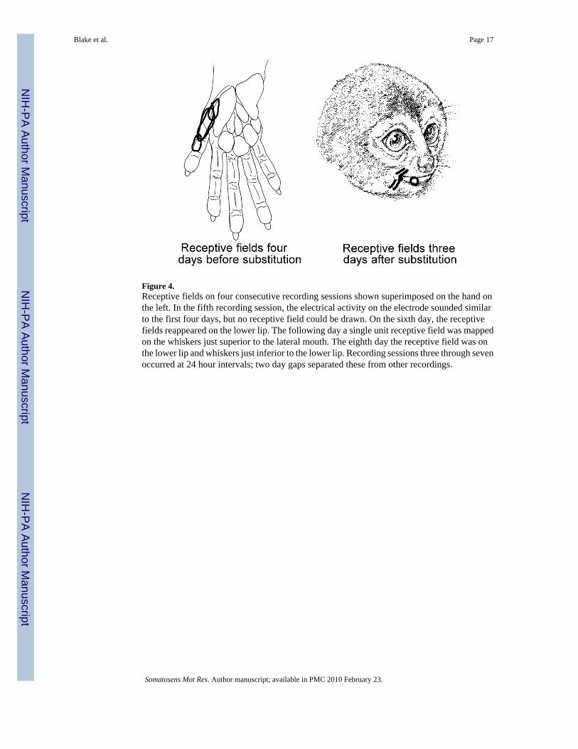
Figure 4.Receptive fields on four consecutive recording sessions shown superimposed on the hand onthe left. In the fifth recording session, the electrical activity on the electrode sounded similarto the first four days, but no receptive field could be drawn. On the sixth day, the receptivefields reappeared on the lower lip. The following day a single unit receptive field was mappedon the whiskers just superior to the lateral mouth. The eighth day the receptive field was onthe lower lip and whiskers just inferior to the lower lip. Recording sessions three through sevenoccurred at 24 hour intervals; two day gaps separated these from other recordings.
Blake et al. Page 17
Somatosens Mot Res. Author manuscript; available in PMC 2010 February 23.
NIH
-PA Author Manuscript
NIH
-PA Author Manuscript
NIH
-PA Author Manuscript

Figure 5.One representative plane of thalamic penetrations in omt592. Electrode penetrations werevertical in a 250 μm horizontal grid. Receptive field samples were derived each 125 μm.Different colors indicate each digit’s responses. Abbreviations: d1, digit one; d2, digit two;d1d, digit one, distal phalange; H, hairy skin response; G, glabrous skin response; w, whole.All responses were evoked by glabrous skin stimulation unless otherwise indicated.
Blake et al. Page 18
Somatosens Mot Res. Author manuscript; available in PMC 2010 February 23.
NIH
-PA Author Manuscript
NIH
-PA Author Manuscript
NIH
-PA Author Manuscript
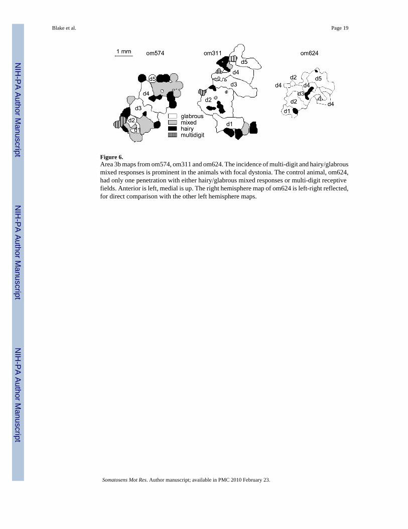
Figure 6.Area 3b maps from om574, om311 and om624. The incidence of multi-digit and hairy/glabrousmixed responses is prominent in the animals with focal dystonia. The control animal, om624,had only one penetration with either hairy/glabrous mixed responses or multi-digit receptivefields. Anterior is left, medial is up. The right hemisphere map of om624 is left-right reflected,for direct comparison with the other left hemisphere maps.
Blake et al. Page 19
Somatosens Mot Res. Author manuscript; available in PMC 2010 February 23.
NIH
-PA Author Manuscript
NIH
-PA Author Manuscript
NIH
-PA Author Manuscript
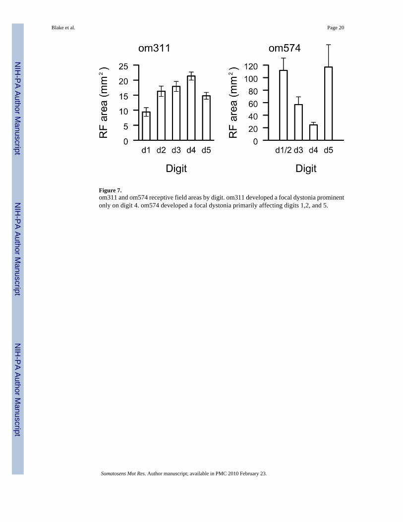
Figure 7.om311 and om574 receptive field areas by digit. om311 developed a focal dystonia prominentonly on digit 4. om574 developed a focal dystonia primarily affecting digits 1,2, and 5.
Blake et al. Page 20
Somatosens Mot Res. Author manuscript; available in PMC 2010 February 23.
NIH
-PA Author Manuscript
NIH
-PA Author Manuscript
NIH
-PA Author Manuscript

Figure 8.A collection of receptive fields for d1/2 and d4 for om574. After focal dystonia onset, theanimal principally used digits 3 and 4 in the task. Receptive field sizes parallel the spatialdistribution of dystonia as d4 receptive fields are smaller than d1/2 receptive fields. Note alsothat some d1/2 receptive fields are multidigit, or combine hairy and glabrous skin surfaces inthe same receptive field. Each color represents one receptive field.
Blake et al. Page 21
Somatosens Mot Res. Author manuscript; available in PMC 2010 February 23.
NIH
-PA Author Manuscript
NIH
-PA Author Manuscript
NIH
-PA Author Manuscript
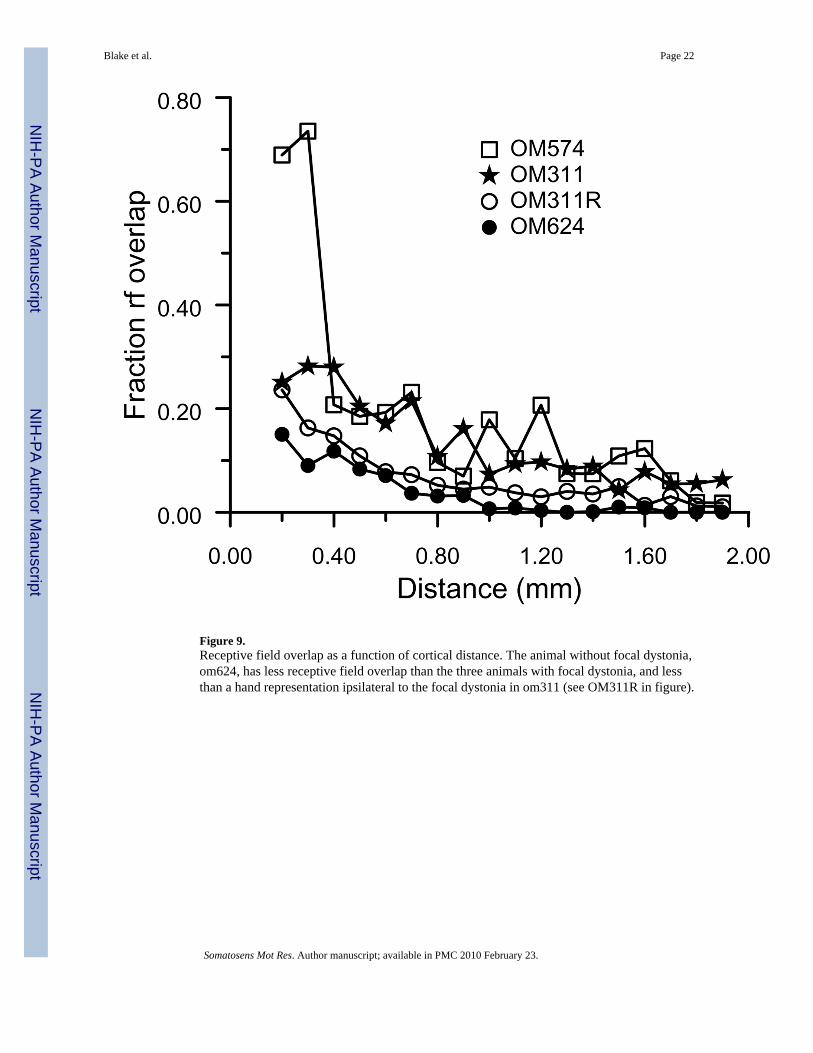
Figure 9.Receptive field overlap as a function of cortical distance. The animal without focal dystonia,om624, has less receptive field overlap than the three animals with focal dystonia, and lessthan a hand representation ipsilateral to the focal dystonia in om311 (see OM311R in figure).
Blake et al. Page 22
Somatosens Mot Res. Author manuscript; available in PMC 2010 February 23.
NIH
-PA Author Manuscript
NIH
-PA Author Manuscript
NIH
-PA Author Manuscript
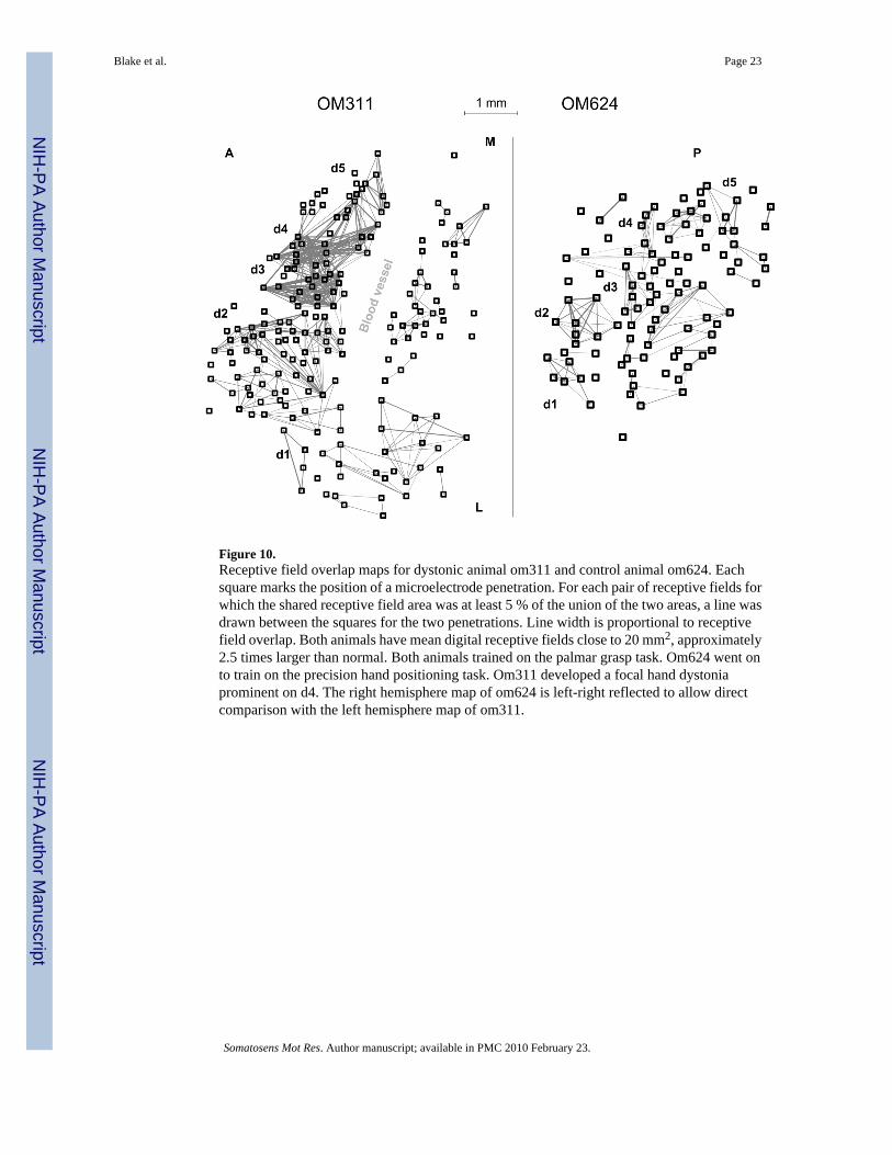
Figure 10.Receptive field overlap maps for dystonic animal om311 and control animal om624. Eachsquare marks the position of a microelectrode penetration. For each pair of receptive fields forwhich the shared receptive field area was at least 5 % of the union of the two areas, a line wasdrawn between the squares for the two penetrations. Line width is proportional to receptivefield overlap. Both animals have mean digital receptive fields close to 20 mm2, approximately2.5 times larger than normal. Both animals trained on the palmar grasp task. Om624 went onto train on the precision hand positioning task. Om311 developed a focal hand dystoniaprominent on d4. The right hemisphere map of om624 is left-right reflected to allow directcomparison with the left hemisphere map of om311.
Blake et al. Page 23
Somatosens Mot Res. Author manuscript; available in PMC 2010 February 23.
NIH
-PA Author Manuscript
NIH
-PA Author Manuscript
NIH
-PA Author Manuscript

NIH
-PA Author Manuscript
NIH
-PA Author Manuscript
NIH
-PA Author Manuscript
Blake et al. Page 24
Table 1
Behavioral data versus time for omt592
OMt592
Week since onset Trials/min Percent correct
3 19 21
4 14 23
5 14 22
6 6 13
7 9 16
Somatosens Mot Res. Author manuscript; available in PMC 2010 February 23.

NIH
-PA Author Manuscript
NIH
-PA Author Manuscript
NIH
-PA Author Manuscript
Blake et al. Page 25
Table 2
Behavioral data versus time for om311
OM311
Month of training Trials/min Percent correct
1 15 98
2 12 93
3 10 63
4 7 39.6
Mapped
Somatosens Mot Res. Author manuscript; available in PMC 2010 February 23.

NIH
-PA Author Manuscript
NIH
-PA Author Manuscript
NIH
-PA Author Manuscript
Blake et al. Page 26
Table 3
Behavioral data versus time for om574
OM574
Month of training Trials/min Percent correct
1 22 65
2 36 90
3 44 90
4 50 90
5 20 80
6 29 60
7 30 65
8 20 62
9 13 70
10 13 30
Mapped
Somatosens Mot Res. Author manuscript; available in PMC 2010 February 23.
Related Documents